Multi-Organ Involvement in COVID-19 - MDPI
19
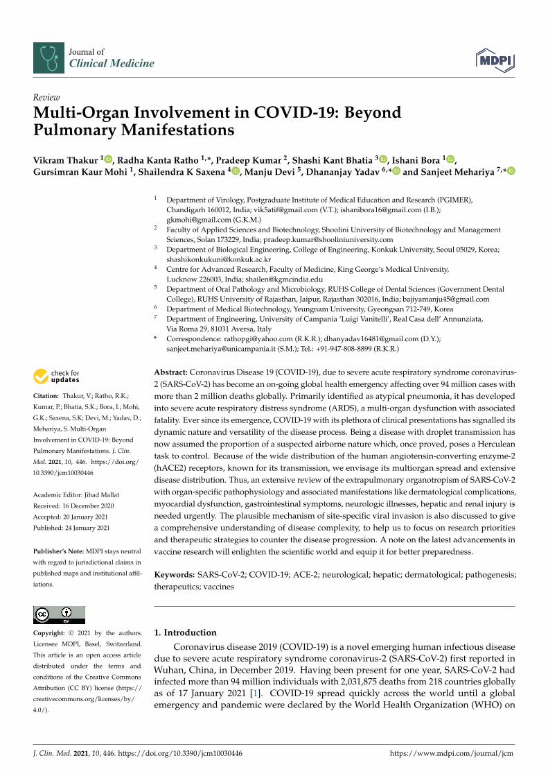
Journal of Clinical Medicine Review Multi-Organ Involvement in COVID-19: Beyond Pulmonary Manifestations Vikram Thakur 1 , Radha Kanta Ratho 1, *, Pradeep Kumar 2 , Shashi Kant Bhatia 3 , Ishani Bora 1 , Gursimran Kaur Mohi 1 , Shailendra K Saxena 4 , Manju Devi 5 , Dhananjay Yadav 6, * and Sanjeet Mehariya 7, * Citation: Thakur, V.; Ratho, R.K.; Kumar, P.; Bhatia, S.K.; Bora, I.; Mohi, G.K.; Saxena, S.K; Devi, M.; Yadav, D.; Mehariya, S. Multi-Organ Involvement in COVID-19: Beyond Pulmonary Manifestations. J. Clin. Med. 2021, 10, 446. https://doi.org/ 10.3390/jcm10030446 Academic Editor: Jihad Mallat Received: 16 December 2020 Accepted: 20 January 2021 Published: 24 January 2021 Publisher’s Note: MDPI stays neutral with regard to jurisdictional claims in published maps and institutional affil- iations. Copyright: © 2021 by the authors. Licensee MDPI, Basel, Switzerland. This article is an open access article distributed under the terms and conditions of the Creative Commons Attribution (CC BY) license (https:// creativecommons.org/licenses/by/ 4.0/). 1 Department of Virology, Postgraduate Institute of Medical Education and Research (PGIMER), Chandigarh 160012, India; [email protected] (V.T.); [email protected] (I.B.); [email protected] (G.K.M.) 2 Faculty of Applied Sciences and Biotechnology, Shoolini University of Biotechnology and Management Sciences, Solan 173229, India; [email protected] 3 Department of Biological Engineering, College of Engineering, Konkuk University, Seoul 05029, Korea; [email protected] 4 Centre for Advanced Research, Faculty of Medicine, King George’s Medical University, Lucknow 226003, India; [email protected] 5 Department of Oral Pathology and Microbiology, RUHS College of Dental Sciences (Government Dental College), RUHS University of Rajasthan, Jaipur, Rajasthan 302016, India; [email protected] 6 Department of Medical Biotechnology, Yeungnam University, Gyeongsan 712-749, Korea 7 Department of Engineering, University of Campania ‘Luigi Vanitelli’, Real Casa dell’ Annunziata, Via Roma 29, 81031 Aversa, Italy * Correspondence: [email protected] (R.K.R.); [email protected] (D.Y.); [email protected] (S.M.); Tel.: +91-947-808-8899 (R.K.R.) Abstract: Coronavirus Disease 19 (COVID-19), due to severe acute respiratory syndrome coronavirus- 2 (SARS-CoV-2) has become an on-going global health emergency affecting over 94 million cases with more than 2 million deaths globally. Primarily identified as atypical pneumonia, it has developed into severe acute respiratory distress syndrome (ARDS), a multi-organ dysfunction with associated fatality. Ever since its emergence, COVID-19 with its plethora of clinical presentations has signalled its dynamic nature and versatility of the disease process. Being a disease with droplet transmission has now assumed the proportion of a suspected airborne nature which, once proved, poses a Herculean task to control. Because of the wide distribution of the human angiotensin-converting enzyme-2 (hACE2) receptors, known for its transmission, we envisage its multiorgan spread and extensive disease distribution. Thus, an extensive review of the extrapulmonary organotropism of SARS-CoV-2 with organ-specific pathophysiology and associated manifestations like dermatological complications, myocardial dysfunction, gastrointestinal symptoms, neurologic illnesses, hepatic and renal injury is needed urgently. The plausible mechanism of site-specific viral invasion is also discussed to give a comprehensive understanding of disease complexity, to help us to focus on research priorities and therapeutic strategies to counter the disease progression. A note on the latest advancements in vaccine research will enlighten the scientific world and equip it for better preparedness. Keywords: SARS-CoV-2; COVID-19; ACE-2; neurological; hepatic; dermatological; pathogenesis; therapeutics; vaccines 1. Introduction Coronavirus disease 2019 (COVID-19) is a novel emerging human infectious disease due to severe acute respiratory syndrome coronavirus-2 (SARS-CoV-2) first reported in Wuhan, China, in December 2019. Having been present for one year, SARS-CoV-2 had infected more than 94 million individuals with 2,031,875 deaths from 218 countries globally as of 17 January 2021 [1]. COVID-19 spread quickly across the world until a global emergency and pandemic were declared by the World Health Organization (WHO) on J. Clin. Med. 2021, 10, 446. https://doi.org/10.3390/jcm10030446 https://www.mdpi.com/journal/jcm
-
Upload
khangminh22 -
Category
Documents
-
view
0 -
download
0
Transcript of Multi-Organ Involvement in COVID-19 - MDPI
Journal of
Clinical Medicine
Review
Multi-Organ Involvement in COVID-19: BeyondPulmonary Manifestations
Vikram Thakur 1 , Radha Kanta Ratho 1,*, Pradeep Kumar 2, Shashi Kant Bhatia 3 , Ishani Bora 1 ,Gursimran Kaur Mohi 1, Shailendra K Saxena 4 , Manju Devi 5, Dhananjay Yadav 6,* and Sanjeet Mehariya 7,*
�����������������
Citation: Thakur, V.; Ratho, R.K.;
Kumar, P.; Bhatia, S.K.; Bora, I.; Mohi,
G.K.; Saxena, S.K; Devi, M.; Yadav, D.;
Mehariya, S. Multi-Organ
Involvement in COVID-19: Beyond
Pulmonary Manifestations. J. Clin.
Med. 2021, 10, 446. https://doi.org/
10.3390/jcm10030446
Academic Editor: Jihad Mallat
Received: 16 December 2020
Accepted: 20 January 2021
Published: 24 January 2021
Publisher’s Note: MDPI stays neutral
with regard to jurisdictional claims in
published maps and institutional affil-
iations.
Copyright: © 2021 by the authors.
Licensee MDPI, Basel, Switzerland.
This article is an open access article
distributed under the terms and
conditions of the Creative Commons
Attribution (CC BY) license (https://
creativecommons.org/licenses/by/
4.0/).
1 Department of Virology, Postgraduate Institute of Medical Education and Research (PGIMER),Chandigarh 160012, India; [email protected] (V.T.); [email protected] (I.B.);[email protected] (G.K.M.)
2 Faculty of Applied Sciences and Biotechnology, Shoolini University of Biotechnology and ManagementSciences, Solan 173229, India; [email protected]
3 Department of Biological Engineering, College of Engineering, Konkuk University, Seoul 05029, Korea;[email protected]
4 Centre for Advanced Research, Faculty of Medicine, King George’s Medical University,Lucknow 226003, India; [email protected]
5 Department of Oral Pathology and Microbiology, RUHS College of Dental Sciences (Government DentalCollege), RUHS University of Rajasthan, Jaipur, Rajasthan 302016, India; [email protected]
6 Department of Medical Biotechnology, Yeungnam University, Gyeongsan 712-749, Korea7 Department of Engineering, University of Campania ‘Luigi Vanitelli’, Real Casa dell’ Annunziata,
Via Roma 29, 81031 Aversa, Italy* Correspondence: [email protected] (R.K.R.); [email protected] (D.Y.);
[email protected] (S.M.); Tel.: +91-947-808-8899 (R.K.R.)
Abstract: Coronavirus Disease 19 (COVID-19), due to severe acute respiratory syndrome coronavirus-2 (SARS-CoV-2) has become an on-going global health emergency affecting over 94 million cases withmore than 2 million deaths globally. Primarily identified as atypical pneumonia, it has developedinto severe acute respiratory distress syndrome (ARDS), a multi-organ dysfunction with associatedfatality. Ever since its emergence, COVID-19 with its plethora of clinical presentations has signalled itsdynamic nature and versatility of the disease process. Being a disease with droplet transmission hasnow assumed the proportion of a suspected airborne nature which, once proved, poses a Herculeantask to control. Because of the wide distribution of the human angiotensin-converting enzyme-2(hACE2) receptors, known for its transmission, we envisage its multiorgan spread and extensivedisease distribution. Thus, an extensive review of the extrapulmonary organotropism of SARS-CoV-2with organ-specific pathophysiology and associated manifestations like dermatological complications,myocardial dysfunction, gastrointestinal symptoms, neurologic illnesses, hepatic and renal injury isneeded urgently. The plausible mechanism of site-specific viral invasion is also discussed to givea comprehensive understanding of disease complexity, to help us to focus on research prioritiesand therapeutic strategies to counter the disease progression. A note on the latest advancements invaccine research will enlighten the scientific world and equip it for better preparedness.
Keywords: SARS-CoV-2; COVID-19; ACE-2; neurological; hepatic; dermatological; pathogenesis;therapeutics; vaccines
1. Introduction
Coronavirus disease 2019 (COVID-19) is a novel emerging human infectious diseasedue to severe acute respiratory syndrome coronavirus-2 (SARS-CoV-2) first reported inWuhan, China, in December 2019. Having been present for one year, SARS-CoV-2 hadinfected more than 94 million individuals with 2,031,875 deaths from 218 countries globallyas of 17 January 2021 [1]. COVID-19 spread quickly across the world until a globalemergency and pandemic were declared by the World Health Organization (WHO) on
J. Clin. Med. 2021, 10, 446. https://doi.org/10.3390/jcm10030446 https://www.mdpi.com/journal/jcm

J. Clin. Med. 2021, 10, 446 2 of 19
30 January and 11 March 2020, respectively [2]. Untiring efforts are being invested tounderstand the origin, transmission, and pathogenesis of COVID-19 so that effectivetherapeutic agents, as well as an effective vaccine, can be developed. The R (reproductivenumber) for SARS-CoV-2 is estimated between 1.5–3.5 in comparison to 2.0 of SARS 2002,however, the case fatality rate (CFR) is around 2–3% in SARS-CoV-2 in comparison to 10%for SARS 2002 [3,4].
Of the seven coronaviruses (CoVs), 229E, NL63, OC43, and HKU1 are known for self-limiting upper respiratory tract infections [5], whereas Middle East respiratory syndromecoronavirus (MERS-CoV), SARS-CoV, and the novel SARS-CoV-2 end up with life-threateningrespiratory failure and multi-organ dysfunction [6,7]. SARS-CoV-2 through spike (S) glyco-proteins recognizes and binds specifically to the human angiotensin-converting enzyme 2(hACE2) receptors expressed on type-II alveolar epithelial cells for its entry [8]. SARS-CoV-2has a stronger binding affinity with ACE2 along with cellular transmembrane serine protease 2(TMPRSS2) imparting virulence and aggressive properties. Following the SARS-CoV-2 bindingto alveolar epithelial cells, the innate and adaptive immune system is activated leading tocytokine-release syndrome (CRS) or macrophage activation syndrome (MAS). Increased produc-tion of interleukin (IL-1, IL-6, IL-8) cytokines in plasma resulting in dyspnea, acute respiratorydistress syndrome (ARDS), and death [9]. High levels of SARS-CoV-2 shedding in the upperrespiratory tract, even among presymptomatic patients, is a key factor in the transmissibilityof COVID-19.
2. Clinical Presentation and Transmission of Coronavirus Disease 2019 (COVID-19)
Because of the novel nature of the virus and lack of immunity, the presentations aredynamic and frequently changing. Being an unknown cause of atypical pneumonia, theinfection gradually progressed affecting the population in Hubai province, China andthereafter, spread to different countries, the presentations varied from mild respiratorytract infection to severe pneumonia and ARDS or multi-organ dysfunction with increasedmortality [10]. Other typical presentations like fever, cough, diarrhea, hemoptysis, rhin-orrhea, shortness of breath, myalgia, fatigue and severe dyspnoea, lymphopenia, chestradiographic findings like ground-glass opacity are observed in COVID-19 [11]; 20% ofCOVID-19 patients in an older age group with pre-existing morbidities present with severerespiratory illness and ARDS whereas children and young adults have a milder illness andbetter prognosis [12].
The asymptomatic cases contributed to a major source of the virus and resulted in ahigh rate of community transmission, and thus extensive screening was required [13]. Ithas been estimated that up to 86% of cases with unusual presentations might have beenmissed in China [14]. In the beginning, COVID-19 was suspected with unusual respiratorysymptoms, whereas over the progression of the pandemic involving different countriesthe extra-pulmonary symptoms like the neurological, cardiac, renal, gastrointestinal tract,ocular, vascular, olfactory including anosmia and ageusia were reported. The multior-gan manifestations may be correlated due to the abundancy of the ACE2 receptors invarious organs [15–18] and observed with symptoms including diarrhea, poor appetite,nausea, vomiting (digestive); headache, and confusion (nervous), palmus, chest distress(cardiovascular) [19]. SARS-CoV-2 is commonly transmitted from person-to-person mainlyby respiratory droplets and fomites through cough, sneeze, or by droplet inhalation andcontact transmission with oral, nasal and eye, mucous membrane, including saliva.
COVID-19 diagnosis is mainly made on radiological settings like X-ray, chest com-puted tomography (CT) scan, and laboratory findings like lymphopenia and elevatedLactate Dehydrogenase (LDH) [12]. Nasopharyngeal and oropharyngeal swabs help invirus identification through nucleic acid detection by real-time polymerase chain reaction(RT-PCR) which is the method of choice for lab diagnosis as isolation in cell lines requiresBSL-3/4 facilities [20]. Such labs are highly specialized to deal with potentially deadlyinfectious and exotic agents requiring the most stringent containment. The classical char-

J. Clin. Med. 2021, 10, 446 3 of 19
acteristics of BSL-4 is a full-body, air-supplied, positive pressure suit, class III biologicalsafety cabinet, and rooms with negative pressure facility.
3. Cutaneous Manifestations of Severe Acute Respiratory Syndrome Coronavirus-2(SARS-CoV-2)
Skin rashes and purpuric plaques are an interesting clinical presentation of classicalcoronavirus infections. The first report of the cutaneous manifestations was reported fromItaly, where 20.4% (18/88) hospitalized COVID-19 patients developed an erythematousrash (14), widespread urticaria (3), and chickenpox-like vesicles (1) distributed in the trunkarea [21]. In severe cases, erythematous rash, and localized or widespread urticarial rashesseem to be the most common cutaneous manifestation whereas in China only 0.2% (2/1099)confirmed COVID-19 cases had skin rashes [22].
Fernandez et al. [23] had reported a skin biopsy of a 32-year-old woman from Francewith COVID-19 having urticariform rash with perivascular infiltration of lymphocytes,eosinophils, and upper dermal edema on histopathology. Urticaria (1.4%) is also reported ascutaneous symptoms. A rare COVID-19 associated varicella-like papulovesicular exanthemwas first observed in Italian patients by Marzano et al. [24]. Lesions were varied fromscattered to diffuse with vesicular predominance in 12 (54.5%) patients with trunk andlimbs involvement generally appearing 3 days after systemic symptoms. Fever, cough,headache, weakness, coryza, dyspnea, hyposmia, and hypogeusia were common systemicsymptoms reported. However, SARS-CoV-2 detection in skin lesional was not performedbut still represents a useful clue to suspect COVID-19 in asymptomatic patients. A dengue-like petechial rash with thrombocytopenia was reported by Joob and Wiwanitkit [25] in aCOVID-19 patient from Thailand. Unusual skin manifestations like confluent erythematous-yellowish papules on both heels of a 28-year-old COVID-19 infected woman with symptomsof diarrhea, ageusia, and anosmia has been reported [26].
In a few COVID-19 patients, atopic dermatitis, and psoriasis has been aggravatedas pre-existing skin disease. Joob and Wiwanitkit [27] presented an 84-year-old womanwith arterial hypertension history having COVID-19 related bilateral pneumonia, laterdeveloped mild pruriginous rashes in the peri-axillary area and coalescing macules inflexural regions. Thus, various studies have reflected the possibilities of potential skinlesions of COVID-19.
4. Hepatic Manifestations of SARS-CoV-2
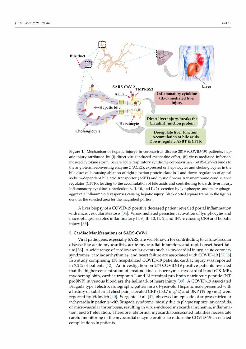
Though primarily a respiratory pathogen, shreds of evidence indicate the liver as anextra-pulmonary site for SARS-CoV-2 infection causing liver injury ranging between 14.8%to 78% [28,29]. The possible mechanism of hepatic injury in COVID-19 could either be virusrelated cytopathic effect or infection-induced cytokine storm. Two independent studies onhealthy cohorts by single RNA sequencing data demonstrated significant ACE2 expression(59.7%) in cholangiocytes. SARS-CoV-2 binds to the ACE2 expressed cholangiocytes andbeing facilitated its entry by TMPRSS2 [30]. Higher coexpression of ACE-2 and TMPRSS2in human trophoblast cell surface antigen 2 (TROP2high) cholangiocyte progenitor cellsof the liver has been reported [31]. The human liver ductal organoid model revealed thatcholangiocyte permissiveness for SARS-CoV-2 causes direct liver injury leading to theaccumulation of bile acids [32]. In 54% of COVID-19 patients, the ACE2 expression wasfound to be high in bile duct cells as evidenced by elevated gamma-glutamyl transferase(GGT) levels [33]. Ablation of tight junction protein claudin 1 and down-regulation ofapical sodium-dependent bile acid transporter (ASBT) and cystic fibrosis transmembraneconductance regulator (CFTR) might be the contributing factors towards liver injury inCOVID-19 [32] (Figure 1).
J. Clin. Med. 2021, 10, 446 4 of 19
J. Clin. Med. 2021, 10, x FOR PEER REVIEW 4 of 20
and macrophages secretes inflammatory IL-6, IL-10, IL-2, and IFN-c causing CRS and he-
patic injury [35].
Figure 1. Mechanism of hepatic injury: in coronavirus disease 2019 (COVID-19) patients, hepatic
injury attributed by (i) direct virus-induced cytopathic effect; (ii) virus-mediated infection-induced
cytokine storm. Severe acute respiratory syndrome coronavirus-2 (SARS-CoV-2) binds to the angi-
otensin-converting enzyme 2 (ACE2), expressed on hepatocytes and cholangiocytes in the bile
duct cells causing ablation of tight junction protein claudin 1 and down-regulation of apical so-
dium-dependent bile acid transporter (ASBT) and cystic fibrosis transmembrane conductance reg-
ulator (CFTR), leading to the accumulation of bile acids and contributing towards liver injury.
Inflammatory cytokines (interleukin-6, IL-10, and IL-2) secretion by lymphocytes and macro-
phages aggravate inflammatory responses causing hepatic injury. Black dotted square frame in the
figures denotes the selected area for the magnified portion.
5. Cardiac Manifestations of SARS-CoV-2
Viral pathogens, especially SARS, are well-known for contributing to cardiovascular
disease like acute myocarditis, acute myocardial infarction, and rapid-onset heart failure
[36]. A wide range of cardiovascular events such as myocardial injury, acute coronary
syndromes, cardiac arrhythmias, and heart failure are associated with COVID-19 [37,38].
In a study comprising 138 hospitalized COVID-19 patients, cardiac injury was reported in
7.2% of patients [12]. An investigation on 273 COVID-19 positive patients revealed that the
higher concentration of creatine kinase isoenzyme- myocardial band (CK-MB), myohemoglo-
bin, cardiac troponin I, and N-terminal pro-brain natriuretic peptide (NT-proBNP) in venous
blood are the hallmark of heart injury [39]. A COVID-19 associated Brugada type I electrocar-
diographic pattern in a 61-year-old Hispanic male presented with a history of substernal chest
pain, elevated CRP (150.7 mg/L) and BNP (19 pg/mL) were reported by Vidovich [40]. Sor-
gente et al. [41] observed an episode of supraventricular tachycardia in patients with Brugada
syndrome, mostly due to plaque rupture, myocarditis, or microvascular thrombosis, resulting
in virus-induced myocardial ischemia, inflammation, and ST elevation. Therefore, abnormal
myocardial-associated fatalities necessitate careful monitoring of the myocardial enzyme pro-
files to reduce the COVID-19-associated complications in patients.
Possible Mechanism of Cardiac Manifestations
TMPRSS2Liver
Cholangiocyte
Bile duct
Hepatic bile
ACE2
Hepatocyte
SARS-CoV-2
Direct liver injury, breaks the Claudin1 junction protein
Deregulate liver functionAccumulation of bile acids
Down-regulate ASBT & CFTR
Inflammatory cytokine (IL-6) mediated liver
injury
Figure 1. Mechanism of hepatic injury: in coronavirus disease 2019 (COVID-19) patients, hep-atic injury attributed by (i) direct virus-induced cytopathic effect; (ii) virus-mediated infection-induced cytokine storm. Severe acute respiratory syndrome coronavirus-2 (SARS-CoV-2) binds tothe angiotensin-converting enzyme 2 (ACE2), expressed on hepatocytes and cholangiocytes in thebile duct cells causing ablation of tight junction protein claudin 1 and down-regulation of apicalsodium-dependent bile acid transporter (ASBT) and cystic fibrosis transmembrane conductanceregulator (CFTR), leading to the accumulation of bile acids and contributing towards liver injury.Inflammatory cytokines (interleukin-6, IL-10, and IL-2) secretion by lymphocytes and macrophagesaggravate inflammatory responses causing hepatic injury. Black dotted square frame in the figuresdenotes the selected area for the magnified portion.
A liver biopsy of a COVID-19 positive deceased patient revealed portal inflammationwith microvesicular steatosis [34]. Virus-mediated persistent activation of lymphocytes andmacrophages secretes inflammatory IL-6, IL-10, IL-2, and IFN-c causing CRS and hepaticinjury [35].
5. Cardiac Manifestations of SARS-CoV-2
Viral pathogens, especially SARS, are well-known for contributing to cardiovasculardisease like acute myocarditis, acute myocardial infarction, and rapid-onset heart fail-ure [36]. A wide range of cardiovascular events such as myocardial injury, acute coronarysyndromes, cardiac arrhythmias, and heart failure are associated with COVID-19 [37,38].In a study comprising 138 hospitalized COVID-19 patients, cardiac injury was reportedin 7.2% of patients [12]. An investigation on 273 COVID-19 positive patients revealedthat the higher concentration of creatine kinase isoenzyme- myocardial band (CK-MB),myohemoglobin, cardiac troponin I, and N-terminal pro-brain natriuretic peptide (NT-proBNP) in venous blood are the hallmark of heart injury [39]. A COVID-19 associatedBrugada type I electrocardiographic pattern in a 61-year-old Hispanic male presented witha history of substernal chest pain, elevated CRP (150.7 mg/L) and BNP (19 pg/mL) werereported by Vidovich [40]. Sorgente et al. [41] observed an episode of supraventriculartachycardia in patients with Brugada syndrome, mostly due to plaque rupture, myocarditis,or microvascular thrombosis, resulting in virus-induced myocardial ischemia, inflamma-tion, and ST elevation. Therefore, abnormal myocardial-associated fatalities necessitatecareful monitoring of the myocardial enzyme profiles to reduce the COVID-19-associatedcomplications in patients.
J. Clin. Med. 2021, 10, 446 5 of 19
Possible Mechanism of Cardiac Manifestations
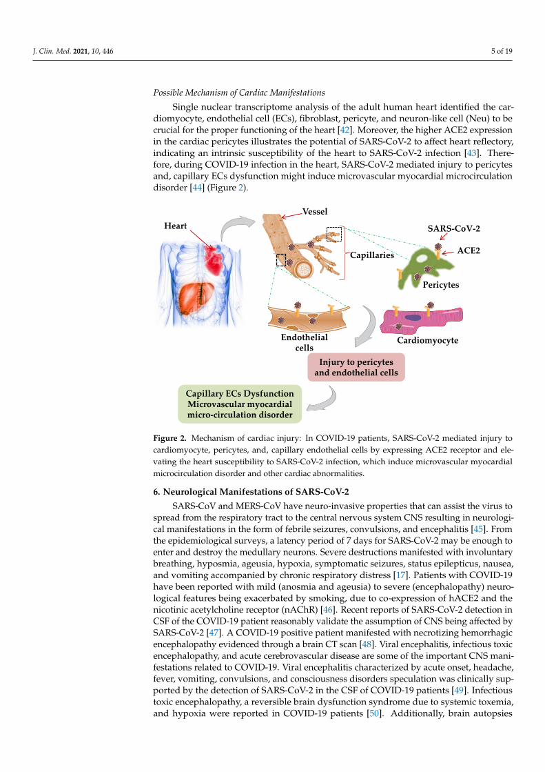
Single nuclear transcriptome analysis of the adult human heart identified the car-diomyocyte, endothelial cell (ECs), fibroblast, pericyte, and neuron-like cell (Neu) to becrucial for the proper functioning of the heart [42]. Moreover, the higher ACE2 expressionin the cardiac pericytes illustrates the potential of SARS-CoV-2 to affect heart reflectory,indicating an intrinsic susceptibility of the heart to SARS-CoV-2 infection [43]. There-fore, during COVID-19 infection in the heart, SARS-CoV-2 mediated injury to pericytesand, capillary ECs dysfunction might induce microvascular myocardial microcirculationdisorder [44] (Figure 2).
J. Clin. Med. 2021, 10, x FOR PEER REVIEW 5 of 20
Single nuclear transcriptome analysis of the adult human heart identified the cardi-
omyocyte, endothelial cell (ECs), fibroblast, pericyte, and neuron-like cell (Neu) to be cru-
cial for the proper functioning of the heart [42]. Moreover, the higher ACE2 expression in the
cardiac pericytes illustrates the potential of SARS-CoV-2 to affect heart reflectory, indicating
an intrinsic susceptibility of the heart to SARS-CoV-2 infection [43]. Therefore, during COVID-
19 infection in the heart, SARS-CoV-2 mediated injury to pericytes and, capillary ECs dysfunc-
tion might induce microvascular myocardial microcirculation disorder [44] (Figure 2).
Figure 2. Mechanism of cardiac injury: In COVID-19 patients, SARS-CoV-2 mediated injury to
cardiomyocyte, pericytes, and, capillary endothelial cells by expressing ACE2 receptor and elevat-
ing the heart susceptibility to SARS-CoV-2 infection, which induce microvascular myocardial mi-
crocirculation disorder and other cardiac abnormalities.
6. Neurological Manifestations of SARS-CoV-2
SARS-CoV and MERS-CoV have neuro-invasive properties that can assist the virus
to spread from the respiratory tract to the central nervous system CNS resulting in neu-
rological manifestations in the form of febrile seizures, convulsions, and encephalitis [45].
From the epidemiological surveys, a latency period of 7 days for SARS-CoV-2 may be
enough to enter and destroy the medullary neurons. Severe destructions manifested with
involuntary breathing, hyposmia, ageusia, hypoxia, symptomatic seizures, status epilep-
ticus, nausea, and vomiting accompanied by chronic respiratory distress [17]. Patients
with COVID-19 have been reported with mild (anosmia and ageusia) to severe (encepha-
lopathy) neurological features being exacerbated by smoking, due to co-expression of
hACE2 and the nicotinic acetylcholine receptor (nAChR) [46]. Recent reports of SARS-
CoV-2 detection in CSF of the COVID-19 patient reasonably validate the assumption of
CNS being affected by SARS-CoV-2 [47]. A COVID-19 positive patient manifested with
necrotizing hemorrhagic encephalopathy evidenced through a brain CT scan [48]. Viral
encephalitis, infectious toxic encephalopathy, and acute cerebrovascular disease are some
of the important CNS manifestations related to COVID-19. Viral encephalitis character-
ized by acute onset, headache, fever, vomiting, convulsions, and consciousness disorders
speculation was clinically supported by the detection of SARS-CoV-2 in the CSF of
COVID-19 patients [49]. Infectious toxic encephalopathy, a reversible brain dysfunction
syndrome due to systemic toxemia, and hypoxia were reported in COVID-19 patients [50].
Additionally, brain autopsies showed tissue edema and partial neuronal degeneration in
Vessel
Capillaries
Pericytes
Endothelial cells
ACE2
SARS-CoV-2
Cardiomyocyte
Injury to pericytesand endothelial cells
Capillary ECs DysfunctionMicrovascular myocardial micro-circulation disorder
Heart
Figure 2. Mechanism of cardiac injury: In COVID-19 patients, SARS-CoV-2 mediated injury tocardiomyocyte, pericytes, and, capillary endothelial cells by expressing ACE2 receptor and ele-vating the heart susceptibility to SARS-CoV-2 infection, which induce microvascular myocardialmicrocirculation disorder and other cardiac abnormalities.
6. Neurological Manifestations of SARS-CoV-2
SARS-CoV and MERS-CoV have neuro-invasive properties that can assist the virus tospread from the respiratory tract to the central nervous system CNS resulting in neurologi-cal manifestations in the form of febrile seizures, convulsions, and encephalitis [45]. Fromthe epidemiological surveys, a latency period of 7 days for SARS-CoV-2 may be enough toenter and destroy the medullary neurons. Severe destructions manifested with involuntarybreathing, hyposmia, ageusia, hypoxia, symptomatic seizures, status epilepticus, nausea,and vomiting accompanied by chronic respiratory distress [17]. Patients with COVID-19have been reported with mild (anosmia and ageusia) to severe (encephalopathy) neuro-logical features being exacerbated by smoking, due to co-expression of hACE2 and thenicotinic acetylcholine receptor (nAChR) [46]. Recent reports of SARS-CoV-2 detection inCSF of the COVID-19 patient reasonably validate the assumption of CNS being affected bySARS-CoV-2 [47]. A COVID-19 positive patient manifested with necrotizing hemorrhagicencephalopathy evidenced through a brain CT scan [48]. Viral encephalitis, infectious toxicencephalopathy, and acute cerebrovascular disease are some of the important CNS mani-festations related to COVID-19. Viral encephalitis characterized by acute onset, headache,fever, vomiting, convulsions, and consciousness disorders speculation was clinically sup-ported by the detection of SARS-CoV-2 in the CSF of COVID-19 patients [49]. Infectioustoxic encephalopathy, a reversible brain dysfunction syndrome due to systemic toxemia,and hypoxia were reported in COVID-19 patients [50]. Additionally, brain autopsies
J. Clin. Med. 2021, 10, 446 6 of 19
showed tissue edema and partial neuronal degeneration in deceased patients of COVID-19infection. The SARS-CoV-2 infection has been widely reported to cause CRS, leading toacute cerebrovascular disease. Also, elevated levels of D-dimer and reduced platelet countin critically ill SARS-CoV-2 patient predispose to acute cerebrovascular events. Recent caseseries from China and the US describe ischaemic or hemorrhagic stroke, Guillain-Barrésyndrome (GBS), or acute necrotizing encephalopathy (ANE), as neurological symptomsamong COVID-19 patients [51,52] (Table 1).
Table 1. COVID-19 cases with neurological signs and manifestations.
Case/Study Symptoms (Neurological)
79-year-old man(Positive-Oropharyngeal swab) [53]
Fever, acute loss of consciousness, and bilateralextensor plantar reflexes.Intracerebral hemorrhage in the right brainhemisphereIntraventricular and subarachnoidhemorrhage.
The first case of SARS-CoV-2 associatedmeningitis.Absence of SARS-CoV-2 RNA innasopharyngeal swab.Positive in CSF [54]
Generalized fatigue and fever, transientgeneralized seizures, and neck stiffness.Convulsion accompanied by unconsciousness.Hyperintense signal in the right mesialtemporal lobe in the brain along withsignificant paranasal sinusitis.
SARS-CoV-2 positive male with encephalitis,however, CSF is negative [55]
Fever, shortness of breath, lymphopenia,multiple subpleural ground-glass opacities inthe chest, and myalgia. Progression ofconsciousness towards the confusion withsigns of meningeal irritation.
Mechanisms of Neurotropism and Neuroinvasion
Brain infiltration through the olfactory nerves following intranasal administrationcould be a possibility as with SARS-CoV and MERS-CoV in the transgenic mice model.There are numerous predicted pathways for CNS invasion by SARS-CoV-2 to cause neu-ronal damage (Figure 3).
In the hematogenous route, SARS-CoV-2 binds to the ACE2 expressing capillaryendothelium and enters the CNS by a breach in the blood–brain barrier (BBB) resulting inhigh blood pressure with the risk of cerebral hemorrhage. Through dynein and kinesinmotor proteins, SARS-CoV-2 infects sensory or motor nerve endings by retrograde oranterograde neuronal transport as a neuronal pathway [56,57]. In the olfactory neurontransport, SARS-CoV-2 can enter the CNS/brain through the olfactory tract by olfactorynerves in the nasal cavity/epithelium and the olfactory bulb in the forebrain, causinginflammation and demyelinating reactions. Moreover, diffusion of alveolar and interstitialinflammatory exudation, results in anabolic metabolism in brain cells, leading to hypoxiaand ischemic stroke. Circumventricular organs and cerebrovascular endothelial cellsexpressing ACE2 receptors regulate multiple neurological functions including regulationof hormone formation, the sympathoadrenal system, vascular autoregulation, and cerebralblood flow [58,59]. COVID-19 results in a large number of fatalities, mostly due to multipleorgan failure induced systemic inflammatory response syndrome (SIRS). The persistenceand ability of SARS-CoV-2 to infect and activate macrophages, microglia, astrocytes, andglial cells in the CNS induces a pro-inflammatory state by secretion of IL-6, IL-12, IL-15,and tumor necrosis factor-α (TNF-α) [60]. The IL-6 mediated cytokine storm, results inacute necrotizing encephalopathy (ANE) causing neuroinflammation in addition to a surgein interleukin IL-2, IL-7, interferon-γ, monocyte chemoattractant protein 1, and TNF-αleading to hyper inflammation, encephalopathy, and even stroke [61].

J. Clin. Med. 2021, 10, 446 7 of 19
J. Clin. Med. 2021, 10, x FOR PEER REVIEW 7 of 20
Table 1. COVID-19 cases with neurological signs and manifestations.
Case/Study Symptoms (Neurological)
79-year-old man
(Positive-Oropharyngeal swab) [53]
Fever, acute loss of consciousness, and bilateral extensor plan-
tar reflexes.
Intracerebral hemorrhage in the right brain hemisphere
Intraventricular and subarachnoid hemorrhage.
The first case of SARS-CoV-2 associated meningitis.
Absence of SARS-CoV-2 RNA in nasopharyngeal swab.
Positive in CSF [54]
Generalized fatigue and fever, transient generalized seizures,
and neck stiffness.
Convulsion accompanied by unconsciousness. Hyperintense
signal in the right mesial temporal lobe in the brain along
with significant paranasal sinusitis.
SARS-CoV-2 positive male with encephalitis, however,
CSF is negative [55]
Fever, shortness of breath, lymphopenia, multiple subpleural
ground-glass opacities in the chest, and myalgia. Progression
of consciousness towards the confusion with signs of menin-
geal irritation.
Mechanisms of Neurotropism and Neuroinvasion
Brain infiltration through the olfactory nerves following intranasal administration
could be a possibility as with SARS-CoV and MERS-CoV in the transgenic mice model.
There are numerous predicted pathways for CNS invasion by SARS-CoV-2 to cause neu-
ronal damage (Figure 3).
Olfactory bulb
Blood stream
Vagus nerve
Direct pathways:1. Through olfactory nerve2. Blood circulation 3. Neuronal pathway
SARS-CoV-2infected CSF
SARS-CoV-2Infected nerve cells
Hippocampus
Medullaoblongata
Neurons
OligodendrocyteSARS-CoV-2
1
2
3
SARS-CoV-2
Lungs
Brain
• Neuroinflammation and demyelination• Acute cerebrovascular disease• Acute necrotizing encephlopathy• Necrotizing hemorrhagic encephalopathy
Figure 3. Predictable model for SARS-CoV-2 induced neurological manifestations: Numerous pathways predicted for centralnervous system CNS invasion by SARS-CoV-2 to cause neuronal damage. In the olfactory neuron transport, SARS-CoV-2can infiltrate the CNS/brain through the olfactory tract by olfactory nerves in the nasal cavity/epithelium and the olfactorybulb in the forebrain, causing inflammation and demyelinating reactions. In the hematogenous route, SARS-CoV-2 binds tothe receptor ACE2 expressed in the capillary endothelium and enters the CNS by a breach in the blood–brain barrier (BBB)resulting in high blood pressure with the risk of a cerebral hemorrhage. Infected and activated macrophages, microglia,astrocytes, and glial cells in the CNS induce a pro-inflammatory state by secretion of IL-6, IL-12, and IL-15 resulting in acutenecrotizing encephalopathy (ANE), hyper inflammation, and encephalopathy.
7. Renal Manifestations
Renal injury is the commonly reported COVID-19 associated renal manifestationsreflecting the renal tropism of SARS-CoV-2 [62]. The burden of acute kidney injury (AKI)with COVID-19 infection was relatively low, ranging from 3–9% to as high as 15% [63,64].Further evidence supported the renal tropism of SARS-CoV-2 by the isolation of viral RNAfrom urine and albuminuria and hematuria in COVID-19 infection [65,66]. Puelles et al. [67]quantified the SARS-CoV-2 viral load in autopsy tissue samples obtained from 22 COVID-19 positive deceased patients; 17 (77%) had more than two coexisting conditions, whichwas associated with SARS-CoV-2 tropisms for the kidneys, even in patients without ahistory of chronic kidney disease. Three out of six patients on autopsy had a detectableSARS-CoV-2 viral load preferentially in glomerular cells as shown in Table 2.
J. Clin. Med. 2021, 10, 446 8 of 19
Table 2. COVID-19 positive cases showing renal manifestations.
Case/Study Symptoms (Renal)
85 COVID-19 positive patientsAcute kidney injury (AKI) in 27% ofpatients [68]
High risk of AKI associated with age >60 years,coexisting hypertension, and coronaryartery disease.Severe acute tubular injury with macrophageinfiltration, and detection of viral antigen (6)
701 hospitalized patients [63] Proteinuria (44%), Haematuria (27%), andAcute kidney injury (3.2%)
201 maintenance hemodialysis (MHD) patients.Five had COVID-19 pneumonia [69]
Diarrhea as common presenting symptoms(4/5), fever (3/5), fatigue (3/5), dyspnoea(2/5), and abdominal pain (2/5), lymphopenia(5/5), with ground-glass opacity.Fever, cough, and dyspnoea were absent.
230 haemodialysis patients; 16.1% (37)diagnosed with COVID-19 [70]
Lymphopenia, lower levels of inflammatorycytokines, and mild clinical disease
The Potential Mechanisms of Renal Manifestations
In silico analysis of single-cell RNA sequencing revealed the enriched RNA expressionof ACE2, TMPRSS2, and cathepsin L (CTSL) in podocytes and tubule epithelial cells whichmight facilitate the SARS-CoV-2 associated kidney injury [71,72]. ACE2 is expressed onthe renal epithelial and bladder cells which counter the activation of the renin-angiotensin-aldosterone system (RAAS) [73]. SARS-CoV-2 can bind and injure the renal epithelial cells,thereby disrupting the body fluid and electrolyte homeostasis in addition to the erythropoi-etin and vitamin D production. Organ crosstalk (lung-kidney and heart-kidney), cytokinedamage by IL-6, and systemic effects could be the underlying mechanisms for renal injury.During lung–kidney crosstalk, tubular epithelium enhances the IL-6 upregulation in serumresulting in increased alveolar-capillary permeability and pulmonary hemorrhage [74].
8. Gastrointestinal (GIT) Manifestations
Gastrointestinal (GIT) manifestations are revealed in 10.6% of patients with SARS and30% of patients with MERS had diarrhea as the clinical symptoms [75]. The first case ofSARS-CoV-2 infection with nausea, vomiting, and abdominal discomfort was reportedfrom the U.S. where viral RNA was detected from the stool and respiratory specimen of aCOVID-19 patient on day 7 of illness [76]. Clinically diarrhea was reported in 1–3.8% cases,diarrhea, and nausea in 10.1%, and vomiting in 3.6% [12].
Lin et al. [77] observed diarrhea and abdominal pain as evidenced in 20–50% ofCOVID-19 patients, which sometimes preceded respiratory symptoms. A 25-year-oldfemale with respiratory symptoms was negative for SARS-CoV-2 in the pharyngeal aspiratebut positive in the fecal sample. This might indicate feces as a source of virus transmissionand the GIT region as an extrapulmonary site for virus replication [78]. Thus, earlymonitoring of viral RNA in the fecal specimens might benefit the disease prediction evenbefore respiratory symptoms (Table 3).

J. Clin. Med. 2021, 10, 446 9 of 19
Table 3. Case studies showing gastrointestinal (GIT) manifestations in COVID-19 patients.
Case/Study Symptoms (GIT/others)
26/84 COVID-19 patients with diarrhea [79] Headache, myalgia, fatigue, cough, sputumproduction, nausea, vomiting
204 COVID-19 patientsGIT symptoms in 99 (48.5%)Cross-sectional study, China [80]
Anorexia (83.8%)Diarrhea (29.3%)
651 COVID-19 patients11.4% (74) patients with GI symptoms28% lack respiratory symptoms [81]
Diarrhea, nausea, and vomiting
95 COVID-19 patients61.1% cases with GI symptoms52.4% (22) positive faecal samples [77]
Diarrhoea (24.2%),Nausea (17.9%),Elevated transaminases (32.6%)
1141 COVID-19 patientsGIT symptoms in 16% of patientsA retrospective study, China [82]
Anorexia, nausea, vomiting, diarrhea,abdominal pain
The presence of SARS-CoV-2 in stool samples even after 11 days of viral clearancefrom respiratory tract samples in over half of patients indicates the alternative route ofexcretion of the virus [83]. Similarly, Xu et al. [84] reported 8 of the 10 infected childrenhaving persistent positive SARS-CoV-2 in rectal swabs, where nasopharyngeal swabs werenegative for the virus. In a multicentric study with 1992 patients, 34% of them experienceddiarrhea whereas 53% experienced one of the GIT symptoms. However 74% of the caseswere mild and not associated with severe clinical course [85]. Despite the ability of SARS-CoV-2 to establish a robust infection in GIT; it might be inactivated by human colonicfluids in the intestinal lumen, hence the viral RNA transiting through GIT and sheddingthrough the feces may not be infectious [86]. However, live SARS-CoV-2 was detectedusing electron microscopy in stool samples from two patients, which might focus on thepotentiality of fecal transmission [87]. Even though there is evidence of GI symptoms dueto SARS-CoV-2, its role in the disease process is yet to be demonstrated.
Possible Mechanism for GIT Manifestations
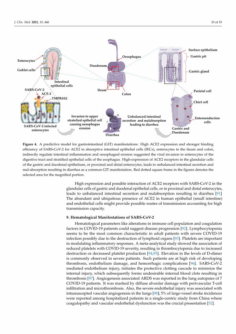
The causative mechanism for GIT manifestation in COVID-19 positive patients isnot well studied. Single-cell transcriptomic analysis of GIT (stomach, colon, ileum, andesophagus) indicated high ACE2 expression in absorptive intestinal epithelial cells (IECs)in the ileum and colon [88]. The ACE2 receptor indirectly regulates intestinal inflammationwhereas TMPRSS2 is crucial for viral fusion [89]. Various hypothetical models werepredicted: Higher co-expression of ACE2 and TMPRSS2 and stronger binding efficiency ofSARS-CoV-2 for ACE2, located on the mature enterocytes in the ileum and colon, triggersepithelial cell fusion by TMPRSS2 and TMPRSS4 (inducing S cleavage and enhancing thefusogenic activity of the virus) suggesting the viral invasion of enterocytes of the digestivetract and stratified epithelial cells of the esophagus, resulting in oesophageal erosion [90](Figure 4).
J. Clin. Med. 2021, 10, 446 10 of 19
J. Clin. Med. 2021, 10, x FOR PEER REVIEW 10 of 20
A retrospective study, China [82]
The presence of SARS-CoV-2 in stool samples even after 11 days of viral clearance
from respiratory tract samples in over half of patients indicates the alternative route of
excretion of the virus [83]. Similarly, Xu et al. [84] reported 8 of the 10 infected children
having persistent positive SARS-CoV-2 in rectal swabs, where nasopharyngeal swabs
were negative for the virus. In a multicentric study with 1992 patients, 34% of them expe-
rienced diarrhea whereas 53% experienced one of the GIT symptoms. However 74% of
the cases were mild and not associated with severe clinical course [85]. Despite the ability
of SARS-CoV-2 to establish a robust infection in GIT; it might be inactivated by human
colonic fluids in the intestinal lumen, hence the viral RNA transiting through GIT and
shedding through the feces may not be infectious [86]. However, live SARS-CoV-2 was
detected using electron microscopy in stool samples from two patients, which might focus
on the potentiality of fecal transmission [87]. Even though there is evidence of GI symp-
toms due to SARS-CoV-2, its role in the disease process is yet to be demonstrated.
Possible Mechanism for GIT Manifestations
The causative mechanism for GIT manifestation in COVID-19 positive patients is not
well studied. Single-cell transcriptomic analysis of GIT (stomach, colon, ileum, and esoph-
agus) indicated high ACE2 expression in absorptive intestinal epithelial cells (IECs) in the
ileum and colon [88]. The ACE2 receptor indirectly regulates intestinal inflammation
whereas TMPRSS2 is crucial for viral fusion [89]. Various hypothetical models were pre-
dicted: Higher co-expression of ACE2 and TMPRSS2 and stronger binding efficiency of
SARS-CoV-2 for ACE2, located on the mature enterocytes in the ileum and colon, triggers
epithelial cell fusion by TMPRSS2 and TMPRSS4 (inducing S cleavage and enhancing the
fusogenic activity of the virus) suggesting the viral invasion of enterocytes of the digestive
tract and stratified epithelial cells of the esophagus, resulting in oesophageal erosion [90]
(Figure 4).
Oesophagus
Duodenum
IleumColon
Intestinal epithelial cells
Goblet cells
Enterocytes
SARS-CoV-2ACE-2
TMPRSS2
Surface epithelium
Gastric pit
Gastric gland
SARS-CoV-2 infected enterocytes
Parietal cell
Chief cell
Enteroendocrinecells
Gastric and Duodenum
Invasion to upper stratefied epthelial cell
causing oesophagus erosion
Unbalanced intestinal secretion and malabsorption
leading to diarrhea
Diarrhea
Figure 4. A predictive model for gastrointestinal (GIT) manifestations: High ACE2 expression and stronger bindingefficiency of SARS-CoV-2 for ACE2 in absorptive intestinal epithelial cells (IECs), enterocytes in the ileum and colon,indirectly regulate intestinal inflammation and oesophageal erosion suggested the viral invasion to enterocytes of thedigestive tract and stratified epithelial cells of the esophagus. High-expression of ACE2 receptors in the glandular cellsof the gastric and duodenal epithelium, or proximal and distal enterocytes, leads to unbalanced intestinal secretion andmal-absorption resulting in diarrhea as a common GIT manifestation. Red dotted square frame in the figures denotes theselected area for the magnified portion.
High expression and possible interaction of ACE2 receptors with SARS-CoV-2 in theglandular cells of gastric and duodenal epithelial cells, or in proximal and distal enterocytes,leads to unbalanced intestinal secretion and malabsorption resulting in diarrhea [91]The abundant and ubiquitous presence of ACE2 in human epithelial (small intestine)and endothelial cells might provide possible routes of transmission accounting for hightransmission capacity.
9. Hematological Manifestations of SARS-CoV-2
Hematological parameters like alterations in immune cell population and coagulationfactors in COVID-19 patients could suggest disease progression [92]. Lymphocytopeniaseems to be the most common characteristic in adult patients with severe COVID-19infection possibly due to the destruction of lymphoid organs [93]. Platelets are importantin modulating inflammatory responses. A meta-analytical study showed the association ofreduced platelets with COVID-19 severity, resulting in thrombocytopenia due to increaseddestruction or decreased platelet production [94,95]. Elevation in the levels of D-dimeris commonly observed in severe patients. Such patients are at high risk of developingthrombosis, endothelium damage, and hemorrhagic complications [96]. SARS-CoV-2mediated endothelium injury, initiates the protective clotting cascade to minimize theinternal injury, which subsequently forms undesirable internal blood clots resulting inthrombosis [97]. Angiogenesis associated ARDS was reported in the lung autopsies of 7COVID-19 patients. It was marked by diffuse alveolar damage with perivascular T-cellinfiltration and microthrombosis. Also, the severe endothelial injury was associated withintussuscepted vascular angiogenesis in the lungs [98]; 5% of large-vessel stroke incidenceswere reported among hospitalized patients in a single-centric study from China wherecoagulopathy and vascular endothelial dysfunction was the crucial presentation [52].
J. Clin. Med. 2021, 10, 446 11 of 19
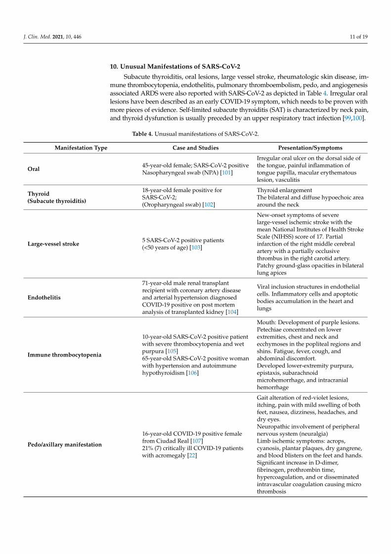
10. Unusual Manifestations of SARS-CoV-2
Subacute thyroiditis, oral lesions, large vessel stroke, rheumatologic skin disease, im-mune thrombocytopenia, endothelitis, pulmonary thromboembolism, pedo, and angiogenesisassociated ARDS were also reported with SARS-CoV-2 as depicted in Table 4. Irregular orallesions have been described as an early COVID-19 symptom, which needs to be proven withmore pieces of evidence. Self-limited subacute thyroiditis (SAT) is characterized by neck pain,and thyroid dysfunction is usually preceded by an upper respiratory tract infection [99,100].
Table 4. Unusual manifestations of SARS-CoV-2.
Manifestation Type Case and Studies Presentation/Symptoms
Oral 45-year-old female; SARS-CoV-2 positiveNasopharyngeal swab (NPA) [101]
Irregular oral ulcer on the dorsal side ofthe tongue, painful inflammation oftongue papilla, macular erythematouslesion, vasculitis
Thyroid(Subacute thyroiditis)
18-year-old female positive forSARS-CoV-2;(Oropharyngeal swab) [102]
Thyroid enlargementThe bilateral and diffuse hypoechoic areaaround the neck
Large-vessel stroke 5 SARS-CoV-2 positive patients(<50 years of age) [103]
New-onset symptoms of severelarge-vessel ischemic stroke with themean National Institutes of Health StrokeScale (NIHSS) score of 17. Partialinfarction of the right middle cerebralartery with a partially occlusivethrombus in the right carotid artery.Patchy ground-glass opacities in bilaterallung apices
Endothelitis
71-year-old male renal transplantrecipient with coronary artery diseaseand arterial hypertension diagnosedCOVID-19 positive on post mortemanalysis of transplanted kidney [104]
Viral inclusion structures in endothelialcells. Inflammatory cells and apoptoticbodies accumulation in the heart andlungs
Immune thrombocytopenia
10-year-old SARS-CoV-2 positive patientwith severe thrombocytopenia and wetpurpura [105]65-year-old SARS-CoV-2 positive womanwith hypertension and autoimmunehypothyroidism [106]
Mouth: Development of purple lesions.Petechiae concentrated on lowerextremities, chest and neck andecchymoses in the popliteal regions andshins. Fatigue, fever, cough, andabdominal discomfort.Developed lower-extremity purpura,epistaxis, subarachnoidmicrohemorrhage, and intracranialhemorrhage
Pedo/axillary manifestation
16-year-old COVID-19 positive femalefrom Ciudad Real [107]21% (7) critically ill COVID-19 patientswith acromegaly [22]
Gait alteration of red-violet lesions,itching, pain with mild swelling of bothfeet, nausea, dizziness, headaches, anddry eyes.Neuropathic involvement of peripheralnervous system (neuralgia)Limb ischemic symptoms: acrops,cyanosis, plantar plaques, dry gangrene,and blood blisters on the feet and hands.Significant increase in D-dimer,fibrinogen, prothrombin time,hypercoagulation, and or disseminatedintravascular coagulation causing microthrombosis

J. Clin. Med. 2021, 10, 446 12 of 19
Previous studies showed cross-reaction of SARS-CoV-2 antigen and antibodies inpatients with rheumatoid arthritis, systemic sclerosis, and systemic lupus erythemato-sus (SLE) [108] so there is the possibility of viral arthritis and musculoskeletal pain inCOVID-19 patients, possibly due to posttranslational modification of peptides, or molec-ular mimicry activating T cells or epitope spreading due to T-cell associated damage bythe virus leading to autoreactive T cells. SARS-CoV-2 through the ACE2 receptor directlyinfects the endothelial cell and facilitates the induction of endothelitis in several organs.Diffuse endothelial inflammation by host inflammatory response results in the recruitmentof immune cells causing widespread endothelial dysfunction associated with apoptosisand pyroptosis. This results in shifting of vascular endothelium equilibrium towardsvasoconstriction (microvascular dysfunction), resulting in inflammation with associatedtissue edema, and a procoagulant state explaining the systemic impaired microcirculatoryfunction in different vascular beds [109].
SARS-CoV-2 also directly attacks human epithelial cells of alveoli, large and smallarteries, small intestine, and vascular endothelial cells. Counteracting the innate immunesystem activates and induces cytokine storms (IL-6) damaging the microvascular system. Itactivates the coagulation system while inhibiting fibrinolysis and anticoagulation systemsthat stimulate the liver to synthesize more thrombopoietin, fibrinogen leading to extensivethrombosis in microvessels [110]. Antiphospholipid antibodies result in endothelial injury,platelet activation, and thrombosis, with hypercoagulation. COVID-19 patients with highD-dimer levels and hypercoagulable state were associated with sudden onset of oxygendeterioration, respiratory distress, and reduced blood pressure resulting in pulmonarythromboembolism (PTE) [111].
11. COVID-19 in Immunocompromised Solid-Organ Transplant Recipients
Solid-organ transplant (SOT) recipients are high-risk individuals, usually on im-munosuppressive therapy. Interestingly, in 2003 SARS-CoV in 2003 and 2012 MERS-CoVpandemic, SOT recipients did not appear to be associated with adverse outcomes. Therole of different immunosuppressive agents such as calcineurin inhibitors and intravenousimmunoglobulin (IVIG) in COVID-19 disease has not been established due to limiteddata on COVID-19 in transplant recipients. The typical presentation of COVID-19 in SOTrecipients is the classic triad of fever, fatigue, and dry cough. A 75-year-old male anda 52-year-old female at 120 and 8 months post-transplant, respectively were diagnosedwith stable graft function in males and AKI in females respectively. Extensive bilateralground-glass opacities were the common lung abnormality reported in both cases [112].
A case of a 50-year-old COVID-19 positive man with 3rd kidney transplant recipientwith IgA nephropathy induced end-stage renal disease manifested with the gastrointestinalviral disease (3–5%) and fever, further progressing to respiratory symptoms in 48 h [113]. A39-year-old diabetic dual-organ (heart/kidney) transplant recipient positive for COVID-19had a mild clinical course with minimal supportive care with no evidence of any graftrejection despite being on three immunosuppressive agents. The patient had additionalrisk factors of hypertension, diabetes mellitus, and morbid obesity, lymphopenia, elevatedCRP, IL-6, D-dimer, and troponin I levels [114]. Li et al. [115] reported two heart transplantrecipients with COVID-19 from Wuhan, were successfully treated, and survived.
12. Co-Infections with COVID-19: Viral, Bacterial, and Fungal
Diagnosing co-infections is complex owing to the clinical conditions of the infectedpatients [116]. During the 2003 SARS-CoV outbreak, invasive pulmonary aspergillosiswas reported in only 4 among the 8422 probable SARS cases [117]. The risk of develop-ing invasive pulmonary aspergillosis in COVID-19 patients is high as described in Francewhere 9 out of 27 (33%) COVID-19 patients with invasive pulmonary aspergillosis admittedto an intensive care unit (ICU) [118] and 5 in Germany (26% of 19 admitted) proved inhistopathology of autopsy [119]. Zhou et al. [120] showed that 50% of patients with COVID-19 died due to secondary bacterial infections. Patients with chronic obstructive pulmonary
J. Clin. Med. 2021, 10, 446 13 of 19
disease (COPD) will have underlying chronic bacterial infections before SARS-CoV-2 in-fection. Wang et al. [121] reported a case of a 37-year-old man, from Wuhan infected bySARS-CoV-2 and human immunodeficiency virus (HIV) simultaneously, highlighting theco-infection might damage T lymphocytes, impairing the immune system, B-cell dysfunc-tion resulting in abnormal polyclonal activation and prolongation of the disease process(2 months). Chest CT showing multiple infiltrations in both lungs while nasopharyngealswab positivity confirmed the SARS-CoV-2 infection, accompanied by dyspnea, chest pain,and palpitation. The significant decrease in the total number of immune cells i.e., B cells,T cells, and NK cells were also correlated with COVID-19 severity.
13. Advancements in Vaccine Research
As per the draft landscape of COVID-19 [122] dated 15 January 2021; 63 candidatevaccines are in line with clinical evaluations with 13 vaccines currently at phase 3 trial. TwomRNA vaccines i.e., 3LNP-mRNA by Pfizer-BioNTech and RNA LNP encapsulated mRNAvaccine jointly by Moderna and National Institute of Health (NIH) demonstrated an efficacyof 95% and were recently approved for the emergency use under the Emergency UseAuthorization (EUA) by the US Food and Drug Administration (FDA). Another promisingvaccine i.e., ChAdOx1 (chimpanzee adenovirus vaccine vector) with comparable efficacyhas been developed by AstraZeneca with Oxford University, which is a non-replicatingversion of adenovirus containing the genetic sequence of surface spike protein which isproduced after vaccination and prime the immune system to attack against the SARS-CoV-2viral infection.
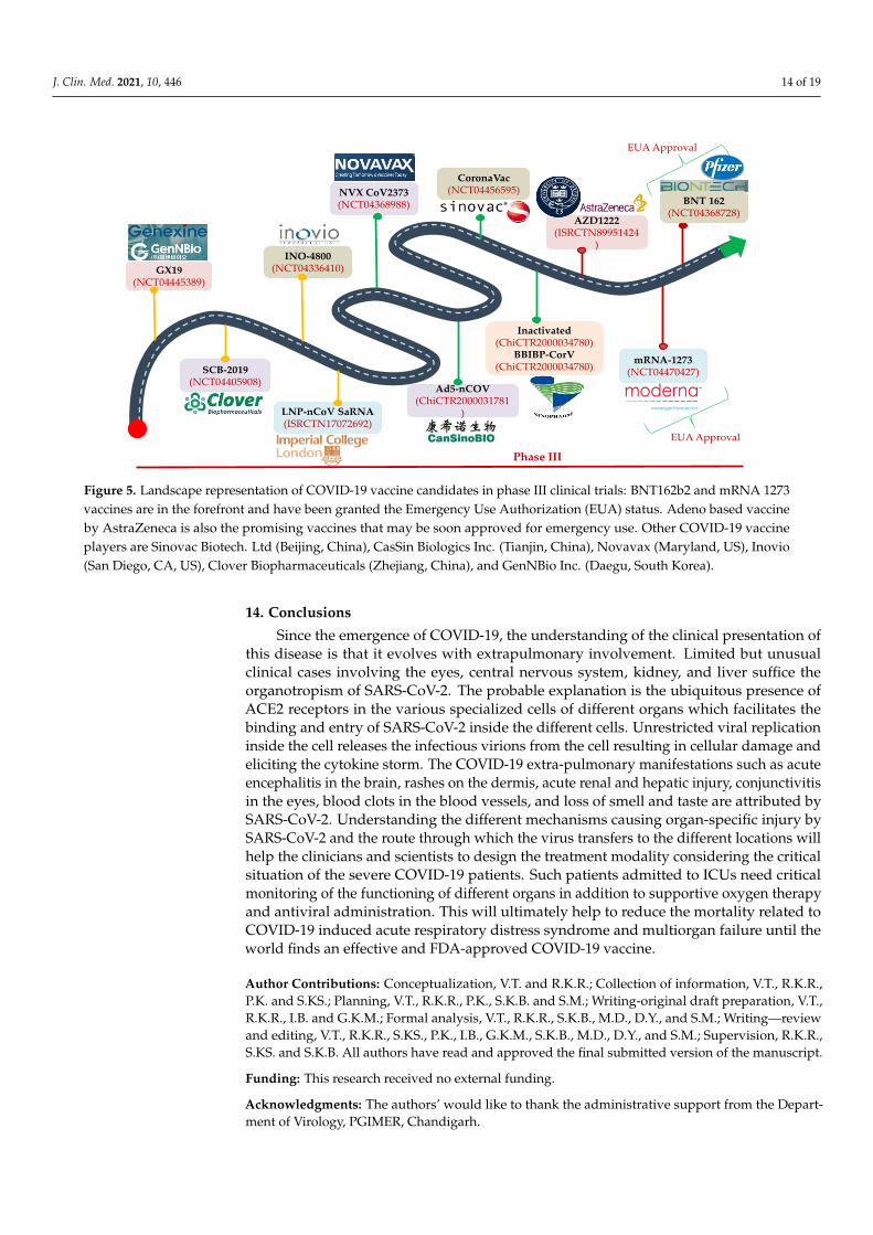
The other important vaccine candidates undergoing clinical trials are the inactivatedSARS-CoV-2 vaccines (Vero cells) developed by Sinopharm in collaboration with ChinaNational Biotec Group Corporation and Wuhan Institute of Biological Products, inactivatedSARS-CoV-2 vaccines by Sinovac Research and Development Co. Ltd., Beijing, Chinaand Gam-COVID-Vac Adeno-based (rAd26-S+rAdS-S) by Gamaleya Research Institute,Moscow, Russia etc. and these are depicted in Figure 5. Recently, two vaccine candidateswere approved in India for clinical trials. Bharat Biotech International Limited devel-oped an indigenous inactivated BBV152 (COVAXIN) COVID vaccine candidate with thecollaborative work of the National Institute of Virology and Indian Council of MedicalResearch. The other one is ZyCov-D from Zydus Cadila, Ahmedabad, India which isalso promising. India is also working with FluGen, Madison, USA and the University ofWisconsin-Madison, Madison, USA on an intranasal vaccine called CoroFlu with the Sgene of SARS-CoV-2 insertion, built on the backbone of M2sr, a self-limiting version ofinfluenza virus that induces immunity against COVID-19 and influenza (expressing Hprotein), lacking the M2 gene by restricting the replication with one cycle only. Besides,more than 173 vaccine candidates are in the pre-clinical stage. Considering the efforts, andthe preliminary results from the various studies, hopefully by early 2021 we may have anapproved effective vaccine available for human use to control the pandemic.
J. Clin. Med. 2021, 10, 446 14 of 19
J. Clin. Med. 2021, 10, x FOR PEER REVIEW 14 of 20
while nasopharyngeal swab positivity confirmed the SARS-CoV-2 infection, accompanied
by dyspnea, chest pain, and palpitation. The significant decrease in the total number of
immune cells i.e., B cells, T cells, and NK cells were also correlated with COVID-19 sever-
ity.
13. Advancements in Vaccine Research
As per the draft landscape of COVID-19 [122] dated 15 January 2021; 63 candidate
vaccines are in line with clinical evaluations with 13 vaccines currently at phase 3 trial.
Two mRNA vaccines i.e., 3LNP-mRNA by Pfizer-BioNTech and RNA LNP encapsulated
mRNA vaccine jointly by Moderna and National Institute of Health (NIH) demonstrated
an efficacy of 95% and were recently approved for the emergency use under the Emer-
gency Use Authorization (EUA) by the US Food and Drug Administration (FDA). Another
promising vaccine i.e., ChAdOx1 (chimpanzee adenovirus vaccine vector) with compara-
ble efficacy has been developed by AstraZeneca with Oxford University, which is a non-
replicating version of adenovirus containing the genetic sequence of surface spike protein
which is produced after vaccination and prime the immune system to attack against the
SARS-CoV-2 viral infection.
The other important vaccine candidates undergoing clinical trials are the inactivated
SARS-CoV-2 vaccines (Vero cells) developed by Sinopharm in collaboration with China
National Biotec Group Corporation and Wuhan Institute of Biological Products, inacti-
vated SARS-CoV-2 vaccines by Sinovac Research and Development Co. Ltd., Beijing,
China and Gam-COVID-Vac Adeno-based (rAd26-S+rAdS-S) by Gamaleya Research In-
stitute, Moscow, Russia etc. and these are depicted in Figure 5. Recently, two vaccine can-
didates were approved in India for clinical trials. Bharat Biotech International Limited
developed an indigenous inactivated BBV152 (COVAXIN) COVID vaccine candidate with
the collaborative work of the National Institute of Virology and Indian Council of Medical
Research. The other one is ZyCov-D from Zydus Cadila, Ahmedabad, India which is also
promising. India is also working with FluGen, Madison, USA and the University of Wis-
consin-Madison, Madison, USA on an intranasal vaccine called CoroFlu with the S gene
of SARS-CoV-2 insertion, built on the backbone of M2sr, a self-limiting version of influ-
enza virus that induces immunity against COVID-19 and influenza (expressing H pro-
tein), lacking the M2 gene by restricting the replication with one cycle only. Besides, more
than 173 vaccine candidates are in the pre-clinical stage. Considering the efforts, and the
preliminary results from the various studies, hopefully by early 2021 we may have an
approved effective vaccine available for human use to control the pandemic.
CoronaVac(NCT04456595)
GX19(NCT04445389)
NVX CoV2373(NCT04368988)
AZD1222(ISRCTN89951424
)
Ad5-nCOV(ChiCTR2000031781
)
mRNA-1273(NCT04470427)
Phase III
Inactivated(ChiCTR2000034780)
BBIBP-CorV(ChiCTR2000034780)
BNT 162(NCT04368728)
LNP-nCoV SaRNA(ISRCTN17072692)
INO-4800(NCT04336410)
SCB-2019(NCT04405908)
EUA Approval
EUA Approval
Figure 5. Landscape representation of COVID-19 vaccine candidates in phase III clinical trials: BNT162b2 and mRNA 1273vaccines are in the forefront and have been granted the Emergency Use Authorization (EUA) status. Adeno based vaccineby AstraZeneca is also the promising vaccines that may be soon approved for emergency use. Other COVID-19 vaccineplayers are Sinovac Biotech. Ltd (Beijing, China), CasSin Biologics Inc. (Tianjin, China), Novavax (Maryland, US), Inovio(San Diego, CA, US), Clover Biopharmaceuticals (Zhejiang, China), and GenNBio Inc. (Daegu, South Korea).
14. Conclusions
Since the emergence of COVID-19, the understanding of the clinical presentation ofthis disease is that it evolves with extrapulmonary involvement. Limited but unusualclinical cases involving the eyes, central nervous system, kidney, and liver suffice theorganotropism of SARS-CoV-2. The probable explanation is the ubiquitous presence ofACE2 receptors in the various specialized cells of different organs which facilitates thebinding and entry of SARS-CoV-2 inside the different cells. Unrestricted viral replicationinside the cell releases the infectious virions from the cell resulting in cellular damage andeliciting the cytokine storm. The COVID-19 extra-pulmonary manifestations such as acuteencephalitis in the brain, rashes on the dermis, acute renal and hepatic injury, conjunctivitisin the eyes, blood clots in the blood vessels, and loss of smell and taste are attributed bySARS-CoV-2. Understanding the different mechanisms causing organ-specific injury bySARS-CoV-2 and the route through which the virus transfers to the different locations willhelp the clinicians and scientists to design the treatment modality considering the criticalsituation of the severe COVID-19 patients. Such patients admitted to ICUs need criticalmonitoring of the functioning of different organs in addition to supportive oxygen therapyand antiviral administration. This will ultimately help to reduce the mortality related toCOVID-19 induced acute respiratory distress syndrome and multiorgan failure until theworld finds an effective and FDA-approved COVID-19 vaccine.
Author Contributions: Conceptualization, V.T. and R.K.R.; Collection of information, V.T., R.K.R.,P.K. and S.KS.; Planning, V.T., R.K.R., P.K., S.K.B. and S.M.; Writing-original draft preparation, V.T.,R.K.R., I.B. and G.K.M.; Formal analysis, V.T., R.K.R., S.K.B., M.D., D.Y., and S.M.; Writing—reviewand editing, V.T., R.K.R., S.KS., P.K., I.B., G.K.M., S.K.B., M.D., D.Y., and S.M.; Supervision, R.K.R.,S.KS. and S.K.B. All authors have read and approved the final submitted version of the manuscript.
Funding: This research received no external funding.
Acknowledgments: The authors’ would like to thank the administrative support from the Depart-ment of Virology, PGIMER, Chandigarh.

J. Clin. Med. 2021, 10, 446 15 of 19
Conflicts of Interest: The authors declare no conflict of interest in the submitted manuscript.
References1. Coronavirus Update (Live): 94,993,884 Cases and 2,031,875 Deaths from COVID-19 Virus Pandemic—Worldometer. Available on-
line: https://www.worldometers.info/coronavirus/ (accessed on 17 January 2021).2. Cucinotta, D.; Vanelli, M. WHO declares COVID-19 a pandemic. Acta Biomed. 2020, 91, 157–160. [CrossRef] [PubMed]3. Petersen, E.; Koopmans, M.; Go, U.; Hamer, D.H.; Petrosillo, N.; Castelli, F.; Storgaard, M.; Khalili, S.A.; Simonsen, L. Comparing
SARS-CoV-2 with SARS-CoV and influenza pandemics. Lancet Infect. Dis. 2020, 20, e238–e244. [CrossRef]4. Wang, C.; Horby, P.W.; Hayden, F.G.; Gao, G.F. A novel coronavirus outbreak of global health concern. Lancet 2020, 395, 470–473.
[CrossRef]5. Corman, V.M.; Muth, D.; Niemeyer, D.; Drosten, C. Hosts and Sources of Endemic Human Coronaviruses. In Advances in Virus
Research; Academic Press Inc.: New York, NY, USA, 2018; Volume 100, pp. 163–188.6. Gralinski, L.E.; Baric, R.S. Molecular pathology of emerging coronavirus infections. J. Pathol. 2015, 235, 185–195. [CrossRef]
[PubMed]7. Neerukonda, S.N.; Katneni, U. A Review on SARS-CoV-2 Virology, Pathophysiology, Animal Models, and Anti-Viral Interventions.
Pathogens 2020, 9, 426. [CrossRef]8. Xia, S.; Zhu, Y.; Liu, M.; Lan, Q.; Xu, W.; Wu, Y.; Ying, T.; Liu, S.; Shi, Z.; Jiang, S.; et al. Fusion mechanism of 2019-nCoV and
fusion inhibitors targeting HR1 domain in spike protein. Cell. Mol. Immunol. 2020, 17, 765–767. [CrossRef]9. Zhang, C.; Wu, Z.; Li, J.W.; Zhao, H.; Wang, G.Q. Cytokine release syndrome in severe COVID-19: Interleukin-6 receptor
antagonist tocilizumab may be the key to reduce mortality. Int. J. Antimicrob. Agents 2020, 55. [CrossRef]10. Wu, Z.; McGoogan, J.M. Characteristics of and Important Lessons from the Coronavirus Disease 2019 (COVID-19) Outbreak in
China: Summary of a Report of 72314 Cases from the Chinese Center for Disease Control and Prevention. JAMA J. Am. Med.Assoc. 2020, 323, 1239–1242. [CrossRef]
11. Zhu, N.; Zhang, D.; Wang, W.; Li, X.; Yang, B.; Song, J.; Zhao, X.; Huang, B.; Shi, W.; Lu, R.; et al. A Novel Coronavirus fromPatients with Pneumonia in China, 2019. N. Engl. J. Med. 2020, 382, 727–733. [CrossRef]
12. Wang, D.; Hu, B.; Hu, C.; Zhu, F.; Liu, X.; Zhang, J.; Wang, B.; Xiang, H.; Cheng, Z.; Xiong, Y.; et al. Clinical Characteristics of 138Hospitalized Patients with 2019 Novel Coronavirus-Infected Pneumonia in Wuhan, China. JAMA J. Am. Med. Assoc. 2020, 323,1061–1069. [CrossRef]
13. Gandhi, M.; Yokoe, D.S.; Havlir, D.V. Asymptomatic Transmission, the Achilles’ Heel of Current Strategies to Control Covid-19.N. Engl. J. Med. 2020, 382, 2158–2160. [CrossRef] [PubMed]
14. Li, R.; Pei, S.; Chen, B.; Song, Y.; Zhang, T.; Yang, W.; Shaman, J. Substantial undocumented infection facilitates the rapiddissemination of novel coronavirus (SARS-CoV-2). Science 2020, 368, 489–493. [CrossRef] [PubMed]
15. Madjid, M.; Safavi-Naeini, P.; Solomon, S.D.; Vardeny, O. Potential Effects of Coronaviruses on the Cardiovascular System: AReview. JAMA Cardiol. 2020, 5, 831–840. [CrossRef] [PubMed]
16. Rothan, H.A.; Byrareddy, S.N. The epidemiology and pathogenesis of coronavirus disease (COVID-19) outbreak. J. Autoimmun.2020, 109. [CrossRef]
17. Li, Y.C.; Bai, W.Z.; Hashikawa, T. The neuroinvasive potential of SARS-CoV2 may play a role in the respiratory failure ofCOVID-19 patients. J. Med. Virol. 2020, 92, 552–555. [CrossRef]
18. Yeo, C.; Kaushal, S.; Yeo, D. Enteric involvement of coronaviruses: Is faecal–oral transmission of SARS-CoV-2 possible? LancetGastroenterol. Hepatol. 2020, 5, 335–337. [CrossRef]
19. Sahin, A.R. 2019 Novel Coronavirus (COVID-19) Outbreak: A Review of the Current Literature. Eurasian J. Med. Oncol. 2020.[CrossRef]
20. Tang, Y.W.; Schmitz, J.E.; Persing, D.H.; Stratton, C.W. Laboratory diagnosis of COVID-19: Current issues and challenges. J. Clin.Microbiol. 2020, 58. [CrossRef]
21. Recalcati, S. Cutaneous manifestations in COVID-19: A first perspective. J. Eur. Acad. Dermatol. Venereol. 2020, 34, e212–e213.[CrossRef]
22. Guan, W.; Ni, Z.; Hu, Y.; Liang, W.; Ou, C.; He, J.; Liu, L.; Shan, H.; Lei, C.; Hui, D.S.C.; et al. Clinical Characteristics ofCoronavirus Disease 2019 in China. N. Engl. J. Med. 2020, 382, 1708–1720. [CrossRef]
23. Fernandez-Nieto, D.; Ortega-Quijano, D.; Segurado-Miravalles, G.; Pindado-Ortega, C.; Prieto-Barrios, M.; Jimenez-Cauhe, J.Comment on: Cutaneous manifestations in COVID-19: A first perspective. Safety concerns of clinical images and skin biopsies.J. Eur. Acad. Dermatol. Venereol. 2020, 34, e252–e254. [CrossRef] [PubMed]
24. Marzano, A.V.; Genovese, G.; Fabbrocini, G.; Pigatto, P.; Monfrecola, G.; Piraccini, B.M.; Veraldi, S.; Rubegni, P.; Cusini, M.;Caputo, V.; et al. Varicella-like exanthem as a specific COVID-19–associated skin manifestation: Multicenter case series of 22patients. J. Am. Acad. Dermatol. 2020, 83, 280–285. [CrossRef] [PubMed]
25. Joob, B.; Wiwanitkit, V. COVID-19 can present with a rash and be mistaken for dengue. J. Am. Acad. Dermatol. 2020, 82, e177.[CrossRef] [PubMed]
26. Estébanez, A.; Pérez-Santiago, L.; Silva, E.; Guillen-Climent, S.; García-Vázquez, A.; Ramón, M.D. Cutaneous manifestations inCOVID-19: A new contribution. J. Eur. Acad. Dermatol. Venereol. 2020, 34, e250–e251. [CrossRef] [PubMed]
J. Clin. Med. 2021, 10, 446 16 of 19
27. Joob, B.; Wiwanitkit, V. Reply to: ‘Various forms of skin rash in COVID-19: Petechial rash in a patient with COVID-19 infection’.J. Am. Acad. Dermatol. 2020, 83, e143. [CrossRef]
28. Cai, Q.; Huang, D.; Ou, P.; Yu, H.; Zhu, Z.; Xia, Z.; Su, Y.; Ma, Z.; Zhang, Y.; Li, Z.; et al. COVID-19 in a designated infectiousdiseases hospital outside Hubei Province, China. Allergy Eur. J. Allergy Clin. Immunol. 2020, 75, 1742–1752. [CrossRef]
29. Zhang, B.; Zhou, X.; Qiu, Y.; Song, Y.; Feng, F.; Feng, J.; Song, Q.; Jia, Q. Clinical characteristics of 82 cases of death from COVID-19.PLoS ONE 2020, 15, e0235458. [CrossRef]
30. Chai, X.; Hu, L.; Zhang, Y.; Han, W.; Lu, Z.; Ke, A.; Zhou, J.; Shi, G.; Fang, N.; Fan, J.; et al. Specific ACE2 expression incholangiocytes may cause liver damage after 2019-nCoV infection. bioRxiv 2020. [CrossRef]
31. Wen Seow, J.J.; Pai, R.; Mishra, A.; Shepherdson, E.; Lim Hon, T.K.; Goh Brian, K.P.; Chan Jerry, K.Y.; Chow Pierce, K.H.; Ginhoux,F.; DasGupta, R.; et al. scRNA-seq reveals ACE2 and TMPRSS2 expression in TROP2 + Liver Progenitor Cells: Implications inCOVID-19 associated Liver Dysfunction. bioRxiv 2020. [CrossRef]
32. Zhao, B.; Ni, C.; Gao, R.; Wang, Y.; Yang, L.; Wei, J.; Lv, T.; Liang, J.; Zhang, Q.; Xu, W.; et al. Recapitulation of SARS-CoV-2infection and cholangiocyte damage with human liver ductal organoids. Protein Cell 2020, 11, 771–775. [CrossRef]
33. Zhang, C.; Shi, L.; Wang, F.S. Liver injury in COVID-19: Management and challenges. Lancet Gastroenterol. Hepatol. 2020, 5,428–430. [CrossRef]
34. Xu, Z.; Shi, L.; Wang, Y.; Zhang, J.; Huang, L.; Zhang, C.; Liu, S.; Zhao, P.; Liu, H.; Zhu, L.; et al. Pathological findings of COVID-19associated with acute respiratory distress syndrome. Lancet Respir. Med. 2020, 8, 420–422. [CrossRef]
35. Liu, J.; Li, S.; Liu, J.; Liang, B.; Wang, X.; Wang, H.; Li, W.; Tong, Q.; Yi, J.; Zhao, L.; et al. Longitudinal characteristics oflymphocyte responses and cytokine profiles in the peripheral blood of SARS-CoV-2 infected patients. EBioMedicine 2020, 55.[CrossRef] [PubMed]
36. Wu, L.; O’Kane, A.M.; Peng, H.; Bi, Y.; Motriuk-Smith, D.; Ren, J. SARS-CoV-2 and cardiovascular complications: From molecularmechanisms to pharmaceutical management. Biochem. Pharmacol. 2020, 178, 114114. [CrossRef] [PubMed]
37. Bonow, R.O.; Fonarow, G.C.; O’Gara, P.T.; Yancy, C.W. Association of Coronavirus Disease 2019 (COVID-19) with MyocardialInjury and Mortality. JAMA Cardiol. 2020, 5, 751–753. [CrossRef]
38. Inciardi, R.M.; Lupi, R.; Zaccone, G.; Italia, L.; Raffo, M.; Tomasoni, D.; Cani, D.S.; Cerini, M.; Farina, D.; Gavazzi, E.; et al. CardiacInvolvement in a Patient with Coronavirus Disease 2019 (COVID-19). JAMA Cardiol. 2020, 5, 819–824. [CrossRef]
39. Han, H.; Xie, L.; Liu, R.; Yang, J.; Liu, F.; Wu, K.; Chen, L.; Hou, W.; Feng, Y.; Zhu, C. Analysis of heart injury laboratory parametersin 273 COVID-19 patients in one hospital in Wuhan, China. J. Med. Virol. 2020, 92, 819–823. [CrossRef]
40. Vidovich, M.I. Transient Brugada-Like Electrocardiographic Pattern in a Patient with COVID-19. JACC Case Rep. 2020, 2,1245–1249. [CrossRef]
41. Sorgente, A.; Capulzini, L.; Brugada, P. The Known Into the Unknown. JACC Case Rep. 2020, 2, 1250–1251. [CrossRef]42. Chen, L.; Li, X.; Chen, M.; Feng, Y.; Xiong, C. The ACE2 expression in human heart indicates new potential mechanism of heart
injury among patients infected with SARS-CoV-2. Cardiovasc. Res. 2020, 116, 1097–1100. [CrossRef]43. Zheng, Y.Y.; Ma, Y.T.; Zhang, J.Y.; Xie, X. COVID-19 and the cardiovascular system. Nat. Rev. Cardiol. 2020, 17, 259–260. [CrossRef]
[PubMed]44. Bilimoria, J.; Singh, H. The Angiopoietin ligands and Tie receptors: Potential diagnostic biomarkers of vascular disease. J. Recept.
Signal Transduct. 2019, 39, 187–193. [CrossRef] [PubMed]45. Desforges, M.; Coupanec, A.L.; Dubeau, P.; Bourgouin, A.; Lajoie, L.; Dube, M.; Talbot, P.J. Human coronaviruses and other
respiratory viruses: Underestimated opportunistic pathogens of the central nervous system? Viruses 2019, 12, 14. [CrossRef][PubMed]
46. Kabbani, N.; Olds, J.L. Does COVID19 Infect the Brain? If So, Smokers Might Be at a Higher Risk. Mol. Pharmacol. 2020, 97,351–353. [CrossRef] [PubMed]
47. Zhou, L.; Zhang, M.; Wang, J.; Gao, J. Sars-Cov-2: Underestimated damage to nervous system. Travel Med. Infect. Dis. 2020, 36.[CrossRef]
48. Poyiadji, N.; Shahin, G.; Noujaim, D.; Stone, M.; Patel, S.; Griffith, B. COVID-19-associated acute hemorrhagic necrotizingencephalopathy: Imaging features. Radiology 2020, 296, E119–E120. [CrossRef]
49. Miller, E.H.; Namale, V.S.; Kim, C.; Dugue, R.; Waldrop, G.; Ciryam, P.; Chong, A.M.; Zucker, J.; Miller, E.C.; Bain, J.M.; et al.Cerebrospinal Analysis in Patients With COVID-19. Open Forum Infect. Dis. 2020, 7. [CrossRef]
50. Wu, Y.; Xu, X.; Chen, Z.; Duan, J.; Hashimoto, K.; Yang, L.; Liu, C.; Yang, C. Nervous system involvement after infection withCOVID-19 and other coronaviruses. Brain Behav. Immun. 2020, 87, 18–22. [CrossRef]
51. Mao, L.; Jin, H.; Wang, M.; Hu, Y.; Chen, S.; He, Q.; Chang, J.; Hong, C.; Zhou, Y.; Wang, D.; et al. Neurologic Manifestations ofHospitalized Patients with Coronavirus Disease 2019 in Wuhan, China. JAMA Neurol. 2020, 77, 683–690. [CrossRef]
52. Li, Y.; Li, M.; Wang, M.; Zhou, Y.; Chang, J.; Xian, Y.; Wang, D.; Mao, L.; Jin, H.; Hu, B. Acute cerebrovascular disease followingCOVID-19: A single center, retrospective, observational study. Stroke Vasc. Neurol. 2020, 5, 279–284. [CrossRef]
53. Sharifi-Razavi, A.; Karimi, N.; Rouhani, N. COVID-19 and intracerebral haemorrhage: Causative or coincidental? New MicrobesNew Infect. 2020, 35, 100669. [CrossRef] [PubMed]
54. Moriguchi, T.; Harii, N.; Goto, J.; Harada, D.; Sugawara, H.; Takamino, J.; Ueno, M.; Sakata, H.; Kondo, K.; Myose, N.; et al. Afirst case of meningitis/encephalitis associated with SARS-Coronavirus-2. Int. J. Infect. Dis. 2020, 94, 55–58. [CrossRef] [PubMed]

J. Clin. Med. 2021, 10, 446 17 of 19
55. Ye, M.; Ren, Y.; Lv, T. Encephalitis as a clinical manifestation of COVID-19. Brain Behav. Immun. 2020, 88, 945–946. [CrossRef][PubMed]
56. Baig, A.M.; Khaleeq, A.; Ali, U.; Syeda, H. Evidence of the COVID-19 Virus Targeting the CNS: Tissue Distribution, Host-VirusInteraction, and Proposed Neurotropic Mechanisms. ACS Chem. Neurosci. 2020, 11, 995–998. [CrossRef] [PubMed]
57. Portals of Viral Entry into the Central Nervous System. In The Blood-Brain Barrier in Health and Disease, Volume Two; CRC Press:Boca Raton, FL, USA, 2015; pp. 37–61.
58. Saavedra, J.M. Brain angiotensin II: New developments, unanswered questions and therapeutic opportunities. Cell. Mol. Neurobiol.2005, 25, 485–512. [CrossRef]
59. Butowt, R.; Bilinska, K. SARS-CoV-2: Olfaction, Brain Infection, and the Urgent Need for Clinical Samples Allowing Earlier VirusDetection. ACS Chem. Neurosci. 2020. [CrossRef]
60. Wan, S.; Yi, Q.; Fan, S.; Lv, J.; Zhang, X.; Guo, L.; Lang, C.; Xiao, Q.; Xiao, K.; Yi, Z.; et al. Characteristics of lymphocyte subsetsand cytokines in peripheral blood of 123 hospitalized patients with 2019 novel coronavirus pneumonia (NCP). medRxiv 2020.[CrossRef]
61. Mehta, P.; McAuley, D.F.; Brown, M.; Sanchez, E.; Tattersall, R.S.; Manson, J.J. COVID-19: Consider cytokine storm syndromesand immunosuppression. Lancet 2020, 395, 1033–1034. [CrossRef]
62. Pei, G.; Zhang, Z.; Peng, J.; Liu, L.; Zhang, C.; Yu, C.; Ma, Z.; Huang, Y.; Liu, Y.; Yao, Y.; et al. Renal involvement and earlyprognosis in patients with COVID-19 pneumonia. J. Am. Soc. Nephrol. 2020, 31, 1157–1165. [CrossRef]
63. Cheng, Y.; Luo, R.; Wang, K.; Zhang, M.; Wang, Z.; Dong, L.; Li, J.; Yao, Y.; Ge, S.; Xu, G. Kidney disease is associated within-hospital death of patients with COVID-19. Kidney Int. 2020, 97, 829–838. [CrossRef]
64. Gabarre, P.; Dumas, G.; Dupont, T.; Darmon, M.; Azoulay, E.; Zafrani, L. Acute kidney injury in critically ill patients withCOVID-19. Intensive Care Med. 2020, 46, 1339–1348. [CrossRef] [PubMed]
65. Sun, J.; Zhu, A.; Li, H.; Zheng, K.; Zhuang, Z.; Chen, Z.; Shi, Y.; Zhang, Z.; Chen, S.; Liu, X.; et al. Isolation of infectiousSARS-CoV-2 from urine of a COVID-19 patient. Emerg. Microbes Infect. 2020, 9, 991–993. [CrossRef] [PubMed]
66. Ling, Y.; Xu, S.B.; Lin, Y.X.; Tian, D.; Zhu, Z.Q.; Dai, F.H.; Wu, F.; Song, Z.G.; Huang, W.; Chen, J.; et al. Persistence and clearanceof viral RNA in 2019 novel coronavirus disease rehabilitation patients. Chin. Med. J. 2020, 133, 1039–1043. [CrossRef]
67. Puelles, V.G.; Lutgehetmann, M.; Lindenmeyer, M.T.; Sperhake, J.P.; Wong, M.N.; Allweiss, L.; Chilla, S.; Heinemann, A.; Wanner,N.; Liu, S.; et al. Multiorgan and Renal Tropism of SARS-CoV-2. N. Engl. J. Med. 2020, 383, 590–592. [CrossRef] [PubMed]
68. Diao, B.; Wang, C.; Wang, R.; Feng, Z.; Tan, Y.; Wang, H.; Wang, C.; Liu, L.; Liu, Y.; Wang, G.; et al. Human kidney is a target fornovel severe acute respiratory syndrome coronavirus 2 (SARS-CoV-2) infection. medRxiv 2020. [CrossRef]
69. Wang, R.; Liao, C.; He, H.; Hu, C.; Wei, Z.; Hong, Z.; Zhang, C.; Liao, M.; Shui, H. COVID-19 in Hemodialysis Patients: A Reportof 5 Cases. Am. J. Kidney Dis. 2020, 76, 141–143. [CrossRef]
70. Wang, H. Maintenance Hemodialysis and COVID-19: Saving Lives With Caution, Care, and Courage. Kidney Med. 2020, 2,365–366. [CrossRef]
71. He, Q.; Mok, T.N.; Yun, L.; He, C.; Li, J.; Pan, J. Single-cell RNA sequencing analysis of human kidney reveals the presence ofACE2 receptor: A potential pathway of COVID-19 infection. Mol. Genet. Genom. Med. 2020, 8. [CrossRef]
72. Lin, W.; Hu, L.; Zhang, Y.; Ooi, J.D.; Meng, T.; Jin, P.; Sing, X.; Peng, L.; Song, L.; Xiao, Z.; et al. Single-cell Analysis of ACE2Expression in Human Kidneys and Bladders Reveals a Potential Route of 2019-nCoV Infection. bioRxiv 2020. [CrossRef]
73. Fan, C.; Li, K.; Ding, Y.; Lu, W.; Wang, J. ACE2 Expression in Kidney and Testis May Cause Kidney and Testis Damage after2019-nCoV Infection. MedRxiv 2020. [CrossRef]
74. Husain-Syed, F.; Slutsky, A.S.; Ronco, C. Lung-kidney cross-talk in the critically ill patient. Am. J. Respir. Crit. Care Med. 2016, 194,402–414. [CrossRef] [PubMed]
75. Leung, W.K.; To, K.F.; Chan, P.K.S.; Chan, H.L.Y.; Wu, A.K.L.; Lee, N.; Yuen, K.Y.; Sung, J.J.Y. Enteric involvement of severe acuterespiratory syndrome-associated coronavirus infection. Gastroenterology 2003, 1251011–1251017. [CrossRef]
76. Holshue, M.L.; DeBolt, C.; Lindquist, S.; Lofy, K.H.; Wiesman, J.; Bruce, H.; Spitters, C.; Ericson, K.; Wilkerson, S.; Tural, A.; et al.First Case of 2019 Novel Coronavirus in the United States. N. Engl. J. Med. 2002, 382, 929–936. [CrossRef] [PubMed]
77. Lin, L.; Jiang, X.; Zhang, Z.; Huang, S.; Zhang, Z.; Fang, Z.; Gu, Z.; Gao, L.; Shi, H.; Mai, L.; et al. Gastrointestinal symptoms of 95cases with SARS-CoV-2 infection. Gut 2020, 69, 997–1001. [CrossRef] [PubMed]
78. Chen, L.; Lou, J.; Bai, Y.; Wang, M. COVID-19 disease with positive fecal and negative pharyngeal and sputum viral tests. Am. J.Gastroenterol. 2020, 115, 790. [CrossRef] [PubMed]
79. Wei, X.-S.; Wang, X.; Niu, Y.R.; Ye, L.L.; Peng, W.B.; Wang, Z.H.; Yang, W.B.; Yang, B.H.; Zhang, J.C.; Ma, W.L.; et al. ClinicalCharacteristics of SARS-CoV-2 Infected Pneumonia with Diarrhea. SSRN Electron. J. 2020. [CrossRef]
80. Pan, L.; Mu, M.; Yang, P.; Sun, Y.; Wang, R.; Yan, J.; Li, P.; Hu, B.; Wang, J.; Hu, C.; et al. Clinical Characteristics of COVID-19Patients With Digestive Symptoms in Hubei, China. Am. J. Gastroenterol. 2020, 115, 766–773. [CrossRef]
81. Jin, X.; Lian, J.S.; Hu, J.H.; Gao, J.; Zheng, L.; Zhang, Y.M.; Hao, S.R.; Jia, H.Y.; Cai, H.; Zhang, X.L.; et al. Epidemiological, clinicaland virological characteristics of 74 cases of coronavirus-infected disease 2019 (COVID-19) with gastrointestinal symptoms.Gut 2020, 69, 1002–1009. [CrossRef]
82. Luo, S.; Zhang, X.; Xu, H. Don’t Overlook Digestive Symptoms in Patients With 2019 Novel Coronavirus Disease (COVID-19).Clin. Gastroenterol. Hepatol. 2020, 18, 1636–1637. [CrossRef]

J. Clin. Med. 2021, 10, 446 18 of 19
83. Wu, Y.; Guo, C.; Tang, L.; Hong, Z.; Zhou, J.; Dong, X.; Yin, H.; Xiao, Q.; Tang, Y.; Qu, X.; et al. Prolonged presence of SARS-CoV-2viral RNA in faecal samples. Lancet Gastroenterol. Hepatol. 2020, 5, 434–435. [CrossRef]
84. Xu, Y.; Li, X.; Zhu, B.; Liang, H.; Fang, C.; Gong, Y.; Guo, Q.; Sun, X.; Zhao, D.; Shen, J.; et al. Characteristics of pediatricSARS-CoV-2 infection and potential evidence for persistent fecal viral shedding. Nat. Med. 2020, 26, 502–505. [CrossRef][PubMed]
85. Elmunzer, B.J.; Spitzer, R.L.; Foster, L.D.; Merchant, A.A.; Howard, E.F.; Patel, V.A.; West, M.K.; Qayed, E.; Nustas, R.; Zakaria, A.;et al. Digestive Manifestations in Patients Hospitalized with COVID-19. Clin. Gastroenterol. Hepatol. 2020. [CrossRef] [PubMed]
86. Wölfel, R.; Corman, V.M.; Guggemos, W.; Seilmaier, M.; Zange, S.; Muller, M.A.; Niemeyer, D.; Jones, T.C.; Vollmar, P.; Rothe, C.;et al. Virological assessment of hospitalized patients with COVID-2019. Nature 2020, 581, 465–469. [CrossRef] [PubMed]
87. Wang, W.; Xu, Y.; Gao, R.; Lu, R.; Han, K.; Wu, G.; Tan, W. Detection of SARS-CoV-2 in Different Types of Clinical Specimens.JAMA J. Am. Med. Assoc. 2020, 323, 1843–1844. [CrossRef] [PubMed]
88. Zhang, H.; Kang, Z.; Gong, H.; Xu, D.; Wang, J.; Li, Z.; Li, Z.; Cui, X.; Xiao, J.; Zhan, J.; et al. Digestive system is a potentialroute of COVID-19: An analysis of single-cell coexpression pattern of key proteins in viral entry process. Gut 2020, 69, 1010–1018.[CrossRef]
89. Hashimoto, T.; Perlot, T.; Rehman, A.; Trichereay, J.; Ishiguro, H.; Paolino, M.; Sigl, V.; Hanada, T.; Hanada, R.; Lipinski, S.; et al.ACE2 links amino acid malnutrition to microbial ecology and intestinal inflammation. Nature 2012, 487, 477–481. [CrossRef]
90. Hoffmann, M.; Kleine-Weber, H.; Schroeder, S.; Kruger, N.; Herrler, T.; Erichsen, S.; Schiergens, T.S.; Herrler, G.; Wu, N.H.;Nitsche, A.; et al. SARS-CoV-2 Cell Entry Depends on ACE2 and TMPRSS2 and Is Blocked by a Clinically Proven ProteaseInhibitor. Cell 2020, 181, 271–280.e8. [CrossRef]
91. Liang, W.; Feng, Z.; Rao, S.; Xiao, C.; Xue, X.; Lin, Z.; Zhang, Q.; Qi, W. Diarrhoea may be underestimated: A missing link in 2019novel coronavirus. Gut 2020, 69, 1141–1143. [CrossRef]
92. Słomka, A.; Kowalewski, M.; Zekanowska, E. Coronavirus disease 2019 (COVID–19): A short review on hematological manifesta-tions. Pathogens 2020, 9, 493. [CrossRef]
93. Ludvigsson, J.F. Systematic review of COVID-19 in children shows milder cases and a better prognosis than adults. Acta Paediatr.Int. J. Paediatr. 2020, 109, 1088–1095. [CrossRef]
94. Xu, P.; Zhou, Q.; Xu, J. Mechanism of thrombocytopenia in COVID-19 patients. Ann. Hematol. 2020, 99, 1205–1208. [CrossRef][PubMed]
95. Lippi, G.; Plebani, M.; Henry, B.M. Thrombocytopenia is associated with severe coronavirus disease 2019 (COVID-19) infections:A meta-analysis. Clin. Chim. Acta 2020, 506, 2020. [CrossRef] [PubMed]
96. Maier, C.L.; Truong, A.D.; Auld, S.C.; Polly, D.M.; Tanksley, C.L.; Duncan, A. COVID-19-associated hyperviscosity: A linkbetween inflammation and thrombophilia? Lancet 2020, 395, 1758–1759. [CrossRef]
97. Biswas, S.; Thakur, V.; Kaur, P.; Khan, A.; Kulshrestha, S.; Kumar, P. Blood clots in COVID-19 patients: Simplifying the curiousmystery. Med. Hypotheses 2020, 146, 110371. [CrossRef]
98. Ackermann, M.; Verleden, S.E.; Kuehnel, M.; Haverich, A.; Welte, T.; Laenger, F.; Vanstapel, A.; Werlein, C.; Stark, H.; Tzankov,A.; et al. Pulmonary Vascular Endothelialitis, Thrombosis, and Angiogenesis in Covid-19. N. Engl. J. Med. 2020, 383, 120–128.[CrossRef]
99. Wei, L.; Sun, S.; Xu, C.H.; Zhang, J.; Xu, Y.; Zhu, H.; Peh, S.C.; Korteweg, C.; McNutt, M.A.; Gu, J. Pathology of the thyroid insevere acute respiratory syndrome. Hum. Pathol. 2007, 38, 95–102. [CrossRef]
100. Benbassat, C.A.; Olchovsky, D.; Tsvetov, G.; Shimon, I. Subacute thyroiditis: Clinical characteristics and treatment outcome infifty-six consecutive patients diagnosed between 1999 and 2005. J. Endocrinol. Investig. 2007, 30, 631–635. [CrossRef]
101. Chaux-Bodard, A.-G.; Deneuve, S.; Desoutter, A. Oral manifestation of Covid-19 as an inaugural symptom? J. Oral Med. OralSurg. 2020, 26, 18. [CrossRef]
102. Brancatella, A.; Ricci, D.; Viola, N.; Sgrò, D.; Santini, F.; Latrofa, F. Subacute Thyroiditis After Sars-COV-2 Infection. J. Clin.Endocrinol. Metab. 2020, 105, 2367–2370. [CrossRef]
103. Oxley, T.J.; Mocco, J.; Majidi, S.; Kellner, C.P.; Shoirah, H.; Singh, I.P.; De Leacy, R.A.; Shigematsu, T.; Ladner, T.R.; Yaeger, K.A.;et al. Large-Vessel Stroke as a Presenting Feature of Covid-19 in the Young. N. Engl. J. Med. 2020, 382, e60. [CrossRef]
104. Varga, Z.; Flammer, A.J.; Steiger, P.; Haberecker, M.; Andermatt, R.; Zinkernagel, A.S.; Mehra, M.R.; Schuepbach, R.A.; Ruschitzka,F.; Moch, H. Endothelial cell infection and endotheliitis in COVID-19. Lancet 2020, 395, 1417–1418. [CrossRef]
105. Tsao, H.S.; Chason, H.M.; Fearon, D.M. Immune thrombocytopenia (ITP) in a pediatric patient positive for SARS-CoV-2.Pediatrics 2020, 146. [CrossRef]
106. Zulfiqar, A.-A.; Lorenzo-Villalba, N.; Hassler, P.; Andrès, E. Immune Thrombocytopenic Purpura in a Patient with Covid-19.N. Engl. J. Med. 2020, 382, e43. [CrossRef]
107. Nirenberg, M.S.; Del Mar Ruiz Herrera, M. Foot Manifestations in a COVID-19 Positive Patient: A Case Study. J. Am. Podiatr. Med.Assoc. 2020. [CrossRef]
108. Arora, G.; Kassir, M.; Jafferany, M.; Galadari, H.; Lotti, T.; Satolli, F.; Sadoughifar, R.; Sitkowska, Z.; Goldust, M. The COVID-19outbreak and rheumatologic skin diseases. Dermatol. Ther. 2020, 33. [CrossRef]
109. Flammer, A.J.; Anderson, T.; Celermajer, D.S.; Creager, M.A.; Deanfield, J.; Ganz, P.; Hamburg, N.M.; Luscher, T.F.; Shechter, M.;Taddei, S.; et al. The assessment of endothelial function: From research into clinical practice. Circulation 2012, 126, 753–767.[CrossRef]

J. Clin. Med. 2021, 10, 446 19 of 19
110. Zhou, Y.; Fu, B.; Zheng, X.; Wang, D.; Zhao, C.; Qi, Y.; Sun, R.; Tian, Z.; Xu, X.; Wei, H. Aberrant pathogenic GM-CSF + T cellsand inflammatory CD14+ CD16+ monocytes in severe pulmonary syndrome patients of a new coronavirus. Natl. Sci. Rev. 2020.[CrossRef]
111. Danzi, G.B.; Loffi, M.; Galeazzi, G.; Gherbesi, E. Acute pulmonary embolism and COVID-19 pneumonia: A random association?Eur. Heart J. 2020, 41, 1858. [CrossRef]
112. Gandolfini, I.; Delsante, M.; Fiaccadori, E.; Zaza, G.; Manenti, L.; Antoni, A.D.; Peruzzi, L.; Riella, L.V.; Cravedi, P.; Maggiore, U.COVID-19 in kidney transplant recipients. Am. J. Transpl. 2020, 20, 1941–1943. [CrossRef]
113. Guillen, E.; Pineiro, G.J.; Revuelta, I.; Rodriguez, D.; Bodro, M.; Moreno, A.; Campistol, J.M.; Diekmann, F.; Ventura-Aguiar, P.Case report of COVID-19 in a kidney transplant recipient: Does immunosuppression alter the clinical presentation? Am. J.Transplant. 2002, 20, 1875–1878. [CrossRef]
114. Hsu, J.J.; Gaynor, P.; Kamath, M.; Fan, A.; Al-Saffar, F.; Criz, D.; Nsair, A. COVID-19 in a high-risk dual heart and kidneytransplant recipient. Am. J. Transplant. 2020, 20, 1911–1915. [CrossRef]
115. Li, F.; Cai, J.; Dong, N. First cases of COVID-19 in heart transplantation from China. J. Heart Lung Transpl. 2020, 39, 496–497.[CrossRef]
116. Cox, M.J.; Loman, N.; Bogaert, D.; O’Grady, J. Co-infections: Potentially lethal and unexplored in COVID-19. Lancet Microbe 2020,1, e11. [CrossRef]
117. Wang, H.; Ding, Y.; Li, X.; Yang, L.; Zhang, W.; Kang, W. Fatal Aspergillosis in a Patient with SARS Who Was Treated withCorticosteroids. N. Engl. J. Med. 2003, 349, 507–508. [CrossRef]
118. Alanio, A.; Dellière, S.; Fodil, S.; Bretagne, S.; Mégarbane, B. Prevalence of putative invasive pulmonary aspergillosis in criticallyill patients with COVID-19. Lancet Respir. Med. 2020, 8, e48–e49. [CrossRef]
119. Koehler, P.; Cornely, A.A.; Bottiger, B.E.; Dusse, F.; Eichenauer, A.S.; Fuchs, F.; Hallek, M.; Jung, N.; Klein, F.; Persigehl, T.; et al.COVID-19 associated pulmonary aspergillosis. Mycoses 2002, 63, 528–534. [CrossRef]
120. Zhou, F.; Yu, T.; Du, R.; Fan, G.; Liu, Y.; Liu, Z.; Xiang, J.; Wang, Y.; Song, B.; Gu, X.; et al. Clinical course and risk factorsfor mortality of adult inpatients with COVID-19 in Wuhan, China: A retrospective cohort study. Lancet 2020, 395, 1054–1062.[CrossRef]
121. Wang, M.; Luo, L.; Bu, H.; Xia, H. One case of coronavirus disease 2019 (COVID-19) in a patient co-infected by HIV with a lowCD4+ T-cell count. Int. J. Infect. Dis. 2020, 96, 148–150. [CrossRef]
122. Draft Landscape of COVID-19 Candidate Vaccines. Available online: https://www.who.int/publications/m/item/draft-landscape-of-covid-19-candidate-vaccines (accessed on 18 January 2021).