Solid Organ Pre-Transplant Testing OR1 OR2
162
Tuesday, September 29, 2015 2:00 PM - 3:30 PM Abstract Session 1: Solid Organ Pre-Transplant Testing OR1 SCANDIATRANSPLANT ACCEPTABLE MISMATCH PROGRAM; A BRIDGE TO TRANSPLANTING THE HIGHLY IMMUNIZED KIDNEY PATIENTS? Pernille Koefoed-Nielsen 1 , Ilse Duus Weinreich 2 , Bjarne Møller 1 , Torbjørn Leivestad 3 . 1 Aarhus University Hospital, Aarhus N, Denmark; 2 Aarhus University Hospital, Aarhus N, Denmark; 3 Oslo University Hospital Rikshospitalet, Oslo, Norway Aim: Scandiatransplant is the organ exchange organization for the Nordic countries. The number of highly immunized (HI) patients on the waiting list (WL) for deceased donor kidney transplant is steadily increasing and the HI patients wait longer than non-immunized patients do. The Scandiatransplant Acceptable Mismatch Program (STAMP) started in March 2009. The aim was to improve the probability for a HI patient to receive a suitable kidney graft from a deceased donor. Methods: Patients are HLA typed at split level (HLA-A, B, C, DR, DQ) using serological or genomic techniques. The patients fulfill all the following criteria: a) On WL > 1 year, b) HI, PRA ≥ 80% based on CDC and/or solid phase assay, c) HI in two consecutive samples over a period of more than 3 months, d) Antibody reactivity against HLA cl I and/or II antigens, e) The last tested sample drawn less than 3 months before acceptance. Eligible patients must have acceptable mismatches defined. We regard HLA antigens which the kidney patient has not developed clinical relevant antibodies (MFI>1000) towards as acceptable. Upon registration, a transplantability score (TS) is calculated that take HLA antigens and AB0 blood group into account. The TS gives the likelihood of finding a suitable donor by counting the number of compatible donors in 1000 recently HLA typed deceased donors within Scandiatransplant. Results: In the study period (March 2009 - February 2015) 94/245 patients on the STAMP WL were transplanted, the mean waiting time for the transplanted patients being 137 days (2-721). In the same period, 4338/8583 patients on the ordinary WL were transplanted, with a mean waiting time of 421 days (0-2260). The TS ranged from 0- 87/1000. The patients with a score ≥ 10/1000 (n=40) were transplanted, with a mean waiting time of 40 days, while the patients with a TS < 10/1000 (n=54) had a mean waiting time of 193 days. Ninety percent of the patients remaining on STAMP for more than a year have a TS < 2/1000. Graft survival after 4 years is at the same level after transplant on STAMP compared with the ordinary WL. Conclusion: STAMP is a feasible and immunologically safe way to transplant HI patients. Patients with a TS <2/1000 donors have a poor chance of getting a transplant even on this prioritized list. TS above 10 /1000 donors predicts a short waiting time for a compatible donor kidney. OR2 HLA CLASS II ANTIGEN MATCHING EFFECT ON KIDNEY TRANSPLANTATION Yong Cho, James C. Cicciarelli, Nathan A. Lemp, Noriyuki Kasahara, Michelle L. Altrich. Viracor-IBT Laboratories, Los Angeles, CA Aim: HLA class II-restricted regulatory T cell (Treg) epitopes in IgG (also called "Tregitopes") have been reported to suppress immune responses to co-administered antigens by stimulating the expansion of natural Tregs (nTregs). Under the current kidney allocation system in the United States, the only points given for HLA matching are for 0 and 1 HLA-DR antigen mismatches (DRMM), which receive 2 and 1 points, respectively. Thus, we investigated the potential for Tregitope-mediated antigen-specific tolerance induction via searching for an HLA-DR antigen matching effect on deceased donor (DD) kidney transplantation outcomes. Methods: During 2000-2013, a total of 133,235 patients who received a deceased donor kidney transplant alone were included in this study using the OPTN/UNOS data as of Sept. 30, 2014. In order to investigate HLA-DR antigen matching effect on graft survival, recipients who received a deceased donor kidney with 1 DRMM
-
Upload
khangminh22 -
Category
Documents
-
view
2 -
download
0
Transcript of Solid Organ Pre-Transplant Testing OR1 OR2
Tuesday, September 29, 2015 2:00 PM - 3:30 PM
Abstract Session 1: Solid Organ Pre-Transplant Testing
OR1 SCANDIATRANSPLANT ACCEPTABLE MISMATCH PROGRAM; A BRIDGE TO TRANSPLANTING THE HIGHLY IMMUNIZED KIDNEY PATIENTS? Pernille Koefoed-Nielsen1, Ilse Duus Weinreich2, Bjarne Møller1, Torbjørn Leivestad3. 1Aarhus University Hospital, Aarhus N, Denmark; 2Aarhus University Hospital, Aarhus N, Denmark; 3Oslo University Hospital Rikshospitalet, Oslo, Norway
Aim: Scandiatransplant is the organ exchange organization for the Nordic countries. The number of highly immunized (HI) patients on the waiting list (WL) for deceased donor kidney transplant is steadily increasing and the HI patients wait longer than non-immunized patients do. The Scandiatransplant Acceptable Mismatch Program (STAMP) started in March 2009. The aim was to improve the probability for a HI patient to receive a suitable kidney graft from a deceased donor. Methods: Patients are HLA typed at split level (HLA-A, B, C, DR, DQ) using serological or genomic techniques. The patients fulfill all the following criteria: a) On WL > 1 year, b) HI, PRA ≥ 80% based on CDC and/or solid phase assay, c) HI in two consecutive samples over a period of more than 3 months, d) Antibody reactivity against HLA cl I and/or II antigens, e) The last tested sample drawn less than 3 months before acceptance. Eligible patients must have acceptable mismatches defined. We regard HLA antigens which the kidney patient has not developed clinical relevant antibodies (MFI>1000) towards as acceptable. Upon registration, a transplantability score (TS) is calculated that take HLA antigens and AB0 blood group into account. The TS gives the likelihood of finding a suitable donor by counting the number of compatible donors in 1000 recently HLA typed deceased donors within Scandiatransplant. Results: In the study period (March 2009 - February 2015) 94/245 patients on the STAMP WL were transplanted, the mean waiting time for the transplanted patients being 137 days (2-721). In the same period, 4338/8583 patients on the ordinary WL were transplanted, with a mean waiting time of 421 days (0-2260). The TS ranged from 0-87/1000. The patients with a score ≥ 10/1000 (n=40) were transplanted, with a mean waiting time of 40 days, while the patients with a TS < 10/1000 (n=54) had a mean waiting time of 193 days. Ninety percent of the patients remaining on STAMP for more than a year have a TS < 2/1000. Graft survival after 4 years is at the same level after transplant on STAMP compared with the ordinary WL. Conclusion: STAMP is a feasible and immunologically safe way to transplant HI patients. Patients with a TS <2/1000 donors have a poor chance of getting a transplant even on this prioritized list. TS above 10 /1000 donors predicts a short waiting time for a compatible donor kidney.
OR2 HLA CLASS II ANTIGEN MATCHING EFFECT ON KIDNEY TRANSPLANTATION Yong Cho, James C. Cicciarelli, Nathan A. Lemp, Noriyuki Kasahara, Michelle L. Altrich. Viracor-IBT Laboratories, Los Angeles, CA
Aim: HLA class II-restricted regulatory T cell (Treg) epitopes in IgG (also called "Tregitopes") have been reported to suppress immune responses to co-administered antigens by stimulating the expansion of natural Tregs (nTregs). Under the current kidney allocation system in the United States, the only points given for HLA matching are for 0 and 1 HLA-DR antigen mismatches (DRMM), which receive 2 and 1 points, respectively. Thus, we investigated the potential for Tregitope-mediated antigen-specific tolerance induction via searching for an HLA-DR antigen matching effect on deceased donor (DD) kidney transplantation outcomes. Methods: During 2000-2013, a total of 133,235 patients who received a deceased donor kidney transplant alone were included in this study using the OPTN/UNOS data as of Sept. 30, 2014. In order to investigate HLA-DR antigen matching effect on graft survival, recipients who received a deceased donor kidney with 1 DRMM

(n=57,436) were divided into 2 groups: (1) Group I, HLA-DR10, 14, or 16 antigen match (n=1,436); (2) Group II, no HLA-DR10, 14, or 16 antigen match (n=56,000). For reference groups, 0 DRMM (n=29,223) and 2 DRMM (n=46,576) groups were included in the study. Results: Stepwise decreases in graft survival rates were seen (Figure 1). Group I showed the highest survival rates, followed by 0 DRMM (P=0.146 vs Group I), Group II (P<0.001 vs Group I), and 2 DRMM group (P<0.001 vs Group I). After adjusting for confounders (such as donor, recipient, pre- and post-transplant risk factors, not shown here), Group I yielded significantly superior graft survival rates compared to Group II (P<0.001). Conclusion: HLA-DR10, 14, or 16 matching, despite other donor HLA-DR antigen mismatches, was associated with graft survival superior to other HLA-DR matches. This effect could be associated with Tregitope-mediated antigen-specific tolerance induction, due to more efficient presentation of IgG-derived Tregitopes by matched HLA-DR10, 14, or 16 proteins.
OR3 CLINICAL RELEVANCE OF PREFORMED “ACCEPTABLE” DONOR SPECIFIC ANTIBODIES IN KIDNEY TRANSPLANTATION Antonina Piazza1, Giuseppina Ozzella1, Elvira Poggi1, Manfreda Annarite2, Lucia Spano2, Silvia Sinopoli2, Andrea Giaffreda2. 1Regional Transplant Center, Lazio - CNR IFT UOS S. Camillo Hospital, Rome, Italy; 2Regional Transplant Center, Lazio, Rome, Italy
Aim: Preformed HLA cytotoxic antibodies, specific for mismatched HLA molecules of the potential donor, represent an absolute contraindication in kidney transplantation. New techniques, like the Luminex-Single Antigen Beads assay, are very sensitive allowing detection of HLA donor specific antibodies (DSAs) at low mean fluorescence intensity (MFI) values. However, few evidence on the clinical relevance of such low “strength” DSAs have been reported. Methods: Graft outcome (follow-up 34.2±19.8 months) of 99 pre-sensitized patients (%FlowPRA class I = 41±33; %FlowPRA class II = 30±36), transplanted between May 2007 and June 2014 on the basis of both CDC-XM and FC-XM negative results, was analyzed. Five patients were excluded from the study because of primary non-function of the graft (3 patients) or death for non-immunologic causes with non-functional graft (2 patients). Forty-seven (47%) patients did not have pre-formed HLA DSAs; the remaining 52 (53%) patients had “acceptable” DSAs (MFI≤5000) specific for HLA-A/B/C/DR/DQB molecules or anti-DPB/DQA DSAs with high MFI values (>5000). HLA class I DSAs were present in 28 patients; five of these showed DSAs against more than one donor HLA

molecules. HLA class II DSAs were present in 19 patients; three of these showed anti-DPB/DQA DSAs with MFI>5000. The remaining five patients had both HLA class I and class II DSAs; one of these had high level of anti-DP DSA. Results: Analyzing graft outcome of the remaining 94 transplanted patients with functioning graft, we did not evidenced any significant difference between DSA positive patients and DSA negative patients (rejection: 10.4% vs. 6.5%, P = ns; graft loss: 12.5% vs. 10.9%, P = ns). In particular, among the 48 DSA positive patients, three (6.2%) had humoral rejection without graft loss; two (4.2%) had cellular rejection, one of these lost the graft. Six DSA positive patients suffered graft failure that was never due to antibody-mediated rejection related to pre-formed DSAs. Conclusion: The results of this study show that pre-formed DSAs with low mean fluorescence intensity values do not represent a contraindication in kidney transplantation. An accurate evaluation of the “strength” of Luminex-detected DSA allows transplanting patients with clinically “irrelevant” HLA antibodies.
OR4 A CASE OF HEART VALVE REPLACEMENT LEADING TO EXTENSIVE SENSITIZATION Christine Yamniuk1, Patricia Campbell1, Jackson Wong2, Luis Hidalgo1. 1University of Alberta, Edmonton, AB, Canada; 2University of Alberta, Edmonton, AB, Canada
HLA antibody (Ab) sensitization is a major barrier to transplantation. We present a case of a 9 year old girl who was assessed for heart-lung transplantation due to left ventricular outflow tract obstruction and pulmonary venous hypertension. Assessment with single antigen bead (SAB) testing for HLA Ab revealed a cPRA=100%; the key sensitizing event was an aortic heart valve homograft 6 months prior. The patient was also transfused and had been on ECMO. The patient was unsensitized prior to the homograft and suffered a S. pneumonae infection that may have contributed to sensitization. Patient and homograft donor were HLA typed at all loci including DQA1,DPA1, and DPB1. Only HLA-C was matched, with all other loci fully mismatched except for 1/2 for DPA1 and DPB1. The degree of sensitization was high for both class I and II HLA - cPRA=100% for each (Canadian cPRA) calculated for Abs with an MFI>1000. Epitope spreading was evident with SAB testing for total IgG. When assessed for C1Q-binding IgG, the only Abs detected were to mismatched homograft antigens and closely related antigens, which still produced a cPRA=96%(Table 1). Much of the sensitization was due to the patient’s HLA type which lacked public class I epitopes; Aw4, and class II; DR51/52/53, as well as DQ1 and DQ3, related to being homozygous for least common class II HLA types as described in Figure 1. The patient is currently on the waitlist and is unlikely to find a compatible donor. This case highlights the significant impact of HLA mismatches in homograft implants, particularly in patients with unique HLA types. It also provides insight into the properties of HLA Abs formed in the absence of immunosuppression, as evidenced by the differences in SAB testing for total IgG versus C1Q-binding IgG.

L. Hidalgo: Speaker's Bureau; Company/Organization; Thermo Fisher (One Lambda).
OR5 IGG SUBCLASS AND CONCENTRATION ARE DETERMINANTS OF HLA CLASS I ANTIBODY CAPACITY TO FIX COMPLEMENT IN IN VITRO CLINICAL AND FUNCTIONAL ASSAYS. Nicole M. Valenzuela1, Kimberly A. Thomas1, Minas Zartarian1, Jason Song1, K Ryan Trinh2, Sherie L. Morrison2, Elaine F. Reed1. 1UCLA, Los Angeles, CA; 2UCLA, Los Angeles, CA
It has been presupposed that “complement (C’) fixing” HLA antibodies (Ab) detected by C1qScreen or CDC are IgG1 or IgG3. We tested monoclonal HLA Ab of each human IgG subclass to evaluate the effect of subclass in in vitro complement assays. Methods: Chimeric HLA I Ab carrying the same variable region (pan HLA I) and human IgG1-4 constant regions were were diluted (0.01-20µg/mL) in human serum containing no HLA Ab, and tested in One Lambda LabScreen, C1qScreen, and Immucor LifeCodes Single Antigen and C3d Assays. C4d deposition on B cells and human endothelium was measured by flow cytometry, and cytotoxicity was measure using standard rabbit (rb-CDC) and human C’ (hum-CDC) assays. Results: IgG-MFI, C1q-MFI, and C3d-MFI were dependent upon Ab concentration; however, the dynamic linear ranges of these assays was quite different. IgG-MFI reached saturation at lower Ab concentrations (500ng/mL for IgG1) than C1q-MFI, while C1q-MFI became negative at Ab concentrations (0.125ng/mL) still detectable by IgG-MFI. Lower amounts of Ab, including IgG2, were detectable by C3d assay than by C1qScreen. Ab concentration correlated well with C1q-MFI and C3d positivity. There was no linear relationship between IgG-MFI and C1q-MFI for any subclass, but C3d deposition did correlate with IgG-MFI. A threshold for C1q positivity was observed which differed for each subclass; for example, 15000 IgG1-MFI translated to C1q-MFI>1000. Beads with lowest antigen density often had lowest IgG-MFI, C1q-MFI and C3d-MFI signals, with some false negative C’ results. Subclass-specific differences in C’ activation were observed. IgG2 triggered unanticipated strong C1q deposition in
C1qScreen, and was nearly as potent as IgG1, but had lower potency in the C3d assay. Positive rb-CDC reactions were observed with IgG1, IgG3, and, unexpectedly, IgG2. In the hum-CDC, no chimeric subclass caused a positive reaction. Deposition of human C4d on the surface of cells could be detected, in a dose- and subclass-dependent manner. Conclusions:Our results highlight the dependence of C’ fixation and activation on Ab subclass and concentration, and illuminate important caveats to interpreting these assays. IgG-MFI did not directly correlate with C1q-MFI, there was a linear relationship between Ab concentration and C’ deposition in both C1qScreen and the C3d assay.
OR6 INCREASED RELIANCE ON THE VIRTUAL CROSSMATCH UNDER THE NEW KIDNEY ALLOCATION SCHEMA (KAS). Ronald F. Parsons, Howard M. Gebel, Nicole A. Turgeon, Robert A. Bray. Emory University, Atlanta, GA
Aim: On 12/4/2014, the OPTN implemented a new KAS for deceased donor (DD) transplantation. Among the many changes was increased priority for highly sensitized (HS) candidates (cPRA >99%). Such candidates now have the highest priority for national (100%) and regional (99%) sharing. Initial OPTN data indicate that these candidates receive ~15% of all DD transplants. While the new KAS has introduced broader sharing, it has also presented new logistical and time-sensitive challenges. Therefore, we sought to assess the utility of the virtual crossmatch (vXM) in the new KAS. Methods: Between 12/04/2014 and 3/27/2015, we performed 64 DD transplants. Among transplanted patients with cPRA >99%, we assessed whether the transplant was performed based on a prospective, physical crossmatch (pXM) or vXM. The vXM was defined as the absence of donor specific antibody. For all vXM-based transplants, we reviewed results of the retrospective pXM, graft function and episodes of early rejection. Results: During this time period, 24/64 (37.5%) of the DD transplants were performed in HS candidates. Among this group, 23/24 (96%) kidneys were imported and 16 (66.6%) were transplanted solely on a prospective vXM to minimize cold ischemia time. For all vXM-based transplants, a pXM was performed concurrently with the transplant. In no instance was the pXM unexpectedly positive due to HLA antibody. Most importantly, there were no instances of hyperacute or accelerated graft rejection among any of the HS candidates transplanted based on a vXM. Conclusions:Due to the new KAS, centers are now receiving more organ offers for HS patients. Frequently, offers come from centers at great distances and often there is insufficient time to ship material for a prospective pXM. Rather, centers must accept or decline what may be a patient's only opportunity for a compatible organ, solely on a vXM. Additionally, vXM-based transplantation minimizes cold ischemia time. Our data demonstrate that a vXM can identify HS candidates who can proceed safely to transplant without a prospective pXM. Limitations to performing a vXM include; incomplete/incorrect donor HLA type, lack of current patient serum or equivocal donor specific antibodies. Nonetheless, the vXM can prove beneficial for allocating organs to the most disadvantaged candidates.
OR7 ANTIBODIES TO MHC CLASS I INDUCE TRANSCRIPTION FACTOR ZBTB7A AND REGULATE DEVELOPMENT OF AUTOIMMUNITY LEADING TO CHRONIC REJECTION Deepak Nayak, Fangyu Zhou, Monal Sharma, Zhongping Xu, Andrew Gelman, Thalachallour Mohanakumar. Washington University School of Medicine, St. Louis, MO
Aim: Chronic rejection, bronchiolitis obliterans syndrome (BOS), is a major hurdle following human lung transplantation. Antibodies (Ab) to HLA (DSA) and lung-associated antigens (Collagen V (Col-V) and K-alpha-1 Tubulin (Kα1T) have been associated with development of BOS. Our goal was to demonstrate early events (genes and their role in inflammation) associated with administration of anti-MHC class I (H-2Kb) into murine (C57BL/6) lungs that precede cellular and humoral autoimmunity and chronic rejection. Methods: We analyzed molecular signatures arising from anti-MHC administration by genechip microarray. Zbtb7a was selected for functional analysis. siRNA-Lentivirus was used to knockdown Zbtb7a in lungs to study its role in Ab induced chronic rejection. Kinetics of Ab development and T cell responses was tested by ELISA and ELISPOT. Changes in leukocyte profile were analyzed by flow cytometry and gene expression levels by real-time PCR. Results: In genechip assay, 12 genes including Zbtb7a were significantly (p<0.005) upregulated (>1.5 fold) at 4 h
post Ab administration. Use of siRNA-Lentivirus knocked down Zbtb7a expression. Following anti-MHC I, Zbtb7a knockdown demonstrated significant (p<0.001) reduction in anti-Kα1T and anti-Col-V, and remained free from inflammation and fibrosis. Further, anti-MHC elicited lower Kα1T and Col-V specific Th17 and Th1 cells. Moreover, less infiltration of neutrophil and B cell were seen in lungs and decreased levels of B cell (CXCL13) and neutrophil (CXCL15) chemoattractants were observed. Conclusions:We demonstrate that DSA activates unique molecular signature involved in lung autoimmunity. Zbtb7a, as a transcription factor induced by DSA, is a “master regulator” of B and T cell development and has novel inflammatory functions leading to chronic rejection. By targeted knockdown, we established that Zbtb7a has an obligatory role in the amplification of inflammatory circuits where its loss rendered protection from Ab induced obliterative airway disease.
Tuesday, September 29, 2015 2:00 PM - 3:30 PM
Abstract Session 2: New Assays (Genomic/Proteomics)
OR22 TOWARDS CLINICAL NGS HLA-TYPING: A PERFORMANCE COMPARISON OF NEXT-GENERATION SEQUENCING TECHNOLOGIES FOR DNA HLA TYPING IN A CLINICAL DIAGNOSTIC ENVIRONMENT Karen Sherwood1, Jennifer Beckrud1, Lenka Allan1, Alex Lindell2, Ali Crawford3, Nate Baird2, Brad Baas4, Damian Goodridge2, Peter Meintjes5, Robert Pollok6, Keith Kurutz7, Kathryn Tinckam8, Robert Liwski9, Paul Keown1. 1University of British Columbia, Vancouver, BC, Canada; 2Illumina Inc and Conexio Software, San Diego, CA; 3Illumina Inc. and Conexio Sofware, San Diego, CA; 4Illumina Inc and Conexio Softare, San Diego, CA; 5Omixon, Cambridge, MA; 6Omixon, Cambridge, BC; 7One Lambda, Canoga Park, CA; 8University Health Network, Toronto, ON, Canada; 9Dalhousie University, Halifax, NS, Canada
Next generation sequencing (NGS) offers a new paradigm in HLA genotyping that will fundamentally change the way clinical laboratories report patient genotypes. The technique reveals entire gene sequences with ultra-high base resolution and can resolve phase ambiguities for multiple samples in a single assay. While implementation of NGS is challenging in clinical diagnostics, the definite advantages and new diagnostic possibilities make the switch to the technology inevitable. Recently a number of commercially available NGS-based HLA kits have come on the market. On behalf of the Canadian HLA network, we present a comparative study of 4 of these. HLA genotyping requires three basic steps; PCR, sequencing of amplicon library, and allele assignment. The relative performance of each step was assessed. We evaluated three commercially available NGS HLA typing kits; Illumina’s TruSight, Omixon’s Holotype HLA and GenDx’s NGSgo kit. We also had early access to OneLambda’s kit. All amplicon libraries were sequenced using either the Illumina MiSeq or Ion Torrent PGM platform. Each of the commercial kits included the necessary proprietary allele assignment software. All methods provided targeted capturing of the classical class I (HLA-A, B, C) and class II HLA genes (HLA-DRB1, DQA1, DQB1, DPB1) as a minimum. All calling algorithms provided allele-calling to at least three-field resolution. A validation panel of 48 clinical and proficiency testing samples were analyzed using all 4 methods and performed by the same technician. All samples had known HLA alleles obtained by LABType SSO/SSP typing (4 digit). A total of 1680 loci were analyzed. An overview of each protocol with our experience on sequence performance efficiencies, read depth uniformity and ambiguity analysis will be presented. Briefly, targeted NGS HLA typing kits were found to be easy to use, flexible to the need of the clinical laboratory and priced comparable to HD SSO. Results were highly congruous with standard SSO/SSP typing. Our studies show that NGS is fully feasible for routine use, and offers precise, ultra-high resolution, complete sequence, cost-efficient high-throughput HLA DNA typing which provides informative data and improved HLA matching for medical research, transplantation medicine, and HLA-related disease diagnosis.
OR23 ELUCIDATING THE TARGETOME OF THE HLA-B INTRON 4 DERIVED MIRNA, MIR-6891 AND ALLELE SPECIFIC MIRNA ISOFORMS

Peter M. Clark1, Nilesh Chitnis1, Bradley F. Johnson2, Malek Kamoun2, Dimitri Monos1,2. 1The Children's Hospital of Philadelphia, Philadelphia, PA; 2University of Pennsylvania School of Medicine, Philadelphia, PA
Aim: Next generation sequencing of HLA loci facilitates the full characterization of HLA alleles at an unprecedented rate. However, little is known about the influence of HLA intronic sequence variation in the pathophysiology of transplant medicine or immune-mediated disease. Exploration of HLA intronic loci reveals that an annotated miRNA hairpin, miR-6891 is derived from intron 4 of HLA-B (Figure 1), giving rise to two mature miRNA transcripts of unknown function. RNA-seq data reveals that miR-6891-5p is expressed in B lymphocytes, supporting the hypothesis that miR-6891 plays a role in B lymphocyte homeostasis. Utilizing computational approaches, we have identified significantly enriched targets of miR-6891-5p and enriched pathways of significant, targeted genes. Methods: In silico RNA folding, multiple sequence alignment and sequence logo plots of annotated IMGT HLA-B intron 4 sequence variants were generated using MATLAB to identify energetically favorable pre-miRNA hairpins and visualize allele diversity. Twelve independent miRNA target prediction algorithms were run in order to identify significant miR-6891-5p targets (p ≤ 0.05). Functional enrichment of miR-6891-5p targets was performed using DAVID. Results: There are eight unique sequence variants of HLA-B intron 4, derived from 194 IMGT HLA-B alleles with fully characterized intron 4 sequences. Every sequence variant is found to form stable pre-miRNA hairpin structures with permissible free-energies. MiR-6891-5p harbors no polymorphisms across annotated alleles, whereas miR-6891-3p was found to harbor two polymorphic positions, including one at position 5 of the seed region. Focused interpretation of high confidence, 3’ UTR targets of miR-6891-5p reveals enrichment of both T-cell and B-cell receptor signaling pathways. Conclusions: Our analysis reveals that the HLA-B intron 4 derived miRNA, miR-6891-5p plays a role in regulating T-cell and B-cell receptor signaling pathways through translational suppression of targeted mRNA transcripts.

OR24 ALLO-HLA REACTIVITY BY HIV-SPECIFIC T-CELLS: A POTENTIAL ADJUNCT TO HIV VACCINE DESIGN? Lloyd J. D'Orsogna1, Frans H.J. Claas2, Mina John1, Coral-Ann Almeida1, Paula van Miert2, Sonia Fernandez1, Yvonne Zoet2. 1Fiona Stanley Hospital, Murdoch, Australia; 2Leiden University Medical Centre, Leiden, Netherlands
Aim: The rate of new HIV infections continues to be high, particularly in the developing world. An effective preventative HIV vaccine remains elusive and therefore novel vaccine strategies are urgently required. We have recently reported that allo-HLA crossreactivity by EBV, CMV, VZV and influenza virus-specific T cells is common, and also that specific allo-HLA stimulation can conversely be used to augment a virus-specific T cell response. We hypothesized that HIV-specific T cells can be stimulated by allogeneic HLA molecules. Methods: Multiple HIV-1 specific CD8 T cell clones were generated, using single cell sorting based on HIV peptide/HLA tetrameric complex staining. The generated T cell clones were assayed for alloreactivity against a panel of single HLA expressing cell lines (SALs), using cytokine assay, CD137 upregulation and cytotoxicity as readout. Results: HIV-specific T cells do crossreact against allogeneic HLA molecules. For example, a HIV Gag RK9/HLA-A3 specific T cell clone with TCR Vb23 recognised allogeneic HLA-A*69:01. A HIV Gag KK10/HLA-B27 specific T cell clone with TCR Vb5.1 usage recognized allogeneic HLA-A*33:03 and HLA-B*57:01. A HIV Gag KF11/HLA-B57 specific T cell clone with Vb17 usage recognized allogeneic HLA-B*44:02. Allo-HLA reactivity by HIV-specific T cells was specific to the HIV target peptide/HLA restriction and Vb usage of the T cells. Overall
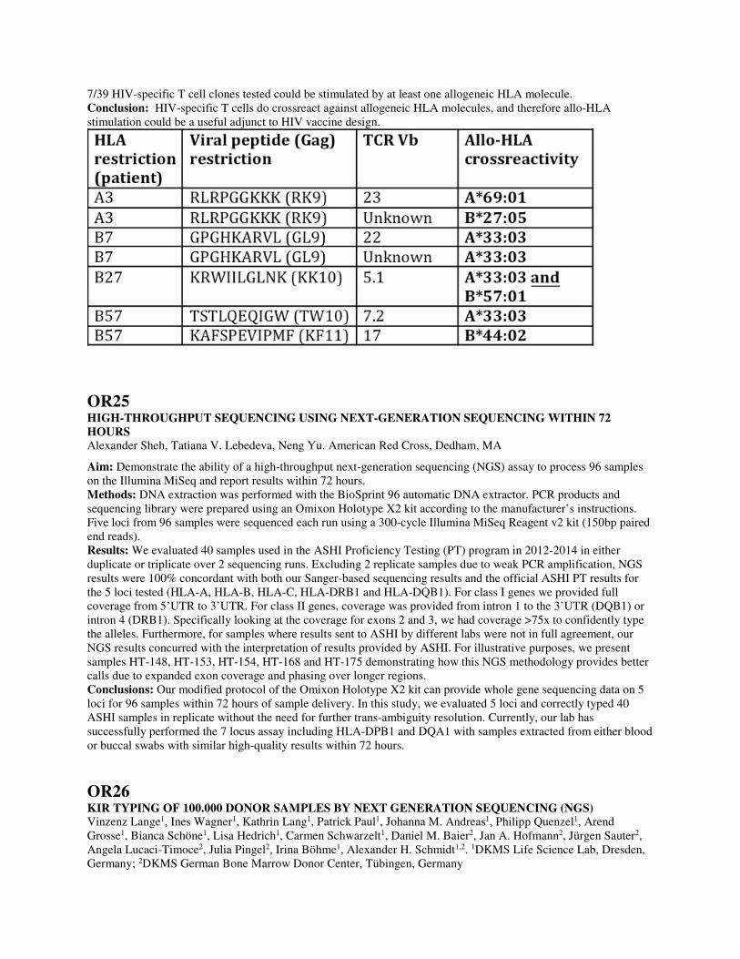
7/39 HIV-specific T cell clones tested could be stimulated by at least one allogeneic HLA molecule. Conclusion: HIV-specific T cells do crossreact against allogeneic HLA molecules, and therefore allo-HLA stimulation could be a useful adjunct to HIV vaccine design.
OR25 HIGH-THROUGHPUT SEQUENCING USING NEXT-GENERATION SEQUENCING WITHIN 72 HOURS Alexander Sheh, Tatiana V. Lebedeva, Neng Yu. American Red Cross, Dedham, MA
Aim: Demonstrate the ability of a high-throughput next-generation sequencing (NGS) assay to process 96 samples on the Illumina MiSeq and report results within 72 hours. Methods: DNA extraction was performed with the BioSprint 96 automatic DNA extractor. PCR products and sequencing library were prepared using an Omixon Holotype X2 kit according to the manufacturer’s instructions. Five loci from 96 samples were sequenced each run using a 300-cycle Illumina MiSeq Reagent v2 kit (150bp paired end reads). Results: We evaluated 40 samples used in the ASHI Proficiency Testing (PT) program in 2012-2014 in either duplicate or triplicate over 2 sequencing runs. Excluding 2 replicate samples due to weak PCR amplification, NGS results were 100% concordant with both our Sanger-based sequencing results and the official ASHI PT results for the 5 loci tested (HLA-A, HLA-B, HLA-C, HLA-DRB1 and HLA-DQB1). For class I genes we provided full coverage from 5’UTR to 3’UTR. For class II genes, coverage was provided from intron 1 to the 3’UTR (DQB1) or intron 4 (DRB1). Specifically looking at the coverage for exons 2 and 3, we had coverage >75x to confidently type the alleles. Furthermore, for samples where results sent to ASHI by different labs were not in full agreement, our NGS results concurred with the interpretation of results provided by ASHI. For illustrative purposes, we present samples HT-148, HT-153, HT-154, HT-168 and HT-175 demonstrating how this NGS methodology provides better calls due to expanded exon coverage and phasing over longer regions. Conclusions: Our modified protocol of the Omixon Holotype X2 kit can provide whole gene sequencing data on 5 loci for 96 samples within 72 hours of sample delivery. In this study, we evaluated 5 loci and correctly typed 40 ASHI samples in replicate without the need for further trans-ambiguity resolution. Currently, our lab has successfully performed the 7 locus assay including HLA-DPB1 and DQA1 with samples extracted from either blood or buccal swabs with similar high-quality results within 72 hours.
OR26 KIR TYPING OF 100.000 DONOR SAMPLES BY NEXT GENERATION SEQUENCING (NGS) Vinzenz Lange1, Ines Wagner1, Kathrin Lang1, Patrick Paul1, Johanna M. Andreas1, Philipp Quenzel1, Arend Grosse1, Bianca Schöne1, Lisa Hedrich1, Carmen Schwarzelt1, Daniel M. Baier2, Jan A. Hofmann2, Jürgen Sauter2, Angela Lucaci-Timoce2, Julia Pingel2, Irina Böhme1, Alexander H. Schmidt1,2. 1DKMS Life Science Lab, Dresden, Germany; 2DKMS German Bone Marrow Donor Center, Tübingen, Germany

Aim: Typing of potential donors for all transplantation relevant factors at registration can speed up donor selection which benefits certain patients. Despite several studies regarding the effect of the KIR repertoire on the outcome of hematopoietic stem cell transplantation (HSCT), KIR typing data is commonly not available for donor selection - to some extent due to the costs of conventional KIR typing methods. Our amplicon-based NGS typing approach has reduced HLA typing costs considerably. Here, we report a method for cost effective high-throughput KIR typing that enables the addition of KIR typing to the standard profile for all newly registered donors. Methods: Exons 4, 5 and 7 are amplified in three PCR reactions using primer mixes targeting all KIR genes. The PCR products contain sample specific identification sequences and can therefore be combined for joint sequencing on Illumina MiSeq or HiSeq instruments. Up to 4800 samples are sequenced on one 2x250 rapid run HiSeq flowcell yielding on average about 60,000 reads per sample evenly split between HLA and KIR. Sequencing data is analyzed by neXtype, an inhouse software, and currently yields presence/absence calls for the KIR genes with KIR2DS4 and KIR2DS4N being distinguished and KIR2DL5A and KIR2DL5B being combined into KIR2DL5. Results: Validation yielded 100 % concordance with the pretypings for all 109 samples passing the predefined internal quality criteria. Within the first 4 months in 2015 we performed successful KIR typing for more than 100.000 samples. Initial analysis indicates that haplotypes lacking the KIR core genes 3DP1 and 2DL4 are more common than previously anticipated. Conclusion: Our amplicon-based NGS typing approach enables us to type up to 5000 samples/day for KIR. Typing costs including KIR increased only moderately compared to the former profile including 6 HLA loci (A, B, C, DRB1, DQB1 and DPB1), CCR5, and blood groups ABO and Rh. This facilitated DKMS to expand the profile for all newly registered donors to include KIR typing.
OR27 MONOCLONAL ANTIBODY RL41A RECOGNIZES CISPLATIN RESISTANT OVARIAN CANCER CELLS VIA HLA-A2 Saghar Kaabinejadian1, Andrea Patterson1, Wilfried Bardet1, Kenneth Jackson1, Cutis McMurtrey1, Timea Wichner2, Oriana Hawkins2, Jon Weidanz2, William Hildebrand1. 1University of Oklahoma Health Sciences Center, Oklahoma City, OK; 2Texas Tech University Health Sciences Center, Abilene, TX
Aim: Cisplatin is widely used as a chemotherapeutic drug in the treatment of ovarian cancer. Resistance to cisplatin occurs in about one-third of women during the primary course of treatment and in all patients treated for recurrent disease. We hypothesized that the HLA class I of cisplatin-resistant ovarian cancer cells presents peptides distinct to these cells as compared to sensitive cells and that HLA/peptide complexes unique to cisplatin-resistant cells would be valuable targets for immunotherapeutic intervention. Methods: To identify peptides that are uniquely presented by the HLA of cisplatin resistant ovarian cancer cells, the intrinsic cisplatin-resistant cells (SKOV3) and sensitive cells (A2780, OV90, FHIOSE) were transfected to express soluble HLA-A*02:01. Transfected resistant and sensitive cells were grown in separate bioreactors. Harvested HLA were purified by immunoaffinity chromatography, and high throughput comparative mass spectrometry was employed to identify the peptids unique to cisplatin-resistant cells. A T cell receptor mimic monoclonal antibody (TCRm mAb) was then generated against cisplatin resistant peptide/HLA-A*02:01 complex, using mouse immunization and hybridoma technology. Results: Peptide sequences distinct to cisplatin-resistant cells were identified including a peptide (VMF11) derived from thioredoxin interacting protein (TXNIP) that was present in abundance in SKOV3. Next a TCRm mAb (RL41A) was produced against A*02:01/VMF11 complex. The specificity and affinity of RL41A toward VMF11/A*02:01 complex was shown by staining peptide-pulsed T2 cells and surface plasmon resonance respectively. Staining of ovarian cancer cells by flow cytometry also showed that RL41A was able to only stain cisplatin-resistant cells and not the sensitive ones. Conclusion: We therefore report the successful development of a TCRm mAb that could be an attractive candidate for further validation using cisplatin-resistant and sensitive primary ovary tissues. W. Hildebrand: Scientific/Medical Advisor; Company/Organization; Pure MHC, LLC.
OR28 VALIDATION OF 2070 COMMON, RARE, AND NOVEL HLA ALLELES USING ILLUMINA TRUSIGHT® HLA ULTRA-HIGH-RESOLUTION SEQUENCING
Fiona Yamamoto1, Alex Lindell2, Brad Baas2, Ali Crawford2, Mellisa Won2, Nate Baird2, Mathew W. Anderson3, James Nytes4, Jennifer J. Schiller4, Damian Goodridge5, Dolly B. Tyan6. 1Stanford University, Palo ALto, CA; 2Illumina Inc., San Diego, CA; 3BloodCenter of Wisconsin, Milwaukee, WI; 4BloodCenter of Wisconsin, Milwaukee, WI; 5Stanford University, Western Australia, Australia; 6Stanford University, Palo ALto, CA
Aim: Sanger sequencing of HLA suffers from the inability to set phase in certain heterozygous combinations. NGS overcomes these problems, but the reliability is not well described. We tested the NGS TruSight® HLA Sequencing Panel (Illumina) for its ability to provide full length, genomic, accurate, unambiguous, phase-resolved HLA genotyping in a single assay on a panel of 145 specimens. Residual clinical, PT, and IHWG DNA samples from blood, buccal swabs, cell lines of varying quality, concentration, and age, were selected to cover every known antigen, as well as rare and novel alleles. Methods: The 145 samples (2070 alleles) included 26 novel variants, 7 null alleles, and 7 PT samples. We used the Illumina NGS HLA genotyping system end-to-end, from Long-Range PCR and library preparation to sequencing on the MiSeq. FASTQ files were analyzed by Conexio Assign, providing phase-resolved genotyping results for HLA-A, -B, -C, -DRB1, -DRB3, -DRB4, -DRB5, -DQA1, -DQB1, -DPA1, -DPB1. We calculated concordance and unambiguous allele level identity in comparison to previously typed high resolution typed results (Sanger/SSP/SSO combined). Results: 87.1% of all clusters (500 cycles) had a Q30 quality score (error rate of 0.1-0.01%). Concordance with original typing was 98.5%. Subdividing concordance, we found: 65.1% identical and unambiguous in reference and NGS; 20.8% unambiguous NGS, ambiguous reference; 9% unambiguous reference, ambiguous NGS; 3.7% ambiguous in both reference and NGS; and 1.5% discordant. Unambiguous reference/ambiguous NGS was the result of primer placement. Discordance (31 alleles) was due to novel alleles, reference errors, homopolymers and microsatellites, pseudogenes, and 3 instances of contamination. Benefits of NGS included discovery and resolution of novel alleles, identification and correction of IHWG reference and clinical mistypings. Analysis of 24 samples for 11 loci took ~3-3.5 hrs. Conclusions: TruSight® HLA Sequencing system is a reliable, accurate, comprehensive, ultra-high-resolution HLA typing method that can be easily implemented in the laboratory. For labs needing the highest resolution typing, it reduces the number of ancillary tests that must be performed to resolve ambiguities from ~50% down to ~14%. For registry labs, the concordance rate equals or exceeds 98.5%. F. Yamamoto: Grant/Research Support; Company/Organization; Illumina, Conexio. A. Lindell: Grant/Research
Support; Company/Organization; Illumina, Conexio. B. Baas: Grant/Research Support; Company/Organization;
Illumina, Conexio. A. Crawford: Grant/Research Support; Company/Organization; Illumina, Conexio. M. Won:
Grant/Research Support; Company/Organization; Illumina, Conexio. N. Baird: Grant/Research Support;
Company/Organization; Illumina, Conexio. M.W. Anderson: Grant/Research Support; Company/Organization;
Illumina, Conexio. J. Nytes: Grant/Research Support; Company/Organization; Illumina, Conexio. J.J. Schiller:
Grant/Research Support; Company/Organization; Illumina, Conexio. D. Goodridge: Grant/Research Support;
Company/Organization; Illumina, Conexio. D.B. Tyan: Grant/Research Support; Company/Organization; Illumina,
Conexio.
Tuesday, September 29, 2015 2:00 PM - 3:30 PM
Workshop 2: Solid Organ Immunotherapy/Rejection
OR15 DE NOVO DEVELOPMENT OF DSA FOLLOWING HUMAN LUNG TRANSPLANTATION IS ASSOCIATED WITH CHANGES ON CIRCULATING MICRO-RNA INVOLVED IN T AND B CELL REGULATION AND FIBROGENESIS Zhongping Xu1, Deepak Nayak1, Elbert Trulock2, Ramsey Hachem2, Daniel Kreisel1, Thalachallour Mohanakumar3. 1Washington University School of Medicine, St. Louis, MO; 2Washington University School of Medicine, St. Louis, MO; 3Washington University School of Medicine, St. Louis, MO
Aim: Chronic rejection (bronchiolitis obliterans syndrome (BOS)) is the major limitation for long-term survival after lung transplantation (LTx). Its pathogenesis, however, is poorly understood and no effective predictive biomarkers have been identified. Several studies have shown that de novo development of antibodies to donor mismatched HLA (DSA) is a significant risk factor for the development of BOS. Methods: Thirty LTx recipients from Barnes Jewish Hospital/Washington University School of Medicine (10 stable DSA- and BOS-, 10 DSA+ BOS- and 10 DSA+ and BOS+) were analyzed for circulating microRNAs (miRNAs). MiRNAs expression in the recipients’ serum was detected using RNA extraction and quantitative PCR analysis. Results: We identified eight miRNAs which were selectively expressed on lung allograft recipients with de novo developed DSA and diagnosed with BOS in comparison to stable LTx without DSA and BOS: miR-369-5p, miR-144, miR-134, miR-10a, miR-195 miR-142-5p, miR-133b, and miR-155 (p<0.01). Among them, transforming growth factor beta (TGF-β) associated miRNAs: miR-369-5p was down regulated (2.3 fold, p<0.001), miR-144 was up-regulated (3.1 fold, p<0.001) in the serum of LTx with DSA compared to stable, indicating their role in fibrogenesis mediated by TGF-β signalling. In addition, miR-134 involved in B cell receptor pathway was decreased (p<0.001) in the serum of LTx with DSA and BOS, demonstrating its role in B cell activation and DSA development following LTx. Furthermore, miR-10a, known to be associated with Treg development, was down-regulated (p<0.001) in LTx with DSA and BOS, demonstrating its involvement in down regulation of Treg function leading to development of DSA. Finally, results obtained from independent validation using an independent cohort of 9 stable, 7 DSA+BOS- , and 10 DSA+BOS+ LTx demonstrated that these miRNAs can discriminate LTx with development of DSA and BOS from those stable LTx without DSA and BOS (p<0.01). Conclusion: Our results, for the first time, demonstrated differential expression of circulating miRNAs in lung allograft recipients with de novo development of DSA and BOS. These dysregulated miRNAs are involved in T and B cell regulation and fibrogenesis, indicating their role in the development of DSA leading to chronic lung allograft rejection.
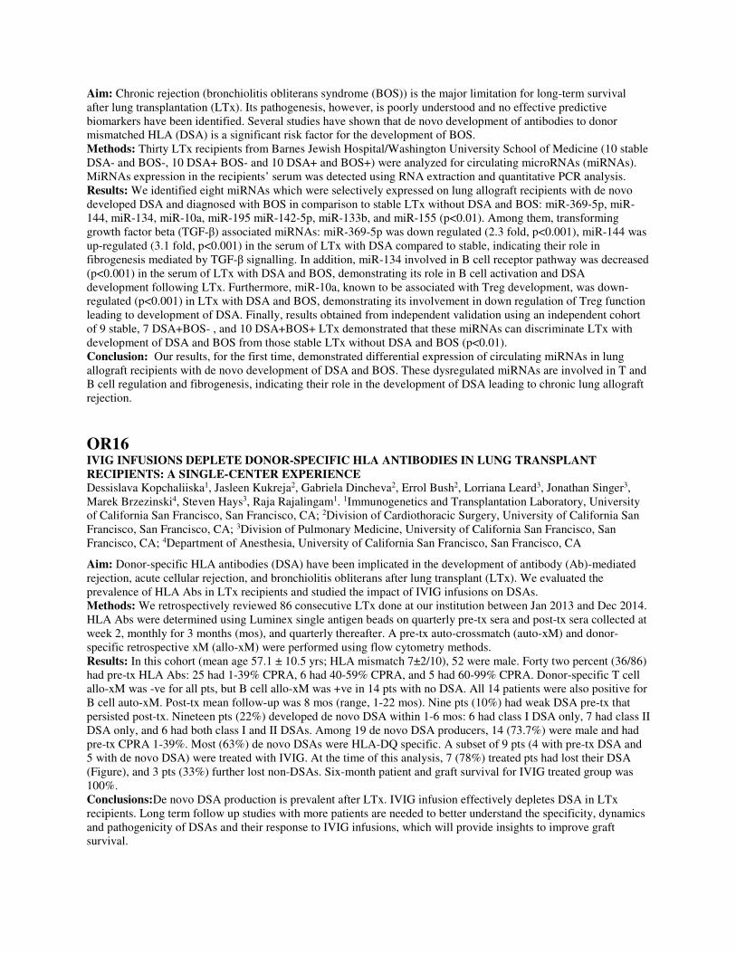
OR16 IVIG INFUSIONS DEPLETE DONOR-SPECIFIC HLA ANTIBODIES IN LUNG TRANSPLANT RECIPIENTS: A SINGLE-CENTER EXPERIENCE Dessislava Kopchaliiska1, Jasleen Kukreja2, Gabriela Dincheva2, Errol Bush2, Lorriana Leard3, Jonathan Singer3, Marek Brzezinski4, Steven Hays3, Raja Rajalingam1. 1Immunogenetics and Transplantation Laboratory, University of California San Francisco, San Francisco, CA; 2Division of Cardiothoracic Surgery, University of California San Francisco, San Francisco, CA; 3Division of Pulmonary Medicine, University of California San Francisco, San Francisco, CA; 4Department of Anesthesia, University of California San Francisco, San Francisco, CA
Aim: Donor-specific HLA antibodies (DSA) have been implicated in the development of antibody (Ab)-mediated rejection, acute cellular rejection, and bronchiolitis obliterans after lung transplant (LTx). We evaluated the prevalence of HLA Abs in LTx recipients and studied the impact of IVIG infusions on DSAs. Methods: We retrospectively reviewed 86 consecutive LTx done at our institution between Jan 2013 and Dec 2014. HLA Abs were determined using Luminex single antigen beads on quarterly pre-tx sera and post-tx sera collected at week 2, monthly for 3 months (mos), and quarterly thereafter. A pre-tx auto-crossmatch (auto-xM) and donor-specific retrospective xM (allo-xM) were performed using flow cytometry methods. Results: In this cohort (mean age 57.1 ± 10.5 yrs; HLA mismatch 7±2/10), 52 were male. Forty two percent (36/86) had pre-tx HLA Abs: 25 had 1-39% CPRA, 6 had 40-59% CPRA, and 5 had 60-99% CPRA. Donor-specific T cell allo-xM was -ve for all pts, but B cell allo-xM was +ve in 14 pts with no DSA. All 14 patients were also positive for B cell auto-xM. Post-tx mean follow-up was 8 mos (range, 1-22 mos). Nine pts (10%) had weak DSA pre-tx that persisted post-tx. Nineteen pts (22%) developed de novo DSA within 1-6 mos: 6 had class I DSA only, 7 had class II DSA only, and 6 had both class I and II DSAs. Among 19 de novo DSA producers, 14 (73.7%) were male and had pre-tx CPRA 1-39%. Most (63%) de novo DSAs were HLA-DQ specific. A subset of 9 pts (4 with pre-tx DSA and 5 with de novo DSA) were treated with IVIG. At the time of this analysis, 7 (78%) treated pts had lost their DSA (Figure), and 3 pts (33%) further lost non-DSAs. Six-month patient and graft survival for IVIG treated group was 100%. Conclusions:De novo DSA production is prevalent after LTx. IVIG infusion effectively depletes DSA in LTx recipients. Long term follow up studies with more patients are needed to better understand the specificity, dynamics and pathogenicity of DSAs and their response to IVIG infusions, which will provide insights to improve graft survival.
OR17 HEME OXYGENASE-1 MODULATES HLA CLASS I ANTIBODY-DEPENDENT ENDOTHELIAL CELL ACTIVATION Eva Zilian1, Hendry Saragih1, Oliver Hiller1, Abid Aljabri1, Constanca Figueiredo1, Rainer Blasczyk1, Gregor Theilmeier2, Jan Ulrich Becker3, Jan Larmann4, Stephan Immenschuh1. 1Hannover Medical School, Hannover, Germany; 2Hannover Medical School, Hannover, Germany; 3University of Cologne, Cologne, Germany; 4Hannover Medical School, Hannover, Germany
Aim: Antibody-mediated rejection (AMR) is a key limiting factor for long-term graft survival in heart and kidney transplantation. Activation of endothelial cells (ECs) via complement-independent effects of human leukocyte antigen class I (HLA I) antibodies (Abs) plays a major role in the pathogenesis of AMR. As the antioxidant enzyme heme oxygenase (HO)-1 is known to have cell type-specific anti-inflammatory effects in the endothelium, we investigated its role on HLA I Ab-dependent activation of human ECs. Methods: Regulation of inducible proinflammatory endothelial adhesion molecules and chemokines (VCAM-1, ICAM-1, IL-8 and MCP-1) by monoclonal pan- and allele-specific HLA I Abs was determined in cell cultures of primary human umbilical venous, aortic macrovascular and microvascular ECs. HO-1 was modulated by pharmacological regulators and siRNA-mediated knockdown. Adherence of THP-1 monocytes to ECs was determined by leukocyte adhesion assay. Results: Exposure of human macro- and microvascular EC cultures to HLA I Abs caused endothelial activation, as indicated by up-regulation of VCAM-1, ICAM-1, MCP-1 and IL-8. This up-regulation was mediated via the phosphatidylinositol-3 kinase (PI3K)/Akt and NF-κB pathways. Pharmacological induction of HO-1 with cobalt-protoporphyrin IX reduced, whereas inhibition of HO-1 with either zinc-protoporphyrin IX or siRNA-mediated knockdown increased HLA I Ab-dependent EC activation. Binding of THP-1 monocytes was enhanced in HLA I Ab-stimulated ECs. This effect was counteracted by HO 1 up-regulation. Conclusion: HLA I Ab-dependent EC activation is modulated by specific HO-1 up-regulation. Thus, targeted

regulation of endothelial HO-1 may be a novel therapeutic approach for the treatment of AMR in kidney and heart transplantation. S. Immenschuh: Grant/Research Support; Company/Organization; Else Kröner-Fresenius Stiftung EKFS
2012_A309.
OR18 C3D-BINDING DE NOVO DONOR-SPECIFIC HLA ANTIBODIES AND ANTIBODY-MEDIATED REJECTION OF KIDNEY TRANSPLANTS Dessislava Kopchaliiska1, Manpreet Singh2, Owen Buenaventura1, Vasishta Tatapudi2, Stephen Tomlanovich2, Raja Rajalingam1. 1Immunogenetics and Transplantation Laboratory, University of California San Francisco, San Francisco, CA; 2Division of Nephrology, Kidney Transplant Service, University of California San Francisco, San Francisco, CA
Aim: Antibody-mediated rejection (AMR) is a major cause of kidney graft loss, yet assessment of individual risk at diagnosis is impeded by the lack of a reliable prognosis assay. Here, we tested whether the capacity of HLA antibodies to bind complement component C3d allows accurate risk stratification at the time of AMR diagnosis. Methods: Sera from kidney transplant recipients, who underwent a protocol or for-cause kidney biopsy and had detectable de novo DSA (by One Lambda) at the time of biopsy (median 3.8 yrs post-tx), were included in this study. These serum samples were re-tested using the Immucor single antigen beads with and without C3d detection system. Results: This study included samples from 123 kidney recipients (70 males; 14 re-Tx; 46 LD) transplanted in our center between 1989 and 2011. Fifty-seven patients (46%) had C3d-binding DSA. Most C3d-binding DSAs were high MFI DSAs (11700+5188), and only 4/57 (7%) C3d-binding DSAs had <5000 MFI. Seventy percent of the patients with C3d-binding DSA (40/57) had AMR (18 aAMR and 22 cAMR) and C4d-positive biopsies; twenty-six percent (15/57) had ACR, and four percent (2/57) had negative biopsies. Fifty two present of the patients (34/66) in the C3d-negative DSA group had DSA with MFI<5000. Some of the weak and moderate DSA detected by One Lambda single antigen bead reagents were not detected with the Immucor SAB. Among the patients with C3d-negative DSA, thirty-five percent (23/66) had AMR (7 aAMR and 16 cAMR); twelve percent (8/66) had ACR and thirty-three percent (22/66) had C4d-positive biopsies. In most cases 15/22 (68%), the C4d-positive biopsies were observed in patients with strong DSA (MFI>5000). Conclusions:Our data indicate a strong correlation between the presence of C3d-binding DSAs and AMR. C3d-binding antibodies seem to be prevalent to stronger antibodies. Further studies are needed to evaluate whether the presence of C3d-binding donor-specific antibodies can predict AMR and identify patients who are at increased risk of allograft failure.

OR19 SUCCESSFUL REVERSAL OF SEVERE KIDNEY ALLOGRAFT REJECTION MEDIATED BY ANGIOTENSIN II TYPE 1-RECEPTOR ANTIBODIES James H. Lan1,2, Qiuheng Zhang1, Elaine F. Reed1, Uttam Reddy3. 1UCLA Immunogenetics Center, Los Angeles, CA; 2University of British Columbia, Vancouver, BC, Canada; 3UCLA David Geffen School of Medicine, Los Angeles, CA
Case Study: The patient is a non-sensitized 64 year-old African American male who received a split en bloc deceased donor kidney transplant with standard induction using solumedrol and basiliximab. Post-discharge his renal function remained stable with a baseline creatinine between 1.0-1.2 mg/dL. During this time he experienced severe GI side effects related to MMF which led to its temporary discontinuation for 2 weeks, followed by resumption at reduced dosage (250 mg BID). One week later, his creatinine acutely worsened from 1.0 to 3.1 mg/dL. The patient’s allograft pathology showed mixed acute C4d+ antibody-mediated rejection (AMR) and cell-mediated rejection with transmural arteritis (Banff scores g1, t3, i3, v3, ptc1). Surprisingly, neither anti-HLA nor MICA antibodies were identified in any of his post-transplant sera. Further investigation uncovered anti-angiotensin II type-1 receptor antibodies (AT1R ab) and endothelial cell crossmatch (ECXM) positivity which correlated with the timing of his acute rejection. Fig. 1 illustrates the kinetics of the patient’s AT1R ab level in relationship to his clinical course. Pre-transplant, high AT1R ab binding was detected at 18 U/ml (positive cutoff > 17 U/ml) - this reactivity increased to 27 U/ml on the day of his allograft biopsy. In parallel with this surge, the patient’s creatinine deteriorated to a peak of 5.7 mg/dL necessitating temporary dialysis. Of note, his blood pressure which had been previously well-controlled converted to a state of hypertensive urgency requiring IV nicardipine infusion. He was treated aggressively with ATG, plasmapheresis, IVIG, and losartan. After one week of treatment his AT1R ab level became undetectable; remarkably, the patient also showed rapid clinical recovery and control of his blood pressure. Post-discharge his renal function returned to his previous baseline. He continues to take losartan for AT1R blockade and is treated with short courses of plasmapheresis and IVIG to reduce his anti-AT1R level as necessary.

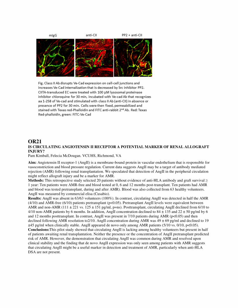
OR20 HLA CLASS II LIGATION BY ANTIBODY INDUCES ENDOTHELIAL CELL PERMEABILITY BY PROMOTING ENDOCYTOSIS OF VE-CADHERIN Fang Li, Elaine F. Reed. UCLA, Los Angeles, CA
Aim: Increased microvascular dilatation and permeability occurs during allograft rejection and contributes to fibrotic remodeling and organ dysfunction in transplant recipients. Endothelial cells (EC) control vascular permeability by regulating cell-to-cell junctions. We hypothesize that HLA class II ligation on EC transduces signals that disrupt cell-cell tight junctions resulting in increased cell permeability. Methods: Class II expression was achieved by transducing EC with adenoviral recombinant CIITA. EC were stimulated with mAb against monomorphic determinant present on all HLA class II antigens (F26C6G1). Western blots were used to characterize protein phosphorylation. Cell permeability was measured by FITC-dextran transwell permeability assay and Ve-Cadherin (Ve-Cad) surface expression was determined by flow cytometry. Cell junctions and Ve-cad internalization were quantified by immunofluorescence microscopy. Results: Treatment of EC with anti-class II Ab activated Src, stimulated Ve-Cad phosphorylation at tyr685 and increased Ve-Cad internalization (Fig). Class II ligation decreased Ve-cad surface expression by 42±5%, which was blocked by PP2. In the presence of chloroquine, class II ligation resulted in a 27±4% increase in Ve-cad internalization. Immunofluorescent studies showed that Ve-Cad in the vesicles was phosphorylated and class II ligation increased pY685-Ve-Cad expression. Preincubation of EC with PP2 decreased Ve-cad phosphorylation and internalization. Class II Ab increased FITC-dextran permeability confirming that class II signaling resulting in increased vascular permeability. Our data show that class II ligation on EC mediates the disassembly of intercellular junctions by stimulating Ve-Cad phosphorylation and internalization in a Src dependent manner. Conclusions:We provide evidence that HLA class II DSA promote endocytosis of Ve-Cad via Src activation, disrupting endothelial barrier function and contributing to fibrotic remodeling and organ dysfunction.
OR21 IS CIRCULATING ANGIOTENSIN II RECEPTOR A POTENTIAL MARKER OF RENAL ALLOGRAFT INJURY? Pam Kimball, Felecia McDougan. VCUHS, Richmond, VA
Aim: Angiotensin II receptor-1 (AngII) is a membrane-bound protein in vascular endothelium that is responsible for vasoconstriction and blood pressure regulation. Current data suggests AngII may be a target of antibody mediated rejection (AMR) following renal transplantation. We speculated that detection of AngII in the peripheral circulation might reflect allograft injury and be a marker for AMR. Methods: This retrospective study selected 20 patients without evidence of anti-HLA antibody and graft survival ≥ 1 year: Ten patients were AMR-free and blood tested at 0, 6 and 12 months post-transplant. Ten patients had AMR and blood was tested pretransplant, during and after AMR). Blood was also collected from 63 healthy volunteers. AngII was measured by commercial elisa (Cusabio). Results: AngII was absent in 63/63 volunteers (100%). In contrast, circulating AngII was detected in half the AMR (4/10) and AMR-free (6/10) patients pretransplant (p<0.05). Pretransplant AngII levels were equivalent between AMR and non-AMR (111 ± 221 vs. 125 ± 151 pg/ml, p=ns). Posttransplant, circulating AngII declined from 6/10 to 4/10 non-AMR patients by 6 months. In addition, AngII concentration declined to 84 ± 135 and 22 ± 50 pg/ml by 6 and 12 months posttransplant. In contrast, AngII was present in 7/10 patients during AMR (p<0.05) and then declined following AMR resolution to2/10. AngII concentration during AMR was 49 ± 69 pg/ml and declined to 19 ±45 pg/ml when clinically stable. AngII appeared de novo only among AMR patients (5/10 vs. 0/10, p<0.05). Conclusions:This pilot study showed that circulating AngII is lacking among healthy volunteers but present in half of patients awaiting renal transplantation. Neither the presence or the concentration of AngII pretransplant predicted risk of AMR. However, the demonstration that circulating AngII was common during AMR and resolved upon clinical stability and the finding that de novo AngII expression was only seen among patients with AMR suggests that circulating AngII might be a useful marker in detection and treatment of AMR, particularly when anti-HLA DSA are not present.
Wednesday, September 30, 2015 2:30 PM - 4:00 PM
Abstract Session 3: HLA for Anthropology/Disease Association/Genetic Polymorphism (MHC/MIC/KIR/Cytokines)
OR8 HIV INFECTION LEADS TO THE PRESENTATION OF UNEXPECTEDLY LONG PEPTIDES BY HLA-A*11:01 Jane C. Yaciuk1,2, Steven Cate1, Curtis P. McMurtrey1,2, Matthew Skaley1, Wilfried Bardet1, Kenneth W. Jackson1, William H. Hildebrand1,2. 1University of Oklahoma Health Sciences Center, Oklahoma City, OK; 2Pure MHC, LLC, Oklahoma City, OK
Aim: Vaccines for HIV-1 have proven ineffective due to the inability of the immune response to cope with HIV-1 diversity/antigenic variation. We proposed to identify host (human) ligands uniquely presented by Class I HLA during infection as these host targets are not likely to mutate. Deep Ligand Sequencing (DLS) was used to determine changes in the class I HLA presented host ligand repertoire after HIV-1 infection of CD4+ T cells. In this manner we identified host-derived ligands that mark the surface of HIV infected cells. Methods: Soluble class I HLA-A*11:01 (sHLA) was harvested from HIV-1 (NL4-3)-infected and uninfected human CD4+ SUP-T1 cells. Ligands from both infected and uninfected cells were purified and then fractionated by high pH HPLC. NanoLC-MS ligand fragment spectra were collected on all peptide containing fractions. Ligand sequences were determined from tandem MS spectra (DDA & DIA) using PEAKS v7.0 and Protein Pilot v4.5 at a 1% FDR. Ligand intensity data was extracted from the DIA spectra using Peakview v2.1 and the SWATH microapp. Normalization and log fold increase were determined using Excel. Results: Quantitative values were obtained for a total of 5222 distinct HLA-A11 ligands from both infected and uninfected cells. Strikingly, over half of the ligands (55%) were unique to HIV-1 infected cells. These unique host ligands were significantly longer than the ligands found in uninfected cells with an average length of 12.2 amino acids as compared to 10.6 amino acids for uninfected ligands. Although ligands were longer, there were no significant changes in motif, and there was no difference in the source proteins providing peptides. Conclusions:Our group and others have previously reported long (>11 aa) HIV-1 peptide ligands that are recognized by CTL. Here, we observe a significant shift in the length of the host ligand repertoire as well. This suggests that HIV alters the antigen processing pathway to increase ligand length, possibly by inhibition of host cell peptidases. Since HLA-A11 is more permissive in binding longer ligands, we are able to observe this shift in length. However, with allomorphs like HLA-A2 that are not as length permissive, this could represent an immune escape mechanism for HIV. This ability to bind long ligands may partially explain why HLA-A11 is considered a correlate of protection. W.H. Hildebrand: Consultant; Company/Organization; Pure MHC, LLC.
OR9 MULTIETHNIC RNA SEQUENCING ANALYSIS OF HLA REVEALS HLA-ALLELE SPECIFIC EQTLS Hanna M. Ollila1, Otto Jolanki1, Jill A. Hollenbach2, Paul Norman3, Emmanuel Mignot1. 1Stanford University, Palo Alto, CA; 2University of California San Francisco, San Francisco, CA; 3Stanford University, Palo Alto, CA
Aim: The HLA is major regulator for immune responses and several auto-immune or inflammatory diseases show a strong association with specific HLA alleles. However, it remains unclear what is the effect of individual HLA alleles on variation in cell phenotype and gene expression that potentially contribute to disease predisposition. Our aim was to characterize the role of individual HLA-alleles on gene expression over two ethnic groups and multiple populations. Methods: We used RNA sequencing from lymphoblastoid cell lines, including HLA region, combined with HLA typing data from the 1000 genomes and Geuvadis projects in 462 individuals from European (CEPH, Finnish, British, Toscani) and African (Yoruba) populations. The analysis was normalized using PANAMA and analyzed with linear mixed model in R v3.2.0. Results: The effect of HLA-alleles on gene expression revealed several genome-wide significant cis-eQTLs for

other genes at the HLA region. Importantly, we also characterized several robust trans-eQTL signals for HLA class I and class II alleles (P<1*10-8) that were enriched in genes mediating immune responses and shared among different populations. Conclusion: HLA-alleles have a robust effect on gene expression and cellular phenotype that is shared across multiple populations.
OR10 GENETIC PREDISPOSITION TO INTERLEUKIN-10 PRODUCTION INFLUENCES EPSTEIN-BARR VIRUS REACTIVATION AFTER RENAL TRANSPLANTATION Gaurav Tripathi1, Abdulnaser Abadi1, Poonam Dharmani-Khan1, Lee Anne Tibbles1, Serdar Yilmaz1, Noureddine Berka2,1, Faisal M. Khan1,2. 1University of Calgary, Calgary, AB, Canada; 2Calgary Laboratory Services, Calgary, AB, Canada
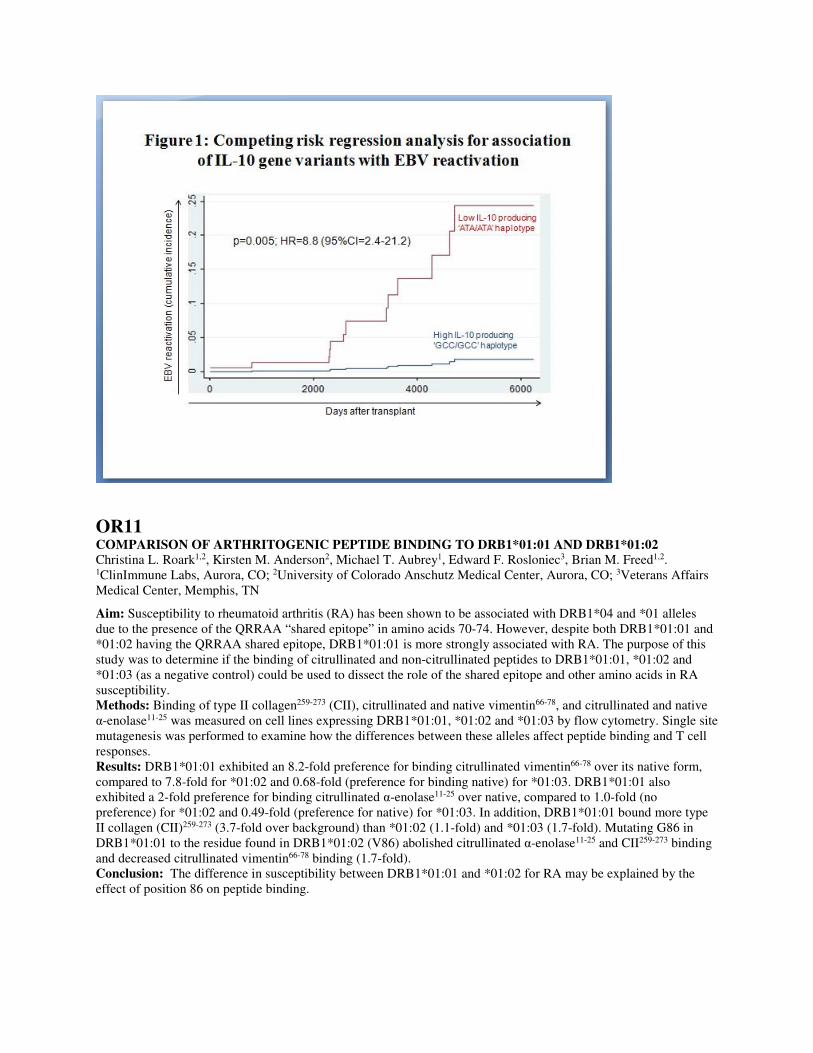
Aim: Epstein-Barr virus (EBV) is responsible for posttransplant lymphoproliferative disorder (PTLD) after solid organ transplantation. A poor anti-EBV immune response in transplant recipients ultimately leads to EBV reactivation and complications. Genetic predisposition of cytokine production is a feature of variation in cytokine gene regulatory regions that may influence the anti-EBV immune response. We analyzed a panel of 17 cytokine gene variants and examined their influence on EBV reactivation after kidney transplantation. Methods: A total of 270 renal transplant recipients (discovery cohort, n=189 and validation cohort, n=81) were analyzed. Seventeen gene variants located in the regulatory and/or exonic regions of 11 cytokine/cytokine receptor genes were genotyped by Luminex based SSO panels or direct sequencing. EBV reactivation was defined by EBV DNAemia. Results: Three variants of IL-10 promoter region (-1082G/A, -819C/T, -592C/A) that lead to low production of IL-10 cytokines were found strongly associated with EBV reactivations. The multivariate logistic regression analysis showed that recipients carrying low IL10 producing haplotypes (ATA) have higher incidence of EBV reactivation (p=0.005, HR=8.8, 95% CI: 2.4-21.2) (Figure 1). The competing risk regression analysis showed that the cumulative incidences of EBV reactivation in recipients carrying low IL-10 producing (ATA) haplotype was 24% (95%CI: 18-52%), whereas those in recipients carrying high IL-10 producing (GCC) haplotype was only 3% (95%CI: 1-8%). Conclusions:Renal transplant recipient carrying low IL-10 producing gene variants have higher incidence of EBV reactivation. The findings may lead to development of a better and broader predictive model for EBV reactivations after transplantation involving genetic, serum and functional biomarkers.
OR11 COMPARISON OF ARTHRITOGENIC PEPTIDE BINDING TO DRB1*01:01 AND DRB1*01:02 Christina L. Roark1,2, Kirsten M. Anderson2, Michael T. Aubrey1, Edward F. Rosloniec3, Brian M. Freed1,2. 1ClinImmune Labs, Aurora, CO; 2University of Colorado Anschutz Medical Center, Aurora, CO; 3Veterans Affairs Medical Center, Memphis, TN
Aim: Susceptibility to rheumatoid arthritis (RA) has been shown to be associated with DRB1*04 and *01 alleles due to the presence of the QRRAA “shared epitope” in amino acids 70-74. However, despite both DRB1*01:01 and *01:02 having the QRRAA shared epitope, DRB1*01:01 is more strongly associated with RA. The purpose of this study was to determine if the binding of citrullinated and non-citrullinated peptides to DRB1*01:01, *01:02 and *01:03 (as a negative control) could be used to dissect the role of the shared epitope and other amino acids in RA susceptibility. Methods: Binding of type II collagen259-273 (CII), citrullinated and native vimentin66-78, and citrullinated and native α-enolase11-25 was measured on cell lines expressing DRB1*01:01, *01:02 and *01:03 by flow cytometry. Single site mutagenesis was performed to examine how the differences between these alleles affect peptide binding and T cell responses. Results: DRB1*01:01 exhibited an 8.2-fold preference for binding citrullinated vimentin66-78 over its native form, compared to 7.8-fold for *01:02 and 0.68-fold (preference for binding native) for *01:03. DRB1*01:01 also exhibited a 2-fold preference for binding citrullinated α-enolase11-25 over native, compared to 1.0-fold (no preference) for *01:02 and 0.49-fold (preference for native) for *01:03. In addition, DRB1*01:01 bound more type II collagen (CII)259-273 (3.7-fold over background) than *01:02 (1.1-fold) and *01:03 (1.7-fold). Mutating G86 in DRB1*01:01 to the residue found in DRB1*01:02 (V86) abolished citrullinated α-enolase11-25 and CII259-273 binding and decreased citrullinated vimentin66-78 binding (1.7-fold). Conclusion: The difference in susceptibility between DRB1*01:01 and *01:02 for RA may be explained by the effect of position 86 on peptide binding.

OR12 INCREASED HLA HOMOZYGOSITY IN LYMPHOMA BUT NOT IN ACUTE MYELOGENOUS LEUKEMIA Danielle R. Crow, Michael T. Aubrey, Brian M. Freed. ClinImmune Labs, Aurora, CO
Aim: Previous studies have shown an association between HLA and disease processes, such as CLL. The goal of this study was to determine if HLA homozygosity was associated with lymphoma or AML. Methods: We analyzed 530 lymphoma and 750 AML patients who were typed for possible stem cell transplant from late 2006 to early 2015. All patients were typed at HLA-A, B, C, DRB1, and DQB1. Control data consisted of 4683 normal subjects whose HLA alleles were imputed from GWAS analysis. Homozygosity was defined as two alleles belonging to the same P-group at any HLA locus. Results: 40.6% of the lymphoma patients were homozygous for one or more HLA locus (p=0.005), compared to 34.4% of the control subjects and 33.2% of AML patients (p=0.562). The HLA-A,B and DQB1 loci appeared to contribute most of the difference in homozygosity. Patients who were diagnosed with lymphoma at an earlier age (<40 years, n=116) had a higher degree of homozygosity (47.4%) than patients >40 years old (n=414, 38.6%), although the difference is not statistically significant (p=0.11) with this sample size. Conclusions:HLA homozygosity, particularly at HLA-A,B and DQB1, is associated with a higher incidence of lymphoma, but not AML.

OR13 HUMAN LEUKOCYTE ANTIGEN ANALYSIS USING HIGH RESOLUTION SNP DATA: IMPUTATION, ASSOCIATION AND AMINO ACID BINDING POCKET RESIDUES INVESTIGATION IN IGAD PATIENTS Che Kang Lim1,2, Yeow Tee Goh3,4, Lennart Hammarstrom2. 1Singapore General Hospital, Singapore, Singapore; 2Karolinska Institutet, Stockholm, Sweden; 3Division of Research, Singapore General Hospital, Singapore, Singapore; 4Singapore General Hospital, Singapore, Singapore
Background: Genetic variation at human leukocyte antigen (HLA) alleles influences many phenotypes and mediate susceptibility to a wide range of human diseases include Immunoglobulin A Deficiency (IgAD). The high resolution SNP genotyping has enabled large cohort studies in this complex region. Aim: To Investigate the HLA association of IgAD using high resolution SNP data and to identify the combinations of biologically relevant amino acid (AA) residues directly involved in disease. Methods: HLA imputation was performed in 1046 samples including 758 IgAD individual and 288 ethnically matched controls. The imputation results were further confirmed by high resolution typing. Subsequently, multiple tools include SKDM HLA tools, SNP2HLA and Plink were used to investigate the amino acid binding pocket association in IgAD cohorts. Finally, 3D visualization of the predicted HLA amino acid sequence was performed using RaptorX. Results: Strong IgAD association were observed in HLA- B*0801 (p= 5.364*10-14), HLA- B*1402 (p=0.0016), HLA-DRB1*0301(p= 3.276*10-12), HLA-DRB1*0102(p= 0.001*10-12), HLA-DRB1*1201(p= 1.684*10-4), HLA-DQB1*0201 (p= 1.835*10-12). Conversely, HLA-B*1501(p= 1.358*10-5), HLA-B*0702 (p= 5.662*10-7), HLA-DRB1*1501(p= 6.34*10-23), HLA-DQB1*0402 (p= 0.004) and HLA-DQB1*0602 (p= 9.396*10-23) showed strong protection against IgAD. In addition, most significant AA residues in different HLA region associated with IgAD are: HLA-B ((AA 9, Asp, pockets 2 and 3, p= 8.047*10-14); (AA 156, Asp, pockets 4 and 5, p= 4.396*10-13)); HLA-DRB1 ((AA 26, Tyr, pocket 4, p= 8.249*10-10); (AA 67, Leu, pocket 7, p= 5.236*10-8)); HLA-DQB1 ((AA 38, Val, pocket 9, p= 4.894*10-10); (AA 37, Ile, pocket 9, p= 8.216*10-9), (AA 28 Ser and AA 47 Phe, pocket 7, p= 8.216*10-9); (AA 30, Ser, pocket 6, p= 8.216*10-9); (AA 71, Lys, pockets 4 and 7, p= 8.216*10-9)). Conclusion: The results suggested that certain peptide-binding residues of the HLA class I and class II molecule is associated with susceptibility to IgAD.
OR14 CHARACTERIZATION OF HLA CLASS I NEW ALLELES WITH INSERTIONS AND DELETIONS IN EXONS AND INTRONS Nezih Cereb, Soo Young Yang. Histogenetics, Ossining, NY
Recent advances in DNA sequencing technologies, the so-called Next Generation Sequencing technologies (NGS), introduced breakthroughs in deciphering the complex genetic information in all living species in large scale and affordable level. Since October 2014, we have been routinely using PacBio for HLA class I typing for resolving exon shuffling ambiguities and the new alleles. We have performed more than 5000 HLA-ABC 1 kb amplicon sequencing and typing on PacBio platform. Recently we also introduced full gene length typing -3.5 kb-8 exons and seven introns as routine typing method for the new alleles that have been detected but were not be able to be sequenced in isolation, especially the ones with insertions and deletion. In this study we will summarize characterization of several hundred new alleles with insertions and deletions in exons and introns using PacBio platform
Wednesday, September 30, 2015 2:30 PM - 4:00 PM
Abstract Session 4: Solid Organ Transplantation
OR29

COMPUTER ASSISTED ALGORITHM FOR ASSESSING HLA DPB1 DONOR:RECIPIENT COMPATIBILITY. Geoffrey H. Smith1, Howard M. Gebel2, Robert A. Bray2. 1Emory University, Atlanta,, GA; 2Emory University, Atlanta, GA
Aim: In November 2014, the OPTN/UNOS Board of Directors mandated that DPB1(DP) typing be performed for deceased donors (DD) predicated on data showing that a significant number of highly sensitized patients possessed antibodies to these antigens (AJT 15:284; 2015). Currently, there are >430 DP alleles but only 27 are represented on single antigen bead (SAB) products. Thus, for donor DP antigens that do not correspond to a SAB, a sensitized recipient’s compatibility cannot be easily determined. To address this problem, we developed a computer-assisted tool to assess donor:recipient (D/R) DPB1 compatibility. Methods: We compared the amino acid (AA) sequence of D/R DP alleles to the AA sequence of DP alleles to which the recipient had antibodies as defined by SAB testing. Compatibility assessments were based on the 6 recognized hypervariable regions (HVRs A-F) for DPB1 (Tissue Antigens 75:278; 2010) associated with HLA antibodies. Based on this comparison, donor DP alleles were classified as “incompatible” or “likely compatible” using a custom Java web application. Results: This software tool creates a static equivalency table for all DP alleles, classifying them based on similarity of the 6 HVRs. Furthermore, it specifically compares the D/R HVR AA sequences and renders an assessment of compatibility (Fig 1). Conclusions: Given the impending requirement for DP typing, we reasoned that a tool to predict D/R DP compatibility would be useful, particularly since there are far more DP alleles than present on any current SAB products. While the software tool successfully identified incompatible pairings, it was limited to differences in the 6 common HVRs; the tool was not designed to identify incompatibilities due to SNP differences between DP alleles sharing the same 6 HVRs. Nonetheless, we believe this tool will be an invaluable asset to organ allocation, readily identifying incompatibilities between donors and recipients for the 6 DP HVRs and, by extension, inferring when compatibility between donors and recipients is likely.

OR30 HLA-DPB1* ANTIGEN FREQUENCIES IN DECEASED DONORS:MINING THE DATA Aaron Karas1, Nalaja Marcus-Freeman1, Charlene Breitenbach2, Tracy T. McRacken3, Robert A. Bray1, Howard Gebel1. 1Emory University Hosptial, Atlanta, GA; 2Henrico Doctors' Hospital, Richmond, VA; 3Sentara Norfollk General Hospital, Norfolk, VA
Aim: The OPTN recently implemented a new process wherein renal transplant candidates with cPRA values of 99% and 100% are given regional and national priority, respectively, for deceased donor (DD) kidneys. While the majority of these highly sensitized patients have HLA-DP antibodies, allocation offers frequently occur without donor HLA-DP typing. Furthermore, information regarding the frequency and distribution of HLA-DP antigens is not readily available. Herein we report the frequency and distribution of HLA-DP antigens in 1168 DDs typed over the past three years. Methods: HLA-DPB1* typing was performed by SSO, SSP (One Lambda, Thermal-Fisher) or RT-PCR (Linkage Biosciences). Results: Donors were 58.9% white, 32.4% black, 7.3% Hispanic and 1.4% API. As shown below, the frequency and distribution of HLA-DPB1 antigens varied significantly by race. For example, HLA-DPB1*04:01 is seen in 64.6% of whites and 16.2% of blacks while HLA-DPB1*01:01 is in 53.4% of black donors and only 9.1% of whites. Some HLA-DPB1* alleles appear racially restricted; e.g., HLA-DPB1*10:01 was observed in white and Hispanic but not black donors, while HLA-DPB1*85:01 was seen only in black donors. Multiple HLA-DPB1* alleles with a frequency of >1% are not represented on any single antigen bead (SAB) products used to detect HLA antibodies. In

contrast, SAB manufacturers allotted up to three beads for HLA-DPB1* 28:01, an allele not observed in any of the 1168 donors in this study. Conclusions: OPO laboratories do not yet uniformly perform HLA-DP typing of deceased donors. In the absence of HLA-DP typing information, frequency tables as presented here can aid in the decision process of whether to accept/reject offers for patients with HLA-DPB1* antibodies. Hopefully, these data will stimulate bead manufacturers to provide SAB targets that more accurately reflect donor antigen distribution.

OR31 EPVIX - INNOVATIVE FREE SOFTWARE TO PERFORM EPITOPE VIRTUAL CROSSMATCH. IMPLEMENTATION AND VALIDATION IN A STATE OF BRAZIL Raimundo Antônio Cardoso Jr.1, Adalberto S. da Silva1, Luiz Claudio D. M. Sousa2, Mário Sérgio C. Marroquim1, Antônio Gilberto B. Coelho1, Glauco Willcox3, Bruno M. Correa3, João Marcelo M. Andrade4, Antonio Vanildo S. Lima1, Semiramis J. H. do Monte1. 1Universidade Federal do Piauí, Teresina, Brazil; 2Universidade Federal do Piauí, Teresina, Brazil; 3Laboratório HLA Diagnóstico, Recife, Brazil; 4Instituto de Medicina Integral Professor Fernando Figueira / UGT-IMIP, Recife, Brazil
Aim: To achieve the highly desirable identification of lower risk donor for hypersensitized recipients is a potentially achievable issue through a fine-tuned crossmatch. We realize that the optimal way to perform such special crossmatch is to perform the antibody recognition analysis down to the HLA epitopes (eplets) level. Method:development of EpViX, a user-friendly free web-based application that (1) easily runs on tablet, smartphone or computer, (2) is integrated to important free immunogenetics and population genetics resources available on the web, such as OPTN, IMGT/HLA and Epitope Registry and, (3) performs the epitope virtual crossmatch (EvXM) during the allocation process to all potential recipients with historic and actual panels. Results: EpViX software implementation and validation were accomplished with kidney recipients (total of 678, 52% non-sensitized and 12% hypersensitized) from Pernambuco state, Brazil. For the validation, all the deceased donors were typed by PCR-SSO, for HLA loci - A, - B, - C, - DRB1345, - DQA, - DQB and - DPA, - DPB. During the 11 month-validation period, 91 deceased donations, 4867 EvXM and 771 CDC occurred. In this period, the maximum time elapsed between kidney capture and allocation was 10 hours. EpViX showed to be accurate (94%), sensitive (91%), specific (95%), with high positive (89%) and negative (96%) prediction values. Compared to CDC the total number of discordance was 6% (2.8% FN and 3.2% FP). Interestingly, our results showed that 5% of the recipients that would be unacceptable for transplant based on CDC test are in fact acceptable when evaluated through the fine-tuning EpViX analysis. Besides, using EpViX , we found low-immunological risk donor or acceptable DSA for 45% of hypersensitized recipientes. In conclusion, we successfully developed the EpViX software that helps people to work collaboratively during the transplantation process of one solid organ and performs the epitope virtual crossmatch, thus saving time. This new tool accelerates the process of organ allocation and multiplies the chances that a hypersensitized recipient has in finding a low-immunological risk donor.
OR32 THE NEW KIDNEY ALLOCATION SYSTEM DISADVANTAGES PATIENTS WITH CPRA 90-98% - A SINGLE CENTER ANALYSIS Amy Hahn1, Maryanne Mackey1, Don Constantino1, Ashar Ata2, David Conti2. 1Albany Medical College, Albany, NY; 2Albany Medical College, Albany, NY
Aim: The new UNOS kidney allocation system (KAS) awards 212 points to renal transplant candidates (RTC) with cPRA of 99% or 100%, allowing for national sharing. We hypothesized that this might disadvantage RTC whose cPRA is high (>90%) but less than 99%. Methods: We analyzed all import organ offers to RTC at our center for 5 months before and after the 12/4/14 change to the new KAS. We counted offers where the top RTC had cPRA of 90-98% or >98%. We compared number of offers, matchrun sequence number (MSN), HLA mismatch (MM), kidney donor profile index (KDPI), and transplants for these groups. Results: On 12/4/14 there were 13 RTC with cPRA 90-98% and 24 with cPRA >98% on the active kidney waitlist. Comparisons of organ offers are shown in table 1. Pre-KAS there were 379 total kidney offers: 17.2% to RTC with cPRA 90-98% and 3.2% to cPRA >98%. Post-KAS about 13% of offers (N=419) went to each group (p<0.001, Pearson X2). Pre-KAS the MSN for offers to cPRA >98% RTC was higher by 680 places than for cPRA 90-98% RTC. Post-KAS the MSN for the cPRA 90-98% RTC was higher by about 925 places, giving them lower priority for offers and making them less likely to receive the organ even when provisionally accepted. The mean total HLA MM (A+B+DR) was significantly higher for 90-98% RTC than for >98% RTC pre-KAS. This difference between the two groups decreased after KAS but still remained significant. The mean KDPI of donor offers did not differ between the two groups before or after KAS implementation. The number of transplants of >98% RTC increased dramatically from 0 to 5 (all import) post-KAS but transplants of 90-98% RTC decreased from 3 (1 import, 2 local) to 2 (1 local, 1 transplanted elsewhere). Conclusions: The new KAS has benefitted the >98% cPRA RTC waiting for kidneys by increasing the number of
organs offered and transplanted. The organs offered pre- and post-KAS to this group were not significantly different in terms of HLA MM and KDPI. In contrast, the 90-98% cPRA group had fewer offers post-KAS and fewer transplants, but the offered kidneys were better HLA matched.
cPRA 90% to 98%
cPRA >98%
p (Wilcoxon)
N Mean±S.E. N Mean±S.E. Difference (95% Confidence Interval)
Pre KAS
sequence # 65 609.4±120.0 12 1289.7±526.1 -680.3 (-1390.0 to 29.4) 0.423
total HLA MM
4.2±0.2 2.8±0.6 1.5 (0.6 to 2.4)
0.009
KDPI 64.7±2.9 59.8±6.0 4.9 (-9.6 to 19.3)
0.372
Post KAS
sequence # 54 928.9±158.1 55 3.5±0.7 925.4 (614.9 to 1235.8) <0.001
total HLA MM
3.8±0.2 3.2±0.2 0.6 (0.1 to 1.1)
0.029
KDPI 59.9±3.0 51.1±3.8 8.8 (-0.9 to 18.4)
0.097
OR33 COMPARISON OF UCLA PATIENTS TRANSPLANTED FROM THE DECEASED DONOR WAITLIST DURING THE NEW AND PREVIOUS UNOS KIDNEY ALLOCATION SYSTEMS Michelle J. Hickey1, Ying Zheng1, Ariel Moradzadeh2, Nima Nassiri2, Xiaohai Zhang1, James Lan1, Nicole Valenzuela1, Eileen W. Tsai3, Jennifer Q. Zhang1, Jeffrey Veale4, David Gjertson1, Michael Cecka1, Elaine F. Reed1. 1UCLA Immunogenetics Center, Los Angeles, CA; 2UCLA David Geffen School of Medicine, Los Angeles, CA; 3UCLA David Geffen School of Medicine, Los Angeles, CA; 4UCLA David Geffen School of Medicine, Los Angeles, CA
Aim: UNOS implemented a new kidney allocation system (New KAS) on December 4, 2014 with the goal of increasing patient and allograft post-transplant survival. We aimed to determine the effects of the New KAS on UCLA patients transplanted from the deceased donor waitlist in comparison to the previous allocation system (Previous KAS). Methods: We evaluated isolated kidney transplants from the deceased donor waitlist during the first three months of the new KAS (12/4/2014-3/4/2015) and compared to the same time period during the Previous KAS (12/4/2013-3/4/2014). Demographic and clinical information were collected by reviewing the patient’s UNOS removal data and medical record. Information describing deceased donors were gathered from UNOS DonorNet. Results: The total number of deceased donor isolated kidney transplants was increased in the New KAS as compared to the Previous KAS (42 vs 26). Transplant of regraft patients and of highly sensitized patients with cPRA >98% was also significantly increased (New KAS vs Previous KAS, 42.9% vs 11.5%, p<0.007, and 31.0% vs 0.0%, p<0.001, respectively). In the New KAS, the percentage of patient’s receiving allografts imported from outside our local area was increased (33.3 vs 19.2). Imported organs were allocated either to very highly sensitized (>99% cPRA) patients receiving a second transplant (71.4%) or had very high KDPI and were allocated to patients with 0% cPRA (21.4%). Recipients and donors with age differences exceeding 15 years was decreased in the New KAS as compared to the Previous KAS (50.0 vs 29.0%, p<0.12). There was a 76% reduction in transplant to patients in the 65+ age group in the New KAS (p<0.016). We have not observed a paucity in transplant of pediatric patients (0-17 years, New KAS vs Previous KAS, 9.5% vs 3.8%, respectively). The percentage of patients transplanted with preformed donor specific antibody (DSA) is increased in the New KAS in comparison to the Previous KAS (33.3 vs 15.4%, respectively). Outcome measures will be evaluated after 3 months follow up.

Conclusion: The data show that the New KAS is working as designed to better age match recipients and donors and to increase transplantation of very highly sensitized patients through broader sharing.
OR34 FLOW AND VIRTUAL CROSS MATCH EXCHANGE- A NEW COMPONENT OF THE INTERNATIONAL CELL EXCHANGE Arlene F. Locke, James H. Lan, Qiuheng Zhang, David Gjertson, Elaine F. Reed. UCLA Immunogenetics Center, Los Angeles, CA
Aim: Single antigen bead assays provide investigators with a means to accurately identify the presence and relative strength of HLA antibodies through the use of MFI (Mean Fluorescence Intensity) values. However, inter-laboratory variation to the interpretation of MFI values creates the potential for discordant results. The aim of this study was to provide a medium to compare and correlate MFI data together with flow crossmatch outcomes. Methods: In 2014, the International Cell Exchange program introduced the UCLA Flow and Virtual Cross Match Exchange. In three exchanges, a total of 6 cells and 12 sera were tested by 17 laboratories around the world. T and B flow cytometric crossmatch results, along with class I and class II single antigen data were reported and analyzed. Fifteen laboratories used a Luminex single antigen bead assay (LSAB) and 2 used a flow single antigen bead assay (FSAB). To further investigate the correlation between MFI values and positive flow crossmatch results, the strength of donor-specific antibodies were compared with their crossmatch outcomes. In addition, coefficients of variations (%CV) were calculated using the MFI reported for each DSA and by each laboratory. We also examined the prediction of positive T/B virtual crossmatches in relationship to the strength of DSAs identified. Results: The mean class I and class II DSA MFI of the positive T and B flow crossmatches were 15355 and 14641, respectively. Among labs reporting LSAB MFI, the mean %CV was 26%. Overall, 16 (67%) T-cell and 8 (33%) B-cell flow crossmatch challenges were reported among the labs without discordance. When restricting analysis to DSA MFI greater than mean values, the T flow concordance rate improved to 79%, while B flow concordance remained poor at 31%. In contrast, high concordance rates were observed for virtual crossmatch predictions: 92% (22/24) for T- and 88% (21/24) for B-cell crossmatches. Conclusions: The data suggests that the variability of cell based assays is higher than that observed in solid phase SAB test. It also suggests that variability exists in the sensitivity of flow crossmatch testing among laboratories and that this variability is correlated with DSA strength. As such, educational activities should continue to provide standards for quality control of antibody and crossmatch testing methods.
OR35 IgG SUBCLASS ANALYSIS IN C4d+ DSA POSITIVE PATIENTS James C. Cicciarelli1,2,3,4, Youngil Chang1,3,5, Nathan A. Lemp1,3, Michael Koss1,2,3, Tariq Shah6,7, Rolando Montes4, Bruce Williams4, Noriyuki Kasahara1,3, David I. Min5,6, Robert Naraghi6,7. 1Viracor-IBT Laboratories, Los Angeles, CA; 2USC Keck School of Medicine, Los Angeles, CA; 3MNIT Foundation, Los Angeles, CA; 4Sharp Healthcare HLA Lab, San Diego, CA; 5Western University of Health Sciences, Pomona, CA; 6St. Vincent Medical Center, Los Angeles, CA; 7Transplant Research Institute, Los Angeles, CA
Aim: The association between donor specific antibodies (DSA) and renal transplant rejection has been generally established, but there are cases when a DSA is present without rejection. The Luminex® Single Antigen (SA) assay is the current standard for identifying HLA IgG antibody specificities and determining if they are donor specific. As a means to improve the ability to discriminate between harmful and benign antibodies, the SA C1q assay was developed to detect antibodies that are capable of fixing complement. We also have adapted the standard SA IgG assay by utilizing IgG-subclass specific monoclonal antibodies. We sought to explore the detection capacity of the three SA assays and their correlation with renal transplant rejection. Methods: 23 patients were selected who had received biopsies due to graft dysfunction and all had C4d deposition on the peritubular capillaries. Out of the 23 patients who presented with graft dysfunction, 5 proceeded to graft loss. Serum samples collected within a week of the biopsy were used for Luminex SA IgG, C1q and IgG subclass analysis. Results: All 23 C4d+ patients had SA IgG DSAs with an average of 12,500 MFI (cumulative DSA MFI). 25 control patients with C4d- biopsies had average DSAs less than 500 MFI. The C1q assay showed 16 positive results out of the 23 DSA and C4d+ patients. The C1q assay appeared to be less sensitive particularly when multiple low level

IgG DSAs occurred. There was no significant correlation to graft loss and C1q positivity. The IgG subclass assay showed complement fixing IgG1 in 20 of 23 patients. IgG2 DSAs were found in six patients and IgG3 DSAs were found in two patients, all in conjunction with IgG1 DSAs. Interestingly, IgG4 was seen in 10 of the 23 recipients’ sera, but always along with complement fixing IgG1. The detection of IgG4 subclass did not correspond to any enhanced graft survival outcome. Conclusions: Cumulative DSA's above 10,000 MFI were associated with C4d deposition and complement fixation. In this study of patients with C4d+ biopsies, 100% had IgG DSAs, 70% had C1q+ DSAs, and 83% had complement fixing IgG subclass antibodies. We could not demonstrate any enhanced benefit of IgG4. However, in this cohort of patients, IgG4 appeared coincidently to IgG1, and we have previously seen excellent function in patients when IgG4 DSA exists alone.
Thursday, October 1, 2015 2:30 PM - 4:00 PM
Special Abstract Session: Scholar Awards
OR36 LIGANDS DISTINCT TO OVARIAN CANCER HLA-A*02:01 Andrea M. Patterson, Curtis McMurtry, Wilfried Bardet, Saghar Kaabinejadian, Ken Jackson, William Hildebrand. University of Oklahoma Health Sciences Center, Oklahoma City, OK
Aim: T cells recognize cancer cells via HLA/peptide complexes, and class I HLA-restricted tumor infiltrating T cells correspond to favorable prognosis in ovarian cancer, the most deadly gynecologic disease. The identification of tumor-marking HLA/peptide complexes empowers bourgeoning cancer immunotherapy strategies such as therapeutic vaccination or transgenic T cell therapy. While predictive algorithms are highly useful, a large proteomic study can both inform predictive strategies and directly identify novel cancer-associated peptides. Here, ligand elution and peptide sequencing was used to characterize ligands defining the HLA-A*02:01 of cancerous ovarian cells. Methods: A secreted form of HLA-A*02:01 was stably transfected into the human ovarian cancer lines SKOV-3, A2780, OV-90 as well as the immortalized normal ovarian line FHIOSE. HLA was harvested from bioreactors of each cell line and purified by affinity chromatography. Purified peptides were fractionated by two dimensions of high-pressure liquid chromatography followed by tandem mass spectrometry and PEAKS software for peptide sequencing. Ingenuity Pathway Analysis and DAVID Bioinformatics Databases were used for source protein analysis. Results: Over 30,000 peptides on average were identified at a 1% false discovery rate for each line, with peptide lengths matching expected frequencies. A clear overarching HLA-A*02:01 motif was observed, and novel length-specific motif nuances were detectable. Source protein enrichments were identified, and multiple novel as well as previously predicted peptides were found from characterized ovarian cancer antigens such as p53, human epidermal growth factor receptor 2 (HER2), mesothelin, vascular endothelial growth factor alpha (VEGFA), and epidermal growth factor receptor (EGFR). Finally, a comparative analysis between cell lines revealed consistent and defining ligands among the ovarian cancer lines. Conclusions: Class I HLA gathered from tumor cells in this large-scale study provided for the direct characterization of the cancerous ligandome with unprecedented breadth. This brought to light individual peptides consistent and distinct to the HLA-A*02:01 of cancerous cells, and these ligands represent candidate peptides for use in ovarian cancer immunotherapy.
OR37 ANTIBODY LIGATION OF HLA B*44:03 FAILS TO STIMULATE ENDOTHELIAL CELL PROLIFERATION DUE TO A FAILURE TO RECRUIT INTEGRIN BETA4. Yi-Ping Jin, Elaine F. Reed. University of California Los Angeles, Los Angeles, CA
Aim: Antibody-mediated rejection (AMR) is an important clinical problem after solid organ transplantation. Recipients producing posttransplant HLA antibodies (Ab) are at a higher risk for acute and chronic AMR. Notably,
not all patients producing post-transplant Ab develop AMR. We postulated that distinct HLA class I alleles may differ in their capacity to transduce signals. Our previous data showed that HLA-B*44:03 was less efficient than other B locus molecules, including B*44:02, to stimulate signal transduction networks in endothelial cells (EC) despite having a similar binding capacity. We tested the hypothesis that failure of B*44:03 to stimulate signal transduction and EC proliferation was due to its inability to recruit integrin β4. Method:Primary human aortic EC (HAEC) were stimulated with human HLA-B12 allele specific mAb, and phosphorylation of signaling proteins was detected by Western Blot. For protein:protein complex formation, HAEC were stimulated with F(ab’)2 fragment of anti-HLA class I (HLA I) mAb W6/32 or mouse IgG as a control. Cell lysates were immunoprecipitated with a mouse anti-HLA-B12 mAb recognizing both B*44:02 and B*44:03 alleles and complex formation between class I and integrin β4 was detected by Western Blot. Gene silencing was determined with siRNA transfection. EC proliferation was measured by BrdU incorporation and analyzed by flow cytometry. Results: Treatment of HAEC with anti-B12 mAb stimulated a increase in phosphorylation of Src, mTOR, ERK and Akt in B*44:02, but not in B*44:03 HAEC. Co-immunoprecipitation experiments showed complex formation between HLA I and integrin β4 in B*44:02 HAEC, but not in HAEC carrying B*44:03. Furthermore, transfection of EC with integrin β4 siRNA inhibited HLA-B12 mAb-induced protein phosphorylation and cell proliferation. Conclusions: Our results demonstrate that HAEC carrying B*44:03 fail to transduce proliferative signals because
they are unable to recruit integrin β4 into a signaling complex. Disrupting complex formation between integrin β4 and HLA I may provide a new therapeutic target to prevent AMR.
OR38 BENEFICIAL EFFECT OF HLA-E MISMATCHES ON THE OUTCOME OF HEMATOPOIETIC STEM CELL TRANSPLANTATION IN ACUTE LYMPHOBLASTIC LEUKEMIA PATIENTS Chrysanthi Tsamadou1,2, Daniel Fuerst1,2, Dietger Niederwieser3, Donald Bunjes4, Christine Zollikofer1,2, Martin Gramatzki5, Renate Arnold6, Hubert Schrezenmeier1,7, Joannis Mytilineos1,8,7. 1Institute of Clinical Transfusion Medicine and Immunogenetics, German Red Cross Blood Transfusion Service, Ulm, Germany; 2Institute of Transfusion Medicine, University of Ulm, Ulm, Germany; 3University of Leipzig, Leipzig, Germany; 4University of Ulm, Ulm, Germany; 5University of Kiel, Kiel, Germany; 6Charité Campus Virchow Berlin, Berlin, Germany; 7Institute of Transfusion Medicine, University of Ulm, Ulm, Germany; 8German Registry for Stem Cell Transplantation, Ulm, Germany
The effect of NK-KIR alloreactivity on HSCT outcome, although established for myeloid malignancies, has not been confirmed for lymphoid leukemic disorders. Lower expression of KIR ligands on lymphoid malignant cells may account for the differences observed between the two disease groups as to a possible “KIR effect”. The aim of this study was to investigate the role of HLA-E as non-KIR NK-ligand in an ALL HSCT context. 143 ALL patients (P) undergone 10/10 HLA matched unrelated HSCT (MUT) and their donors (D) were HLA-E genotyped by sequence based typing. Two alleles (01:01, 01:03) and three genotypes (01:01/01:01, 01:01/01:03 and 01:03/01:03) were identified and assigned accordingly. The effect of P and D HLA-E genotype as well as of P-D HLA-E match grade was assessed using univariate Kaplan-Meier (KM), multivariate Cox regression and competing risks analyses. OS (overall survival), DFS (disease free survival), RI (relapse incidence) and TRM (transplantation related mortality) were set as endpoints and statistical significance was set to a p <0.05. The HLA-E frequencies found were in accordance with those previously reported. 90/143 (63%) pairs were HLA-E matched (M) and 53/143 (37%) were HLA-E mismatched (MM) with balanced distribution of patients in each group with respect to age and disease stage. KM analysis revealed statistically significant improved 5y OS rates (64% vs 38%, p=0.017) in the HLA-E MM compared to the HLA-E M pairs. Multivariate Cox regression analysis confirmed these results for both OS (HR=0.48, CI=0.27-0.86, p=0.015) and DFS (HR=0.6, CI=0.37-0.99, p=0.047). RI was also lower in HLA-E MM pairs (25% vs 30%) but did not reach statistical significance. P as well as D HLA-E genotypes showed no effect on the study endpoints. Our results indicate an association between HLA-E MM and prognosis of ALL patients after MUT. A plausible explanation could be a potentially enhanced GVL effect mediated both by NK and NK-CTLs, however, in vitro studies are required in order to define the underlying biological mechanisms. In conclusion, our study is the first so far to provide information regarding the role of HLA-E matching in ALL patients undergoing HSCT. These initial findings, however, have to be confirmed by larger cohort studies before further conclusions can be drawn.

OR39 DE NOVO ASSEMBLY OF THE MAJOR HISTOCOMPATIBILITY COMPLEX USING SINGLE-MOLECULE REAL-TIME SEQUENCING OF LARGE CONTIGUOUS DNA FRAGMENTS CAPTURED BY TARGETED REGION SPECIFIC EXTRACTION Peter M. Clark, Mark Kunkel, Hilary Mehler, Dimitri Monos. The Children's Hospital of Philadelphia, Philadelphia, PA
Aim: Utilize our region-specific extraction (RSE), targeted DNA capture methodology to generate large, contiguous DNA fragments (5-60Kbp) from the MHC for sequencing on the PacBio RSII single molecule real-time sequencing (SMRT) platform to produce long sequenced reads (10-15Kbp) for de novo assembly and characterization of the MHC. This unique combination of technologies produces long sequenced reads (up to 60Kbp) that may eventually enable the construction of large, phased haplotype blocks and haplotype resolved de novo assembly of the MHC. Methods: Genomic DNA from the homozygous cell line, COX (which has a fully characterized MHC haplotype) was enriched for 4Mbp of the MHC (chr6:29618227-33618227) using the RSE DNA capture methodology [1]. DNA fragment lengths were calculated using a BioAnalyzer prior to sequencing. SMRTbell DNA libraries were constructed according to the Pacbio standard protocol “20kb Template Preparation Using BluePippin Size-selection system”. Libraries were sequenced on the PacBio RS II instrument (P6-C4 chemistry). Computational analysis was carried out using the PacBio SMRT portal HGAP 2 de novo assembly algorithm. Assembled contigs were evaluated using QUAST with the COX haplotype sequence as reference. Results: Captured DNA fragments from the MHC were calculated to be ~12Kbp on average (ranging from ~5-60Kbp). The observed read length distribution following PacBio RSII sequencing reveals an average read length of ~3.5Kbp, with some reads as long as 60Kbp. We are able to de novo assemble 91% of the targeted region, with 99.99% accuracy. The N50 and NG50 for the assembly were calculated to be 33,234 bp and 92,824 bp respectively. The largest contig aligned to the COX reference was found to be ~200Kbp. Conclusions: Our targeted resequencing and de novo assembly approach represents a comprehensive method to characterize 4Mbp of the Human MHC. We demonstrate the unique ability to de novo assemble and fully characterize 91% of the targeted MHC for the homozygous cell line COX with 99.9% accuracy as compared to the annotated COX haplotype reference sequence. 1. Ferriola, D., et al., 52-OR: Targeted Enrichment for Complete Characterization of 1.4Mb of the MHC With Next Generation Sequencing. Human Immunology, 2010. 71, Supplement 1(0): p. S144.
Thursday, October 1, 2015 2:30 PM - 4:00 PM
Abstract Session 5: Immunogenetics
OR40 SITE OF HLA-B/C RECOMBINATION IDENTIFIED BY STR ANALYSIS AND SEQUENCING Chrisitina EM Voorter1, Mathijs Groeneweg1, Harry C. Schouten2, Lisette Groeneveld1, Marcel GJ Tilanus1. 1Maastricht University Medical Center, Maastricht, Netherlands; 2Maastricht University Medical Center, Maastricht, Netherlands
Recombination events have been reported between HLA-A/C, HLA-B/DRB1 and HLA-DQB1/DPB1. To our knowledge no proven recombination event between HLA-B and -C has ever been found. In a sibling of a stem cell transplant candidate, the candidate and 3 siblings were typed and a pedigree with 4 different haplotypes could be established, but with very rare B-C segregation patterns (B*35~C*05, B*44~C*04). We performed STR analysis (Identifiler, ABI) to confirm sib relationship amongst the 4 individuals. If common HLA-B ~C associations (B*35~C*04, B*44~C*05) were considered, 3 haplotypes could be identified and segregation patterns revealed one common haplotype for all individuals and the occurrence of a C/B recombination in one of the individuals. To investigate this possibility STR analysis was performed with informative markers D6S2838, D6S2851 (telomeric of HLA-A), D6S510, D6S265, D6S2931 (between HLA-A and -C), D6S2811, D6S2928 (between HLA-C and -B), MIB, D6S2792, D6S2787, D6S2920, D6S2894, D6S2883 (between HLA-B and -DRB1), D6S1666 and G51152 (centromeric of HLA-DQB1). The individual with the possible recombination event showed STR markers identical to one sibling with the same HLA-A and -C typing up to marker D6S2928, whereas the markers between HLA-B
and -DRB1 and telomeric of DQB1 were identical to the other two siblings with identical HLA-B, -DRB1 and -DQB1 typing results. The region of the recombination was further narrowed by sequencing the area between HLA-B and D6S2928. This showed the recombination event to have taken place in the highly conserved sequence between 847 and 1228 bases after the HLA-B STOP codon. In conclusion, this is the first report confirming a recombination event between HLA-B and -C identified in a family of a stem cell transplant candidate.
OR41 A SURVEY OF CANCER SOMATIC MUTATIONS IN THE HLA REGION Amy E. Kennedy1, Ximena V. Qadir1, Crystal A. Lane1, Mehmet Tevfik Dorak2. 1Epidemiology and Genomics Research Program, Division of Cancer Control and Population Sciences, National Cancer Institute, National Institutes of Health, Rockville, MD; 2Liverpool Hope University, Liverpool, United Kingdom
Although a number of cancer associations with HLA region SNPs have been reported, there is still no conclusion on whether the HLA complex contains cancer susceptibility genes or if the associations reflect alterations in immune surveillance. To identify the HLA-linked non-HLA genes playing a role in carcinogenesis, we screened germline variants of the HLA region (chr6:28.5 to 34.5Mb; hg19) that also exist as somatic mutations in cancers. Examination of 285,609 HLA region SNPs in the COSMIC database revealed that 660 of them have been detected in 747 cancer samples, some in up to three different cancers. Most (n=248) of the 660 SNPs detected as cancer somatic mutations were missense SNPs, with 36 located in the HLA-A, B, C, DRA/DRB1, or DQA1/DQB1 genes, and only few were assessed as possibly damaging by PolyPhen. Overall, 28.6% of the SNPs were highly deleterious (in the top one percentile in the genome), and likely to be driver mutations as assessed by CADD scores (>20; overall median=13.7, as compared to median CADD score of 7.0 for missense mutations). The HLA region genes most deleteriously mutated in cancer consisted of antigen processing pathway genes TAP1/2/BP and PSMB8/9, and cancer-related genes MSH5 and DDR1. Of these, TAPBP expression showed a correlation with breast and gastric cancer survival in the Kaplan-Meier Plotter (P<9E-04). All three occurrences of EHMT2 mutations (a methyltransferase) in cancer were highly deleterious. 32 SNPs were contained within the CpG island cg00082981 located at the 5' end of HLA-A. HLA-A was also the classical HLA gene with the highest number of mutations along with -DRB1 (16 each), with HLA-A mutations ranking more deleterious. Noteworthy transcription factor binding site alterations by SNPs included PAX-5, PPARG and MYC. None of the 660 SNPs were in the GWAS catalog for a cancer association, but GRASP analysis revealed associations with lung, cervical, and nasopharyngeal cancer (P<5x10E-08). At the statistical threshold of <10E-04, there were further lung and breast cancer associations. The results of this survey implicated the HLA region in carcinogenesis, drew attention to the non-HLA genes as candidate cancer susceptibility genes, and suggested alternative mechanisms for the involvement of HLA region genes in carcinogenesis.
OR42 POTENTIAL MEDIATION OF HLA AND CANCER ASSOCIATIONS VIA NON-CODING RNAs Izabela Stasik1, Ken I. Mills2, Mehmet Tevfik Dorak1. 1Liverpool Hope University, Liverpool, United Kingdom; 2Queens University Belfast, Belfast, United Kingdom
The HLA complex is the most gene dense and polymorphic part of the genome which also contains the strongest trans-eQTLs in the genome. We noted the presence of multiple trans-eQTLs for the DNA repair gene XRCC6 (P<5x10⁻⁹) across the HLA region from 29.5MB to 32.5MB (chromosome 6) despite the absence of any cis-eQTL for XRCC6 in its vicinity in chromosome 22. Other than the HLA-linked ones, there is no trans-eQTL for XRCC6 in the genome. Having observed highly significant (P40) mapped to 26 genes including two uncharacterized and four non-coding RNA (ncRNA) genes (HCG9 & 22, lincRNA243 & 1149) in the HLA region. Many also act as cis-eQTLs for local genes, and most of the strongest (P<10-8) cis-eQTLs map to HLA-region ncRNA genes. Their local target genes include an uncharacterized intergenic region 5' upstream of HLA-B (n=6), the ncRNA gene HCG22 (n=4) and HLA-C (n=1). Examination of the ImmunoChip results on IHWG cell lines did not suggest a common lineage or linkage to an HLA haplotype for these SNPs. Screening of GWAS catalog and dbGAP for associations of the 42 trans-eQTLs with cancer risk revealed existing associations with lung (rs3117582, rs2395185), breast (rs3130544) and liver (rs9275572) cancer, Hodgkin (rs2395185) and non-Hodgkin (rs2647012; rs6457327) lymphoma, and another SNP (rs2596503) has a reported association with glioblastoma. The examination of the Microarray Innovations in Leukemia (MILE) data revealed highly significant inverse correlations between HCG22

and XRCC6 expression levels both in normal bone marrow (P=0.0001, r=-0.43, n=74) and leukemia samples (P<10E-36, r=-0.27, n=2022). HCG22 levels also showed an equally strong inverse correlation with TP53 expression. It appears that HLA region genetic variation correlates with expression of HCG22, which in turn inversely correlates with XRCC6 and TP53 levels. The overall observations suggest a non-immunological mechanism for the involvement of HLA region genetic variation in inherited cancer susceptibility, and implicate an ncRNA-mediated mechanism for trans-eQTL effect on XRCC6.
OR43 FOUR DRβ1 AMINO ACIDS MEDIATE PREFERENTIAL BINDING OF CITRULLINATED PEPTIDES AND COLLAGEN Kirsten M. Anderson1, Christina L. Roark2, Mary Portas2, Michael T. Aubrey2, Edward F. Rosloniec3, Brian M. Freed2. 1University of Colorado Denver, Aurora, CO; 2ClinImmune Labs, Aurora, CO; 3Veterans Affairs Medical Center, Memphis, TN
Aim: Several polymorphic amino acids within the peptide-binding groove of the DRβ1 molecule correlate with rheumatoid arthritis disease susceptibility and resistance. We have previously demonstrated through assessment of peptide binding to 97 HLA class II alleles on Luminex beads that susceptible alleles exhibit a preference for citrullinated peptides over native. In this study, we assess the importance of these amino acids in binding and T cell responses to potentially arthritogenic peptides to alleles containing mutations in the peptide binding groove. Methods: We measured binding of native and citrullinated forms of vimentin66-78 and α-enolase11-25 and non-citrullinated type II collagen259-273, to DRβ1*04:01, DRβ1*04:02 and DRβ1*08:01 wild type alleles and DRβ1*04:01 with the following mutations: L67I, L67F, Q70D, K71E, K71R, A74L and G86V. We also measured T cell hybridoma responses to type II collagen259-273 when presented on the aforementioned alleles. Results: The most susceptible allele, DRβ1*04:01, preferred citrullinated vimentin66-78 and citrullinated α-enolase11-
25 over the native forms. Resistant alleles DRβ1*04:02 and *08:01 exhibited either no preference or preferred the native forms of these peptides. Similarly, collagen259-273 bound to DRβ1*04:01, but not to *04:02 or *08:01. Individually mutating susceptible DRβ1*04:01 at positions 70, 71, 74 and 86 to the corresponding resistance residues in DRβ1*04:02 or *08:01 dramatically reduced the specificity for citrullinated peptides and collagen. T cell hybridomas responded to collagen259-273 presented on DRβ1*04:01 and DRβ1*04:01 with mutations at position 67. However, despite binding being conserved among some mutations, negatively charged amino acids at positions 70 and 71 eliminated the T cell response to collagen. Conclusions: These observations suggest that Q70, K71, A74 and G86 in DRβ1 mediate binding preference of both citrullinated and non-citrullinated arthritogenic peptides and are important for T cell responses.

OR44 EVALUATION OF HLA TYPING AMBIGUITY IN THE US REGISTRY Vanja Paunic, Loren Gragert, Joel Schneider, Martin Maiers. National Marrow Donor Program, Minneapolis, MN
Aim: As clinical matching definitions for transplantation evolve there is a need to quantify the degree of uncertainty in a typing result relative to a particular resolution target. We aimed to develop such a measure and then applied it to quantifying the improvement in typing resolution of the Be The Match® Registry since 1993. Methods: We describe and apply a new typing ambiguity score, based on the likelihood of self-match which allows for comparison of HLA typings across different methods, data sets and populations. In order to compute the typing ambiguity score, we perform HLA genotype imputation on 14 million donors using high-resolution haplotype frequencies generated from unrelated donors from the National Marrow Donor Program database for 5 population categories. For this experiment the resolution target was 5-locus ARS exons with equivalent amino acid sequence. Results: The Registry has seen an increasing trend in the score over time for all populations and all HLA loci, with the overall scores for recruitment typing rising from 0.31 in 1993 to 0.96 in 2015. Many discontinuities in scores coincide with changes in recruitment typing policy, such as the transition of HLA-A and B typing from serology to DNA, the start of sequence based typing, the inclusion of HLA-C and DQB1 at recruitment, etc. We find evidence that oligo-based kits were tuned to reduce typing ambiguity primarily for majority race/ethnic groups as European American donors generally had the most rapid increase in scores, while African American donors have the lowest scores and a slower increase in scores. Finally, we show that new recruitment HLA typing performed for the US registry today has very little ambiguity under the current standard of matching at HLA-A, C, B, DRB1, and DQB1, using the current laboratory methods that employ next-generation sequencing or sequence-based typing with panels of group-specific sequencing primers. Conclusion: Our typing ambiguity score objectively measured the improvement in HLA typing within the US registry from 1993 to the current state of high-resolution typing. We next aim to assess ambiguity among global

registries in BMDW. This method is general and can be applied to other loci (e.g. DPB1, DPA1, DQA1) other systems (KIR) and other definitions of allele (all-exons or full-gene).
OR45 SELECTIVE PRESSURE ON HLA HAPLOTYPES TO MAXIMISE INTERACTION WITH NATURAL KILLER CELLS Pedr Cano1, Kay Cao2, Jean Garcia-Gomez3, Ketevan Gendzekhadze3. 1Blood Systems Inc., Scottsdale, AZ; 2MD Anderson Cancer Center, Houston, TX; 3City of Hope, Duarte, CA
Aim: Analysis of the distribution of KIR ligands in HLA haplotypes. Methods: 6919 haplotypes were reconstructed by family segregation analysis from high-resolution typing. KIR ligand status was assign to class I alleles. Results: Considering only the B locus, there are 2477 (36%) Bw4 haplotypes and 4442 (64%) Bw6 haplotypes. Considering both the A and the B loci, there are 3254 (47%) Bw4 haplotypes and 3614 (53%) non-Bw4 haplotypes. The distribution of the Bw4-KIR ligand isoform is not balanced when considering only HLA-B, but it is when considering both HLA-A and HLA-B. 1170 out of 6919 (27%) HLA haplotypes carry Bw4 HLA-A alleles. This frequency does not change significantly in the presence (26%) or absence (28%) of a Bw4 allele in the HLA-B locus. In the presence of Bw4 KIR ligand (in HLA-A or HLA-B) there is a balanced distribution of the KIR ligand HLA-C isoforms with 1655 (51%) HLA haplotypes for 77(N)-80(K) (C-2) and 1599 (49%) haplotypes for 77(S)-80(N) (C-1). In the absence of Bw4 KIR ligand, however, the C-1 isoform is much more frequent with 2629 (72%) haplotypes than the C-2 isoform with 1032 (28%) haplotypes. Overall, without taking Bw4 into account, there are 4228 (61%) HLA haplotypes with the C-1 isoform and 2687 (39%) with the C-2 isoform. Conclusions: (1) The presence and absence of HLA Bw4 KIR ligand are in a balanced distribution. (2) In the presence of the Bw4 ligand, the 2 HLA-C ligand isoforms (C-1/C-2) are in a balanced distribution. (3) In the absence of the Bw4 ligand, the C-1 isoform is much more frequent (72%) than the C-2 isoform (28%). This disruption of the balance between the 2 HLA-C ligand isoforms compensates for the lack of KIR-3DL1/KIR-3DS1 ligand. Having a balanced even distribution (50/50) of a dimorphic genotype (Bw4 versus absence of Bw4, or C-1 versus C-2) maximises the likelihood of heterozygosity. HLA haplotypes are subject to selective pressure to maximise interaction with natural killer cells.
OR46 USING ALL YOUR TOOLS: MYELODYSPLASTIC SYNDROME (MDS) PATIENT AND POTENTIAL DONORS HLA-C AMBIGUITY GUIDE US TO EXPLORE OUTSIDE OF THE EXONS CODING THE PEPTIDE BINDING DOMAIN OF HLA-C. Angelica DeOliveira, Melissa Baker, Runying Tian, Candace Young, Wendy Hanshew. Duke University Medical Center, Durham, NC
Aim: An MDS patient has become a candidate for allogeneic hematopoietic stem cell transplantation (HSCT). Samples from patient and five donors were received for HLA typing. HLA-C results were ambiguous in the presence of HLA-C*02:06. We investigated the reason for the variation observed. Methods: HLA-Class I high resolution typing was performed by SBT using the group specific strategy (Protrans®S3 HLA-C), allowing most of the alleles to be amplified individually and sequenced as hemizygous. The S3 HLA-C kit sequences the complete exons 2 and 3 of all HLA-C alleles and exon 4 of some. The kit has a locus specific reaction including “all alleles” that we always sequence, since it provides useful data to resolve several relevant ambiguities within exon 4; with the exception of the alleles not having exon 4 data available. This case was unique because some family members were assigned as HLA-C*02:06, 07: AAGAJ (07:01/07:06/07:18/07:343), and others, including the patient, were HLA-C*02:06, 07: ADCTY (07:01/07:06/07:18/07:166/07:343). Upon data review, it was noticed that the multiple NMDP codes referring to the same allele were confusing the clinicians needing to make decisions based on the HLA typing results. We re-sequenced one SBT HLA-C reaction from patient and potential donors to obtain hemizygous data of HLA-C*02:06 exon 4.
Results: Lack of exon 4 data of HLA-C*02:06 has caused nucleotide position 623 to be ambiguous since only the HLA-C*07:01+ alleles had exon 4 data available. Position 623 was heterozygous on “all alleles” sequencing data of exon 4. We submitted the re-sequenced exon 4 data to IMGT/HLA for review. The submission, HWS 10025170, was confirmed by the WHO Nomenclature Committee for Factors of the HLA System on 02/28/2015. Upon having HLA-C*02:06 exon 4 sequencing data confirmed, we edited the HLA typing reports, the clinicians acknowledged our effort to reduce ambiguities, facilitating their donor selection process; and we made a contribution to the HLA community making exon 4 data of HLA-C*02:06 available at the IMGT/HLA. Conclusions: Ambiguities, such as this one, caused by incomplete data of HLA alleles may be prevented when all HLA alleles have complete sequences available, until then, we will keep generating the best resolution results possible using wisely the methods and tools we have available.
Tuesday, September 29, 2015 5:30 PM - 7:00 PM
Poster Session
P001 EFFECT OF HLA ON DEVELOPMENT OF ASTHMA Batool Mutar Mahdi1, Arwa Tahrir AlHadithi2, Hyam Raouf Al-Hammamy1, Haider Hashim Zalzala1, Laheeb Ali Abid1, Zena Nehad Shaker1. 1Al-Kindy College of Medicine, Baghdad, Iraq; 2Al-Kindy College of Medicine, Baghdad, Iraq
Background: Asthma is a heterogeneous disease characterized by a chronic inflammation of the airways caused by the interaction of genetic susceptibility with environmental factors. Inflammation and remodeling are critical components of asthma. It is shown that many genes are involved in the pathogenesis of asthma. Aim of study: The association between HLA-DRB1 alleles and development of asthma in a sample of Iraqi Arab Muslims. Patients and Methods: A case-control study was carried out in Medical city Teaching Hospital, Baghdad-Iraq. Patients with asthma attended this hospital in the period September - 2013 to January - 2015. HLA -DRB1genotyping was done using a panel of sequence-specific oligonucleotide probes (SSOP) using HLA-DRB1amplification and hybridization kits (SSO HLA type DRB1 plus and Mastermix for HLA type DRB1 Amp plus kits -Innogenetics-Belgium) using automated method by AutoLipa - 48Innogenetics-Belgum. Results: There was an increased frequency of HLADRB1*03:01:01 in control group compared with patients group (P=0.009, Odds ratio=0.1228, 95% CI: 0.0254-0.5930). Other allele like HLA-DRB1* 070101was significantly increased in asthmatic patients in comparism with control group (P=0.005, Odds ratio=6.641, 95% CI: 1.7319-25.4657). Conclusions:HLA alleles have an effect on development asthma in patients with HLA-DRB1*070101 while HLADRB1* 030101 is a protective allele in Iraqi Arab Muslims individuals against development of asthma.
P002 LUMINEX CROSSMATCH FOR PRETRANSPLANT EVALUATION OF RENAL TRANSPLANT RECIPIENTS Vandana Lal, Mahendra N. Mishra. Dr Lal Path Labs Pvt. Ltd. National Reference Laboratory, New Delhi, India
Aim: To evaluate the usefulness of Luminex crossmatch for detection of donor specific antibodies. Methods: Four hundred and eighty -nine (434 patients/450 donors) consecutive patient samples including 52 (10.6%) retransplants were tested. Complement Dependent Cytotoxicity (CDC) B and T cell crossmatch by Extended NIH method and Luminex crossmatch was done for all patients of which thirty -six samples were tested multiple times for treatment monitoring. Panel reactive antibody (PRA) screen was done on 243 samples (49.3%). Results: CDC crossmatch was positive in 21 patients. HLA class II IgG was detected in 162 samples (31%), class I in 41 samples (8.1%) and both classes of IgG were present in 68 samples (14.1%). Correlation with clinical and /or laboratory parameters was observed in 386 /489 (78.9%) samples. One hundred and three samples (21.1%) were discordant including 29 unsensitized patients and 74 with negative antibodies on further testing. Table 1 shows the

correlation of LXM with other parameters. Conclusion: Luminex crossmatch must be combined with antibody screen for pretransplant evaluation as false positive results were detected for nearly one - fifth of samples
Table 1 Correlation of Luminex crossmatch with other parameters
Parameter Number Discordant
PRA screen 243 64
Serial follow up 26 (80) -
PRA Quantitative 10 2
Single Antigen Bead 8 2
Biopsy and renal function 10 -
Unsensitized patients 103 29
P003 ANALYSIS OF MELT CURVE HISTORY AND CHARACTERISTICS ENHANCES RESOLUTION AND THE ABILITY TO EVALUATE REACTION DISPOSITION IN THE LINKSEQ REAL-TIME SSP ASSAY Wayne Shumway, Farrokh Tabatabai. LifeLink Foundation, Inc., Tampa, FL
The LinkSeq Real-Time SSP system generates typing information by using custom software to analyze the SYBR Green melting point data obtained from HLA amplicons. A 384 well plate configuration that defines ‘antigen level’ and low resolution assignments at 11 HLA loci has allowed our laboratory to obtain clear and complete ‘first pass’ typing results more reliably, with less effort and in less time than with any other system we have used. As with any system however, occasionally the results obtained will be less than clear. In such cases, the ability to efficiently analyze the performance of individual reagents determines whether or not, and how easily, an accurate interpretation can be made without further testing. Details of the interpretation algorithm used by the product software are proprietary, but users are able to view a graphic representation of the melt curve in each well. For easy reference, sample melt curves are overlaid on plots that simultaneously display two reference melt curves, those representing the expected positive and negative reactions. Using software developed in our laboratory, all the melt curves obtained over time for each individual well, along with their associated HLA typing results, can be reviewed and compared. “No Call” reaction patterns that can become one of two common assignments by either forcing what appears to be a clearly negative well to be positive, or by forcing what appears to be an equally clearly positive well to be negative, and other apparently uninterpretable scenarios, can often be easily resolved using the data obtained from these cumulative analyses. Variability in the true positive reactions obtained from different alleles amplified by the same primers has also been identified (such as melt curves distinguishing DRB5*01 and DRB5*02 within a generic DRB5* reaction), effectively increasing the resolution of the assay. Analysis of real-time SSP reaction history is a useful tool for determining when otherwise questionable results should be considered either ‘positive’ or ‘negative’, and for identifying melt curve variations that can resolve the allele being detected by a single primer pair to within a subset of the known targets.
P004 KIR SHAPES HLA Ketevan Gendzekhadze1, Jean Garcia-Gomez1, Pedro Cano2, David Senitzer1. 1City of Hope, Duarte, CA; 2Blood Systems Laboratories, Tempe, AZ
Aim: Receptor-ligand (KIR-HLA) interaction of natural killer (NK) cells with HLA antigens plays critical role in immune responses against viral infection and tumor transformation. Both gene families are highly polymorphic, their co-evolution and overall diversity is critical for population survival and reproduction. HLA class I antigens can be grouped as KIR ligands: A*03, A*11; HLA-A (Bw4), HLA-B (Bw4- I or T at position 80) and HLA-C (C1/C2), there are recognized by lineage II and III KIRs. Methods: We estimate (Arlequin software) HLA Class I allele and haplotype frequencies for 3239 HSC transplant candidates at City of Hope typed during 2010-2014 at high resolution (SBT, SSO and/or Group-specific SSP).

Results: (Table): 75% of HLA-A and 64% of HLA-B lacks epitopes for KIR. Four main groups (A3/11, Bw4-A, Bw4-B-80I and Bw4-B-80T) binding KIR lineage II are distributed evenly (16-19% each). 50% of population is negative for any Bw4 haplotype, however, only 5% has both Bw4 (A/B). Majority of patients have C1 (62% allele frequency), however, their overall expression (frequency corrected by MFI coefficient as published in Science 2013) is similar to less frequent C2 group. In conclusion, HLA class I haplotypes have evolved to maximize interaction of KIR receptors.
KIR ligand Frequencies
HLA Frequency (%)
A*03, *11 16
A-Bw4 19
B-Bw4-80I 19
B-Bw4-80T 18
C1 62
C2 38
C1 (Corrected) 46
C2 (Corrected) 54
P005 LOSS OF BW6 ALLELE IN THE PATHOGENESIS OF MALIGNANT HAEMATOPOIETIC DISEASE Pedro Cano, Brant Ostland. Blood Systems Inc., Tempe, AZ
Sample from patient with myelodysplastic syndrome shows a single haplotype by sequencing typing: A*03:01:01:01 - B*44:10 - C*04:01:01:01 - DRB1*10:01:01 - DQB1*05:01:01:01.This is an extremely rare haplotype not expected to ever appear in homozygous form. Further testing by SSP reveals a second haplotype: A*03:01:01:01 - B*40:02:01 - C*02:02:02 - DRB1*13:01:01 - DRB3*02:02:01:01 - DQB1*06:03:01. Sequencing depends on balanced PCR amplification of the two alleles present at a locus. SSP does not. For each PCR reaction in SSP, when both alleles are expected to be amplified, it does not matter if one or two alleles are amplified; when only one allele is expected to be amplified, there is no need for balanced amplification because only one allele is amplified. In a situation where there are two populations of cells, HLA-diploid normal cells on one hand, and HLA-haploid (with chromosome 6 deletion) malignant cells on the other hand, the first population being in minority, sequencing will only detect one haplotype, the one present in malignant cells, while SSP might detect the two haplotypes in the normal cells. In severe blast crisis not even SSP might be able to detect the deleted haplotype. It should be noted that the haplotype lost is a Bw6 haplotype, while the haplotype preserved is a Bw4 haplotype. This confirms the conclusions from previous cases where Bw6 alleles have been found to the target of HLA malignant eradication. HYPOTHESIS: The loss of a Bw4 allele turns off T-cell cytotoxicity, but turns on NK-cell cytotoxicity still preserving cytotoxicity against malignant cells, while the loss of Bw6 allele turns off T-cell cytotoxicity without turning on NK-cell cytotoxicity eliminating immune defence against malignant cells. The possibility of HLA DNA loss or DNA mutations as a result of the malignant pathogenesis in haematopoietic malignancies must be taken into account in routine HLA typing for bone marrow transplantation. The loss of non-Bw4 HLA-A alleles and Bw6 alleles in these cases must be considered in the study of the pathogenesis of malignant haematopoietic disease.
P006 SEROLOGIC EPITOPES DEFINED ACROSS MULTIPLE EXONS Pedro Cano. Blood Systems Inc., Scottsdale, AZ
Aim: Identification of anti-HLA antibody specificities with complex molecular definition across multiple exons. Methods: Epitope analysis of anti-HLA antibody specificity studies using Luminex single-antigen beads. Results: B*42:01:01 has the same exon 2 as B*07:02:01 and the same exon 3 as B*08:01:01. B*42:01:01 should then share with B*07:02:01 the epitopes coded by exon 2 and with B*08:01:01 the epitopes coded by exon 3. In

fact, antibody reactivity patterns show that sera reactive with B*42:01:01 is typically also reactive with B*08:01:01 or B*07:02:01 or both. Nevertheless there are sera that is reactive with B*42:01:01 but not with either B*08:01:01 or B*07:02:01. One might think that this unique reactivity must be against an epitope in a section of the protein in the transition of the segment coded by exon 2 and that coded by exon 3, which happens to be amino acid position 91, absent in both B*07:02:01 and B*08:01:01. But these two alleles have the same sequence in the 72-113 amino acid position segment, so the epitope that explains this reactivity must be outside this segment. A possible definition of this epitope can be provided by isoform 67(Y)-69(A)-70(Q)-71(A) in the first alpha helix and 114(N) in the second beta-pleated sheet, in relatively close spatial proximity. The isoform present in B*42:01:01 for this epitope is not present in either B*08:01:01 or B*07:02:01. It is, however, present in B*54:01:01, B*55:01:01, B*55:02:01, B*56:01:01, B*81:01 and B*82:01. Out of 4 serum samples with reactivity against B*42:01:01, but not against either B*07:02:01 or B*08:01:01, 3 of them had also reactivity against B*54:01:01, B*55:01:01, B*55:02:01, B*56:01:01, B*81:01 and B*82:01. Other examples exists such as variations of the Bw4 where B*13:01:01 is excluded, defined not only positions 82-83 coded by exon 2, but also by position 145 coded by exon 3. Conclusions: The molecular definition of serologic epitopes is often simple in the form of close amino acid positions, but on other occasions it requires the complex interaction of distant amino acid positions. Algorithmic predictions of serologic epitopes is unsuccessful in the identification of these complex epitopes, which can only be identified by careful analysis of empirical data.
P007 THE COMPLEMENT INTERFERENCE PHENOMENON AS A CAUSE OF UNDERESTIMATION OF HIGH TITER ANTI-HLA ANTIBODIES IN A RENAL TRANSPLANT RECIPIENT Angeliki G. Vittoraki1, Kostas G. Stylianou2, Dimitra A. Skoumi1, Sofia I. Ioannou1, Vasiliki P. Vrani1, Hariklia H. Gakiopoulou3, Eugene K. Daphnis2, Aliki G. Iniotaki1. 1National Tissue and Typing Center,'G.Gennimatas' Hospital, Athens, Greece; 2Nephrology Department, University Hospital of Heraklion, Crete, Greece; 3Pathology Department, University of Athens, Athens, Greece
Single antigen bead assay is the most sensitive laboratory tool for the anti-HLA antibody (Ab) identification in renal transplant recipients. However, this assay was shown to be prone to the prozone effect (PrE), giving false negative results in sera with high titer anti-HLA Ab. The ethylenediaminetetraacetic acid-EDTA’s Ca++ chelating effect on the C1 complement was described to eliminate the PrE allowing the anti-HLA Ab identification. A mean fluorescence intensity (MFI)≥2 fold increase in neat versus EDTA treated sera was considered relevant to PrE. We report a case of chronic Ab mediated rejection in a renal transplant recipient with anti-HLA-DQA1*05:05,DQB1*03:01 donor specific Ab (DSA), revealed after treatment of the sera with EDTA. Female patient, 36 years old, diagnosed with juvenile nephronophthisis, received a first graft from a living related donor in 1991. During the first year post transplantation the patient experienced two biopsy proven reversible rejection episodes and until 2010 she had no other evidence of graft malfunction. De novo DSA were detected in 2010, as shown on the table. In 2015, due to inconsistency between the sudden elevation of serum creatinine levels and absence of DSA, the patient’s sera were further analyzed after EDTA treatment. DSA with high MFI were detected post-EDTA treatment. The presence of DSA was confirmed with positive B-Flow crossmatch with the donor. Also, a kidney biopsy on 03/26/2015 showed chronic antibody mediated rejection with C4d deposition. Taking into account that the follow up post- transplantation is based on HLA Ab detection and identification, these results highlight the importance of using patients’ sera with EDTA treatment to abolish the PrE before the performance of single antigen bead assay, in order to detect the presence of HLA antibodies.
Summary of follow-up results
Anti-HLA-DQA1*05:05,DQB1*03:01 DSA (MFI using Luminex-Single antigen bead assay) *NT=not tested
Sample’s date
Serum Creatinine levels (mg/dl)
% PRAs class I & II
T/B Flow Crossmatch with donor cells
Immunosuppression Neat Sera
EDTA-Sera (1:20 dilution)
Sera-0.9%NaCl: 1:10 dilution
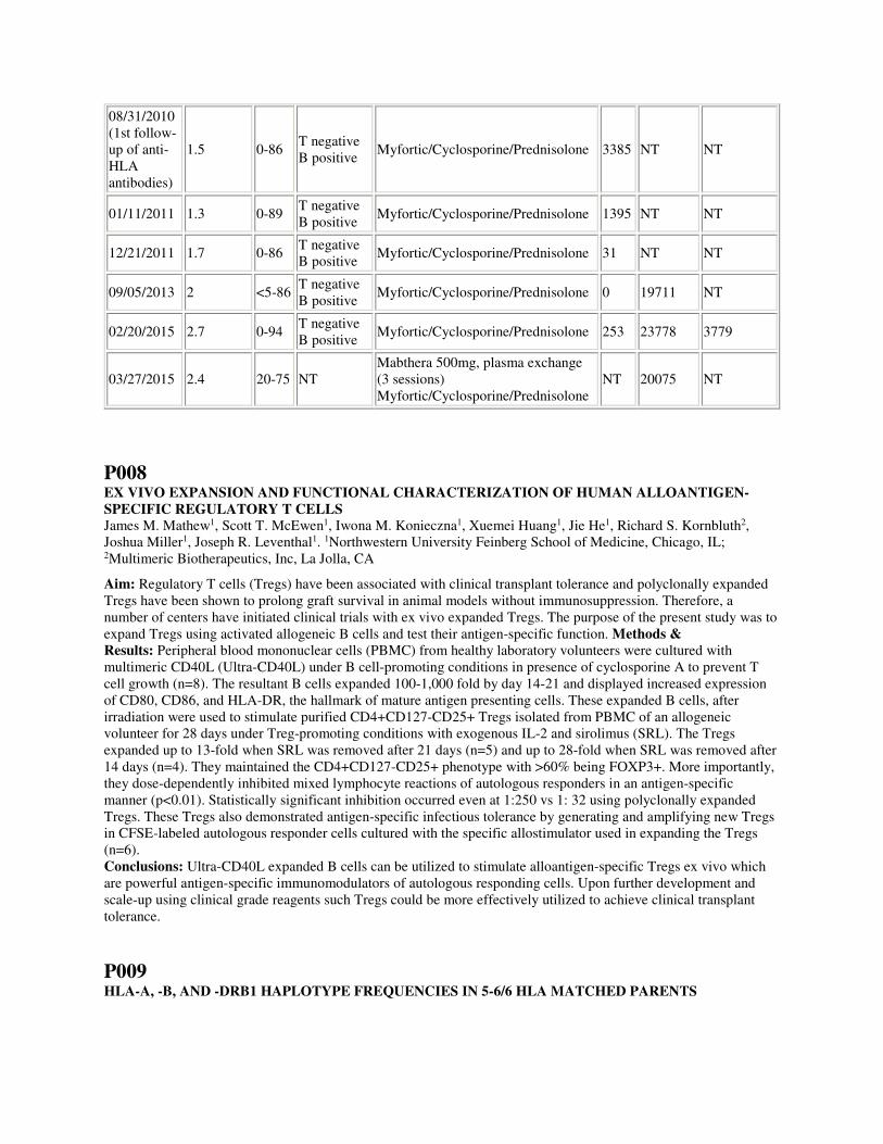
08/31/2010 (1st follow-up of anti-HLA antibodies)
1.5 0-86 T negative B positive
Myfortic/Cyclosporine/Prednisolone 3385 NT NT
01/11/2011 1.3 0-89 T negative B positive
Myfortic/Cyclosporine/Prednisolone 1395 NT NT
12/21/2011 1.7 0-86 T negative B positive
Myfortic/Cyclosporine/Prednisolone 31 NT NT
09/05/2013 2 <5-86 T negative B positive
Myfortic/Cyclosporine/Prednisolone 0 19711 NT
02/20/2015 2.7 0-94 T negative B positive
Myfortic/Cyclosporine/Prednisolone 253 23778 3779
03/27/2015 2.4 20-75 NT Mabthera 500mg, plasma exchange (3 sessions) Myfortic/Cyclosporine/Prednisolone
NT 20075 NT
P008 EX VIVO EXPANSION AND FUNCTIONAL CHARACTERIZATION OF HUMAN ALLOANTIGEN-SPECIFIC REGULATORY T CELLS James M. Mathew1, Scott T. McEwen1, Iwona M. Konieczna1, Xuemei Huang1, Jie He1, Richard S. Kornbluth2, Joshua Miller1, Joseph R. Leventhal1. 1Northwestern University Feinberg School of Medicine, Chicago, IL; 2Multimeric Biotherapeutics, Inc, La Jolla, CA
Aim: Regulatory T cells (Tregs) have been associated with clinical transplant tolerance and polyclonally expanded Tregs have been shown to prolong graft survival in animal models without immunosuppression. Therefore, a number of centers have initiated clinical trials with ex vivo expanded Tregs. The purpose of the present study was to expand Tregs using activated allogeneic B cells and test their antigen-specific function. Methods & Results: Peripheral blood mononuclear cells (PBMC) from healthy laboratory volunteers were cultured with multimeric CD40L (Ultra-CD40L) under B cell-promoting conditions in presence of cyclosporine A to prevent T cell growth (n=8). The resultant B cells expanded 100-1,000 fold by day 14-21 and displayed increased expression of CD80, CD86, and HLA-DR, the hallmark of mature antigen presenting cells. These expanded B cells, after irradiation were used to stimulate purified CD4+CD127-CD25+ Tregs isolated from PBMC of an allogeneic volunteer for 28 days under Treg-promoting conditions with exogenous IL-2 and sirolimus (SRL). The Tregs expanded up to 13-fold when SRL was removed after 21 days (n=5) and up to 28-fold when SRL was removed after 14 days (n=4). They maintained the CD4+CD127-CD25+ phenotype with >60% being FOXP3+. More importantly, they dose-dependently inhibited mixed lymphocyte reactions of autologous responders in an antigen-specific manner (p<0.01). Statistically significant inhibition occurred even at 1:250 vs 1: 32 using polyclonally expanded Tregs. These Tregs also demonstrated antigen-specific infectious tolerance by generating and amplifying new Tregs in CFSE-labeled autologous responder cells cultured with the specific allostimulator used in expanding the Tregs (n=6). Conclusions: Ultra-CD40L expanded B cells can be utilized to stimulate alloantigen-specific Tregs ex vivo which are powerful antigen-specific immunomodulators of autologous responding cells. Upon further development and scale-up using clinical grade reagents such Tregs could be more effectively utilized to achieve clinical transplant tolerance.
P009 HLA-A, -B, AND -DRB1 HAPLOTYPE FREQUENCIES IN 5-6/6 HLA MATCHED PARENTS
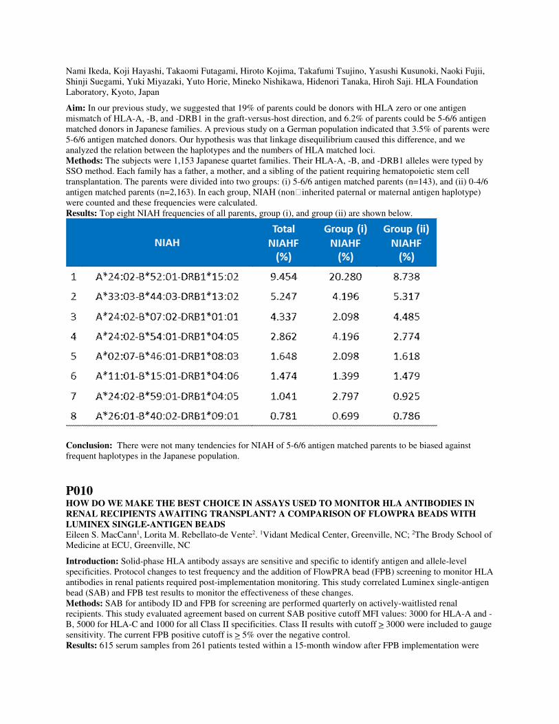
Nami Ikeda, Koji Hayashi, Takaomi Futagami, Hiroto Kojima, Takafumi Tsujino, Yasushi Kusunoki, Naoki Fujii, Shinji Suegami, Yuki Miyazaki, Yuto Horie, Mineko Nishikawa, Hidenori Tanaka, Hiroh Saji. HLA Foundation Laboratory, Kyoto, Japan
Aim: In our previous study, we suggested that 19% of parents could be donors with HLA zero or one antigen mismatch of HLA-A, -B, and -DRB1 in the graft-versus-host direction, and 6.2% of parents could be 5-6/6 antigen matched donors in Japanese families. A previous study on a German population indicated that 3.5% of parents were 5-6/6 antigen matched donors. Our hypothesis was that linkage disequilibrium caused this difference, and we analyzed the relation between the haplotypes and the numbers of HLA matched loci. Methods: The subjects were 1,153 Japanese quartet families. Their HLA-A, -B, and -DRB1 alleles were typed by SSO method. Each family has a father, a mother, and a sibling of the patient requiring hematopoietic stem cell transplantation. The parents were divided into two groups: (i) 5-6/6 antigen matched parents (n=143), and (ii) 0-4/6 antigen matched parents (n=2,163). In each group, NIAH (non‐inherited paternal or maternal antigen haplotype) were counted and these frequencies were calculated. Results: Top eight NIAH frequencies of all parents, group (i), and group (ii) are shown below.
Conclusion: There were not many tendencies for NIAH of 5-6/6 antigen matched parents to be biased against frequent haplotypes in the Japanese population.
P010 HOW DO WE MAKE THE BEST CHOICE IN ASSAYS USED TO MONITOR HLA ANTIBODIES IN RENAL RECIPIENTS AWAITING TRANSPLANT? A COMPARISON OF FLOWPRA BEADS WITH LUMINEX SINGLE-ANTIGEN BEADS Eileen S. MacCann1, Lorita M. Rebellato-de Vente2. 1Vidant Medical Center, Greenville, NC; 2The Brody School of Medicine at ECU, Greenville, NC
Introduction: Solid-phase HLA antibody assays are sensitive and specific to identify antigen and allele-level specificities. Protocol changes to test frequency and the addition of FlowPRA bead (FPB) screening to monitor HLA antibodies in renal patients required post-implementation monitoring. This study correlated Luminex single-antigen bead (SAB) and FPB test results to monitor the effectiveness of these changes. Methods: SAB for antibody ID and FPB for screening are performed quarterly on actively-waitlisted renal recipients. This study evaluated agreement based on current SAB positive cutoff MFI values: 3000 for HLA-A and -B, 5000 for HLA-C and 1000 for all Class II specificities. Class II results with cutoff > 3000 were included to gauge sensitivity. The current FPB positive cutoff is > 5% over the negative control. Results: 615 serum samples from 261 patients tested within a 15-month window after FPB implementation were
evaluated for agreement by patient number and number of samples tested. Patient Numbers: SAB and FPB Class I results did not agree in 84 patients (32.2%); 63.1% of patients had false positive (FP) and 36.9% had false negative (FN) results. With a MFI > 1000 cutoff, Class II results did not agree in 119 patients (45.6%); 0.8% of patients had FP and 99.2% had FN results. With a MFI > 3000, Class II results did not agree in 49 patients (18.8%); 18.4% of patients had FP and 81.6% had FN results. Sample Numbers: SAB and FPB Class I results did not agree in 162 serum samples (26.3%); 60.5% of samples had FP and 39.5% had FN results. With a MFI > 1000 cutoff, Class II results did not agree in 312 samples (50.7%); 11.9% of samples had FP and 88.1% had FN results. With a MFI > 3000 cutoff, Class II results did not agree in 128 samples (20.8%); 33.6% of samples had FP and 66.4% had FN results. Conclusion: HLA antibody monitoring is important to anticipate donor-specific reactivity in renal transplant recipients. If ID and screening assays are used to monitor these levels, an acceptable degree of correlation between methods is important. The FPB assay is not as sensitive or specific as SAB, especially in detecting weak, clinically-significant specificities. This study identified weakness in our protocol, leading us to evaluate other HLA antibody assay options.
P011 DE NOVO DEVELOPMENT OF DSA AND AUTOANTIBODIES TO LUNG ASSOCIATED SELF-ANTIGENS IS ASSOCIATED WITH UPREGULATION OF TRANSCRIPTION FACTOR ZBTB7A: ROLE IN CHRONIC REJECTION FOLLOWING HUMAN LUNG TRANSPLANTATION Deepak Nayak1, Fangyu Zhou1, Ramsey Hachem2, Daniel Kreisel1, Thalachallour Mohanakumar1. 1Washington University School of Medicine, St. Louis, MO; 2Washington University School of Medicine, St. Louis, MO
Aim: Administration of anti-MHC class I (H-2Kb) leads to induction of transcription factor Zinc finger and BTB domain containing 7A (Zbtb7a) leading to inflammatory cascade and chronic rejection in a murine obliterative airway disease model. Development of DSA and Abs to lung-associated self-antigens (K-alpla-1 Tubulin (Kα1T) and Collagen V (Col-V) are significant risk factors in development of bronchiolitis obliterans syndrome (BOS) following human lung transplantation (LTx). The current study focused on the role of Zbtb7a in development of BOS following human LTx. Methods: We analyzed 25 LTx patients that developed BOS years after LTx as well as 10 stable LTx performed at Barnes-Jewish Washington University. Kinetics of Zbtb7a expression was studied in bronchoalveolar lavage (BAL) cells. Quantitative PCR analysis was performed to evaluate mRNA levels and data was compared with the DSA, autoantibody and immunosuppression. Results: Increases in the Zbtb7a expression was observed in BAL cells collected at visits immediately prior to clinical BOS diagnosis compared with earlier visits. Zbtb7a expression was significantly (p<0.01) increased (150%) in pre-BOS BAL cells. On the contrary, stable patients did not demonstrate any change in Zbtb7a levels. All of the BOS+ patients had developed DSA and Abs to Kα1T and Col-V. Zbtb7a expression in13 patients who received Ab directed therapy following DSA demonstrated significant reduction along with decrease in DSA and Abs to lung self-antigens. Therefore, Zbtb7a activation is an important inducer of the DSA and autoantibody. Conclusions: Up regulation of Zbtb7a was positively associated with de novo DSA and Abs to lung-associated self-antigens. Ab directed therapy significantly reduced Zbtb7a expression. We propose that Zbtb7a may be used as a prognostic marker in evaluating Ab-induced inflammation in the lung as well as efficacy of Ab directed therapy following development of DSA.
P012 INTER-RUN VARIATION OF LUMINEX SINGLE-ANTIGEN ASSAY FOR THE DETECTION OF ANTIBODIES TO HLA Chang Liu1, Donna Phelan2, Thalachallour Mohanakumar3. 1Washington University School of Medicine, St. Louis, MO; 2Barnes-Jewish Hospital, St. Louis, MO; 3Washington University School of Medicine, St. Louis, MO
Aim: Luminex single-antigen assay (SA) utilizes multiplexed beads to detect specific antibodies to HLA. The approach to quality control during the daily operation in an HLA laboratory has not been well defined. This study aims to examine the inter-run variation of MFIs using a stable positive control (PC) specimen, and to explore possible control rules (CRs) for judging the acceptability of each run. Methods: To control for multiple beads with MFIs flanking an internal MFI cutoff of 2000, sera of highly sensitized
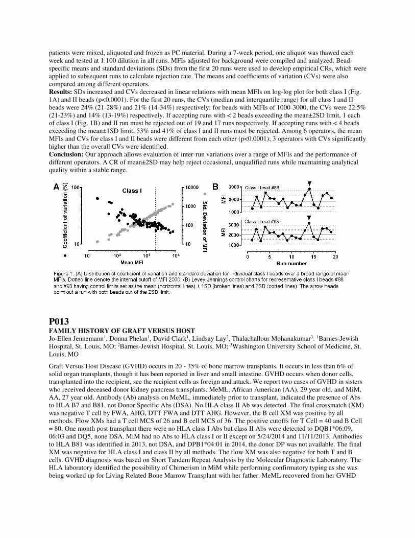
patients were mixed, aliquoted and frozen as PC material. During a 7-week period, one aliquot was thawed each week and tested at 1:100 dilution in all runs. MFIs adjusted for background were compiled and analyzed. Bead-specific means and standard deviations (SDs) from the first 20 runs were used to develop empirical CRs, which were applied to subsequent runs to calculate rejection rate. The means and coefficients of variation (CVs) were also compared among different operators. Results: SDs increased and CVs decreased in linear relations with mean MFIs on log-log plot for both class I (Fig. 1A) and II beads (p<0.0001). For the first 20 runs, the CVs (median and interquartile range) for all class I and II beads were 24% (21-28%) and 21% (14-34%) respectively; for beads with MFIs of 1000-3000, the CVs were 22.5% (21-23%) and 14% (13-19%) respectively. If accepting runs with < 2 beads exceeding the mean±2SD limit, 1 each of class I (Fig. 1B) and II run must be rejected out of 19 and 17 runs respectively. If accepting runs with < 4 beads exceeding the mean±1SD limit, 53% and 41% of class I and II runs must be rejected. Among 6 operators, the mean MFIs and CVs for class I and II beads were different from each other (p<0.0001); 3 operators with CVs significantly higher than the overall CVs were identified. Conclusion: Our approach allows evaluation of inter-run variations over a range of MFIs and the performance of different operators. A CR of mean±2SD may help reject occasional, unqualified runs while maintaining analytical quality within a stable range.
P013 FAMILY HISTORY OF GRAFT VERSUS HOST Jo-Ellen Jennemann1, Donna Phelan1, David Clark1, Lindsay Lay2, Thalachallour Mohanakumar3. 1Barnes-Jewish Hospital, St. Louis, MO; 2Barnes-Jewish Hospital, St. Louis, MO; 3Washington University School of Medicine, St. Louis, MO
Graft Versus Host Disease (GVHD) occurs in 20 - 35% of bone marrow transplants. It occurs in less than 6% of solid organ transplants, though it has been reported in liver and small intestine. GVHD occurs when donor cells, transplanted into the recipient, see the recipient cells as foreign and attack. We report two cases of GVHD in sisters who received deceased donor kidney pancreas transplants. MeML, African American (AA), 29 year old, and MiM, AA, 27 year old. Antibody (Ab) analysis on MeML, immediately prior to transplant, indicated the presence of Abs to HLA B7 and B81, not Donor Specific Abs (DSA). No HLA class II Ab was detected. The final crossmatch (XM) was negative T cell by FWA, AHG, DTT FWA and DTT AHG. However, the B cell XM was positive by all methods. Flow XMs had a T cell MCS of 26 and B cell MCS of 36. The positive cutoffs for T Cell = 40 and B Cell = 80. One month post transplant there were no HLA class I Abs but class II Abs were detected to DQB1*06:09, 06:03 and DQ5, none DSA. MiM had no Abs to HLA class I or II except on 5/24/2014 and 11/11/2013. Antibodies to HLA B81 was identified in 2013, not DSA, and DPB1*04:01 in 2014, the donor DP was not available. The final XM was negative for HLA class I and class II by all methods. The flow XM was also negative for both T and B cells. GVHD diagnosis was based on Short Tandem Repeat Analysis by the Molecular Diagnostic Laboratory. The HLA laboratory identified the possibility of Chimerism in MiM while performing confirmatory typing as she was being worked up for Living Related Bone Marrow Transplant with her father. MeML recovered from her GVHD

and continues to do well. MiM succumbed to her GVHD, 74 days post transplant. These two cases clearly demonstrate the importance of analyzing for GVHD following kidney pancreas transplantation.
P014 WAIT TIME DECONSTRUCTED Jean Heneghan, Ronald Charlton. Tulane University, New Orleans, LA
Transplant (TX) physicians often ask us: What effects do ABO and HLA antibody sensitization have on my patient's ability to get an organ offer? This study attempts to answer that question by analyzing waitlist(WL) and TX patient data, ABO and HLA antigen frequency data over a 6 year period for the Tulane kidney transplant program. Data was stratified by presensitization, sex, ABO and TX status. We analyzed this data along with HLA antigen frequency and pertinent organ offer statistics to determine the projected wait time for a highly sensitized (99%) regraft patient with ABO=O. On average, presensitized Tulane WL patients wait 50% longer for an organ than unsensitized patients. The following table gives median wait time stratified by sex, ABO, and TX status.
Median Wait Time in Months - Transplanted vs. Wait Listed Patients
Transplanted Patients WaitListed Patients
ABO AB A O B ABO AB A O B
Female Primary 13 18 28 24 Female Primary 12 24 31 34
Female Regraft 38 23 60 40 Female Regraft 61 53 48 60
Male Primary 10 16 26 37 Male Primary 17 17 35 34
Male Regraft 50 18 33 NA Male Regraft NA 45 41 54
The following table includes transplant (TX) vs. wait list(WL) patient data given in months wait time stratified by sex and CPRA level.
Median Wait Time stratified by Sex and CPRA
Primary Transplant Regraft Transplant
TX TX WL WL TX TX WL WL
CPRA M F M F CPRA M F M F
<20% 20 18 29 20 <20% NA NA NA NA
20-79% 24 26 34 28 20-79% 36 38 41 NA
>80% 22 19 50 43 >80% 28 31 48 43

Using the above, ABO and HLA antigen frequencies and frequency and types of organ offers, we projected wait time until a compatible organ offer for a 99% CPRA, regraft male patient awaiting a standard criteria donor with ABO-O in excess of 14 years for an in-state offer. The wait for a regional or national offer is projected to be 63 months, but may be less in the future due to the new allocation system. Wait time is highly dependent on ABO and sensitization, but we also see a disparity in wait times for WL patients vs. TX patients pointing to the effect of HLA antigen frequency. All sensitization is not equal. Many still on the WL likely have less frequent HLA types and higher levels of HLA antibodies to common HLA antigens than those transplanted given all else equal. These data trends provide the TX physician a better understanding of factors affecting wait time.
P015 MEDICAL RECORD CONVERGENCE FOR HLA REPORTING ACROSS THE ENTERPRISE Lisa Hallaway1, Laurie Wakefield1, Justin Kreuter1, Lauren Rowans2, David Kahle2, Mohamed Elrefaei2, Petrina Genco2, Yvonne Desmarteau3, Scott Stoll3, Marcelo J. Pando3, Riccardo Valdez3, Hieu La1, Michelle Mattey2, Barb Feehan1, Brooke Kloeppel4, Scot Townshend4, Manish Gandhi1. 1Mayo Clinic, Rochester, MN; 2Mayo Clinic, Jacksonville, FL; 3Mayo Clinic, Phoenix, AZ; 4SystemLink Inc., Sterling, VA
Aim: One of Mayo Clinic’s missions is to standardize care through an integrated clinical practice. Reporting histocompatibility results is very complex and HLA labs have different ways of interpreting results. By standardizing across the three sites we improve the interchangeability of test results, reduce resources to support our practice, provide the best standard of care for our patients and support for our physicians for the Mayo paired kidney exchange program. Methods: Integration was defined as: • Each lab will use HistoTrac as the lab management system. • HistoTrac will interface with the lab integrated system (LIS) for processing of orders and reporting results to the medical record. • Each lab will report the same discreet result values and interpretive reports. • Each lab will use the same vendor and lot number if possible for antibody and typing tests. A project group was created which includes: a director and technical specialist from each site, a business analyst, and a computer programmer. This team worked together using conference calls and web meetings to establish standardization for reporting HLA typing, antibody and crossmatch results. Results: The results reported at each site will use the same test definitions, reporting templates, and will be transferred to the LIS. We encountered some obstacles due to: lab practice differences, transplant policies and business requirements. The convergence effort is still ongoing at each site: • MN: update reporting and implement interpretive reports • AZ: implement an interface to the LIS and update interpretive reports • FL: implement HistoTrac with an interface to the LIS and update interpretive reports Conclusion: All histocompatibility data and interpretive reports will be available in the medical record at each mayo site. Convergence of reporting test results will improve the communication and understanding when evaluating a patient while making the process cost effective. The next step in convergence is reporting to one medical record at all three sites.

P016 LIGATION OF HLA CLASS II MOLECULES ON ENDOTHELIAL CELLS INDUCES P-SELECTIN CELL SURFACE EXPRESSION AND MYELOID CELL RECRUITMENT Fang Li, Nicole M. Valenzuela, Elaine F. Reed. UCLA, Los Angeles, CA
Aim: A hallmark of chronic antibody-mediated rejection is the presence of leukocytes infiltrates within the allograft. We tested the hypothesis that ligation of HLA class II on EC mediates myeloid cell recruitment by inducing p-selectin presentation through Weibel Palade body exocytosis. Methods: Class II expression was induced by adenoviral transfection of EC with CIITA. Western blots were used to quantitate protein phosphorylation. Monocyte adherence was measured by counting CFSE-labeled monocytes (MonoMac 6) recruited to HLA-class II Ab stimulated EC in the presence/absence of Fc receptor blockade with human IgG. Transendothelial cell migration was determined using a transwell assay. P-selectin expression was detected by Cell ElISA. Results: class II ligation by antibody (Ab) against a monomorphic epitope determinant on class II antigens significantly increased monocyte transendothelial migration in a Fc-independent manner. Class II Ab-mediated myeloid cell transendothelial migration was attenuated by blockading with recombinant PSGL-1 (rPSGL-1). Class II Ab caused a 1.54±0.11 fold increases in monocyte adherence that was inhibited by pretreatment with rPSGL-1. In addition, pretreatment of EC with Src inhibitor PP2 and ERK inhibitor U0126 resulted in decreased myeloid cell recruitment (Fig. 1). Furthermore, PP2 and U0126 diminished class II induced p-selectin cell surface expression (Fig. 2). Conclusions: We provide evidence that class II DSA promotes a MAPK-mediated monocyte adherence by upregulating EC p-selectin expression, stimulating monocyte adherence and infiltration into allografts, leading to transplant vasculopathy.
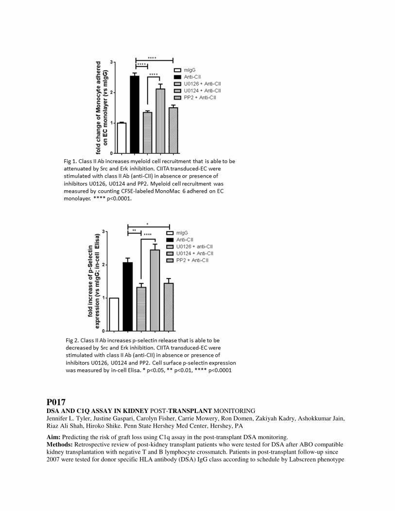
P017 DSA AND C1Q ASSAY IN KIDNEY POST-TRANSPLANT MONITORING Jennifer L. Tyler, Justine Gaspari, Carolyn Fisher, Carrie Mowery, Ron Domen, Zakiyah Kadry, Ashokkumar Jain, Riaz Ali Shah, Hiroko Shike. Penn State Hershey Med Center, Hershey, PA
Aim: Predicting the risk of graft loss using C1q assay in the post-transplant DSA monitoring. Methods: Retrospective review of post-kidney transplant patients who were tested for DSA after ABO compatible kidney transplantation with negative T and B lymphocyte crossmatch. Patients in post-transplant follow-up since 2007 were tested for donor specific HLA antibody (DSA) IgG class according to schedule by Labscreen phenotype

beads/Single Antigen beads (SAB, One Lambda), and if detected, reflexed to C1q assay (One Lambda) since 2011. Results: Of the 437 post-kidney transplant patients (range: 1-27 years post-transplant, mean 9.1 years), de novo DSA was detected in 74 (16.9%). C1q assay was performed in 50/74 DSA positive patients: Positive at least once N=35 (70%) and Negative N=15. DSA in 26 patients were tested for > one year; persistently positive C1q, N=17, eventually became C1q negative N=6, or became DSA negative N=3. Overall, C1q positive DSA presented higher maximum MFI detected by both phenotype beads and SAB, and resulted in higher graft loss. C1q MFI was similar between graft lost and functional (defined by not on dialysis). Regardless of C1q status, graft loss was observed after month-to-years with DSA, indicating chronic antibody mediated rejection. Conclusions: Patients with C1q positive DSA had higher MFI (IgG) levels and resulted in higher graft loss.
P018 COMPARISON OF CDC-AHG, C1Q-AHG, AND STANDARD SINGLE ANTIGEN BEAD TESTING IN PATIENTS REFRACTORY TO PLATELET TRANSFUSION Anne Halpin, Amiah Klein, Luis Hidalgo, Patricia Campbell, Susan Nahirniak. University of Alberta, Edmonton, AB, Canada
Aim: Patients refractory to platelet transfusion are tested for class I HLA antibody (Ab). CDC-AHG has been used due to cost, turnaround time (TAT) and clinically appropriate sensitivity for this indication. This study compares CDC-AHG, standard single antigen bead (SAB) testing and C1Q-AHG SAB testing to determine if more efficient and clinically relevant methods are available for refractory patients. Methods: Patients were tested for class I HLA Ab by CDC-AHG (Lambda Cell Tray™), SAB (LABScreen® Single Antigen), and C1q-AHG (C1qScreen™ + wash and AHG); sera were EDTA treated prior to SAB/C1q testing. Sera were also tested by SAB at 1/8 dilution (n=6).36 samples were tested from 35 patients. Results: CDC results were 0% PRA (n=6), 17-74% (n=15), and >80% (n=15). If Ab specificity could be assigned by CDC, these Ab were detected by SAB and C1q but additional specificities were present including in 2 of the
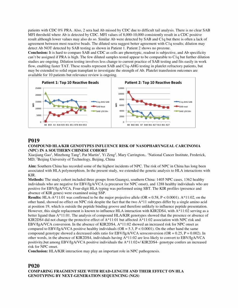
patients with CDC 0% PRA. Also, 2 sera had Ab missed by CDC due to difficult tail analysis. There is no clear SAB MFI threshold where Ab is detected by CDC. MFI values of 8,000-10,000 consistently result in a CDC positive result although lower values may also do so. Similar Ab were detected by SAB and C1q but there is often a lack of agreement between most reactive beads. The diluted sera suggest better agreement with C1q results; dilution may detect Ab NOT detected by SAB testing as shown in Patient 1. Patient 2 shows no prozone. Conclusion: It is hard to compare SAB and CDC as cells are phenotypic, readout is subjective, and Ab specificity can’t be assigned if PRA is high. The few diluted samples tested appear to be comparable to C1q but further dilution studies are ongoing. Dilution testing involves less change to current practice of SAB testing and fits easily in work flow, enabling faster TAT. These results represent SAB and C1q-AHG testing in platelet refractory patients, but may be extended to solid organ transplant to investigate the strength of Ab. Platelet transfusion outcomes are available for 10 patients but relevance review is ongoing.
P019 COMPOUND HLA/KIR GENOTYPES INFLUENCE RISK OF NASOPHARYNGEAL CARCINOMA (NPC) IN A SOUTHERN CHINESE COHORT Xiaojiang Gao1, Minzhong Tang2, Pat Martin1, Yi Zeng2, Mary Carrington1. 1National Cancer Institute, Frederick, MD; 2Beijing University of Technology, Beijing, China
Aim: Southern China has recorded some of the highest incidents of NPC. The risk of NPC in China has long been associated with HLA polymorphism. In the present study, we extended the genetic analysis to HLA interactions with KIR. Methods: The study cohort included three groups from Guangxi, southern China: 1405 NPC cases, 1362 healthy individuals who are negative for EBV/IgA/VCA (a precursor for NPC onset), and 1288 healthy individuals who are positive for EBV/IgA/VCA. Four-digit HLA typing was performed using SBT. The KIR profiles (presence and absence of KIR genes) were examined using SSP. Results: HLA-A*11:01 was confirmed to be the major projective allele (OR = 0.58, P < 0.0001). A*11:02, on the other hand, showed no effect on NPC risk despite the fact that the two A*11 subtypes differ by a single amino acid at position 19, which is outside the peptide binding groove and therefore unlikely to influence peptide presentation. However, this single replacement is known to influence HLA interaction with KIR2DS4, with A*11:02 serving as a better ligand than A*11:01. The analysis of compound HLA/KIR genotypes showed that the presence or absence of KIR2DS4 did not change the protective effect of A*11:01 but affected A*11:02 association with NPC risk and EBV/IgA/VCA conversion. In the absence of KIR2DS4, A*11:02 showed an increased risk for NPC onset as compared to EBV/IgA/VCA positive healthy individuals (OR = 5.3, P < 0.0001). On the other hand the same compound genotype showed a decreased odds ratio for EBV/IgA/VCA seroconversion (OR = 0.25, P = 0.002). In other words, in the absence of KIR2DS4, individuals having A*11:02 are less likely to convert to EBV/IgA/VCA positivity,but among EBV/IgA/VCA positive individuals the A*11:02+/ KIR2DS4- genotype confers an increased risk for NPC onset. Conclusion: HLA/KIR interaction may play an important role in NPC pathogenesis.
P020 COMPARING FRAGMENT SIZE WITH READ-LENGTH AND THEIR EFFECT ON HLA GENOTYPING BY NEXT-GENERATION SEQUENCING (NGS)

Tracie Profaizer, Eszter Lazar-Molnar, Julio C. Delgado, Attila Kumanovics. ARUP Laboratories, Salt Lake City, UT
Aim: We have previously studied the effect of shorter DNA fragments (less than 600 bp) on HLA typing by NGS of HLA-A, B, C, DRB1 and DQB on the Illumina MiSeq and found that 100-300 bp fragments produce more ambiguous typing results. Here, we evaluated the same reagents but altered the conditions to produce longer DNA fragments in order to determine if the number of ambiguous typing results could be reduced. We have previously shown that Illumina 2x250 sequencing resulted in less ambiguity than 2x150 bp sequencing. Here, we asked if further increasing the sequencing read length (2x300 bp) leads to additional gains in accuracy. Methods: Ten IHWG samples had long-range PCR performed for HLA-A, B, C, DRB1 and DQB1 using a combination of in-house and commercial primers. After PCR clean-up and quantification, each sample’s 5 loci were pooled. Using the NEBNext dsDNA Fragmentase kit and various incubation times, the long-range PCR products were digested to produce 600-1200 bp fragments size selected with magnetic beads (AMPure Beads, Beckman Coulter) or the Blue PippinPrep (Sage Science). The NEBNext Library Prep Kit was used to complete library preparation. Unique indices were ligated onto each sample. Each sample library was measured for size and quantified. Next, all sample libraries were equimolarly pooled and two Illumina Reagent kits were run, v2 (500 cycle) and v3 (600 cycle), each loaded at a 10pM concentration. Fastq files were analyzed using Omixon’s Target v.1.8.1 software for HLA genotyping. Results: Ninety-six percent of the IHWG samples, each with 5 loci and an average fragment size of 684 ± 73 bp, typed correctly without any ambiguity. Samples with an average fragment size of 973 ± 132 bp performed the same, 96%. There was no significant difference in results between the 2x250 and 2x300 read length kits using samples with shorter (684 bp) or longer (973 bp) fragment sizes. Conclusions: We have now compared fragment sizes from 100-300 bp, to 900-1200 bp, and found that generally, fragment sizes 600 bp or greater produce the most correct and unambiguous HLA typing assignments. We have previously determined that 2x250 read length kit performs better than the 2x150 kit, and now we show that the 2x250 read length kit is preferred over the 2x300 bp kit due to its shorter run time (40 hours vs. 60 hours) with equal performance.
P022 THE UTILITY OF CIRCULATING DONOR SPECIFIC ANTIBODY TO PREDICT BIOPSY-PROVEN ANTIBODY-MEDIATED REJECTION AND TO PROVIDE PROGNOSTIC VALUE AFTER HEART TRANPSPLANTATION IN CHILDREN Julio C. Delgado1, Adam L. Ware2, Elisabeth Malmberg1, Elizabeth Hammond3, Dylan V. Miller3, Josef Stehlik3, Abdallah Kfoury3, Patricia Revelo3, Aaron Eckhauser3, Melanie D. Everitt3. 1ARUP Institute for Clinical and Experimental Pathology, Department of Pathology, University of Utah School of Medicine, Salt Lake City, UT; 2University of Utah School of Graduate Medical Education, Salt Lake City, UT; 3Utah Transplantation Affiliated Hospitals (U.T.A.H.) Cardiac Transplant Program, Intermountain Health Care, VA Hospital, University of Utah Healthcare, Salt Lake City, UT
Aim: Heart transplantation (HT) is a life-saving option for children with end-stage heart failure. Antibody-mediated rejection (AMR) is a significant cause of morbidity and mortality in HT. The current diagnostic criteria for AMR are based on endomyocardial biopsy (EMB) showing evidence of C4d and C3d deposition, and capillary endothelial changes, including neutrophil and macrophage infiltration, and interstitial edema. Although the presence of donor specific antibody (DSA) is a risk factor for the development of AMR after HT, it is currently not required for diagnosis of AMR. In this study, we aimed to investigate the sensitivity, specificity, predictive values and prognostic implications of circulating DSA using EMB as the gold standard for AMR diagnosis in a cohort of pediatric HT recipients. Methods: Retrospective study in pediatric HT patients that had follow-up care between 2009-2013 and had at least one EMB paired with DSA testing within 3 days of the biopsy. Positive DSA was defined at a mean fluorescent intensity (MFI) ≥ 2000 using single antigen bead testing. Results: Of the 66 patients included, 26 (39%) had at least 1 HLA DSA positive test. Of the 26 patients with positive DSA, 18 (69%) had at least one antibody against HLA-DQ. In 7 (27%) of the 26 patients with positive DSA, the only antibody detected was against HLA-DQ. Of 236 DSA EMB pairs analyzed, 132 (56%) pairs were negative for DSA, 23 (10%) pairs had HLA DSA Class I only, 59 (25%) pairs had HLA DSA Class II only, and 22 (9%) pairs had both HLA class I and II DSA. HLA DSA testing had a sensitivity= 92.6%, specificity= 62.2%, PPV= 24.0%, and NPV= 98.5% for biopsy-proven AMR. Kaplan-Meier analysis showed that patients with positive HLA
DSA results and biopsy-proven AMR trended toward a higher incidence of cardiovascular mortality, and coronary artery vasculopathy after 3 years compared to patients without circulating DSA and a negative biopsy. Conclusions: The results of HLA-DSA testing in this cohort, showing excellent sensitivity and NPV for biopsy-proven AMR suggest for the first time, that DSA testing could be used for non-invasive prediction of AMR absence in heart transplantation, and avoid unnecessary biopsies. The value of DSA testing for biopsy-proven AMR and outcome prognosis in HT is important to study in future multi-center prospective studies.
P023 AN INTEGRATED GENOTYPING APPROACH FOR HLA AND OTHER COMPLEX GENETIC SYSTEMS Daniel E. Geraghty1, Wyatt C. Nelson1, Chul-Woo Pyo1, David Vogan1, Ruihan Wang1, Carly Hennessey1, Anajane Smith1, Shalini Pereira1, Akiko Ishitani2. 1Fred Hutchinson Cancer Research Center, Seattle, WA; 2Nara Medical University, Nara, Japan
Clinical immunogenetics laboratories performing routine sequencing of human leukocyte antigen (HLA) genes in support of hematopoietic cell transplantation are motivated to upgrade to next-generation sequencing (NGS) technology by its potential for cost savings as well as testing accuracy and flexibility. While NGS machines are available and simple to operate, there are few systems available that provide comprehensive sample preparation and data analysis workflows to complete the process. We report on the development and testing of the Integrated Genotyping System (IGS), which has been designed to specifically address the challenges associated with the adoption of NGS in clinical laboratories. To validate the system for a variety of sample DNA sources, we tested DNA specimens from whole blood, dried blood spots, buccal swabs, and lymphoblastoid cell lines. HLA class I and class II genotypes were derived from amplicon sequencing of HLA-A, -B, -C for exons 1-7 and HLA-DPA1, -DPB1, -DQA1, -DQB1, -DRB1, -DRB3, -DRB4, -DRB5 for exons 1-4. Additionally, to demonstrate the extensibility of the IGS to other genetic loci, assays for KIR haplotyping and MICA and KIR allele typing were developed using a workflow based on the HLA system. These results are discussed with respect to their applications in the clinical setting and consequent potential for advancing precision medicine. A. Smith: Consultant; Company/Organization; Scisco Genetics Inc. S. Pereira: Consultant;
Company/Organization; Scisco Genetics Inc. A. Ishitani: Employee; Company/Organization; Scisco Genetics Inc.
P024 USING NEXT GENERATION SEQUENCING TO CHARACTERIZE POTENTIAL HLA CLASS I NOVEL ALLELES Ana M. Lazaro1, Lihua Hou1, Bin Tu1, Carly Masaberg1, Beth Beduhn2, Damian Goodridge3, Jennifer Ng1, Carolyn K. Hurley1. 1C. W. Bill Young Marrow Donor Recruitment and Research Program. Georgetown University, Rockville, MD; 2Be the Match (NMDP), Minneapolis, MN; 3Conexio Genomics Pty Ltd, Fremantle, Western Australia, Australia
Aim: We explored the impact of using next generation sequencing (NGS) of the entire gene in 300 samples carrying potential new or rare HLA class I alleles. Methods: Samples had been previously typed for exons 2+3 by SBT sequence based typing (Sanger) and/or by (SSO) sequence specific oligoprobes. Targeted class I loci were amplified using locus-specific PCR primers described by Hozomichi et al. Sonication was used to obtain 400 base pair fragments; an Illumina True Seq Nano kit was used for end repair and adaptor/index ligation. Libraries were pooled and paired end sequences were obtained with a MiSeq. AssignTM MPS software (Connexio, Genomics) was used for analysis. Results: Of the 600 alleles sequenced, 69 (11.5%) novel alleles (HLA-A: 27, B: 31, C: 11) differed in the antigen recognition domain (ARD)-encoding exons 2 and/or 3. Ten additional alleles (1.7 %) carried previously unobserved differences in non-ARD-encoding exons (exon 1:2, exon 4:5, exon 5:2, exon 7:1). Thirty three alleles (5.5%) appeared to exhibit novel sequences only in introns. At HLA-A*03:01:01:01 a novel insertion of 19 bp was observed in intron 2 in two different cells. One hundred and fifty seven (26%) alleles had incomplete sequences in the IMGT/HLA data base, of which most of these (121) had intron sequences identical to the closest related allele. The remaining 331 alleles were primarily common and well documented alleles. Conclusion: NGS provided complete, unambiguous, high resolution allele assignments. In this more comprehensive

analysis, there was less variation from the IMGT reference database than expected. Most of the introns of alleles with incomplete sequences were conserved when compared to their putative ancestral allele. L. Hou: Other (Identify); Company/Organization; Intellectual property. B. Tu: Other (Identify);
Company/Organization; Intellectual property. C. Masaberg: Other (Identify); Company/Organization; Intellectual
property. B. Beduhn: Employee; Company/Organization; National Marrow Donor Program. D. Goodridge:
Employee; Company/Organization; Connexio Genomics. J. Ng: Other (Identify); Company/Organization;
Intellectual property. C.K. Hurley: Other (Identify); Company/Organization; Intellectual property.
P025 DECREASED ALLOREACTIVITY TOWARD NON-INHERITED MATERNAL ANTIGEN (NIMA) ALLOWS SUCCESSFUL KIDNEY TRANSPLANTATION IN REGRAFT PATIENT: CASE REPORT Abdelhamid Liacini, Rabab Al-Attas. King Fahad Specialist Hospital, Dammam, Saudi Arabia
Background: During pregnancy contact with paternal HLA- antigen can lead to activation of maternal immune system while exposure of the child to the non inherited (NIMA) antigens may lead to tolerance due to immaturity of the child immune system in utero. Multiple studies on hematopoietic stem cell transplantation demonstrate this beneficial effect but studies on NIMA-specific tolerance in renal transplant are generally few and demonstrated improved kidney graft from sibling who is mismatched at the NIMA haplotype. We report here a case of a successful kidney retansplant from a sibling who is mismatched at the NIMA after failed initial graft from the mother. 26-year-oldfemale with ESRD was referred for second kidney transplantation. Her mother offered the first graft which shortly after the transplantation possibly due to chronic rejection. Investigation revealed weak DSA against DQ2. A haploidentical brother offered the second NIMA-mismatched graft. FXM results were negative for both T and B with second donor. She received Thymoglobulin induced second transplant from her brother. The first post transplant SAB results showed remarkable increase in DQ2, MFI of 11380., We alerted the clinician to this increment in DSA, so he put the patient under close observation despite normal creatinine level. A kidney biopsy was unremarkable. Because of persistence elevation of DSA in view of normal kidney function the treating team questioned our reports and a second biopsy was performed which showed the same findings, however C4d was focally positive in the second biopsy. Consequently the patient received full rejection therapy. The lack of expected response in DSA level guided us to reanalyze our data. HR typing confirmed HLA-DQB1*02:02 associated with DQA1*02:01 for both donors but careful analysis of DQ2 coated bead reaction showed that what we reported as DSA was actually non donor specific and directed against sub group of DQ2 indicating tolerance to mismatch NIMA despite repeated exposures. Our case is a further confirmation for the superior graft outcome from sibling expressing NIMA compared to maternal graft expressing the same NIMA.
P026 PITFALL IN THE INTERPRETATION OF DQ HLA- ANTIBODIES Rabab Al- Attas1, Dalal AlAbduladheem1, Adel A Shawhatti2, Ricardo Lopez1, Abdelhamid Liacini2, Saber AlZahrani2, Khalid Akkari3, Nasreen Hassan4, Abdulnaser Alabadi3. 1King Fahad Specialist Hospital, Dammam, Saudi Arabia; 2King Fahad Specialist Hospital, Dammam, Saudi Arabia; 3King Fahad Specialist Hospital, Dammam, Saudi Arabia; 4King Fahad Specialist Hospital, Dammam, Saudi Arabia
HLA-DQ consist of two polymorphic chains, both contribute to immunization. SAB testing which ignores the DQα, can lead to erroneous DQ -antibody assignments. Pitfall in the interpretation of DQ antibodies in the cases presented here had declined immunologically compatible transplant and exposed the second one to unnecessary immunesuppression. The first patient was a young male referred for kidney transplantation. HLA- antibodies revealed positive DQ7, DQ8, and DQ9 antibodies. B- Flow cross matches with the three evaluated potential living related donors was positive with donor1, 2 (sister and cousin1) but negative with the (cousin2) donor3. All donor typed DQ8. The negative result with donor3 was unexpected. DQA1*component was thought to be responsible however, bead reactivity analysis could not rule out contribution of B*03:02. The possibility of unexpressed DQ8 antigen was not supported by serological and high resolution typing which confirmed DQA1* 03:02/DQB1*03:02 in the first two donors &DQA1*03:01/DQB1*03:02 in donor3. As the difference between DQA1*03:02 and DQA1*03:01 alleles is outside the antigen recognition site the individual DQA1*03:01 should not account for different immune response. Furthermore, DQA1*03:01, DQB1*03:02 coated beads was positive. Flow PRA class II bead on BD showed positive response to homozygous DQA1*03:02 beads. The inconclusive results in assessing the

immunological risk with the donor3, denied patient transplantation for long time before he underwent uneventful transplant. Patient2 was a lady who had failed first graft from her mother and came for retansplant from her full brother. She had weak DQ2 DSA against her mother who shared this antigen with the brother. The first post retansplant SAB showed elevated DQ2, she was put under close observation despite normal creatinine level. A kidney biopsy was unremarkable in the first time but was C4d focally positive in the second one. She received anti-rejection therapy. HR typing confirmed DQB1*02:02/DQA1*02:01 for both donors. Reanalysis of bead reaction revealed the antibody we report was against DQB1*02:01/DQA*05:01 allele. The patient is now14 months post transplant with good renal function. In conclusion the results of SAB were not conclusive and misleading in the 2 patients respectively.
P027 NEXT GENERATION DNA SEQUENCING OF FULL LENGTH HLA CLASS I GENES YIELDS CONCORDANT ASSIGNMENTS AND IDENTIFIES THE LIKELIHOOD OF VARIATION OUTSIDE OF EXONS ENCODING THE ANTIGEN RECOGNITION DOMAIN Lihua Hou1, Damian Goodridge2, Candice Brooks3, Linta Thampi Thampi3, Kanthi Kariyawasam3, Carolyn Hurley4. 1C.W.Bill Young Marrow Donor Recruitment and Research Program, Rockville, MD; 2Connexio Genomics, Freemantle, Australia; 3Georgetown University, Rockville, MD; 4Georgetown University, Rockville, MD
Aim: We have developed an allele-level resolution next generation sequencing (NGS) strategy for HLA class I alleles and used it to explore HLA diversity in a registry donor population. Methods: Long-range amplification of full-length HLA-A, -B, -C loci was performed in separate polymerase chain reactions using DNA from a buccal swab. Amplicons from one individual were then pooled and sheared by sonication. A library was constructed using Illumina’s TruSeq Nano kit and the DNA fragments tagged with one unique index combination. Libraries from 96 individuals were combined and sequenced simultaneously in a single 500 cycle (V2) paired-end run using an Illumina MiSeq. Data analysis used Conexio Genomics AssignTM MPS software. Results: The typing protocol was validated with 552 donor registry samples previously typed by Sanger sequencing and probe hybridization at single genotype G level resolution, yielding a concordance of 99%. NGS yielded primarily single allele-level genotypes. Excluding amplification failures and inability to obtain an NGS assignment (4% loci), 32 discrepant NGS allele assignments were attributed to poor PCR amplification or loss of reads during assembly by software. Complete exon sequences identified 53 known A alleles (48 common or well-documented (CWD)), 89 B alleles (83 CWD), 52 C alleles (47 CWD). Ten potential new alleles with exon variation outside of exons 2+3 and over 25% of samples with potential intron variation require further study. The ability to make assignments based on specific regions of the sequence (e.g., exons only) was critical so that typings could be rapidly transmitted to the registry prior to a more comprehensive analysis of the diversity. Conclusions: The protocol for NGS of class I alleles provides accurate results and should facilitate high volume high resolution typing of donor registry populations. The extent of new variation in exons is limited but the potential for intron variation is high. C. Hurley: Other (Identify); Company/Organization; Intellectual property.
P028 IDENTIFICATION AND SEQUENCING OF NOVEL ALLELES: UTILITY OF NEXT-GENERATION SEQUENCING METHODS Nicholas K. Brown1, Taba Kheradmand1,2, Brenda M. Issangya1, Rebecca L. Upchurch1, Jerome G. Weidner1, Jinguo Wang1, Susana R. Marino1. 1University of Chicago, Chicago, IL; 2Gift Of Life Michigan, Ann Arbor, MI
Aim: Sanger sequencing-based typing (SBT) requires multiple steps to sequence novel HLA alleles, as allele-specific (AS) primers are needed to separate novel from other encoded alleles. Next-generation sequencing (NGS) includes phase information, and has the potential to sequence novel alleles without AS primers. Here we compare the process of novel allele identification and sequencing using SBT and NGS assays. Methods: Novel alleles were identified in peripheral blood during routine clinical testing using Atria SBT kits (Abbot Molecular). Germline encoding was verified by typing buccal samples. For Sanger sequencing of novel alleles, AS amplification primers were designed to take advantage of nucleotide polymorphisms between encoded alleles, and patients with homozygous loci were used to verify primer specificity. Amplified products were Sanger

sequenced using custom or commercially-available primers. For NGS sequencing, the Holotype X4 (Omixon) kit was used in its default configuration. Results: Four novel, germline-encoded HLA alleles were identified in three patients. Novel HLA-A and B alleles were sequenced using AS amplification primers and Sanger sequencing. A novel DQB1 allele previously identified but not resolved by SBT was sequenced using the NGS assay. A novel DQB1 allele not identified by SBT, as the polymorphism was outside the covered region, was identified and sequenced by NGS. The names HLA-A*66:22, B*07:238, DQB1*03:180, and DQB1*06:01:15 have been officially assigned by the WHO Nomenclature Committee. Conclusions: While SBT assays used in the clinical setting are able to identify novel alleles, subsequent sequencing is a cumbersome and time-consuming process. Furthermore, SBT assays may miss novel polymorphisms outside of the exons covered by the assay. NGS systems, on the other hand, can identify and sequence novel alleles over a larger genomic region as part of the normal clinical workflow. However, we experienced issues with the Omixon NGS kit, including the inability to sequence buccal cell DNA, and difficulties amplifying the DQB1 locus, which could have impacted clinical workflow had NGS been the primary method of testing. As the emerging technology of NGS is introduced to complement established SBT assays, it is important to evaluate the strengths and weaknesses of each.
P029 COMBINING ONE STEP SANGER SEQUENCING WITH PHASING PROBE HYBRIDIZATION FOR CLASS I TYPING YIELDS RAPID, G-GROUP RESOLUTION DEFINING 99% OF UNIQUE FULL LENGTH PROTEIN SEQUENCES Bin Tu1, Carly Masaberg1, Jar How Lee2, Daniel Behm3, John Sells3, Paul Tausch2, Lihua Hou1, Jennifer Ng1, Carolyn Katovich Hurley1. 1Georgetown University, Rockville, MD; 2One Lambda Thermo Fisher, Canoga Park, CA; 3Data Blueprint, Glen Allen, VA
Aim: Sanger-based DNA sequencing of exons 2+3 of HLA class I alleles from a heterozygote frequently results in two or more alternative genotypes. In order to reduce the time and effort required to produce a single high resolution HLA genotype, samples were typed in parallel by Sanger sequencing and probe hybridization to obtain G level typing resolution. Methods: The panel of probes used was designed to yield high to intermediate resolution typing and to provide the phasing necessary to resolve common alternative genotypes. Class I assignments from more than 30,000 samples from registry donors were typed using this strategy supported by an in-house data merger and management software. To monitor accuracy and resolution, 552 samples were retyped by a next generation full length gene sequencing (NGS) strategy. Results: More than 95% of the class I assignments were at the level of a single G-level genotype compared to ~26% for European Americans when probe data were not included. The work load was reduced by at least 70% by eliminating secondary sequencing assays and by dramatically reducing the time needed for data review since the combined method addresses the weaknesses of each single method. Typing accuracy was increased. 99% of Sanger + probe typing results were concordant with NGS; all 32 allele discrepancies were attributed to weaknesses in the NGS data. Furthermore, NGS also demonstrated that only 0.9% of the G level assignments resulted in different full length protein sequences (e.g., C*07:01:01G yielded C*07:18 with NGS). The combined method had a rapid turnaround time compared with NGS and previous methods applied alone. Conclusions: Our combined method routinely provides biologically relevant typing resolution at the level of the antigen recognition domain. It can be applied to both single sample or to large volume typing supporting either bone marrow or solid organ transplantation using technology currently available in many HLA laboratories and is cost effective when compared with current NGS kits. B. Tu: Other (Identify); Company/Organization; Intellectual property. C. Masaberg: Other (Identify);
Company/Organization; Intellectual property. J. Lee: Employee; Company/Organization; Thermo Fisher. D.
Behm: Employee; Company/Organization; Data Blueprint. J. Sells: Employee; Company/Organization; Data
Blueprint. P. Tausch: Employee; Company/Organization; Thermo Fisher. L. Hou: Other (Identify);
Company/Organization; Intellectual property. J. Ng: Other (Identify); Company/Organization; Intellectual
property. C.K. Hurley: Other (Identify); Company/Organization; Intellectual property.
P030 LARGE FRAGMENT TARGET ENRICHMENT AND SEQUENCING OF THE 4.4 MB MAJOR HISTOCOMPATIBILITY COMPLEX BY REGION-SPECIFIC EXTRACTION AND HISEQ 2500 Hannah Rabenstein1, Barbara Bangol1, Suzanne Linke2, Riccardo Brumm1, Sebastian Poggel1, Sebastian Eck1, Karl-Heinz Ott2, Johannes Dapprich2, Kaimo Hirv1. 1Center for Human Genetics and Laboratory Diagnostics, Martinsried, Germany; 2Generation Biotech, Lawrenceville, NJ
Due to the extensive polymorphism of the major histocompatibility complex (MHC), only limited progress has been made in high-resolution MHC sequencing, one of the initial requirements to elucidate the molecular immunopathology of several hundred MHC-associated diseases. We present an approach for sequencing the entire MHC with ≈1200 specific capture primers. Using target sites known to be highly conserved, Region-Specific Extraction (RSE, Generation Biotech) was used to target MHC segments of up to 40kb length and enrich the contiguous 4.4Mb MHC for sequencing. RSE uses a single primer extension-step that attaches a specific “handle” to genomic DNA target segments and firmly locks the selected DNA onto magnetic beads for capture. The captured segments extend far into both directions from any single capture point. This makes an RSE capture primer set essentially immune to changes in variable sections and avoids difficulties caused by genomic duplications, pseudo-genes, gene homologues and new sequence insertions. After RSE, unique oligonucleotide indexes were ligated to each DNA sample for parallel, paired-end 2x250bp sequencing on a HiSeq2500 (Illumina). Read alignment (hg19 reference genome), local re-alignment, variant calling and annotation were performed by an in-house data analysis pipeline consisting of the CLC Genomics Workbench (Qiagen) and custom developed scripts. In our experiments, an up to 80-fold target enrichment was achieved, which provides an average coverage of about 200 across the 4.4Mb target region. In samples with high coverage, >90% of the target region can uniformly be sequenced with >20x coverage. We expect that optimized target enrichment enables to sequence at least 16 samples per flow-cell with appropriate coverage for >90% of the MHC region while maintaining the advantages of RSE in avoiding variation-dependent capture and allelic dropout. The combination of the RSE enrichment and massively parallel sequencing on HiSeq is a feasible approach for whole MHC sequencing. Further improvements in target enrichment efficiency and long-read/high-accuracy sequencing can turn it into a cost-effective and user-friendly tool in many clinical and research settings such as transplantation medicine, inflammatory or autoimmune disorders, pharmacogenomics, and many more. H. Rabenstein: Employee; Company/Organization; Center for Human Genetics and Laboratory Diagnostics. B.
Bangol: Employee; Company/Organization; Center for Human Genetics and Laboratory Diagnostics. S. Linke:
Employee; Company/Organization; Generation Biotech. R. Brumm: Employee; Company/Organization; Center for
Human Genetics and Laboratory Diagnostics. S. Poggel: Employee; Company/Organization; Center for Human
Genetics and Laboratory Diagnostics. S. Eck: Employee; Company/Organization; Center for Human Genetics and
Laboratory Diagnostics. K. Ott: Employee; Company/Organization; Generation Biotech. J. Dapprich: Employee;
Company/Organization; Generation Biotech. K. Hirv: Employee; Company/Organization; Center for Human
Genetics and Laboratory Diagnostics.
P031 CONUNDRUM: A NEGATIVE CROSSMATCH IN A PATIENT WITH HIGH LEVEL DONOR SPECIFIC ANTIBODY Michael J. Rewinski, Tod Alberghini, Laurine Bow, Joseph Devivo, Pamela Doyle, Patricia Michalski, Julio Ortiz-Rivera. Hartford Hospital, Hartford, CT
With the advent of sensitive luminex single antigen bead (SAB) antibody testing, virtual crossmatches have become reliable ways to predict and eliminate incompatible donor-recipient pairs. In our laboratory, luminex SAB testing (One Lambda) has repeatedly shown that patient serum containing antibody levels with normalized median fluorescence intensity (MFI) values greater than 5,000 will yield a positive flow crossmatch almost 95% of the time. We present a patient (EP) with high level Donor Specific Antibody (DSA) to DQ7 who had a negative crossmatch with her DQ7 positive donor. EP and her living donor (AH) did not match for any HLA antigens. Most of the crossmatching in our lab is performed prior to HLA typing, so the initial negative crossmatch between this pair was not noteworthy. However, following low resolution SSP typing by RT-PCR (Linkage Biosciences), donor AH typed phenotypically for DQ7. Since EP was known to have DQ7 antibodies by both luminex ID and SAB (MFI=8,241) assays, it was predicted that this crossmatch would be positive. The flow crossmatch between EP and AH, however, was B and T cell negative, with a B cell delta channel shift (DCS) of only 13. Higher resolution typing by SSP and

SSO (One Lambda) was then performed to confirm and elucidate the class II, specifically DQB, typing. Interestingly, AH was shown to be DQB1*03:03, DQB1*03:19 which is phenotypically DQ9, DQ7 or DQ9, - (presumably no serological equivalent or weakly expressed antigen). The SAB assay has five DQ7 beads, all of which contain antigens isolated from DQB1*03:01 cells, but with different DQA associations. With this information, it was not surprising that this pair’s crossmatch was negative. Additional surrogate crossmatches were then performed with EP sera against known DQB1*03:01 and DQB1*03:19 typed cells. Two cells expressing DQB1*03:01 had positive B cell crossmatches with DCS of 106 and 120, while two cells which typed as DQB1*03:19 had negative B cell crossmatches (DCS=44 and 9). This particular donor-recipient pair emphasizes that virtual crossmatches, while valuable, cannot be considered accurate for every donor and recipient. Further studies need to be performed to determine the clinical significance of a negative crossmatch in the presence of DSA to closely related, at the allelic level, HLA antigens.
P032 CHALLENGE OF A DPB1 NULL ALLELE TYPING BY USING LUMINEX-RSSO Runying Tian, Adella Clark, Amanda Dusenberry, Angelica DeOliveira, Wendy Hanshew, Dong-Feng Chen. Duke University Medical Center, Durham, NC
Aim: Probe design and quality control for DNA typing of certain HLA alleles may be challenged due to unavailability of DNA with the known alleles to serve as quality control. Unexpected hybridization pattern in rSSOP typing may suggest either presence of novel allele or presence of non-specific probe hybridization. Recently, we observed an unexpected typing with a DPB1 typing. Methods: Subject was a potential solid organ recipient. Low resolution typing of HLA class I and class II was performed by rSSO (Thermo Fisher) and SSP (OlerupSSP) methodologies. High resolution typing of HLA-DPB1 was performed by using Protrans SBT methodology. Results: The Luminex-rSSOP typed the subject as A*02:XX,29:XX; B*35:XX,58:XX; C*04:XX,16:XX; DRB1*12:XX,13:XX; DRB3*02:XX; DQA1*01:XX,01:XX; DQB1*05:XX,06:XX; DPA1*01:XX,03:XX; DPB1*105:01,61:01N. However, the DPB1 typing assignment was given with two questionable beads (bead 20 and 76). Investigations of all probes of DPB1*61:01N indicated that the probe coated on bead 20 was within exon 2 (codon 61-67) and bead 76 was within exon 3 (codon175-181). The probe on bead 20 was AGC CAG AAG GAC CTC CTG GAG with one nucleotide difference from DPB1*61:01N at codon 67 (AGC CAG AAG GAC CTC CTG TAG). The reactivity of bead 76 to DPB1*61:01N is unknown because the exon 3 sequence of this allele is not available in IMGT Database. Therefore, the specificity of bead 76 to DPB1*61:01N cannot be determined. However, bead 76 probe pattern might reflect the right sequence. Based on this information, bead 20 is expected to be negative if DPB1*61:01N is present. But in this test, bead 20 was clearly positive. It is known that the bead 30 was designed specifically for DPB1*61:01N which was positive in the typing. Thus, we suspected that the specificity of bead 20 probe was compromised in its hybridization to the DPB1*61:01N amplicons. Additional SSP and SBT tests confirmed the presence of DPB1*105:01 and DPB1*61:01N. Conclusions: Our findings suggest that probe bead 20 non-specifically hybridized to the allele of DPB1*61:01N. This probe may need more tests for improvement. This finding was discussed with the manufacturer and the DNA may serve for the QC.
P033 EPLET/EPITOPE ANALYSIS OF ANTI-HLA ANTIBODIES CAN BE USED TO DETERMINE POSITIVE MFI CUT OFF OF COMPLEMENT BINDING ANTIBODIES Andrew Lobashevsky, Kevin Rosner, Melinda Kincade, Nancy Higgins. Indiana University Health, Methodist Hospital, Histocompatibility Laboratory, Indianapolis, IN
Aim: Detection of HLA specific antibodies (Ab) plays a critical role in monitoring of transplant recipients (TR). Their concentration, epitope specificity, isotype and ability to bind complement represent the main components of analysis. In this study, we have demonstrated that HLA class 1 epitopes recognized by complement binding (C1q+) Ab differ from those recognized by non-complement binding (C1q-) Ab. Methods: Analysis of anti-HLA Ab was performed in 21 TR DTT treated serum using single antigen bead (SAB) Luminex-IgG technology. Complement binding activity of Ab was investigated using Luminex-C1q (final concentration 150.0 µg/ml) solid phase (One Lambda, Inc) assay. HLAMatchmaker computer algorithm was used
for HLA-A/B epitope/eplet analysis. Data analysis was performed using ANOVA statistics. Results: Difference between HLA eplet pattern(s) recognized by Ab was detected in 18 sera when HLA specificity (MFI values) of complement binding (C1q +) and non-complement binding (C1q-) Ab were compared. Limited epitope number of Ab reactivity was not observed in 2 sera, and one serum showed uncertain eplet pattern. Representative eplet specific Ab analysis in a single serum is shown in the Table 1. The largest MFI values of C1q negative Ab were used for subsequent statistical analysis. In the representative serum this value was 231. Using this approach we were able to determine the MFI values range (280-500) of positive cut off for Ab demonstrating C1q binding. Conclusions: In 86% of sera (18/21) tested HLA epitope/eplet restricted pattern of Ab reactivity was observed. Functional diversity of reactivity patterns of anti-HLA Ab obtained with Luminex-IgG and Luminex-C1q assays has been demonstrated. C1q positive MFI cut off range of 280-500 can be used in order to better identify complement binding Ab in TR serum.
P034 ANTI-HLA ANTIBODIES IN PEDIATRIC RENAL TRANSPLANT RECIPIENTS - ANALYSIS OF POTENTIAL TRIGGERS Subal K. Pradhan, Martin Bitzan, Lorraine Bell, Chee Loong Saw. McGill University Health Centre, Montreal, QC, Canada
Aim: HLA antigen sensitization poses a challenge both before and after renal transplantation (KTx). Its etiology and clinical importance in pediatric transplant recipients is not well understood. The aim of the study was to examine potential causes of HLA antibody formation in children pre- and post KTx. Methods: A retrospective chart review of pediatric renal allograft recipients ages 1-18 years and transplanted between January 2009 and December 2014 was performed. Data extracted included potential sensitizing events (transfusion, rejection, infection, vaccination) and anti-HLA antibody (Ab) status. Ab were detected by flow cytometry-based single antigen bead assays. A mean fluorescence intensity (MFI) > 1000 was considered positive. Other events such as delayed graft function were not included in the analysis. Results: Of a total of 20 transplanted patients (8 females), 5 received a living-related and 15 a de-ceased donor graft. The median age at KTx was 9.2 years. Eleven of the 20 patients (55%) had detectable HLA Ab during a median follow-up of 3.1 years. Two patients had de-novo donor-specific antibodies (DSA) and two had self-antibodies. Major possible causes of HLA sensitization were blood and platelet transfusions, infections and vaccinations (Table). Mono-specific HLA Ab were identified in 5, multi-specific Ab in 3, and mono- and multi-specific Ab (at discrete occasions) in 3 children. A possible sensitizing immunological trigger was noted in 1 patient with mono-specific, 6 patients with multi-specific, and 1 patient with sequential detection of mono- and multi-specific HLA Ab. Conclusion: Our results demonstrate that transfusions, infections and/or immunizations may sensitize pediatric graft recipients leading to mono- or multi-specific HLA Ab. Their clinical significance, specifically concerning antibody-mediated rejection and graft outcome, remains to be established.
Pre- and post-transplant anti-HLA antibodies in children
Potential immunological triggers (*) Pre-transplant Post-transplant
Blood or platelet transfusions 2 3

Transplant rejection - 1
Infection (urinary tract infection/urosepsis, BK virus nephropathy, EBV proliferation)
- 4
Vaccination 2 1
No apparent preceding trigger 6 -
* Seven of the 11 patients were noted to develop different HLA antibodies at discrete intervals that were linked to one or more possible triggers
P035 ALLELIC ANTIBODIES AND THEIR IMPACT ON ORGAN ALLOCATION TO HIGHLY SENSITIZED PATIENTS Neal N. Iwakoshi, Robert A. Bray, Howard M. Gebel. Emory University Hospital, Atlanta, GA
Aim: Based largely on the success of virtual crossmatching (vXM), the OPTN implemented a new kidney allocation process. Specifically, transplant candidates with cPRA values of 98, 99% and 100% now have priority for deceased donor (DD) kidneys. Recent studies revealed that the majority of these highly sensitized patients (HSPs) had HLA-DP antibodies, meaning that vXM was not possible unless the corresponding donor HLA-DP type was provided. A similar situation occurs if patients possess allele specific HLA antibodies and donors with the corresponding antigen are not allele level typed. Here again, vXM is not possible. In this study, we evaluated the distribution and frequency of allele specific HLA antibodies among HSPs. The allelic antibodies represented on SAB products are shown in the table below. Methods: Luminex SAB assays were performed with sera from all listed kidney candidates with cPRA >98% (n=242). An antibody was considered positive when the MFI was >2000. Results: Of 189 black, 44 white and 9 Hispanic HSPs, 85% (n=160), 89%(n=39) and 78% (n=11), respectively, had at least one allele specific antibody.The average number of allele specific antibodies per patient was 1.28±0.71 (Class I) and 2.6±1.59 (Class II). The most prominent allele specific antibodies are shown below (shaded). MFI values for the allele specific antibodies ranged from 2000->25000. Conclusions: These data clearly show that allele specific HLA antibodies are common in HSPs. They are likely the tip of the iceberg since the only detectable allelic antibodies are those represented on the SAB panel. The “real” number of allelic antibodies is likely much greater. High resolution typing of DDs, at least for those alleles to which antibodies are commonly detected, needs to be provided in a timely fashion in order to perform accurate vXM for HSPs with allele specific antibodies.

P036 FLOW CYTOMETRIC CROSSMATCH RESULTS USING LYMPHOCYTES ISOLATED FROM DONOR PERIPHERAL BLOOD AND SPLEEN TISSUE ON FIVE CONSECUTIVE DAYS Deborah K. Falbo, Nisar A. Baig, Steven R. De Goey, Laurie L. Wakefield, Justin D. Kreuter, Manish J. Gandhi. Mayo Clinic, Rochester, MN
Aim: A negative flow crossmatch (FXM) is considered as a “go ahead” to transplant solid organs at many centers. FXM is a biologic assay where donor lymphocytes are reacted with recipient sera to identify donor specific antibodies (DSA). Lymphocytes can be derived from different sources like peripheral blood (PB), lymph node or spleen. Mix of cellular components from all three sources are different and also may have different HLA expression depending on the age of the sample. We present our data to understand this variability. Methods: Lymphocytes were isolated from PB and spleen of the same donor on five consecutive days using a different sample aliquot. Lymphocytes from PB were isolated by Ficoll-Hypaque technique. Lymphocytes from spleen tissue were isolated using a cell separation kit (Stemcell Technologies). Recovered cells were treated with 1mg/2mL pronase and re-suspended for testing in PBS containing 2% fetal calf serum. FXM was performed in a tray after mixing 3x10^5 cells with 20µl of serum. After washing, cells were labeled with FITC goat anti-human IgG, anti-CD3 PerCP and anti-CD19 PE then acquired on a FACS Calibur to detect DSA. A comparison of the FXM mean channel shift (MCS) results for PB and spleen with a cutoff of 52 for T-cells and 106 for B-cells were made for 5 consecutive days.

Results: The negative control mean fluorescence intensity (MFI) increased with specimen age for PB and spleen (Figure A), which caused a decrease in MCS in the positive control over time for both lymphocyte sources (Figure B). Patient samples with FXM results close to the T-cell and B-cell cutoffs were discrepant based on lymphocyte source and age of specimen (Table 1). Conclusions: Older samples have a higher negative control which results in a decrease in the MCS and may cause borderline positive results to be reported as negative. The isolation of lymphocytes from PB by day 5 proved to be difficult as the prep was contaminated with whole blood components. Our data showed that the interpretation is dependent on the donor sample source and quality.
P037 IMPROVED HLA TYPING BY NEXT-GENERATION DNA SEQUENCING Stephanie Conklin1, Bing Yang1, Nate Baird2, Brad Baas2, Ali Crawford2, Melissa Won2, Nancy Kim2, Damian Goodridge3, Alex Lindell2, Gerald P. Morris1. 1University of California, San Diego, La Jolla, CA; 2Illumina, Inc., San Diego, CA; 3Conexio Genomics, Fremantle, Australia
Aim: High-resolution HLA typing by DNA sequencing has enabled accurate matching of donors and recipients in allogeneic hematopoietic stem cell transplantation (HSCT). Most laboratories perform high-resolution HLA typing by Sanger-based DNA sequencing methodology. Sequence-based typing (SBT) using this methodology has significant limitations preventing unambiguous HLA typing, often requiring additional testing to provide clinically-useful information. Methods: This study examined the potential benefit of next-generation DNA sequencing (NGS) for accurate HLA typing. DNA samples extracted from peripheral blood leukocytes from potential HSCT donors and recipients being tested by SBT for high-resolution HLA typing (n = 95) were tested in parallel using Illumina TruSight HLA sequencing reagents. In brief, amplicons for HLA-A, -B, -C, -DRB1, -DRB3/4/5, -DQA1, -DQB1, -DPA1, and -DPB1 were generated by long-range PCR, fragmented and indexed using Nextera XT v2 reagents, and sequenced using a MiSeq Reagent kit v2 (250 cycle paired-end). Data were analyzed blinded using Conexio Genomics Assign
software. Results: NGS generated unambiguous 2-field high-resolution HLA typing results for 94.2% of alleles typed (Table 1). Ambiguities were clustered, with HLA-B*44:02/19N, -DPB1*04:01/105:01 with -DPB1*04:02/126:01, and HLA-DRB1*03, -DRB1*12, and -DRB1*15 representing 69.4% of all ambiguities. NGS results were 99.8% concordant with SBT. Importantly, NGS resolved ambiguous typings from SBT. Improved resolution was derived from the nearly full-gene sequencing and phased sequence analysis enabled by NGS. Conclusions: NGS presents a significant technical advantage for accurate high-resolution HLA typing. The nearly full-gene sequencing combined with phased stranded sequencing is useful for resolving common ambiguities observed using current SBT methodology.
Table 1. NGS and SBT HLA typing
Unambiguous 2-field allele typing
HLA SBT SBT + additional NGS NGS concordance
A 15.8% 89.5% 98.9% 100%
B 12.6% 88.9% 91.0% 100%
C 3.2% 54.2% 98.9% 100%
DRB1 66.3% 94.7% 84.7% 100%
DRB3/4/5 92.1% 98.9%
DQA1 97.9% 98.9%
DQB1 29.5% 92.1% 99.5% 99.5%
DPA1 97.9% 100%
DPB1 64.2% 81.6% 91.6% 97.9%
N. Baird: Employee; Company/Organization; Illumina, Inc. B. Baas: Employee; Company/Organization; Illumina,
Inc. A. Crawford: Employee; Company/Organization; Illumina, Inc. M. Won: Employee; Company/Organization;
Illumina, Inc. N. Kim: Employee; Company/Organization; Illumina, Inc. D. Goodridge: Employee;
Company/Organization; Conexio Genomics. A. Lindell: Employee; Company/Organization; Illumina, Inc. G.P.
Morris: Grant/Research Support; Company/Organization; Illumina. 2. Consultant; Company/Organization;
Viracor-IBT Laboratories. 4. Scientific/Medical Advisor; Company/Organization; Neuralstem, Inc.
P038 IMMUNOMAGNETIC DEPLETION OF RBCS DIRECTLY FROM WHOLE BLOOD WITHOUT THE NEED FOR HYPOTONIC LYSIS OR SEDIMENTATION Carrie E. Peters1, Taylor N. Lee1, Steven M. Woodside1, Terry E. Thomas1, Allen C. Eaves1,2. 1STEMCELL Technologies Inc, Vancouver, BC, Canada; 2BC Cancer Agency, Vancouver, BC, Canada
Aim: Sample analysis in HLA laboratories frequently requires red blood cell (RBC) depletion as a first step. However, hypotonic lysis of whole blood may be deleterious to certain cell types, and RBC removal by hetastarch sedimentation is inconsistent. Furthermore, RBC depletion with agents such as ammonium chloride is often ineffective, leaving samples that still contain many RBCs. We have developed an immunomagnetic method to deplete RBCs from whole blood samples that is rapid, effective, and leaves untouched total nucleated cells (TNC) ready for downstream assays. Methods: RBCs were removed either magnetically using the EasySep™ RBC Depletion Reagent or using ammonium chloride lysis (control). To perform magnetic RBC depletion, whole blood was diluted 1:1 with buffer (phosphate buffered saline plus 2 mM EDTA), the EasySep™ RBC Depletion Reagent added and the sample placed in an EasySep™ magnet for 5 min. Magnetically labeled RBCs were retained in the magnet and TNC were simply poured or pipetted off. The Reagent addition, magnet incubation and pour-off steps were repeated once more, followed in some cases with one more final magnet incubation and pour-off. The number of TNC recovered were counted and the samples evaluated by flow cytometry for RBC content (CD45- / Glycophorin A+) and viability (7AAD-). Results: 4.2 ± 1.3 x 10^6 TNC per mL of starting whole blood were recovered when using the EasySep™ RBC

Depletion Reagent, compared to 5.5 ± 2.1 x 10^6 TNC recovered from the same samples by ammonium chloride lysis (n=8; p<0.05). RBC contamination of the TNC fraction was significantly lower when samples were treated with the EasySep™ RBC Depletion Reagent rather than ammonium chloride (1.2 ± 1.3% versus 19 ± 18% of total events were CD45- / Glycophorin A+ respectively; p<0.05). The viability of the TNC obtained by either method was 99 ± 1%. Conclusions: RBCs can be immunomagnetically depleted from a sample of whole blood in 10 - 15 minutes, without exposing the cells to hypotonic lysis or the need for hetastarch sedimentation. The isolated nucleated cells are immediately ready for analysis or further downstream assays. C.E. Peters: Employee; Company/Organization; STEMCELL Technologies Inc. T.N. Lee: Employee;
Company/Organization; STEMCELL Technologies Inc. S.M. Woodside: Employee; Company/Organization;
STEMCELL Technologies Inc. T.E. Thomas: Employee; Company/Organization; STEMCELL Technologies Inc.
A.C. Eaves: Other (Identify); Company/Organization; Owner STEMCELL Technologies Inc.
P039 HISTOPATHOLOGY OF BIOPSY IN PATIENTS WITH ANTI-ENDOTHELIAL CELL ANTIBODY AND ANGIOTENSIN II TYPE 1 RECEPTOR ANTIBODY Mary Carmelle Philogene1, Serena Bagnasco2, Denise Kielek1, Melissa Jerasano1, Inessa Kaplan1, Duska Dragun3, Edward Kraus1, Mary Sue Leffell1, Andrea A. Zachary1, Annette M. Jackson1. 1The Johns Hopkins University, Baltimore, MD; 2The Johns Hopkins University, Baltimore, MD; 3Charité - Universitätsmedizin, Berlin, Germany
Aim: To compare the histologic characteristics of graft injury in the presence or absence of antibodies to endothelial cells (AECA) and angiotensin II type 1 receptor (AT1R-Ab). Method:70 kidney recipients transplanted at the Johns Hopkins Comprehensive Transplant Center, between 1988 and 2014 were evaluated to investigate graft dysfunction. Biopsy proven rejection was defined according the Banff 2013 criteria. The specificity and level of donor specific HLA antibodies (HLA-DSA) were evaluated using HLA phenotype (Lifecodes, class I and II ID panels, Immucor, San Diego, CA) and single antigen bead panels (One Lambda, Canoga Park CA) performed on a Luminex platform. Non-HLA antibody detection was performed using quantitative ELISA (CellTrend GmbH, Luckenwalde, Germany) and precursor endothelial cell flow cytometric crossmatch, ECXM (XM-ONE Absorber AB, Stockolm, Sweden). Results: Patients were divided into three groups based on AT1R-Ab levels (positive >17 Units/ml; borderline 10-17 Units/mL and negative <10 Units/mL). AECA assessment was performed in patients whose HLA antibody was insufficient to be detected in the ECXM. There was no significant difference in patient and donor demographics among the three groups. At time of transplantation, HLA-DSA was present in 65%, 68%, and 64% of the patients in the three AT1R-Ab categories respectively. There was a greater number of positive ECXMs in the AT1R-Ab positive group compared to AT1R-Ab borderline and AT1R-Ab negative (24%, 9%, 12%). At 5 days post-transplantation, the average serum creatinine for AT1R-Ab positive, borderline and negative was 3.8, 2.3 and 1.3 respectively. At time of graft dysfunction, there was no significant difference in presence of HLA-DSA between the 3 groups (65%, 57%, 68%). The incidence of biopsy proven antibody mediated rejection was 41%, 26% and 17% for AT1R-Ab positive, borderline and negative. The average peritubular capillaritis (ptc) scores were significantly higher in the AT1R-Ab positive group compared to borderline and negative in patients who had HLA-DSA (2.3, 1.1, 0.6) and in patients with no HLA-DSA (1.7, 0.4, 0.2). Conclusion: This preliminary evaluation suggests that there is increased microvascular injury in the presence of AECA and AT1R-Ab.
P040 TREATING SERA WITH ETHYLENEDIAMINETETRAACETIC ACID: A PROMISING TECHNICAL SOLUTION FOR THE COMPLEMENT-MEDIATED PROZONE EFFECT IN ANTI-HLA ANTIBODY DETECTION BY SINGLE ANTIGEN BEAD ASSAY Angeliki G. Vittoraki, Sofia I. Ioannou, Hellen M. Vallindra, Alexandra A. Siorenta, Antonis G. Milonas, Nikos M. Seimenis, Maria D. Apostolaki, Aliki G. Iniotaki. National Tissue and Typing Center,'G.Gennimatas' Hospital, Athens, Greece
Aim: The Luminex-single antigen bead (SAB) assay is considered the most sensitive method for the identification of anti-HLA antibodies (Ab). However, false negative results can occur due to the prozone effect (PrE) leading to

underestimation of some high titer anti-HLA Ab. The aim of this study was to evaluate the ethylenediaminetetraacetic acid-EDTA’s Ca++ chelating effect on the C1 complement in eliminating the PrE in anti-HLA Ab identification by SAB. Method:Ten patients with PRAs>65% were included in the study. Neat and EDTA treated samples were tested using SAB for either HLA class I (n=14) or class II (n=10) specificities. A mean fluorescence intensity (MFI)≥2 fold increase in EDTA treated versus neat sera was considered relevant to PrE. The PrE was confirmed with 1:10 dilution and with dithiothreitol treatment of the sera. To confirm the clinical significance of the new identified Ab, sera with defined PrE and MFI value<1000 in neat sera were further tested with AHG-CDC crossmatch against donor cells expressing the relevant HLA. Results: PrE was found in 7/10 patients concerning either HLA class I (n=4) or class II (n=3) Ab specificities. Using an MFI=1000 as a cut off, six ‘new’ HLA specificities were identified either HLA class I (HLA-A*01:01, A*34:02, A*11:02) or class II (DQB1*03:01, DQB1*03:02, DQB1*03:03). However, a significant shift of MFI value of HLA Ab class I and II was observed. More precisely, 38 HLA-class I Ab directed against HLA-A (n=31) and HLA-B (n=7) showed a significant mean MFI value shift from 5162 (266 lower value) to 20207. Regarding HLA class II Ab, 26 directed against HLA-DQ (n=23) and HLA-DR (n=3) showed a significant shift from MFI 4419 (113 lower value) to 23795. New HLA Ab specificities detected after EDTA treatment were confirmed with a strong positive AHG-CDC crossmatch. Conclusion: Taking into account that selection of potential recipients pre-Tx as well as the follow up post-Tx is based to HLA Ab identification, these preliminary results highlight the importance of using EDTA treatment of the patient’s sera prior to SAB assay, in order to abolish the prozone effect in HLA antibody detection.
P041 INDICATORS OF RITUXIMAB RESPONSIVENESS IN ANTIBODY-MEDIATED REJECTION AFTER KIDNEY TRANSPLANTATION Stephan Immenschuh1, Eva Zilian1, Maximilian E. Daemmrich2, Wilfried Gwinner3, Jan Ulrich Becker4, Cornelia A. Blume5. 1Hannover Medical School, Hannover, Germany; 2Hannover Medical School, Hannover, Germany; 3Hannover Medical School, Hannover, Germany; 4University of Cologne, Cologne, Germany; 5Hannover Medical School, Hannover, Germany
Aim: Treatment of patients with antibody-mediated rejection (AMR) after kidney transplantation with rituximab is ambiguous. Due to its unknown efficiency and serious side-effects, biomarkers, that are predictive for responsiveness to rituximab therapy in AMR patients, are required. Methods: Twenty renal transplant patients were included in this retrospective study. Selection was based on Renal Index Biopsies, classified using Banff, where at least two AMR diagnostic criteria were positive within the three months prior to rituximab therapy. Patients were categorized into responders (R) and non-responders (NR) depending on whether they returned to dialysis within 6 months after initiation of rituximab treatment. Clinical, histopathological (Banff classification) and serological parameters were compared between both groups by t-test, Mann-Whitney-U-test or Likelihood-Ratio-Chi-square-test. Results: In comparisons between the groups, the R group had a 1.5-fold higher level of estimated glomerular filtration rate (eGFR) and a 4-fold lower level of proteinuria. By contrast, there were no differences in the histological scores for chronic transplant lesions between the groups. The t- and i-scores were higher in NRs, whereas Banff-C4d-scores of peritubular capillaries were increased in the Rs. Transplant biopsies in the Rs exhibited more CD138+cell infiltrates. Serological determination of HLA antibodies showed higher positivity for HLA class II donor-specific antibodies in the R group. No significant differences in other clinical criteria were found. Conclusions: Increased proteinuria, decreased graft function and a higher grade of tubulitis and inflammation in AMR are negative predictors for responsiveness to rituximab therapy. Rituximab therapy therefore should be initiated in an early phase of AMR.
P042 3D PRINTING FOR THE HLA LABORATORY Brian Iglehart, Annette Jackson. Johns Hopkins University, Baltimore, MD

Aim: Additive manufacturing commonly referred to as 3D printing has become accessible due to consumer grade equipment and new materials. Next generation sequencing (NGS) required the development of multiple automation accessories. These items would previously have been made using computer numerical controlled (CNC) machining. The aim of this study was to evaluate the suitability of Fused Deposition Modeling (FDM) 3D printing for design, prototyping, and fabrication of NGS automation accessories. Method:An FDM 3D printer was evaluated using two types of materials (Polylactic acid (PLA), XT-Copolyester). The design, prototyping, and fabrication process was compared to CNC machining. Case 1: Adhesive foil is used to seal plates. The pierced foil binds to the tips and lifts the plate off the deck. An accessory was created to hold the plate down. Case 2: A silicon film is attached to the lid underside, preventing well to well contamination during shaking. Deck stands were developed to place the lid. Results: PLA was too brittle and broke under stress. XT-Copolyester is a high strength, FDA food contact compliant material, which withstood disinfecting chemicals. 3D printing was superior to CNC machining for rapid prototyping, complex structures, entry cost, and operational safety. The 3D printer can be run unattended. CNC machining has more material choices and results in tighter tolerances. Both techniques required comparable amounts of on-instrument time. Conclusion: FDM 3D printing is useful for development and rapid iterative prototyping. CNC machining is better for items requiring tight manufacturing tolerances with known material characteristics. 3D printing is a low cost alternative and produced quality parts for NGS automation.
3D Printing CNC Machining
Software Packages 1. Computer-aided design2. Slicing3. Instrument control
1. Computer-aided design2. Computer-aided manufacturing3. Instrument control
On-instrument time
Case 1: Hinge base - 44 minutes Hinge lever - 55 minutesCase 2: 40 minutesTotal = 139 minutes
Case 1: Hinge base - 50 minutes Hinge lever - 31 minutesCase 2: 32 minutesTotal = 113 minutes
Pre/Post instrument effort
Post cleanup only Pre(Stock material size/squaring)Post clean up
Initial instrument cost
$1200 $10,000
Operational safety Hot nozzle (200-250C)(safe) Rotating cutting tool (dangerous)
Material choices Non-regulated manufacturing, Bio-compatibility unknown
Manufactured to industry standards, Bio-compatibility known
Manufacturing structures
High complexity - sharp intersection, internal structures
Low complexity - rounded corners, minimal internal structures

P043 IGM DONOR SPECIFIC ANTIBODY IS ASSOCIATED WITH ANTIBODY-MEDIATED REJECTION IN TWO ADULT KIDNEY RECIPIENTS IN THE ABSENCE OF IGG HLA ANTIBODIES Anthony Nizio1, Keith Superdock2, Carlo Ramirez2, S. John Swanson3, Velma Scantlebury3, Stephanie Gilibert3, Beth Colombe1. 1Thomas Jefferson University Hospital, Philadelphia, PA; 2Lankenau Hospital, Wynnewood, PA; 3Christiana Care Health System, Newark, DE
Aim: HLA antibodies of the IgM isotype have been considered as benign to transplant outcome. We report two cases of IgM-only AMR concurrent with rejection and after treatment in two kidney transplant patients in the absence of IgG anti-HLA antibodies. No immunoglobulin class switching of these antibodies was observed. Methods: Patients were screened for HLA antibodies of isotypes IgG, IgM and by the C1q assay utilizing the LabScreen Single Antigen assays by One Lambda (ThermoFisher, Canoga Park CA) on the Lumixex platform. Results: The first patient, unsensitized, experienced a cellular and humoral rejection episode within 3 weeks of transplant, with DSA’s to A25 and B7. Standard IgG screens were negative, but DSA was detected in the C1q and IgM screens. Three months post-transplant, a second biopsy showed continued AMR with DSA to only IgM. The rejection was successfully treated. Eight months post-transplant, the patient is stable but continues to make only IgM DSA. A second patient rejected a zero matched sibling graft soon after transplantation in 2007. By a CDC antibody screen, she had DSA to the donor’s A1 and B51 antigens. The rejection was successfully treated. Six years later, a second rejection occurred with class II DSA detected to donor’s DQ2 and DQ7 by the Single Antigen IgG assay. The rejection was successfully treated over the following nine months. One year after treatment, EDTA-enhanced IgG screens for class I and II were negative, but C1q and IgM screens detected DSA to donor B51 and DR11. Subsequent IgM screens also detected additional antibodies to donor A1 and DQA1*02:01. None of these antibodies have class-switched to IgG. Conclusions: These cases illustrate the value of adding IgM and/or C1q assays to antibody screening of rejection samples to detect DSA and follow treatment efficacy. These data strongly suggest that IgM HLA DSAs can have deleterious effects on transplanted kidneys.
P044 THE DQ2 ALLELE, DQB1*02:02, DIFFERENTIATES MILD FROM SEVERE HISTOLOGY IN ADULT CELIAC DISEASE PATIENTS Beth W. Colombe. Thomas Jefferson University Hospital, Philadelphia, PA
Aim: Celiac disease (CD) is a T cell, immune-mediated, small bowel disease caused by gluten exposure in patients with a genetic predisposition. It is highly associated with DQ2 and DQ8. Most CD patients type as either DQA1*05:01/DQB1*02:01,or DQA1*03:01/DQB1*03:02. A third group with DQ2 subtype DQA1*02:01/DQB1*02:02 was also noted. We examined the clinical characteristics of four groups of CD patients, including those positive for both DQ2 and DQ8. Methods: Tissue typing was performed with LabType class II kits (One Lambda ThermoFisher,Canoga Park CA). SSP was performed to confirm alleles. Patient data was derived from a retrospective, cross sectional chart review. The Marsh Classification was used to characterize patients according to histological findings including degree of villous blunting, intraepithelial lymphocytosis and lamina propria expansion. Marsh Scoring ranges from M0 (normal) to M4 (total villus blunting). Results: Of 89 adult patients with biopsy-proven CD, 45 ( 51%) carried the expected HLA DQA1*05:01/DQB1:02:01(DQ2); 15 (16%) had DQA1*03:01/DQB1*03:02(DQ8), 7 (8%) had DQB1*03:02 plus DQB1*02:01(DQ8 + DQ2) together, and 22(25%) typed as DQA1*02:01/DQB1*02:02(DQ2). The M0 and M1 combined score was found in 64% of the DQB1*02:02 group compared with 31% in the DQB1*02:01 group (OR = 0.26, P = .017).
Marsh Score 0-1 Marsh Score 3-4
AllCeliac (n = 89) 37 (41.6%) 51 (57.3%)
DQA1*02:01 DQB1*02:02 (n = 22) 14 (63.6%) 8 (36.4%)
DQA1*03:01 DQB1*03:02 (n = 15) 7 (46.7%) 8 (53.3%)
DQA1*05:01 DQB1*02:01 (n = 45) 14 (31.1%) 31 (68.9%)

DQA1*05:01 DQB1*02:01/ DQA1*03:01 DQB1*03:02 (n = 7) 2 (28.6%) 5 (57.1%)
Conclusions: Patients with DQB1*02:02 are four times more likely to have Marsh Scores of 0-1, than patients with DQB1*02:01. These less severe histologic changes may be associated with a better prognosis. Our study reports a 24% prevalence of the allele DQB1*02:02 compared with previous reports of 4% in adults.
P045 DETECTION OF DONOR CELL-FREE DNA USING DIGITAL PCR IN LUNG TRANSPLAT RECIPIENTS WITH GRAFT REJECTION AND INFECTION Jun Zou1, Brian Duffy2, Nancy Steward3, Ramsey Hachem4, Thalachallour Mohanakumar3. 1Washington University School of Medicine, St. Louis, MO; 2Barnes-Jewish Hospital, Washington University School of Medicine, St. Louis, MO; 3Washington University School of Medicine, St. Louis, MO; 4Washington University School of Medicine, St. Louis, MO
Aim: Bronchoscopy and transbronchial lung biopsy are currently the gold standard for detection of acute rejection and chronic dysfunction (bronchiolitis obliterans syndrome [BOS]) following human lung transplantation (LTx). However, these procedures are expensive and invasive. Up to now, few new methods have demonstrated clinical utility, therefore development of noninvasive biomarkers of rejection are needed. Methods: We developed, optimized and validated a novel method to rapidly quantify donor-derived circulating cell free DNA (DcfDNA) as a potential non-invasive biomarker for lung allograft rejection. The method involves the development of a panel of probes that specifically target a unique sequence on human leucocyte antigen (HLA) alleles. After Tx, donor/recipient probes are chosen based on the mismatched HLA loci, followed by droplet digital PCR used as a quantitative assay to accurately track the trace amount of DcfDNA in a 100-1000 fold excess of recipient background DNA. Result: With as low as 30pg genomic DNA, we are able to provide an absolute measurement of the copy numbers of target DNA molecule. Each probe was specific as the average false positive rate was about 1 per 100,000 reads. With constant level of background cfDNA mimicking recipient cfDNA, another serially diluted cfDNA was used as donor cfDNA were spiked in. The fraction of spiked cfDNA was measured and calculated. A quantitative linearity was observed spanning all dilutions. The fraction of spiked cfDNA was accurately measured down to 0.2% of total cfDNA. We applied this method to a pilot set of sixty sera from nineteen LTx recipients grouped into biopsy-proven acute rejection, BOS or stable. The level of DcfDNA was significantly elevated in patients with acute rejection (7.9 ± 2.2%, n=16), compared to stable (2.76 ± 1.4%, n=22) or BOS (1.70 ± 0.35, n=22). We also demonstrate significant elevations of DcfDNA in the stable or BOS patients undergoing severe lung infection (4.6 ± 1.82, n=16), suggesting a higher level of turnover rate in the grafted organ during infection. Conclusions: We developed and validated a clinically feasible application of digital PCR to quantify DcfDNA, which can be used as a non-invasive biomarker for acute lung allograft rejection and infection.
P046 PRE-TRANSPLANT ASSESSMENT OF NON-HLA ANTIBODY IN INCOMPATIBILITY KIDNEY TRANSPLANT RECIPIENTS Mary Carmelle Philogene1, Annette Jackson1, Jennifer Metz1, June Jones1, Robert A. Montgomery2, Duska Dragun3, Mary Sue Leffell1, Andrea Zachary1. 1The Johns Hopkins University School of Medicine, Baltimore, MD; 2The Johns Hopkins University School of Medicine, Baltimore, MD; 3Charité - Universitätsmedizin, Berlin, Germany
Aim: To determine the contribution of antibodies to angiotensin II type 1 receptor (AT1R-Ab) and anti-endothelial cell antibody (AECAs) in patients receiving an incompatible kidney transplant, defined as presence of donor specific HLA antibodies(HLA-DSA) or ABO incompatibility between donor and recipient. Methods: AT1R-Ab and AECAs were evaluated prior to transplantation for 54 recipients of an incompatible kidney allograft. The specificity and level of HLA-DSA were evaluated using HLA phenotype (Immucor Gen-Probe, San Diego, CA) and Single Antigen Beads (One Lambda, Canoga Park CA) performed on a Luminex platform. AT1R-Ab and AECAs detection were performed using quantitative ELISA (CellTrend GmbH, Luckenwalde, Germany) and precursor endothelial cell flow cytometric crossmatch (XM-ONE Absorber AB, Stockolm, Sweden). Biopsy diagnosis of rejection was defined according to the Banff 2013 criteria. Results: There were no significant differences in patient or donor demographics, presence (72% versus 69%) or
strength of HLA-DSA and AECAs between patients with an AT1R-Ab> 17 Units/ml versus AT1R-Ab< 17 Units/ml. The incidence of AT1R-Ab> 17 Units/ml was greater in patients who had lost a previous graft (96% versus 41%; p = 0.0001). The group with AT1R-Ab> 17 Units/ml received an average of 3 (range 0-8) plasmapheresis and IVIG treatments prior to transplantation compared to 2 (range 0-6) treatments in the group with AT1R-Ab< 17 Units/ml to remove HLA or ABO antibody prior to transplantation. Three of 25 patients (12%) in the AT1R-Ab> 17 Units/ml received eciluzumab compared to 4 of 29 (14%) in the AT1R-Ab< 17 Units/ml. Creatinine levels at 5 days post-transplantation were 2.7 and 1.2 respectively for the two groups. Post-transplantation, six kidney transplant recipients with strongly positive AT1R-Ab and no or very low HLA-DSA (< 1000 MFI) were treated for biopsy confirmed AMR. Conclusion: Among patients undergoing desensitization for HLA or ABO incompatibility, AT1R-Ab with strength > 17 Units/ml, had higher SCr levels at 5 days post-Tx compared to those with AT1R Ab levels< 17 Units/ml. The data presented here agree with the reports of others that AT1R-Ab, by itself, can result in AMR. Preemptive detection of AT1R-Ab in patients who are at increased risk of rejection is warranted.
P047 THE NEW KIDNEY ALLOCATION SYSTEM-WHO ARE WE PENALIZING? Chantale Lacelle1, Ian-Michael Salvador2. 1University of Texas Southwestern Medical Center, Dallas, TX; 2University of Texas Southwestern Medical Center, Dallas, TX
The new kidney allocation system implemented at the end of 2014 attributes additional allocation points to sensitized patients using a sliding cPRA scale. In addition patients with >98% cPRA receive regional/national allocation priority due to their high level of sensitization. Unfortunately,the new allocation system does not allow for the input of HLA-DQA and HLA-DP unacceptable antigens for the computation of cPRA. To assess the number of patients affected by the lack of programming for HLA-DQA and -DP, we reviewed the antibody data of our adult kidney waitlist population composed of 485 patients. We found that 7.6% of our patients had HLA-DP and/or HLA-DQA antibodies with MFI values >3000 for which they were not receiving additional allocation points due to our inability to enter HLA-DQA and -DP specificities as unacceptable antigens. The vast majority of the affected patients (86.5%) had HLA-DP antibodies and cPRA of <98%. In fact 75.7% of patients with HLA-DQA or HLA-DP antibodies had cPRA of <98% while 24.3% had cPRA >98% and thus were eligible for regional/national priority. Our gathered data strongly advocates the implementation of new allocation software allowing for the listing of HLA-DQA and HLA-DP unacceptable antigens and for computation of these unacceptable antigens in the cPRA. Per OPTN data as of 4/22/15 there were 123,193 people waiting for a kidney transplant in the US. Using our center specific data on a national scale, it means that 9300 people are not receiving allocation points for unacceptable antigens against HLA-DP and/or -DQA. Our data also supports the implementation of mandatory typing of donors for HLA-DP and DQA since 24.3% of patients with HLA-DQA and -DP antibodies have cPRA >98%. Mandatory typing of donors for HLA-DP and -DQA would allow for faster organ allocation and unnecessary shipment of organ for regional/national share to patient having unacceptable HLA-DQA or -DP antibodies.
P048 ENHANCED CHARACTERIZATION OF 109 GENOMIC DNA REFERENCE MATERIAL FOR 6 HLA LOCI BY NEXT-GENERATION SEQUENCING (NGS) Deborah Ferriola1, Jamie Duke1, Anh Huynh1, Alison Gasiewski1, Marianne Rogers1, Larissa Slavich1, Laura McLaughlin1, Larry Jennings2, Maria Bettinotti3, Lisa Kalman4, Dimitri Monos1,5. 1The Children's Hospital of Philadelphia, Philadelphia, PA; 2Ann and Robert H. Lurie Children’s Hospital of Chicago, Chicago, IL; 3Johns Hopkins School of Medicine, Baltimore, MD; 4Centers for Disease Control and Prevention, Atlanta, GA; 5University of Pennsylvania, Philadelphia, PA
Aim: Provide complete, unambiguous characterization of 6 HLA loci (HLA-A, B, C, DPB1, DQB1, and DRB1) for 109 publicly available cell lines from Coriell Cell Repository, in collaboration with the Center for Disease Control and Prevention’s Genetic Testing Reference Material program (GeT-RM). These cell lines have previously been characterized for 5 pharmacogenetic (PGx) genes (Pratt, V et al J Mol Diag 2010 12(6):835-846) and HLA genes using multiple assays and are intended for use as reference materials for clinical genetic testing laboratories. Method:DNA samples were amplified at 6 HLA loci using Omixon Holotype primers delineating full length genes (5'UTR to 3'UTR) with the exception of DRB1 (intron 1 to intron 4). Sequencing libraries were prepared using

Holotype X4 kits for NGS on the Illumina MiSeq. Target (Omixon) and NGSengine (GenDX) were used to analyze the NGS data and results were compared to HLA typing generated by Sanger sequencing (SBT) using SSO and SSP to resolve ambiguities. Results: 187 unique alleles were identified. The table below indicates the percent of people in the given population that will carry one of the alleles included in this study (Maiers, M et al Hum Imunol 2010 68(9):779-788). DPB1 frequencies were not available from this source.
Locus European (EUR) African American (AFA) Asian Pacific Islander (API) Hispanic (HIS)
HLA-A (n=30) 99.37% 94.97% 82.52% 95.38%
HLA-B (n=52) 98.37% 82.68% 73.34% 84.12%
HLA-C (n=27) 99.64% 93.45% 91.76% 95.75%
HLA-DRB1 (n=35) 96.58% 87.53% 86.99% 88.32%
HLA-DQB1 (n=16) 99.62% 99.13% 99.91% 99.54%
HLA-DPB1 (n=27) NA NA NA NA
We observed 98 5% concordance between HLA typing results using SBT and NGS, with discordance caused by sequence differences outside regions characterized by SBT (n=21), and errors in SBT/SSP/SSO typing (n=2). Ambiguities persist in 32/1308 allele calls, with the majority due to alternative cis/trans combinations of exons 2 and 3 in DPB1 (n=30) and 2 caused by polymorphisms in the unsequenced exon 1 of the DRB1 gene. Conclusion: NGS enhanced the characterization of alleles in this study by identifying errors and allowing analysis of regions not examined by other typing methods. These samples will be useful for assay development and validation, quality control and proficiency testing, and should help to improve the accuracy of PGx and HLA testing in clinical laboratories. D. Monos: Other (Identify); Company/Organization; Royalty.
P049 HLA-DRB1 SEROLOGIC EPITOPES Pedro Cano, Brant Ostland. Blood Systems Inc., Scottsdale, AZ
Aim: Definition of anti-HLA-DRB1 antibody specificities in terms of amino acid sequence properties of the target alleles. Methods: Antibody specificities are analysed using single-antigen beads by One Lambda. We define an epitope as a set of amino acid positions that could account for the interaction of HLA alleles with anti-HLA antibodies. We call ‘isoforms’ the amino acid variations at those positions encountered in clinical antibody specificities. A propositional logic algorithm is used to minimise the number of epitopes and maximise the number of clinical antibody specificities accounted for by each epitope. In defining an epitope, some of its isoforms are associated with known antibody specificities, the rest are presumed antibody specificities not yet identified. Results: In 60 samples with anti-DRB1 antibodies, apart from known specificities, some presumed antibody specificities were revealed, such as: [DR4+DR8+DR11+DRB1*13:03:01] (but not DRB1*13:01:01), and [DR1+DR4+DR7+DR9+DR10+DR15+DR16]. In addition our data showed five samples with a new specificity defined by positions 70-73 with the isoform 70(D)-73(A): [DRB1*01:03+DRB1*04:02, DR8, DR11, DR12, DR13, DR16] (excluding DRB1*01:01 and DRB1*04:01) Distinct antibody reactivity were found in the following DRB1 allele pairs: 14:01/14:02, 01:01/01:03, 04:01/04:02, 13:01/13:03, 09:01/ 09:02. [Full table of DRB1 epitopes to be presented at ASHI meeting.] Conclusions:(1) Epitopes are defined by amino acids coded both in exon 2 and exon 3. (2) The same amino acid positions can be involved in the definition of different epitopes. (3) Serologic antibody specificity has a level of granularity that goes beyond the official serologic equivalent nomenclature. (4) The confirmation of the predictions made by our model in the form of presumed specificities proves its value in explaining antibody specificites.
P050 HLA-B21 IS NOT AN EPITOPE, BUT A CLUSTER OF DIFFERENT EPITOPES Pedro Cano. Blood Systems Inc., Scottsdale, AZ

Aim: Definition of the anti-HLA-B21 antibody specificity in terms of amino acid sequence properties of the target alleles. Methods: Antibody specificities are analysed using single-antigen beads by One Lambda. We define an epitope as a set of amino acid positions that could account for the interaction of HLA alleles with anti-HLA antibodies. We call ‘isoforms’ the amino acid variations at those positions encountered in clinical antibody specificities. A propositional logic algorithm is used to minimise the number of epitopes and maximise the number of clinical antibody specificities accounted for by each epitope. In defining an epitope, some of its isoforms are associated with known antibody specificities, the rest are presumed antibody specificities not yet identified. Results: The search for the molecular characterisation of B21 led to the identification of the complex epitope defined by amino acid positions 113, 114, 116, 152, 156 and 163. The isoforms of this epitope include the following well-known HLA-B specificities: B18, B7, B16, B82, B13, B5, B44, B14, B37, B73, B45 and a restricted form of B21. In addition, this epitope accounts for the following additional antibody specificities: [B27+B47], [B40+B48+B81], [B8+B41+B42], [B22+B59] and [B35+B53+B58+B5701], some of which have been confirmed empirically. In spite of its explanatory and predictive power, this epitope does not account for the classical B21 serologic specificity, but for the restricted B21 specificity including B49 and B50. The classical [B49 + B50 + B4005] B21 specificity is accounted for by the epitope defined by amino acid positions 24, 32, 152, 156 and 163. In addition, the isolated specificities [B50 + B4005] and [B49] are explained by positions 24, 32, 77, 152, 156 and 163; and the [B50] specificity by itself by positions 77, 113, 114 and 116. Conclusion: The classical serologic specificity B21 is not a unique specificity but a cluster of multiple specificities associated with B49 and B50 and defined by different epitopes. One epitope accounts for multiple serologic specificities, and what we call one specificity is accounted for by multiple epitopes. It is important to clarify conceptually what epitopes and serologic specificities are.
P051 BLIND SPOTS IN THE ANALYSIS OF HLA CLASS II DNA SEQUENCES Pedro Cano. Blood Systems Inc., Scottsdale, AZ
Aim: To evaluate differences in exon 3 in class II alleles. Methods: Typing is based on SSP and sequencing. Antibody specificity analysis is based on One Lambda single-antigen-bead Luminex technology. Results: The following DR serologic specificities seen in clinical samples are defined by amino acid positions coded in exon 3: DR1 {position 96}; [DR7+DR9] and [DR3+DR8+DR11+DR12+DR13+DR14] {positions 96-98}, [DR3+DR4+DR8+DR10+DR11+DR12+DR13+DR14] {position 140}, [DR4+DR7+DR9] {position 98}, [DR7+DR9+DR10] {position 181}, [DR4+DR10] {position 120}, etc. Similarly, the following DQ serologic specificities are also defined in exon 3: [DQ3+DQ4] {position 140}, [DQ2+DQ3+DQ4+DQ6] and [DQ5] {position 116}. Positions 116 and 140 actually define 2 of the 5 known dimorphic DQ serologic epitopes, the other 3 are defined by positions 52, 77 and 84. DQB1*05:01:01:01 and DQB1*05:02:01 have distinct serologic reactivity that appears to be defined by amino acid positions 125-126 coded in exon 3. There are alleles like DRB1*14:01:01/DRB1*14:54:01, both known to be common, that are differentiated in exon 3 in an area that is not known to define any serologic epitope. There are other alleles also differentiated in exon 3, like DRB5*02:02/DRB5*02:06 or DRB4*01:01:01:01/DRB4*01:06 or DRB3*01:01:02:01/ DRB3*01:16 or DRB3*02:01/DRB3*02:24, with a distribution and frequency that is not known because exon 3 sequencing for DRB345 is not routinely available. Nevertheless 10 samples supposed to have DRB3*02:01 all typed as DRB3*02:24 by SSP and sequencing, suggesting that what we have been calling DRB3*02:01 for years is actually DRB3*02:24. Conclusions: In class II alleles exon 3 has a significantly lower level of entropy compared to exon 2 and many class II alleles are fully defined by exon 2 sequences with exon 3 sequence variations that appear to be irrelevant. Nevertheless antibody specificities for DRB1 and DQB1 alleles are known to be defined not only by exon 2 amino acid positions, but also by positions coded in exon 3. It is quite possible that the section of the protein coded by exon 3 affects histocompatibility, although this has not been studied, partly because the analysis of exon 3 is not routine practice in HLA typing and there is insufficient data to study the effect on clinical outcome.
P052

DRUG INDUCED ALLOREACTIVITY: A NEW PARADIGM FOR ALLORECOGNITION Lloyd J. D'Orsogna1, Frans H.J. Claas2, Mina John1, Coral-Ann Almeida1, Paula van Miert3, Sonia Fernandez4, Yvonne Zoet3. 1Fiona Stanley Hospital, Murdoch, Australia; 2Leiden University Medical Centre, Leiden, Netherlands; 3Leiden University Medical Centre, Leiden, Netherlands; 4University of Western Australia, Murdoch, Australia
Aim: Abacavir administration is associated with drug induced hypersensitivity reactions in HIV patients expressing the HLA-B*57:01 allele. However the immunological effects of abacavir administration in an HLA-B57 mismatched transplantation setting has not been studied. We hypothesized that abacavir exposure would induce de-
novo HLA-B57 specific allorecognition. Methods: Multiple HIV-specific CD8 T cell clones were generated from HIV infected patients negative for the HLA-B57 antigen, using single cell sorting based on HIV peptide/HLA tetrameric complex staining. The generated T cell clones were assayed for alloreactivity against a panel of single HLA expressing cell lines (SALs), in the presence or absence of abacavir. Cytokine assay, CD137 upregulation and cytotoxicity were used as readout. Results: Abacavir exposure did induce de-novo HLA-B57 allorecognition by HIV-specific T cells. A Gag RK9/HLA-A3 specific T cell clone, from an HLA-B57 negative HIV patient, did recognize allogeneic HLA-B57 but only in the presence of abacavir. Abacavir did not induce recognition of any other allogeneic HLA molecules. Another clone from the same patient with the same specificity, but with different TCR Vb usage, did not recognize allogeneic HLA-B57 in the presence of abacavir, suggesting TCR Vb specificity of the drug induced allorecognition. Conclusion: Results presented here provide the first evidence that administration of a drug could induce specific allorecognition of mismatched HLA molecules in the transplant setting. Furthermore, HIV-specific memory T cells themselves may participate in the abacavir induced alloreactivity. We suggest that HIV-positive recipients of a HLA-B57 mismatched graft should not receive abacavir until further studies are completed.
P053 ANTI-HLA ANTIBODIES DETECTED BEFORE AND ANTI-HLA ANTIBODIES DETECTED BEFORE AND AFTER HLA-12/12 MATCHED UNRELATED DONOR OF ALLO-HSCT CAN NEGATIVELY IMPACT THE OUTCOMES, BUT HLA-DP LOCI MISMATCHES HAVE NO SIGNIFICANCE Jun He, Zhijuan Pan, Xiaoni Yuan, Yang Li, Xiaojin Wu, Wenjuan Zhu. First Affiliated Hospital of Soochow University, Suzhou, China
Aim: To investigate the impact of patients with anti-HLA antibodies or with donors mismatched at HLA-DP locus on outcomes after unrelated-donor hematopoietic stem cell transplantation (HSCT). Method:We explored the data from 123 patients that received allo-HSCT matched for HLA-A, B, C, DRB1, DQB1, and DQA1 (12/12) from unrelated donors from the CMDP with a 2-year follow up. The examination of anti-HLA antibodies was scheduled at 3 time points: before the start of conditioning treatment and 1 month, 3 months after transplantation. Results: The presence of anti-HLA antibodies detected before and 1 month, 3 months after transplantation was 37.4% (46/123), 40.2% (47/117), 22.6% (24/106), respectively. 16.4% (18/110) recipients with donors matched in HLA-DPA1 and HLA -DPB1 loci, and the mismatched rate was 83.6% (92/110). Patients with preformed anti-HLA antibodies had delayed platelet recovery. Anti-HLA antibodies detected before and after transplantation were responsible for increased occurrence of grade 2-4 acute graft-versus host disease (aGVHD) and reduced overall survival (OS), especially in acute myeloid leukemia and myelodysplastic syndrome patients. Pre-existing HLA-Ab was an independent risk factor for the GVHD and OS. Unfortunately, we found that HLA-DP mismatches have nothing to do with outcomes after transplantation. For HLA-Ab negative group, HLA-DP matched subgroup had a modest trend towards a lower rate of aGVHD, and a higher occurrence of OS and DFS compared with HLA-DP mismatched subgroup. Conclusions: Our results suggest that anti-HLA antibodies detected before and after transplantation had a negative effect of outcomes, but HLA-DP loci mismatches have no significance after HLA- 12/12 matched unrelated donor of allo-HSCT. Anti-HLA antibodies should be the primary consideration in the setting of HLA 12/12-matched unrelated-donor HSCT, followed by the status of HLA-DP matching.
P054

USUAL AND UNUSUAL ASSOCIATION OF DRB4*01:03N NULL ALLELE, WITH HLA DRB1* DQB1* HAPLOTYPES: FIRST REPORT FROM ARAB POPULATION Shaima Kebasi1, Abdelhamid Liacini2, Adel Shawhatti3, Hassan AlHarbi3, Rabab Al-Attas4. 1King Fahad Specialist Hospital, Dammam, Saudi Arabia; 2King Fahad Specialist Hospital, Dammam, Saudi Arabia; 3King Fahad Specialist Hospital, Dammam, Saudi Arabia; 4King Fahad Specialist Hospital, Dammam, Saudi Arabia
Identification of non-expressed HLA- alleles in recipients might be of critical importance in situation where donor tissues express the respective alleles is transplanted to these patients. So far the DRB4*01:03N null allele had been found exclusively in persons carrying DR7, DQ9 haplotype. Occasional exceptional association with DRB1*04, DQ8 had been reported in Caucasians. Here we describe the unusual association of DRB4*0103N null allele, with DR7-DQ2 in individuals of Arab origin. Methods: during four years period medium resolution typing for HLA-DRB1, -DRB3, 4, 5 and -DQB1 was performed by Luminex-based SSO for all kidney and hematopoietic stem cell transplant patients and their donors (total of 4223). High-resolution PCR -SSP genotyping typing of HLA-DRB3/4/5 was done to confirm null alleles. Results: Total of 44 DRB4*01:03N were identified. 35 had the usual DR7, DQ9 association, 7 had DR7, DQ2 association and in 2 patients the association could not be assigned definitely as they had both DR7, DQ2 and DR4,DQ8 haplotypes , the two haplotypes can be associated with DRB4*01:03N. Table 1 and 2 summarizes the haplotype association with DRB4*01:03N and HLA-characteristic of the patients likely to have DR7, DQ2 haplotypes respectively. One fifth of our population possibly have novel association of DRB1* 01:03N with DR7, DQ2 haplotype. The existence of this association may be due to recombination event between DR7, DR53, DQ2 and the HLA-DR7, DQ9 haplotype.
P055 DONOR KIR GENOTYPE BX1 HAD A NEGATIVE EFFECT WHILE CENTROMERIC B-SPECIFIC GENE MOTIFS HAD A POSITIVE EFFECT OF SURVIVAL FOR STANDARD RISK AML PATIENTS AFTER UNRELATED DONOR HEMOTOPOIETIC STEM CELL TRANSPLANTATION`

Xiaojing Bao, Miao Wang, Huifeng Zhou, Huanhuan Zhang, Xiaojin Wu, Jun He. First Affiliated Hospital of Soochow University, Suzhou, China
Aim: To investigate the impact of donor Killer Immunoglobulin-like Receptors (KIR) genotypes and centromeric motif B (Cen-B) on clinical outcomes after unrelated donor (URD) hematopoietic stem cell transplantation (HSCT) for Chinese population. Method:A total of 210 Chinese patients underwent URD-HSCT were investigated in this 4-year retrospective study. KIR genotyping and HLA typing were performed for the population. Results: The risk of transplant from Bx1 donors and the benefit of the presence of Cen-B (ignoring the number) were observed for standard risk AML/MDS patients Donor KIR genotype Bx was associated with significant improved OS (P= .026) and RFS (P= .021), and reduced NRM (P= .017) for AML/MDS patients. A much worse survival was observed for transplants from Bx1donors compared to Bx2, Bx3, and Bx4 donors for patients in CR1, (n=82; OS: P=.024; RFS: P=.021). Transplant form donors with Cen-B improved OS (HR= .256; 95% CI= .084 to .774; P= .016) and RFS (HR= .252; 95% CI= .084 to .758; P= .014) for AML/MDS patients in CR1. However, the effect did not increase with the number of Cen-B motifs (cB/B vs. cA/B; OS: P= .755; RFS: P= .768). No effect was observed for high risk AML/MDS and ALL/NHL, CML patients. Conclusions: Avoiding the selection of HSCT donors with KIR genotype Bx1 is strongly advisable for AML/MDS patients of standard risk. The presence of cen-B motif rather than the number was more important in donor selection for Chinese population.
P056 FREQUENCY AND HAPLOTYPE ASSOCIATION OF HLA-DQB1*03:19 IN SAUDI ARABIAN POPULATION Ahmed Al-Otaibi1, Adel Shawhatti1, Saber Zahrani2, Rabab Al-Attas1, Abdelhamid Liacini3. 1King Fahad Specialist Hospital, Dammam, Saudi Arabia; 2King Fahad Specialist Hospital, Dammam, Saudi Arabia; 3King Fahad Specialist Hospital, Dammam, Saudi Arabia
Background: The relatively new HLA-antigen DQB1*0319 allele was identified in 2006 and included as common allele in the updated common and well documented allele (CWD) list. HLA-DQB1*03:19 allele differs from DQB1*03:01allele at nucleotide position 554 (C͢T) in Exon 2, which results in Thr to Ile amino acid exchange. This allele shows a strong association with DRB1*11:02. Rare association with DRB1*13:04 had been also reported. Objective: To assess the frequency of DQB1*03:19 allele in Saudi population we reviewed all HLA- typing results of our database (4000). The samples represent patients and corresponding donors referred to the only transplant center covering the largest province in Saudi Arabia during five years period. Methods: Medium resolution typing for HLA-DRB1 and -DQB1 was performed by Luminex-based SSO method. Sequencing based typing (SBT) was performed by Atria Genetics HLA SBT kits and analyzed on ABI 3130 Genetic Analyzer using ASSIGN 3.6 software (Conexio Genomics). Results: HLA DQA1*05:05 -DQB1*03:19 allele was observed in 124 (3%) individuals. The data showed that the majority of these DQB1*03:19 samples (65%) were also positive for either, DRB1*11:01 or DRB1*11:02 and DRB3*02:02 alleles, suggesting that the haplotype DRB1*11:01/02, DRB3*02:02, is the commonest haplotype having this antigen. The next common haplotype was DQB1*03:19, DRB1*08:04 (15%), followed by DQB1*03:19, DRB1*07:03 (11%) followed by DQB1*03:19, DRB1*03:01 (9%). Conclusion: Our data suggests that DQB1*03:19 is observed frequently in Saudi population and the commonest haplotype reported is in agreement with the known DRB1*11:01/02 association, however unusual association with DRB1*08:04 or 07:03 or 03:01 probably is unique to our population.
P057 HOW MASKED HIGH-TITER HLA ANTIBODIES CHANGE IN HIGHLY SENSITIZED PATIENTS AFTER HEMATOPOIETIC STEM CELL TRANSPLANTATION (HSCT) Michiko Taniguchi, Jean Garcia-Gomez, Leyba Barcelona, Ketevan Gendzekhadze, David Senitzer. City of Hope Medical Center, Duarte, CA

Aim: A high incidence of pre-transplant HLA antibodies (pre-tx HLA Abs) has been reported in HSCT candidates. While the association of that incidence and tx outcome has been studied, the detailed evaluation of pre- and post-tx changes that is critical for monitoring those Abs has been lacking. So we characterized pre- and post-tx HLA Abs in two highly sensitized HSCT patients who received reduced-intensity conditioning (RIC). Methods: Serum samples were screened by Single Antigen and C1q assay. IgG was considered positive with mean fluorescent intensity (MFI) >3000, weak with MFI 600-3000, C1q considered positive with MFI >1000. Dilution effect was compared between neat and 1:10. Results: After serum dilution, only 23% of neat positive IgG stayed positive in patient 1 (pt1), 21% in pt2; but 85% and 65% of neat weak IgG became positive - the masked Abs - in the pre-tx sera of, respectively, pt1 and 2 (Fig). Correlation between IgG and C1q MFI was high in 1:10 IgG (R² = 0.79 for pt1, R² = 0.82 for pt2) but not in neat IgG. Comparing C1q-fixing of pre- and post-tx sera, 13% of pt1’s pre-tx Abs stayed positive 186 days post-tx while 77% of pt2’s stayed positive 30 days post-tx. Moreover, all of pt2’s neat weak IgG that were C1q-positive stayed strong C1q-positive (MFI >20,000) while a majority of neat positive IgG that were C1q-positive could not fix C1q after HSCT. Conclusions: This report is first to show that highly sensitized pre-HSCT pts had two distinct IgG populations: detectable low-titer Abs with low C1q-fixing ability and masked high-titer, high C1q-fixing Abs - which were the majority of Abs that kept high C1q-fixing ability post-tx. Given today’s trend of increased haploidentical and mismatched-unrelated donors and RIC, identifying those masked pre-tx Abs with appropriate serum dilution is critical if we are to avoid missing potential DSA. Persistence of pre-tx Abs, post-tx, with C1q-fixing intact, is likely due to pts’ residual immune system surviving RIC. The clinical impact of persistent C1q-positive post-tx Abs awaits further studies.
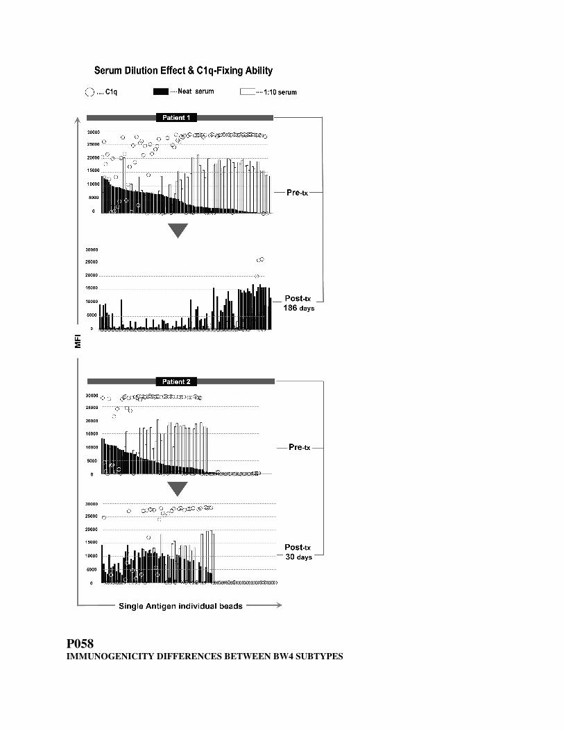
P058 IMMUNOGENICITY DIFFERENCES BETWEEN BW4 SUBTYPES

Annette M. Jackson, Donna P. Lucas, Zhimin Xiang, Julie A. Houp, Andrea A. Zachary. Johns Hopkins University, Baltimore, MD
Aim: Donor HLA-specific antibody represents a risk for rejection or engraftment failure following HLA mismatched solid organ or hematopoietic stem cell transplantation. We have examined the immunogenicity of Bw4 subtype mismatches in 211 renal recipients post-transplant. Methods: HLA typing was performed using Luminex based rSSOP (LAB Type, One Lambda). HLA single antigen panels (LABScreen, One Lambda) were used to identify HLA-Bw4 sensitization. Results: Bw4 epitopes present on HLA-B and HLA-A antigens can be divided into 8 subtypes based on amino acid differences at positions 77-83 (Voorter et al. Tissue Antigens, 2000). Furthermore, residues 80-83 have been shown to impact the electrostatic charge and explain observed serological Bw4 patterns (Kosmoliaptsis et al. Human Immunol. 2011). Bw4 motifs TALR and TLLR (expressed on HLA-B*44:02, B*13:01 and B*37:01, B*27:05, B47:01, respectively) have similar electrostatic potentials, but are uniquely different from the IALR Bw4 motif (expressed on HLA-B*57:01, 58:01, 49:01, 51:01, 53:01, 15:16, 15:13, 38:01 59:01 and HLA-A23:01, 24:02, 24:03, 25:01,32:01). Previously, using serum/cell adsorption experiments, we have shown that sensitization to the IALR can occur in recipients possessing TALR or TLLR. The incidence of sensitization to these different Bw4 motifs is shown in Table 1.The IALR motif evoked the highest incidence of sensitization in recipients possessing Bw6, Bw4-TALR, or Bw4-TLLR in their own phenotype. In contrast, mismatches involving TALR or TLLR provoked little sensitization in recipients possessing IALR. Conclusions: Structural and electrostatic potential differences between Bw4 subtypes appear sufficient to elicit alloimmune responses. Exposure to IALR, the most common Bw4 motif, can provoke a substantial alloantibody barrier to prospective donors (CPRA of 54%). Moreover, Bw4 sensitization may be relevant when selecting HLA allele mismatched donors for antigens that contain multiple Bw4 subtypes (i.e. HLA-B27, B38, B44, and B53).
Incidence of HLA-Bw4 sensitization across phenotypes
Recipient Phenotype Donor Bw4 mismatch % sensitized
Bw6 IALR 55/89, 62%
Bw6 TLLR or TALR 13/30, 43% p=0.09
TLLR &/or TALR IALR 24/52, 46%
IALR TLLR &/or TALR 3/33, 9% p=0.0003
TLLR TALR 1/3, 33%
TALR TLLR 0/4, 0%
P059 THE EXPRESSION OF HLA GENES IN PRE-IMPLANTATION KIDNEY BIOPSIES IS ASSOCIATED WITH GRAFT DYSFUNCTION AT 1 YEAR POST-TRANSPLANTATION Karina L. Mine1, Maria Gerbase-DeLima1,2. 1Associação Fundo de Incentivo à Pesquisa - AFIP, São Paulo, Brazil; 2Universidade Federal de São Paulo, São Paulo, Brazil
Aim: The aim of this study was to investigate the expression of HLA genes in pre-implantation kidney biopsies (PIB) in relation to graft function after kidney transplantation (Tx) from deceased donors. Methods: The analyses were performed using the expression data of PIB from our previous microarray study (paper in submission). Herein we analyzed the expression of HLA class I (A, B, C) and class II (DRA, DQA1, DQB1, DQA2, DQB2, DPA1, DPB1) genes in relation to delayed graft function (DGF) occurrence (27 cases with and 27 without) and graft dysfunction (estimated creatinine clearance ≤ 45 mL/min) at 1 year (y) post- Tx (10 cases with and 41 cases without graft dysfunction). Finally, the genes were tested in an independent data set from the literature. Results: No differences in expression levels of any HLA gene were observed in cases with or without DGF. HLA-B and HLA-C were not differentially expressed between kidneys that presented deteriorated or stable renal function at
1y. On the other hand, kidneys that presented deteriorated function at 1y post-Tx had significantly higher expression of HLA-A (p= 0.02), -DRA (p=0.003), -DQA1 (p=0.002), -DQB1 (p=0.00002), HLA-DQA2 (p=0.0008), -DQB2 (p=0.00005), -DPA1 (p=0.02), -DPB1 (p=0.0006). All these genes, with the exception of HLA-DQA2, were represented in the microarray study performed by Park et al, 2010, in biopsies collected at 1y post-Tx and were also associated with poor outcome, represented by biopsies with interstitial fibrosis/tubular atrophy (IF/TA) and inflammation. Conclusion: The association between increased expression of HLA genes in PIB and poor renal function at 1y post-Tx is consistent with the assumption that heightened expression of HLA genes contributes to increase allograft immunogenicity and consequently lowers long-term graft function and survival. Interestingly, among the genes with increased expression were the HLA-DQA2 and -DQB2 genes, whose normal expression pattern has not yet been determined.
P060 C*08:02/16:02 VERSUS C*05:01/16:01, AMBIGUOUS PAIRS PARTICULARLY SUSCEPTIBLE TO TYPING ERROR Steven Goldstein1, Maxim Norkin2, Mai Ta3. 1University of Florida, Gainesville, FL; 2University of Florida, Gainesville, FL; 3UF Health, Gainesville, FL
We present four individuals initially determined to be C*08:02:01:01 & 16:02:01 but subsequently typed as C*05:01:01:01 & 16:01:01. These consist of a patient typed in our laboratory as well as three unrelated donors (URDs). The URDs had initial typing performed at National Marrow Donor Program (NMDP) laboratories and confirmatory typing performed in our lab. Typing for our patient was performed by sequence specific oligonucleotide probe (SSOP, One Lambda) and sequence based typing (SBT, Conexio) techniques. SBT included both the C*08:02/16:02 and C*05:01/16:01 types as ambiguous pairs. These were resolved by SSOP showing the apparent patient C* locus types as 08:02 and 16:02. The patient had a diagnosis of mantle cell lymphoma and an unrelated donor search was conducted based on the type of A*02:01/29:02, B*44:02/44:03, C*08:02/16:02, DRB1*04:01/07:01. Samples from three potentially matched URDs were received. All were listed in the NMDP registry as C*08:02/16:02. Confirmatory typing on the URDs in our laboratory by SBT showed the same ambiguous pair as expected. However, SSOP appeared to rule out the C*08:02/16:02 combination listed by the NMDP and indicated that C*05:01/16:01 was the correct type for all three donors. To better confirm the C* locus types, sequencing with an allele-specific sequencing primer (HARP) was performed on all four individuals. C* locus typing by sequence specific primers was also performed for three. These methods identified C*05:01 and 16:01 as the correct type in the patient and all three URDs. Review of the initial patient typing showed a single false negative DNA probe. This probe in the One Lambda SSOP kit separates the ambiguous pairs and is susceptible to false negative results. It was near the cut-off value in our patient as well as one of the donors. Laboratories using the techniques of One Lambda SSOP with SBT for high resolution typing should be aware of the possibility of an incorrect type when the C*08:02/16:02 pair is identified. Close attention to B*/C* associations is also warranted as the C*05:01/16:01 pair is expected to be considerably more frequent than C*08:02/16:02 in association with B*44:02/44:03. Additional potential C*08:02/16:02 URDs were listed in the initial search. The NMDP was notified that investigation of those donors might be useful.
P061 DIFFERENTIAL HLA CLASS I AND II ASSOCIATIONS IN PEMPHIGUS FOLIACEUS AND PEMPHIGUS VULGARIS PATIENTS FROM AN ENDEMIC BRAZILIAN REGION Maria Jose F. Brochado1, Daniela F. Nascimento1, Neifi Hassan S. Deghaide2, Wilson Baleotti3, Eduardo A. Donadi1, Ana M. Roselino1. 1University of Sao Paulo, Ribeirao Preto, Brazil; 2Hospital of Clinics of Ribeirao Preto, Ribeirao Preto, Brazil; 3Faculty of Medicine of Marilia, Marilia, Brazil
Aim: Pemphigus comprise a group of blistering diseases caused by autoantibodies against desmogleins (Dsg). Two major clinical forms have been recognized: pemphigus foliaceus (PF) and pemphigus vulgaris (PV). The Northeast region of Sao Paulo State, Brazil, is endemic for both forms of pemphigus. In this study, we evaluated the association of HLA class I and II alleles with susceptibility to PF and PV in this Brazilian population. Methods: All patients (86 with PF and 82 with PV, exhibiting anti-Dsg1 antibodies for PF and anti- Dsg1/3 for PV) and 1592 controls were from the same region. HLA typing was carried out using PCR-SSOP. The allelic frequencies

and comparative analyses were performed using Fisher´s exact test and Odds Ratio. P≤ 0.002 was considered to be significant. Results: Susceptibility to PF was associated with HLA-A*11, A*33, B*14, HLA-DRB1*01:01/ *01:02/*16:01, DQA1*01/*01:02/*03, and DQB1*05:01, whereas HLA-A*02, HLA-DRB1*07:01/ *11:01/*13:01, DQA1*05 and DQB1*03:01 were associated with protection. Susceptibility to PV was associated with HLA-A*26, HLA-B*38, HLA-C*12 and HLA-DRB1*04:02/*08:04/ *14:01, DQA1*03/*03:01 and DQB1*03:02/*05:03, whereas HLA-B*15 and HLA-DRB1*07:01, HLA-DQB1*06:02 were related to protection. Conclusion: Historically, our region is endemic for PF, which affects younger and rural zone patients. In the last two decades, PV incidence has dramatically increased surpassing PF incidence, affecting younger and urban patients, and also becoming endemic. We describe for the first time the differential associations for PF and PV in the same Brazilian population. Although only two alleles groups (DQA1*03 and DRB1*07:01) were shared by both pemphigus disorders, the results of the present study emphasize the distinct pattern of HLA susceptibility/protection alleles. A.M. Roselino: Grant/Research Support; Company/Organization; FAPESP.
P062 GENETIC ASSOCIATION BETWEEN HLA CLASS I ALLELES AND HEPATITIS B VIRUS-RELATED HEPATOCELLULAR CARCINOMA IN A SOUTHERN CHINESE POPULATION. Minzhong Tang1, Yuming Zheng1, Yonglin Cai1, Xiaojiang Gao2, Mary Carrington2, Tao Peng3. 1Wuzhou Red Cross Hospital, Wuzhou, China; 2National Cancer Institute, Frederick, MD; 3First Affiliated Hospital of Guangxi Medical University, Nanning, China
Aim: Hepatocellular carcinoma (HCC) is a complex disease with multi-factorial in etiology. To investigate the association between HLA class I allele phenotypes and HCC, we have conducted a case-control study in a Chinese HCC cohort from a minority region in southern China. Methods: HLA class I genes were molecularly typed in 329 individuals with HCC and 342 non-HCC matched controls with normal liver enzyme levels. Results: Stratified analysis performed in the present study demonstrated that A*0201 (odds ratio [OR], 0.34; P=0.006), B*3802 (OR, 1.87; P=0.02) and Cw*0401 (OR, 0.34; P=0.02) were associated with HCC in Han Chinese; B*1501 (OR, 0.09; P=0.04), B*4001 (OR, 0.33; P=0.009), Cw*0702 (OR, 0.53; P=0.04) and Cw*1402 (OR, 7.88; P=0.02) were associated with HCC in Zhuang Chinese; B*1527 (OR, 0.08; P=0.005), B*4001 (OR, 0.49; P=0.01) and Cw*0401 (OR, 0.19; P=0.0003) have shown significant if only HBV positive was took into account. Conclusion: The associations with class I alleles showed that particular HLA phenotypes are associated with varied HCC risks in different ethnic groups. Interestingly, Cw*0401 was the only allele showing significance after correction and had been present in both Han ethnic group and Hepatitis B virus (HBV) positive group which has potential relationship to anti-tumor CTL or NK cell responses. M. Carrington: Grant/Research Support; Company/Organization; This work was supported in part by USA
National Institutes of Health Grants (ES05116 and ES09089). T. Peng: Grant/Research Support;
Company/Organization; This work was supported in part by the National Nature Science Foundation of China
(NSFC 30460143, 30560133, 30760243), Ministry of Education (NCET Program in University 2009).
P063 EVALUATION OF DISCREPANCIES BETWEEN LUMINEX SINGLE ANTIGEN BEAD ASSAYS Deborah O. Crowe, Angie Morris. DCI Laboratory, Nashville, TN
Currently there are two manufacturers offering HLA Antibody testing using the Luminex platform. Our laboratory incorporates both methods in our testing protocol. We have documented discrepancies and use them to help us identify samples that need follow-up verification of unacceptable antigens. More discrepancies are found with Class I than with Class II and some appear to be major differences. The purpose of this study was to examine the discrepancies with Class I single antigen testing between the two manufacturers with respect to correlation with Flow crossmatch results and MFI values. For this study discrepant samples from a single run of Class I single antigen beads were chosen. An antibody of interest was chosen for each sample and surrogate cells were found which did not have HLA detected by other Class I antibodies reported in the same sample. Flow crossmatch was performed using a BD FACsCaliber instrument on a 1024 scale. All discrepancies in this study resulted from an
antibody being called by Method A that was not detected using Method B. Surrogate crossmatches were done to determine if there was a sensitivity problem wiith Method B or a specificity problem with Method A. Twenty-seven crossmatches were examined which included eleven different specificities. Fourteen samples were T cell positive and thirteen were T cell negative. Of the fourteen that were T cell positive, six were B cell negative, which could indicate non-HLA reactivity. Therefore, it is possible that 19 of the 27 specificities detected by Method A may not be clinically relevant. The discrepancies between methods is of concern and emphasizes the necessity of including manufacturers and MFI cutoffs when publishing Luminex single antige bead results. Most HLA laboratories use single antigen results for determining unacceptable antigens and virtual crossmatches. More specificites were called with Method A using our current cutoff, but at least half of these did not result in a positive Flow crossmatch. Re-evaluation of the cutoff suggests that the cutoff may be different for certain loci or certain specifcities. The use of both methods may have value in fine-tuning the unacceptable antigens and illuminating those specificities that should be confirmed.
P064 ALLOANTIBODIES REACTIVITY WITH ALLOGENIC HUMAN CARDIAC STEM/PROGENITORS CELLS DESTINED FOR CARDIAC REPAIR Rachid Hocine. INSERM U976 Dermatologie Immunologie Oncologie, PARIS, France
Introduction: Allogenic human cardiac-derived stem/progenitor cells (hCPC) are promising therapeutics for repairing and regenerating injured inflamed myocardium. Beside their capacity to regenerate cardiac cell these cells act through paracrine effects. Immunologically, allogenic hCPC have HLA and PD-L1-dependent “tolerogenic/immunomodulatory” behavior that could contribute to their repairing/regenerating capacity. Although “safe” in the context of cellular allogenic immune response resolving their reactivity with determinants of grafts rejection or persistence, Donor-Specific Antibodies (DSA) in particular those against HLA (DSA-HLA) is mandatory for their clinical application. Methods: We investigated the reactivity of hCPC with sera from transplantation patients screened for anti HLA (class I, class II) antibodies with clinical conventional procedures. Using tailored crossmatch and experimental allogenic cellular assays we determined the susceptibility of hCPC (n=9) to CDC (complement dependent cytotoxicity) and to ADCC (Antibody dependent cellular cytotoxicity) upon their recognition by anti-HLA-I and HLA-II specific for their haplotype. Results: The reactivity of hCPC with alloantibodies was dependent on their HLA expression level. Sera containing anti HLA-I, but not anti HLA-II, specific antibodies induced considerable CDC and ADCC in hCPC, indicating that the presence of DSA-HLA I but not DSA-HLA II, antibodies could be deleterious. Anti-HLA can also induce signals via HLA I or HLA II molecules, modulating the activity and fate of hCPC. Our preliminary data demonstrate that engagement of HLA-I but more importantly HLA-II with specific antibodies induces rapid phosphorylation of MAPK/Erk, major pathway of cells proliferation and / or survival suggesting a potential beneficial effect of DSA-HLA. Conclusions: Allogenic hCPC are low-risk therapeutics in regard of cellular response but precaution should be taken in regard of humoral response, in particular, for patients possessing DSA-HLA I. Although warranted further investigation, signaling via HLA might counterbalance eventual deleterious effects, which calls upon proper evaluation through well-established immune testing platforms to maximize the benefit while minimizing the risk of allogenic hCPC therapeutics.
P065 THE ARG107GLY MISMATCH IMPACTS THE CD94/NKG2A/2C BALANCE: IMPLICATIONS FOR HLA-E MATCHING? Alexander A. Celik, Thomas Kraemer, Trevor Huyton, Rainer Blasczyk, Christina Bade-Doeding. Hannover Medical School, Hannover, Germany
Aim: The highly conserved HLA-E is an effective inhibitory molecule for malignant cells in order to prevent their fate from NK cell killing. The few polymorphic HLA-E alleles are restricted to two functional variants HLA-E*01:01Arg107 and HLA-E*01:03Gly107, both of them have been described to present a limited set of peptides derived from class I leader sequences. We could previously demonstrate how the immunological response to peptide-HLA-E complexes is dictated through the sequence of the bound peptide. Immunological and structural

implications of the micropolymorphism at position 107 are still not investigated systematically, yet. Methods: To assess functional differences of the two alleles, we systematically analyzed the E*01:01- and E*01:03-restricted peptide repertoire using soluble HLA technology and mass spectrometry. Artificial APCs expressing E*01:01 or E*01:03 bound to the same peptide were established to analyze the exclusive magnitude of residue 107 on NKG2/CD94 receptor engagement. Structural investigation of HLA-E variants bound to the same peptide was performed. Results: The HLA-E*01:03 restricted peptides were 9-17 amino acids in length and differed in their biochemical properties. No similarities to HLA-E*01:01 restricted peptides were observed. The differential peptide restriction highlighted the functional differences of both alleles that could be further investigated using artificial APCs presenting single peptide-HLA-E complexes in cellular assays with NK cells. Differential affinity of E*01:01 or E*01:03 to NKG2/CD94 receptors could be verified and consequently confirmed in cytotoxicity assays and structural analysis. The Arg107Gly mismatch account for an alteration of the alpha2 domain through a His155 reorientation. Conclusion: These results imply the recommendation for an HLA-E matching in HSCT.
P066 IN SILICO ANALYSIS OF DESMOGLEIN PEPTIDE BINDING SITES TO HLA-DRA/DRB1 AND DQA1/DQB1 HETERODIMERS IN BRAZILIAN PATIENTS PRESENTING WITH PEMPHIGUS FOLIACEUS OR PEMPHIGUS VULGARIS Wilson Baleotti1, Antonio Fabron2, Marcelo O. Ruiz2, Maria Jose F. Brochado3, Ana M. Roselino3, Eduardo A. Donadi3. 1Faculty of Medicine of Marilia, Marilia, Brazil; 2Laboratorio Diagnosticos do Brasil (DB), Curitiba, Brazil; 3University of Sao Paulo, Ribeirao Preto, Brazil
Aim: Autoantibodies against desmoglein-1 (Dsg1) and Dsg3 molecules are the hallmarks for pemphigus foliaceus (PF) and pemphigus vulgaris (PV), respectively. Overall, PF susceptibility (DRB1*01:01/*01:02, DQA1*01/*01:02/*03 and DQB1*05:01) and protection (DRB1*07:01/*11:01/*13:01, DQA1*05 and DQB1*03:01) alleles were distinct from susceptibility (DRB1*04:02/*08:04/*14:01, DQA1*03:01 and DQB1*03:02/*05:03) and protection (DRB1*07:01, DQB1*06:02) alleles observed for Brazilian PV patients. We performed an in silico study to evaluate desmoglein peptide binding sites for HLA class II molecules in PF and PV patients. Methods: HLA class II dimers were tested as anchors for 15-length amino acid peptides from Dsg1 or Dsg3 proteins. The NetMHCIIpan software version 3.0 was used for prediction results, considering thresholds for: i) strong (IC50) 50.000 nM and ii) weak (IC50) 500.000 nM binding peptides. Results: PF and Dsg1: The number of strong/weak binders was greater for DRB1 susceptibility molecules in comparison to DRB1 protection molecules. The DQA1*05:01/DQB1*03:01 and DQA1*05:10/DQB1*03:01 protection dimers presented higher number of weak binders compared to strong binders, while DQA1*01:02/DQB1*05:01 and DQA1*03:01/DQB1*05:01 susceptibility dimers showed no strong binders. PV and Dsg1: DRB1*08:04 exhibited greater strong/weak binders than other susceptibility molecules, and DQA1/DQB1 dimers did not present strong binders. PF and Dsg3: Results were closely similar to those reported for Dsg1; however protection molecules exhibit greater weaker binders. PV and Dsg3: Susceptibility and protection HLA molecules presented almost weak binders to Dsg3. Conclusion: Overall, HLA-DRB1 molecules, particularly in PF, exhibited stronger binding peptides for Dsg1 protein. DRB1 molecules presented greater strong binders as compared to DQA1/DQB1 dimers in both pemphigus.
P067 EVALUATION OF SNPS ON THE TOLL-LIKE RECEPTORS, NOD-LIKE RECEPTORS AND CD14 GENE IN THE RECOGNITION AND IN AUTOPHAGIC MODULATION IN RESPONSE TO INFECTION BY MYCOBACTERIUM LEPRAE. Priscila S. Mazini1,2, Paulo Rodrigues-Santos2,3, Manuel Santos-Rosa2, Hugo A. Vicentin1, Andressa H. Shinzato1, Mariane H. Shinzato1, Ana M. Sell1, Jeane E L Visentainer1. 1Universidade Estadual de Maringa, Maringa, Brazil; 2Immunology Institute, Faculty of Medicine University of Coimbra, Coimbra, Portugal; 3Immunology and Oncology Laboratory, Coimbra, Portugal

Aim: The present study investigated single nucleotide polymorphisms (SNPs) in genes of Toll-like receptors, NOD-like receptors and CD14 gene in the Brazilian population who developed leprosy per se (N=200) and compared to healthy contacts (N=250). Methods: The genotyping was performed using PCR-SSP (polymerase chain reaction - sequence-specific primers). Results: TLR1 (rs5743618) genotype GG was associated to protection for development of disease: OR= 0.52. TLR2 (rs1816702) genotype CC and allele C were associated to susceptibility (OR=2.91; 2.27), while the genotype CT and allele T to protection (OR=0.44; 0.44). Increased frequencies of TLR2 (rs4696483) genotypes CC and TT suggest risk (OR=1.56; 2.90) for development of disease, while the decreased frequency of CT suggests protection (OR=0.51). Increased frequencies of TLR4 (rs2149356) genotypes: AA and CA, and allele A suggest risk (OR=2.48, 2.20, and 2.46, respectively) and genotype CC and allele C suggest protection (OR=0.51, 0.53) to disease. The frequencies of TLR9 (rs352139) genotype GG and allele G (OR=0.61, 0.73 suggest risk and genotype AA and allele A protection (OR=2.04, 1.38). The genotype CT of CD14 -260 suggest risk (OR= 5.59) while genotype CC suggest protection (OR= 0.18). The genotype AG for CD14 cod204, suggest risk (OR=2.53), while the genotype AA suggest protection (OR=0.40). Statistical differences were observed for the SNP R702W (2104C>T) of NOD2 gene: The genotype CC and allele C suggest protection (OR= 0.40); and to allele T suggest risk, OR= 2.52. Conclusions: This study shows that polymorphisms of TLR, NOD2 and CD14 genes act as responsible for triggering an adequate immune response in the context of infection and disease leprosy.
P068 CLINICAL SIGNIFICANCE OF DONOR SPECIFIC ANTI-HLA DR51/52/53 ANTIBODIES FOR ANTIBODY MEDIATED REJECTION IN KIDNEY TRANSPLANT RECIPIENTS Borae G. Park1, Yongjung Park2, Kyu Ha Huh3, Myoung Soo Kim3, Yu Seun Kim3, Hyon-Suk Kim1. 1Yonsei University, Seoul, Korea, Republic of; 2National Health Insurance Service Ilsan Hospital, Goyang, Korea, Republic of; 3Yonsei University, Seoul, Korea, Republic of
Aim: The presence of anti-HLA donor-specific antibody (DSA) increases the risk of antibody-mediated rejection (ABMR) after kidney transplantation (KT). However, the clinical relevance of anti-HLA-DR51/52/53 antibodies remains unclear because of their weak antigen expression. This study evaluated the association between anti-HLA DR51/52/53 DSA and ABMR. Methods: We retrospectively reviewed the single panel reactive antibody (PRA) results of 130 patients tested between August 1, 2009 and March 6, 2015 to evaluated elevated serum creatinine levels after allograft KT. Single PRA analysis was performed using Luminex assay kits (LIFECODES LSA Class I and II, Immucor, USA). We reviewed the clinical course and biopsy results of the patients with anti-HLA-DR51/52/53 DSAs. Results: Post-KT DSAs were identified in 89 (68.5%) of the 130 patients: 26 (29.2%) class I and 63 (70.8%) of class II peak DSAs. Figure 1 showed the patients distribution according to the presence of DSA. Thirteen patients had anti-HLA-DR51/52/53 DSAs. Three patients diagnosed biopsy-proven ABMR with anti-HLA-DR 51/52/53 peak DSA alone. One patient who developed anti-HLA-DR DSA 13 days after KT showed a rapid increased in anti-HLA-DR51 DSA and had biopsy-proven ABMR. Conclusion: Although the expression of HLA-DR51/52/53 antigen was weak, but anti-HLA-DR51/52/53 DSA was correlated with biopsy-proven ABMR. Therefore, anti-HLA-DR51/52/53 DSA has clinical significance and these antibodies must be evaluated after transplantation.

P069 HIGH-THROUGHPUT AND COST-EFFECTIVE NGS STRATEGY FOR INTERMEDIATE-RESOLUTION HLA TYPING Inge Van Rooy, Claudia Rebel, Roel Van Aard, Michelle Bacelar, Bram Luiken, Loes Van de Pasch, Evelien Bouwmans, Raul Kooter, Erik Rozemuller, Maarten Penning, Wietse Mulder, Nienke Westerink. GenDx, Utrecht, Netherlands
Aim: The National Marrow Donor Program (NMDP) requires that each newly recruited donor is typed at intermediate resolution level for HLA-A, -B, -C, -DRB1 and optionally -DQB1. Therefore we developed an NGS assay specifically designed for intermediate resolution HLA typing (class I exon 2 & 3, and class II exon 2) that is suitable for high-throughput. PCR conditions were optimized such to reduce the costs of reagents. Moreover, the limited size of the amplicon allows for an increased sample capacity per run on either the MiSeq or Ion Torrent platform: a four-fold increase on average compared to the whole-gene amplification strategy. Method:We integrated this NGSessenz strategy into the previously developed NGSgo workflows for MiSeq and Ion Torrent platforms. For the MiSeq platform, high-throughput and reproducibility was verified by running 4x 96 DNA samples on a micro flow cell. For IonTorrent, we verified the capacity of the smallest (314 chip) and the largest (318 chip) by running a variety of samples. For each sample, loci were pooled prior to the execution of the NGSgo platform-specific library preparation and subsequent NGS procedures were performed. Results: We will present the NGS data that were obtained for the robustness of amplification, mappability of the reads, read depth per locus, concordance of NGS typing with reference type, reproducibility of the NGS data generated, and the capacity of the NGSessenz assay on the different platforms. Conclusions: Taken together, we will demonstrate the major advantage of this new NGSessenz strategy in ease-of-use and cost-effectiveness for HLA registry typing by means of NGS.

I. Van Rooy: Employee; Company/Organization; GenDx. C. Rebel: Employee; Company/Organization; GenDx. R.
Van Aard: Employee; Company/Organization; GenDx. M. Bacelar: Employee; Company/Organization; GenDx. B.
Luiken: Employee; Company/Organization; GenDx. L. Van de Pasch: Employee; Company/Organization; GenDx.
E. Bouwmans: Employee; Company/Organization; GenDx. R. Kooter: Employee; Company/Organization; GenDx.
E. Rozemuller: Stock Shareholder; Company/Organization; GenDx. M. Penning: Employee;
Company/Organization; GenDx. W. Mulder: Stock Shareholder; Company/Organization; GenDx. N. Westerink:
Employee; Company/Organization; GenDx.
P070 DIGGING DEEP: IDENTIFICATION OF A HIDDEN CROSSOVER Patricia Weingard, Nalaja Marcus-Freeman, Robert A. Bray, Howard M. Gebel. Emory University Hospital, Atlanta, GA
Aim: When patients requiring stem cell transplantation have multiple siblings as potential donors, HLA typing of all candidates is performed and haplotypes are ascertained. Recently, we began to routinely include HLA-DP typing in our analysis. Due to a hot spot of recombination between the HLA-DQ and HLA-DP loci, DP mismatches among otherwise HLA-A, B, C, DRB1 and DQB1 matched siblings are not an unexpected occurrence. In the family study shown below, a sibling who was HLA-DP mismatched to the patient was also mismatched at the HLA-DRB*3 locus. Interestingly, this DRB3* mismatch would not have been identified had high resolution typing for the HLA-DRB3*/4*/5* loci not been performed. Methods: High resolution typing for the HLA-A*, B*, C*, DRB1*, DRB3*, DRB4*, DRB5*, DQA*, DQB*, DPA* and DPB* loci was performed by SBT and/or SSOP. Results: HLA typing results and haplotype assignments are shown below. Sibling 1 and the patient are HLA identical, sharing the "a" and "c" haplotypes. Sibling 2 inherited the alternative maternal and paternal haplotypes ("b" and "d"). Sibling 3 has a unique haplotype ("a/b" and "c") resulting from a paternal recombination (represented by gray shading in the table below). Conclusions: Although likely a rare event, this case illustrates that recombination between the HLA-B and HLA-DRB3 locus resulted in a HLA-DRB3* mismatch between the patient and a potential donor. Due to sharing of the DRB1*, DQA and DQB alleles on the "a" and "b" haplotypes, this sibling would have been considered to be only an HLA-DP mismatch with the patient. Collectively, this case illustrates the utility of performing high resolution HLA typing for all the polymorphic HLA loci. -

P071 IMPORTANCE OF ROUTINE COMPARISONS BETWEEN LUMINEX INSTRUMENTS WITHIN A SINGLE LABORATORY. Mayuri Muchhala, Lisa Wilmoth-Hosey, Nalaja Marcus-Freeman, Howard M. Gebel, Robert A. Bray. Emory University, Atlanta, GA
Aim: The development of solid phase single antigen bead (SAB) assays for HLA testing has allowed for better identification and monitoring of HLA antibodies. However, standardization of testing and interpretation has not occurred and several variables can contribute to variation within and between laboratories. Instrument standardization, when multiple machines are used in the same laboratory, is of critical importance especially when following patients longitudinally pre- or post-transplant. The purpose of this report is to highlight the value of routine instrument comparisons. Methods: To compare fluorescence MFI values between Luminex instruments, 6 informative samples were tested using Class I SABs(One Lambda, Inc). Testing was performed in one tray and immediately prior to running; samples were split to two trays. This allowed the same test sample to be run simultaneously on 2 different machines. Data were collected and differences in MFI values for the same bead were compared between instruments. Results: Initial comparison of individual bead MFI values between Lum-1 and Lum-2 showed significant variance of up to 20% (acceptable value <10%)(Fig. 1A). Subsequently, service was performed on both instruments to align, standardize and calibrate. Lum-1 was found to have a failing laser even though it passed daily QC. Testing was then repeated. Post instrument service now showed a marked decrease in the variance between the 2 instruments (<6%).(Fig. 1B) Conclusion: Our data clearly show the value of performing sample comparisons between Luminex instruments within the same laboratory. Luminex controls do not assure instrument comparability. Instruments not meeting predetermined standards should be serviced and acceptable values reestablished. Data should be collected on a semi-

annual basis to reconfirm that the results between instruments are comparable. This practice ensures that MFI values are similar between instruments and allows labs to confidently evaluate and compare MFI values from multiple instruments.
P072 HIGH-RESOLUTION HLA TYPINGS OBTAINED FOR HIGH-THROUGHPUT NGS USING THE ILLUMINA MISEQ PLATFORM

Loes Van de Pasch, Michelle Bacelar, Claudia Rebel, Roel Van Aard, Raul Kooter, Erik Rozemuller, Maarten Penning, Evelien Bouwmans, Nienke Westerink. GenDx, Utrecht, Netherlands
Aim: Next-generation sequencing (NGS) technology generates sequences from single DNA molecules that enables unique identification of the paternal and maternal alleles. The NGS technology has great potential in the application of HLA typing for diagnostic purposes and generating reliable, unambiguous HLA typing results in a high-throughput fashion. Here we present data obtained for 11 HLA loci using a HLA typing assay dedicated for high-throughput NGS-based HLA typing on the Illumina MiSeq. The implementation of this high-throughput NGS strategy would be highly advantageous to obtain robust high-resolution HLA typing results for registry labs and routine diagnostic purposes. Method:A large genomic DNA reference panel, including UCLA reference samples, were selected for high-throughput NGS HLA typing. The whole gene of HLA-A, -B, -C, and the essential genomic regions of HLA-DRB1, -DRB345, -DQA1, -DQB1, -DPA1 and -DPB1 were PCR-amplified in a single PCR for each locus. Amplicons of each sample were pooled and processed using our NGSgo® DNA library preparation workflow for Illumina MiSeq. During library preparation the samples were fragmented, barcoded and multiplexed. Finally paired-end sequencing (2x150 cycles) was performed on the Illumina MiSeq platform using a standard flow cell and data was analysed using our HLA typing software NGSengine®. Results: Paired-end NGS data was obtained for 11 HLA loci (class I and II) of most genomic DNA reference samples in a single Illumina MiSeq sequencing run. The NGS data covered the complete HLA region of interest by highly accurate measures for all HLA loci tested. All HLA alleles were effectively separated and, in most cases, could be phased throughout the gene. For the majority of samples, unambiguous third- and fourth-field resolution typing were obtained for all loci that are in concordance with the second-field or higher reference types. Conclusions: The high-throughput NGSgo® strategy that we have developed demonstrates to be a powerful method to perform high-resolution HLA typing by means of NGS in a robust and reliable manner. L. Van de Pasch: Employee; Company/Organization; GenDx. M. Bacelar: Employee; Company/Organization;
GenDx. C. Rebel: Employee; Company/Organization; GenDx. R. Van Aard: Employee; Company/Organization;
GenDx. R. Kooter: Employee; Company/Organization; GenDx. E. Rozemuller: Stock Shareholder;
Company/Organization; GenDx. M. Penning: Employee; Company/Organization; GenDx. E. Bouwmans:
Employee; Company/Organization; GenDx. N. Westerink: Employee; Company/Organization; GenDx.
P073 RAPID MICROCHIMERISM ANALYSIS USING MULTIPLEXED REAGENTS AND NOVEL SOFTWARE Bram Luiken1, Job Geerlings1, Nienke Westerink1, Erik Rozemuller1, Doug Bost2. 1GenDx, Utrecht, Netherlands; 2KimerDx BV, Utrecht, Netherlands
Aim: To overcome the sensitivity and analysis shortcomings of STR-based transplant monitoring methods, and to improve upon the current state of the art in qPCR-based chimerism analysis, KimerDx has developed a new assay and software suite. Method:Our reagents employ a multiplexed panel of 30 qPCR research assays and software to genotype multiple samples on a single plate and identify informative markers. The software presents the marker choices in their genomic context, allowing for informed decision-making and use in post-transplant monitoring. The genotyping results are stored by the software for recall during subsequent monitoring. Results: Our software facilitates post-transplant monitoring by: allowing operators to customize their plate configuration or work with a lab-defined template, performing all calculations necessary to execute the test, generating a printable protocol to assist laboratory work and by generating templates to import into the qPCR machines. The software accepts standard qPCR data output for generating results. Reports from a monitoring test may be generated from a single time point or using the longitudinal data collected from the individual sample over time - providing a temporal view for better understanding rejection or relapse kinetics. Conclusion: Our reagents and software are compatible with qPCR platforms from multiple vendors. The software solution reduces manual calculations, increases flexibility in experimental execution, generates complete protocols for use in the lab and stores results. B. Luiken: Employee; Company/Organization; GenDx. J. Geerlings: Employee; Company/Organization; GenDx.
N. Westerink: Employee; Company/Organization; GenDx. E. Rozemuller: Stock Shareholder;

Company/Organization; GenDx, KimerDx. D. Bost: Employee; Company/Organization; KimerDx BV. 6. Stock
Shareholder; Company/Organization; KimerDx BV.
P074 EVALUATION OF THE HLA-F VARIABILITY IN BRAZIL BY NEXT GENERATION SEQUENCING Erick C. Castelli1, Thálitta H.A. Lima1, Renato V. Buttura1, Michelle A. Paz1, Iane O. P. Porto1, Jaqueline Ramalho1, Andreia S. Souza1, Luciana C. Veiga-Castelli2, Celso T. Mendes-Junior3, Eduardo A. Donadi2. 1UNESP - Univ Estadual Paulista, Botucatu-SP, Brazil; 2School of Medicine of Ribeirão Preto - USP, Ribeirao Preto - SP, Brazil; 3Faculdade de Filosofia, Ciências e Letras de Ribeirão Preto, Ribeirao Preto - SP, Brazil
Aim: HLA-F is a non-classical HLA class I gene and is distinguished from its classical counterparts by low allelic polymorphism and distinctive expression patterns. The exact function of HLA-F remains unknown. It is believed that HLA-F has a tolerogenic function, based on its low variability and sequence conservation among primates. Currently, there is little information regarding the HLA-F allelic variation among human populations and available studies evaluated only a fraction of the HLA-F gene segment and/or searched for known alleles only. Here we evaluated the HLA-F variability (from -300 to the stop codon at position +2944, including introns) in 72 samples from the Southeastern Brazil, using Next Generation Sequencing procedures. Methods: HLA-F was amplified by PCR using generic primers. Sequencing libraries were obtained by using Nextera XT DNA (Illumina) and sequenced at the MiSeq platform (together with other HLA class I genes). Paired-end sequences were filtered and mapped by using a local software named hla-mapper, which allows a reliable sequence mapping when several HLA genes are sequenced together. This procedure takes into account data from IMGT/HLA and 1000Genomes databases to properly assign each pair of sequences to the correct gene. Genotype calling was performed combining the Genome Analysis Toolkit (GATK) and local applications to get correct genotypes. Missing alleles and haplotypes were inferred by using the PHASE method, however, the known phase between variable sites (obtained by GATK) was considered. Results: We found 39 variable sites arranged into 14 haplotypes. Of those, five were identical to a known haplotype described by the IMGT/HLA database, that includes F*01:01:01:01, F*01:01:01:08, F*01:01:03:01, F*01:01:03:03 and F*01:03:01:01, and they represent 75% of all haplotypes. The remaining haplotypes did present one or two nucleotide differences from a known IMGT/HLA haplotype. However, most of them were found at least twice in the present series. In addition, four new variable sites were detected. Conclusion: we present here a strategy to evaluate HLA-F by NGS and the results do indicate that it is indeed conserved at the protein level, but the HLA-F worldwide haplotype variability might be higher than the one already described by IMGT/HLA.
P075 MONOCYTE RECRUITMENT TO HLA CLASS I ANTIBODY-ACTIVATED ENDOTHELIAL CELLS IS DEPENDENT UPON MTOR Sahar Salehi, Elaine F. Reed. UCLA, Los Angeles, CA
Aim: Antibody-mediated rejection (AMR) persists as a major issue affecting solid organ transplants. The purpose of this study was to investigate the proximal signaling events that regulate mTOR-mediated leukocyte recruitment following HLA I stimulation in endothelial cells (EC). Methods: For confocal microscopy experiments, EC were treated with rapamycin for 24 hours, stimulated with HLA antibodies, incubated for 1 h with sheep anti-ICAM-1 antibody, followed by 30 min incubation with secondary FITC-labeled goat anti-sheep IgG and then fixed. Cells were imaged by a Zeiss confocal microscope. EC were treated with l mTOR inhibitors (RAD001 or Rapamycin) or U0126, PKC, Rho or ROCK inhibitors, then stimulated with HLA I antibodies. Phosphorylation of downstream effectors of the mTOR and Rho pathways were measured by Western blot. Recruitment of primary monocytes to EC was determined by static adherence assays. Results: Using flow-based adherence assays, we previously discovered that mTOR inhibition impairs the ability of EC to support firm adhesion of tethered monocytes by affecting late stage ICAM-1 clustering, which has been attributed to Rho signaling. As demonstrated by confocal microscopy, this impairment was due to a decrease in association of actin stress fibers with ICAM-1, which disrupts ICAM-1 clustering. We determined by Western blot that mTOR operates upstream of RhoA, as inhibition of Rho did not disrupt phosphorylation of proteins downstream of the mTOR pathway, though inversely, mTOR inhibition blocked phosphorylation of downstream effectors of
Rho. mTOR in turn regulates ERM phosphorylation, which is crucial for ICAM-1 clustering. RhoA pathway inhibition of EC blocked HLA-I antibody-induced recruitment of primary monocytes in static adherence assays. Conclusion: Our findings connect mTOR-mediated class I signaling to cytoskeletal remodeling involving RhoA and ERM proteins, suggesting that mTOR inhibition reduces leukocyte recruitment to activated endothelium by impairing firm adhesion.
P076 DIFFERENCES IN BLOOD COMPOSITION BETWEEN DECEASED AND LIVING DONORS: COULD IT AFFECT CROSSMATCHES? Chantale Lacelle1, Amanda Cameron2. 1University of Texas Southwestern Medical Center, Dallas, TX; 2University of Texas Southwestern Medical Center, Dallas, TX
Until recently, most deceased donor crossmatches performed in our laboratory used cells isolated from node or spleen. However, due to changes at the OPO level it has become difficult to obtained pre-harvest node for crossmatches and donor blood is now routinely used. Since donors often receive blood transfusions and drugs in an attempt to save their lives, the effect of these factors on blood composition needs to be evaluated. We analyzed blood samples collected from deceased and living donors for white blood cell count (WBC) and percentage of lymphocytes, monocytes, and granulocytes using the COULTER® AC•T diff2™ Analyzer. We found that deceased donors had a markedly different blood composition than living donors. The WBC count of deceased donor (14.2x10³ cells/µL) was statistically higher than that of living donor (6.58x10³ cells/µL, p<0.05). Further, deceased donors had a significantly lower percentage of lymphocytes (13.1% vs 25.3%) and higher percentage of granulocytes (81.8% vs 66.7%) in their blood than living donors. No statistical difference was found in the percentage of monocytes. The observed differences in blood composition resulted in changes in cell preparation when the samples were separated using Ficoll. After Ficoll separation, cell preparations obtained from the blood of deceased donors had an average composition of 34.8% lymphocytes, 16.4% monocytes and 48.7% granulocytes, while cell preparations from living donors had an average of 82.8% lymphocytes, 9.3% monocytes and 7.8% granulocytes. The changes in percentage of lymphocytes and granulocytes were statistically significant (p<0.05). Our data indicates that blood composition varies between deceased and living donors and that Ficoll alone may not be an optimal isolation method for deceased donor blood. An excessive amount of granulocytes in cell preparations may affect crossmatch results as the expression of HLA may differ between lymphocytes and granulocytes. Additionally, this raises the questions as to whether the factors influencing the blood compartment of deceased donors also affect the expression of HLA class I and II on lymphocytes. Further studies will be needed to address whether the expression of HLA class I and II on lymphocytes isolated from decease donors differs from that of lymphocytes isolated from living donors.
P077 DPB1 T-CELL EPITOPES: MOLECULAR DEFINITION Pedro Cano. Blood Systems Inc., Scottsdale, AZ
Aim: Molecular characterisation of known differences of T-cell recognition of DPB1 alleles. Methods: Propositional logic algorithm to search for best sequence properties to account for reported T-cell recognition differences. Results: DPB1*09:01:01, DPB1*10:01 and DPB1*17:01 have been reported by Fleischhauer et al. to be mismatch high-risk (group 1) and DPB1*03:01:01, DPB1*14:01:01 and DPB1*45:01 to be mismatch intermediate-risk (group 2). DPB1 alleles have been previously classified in 4 serologic groups based on 2 dimorphic epitopes, the first epitope defined by amino acid position 56 and the second epitope by positions 85, 86 and 87. Fleischhauer’s group 1 and group 2 DPB1 alleles are both included in serologic group DP3 characterised by 56(E) on one hand and 85(E)-86(A)-87(V) on the other. [Human Immunology 2009 70:836] DP3 alleles can be further classified by amino acid positions 11, 65 and 76 into 3 subgroups that match Fleischhauer’s mismatch high-risk groups. This molecular characterisation into subgroup must be interpreted in the light of higher level groups defined by the 56 and 85-86-87 two dimorphic serologic epitopes. In this context, within the DP3 serologic group, T-cell group 1 is characterised by 11(L)-65(I) with tolerance for variation at position at position 76. T-cell group 2 is characterised by 65(L)-76(V) with tolerance for variation at position 11. Conclusions: If the molecular characterisation of DPB1 T-cell epitopes presented here is correct, then
DPB1*35:01:01 and the DPB1*17:01 exon 3 variant DPB1*131:01 should also be included in the high-risk group 1 together with DPB1*09:01:01, DPB1*10:01 and DPB1*17:01. DPB1*104:01 and the DPB1*03:01:01 exon 3 variant DRB1*124:01 should be included in group 2 together with DPB1*03:01:01, DPB1*14:01:01 and DPB1*45:01. DPB1*57:01 might also belong in group 2 depending on the tolerance for variation at position 11 in this group. (All these alleles are known common alleles. Undocumented alleles are excluded.)
P078 UNAMBIGUOUS HLA GENOTYPING USING LONG-RANGE PCR AND NEXT-GENERATION SEQUENCING (NGS) ON BUCCAL-EXTRACTED DNA: RESULTS FROM 4,000 DONOR RECRUITMENT SAMPLES James H. Lan1,2, Yuxin Yin1, David Nguyen1, Yung-Tsi Bolon3, Brianna Springer3, Katsuyuki Saito4, David Berman4, Tim Hague5, Gyorgy Horvath5, Krisztina Rigo5, Elaine F. Reed1, Qiuheng Zhang1. 1UCLA Immunogenetics Center, Los Angeles, CA; 2University of British Columbia, Vancouver, BC, Canada; 3National Marrow Donor Program, Minneapolis, MN; 4One Lambda, Thermo Fisher Scientific, Canoga Park, CA; 5Omixon LTD, Budapest, Hungary
Aim: Here we report the feasibility and results of using long-range PCR and clonal sequencing by NGS to deliver unambiguous HLA typing on over 4,000 buccal-based donor recruitment samples. Methods: Multiplex long-range PCR primers co-amplified HLA-A, -B, -C from promoter to 3’-UTR. DRB1 and DQB1 were amplified in separate reactions to cover exons 2-3. Library construction was performed using Illumina TruSeq Nano, followed by 2x250 bp paired-end sequencing on MiSeq. HLA typing was assigned using a combination of two independent computational algorithms to ensure high confidence in allele calling. Consensus sequence and typing results were reported in HML 0.97 format for data consumption. Results: Initial NGS validation was accomplished on 72 DNA Exchange samples isolated from blood and 154 donor recruitment buccal-derived DNA. Overall, NGS-HLA typing was 99.6% concordant with reference results. Primer- and software-related allele mis-assignments accounted for the small number of discrepancies observed, which were later optimized to further strengthen typing accuracy. Next, we introduced robotics to streamline the complex library construction process, achieving an impressive typing throughput of 384 samples (1920 loci) per week. Using this workflow, over 4,000 samples to date have been typed under high-resolution in a bone marrow donor recruitment pilot study. Despite known challenges of nucleic acid degradation and low DNA concentration commonly associated with buccal-based specimens, our assay was successful in typing 97.1% of sample loci. Overall, NGS typing accuracy was 99.02% based on blind QC of 256 loci. N=59 rare alleles were identified during this period and were all confirmed to be accurate by an alternative method. Finally, long-range sequencing delivered unambiguous 6-8 digit typing resolution in HLA-A -B, -C, and allele-level G group results in -DRB1 and -DQB1 in all samples reported. Conclusion: Long-range, unambiguous HLA genotyping is achievable on clinical buccal-extracted DNA. Importantly, full-length gene sequencing and the ability to curate raw sequence data in a standardized HML format could permit future interrogation of the impact of introns, expanded exons, and other gene regulatory sequences on clinical outcomes in transplantation.
P079 WHEN SILENT MUTATIONS SPEAK UP: FINE GRANULARITY IN LINKAGE DISEQUILIBRIUM Pedro Cano, Art Hoffman. Blood Systems Inc., Scottsdale, AZ
Aim: Silent mutations and mutations in non-coding regions are traditionally considered clinically irrelevant and ignored in clinical testing. Are we missing any valuable information in the data we discard? Methods: Sequences obtained by standard SBT methods are reanalised with BLAST and ad hoc sequence databases to study sequence segments typically overlooked. Results: Examples of some observations made: C*03:03:01 goes with B*15:01:01:01, B*55:01:01 and B*07:02:01, but C*03:03:04 goes with B*44:03:01. C*03:04:01:01 goes preferentially with B*40:01:02 and less frequently with B*15:01:01:01, while C*03:04:02 goes commonly with B*15:10:01 and with B*53:01:01, which is also seen with the first allele. C*05:01:01:02 goes with B*44:02:01:01 and C*05:01:01:01 goes with B*18:01:01:01. C*06:02:01:01 goes with, B*13:02:01, B*57:01:01, B*37:01:01 and B*47:01:01:01; C*06:02:01:02 goes with B*50:01:01 and B*50:02; and C*06:02:01:03 goes with B*45:01. C*07:02:01:03 goes with B*07:02 and
C*07:02:01:01 goes with a variety of B alleles including B27:05, B*39:01 and B*38:02. C*15:05:02 goes with both B*07:05 and B*07:06, two alleles that are themselves difficult to tell apart; and C*15:05:01 goes with B*73:01 [Full results to be presented at ASHI 2015.] Conclusions: Most of the time allele variants with silent mutations for a common allele are very rare or even poorly documented, but from time to time we see a common allele present in 2 silent-mutation forms. (Here we include intronic mutations with silent mutations.) Typically these variants are ignored and alleles are reported only with the first 2 fields or first 4 digits because only the protein product, and not the DNA sequence, is relevant in a clinical setting. When we look at linkage disequilibrium between HLA loci we find however that these silent-mutation variants of common alleles sometimes have strikingly different associations with alleles at other loci. Genotype analysis and linkage disequilibrium analysis should be based on information provided by the full DNA sequence, including silent mutations and intronic mutations, and not just on the coding sequence that defines a protein with a unique sequence of amino acids. Next generation sequencing will optimize this process.
P080 COMPARISON OF THE COST ASSOCIATED WITH AN IMMUNE MONITORING PROGRAM TO TREATMENT OF REJECTION Peter T. Jindra1, Rachael Bedard2, Ronald H. Kerman1. 1Baylor College of Medicine, Houston, TX; 2CHI St. Luke's - Baylor St. Luke's Medical Center (BSLMC), Houston, TX
Following transplantation, patients often receive a standardized regimen of maintenance immunosuppression. Biomarker monitoring could help to individualize immune therapy, giving adequate immunosuppression to high risk patients while possibly avoiding toxicity and infection in low risk patients. Furthermore, early detection and immunosuppression dosage changes before clinical dysfunction maybe a cost effective strategy to minimize rejection episodes. The aim of our study was to define the cost associated with an anti-HLA antibody monitoring program over 5 years compared to treating patients for a single acute rejection episode. We hypothesize having a donor specific HLA antibody monitoring program would help to define early rejection before clinical symptoms arise and therefore be a cost benefit. Our study included 23 heart transplant patients diagnosed with either acute cellular rejection (ACR), antibody mediate rejection (AMR) or both. We calculated the average cost associated with the rejection episode including hospital days and treatment for an ACR, AMR or both. For comparison we calculated the cost for an donor specific anti-HLA antibody monitoring program using Flow PRA, HLA antibody specificities and specificity titers using One Lambda Reagents over a 5 year follow-up period. We found the average cost to treat AMR was 200 times greater than the cost for ACR. ACR rejections were treated with steroids, whereas AMR required multiple rounds of plasmaphersis, IVIG and Rituxan. The cost for an HLA monitoring program over 5 years including FPRA and HLA antibody specificities was comparable to a round of plasmapheresis or a single endomyocardial biopsy and appears to offer patients at risk for AMR due to strong sensitization history a cost effective early detection method upon which immunosuppression could be adjusted. The data suggest that early prediction of acute rejection may allow for optimization of therapy prior to major graft damage and make earlier cost effective preventative interventions possible. As a cost effective strategy we propose performing dose adjustments of maintenance immunosuppression after detection of DSA, preventing the need for later costly rejection treatment.
P081 OPTIMIZATION OF SOLID PHASE AND FLOW CYTOMETRY CROSSMATCH ASSAYS THROUGH THE USE OF DTT TREATED PATIENT SERA Katharine Spanjer1, Kirsten Anderson1,2, Brian Freed1. 1ClinImmune Labs, Aurora, CO; 2University of Colorado Anschutz Medical Center, Aurora, CO
Aim: To determine the effects of DTT treatment of patient sera on C1 interference in mixed bead, single antigen, C1q and flow cytometric assays. Methods: For all experiments, patient sera were treated with 0.005M DTT. C1qrs (C1) interference was confirmed for 10 sera samples by dilution studies. We compared single antigen bead (SAB) results for these sera with and without DTT treatment. 4 of these same sera known to contain complement binding antibodies were used to evaluate the effects of DTT treatment in the C1q assay. The assay for C1q was followed as per manufacturer’s instructions with the exception of the heat inactivation step. We compared our results to IgG subclass testing. 26 T cell and 24 B
cell flow crossmatch assays (XM) were run at RT, 4°C, and with DTT treatment. Results: C1 interference was eliminated from all solid phase assays. Repeat testing of the 4 sera using PE-conjugated monoclonal antibodies to the 4 IgG Subclasses confirmed that IgG1 antibodies were present and were binding to the same specificities that had been blocked in the SAB assay due to C1 interference. A modified C1q procedure in which DTT treatment could be done without interfering with the C1q reagent was developed. Results of the modified procedure correlated very well with results of both the IgG subclass testing and the SAB DTT validation. All XMs that showed decreased event counts/C1 interference at RT (30 of 50) showed higher event counts in 4°C and DTT XM methods, demonstrating that C1 interference was eliminated in 100% of the affected XMs. 100% specificity was seen in both the 4°C and DTT methods, however, both the RT and 4°C XMs had one false negative. Lower MFI values were seen in the RT and 4°C XMs when compared with DTT treated XMs. On average, positive T cell XM values increased by 920 MFI at 4°C and 1,793 MFI with DTT; positive B cell XM values increased by 4,894 MFI at 4°C and 5,036 MFI with DTT. All XMs run using DTT achieved expected positive or negative results based on the presence/absence of DSA. Conclusion: We have eliminated false negative reactivity in solid phase PRA and flow cytometry XM by incorporating DTT treatment into all assays.
P082 RESOLUTION OF HLA-A*68:11N ALLELE BY ONE LAMBDA LABTYPE® REVERSE SEQUENCE SPECIFIC OLIGONUCLEOTIDE TYPING KIT Randy Williams, Sandy Warnock, Lois Regen, Maggie Sprague, Gansuvd Balgansuren. Seattle Cancer Care Alliance, Seattle, WA
Aim: Resolution of the ambiguous combination of HLA-A*68:01:02:01/68:11N allele in HSCT patient and donor pairs is now mandatory by NMDP Policy. Laboratories are required to test for non-expressed alleles when they exist as a possibility within the assigned G group. In our laboratory this occurs in the case of A*68:01:02:01 which gives an ambiguous A*68:01/68:11N result by the Sanger sequencing-method. Method:One Lambda® HD rSSO kits have developed several probes to identify rare or uncommon alleles with no positive quality control due to the very uncommon/rare nature of these alleles. Therefore, it is difficult to assess whether we can actually identify those specificities with confidence. The Fred Hutchinson Cancer Research Center’s Research Cell Bank made available to us two DNA samples from the 13th IHWG workshop characterized as having A*68:11N allele (9418 and 9419). In addition, several samples with A*68:01:02:01 and/or A*68:02G were included to ensure that the definitive bead #94 for the A*68:11N allele would not go false positive for other expressed A*68 alleles. Results: Using One Lambda’s RSSOH1A® lot 008 HLA typing kit we tested IHWG workshop samples 9418 and 9419. Sample 9418 tested as A*03:MJMJ and A*68:11N. Sample 9419 typed as A*24:02G and A*68:01:02:01/68:11N/68:33. Inspection of sample 9419 showed that A*68:01:02:01 and 68:33 were associated in combination with an A*24:36N (a highly unlikely event), thus ruling out the possibility of A*68:01:02:01 or 68:33. Bead #94 was strongly reactive with both samples, indicating the presence of the HLA-A*68:11N allele. Although bead #94 should be positive with other rare HLA-A alleles, the bead should not be positive with the ambiguous combination found in Sanger sequencing. Conclusion: One Lambda’s RSSOH1A® test kit can positively identify the A*68:11N allele, which in combination with Sanger sequencing can resolve the ambiguous A*68:01:02:01/68:11N result.
P083 EVALUATION OF POLYREACTIVE MONOCLONAL ANTIBODIES USING HLA SOLID PHASE ASSAYS Justin Mostecki1, Baoshan Gao2, Sarah B. See3, Kara Crowley1, Emmanuel Zorn3, Bryan Ray1. 1Immucor, Stamford, CT; 21Transplant Center, Department of Surgery,, Boston, MA; 3Columbia University Medical Center, New York, NY
It is well established that HLA antibodies play an important role in antibody-mediated rejection (AMR) and there is a growing body of literature demonstrating that non-HLA antibodies may also contribute to AMR. To study the source of non-donor specific HLA antibodies and self-reactive antibodies in patients with kidney graft loss, Porcheray et al. (Am J Transplant 2012; 12: 2088 - 2097) expanded immortalized polyreactive B-cell clones from a

patient with kidney graft rejection and showed that some of these natural antibodies are cross-reactive to HLA and self-antigens. Gao et al. (Am J Transplant 2014; 14:1581 - 1591) also showed that the presence of IgG natural antibodies, with activity to apoptotic cells, correlates with late kidney allograft loss even after excluding patients with high reactivity to HLA. We have further characterized the HLA reactivity of 4 polyreactive B-cell clones isolated by Porcheray, Gao et al. using solid phase assays (Immucor, Inc.). We report here that while these clones do not react with HLA on the Immucor single antigen beads, they react to other HLA products, with some variation between different solid phase assays. Their reactivity appears to be enhanced upon denaturation, as judged by the monoclonal antibody 210.4, which reacts with denatured antigen, suggesting that they primarily react to cryptic epitopes. B. Ray: Employee; Company/Organization; Immucor.
P084 IL2 POLIMORPHISMS HAVE NO IMPACT ON RISK, BUT GENETIC VARIANTS IN IFNG HAVE A PROTECTIVE ROLE IN ACUTE LYMPHOBLASTIC LEUKEMIA AND ARE ASSOCIATED WITH GENDER IN MEXICAN CHILDREN Betsy Gonzalez1,2, Hilario Flores-A1,2, Diego Sanchez2, Carmen Alaez1, Clara Gorodezky1,2. 1InDRE, Secretary of Health, Mexico City, D.F., Mexico; 2Fundacion Comparte Vida A.C., Mexico City, D.F., Mexico
Aim: Acute lymphoblastic leukemia (ALL) is the most common cancer in children (26%). ALL patients present a defective immune system with deregulation of cytokine production. A lower population of IL-2 and INF-γ producer cells Th1 and Tc1 are evident in ALL patients. These cytokines promote cellular immunity and its protein production may be altered by inherited polymorphisms. The aim of this study was to evaluate the role of IFNG(+874 A/T) and IL2(-330 G/T) SNPs in the expression of ALL in Mexican children and the gender influence in susceptibility to the disease. Methods: ALL patients (N=148) were genotyped for INFG and 110 for IL2; 376 healthy controls were included for comparison. All individuals are Mexican Mestizos, born in Mexico. The patients' age ranged between 1-17 years; 59.4 % were males and 40.6 % females. DNA was obtained from peripheral blood samples using the Maxwell16 instrument. Allele discrimination of IFNG +874A/T(rs2430561) and IL2 -330 G/T(rs2069762) was performed using a Taqman real time PCR assay and an ABI 7500 Real-Time PCR equipment. Results: A protective role of IFNG(+874A/T) SNP was shown for the expression of ALL. The frequency of the T allele (OR=0.482; p=0.00005) and TT genotype (OR=0.052 p=0.000006) were found decreased in the patients, while AA genotype was increased (OR=1.86;CI 1.25-2.76;(p=0.003). Upon sex-stratification, TT was protective in both, males (OR=0.087; p=0.003) and females (OR=0.127; p=0.02). IL2(-330G/T) SNP did not show any statistical deviation between cases and controls. Conclusions: IL2(-330 G/T) was not associated with the development of childhood ALL. However, our results support the role of IFNG (+874 A/T) in surveillance against tumor cells in ALL in Mexican Mestizo children, IFNG +874 TT genotype has been associated with high cytokine production, since +874T matches with the transcription factor NF-ĸB binding site that may enhance IFNG transcription. The cytokine has antitumor and anti-proliferative properties; and our data agree with other reports emphasizing the role of IFN-γ alleles in immune surveillance and its posible effect as an immunotherapeutic agent in certain forms of ALL. Moreover, recent data suggest the association of TT genotype on age at clinical onset in B- lineage ALL, in older children expressing the TT high producing IFN-γ genotype.
P085 IL6 (-174G/C) SNP BUT NOT IL4 (-590C/T) POLYMORPHYSMS CONTRIBUTES TO THE DEVELOPMENT OF ACUTE LYMPHOBLASTIC LEUKEMIA, AND IS INVOLVED IN GENDER SPECIFIC RISK IN MEXICAN CHILDREN Betsy Gonzalez1,2, Hilario Flores-A1,2, Diego Sanchez2, Carmen Alaez1, Clara Gorodezky1,2. 1InDRE, Secretary of Health, Mexico City, D.F., Mexico; 2Fundacion Comparte Vida A.C., Mexico City, D.F., Mexico
Aim: The characteristic of acute lymphoblastic leukemia (ALL) is the clonal proliferation and accumulation of immature lymphoid cells. About 80% of children are cured with the nowadays treatment. IL-6 contributes to the systemic effects of inflammation in innate immunity but in adaptive immunity, it stimulates the growth of B-cells and acts as a growth factor for myeloma cells. IL-4 suppresses cellular immunity stimulating the development of
TH2/Tc2 cells. ALL patients present high populations of IL-4 producer cells, with higher levels of IL-6. Variation in SNPs may lead to differential gene transcription, translation and protein structure. The purpose of this study was to evaluate the role of IL6 (-174G/C) and IL4 (-590C/T) SNPs in the development of ALL in Mexican children. Methods: A total of 149 (58.4% were males) and 145 (58.6% were males) ALL patients and 376 controls were genotyped for IL4 and IL6 respectively. The patients were aged 1-17 and all of them are Mexican Mestizos born in Mexico. DNA was obtained from blood samples using a Maxwell16 instrument. Allele discrimination of IL6 -174G/C (rs1800795) and IL4-590C/T (rs2243250) was performed using a Taqman real time PCR assay on an ABI 7500 Real-Time PCR System. The study meets the criteria of the declaration of Helsinki. Results: A low risk for ALL development may be associated with IL6 (-174 G/C) SNP, since the GC genotype was decreased in the patients(OR=0.53; CI 0.31-0.91; p=0.02). The frequency of IL6 (-174G/C) stratified by sex, showed a higher risk for the CC genotype in males (OR=5.16; CI 1.02-26.21; p=0.02). No association was found with ALL and IL4 (-590C/T) alleles/genotypes. The allele distribution of healthy controls is in Hardy-Weinberg equilibrium. Conclusions: We suggest a protective role of IL6 (-174 GC) GC genotype that confers a lower risk for the development of ALL in Mexican Mestizo children and a higher risk for males carrying the CC genotype, agreeing with published data. IL-6 is a pro-inflammatory cytokine that regulates immune reactions in acute phases. The GC genotype that produces higher levels of IL-6 may confer a lower risk for the development of ALL, whilst CC genotype which is a low IL-6 producer may be of high risk in males, as pointed out by others. These data together with HLA results, may be helpful in transplantation
P086 THE OLD, THE NEW, AND THE UNEXPECTED: A PEEK AT THE IMPACT OF THE NEW KIDNEY ALLOCATION SYSTEM ON UNEXPECTED POSITIVE CROSSMATCH RATE Taba Kheradmand, Katrina Tanner, Walter Herczyk, Richard Pietroski, Sam Ho. Gift of Life Michigan, Ann Arbor, MI
Aim: Unexpected positive crossmatches (+XM) negatively impact deceased donor organ allocation by increasing donor case duration and at times leading to organ discards. The new OPTN Kidney Allocation System (KAS) was implemented on Dec 4, 2014, aiming to facilitate the transplantation of highly sensitized recipient candidates with cPRA of ≥98%. Here we examined the 5-year trend of unexpected +XM rate and the impact thus far of the new KAS on the organ allocation process in Michigan. Methods: The unexpected +XM rates (# of positive XM/total # of XM) from 2010 to 2014 among all kidney/pancreas waitlist candidates of the 8 Michigan transplant centers were examined. The +XM rate between Dec 4, 2014 and May 4, 2015 was specifically compared to the same 5-month period of the previous four years. Results: Comparison of the unexpected +XM rates showed a steady decline over the studied period; the state average rate fell from 23% in 2010 and 18% in 2011, to below 16% in 2012-2014. This decline was most significant with the recipient candidates from one of the transplant centers, of which the +XM rate dropped from 38% in 2010 and 32% in 2011, to 14% in 2012-13 and 19% in 2014. The decrease was in part attributed to their laboratory’s reassessment and modification of the antibody testing algorithm. As anticipated, since the new KAS, while the total number of XM performed has increased by 30% from approximately 400 to 520/month, the overall state +XM rate was comparable at 15% to the same 5-month periods of the previous years. However, due to unidentified reasons, the rate of when additional round(s) of XM was required to identify compatible recipients for a given donor significantly increased by 85% from 2.6 to 4.8 donor/month. Conclusions: Unexpected +XM are frequently the results of inadequate recognition of incompatible donor HLA antigens, which can largely be attributed to the inherent variability in assay sensitivity, cut-off values and testing algorithms amongst different HLA labs. The new KAS further highlights the importance for each HLA lab to routinely monitor their +XM rate, which allows a constant assessment and appropriate adjustment in such areas as necessary. Unexpected +XM rate should be minimized to eliminate the need for additional XM, to facilitate the timeliness of organ allocation, and to prevent unnecessary organ discards.
P087 OVERCOMING OF IMMUNOLOGICAL BARRIERS IN RENAL TRANSPLANTATION. EXPERIENCE OF KING FAISAL SPECIALIST HOSPITAL & RESEARCH CENTRE, JEDDAH

Ashraf Dada, Osama Al Suraihi, Nabeela Al BAZ. King Faisal Specialist Hospital & Research Centre, Jeddah, Saudi Arabia
Aim: To assess outcomes of desensitization in the management of immunological hurdles in renal transplantation program and to share our experience with peer groups. Methods: Since 2010 we established a desensitization program at our hospital using therapeutic plasma exchange (TPE). We performed TPE in cases when DSA-HLA titer >5000 MFI. 1.5 of patient’s total plasma were removed every other day and replaced with albumin 5%, the procedure was continued to a level below 2000 MFI. TPE procedure is followed immediately or within maximum of 2 hours by infusion of IVIG or anti-thymocyte globulin. The standard immunosuppressive protocol at our transplant Centre, Induction: Basiliximab and Methylprednisolon; Maintenance: Mycophenolate mofetil, Prednisone and Tacrolimus. Single antigen beads based assay was used to determine the level of DSA in recipients’ serum. Complement fixing HLA-Ab detected by C1q assay provided by one lambda. Flow HLA-cross match performed routinely, while CDC cross match performed only in selective cases. Cutoff off 1000 MFI point out positive result for DSA and 30 MSC and 21 MSC for B and T cell cross match respectively. Results: About 8 % of all live kidney transplants (N = 31/428) were carried out successfully through HLA-antibodies without any signs of rejection. In two cases, negative C1q results were observed with an immediately function of transplanted kidney, despite high DSA titer of MFI >10000. One of these two cases was in flow cross positive for both T (+209 MCS) and B (+224 MCS) cell IgG, the other one with negative cross match. Conclusions:Using desensitization and therapeutic concept of TPE, organ rejection can be avoided in selected cases of ABO and HLA- incompatibility. However, the interpretation of DSA and flow cross match was more accurate and predictive when the antibody results of C1q assay were considered along with the high resolution HLA typing.
P088 EFFECT OF TIME AND TEMPERATURE ON THE SENSITIVITY OF C3D COMPLEMENT DETECTION WITH HLA SINGLE ANTIGEN BEADS Diana Dalfo, Ning Jiang, Aroha Contreras, Ivan Balazs. Immucor Transplant Diagnostics, Inc, Stamford, CT
The C3d Detection assay, consisting of PE-labeled anti-human C3d antibody, Complement Serum and a Positive Control for C3d, is a reagent set used for the qualitative detection of complement C3d bound to antibodies/antigen complex. These reagents used in conjunction with a solid phase assays, such as Immucor’s HLA recombinant single antigen beads, have the ability to detect donor specific HLA antibodies (DSA) and may help identify C3d+DSA+ patients with higher risk of organ rejection (Sicard A et al. J Am Soc Nephrol. 2015 26: 457-67). C3d is one of the final degradation product of C3, it binds to antigen and interacts with CR2 on B-cells stimulating there proliferation. C3 is a key protein in the complement system, since it is the most abundant protein and has the ability to activate itself which greatly magnifies the response and C3d production. The other major end product of C3 are the components of the membrane attack complex (MAC) that form the basis of the complement dependent cytotoxicity (CDC) assay. The objective of this study was to examine the correlation of the C3d/HLA single antigen assay with CDC and at parameters that may increase C3d production thus increasing sensitivity of C3d detection in serum with low titer HLA antibody. In vitro studies have shown that the formation of the final components of the complement cascade are temperature and time dependent. Our results with the Luminex-based C3d Detection assay confirmed that observation and showed that incubation with Complement Serum at increasing temperatures (from 20oC to 27oC) and/or longer times (from 30 to 120 minutes) will result in several fold increase of the C3d specific MFI values of low titer serum. This results in increased sensitivity, easier interpretation of the results and significant increase in the correlation of C3d with the CDC assay. In conclusion, the ability to vary the sensitivity of the C3d assay may increase its utility in analyzing patient samples and potentially complement (no pun intended) the standard CDC assay. D. Dalfo: Employee; Company/Organization; Immucor. N. Jiang: Employee; Company/Organization; Immucor. A.
Contreras: Employee; Company/Organization; Immucor. I. Balazs: Employee; Company/Organization; Immucor.
P089 ACUTE ANTIBODY-MEDIATED REJECTION IN PRESENCE OF MICA-DSA AND SUCCESSFUL RENAL RE-TRANSPLANT WITH NEGATIVE-MICA VIRTUAL CROSSMATCH

Yizhou Zou1, Yingzi Ming2, Juan Hu1, Qizhi Luo1, Weiguang Luo1. 1Central South University Xiangya School of Medicine, Changsha, China; 2The Third Xiangya Hospital of Central South University China, Changsha, China
Aim: The polymorphic MICA molecules, which are expressed on the surface of vascular endothelial cells, might be antigenic targets for antibody mediated rejection (AMR), As a well-documented case of acute rejection due to antibodies against MICA has not been reported, in this study, we provided that a virtual crossmatch for MICA, performed in addition to the HLA histocompatibility assay, will benefit kidney transplant recipient with sensitization of MICA antigens. Methods: Sequencing based typing for MICA was performed on donors and recipient. MICA antibodies were tested by single antigen Luminex bead array prepared in our own laboratory. Flow crossmatch and random endothelial cell crossmatch were performed. We determined whether the patient had MICA-DSA for a given donor by performing MICA genotyping of donor. The algorithm of MICA virtual crossmatch was based on the results of missed MICA allele or alleles of a given donor and the specificity of MICA antibodies in recipient’s serum. Results: We describe the incidence of acute C4d+ AMR in a patient who had received a first kidney transplant with a zero HLA antigen mismatch. Retrospective analysis of post-transplant T and B cell crossmatches were negative, but a high level of MICA alloantibody was detected in sera collected both before and after transplant. The DSA against the first allograft mismatched MICA*018 was in the recipient. Flow cytometry and cytotoxicity tests with five samples of freshly isolated human umbilical vein endothelial cells demonstrated the alloantibody nature of patient’s MICA-DSA. Prior to the second transplant, a MICA virtual crossmatch and T and B cell crossmatches were used to identify a suitable donor. The patient received a second kidney transplant, and allograft was functioning well at one-year follow-up. Conclusion: Our study indicates that MICA virtual crossmatch is important in selection of a kidney donor if the recipient has been sensitized with MICA antigens.
P090 CASE STUDY: USE OF A2 AND DONOR RED BLOOD CELLS FOR ANTI ABO A TITER Maureen M. Miller, Laurine Bow. Yale University School of Medicine, New Haven, CT
ABO incompatibility (ABOi) is no longer an absolute contraindication for kidney transplantation. Recipients who are either blood group O or B have successfully received kidney transplants from donors who are blood group A, as long as their titer against the donor A antigen is low. Although the predominant A antigen subtype in the population is A1, less than 20% of blood group A individuals are of other subtypes, with weaker antigen expression, A2 being the most common of these. Accurate quantification of the amount of Anti A antibody titer in the recipient against the intended donor is important to determine compatibility for ABOi transplants. A review of the literature demonstrated that there is a high degree of technique variability, especially concerning the target cells utilized to determine such titers. Accordingly, we decided to use A2 and donor RBCs as well as our standard A1 RBCs in our assays, in order to determine the differences in titer when various A subtype cells are utilized as the target. Our case is a 54 year old male; 0% CPRA, blood group O recipient, whose donor was ABO typed as a non-A1. Flow T and B cell crossmatch negative. The transplant program decided to move forward with the ABO incompatible transplant since no other live donors were available. Determination of the Anti-A titer was as follows: Recipient sera was serially diluted with saline from 1:1 to 1:512. One drop of RBC suspension is added to 2 drops (100 ul) of each sera dilution. Three different titrations were performed using commercially produced A1 and A2 RBC as well as donor RBC. Tubes were incubated at RT for 30 minutes, centrifuged and agglutination macroscopically observed and recorded. Tubes were then incubated at 37C for 30 minutes, washed 4 times with saline, IgG added then centrifuged and agglutination recorded again. This procedure allows for determination of both IgM and IgG antibodies against blood group A antigens. IgG titers were 1:16 with A1 RBC; 1:8 with A2 RBC; and 1:8 with donor RBC. Our transplant program considers an IgG titer less than or equal to 1:8 acceptable and the transplant was performed. Recipient is out 11 weeks with no rejection episodes.
P091 AUTOMATION OF THE AMPLIFICATION AND LIBRARY PREPARATION STEPS FOR HLA TYPING USING THE NGS-BASED HOLOTYPE HLA KIT AND THE HAMILTON STARLET SYSTEM Yanping Huang1, Matt Galbraith2, Anh B. Huynh1, Deborah Ferriola1, Dimitri Monos1,3. 1Children's Hospital of Philadelphia, Philadelphia, PA; 2Hamilton Robotics, Reno, NV; 3University of Pennsylvania, Philadelphia, PA

Aim: Automate amplification and library preparation using the NGS-based Holotype HLA kit for 7 loci (A, B, C, DRB1, DPB1, DQA1 and DQB1). Methods: Amplification and library preparation as dictated by the Omixon Holotype kit were automated on 2 Hamilton STARlet systems. PCR setup is performed on a dedicated pre-PCR STARlet with no peripheral equipment. A user interface allows input of sample ID, DNA concentration, and loci to be amplified from a .csv file. Pre-PCR programs include master mix preparation, DNA dilution, and distribution of DNA and master mix to 2 PCR plates (Class I+ Class II). Plates are sealed by a technologist and placed on thermal cyclers. After amplification, plates are placed on the post-PCR STARlet equipped with an Agilent plate sealer, temperature controlled carrier, and Trobot (thermal cycler). Post-PCR setup includes amplicon clean up with ExoSAP-IT, followed by Quantiflour reaction preparation (read on a SpectroMax fluorometer), and amplicon normalization with water. In the Holotype X4 configuration, libraries are created for samples as individual loci and as pools of seven loci in parallel. For library preparation on the STARlet, diluted amplicons are fragmented, end repaired and ligated to indexed adaptors. Indexed libraries are pooled, concentrated with AMPure beads, then size selected on a Blue Pippin. Library is quantified by qPCR and loaded on an Illunima Miseq for sequencing. Methods automated on STARLET: Pre-PCR: Master mixture preparation X4 PCR preparation Post-PCR: ExoSAP-IT Quantitation with Promega Quantifluor Normalization and pooling of amplicons Fragmentation End repair Adaptor ligation Pooling of final library qPCR preparation Manual Methods: Bead concentration of final pool Size selection Results: The pre-PCR methods have been validated using 145 samples, each amplified for 7 HLA loci and implemented in clinical practice. Post-PCR library preparation methods have been generated and are in the validation stage. Conclusion: The Holotype HLA typing protocol is relatively simple and easy to perform manually. However, automation has been a key factor in reducing overall cost, hands-on time and opportunities for errors. The final result of this work is an efficient robotic method central to an optimized and high-throughput workflow for HLA typing. D. Ferriola: Other (Identify); Company/Organization; Royalty. D. Monos: Other (Identify);
Company/Organization; Royalty.
P092 DEVELOPMENT OF NEXT GENERATION SEQUENCING AUTOMATION FOR ILLUMINA'S TRUSIGHT HLA SEQUENCING PANEL Brian Iglehart, Annette Jackson. Johns Hopkins University, Baltimore, MD
Aim: Next Generation Sequencing (NGS) requires multiple sample manipulation steps. Experience during illumina’s TruSight HLA beta test emphasized the need for automation. The aim is to develop NGS automation to reduce manual testing errors, test variability, and increase setup flexibility. Method:Custom workflow, pre and post PCR automation programs were developed and tested. Tray maps were designed to be flexible and efficient for automation. Eleven samples and negative controls were run twice for all 11 loci. Plate locations for the negative controls were varied between runs. Run to run variability and well to well contamination were evaluated. Typings were compared to Sanger or rSSO results. Results: PCR is 15 minutes manual testing and 30 minutes of automation. Post PCR is 30 minutes manual testing and 3 hours of automation. All negative control wells were negative as verified by agarose gel and software analysis. HLA-A, B, DPB1, DQA1, and DQB1 were concordant. One HLA-C typing required two manual adjustments that were flagged by the software. Three HLA-DPA1 samples rendered no typing despite quality sequence, resolution is pending. Two HLA-DRB1 samples with the same typing required one adjustment. One HLA-DRB3 sample required exclusion of all exons except 2 and 3. Greater than 90% of the reads had a Q30 or higher. The average read depth was slightly higher for run 1 than 2 but both passed the cutoff of 100. Conclusion: The workflow and automation required minimal hands on time. Testing concordance was high. No well to well contamination was detected and run to run variability was low. The analysis software flagged problems, which required minimal adjustments. The custom workflow and automation is suitable for clinical implementation of NGS.

Concordance (No manual adjustments)
% of Reads with Q30 or Higher
Average Depth of Sequence
Run1 Run2 Run1 Run2 ABS(Run1-2) Run1 Run2 ABS(Run1-2)
HLA-A 11/11 11/11 95 93 2 241 227 14
HLA-B 11/11 11/11 91 91 0 242 238 4
HLA-C 10/11 11/11 91 91 0 233 213 20
HLA-DPA1 8/11 8/11 93 94 1 161 143 18
HLA-DPB1 11/11 11/11 92 91 1 159 138 21
HLA-DQA1 11/11 11/11 97 96 1 236 214 22
HLA-DQB1 11/11 11/11 92 90 2 185 151 34
HLA-DRB1 10/11 10/11 94 92 2 196 164 32
HLA-DRB3,4,5
11/11 10/11 94 92 2 196 166 30
P093 COMPARISON OF SBT AND LUMINEX HLA TYPING ON BMR CHIMERISM PATIENT Toni Lyrenmann, Carolynn Scalf, David Maurer, Timothy Krepski. University of Minnesota Health Fairview, Minneapolis, MN

Aim: Molecular HLA typing by current methodology can be difficult due to partial engraftment (chimerism) when a recipient has had a previous stem cell transplant. This abstract will describe such a patient. The patient was first typed using an EDTA sample with SSOP (Luminex) and SBT (Sanger) and found to have strong chimerism interference. A second sample from a buccal swab was requested and tested by both SSOP (Luminex) and SBT (Sanger). Methods: This patient’s first sample tested was from EDTA. HLA typing was unable to be obtained by Luminex or SBT due to the strong chimerism interference. A buccal swab was requested. The EDTA sample had DNA extracted using a column preparation. DNA from the buccal swab was extracted by magnetic bead preparation. Both DNA samples were tested using our routine Luminex based SSOP typing and Sequence Based Typing (Sanger) protocols. Results: The SSOP (Luminex) results showed numerous weak reactions, indicating contamination or chimerism in the A, DQB and DRB1 genes. See the SSOP (Luminex) results below. After reviewing the weak reaction beads, they were found to be similar to the typing of a previous transplanted double cord blood donor (Cord #2). However, the SBT results did not display the interfering reactions due to chimerism.
(Note: Additional slides for A and DRB1 loci will be available on the poster) Conclusions: Previously transplanted stem cell recipients require additional information in order to be HLA typed correctly by current methods. We have shown that various sample types, multiple methodologies and HLA typing from previous stem cell donor maybe required to successfully type such recipients. SBT is the most reliable and efficient method.
P094 PREVALENCE OF HLA ANTIBODIES IN FEMALE BLOOD DONORS IN SINGAPORE Marieta Chan, Ramir Alcantara, Amy To. Health Sciences Authority, Singapore, Singapore
Aim: Antibodies against the human leukocyte antigen (HLA) in donors' blood are implicated in the development of transfusion-related acute lung injury (TRALI). The majority of TRALI cases are caused by transfusion of plasma-rich components from female blood donors with HLA antibodies. In Singapore, female donors constitute about 32-34% of the total donor population. While HLA Class I and Class II antibody tests are widely available, the baseline prevalence of alloimmunized female donors has not been established. Methods: The study was completed within a month long period to ensure no repeated donors. There were 6,519 whole blood and 545 apheresis donors and of which 2,210 (34%) and 18 (3%) were females respectively. A total of 315 female high risk blood donors were screened for HLA Class I and II (IgG) antibodies using flow cytometric panel reactive antibody method (Flow-PRA). Female donors were considered to be high risk if they have a history of transfusion, abortion, miscarriage or pregnancy. Results: Among the 315 high risk female donors screened for HLA antibody, 105 donors (33.4%) demonstrated HLA sensitization. Of these 105, 58 (55.2%) were positive for Class I only, 33 (31.4%) were positive for Class II only and 14 (13.3%) were positive for both Class I and II antibodies. There were a total of 2228 female donors

during the study period, of which 315 (14.1%) were found to be highly risked for alloimmunization. The percentage of sensitized female donors out of the total female donor population was 4.7%. Conclusions:The overall prevalence of high-risk female donors is 14.1% and the prevalence of high-risk alloimmunized female donors is at 4.7%. The prevalence results from this study will help address whether reduction strategies involving the avoidance of plasma products from all female donors as well as testing for HLA and neutrophil antibodies should be recommended or implemented as measures to prevent TRALI within our national blood programme.
P095 ESTABLISHING A NEXT GENERATION SEQUENCING LABORATORY FOR HIGH RESOLUTION HLA TYPING Kim McGowan, Laurine Bow. Yale University School of Medicine, New Haven, CT
Aim: To evaluate NGS (Next Generation Sequencing) platforms for high resolution HLA typing in a clinical setting. HLA mismatches can have a significant effect on transplantation outcome. Current low resolution HLA typing techniques are unable to provide coverage for the growing number of known alleles. Sanger sequencing, the standard for high resolution typing, cannot provide full gene coverage or phasing. Newly developed Next Generation Sequencing kits accomplish full length gene sequencing and allele phasing. NGS reduces ambiguity in HLA genotyping, providing the best possible results for transplant matching. Full gene sequencing also allows for the discovery of novel alleles. Method:Two assay platforms are being evaluated. Whole genes are amplified by long range polymerase chain reaction, yielding amplicons ~3-10 Kilobases in length. Long amplicons are randomly fragmented using proprietary enzymatic methods. Unique “barcode” sequence adapters are ligated to fragment ends. Fragments are combined to create a paired end sequencing library. Size selection is performed for best quality sequencing reads. Libraries are sequenced on the Illumina MiSeq (40 hours). Analysis software assembles reads and assigns HLA typing based on IMGT (international ImMunoGeneTics project)/HLA database information. Results: Our first sequencing run (12 samples - 96 loci) yielded 97% concordance. Data points were eliminated due to: no amplification or sequencing due to possible degraded DNA (4), novel variant (1), full reference sequence unknown (2), analysis errors since resolved with software development (6). Issues requiring troubleshooting include amplification failure at one locus and uneven sequence coverage for Class I loci. Additional sequencing runs will be analyzed and compared. Limitations of NGS include blocking of SNP positions due to primer placement and lack of full IMGT sequences of some alleles. Conclusion: Workflow complexity, turnaround time, and cost of instrumentation and consumables must be considered in a decision to adopt NGS in a clinical HLA laboratory. Meticulous techniques which can impact outcome must be mastered. NGS is useful to sequence multiple samples using DNA barcodes which makes it viable for routine use, which may significantly improve transplantation outcome.
P096 IDENTIFICATION OF ELEVEN NOVEL HLA ALLELES BY SEQUENCE BASED TYPING (SBT): A*02:557, A*11:01:64, B*40:299, B*53:37, C*01:98N, C*02:14:02, C*04:195, C*08:110, C*08:111, DRB1*03:110, DRB*13:184 Brandt Moore, Weicheng Zhao, Dana Willis, Asdrubal Lopez, Siqi Liao, Titus Barnes, Hai-Ho Hoang, Vinh Ngo, William Hamm, Vickie Mai, Qing Wang, Edward Guerrero, Kai Cao. UT MD Anderson Cancer Center, Houston, TX
Accurate HLA typing and matched donor selection is essential for graft outcome in patients receiving a stem cell transplant. Misidentification of new alleles can result in graft failure or acute graft vs. host disease. This study identifies eleven novel HLA alleles at class I and class II loci. Method: Unusual or potentially novel HLA alleles were detected during sequence specific oligonucleotide (SSO) intermediate resolution typing. Characterization of novel alleles was performed by sequence based typing (SBT) using group specific and locus specific primers. Results: Eleven novel HLA sequences were identified and characterized in 14 individuals, including HLA-A* 11:01:64 in a patient and a haploidentical sibling, C*04:195 in a patient and a fully matched sibling, and DRB1*03:110 in two haploidentical sibling donors. Nine of the eleven novel HLA alleles resulted in an amino acid

substitution. The null allele C*01:98N resulted in a premature stop codon in exon 2. Six out of the eleven novel alleles have nucleotide/amino acid changes in exon 4 and the rest in exon 2. Table 1 shows the differences of nucleotides and amino acids between these novel alleles and their most homologous allele and the location of the changes. Discussion: Identification and characterization of novel HLA alleles is important for the selection of appropriate stem cell transplant donors aimed at improving graft outcome and patient survival.
Table 1. Novel HLA allele nucleotide and amino acid changes
Novel Allele Most homologous allele Exon Nucleotide change Amino acid change
A*02:557 A*02:03:01 4 704 GCG->GTG 211 A->V
A*11:01:64 A*11:01:01 4 627 CCC->CCG 185 P->P
B*40:299 B*40:01:01 4 736 GAG->AAG 222 E->K
B*53:37 B*53:01:01 4 728 CGG->CAG 219 R->Q
C*01:98N C*01:02:01 2 286-287 del 72 Q->D, stop@73
C*02:14:02 C*02:10 2 97 TAC->GAC 9 Y->D
C*04:195 C*04:01:01:01 4 731 GAT->GGT 220 D->G
C*08:110 C*08:02:01:01 4 851 CAT->CTT 260 H->L
C*08:111 C*08:02:01:01 2 319 GGC->TGC 83 G->C
DRB1*03:110 DRB1*03:01:01:01 2 257 GAT->GCT 57 D->A
DRB1*13:184 DRB1*13:01:01 2 296 GAC->GTC 70 D->V
P097 TRI-ALLELIC TPOX GENOTYPE OBSERVED IN ENGRAFTMENT MONITORING BASELINE Chris McFarland, Ada Ng, Lois Regen, Gansuvd Balgansuren. Seattle Cancer Care Alliance, Seattle, WA
Aim: TPOX is a short tandem repeat (STR) DNA locus having a four nucleotide repeat pattern (AATG). It is seen to have a greater frequency of tri-allelic genotypes than other STRs, although such genotypes are relatively rare overall. Here we describe a tri-allelic genotype found in the TPOX DNA locus of an engraftment monitoring baseline sample. Methods: DNA was extracted from a patient’s pre-transplant peripheral blood lymphocytes using the Qiagen Mini Blood Kit. This DNA was amplified on an Applied Biosystems 9700 thermocycler using Promega’s PowerPlex16 System human identity kit. The amplicon was run on an Applied Biosystems 3130xl Genetic Analyzer, and the data evaluated with GeneMapper ID-X software. Results: The DNA profile appeared normal, except in the TPOX locus (2p23-2pter). TPOX presented an 8-9-10 genotype with the three alleles having similar intensity. This is one of two types of tri-allelic pattern that might be discerned, the other having two alleles with different intensity than the third. The observed pattern suggests a chromosomal rearrangement. Karyotype analysis showed no occurrence of 2p trisomy. Conclusion: We report a TPOX tri-allelic subject where the tri-allelic pattern is not the result of a visible 2p trisomy. This is concordant with other findings of allele 10 being the predominant extra TPOX allele and suggesting that it is transmitted on the X chromosome.
P098 THE NEW OPTN KIDNEY ALLOCATION SYSTEM: AN EARLY LOOK AT THE TRANSPLANT TREND IN THE HIGHLY SENSITIZED PATIENT COHORT Taba Kheradmand1, Daniel Ramon2, Gabriel Maine3, Susan Forney4, Sharon Skorupski5, Omar Fagoaga5,6, John A. Gerlach4, Sam Ho1. 1Gift of Life Michigan, Ann Arbor, MI; 2University of Michigan Medical School, Ann Arbor, MI; 3William Beaumont Hospital, RoyalOak, MI; 4Michigan State University, East Lansing, MI; 5Henry Ford Hospitals, Detroit, MI; 6The Detroit Medical Center, Detroit, MI
Aim: The new OPTN Kidney Allocation System (KAS) is expected to prioritize highly sensitized candidates nationwide. Many of these candidates are likely to have antibodies to DP and/or DQA antigens, which are not accounted for in the assessment of cPRA and unacceptable antigens listed in UNOS. This can compromise the feasibility of performing reliable virtual crossmatches (XMs). Herein, we evaluated the impact of new KAS thus far, on kidney allocation for highly sensitized candidates of cPRA ≥98%. Methods: Data was collected from the 8 renal transplant centers in Michigan for recipients with cPRA ≥98%, between Dec 4, 2014 and Apr 6, 2015 and the same 4-month period of the previous four years. In this cohort, we evaluated: 1) number of kidney offers; 2) frequency of DP/DQA antibodies; 3) “unexpected” positive XMs; 4) transplant rates; 5) number of out-of-state kidney offers; and 6) kidney discard rate. Results: The number of kidney offers to recipients with cPRA ≥98% revealed a 160% increase, from 5 pre-KAS to 13 post-KAS offers/month. Antibodies to DP and/or DQA were present in 58% of these recipients (30/52), and “unexpected” B-cell positive XMs due to such antibodies were observed in 30% of the XMs (9/30). Overall, the transplant rate for this group increased significantly to 42% (22 transplanted/52 offers) in comparison to a total of 13% (11/82) from the same time period in the past 4 years. Approximately, 1/3 of transplanted recipients from the new-KAS cohort had DP/DQA antibodies. In addition, since the new KAS, out-of-state kidney offers increased significantly by 11-fold to 33 in the 4-month period compared to that of the previous 4 years. Lastly, kidney discard rate slightly elevated to 22% (10 discards/46 kidneys) compared to an average of 17% (4/23), which could in part be attributed to the “unexpected” positive XMs. Conclusions: To date, our data suggested that despite the unaccounted DP/DQA antibodies and “unexpected” positive XMs, the new KAS has enhanced organ allocation for the highly sensitized candidates, signified by the increased transplant rate. At this pace, we project 66 of these recipients in Michigan will be transplanted by year’s end. The increase in kidney discard rate especially due to positive XMs warrants further attention.
P099 USE OF RELATIVE RATIO TO ANALYZE DONOR SPECIFIC ANTIBODY- ONE CENTER'S EXPERIENCE Kristin Gilbert, Jennifer Mendiolina, Angela Kessler, Andres Rondon, Ryan Keiser, Kimberly DiSante, Matthew Fair, Timothy Kocher, Marilyn Wetmore, Robert Cirocco. Lehigh Valley Health Network, Allentown, PA
Aim: To determine whether the use of Relative Ratio (RR) calculations would prove to be a better analysis for Donor Specific Antibody (DSA) testing as opposed to the currently used MFI cutoffs. The RR calculation is thought to standardize Single Antigen tests from one run to another and allow for comparison and monitoring of trends. Methods: A retrospective analysis of 12 post-transplant patients who were identified as having specific DSA detected a minimum of 3 times was conducted using the following RR calculation (presented by Deborah Crowe at the 2010 ASHI Regional Meeting): Negative Background (NBG) Ratio * 100/ Positive/Negative Ratio. All tests were performed using One Lambda Single Antigen Luminex kits. Results: Of the 12 patients, 6 had Class I DSA and 7 had Class II DSA, based on a MFI cutoff of 1,500. Cutoffs for RR have not been established for our lab, however a great variability in RR values was seen within the same patients' tests when looking at specific DSA beads. Some patients demonstrated an expected result of high RR values at the onset of DSA that decreased as antibody reduction treatment was given to the patient, also evidenced by changes in MFI values, while others showed no such patterns. A comparison of a sample of this data can be seen in Figures 1 and 2.

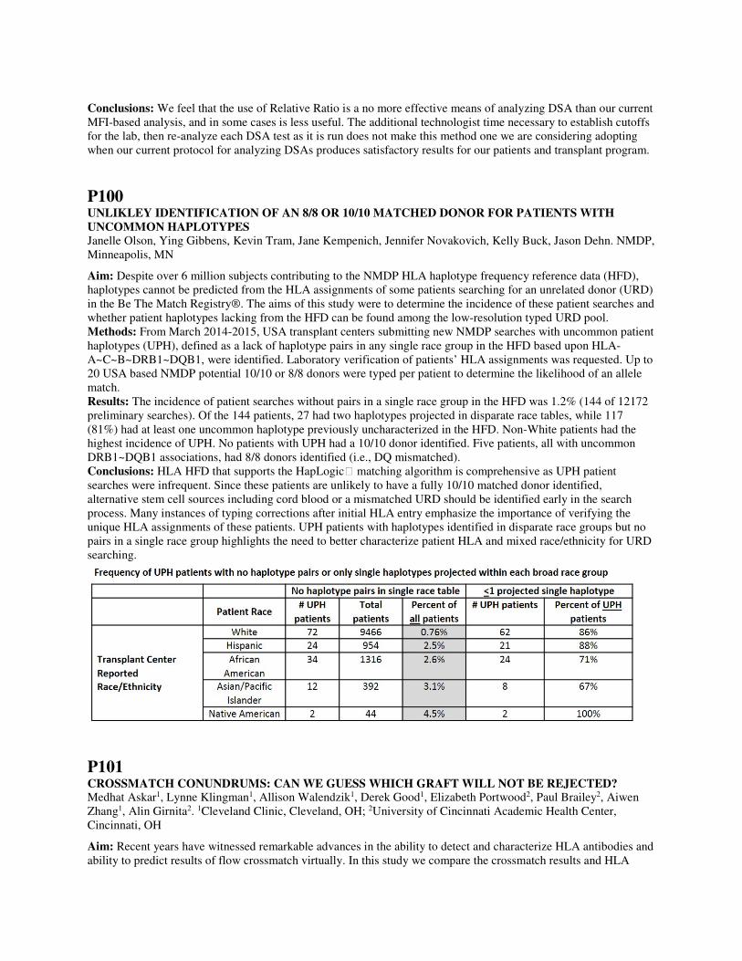
Conclusions: We feel that the use of Relative Ratio is a no more effective means of analyzing DSA than our current MFI-based analysis, and in some cases is less useful. The additional technologist time necessary to establish cutoffs for the lab, then re-analyze each DSA test as it is run does not make this method one we are considering adopting when our current protocol for analyzing DSAs produces satisfactory results for our patients and transplant program.
P100 UNLIKLEY IDENTIFICATION OF AN 8/8 OR 10/10 MATCHED DONOR FOR PATIENTS WITH UNCOMMON HAPLOTYPES Janelle Olson, Ying Gibbens, Kevin Tram, Jane Kempenich, Jennifer Novakovich, Kelly Buck, Jason Dehn. NMDP, Minneapolis, MN
Aim: Despite over 6 million subjects contributing to the NMDP HLA haplotype frequency reference data (HFD), haplotypes cannot be predicted from the HLA assignments of some patients searching for an unrelated donor (URD) in the Be The Match Registry®. The aims of this study were to determine the incidence of these patient searches and whether patient haplotypes lacking from the HFD can be found among the low-resolution typed URD pool. Methods: From March 2014-2015, USA transplant centers submitting new NMDP searches with uncommon patient haplotypes (UPH), defined as a lack of haplotype pairs in any single race group in the HFD based upon HLA-A~C~B~DRB1~DQB1, were identified. Laboratory verification of patients’ HLA assignments was requested. Up to 20 USA based NMDP potential 10/10 or 8/8 donors were typed per patient to determine the likelihood of an allele match. Results: The incidence of patient searches without pairs in a single race group in the HFD was 1.2% (144 of 12172 preliminary searches). Of the 144 patients, 27 had two haplotypes projected in disparate race tables, while 117 (81%) had at least one uncommon haplotype previously uncharacterized in the HFD. Non-White patients had the highest incidence of UPH. No patients with UPH had a 10/10 donor identified. Five patients, all with uncommon DRB1~DQB1 associations, had 8/8 donors identified (i.e., DQ mismatched). Conclusions: HLA HFD that supports the HapLogic‐ matching algorithm is comprehensive as UPH patient searches were infrequent. Since these patients are unlikely to have a fully 10/10 matched donor identified, alternative stem cell sources including cord blood or a mismatched URD should be identified early in the search process. Many instances of typing corrections after initial HLA entry emphasize the importance of verifying the unique HLA assignments of these patients. UPH patients with haplotypes identified in disparate race groups but no pairs in a single race group highlights the need to better characterize patient HLA and mixed race/ethnicity for URD searching.
P101 CROSSMATCH CONUNDRUMS: CAN WE GUESS WHICH GRAFT WILL NOT BE REJECTED? Medhat Askar1, Lynne Klingman1, Allison Walendzik1, Derek Good1, Elizabeth Portwood2, Paul Brailey2, Aiwen Zhang1, Alin Girnita2. 1Cleveland Clinic, Cleveland, OH; 2University of Cincinnati Academic Health Center, Cincinnati, OH
Aim: Recent years have witnessed remarkable advances in the ability to detect and characterize HLA antibodies and ability to predict results of flow crossmatch virtually. In this study we compare the crossmatch results and HLA
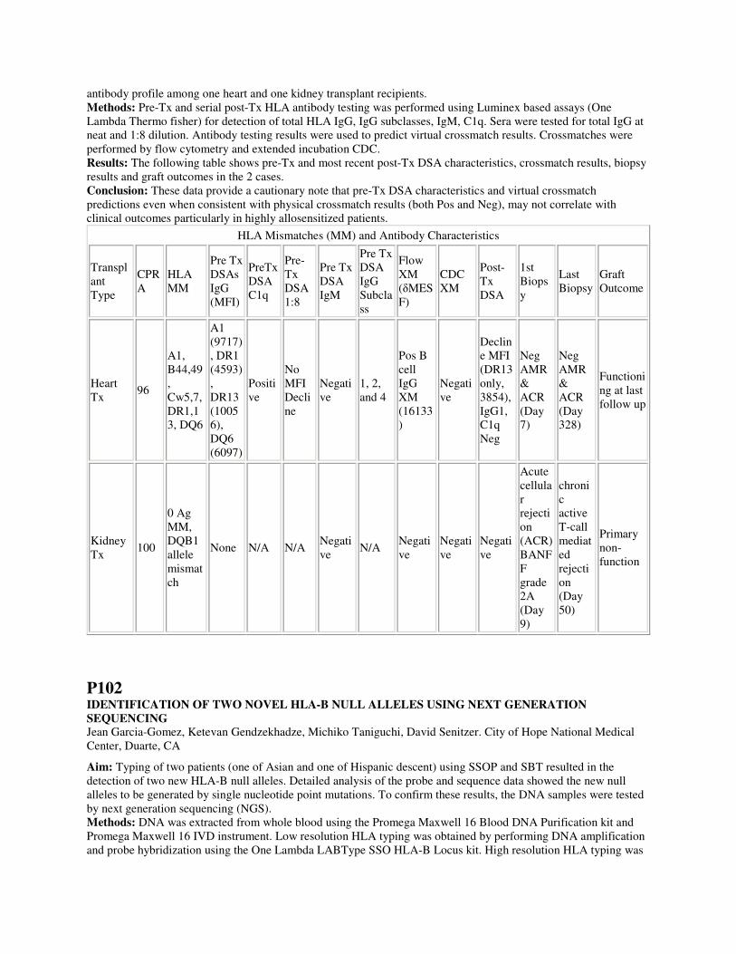
antibody profile among one heart and one kidney transplant recipients. Methods: Pre-Tx and serial post-Tx HLA antibody testing was performed using Luminex based assays (One Lambda Thermo fisher) for detection of total HLA IgG, IgG subclasses, IgM, C1q. Sera were tested for total IgG at neat and 1:8 dilution. Antibody testing results were used to predict virtual crossmatch results. Crossmatches were performed by flow cytometry and extended incubation CDC. Results: The following table shows pre-Tx and most recent post-Tx DSA characteristics, crossmatch results, biopsy results and graft outcomes in the 2 cases. Conclusion: These data provide a cautionary note that pre-Tx DSA characteristics and virtual crossmatch predictions even when consistent with physical crossmatch results (both Pos and Neg), may not correlate with clinical outcomes particularly in highly allosensitized patients.
HLA Mismatches (MM) and Antibody Characteristics
Transplant Type
CPRA
HLA MM
Pre Tx DSAs IgG (MFI)
PreTx DSA C1q
Pre-Tx DSA 1:8
Pre Tx DSA IgM
Pre Tx DSA IgG Subclass
Flow XM (δMESF)
CDC XM
Post-Tx DSA
1st Biopsy
Last Biopsy
Graft Outcome
Heart Tx
96
A1, B44,49, Cw5,7, DR1,13, DQ6
A1 (9717), DR1 (4593), DR13 (10056), DQ6 (6097)
Positive
No MFI Decline
Negative
1, 2, and 4
Pos B cell IgG XM (16133)
Negative
Decline MFI (DR13 only, 3854), IgG1, C1q Neg
Neg AMR & ACR (Day 7)
Neg AMR & ACR (Day 328)
Functioning at last follow up
Kidney Tx
100
0 Ag MM, DQB1 allele mismatch
None N/A N/A Negative
N/A Negative
Negative
Negative
Acute cellular rejection (ACR) BANFF grade 2A (Day 9)
chronic active T-call mediated rejection (Day 50)
Primary non-function
P102 IDENTIFICATION OF TWO NOVEL HLA-B NULL ALLELES USING NEXT GENERATION SEQUENCING Jean Garcia-Gomez, Ketevan Gendzekhadze, Michiko Taniguchi, David Senitzer. City of Hope National Medical Center, Duarte, CA
Aim: Typing of two patients (one of Asian and one of Hispanic descent) using SSOP and SBT resulted in the detection of two new HLA-B null alleles. Detailed analysis of the probe and sequence data showed the new null alleles to be generated by single nucleotide point mutations. To confirm these results, the DNA samples were tested by next generation sequencing (NGS). Methods: DNA was extracted from whole blood using the Promega Maxwell 16 Blood DNA Purification kit and Promega Maxwell 16 IVD instrument. Low resolution HLA typing was obtained by performing DNA amplification and probe hybridization using the One Lambda LABType SSO HLA-B Locus kit. High resolution HLA typing was
achieved by DNA amplification and subsequent sequencing of the amplified product using the Conexio Genomics SBT Resolver kits. Confirmation testing of the novel HLA-B null alleles was performed by amplifying the DNA samples using the Scisco Genetics NGS HLA kit. Results: Results from SSOP, SBT and NGS testing confirmed the presence of two new null alleles. The first null allele from the Hispanic individual is similar to B*35:01:01:01 with a mutation at position 295 (codon 75) in exon 2. This position appears to be a common polymorphic site for HLA Class I null alleles: A*02:366N, A*03:178N, A*24:60N, B*08:72N, B*15:94N, B*15:294N, B*44:58N, B*51:41N, C*01:69N, C*03:277N, C*07:152N, C*12:84N and C*17:27N. The nucleotide change (GAG->TAG) results in the creation of a stop codon. The second null allele from the Asian individual is similar to B*15:01:01:01 with a mutation at position 862 (codon 264) in exon 4. The nucleotide change (GGA->TGA) results in the creation of a stop codon. There are no documented polymorphisms at this position for any other allele. However, A*02:43N has an insertion at codon 236 that causes a frameshift and premature stop at codon 264. Conclusions: Identification of null alleles is important in determining what donors should be evaluated and selected for hematopoietic stem cell transplants. Misidentification could have an impact on graft outcome and lead to complications such as graft-versus-host disease. NGS has shown to be a valuable method for the identification of these kinds of new variants. Furthermore, the variant in exon 4 may suggest that typing outside of the HLA Class I peptide binding domains (exon 2 & 3) should be performed.
P103 HIGH THROUGHPUT, LOW COST DETECTION OF GAMMA BLOCK SNPS USING A CAPTURE PROBE TARGET ENRICHMENT NEXT GENERATION SEQUENCING ASSAY Hayley Hogan, Rhys Cransberg, Megan Jordan, Bin Jiao, David Sayer. Conexio, Perth, Australia
Aims: Previous studies have demonstrated that mismatching for SNPs in the Gamma block of the central MHC increases the risk of severe acute GvHD following unrelated HSCT. These studies were performed using PCR-SSP, a protocol that is not amenable to high throughput testing. In order to facilitate high throughput testing for retrospective studies we have developed a capture probe based Next Generation sequencing assay for the detection of Gamma Block SNPs. Methods: We designed 120 mer capture probes tiling the genetic region of interest. 200ng of DNA from 20 samples that had previously been Gamma-Type™ tested by PCR-SSP were fragmented using a Covaris M220 Ultrasonicator™ using the following conditions: duty factor 20%, peak incident power 50W, 200 cycles per burst, for 45 seconds at a temperature of 20°C - to give a peak distribution of fragments approximately 550bp in length. The fragments were repaired, size selected by a dual-bead based protocol, adenylated and adapters were ligated. The fragments were enriched and capture probes were used to isolate fragments containing Gamma block specific sequences. The Gamma block specific fragments were sequenced using a MiSeq® Next Generation DNA Sequencer, generating 300bp paired-end reads. The Gamma-Type™ variant positions were annotated within Assign™ MPS resulting in automated reporting of Gamma Block SNPs enabling direct comparison between donors and patients. Results: Even coverage of >1000 calls were obtained for all samples across all positions. There was complete concordance between the PCR-SSP results and the capture probe assay, with additional polymorphisms seen for two of the samples. The additional polymorphisms did not affect the matching/mismatching outcome with the donor/patient pairs. Conclusion: The capture probe approach is amenable to low cost and high throughput testing with the added advantage that the precise sequence is reported, whereas by PCR-SSP, only the PCR target sequence is detected. Furthermore the Gamma Block capture probes can be included with HLA probes to provide complete HLA and Gamma Block SNP detection in one assay. Large retrospective studies have the potential to identify the non-HLA regions within the MHC that influence outcomes in unrelated HSCT.
P104 POSITIVE B CELL FLOW CYTOMETRY CROSSMATCH/FULL HLA MATCHED RENAL TRANSPLANT PAIR
Moheeb Al-Awwami1, Hassan Al-Eid2, Tariq Ali2, Syed Raza2, Amal Algharably1, Mariam Garcia1, Fadi Alzayer1. 1King Faisal Specialist Hospital and Research Center, Riyadh, Saudi Arabia; 2King Faisal Specialist Hospital and Research Center, Riyadh, Saudi Arabia
Background: An ABO incompatible non sensitized male renal transplant patient was worked-up with his full HLA matched brother. The patient was desensitized with rituximab (500 mg 2 weeks prior to transplant) and four sessions of plasmapheresis and small dose intravenous immunoglobulin (100 mg/kg after each plasma session). The crossmatch included both current serum (the day before transplant) and previous serum (before the start of desensitization). Method:A standard three color flow cytometry crossmatch was performed against the potential donor (1mg/ml pronase treated cells) and the serum was tested by Luminex PRA screen (One Lambda Inc.). An evaluation of the pronase effectiveness was assessed using anti- CD20 monoclonal antibodies. Results: The patient PRA screen was negative for HLA class I and II and the crossmatch results were negative for T cells and positive B cell IgG (133 MCS) for the current serum and negative T and B cell IgG for the previous serum. Staining the B cells used in the crossmatch with anti-CD20 demonstrated the complete absence of the CD20 receptors. The auto B cell IgG flow crossmatch was not assessed due to absence of patients’ B cells as a result of rituximab effect. Conclusion: This case demonstrates that the desensitization using rituximab, plasmapheresis, and intravenous immunoglobulin for depleting antibodies in ABO incompatible renal transplant patients prior to transplantation can cause B cell nonspecific binding despite treating the cells with pronase. This nonspecific binding results in positive B cell IgG flow crossmatch. Using different anti-CD20 monoclonal antibodies for the evaluation efficacy of pronase activity is suggested to confirm complete cleavage of the CD20 receptors.
P105 HLA-DQB1*03:25: A RARE ALLELE WITH A SEROLOGICAL EQUIVALENCY OF HLA-DQ4 Zeying Du1, Jaishree Patel1, David Freedom2, Brian Norton1, Christine Braun1, Bozena Labuda2, Andrés Jaramillo2. 1Loyola University Medical Center, Maywood, IL; 2Gift of Hope Organ & Tissue Donor Network, Itasca, IL
Aim: HLA-DBQ1*03:25, a rare allele of Hispanic origin identified in 2008, has not yet been assigned a serological equivalent. A recent observation in our center of a 75 year old Hispanic female patient typed as DQB1*03:25 who also displayed HLA-DQ7, -DQ8 and -DQ9 antibodies triggered an investigation of the serological equivalency for this allele. Methods: Molecular HLA typing was performed by LabType SSO (One Lambda), LinkSeq Real-Time PCR HLA typing (Linkage BioSciences) and LIFECODES HLA SSO Typing (Immucor). Serological HLA typing was performed by Lambda Monoclonal Trays (LMT) HLA class II typing tray (One Lambda). Antibody analysis was performed by LABScreen Single Antigen HLA class I and class II kits (One Lambda) and LIFECODES class I and class II ID kits (Immucor). Results: The patient was initially typed by LabType SSO as HLA-A*02, 03; B*39, 44; C*04, 07; DRB1*07, 09; DQB1*02:02, 03:25. Further DNA typing by real-time PCR SSP confirmed the same results. DQB1 typing further confirmed by LIFECODES SSO typing. Since the patient presented HLA-DQ7, -DQ8 and -DQ9 antibodies, serological HLA class II typing was subsequently performed yielding the following Results: DR7, 9; DQ2, 4. DNA and protein sequence alignments of the DQB1*03:25 allele with other DQB1 alleles showed that its sequence is quite similar with the DQB1*04:03 allele, but differs significantly from the DQB1*03 alleles. The sequence difference between DQB1*03:25 and DQB1*04:03 is only one amino acid at codon 9 in exon 2 (Tyr vs. Phe). Thus, DQB1*03:25 shows almost identical immunogenicity to DQB1*04:03 in the context of their sequences. When compared with the first case of HLA-DQB1*03:25 in IMGT/HLA database, it was noticed that both cases carry the DRB1*09-DQB1*03:25 haplotype. Conclusion: This is the first time to confirm that the serological equivalent of DQB1*03:25 is DQ4. In this regard, accurate antigenic entry of deceased donor HLA typing into UNET is critical since it affects multiple areas of organ allocation, particularly given the increased utilization of virtual crossmatching to determine the compatibility of a potential donor prior to organ allocation. In addition, based on our knowledge, this is the second case of the DQB1*03:25 allele in the Hispanic population. It suggests an expanded population study needed to document this allele well.
P106 THE SOONER THE BETTER: OUR LABORATORY EXPERIENCEWITH CHORIONIC VILLUS SAMPLING (CVS) HLA TYPING PRIOR TO HSCT Angelica DeOliveira1, Jennifer Deitz1, Candace Young1, Michelle McLaughlin1, Wendy Hanshew2, Dongfeng Chen2. 1Duke University, Durham, NC; 2Duke University Medical Center, Durham, NC
Aim: Allogeneic Hematopoietic stem cell transplantation(HSCT) will prolong and improve the quality of life of babies born affected by inherited metabolic diseases. Genetic testing and preparation for a potential HSCT starts at 10 weeks of mother's pregnancy. This study aims to describe our strategy to obtain an HLA typing from the minuscule tissue fragments submitted for HLA typing in preparation for an HSCT just after birth. Methods: Mothers carrying babies that may be affected with Hurler or Krabbe A syndromes are the most frequent challenges at Duke Hospital. As needed, CVS is submitted to our laboratory and DNA is extracted using a micro method developed specifically for these samples to allow High Resolution HLA typing. Since maternal blood contamination of the CVS is a common finding, High Resolution HLA typing on the parents is also done to assist with the identification of all HLA alleles present on the CVS. Our laboratory sequencing strategy for HLA-A, B, C, DRB1 & DQB1 identifies most alleles in a hemizygous form and duplication of the same allele (potential homozygosis) is always resolved by both parents HLA typing. All positive SBT amplification reactions obtained for the CVS DNA are sequenced, and based on parents typing results we are able to identify, first the paternal haplotype present followed by the maternal one. The software SeqPilot®, used for high resolution data analysis, allows the analysis of all positive reactions chosen to be sequenced. Results: The CVS samples we have tested (total of 20 cases) have provided the correct pool of alleles expected to be present and it facilitated cord blood searches for these unborn babies, providing an increased chance for successful treatment. Conclusions:The HLA typing strategy we apply to CVS is only possible due to our level of expertise extracting DNA from small tissue fragments and the flexibility and data quality offered by the High Resolution HLA Typing methodology and software data analysis we use. The affected and genetic carrier unborn babies get a much higher chance at cure and survival when their diagnosis and treatment can be done earlier in their lives.
P107 COPY NUMBER VARIATION (CNV) OF KIR GENES CHARACTERIZED USING qRT-PCR TECHNIQUE Jerome Saltarrelli1, Dana Willis1, Katy Rezvani2, Kai Cao1. 1UT MD Anderson Cancer Center, Houston, TX; 2UT MD Anderson Cancer Center, Houston, TX
Killer immunoglobulin-like receptors (KIR) are a group of cell surface proteins on the surface of natural killer cells. KIR genes are located in close proximity to one another in a head-to-tail fashion on chromosome 19. This arrangement permits misalignments or unequal crossover which can lead to deletions, duplicates or recombination. The non-reciprocal recombination increases the possibility of inequitable segregation of KIR genes leading to increased copies. The aim of the study was to develop assay for KIR CNV. Method:We developed a KIR CNV assay for 2DL1, 2DL2, 2DL3, 2DS1, and 2DS2 by qRT-PCR (TaqMan® Copy Number Assay, Applied Biosystems) using primers and probes described by Jiang W. et al (Genome research, 2012). Forty-three UCLA KIR exchange DNA and 14 members of 2 families with the present of these KIR were tested. Copy numbers were obtained using Life Technologies Copy Caller Software. Results: Fig. 1 shows the CNV results of KIR2DL1 in 26 subjects. Of the 43 samples, majority showed 1 copy (63.0 - 84.6%) and some have 2 copies (12.5 - 29.6%) of all the 5 KIR genes. Three copies were observed only for KIR2DL1 in 7.4% of the subjects and 2DL3 in 3.8% of the subjects. None of the subjects has 4 copies in any of these KIR genes (Fig. 2). A member in 1 family carries 3 copies of KIR2DL1 with 2 copies in one haplotype and 1 copy in the second; another sibling bears a recombinant maternal haplotype (data not shown). Conclusions: KIR genes are polymorphic with a large number of allelic variants and also display copy number variations. One or 2 copies are more common than 3 or more copies. The immunological relevance of CNV in certain KIR genes has been revealed in virus infection such as resistance against human immunodeficiency virus or increased clearance of hepatitis C virus. KIR CNV may also have impact on stem cell transplant outcome.
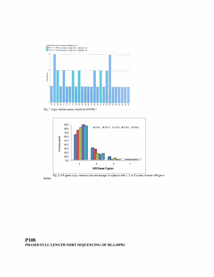
P108 PHASED FULL LENGTH SMRT SEQUENCING OF HLA-DPB1
Kathrin Lang1, Gerhard Schöfl1, Carolin Zweiniger1, Christoph König2, Maarten Penning3, Erik Rozemuller3, Sylvia Clausing4, Yannick Duport4,5, Nicola Gscheidel5, Sylke Winkler5, Vinzenz Lange1, Irina Boehme1, Alexander H. Schmidt1,6. 1DKMS Life Science Lab, Dresden, Germany; 2Pacific Biosciences, Menlo Park, CA; 3GenDx, Utrecht, Netherlands; 4CRTD-Centrum for Regenerative Therapies, Dresden, Germany; 5Max Planck Institute of Molecular Cell Biology and Genetics, Dresden, Germany; 6DKMS German Bone Marrow Donor Center, Tübingen, Germany
Aim: In contrast to exon-based HLA-typing approaches, whole gene genotyping crucially depends on full-length sequences submitted to the IMGT/HLA Database. Currently, full-length sequences are provided for only 7 out of 520 HLA-DPB1 alleles. Therefore, we developed a fully phased whole-gene sequencing approach for DPB1, to facilitate further exploration of the allelic structure at this locus. Methods: Primers were developed flanking the UTR-regions of DPB1 resulting in a 12 kb amplicon. Using a 4-primer approach, secondary primers containing barcodes were combined with the gene-specific primers to obtain barcoded full-gene amplicons in a single amplification step. Amplicons were pooled, purified, and ligated to SMRT bells (i.e. annealing points for sequencing primers) following standard protocols from Pacific Biosciences. Taking advantage of the SMRT chemistry, pools of 48 amplicons were sequenced full length in single runs on a Pacific Biosciences RSII instrument. Demultiplexing was performed using the SMRT portal. Sequence analysis was performed using the NGSengine software (GenDx). Results: We analyzed a set of 48 randomly picked samples. With 3 exceptions due to PCR failure, all genotype assignments conformed to standard genotyping results based on exons 2 and 3. Allelic proportions for heterozygous positions were evenly distributed (range 0.4 - 0.6) for all samples, suggesting unbiased amplifications. Despite the high per-read raw error rates typical for SMRT sequencing (~15%) the consensus sequence proved highly reliable. All consensus sequences for exons 2 and 3 were in full accordance with their MiSeq-derived sequences. We describe novel intronic sequence variation of the 7 so far genomically defined alleles, as well as 7 whole-length DPB1 alleles with hitherto unknown intronic regions. One of these alleles (HLA-DPB1*131:01) is classified as rare. Conclusion: Here we present a whole gene amplification and sequencing workflow for DPB1 alleles utilizing single molecule real-time (SMRT) sequencing from Pacific Biosciences. Validation of consensus sequences against known exonic sequences highlights the reliability of this technology. This workflow will facilitate amending the IMGT/HLA Database for DPB1.
P109 REASONS FOR FAILING OFFERS IN A KIDNEY PAIRED DONATION PROGRAM: A 7-YEAR ANALYSIS Stanislaw Stepkowski1, David Fumo2, Connie Smith3, Annette Blair3, Michael Rees2. 1University of Toledo, Toledo, OH; 2University of Toledo, Toledo, OH; 3University of Toledo, Toledo, OH
Aim: Computer-identified possible Kidney Paired Donation (KPD) exchanges are presented as offers to participating transplant programs and eventually converted into transplants. We evaluated reasons why these offers failed to progress into successful transplants. Methods/ Results: The Alliance for Paired Donation (APD) has an optimization algorithm to generate possible KPD exchanges, which are presented to transplant centers for their review as offers. Each offer may be composed of one to multiple 1-way exchanges. We tracked the progress in moving from offers to transplants and recorded the reason for failure. Between January 2008 and August 2014, the APD made 981 offers. Of these offers 181 resulted in transplants while 800 failed, namely 268 failed for specific reasons while 529 failed as they were dependent on the same 268 1-ways. Overall, specific reasons accounted for 57% of all failures with: 25% positive flow crossmatch (FXM); 13% competing offer outside the APD; and, 19% declined of donors by transplant centers (Figure 1). Our detail analysis showed that the 25% positive FXM with negative virtual crossmatch (VXM) correlated with an increased number of highly sensitized patients: the pool of sensitized patients with a PRA >80% has increased from 31% in 2008 to 47% in 2014. Since many patients choose to enroll in multiple KPD registries some offers resulted in futile exchanges with patients already committed to alternative exchanges. Finally, donors were declined by transplant centers. To avoid these specific reasons for failure, the APD has begun pre-review consulting about donors and recipients prior to making a formal offer. Conclusions:To prevent specific reasons for failure the APD has implemented the following changes: 1) testing of all donors for HLA-DQA, DPA and DPB; 2) expansion of the donor pool to increase options for highly sensitized patients; and 3) improved coordination with other KPD programs to avoid competing offers.

P110 KIDNEY TRANSPLANT OUTCOMES IN PATIENTS WITH CPRA VALUES ≥ 99% AFTER THE IMPLEMENTATION OF THE NEW OPTN/UNOS KIDNEY ALLOCATION SYSTEM (KAS)-A SINGLE CENTER STUDY Navdeep Kaur1, Simin Goral1, Jane Kearns2, Peter L. Abt3, Ali Naji3, Matthew H. Levine3, Malek Kamoun2. 1The Hospital of the University of Pennsylvania, Philadelphia, PA; 2The Hospital of the University of Pennsylvania, Philadelphia, PA; 3The Hospital of the University of Pennsylvania, Philadelphia, PA
Aim: Sensitized patients have prolonged wait times and a greater incidence of death while on the waiting list. On December 4, 2014, OPTN/UNOS activated a new KAS for deceased donors (DD) that gives priority at the regional level to candidates with calculated panel reactive antibody (CPRA) value = 99% and at the national level to patients with CPRA = 100%. Transplants performed at our center under the new KAS were reviewed to assess early outcomes. Methods: Single center, retrospective data review of patients with CPRA values >98% transplanted between December 4, 2014 and April 30, 2015. Results: We performed 51 DD kidney transplants since the new KAS. 24 crossmatches were performed on patients with CPRA >98%, 13 crossmatches were reported as compatible and 8 patients were transplanted. Characteristics of the patient cohort are detailed in Table 1. Majority of the patients were multiparous women and had history of blood transfusions. Seven patients (87.5%) have functioning grafts at median follow up of 94 (52,128) days. One patient had primary non-function. One patient developed a weak anti-HLA class I DSA that resolved subsequently. None of the patients had rejection episodes. Conclusion: At our center, since the new KAS was implemented, 8 highly sensitized patients were quickly transplanted with excellent short term outcomes and without early rejection episodes. Long-term follow-up is needed to understand the consequences of this new KAS.

Table 1: Patient characteristics
Patient #
Age at Transplant
Gender
# of Pregnancies
Blood transfusions prior to transplant
# of prior transplants
Days on Wait List
Days on dialysis prior to transplant
Calculated panel reactive antibody (%)
Estimated post-transplant survival (%)
Kidney donor profile index (%)
Graft function at 30 days
Serum creatinine at 30 days (mg/dl)
Donor specific antibodies
Rejection Episodes
1 60 Female
1 Yes 1 590
711 99 65 51 Yes 1.77 No No
2 52 Male N/A Yes 1 1462
1462 99 79 85 No 6.42 No No
3 57 Female
3 No 0 506
0 100 45 8 Yes 1.47 No No
4 61 Female
1 Yes 1 3439
2589 100 85 32 Yes 1.19 Yes-A 31
No
5 26 Female
0 Yes 0 1239
1337 100 4 53 Yes 0.99 No No
6 53 Female
5 Yes 0 188
0 100 21 24 Yes 0.97 No No
7 56 Male N/A No 1 2078
1831 100 70 25 Yes 1.3 No No
8 50 Male N/A No 1 2527
1737 100 78 41 Yes 1.61 No No
P111 UNEXPECTED ASSOCIATION OF REDUCED ACTIVATING KIR-HLA GENOTYPES WITH AUTOIMMUNE VOGT-KOYANAGI-HARADA DISEASE Raja Rajalingam1, Madeline Yung2, Akira Meguro3, Nobuhisa Mizuki3, Ralph D. Levinson2. 1Immunogenetics and Transplantation Laboratory, University of California San Francisco, San Francisco, CA; 2Jules Stein Eye Institute, University of California at Los Angeles, Los Angeles, CA; 3Yokohama City University School of Medicine, Yokohama, Japan
Aim: Vogt-Koyanagi-Harada (VKH) is an uncommon autoimmune disease characterized by chronic, bilateral, diffuse, granulomatous uveitis with accompanying dermatologic, neurologic and auditory involvement. There is a higher rate of VKH in people of Asian, Mestizo, and Mediterranean descent. Our preliminary studies of small cohorts of Japanese and Mestizo reported higher incidence of activating KIRs, which corroborates the autoimmune nature of VKH. Herein, we extended our pilot study with Japanese to a larger cohort. Methods: We characterized 196 patients with VKH and 209 healthy controls from Yokohama City University School of Medicine, Japan for KIR and HLA class I genes using Luminex rSSO. Results: Differences between controls and patients in the distribution of KIR genotypes, HLA allotypes, and KIR-HLA pairs were tested by two-tailed Fisher's exact probabilities (Table). In contrast to previous findings, Bx KIR genotypes occurred at a lower frequency in patients with VKH compared to controls (39.3% vs. 52.6%, p=0.007). The Bx decrease was due to the differences at the centromeric half of the KIR gene complex, and consequently all variable B haplotype KIR genes at the centromeric half (2DS2, 2DL2, 2DS3, 2DL5) were reduced in patients. Compared to controls, certain HLA allotypes were significantly increased (B54, B56, Cw1) or decreased (A33, B7, B44, B46, B52, Cw12, and A2-B46-Cw1 combination) in VKH. Among all known HLA class I ligands, only Bw4 showed a significant decrease in patients (42.4% vs. 55.5%, p=0.0097). The decrease in 2DL2 and in Bw4 was

associated with a reduction in 2DL2+C1/C2 and 3DL1+Bw4 combinations in VKH. Conclusions: Decreased inhibitory KIR-HLA gene combinations in patients with VKH suggest the possibility of impaired class I mediated licensing, thus generating hyporesponsive NK cells. Absence of activating KIRs 2DS2 and 2DS3 further support the notion of a poorly responding NK cells in VKH. NK cell hyporesponsiveness to certain infections might trigger an autoimmune response and pathogenesis of VKH.
P112 COMPARISON OF EDTA VERSUS DTT TREATMENT IN OVERCOMING THE PROZONE EFFECT IN A LUMINEX-BASED HLA ANTIBODY ASSAY Jinguo Wang1, James R. Meade2, Nicholas K. Brown1, Jerome G. Weidner3, Susana R. Marino1. 1The University of Chicago, Chicago, IL; 2The University of Chicago Medicine, Chicago, IL; 3The University of Chicago, Chicago, IL
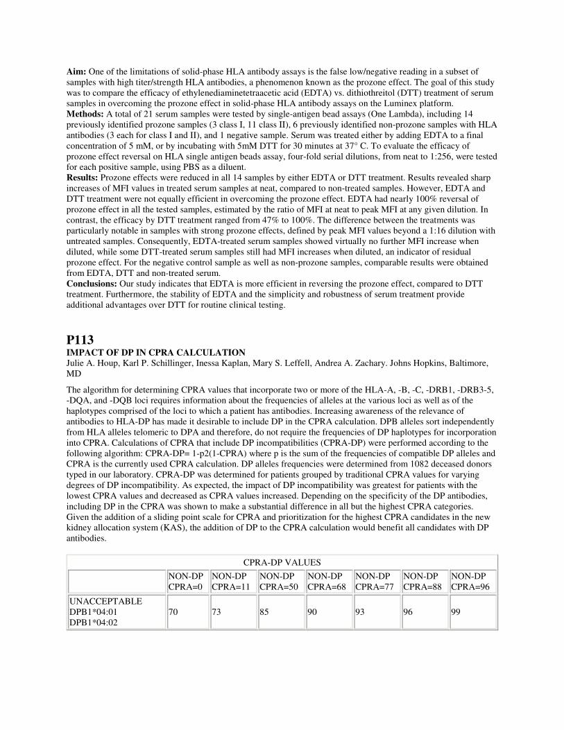
Aim: One of the limitations of solid-phase HLA antibody assays is the false low/negative reading in a subset of samples with high titer/strength HLA antibodies, a phenomenon known as the prozone effect. The goal of this study was to compare the efficacy of ethylenediaminetetraacetic acid (EDTA) vs. dithiothreitol (DTT) treatment of serum samples in overcoming the prozone effect in solid-phase HLA antibody assays on the Luminex platform. Methods: A total of 21 serum samples were tested by single-antigen bead assays (One Lambda), including 14 previously identified prozone samples (3 class I, 11 class II), 6 previously identified non-prozone samples with HLA antibodies (3 each for class I and II), and 1 negative sample. Serum was treated either by adding EDTA to a final concentration of 5 mM, or by incubating with 5mM DTT for 30 minutes at 37° C. To evaluate the efficacy of prozone effect reversal on HLA single antigen beads assay, four-fold serial dilutions, from neat to 1:256, were tested for each positive sample, using PBS as a diluent. Results: Prozone effects were reduced in all 14 samples by either EDTA or DTT treatment. Results revealed sharp increases of MFI values in treated serum samples at neat, compared to non-treated samples. However, EDTA and DTT treatment were not equally efficient in overcoming the prozone effect. EDTA had nearly 100% reversal of prozone effect in all the tested samples, estimated by the ratio of MFI at neat to peak MFI at any given dilution. In contrast, the efficacy by DTT treatment ranged from 47% to 100%. The difference between the treatments was particularly notable in samples with strong prozone effects, defined by peak MFI values beyond a 1:16 dilution with untreated samples. Consequently, EDTA-treated serum samples showed virtually no further MFI increase when diluted, while some DTT-treated serum samples still had MFI increases when diluted, an indicator of residual prozone effect. For the negative control sample as well as non-prozone samples, comparable results were obtained from EDTA, DTT and non-treated serum. Conclusions: Our study indicates that EDTA is more efficient in reversing the prozone effect, compared to DTT treatment. Furthermore, the stability of EDTA and the simplicity and robustness of serum treatment provide additional advantages over DTT for routine clinical testing.
P113 IMPACT OF DP IN CPRA CALCULATION Julie A. Houp, Karl P. Schillinger, Inessa Kaplan, Mary S. Leffell, Andrea A. Zachary. Johns Hopkins, Baltimore, MD
The algorithm for determining CPRA values that incorporate two or more of the HLA-A, -B, -C, -DRB1, -DRB3-5, -DQA, and -DQB loci requires information about the frequencies of alleles at the various loci as well as of the haplotypes comprised of the loci to which a patient has antibodies. Increasing awareness of the relevance of antibodies to HLA-DP has made it desirable to include DP in the CPRA calculation. DPB alleles sort independently from HLA alleles telomeric to DPA and therefore, do not require the frequencies of DP haplotypes for incorporation into CPRA. Calculations of CPRA that include DP incompatibilities (CPRA-DP) were performed according to the following algorithm: CPRA-DP= 1-p2(1-CPRA) where p is the sum of the frequencies of compatible DP alleles and CPRA is the currently used CPRA calculation. DP alleles frequencies were determined from 1082 deceased donors typed in our laboratory. CPRA-DP was determined for patients grouped by traditional CPRA values for varying degrees of DP incompatibility. As expected, the impact of DP incompatibility was greatest for patients with the lowest CPRA values and decreased as CPRA values increased. Depending on the specificity of the DP antibodies, including DP in the CPRA was shown to make a substantial difference in all but the highest CPRA categories. Given the addition of a sliding point scale for CPRA and prioritization for the highest CPRA candidates in the new kidney allocation system (KAS), the addition of DP to the CPRA calculation would benefit all candidates with DP antibodies.
CPRA-DP VALUES
NON-DP CPRA=0
NON-DP CPRA=11
NON-DP CPRA=50
NON-DP CPRA=68
NON-DP CPRA=77
NON-DP CPRA=88
NON-DP CPRA=96
UNACCEPTABLE DPB1*04:01 DPB1*04:02
70 73 85 90 93 96 99

UNACCEPTABLE DPB1*01:01 DPB1*13:01
22 30 61 75 82 91 97
UNACCEPTABLE DPB1*03:01 DPB1*06:01 DPB1*09:01
23 31 61 75 82 91 97
P114 NGS HLA TYPING - SINGLE CENTER EXPERIENCE WITH OMIXON HOLOTYPE HLA X4 KIT AND HLA TWIN DATA ANALYSIS Jinguo Wang1, Brenda Maria A. Issangya2, Bradley C. Long3, Jerome G. Weidner2, Jeremy P. Segal1, Susana R. Marino1. 1The University of Chicago, Chicago, IL; 2The University of Chicago Medicine, Chicago, IL; 3The University of Chicago Medicine, Chicago, IL
Aim: Given the high throughput, better coverage, and free of phase ambiguity with HLA typing using next generation sequencing (NGS) technology, we decided to test one of the commercially available kits (Holotype HLA X4 ) offered by Omixon. Methods: Total of 32 samples (27 PBMC derived patient DNA samples and 5 UCLA survey DNA samples) were sequenced by NGS for HLA-A, B, C, DRB1 and DQB1 using Holotype HLA X4. All samples were previously typed at high-resolution by combinations of SBT/SSP. This includes 23 HLA-A, 28 B, 21 C, 28 DRB1, 14 DQB1 CWD2.0 alleles and also 2 non-CWD and 2 novel alleles. With Omixon HLA X4 kit, both individual PCR amplicon barcode and pooled amplicon barcode strategies were evaluated on Illumina MiSeq analyzer. Results: Long-range PCR gave robust amplifications on all samples for HLA-A, B, C (full coverage) and DRB1 (exon 2-4). DQB1 set 2 PCR (exon 2-5) had a failure rate of 3% (1/32) and DQB1 set 1 PCR (exon 1-4) had a failure rate of 21.9% (7/32, 5 of them from UCLA survey samples). The accuracy of NGS typing was 100% concordant with previous typing for HLA-A, B, C in both PCR amplicon individually barcoded and pooled amplicon barcoded format. Accuracy dropped to 97% for DRB1 and DQB1 assignment in both formats due to technical errors (1/32) or failed DQB1 PCR (1/32). Non-CWD alleles as well as novel alleles were correctly identified. No ambiguities were present in any of the final typing assignments. Using default quality control matrix of 15 parameters from the HLA Twin software, 4/158 resolved HLA alleles gave “red” light in the two traffic light system for amplicon individually barcoded samples, compared to 7/158 alleles in pooled amplicon barcoded samples. Regardless, none of the sample with “red” flagged QC parameter compromised the final HLA typing assignment. Conclusions: Our experience with Omixon Holotype HLA X4 kit indicates that HLA-NGS typing indeed has advantages over Sanger SBT method. The Omixon NGS library construction is easy to perform without noticeable bias introduction. The beta version HLA Twin analysis software is user friendly & performs well with both amplicon individually barcoded as well as pooled amplicon barcoded samples with extensive data quality monitoring. However, the robustness of DQB1 PCR requires improvement & the dual algorithm of HLA Twin software needs more transparency.
P115 AUTOLOGOUS CROSSMATCHES: HOW USEFUL ARE THEY IN THE EVALUATION OF POSITIVE DONOR FLOW CYTOMETRIC CROSSMATCHES? Deanna Manna, Anne Halpin, Patricia Campbell, Luia Hidalgo. University of Alberta, Edmonton, AB, Canada
Aim: Our laboratory testing algorithm includes autologous flow cytometry crossmatch (auto FCXM) on all positive (pos) donor/allo FCXM. Depending on the assessed immunological risk of the donor/recipient pair and the availability of the recipient sample, the auto FCXM may be set up on call or as a STAT test. The aim of this study was to evaluate the utility of auto XM to aid in the investigation of pos allo FCXMs. Methods: FCXMs performed between January to June of 2014 were reviewed (n=214); negative (neg) allo-FCXMs were excluded from this analysis as no auto FCXM was performed. All FCXMs were performed using a plate based

method; all cells were treated with pronase. Donors and recipients were typed for all loci including HLA-C*, -DQA1*, -DPB1* and -DPA1* by rSSO. Positive FCXMs were further examined by LABScreen® single antigen beads (SAB). There were 23 pos allo FCXM results but auto-FCXM could not be performed on 4 recipients. Thus auto XM data are available on 19 recipients. Results: Of the 19 pos allo FCXMs, only 4 auto FCXMs were positive (21%) with three demonstrating preferential T cell reactivity. There were 15 results that were allo FCXM pos/ auto FCXM neg. SAB testing revealed that seven allo-FCXM results were explained by HLA DSA (37%), and 8 had no DSA detected (42%). Conclusions: Since 79% of the autologous crossmatches were negative and all were followed up with antibody screening, it may be reasonable to remove autologous crossmatching from our testing algorithm. We are currently performing a cost analysis of alternative testing algorithms to include urgent SAB testing. As virtual crossmatching becomes more prevalent, it is increasingly important to evaluate current antibody identification as reflected by the unexpected positive donor XM results due to new, previously undetected DSA. The auto FCXM did not aid in the explanation of the positive allo FCXM without DSA and the cause of reactivity remains unclear in these cases. L. Hidalgo: Speaker's Bureau; Company/Organization; Thermo-Fisher/ One Lambda Inc.
P116 WHY DO SOME 100% CPRA RENAL TRANSPLANT CANDIDATES GET MORE OFFERS WITH THE NEW KAS THAN OTHERS? Maryanne Mackey1, Don Constantino1, Ashar Ata2, Amy Hahn1. 1Albany Medical College, Albany, NY; 2Albany Medical College, Albany, NY
Aim: In the 5 months after implementation of the new kidney allocation system (KAS), import kidney offers to 100% cPRA renal transplant candidates (RTC) have increased due to prioritization with an additional 212 points. We noted that while some RTC were receiving multiple provisional offers, other patients were not receiving any. We hypothesized that the patients receiving fewer offers had more unacceptable antigens (UA) entered in UNet, had a less common blood type, or had UA against broad specificities. Methods: Active RTC with 100% cPRA from a single center were examined for: total numbers of class I and class II UAs; high frequency broad UAs such as Bw4, Bw6, DR52, etc.; wait time on 12/4/14; expected post-transplant survival (EPTS) score; and ABO blood type. DQA and DPB UAs were also examined, although they can't be entered in UNet. RTC were stratified by number of offers into 3 groups (0, 1-2, and ≥4 offers), with differences analyzed using Students t-test. Three RTC transplanted within 1 month of KAS were omitted. Results: Comparisons are shown in Table 1. The group with 0 offers had a significantly greater number of class I UAs compared to the other groups. For class II UAs, the 0 and 1-2 groups had a significantly greater number than the ≥4 group (p=0.002). Closer analysis of class II UAs showed that the ≥4 group had a lower incidence of DR52 UA (17%) vs the other groups (50% and 63%). DR51 UA had an incidence of 50% in the 2 groups with the fewest offers, compared to 33% in the ≥4 group. DR53, Bw4, Bw6, Cw, DQ, DPB, mean EPTS score, wait time, and ABO were not significantly different between groups. Conclusion: Although this is a single center study with small numbers, these findings may be reproduced with national data. RTC with many UAs may have an extremely narrow window of acceptable mismatches (MM). Donors with 0 MM at one or more loci or with unusual HLA phenotypes may be their only source of offers. Additional strategies may be needed, including sufficient additional points to achieve allocation when there are 0MM at the HLA-A or -B loci.
0 Offers (N=4) 1-2 Offers (N=8) ≥4 Offers (N=6)
Mean±S.E. Mean±S.E. Difference vs 0 (95% CI)
p Mean±S.E. Difference vs 0 (95% CI)
p
class I UA
67.8±4.6 39.4±23.8 -28.4 (-52.1 to -4.7) 0.022 42.2±13.5 -25.6 (-50.6 to -0.6) 0.045
class II UA
14.8±6.7 17.0±3.4 2.3 (-3.5 to 8.1) 0.424 8.3±4.1 -6.4 (-12.6 to -0.3) 0.042
EPTS 48.5±35.2 35.9±35.3 -12.6 (-55.0 to 29.7) 0.535 46.0±26.1 -2.5 (-47.1 to 42.1) 0.907
wait time
3464.5±1925.5 2384.8±1968.4 -1079.8 (-3626.7 to 1467.2)
0.380 2584.2±1942.5 -880.3 (-3565.0 to 1804.3)
0.495
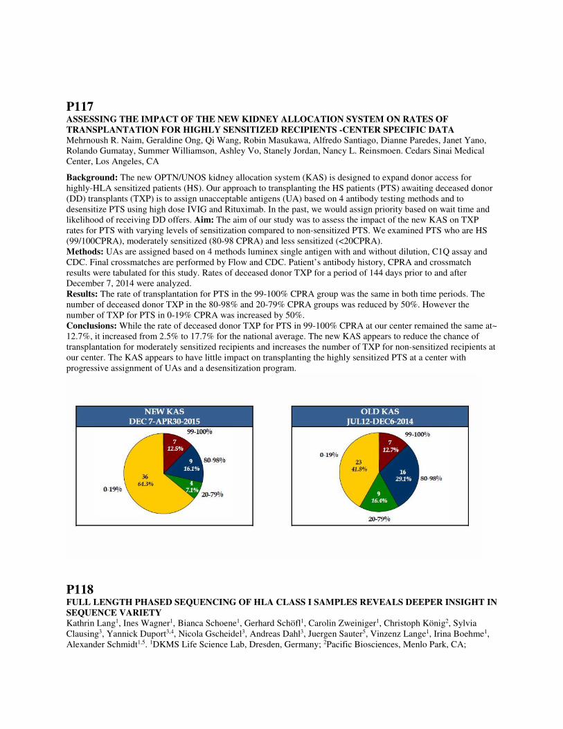
P117 ASSESSING THE IMPACT OF THE NEW KIDNEY ALLOCATION SYSTEM ON RATES OF TRANSPLANTATION FOR HIGHLY SENSITIZED RECIPIENTS -CENTER SPECIFIC DATA Mehrnoush R. Naim, Geraldine Ong, Qi Wang, Robin Masukawa, Alfredo Santiago, Dianne Paredes, Janet Yano, Rolando Gumatay, Summer Williamson, Ashley Vo, Stanely Jordan, Nancy L. Reinsmoen. Cedars Sinai Medical Center, Los Angeles, CA
Background: The new OPTN/UNOS kidney allocation system (KAS) is designed to expand donor access for highly-HLA sensitized patients (HS). Our approach to transplanting the HS patients (PTS) awaiting deceased donor (DD) transplants (TXP) is to assign unacceptable antigens (UA) based on 4 antibody testing methods and to desensitize PTS using high dose IVIG and Rituximab. In the past, we would assign priority based on wait time and likelihood of receiving DD offers. Aim: The aim of our study was to assess the impact of the new KAS on TXP rates for PTS with varying levels of sensitization compared to non-sensitized PTS. We examined PTS who are HS (99/100CPRA), moderately sensitized (80-98 CPRA) and less sensitized (<20CPRA). Methods: UAs are assigned based on 4 methods luminex single antigen with and without dilution, C1Q assay and CDC. Final crossmatches are performed by Flow and CDC. Patient’s antibody history, CPRA and crossmatch results were tabulated for this study. Rates of deceased donor TXP for a period of 144 days prior to and after December 7, 2014 were analyzed. Results: The rate of transplantation for PTS in the 99-100% CPRA group was the same in both time periods. The number of deceased donor TXP in the 80-98% and 20-79% CPRA groups was reduced by 50%. However the number of TXP for PTS in 0-19% CPRA was increased by 50%. Conclusions: While the rate of deceased donor TXP for PTS in 99-100% CPRA at our center remained the same at~ 12.7%, it increased from 2.5% to 17.7% for the national average. The new KAS appears to reduce the chance of transplantation for moderately sensitized recipients and increases the number of TXP for non-sensitized recipients at our center. The KAS appears to have little impact on transplanting the highly sensitized PTS at a center with progressive assignment of UAs and a desensitization program.
P118 FULL LENGTH PHASED SEQUENCING OF HLA CLASS I SAMPLES REVEALS DEEPER INSIGHT IN SEQUENCE VARIETY Kathrin Lang1, Ines Wagner1, Bianca Schoene1, Gerhard Schöfl1, Carolin Zweiniger1, Christoph König2, Sylvia Clausing3, Yannick Duport3,4, Nicola Gscheidel3, Andreas Dahl3, Juergen Sauter5, Vinzenz Lange1, Irina Boehme1, Alexander Schmidt1,5. 1DKMS Life Science Lab, Dresden, Germany; 2Pacific Biosciences, Menlo Park, CA;

3CRTD-Centrum for Regenerative Therapies, Dresden, Germany; 4Max Planck Institute of Molecular Cell Biology and Genetics, Dresden, Germany; 5DKMS German Bone Marrow Donor Center, Tübingen, Germany
Aim: The vast majority of donor typing relies on sequencing exons 2 and 3 of HLA class I genes (HLA-A, -B, -C). With such an approach certain allele combinations do not result in the anticipated “high resolution” (G-code) typing, due to the lack of exon-phasing information. To resolve ambiguous typing results for a haplotype frequency project, we established a whole gene sequencing approach for HLA class I, facilitating also an estimation of the degree of sequence variability outside the commonly sequenced exons. Methods: Primers were developed flanking the UTR regions resulting in similar amplicon lengths of 4.2-4.4 kb. Using a 4-primer approach, secondary primers containing barcodes were combined with the gene specific primers to obtain barcoded full-gene amplicons in a single amplification step. Amplicons were pooled, purified, and ligated to SMRT bells (i.e. annealing points for sequencing primers) following standard protocols from Pacific Biosciences. Taking advantage of the SMRT chemistry, pools of 48-72 amplicons were sequenced full length and phased in single runs on a Pacific Biosciences RSII instrument. Demultiplexing was achieved using the SMRT portal. Sequence analysis was performed using NGSengine software (GenDx). Results: We successfully performed full-length gene sequencing of 1003 samples, harboring ambiguous typings of either HLA-A (n=46), HLA-B (n=304) or HLA-C (n=653). Despite the high per-read raw error rates typical for SMRT sequencing (~15%) the consensus sequence proved highly reliable. All consensus sequences for exons 2 and 3 were in full accordance with their MiSeq-derived sequences. Unambiguous allelic resolution was achieved for all samples. We observed novel intronic, exonic as well as UTR sequence variations for many of the alleles covered by our data set. This included sequences of 600 individuals with HLA-C*07:01/C*07:02 genotype revealing the extent of sequence variation outside the exons 2 and 3. Conclusion: Here we present a whole gene amplification and sequencing approach for HLA class I genes. The maturity of this approach was demonstrated by sequencing more than 1000 samples, achieving fully phased allelic sequences. Extensive sequencing of one common allele combination hints at the yet to discover diversity of the HLA system outside the commonly analyzed exons.
P119 EVALUATION OF YIELD AND INTEGRITY OF DNA SAMPLES EXTRACTED FROM CORD BLOOD SEGMENTS AND RED CELLS FRACTIONS USING THE MAXWELL® RSC INSTRUMENT Renata Santos, Angelica DeOliveira, Jennifer Deitz, Porsha Barfield, Amanda Dusenberry, Wendy Hanshew, Dong-Fen Chen. Duke University Medical Center, Durham, NC
Aim: The number of cord blood samples received for a variety of applications has increased in our laboratory. This has led us to evaluate an automated method to improve the TAT and yield of the DNA extraction. Our current procedure for cord blood samples requires an overnight incubation that is labor intensive. Cord blood segments and frozen red cell fractions were received from the Duke Cord Blood Bank to help us evaluate the efficiency and yield of the Maxwell Rapid Sample Concentration (RSC) automation recently released by Promega. Method:The initial volume of the samples received for the study were; 100µl cord blood segments and 600µl frozen red cell fractions. The average of the total nucleated cell count was 7.4 x 10⁷ cells/ ml for the cord segments and 1.6x10⁷ cells/ml for the red cell fractions. The samples were thawed, RNAse and Proteinase K was added, followed by a 30 minute incubation. Then each sample was placed into the Maxwell RSC automation in a designated slot. The Maxwell RSC uses a paramagnetic particle to optimize sample capture, washing, and purification by a cellulose-based binding of nucleic acids. The final elution volume was determined to be 100 µl and the equipment Blood Protocol was used for all extractions. The total time for the extraction is 35 minutes (hands off). HLA typing were performed using; RT -PCR, Luminex, SSP and SBT. Results: Nine samples were isolated by Maxwell RSC (six cord blood and three cell fractions). The DNA yield from the cord segments ranged from 130ng/ µl to 477ng/µl with the total average yield of 34µg. The red cells fraction range was from 424ng/µl to 715ng/µl with the total average yield of 50µg. The 260/280 OD ratios were within 1.8-2.0 for both sample sources. Each DNA extraction had its integrity evaluated with low melting agarose gel electrophoresis. The three DNA samples were typed by all of our routine HLA typing methodologies generated successful HLA typing. Conclusion: The Maxwell RSC appears to have high quality results, easy and reliable operation, flexible protocol, and it reduces the TAT for our laboratory to obtain the DNA from cord blood samples requiring HLA typing.
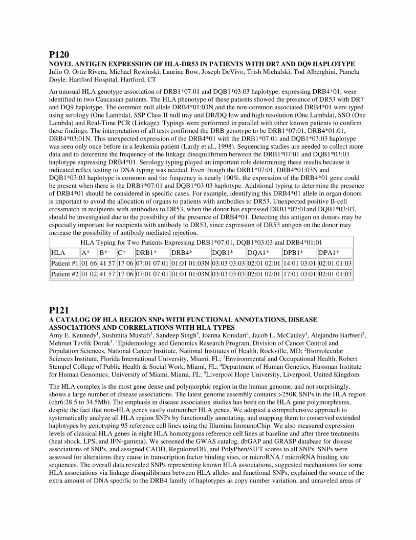
P120 NOVEL ANTIGEN EXPRESSION OF HLA-DR53 IN PATIENTS WITH DR7 AND DQ9 HAPLOTYPE Julio O. Ortiz Rivera, Michael Rewinski, Laurine Bow, Joseph DeVivo, Trish Michalski, Tod Alberghini, Pamela Doyle. Hartford Hospital, Hartford, CT
An unusual HLA genotype association of DRB1*07:01 and DQB1*03:03 haplotype, expressing DRB4*01, were identified in two Caucasian patients. The HLA phenotype of these patients showed the presence of DR53 with DR7 and DQ9 haplotype. The common null allele DRB4*01:03N and the non-common associated DRB4*01 were typed using serology (One Lambda), SSP Class II null tray and DR/DQ low and high resolution (One Lambda), SSO (One Lambda) and Real-Time PCR (Linkage). Typings were performed in parallel with other known patients to confirm these findings. The interpretation of all tests confirmed the DRB genotype to be DRB1*07:01, DRB4*01:01, DRB4*03:01N. This unexpected expression of the DRB4*01 with the DRB1*07:01 and DQB1*03:03 haplotype was seen only once before in a leukemia patient (Lardy et al., 1998). Sequencing studies are needed to collect more data and to determine the frequency of the linkage disequilibrium between the DRB1*07:01 and DQB1*03:03 haplotype expressing DRB4*01. Serology typing played an important role determining these results because it indicated reflex testing to DNA typing was needed. Even though the DRB1*07:01, DRB4*01:03N and DQB1*03:03 haplotype is common and the frequency is nearly 100%, the expression of the DRB4*01 gene could be present when there is the DRB1*07:01 and DQB1*03:03 haplotype. Additional typing to determine the presence of DRB4*01 should be considered in specific cases. For example, identifying this DRB4*01 allele in organ donors is important to avoid the allocation of organs to patients with antibodies to DR53. Unexpected positive B-cell crossmatch in recipients with antibodies to DR53, when the donor has expressed DRB1*07:01and DQB1*03:03, should be investigated due to the possibility of the presence of DRB4*01. Detecting this antigen on donors may be especially important for recipients with antibody to DR53, since expression of DR53 antigen on the donor may increase the possibility of antibody mediated rejection.
HLA Typing for Two Patients Expressing DRB1*07:01, DQB1*03:03 and DRB4*01:01
HLA A* B* C* DRB1* DRB4* DQB1* DQA1* DPB1* DPA1*
Patient #1 01 66 41 57 17 06 07:01 07:01 01:01 01:03N 03:03 03:03 02:01 02:01 14:01 03:01 02:01 01:03
Patient #2 01 02 41 57 17 06 07:01 07:01 01:01 01:03N 03:03 03:03 02:01 02:01 17:01 03:01 02:01 01:03
P121 A CATALOG OF HLA REGION SNPs WITH FUNCTIONAL ANNOTATIONS, DISEASE ASSOCIATIONS AND CORRELATIONS WITH HLA TYPES Amy E. Kennedy1, Sushmita Mustafi2, Sandeep Singh3, Ioanna Konidari4, Jacob L. McCauley4, Alejandro Barbieri2, Mehmet Tevfik Dorak5. 1Epidemiology and Genomics Research Program, Division of Cancer Control and Population Sciences, National Cancer Institute, National Institutes of Health, Rockville, MD; 2Biomolecular Sciences Institute, Florida International University, Miami, FL; 3Environmental and Occupational Health, Robert Stempel College of Public Health & Social Work, Miami, FL; 4Department of Human Genetics, Hussman Institute for Human Genomics, University of Miami, Miami, FL; 5Liverpool Hope University, Liverpool, United Kingdom
The HLA complex is the most gene dense and polymorphic region in the human genome, and not surprisingly, shows a large number of disease associations. The latest genome assembly contains >250K SNPs in the HLA region (chr6:28.5 to 34.5Mb). The emphasis in disease association studies has been on the HLA gene polymorphisms, despite the fact that non-HLA genes vastly outnumber HLA genes. We adopted a comprehensive approach to systematically analyze all HLA region SNPs by functionally annotating, and mapping them to conserved extended haplotypes by genotyping 95 reference cell lines using the Illumina ImmunoChip. We also measured expression levels of classical HLA genes in eight HLA homozygous reference cell lines at baseline and after three treatments (heat shock, LPS, and IFN-gamma). We screened the GWAS catalog, dbGAP and GRASP database for disease associations of SNPs, and assigned CADD, RegulomeDB, and PolyPhen/SIFT scores to all SNPs. SNPs were assessed for alterations they cause in transcription factor binding sites, or microRNA / microRNA binding site sequences. The overall data revealed SNPs representing known HLA associations, suggested mechanisms for some HLA associations via linkage disequilibrium between HLA alleles and functional SNPs, explained the source of the extra amount of DNA specific to the DRB4 family of haplotypes as copy number variation, and unraveled areas of

the HLA region with insufficient coverage in GWAS arrays. The SNPs with the most deleterious effects were mainly in non-HLA genes of the region, and most have not been included in GWAS arrays. We also noted that there are more disease associations in the HLA region than listed in the GWAS catalog. Given the insufficient coverage in GWAS arrays due to difficulties in assay design and structural variation, sequencing-based studies are needed to unravel even more associations missed by GWAS. The catalog produced in this work combines HLA and SNP data with functional effects and induced expression levels, and should be of great value in the design and interpretation of HLA and disease association studies.
P122 KIR-HLA GENOTYPES SUGGESTIVE OF OPPOSING NATURAL KILLER CELL RESPONSES ARE ASSOCIATED WITH TUBERCULOSIS AND SARCOIDOSIS, THE GRANULOMATOUS DISEASES WITH SIMILAR PATHOLOGY Raja Rajalingam1, Surendra K. Sharma2. 1Immunogenetics and Transplantation Laboratory, University of California San Francisco, San Francisco, CA; 2Division of Pulmonary, Critical Care and Sleep Medicine, All India Institute of Medical Sciences, New Delhi, India
Aim: The clinical, radiological and pathological similarities between pulmonary tuberculosis (PTB) and sarcoidosis (PS) can make disease differentiation challenging, particularly in countries with high burden of TB. Herein we analyzed KIR-HLA genotypes that might provide insight into immunological relationship between the diseases and allow us to distinguish between them. Methods: We typed 375 PTB patients, 201 PS patients and 249 healthy controls collected from All India Institute of Medical Sciences, India for KIR and HLA class I genes using Luminex rSSO. All controls were +ve to PPD, but displayed normal chest radiograph and did not suffer from any disease. Results: Differences between controls and patients in KIR genotypes, HLA ligands, and KIR-HLA pairs were tested by two-tailed Fisher's exact probabilities (Table). Distribution of AA and Bx KIR genotype did not vary between study groups. However, B haplotype-specific KIRs of centromeric region (2DL2, 2DS3) were reduced significantly in PS. Moreover, 3DL1+Bw4 and 2DL2+C1 pairs decreased significantly in PS. These results suggest the possibility of generating functionally compromised NK cells due to missing 3DL1+Bw4 and/or 2DL2+C1-mediated licensing, which is consistent with known diminished cellular immunity in PS. Contrasting to PS, telomeric half of KIR complex was associated with PTB. Specifically, an increase of B-haplotype associated KIRs (3DS1, 2DS1) and a decrease of A-haplotype associated KIRs (3DL1, 2DS4) were observed in PTB. Moreover, a substantial increase of 2DL1+C2 (p=0.0001) and a decrease of 3DL1+Bw4 (p=0.023) were observed in PTB. These results suggest the possibility of generating more activating NK cells by 2DL1+C2-mediated licensing and expressing activating KIR 3DS1/2DS1 phenotypes, which is consistent with active immune response in PTB. Conclusions: Association of distinct KIR-HLA genotypes predicts potentially distinct immune mechanisms for PTB and PS, and provides a more promising approach to differential disease diagnosis.
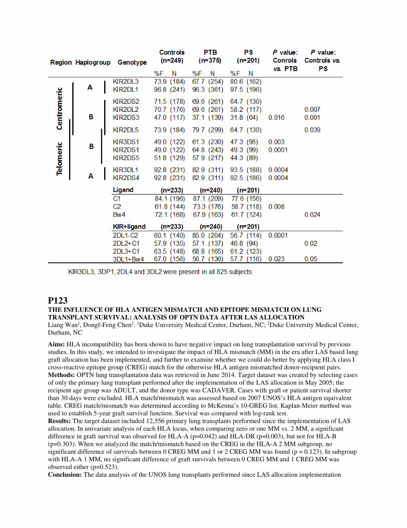
P123 THE INFLUENCE OF HLA ANTIGEN MISMATCH AND EPITOPE MISMATCH ON LUNG TRANSPLANT SURVIVAL: ANALYSIS OF OPTN DATA AFTER LAS ALLOCATION Liang Wan1, Dongf-Feng Chen2. 1Duke University Medical Center, Durham, NC; 2Duke University Medical Center, Durham, NC
Aims: HLA incompatibility has been shown to have negative impact on lung transplantation survival by previous studies. In this study, we intended to investigate the impact of HLA mismatch (MM) in the era after LAS based lung graft allocation has been implemented, and further to examine whether we could do better by applying HLA class I cross-reactive epitope group (CREG) match for the otherwise HLA antigen mismatched donor-recipient pairs. Methods: OPTN lung transplantation data was retrieved in June 2014. Target dataset was created by selecting cases of only the primary lung transplant performed after the implementation of the LAS allocation in May 2005; the recipient age group was ADULT, and the donor type was CADAVER. Cases with graft or patient survival shorter than 30 days were excluded. HLA match/mismatch was assessed based on 2007 UNOS’s HLA antigen equivalent table. CREG match/mismatch was determined according to McKenna’s 10-GREG list. Kaplan-Meier method was used to establish 5-year graft survival function. Survival was compared with log-rank test. Results: The target dataset included 12,556 primary lung transplants performed since the implementation of LAS allocation. In univariate analysis of each HLA locus, when comparing zero or one MM vs. 2 MM, a significant difference in graft survival was observed for HLA-A (p=0.042) and HLA-DR (p=0.003), but not for HLA-B (p=0.303). When we analyzed the match/mismatch based on the CREG in the HLA-A 2 MM subgroup, no significant difference of survivals between 0 CREG MM and 1 or 2 CREG MM was found (p = 0.123). In subgroup with HLA-A 1 MM, no significant difference of graft survivals between 0 CREG MM and 1 CREG MM was observed either (p=0.523). Conclusion: The data analysis of the UNOS lung transplants performed since LAS allocation implementation
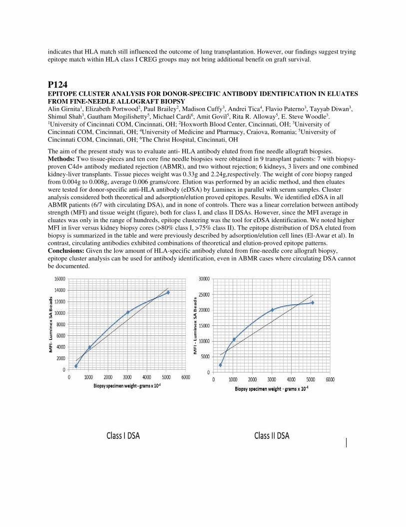
indicates that HLA match still influenced the outcome of lung transplantation. However, our findings suggest trying epitope match within HLA class I CREG groups may not bring additional benefit on graft survival.
P124 EPITOPE CLUSTER ANALYSIS FOR DONOR-SPECIFIC ANTIBODY IDENTIFICATION IN ELUATES FROM FINE-NEEDLE ALLOGRAFT BIOPSY Alin Girnita1, Elizabeth Portwood2, Paul Brailey2, Madison Cuffy3, Andrei Tica4, Flavio Paterno3, Tayyab Diwan3, Shimul Shah3, Gautham Mogilishetty5, Michael Cardi6, Amit Govil5, Rita R. Alloway5, E. Steve Woodle3. 1University of Cincinnati COM, Cincinnati, OH; 2Hoxworth Blood Center, Cincinnati, OH; 3University of Cincinnati COM, Cincinnati, OH; 4University of Medicine and Pharmacy, Craiova, Romania; 5University of Cincinnati COM, Cincinnati, OH; 6The Christ Hospital, Cincinnati, OH
The aim of the present study was to evaluate anti- HLA antibody eluted from fine needle allograft biopsies. Methods: Two tissue-pieces and ten core fine needle biopsies were obtained in 9 transplant patients: 7 with biopsy-proven C4d+ antibody mediated rejection (ABMR), and two without rejection; 6 kidneys, 3 livers and one combined kidney-liver transplants. Tissue pieces weight was 0.33g and 2.24g,respectively. The weight of core biopsy ranged from 0.004g to 0.008g, average 0.006 grams/core. Elution was performed by an acidic method, and then eluates were tested for donor-specific anti-HLA antibody (eDSA) by Luminex in parallel with serum samples. Cluster analysis considered both theoretical and adsorption/elution proved epitopes. Results. We identified eDSA in all ABMR patients (6/7 with circulating DSA), and in none of controls. There was a linear correlation between antibody strength (MFI) and tissue weight (figure), both for class I, and class II DSAs. However, since the MFI average in eluates was only in the range of hundreds, epitope clustering was the tool for eDSA identification. We noted higher MFI in liver versus kidney biopsy cores (>80% class I, >75% class II). The epitope distribution of DSA eluted from biopsy is summarized in the table and were previously described by adsorption/elution cell lines (El-Awar et al). In contrast, circulating antibodies exhibited combinations of theoretical and elution-proved epitope patterns. Conclusions: Given the low amount of HLA-specific antibody eluted from fine-needle core allograft biopsy, epitope cluster analysis can be used for antibody identification, even in ABMR cases where circulating DSA cannot be documented.
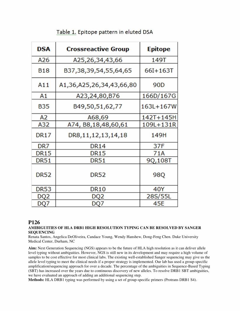
P126 AMIBIGUITIES OF HLA DRB1 HIGH RESOLUTION TYPING CAN BE RESOLVED BY SANGER SEQUENCING Renata Santos, Angelica DeOliveira, Candace Young, Wendy Hanshew, Dong-Feng Chen. Duke University Medical Center, Durham, NC
Aim: Next Generation Sequencing (NGS) appears to be the future of HLA high resolution as it can deliver allele level typing without ambiguities. However, NGS is still new in its development and may require a high volume of samples to be cost effective for most clinical labs. The existing well-established Sanger sequencing may give us the allele level typing to meet the clinical needs if a proper strategy is implemented. Our lab has used a group-specific amplification/sequencing approach for over a decade. The percentage of the ambiguities in Sequence-Based Typing (SBT) has increased over the years due to continuous discovery of new alleles. To resolve DRB1 SBT ambiguities, we have evaluated an approach of adding an additional sequencing step. Methods: HLA DRB1 typing was performed by using a set of group-specific primers (Protrans DRB1 S4).
Ambiguities observed in S4 SBT were then resolved by performing an additional amplification and/or sequencing with selected primers (Protrans Domino Stone). Sequencing data were analyzed and allele level typing was assigned by using SBT software (SeqPilot, JSI). Results: Six samples that were initially typed with ambiguities using DRB1 S4 reagent were sequenced with Domino Stones primers. Multiple samples were typed with the same HLA-DRB1 ambiguity. All the ambiguities were resolved with an exception for DRB1*12:01 and 12:10 which are different from each other in exon 1 (Table 1). The final typing assignments can be obtained within 3 days. Conclusions: Our typing results suggest that the new approach of adding an additional sequencing step may resolve most of the DRB1 typing ambiguities and deliver the allele level typing needed by transplant clinicians. Studies in resolving ambiguities of HLA DQB1 with Domino Stone primers are under evaluation.
Ambiguities tested by Domino Stones.
ProtransDRB1 S4 (HLA Ambiguities) Domino Stones
DRB1*13:01/13:117/13:190 DRB1*13:01
DRB1*07:01/07:34 DRB1*07:01
DRB1*01:01/01:50,13:01/13:117/13:190 DRB1*01:01,13:01
DRB1*07:01/07:34,12:01/12:06/12:10/12:17 DRB1*07:01,12:01/12:10
P127 RESIDUAL SERUM RITUXIMAB INDUCES FALSE POSITIVE FLOW CYTOMETRIC B CELL CROSSMATCH MORE THAN 6 MONTHS POST-INFUSION Eszter Lazar-Molnar1, Denise Hurst2, Ann Pole2, Julio C. Delgado1. 1University of Utah, Salt Lake City, UT; 2University of Utah Health Care, Salt Lake City, UT
Case 1: 18 year old female patient was listed for transplant due to glomerulonephritis and kidney failure. She was diagnosed with microscopic polyangiitis since childhood. Her transplant workup indicated the absence of HLA class I and class II antibodies, but unexpectedly, she had a positive B cell crossmatch with a virtually matched deceased donor. Case 2: 58 year old female patient with autoimmune glomerulonephritis was listed for transplant due to end stage renal disease. She was scheduled to receive a living donor kidney from her sister. Her transplant workup showed no detectable HLA class I or class II antibodies, but she had an unexpected positive B cell crossmatch. Further investigation revealed that both patients received rituximab treatment for their autoimmune disease 7, and 8 months earlier. With no HLA explanation, it seemed likely that the positive B cell crossmatch was caused by residual rituximab in the patients’ sera. Rituximab is a chimeric monoclonal antibody that binds to CD20 expressed on B cells, inducing depletion. Initially approved for the treatment of B cell lymphoma, it is increasingly used for the treatment of autoimmune diseases. Although the expected half-life of the drug is 14-21 days, significant variations have been reported, complicated by the lack of standardized dosing in various autoimmune diseases. Detected in the serum many months post-infusion, rituximab interferes with cytotoxic crossmatch due to inducing complement-dependent cytotoxicity, and with flow cytometric crossmatch due to being recognized by the anti-human antibody used in the assay. To confirm interference by the drug, the patients’ sera were pre-treated with an idiotype-specific antibody to rituximab, which resulted in negative B cell crossmatch. Both patients were successfully transplanted. Although rituximab was previously reported to interfere with B cell crossmatch 2-3 months after the dose, these cases demonstrate that false positive B cell crossmatch can occur as long as 7 or 8 months post-infusion. Thorough record of the patients’ medication history is essential for the transplant workup. Methodologies that eliminate cell surface CD20 such as pronase treatment, or remove rituximab by antibody absorption should be added to the routine procedure, when rituximab-containing serum is used for crossmatch.
P128 HLA TYPING BY NGS IN THE CLINICAL LAB: OUR ONE AND A HALF YEARS EXPERIENCE Jamie Duke1, Deborah Ferriola1, Anh Huynh1, Steve Heron1, Allison Gasiewski1, Yanping Huang1, Curt Lind1, Laura McLaughlin1, Hilary Mehler1, Anna Papazoglou1, Marianne Rogers1, Larissa Slavich1, Rita Walker1, Dimitri Monos1,2. 1The Children's Hospital of Philadelphia, Philadelphia, PA; 2University of Pennsylvania, Philadelphia, PA

Aim: We present our experience using the Holotype™ Kit by Omixon for next generation sequencing (NGS) of HLA genes in a clinical setting for one and a half years, starting December 2013. Methods: Samples were prepared using the X4 Holotype™ kit by Omixon for up to 7 loci (full length HLA-A, B, C, DQA1 and DQB1, intron 1 to intron 4 of DRB1, and intron 1 to 3’ UTR of DPB1) and were sequenced in both an individual and pooled amplicon approach using 2x251 bp reads on the Illumina MiSeq. Samples were also characterized in parallel with a second method (Sanger SBT, SSO, or SSP) to confirm the genotyping results by NGS and identify possible causes of discrepancies, if any. Two analysis programs were used to genotype the NGS data, Target by Omixon (v 1.7/1.8) and NGSengine by GenDX (v 1.3/1.6). Results: A total of 1046 samples have been sequenced: 497 (47%) related to bone marrow transplant cases, 250 (24%) related to solid organ transplantation, and 299 (29%) samples being either proficiency, outreach or disease association studies. Approximately 975 typings were performed for HLA-A, B, C, DRB1 and DQB1, and 600 typings for DQA1 and DPB1. On average, one sequencing run was performed per week with 12 samples on the MiSeq for a total of 83 runs utilizing both nano and full flow cells. At best, the turnaround time from amplification to analysis completion was 4 days, but averages 10 days due to the laboratory workflow and a single sequencing run per week. Concordance was 99.7% between NGS and secondary typing methods (SBT/SSP/SSOP). Discordance was due to incorrect or incomplete (ambiguous) SBT/SSP typing, allele imbalance, NGS genotyping software errors, incorrect reporting of results and new alleles. Two genotyping programs were used for analysis to safeguard against systematic analysis errors and agree for 94.8% of the best match allele calls. Ambiguous results are most common at the DPB1 locus due to a lack of phasing between exons 2 and 3 or the unsequenced exon 1 (17% of alleles) and the DRB1 locus due to not sequencing exon 1 (3.8% of alleles). No ambiguities were detected among the other loci. Conclusions: We have genotyped over 1,000 samples using NGS, with a very high concordance rate with other HLA typing methods and low ambiguity rates for DRB1 and DPB1 as described above. This technology is therefore very appropriate for HLA typing in a clinical setting. D. Monos: Grant/Research Support; Company/Organization; Omixon. 7. Other (Identify); Company/Organization;
Royalty from Omixon.
P129 DIFFERENTIAL ROLE OF INTEGRIN BETA 4 IN HLA CLASS-I INDUCED ENDOTHELIAL CELL ACTIVATION Nwe Nwe Soe, Xiaohai Zhang, Yi-Ping Jin, Elaine F. Reed. David Geffen School of Medicine, UCLA, Los Angles, CA
Aim: The development of anti-donor HLA class I (HLA-I) antibodies following solid organ transplant is associated with acute and chronic rejection and graft failure. Following ligation with antibody, HLA class I molecules form a molecular partnership with integrin β4 (ITGB4) to transduce activation and proliferative signals in endothelial cells (EC) contributing to rejection. We hypothesize that determination of the domains on the integrin β4 molecule that interact with HLA-I will provide critical information for the development of novel therapeutics for the treatment of chronic rejection. Methods: To identify the ITGB4 intracellular domain(s) that associates with HLA-І, we used the ITGB4 full length (Wild Type) construct or deletion mutants that lack the Calx-β, fibronectin type III repeat FNIII 1-4 or connecting segment (CS) domain . These constructs were sub-cloned into an adenovirus-based vector and adenovirus encoding WT or mutants were generated. Endogenous ITGB4 expression was suppressed using siRNA transfection. Primary human aortic endothelial cells (HAEC) lacking endogenous ITGB4 were infected with adenovirus and then EC activation was determined by analyzing protein phosphorylation, stress fiber formation, cell migration and proliferation. Results: Our results demonstrate that HLA-І binds to ITGB4 via the CS domain. The CS domain is required for the phosphorylation of Src, Paxillin, Akt, ERK1/2 and S6RP. The FNIII 3&4 domains are required for phosphorylating Paxillin, Akt, ERK1/2 and S6RP, but not Src. HLA-І phosphorylates ITGB4 at Tyr1494 and Tyr1526 in a Src dependent manner. Furthermore, deletion of CS, FNIII3&4, Calx-β inhibited HLA-І induced EC stress fiber formation, migration and proliferation. Moreover, HLA-I induces association between ITGB4 and the adaptor protein Shc which depends on CS and FNIII3&4 domains. Suppression of endogenous Shc protein by siRNA inhibited HLA-І induced EC migration, proliferation and phosphorylation of Akt, ERK1/2 and S6RP, but not Src. Conclusion: Understanding the molecular mechanisms underlying HLA antibody-induced EC proliferation and migration should enable the development of novel therapeutic strategies for the treatment of antibody-mediated chronic transplant rejection.

P130 VALIDATION OF A CLINICAL ASSAY FOR SPECIFIC GENOTYPING OF A KIR2DL1 ALLELIC POLYMORPHISM Paula Arnold1, Po-Chien Chou1, Rafijul Bari2, Wing Leung2. 1St. Jude Children's Research Hospital, Memphis, TN; 2St. Jude Children's Research Hospital, Memphis, TN
Aim: A number of transplant centers currently use killer-cell immunoglobulin-like receptor (KIR) genotyping as criteria for donor selection for hematopoietic stem cell transplant (HSCT) and natural killer (NK) cell protocols. Several factors contribute to donor selection, including presence or absence of multiple inhibitory and stimulatory KIR, centromeric vs. telomeric KIR haplotypes, “A” vs. “B” content KIR haplotypes, and NK cell education or licensing. Recent studies at our institution demonstrated that amino acid residue 245 of the KIR2DL1 protein is important in determining the signaling capacity, and had a significant effect on the overall outcome of HSCT. Patients who received a donor graft containing KIR2DL1 alleles encoding an arginine at position 245 (KIR2DL1-R245) had better survival and lower incidence of disease progression than the patients who received a donor graft containing KIR2DL1 alleles encoding a cysteine at the same position (KIR2DL1-C245). The goal of this work was to validate a lab-developed clinical test to distinguish the KIR2DL1 R245/C245 allelic polymorphism and to facilitate donor selection based on this criterion. Methods: Our research laboratory developed a real-time PCR single nucleotide polymorphism (SNP) assay that specifically detects alleles of the KIR2DL1 gene encoding arginine or cysteine at position 245, and avoids the interference of 2DL2 alleles that share the same sequence flanking this region. The HLA laboratory used the backbone of this assay to validate it for clinical use as a lab-developed companion test to current KIR genotyping. Results: The SNP assay performed according to the required qualifications, including accuracy, precision, sensitivity, and specificity. Samples tested showed 100% concordance with the expected genotyping of the “Gold Standard,” cell lines with sequenced KIR2DL1 alleles from the CIBMTR Repository. Conclusions:This SNP assay was validated for clinical use in the HLA laboratory, and should be in use in the near future as a companion test to the KIR genotyping currently used for donor selection in HSCT. P. Arnold: Grant/Research Support; Company/Organization; American Lebanese Associated Charities (ALSAC). P.
Chou: Grant/Research Support; Company/Organization; American Lebanese Syrian Associated Charities (ALSAC).
R. Bari: Grant/Research Support; Company/Organization; American Lebanese Syrian Associated Charities
(ALSAC), Assisi Foundation of Memphis. 7. Other (Identify); Company/Organization; Dr. Bari is named as an
inventor on patent applications claiming a SNP assay used for KIR allele typing, which is owned by SJCRH and is
licensed to a commercial entity. W. Leung: Grant/Research Support; Company/Organization; American Lebanese
Syrian Associated Charities (ALSAC), Assisi Foundation of Memphis. 7. Other (Identify); Company/Organization;
Dr. Leung is named as an inventor on patent applications claiming a SNP assay used for KIR allele typing, which is
owned by SJCRH and is licensed to a commercial entity.
P131 GENERATION OF HIGH CONFIDENCE HLA GENOTYPING AND CONSENSUS SEQUENCES FOR CLASS I HLA LOCI USING THE NGS-BASED OMIXON HOLOTYPE HLA TYPING SYSTEM AND THE ILLUMINA MISEQ PLATFORM Peter M. Clark, Jamie Duke, Deborah Ferriola, Anna Papazoglou, Aniqa Hassan, Dimitri Monos. The Children's Hospital of Philadelphia, Philadelphia, PA
Aim: Generate HLA genotyping and high confidence consensus sequences for 50 samples at HLA-A, B and C loci using the Omixon Holotype HLA typing system. Methods: The Omixon Holotype HLA typing kit was used to generate sequencing libraries for 50 samples for HLA-A, B and C loci, and sequenced on an Illumina MiSeq. HLA typing and consensus sequence generation was performed using Omixon Twin, GenDx NGSengine and a custom pipeline, which relies on stringently mapping reads to the genotyped alleles and using SAMtools to generate the consensus. Consensus sequences generated by each method were aligned pairwise to determine concordance using a semi-global Needleman-Wunsch algorithm. When the sequences generated by each method were concordant, sequences were aligned to the IMGT reference sequence of the genotyped allele, to annotate novel alleles and annotate the sequence of incomplete alleles. When sequences generated by each method were discordant, sequences were aligned to the genotyped allele for manual inspection and assessment of algorithm performance. Results: HLA genotyping results for all 300 alleles (50 samples at 3 loci) were 100% concordant between

NGSengine and Omixon Twin, after manually resolving discrepancies (6/300). Consensus sequences were generated for every allele (300) encompassing 31 unique HLA-A, 52 unique HLA-B and 31 unique HLA-C alleles. Our analysis facilitated the annotation of full gene sequences for 11 HLA alleles for which there is no complete IMGT reference sequence (A*02:04, B*14:03, B*15:08:01, B*15:30, B*27:02:01, B*35:04:01, B*35:43:01, B*39:09:01, B*51:08:01, B*58:02 and C*07:18) and facilitated the discovery of two novel HLA-A alleles, with intronic substitutions. For the remaining alleles, consensus sequences were compared against the reference sequences of the genotyped alleles and occasional differences between consensus sequences and reference sequences were manually inspected to evaluate algorithm performance. It has been determined that depth of coverage is critical in phasing and base calling, which is necessary to generate credible consensus sequences. Conclusions: Generation of high confidence HLA class I consensus sequences is possible using the Omixon Holotype HLA typing system provided that adequate depth of uniform coverage is obtained from sequencing. D. Monos: Other (Identify); Company/Organization; Omixon Royalties.
P132 THE EFFECT OF PRONASE ON LYMPHOCYTE SURFACE MARKERS AND IMPLICATIONS FOR FLOW CYTOMETRIC CROSSMATCH Eszter Lazar-Molnar1, Laura Spruit2, Ann Pole2, Kevin Williams2, Michelle R. Taylor2, Julio C. Delgado1. 1University of Utah, Salt Lake City, UT; 2University of Utah Health Care, Salt Lake City, UT
Aim: Pronase treatment of lymphocytes has been used to improve the specificity and sensitivity of the flow cytometric crossmatch for almost 20 years, due to its ability to remove Fc receptors from B cells and reduce non-specific background. Due to controversy in the published data, and significant variations in laboratory practices about the use of pronase, our goal was to determine the concentration-dependent effect of pronase on lymphocyte surface molecules, such as CD32 (FcγRIIB), HLA class I and II, CD3, CD19, and CD20, and to provide a standardized procedure to improve the performance of flow cytometric crossmatch assays. Methods: Lymphocytes from 8 donors were isolated from blood, lymph nodes (LN) or spleen, and were digested using different concentrations of pronase ranging from 0.3 through 2 mg/ml. Surface CD32, CD3, CD19, CD20, HLA class I and class II were monitored by antibody staining using multi-color flow cytometry. Results: Digestion with pronase at 0.6 mg/ml concentration or higher decreased CD32 expression by 80% on peripheral blood B cells and 85-90% on LN and splenic B cells. Pronase treatment did not reduce expression of HLA class I, however, it increased W6/32 antibody staining by 20-40% on B cells, and 150-200% on T cells. Similarly, pronase did not decrease HLA class II expression, but increased Tu39 antibody staining by 10-30% on lymph node and blood B cells. Binding of the CD3 antibody on pronase-treated T cells was increased by 2-2.5 fold, compared to untreated cells. There was no significant effect on CD19 expression up to 1 mg/ml, but 45-55% decrease was observed at 2 mg/ml. Pronase reduced CD20 expression in a concentration-dependent manner by up to 98-99% at 2 mg/ml. Conclusions: Pronase treatment increases specificity of the crossmatch assay since it reduces CD32 expression on B cells. Our data show that concentrations higher than 0.6 mg/ml do not decrease CD32 expression any further. Pronase treatment is safe since it does not decrease HLA class I or class II expression, but rather increases pan-HLA antibody binding, likely due to receptor “unmasking”, thus increasing the sensitivity of the crossmatch assay. Finally, increasing pronase concentration to 2 mg/ml eliminates B cell CD20 expression, a procedure that can help to eliminate interferences by rituximab-containing sera used for crossmatch.
P133 DERIVING HLA GENOTYPING FROM WHOLE GENOME SEQUENCING DATA USING OMIXON HLA TWIN(TM) IN G3’S GLOBAL CLINICAL STUDY Szilveszter Juhos1, Tünde Vágó1, Deborah Ferriola2, Jamie Duke2, Szilárd Vörös3, Bradley O. Brown3, Idean Marvasty3, Tim Hague1, Dimitri Monos2. 1Omixon Ltd, Budapest, Hungary; 2Children's Hospital of Philadelphia, Philadelphia, PA; 3Global Genomics Group, Richmond, VA
Aim: Identify the degree of concordance and the ambiguities when DNA samples are analyzed using two HLA genotyping systems; Whole Genome Sequencing (WGS) data analyzed by Omixon HLA Target software and genotyping by the Holotype HLA typing system. Methods: WGS data were generated of 235 human samples from G3's GLOBAL study (NCT01738828) using
paired 100 bps long HiSeq Illumina ~30x coverage and analyzed by the Omixon HLA Target software. Genotyping was also performed for the same samples using the Holotype HLA typing system, whereby the generated libraries were sequenced on the Illumina MiSeq using the 250 bps paired-end sequencing protocol. Data were analyzed by the Omixon HLA Twin software. Concordance at the three-field level between the two approaches was assessed for each of the HLA-A, B, C, DQB1 and DRB1 loci. Ambiguities were also determined for each of the typing methods. Results: A pairwise comparison between the two NGS-based methods shows that in average they were 97.2% concordant at the three-fields level (HLA-A: 450/470 95.7%, HLA-B: 464/470 98.7%, HLA-C: 464/470 98.7%, HLA-DQB1: 438/470 93.2%, HLA-DRB1 467/468 99.8%). Most of the discrepancies were due to allele dropout in the WGS sample or systematic errors of the software. The 41 non-systematic mistypings were due to either insufficient coverage of the WGS data or algorithmic problems. The Holotype HLA system did not generate any ambiguities for the HLA-A, -B, -C and HLA-DQB1 loci at the three-fields typing level. However, as intron 1 and exon 1 of DRB1 are not targeted, 1.4% of ambiguities were generated for the DRB1 locus.The WGS typing considers only the exons of the reference database, leading to phase ambiguities and overall ambigous typings of 9.8%, 4.3%, 7.7%, 8.3% and 3.4% for HLA-A, B, C, DQB1 and DRB1 respectively. Conclusions:HLA genotyping using WGS data derived through next-generation sequencing and analyzed by the Omixon HLA Twin software is possible at 97.2% accuracy level. Future improvements of the software addressing ambiguities and remaining systematic errors can make HLA typing from WGS data more reliable. S. Juhos: Employee; Company/Organization; Omixon Ltd. T. Vágó: Employee; Company/Organization; Omixon
Ltd. D. Ferriola: Employee; Company/Organization; Children's Hospital of Philadelphia. J. Duke: Employee;
Company/Organization; Children's Hospital of Philadelphia. S. Vörös: Employee; Company/Organization; Global
Genomics Group. B.O. Brown: Employee; Company/Organization; Global Genomics Group. I. Marvasty:
Employee; Company/Organization; Global Genomics Group. T. Hague: Employee; Company/Organization;
Omixon Ltd. D. Monos: Employee; Company/Organization; Children's Hospital of Philadelphia.
P134 RAPID AND AUTOMATED FLOW CYTOMETRY CROSSMATCH FOR OPTIMIZING INTER-ASSAY AND INTER-OPERATOR CONSISTENCY Prabhakar Putheti1,2, Jeannette Knight1, Chaquetta Grace1, Vijay K. Sharma1,2. 1The Rogosin Institute, New York, NY; 2Weill-Cornell Medical Center, New York, NY
Background: Flow cytometry crossmatch (FCXM) is a high sensitive tool to identify donor cell specific antibodies in recipient’s serum. However, the procedure is time consuming and fraught with inter-assay and inter-operator variabilities. Aim: To optimize instrumentation for automated tagging (AT) in FCXM that will both quicken the procedure and exclude inter-operator variability. Methods: Serum, spleen, or peripheral blood mononuclear cells were isolated from human subjects. AT was performed using a combination of Precision-XS and EL406 instruments (BioTek Instruments, Inc.) and compared to manual tagging (MT) (both in 96 well plates). FITC-goat α-human IgG channel shift (CS) of >40 for T cells and >50 for B cells was considered positive. Results: AT of two cell specimens took 90 minutes vs. 100 minutes by MT. AT did not cause cell loss. Automated Tagging vs. Manual Tagging: AT and MT of specimens (N=6) provided similar CS results (T cell- (P=0.8) and B cell-FCXM (P= 0.9); (Fig 1A)) and median channel intensity results for T cell FCXM (%CV: negative control (NC)=8 vs. 11; positive control (PC)=10 vs. 14) and B cell FCXM (%CV: NC=6 vs. 17; PC=10 vs. 9). Intra-assay variability: When AT was performed in duplicates in a plate (N=3), no intra-assay variability was noted in CS results (T cell FCXM: P=0.1; B cell FCXM: P=0.1 (Fig 1B)) or in median channel intensity results [T cell FCXM (%CV: NC=1 vs. 5; PC=4 vs. 2) and B cell FCXM (%CV: NC=2 vs. 1; PC=5 vs. 4).] Inter-assay variability: When AT was performed in two different plates (N=3), no inter-assay variability was noted in CS results (T cell FCXM: P=0.8; B cell FCXM: P=0.9 (Fig 1C)) or in median channel intensity results [T cell FCXM (%CV: NC=8; and PC=6) and B cell FCXM (%CV: NC=11; PC=5)]. Inter-operator variability: Inter-operator variability was not noted when FCXM were performed by AT (Fig 1D). Conclusions:Combination of Precision-XS- and EL406-instruments helps making a FCXM faster and automatous, and provides reliable results with inter-assay and inter-operator consistency.
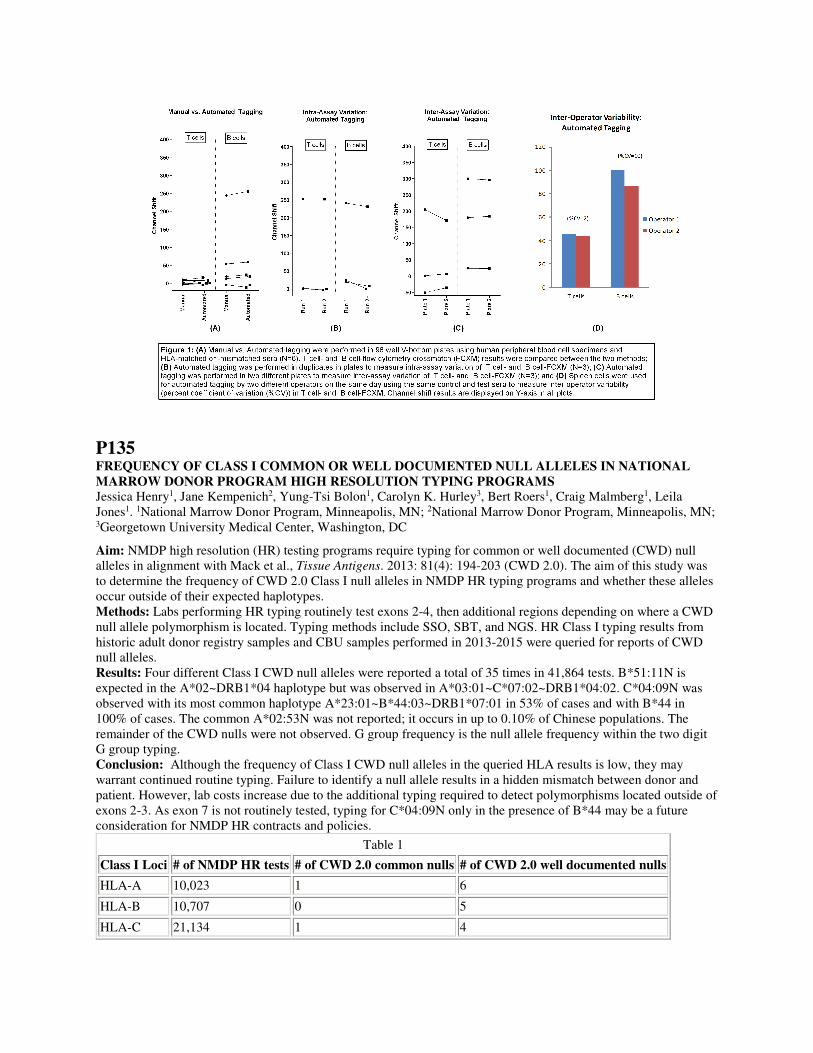
P135 FREQUENCY OF CLASS I COMMON OR WELL DOCUMENTED NULL ALLELES IN NATIONAL MARROW DONOR PROGRAM HIGH RESOLUTION TYPING PROGRAMS Jessica Henry1, Jane Kempenich2, Yung-Tsi Bolon1, Carolyn K. Hurley3, Bert Roers1, Craig Malmberg1, Leila Jones1. 1National Marrow Donor Program, Minneapolis, MN; 2National Marrow Donor Program, Minneapolis, MN; 3Georgetown University Medical Center, Washington, DC
Aim: NMDP high resolution (HR) testing programs require typing for common or well documented (CWD) null alleles in alignment with Mack et al., Tissue Antigens. 2013: 81(4): 194-203 (CWD 2.0). The aim of this study was to determine the frequency of CWD 2.0 Class I null alleles in NMDP HR typing programs and whether these alleles occur outside of their expected haplotypes. Methods: Labs performing HR typing routinely test exons 2-4, then additional regions depending on where a CWD null allele polymorphism is located. Typing methods include SSO, SBT, and NGS. HR Class I typing results from historic adult donor registry samples and CBU samples performed in 2013-2015 were queried for reports of CWD null alleles. Results: Four different Class I CWD null alleles were reported a total of 35 times in 41,864 tests. B*51:11N is expected in the A*02~DRB1*04 haplotype but was observed in A*03:01~C*07:02~DRB1*04:02. C*04:09N was observed with its most common haplotype A*23:01~B*44:03~DRB1*07:01 in 53% of cases and with B*44 in 100% of cases. The common A*02:53N was not reported; it occurs in up to 0.10% of Chinese populations. The remainder of the CWD nulls were not observed. G group frequency is the null allele frequency within the two digit G group typing. Conclusion: Although the frequency of Class I CWD null alleles in the queried HLA results is low, they may warrant continued routine typing. Failure to identify a null allele results in a hidden mismatch between donor and patient. However, lab costs increase due to the additional typing required to detect polymorphisms located outside of exons 2-3. As exon 7 is not routinely tested, typing for C*04:09N only in the presence of B*44 may be a future consideration for NMDP HR contracts and policies.
Table 1
Class I Loci # of NMDP HR tests # of CWD 2.0 common nulls # of CWD 2.0 well documented nulls
HLA-A 10,023 1 6
HLA-B 10,707 0 5
HLA-C 21,134 1 4
Table 2
Observed CWD null alleles Donor CBU Frequency (%) G group frequency (%) Polymorphism location(s)
B*51:11N 2 0 0.019 0.174 Exon 4
C*04:09N 18 12 0.142 0.484 Exon 7
C*05:07N 1 1 0.005 n/a Exon 3
C*07:32N 1 0 0.005 n/a Exon 2-4
P136 KIR GENE GENOTYPE FREQUENCIES IN DONORS OF GERMAN, TURKISH, AND POLISH DESCENT Jürgen Sauter1, Angela Lucaci-Timoce1, Christine Gnahm1, Jan Hofmann1, Irina Böhme2, Vinzenz Lange2, Julia Pingel1, Alexander Schmidt1. 1DKMS German Bone Marrow Donor Center, Tübingen, Germany; 2DKMS Life Science Lab, Dresden, Germany
Aim: Killer cell immunoglobulin-like receptors (KIRs) are expressed on cells of the innate immune system. The KIR system has been found to be important in donor selection for hematopoietic stem cell transplantation in certain circumstances (Cooley et al. Blood, 2010). In addition to the frequency of these 17 KIR loci in donors of German, Polish, and Turkish descent, we present the most common KIR genotypes in these ethnic groups as well as the frequency of KIR haplotypes. Methods: More than 82,000 donors have been recruited from January through April 2015 by DKMS in Germany and typed for absence or presence of 17 KIR genes and pseudogenes using an NGS-based amplicon approach targeting exons 4, 5, and 7 of the respective genes. While KIR2DL5A and KIR2DL5B genes are not distinguished, KIR2DS4 and KIR2DS4N are reported separately, as null alleles are not biologically functional. Frequencies are given by the relative fraction of typing results. Results: 70,411 of the donors recruited indicated to be of German descent, 4,989 of Turkish and 860 of Polish descent. The most common KIR genotype in all 3 ethnicities includes the presence of only 2DL1, 2DL3, 2DL4, 2DP1, 2DS4N, 3DL1, 3DL2, 3DL3, and 3DP1 genes. For all populations, the most frequent KIR genotype, according to the classification of Cooley et al., is B/2, while B/4 is most rare.
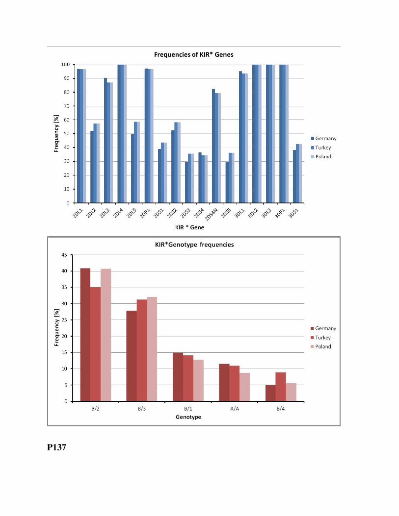
P137
CASE REPORT ILLUSTRATING TRANSPLANT DECISION MAKING BASED ON ASSESSING CLINICAL RELEVANCE OF APPARENT DONOR SPECIFIC ANTIBODIES, ALLELE SPECIFIC ANTIBODIES, AND SHARED EPITOPE PATTERNS Soumya Pandey, Bobbie Rhodes-Clark, Terry Harville. University of Arkansas for Medical Sciences, Little Rock, AR
Patient: 36 yo WM, IgA nephropathy. LABScreen SA beads (SAB): anti-B*44:02 (MFI=3685), B*44:03 (496). Quarterly testing: anti-B*44:02 (3685-5482), B*44:03 (121-1065). Other Class I antibodies had lower MFI; expected epitope pattern absent. No significant Class II antibodies. Donor expresses B*44:02. At transplant: Anti-B*44:02 (5411), B*44:03 (1065). Positive virtual-XM predicted; Flow-XM negative. CDC-XM: negative. Subsequent assays: Lambda Antigen Trays: no anti-B44 detected. C1q and iBeads: negative for anti-B*44:02 (1 and 0, respectively) and B*44:03 (1 and 0, respectively). Flow- and CDC-XM with 3 surrogates expressing B*44:02: all negative. The results suggest that anti-B*44:02 does not activate complement, and would not be expected to result in humoral rejection. iBeads detect anti-HLA antibodies binding to HLA in the “native-conformation”, as would be expected to be expressed on cell surfaces. The lack of detection with iBeads further supports that the anti-B*44:02 found by routine SAB was directed against denatured/partially-denatured antigen. Further, the expected epitope pattern was absent. Thus, this putative allele-specific antibody (ASA) anti-B*44:02 antibody would be predicted to have no clinical relevance, similarly so for anti-B*44:03. The patient is currently greater than 2 years post-transplantation with stable renal function, and no signs of AMR. Post-transplant SAB continue to detect anti-B*44:02 and B*44:03; however continue to be essentially absent by C1q or iBeads assays. This case emphasizes that potential DSA/ASA should be carefully scrutinized for the ability to activate complement and expected shared patterns of epitopes, as discriminators of potential clinical relevance. Additional testing beyond routine SAB will likely be required to assess these issues. Conclusions:1) The “stringent” use of the “virtual XM” may exclude XM-compatible donors due to “apparent” DSA. 2) Additional testing should be considered to determine whether DSA can activate complement. 3) The “extended” non-DSA anti-HLA pattern should be evaluated for the expected shared epitope patterns. 4) Further testing should be considered for discriminating antibodies to denatured vs native-conformation. 5) The Flow-XM remains the “gold standard” for making the final decision to transplant. T. Harville: Scientific/Medical Advisor; Company/Organization; Baxter Healthcare, CSL Behring. 7. Other
(Identify); Company/Organization; Medical Board of Arkansas Regional Organ Recovery Agency.
P138 CHARACTERIZATION OF COPY NUMBER VARIATIONS (CNV) IN THE NKG2C RECEPTOR GENE Kai Cao1, Weicheng Zhao1, Nathaniel Smith1, Yudith Carmazzi1, Elizabeth Shpall2, Katy Rezvani2. 1UT MD Anderson Cancer Center, Houston, TX; 2UT MD Anderson Cancer Center, Houston, TX
The cytolytic function of natural killer (NK) cells is controlled by a balance of activatory and inhibitory signals transduced by specific NK-cell receptors. NKG2 family is one of the superfamilies of these receptor genes located on chromosome 12 in the NKC gene complex. NKG2C is expressed as heterodimers with CD94 and they deliver activating signals upon contacting HLA-E via adaptor molecules. NKG2C genes are not only polymorphic and also NKG2C gene deletion was identified. The aim of the study was to develop a genotyping assay for the detection of the wild type and the deletion of the NKG2C gene. Method: Copy numbers of the NKG2C genes were genotyped and assessed in 352 subjects by PCR-SSP method using a set of primer pairs described previously (Mararu M & Miyashita R, etc.). Two pairs of primers are used to detect the NKG2C genotypes: one pairs to amplify a 201 bp fragment from NKG2C wild type (wt) carrier; and the second pairs to amplify a 411 bp fragment from NKG2C deletion (del) carrier. A single-tube PCR-SSP genotyping strategy combining the two sets of primers was modified from Moraru M, etc (Tissue Antigens, 2012). Results: The presence or absence of NKG2C gene was determined by the single-tube PCR-SSP assay. Three genotypes were observed in the study subjects: wild type homozygous carrier (wt/wt), wild type and deletion heterozygous carrier (wt/del), and deletion homozygous carrier (del/del). Copy number of NKG2C in 3 International Histocompatibility Workshop (IHW) cells was assessed and confirmed: IHW 9043 as NKG2Cwt/wt, IHW 9047 as NKG2Cwt/del, and IHW 9044 as NKG2Cdel/del. Of the 352 subjects, wt/wt genotype accounted for 62.82% (n=221), wt/del for 32.88% (n=116), and del/del for 4.3% (n=15). The allele frequency of NKG2C deletion was 20.74% in this study group. Conclusion: Studies have shown that NK cells expressing the activating receptor NKG2C preferentially expand
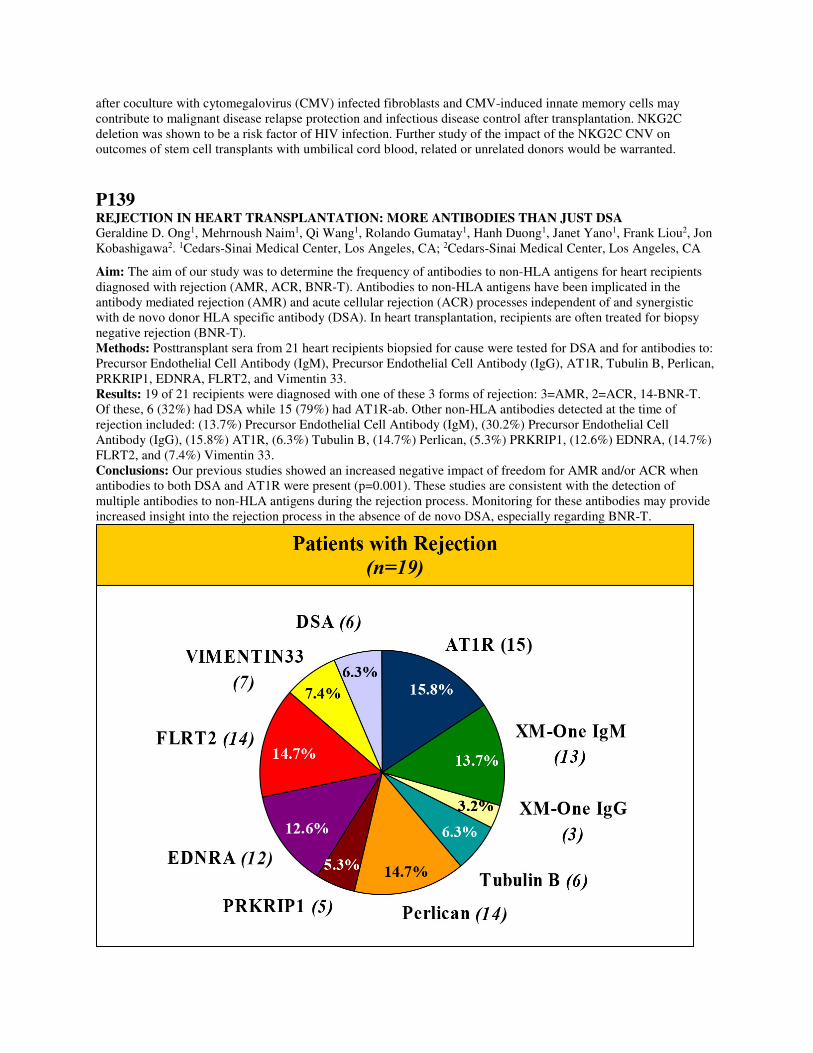
after coculture with cytomegalovirus (CMV) infected fibroblasts and CMV-induced innate memory cells may contribute to malignant disease relapse protection and infectious disease control after transplantation. NKG2C deletion was shown to be a risk factor of HIV infection. Further study of the impact of the NKG2C CNV on outcomes of stem cell transplants with umbilical cord blood, related or unrelated donors would be warranted.
P139 REJECTION IN HEART TRANSPLANTATION: MORE ANTIBODIES THAN JUST DSA Geraldine D. Ong1, Mehrnoush Naim1, Qi Wang1, Rolando Gumatay1, Hanh Duong1, Janet Yano1, Frank Liou2, Jon Kobashigawa2. 1Cedars-Sinai Medical Center, Los Angeles, CA; 2Cedars-Sinai Medical Center, Los Angeles, CA
Aim: The aim of our study was to determine the frequency of antibodies to non-HLA antigens for heart recipients diagnosed with rejection (AMR, ACR, BNR-T). Antibodies to non-HLA antigens have been implicated in the antibody mediated rejection (AMR) and acute cellular rejection (ACR) processes independent of and synergistic with de novo donor HLA specific antibody (DSA). In heart transplantation, recipients are often treated for biopsy negative rejection (BNR-T). Methods: Posttransplant sera from 21 heart recipients biopsied for cause were tested for DSA and for antibodies to: Precursor Endothelial Cell Antibody (IgM), Precursor Endothelial Cell Antibody (IgG), AT1R, Tubulin B, Perlican, PRKRIP1, EDNRA, FLRT2, and Vimentin 33. Results: 19 of 21 recipients were diagnosed with one of these 3 forms of rejection: 3=AMR, 2=ACR, 14-BNR-T. Of these, 6 (32%) had DSA while 15 (79%) had AT1R-ab. Other non-HLA antibodies detected at the time of rejection included: (13.7%) Precursor Endothelial Cell Antibody (IgM), (30.2%) Precursor Endothelial Cell Antibody (IgG), (15.8%) AT1R, (6.3%) Tubulin B, (14.7%) Perlican, (5.3%) PRKRIP1, (12.6%) EDNRA, (14.7%) FLRT2, and (7.4%) Vimentin 33. Conclusions: Our previous studies showed an increased negative impact of freedom for AMR and/or ACR when antibodies to both DSA and AT1R were present (p=0.001). These studies are consistent with the detection of multiple antibodies to non-HLA antigens during the rejection process. Monitoring for these antibodies may provide increased insight into the rejection process in the absence of de novo DSA, especially regarding BNR-T.

P140 MULTIPLE HIGH MFI CLASS I DSA CAN CAUSE ACUTE AMR OF KIDNEY ALLOGRAFT IN SIMULTANEOUS LIVER AND KIDNEY TRANSPLANT RECIPIENTS: OUTCOME ANALYSIS FROM A SINGLE-CENTER Raja Rajalingam1, Sindhu Chandran2, Kelly Cunniffe1, Sandy Feng3. 1Immunogenetics and Transplantation Laboratory, University of California San Francisco, San Francisco, CA; 2Division of Nephrology, Kidney Transplant Service, University of California San Francisco, San Francisco, CA; 3Kidney and Liver Transplant Service, University of California San Francisco, San Francisco, CA
Aim: Crossmatch (xM) positive (+ve) simultaneous liver and kidney transplant (SLKT) recipients remain at risk for antibody-mediated rejection (AMR) of the kidney allograft. To investigate the risk associated with donor-specific HLA antibodies (DSA), we studied 23 consecutive patients who received a SLKT at UCSF since 12/2013. Methods: Donors and recipients were HLA-typed using Luminex rSSO. Recipient sera collected pre-tx, 1 wk and 3 mo post-tx were analyzed for HLA antibodies using Luminex single antigen beads. CPRA was determined using 2000 MFI cutoff. Immunosuppression was steroids, tacrolimus, and mycophenolate without induction. AMR was diagnosed by kidney biopsy. Results: Among 23 patients (pts), 17 (74%) had CPRA>1%; 6 (26%) had CPRA>85%. Eight pts had pre-tx DSA: 6 with class I only; 1 had class II only, and 1 had both class I+II. Four pts had >2 DSAs. Most DSAs (and CREG) were lost by 1 wk post-tx but non-DSAs persisted >3 mos (Figure). Two pts with 0% CPRA developed a de novo DSA at 3 mos: 1 had B44 (MFI 5589), other had DQA05 (MFI 2303). Two pts developed early kidney AMR. Pt 1 (CPRA100%) with 5 high MFI (>20K MFI) class I DSAs (A1, A24, B7, B37, Cw7) and +ve CDC & +ve Flow T&B cell xMs suffered primary non-function. 3 DSAs declined significantly by 1 wk, but 2 DSAs persisted with MFI>8000 even 50 days post-tx. Case 2 (CPRA 0%; previous liver tx recipient) received SLKT from a well-matched (1C, 2DR, 2DQ, 1DP; no repeat mismatch) donor with +ve CDC and weakly +ve Flow T&B cell xMs. Allo-xM with 3rd party donors and auto-xM were CDC +ve but Flow -ve. All CDC xMs were -ve with heat-inactivated complement. xM with a 3rd party endothelial cell-line was weakly +ve. Renal allograft function recovered with AMR treatment. Conclusion: Reduction of circulating HLA antibody titers after SLKT appears to be donor HLA (and CREG) specific. Kidney AMR occurred with multiple high MFI Class I DSAs or complement-fixing non-HLA IgG antibodies. Longitudinal analyses show the specificity, kinetics, and stability of HLA antibody absorption by liver allografts.

P141 RAPID AND QUALITY CONTROLLED SCREENING ASSAY FOR THE HLA-B57 ANTIGEN BY FLOW CYTOMETRY Tricia L. Crawford1, Eric T. Weimer2, John L. Schmitz2. 1UNC HealthCare, Chapel Hill, NC; 2The University of North Carolina at Chapel Hill, Chapel Hill, NC
Aim: The presence of the genotype HLA-B*57:01 is highly predictive of Abacavir hypersensitivity. The need to identify the presence of this antigen in the HIV population is pertinent to treatment. Traditional molecular methods of HLA-B*57:01 with high specificity come often with long turn-around times and high costs. Our laboratory developed a flow cytometric screening method to identify the HLA-B*57 antigen for an expedited negative result. Methods: 59 random patients were tested for the HLA-B*57 antigen utilizing a two-step flow cytometry staining protocol. Whole blood was incubated with CD3-APC and a biotinylated B17 monoclonal antibody. The monoclonal HLA-B*17 antibody detects the presence of the HLA- B*57 or B*58 antigens on the cell surface. To enable detection of the B57 antibody, streptavidin-PE was added. An internal positive/negative control was developed to confirm assay performance. 10µL of Flow PRA 1 screening beads (One Lambda) were added to the patient sample. Based on SSC and FSC, beads are easily distinguishable from patient cells. All samples were confirmed by molecular HLA typing using real time PCR or SSOP. Results: Of the 59 patient samples, 12 were considered positive with a Mean MFI of 8622 ± 3962. 2 of the 12 positive screens were positive for HLA-B*57:01 by molecular typing. Based on the data, a cutoff was set with a mean MFI of 500. None of the negative screens had either HLA-B*57 or B*58 present by molecular typing. No cross-reactivity was observed from the 5 CREG group. Thus there is a lower likelihood of detecting antigens outside of HLA-B*57 and HLA-B*58. Conclusions: The screening of patients for the presence of HLA-B*57:01 is important prior to initiation of Abacavir therapy. An estimated 0.1-2.5% of the US population is positive for HLA-B*57:01, so the majority of patients will be negative. This rapid flow cytometry assay allows for faster turn-around time for B*57:01 negative patients. The

introduction of an internal positive control into the assay provides quality control that previous HLA-B*57 screening assays failed to provide.
P142 ARE IGM DONOR-SPECIFIC HLA ANTIBODIES (DSA) CLINICALLY RELEVANT? Paul Warner, Idoia Gimferrer, Danny Youngs, Erin Doran, Aaron Ing, Kate Arsenault, Karen Nelson. Bloodworks Northwest, Seattle, WA
Aim: The clinical relevance of IgM alloantibodies (Ab) directed against donor HLA (IgM DSA) in solid organ transplant recipients is not clear. To study this issue, we conducted a retrospective study utilizing flow cytometric crossmatching and solid-phase antibody testing. Methods: From 1998-2006 we incorporated an anti-IgM reporter antibody into our flow cytometric crossmatch (FCXM) assay. The frozen sera from the patients that had a clearly positive IgM crossmatch (≥ 40 mean channel shift or MCS, 256 scale) and were transplanted (negative IgG crossmatches) were tested for IgM HLA reactivity by a solid-phase screening assay. The sera that were positive on the screening assay were then tested for IgM HLA specificity by single-antigen beads. If IgM antibodies specific for HLA were identified, they were then compared to the HLA type of the transplant donors. Graft/patient outcome was obtained from UNet. Results: In 10,150 IgM FCXMs, 2,877 resulted in transplant. The IgM FCXM was positive (MCS≥15) in 158 (5.5%) of the cases. In 59 patients with IgM FCXM MCS ≥40, 29 were positive for IgM HLA Abs in the screening assay. IgM DSA were detected in 18 recipients (3 deceased donor renal, 6 living donor renal, 1 renal/pancreas, 1 pancreas, 3 renal/liver, 1 liver, and 3 lung). The antibodies ranged from weak (1,000 MFI) to strong (10,000 MFI). Class I IgM DSA were detected in 15 recipients, Class II IgM DSA were found in one recipient, and 2 recipients had both. There was one case of a lung transplant recipient who died 12 days post-transplant, and the cause of graft loss was listed as acute rejection in UNet. Serum samples obtained at 3 and 11 days post-transplant were negative for IgG DSA, and the IgM DSA were similar to those detected in the pre-transplant serum. All other patients were either lost to follow up (N = 5, range = 1-6 yr), had a functioning graft at last report (N = 8, range = 7 - 14 yr), died with a functioning graft (N = 3, range = 1 mo - 9 yr), and one patient had graft failure due to drug toxicity at 5 yr. Conclusions: IgM Abs specific for HLA are relatively uncommon, compared to IgG (data not shown). In the relatively small number of patients that were identified as having IgM DSA, except for a single recipient where we could not exclude the effect, there was no evidence that IgM DSA were detrimental to graft function/survival.
P143 CLINICAL VALIDATION OF NEXT GENERATION SEQUENCING FOR HLA TYPING USING TRUSIGHT HLA Maureen C. Montgomery1, Eric T. Weimer2. 1UNC HealthCare, Chapel Hill, NC; 2The University of North Carolina at Chapel Hill, Chapel Hill, NC
Aim: Sanger sequencing is considered the gold standard for high-resolution HLA typing. The complexity of the HLA region leads to an inability to phase heterozygous positions thus additional testing is required to achieve allele level typing. With next generation sequencing (NGS), HLA typing should yield high-resolution typing with limited need for additional testing. Thus lowering laboratory associated costs and potentially reducing turnaround times. Methods: 59 unique samples (1383 total HLA loci) previously high-resolution HLA typed were typed via NGS (HLA-A, -B, -C, -DPA, -DPB, -DQA, -DQB, -DRB1/3/4/5). 7 samples were run for inter- and intra-assay precision.Illumina TruSight HLA typing kits were used to perform the NGS testing. 24 samples were amplified per run using long-range PCR. Two technologists were utilized for library preparation. Protocols were adapted from the Illumina guidelines. Sequencing was performed using 2x250bp paired end reads on the MiSeq system with a 1% spike-in of PhiX as quality control. Results: 41 (2.9%) of the 1383 HLA loci had ambiguities, all within DRB1. HLA typing using TruSight HLA caused a 94.5% reduction in the number of ambiguities compared to Sanger sequencing. 2 (0.1%) of the 1383 loci were mismatched from the known Sanger sequence. Unfiltered, the overall accuracy was 99.9% and precision was 100%.

Conclusions:Achieving high-resolution HLA typing on 24 patients at 11 HLA loci in a single run without substantial additional testing significantly cuts the laboratory associated costs. The DRB1 ambiguities resulted from the inability to exclude rare HLA alleles due to primer binding sites within exon 2. The 2 mismatches involved 1 DRB amplicon that appears to have been contaminated. Using those mismatches, an average Q30 score of >81% (88% for DRB3/4/5) and depth of coverage ≥100 was determined necessary for HLA typing assignment. Application of these criteria would have flagged the mismatched samples for repeat testing which resulted in 100% accuracy.
P144 A NEW HLA-C*02 NULL ALLELE DESCRIBED IN QUILOMBO REMNANTS SAMPLES FROM SOUTHERN BRAZIL Kelly Nunes1,2, Margareth A. Torres2, Bruno Z. Piovezan2, Gerlandia N. Pontes2, Lilian Kimura1, Juliana E.p Carnavalli1, Regina C. Mingroni-Netto1, Diogo Meyer1, Maria E. Moraes2. 1Institute of Biosciences, Sao Paulo, Brazil; 2JRM-Investigacoes Imunologicas, Rio de Janeiro, Brazil
The present study describes a new HLA-C null allele in African-derived Quilombo communities from the Ribeira River Valley in the state of São Paulo, Brazil. Quilombo are small communities founded by runway, abandoned and freed African-descendent slaves, in the late seventeen century. In the São Paulo state, Quilombo remnants are confined to a small area, on the southern border along the Ribeira River Valley. In the present study, 146 individuals from 12 Quilombo communities from the Ribeira River Valley (Abobral, André Lopes, Galvão, São Pedro, Pedro Cubas, Pilões, Nhunguara, Sapatu, Ivaporanduva, Maria Rosa, Poças and Reginaldo) were typed for HLA-A, -B, -C and DRB1 by PCR-SBT (Thermo Fisher). We identified a new HLA-C*02 null in three individuals: an 84 yo female (sample 20), a 38 yo female (sample 75) and an 18 yo male (sample 137), from Galvão, São Pedro and Pedro Cubas communities respectively. This new allele presented an insertion of seven nucleotides (TCGCCGT) in exon 2 at codon 50, leading to a frame shift and generating a subsequent stop at codon 76. In all the three cases the HLA-C*02 null allele was associated with the following haplotype: A*80:01~C*02N~B*18:01:01G~DRB1*07:01. To further characterize this allele and verify if it was exclusively found in this Quilombo communities, we performed a parallel study, typing the HLA-C locus by PCR-SBT in six unrelated urban Brazilian Afro-descendants individuals from São Paulo state. They were selected based on the HLA-A, -B, -C, -DRB1 haplotype and were previously typed as HLA-A*80, B*18, C*02, DRB1*07 (Intermediate resolution, PCR-SSO). The new HLA-C*02null was not observed in any of them. The kinship coefficient, estimated by Moment method based on 600K genomewide SNPs, between the 3 individuals are: θ Samples 20-75 = 0.0271, θ Samples 20-137 = 0.0278 and θ Samples 75-137 = 0.0170. This result shows that the individuals have some kinship degree and suggests that the shared null allele/haplotype is derived from a unique common ancestor. This new allele was submitted to the GenBank (accession numbers: KR698080, KR698081 and KR698082 for samples 20, 75 and 137 respectively) and IMGT for official nomenclature.
P145 UNEXPLAINABLE STRONG FLOW CYTOMETRIC AND CDC B CELL POSITIVE CROSSMATCHES ATTRIBUTED TO THE “PROZONE PHENOMENON” OF A DSA TO DQA1*05:05 Ina Kurbegovic-Skaljic, Maria Oppermann, Michele Higgins Prod, Sylvia Piggott, Erin Christiansen, Nadya Parmakova, Siva Kanangat. Rush University Medical Center, Chicago, IL

Aim: To resolve Positive Allo Flow cytometric and CDC B cell crossmatches in the absence of any detectable strong DSAs Patient: A 53 Year Old male who had received a kidney from a Sibling had Graft Failure. Ten years later requiring a second transplant. The Patient was Negative for HLA-Class I antibodies, with high Class II PRA with several DR, DQB and DQA antibodies consistently with fluctuating MFI. Materials and Methods: The recipient and donors were typed for all HLA Loci A, B, C, DRB1, DQA1, DQB1, DRB3/4/5, DPA1 and DPB1 using r-PCR-SSOP. The antibody assays were carried out by solid phase assays on Luminex platform. Results: T cell flow crossmatches were Negative, B cells showed a median channel shift (MCS) of 350 in undiluted and 405 in 1:4 diluted serum samples (cut-off for B cell positivity >78). The CDC B cell crossmatch also was strongly positive. Only DSA was DQA1 *05:05 with an MFI of 2200 which persisted with fluctuating MFI. Phenotypic bead analysis showed high Class II PRA with possible DR 11 and DR52/53 DSAs along with potential antibodies towards DQA1*05; DQA1*03 --- and the donor was a DQA*05:05. Phenotypic beads analysis indicated a possible skewed DSA towards DQA*05:05 Single antigen bead assay on diluted serum samples indicated strong prozone as shown by the MFI of DQA1* 05:05 antibody in undiluted, 1:5 and 1:10 diluted sera 2172; 9226; and17,641 respectively. Conclusions: The observed high MCS Flow B cell and strong CDC B Cell cross matches were due to high levels of DQA1*05:05 DSA which was not appreciated for its full strength in the initial SAB assay in Undiluted serum due to the prozone phenomenon of high antibody concentration. The possibility of “Prozone Phenomenon” should be considered if strong cross match reactivity is observed when undiluted serum showed “Low levels or No DSA”in highly sensitized patients.
P146 DEFINING THE NON-SENSITIZED PATIENT: A COMPARISON OF HLA ANTIBODY SCREENING AND IDENTIFICATION METHODS Carley Shaut, Brent Gardner, Douglas Norman. Oregon Health & Science University, Portland, OR
Aim: Luminex Single Antigen beads (SAB) are widely used to identify HLA specificities and assign avoid antigens for kidney transplantation. However in our centers, “non-sensitized” waitlist patients are monitored using less sensitive screening kits. To confirm the lack of HLA sensitization, we evaluated sera by SAB. In this study, we compared several antibody screening and identification kits to uncover test discrepancies and investigate possible false reactions. Methods: During initial evaluation and listing, patients were defined as “non-sensitized” when the Class I/II FlowPRA screening beads (One Lambda) were negative. Presumed non-sensitized waitlist patients are screened monthly by the Lifecodes Lifescreen kit (Immunocor) and screened yearly by FlowPRA screening beads. To ensure that no HLA antibodies were missed by this protocol, 116 patient samples were tested by One Lambda Class I/II Labscreen Single Antigen beads (1000 MFI cutoff). When discrepancies between the three assays were identified, sera were tested by the Immunocor ID assay and/or One Lambda Labscreen PRA beads. Additional analyses by surrogate crossmatches are ongoing. Results: Of the 116 samples tested from non-sensitized patients, 22% were positive by the Labscreen Class I SAB and 33% were positive by the Labscreen Class II SAB but negative by the screening kits. The positive specificities spanned all HLA loci, but there were some beads that were positive more than expected. Most notably, the DRB1*04:04 bead was positive in 10% of cases (12/116) with a range of 1080-2524 MFI, and in three cases the patient’s HLA type was DR4. Other specificities observed three or more times included: A80, B45, B76, Cw15, DR18, DP1, DP11, bead DQA1*01:04/DQB1*06:04. In four cases, patients presented with new UNet unacceptable antigens (≥3000 MFI), and in three cases this led to a positive cPRA (4-17%). Conclusions:Unexpected positive specificities were detected by the Labscreen SAB assay in otherwise non-sensitized kidney waitlist patients. Our data are consistent with other studies that compared multiple test assays to interrogate false reactions. Unexpected SAB reactions should be investigated with additional screening assays and/or surrogate crossmatches. In some cases, common false positive beads can be ignored during test interpretation.
P147

ASSESSING THE CORRELATION BETWEEN C3D-BINDING DE NOVO DONOR SPECIFIC HLA ANTIBODIES AND REJECTION IN KIDNEY TRANSPLANT RECIPIENTS Matthew Cusick, Kelley Haarberg, John Friedewald, Anat R. Tambur. Northwestern University, Chicago, IL
Aim: Solid phase assays have the ability to detect even low levels of HLA antibodies, but potentially lack the ability to distinguish between clinically relevant or irrelevant DSA. Here we test the clinical significance of a novel C3d binding assay. Methods: Serum samples from 85 kidney transplant recipients were retrospectively analyzed using the Immucor© Single Antigen immunoassays, to detect either IgG or C3d binding HLA antibodies. Inclusion criteria included patients that developed de novo DSA by Luminex One Lambda (OL) single antigen assay and had a biopsy obtained at the same time point as the serum sample. Median fluorescence intensity (MFI) value of 1000 was arbitrarily used to assign positive responses. Results: Of the 85 patients: 21 had AMR, 12 cell-mediated rejection (CMR), 11 mixed rejection (AMR + CMR), 4 with pathologic finding other than rejection, such as BK virus, and 41 had normal biopsies. C3d DSA were detected in 16/85 patients with the majority having AMR in 11/16 patients (69%) compared to 1/16 patients (6%) with CMR, 2/16 patients (12.5%) with a mixed rejection, and 2/16 patients (12.5%) with negative biopsies. These 2 negative biopsy patients had strong MFI values for Immucor IgG DSA and strong titers on the OL platform; moreover, both of these patients went on to have AMR later on. Immucor IgG assay detected 29/85 patients with DSA, in which, the majority of C3d positive DSA (16/16) being Immucor IgG class II DSA-positive. Immucor IgG positive DSA with negative C3d results had lower MFI (Fig. 1). All the C3d positive assays corresponded to moderate to high titer OL results (titers ≥ 1:64), although three samples had class II specificities with titers ≥1:128 were not C3d positive. Conclusion: C3d-binding antibodies seem to be associated with stronger antibodies with at least moderate level titer and rejection. Further studies are required to substantiate our results.
P148 IMPACT OF THE NEW KIDNEY ALLOCATION SYSTEM (KAS) FOR CPRA >98% RECIPIENTS ON TRANSPLANT LABORATORY OPERATIONS - AN OPO PERSPECTIVE Prakash Rao, Donna King, Misty Marchioni, Bridget Figueiredo, Patricia Harris, Carolyn Welsh, Sharyn Sawczak, David O'Hara. NJ Sharing Network, New Providence, NJ
Aim: The new Kidney Allocation System (KAS) implemented on December 4, 2014 was developed to improve the discard rates of kidneys, give patients with extensive wait time and high Panel Reactive Antibody (PRA) access to transplantation and improve re-transplant rates. Under the new system, highly sensitized patients are given priority

by allowing additional points for candidates with Calculated Panel Reactive Antibody (CPRA) >98% thus enabling them to receive regional and national priority kidney offers. Methods: The NJ Sharing Network (NJSN) is an organ procurement organization equipped with a transplant laboratory. This study evaluated the impact of the new KAS on NJSN’s transplant laboratory operations by measuring the number of (1) deceased donor (DD) crossmatches, (2) recipients transplanted, (3) blood samples shipped after the new KAS was implemented, and (4) kidneys discarded, and compared them to the same parameters prior to the KAS implementation. Results: More crossmatches were performed on import DD than local DD. The percentage of imported DD that were crossmatched with recipients was 58% between December 4, 2014 and April 2, 2015 as compared to 22% during the same timeframe the prior year. The number of virtual crossmatches performed increased significantly after implementation of the new KAS. There were 21 patients with CPRAs over 98% who received a kidney transplant between December 19, 2014 and May 1, 2015. Of the 21 transplanted organs, only 4 were from local DD, the remaining were import donors. There were 0 transplants from patients with CPRAs over 98% from the same timeframe the prior year. There were 22 DD kidney blood samples shipped to other transplant laboratories during the first quarter of 2015 as compared to 4 blood samples shipped during the same timeframe in 2014 resulting in a 5.5 fold increase. The 2015 year-to-date kidney discard rate is 24% as compared to 17% in 2014, and 10% in 2013. Conclusions: We observed (1) an increase of imported DD being crossmatched, (2) patients with CPRAs over 98% receiving transplants, (3) an increase in the number of blood samples being shipped to other transplant laboratories and (4) an increase in the number of kidneys being discarded.
P149 DONOR-SPECIFIC ANTIBODIES IN KIDNEY DECEASED DONOR TRANSPLANTATION Eric K. Ho1, Xiuwei Tang1, Li Lingzhi1, Lloyd E. Ratner2, George Vlad1, Rodica E. Vasilescu1. 1Columbia University, New York, NY; 2Columbia University, New York, NY
Aim: Pre-sensitization against a broad array of HLA is associated with prolonged waiting times and inferior kidney allograft survival. Although the use of solid phase assay (SPA) for the detection and characterization of anti-HLA antibodies provides greater sensitivity than complement-dependent lymphocytotoxicity (CDC) assay, it often detects donor specific antibodies (DSA) which turn out to be clinically irrelevant. In this study, our aim is to determine whether kidney deceased donor recipients with preformed DSA detectable in serum obtained prior to transplant are at an increased risk of graft failure. Methods: We studied 640 patients who received a kidney transplant from a deceased donor between January 1, 2009 and December 31, 2014 at Columbia University Medical Center. Sera collected from the recipients prior to transplantation were tested for DSA by CDC and SPA with single antigen coated beads. All patients were transplanted with negative CDC crossmatch. Results: We analyzed the actuarial graft survival in patients with a primary allograft (n=525) and in patients with a secondary allograft (n=115). Kidney allograft survival was significantly higher in primary allograft recipients (85%) than in secondary allograft recipients (71%) at 6 years. (P=0.02) Forty-six out of 525 (8.7%) primary kidney allograft recipients and sixty-two out of 115 (53.9%) secondary kidney allograft recipients had SPA-detectable DSA in sera collected prior to transplantation. Six years following transplantation of primary kidney allografts the actuarial graft survival was 83% and 85%, respectively, regardless of whether DSA were detectable in recipients’ pre-transplantation sera. (P=0.39) In recipients of secondary kidney allografts with and without SPA-detectable DSA, the actuarial graft survival was 67% and 77% at 6 years, respectively. (P=0.12) Conclusions: The presence of SPA-detectable DSA in recipients of kidney allografts from deceased donors is associated with lower long term (6 year) actuarial graft survival although the difference does not reach statistical significance.
P150 PREDICTING DONOR SPECIFIC CROSSMATCH OUTCOME USING AN IN-HOUSE GENERATED VIRTUAL CROSSMATCH ALGORITHM Prakash Rao, Cecil Rhodes, Donna King, Dayanand Deo, Patricia Harris, Julien Napoleon, Ijeoma Okere. NJ Sharing Network, New Providence, NJ
Aim: The new Kidney Allocation Scheme (KAS) has increased requests for virtual crossmatches (VXM) to predict the possibility of a negative donor specific cross-match. This study evaluates the efficacy of an in-house generated algorithm to predict and quantify the allogeneic cross-match. Method:A retrospective review was conducted on 50 Flow Cytometry Crossmatch (FLXM) results that were collected from the database reflecting several different median channel shift (MCS) values. The corresponding HLA Class I and Class II antibody screening was performed using FlowPRA screen beads (One Lambda). Luminex Class I and Class II single antigen bead (SAB) testing and analyses including for mean fluorescent intensity (MFI) was performed. A calibration curve for predicting the VXM was developed by comparing the MFI values for the donor specific antibodies (DSAs) obtained from the Luminex SAB results to the corresponding MCS values from the FLXM results. A regression was performed on the MCS versus MFI values and the slope was calculated. Results: We reviewed 246 FLXM results that were positive for T-cell and/or B-cell, performed between August 2008 and February 2013. We substituted the highest MFI values from the database for either the HLA class I or class II specificity in the VXM algorithm to obtain the predicted MCS values. We observed that our in-house generated VXM algorithm had an overall positive predictive value of 91% for the T-cell and B-cell crossmatches. MFI values above 1200 predicted a positive cross-match for HLA class I specificities while MFI values above 3000 predicted a positive crossmatch for HLA class II specificities. There were about 9% crossmatches that were predicted as negative using our VXM algorithm that were positive by FLXM evaluation. This could be due to Prozone like phenomenon, antibodies dependent on the conformational characteristics of the antigen, antibody avidity and affinity, or allele specificities not on the Luminex SAB panel. Conclusion: The VXM algorithm shows a high probability of accurately predicting FLXM results, and may mimic the sensitivity of the final crossmatch reducing the time and cost for deceased donor work-up. This could allow for quicker organ allocation for sensitized patients.
P151 CHAINS OVER CYCLES ARE SELECTED BY COMPUTER SOFTWARE OPTIMIZATION IN THE KIDNEY PAIRED DONATION PROGRAM Stanislaw Stepkowski1, David Fumo2, Connie Smith3, Annette Blair3, Michael Rees2, Michael Rees2. 1University of Toledo, Toledo, OH; 2University of Toledo, Toledo, OH; 3University of Toledo, Toledo, OH
Aim: Failure to convert computer-identified possible exchanges into transplants limits Kidney Paired Donation (KPD) program. We have analyzed the efficiency of simultaneous closed loop exchanges (cycles) vs. non-simultaneous extended altruistic donor (NEAD) chains in generating KPD transplants. Methods/ Results: The Alliance for Paired Donation (APD) utilizes an optimization algorithm selecting cycles or chains. All chains are initiated by a non-directed donor (NDD) and end with a bridge donor, who enters back into the pool. This bridge donor can then initiate another NEAD chain. Computer-generated potential cycles and NEAD chains are presented to transplant centers for review as formal offers. We tracked the progress in moving from offers to completed transplants to calculate the success rate. Between 2007 and 2014 the APD performed 225 transplants: 194 within the APD and 31 in collaboration with other KPD programs. Of the 194 APD transplants, 61 (31.4%) were performed through cycles (Figure 1a) and 133 through NEAD chains (68.6%; Figure 1b), showing that chains were two times more successful than cycles. The APD made 447 offers to transplant centers, of which 173 (38.7%) were cycles and 274 (61.3%) were chains. Consequently, the success rate of cycles was 14.5%, chains was 26.6%, while the overall success rate of 21.9%. Conclusions: Although chains were not actively sought over cycles, the results showed that chains were preferentially found by the optimization algorithm and more successfully progressed to completed transplants than cycles. Thus, the concept of a bridge-donor introduces significant flexibility in generating transplants. We propose that NEAD chains are the most effective tool to increase the efficiency in KPD programs.
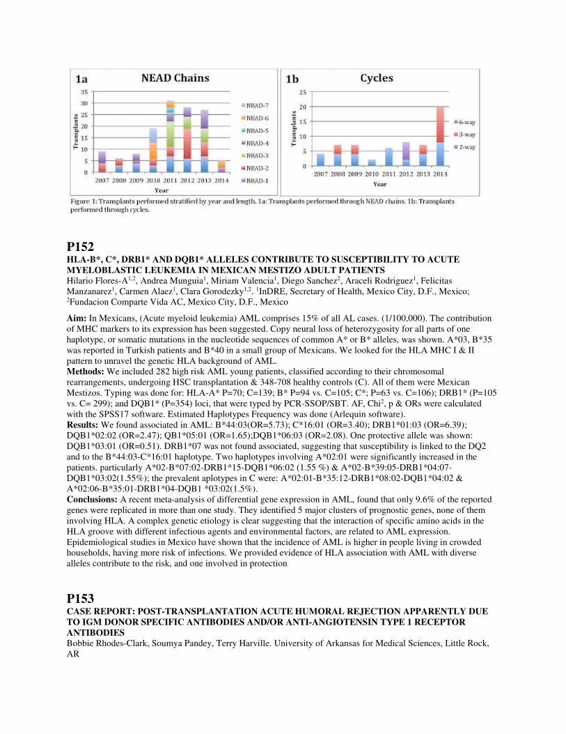
P152 HLA-B*, C*, DRB1* AND DQB1* ALLELES CONTRIBUTE TO SUSCEPTIBILITY TO ACUTE MYELOBLASTIC LEUKEMIA IN MEXICAN MESTIZO ADULT PATIENTS Hilario Flores-A1,2, Andrea Munguia1, Miriam Valencia1, Diego Sanchez2, Araceli Rodriguez1, Felicitas Manzanarez1, Carmen Alaez1, Clara Gorodezky1,2. 1InDRE, Secretary of Health, Mexico City, D.F., Mexico; 2Fundacion Comparte Vida AC, Mexico City, D.F., Mexico
Aim: In Mexicans, (Acute myeloid leukemia) AML comprises 15% of all AL cases. (1/100,000). The contribution of MHC markers to its expression has been suggested. Copy neural loss of heterozygosity for all parts of one haplotype, or somatic mutations in the nucleotide sequences of common A* or B* alleles, was shown. A*03, B*35 was reported in Turkish patients and B*40 in a small group of Mexicans. We looked for the HLA MHC I & II pattern to unravel the genetic HLA background of AML. Methods: We included 282 high risk AML young patients, classified according to their chromosomal rearrangements, undergoing HSC transplantation & 348-708 healthy controls (C). All of them were Mexican Mestizos. Typing was done for: HLA-A* P=70; C=139; B* P=94 vs. C=105; C*; P=63 vs. C=106); DRB1* (P=105 vs. C= 299); and DQB1* (P=354) loci, that were typed by PCR-SSOP/SBT. AF, Chi2, p & ORs were calculated with the SPSS17 software. Estimated Haplotypes Frequency was done (Arlequin software). Results: We found associated in AML: B*44:03(OR=5.73); C*16:01 (OR=3.40); DRB1*01:03 (OR=6.39); DQB1*02:02 (OR=2.47); QB1*05:01 (OR=1.65);DQB1*06:03 (OR=2.08). One protective allele was shown: DQB1*03:01 (OR=0.51). DRB1*07 was not found associated, suggesting that susceptibility is linked to the DQ2 and to the B*44:03-C*16:01 haplotype. Two haplotypes involving A*02:01 were significantly increased in the patients. particularly A*02-B*07:02-DRB1*15-DQB1*06:02 (1.55 %) & A*02-B*39:05-DRB1*04:07-DQB1*03:02(1.55%); the prevalent aplotypes in C were: A*02:01-B*35:12-DRB1*08:02-DQB1*04:02 & A*02:06-B*35:01-DRB1*04-DQB1 *03:02(1.5%). Conclusions: A recent meta-analysis of differential gene expression in AML, found that only 9.6% of the reported genes were replicated in more than one study. They identified 5 major clusters of prognostic genes, none of them involving HLA. A complex genetic etiology is clear suggesting that the interaction of specific amino acids in the HLA groove with different infectious agents and environmental factors, are related to AML expression. Epidemiological studies in Mexico have shown that the incidence of AML is higher in people living in crowded households, having more risk of infections. We provided evidence of HLA association with AML with diverse alleles contribute to the risk, and one involved in protection
P153 CASE REPORT: POST-TRANSPLANTATION ACUTE HUMORAL REJECTION APPARENTLY DUE TO IGM DONOR SPECIFIC ANTIBODIES AND/OR ANTI-ANGIOTENSIN TYPE 1 RECEPTOR ANTIBODIES Bobbie Rhodes-Clark, Soumya Pandey, Terry Harville. University of Arkansas for Medical Sciences, Little Rock, AR

Patient: 46 yo (gravida 6) BF, IgA nephropathy. Pre-Transplant LABScreen SA beads (SAB): anti-B57 (MFI=11,125), B58 (9163), DP1 (1871). Class I and Class II DSA not detected. See Table 1. Virtual-XM predicted negative; Deceased Donor Pre-Transplant Flow- and CDC-XM were negative. Renal function was poor following transplant. By day 3 post-transplant clinical findings were consistent with AMR. Day 4 renal biopsy revealed patchy C4d+ in peritubular capillaries consistent with AMR. Nephrectomy was performed on post-transplant day 7. Additional assays were performed on pre- and 3 wk post-transplant sera to evaluate for reasons for AMR, including: One Lambda LABScreen SAB Assay for C1q; and modified for C1q+AHG, detection of IgM, and use of non-heat inactivated serum for assays; and One Lambda MICA, Anti-AT1R, and Anti-ETAR antibody assays. See Tables. Results: Pre-transplant IgG DSA were negligible. IgM DSA were elevated. Anti-AT1R antibody was elevated. Interpretation: Immediate Post-Transplant Humoral Rejection correlates with presence of IgM DSA and anti-AT1R (IgG) antibodies. Discussion: IgM anti-HLA antibodies are not considered to be relevant for AMR. Anti-AT1R is reported as a cause of acute AMR, but not typically in the immediate post-transplant period. We believe this case argues that IgM DSA appear be relevant, but cannot exclude that either anti-AT1R IgG antibodies alone, or in combination with IgM DSA, may result in AMR. Conclusions: IgM DSA and/or anti-AT1R may result in post-transplant humoral rejection, and therefore should be considered in evaluation for episodes of AMR, especially when IgG DSA are negligible.
Table 1: DSA MFI
Pre-Transplant A1 A2 B7 B8 Cw7 DR103 DR17 DQ2 DQ5
IgG 0 0 0 0 0 0 0 0 0
C 0 63 8 29 5 367 134 99 7
C1q+AHG 336 617 113 0 0 0 0 0 0
IgM 341 8491 3559 4469 3136 190 135 214 35
C1q (No HI) 0 11 0 13 4 12 13 30 4
C1q+AHG (No HI) 83 11,830 1880 1541 735 366 0 151 0
3 wk Post-Transplant A1 A2 B7 B8 Cw7 DR103 DR17 DQ2 DQ5
IgG 0 3552 0 1973 0 1742 0 0 0
C1q 9 169 25 50 12 22 51 11 8
C1q+AHG 0 370 0 0 0 16 0 0 0
IgM 649 4997 3439 1879 1331 -- -- -- --
C1q (No HI) 28 275 41 96 39 61 45 34 41
C1q+AHG (No HI) 0 14,837 4106 1509 0 3898 0 190 0
HI = Heat Inactivation
Table 2: Non Anti-HLA Antibody Assays
Pre-Transplant 3 wk Post-Transplant Normal
AT1R (Angiotensin Type 1 Receptor) 27 U/mL 13 U/mL <10 U/mL
ETAR (Endothelin-1 Type A Receptor) 10 U/mL 10 U/mL <10 U/mL
MICA Neg Neg Neg
T. Harville: Scientific/Medical Advisor; Company/Organization; Baxter Healthcare, CSL Behring. 7. Other
(Identify); Company/Organization; Medical Advisory Board for Arkansas Regional Organ Recovery Agency.
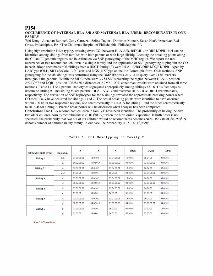
P154 OCCURRENCE OF PATERNAL HLA-A/B AND MATERNAL HLA-B/DRB1 RECOMBINANTS IN ONE FAMILY Wei Dong1, Jonathan Barone1, Carly Carozza1, Selina Taylor1, Dimitrios Monos2, Susan Hsu1. 1American Red Cross, Philadelphia, PA; 2The Children's Hospital of Philadelphia, Philadelphia, PA
Using high resolution HLA typing, crossing over (CO) between HLA-A/B, B/DRB1, or DRB1/DPB1 loci can be identified among siblings from families with both parents or with large sibship. Locating the breaking points along the C I and II genomic regions can be estimated via SNP genotyping of the MHC region. We report the rare occurrence of two recombinant children in a single family and the application of SNP genotyping to pinpoint the CO in each. Blood specimens of 6 siblings from a HSCT family (F) were HLA - A/B/C/DRB1/DQB1/DPB1 typed by LABType (OLI), SBT (SeCore, Life Tech) and NGS (NXType on the Ion Torrent platform, OLI) methods. SNP genotyping for the six siblings was performed using the OMNIExpress-24 v1.1 to query over 713K markers throughout the genome. Within the MHC there were 3,754 SNPs covering the region between HLA-A position 29913067 and DQB1 position 32628428 a distance of 2.7Mb. 100% concordant results were obtained from all three methods (Table 1). The 4 parental haplotypes segregated appropriately among siblings #3 - 6. This fact helps to determine sibling #1 and sibling #2 are paternal HLA - A & B and maternal HLA - B & DRB1 recombinants, respectively. The derivation of SNP haplotypes for the 6 siblings revealed the approximate breaking points where CO most likely have occurred for siblings 1 and 2. The actual breaking points were identified to have occurred within 700 bp in two respective regions, one centromerically to HLA-A for sibling 1 and the other centromerically to HLA-B for sibling 2. Precise break points will be discussed when analysis has been completed. Conclusion: Two HLA recombinant children in family F have been identified. The probability of having the first two older children born as recombinants is (0.01)2(0.99)4 when the birth order is specified. If birth order is not specified, the probability that two out of six children would be recombinants becomes N(N-1)/2 x (0.01) 2(0.99)4. N denotes number of children in any family. In our case, the probability is 15(0.01) 2(0.99)4.

P155 TOWARDS A NEW GOLD STANDARD - NGS CORRECTIONS TO SANGER SBT GENOTYPING RESULTS Efi Melista1, Krisztina Rigo1, Agnes Pasztor1, Mette Christiansen2, Francesca Eleonora Bertinetto3, Peter Meintjes1, Tim Hague1. 1Omixon, Budapest, Hungary; 2Aarhus University Hospital, Aarhus, Denmark; 3Torino Piemonte BMDR, Turin, Italy
Aim: HLA typing is a crucial step prior to transplantation, particularly for bone marrow transplants. Correct allele matching is essential in order to increase the success of transplantation outcomes and decrease the rate of GVHD, thus, obtaining and reporting accurate HLA types is essential. SBT has been considered the Gold Standard for high resolution HLA typing for many years, however, SBT is typically plagued by high levels of ambiguity frequently requiring reflexive testing. Whole-gene sequencing by NGS has the potential to provide fully characterized and fully phased HLA loci. In this study we compare the same 300 samples sequenced by NGS and SBT. Methods: 300 clinical samples from five labs were sequenced at five loci (HLA-A, B, C, DRB1 and DQB1) for a total of 3000 alleles using Holotype HLA on an Illumina MiSeq using the 2x250 bp chemistry. The resulting sequencing data was analyzed and the HLA allele calls were assigned using Omixon’s HLA Twin software. The reference typing results had been previously obtained by SBT or a combination of SBT/SSO/SSP. The NGS results from Holotype HLA were compared to the existing typings to determine concordance among the methods. Results: Six alleles had discordant HLA calls between the NGS result and the SBT ‘reference’ typing. Five of these alleles were discordant at the second field and one sample was discordant at the third field. Mistypings were observed in HLA-A, HLA-C, HLA-DQB1 and HLA-DRB1 among the six discordant samples. The errors were due to a variety of reasons that ranged from failure of SBT to detect a second allele in a sample to noisy SBT trace. Retyping these by SBT confirmed that the NGS result provided the correct typing. Conclusions: NGS is a suitable method for high resolution HLA genotyping. During this comparison we uncovered a number of incorrect SBT typings which NGS can reliably genotype without ambiguity, thus highlighting the importance of adopting this new technique in the clinic. In addition, we were able to fill in previously unknown regions of each allele by generating whole gene consensus sequences.
P156 MAPPING THE HLA FREQUENCIES OF DONOR REGISTRY IN QUEBEC TO DEFINE A BETTER RECRUITMENT STRATEGY Lucie Richard, Marie-Claire Chevrier. Hema-Quebec, Saint-Laurent, QC, Canada
Aim: Hema-Quebec enrolls between 3000 and 4000 new stem cell donors in its registry every year with the ultimate goal of finding a stem cell donor inside Quebec province for 30-40% of our patients. However, we are very far from this goal with less than 4% of our patients finding a donor from the Quebec registry. In response to this, we analysed our donor population with the intention of reorienting our donor recruitment strategy. We wanted to identify the regions which are under-represented in term of donor numbers and haplotypes, since our population has geographical particularities that should be taken into account for recruitment strategy. Method: The HLA-A, -B and -DRB1 molecular typing available for almost 13,000 stem cell registry donors from Quebec were analyzed, using postal codes, for their geographic location. Data from low resolution for HLA-A and -B and intermediate resolution for HLA-DRB1 were pre-processed and analyzed using GENE[RATE] and additional tools. Hardy-Weinberg equilibrium (HWE) was assessed using a nested likelihood procedure. Reynolds’ genetic distances were computed between regions and plotted by PCoA. SAMOVA analyses were performed to identify possible genetic boundaries across Quebec. In parallel, patients with a difficult search over the last few years were analyzed. Patient and donor distributions in the different regions were evaluated in order to identify the need for active recruitment. Results: Globally, the Hema-Quebec stem cell donor registry is mostly Caucasian, although the ethnicity of Montreal region, the biggest city of the province, is reaching more than 50%. In the other administrative regions, ethnicity is much lower. Estimation of the genetic distance between the different regions of Quebec shows that some regions like Saguenay and Gaspésie are clearly differentiated from the others based on their HLA content. These regions however, are much less represented in the registry as compared to Montreal, Laval and Quebec City regions. A recruitment strategy was design to overcome the lack of donors in particular regions. Conclusion: By targeting the different regions of Quebec, the recruitment of donors should increase the
representation of local haplotypes and improve the content of the Hema-Quebec stem cell donor registry in response to the need of our patients.
P157 HLA ANTIBODY EPITOPE AND CLONALITY ARE IMPORTANT DETERMINANTS OF CAPACITY TO FIX COMPLEMENT IN IN VITRO CLINICAL AND FUNCTIONAL ASSAYS Nicole M. Valenzuela1, Kimberly A. Thomas1, Arend Mulder2, Minas Zartarian1, Jason Song1, Elaine F. Reed1. 1UCLA, Los Angeles, CA; 2Leiden University Medical Center, Leiden, Netherlands
We tested human allele-specific HLA class I antibodies (Ab) alone and in combination for their capacity to activate complement (C’) in in vitro assays. Methods: Human HLA I Ab (clones SN607D8, SN230G6, MUL2C6, all IgG1) were diluted (0.01-0.5µg/mL) in human serum containing no HLA Ab, and tested in One Lambda LabScreen, C1qScreen, and Immucor LifeCodes Single Antigen and C3d Assays. C4d deposition on cells was measured by flow cytometry, and cytotoxicity was measure using standard rabbit (rb-CDC) and human C’ (hum-CDC) assays. Results: At 500ng/mL, a pan HLA I Ab recognized all 97 beads with a mean IgG-MFI of around 5000, while anti-A2/B17 IgG1 at the same concentration bound only 6 beads (mean IgG-MFI >20000 on those beads), and anti-A3/A11 bound 8 beads (mean IgG-MFI 17000). PRA beads displayed a similar phenomenon, pointing to a dilution effect of Ab with broad specificities. Anti-A2/B17 IgG1 bound to A2, B57 and B58 beads with uniform IgG-MFI and C1q-MFI >1000. Anti-A3/A11 bound A1, A3, A11, A24, A36 and A80, with highest IgG-MFI on A11 and lowest on A36 (approximately 25% lower than A11). C1q-MFI was positive for all beads except A36, even at the highest concentration tested, and this bead had the lowest antigen density. Dilution of anti-A3/A11 IgG1 led to negative C1q results on A1 and A3, but A11 and A24 remained positive, suggesting that these antigens are crossreactive to the high affinity antigens A11 and A24. Anti-A2/A28 bound to all A2, A68 and A69 beads, with lower IgG-MFI on A*02:03. C1q was positive (>1000MFI) on all but A*02:03, which differs from the other alleles at 149AAH and 151HV epitopes. C1q-MFI and C3d-MFI on the A2 beads (including A*02:03) was dramatically increased when two clones recognizing HLA-A2 (anti-A2/A28 mixed with anti-A2/B17) were present compared with each clone alone at the same total IgG concentration. Dramatically more C4d was deposited on A2+ cells by 2 anti-A2 clones vs. each alone. While anti-A3/A11 IgG1 caused lysis of A3+ B cells in rb-CDC, no cytotoxicity was observed with human C’. Conclusions: Our results illustrate several factors (Ab affinity, reactivity, and antigen density) influencing C1q fixation in C1qSreen. Additionally, these results highlight the synergistic effect of Ab in close proximity on C’ activation and may explain some instances of low IgG-MFI but strong C’ activation.
P158 COMPARISON OF HLA-A AND HLA-B LIGANDOMES Curtis McMurtrey, Wilfried Bardet, Sean Osborn, Ken Jackson, Fredda Schafer, William Hildebrand. University of Oklahoma Health Sciences Center, Oklahoma City, OK
Aim: Advances in mass spectrometry and HLA protein availability have allowed for very a detailed look at the self-peptides naturally presented by HLA. To date, there is considerable variation among studies such that a comparison of the ligands presented by HLA-A and HLA-B has been difficult. Here we set out to characterize the self-ligandome of 5 HLA-A and 3 HLA-B molecules representing various peptide binding supertypes with highly controlled methods - varying only the HLA molecule. Method:HeLa cells were separately transfected with 5 different HLA-A (A1, A2, A3, A11, A24) and 3 HLA-B (B7, B27, B51) as soluble class I gene constructs. HLA-complexes were affinity purified and the eluted ligands were characterized by two-dimensional LCMS. Sequences were identified from the fragment spectra using PEAKS v7.0 at a 1% FDR. Results: We find that nonamers are the preferred length for all alleles, however there is wide diversity in the ligand length distributions between allomorphs. For example, HLA-B51 shows a high tolerance for 8-mers while HLA-A11 equally prefers 11mers and nonamers. However there is no overall length bias when comparing HLA-A to HLA-B. For both HLA-A and HLA-B, one or two ligands are typically sampled per protein. However, proteins like GAPDH, alpha-enolase 1, carbamoyl-phosphate synthase 1, and dynenin provide on average approximately 20 ligands per allomorph. For all allomorphs there is an uneven sampling of proteins in the genome with biases shown

towards particular proteins. Especially noteworthy is that proteins involved in translation, particularly ribosomal RNA binding proteins, are extensively represented by HLA-A and HLA-B. For both HLA-A and HLA-B there is also a significant enrichment of proteins localized to the lumen of the nucleus including histones, proteins involved in RNA splicing, and factors for biogenesis of ribosomes in the nucleolus. Conclusion: The ligands sampled by HLA-A and HLA-B vary in length between the allomorphs in a manner that is highly diverse and independent of locus. Overall, there is little to no difference in the source proteins sampled by HLA-A and HLA-B with all allomorphs showing a ligand sampling bias in RNA binding proteins and nuclear proteins. W. Hildebrand: Scientific/Medical Advisor; Company/Organization; Pure Protein LLC. 5. Employee;
Company/Organization; Pure Protein LLC.
P159 DONOR LEUCOCYTE BUFFY COAT INFUSION TO PATIENTS WITH DONOR SPECIFIC HLA ANTIBODIES UNDERGOING HAPLOIDENTICAL HEMATOPOIETIC STEM CELL TRANSPLANTATION Fleur Aung1, Benjamin Lichtiger1, Uday Popat2, Richard E. Champlin2, Stefan O. Ciurea2. 1The University of Texas MD Anderson Cancer Center, Houston, TX; 2The University of Texas MD Anderson Cancer Center, Houston, TX
Aim: Haploidentical stem cell transplantation (SCT) is an alternative option to treat high risk patients lacking HLA identical donors. When the patient has donor specific antibodies against the Haploidentical donor(s) and there is a need for the patient to undergo transplantation, the question arises as to whether to proceed with the transplantation. It is known that the detection of donor-directed HLA specific alloantibodies (DSA) in recipients of unrelated hematopoietic stem cell transplantations is predictive of graft failure. Method:Five ([5 Females: 0 Males], age median 45 years [range 24-63], diagnosis [AML (2)/ Acute Erythroid Leukemia (1)/Myelofibrosis (1)/Aplastic Anemia (1)] patients with DSAs underwent Haploidentical SCT after undergoing a desensitization protocol of Rituxan/Plasma Exchange/IVG prior to transplant followed by an infusion of Donor Leucocyte buffy coat on Day-1. The buffy coat was prepared from an autologous unit of whole blood from the Haplo donor on day -2. The blood was separated within 8 hours of collection into platelet rich plasma and packed red cells. Approximately 40-50 cc of red cells inclusive of the buffy coat was allowed to be collected in a platelet rich plasma bag. A second centrifugation separated the plasma from the red cells and the buffy coat component. The buffy coat component was then cross matched and infused to the patient. Results: All five patients engrafted median 33 (range 28-40) days post-transplant. Four are alive with median follow-up of 566 days from transplant (range 408-730 days). Two patients are in disease remission (553 and 600 days from transplant), the disease recurred in 2 patients (408 and 730 days from transplant) and one patient died of a viral infection on post-transplant day 88. Conclusion: Although our numbers are small we believe that Leucocyte Buffy coat infusion after desensitization is a very promising approach for patients with DSAs undergoing Haploidentical stem cell transplantation.

Pre- Transplant (Pre-Desensitization) (Post -Desensitization)
Patient HLA-AB - HLA Class I HLA Ab-HLA Class II HLA Ab- HLA Class I HLA Ab-HLA Class II DSA
1
A2,A*66:02,
B13,B18,B27.B35,B39.B41, B42,
B45, B46,B47,B48,B50,B54,B55,
B56, B57,B60,
B61,B62,B64,B65,B67,B7,B71,
B72,B73,B75,B76,
B78,B8,B81,B82,Cw1,
Cw10,Cw12,Cw14,
Cw15,Cw16,Cw2,Cw7,Cw8,Cw9 No HLA Class II Antibodies A*66:02,B67,B7,Cw15 No HLA Class II antibodies A2
2
A1,A11,A2,A23,A24,A25,A26,
A34, A36, A43, A66,A68,
A69,A80,B13,B27,B37,B38,B41,
B44, B45, B47, B49,B50, B53,
B57,B58,B60,B61,B73,B76,
B82,Cw14, Cw18, Cw4, Cw6, Cw7
DQ4,DQ6,DQ7,DQ8,DQ9,D
R1, DR10,
DR103,DR15,DR16,DR9,
DRB1*04-01, DRB1* 04:04,
DRB1*04:05,DRB1*14:02,
DRB3*01:01, DRB3*02:02
A1, A11, A2, A23, A24, A34,
A36, A43, A66, A68, A69, A80,
B13, B35, B41, B44, B45, B47,
B49, B50, B57, B58, B60, B61,
B73, B76, B82, Cw18, Cw4, Cw6,
Cw7
DQ4,DQ6,DQ7,DQ8,DQ9,DR1,
DR10, DR103, DR15, DR16,
DR9, DRB*04:01, DRB1*04:04,
DRB1*04:05, DRB1*14:02,
DRB3*02:02, DR51
A1,B37,Cw6,
DQ9
3 A2,A31,A33,A68,A74 DQ7,DQ8,DQ9 No HLA Class I antibodies DQ7, DQ8, DQ9 A74, DQ7
4
A1,A2,A25,A26,A29,A3,A30,A31,
A32, A33, A34, A66,A68,A69,
B13,B18,B27,B35,B37,B38,B42,
B44,B45,B46,
B47,B49,B50,B51,B52,B53,B54,
B55,B56, B57,B58,B59,
B62,B63,B64,B65,B67,B71
B72,B75,B76,B77,B78,B8,B82 DR4,DR7,DP18,DP4,DP9
A2,A*68:02,A69,B13,B*44:03,B
45,B46, B57,B58,B59,B76,B8 No HLA Class II antibodies B46
5
A*03:01,A23,A24,A25,A32,B13,
B27,B38, B*44:03,B45,B47,B49,
B51, B52,B53,B57,B76
DR11,DRB1*13:01,DR14,DR
15,DR17, DR18,
DRB1*07:01,DRB3*1:01 A43, B27, B44, B45, B47
DR11, DR13, DR14, DR16,
DR17, DR18, DR7,
DR52,DRB1*04:03,
DRB1*04:04, DP1, DP28
A*03:01,
DRB1*13:01,
DRB3*01:01
P160 UNCOMMON DR7-DR53-DQ HAPLOTYPES IN FOUR CANADIAN SUBJECTS Abubaker Me Sidahmed1, Luz Stamm2, Kasia Randrup3, Connie McKinley3, Taleb Rahmanian3, Sharon Zeeben3, Jolene William3, Ling Ling Wang3, Carrera Kostur3, Faisal Khan4, Noureddine Berka1. 1University of Calgary and Calgary Laboratory Service, Calgary, AB, Canada; 2Calgary Laboratory Services, Calgary, AB, Canada; 3Calgary Laboratory Services, Calgary, AB, Canada; 4University of Calgary and Calgary Laboratory Service, Calgary, AB, Canada
Aim: To describe uncommon DR7-DR53-DQ haplotypes in four Canadian subjects that deviate from the frequent common association between DR7, DR53 and DQ antigens. Methods: Medium resolution HLA typing for HLA- DRB1, DRB3/4/5 and DQB1 loci were performed using Luminex based SSO kits (One Lambda Labtype). A high resolution typing using SSP for DRB4 (Olerup SSP kit) and SBT for DRB1 and DQB1 (Life Technologies) were further performed to evaluate these uncommon haplotypes. Results: We detected presence of DRB4*01:01 instead of more frequent DRB4*01:03N in association with the DR7 and DQ9 haplotype in two Canadian subjects. Furthermore, we also detected DRB4 null allele in association with
DR7 and DQ2 in a kidney transplant recipient and a bone marrow patient (Table 1). Conclusion: Here, we report two cases of DRB1*07, DRB4*01, DQB1*03:03 haplotype and two cases of DRB4*01:03N null allele in association with DRB1*07:01 and DQB1*02:02 haplotype. Our findings are in disparity to reported common linkages in which DR7-DQ9 haplotype is commonly seen in association with the null allele DRB4*01:03:01:02N. There are 15 alleles of DRB4; 12 of them produce an expressed DR53 antigen and 3 resulting in null alleles. The presence of these rare haplotypes might be due to unidirectional or bidirectional homologous recombination event that happened between the DR7, DR53, DQ2 and the DR7, DR53-null, DQ9 haplotypes. This is supported by the presence of reported DR7-DQ2 haplotype with a DRB4 null allele. It might also be due to point mutation in DRB4*01:03:01 changing the splice site in case of DR7, DR53null, DQ2 haplotype.
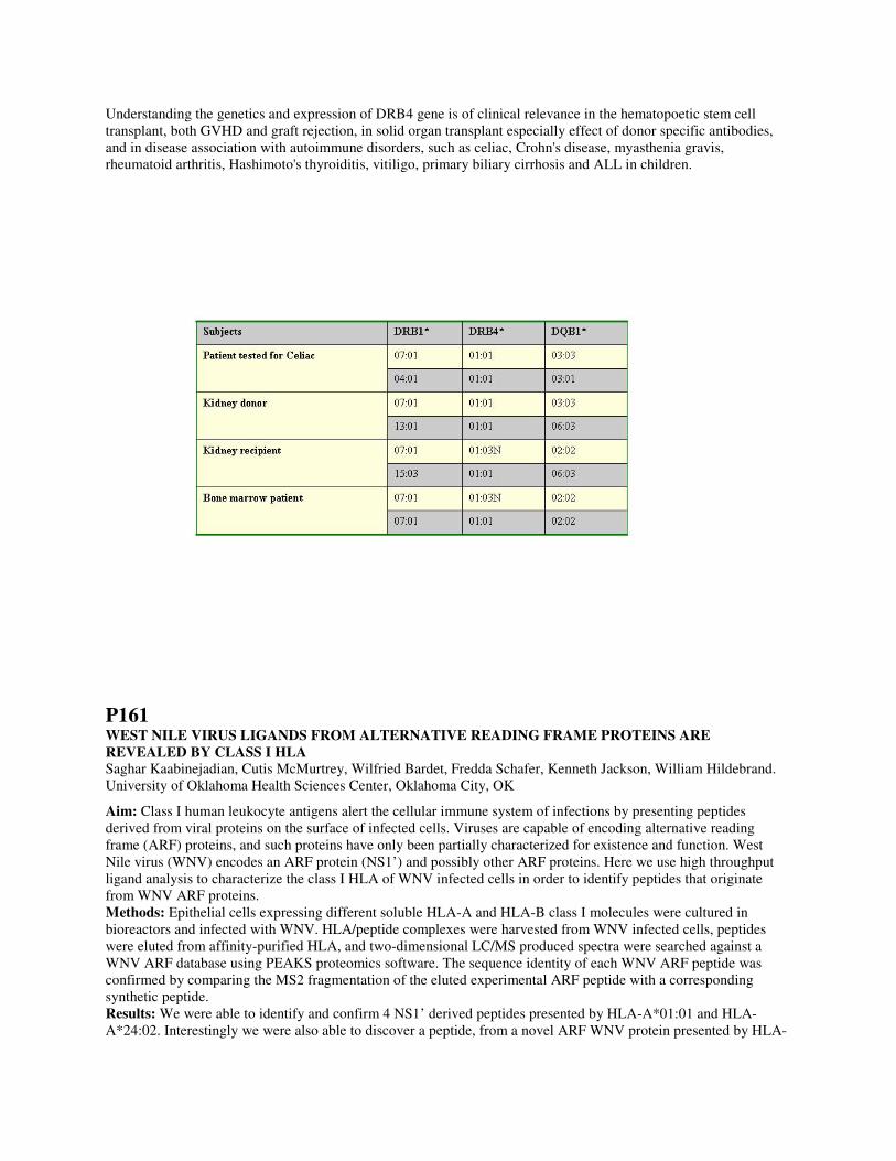
Understanding the genetics and expression of DRB4 gene is of clinical relevance in the hematopoetic stem cell transplant, both GVHD and graft rejection, in solid organ transplant especially effect of donor specific antibodies, and in disease association with autoimmune disorders, such as celiac, Crohn's disease, myasthenia gravis, rheumatoid arthritis, Hashimoto's thyroiditis, vitiligo, primary biliary cirrhosis and ALL in children.
P161 WEST NILE VIRUS LIGANDS FROM ALTERNATIVE READING FRAME PROTEINS ARE REVEALED BY CLASS I HLA Saghar Kaabinejadian, Cutis McMurtrey, Wilfried Bardet, Fredda Schafer, Kenneth Jackson, William Hildebrand. University of Oklahoma Health Sciences Center, Oklahoma City, OK
Aim: Class I human leukocyte antigens alert the cellular immune system of infections by presenting peptides derived from viral proteins on the surface of infected cells. Viruses are capable of encoding alternative reading frame (ARF) proteins, and such proteins have only been partially characterized for existence and function. West Nile virus (WNV) encodes an ARF protein (NS1’) and possibly other ARF proteins. Here we use high throughput ligand analysis to characterize the class I HLA of WNV infected cells in order to identify peptides that originate from WNV ARF proteins. Methods: Epithelial cells expressing different soluble HLA-A and HLA-B class I molecules were cultured in bioreactors and infected with WNV. HLA/peptide complexes were harvested from WNV infected cells, peptides were eluted from affinity-purified HLA, and two-dimensional LC/MS produced spectra were searched against a WNV ARF database using PEAKS proteomics software. The sequence identity of each WNV ARF peptide was confirmed by comparing the MS2 fragmentation of the eluted experimental ARF peptide with a corresponding synthetic peptide. Results: We were able to identify and confirm 4 NS1’ derived peptides presented by HLA-A*01:01 and HLA-A*24:02. Interestingly we were also able to discover a peptide, from a novel ARF WNV protein presented by HLA-
B*35:01. This protein is likely a 21 amino acid long C-terminal extension of NS5 and is presumed to be the result of a +1 frame shift, 38 base pair upstream from the polyprotein stop codon. This viral protein/peptide is of unknown function. Conclusions: We demonstrate that different HLA class I molecules present peptides from the NS1’ protein that is created as a result of ribosomal frameshifting during viral replication. In addition, we show the first evidence of presence of another ARF protein that is produced after WNV infection. How these ligands impact the immune responses toward infected cells remains to be resolved.
P162 HEPATITIS A AND B VACCINATION ASSOCIATED WITH DE NOVO DSA TO A PREVIOUS DONOR’S HLA AND A POSITIVE FCXM TO A POTENTIAL SECOND DONOR: SUCCESSFUL KIDNEY TRANSPLANTATION AFTER DESENSITIZATION Allen J. Norin1, Nabil Sumrani2, David Hochman3, Ballabh Das3, Moro Salifu4, Devon John2. 1SUNY Downstate Medical Center, Brooklyn, NY; 2SUNY Downstate Medical Center, Brooklyn, NY; 3SUNY Downstate Medical Center, Brooklyn, NY; 4SUNY Downstate Medical Center, Brooklyn, NY
A 54 year old African American female with 4 previous pregnancies received a kidney transplant from a living non related donor in November, 2001. Anti-HLA antibodies were not detected by Flow Cytometry cross matches (FCXM) that were negative at the time of the 2001 transplant. The patient’s transplanted kidney failed in Aug 2014 due to chronic rejection and the patient underwent a nephrectomy. At that time donor specific antibody (DSA) against the HLA of the first donor was not detected. The patient was subsequently XM against her daughter in November, 2014. Anti - HLA antibody using screening beads / Luminex technology was not detected and the FCXM was negative. The patient received Hepatitis A and B vaccinations in November, 2014 followed by two subsequent vaccinations in January and March of 2015 (with seroconversion). In February 2015, anti - HLA antibody was detected on routine screen and DSA was detected by single antigen bead (SAB) assays against a Class 1 HLA in common with the first and second donor (the daughter). No other identifiable sensitizing events, e.g., infection or blood transfusion were noted. The patient received 9 plasmapheresis treatments, QOD and IVIG. SAB tests indicated a reduction in DSA during the course of therapy. DSA was not detected after the 8th plasmapheresis treatment and the FCXM was negative. The patient received a kidney transplant 2 days later. Her creatinine came down from 9.9 mg/dl at transplant to an average of 1.35 mg/dl at 3 to 4 weeks after surgery. Conclusions and recommendations: These results suggest that caution should be exercised when considering vaccination of a previously transplanted patient even when the patient has not developed anti - HLA antibodies to potential recall antigens. Frequent anti-HLA antibody detection assays should be performed following vaccination to avoid unexpected positive XMs. Sensitization by viral vaccines may be overcome by standard desensitization protocols that are in current use.
P163 HLA-DRB1, DQA1 AND DQB1 DIVERSITY IN A MIXED POPULATION OF PARANÁ, SOUTHERN BRAZIL Pamela G. Reis1, Ana M. Sell2, Karina M. Sakita2, Ricardo A. Moliterno2, Jeane EL Visentainer2. 1Universidade Estadual de Maringa, Maringa, Brazil; 2Universidade Estadual de Maringa, Maringa, Brazil
Aim: The purpose of this study was to determine the allelic and haplotype frequencies of HLA-DRB1, -DQA1 and -DQB1 in a mixed population from Paraná, Brazil. Methods: Samples of 733 donors registered in the National Registry of Bone Marrow Donors (REDOME) from the northern/northwestern Paraná, Southern Brazil were analyzed. Genotyping of HLA-DRB1 was performed by PCR-SSOP HD (High Definition) with Luminex method (One Lambda®, CA, USA), and of HLA-DQA1 and DQB1 by PCR-SSO (One Lambda ®, CA, USA) medium/high resolution. To resolve ambiguities in HLA-DRB1 and -DQB1, PCR-SSP (One Lambda ®, CA, USA) was used. Allele and haplotype frequencies, Hardy-Weinberg equilibrium (HWE) and linkage disequilibrium (LD) among different loci were determined by software version 3.11 ARLEQUIN. Results: The analysis of the total sample 53, 14 and 23 alleles HLA-DRB1, -DQA1 and DQB1 were identified, respectively. For HLA-DRB1 locus, the most frequent were HLA-DRB1*07:01 (13.16%), *03:01 (8.86%), *11:01 (8.59%), *15:01 (6.07%), *13:01 (5.93%) and *01:01 (5.86%). For the loci HLA-DQA1 and HLA-DQB1, alleles
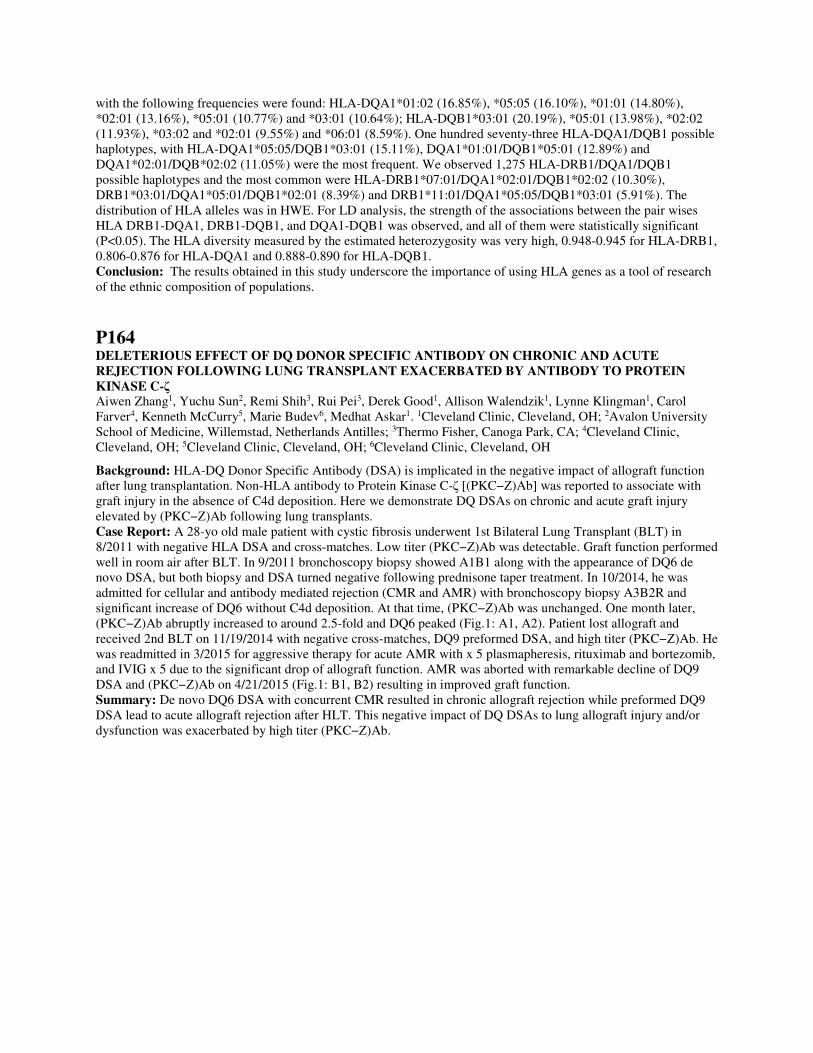
with the following frequencies were found: HLA-DQA1*01:02 (16.85%), *05:05 (16.10%), *01:01 (14.80%), *02:01 (13.16%), *05:01 (10.77%) and *03:01 (10.64%); HLA-DQB1*03:01 (20.19%), *05:01 (13.98%), *02:02 (11.93%), *03:02 and *02:01 (9.55%) and *06:01 (8.59%). One hundred seventy-three HLA-DQA1/DQB1 possible haplotypes, with HLA-DQA1*05:05/DQB1*03:01 (15.11%), DQA1*01:01/DQB1*05:01 (12.89%) and DQA1*02:01/DQB*02:02 (11.05%) were the most frequent. We observed 1,275 HLA-DRB1/DQA1/DQB1 possible haplotypes and the most common were HLA-DRB1*07:01/DQA1*02:01/DQB1*02:02 (10.30%), DRB1*03:01/DQA1*05:01/DQB1*02:01 (8.39%) and DRB1*11:01/DQA1*05:05/DQB1*03:01 (5.91%). The distribution of HLA alleles was in HWE. For LD analysis, the strength of the associations between the pair wises HLA DRB1-DQA1, DRB1-DQB1, and DQA1-DQB1 was observed, and all of them were statistically significant (P<0.05). The HLA diversity measured by the estimated heterozygosity was very high, 0.948-0.945 for HLA-DRB1, 0.806-0.876 for HLA-DQA1 and 0.888-0.890 for HLA-DQB1. Conclusion: The results obtained in this study underscore the importance of using HLA genes as a tool of research of the ethnic composition of populations.
P164 DELETERIOUS EFFECT OF DQ DONOR SPECIFIC ANTIBODY ON CHRONIC AND ACUTE REJECTION FOLLOWING LUNG TRANSPLANT EXACERBATED BY ANTIBODY TO PROTEIN KINASE C-ζ Aiwen Zhang1, Yuchu Sun2, Remi Shih3, Rui Pei3, Derek Good1, Allison Walendzik1, Lynne Klingman1, Carol Farver4, Kenneth McCurry5, Marie Budev6, Medhat Askar1. 1Cleveland Clinic, Cleveland, OH; 2Avalon University School of Medicine, Willemstad, Netherlands Antilles; 3Thermo Fisher, Canoga Park, CA; 4Cleveland Clinic, Cleveland, OH; 5Cleveland Clinic, Cleveland, OH; 6Cleveland Clinic, Cleveland, OH
Background: HLA-DQ Donor Specific Antibody (DSA) is implicated in the negative impact of allograft function after lung transplantation. Non-HLA antibody to Protein Kinase C-ζ [(PKC−Ζ)Ab] was reported to associate with graft injury in the absence of C4d deposition. Here we demonstrate DQ DSAs on chronic and acute graft injury elevated by (PKC−Ζ)Ab following lung transplants. Case Report: A 28-yo old male patient with cystic fibrosis underwent 1st Bilateral Lung Transplant (BLT) in 8/2011 with negative HLA DSA and cross-matches. Low titer (PKC−Ζ)Ab was detectable. Graft function performed well in room air after BLT. In 9/2011 bronchoscopy biopsy showed A1B1 along with the appearance of DQ6 de novo DSA, but both biopsy and DSA turned negative following prednisone taper treatment. In 10/2014, he was admitted for cellular and antibody mediated rejection (CMR and AMR) with bronchoscopy biopsy A3B2R and significant increase of DQ6 without C4d deposition. At that time, (PKC−Ζ)Ab was unchanged. One month later, (PKC−Ζ)Ab abruptly increased to around 2.5-fold and DQ6 peaked (Fig.1: A1, A2). Patient lost allograft and received 2nd BLT on 11/19/2014 with negative cross-matches, DQ9 preformed DSA, and high titer (PKC−Ζ)Ab. He was readmitted in 3/2015 for aggressive therapy for acute AMR with x 5 plasmapheresis, rituximab and bortezomib, and IVIG x 5 due to the significant drop of allograft function. AMR was aborted with remarkable decline of DQ9 DSA and (PKC−Ζ)Ab on 4/21/2015 (Fig.1: B1, B2) resulting in improved graft function. Summary: De novo DQ6 DSA with concurrent CMR resulted in chronic allograft rejection while preformed DQ9 DSA lead to acute allograft rejection after HLT. This negative impact of DQ DSAs to lung allograft injury and/or dysfunction was exacerbated by high titer (PKC−Ζ)Ab.
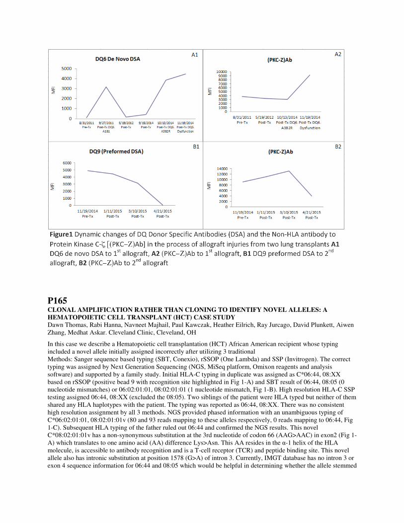
P165 CLONAL AMPLIFICATION RATHER THAN CLONING TO IDENTIFY NOVEL ALLELES: A HEMATOPOIETIC CELL TRANSPLANT (HCT) CASE STUDY Dawn Thomas, Rabi Hanna, Navneet Majhail, Paul Kawczak, Heather Eilrich, Ray Jurcago, David Plunkett, Aiwen Zhang, Medhat Askar. Cleveland Clinic, Cleveland, OH
In this case we describe a Hematopoietic cell transplantation (HCT) African American recipient whose typing included a novel allele initially assigned incorrectly after utilizing 3 traditional Methods: Sanger sequence based typing (SBT, Conexio), rSSOP (One Lambda) and SSP (Invitrogen). The correct typing was assigned by Next Generation Sequencing (NGS, MiSeq platform, Omixon reagents and analysis software) and supported by a family study. Initial HLA-C typing in duplicate was assigned as C*06:44, 08:XX based on rSSOP (positive bead 9 with recognition site highlighted in Fig 1-A) and SBT result of 06:44, 08:05 (0 nucleotide mismatches) or 06:02:01:01, 08:02:01:01 (1 nucleotide mismatch, Fig 1-B). High resolution HLA-C SSP testing assigned 06:44, 08:XX (excluded the 08:05). Two siblings of the patient were HLA typed but neither of them shared any HLA haplotypes with the patient. The typing was reported as 06:44, 08:XX. There was no consistent high resolution assignment by all 3 methods. NGS provided phased information with an unambiguous typing of C*06:02:01:01, 08:02:01:01v (80 and 93 reads mapping to these alleles respectively, 0 reads mapping to 06:44, Fig 1-C). Subsequent HLA typing of the father ruled out 06:44 and confirmed the NGS results. This novel C*08:02:01:01v has a non-synonymous substitution at the 3rd nucleotide of codon 66 (AAG>AAC) in exon2 (Fig 1-A) which translates to one amino acid (AA) difference Lys>Asn. This AA resides in the α-1 helix of the HLA molecule, is accessible to antibody recognition and is a T-cell receptor (TCR) and peptide binding site. This novel allele also has intronic substitution at position 1578 (G>A) of intron 3. Currently, IMGT database has no intron 3 or exon 4 sequence information for 06:44 and 08:05 which would be helpful in determining whether the allele stemmed
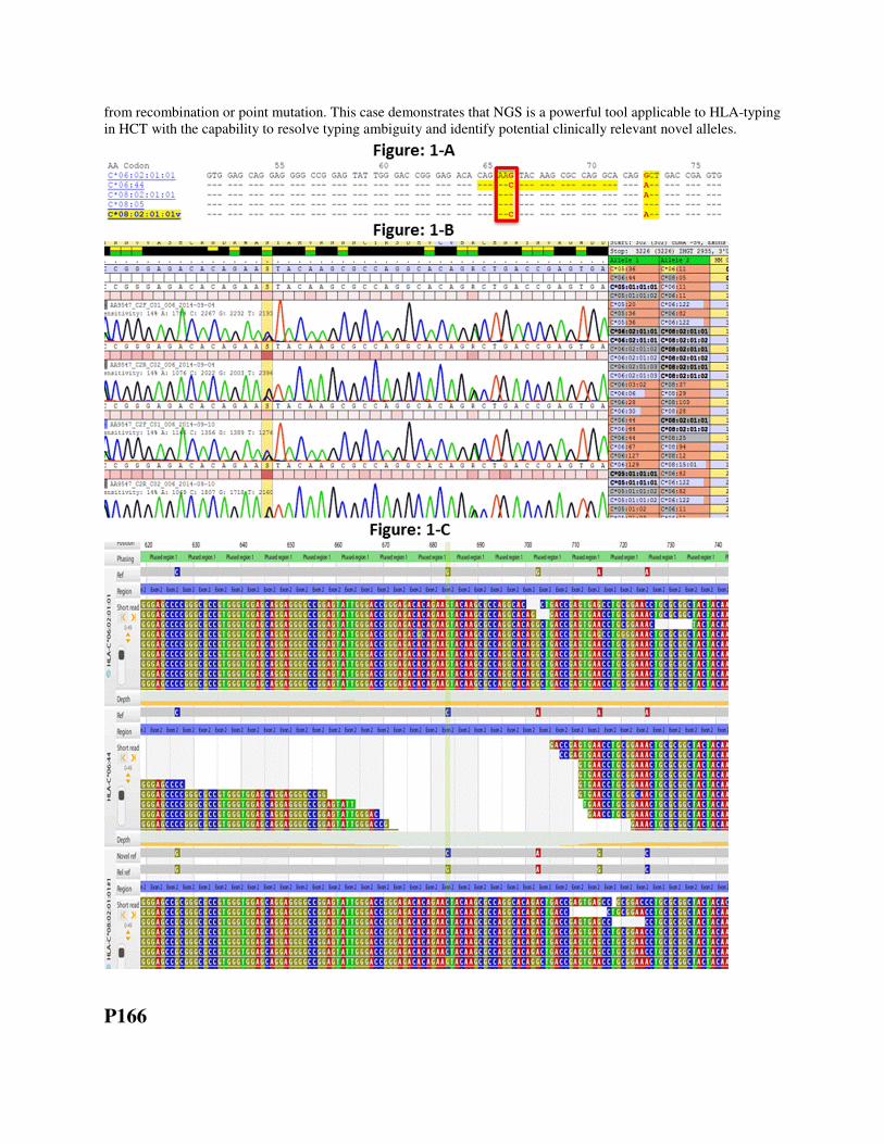
from recombination or point mutation. This case demonstrates that NGS is a powerful tool applicable to HLA-typing in HCT with the capability to resolve typing ambiguity and identify potential clinically relevant novel alleles.
P166
POTENTIAL HYPERACUTE RENAL REJECTION DUE TO NON-HLA ANTIBODY IN A PEDIATRIC PATIENT Matthew Cusick1, Amy Bobrowski2, Kelley M. Haarberg1, Anat AR Tambur1. 1Northwestern University, Chicago, IL; 2Northwestern University and Children's Memorial Hospital, Chicago, IL
Non-HLA antigen targets have been associated with hyperacute, acute, and chronic kidney allograft rejection. Although there is controversy regarding the predictive role of non-HLA antibodies (Abs) in transplant (Txp) outcome - there are cases that indicate causality between the presence of such Abs and graft loss. A 17 month old boy with end-stage kidney disease, secondary to kidney dysplasia, received a living donor Txp from his uncle in April 2014. The presence of HLA abs was tested in 5 sera samples prior to Txp with a PRA of 7% and 0% for class I and II respectively (flow PRA). Single antigen testing confirmed the absence of donor specific HLA Abs (DSA). Initial and final flow cross matches were negative, as expected. During the Txp surgery, it was noted that the kidney became firm, urine output was below expectation and the child became increasingly pressor-dependent. Subsequently, the patient had an episode of ventricular tachycardia and an exploratory laparotomy demonstrated that the Txp kidney became tense and enlarged, with thrombosis of the renal vein. The kidney was nephrectomized within 48 hours and the patient’s status improved significantly. To investigate a potential role for HLA DSA, we tested a 2 days post-Txp serum sample, confirming the absence of HLA-DSA. We also repeated Abs testing on the pre-Txp serum sample ruling out HLA-DSA as the cause of rejection. To assess potential causes for this apparently hyperacute Ab mediated rejection, pre-Txp (pre-14 days) and post-Txp (post-24 days) samples were sent out for AT1R Abs screening and donor specific endothelial cell crossmatch (XM-One). Retrospective testing of a pre- and post-Txp sera for AT1R antibody was out of range for pre-Txp sample and the post-Txp sample was positive (>30 Units/ml). The XM-One assay using endothelial precursors isolated from the donor as targets was strongly positive using a pre-Txp serum but negative using post-Txp serum. Approximately two month’s post-Txp, the patient developed HLA Abs, on top of the AT1R antibodies. In conclusion, this patient still requires a kidney Txp and the question is how to proceed - shall we consider only obtaining an organ from a living donor? This would allow for the necessary time to run XM-One and potentially inhibit AT1R pathway with an AT1 receptor antagonist. What to do if no living donor becomes available?
P167 PREVALENCE OF HLA-DP ANTIBODIES IN PATIENTS AWAITING RENAL TRANSPLANTATION Marjola Furczon1, Michele Higgins Prod2, Sylvia Piggott2, Maria Oppermann1, Siva Kanangat3. 1Rush University Medical Center, Chicago, IL; 2Rush University Medical Center, Chicago, IL; 3Rush University Medical Center, Chicago, IL
Aim: To determine the prevalence of HLA-DP IgG antibodies in patients on Renal Transplant waitlist at Rush University Medical Center, Chicago, IL. Introduction: Role of HLA-DP antibodies in solid organ and stem cell transplantation is evolving. Isolated reports on possible involvement of only HLA-DP DSA in acute rejection episodes (cases where the Donor/Recipient pairs are matched at HLA-Class , DR and DQ)] show the importance of this issue. In the light of HLA molecular typing and antigen specific HLA antibody assays there is an opportunity to evaluate the role of HLA-DP in allograft rejection. As part of this goal we investigated the prevalence of DP antibodies in our renal transplant candidates. Patients: A total of 532 patients with multiple serum samples tested. Methods: HLA-specific antibodies were determined by solid phase assay (One Lambda, Inc. ®). HLA- specific antibody was scored as strong, moderate, or weak depending on their mean fluorescent index (MFI) value of >5000, 2000-5000, 700-2000, respectively. Results: Of the 532 patients, 143 (26.9%) had antibodies against Class II HLA-DP. The number of specific HLA-DP antibodies per patient ranged from 1 to 17 antibodies (Strong, Moderate, Weak), but could also be found entirely in one category as in the case with one patient in which all 16 of the anti-DP antibodies were in the strong MFI category. Conclusions: Quarter of renal transplant candidates on waiting at Rush University Medical center have detectable HLA-DP antibodies with varying MFIs. While 36.5% of those antibodies express a low MFI, 31.4% of the patients had strong MFI. Multiple case studies from various centers have shown that transplant patients perfectly matched per UNOS requirements to their donor may still have antibody mediated rejection due to a mismatched at HLA-DP. Hence donor typing for HLA-DP and monitoring of patients for HLA-DP antibodies seems to be important

P168 HLA ANTIBODY TESTING- IT’S NOT A ONE STOP ASSAY Matthew Cusick, Ashley Ruiz, Kelley M. Haarberg, Nancy Herrera, Anat R. Tambur. Northwestern University, Chicago, IL
HLA laboratories rely on single antigen (SA) assays to determine unacceptable antigens and perform virtual crossmatches (vXM) that can immunologically guide clinicians in the identification of an appropriate donor. Although solid phase antibody detection assays are sensitive, specific and robust- instances arise when further testing is needed to verify results or resolve inconsistencies between these tests. Patient S.T. is a 50 year old male diagnosed with heart failure. Routine work-up in our laboratory is to evaluate % FlowPRA and compare those results to solid phase assays on a Luminex platform which measures the breadth and strength of antibodies in a patient’s sera. Patient S.T. had two samples tested three months apart with a class I FlowPRA of 58% and 62%, respectively. However, The Luminex SA results consistently demonstrated only 5 class I (all B15) antibodies in the positive MFI range leading to a cPRA of about 15%. Moreover, the internal negative control values were >2500 MFI leading us to believe that there may be additional specificities masked by the high background. How should we deal with these contradictory test results? Can we rely on a vXM to accept a heart for this patient? To reconcile these seemingly contradictory results and to determine if the antibodies detected by Luminex assays were relevant, surrogate FCXM were performed. Surrogate Donor 1 (B15 positive) was FCXM positive. Surrogate donor 2 (B15 negative) was FCXM negative indicating that non-specific binding/false-positivity due to high background should not affect FCXM interpretation, Importantly, testing using phenotype beads were not affected by high background and further confirmed the presence of B15 antibodies. Due to the presence of HLA antibodies and the high background exhibited by this patient, a prospective FCXM must be performed prior to accepting an organ. In conclusion, this is an example in which different tests provide contradictory (or non-consistent) results - and our approach to resolve these ambiguities to support successful organ transplantation.
P169 THE KEY DETERMINANTS OF DONOR LYMPHOID CHIMERISM SUPPORT THE DIAGNOSIS OF ACUTE GRAFT VERSUS HOST DISEASE AFTER LIVER TRANSPLANT AND PREDICT THE CLINICAL OUTCOME Aiwen Zhang1, Yuchu Sun2, Brian Eghtesad3, Koji Hashimoto3, David Plunkett1, Dawn Thomas1, Paul Kawcza1, Raymond Jurcago1, Heather Eilrich1, John Fung3, Medhat Askar1. 1Cleveland Clinic, Cleveland, OH; 2Avalon University School of Medicine, Willemstad, Netherlands Antilles; 3Cleveland Clinic, Cleveland, OH
Objective: Management of acute Graft versus Host Disease (aGvHD) following liver transplant (LT) is critical for reducing mortality from this life-threatening disease. The aim of this study was to examine how Donor Lymphoid Chimerism (DLC) determinants associate with the diagnosis and prediction of clinical outcome of aGvHD after LT. Methods: We retrospectively analyzed 17 LT patients (2007-2015) who were clinically suspected and DLC supportive of aGvHD. The DLC was tested by short tandem repeat with an initial 5% as positive cut off. The DLC determinants were: initial positive DLC after 30 days post-LT (InPD>30D); initial positive DLCs in whole blood, T, NK, and CD8+ cells; peak DLCs of >5% in whole blood, >50% in each of T, NK, CD8+ cells respectively; positive DLC in both CD8+ and NK cells at initial phase, DLC 50% in both CD8+ and NK cells at peak phase. Clinical outcome was determined over the following 40-day duration of treatment and/or aGvHD related motility (>40D DuT/M). Logistic regression was used to analyze >40D DuT/M in response to all the variables of DLC determinants. Results: Of 17 aGvHD patients, the prevalence of 100% (16/16) in CD8+ cell was markedly higher than the prevalence of 41% (7/17) in whole blood, 59% (10/17), in T cell, and 38% (6/16) in NK cell at the initial detection of positive DLC. Amongst all analyzed variables of DLC determinants, InPD>30D and DLC 50% in both CD8+ and NK cells at peak phase were all significantly associated with >40D DuT/M. There was a mildly significant association with the individual variables of initial phase positive DLC in T and NK cells, and peak phase >50% DLC in T and NK cells. Regardless of initial phase or peak phase, the individual variables of DLC in whole blood and CD8+ cell had no significant relationship with >40D DuT/M (Figure 1). Conclusion: Initial positive DLC of CD8+ cell is a sensitive and reliable indicator supporting the diagnosis of aGvHD. Both delayed diagnosis and/or peak phase DLC over 50% in both CD8+ and NK cells strongly predicts aGvHD related negative impact following LT.
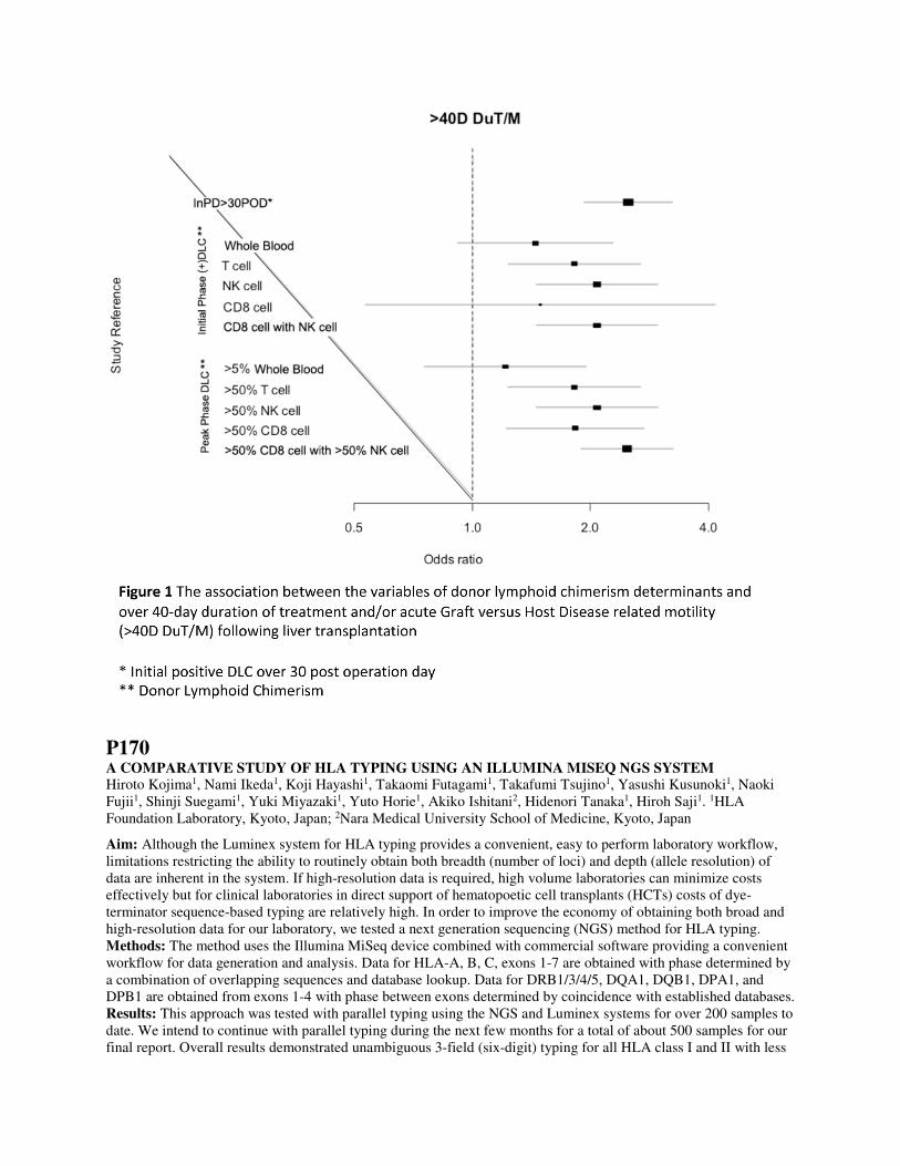
P170 A COMPARATIVE STUDY OF HLA TYPING USING AN ILLUMINA MISEQ NGS SYSTEM Hiroto Kojima1, Nami Ikeda1, Koji Hayashi1, Takaomi Futagami1, Takafumi Tsujino1, Yasushi Kusunoki1, Naoki Fujii1, Shinji Suegami1, Yuki Miyazaki1, Yuto Horie1, Akiko Ishitani2, Hidenori Tanaka1, Hiroh Saji1. 1HLA Foundation Laboratory, Kyoto, Japan; 2Nara Medical University School of Medicine, Kyoto, Japan
Aim: Although the Luminex system for HLA typing provides a convenient, easy to perform laboratory workflow, limitations restricting the ability to routinely obtain both breadth (number of loci) and depth (allele resolution) of data are inherent in the system. If high-resolution data is required, high volume laboratories can minimize costs effectively but for clinical laboratories in direct support of hematopoetic cell transplants (HCTs) costs of dye-terminator sequence-based typing are relatively high. In order to improve the economy of obtaining both broad and high-resolution data for our laboratory, we tested a next generation sequencing (NGS) method for HLA typing. Methods: The method uses the Illumina MiSeq device combined with commercial software providing a convenient workflow for data generation and analysis. Data for HLA-A, B, C, exons 1-7 are obtained with phase determined by a combination of overlapping sequences and database lookup. Data for DRB1/3/4/5, DQA1, DQB1, DPA1, and DPB1 are obtained from exons 1-4 with phase between exons determined by coincidence with established databases. Results: This approach was tested with parallel typing using the NGS and Luminex systems for over 200 samples to date. We intend to continue with parallel typing during the next few months for a total of about 500 samples for our final report. Overall results demonstrated unambiguous 3-field (six-digit) typing for all HLA class I and II with less

than 1% failure rate and complete coincidence with Luminex data. Improvements in both accuracy and resolution are provided by the NGS system, and laboratory effort required is similar. Software and data storage are performed on the cloud, providing rapid analysis, ease of use, and freedom from on site systems support. Conclusion: We detail the comparative typing results and critically examine the NGS workflow towards establishing the efficacy and economy of this NGS approach in laboratories supporting HCTs.
P171 COMMOM, WELL-DOCUMENTED AND RARE HLA ALLELES OBSERVED IN THE BRAZILIAN BONE MARROW REGISTRY Luis Cristovao Porto1, Juliana C. Oliveira1, Juliana P R Motta1, Monica Goldenstein2, Matilde Romero3, Luis Fernando S. Bouzas3, Margareth A. Torres4, Raquel Fabreti5, Evaldo Nascimento5, Bruno Z. Piovezan6, Maria Elisa H. Moraes6. 1Rio de Janeiro State University, Rio de Janeiro, Brazil; 2Federal University Parana, Curitiba, Brazil; 3National Cancer Institute, Rio de Janeiro, Brazil; 4Hospital Israelita Albert Einstein, São Paulo, Brazil; 5Laboratório Imunolab Imunologia de Transplantes, Belo Horizonte, Brazil; 6JRM Investigações Imunológicas, Rio de Janeiro, Brazil
Aim: We intend to characterize alleles with 5 or more counts in non related donors of the Brazilian Bone Marrow Registry (REDOME) that are not listed in common and well documented (CWD) alleles version 2. Method: Identification and comparison of alleles and NMDP alleles codes which first allele in the subtype are not in CWD version 2 from the REDOME data base at February 15, 2015. Results: We identified 30,395 HLA typings with 1,575 alleles or NMDP alelle codes of not common or not well documented among the 3,227,387 donors. A total of 1,213 donor registries corresponding to 158 alleles in A locus, 306 in B locus, 47 in C locus, 136 in DRB1 locus and 37 in DQB1 locus had less of 5 counts. In a few cases (28/283) it was possible to have access to the outputs and from these, 11 were probably misinterpreted results, although in one case A*02:90 was also reported in Chinese donor registry. We also identified 259 alleles with 5-25 counts, 116 alleles with 26-100 counts, 29 alleles with 101-500, and 4 with more than 500. Eighty of these alleles or NMDP allele were most likely misinterpretation due to SSO bead adjustments corresponding to 9,582 donors. Actual SSO output reviewed interpretation and SBT confirmed typings are show in Table 1. Conclusion: Sequencing based typings were on going to confirm these Brazilian CWD HLA alleles and are displayed at www.imunogenetica.org (in Portuguese).
Table 1: SBT confirmed Brazilian CWD
Allele NMDP code(s) n
A*02:26 02:SKVB 38
A*24:28 24:FKTF 10
A*29:13 36
A*34:03 34:ZSH 15
B*18:07 18:YXH 18:YJMH 18:YPKK 85
B*37:04 37:APTR 95
B*41:04 41:DF 41:HT 41:GDV 41:AAR 24
B*56:13 56:AAPJ 56:AXWN 36
DRB1*13:111 25
DRB1*13:48 13:PKDZ 24
P172 TO BATCH OR NOT TO BATCH? LINKSEQ REAL-TIME PCR VS. SSO-LUMINEX, A COMPARISON OF HIGH THROUGHPUT HLA TYPING METHODS Helen LaCarrubba1, Zachary Antovich2, Komal Singh2, Dennis Miguel1, Patty Harris Bowe1, Prakash Rao1. 1NJ Sharing Network, New Providence, NJ; 2Linkage Biosciences, South San Francisco, CA
Aim: For high-throughput histocompatibility applications such as HLA typing of transplant candidates, conventional wisdom suggests SSO with Luminex® provides a fair balance of productivity, turnaround time (TAT) and cost as long as the laboratory can wait to receive sufficient samples to batch them together in one run. A recent, competitive alternative is Linkage Biosciences’ LinkSēq™ HLA typing using real-time PCR. This technology is gaining in popularity and acceptance because of its rapid protocol, far fewer manual steps than either SSO or SSP techniques, intermediate resolution, and fully automated analysis. The goal of this study was to directly compare high throughput performance between Linkage’s LinkSēq real-time PCR technology and One Lambda’s LabType® SSO-Luminex method. Methods: 14 samples were typed by one technologist at nine loci: HLA-A, B, C, DRB1, DRB345, DQA1, and DQB1 using both LinkSēq and LabType. Total TAT was measured starting at initial setup and ending at the final HLA assignment for each method. Results: LinkSēq was 97 minutes faster (4hours and 2min) compared to LabType (5hours and 39min), with less hands-on time and less analysis time. LinkSēq’s analysis software, SureTyper, generated quick and complete data for each sample and also resolved an ambiguity that appeared with one LabType assignment. Conclusion: LinkSēq was faster and required less user intervention compared to LabType, enabling greater laboratory staff productivity. Because LinkSēq doesn’t require batch processing, it is equally efficient running one or 14 samples and thus can deliver on-demand service and the flexibility to add urgent testing as needed.
P173 UNDERSTANDING CLINICAL ORDERING PRACTICES: PLATELET ANTIBODY TESTING Justin Kreuter, Lisa Hallaway, Steven De Goey, Patti Duellman, Laurie Wakefield, Manish Gandhi. Mayo Clinic, Rochester, MN
Aim: We use the platelet serum antibody test (Immucor Pak12G, Norcross, GA) for detection of alloantibodies with platelet and HLA class I specificity; whereas, we use the cell-bound platelet antibody test (Immucor Pak Auto, Norcross, GA) for detection of auto-antibodies with platelet glycoprotein specificity. We performed a retrospective audit to investigate when and how these tests were being ordered. Methods: All in-house serum and cell-bound platelet antibody testing between April 1st, 2014 and March 31st, 2015 were reviewed. Both results and clinical notes surrounding the time of order were reviewed. Results: There were 208 serum platelet antibody tests performed during this time period with 11 repeats. There were 290 cell-bound platelet antibody tests performed during this time period with 13 repeats. Notably, both tests were ordered simultaneously 77 times. The clinical indication for all positive tests is listed in Table 1. The most likely specificities detected when both serum and cell-bound testing were ordered simultaneously are listed in Table 2. Conclusions: While each test appeared to be ordered most commonly for the appropriate reason, a number of tests were ordered for the incorrect clinical indication. Furthermore, because these tests are optimized to evaluate different clinical situations, simultaneous orders demonstrate a lack of understanding about test utility. 37% of serum platelet antibody tests and 26% of cell-bound platelet antibody tests were ordered together. Interestingly, the serum platelet antibody test had a number of orders for “other” reasons, which was predominantly due to lack of clinical documentation. In conclusion, an intervention to facilitate proper ordering practices is needed.
Table 1. Reason for ordering tests that were subsequently positive.
Clinical Indication
Auto-immune Allo-immune Other
Serum 20 (29%) 33 (48%) 16 (23%)
Cell-Bound 51 (69%) 21 (28%) 2 (3%)
Table 2. Most likely specificities when both tests were ordered simultaneously.
Anti-GP No Specificity Anti-HPA Anti-HLA
Serum 15 (18%) 47 (56%) 8 (9%) 14 (17%)
Cell-Bound 18 (23%) 59 (77%) n/a n/a

P174 CHARACTERISTICS OF DONOR-SPECIFIC ANTI-HLA ANTIBODIES IN RENAL TRANSPLANT RECIPIENTS Salim Ghandorah1, Serdar Yilmaz1, Abdulnaser Abadi1, Taleb Rahmanian2, Jinguo Wang2, Faisal M. Khan1,2, Noureddine Berka2,1. 1University of Calgary, Calgary, AB, Canada; 2Calgary Laboratory Services, Calgary, AB, Canada
Introduction: Antibody-mediated rejection (AMR) is the major complication of renal transplantation that significantly affects the renal allograft survival. Complement fixation mediated through donor specific anti-Human Leukocyte Antigen (HLA) antibodies (DSA) is the hallmark of AMR. Different methods have been developed to detect harmful DSAs. However, not all DSAs have the same detrimental effect, as different antibody characteristics might trigger distinct immune responses. Aim: In the present study, we assessed various biological (timing of appearance, strength and specificity) and functional (IgG subtype and c1q binding ability) characteristics of DSA in renal transplant patients. Methods: A total of 315 kidney transplant recipients were accrued for this study and 39 patients with de novo DSA were retrospectively analysed for different DSA characteristics. The analyses were carried out using commercially available SAB assay for IgG determination, C1q assay for complement binding capability and modified SAB assay for IgG subclass determination (One Lambda) Results: Important findings of the study were: (1) majority of the DSAs were denovo antibodies (p=0.0001); primarily against HLA class II antigen (p=0.0005); and typically developed after one year posttransplant (p=0.0001). (2) DSA with highest strength were routinely observed against DQB antigens in comparison to other HLA class I and II antigens (p=0.006). (3) IgG1 was the predominant IgG subclass (49.4%), followed by IgG3 (24.7%), IgG2 (16%), and IgG4 (9.9%). (4) DSA MFI and the IgG1 subclass were strongly correlated with C1q positivity (p= 0.01 and p= 0.009, respectively). Conclusion: Among the functional characteristics, higher MFI of DSA and IgG1 subclass were strongly correlated with C1q positivity. Among the biological characteristics, the majority of DSAs were against HLA class II antigens and developed frequently after one-year post transplantation. Further studies are needed to investigate the clinical relevance of these biological and functional DSA characteristics in predicting renal allograft outcomes.
P175 MYSTERY SENSITIZATION: WHAT HAPPENED IN THE HOSPITAL??? Kelley M. Haarberg, Nancy D. Herrera, Yelena Barabanova, Matthew F. Cusick, Ashley Ruiz, Robert Gordon, Anat R. Tambur. Northwestern University, Chicago, IL
The 26 y/o female, with history of double heart valve replacement, should have had an uneventful heart transplant (txp). The patient (pt) was 0% HLA-Class I, only 12% sensitized to HLA-Class II, and the virtual crossmatch against her donor was negative. Our mystery case, however, had just begun. During the txp, testing of the immediate pre-txp sample revealed the pt was 100% sensitized to HLA-Class I and 24% to Class II, flow crossmatch (FCXM) on the same sample showed strong T and B cell channel shifts, HLA-DSA titered as high as 1:512 and were strongly C1q-binding. This was a surprising increase from results collected one month prior to txp. The clinical txp team was alerted, and while the pt was fine at that time, within 5 hours of txp cardiogenic shock and rejection began. Early detection of sensitization allowed the txp team to arrange for plasmapheresis (PP), Thymoglobulin and Eculizumab before the pt crashed. How had the pt become so sensitized? Was there a sample switch? Re-typing of all samples for HLA antigens confirmed they belonged to the pt. Was the patient sensitized between the time of her first sample drawn one month prior to txp and the time of txp? As an inpatient for 3 weeks prior to txp, the clinical team was able to report she had not incurred any sensitizing events. Protocol fresh frozen plasma and Hepatitis B vaccine were given. FCXM on the 1 month pre-txp sample was negative, correlating with the original virtual. DSAs responded quickly to Eculizumab, Bortezomib and PP. Within 4 days post-txp, DSA titers had fallen to 1:8, and a 1 week post-txp right atrium biopsy was C4d negative. This was the most rapid response to therapy we had ever seen, and we speculate that the hyperacute immune response and quick therapy, including PP and complement inhibitors, precluded a mature complement response and C4d deposition. As of 4 months post-txp, DSA titers were maintained below 1:4 on Tacrolimus, Cellcept and Prednisone, and the pt entered cardio rehabilitation. While likely an anamnestic response pointing to a gap in HLA-specificity data collection, perhaps immunization could have served as a sensitizing event in this case. This case highlights the need for close monitoring for HLA-sensitization and antibody specificity changes following hospitalization and vaccination.

P176 ALLO-SENSITIZATION PROFILE IN RENAL TRANSPLANT PATIENTS WITH SYSTEMIC LUPUS ERYTHEMATOSUS Paul Brailey1, Diana Girnita2, Alin Girnita1. 1University of Cincinnati COM, Cincinnati, OH; 2University of Cincinnati COM, Cincinnati, OH
Aim: Systemic lupus erytematosus (SLE) is the autoimmune disease with the highest prevalence in renal transplant population. The purpose of the present study was to describe the sensitization profile in renal transplant patients with end stage renal disease due to SLE. Methods: A retrospective single-center analysis in the last ten years revealed 26 all renal transplant patients with SLE - twenty females and six males. This group was matched to a 53 patient control group, with no SLE, but matched for gender and age distribution. The HLA polymorphism for -A, -B, -C, -DRB1, -DRB3/4/5, -DQB1, -DQA1, -DPB1 was performed by reverse SSO for intermediate resolution in all patients, while high resolution SBT (Sanger) was used for 12 cases. The auto-crossmatches were performed both by complement-dependent cytotoxicity, and by flowcytometry. The presence and identification of anti-HLA antibody was achieved by single-antigen bead (Luminex) assays. Results: The HLA typing in SLE group has shown a higher (19/26, 73%) prevalence of HLA-DRB1-3/4/5 antigens when compared to control group (8/53, 15%, p<0.05). Furthermore, 4 SLE patients (15%) exhibited a positive cytotoxic and flow auto-crossmatch, compared to only 1 (2%) in controls (p<0.01). The distribution of HLA-specific antibody was 20/26 (77%) in SLE versus 18/53 in controls (33%), p<0.01 The class distribution in SLE group was: 19 anti-class I, and 11 anti class II, versus 11 class I and 7 class II in controls. 22 patients in SLE group exhibited anti-HLA antibody towards public epitopes resulting in a calculated PRA > 50%, while 19 SLE patients exhibited anti-HLA antibody towards public epitopes resulting in a calculated PRA > 80%. The control group had a significantly lower proportion of strong antibody towards public epitopes (4/18 and 2/18, respectively, p<0.01). Conclusions:Renal transplant patients with SLE exhibited a higher prevalence of HLA-DRB1-3/4/5 self antigens, and a higher prevalence of positive auto-crossmatches and strong HLA antibody towards public epitopes.
P177 ALLO IMMUNE RESPONSE, EPITHELIAL MESENCHYMAL TRANSITION AND CHRONIC ALLOGRAFT FIBROSIS- IS THERE A LINK? Saba Rezaeian1, Ina Kurbegovic2, Michele Higgins Prod2, Lela Buckingham2, Marry Beth Flaws1, Demetra Castillo1, Demetra Castillo1, Robert DeCresce2, David Cimbaluk2, Siva Kanangat2. 1Rush University Medical Center, Chicago, IL; 2Rush University Medical Center, Chicago, IL
Introduction: Mechanisms of chronic rejection manifested by irreversible fibrosis remain unclear. Phenotypic conversion of renal tubular epithelial and endothelial cells of the allograft to mesenchymal cells [Epithelial/Endothelial to Mesenchymal Transitioning (EMT/EndOEMT)] in the onset of fibrosis in the transplanted organ is proposed hypothesis. Objectives: To detect EMT by way of gene expression profiling related to EMT using qualitative CR and quantitative real-time PCR covering 84 genes related to EMT. Materials: Anti-HLA Class I: chimeric monoclonal antibody to the constant region of HLA Class I antigen (Invivogen, San Diego CA), EMR8-5: (Abcam®,Cambrige MA), a monoclonal HLA-Class I antibody that reacts with the heavy chains of Human HLA Class I A, B, and C and Pooled Positive Serum (PPS) containing antibodies to all Class I and Class II HLA allo-antigen and some possible non-HLA antigens. Renal Epithelial Cell (REC) and Human Umbilical Endothelial Cell (HUEC) lines (Life Line Cell Technologies, Frederick MD) were used. A total of 37 paraffin embedded renal allograft biopsy samples with varying etiologies and varying percentages of fibrosis were retrieved from Rush University Medical Center in Chicago, IL. An EMT RT2 profiler PCR Array (Qiagen) specific for 84 EMT-related genes was used to assess the expression of these genes in 12 renal allograft biopsies and 2 cell cultures Results and Conclusions:The EMT Gene array profiling by realtime PCR indicated that in both in vitro cell cultures and biopsies there was significant changes in EMT related gene expressions. The overall gene expression pattern revealed up regulation of several EMT related genes in renal epithelial cells, HUEC, and in biopsy samples. The biopsy sample findings on EMT related gene expression array point in the direction of contributing towards finding a molecular signature of chronic allograft rejection.
P178 TARGET ENRICHMENT USING CAPTURE PROBES: THE FUTURE OF HLA TYPING BY NEXT GENERATION SEQUENCING? Hayley Hogan, Rhys Cransberg, Megan Jordan, Damian Goodridge, David Sayer. Conexio, Perth, Australia
Aim: Most target enrichment (TE) procedures for high resolution HLA typing by Next Generation sequencing have utilized full, or part, gene PCR amplification. This is in contrast to many other molecular diagnostic applications where target enrichment is performed using capture probes (CP). One possible reason why TE using CP hasn’t been attempted for HLA is the high level of polymorphisms found in some exons and concern that all existing and undescribed alleles won’t be detected. We have designed a probe set for HLA-A, B, C, DRB1, DRB3,4,5 and DQB1 that eliminates this concern. TE by CP has many advantages over PCR including higher throughput, fewer artefacts, lower costs and the ability to include additional genes without increasing work load. Methods: 200ng of Genomic DNA from 16 individuals with known HLA genotypes were fragmented using a Covaris M220 Ultrasonicator™ using the following conditions: duty factor 20%, peak incident power 50W, 200 cycles per burst, for 45 seconds at a temperature of 20°C - aiming for a peak distribution of fragments 550bp in length. The fragments were then repaired, size selected by a dual-bead based protocol, adenylated and adapters were ligated. The fragments were enriched and Capture-HLA™ probes for multiple loci were used to isolate fragments containing HLA specific sequences. The HLA specific fragments were further enriched and 300bp paired-end reads were then sequenced using a MiSeq® Next Generation DNA Sequencer. The resulting data was then analysed using the Assign™ MPS sequence analysis software. Results: Successful genotyping was obtained for all loci for all samples, including exons 2 and 3 for class I and exon 2 for class II genes. There was even allele balance across all genes. Potential novel alleles were also detected. Conclusions: Validation of this approach is on-going. However preliminary data suggests that the use of CP has the potential to revolutionize HLA typing by providing inexpensive high resolution, high volume HLA typing, with the ability to include additional genes.
P179 A CASE STUDY OF DOUBLE CORD PERSISTENCE: EVERYONE WINS Kelley M. Haarberg1, Yelena Barabanova1, Olga Frankfurt2, Anat R. Tambur1. 1Northwestern University, Chicago, IL; 2Northwestern University, Chicago, IL
Double cord (DC) transplants (txp) as a treatment of choice for hematologic malignancies are much less frequently utilized when compared to MUD stem cell therapies. The vast majority of stem cell recipients (recip) receiving DC txp transition from 100% recip to 100% single cord blood unit (CBU), as usually one CBU “wins” and persists alone. This occurs within a few weeks to a few months post-txp. We present a case study in which neither cord is eliminated, but rather, long term engraftment of both CBUs persists. A 26 y/o, Caucasian, male with CML was conditioned for DC txp with busulfan, thiotepa and fludarabine due to lack of 10/10 MUDs and presence of inhibitor-resistant T315I mutation. DC txp is a low relapse option for CML patients who cannot be treated with the standard inhibitor therapies due to presence of T315I mutation. Engraftment monitoring (EM) analysis was performed by qPCR on day 18, revealing 0% recip, 86% CBU #1 and 14% CBU #2. These unexpected results, and the potential for artifacts of the newer qPCR method led us to confirm EM by qPCR in peripheral blood and bone marrow samples by standard STR analysis. The presence of both CBUs has persisted to 115 days post-txp at 83% CBU #1 and 17% CBU #2, contrary to the more common instance of single CBU engraftment. Both CBUs are a 5/6 antigen match to the recip. Coincidentally, both CBU were typed as HLA-A3; the only mismatched antigen. No anti-HLA antibodies were detected in the recip against this single HLA-A antigen mismatch. The recip and the dominant CBU #1 are blood type O-positive. The minor cord (CBU #2) is B-positive. CML patients are a small fraction of hematologic malignancy cases, and even further represent a very small % of DC recips; the majority being patients diagnosed with AML. The instance of the mixed DC chimerism described here is rare, especially compared to the mixed chimerism of recip and donor commonly seen in pediatric cases of single cord txp. This case is unique in the feature of total absence of recip and a mixture of 2 CBUs persisting in an adult double cord recip. CBUs’ mixed chimerism in adults txp with DCs is considered by some to be a negative prognostic predictor. In the case we present here, the recip is in clinically good condition and currently free of disease. This is a rare case of successful CML therapy upon DC txp in an adult with mixed donor chimerism persistence.

P180 SUCCESSFUL MYELOID REPOPULATION WITH A SECOND HEMATOPOIETIC STEM CELL TRANSPLANT DONOR FOR RELAPSE - AML Allen J. Norin, Lawrence Usher. Stony Brook University Hospital, Stony Brook, NY
A 38 year old female was treated for AML M2 (FLT-3+, 3:10 translocation) with Sorafenib, AraC and Idarubcin. She subsequently received an unrelated HLA matched hematopoietic stem cell transplant (HSC TX). Post transplant monitoring (STR) indicated 100% donor in unseparated peripheral blood cells (PBL) and 100% donor in both lymphoid (CD3+) and myeloid (CD14+, 15+) compartments. At day 651 PBL unseparated was 77% donor, 100% CD3+ and 90% CD14+,15+. Bone marrow showed unseparated 63% donor, CD3+ 98% donor and CD14+,15+ 74% donor. Further studies indicated relapse - AML, 40% blasts in the bone marrow. The patient was given a second HSC TX from an unrelated HLA matched donor. At 60 days after the second HSC TX unsorted PBL was 100% donor, whereas the lymphoid compartment was 95% the first donor, 5% the second donor and the myeloid compartment was 100% second donor STR marker. Conclusion: The second transplant was successful in repopulating the myeloid compartment after therapy for relapse - AML.
P181 IMMUNOGENETICS SOFTWARE AS A SERVICE Wesley Marin1, Derrick Pappas2, Derek Middleton3, Steven Mack4, Jill Hollenbach5. 1University of California, San Francisco, Berkeley, CA; 2Childrens Hospital of Oakland, Oakland, CA; 3Allele Frequencies Database Net, 1111, United Kingdom; 4Center for Genetics, Oakland, CA; 5University of California, San Francisco, San Francisco, CA
Aim: The development of a freely available immunogenetic software as a service (SaaS) platform. Making immunogenetic tools available as web applications offers benefits such as: increased accessibility and ease of use for important analytical methods, consistent access to up-to-date software and data, decreased research costs associated with computational analysis, and consistent application of data analysis standards. Methods: We employed an Amazon Web Services server to host the tools that comprise our platform. Software scripted in the R language for statistical computing were made available using Shiny-Server and software scripted in other languages were made available using Apache HTTP Server. Results: The software currently available include: Bridging ImmunoGenomic Data-Analysis Workflow Gaps (BIGDAWG) for case control association studies; Update NomenCLature (UNCL), which allows a user to bring HLA datasets into compliance with current nomenclature releases; and iHAP beta, which performs population-level haplotype estimation. A JavaScript tool in beta production, HLA Allele Mapmaker (HAM-beta), produces global frequency heat maps from data in the allelefrequencies.net database. These tools can be found at immunogenomics.org/content/software. Conclusions:User interfaces for these programs were made to be simple and user-friendly, while keeping all of the original functionality of the underlying software. Providing immunogenetic tools as a SaaS platform promises to make immunogenetic research faster, simpler and more accessible, particularly for users without strong computational backgrounds, and eliminates the need for local installation of software.
P182 HIGH STRENGTH HLA-DPA1 DONOR-SPECIFIC ANTIBODIES CAUSE POSITIVE CDC B-CELL CROSSMATCH BUT WEAK POSITIVE B-CELL FLOW CROSSMATCH Kelly Cunniffe1, Jack Lai1, John Roberts2, Raja Rajalingam3. 1Immunogenetics and Transplantation Laboratory, University of California San Francisco, San Francisco, CA; 2University of California San Francisco, San Francisco, CA; 3University of California San Francisco, San Francisco, CA
DPA1 antibodies were shown to be a risk of antibody-mediated rejection (AMR). We present a case showing high MFI DPA1 donor-specific antibodies (DSA) causing +ve CDC crossmatch (CxM), but -ve flow xM (FxM). At the time of deceased donor offer, we perform prospective FxM if the intended candidate displays strong DP DSA. This particular case received first kidney transplant in 2004 from a 8/10 mismatched donor. Luminex single antigen bead assay (One Lambda) revealed strong antibodies to all mismatched donor’s HLA and CREG, and thus had 100% CPRA. The patient also displayed strong antibodies to several DPs including to the alpha chains, DPA1*01 and
DPA1*03. The patient received a deceased donor offer in May 2015. Virtual xM (VxM) identified 3 strong DSAs directed to DPA1*01(MFI=16282), DPA1*03(MFI=13167) and DPB1*04(MFI=19568), and predicted positive B FxM. Contrary to the prediction, the donor-specific FxM was -ve (T cell MCS=29; B cell MCS=110). Kidney offer was declined because of multiple high MFI DSAs. To have better insights on our failed VxM prediction, we performed pronase as well as CxMs. The pronase B FxM was weakly +ve with a MCS=209 (+ve cutoff >120 MCS), which is within the range relates to -ve CxM. Unexpectedly, the B CxM with and without DTT-treated sera were turned to be strongly +ve. Additional xMs with a surrogate donor cells expressing a single target DPA1*01:03 also produced similar results, i.e., +ve pronase B FxM with MCS=178 and +ve B CxM. We confirmed DP antibodies assignment using Immucor single antigen beads, and typed previous donor and patient for DPA1 and DPB1 loci. These results ruled out DPB1*04:01 antibody, but cannot confirm DPB1*04:02, because DPB1*04:02 beads in both venders’ systems were paired with DQA1*01:03. It is more likely the patient has only DPA1*01 and DPA1*03 antibodies. Unambiguous assignment of DPA1 antibodies will remain challenging until the single antigen bead array includes multiple DP antigen series with different alpha and beta chain pairs. In conclusion, comprehensive antibody analyses by single antigen beads, and high-resolution typing of DPB1 and DPA1 of donor and recipient are necessary to define DPA1 antibodies. CDC xM is a critical test to determine the risk of hyper acute AMR in patients having strong DPA1 DSAs, and FxM may yield false negative results in these cases.