Retinal capillary involvement in early post-COVID-19 patients
9
RETINAL DISORDERS Retinal capillary involvement in early post-COVID-19 patients: a healthy controlled study Maria Cristina Savastano 1,2 & Gloria Gambini 1,2 & Grazia Maria Cozzupoli 1,2 & Emanuele Crincoli 1,2 & Alfonso Savastano 1,2 & Umberto De Vico 1,2 & Carola Culiersi 1,2 & Benedetto Falsini 1,2 & Francesco Martelli 3 & Angelo Maria Minnella 1,2 & Francesco Landi 2,4 & Francesco Cosimo Pagano 2,4 & Stanislao Rizzo 1,2,5 & Gemelli Against COVID-19 Post-Acute Care Study Group Received: 26 September 2020 /Revised: 14 December 2020 /Accepted: 28 December 2020 # The Author(s) 2021 Abstract Background Systemic vascular involvement in COVID-19 has been identified in several patients: not only endothelial derange- ment and increased permeability are reported to be early hallmarks of organ damage in patients with COVID-19 but are also the most important cause of worsening of clinical conditions in severe cases of SARS-CoV-2 infection. There are several reasons to hypothesize that the eye, and the retina in particular, could be a target of organ damage in SARS-CoV-2 infection. Methods This cohort observational study analyzes OCT angiography and structural OCT of 70 post-COVID-19 patients eval- uated at 1-month hospital discharge and 22 healthy control subjects. Primary outcomes were macular vessel density (VD) and vessel perfusion (VP); structural OCT features were evaluated as secondary outcomes. In addition, patients and healthy volun- teers were evaluated for best corrected visual acuity, slit lamp photograph, and fundus photo image. Results VD and VP in 3 × 3 and 6 × 6 mm scans for SCP and DCP showed no significant differences between the groups. Similarly, CMT and GCL did not reveal significant differences between post-COVID-19 and healthy patients. Nine patients (12.9%) featured retinal cotton wool spots and 10 patients had vitreous fibrillary degeneration. The prevalence of epiretinal membrane and macular hole was similar in the two groups. One case of extra papillary focal retinal hemorrhage was reported in the post-COVID-19 group. Conclusions Macula and perimacular vessel density and perfusion resulted unaltered in mild post-COVID-19 patients at 1-month hospital discharge, suggesting no or minimal retinal vascular involvement by SARS-CoV-2. Keywords COVID-19 . OCT angiography . Retinal vascular layers . Macula . SARS-CoV-2 This article is part of a topical collection on Perspectives on COVID-19 * Emanuele Crincoli [email protected] 1 Ophthalmology Unit, “Fondazione Policlinico Universitario A. Gemelli IRCCS”, Rome, Italy 2 Catholic University of “Sacro Cuore”, Largo A. Gemelli 8, 00198 Rome, Italy 3 Department of Cardiovascular and Endocrine-Metabolic Diseases, and Ageing, “Istituto Superiore di Sanità”, Rome, Italy 4 Department of Geriatrics, Neurosciences and Orthopedics, “Fondazione Policlinico Universitario A. Gemelli IRCCS”, Rome, Italy 5 “Consiglio Nazionale delle Ricerche, Istituto di Neuroscienze”, Pisa, Italy Graefe's Archive for Clinical and Experimental Ophthalmology https://doi.org/10.1007/s00417-020-05070-3
-
Upload
khangminh22 -
Category
Documents
-
view
4 -
download
0
Transcript of Retinal capillary involvement in early post-COVID-19 patients
RETINAL DISORDERS
Retinal capillary involvement in early post-COVID-19 patients:a healthy controlled study
Maria Cristina Savastano1,2& Gloria Gambini1,2 & Grazia Maria Cozzupoli1,2 & Emanuele Crincoli1,2 &
Alfonso Savastano1,2& Umberto De Vico1,2
& Carola Culiersi1,2 & Benedetto Falsini1,2 & Francesco Martelli3 &
Angelo Maria Minnella1,2 & Francesco Landi2,4 & Francesco Cosimo Pagano2,4& Stanislao Rizzo1,2,5
& Gemelli AgainstCOVID-19 Post-Acute Care Study Group
Received: 26 September 2020 /Revised: 14 December 2020 /Accepted: 28 December 2020# The Author(s) 2021
AbstractBackground Systemic vascular involvement in COVID-19 has been identified in several patients: not only endothelial derange-ment and increased permeability are reported to be early hallmarks of organ damage in patients with COVID-19 but are also themost important cause of worsening of clinical conditions in severe cases of SARS-CoV-2 infection. There are several reasons tohypothesize that the eye, and the retina in particular, could be a target of organ damage in SARS-CoV-2 infection.Methods This cohort observational study analyzes OCT angiography and structural OCT of 70 post-COVID-19 patients eval-uated at 1-month hospital discharge and 22 healthy control subjects. Primary outcomes were macular vessel density (VD) andvessel perfusion (VP); structural OCT features were evaluated as secondary outcomes. In addition, patients and healthy volun-teers were evaluated for best corrected visual acuity, slit lamp photograph, and fundus photo image.Results VD and VP in 3 × 3 and 6 × 6 mm scans for SCP and DCP showed no significant differences between the groups.Similarly, CMT and GCL did not reveal significant differences between post-COVID-19 and healthy patients. Nine patients(12.9%) featured retinal cotton wool spots and 10 patients had vitreous fibrillary degeneration. The prevalence of epiretinalmembrane and macular hole was similar in the two groups. One case of extra papillary focal retinal hemorrhage was reported inthe post-COVID-19 group.Conclusions Macula and perimacular vessel density and perfusion resulted unaltered in mild post-COVID-19 patients at 1-monthhospital discharge, suggesting no or minimal retinal vascular involvement by SARS-CoV-2.
Keywords COVID-19 . OCT angiography . Retinal vascular layers .Macula . SARS-CoV-2
This article is part of a topical collection on Perspectives on COVID-19
* Emanuele [email protected]
1 Ophthalmology Unit, “Fondazione Policlinico Universitario A.Gemelli IRCCS”, Rome, Italy
2 Catholic University of “Sacro Cuore”, Largo A. Gemelli 8,00198 Rome, Italy
3 Department of Cardiovascular and Endocrine-Metabolic Diseases,and Ageing, “Istituto Superiore di Sanità”, Rome, Italy
4 Department of Geriatrics, Neurosciences and Orthopedics,“Fondazione Policlinico Universitario A. Gemelli IRCCS”,Rome, Italy
5 “Consiglio Nazionale delle Ricerche, Istituto di Neuroscienze”,Pisa, Italy
Graefe's Archive for Clinical and Experimental Ophthalmologyhttps://doi.org/10.1007/s00417-020-05070-3
Introduction
COVID-19 epidemic started in Wuhan province in December2019 and rapidly evolved into a severe pandemic [1]. Thedisease is caused by the newly discovered SARS-CoV-2which has been proven to determine multi-organ impairment,mostly due to innate immune response overactivation [2].Respiratory tract involvement is the major clinical manifesta-tion of the infection: in the most severe cases, lungs showexudative diffuse alveolar damagewith massive capillary con-gestion often accompanied by microthrombi despiteanticoagulation [3].
Systemic vascular involvement in COVID-19 has beenidentified in several patients: not only endothelial derange-ment and increased permeability are reported to be early hall-marks of organ damage in patients with COVID-19 [4] but arealso the most important cause of worsening of clinical condi-tions in severe cases of SARS-CoV-2 infection [5]. There areseveral reasons to hypothesize that the eye, and the retina inparticular, could be a target of organ damage in SARS-CoV-2infection. First of all, angiotensin-converting enzyme 2(ACE2), which was found to be one of the entry sites ofSARS-CoV-2 within the human capillaries and venulespericytes [6], has also been found in the eyes in connectionwith Muller cells, RPE, and pericytes of endothelial cells pro-viding a critical role in retinal neurovascular function [7]. Inaddition, recent findings detected SARS-CoV-2 viral RNA inthe retina of 3 out of 14 deceased COVID-19 patients [8].Lastly, coronaviruses can cause pyogranulomatous anterioruveitis, choroiditis with retinal detachment, retinal vasculitis,and virus-induced retinal degeneration in feline and murinespecies.
The human eye allows direct optical access to the retinaand its vasculature using non-invasive optical techniques. Inrecent years, the development of optical coherence tomogra-phy angiography (OCTA) has changed the approach of clini-cians and scientists to retinal vascular analysis [9]. OCTA isable to separately visualize superficial and deep macular cap-illary plexa and to provide precise structural measurementsfrom the largest retinal vessels down to the capillaries [10].
The aim of the study is therefore to detect a macular mi-crovascular impairment in early post SARS-CoV2 patientscomparing them to the general population using OCTAimaging.
Methods
Study design and patients’ selection
This observational retrospective institutional cohort study wassupported by Fondazione Policlinico A. Gemelli IRCSS,Catholic University of “Sacro Cuore” Rome, Italy, and de-signed by the investigators of Gemelli Against COVID Post-Acute Care Study Group [11]. Patients who were admitted tohospital from 1st March 2020 to 1st June 2020 due to SARS-CoV2 infection and subsequently recovered from the diseasewere randomly selected from the hospital databases to takepart of the COVID-19 study group. Inclusion criteria to thisgroup were infection testified by 2 successive oropharyngealswabs positive for SARS-CoV-2 genome, healing from thedisease (proven by 2 consecutive negative swabs, resolutionof symptoms, serum detection of anti-SARS-CoV-2 IgGs)and at least one month interval from hospital discharge.
A control group of healthy patients was randomly chosenfrom hospital patients. Inclusion criteria were 2 successiveoropharyngeal swabs negative for SARS-CoV-2 genomeand absence of symptoms suggestive of SARS-CoV-2 infec-tion during the previous months. Exclusion criteria for bothgroups were high myopia (≥ 6 diopters) [12, 13], choroidalatrophy, previously diagnosed glaucoma, retinal occlusivediseases, choroidal neovascularization, central serouschorioretinopathy, infectious choroiditis, ongoing chemother-apy [14], and drug abuse.
The study was approved by the Catholic University/Fondazione Policlinico A. Gemelli IRCCS InstitutionalEthics Committee (protocol ID number: 003220/20). For eachpatient, informed consent was collected and a complete expla-nation of the target protocol was fully provided, in conformityto the declaration of Helsinki. All the authors reviewed the
Key messagesKey messages
Macular vascular and structural damage in post COVID-19 patients has not been evaluated to the present
date.
Macula and perimacular vessel density and perfusion resulted unaltered in mild post COVID-19 patients at
1-month hospital discharge.
Structural optical coherence tomography parameters didn’t differ significantly from healthy controls in a
cohort of early post COVID 19 patients.
Graefes Arch Clin Exp Ophthalmol
manuscript and vouch for the accuracy and completeness ofthe data and for the adherence of the study to the protocol.
Procedures and instruments
All patients underwent a complete ophthalmological exami-nation which included best corrected Snellen visual acuity(BCVA), slit lamp photograph (SL9900 Slit Lamp, CSO,Florence, Italy), fundus photo image (Cobra HD FundusCamera, CSO, Florence, Italy), OCT and OCTA analysis(Zeiss Cirrus 5000-HD-OCT Angioplex, sw version 10.0,Carl Zeiss, Meditec, Inc., Dublin, USA). The right eye wasrandomly chosen for the assessment.
OCT acquired scans were high resolution 5 line HD scan atposterior pole and macular cube (200 × 200). The subfovealchoroidal thickness (SCT) was manually measured on cross-sectional OCT B-scans [15]. Two independent maskedgraders individually assessed all choroidal thickness measure-ment in the fovea region, from the rear edge of the RPE to thechoroid-sclera junction. OCT-A imaging was performedusing a 3 × 3 mm or a 6 × 6 mm volume scan pattern centeredon the fovea. Zeiss Cirrus 5000-HD-OCT Angioplex has ascan rate of 68,000 A-scans per second, central wavelengthof 840 nm, motion tracking to reduce motion artifact, and usesan optical microangiography (OMAG) algorithm for analysis[16]. An image of the superficial capillary plexus (SCP) anddeep capillary plexus (DCP) was generated using automatedlayer segmentation, corrected by manual readjustments of thesegmentation lines. Image processing was performed usingMATLAB v7.10 (Mathworks, Inc.). VD was expressed inpercentage derived from the ratio of the total vessel area (allwhite pixels, defined as pixels with a ratio value between 0.7and 1.0) to the total area of analyzed region (size of the imagein pixels). Angioplex software quantified the average VPusing a grid overlay according to the standard ETDRS sub-fields. VP was defined as the total area of perfused retinalmicrovasculature per unit area in a region of measurement.FAZ perimeter was calculated as the length of the contourbased on pixel-to-pixel distance in a scale and was expressedinmillimeters. The area of FAZwasmeasured by counting thetotal number of pixels within FAZ in a scale multiplying thedimension of a pixel and expressed in square millimeters [17].
Outcome measures and confounders
The main outcomes were differences in SCP and DCP vesseldensity and vessel perfusion between the two study groups.Differences in FAZ area and perimeter, subfoveal choroidalthickness, central foveal thickness, ganglion cell complex av-erage thickness, and RNFL average thickness were consideredas secondary outcomes.
The following were analyzed as potential confounders:presence of vitreomacular traction [18], epiretinal
membrane(H. [19]), macular hole [20], myopia [21], previousvitreoretinal surgery [22], diabetes [23], systemic arterial hy-pertension [24], cognitive impairment [25], previous stroke[26], chronic kidney disease [27].
Statistical analysis
The sample size calculation was performed using G*power(3.1.9.7 software) by setting the desired power of the studyto 80%, the alpha error to 5%, and a clinically significantdifference of 5% in VD. Statistical analysis was conductedusing SPSS software (IBM SPSS Statistics 26.0). As concernsquantitative variables, normality of the distribution was eval-uated using Lilliefors corrected Kolmogorov-Smirnov testand univariate comparison between the 2 groups was per-formed using a 2-tailed T test for independent groups. Linearcorrelations were established using Spearman’s test.Qualitative variables were confronted by means of a chi2 testor Fisher exact test when appropriate. The Bonferroni post hoccorrection was applied in case of multiple comparisons. Theagreement between the two graders in manual measurements(subfoveal choroidal thickness and FAZ perimeter) was deter-mined through intraclass correlation coefficient.
Logistic regression analysis was performed to evaluate theactual strength of the associations detected by the univariateanalysis. A p value < 0.05 was considered as statisticallysignificant.
Results
A total of 70 post-COVID-19 patients with a mean age of 53.7± 14 years (39 males, 31 females) were evaluated at onemonth after discharge. A control group of 22 healthy patients(8 males, 14 females) was assessed for comparison. Healthypatients’mean age was 44.7 ± 11 years, significantly youngerthan that of post-COVID-19 patients (p = 0.006). The post-COVID-19 group was characterized by a mean of 60.3 ± 13.6days from the onset of symptoms and 36.1 ± 12.9 days fromhospital discharge. Supportive therapy included oxygen ther-apy in 45.8%, NIV in 15.7%, and mechanical ventilation in5.7% of the patients; 12.9% of the participants from the post-COVID-19 group spent part of their hospital stay in intensivecare unit. Lastly, most of the patients (77.3%) were treatedwith hydroxychloroquine during hospital admission.
Patients in the post-COVID-19 group showed a significant-ly higher prevalence of systemic arterial hypertension (p =0.047) and diabetes (p = 0.003). In addition, mean BMI inpost-COVID-19 patients was higher than that of patients inthe control group and displayed a trend to linear correlationwith SCP perfusion in 6 × 6 mm acquisitions (R = − 0.2, p =0.071) which was not present in the control group (p = 0.15).Epiretinal membrane was present in 7.1% of the examined
Graefes Arch Clin Exp Ophthalmol
eyes in the post-COVID-19 group; a similar prevalence wasreported for macular hole and vitreomacular traction. None ofthese was statistically more prevalent in this group comparedto the healthy controls. Cotton wool spots were detected in12.9% of the post-COVID-19 population compared to 0% inthe control group (p = 0.09). No anterior ocular inflammationwas observed in the anterior segment evaluation. Superficialocular discomfort has been reported in several patients, 39patients (55.7%) during the course of the disease and 28 pa-tients (40%) described the persistence of ocular discomfortsymptoms after healing by COVID-19. None of them showedconjunctivitis. Vitreous fibrillary degeneration with no signsof inflammation was detected in 10 eyes. To mention, onecase of extra papillary focal retinal hemorrhage was observedin a 58-year-old male patient. His systemic history reportedhypertension, previous history of cardiac ischemia, and recentcoronary stent with double antiplatelet therapy (cardioaspirinand ticagrelor). Detailed baseline characteristics of the studypopulation are reported in Table 1.
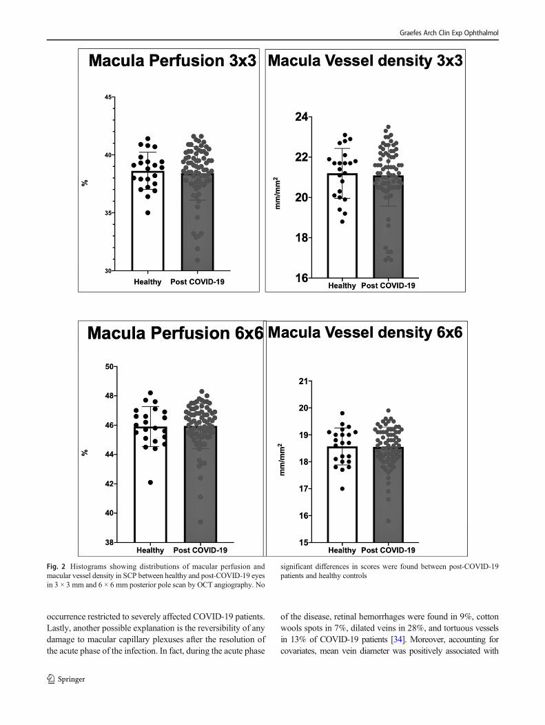
Mean SCP VD in the post-COVID-19 group at 3 × 3 mmand 6 × 6 mm was 21.20 ± 1.2% and 18.49 ± 1.18% respec-tively. Values of SCPVP extracted from the same acquisitions
were 0.386 ± 0.02 for 3 × 3 mm and 0.459 ± 0.03 for 6 × 6mm. With regard to DCP, mean vessel density in the post-COVID group was 21.82 ± 2.51% (3 × 3 acquisitions).Intraclass correlation coefficient revealed a good reliabilityof the measurements of subfoveal choroidal thickness andFAZ perimeter (respectively ICC = 0.884 r = 0.802 and ICC= 0.961, r = 0.929) (Fig. 1).
Results from the inferential analysis revealed no significantdifferences between the two groups in terms of density andperfusion of SCP (see Fig. 2) or DCP. The comparison wasequally inconclusive for 3 × 3 mm and 6 × 6 mm acquisitions.A binary logistic regression was performed to exclude con-founding factors from the analysis but this led to no change inthe detected differences.
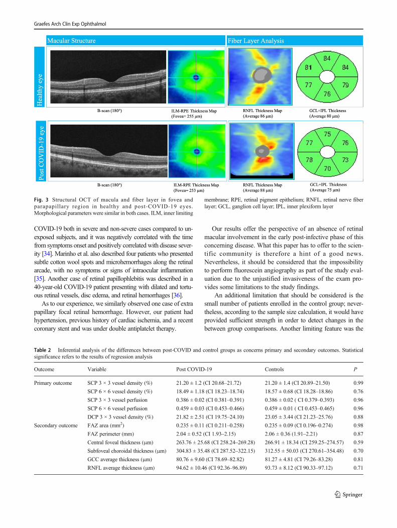
Results from OCTA analysis in COVID-19 group reveal amean FAZ area of 0.235 ± 0.11 mm2, with a FAZ perimeter of2.04 ± 0.52 mm. Central foveal thickness and subfoveal cho-roidal thickness were 263.76 ± 25.68 μm and 304.83 ±35.48 μm respectively. As concerns inner retinal layers, meanGCC thickness was 80.76 ± 9.60μmwhile meanRNFL thick-ness was 94.62 ± 10.46 μm. Among the secondary outcomemeasures, neither OCTA nor structural OCT parameters (see
Table 1 Descriptive analysis ofthe study groups Variable Post COVID-19 Controls P
Age (years) 53.7 ± 14 (CI 50.7–56.7) 44.7 ± 11.3 (CI 40–49.5) 0.006
Sex M = 39/70 (55.7%)
F = 31/70 (44.3%)
M = 8/22 (36.4%)
F = 14/22 (63.6%)
0.061
BCVA (Snellen) 20/22 20/23 0.53
Systemic arterial hypertension 14/70 (20%) 3/22 (13.6%) 0.047
Diabetes 30/70 (42.8%) 2/22 (9.1%) 0.003
BMI 25.63 ± 4.6 (CI 22.89–27.24) 22.75 ± 3.2 (CI 20.9–24.1) 0.041
Chronic kidney disease 6/70 (8.6%) 1/22 (4.5%) 0.12
Cognitive impairment 5/70 (7.1%) 2/22 (9.1%) 0.78
Previous stroke 1/70 (1.4%) 1/22 (4.5%) 0.65
Autoimmune diseases 6/70 (8.6%) 2/22 (9.1%) 0.92
Vitreomacular traction 5/70 (7.1%) 2/22 (9.1%) 0.88
Epiretinal membrane 5/70 (7.1%) 1/22 (4.5%) 0.79
Macular hole 3/70 (4.3%) 0/22 (0%) 0.54
Cotton wool spots 9/70 (12.9%) 0/22 0.09
Previous vitreoretinal surgery 5/70 (7.1%) 1/22 (4.5%) 0.59
Myopia 12/70 (17.1%) 4/22 (18.2%) 0.93
Days since symptoms onset 60.3 ± 13.6
Days since hospital discharge 36.1 ± 12.9
Ocular symptoms during infection 41/70 (58.6%)
Ocular symptoms post infection 30/70 (42.9%)
Intensive care unit admission 9/70 (12.9%)
Oxygen therapy 33/70 (45.8%)
Non-invasive ventilation 11/70 (15.7%)
Mechanical ventilation 4/70 (5.7%)
Graefes Arch Clin Exp Ophthalmol
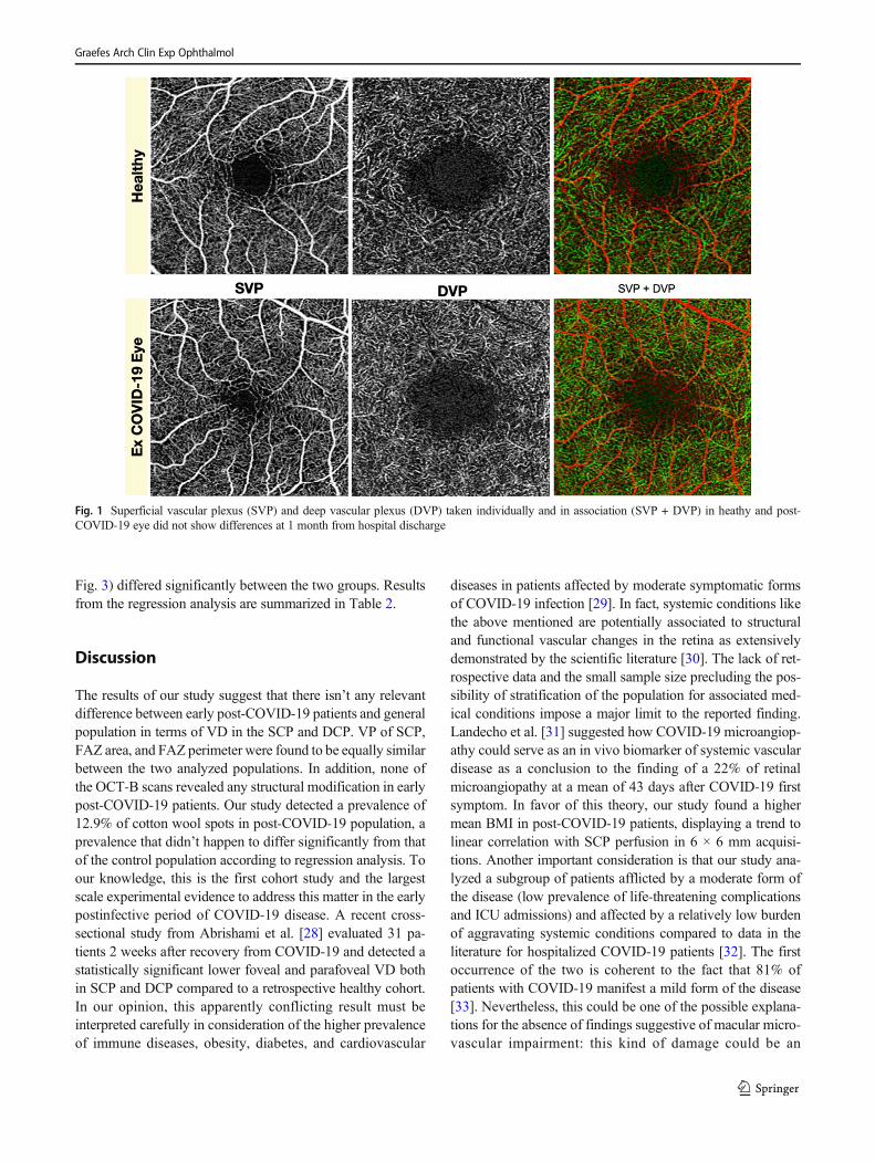
Fig. 3) differed significantly between the two groups. Resultsfrom the regression analysis are summarized in Table 2.
Discussion
The results of our study suggest that there isn’t any relevantdifference between early post-COVID-19 patients and generalpopulation in terms of VD in the SCP and DCP. VP of SCP,FAZ area, and FAZ perimeter were found to be equally similarbetween the two analyzed populations. In addition, none ofthe OCT-B scans revealed any structural modification in earlypost-COVID-19 patients. Our study detected a prevalence of12.9% of cotton wool spots in post-COVID-19 population, aprevalence that didn’t happen to differ significantly from thatof the control population according to regression analysis. Toour knowledge, this is the first cohort study and the largestscale experimental evidence to address this matter in the earlypostinfective period of COVID-19 disease. A recent cross-sectional study from Abrishami et al. [28] evaluated 31 pa-tients 2 weeks after recovery from COVID-19 and detected astatistically significant lower foveal and parafoveal VD bothin SCP and DCP compared to a retrospective healthy cohort.In our opinion, this apparently conflicting result must beinterpreted carefully in consideration of the higher prevalenceof immune diseases, obesity, diabetes, and cardiovascular
diseases in patients affected by moderate symptomatic formsof COVID-19 infection [29]. In fact, systemic conditions likethe above mentioned are potentially associated to structuraland functional vascular changes in the retina as extensivelydemonstrated by the scientific literature [30]. The lack of ret-rospective data and the small sample size precluding the pos-sibility of stratification of the population for associated med-ical conditions impose a major limit to the reported finding.Landecho et al. [31] suggested how COVID-19 microangiop-athy could serve as an in vivo biomarker of systemic vasculardisease as a conclusion to the finding of a 22% of retinalmicroangiopathy at a mean of 43 days after COVID-19 firstsymptom. In favor of this theory, our study found a highermean BMI in post-COVID-19 patients, displaying a trend tolinear correlation with SCP perfusion in 6 × 6 mm acquisi-tions. Another important consideration is that our study ana-lyzed a subgroup of patients afflicted by a moderate form ofthe disease (low prevalence of life-threatening complicationsand ICU admissions) and affected by a relatively low burdenof aggravating systemic conditions compared to data in theliterature for hospitalized COVID-19 patients [32]. The firstoccurrence of the two is coherent to the fact that 81% ofpatients with COVID-19 manifest a mild form of the disease[33]. Nevertheless, this could be one of the possible explana-tions for the absence of findings suggestive of macular micro-vascular impairment: this kind of damage could be an
Fig. 1 Superficial vascular plexus (SVP) and deep vascular plexus (DVP) taken individually and in association (SVP + DVP) in heathy and post-COVID-19 eye did not show differences at 1 month from hospital discharge
Graefes Arch Clin Exp Ophthalmol
occurrence restricted to severely affected COVID-19 patients.Lastly, another possible explanation is the reversibility of anydamage to macular capillary plexuses after the resolution ofthe acute phase of the infection. In fact, during the acute phase
of the disease, retinal hemorrhages were found in 9%, cottonwools spots in 7%, dilated veins in 28%, and tortuous vesselsin 13% of COVID-19 patients [34]. Moreover, accounting forcovariates, mean vein diameter was positively associated with
Fig. 2 Histograms showing distributions of macular perfusion andmacular vessel density in SCP between healthy and post-COVID-19 eyesin 3 × 3 mm and 6 × 6 mm posterior pole scan by OCT angiography. No
significant differences in scores were found between post-COVID-19patients and healthy controls
Graefes Arch Clin Exp Ophthalmol
COVID-19 both in severe and non-severe cases compared to un-exposed subjects, and it was negatively correlated with the timefrom symptoms onset and positively correlated with disease sever-ity [34]. Marinho et al. also described four patients who presentedsubtle cotton wool spots and microhemorrhages along the retinalarcade, with no symptoms or signs of intraocular inflammation[35]. Another case of retinal papillophlebitis was described in a40-year-old COVID-19 patient presenting with dilated and tortu-ous retinal vessels, disc edema, and retinal hemorrhages [36].
As to our experience, we similarly observed one case of extrapapillary focal retinal hemorrhage. However, our patient hadhypertension, previous history of cardiac ischemia, and a recentcoronary stent and was under double antiplatelet therapy.
Our results offer the perspective of an absence of retinalmacular involvement in the early post-infective phase of thisconcerning disease. What this paper has to offer to the scien-tific community is therefore a hint of a good news.Nevertheless, it should be considered that the impossibilityto perform fluorescein angiography as part of the study eval-uation due to the unjustified invasiveness of the exam pro-vides some limitations to the study findings.
An additional limitation that should be considered is thesmall number of patients enrolled in the control group; never-theless, according to the sample size calculation, it would haveprovided sufficient strength in order to detect changes in thebetween group comparisons. Another limiting feature was the
Fig. 3 Structural OCT of macula and fiber layer in fovea andparapapillary region in healthy and post-COVID-19 eyes.Morphological parameters were similar in both cases. ILM, inner limiting
membrane; RPE, retinal pigment epithelium; RNFL, retinal nerve fiberlayer; GCL, ganglion cell layer; IPL, inner plexiform layer
Table 2 Inferential analysis of the differences between post-COVID and control groups as concerns primary and secondary outcomes. Statisticalsignificance refers to the results of regression analysis
Outcome Variable Post COVID-19 Controls P
Primary outcome SCP 3 × 3 vessel density (%) 21.20 ± 1.2 (CI 20.68–21.72) 21.20 ± 1.4 (CI 20.89–21.50) 0.99
SCP 6 × 6 vessel density (%) 18.49 ± 1.18 (CI 18.23–18.74) 18.57 ± 0.68 (CI 18.28–18.86) 0.76
SCP 3 × 3 vessel perfusion 0.386 ± 0.02 (CI 0.381–0.391) 0.386 ± 0.02 ( CI 0.379–0.393) 0.96
SCP 6 × 6 vessel perfusion 0.459 ± 0.03 (CI 0.453–0.466) 0.459 ± 0.01 ( CI 0.453–0.465) 0.96
DCP 3 × 3 vessel density (%) 21.82 ± 2.51 (CI 19.75–24.10) 23.05 ± 3.44 (CI 21.23–25.76) 0.88
Secondary outcome FAZ area (mm2) 0.235 ± 0.11 (CI 0.211–0.258) 0.235 ± 0.09 (CI 0.196–0.274) 0.98
FAZ perimeter (mm) 2.04 ± 0.52 (CI 1.93–2.15) 2.06 ± 0.36 (1.91–2.21) 0.87
Central foveal thickness (μm) 263.76 ± 25.68 (CI 258.24–269.28) 266.91 ± 18.34 (CI 259.25–274.57) 0.59
Subfoveal choroidal thickness (μm) 304.83 ± 35.48 (CI 287.52–322.15) 312.55 ± 50.03 (CI 270.61–354.48) 0.70
GCC average thickness (μm) 80.76 ± 9.60 (CI 78.69–82.82) 81.27 ± 4.81 (CI 79.26–83.28) 0.81
RNFL average thickness (μm) 94.62 ± 10.46 (CI 92.36–96.89) 93.73 ± 8.12 (CI 90.33–97.12) 0.71
Graefes Arch Clin Exp Ophthalmol
younger age of patients in the control group. By the way, it isimportant to notice that this difference was merely statisticaland characterized by a scarce clinical relevance (mean age of54 in the post-COVID group versus mean age of 45 in controlgroup).
Further studies will be needed to address this importantsubject of actuality and confirm or contrast our findings, alsofocusing on the subgroup of post-COVID-19 patient which ismore susceptible to possible retinal microvascular sequelae.
Acknowledgments The authors want to thank Franziska MichaelaLohmeyer, MSc, BcOT, for her editing contribution and all the GemelliAgainst COVID Post-Acute Care Study Group teams: Angiology team:Santoliquido, A., Santoro, L., Nesci, and A., Popolla, V.;Gastroenterology team: Settanni, C.R.; Geriatric team: Benvenuto, F.,Bramato, G., Carfì, A., Ciciarello, F., Lo Monaco, M.R., Martone,A.M., Marzetti, E., Rocchi, S., Rota, E., Salerno, A., Tritto, M.,Calvani, R., Picca, A., Catalano, L., and Savera, G.; Infectious diseaseteam: Fantoni, M., Tamburrini, E., Borghetti, A., and Di Gianbenedetto,S.; Internal Medicine team: Addolorato, G., Franceschi, F., Mingrone, G.,and Stella, L.; Microbiology team: Sanguinetti, M., Cattani, P., andMarchetti, S.; Neurology team: Bizzarro, A. and Lauria, A.;Otolaryngology team: Passali, G.C., Paludetti, G., Galli, J., Crudo, F.,Di Cintio, G., Longobardi, Y., Tricarico, L., and Santantonio, M.;Pediatric team: Buonsenso, D., Valentini, P., Pata, D., Sinatti, D., andDe Rose, C.; Pneumology team: Richeldi, L., Lombardi, F., andCalabrese, A.; Psychiatric team: Sani, G., Janiri, D., Giuseppin, G.,Molinaro, M., and Modica, M.; Radiology team: Natale, L., Larici,A.R., and Marano, R.; and Rheumatology team: Paglionico, A.,Petricca, L., Gigante, L., and Natalello, G.
Author contributions Conceptualization, S Rizzo, MC Savastano, GGambini, MG Cozzupoli, A Savastano; methodology, MC Savastano,G Gambini, E Crincoli, U De Vico; software, E Crincoli, F Martelli;validation, S Rizzo, MC Savastano; formal analysis, E Crincoli, MCSavastano; investigation, C Culiersi, MG Cozzupoli, MC Savastano, GGambini, F Landi, F Pagano, U De Vico; resources, AM Minnella, BFalsini; data curation, C Culiersi, E Crincoli, MG Cozzupoli, MCSavastano, U De Vico; writing—original draft preparation, E Crincoli;writing—review and editing, E Crincoli, A Savastano, MC Savastano;supervision, S Rizzo; project administration, S Rizzo, MC Savastano. Allauthors have read and agreed to the published version of the manuscript.
Funding Open Access funding provided by Università Cattolica delSacro Cuore. General research fund of Fondazione PoliclinicoUniversitario A. Gemelli IRCCS, Catholic University of “Sacro Cuore”,Rome, Italy.
Compliance with ethical standards
Ethics committee authorization ID number 003220/20
Ethical approval All procedures performed in studies involving humanparticipants were in accordance with the ethical standards of the institu-tional and/or national research committee and with the 1964 Helsinkideclaration and its later amendments or comparable ethical standards.
Informed consent Informed consent was obtained from all individualparticipants included in the study.
Conflict of interest The authors declare that they have no conflict ofinterest.
Open Access This article is licensed under a Creative CommonsAttribution 4.0 International License, which permits use, sharing, adap-tation, distribution and reproduction in any medium or format, as long asyou give appropriate credit to the original author(s) and the source, pro-vide a link to the Creative Commons licence, and indicate if changes weremade. The images or other third party material in this article are includedin the article's Creative Commons licence, unless indicated otherwise in acredit line to the material. If material is not included in the article'sCreative Commons licence and your intended use is not permitted bystatutory regulation or exceeds the permitted use, you will need to obtainpermission directly from the copyright holder. To view a copy of thislicence, visit http://creativecommons.org/licenses/by/4.0/.
References
1. WHO,W. (2020).WHODirector-General’s opening remarks at themedia briefing on COVID-19.
2. Li G, Fan Y, Lai Y, Han T, Li Z, Zhou P, Pan P, Wang W, Hu D,Liu X, Zhang Q, Wu J (2020) Coronavirus infections and immuneresponses. J Med Virol 92(4):424–432. https://doi.org/10.1002/jmv.25685
3. Menter T, Haslbauer JD, & Nienhold R. (2020). Post-mortem ex-amination of COVID-19 patients reveals diffuse alveolar damagewith severe capillary congestion and variegated findings of lungsand other organs suggesting vascular dysfunction. 77(2), 198–209.
4. Marchetti M (2020) COVID-19-driven endothelial damage: com-plement, HIF-1, and ABL2 are potential pathways of damage andtargets for cure. Ann Hematol 99(8):1701–1707. https://doi.org/10.1007/s00277-020-04138-8
5. Saba, L., & Sverzellati, N. (2020). Is COVID evolution due to occur-rence of pulmonary vascular thrombosis? Journal of Thoracic Imaging.https://doi.org/10.1097/RTI.0000000000000530
6. Chen L, Li X, Chen M, Feng Y, Xiong C (2020) The ACE2 ex-pression in human heart indicates new potential mechanism of heartinjury among patients infected with SARS-CoV-2. Cardiovasc Res116(6):1097–1100. https://doi.org/10.1093/cvr/cvaa078
7. Zhu P, Verma A, Prasad T, Li Q (2020) Expression and function ofMas-related G protein-coupled receptor D and its ligandalamandine in retina. Mol Neurobiol 57(1):513–527. https://doi.org/10.1007/s12035-019-01716-4
8. Casagrande M, Fitzek A, Püschel K, Aleshcheva G, Schultheiss H-P, Berneking L, Spitzer MS, Schultheiss M (2020) Detection ofSARS-CoV-2 in human retinal biopsies of deceased COVID-19patients. Ocul Immunol Inflamm 28(5):721–725. https://doi.org/10.1080/09273948.2020.1770301
9. Donati, S., Maresca, A. M., Cattaneo, J., Grossi, A., Mazzola, M.,Caprani, S. M., Premoli, L., Docchio, F., Rizzoni, D., Guasti, L., &Azzolini, C. (2019). Optical coherence tomography angiographyand arterial hypertension: a role in identifying subclinical microvas-cular damage? European Journal of Ophthalmology,1120672119880390. https://doi.org/10.1177/1120672119880390
10. Kashani AH, Chen C-L, Gahm JK, Zheng F, Richter GM,Rosenfeld PJ, Shi Y, Wang RK (2017) Optical coherence tomog-raphy angiography: a comprehensive review of current methodsand clinical applications. Prog Retin Eye Res 60:66–100. https://doi.org/10.1016/j.preteyeres.2017.07.002
11. Gemelli Against COVID-19 Post-Acute Care Study Group (2020)Post-COVID-19 global health strategies: the need for an interdisci-plinary approach. Aging Clin Exp Res 32(8):1613–1620. https://doi.org/10.1007/s40520-020-01616-x
Graefes Arch Clin Exp Ophthalmol
12. Al-Sheikh M, Phasukkijwatana N, Dolz-Marco R, Rahimi M, IafeNA, Freund KB, Sadda SR, Sarraf D (2017) Quantitative OCTangiography of the retinal microvasculature and the choriocapillarisin myopic eyes. Invest Ophthalmol Vis Sci 58(4):2063–2069.https://doi.org/10.1167/iovs.16-21289
13. Milani P, Montesano G, Rossetti L, Bergamini F, Pece A (2018)Vessel density, retinal thickness, and choriocapillaris vascular flowin myopic eyes on OCT angiography. Graefe’s Archive for Clinicaland Experimental Ophthalmology = Albrecht Von Graefes ArchivFur Klinische Und Experimentelle Ophthalmologie 256(8):1419–1427. https://doi.org/10.1007/s00417-018-4012-y
14. Daniels AB, Froehler MT, Nunnally AH, Pierce JM, Bozic I, StoneCA, Santapuram PR, Tao YK, Boyd KL, Himmel LE, Chen S-C, DuL, Friedman DL, Richmond A (2019) Rabbit model of intra-arterialchemotherapy toxicity demonstrates retinopathy and vasculopathy re-lated to drug and dose, not procedure or approach. Invest OphthalmolVis Sci 60(4):954–964. https://doi.org/10.1167/iovs.18-25346
15. Minnella AM, Barbano L, &Verrecchia E. (2019). Macular impair-ment in Fabry disease: a morpho-functional assessment by swept-source OCT angiography and focal electroretinography. 60(7):2667-2675.
16. Rosenfeld PJ, DurbinMK, Roisman L, Zheng F,Miller A, RobbinsG, Schaal KB, Gregori G (2016) ZEISS AngioplexTM spectral do-main optical coherence tomography angiography: technical aspects.Dev Ophthalmol 56:18–29. https://doi.org/10.1159/000442773
17. Lu Y,Wang JC, Zeng R, Katz R, Vavvas DG,Miller JW,Miller JB(2019) Quantitative comparison of microvascular metrics on threeoptical coherence tomography angiography devices in chorioretinaldisease. Clinical Ophthalmology (Auckland, N.Z.), 13, 2063–2069.https://doi.org/10.2147/OPTH.S215322
18. Kashani AH, Zhang Y, Capone A, Drenser KA, Puliafito C,Moshfeghi AA, Williams GA, Trese MT (2016) Impaired retinal per-fusion resulting from vitreoretinal traction: a mechanism of retinalvascular insufficiency. Ophthalmic Surgery, Lasers & ImagingRetina 47(3):1–11. https://doi.org/10.3928/23258160-20160229-03
19. Chen H, Chi W, Cai X, Deng Y, Jiang X, Wei Y, Zhang S (2019)Macular microvasculature features before and after vitrectomy inidiopathic macular epiretinal membrane: an OCT angiography anal-ysis. Eye 33(4):619–628. https://doi.org/10.1038/s41433-018-0272-3
20. Wilczyński T, HeinkeA,Niedzielska-Krycia A, JorgD,Michalska-Małecka K (2019) Optical coherence tomography angiography fea-tures in patients with idiopathic full-thickness macular hole, beforeand after surgical treatment. Clin Interv Aging 14:505–514. https://doi.org/10.2147/CIA.S189417
21. Fan H, Chen H-Y, Ma H-J, Chang Z, Yin H-Q, Ng DS-C, CheungCY, Hu S, Xiang X, Tang S-B, Li S-N (2017) Reduced macularvascular density in myopic eyes. Chin Med J 130(4):445–451.https://doi.org/10.4103/0366-6999.199844
22. Romano MR, Cennamo G, Schiemer S, Rossi C, Sparnelli F,Cennamo G (2017) Deep and superficial OCT angiography chang-es after macular peeling: idiopathic vs diabetic epiretinal mem-branes. Graefe’s Archive for Clinical and ExperimentalOphthalmology = Albrecht Von Graefes Archiv Fur KlinischeUnd Experimentelle Ophthalmologie 255(4):681–689. https://doi.org/10.1007/s00417-016-3534-4
23. Vujosevic S, Toma C, Villani E, Gatti V, Brambilla M, Muraca A,Ponziani MC, Aimaretti G, Nuzzo A, Nucci P, De Cilla’, S. (2019)Early detection of microvascular changes in patients with diabetesmellitus without and with diabetic retinopathy: comparison be-tween different swept-source OCT-A instruments. J Diabetes Res2019:2547216. https://doi.org/10.1155/2019/2547216
24. Takayama K, Kaneko H, Ito Y, Kataoka K, Iwase T, Yasuma T,Matsuura T, Tsunekawa T, Shimizu H, Suzumura A, Ra E, AkahoriT, Terasaki H (2018) Novel classification of early-stage systemichypertensive changes in human retina based on OCTA
measurement of choriocapillaris. Sci Rep 8(1):15163. https://doi.org/10.1038/s41598-018-33580-y
25. Jiang H, Wei Y, Shi Y, Wright CB, Sun X, Gregori G, Zheng F,Vanner EA, Lam BL, Rundek T, Wang J (2018) Altered macularmicrovasculature in mild cognitive impairment and Alzheimer dis-ease. Journal of Neuro-Ophthalmology: The Official Journal of theNorth American Neuro-Ophthalmology Society 38(3):292–298.https://doi.org/10.1097/WNO.0000000000000580
26. Peng C, Kwapong WR, Xu S, Muse FM, Yan J, Qu M, Cao Y,Miao H, Zhen Z, Wu B, Han Z (2020) Structural and microvascularchanges in the macular are associated with severity of white matterlesions. Front Neurol 11:521. https://doi.org/10.3389/fneur.2020.00521
27. VadalàM, Castellucci M, Guarrasi G, TerrasiM, La Blasca T,MulèG (2019) Retinal and choroidal vasculature changes associated withchronic kidney disease. Graefe’s Archive for Clinical andExperimental Ophthalmology = Albrecht Von Graefes Archiv FurKlinische Und Experimentelle Ophthalmologie 257(8):1687–1698.https://doi.org/10.1007/s00417-019-04358-3
28. Abrishami M, Emamverdian Z, Shoeibi N, Omidtabrizi A, DaneshvarR, Rezvani TS, Saeedian N, Eslami S,MazloumiM, Sadda S, Sarraf D(2020) Optical coherence tomography angiography analysis of the ret-ina in patients recovered from COVID-19: a case-control study.Canadian Journal of Ophthalmology Journal CanadienD’ophtalmologie. https://doi.org/10.1016/j.jcjo.2020.11.006
29. Vavvas,D.G., Sarraf,D., Sadda, S.R., Eliott, D., Ehlers, J. P.,Waheed,N. K., Morizane, Y., Sakamoto, T., Tsilimbaris, M., & Miller, J. B.(2020). Concerns about the interpretation of OCT and fundus findingsin COVID-19 patients in recent Lancet publication. Eye (London,England). https://doi.org/10.1038/s41433-020-1084-9
30. Flammer J, Konieczka K, Bruno RM, Virdis A, Flammer AJ,Taddei S (2013) The eye and the heart. Eur Heart J 34(17):1270–1278. https://doi.org/10.1093/eurheartj/eht023
31. Landecho MF, Yuste JR, Gándara E, Sunsundegui P, Quiroga J,Alcaide AB, García-Layana A (2020) COVID-19 retinal microan-giopathy as an in vivo biomarker of systemic vascular disease? JIntern Med. https://doi.org/10.1111/joim.13156
32. Mazzaccaro D, Giacomazzi F, Giannetta M, Varriale A,Scaramuzzo R, Modafferi A, Malacrida G, Righini P, Marrocco-Trischitta MM, Nano G (2020) Non-overt coagulopathy in non-ICU patients with mild to moderate COVID-19 pneumonia. J ClinMed 9(6):1781. https://doi.org/10.3390/jcm9061781
33. Wu Z, McGoogan JM (2020) Characteristics of and important les-sons from the coronavirus disease 2019 (COVID-19) outbreak inChina: summary of a report of 72 314 cases from the ChineseCenter for Disease Control and Prevention. JAMA 323(13):1239–1242. https://doi.org/10.1001/jama.2020.2648
34. Invernizzi A, Torre A, Parrulli S, Zicarelli F, SchiumaM, Colombo V,Giacomelli A, Cigada M, Milazzo L, Ridolfo A, Faggion I, Cordier L,Oldani M, Marini S, Villa P, Rizzardini G, Galli M, Antinori S,Staurenghi G, Meroni L (2020) Retinal findings in patients withCOVID-19: results from the SERPICO-19 study. EClinicalMedicine100550. https://doi.org/10.1016/j.eclinm.2020.100550
35. Marinho PM,Marcos AAA, RomanoAC, Nascimento H, Belfort R(2020) Retinal findings in patients with COVID-19. Lancet(London, England), 395(10237), 1610. https://doi.org/10.1016/S0140-6736(20)31014-X
36. Insausti-García, A., Reche-Sainz, J. A., Ruiz-Arranz, C., LópezVázquez, Á., & Ferro-Osuna, M. (2020). Papillophlebitis in aCOVID-19 patient: inflammation and hypercoagulable state.European Journal of Ophthalmology, 1120672120947591. https://doi.org/10.1177/1120672120947591
Publisher’s note Springer Nature remains neutral with regard to jurisdic-tional claims in published maps and institutional affiliations.
Graefes Arch Clin Exp Ophthalmol