EYE INVOLVEMENT IN THE SPONDYLOARTHROPATHIES
14
SPONDYLOARTHROPATHIES 0889-857>(/98 $8.00 + .OO EYE INVOLVEMENT IN THE SPONDYLOARTHROPATHIES Antonio Bafiares, MD, PhD, Cksar Hernbndez-Garcia, MD, PhD, Benjamin Fernandez-Gutierrez, MD, PhD, and Juan A. Jover, MD, PhD Eye inflammation may be a prominent feature of several rheumatic diseases. The spondyloarthropathies, including ankylosing spondylitis (AS), Reiter 's syndrome (RS), psoriatic spondyloarthropathy, spondy- loarthritis associated with inflammatory bowel disease (IBD), and undif- ferentiated spondyloarthropathy, may present a constellation of symp- toms, including inflammatory low back pain, peripheral arthritis, enthesitis, recurrent episodes of diarrhea and eye inflammation. Uveitis, conjunctivitis, and episcleritis or scleritis can all occur in patients with known spondyloarthropathies or may be key symptoms for the diagno- sis of a previously undiagnosed spondyloarthropathy. Furthermore, a subset of patients with acute anterior uveitis (AAU) with exclusively ocular symptoms are also human leukocyte antigen (HLA) B27 positive and have HLA-B27-related idiopathic anterior uveitis (AU), which should also be included in the spectrum of the spondyloarthropathies. To avoid a superficial treatment of all forms of eye inflammation, this article focuses mainly on uveitis, an ocular manifestation of spondy- loarthropathies that entails subtle and complex etiologic, clinical and therapeutic implications. DEFINITION Uveitis is a general term used to define inflammation in the uveal tract, which is the middle layer of the eye (Fig. 1). Uveitis can be This work was supported by Grant No. 97/0789 from the Fondo de Investigacion Sanitaria (FIS) of Spain. From the Service of Rheumatology, Hospital Clinic0 San Carlos, Madrid, Spain RHEUMATIC DISEASE CLINICS OF NORTH AMERICA VOLUME 24 * NUMBER 4 - NOVEMBER 1998 771
-
Upload
independent -
Category
Documents
-
view
1 -
download
0
Transcript of EYE INVOLVEMENT IN THE SPONDYLOARTHROPATHIES
SPONDYLOARTHROPATHIES 0889-857>(/98 $8.00 + .OO
EYE INVOLVEMENT IN THE SPONDYLOARTHROPATHIES
Antonio Bafiares, MD, PhD, Cksar Hernbndez-Garcia, MD, PhD, Benjamin Fernandez-Gutierrez, MD, PhD,
and Juan A. Jover, MD, PhD
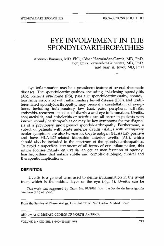
Eye inflammation may be a prominent feature of several rheumatic diseases. The spondyloarthropathies, including ankylosing spondylitis (AS), Reiter 's syndrome (RS), psoriatic spondyloarthropathy, spondy- loarthritis associated with inflammatory bowel disease (IBD), and undif- ferentiated spondyloarthropathy, may present a constellation of symp- toms, including inflammatory low back pain, peripheral arthritis, enthesitis, recurrent episodes of diarrhea and eye inflammation. Uveitis, conjunctivitis, and episcleritis or scleritis can all occur in patients with known spondyloarthropathies or may be key symptoms for the diagno- sis of a previously undiagnosed spondyloarthropathy. Furthermore, a subset of patients with acute anterior uveitis (AAU) with exclusively ocular symptoms are also human leukocyte antigen (HLA) B27 positive and have HLA-B27-related idiopathic anterior uveitis (AU), which should also be included in the spectrum of the spondyloarthropathies. To avoid a superficial treatment of all forms of eye inflammation, this article focuses mainly on uveitis, an ocular manifestation of spondy- loarthropathies that entails subtle and complex etiologic, clinical and therapeutic implications.
DEFINITION
Uveitis is a general term used to define inflammation in the uveal tract, which is the middle layer of the eye (Fig. 1). Uveitis can be
This work was supported by Grant No. 97/0789 from the Fondo de Investigacion Sanitaria (FIS) of Spain.
From the Service of Rheumatology, Hospital Clinic0 San Carlos, Madrid, Spain
RHEUMATIC DISEASE CLINICS OF NORTH AMERICA
VOLUME 24 * NUMBER 4 - NOVEMBER 1998 771

772 BANARES et a1
/ I RETINA
VITREOUS HUMOR
OPTIC DISC
OPTIC NERVE 1"" I
Figure 1 . The eye. Inflammation affecting the iris, the ciliary body, the pars plana and/or the choroid are grouped under the general term of uveitis.
classified according to parameters such as anatomic location, clinical course, or laterality. Thus, based on its anatomic location, uveitis may be denominated as anterior when it involves the iris or the ciliary body (also called iritis or iridocyclifis); posterior when it affects the choroid or, by extension, the retina (also called choroiditis or retinochoroiditis); intermediate uveitis when the inflammation is limited to the vitreum, peripheral retina, and pars plana of the ciliary body (sometimes termed pars planitis); or panuveitis when the whole uvea is involved in the inflammatory process. Uveitis may be unilateral, affecting only one eye at a time (although recurrences may occur in the contralateral eye), or bilateral. Furthermore, uveitis can be classified arbitrarily by its onset as acute or insidious and by its duration as self-limited (with inflammation lasting less than 3 months), chronic (with continuous inflammation for more than 3 months) and recurrent (when acute flares appear after a complete recovery of a self-limited uveitis). Finally, for a correct diagno- sis, the clinician should take into account the presence or absence of morphologic features, such as the presence of granulomatous or non- granulomatous keratitic precipitates, hypopyon, synechiae, cystoid mac- ular edema (CME), band keratopathy, cataract, papillitis, keratouveitis, and snow banks in the pars plana (Table l).3

EYE INVOLVEMENT IN THE SPONDYLOARTHROPATHIES 773
Table 1. MORPHOLOGIC CHARACTERISTIC USED TO DEFINE SUBSETS OF UVElTlS
Characteristic
Keratitic precipitates
HYPoPYon
Synechiae
Cystoid macular edema Band keratopathy
Cataract
Papillitis
Keratouveitis
Snow banks
Definition
Aggregates of inflammatory cells visible at slit lamp examination that accumulate on the endothelial surface of the cornea and on the trabecular meshwork. Can be further classified as granulomatous or nongranulomatous according to the size and to the greasy appearance of the granulomatous keratitic precipitates (so-called “mouton fat”). This term is somewhat misleading and does not imply a pathologic correlation with true granulomas.
A collection of inflammatory cells that prec$itate in the lower angle of the anterior chamber forming a meniscus
Adhesions between the iris and the lens capsule (posterior synechiae) or the iris and the cornea (anterior synechiae) that give the pupil an irregular form
Fluid in the macula resulting in reduced visual acuity Calcium deposition in the Bowman’s membrane of the
cornea that appears as a result of chronic inflammation Lens opacities that develop as a consequence of chronic
inflammation or the use of corticosteroids Inflammation over the optic disc with blurring and elevation
of the optic disc margins A subset of uveitis that appears as a consequence of or
concurrently with a keratitis. Usually represents a viral etiology
A typical accumulation of a white material in the pars plana that resembles snow banks
INCIDENCE OF EYE DISEASE IN SPONDYLOARTHROPATHIES
The reported incidence of rheumatic diseases in patients with uveitis varies substantially in the literature, mainly depending on selection biases and specialty 33, 36 Table 2 reflects the likelihood of uveitis in patients with a given spondyloarthropathy and the comple- mentary likelihood of spondyloarthropathy in patients with uveitis. In the authors’ experience, roughly one third of patients attending the Uveitis Clinic at the Hospital Clinic0 San Carlos (HCSC) with any type of uveitis have either an underlying spondyloarthropathy or an HLA- B27-related idiopathic uvei t i~.~ This proportion increases to two thirds among patients presenting with AAU. Conversely, the incidence of AU in patients with ankylosing spondylitis ranges from 20% to 3070.~’ Fur- thermore, 84% to 90% of patients with HLA-B27-related AAU have AS or RS.12, 32 Conjunctivitis occurs in 30% to 60% of patients with RS, and it is the most frequent ocular complaint in the disease,I8 whereas the incidence of uveitis (12%-37%) and keratitis (4%) in these patients appears to be lower. Only 7% to 16% of patients with psoriatic peripheral arthritis or psoriatic spondylitis develop u ~ e i t i s . ~ ~ Finally, 2% to 9% of patients

774 BANARES et a1
Table 2. LIKELIHOOD OF UVElTlS IN PATIENTS WITH SPONDYLOARTHROPATHIES AND LIKELIHOOD OF SPONDYLOARTHROPATHY IN PATIENTS PRESENTING WITH UVElTlS
Likelihood of Likelihood of Uveitis in Patients Disease in Patients
Disease with the Disease with Uveitis Comments
Ankylosing spondylitis
Reiter's syndrome
Undifferentiated spondyloarthropathy
Psoriatic spondyloarthritis
Inflammatory bowel disease
20-30% 15% of any uveitis, 3&50% of AAU,
positive AAU 84-90% HLA-B27,
12-37% 3% of any uveitis,
- 5% of any uveitis,
5-10% of AAU
8-12% Of AU
7-1 6% ~ 1 % of any uveitis
2-9% 2-3% of any uveitis
Conjunctivitis (3C-60%) is the most common ocular symptom
No data available about incidence bf uveitis in patients with the disease
patients with spondylitis or sacroiliitis
Likelihood increases in patients with arthritis, spondylitis, or cutaneous symptoms
Likelihood increases in
AU = anterior uveitis: AAU = acute anterior uveitis
with IBD (either ulcerative colitis or Crohn's disease) develop uveitis, but the incidence of uveitis may be higher in patients with arthritis, spondylitis or cutaneous disease.I4
CHARACTERIZATION OF SPONDYLOARTHROPATHY- ASSOCIATED UVElTlS
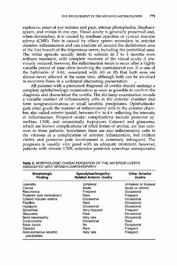
The clinical presentation of idiopathic, AS and RS HLA-B27-related uveitis is quite characteri~tic.~~ Uveitis associated with psoriatic spondy- loarthritis, IBD, or undifferentiated spondyloarthropathy can be less distinctive in its presentation and at times can pose a challenge for the clinician. Furthermore, some of these patients are HLA-B27 positive, whereas others are not. Because many patients present with a mixture of signs and symptoms that do not always fit into established patterns, diagnostic evaluation can be confusing and perhaps explains the diver- gence in published results pertaining to these entities. Table 3 shows some of the differential characteristics of AU associated with spondy- loarthropathies and compares them with those observed in AU associ- ated with other diseases.
Idiopathic, AS-, and RS-, HLA-B27-related uveitis occurs more often in white men between the third and fifth decades of life, but women and other races and age groups can also be affected.38 Signs and symptoms of uveitis in these patients include eye discomfort during a prodromal phase (usually up to 2 days), which is followed by an acute, sometimes
EYE INVOLVEMENT IN THE SPONDYLOARTHROPATHIES 775
explosive, onset of eye redness and pain, intense photophobia, blepharo- spasm, and miosis in one eye. Visual acuity is generally preserved and, when diminished, it is caused by medium opacities or cystoid macular edema (CME). Pain is caused by ciliary spasm secondary to anterior chamber inflammation and can irradiate all around the distribution area of the first branch of the trigeminus nerve, including the periorbital area. The initial episode usually tends to subside in 2 to 4 months even without treatment, with complete recovery of the visual acuity if pre- viously reduced; however, the inflammation tends to recur after a highly variable period of time often involving the contralateral eye. It is one of the hallmarks of AAU associated with AS or RS that both eyes are almost never affected at the same time, although both can be involved in recurrent flares in a unilateral alternating presentation.
All patients with a presumed diagnosis of uveitis should undergo a complete ophthalmologic examination as soon as possible to confirm the diagnosis and characterize the uveitis. The slit-lamp examination reveals a variable number of inflammatory cells in the anterior chamber that form nongranulomatous or small keratitic precipitates. Ophthalmolo- gists often grade the number of inflammatory cells in the anterior cham- ber, also called anterior tyndall, between 0 + to 4 + reflecting the intensity of inflammation. Frequent ocular complications include posterior sy- nechiae, CME, and occasionally hypopyon. Cataract and glaucoma, which are known complications of other forms of uveitis, are less com- mon in these patients. Sometimes there are also inflammatory cells in the vitreous as a complication of anterior inflammation, but evident vitritis and posterior pole involvement is extremely infrequent. The prognosis is usually very good with an adequate treatment; however, patients with chronic CME, extensive posterior synechiae unresponsive
_..
Table 3. MORPHOLOGIC CHARACTERIZATION OF THE ANTERIOR UVElTlS ASSOCIATED WITH SPONDYLOARTHROPATHY
Morphologic Spondyloarthropathy- Other Anterior Finding Related Anterior Uveitis Uveitis
Laterality Course Recurrence Posterior pole involvement Cystoid macular edema Papillitis HYPoPYon Synechiae Glaucoma Band keratopathy Conjunctivitis Snow banks Cataract Granulomatous keratitic
precipitates
Unilateral Acute Frequent Rare Occasional Rare Occasional Very frequent Rare Very rare Occasional No Rare Very rare
Unilateral or bilateral Acute or chronic Occasional Frequent Occasional Occasional Occasional Frequent Occasional Occasional Rare Occasional Frequent Frequent

776 BANARES et a1
to treatment, and a protracted course or evolution to chronicity might have higher rates of complications such as diminished visual acuity, cataract and glaucoma, and a subsequently poorer ocular outcome.
Although the clinical course of uveitis associated with IBD can be indistinguishable from AS- or RS-associated uveitis, it is more frequently bilateral, chronic, and HLA-B27 negative, involving both the anterior and posterior segments of the eye more often.24 Crohn's disease seems to be more frequently associated with uveitis than with ulcerative colitis, and the fact that the spectrum of ocular manifestations is broader in Crohn's disease'O1 24 may account for the differences observed as a whole between IBD and other HLA-B27-related uveitis. In patients at the HCSC, Crohn's disease presented with posterior pole involvement (vi- tritis or retinal vasculitis), had an insidious onset, and was subacute or chronic in course in half of the cases. In contrast, all patients with ulcerative colitis presented with typical unilateral acute anterior uveitis. The IBD can occasionally present with episcleritis, inflammation of the superficial vessels of the sclera, or peripheral corneal ulceration in the sclerocorneal j~nct ion.~
Uveitis associated with psoriatic arthritis or spondylitis is frequently accompanied by conjunctivitis, dry eye, or keratitis and may be indistin- guishable from AS-related uveitis but is less well characterized and often follows a chronic course or presents as acute bilateral uveitis.3,22 Because the prevalence of psoriasis is very high in the general population, one should be cautious and not automatically establish an etiologic relation- ship between cutaneous psoriasis and any given type of uveitis.
THE LINK BETWEEN EYE AND JOINT INFLAMMATION IN SPONDYLOARTHROPATHIES
Current advances in the etiology and physiopathology of the spon- dyloarthropathies are already outlined in other articles of this issue. Findings linking eye and joint inflammation in these diseases are briefly overviewed here. The similarities and differences in the pathogenesis of uveitis and arthritis remain a matter of discussion and research; however, although a great bulk of data derived from experimental animal models and human research indicate certain common pathogenic features, the precise mechanisms underlying the involvement of these tissues re- main unclear.
Experimental animal models of uveitis can be broadly divided into those causing anterior uveitis, which are more relevant for the purposes of this review, and those inducing posterior uveitis. Although class I major histocompatibility complex (MHC) molecules and certain infec- tious agents play a pivotal role in animal models of AU, models of posterior uveitis are mainly based on class I1 MHC molecules and macrophage or T-cell-mediated immune responses to certain uveitogenic peptides of the eye. Thus, a discussion of the animal models linking eye

EYE INVOLVEMENT IN THE SPONDYLOARTHROPATHIES 777
and joint inflammation in the spondyloarthropathies should necessarily focus on the role of HLA-B27 and infectious microorganism^.^, l3
As mentioned earlier, the spectrum of HLA-B27-associated diseases includes a subset of patients with idiopathic AU in whom HLA-B27 is positive, although clinical manifestations are limited to the eye. It has been suggested that HLA-B27 plays a central role linking exogenous factors such as infectious microorganisms with the different clinical manifestations of AS, RS, and AU42; however, this link remains mainly speculative for eye involvement. As occurs with joint disease, no individ- ual HLA-B27 subtype has been associated with eye inflammation. Mod- els of HLA-B27 transgenic rats and mice, currently considered as major evidence for direct HLA-B27 participation in the pathognesis of the spondyloarthropathies, develop spontaneous inflammatory disease af- fecting the gastrointestinal tract, peripheral and vertebral joints, the male genital tract, the skin, and the nails, but AU is infrequent and when present may be highly un~pecific.’~, l9 HLA-B27 transgenic mice immu- nized with a uveitogenic protein, the interphotoreceptor retinoid binding protein, develop uveitis less frequently than the background strain and HLA-A2 transgenic mice.41 Furthermore, uveitis induced in this model, with retinitis, panuveitis, and severe vitritis, is clinically distinct from that observed in HLA-B27-positive humans. The S-antigen, other retinal peptide that can induce posterior uveitis in animal models and is consid- ered a major autoantigen in human disease, shares a sequence homology with a HLA-B27-derived peptide,4O but the significance of this finding in the pathogenesis of HLA-B27-associated uveitis is unclear.
In addition to HLA-B27, several other genes are candidates in the pathogenesis of AAU, either acting independently or in concert. Patients with AS have a tenfold higher likelihood of developing AAU than their HLA-B27-positive relatives without AS.39 Islam and colleagues16 found that DR8, in association with HLA-B27, plays a role in the development of AAU in Japanese patients with AS. Heterozygosity of HLA-B27/ HLA-B60 has also been shown to increase the risk for AS,” but no data exist on the incidence of AAU. Both HLA-DR8 and HLA-B60 have been related with Salmonefla-induced arthritis and weitis.’, 37 Finally, polymorphism in an HLA-linked proteosome subunit gene (LMP2) may also be associated with the development of AAU in patients with AS.25
Thus, together with a genetic background, other factors seem to be needed to trigger AAU. In this sense, the role of gut and urogenital infections have been extensively investigated. Endotoxin-induced uveitis (EIU) has long been recognized as a model for AAU.35 Endotoxin injec- tion in the vitreum or in a distant site (using lipopolysaccharide of the outer membrane of different gram negative bacteria, such as Escherichia coli, Salmonella typhimurium, and Shigella flexneri) in susceptible species and strains induces a bilateral, dose-dependent, and self-limited AAU. The inflammatory process seems to be mediated by the release of a variety of pro-inflammatory cytokines (IL-la, TNF-a, IL-6) and nitric oxide by resident epithelial cells of the iris-ciliary body and cells infil- trating the anterior segment of the eye. This EIU can be inhibited or

778 BANARES et a1
suppressed by TGF-P-1, IL-10, IL-12, anti-CD54 (ICAM-1) monoclonal antibodies (mAb), anti-CDlla (LFA-1) mAb, and inhibitors of nitric oxide synthesis; however, important differences exist between this model and the AAU observed in humans with HLA-B27-related spondy- loarthropathies that have been already o~t l ined .~
Baggia and colleagues2 recently developed a new and promising model of bacterially induced AAU in HLA-B27 transgenic and control nontransgenic rats.2 Transgenic rats expressing a low copy number of HLA-B27 and control rats were exposed to Salmonella or Yersiniu infection and developed AU beginning 7 to 9 days after infection, frequently unilateral, and sometimes lasting for several weeks and recurring after improvement. In other words, the pattern resembled what" occurs in human HLA-B27-related AAU. Surprisingly, despite the expression of HLA-B27, these animals did not develop reactive arthritis after the infection, suggesting that gene copy number is critical for reactive arthri- tis to develop. Furthermore, and more important for the purposes of this review, the presence of a low copy number of HLA-B27 in these rats does not seem to influence either the incidence or severity of uveitis, pointing again to the influence of other genes or factors that might contribute to disease susceptibility.
Another field of research has been the study of the intestinal mucosa and its permeability. Mielants and colleagues were the first to describe the existence of subclinical gut inflammation in two patients with idio- pathic HLA-B27-related AAU.28 Subsequently, the authors observed that 18 of 27 patients with AU had evidence of gut inflammation in biopsy specimens obtained blindly during ileocolonoscopy from ileum, ileocecal valve, or The histopathologic characteristics of these lesions in- clude chronic intestinal inflammation with diffuse increase of chronic inflammatory cells in the lamina propria or partial villous flattening on ileum together with a diminished number of glands and distortion of crypts in the colon in all types of AU. These findings, which are very similar to those found in patients with spondyloarthropathies and HLA- B27 transgenic rats, suggest that either a chronic or a recurrent challenge to the intestinal mucosa or altered mucosa-associated lymphoid tissue might be implicated in the pathogenesis of AU, irrespective of the occurrence of other extraocular symptoms. Interestingly, a HLA-B27- positive phenotype was not a prerequisite for the presence of gut in- flammation. In fact, chronic intestinal inflammation was significantly more prevalent in HLA-B27-negative patients (85%) than in HLA-B27- positive patients with AU (50%), suggesting a role for other non-HLA- B27 factors.
UVElTlS AS A GUIDE FOR THE DIAGNOSIS OF SPONDY LOARTHROPATHY
For the rheumatologist approaching a patient with uveitis, a com- plete ophthalmologic description of the uveitis is essential and can help

EYE INVOLVEMENT IN THE SPONDYLOARTHROPATHIES 779
to avoid unnecessary tests that can delay the treatment and can be occasionally misleading. A chest roentgenogram and an FTA-ABS test for syphilis should be the only tests ordered routinely in all patients with uveitis because sarcoidosis and syphilis can mimic all other causes of uveitis.31 All other tests should be ordered on the basis of clinical suspi- cion. The authors have reported several clinical patterns of uveitis upon presentation that may help the rheumatologist in his diagnostic workup and in his ability to communicate with the referring ophthalm~logist.~
Table 4 shows the clinical patterns of uveitis associated with spondy-
Table 4. CLINICAL PATTERNS OF UVEITIS, SPONDYLOARTHROPATHIES AND DIFFERENTIAL DIAGNOSIS _"
Disease Spondyloarthropathies and Related Differential Diagnoses Diseases
Unilateral acute anterioi recurrent uveitis
Unilateral acute anterior nonrecurrent uveitis
Bilateral AAU
Chronic AU
Posterior uveitis (unilateral or bilateral chorioretinitis, retinal vasculitis)
Intermediate uveitis
Panuveitis with predominant chorioretinitis
Panuveitis with predominant vitritis
Panuveitis with predominant retinal vasculitis
Panuveitis with predominant exudative retinal detachment
Ankylosing spondylitis, Reiter's syndrome, reactive arthritis, idiopathic HLA-627-positive uveitis, undifferentiated spondyloarthropathy (rare), ulcerative colitis (rare), Crohn's disease (rare), psoriasis (rare)
Ankylosing spondylitis, Reiter's syndrome, reactive arthritis, idiopathic HLA-B27-positive uveitis, undifferentiated spondyloarthropathy (rare), ulcerative colitis (rare), Crohn's disease (rare), psoriasis (rare)
Undifferentiated spondyloarthropathy (rare), Crohn's disease (rare), psoriasis (rare)
(rare), Crohn's disease (rare) Undifferentiated spondyloarthropathy
None
Undifferentiated spondyloarthropathy
None (rare), Crohn's disease (rare)
Undifferentiated spondyloarthropathy (rare), Crohn's disease (rare)
Crohn's disease (rare)
Crohn's disease (rare)
Idiopathic HLA-627-negative uveitis, ophthalmologic uveitis, herpes keratouveitis, unspecific viral uveitis, syphilis
Idiopathic HLA-627-negative uveitis, ophthalmologic uveitis, herpes keratouveitis, unspecific viral uveitis, syphilis, other (rare)
Idiopathic HLA-B27-negative uveitis, ophthalmologic uveitis
Idiopathic HLA-627-negative uveitis, juvenile rheumatoid arthritis, sarcoidosis, Sjogren's syndrome
Sarcoidosis, Behpet's disease, systemic lupus erythematosus
Sarcoidosis, pars planitis
Toxoplasmosis, Vogt- Koyanagi-Harada syndrome, ophthalmological well-defined uveitis, idiopathic panuveitis
Idiopathic panuveitis, masquerade syndromes, Behpet's disease, sarcoidosis, tuberculosis
Behpet's disease, idiopathic panuveitis, syphilis, sarcoidosis
Vogt-Koyanagi-Harada syndrome, Behpet's disease
~~
AU = anterior uveitis; AAU = acute anterior uveitis.

780 BANARES et a1
loarthropathies and other specific diagnoses that should be ruled out for each pattern. Spondyloarthropathies are the leading systemic diseases identified in patients presenting with uveitis and represent most sys- temic diseases associated with AAU. Thus, AAU often can be a guide symptom for the diagnosis of previously unknown spondyloarthropa- thies. Sometimes, the patient had symptoms for a variable period of time that were not sufficiently severe to warrant seeking medical attention. In these cases, a simple clinical history reveals the existence of inflamma- tory low back pain, enthesitis, or recurrent episodes of diarrhea of unknown cause. In other cases, uveitis is the first symptom of a systemic disease, preceding the onset of other clinical manifestations. Previously undiagnosed spondyloarthropathies account for almost 54% of-"ptients attending the Uveitis Clinic of the HCSC32, 38 with spondyloarthropathy- related uveitis, a number in accordance with that observed by other authors. In the authors' experience, although differences between pre- viously diagnosed or undiagnosed patients with spondyloarthropathy presenting with AAU are scant, those undiagnosed were often women, had incomplete clinical pictures usually classified as undifferentiated spondyloarthropathy, and had lesser degrees of radiologic sacroiliitis.
The likelihood of spondyloarthropathy, and to a lesser extent IBD, among patients with unilateral acute anterior recurrent uveitis (UAARU) is very high, and all patients presenting with this pattern should undergo a sacroiliac radiograph and eventually HLA-B27 typing. If there is a positive history of abdominal symptoms or there are more than two attacks of AAU per year, colonoscopy with ileal and colon biopsy should be considered because the prevalence of subclinical chronic intestinal inflammation is high in this group.4 In such cases, the existence of subclinical intestinal inflammation may also have potential therapeutic implications.
Unilateral nonrecurrent AAU may represent a first episode of uveitis in the context of a previously known or unknown spondyloarthropathy. Idiopathic HLA-B27-related and non-HLA-B27-related uveitis and spondyloarthropathy-associated uveitis are the most frequent diagnoses for this pattern in the authors' experience. Linssen and Meenkan23 found a rheumatic disease in a half of HLA-B27-positive patients presenting with AAU. This proportion increased to two thirds of patients after 9 years of follow-up; however, only 2 of 35 HLA-B27-negative patients developed a spondyloarthropathy during the same period of time.23 Thus, in a first episode of unilateral AAU, the positivity or negativity of a test for HLA-B27 can have diagnostic and prognostic value. Again, if abdominal symptoms exist, the possibility of IBD should be considered.
Other forms of AU are much less commonly associated with spon- dyloarthropathy. Bilateral or chronic AU are often associated with other rheumatic diseases, such as juvenile rheumatoid arthritis, sarcoidosis, or even Sjogren's syndrome, although occasionally undifferentiated spon- dyloarthropathy or IBD, especially Crohn's disease, might present clini- cally with this pattern. Isolated posterior uveitis and intermediate uveitis are associated with spondyloarthropathy only in extremely rare cases
EYE INVOLVEMENT IN THE SPONDYLOARTHROPATHIES 781
but may herald a sarcoidosis, Behqet’s disease or multiple sclerosis. Atypical patterns of spondyloarthropathy-associated uveitis include sev- eral forms of panuveitis with predominant vitritis, retinal vasculitis, and exudative retinal detachment. These are exceptionally rare in AS and RS, can occur occasionally in certain patients with undifferentiated spon- dyloarthropathy and Crohn’s disease, and are frequent patterns of pre- sentation of sarcoidosis, Behqet’s disease, and Vogt-Koyanagi-Harada syndrome.
A frequent dilemma is whether or not HLA-B27 typing should be performed in patients with undiagnosed uveitis. HLA-B27 typing can have both potential diagnostic and prognostic implications but often is ordered inappropriately.21, 35 For example, a positive result in HLA-B27 typing in a patient with a first episode of AAU might %e useful to presume a high likelihood of spondyloarthropathy-related disease and to establish prognostic implications that include a high probability of recurrence and complete recovery in each episode; however, a positive result in a patient with insidious-onset, chronic, bilateral panuveitis would incorrectly point toward possible spondyloarthropathy.
UVElTlS IN PATIENTS WITH KNOWN SPONDYLOARTHROPATHIES
Another interesting question for the rheumatologist is whether it is possible to anticipate which patients with spondyloarthropathy will develop AAU in the future. In a preliminary study, the authors have compared the clinical characteristics of 89 patients with spondyloarthro- pathy-related uveitis attended at the Uveitis Clinic with another 100 consecutive patients with spondyloarthropathy without uveitis attended at the Rheumatic Diseases Clinic. No differences were observed between both groups in their clinical picture, HLA-B27 status, and incidence of a positive family history; however, patients with AAU were younger at symptom onset and had a longer evolution of their disease and higher degrees of radiologic sacroiliitis. In the authors’ experience, the first episode of uveitis appeared after an average of 7 years with other symptoms of the spondyloarthropathy, but in some cases the uveitis preceded other symptoms of spondyloarthropathy by as many as 15 years or followed them as many as 50 years later. Although some authors have reported a higher incidence of AAU in women with spon- dy10arthropathy,2~ other studies showed no gender difference^.'^, 2o Mak- symowych and colleagues26 reported a correlation between AAU and peripheral arthritis in patients with AS, suggesting that AAU tends to appear more often in patients with severe spondyloarthropathy than in those with mild sp~ndyloarthropathy.~
TREATMENT
As a rule, with the probable exception of Crohn’s disease-related ocular manifestations, ocular symptoms associated with spondyloarthro-

782 BANARES et a1
pathies present an excellent response to topical treatment with corticoste- roids and mydriatics. Initial treatment should be intense and aggressive and includes hourly administration of topical corticosteroids, such as betamethasone, dexamethasone, or prednisolone, which can be com- bined with night administration of a topical corticosteroid cream. Mydri- atics or cycloplegics, such as atropine or phenylephine, should always be used in combination with corticosteroids to prevent synechiae forma- tions and improve the pain secondary to ciliary muscle spasm. The initial dose of mydriatics is usually 1 or 2 drops two or three times a day in the eye involved, but sometimes a more frequent administration (every 3 4 h) is needed. Failure to treat an AAU adequately in the initial phases might delay improvement and lead to erroneously cpsidering the uveitis as refractory to treatment. Once ocular inflammatory signs improve, corticosteroid drops can be tapered during the following 4 to 6 weeks to avoid a new flareup. It is not advisable to withdraw the corticosteroid before 1 month of treatment because recurrences within the same flareup are frequent if treatment is insufficient. It is useful to maintain maximal mydriasis until inflammation is completely controlled and then to use a short-action mydriatic, such as tropicamide or homat- ropine, only at night, enabling the patient to use accommodation during the day. Mydriatics should not be discontinued abruptly. Patients should be followed with ophthalmologic examinations weekly during the phase of active inflammation and then monthly until complete resolution.
Periocular corticosteroid injections are useful when there is a lack of compliance with topical treatment; when there is no improvement after adequate therapy; in very explosive flareups of inflammation; or when recurrences or a tendency to a chronic course occur when tapering topical treatment, which occurs in a small percentage of patients. In a minority of cases in which topical or periocular treatment fails to control the inflammation, short courses of low-medium doses of oral corticoste- roids (prednisone, 10-30 mg/day orally) are indicated.
Studies demonstrating subclinical intestinal inflammation and the role of gram-negative bacteria in patients with AAU led to the hypothe- sis that sulfasalazine might prevent or improve eye disease. Several studies have addressed the role of sulfasalazine in these cases showing both less recurrence and less intense inflammation with the drug7, * Because sulfasalazine is a safe drug, its use can be considered in patients with recurrent flares or intense inflammation. In rare cases with a tendency toward chronic unresponsive inflammation, posterior pole involvement, or in which high doses of oral corticosteroids are needed to control the disease, the use of cytotoxic or immunosuppressive medication, such as methotrexate, azathioprine, or cyclosporine A, may be indicated. CME is not a sign of posterior pole involvement and, therefore, does not necessarily imply a need for immunosuppressant drugs. The role of oral tolerance in the treatment or prevention of spondyloarthropathy- associated uveitis is currently under investigation.

EYE INVOLVEMENT IN THE SPONDYLOARTHROPATHIES 783
References
1. Aihara Y, Shimizu C, Fujiwara Y, et al: Acute anterior uveitis in a child with HLA-B6O after Salmonella enteritis associated with the transient appearance of auto-antibody. Acta Paediatr Jpn 38:286, 1996
2. Baggia S, Lyons JL, Angel1 E, et al: A novel model of bacterially induced acute anterior uveitis in rats and the lack of effect from HLA-B27 expression. J Investig Med 45:295, 1997
3. Baiiares A, Jover JA, Femfindez-Gutikrrez 8, et al: Patterns of uveitis as a guide in making rheumatologic and immunologic diagnoses. Arthritis Rheum 40:358, 1997
4. Baiiares AA, Jover JA, Femfindez-Gutierrez B, et a1 Bowel inflammation in anterior uveitis and spondyloarthropathy. J Rheumatol 22:1112, 1995
5. Careless DJ, Inman RD. Acute anterior uveitis: Clinical and experimental aspects. Semin Arthritis Rheum 24432, 1995
6. Careless DJ, Chiu B, Rabinobitch T, Wade J, et al: Immunogenetic and Gicrobial factors in acute anterior uveitis. J Rheumatol 24:102, 1997
7. Dougados M, Berenbaum F, Maetzel A, et al: The use of sulphasalazine for the prevention of attacks of acute anterior uveitis associated with spondyloarthropathy. Rev Rhum 60:80, 1993
8. Dougados M, van der Linden S, Lirisalo-Repo M, et al: Sulfasalazine in spondy- loarthropathy: A randomized, multicentre, double-blind, placebo controlled study. Arthritis Rheum 36(suppl):92, 1993
9. Edmunds L, Elswood J, Calin A New light on uveitis in ankylosing spondylitis. J Rheumatol 18:50, 1991
10. Ernst B, Lowder C, Meisler M, et a1 Posterior segment manifestations of inflammatory bowel disease. Ophthalmology 981272, 1991
11. Feltkamp TEW Factors involved in the pathogenesis of HLA-827 associated arthritis. Scand J Rheumatol 101(suppl):213, 1995
12. Feltkamp TEW: HLA 827, acute anterior uveitis, and ankylosing spondylitis. Advances in Inflammation Research 9:211, 1985
13. Feltkamp TEW Ophthalmological significance of HLA associated uveitis. Eye 4:489, 1990
14. Greenstein AJ, Janowitz HD, Sachar DB: The extraintestinal complications of Crohn’s disease and ulcerative colitis: A study of 700 patients. Medicine 55:401, 1976
15. Hammer RE, Maika SD, Richardson JA, et a1 Spontaneous inflammatory disease in transgenic rats expressing HLA-B27 and human beta 2m: An animal model of HLA- B27-associated human disorders. Cell 63:1099, 1990
16. Islam SMM, Numaga J, Fujino Y, et al: HLA-DR8 and acute anterior uveitis in ankylos- ing spondylitis. Arthritis Rheum 38:547, 1995
17. Jimknez-Balderas FJ, Mintz G: Ankylosing spondylitis: Clinical course in women and men. J Rheumatol20:2069, 1993
18. Keat A: Reiter’s svndrome and reactive arthritis in perspective. N End J Med - - - 309:1606, 1983
19. Khare SD. Luthra HS. David CS: SDontaneous inflammatorv arthritis in HLA-B27 transgenic’ mice lacking P,-microglob;lin: A model of human’spondyloarthropathies. J Exp Med 182:1153, 1995
20. Kidd B, Mullee M, Frank A, Cawley M: Disease expression of ankylosing spondylitis in males and females. J Rheumatol 15:1407, 1988
21. Kijlstra A: The value of laboratory testing in uveitis. Eye 4732, 1990 22. Lambert JR, Wright V Eye inflammation in psoriatic arthritis. AM Rheum Dis
35:354, 1976 23. Linssen A, Meenken C: Outcomes of HLA-B27-positive and HLA-827-negative acute
anterior uveitis. Am J Ophthalmol 120:351, 1995 24. Lyons JL, Rosenbaum JT Uveitis associated with inflammatory bowel disease com-
pared with uveitis associated with spondyloarthropathy. Arch Ophthalmol 115:61,1997 25. Maksymowych WP, Russell AS Polymorphism in the LMP2 gene influences the
relative risk for acute anterior uveitis in unselected patients with ankylosing spondyli- tis. Clin Invest Med 18:42, 1995

784 BANARES et a1
26. Maksymowych WP, Chou CT, Russell AS Matching prevalence of peripheral arthritis and acute anterior uveitis in individuals with ankylosing spondylitis. Ann Rheum Dis 54128, 1995
27. Marks SH, Bamett M, Calin A: Ankylosing spondylitis in women and men: A case- controlled study. J Rheumatol 10624, 1983
28. Mielants H, Veys EM, Verbraeken H, et al: HLA-827 positive idiopathic acute anterior uveitis: A unique manifestation of subclinical gut inflammation. J Rheumatol 17841, 1990
29. Rodriguez A, Calonge M, Pedroza-Seres M, et a1 Referral patterns of uveitis in a tertiary eye care center. Arch Ophthalmol 114593, 1996
30. Rosenbaum JT Acute anterior uveitis and spondyloarthropathies. Rheum Dis Clin North Am 18:143, 1992
31. Rosenbaum JT An algorithm for the systemic evaluation of patients with uveitis: Guidelines for the consultant. Semin Arthritis Rheum 19:248, 1990
32. Rosenbaum JT: Characterization of uveitis associated with spondyloarthritis. J Rheuma- to1 16:792, 1989
33. Rosenbaum JT: Uveitis. An internist's view. Arch Jntern Med 149:1173, 1989 34. Rosenbaum JT, Wemick T Selection and interpretation of laboratory tests for patients
with uveitis. Int Ophthal Clin 30238, 1990 35. Rosenbaum JT, McDevitt HO, Guss RB, et al: Endotoxin-induced uveitis in rats as a
model for human disease. Nature 286611, 1980 36. Rothova A, Buitenhuis HJ, Meenken C, et al: Uveitis and systemic diseases. Br J
Ophthalmol 76137, 1992 37. Saari KM, Vilppula A, Lassus A, et al: Ocular inflammation in Reiter's disease after
Salmonella enteritidis. Am J Ophthalmol 90:63, 1980 38. Tay-Keamey ML, Schwam BL, Lowder C, et a1 Clinical features and associated sys-
temic diseases of HLA-B27 uveitis. Am J Ophthalmol 121:47, 1996 39. van der Linden SM, Rentsch HU, Gerber N, et al: The association between ankylosing
spondylitis, acute anterior uveitis and HLA-B27 The results of a Swiss family study. Br J Rheumatol 27(suppl):39, 1988
40. Wildner G, Thurau SR: Cross-reactivity between an HLA-B27-derived peptide and a retinal autoantigen peptide: A clue to major histocompatibility complex association with autoimmune disease. Eur J Immunol 242579, 1994
41. Willbanks GA, Rootman DS, Jay V, et al: Experimental autoimmune uveitis in HLA- 827 transgenic mice. Hum Immunol 53:188, 1997
42. Zierhut M, Feltkamp TE, Forrester J, et al: Immunology of the eye and the joint. Immunol Today 15:249,1994
-"
Address reprint requests to Cesar Hernindez-Garcia Service of Rheumatology
Hospital Clinic0 San Carlos c/Prof. Martin Lagos s/n
28040 Madrid Spain
e-mail: [email protected]