Neurologic complications after solid organ transplantation
10
REVIEW Neurologic complications after solid organ transplantation Senzolo Marco, Ferronato Cecilia and Burra Patrizia Gastroenterology, Department of Surgical and Gastroenterological Sciences, University Hospital of Padua, Padua, Italy Introduction Over the past two decades, remarkable advances have been made in the field of organ transplantation and improvements in surgical techniques and perioperative care have reduced the mortality and morbidity of trans- plantation. Neurologic complications are still common after organ transplantation, and are associated with a sig- nificant morbidity. Increased mortality in patients with severe neurologic complications after liver transplantation has been reported only in two studies [1,2]. Approxi- mately, one-third of transplant recipients have neuropsy- chiatric complications and recent studies have shown an incidence ranging from 10% to 59% [3,4]. These neuro- logic complications can be divided into such of those common to all types of transplant and others of those specific to a given type of transplant. A summary of all the published studies reporting neurologic complications after solid organ transplantation is given in Table 1. Common neurologic complications Central nervous system (CNS) complications after solid organ transplantation not caused by or related to failure or impairment of the transplanted organ are mainly attributable to the immunosuppressive therapy and include seizures, opportunistic CNS infections, encepha- lopathy and cerebrovascular diseases. Keywords immunosuppressants neurotoxicity, neurologic complications, organ transplantation. Correspondence Patrizia Burra MD, PhD, Gastroenterology, Department of Surgical and Gastroenterological Sciences, Via Giustiniani 2, 35128 Padua, Italy. Tel.: +39(0) 49 8212892; fax: +39 (0)49 8760820; e-mail: [email protected] Received: 18 April 2008 Revision requested: 14 May 2008 Accepted: 22 September 2008 doi:10.1111/j.1432-2277.2008.00780.x Summary Neurologic complications are common after solid organ transplantation and are associated with significant morbidity. Approximately one-third of transplant recipients experiences neurologic alterations with incidence ranging from 10% to 59%. The complications can be divided into such of those common to all types of transplant and others of those specific to transplanted organ. The most common complication seen with all types of transplanted organ is neurotoxicity attributable to immunosuppressive drugs, followed by seizures, opportunistic central nervous system (CNS) infections, cardiovascular events, encephalopathy and de novo CNS neoplasms. Amongst immunosuppressants, calcineurin inhib- itors are the main drugs involved in neurotoxicity, leading to complications which ranges from mild symptoms, such as tremors and paresthesia to severe symptoms, such as disabling pain syndrome and leukoencephalopathy. Neuro- logic complications of liver transplantation are more common than that of other solid organ transplants (13–47%); encephalopathy is the most common CNS complication, followed by seizures; however, central pontine myelinolysis can appear in 1–8% of the patients leading to permanent disabilities or death. In kidney transplanted patients, stroke is the most common neurologic compli- cation, whereas cerebral infarction and bleeding are more typical after heart transplantation. Metabolic, electrolyte and infectious anomalies represent com- mon risk factors; however, identification of specific causes and early diagnosis are still difficult, because of patient’s poor clinical status and concomitant sys- temic and metabolic disorders, which may obscure symptoms. Transplant International ISSN 0934-0874 ª 2008 The Authors Journal compilation ª 2008 European Society for Organ Transplantation 22 (2009) 269–278 269
-
Upload
khangminh22 -
Category
Documents
-
view
4 -
download
0
Transcript of Neurologic complications after solid organ transplantation

REVIEW
Neurologic complications after solid organ transplantationSenzolo Marco, Ferronato Cecilia and Burra Patrizia
Gastroenterology, Department of Surgical and Gastroenterological Sciences, University Hospital of Padua, Padua, Italy
Introduction
Over the past two decades, remarkable advances have
been made in the field of organ transplantation and
improvements in surgical techniques and perioperative
care have reduced the mortality and morbidity of trans-
plantation. Neurologic complications are still common
after organ transplantation, and are associated with a sig-
nificant morbidity. Increased mortality in patients with
severe neurologic complications after liver transplantation
has been reported only in two studies [1,2]. Approxi-
mately, one-third of transplant recipients have neuropsy-
chiatric complications and recent studies have shown an
incidence ranging from 10% to 59% [3,4]. These neuro-
logic complications can be divided into such of those
common to all types of transplant and others of those
specific to a given type of transplant. A summary of all
the published studies reporting neurologic complications
after solid organ transplantation is given in Table 1.
Common neurologic complications
Central nervous system (CNS) complications after solid
organ transplantation not caused by or related to failure
or impairment of the transplanted organ are mainly
attributable to the immunosuppressive therapy and
include seizures, opportunistic CNS infections, encepha-
lopathy and cerebrovascular diseases.
Keywords
immunosuppressants neurotoxicity, neurologic
complications, organ transplantation.
Correspondence
Patrizia Burra MD, PhD, Gastroenterology,
Department of Surgical and
Gastroenterological Sciences, Via Giustiniani
2, 35128 Padua, Italy. Tel.: +39(0) 49
8212892; fax: +39 (0)49 8760820;
e-mail: [email protected]
Received: 18 April 2008
Revision requested: 14 May 2008
Accepted: 22 September 2008
doi:10.1111/j.1432-2277.2008.00780.x
Summary
Neurologic complications are common after solid organ transplantation and
are associated with significant morbidity. Approximately one-third of transplant
recipients experiences neurologic alterations with incidence ranging from 10%
to 59%. The complications can be divided into such of those common to all
types of transplant and others of those specific to transplanted organ. The most
common complication seen with all types of transplanted organ is neurotoxicity
attributable to immunosuppressive drugs, followed by seizures, opportunistic
central nervous system (CNS) infections, cardiovascular events, encephalopathy
and de novo CNS neoplasms. Amongst immunosuppressants, calcineurin inhib-
itors are the main drugs involved in neurotoxicity, leading to complications
which ranges from mild symptoms, such as tremors and paresthesia to severe
symptoms, such as disabling pain syndrome and leukoencephalopathy. Neuro-
logic complications of liver transplantation are more common than that of
other solid organ transplants (13–47%); encephalopathy is the most common
CNS complication, followed by seizures; however, central pontine myelinolysis
can appear in 1–8% of the patients leading to permanent disabilities or death.
In kidney transplanted patients, stroke is the most common neurologic compli-
cation, whereas cerebral infarction and bleeding are more typical after heart
transplantation. Metabolic, electrolyte and infectious anomalies represent com-
mon risk factors; however, identification of specific causes and early diagnosis
are still difficult, because of patient’s poor clinical status and concomitant sys-
temic and metabolic disorders, which may obscure symptoms.
Transplant International ISSN 0934-0874
ª 2008 The Authors
Journal compilation ª 2008 European Society for Organ Transplantation 22 (2009) 269–278 269

Neurotoxicity related to immunosuppressants
Neurotoxicity related to immunosuppressive drugs,
including cyclosporine, tacrolimus, OKT3 and corticoster-
oids, is the most common neurologic complication after
solid organ transplantation. Rapamycin and mycopheno-
late mofetil are rarely associated with neurotoxicity
[26,27] although recently cases of progressive multifocal
leukoencephalopathy caused by the activation of JC virus
have been reported in patients receiving mycophenolate
mofetil.
Neurologic complications may also have to do with the
recipient’s pretransplant status, the type of organ trans-
planted and postoperative complications. Neurotoxicity
associated with calcineurin inhibitors (CNI) is less com-
mon than nephrotoxicity or hypertension [28,29].
Calcineurin inhibitors
Cyclosporine and tacrolimus bind to immunophillins,
which are low-molecular-weight-intracellular proteins that
facilitate protein folding, intracellular transportation and
the stability of multiprotein complexes. Neurotoxicity may
derive from the same mechanism as the immunosuppres-
sive effect of these drugs, by reducing substrates to cellular
physiologic processes [30,31]. Both calcineurin inhibitors
are very powerful vasoconstrictors, increasing the produc-
tion of endothelin, with the release of thromboxane and
impairment of nitric oxide/cyclic guanosine-3¢,5¢-mono-
phosphate homeostasis, causing an excessive production
of reactive oxygen species. The vasoconstriction may cause
microvascular damage and disrupt the blood brain barrier.
It has been demonstrated that the two drugs do not have
exactly the same effects; accordingly, one may sometimes
be used instead of the other when CNS symptoms occur,
without losing the immunosuppressive effect [32]. CNIs
also have a toxic effect on oligodendrocytes: glial cells
develop intracytoplasmic inclusions when cultured with
cyclosporine. This selective toxicity correlates with the
white matter changes revealed by computed tomography
(CT) and magnetic resonance imaging (MRI) [33]. Cyclo-
sporine and tacrolimus appear to affect neuronal transmis-
sion in specific circuits via the following mechanisms: (i)
inhibition of the gamma-amino butyric acid system, one
of the brain’s primary quietening neural systems, which
may be the mechanism behind the increased seizure activ-
ity in transplant recipients; (ii) neuronal serotonin deple-
Table 1. Incidence of neurologic complications after solid organ transplantation.
Organ Author
Type of
study
Age
group
No.
patients Total (%) Seizure (%) Stroke (%) I.H. (%)
CNS
infections (%) Encephalopathy (%)
Heart Hotson 1976 [5] C Ad,P 83 65 6 7 2 29 ND
Andrews 1990 [6] C Ad 90 8 2 3 2 0 ND
Malheiros 2002 [7] C Ad 62 31 20 0 0 0 20
Cemillan 2004 [8] C Ad 205 48 13 10 ND 2 16
Marchioria 2005 [9] C Ad 48 35 ND 10 ND 4 ND
Perez Miralles 2005 [10] C Ad 322 13.7 1.9 3.5 0.6 ND ND
Zierer 2007 [11] C Ad 200 23 3 5 ND 1 5
Van de Beek [12] C Ad,P 313 19 2 2 <1 ND 9
Lung Goldstein 1998 [13] C P 135 33 20 3 1 1 2
Wong 1999 [14] C Ad,P 100 26 10 4 1 3 ND
Liver Stein 1992 [15] C Ad,P 40 33 20 0 3 0 0
Menegaux 1994 [2] C Ad 273 23 3 2 1 2 ND
Menegaux 1994 [2] C P 118 8 3 0 1 0 ND
Martinez 1998 [16] A Ad 200 ND 17 17 15 11 ND
Martinez 1998 [16] A P 87 ND 24 37 30 11 ND
Bronster 2000 [17] C Ad 463 20 8 0.6 1 1 11
Ghaus 2001 [18] C Ad 45 70 20 ND 4 24 ND
Marchioria 2005 [9] C Ad 241 61 ND 3 2 2 ND
Saner 2006 [19] C Ad 174 25 3 2 5 ND 18
Saner 2007 [20] C Ad 168 27 5 0.6 ND 0 18
Erol 2007 [21] C P 40 35 17 ND ND ND 5
Dhar 2008 [22] C Ad 101 32 4 0 0 0 28
Kidney Adams 1986 [23] C Ad 467 30 6 6 1 4 ND
Marchioria 2005 [9] C Ad 1097 21 ND 6 ND 13 ND
Pancreas–kidney Marchioria 2005 [9] C Ad 15 73 ND ND ND 0 ND
Pancreas Kiok 1988 [24] C Ad 15 ND 13 7 0 0 ND
Intestine Zikovic 2000 [25] C Ad 54 77 17 5 ND 7 ND
I.H., intracranial hemorrhage; ND, not documented; C, clinical; A, autopsy; Ad, adult; P, pediatric.
Neurologic complications of transplantation Marco et al.
ª 2008 The Authors
270 Journal compilation ª 2008 European Society for Organ Transplantation 22 (2009) 269–278

tion, which may explain depression and tremor; (iii) glut-
aminergic N-methyl-d-aspartate receptor inhibition, sug-
gesting a possible role for delirium. Neurologic
complications are more frequent and more severe while
on tacrolimus than on cyclosporine [34]. Ranging from
10% to 28% of the patients on cyclosporine experience
some sort of neurotoxic adverse event [13]. Mild symp-
toms are frequent and include tremor, neuralgia, periph-
eral neuropathy. A fine, postural tremor affecting the
upper extremities and responding to beta-blockers is the
most common minor CNS disorder. Severe symptoms
affect up to 5% of transplant recipients and include psy-
choses, hallucinations, blindness, seizures, cerebellar
ataxia, motor weakness and leukoencephalopathy [35].
Many of the symptoms of tacrolimus-induced neuro-
toxicity are much the same. Mild symptoms include tre-
mor, insomnia, nightmares, headache, vertigo,
dysesthesia, photophobia or mood disturbances. Severe
manifestations include akinetic mutism, seizures, cortical
blindness, focal deficits, psychosis and encephalopathy
[30]. The treatment of immunosuppressive neurotoxicity
consists in correction of electrolyte imbalance and hyper-
tension, immunosuppressant dose reduction and switch-
ing from cyclosporine to tacrolimus or vice versa if
necessary [36,37]. The recent introduction of novel com-
binations, such as CNI plus mycophenolate mofetil or
sirolimus and everolimus, enables lower doses of cyclo-
sporine or tacrolimus to be used without weakening the
immunosuppressive effect [38]. These approaches lead in
most cases to the disappearance of the symptoms and the
reversal of neuroimaging anomalies [39].
Corticosteroids
The incidence of acute side-effects is 3–4% and the most
common neurologic complication is behavioral disorders
including confusion, mood disturbances, manic states and
psychotic reactions [40]. The neurologic complications of
corticosteroids are reversible with a reduction and/or
withdrawal of their intravenous administration [41].
OKT3
OKT3 is a murine immunoglobulin monoclonal antibody
directed against a T-cell surface molecule. OKT3 associ-
ated neurotoxicity is rarely reported. The most frequent
OKT3-related side effect is a flu-like condition, with head-
ache and fever, reported in more than 50% of patients.
OKT3 is rarely associated with neurologic complications
because of its poor neurotoxicity and its current limited
clinical indications. From 5% to 10% of patients treated
with OKT3 develop an acute aseptic meningeal syndrome
[42]. OKT3 causes release of systemic proinflammatory
cytokines, which are responsible of the flu-like syndrome
and may be involved in the pathogenesis of the cerebral
edema. These cytokines may be also involved in meningeal
inflammation, which occurs in the aseptic meningeal syn-
drome [43,44]. Diffuse encephalopathy may rarely occur,
with coma, seizures, psychosis and brain edema. OKT3
side-effects usually occur 24–48 h after starting the treat-
ment. The course is favorable and symptoms regress with-
out the need to discontinue the drug’s administration.
Seizures
Seizures are the second most common neurologic compli-
cation after solid organ transplantation. The most com-
mon risk factors for seizures in transplant recipients are
immunosuppressant toxicity, rapid electrolytic or osmolar
changes, CNS infections, and ischemic or hemorrhagic
brain lesions [45,46]. Seizures may be partial or general-
ized, and are usually tonic-clonic. The clinical diagnosis
of nonconvulsive status epilepticus may sometimes prove
difficult, so EEG helps to identify the syndrome and in
the differential diagnosis with metabolic encephalopathy.
Computed tomography, MRI and laboratory tests are
needed to establish the etiology of seizures and enable
metabolic, toxic and infectious causes to be ruled out.
Additional tests should be performed to rule out hypo-
magnesemia, hyponatremia and hypoglycemia, and to
determine drug levels. Cerebrospinal fluid assay should be
considered when seizures are associated with signs of
meningismus.
Preventive measures focus on controlling the metabolic
parameters and proper drug management. Treating sei-
zures in transplant recipients can be difficult because of
the interference between most antiepileptic and immuno-
suppressive drugs and the usual need of intravenous ther-
apy. Phenytoin is the preferred intravenous
anticonvulsant, while gabapentin and levetiracetam should
be considered as oral anticonvulsants for their efficacy
and lack of hepatic induction [47,48]. Antiepileptic drugs
are strongly protein-bound, while the unbound free drug
in the serum is the active part. Serum proteins are often
altered in transplant recipients so the free drug concentra-
tions may be significantly higher than that in normal
serum. Routine monitoring of the free phenytoin is
expensive but should nonetheless be considered in these
patients. Levels of immunosuppressive drugs should be
carefully monitored during anticonvulsant therapy
because of the common liver cytochrome involved in the
metabolism of both drugs.
Encephalopathy
Encephalopathy presents with a set of symptoms pro-
gressing from a mildly altered consciousness to delirium
and coma. It is often associated with headache, impaired
Marco et al. Neurologic complications of transplantation
ª 2008 The Authors
Journal compilation ª 2008 European Society for Organ Transplantation 22 (2009) 269–278 271

vision, tremor, asterixis, multifocal myoclonus, chorea
and sometimes seizures. Common causes of encephalopa-
thy include neurotoxicity from nonimmunosuppressive
drugs, various metabolic derangements, CNS or systemic
infections, and stroke. Immunosuppressant-related
encephalopathy has been also described with cyclosporine
[49], tacrolimus [50] and, to a lesser extent, OKT3 [51].
Neurotoxicity is usually associated with higher serum
concentrations, but may become apparent at serum levels
within the therapeutic range. Common signs are tremor,
headache, and cerebellar or extrapyramidal signs [35,52].
The most serious complication is reversible posterior leu-
koencephalopathy, presenting with nausea, hematemesis,
headache, loss of vision, seizures and altered conscious-
ness [50], associated with subcortical and deep white-
matter changes [53,54]. The spontaneous resolution of
the syndrome is probably associated with the spontaneous
reduction in hemodynamic disorders. Metabolic encepha-
lopathy is common in transplant recipients and may be
attributable to electrolyte and glucose imbalance. In par-
ticular, hypercalcemia, hypermagnesemia, hypo- and
hypernatremia, hypo- and hyperosmolarity are known to
cause metabolic encephalopathy. Clinical signs are sleep
disorders, apathy, disorientation in space and time, delir-
ium, acute psychotic episodes with agitation, crying, dys-
perceptive disorders, and autonomic dysfunction.
Diagnosis of encephalopathy is mainly clinical; electroen-
cephalogram can reveal slowing of rhythm and appear-
ance of theta activity. Treatment of encephalopathy
focuses on correction of electrolyte and glucose imbalance
and optimization of levels of immunosuppressive drugs.
Infections
Central nervous system infections are documented in a
mean of 5–10% patients after solid organ transplantation
and are associated with a high mortality rate [55]. CNS
infections usually occur 2–6 months after transplantation,
mainly because of immunosuppression and usually
involve systemic infections, especially those affecting the
lung and gastrointestinal tract. The clinical syndromes
may include acute, subacute or chronic meningitis and
encephalitis, and focal deficits because of brain abscesses.
As the usual signs of infection are blunted in immuno-
suppressed patients and infections with uncommon
pathogens often occur, it may be difficult to reach an
early diagnosis. There is a correlation between the time
elapsing after transplantation and the pathogen involved
in the infection: bacterial infections occur in the first
2 months after transplantation, while viral and opportu-
nistic infections are more common 6 months after trans-
plantation. Opportunistic bacterial infections include
pathogens such as Nocardia, Mycobacterium tuberculosis
and Listeria monocytogenes. Fungi are often Cryptococcus
neoformans, Aspergillus fumigatus, Candida, Pneumocystis
carinii. Common viruses include cytomegalovirus (CMV),
Varicella-Zoster virus and Epstein–Barr virus (EBV), her-
pes viruses 1 and 2 (HSV 1 and 2) and BK/JC polyoma
virus are less common. Diagnosis of CNS infections is
based on searching of systemic signs of infections, neuroi-
maging, lumbar puncture, and eventually brain biopsy.
Cerebral spinal fluid (CSF) polymerase chain reaction is
necessary to detect viruses in the liquor.
Bacterial infections
Infections caused by Nocardia have been documented in
1–6% of solid organ transplant recipients [56]. The pri-
mary route of entry is the lung and the CNS is the most
frequent site of secondary dissemination, in the form of
single or multiple brain abscesses. These are rarely caused
by M. tuberculosis: approximately 1% of transplant recipi-
ents with M. tuberculosis have been found to have brain
abscesses [57]. Listeria monocytogenes may present with
isolated bacteremia or meningitis any time after trans-
plantation, though most infections occur in the late post-
transplant period. Meningitis is the most common form
of CNS involvement, but a brainstem encephalitis syn-
drome characterized by cranial nerve palsy, sensory-motor
and cerebellar signs caused by Listeria has also occasion-
ally been reported [58].
Fungal infections
Candida infections are common after transplantation,
especially in the recipients of liver, lung, heart–lung and
pancreas, but CNS lesions are infrequent. Aspergillus is
the agent most frequently responsible for brain abscesses
in organ transplant recipients. The most common neuro-
logic symptoms of Aspergillus are an altered mental state
(86%), seizures (41%), and focal neurologic deficits
(32%), while meningeal signs are less common (19%)
[59]. Concurrent lung involvement has been reported in
83–90% of patients with CNS lesions, suggesting that the
lung is, here again, the route of entry and the CNS the
site of secondary dissemination [59]. Aspergillus invasion
of the blood vessels with subsequent ischemic or hemor-
rhagic infarctions, and solitary or multiple brain abscesses
are the main neuropathologic findings [60]. Infections
caused by Cryptococcus are rare and have been reported
in 0.3–6% of transplant patients [61,62]. Subacute menin-
gitis is the usual presentation of cryptococcal infection,
with symptoms developing over a period of 2–90 days.
Viral infections
Among HSV, HHV-6 is the most neuroinvasive and it
can cause focal encephalitis. Despite frequent occurrence
of systemic CMV infections, encephalitis is uncommon.
Neurologic complications of transplantation Marco et al.
ª 2008 The Authors
272 Journal compilation ª 2008 European Society for Organ Transplantation 22 (2009) 269–278

Reactivation of the JC polyoma virus and oligodendro-
cyte infection results in progressive multifocal leukoence-
phalopathy. Clinical signs include severe and rapidly
progressive dementia, ataxia, visual disturbances and
other focal neurologic deficits, generally progressing to a
vegetative state within 6 months. Unfortunately, all thera-
peutic regimens are still in the experimental phase. It is
important to distinguish between the progressive multifo-
cal leukoencephalopathy related to tacrolimus or cyclo-
sporine from central pontine myelinolysis because the
former is usually reversible [63].
Cerebrovascular events
Strokes are a rare but significant cause of morbidity
and mortality in transplant recipients. They may be
related to the presence of bacterial endocarditis, hyper-
coagulable states, atherosclerosis, vasculitis and cardiac
arrhythmias. Strokes may also be caused by the periop-
erative detachment of arterial emboli from carotid or
intracranial arteries. The main preventive measure is
the adjustment of cerebrovascular risk factors before,
during and after transplantation. Diagnosis of stroke is
made both by clinical symptoms and brain CT scan or
MRI.
De novo CNS malignancies
Organ transplant recipients have a three- to fourfold
higher incidence of malignant disease compared with
the general population [64], because of their reduced
immunosurveillance and high incidence of infections
involving oncogenic viruses. The most common CNS
neoplasms are lymphoma and glioma. The incidence of
post-transplant lymphoproliferative disorders has been
estimated at less than 2% (with higher rates in the
pediatric population) and 27% of cases involve the
CNS and meningi [65–67]. Patients undergoing heart–
lung or liver–bowel transplantation are at highest risk
(5%), while the risk is lower with liver, cardiac and
bone marrow allografts (1–2%), and lowest with kidney
transplantation (<1%) [65]. Many of the reported cases
of CNS lymphomas are associated with prior EBV
infections [68]: this condition is estimated to occur in
3% of liver recipients [69] and in 1–2% of kidney
recipients [70]. The clinical manifestations vary and the
diagnosis is based on neuroradiologic findings and
liquor analysis, but it is often cerebral biopsy that
enables the final diagnosis. Local radiotherapy is the
treatment of choice, sometimes associated with other
measures, such as chemotherapy and antiviral therapy
for EBV-related lymphomas.
Organ-specific neurologic complications
Heart
Neurologic complications occur in 50–70% of heart
transplant recipients and are the primary cause of death
in 20% of these patients [10]. Neurologic complications
in such patients are influenced by their primary disease
and prior cardiovascular and CNS status, the surgical
procedure and postoperative course, and post-transplant
medication. The most common complication is ischemic
stroke, reported in 3–10% of the patients [5,6,9–11], fol-
lowed by drug toxicity. Valve disease as the reason for the
transplant is associated with ischemic stroke; diabetes
mellitus and renal failure are associated with seizures
[10]. Perioperative hemodynamic instability giving rise to
cerebral ischemia and metabolic disorders secondary to
multiple organ failure are the determining factors for
encephalopathy. After heart transplantation, patients show
residual frontal hypoperfusion on SPECT with 99mTc-
hexamethyl-propylene-amineoxime [71] and these cere-
bral anomalies may be because of long-standing cerebral
hypoperfusion resulting from severe heart disease, or mic-
roemboli caused by a cardiovascular bypass, which is a
known cause of encephalopathy in heart recipients
(Fig. 1). While awaiting heart transplantation, moreover,
some patients are treated with artificial hearts and ven-
tricular assist devices, which require anticoagulant therapy
and carry an intrinsic risk of cardioembolic events and
intracranial hemorrhage [72]. Patients with end-stage
heart failure have a high prevalence of cognitive impair-
ments attributable to a decreased brain perfusion second-
ary to poor cardiac function and these conditions may be
partially improved by cardiac transplantation [73]. Car-
diac arrest and prolonged global CNS ischemia may lead
to anoxic encephalopathy.
Lung
The neurologic complications of lung transplantation are
similar to those seen in heart-transplanted patients. The
incidence in the pediatric population is 45% and the
most common presenting symptoms are seizures, fol-
lowed by encephalopathy, headache, depression and focal
neurologic etiologies, followed by stroke, and metabolic
and infectious causes [14].
Kidney
Central nervous system complications after kidney trans-
plantation are reported in 6–21% of recipients [9,23].
Stroke may occur in 8% of renal transplant recipients and
may be facilitated by hypertension, diabetes and acceler-
ated atherosclerosis, which may be acquired during dialysis
Marco et al. Neurologic complications of transplantation
ª 2008 The Authors
Journal compilation ª 2008 European Society for Organ Transplantation 22 (2009) 269–278 273

or after transplantation [74]. Peripheral mononeuritis and
polyneuritis may also occur. An acute femoral neuropathy
can develop in 2% of the patients as a result of periopera-
tive nerve compression by retractors or nerve ischemia.
Patients complain of weakness in the thigh and pain or
sensory deficits on the thigh and inner calf. Most patients
have an excellent chance of recovery [74–76]. Guillain
Barre syndrome may also develop, associated in some
cases with CMV or Campylobacter jejuni infection. Infec-
tions represent the most frequent neurologic complication.
Acute meningitis, usually caused by L. monocytogenes, sub-
acute and chronic meningitis caused by C. neoformans,
focal brain infections caused by A. fumigatus, Toxoplasma
gondii or Nocardia asteroids, and progressive dementia
caused by Polyoma J virus are the most common CNS
infections in kidney transplant recipients [74].
Liver
Neurologic complications of liver transplantation are
more common than other solid organ transplants, rang-
ing from 4% to 70% in the published series [2,9,15–21].
Living donor liver transplantation is associated with a sig-
nificantly lower incidence of neurologic complications
than in patients who receive a cadaveric graft [19]. This
may be because of a better graft quality, shorter cold
ischemia time and recipient’s better baseline conditions at
the time of transplantation. The reason why liver trans-
plantation is associated with such a high risk of neuro-
logic complications is attributable to the complexity of
the surgical procedure, the unfavorable conditions of
patients awaiting transplantation (malnutrition, ionic dis-
orders, coagulopathy) and hepatic encephalopathy before
the transplant [17,29]. Liver cirrhosis patients without
any overt encephalopathy may have mild cognitive altera-
tions, defined as minimal hepatic encephalopathy. It is
important to identify this neuropsychiatric syndrome in
patients awaiting liver transplantation because it may cor-
relate with residual cognitive deficits seen in transplanted
patients [77,78]. The combination of spectral electroence-
phalogram (ECG), PSE neuropsychologic battery and test-
ing partial pressure of ammonia before transplantation
helps to detect minimal hepatic encephalopathy and may
enable its adequate treatment prior to surgery, which
includes advising patients not to drive and adjusting their
priority on the waiting list for liver transplantation [79].
Cirrhotic patients have also revealed alterations in cortical
perfusion on 99mTc-HM-PAO single photon emission
computed tomography and 18F-Fluorodeoxyglucose posi-
tron emission tomography [80,81]. Cerebral blood flow is
lower in patients with alcoholic or viral cirrhosis than in
cholestatic liver disease [82], and it is lower in alcoholics
with cirrhosis than in patients with cirrhosis of other eti-
ology when evaluated by 18F-fluorodeoxyglucose positron
emission tomography [80]. The cerebral anomalies are
found mainly in the frontal lobe and may be because of
potentially irreversible damage caused by alcohol abuse
even in the absence of significant cerebral atrophy. When
evaluated by PET, patients with alcohol abuse may have
metabolic deficiencies in the frontal lobe even after 4 years
of abstinence [81]. Patients with cholestatic liver disease
seem to have less cerebral impairment than viral or alco-
holic patients but they show the same hypoperfusion of
the caudatus, thalamus and cerebellum. Cholestasis may
(a) (b)
Figure 1 99mTc-HM-PAO single-photon emission computed tomography shows residual defect 1 year after heart transplantation (a) compared
to control (b).
Neurologic complications of transplantation Marco et al.
ª 2008 The Authors
274 Journal compilation ª 2008 European Society for Organ Transplantation 22 (2009) 269–278

reduce the permeability of the blood brain barrier by
altering the cholesterol content in the membrane, thereby
reducing the neuronal damage caused by ammonia [83]
(Fig. 2). Liver transplantation normalizes cerebral blood
flow, although the frontal cortex remains significantly
more impaired in patients with alcoholic cirrhosis than in
those with nonalcoholic cirrhosis [80,81]. Encephalopathy
is the most common CNS complication, followed by sei-
zures [17]. Its causes are numerous, including anoxia, pri-
mary graft nonfunction, renal failure, rejection, sepsis,
central pontine myelinolysis (CPM) and drugs [28]. CPM
is a symmetrically demyelinating lesion at the center of
the pons, usually seen in alcoholics and malnourished
patients, attributed to a rapid correction of hyponatremia
[84]. CPM has been reported in 1–8% of patients.
Although CPM is occasionally reversible, the clinical
course often progresses to death over days to weeks.
Seizures are the second most common neurologic com-
plication reported after liver transplantation, with an inci-
dence of 25–45%. The etiology of seizures in liver
recipients is usually related to a CNS lesion such as
stroke, or to CPM or CNS infections. Acute cerebrovascu-
lar disorders occur in 2–6.5% of liver transplant recipi-
ents, mostly with brain hemorrhage [85], which typically
occurs in the frontal and parietal lobes, and less com-
monly in the subcortical areas [86]. Several risk factors
are recognized, including those associated directly with
liver failure (e.g. coagulopathy) and those secondary to
immunosuppressive therapy, such as hypercholesterol-
emia, diabetes and hypertension. Cerebral ischemia may
be caused by cerebral edema, an increase in intracranial
pressure or arterial embolism. The incidence of CNS
infections is estimated at 5%, with a high related mortal-
ity [87]. Listeria monocytogenes, A. fumigatus and C. neo-
formans are the most commonly involved pathogens; viral
infections are rare, and relate to HSV and CMV [87].
CMV, hepatitis B virus or hepatitis C virus may occasion-
ally cause fulminant systemic failure with secondary CNS
involvement [88]. The clinical syndromes include menin-
gitis, meningo-encephalitis, encephalitis and focal deficits
caused by brain abscesses, frequently caused by
Aspergillus.
Pancreas
Major CNS complications of pancreas transplantation
include hypoxic encephalopathy (20%), cerebral and
spinal-cord infarction (7%), and seizures (13%) [24].
These appear to be closely associated with cardiovascular
collapse or cardiac arrest, often occurring after septic,
hemorrhagic or additional surgical-anesthetic stress suf-
fered some time after the transplantation. There are asso-
ciations between visual hallucinations and cyclosporine
therapy, between CSF pleocytosis and OK3 therapy, and
between compressive neuropathy and surgical anesthesia.
It is difficult to distinguish the complications attributable
to pancreas transplantation from those caused by the nat-
ural history of the underlying diabetes [89].
Rel
ativ
e re
gion
al tr
acer
act
ivity
(%
)82.0
84.0
80.0
78.0
76.0
74.0
68.0
66.0
64.0Temporal Frontal Parietal
Cerebral cortical and subcortical areas
AlcoholP = 0.002
P = 0.002
P = 0.001P = 0.002
P = 0.002
P = 0.001
P = 0.001
P = 0.001
NSNS
P = 0.003 Viral
Cholestatic
Controls
Caudatus Putamen Thalamus
72.0
70.0
Figure 2 99-TC-hexamethylpropylene-amine-oxime regional brain activity in patients divided by etiology of liver cirrhosis and controls. Patients
with cholestatic liver disease show cortical perfusion no different from controls and significantly better than in other etiologies of liver cirrhosis.
Marco et al. Neurologic complications of transplantation
ª 2008 The Authors
Journal compilation ª 2008 European Society for Organ Transplantation 22 (2009) 269–278 275
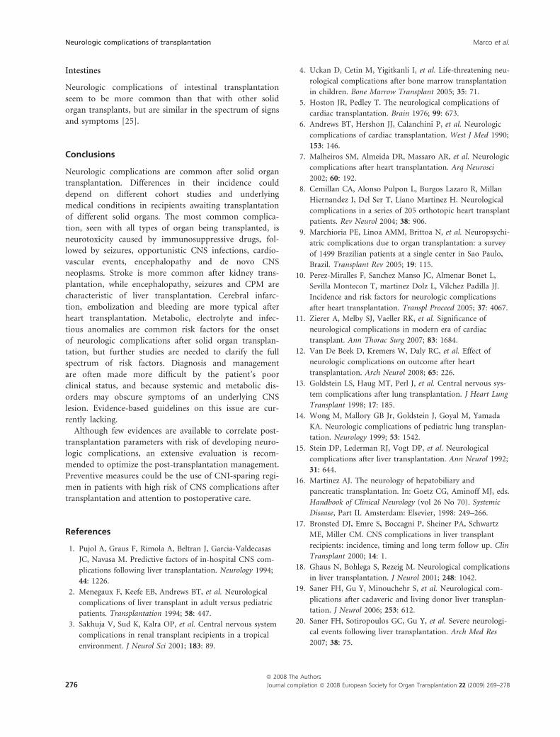
Intestines
Neurologic complications of intestinal transplantation
seem to be more common than that with other solid
organ transplants, but are similar in the spectrum of signs
and symptoms [25].
Conclusions
Neurologic complications are common after solid organ
transplantation. Differences in their incidence could
depend on different cohort studies and underlying
medical conditions in recipients awaiting transplantation
of different solid organs. The most common complica-
tion, seen with all types of organ being transplanted, is
neurotoxicity caused by immunosuppressive drugs, fol-
lowed by seizures, opportunistic CNS infections, cardio-
vascular events, encephalopathy and de novo CNS
neoplasms. Stroke is more common after kidney trans-
plantation, while encephalopathy, seizures and CPM are
characteristic of liver transplantation. Cerebral infarc-
tion, embolization and bleeding are more typical after
heart transplantation. Metabolic, electrolyte and infec-
tious anomalies are common risk factors for the onset
of neurologic complications after solid organ transplan-
tation, but further studies are needed to clarify the full
spectrum of risk factors. Diagnosis and management
are often made more difficult by the patient’s poor
clinical status, and because systemic and metabolic dis-
orders may obscure symptoms of an underlying CNS
lesion. Evidence-based guidelines on this issue are cur-
rently lacking.
Although few evidences are available to correlate post-
transplantation parameters with risk of developing neuro-
logic complications, an extensive evaluation is recom-
mended to optimize the post-transplantation management.
Preventive measures could be the use of CNI-sparing regi-
men in patients with high risk of CNS complications after
transplantation and attention to postoperative care.
References
1. Pujol A, Graus F, Rimola A, Beltran J, Garcia-Valdecasas
JC, Navasa M. Predictive factors of in-hospital CNS com-
plications following liver transplantation. Neurology 1994;
44: 1226.
2. Menegaux F, Keefe EB, Andrews BT, et al. Neurological
complications of liver transplant in adult versus pediatric
patients. Transplantation 1994; 58: 447.
3. Sakhuja V, Sud K, Kalra OP, et al. Central nervous system
complications in renal transplant recipients in a tropical
environment. J Neurol Sci 2001; 183: 89.
4. Uckan D, Cetin M, Yigitkanli I, et al. Life-threatening neu-
rological complications after bone marrow transplantation
in children. Bone Marrow Transplant 2005; 35: 71.
5. Hoston JR, Pedley T. The neurological complications of
cardiac transplantation. Brain 1976; 99: 673.
6. Andrews BT, Hershon JJ, Calanchini P, et al. Neurologic
complications of cardiac transplantation. West J Med 1990;
153: 146.
7. Malheiros SM, Almeida DR, Massaro AR, et al. Neurologic
complications after heart transplantation. Arq Neurosci
2002; 60: 192.
8. Cemillan CA, Alonso Pulpon L, Burgos Lazaro R, Millan
Hiernandez I, Del Ser T, Liano Martinez H. Neurological
complications in a series of 205 orthotopic heart transplant
patients. Rev Neurol 2004; 38: 906.
9. Marchioria PE, Linoa AMM, Brittoa N, et al. Neuropsychi-
atric complications due to organ transplantation: a survey
of 1499 Brazilian patients at a single center in Sao Paulo,
Brazil. Transplant Rev 2005; 19: 115.
10. Perez-Miralles F, Sanchez Manso JC, Almenar Bonet L,
Sevilla Montecon T, martinez Dolz L, Vilchez Padilla JJ.
Incidence and risk factors for neurologic complications
after heart transplantation. Transpl Proceed 2005; 37: 4067.
11. Zierer A, Melby SJ, Vaeller RK, et al. Significance of
neurological complications in modern era of cardiac
transplant. Ann Thorac Surg 2007; 83: 1684.
12. Van De Beek D, Kremers W, Daly RC, et al. Effect of
neurologic complications on outcome after heart
transplantation. Arch Neurol 2008; 65: 226.
13. Goldstein LS, Haug MT, Perl J, et al. Central nervous sys-
tem complications after lung transplantation. J Heart Lung
Transplant 1998; 17: 185.
14. Wong M, Mallory GB Jr, Goldstein J, Goyal M, Yamada
KA. Neurologic complications of pediatric lung transplan-
tation. Neurology 1999; 53: 1542.
15. Stein DP, Lederman RJ, Vogt DP, et al. Neurological
complications after liver transplantation. Ann Neurol 1992;
31: 644.
16. Martinez AJ. The neurology of hepatobiliary and
pancreatic transplantation. In: Goetz CG, Aminoff MJ, eds.
Handbook of Clinical Neurology (vol 26 No 70). Systemic
Disease, Part II. Amsterdam: Elsevier, 1998: 249–266.
17. Bronsted DJ, Emre S, Boccagni P, Sheiner PA, Schwartz
ME, Miller CM. CNS complications in liver transplant
recipients: incidence, timing and long term follow up. Clin
Transplant 2000; 14: 1.
18. Ghaus N, Bohlega S, Rezeig M. Neurological complications
in liver transplantation. J Neurol 2001; 248: 1042.
19. Saner FH, Gu Y, Minouchehr S, et al. Neurological com-
plications after cadaveric and living donor liver transplan-
tation. J Neurol 2006; 253: 612.
20. Saner FH, Sotiropoulos GC, Gu Y, et al. Severe neurologi-
cal events following liver transplantation. Arch Med Res
2007; 38: 75.
Neurologic complications of transplantation Marco et al.
ª 2008 The Authors
276 Journal compilation ª 2008 European Society for Organ Transplantation 22 (2009) 269–278

21. Erol I, Alehan F, Ozcary F, Canan O, Haberal M. Neuro-
logical complications of liver transplantation in pediatric
patients: a single centre experience. Pediatr Transplant
2007; 11: 152.
22. Dhar R, Young GB, Marotta P. Perioperative neurological
complications after liver transplantation are best predicted
by pre transplant hepatic encephalopathy. Neurocrit care
2008; 8: 253.
23. Adams HP, Dawson G, Coffman TJ, et al. Stroke in renal
transplant recipients. Arch Neurol 1986; 43: 113.
24. Kiok M. Neurological complications of pancreatic trans-
plantation. Neurol Clin 1988; 6: 367.
25. Zivkovic S, Shukla C, Abu Elmagd K, et al. Neurological
complications of small bowel transplantation. Neurology
2000; 54: AJ158.
26. Di Benedetto F, Di Sandro S, De Ruvo N, et al. Sirolimus
monotherapy in liver transplantation. Transplant Proc
2007; 39: 1930.
27. Jimenez-Perez M, Lozano Rey JM, Marın Garcıa D, et al.
Efficacy and safety of monotherapy with mycophenolate
mofetil in liver transplantation. Transplant Proc 2006; 38:
2480.
28. Eidelman BH, Abu- Elmagd K, Wilson J, et al. Neurologi-
cal complications of FK 506. Transpl Proc 1991; 93: 3175.
29. Bronster DJ, Emre S, Mor E, Scheiner P, Miller CM, .
Neurological complications of orthotopic liver transplanta-
tion. Mt Sinai J Med 1994; 61: 63.
30. Dawson FM. Immunosoppressants, immunophilins and
the nervous system. Ann Neurol 1996; 40: 559.
31. Paul LC. Overview of side effects of immunosuppressive
therapy. Transplant Proc 2001; 33: 2089.
32. Beresfold T. Neuropsychiatric complications of liver and
other organ transplantation. Liver Transpl 2001; 11: 36.
33. Stoltenburg-Didinger G, Boegner F. Glia toxicity in disso-
ciates cell cultures induced by cyclosporine. Neurotoxicolo-
gy 1992; 13: 179.
34. Mueller AR, Plazt KP, Benchstein WO, et al. Neurotoxicity
after orthotopic liver transplantation. Transplantation
1994; 58: 155.
35. Benchstein WO. Neurotoxicity of calcineurin inhibitors:
impact and clinical management. Transplant Int 2000; 13:
313.
36. Lake KD, Canafax DM. Important interactions of drugs
with immunosuppressive agents used in transplant recipi-
ents. J Antimicrob Chemother 1995; 36: 11.
37. Tezcan H, Zimmer W, Fenstermaker R, Herzig GP,
Schriber J. Severe cerebellar swelling and thrombotic
thrombocytopenic purpura associated with FK506. Bone
Marrow Transplant 1998; 21: 105.
38. Mc Alister VC, Peltekian KM, Malatjalian DA, et al.
Orthotopic liver transplantation using low dose tacrolimus
and sirolimus. Liver Transpl 2001; 7: 701.
39. Bianco F, Fattaposta F, Locuratolo N, et al. Reversible
diffusion MRI abnormalities and transient mutism after
liver transplantation. Neurology 2004; 62: 981.
40. Patchell RA. Neurological complications of organ trans-
plantation. Ann Neurol 1994; 36: 688.
41. Resener M, Martin E, Zipp F, Dichgans J, Martin R. Neu-
rological side-effects of pharmacologic corticoid therapy.
Neverarzt 1996; 67: 983.
42. Adair JC, Woodley SL, O’Connell JB, Call GK,
Baringer JR. Aseptic meningitis following cardiac
transplantation: clinical characteristic and relationship
to immunosuppressive regimen. Neurology 1991; 41:
249.
43. Pittock SJ, Rabinstein AA, Edwards BS, Wijdicks EF.
OKT3 neurotoxicity presenting as akinetic mutism. Trans-
plantation 2003; 7: 1058.
44. Agarwal RK, Ostaszeweski ML, Field LG, et al. Tumor
necrosis factor and interleukin 6 in cerebrospinal fluid of a
patient with recurrent adverse central nervous system
events. Transplant Proc 1993; 25: 2143.
45. Estol JC, Lopez OL, Brenner RP, et al. Seazures after trans-
plantation: a clinicopathological study. Neurology 1989; 39:
1297.
46. Wszolek ZK, Aksamit AJ, Ellingson RJ, et al. Epilectiform
electroencephalographic abnormalities in liver transplant
recipients. Ann Neurol 1991; 30: 37.
47. Wszolek ZK, Steg RE. Seizures after orthotopic liver trans-
plantation. Seizure 1997; 6: 31.
48. Chabolla DR, Harnois DM, Meschia JF. Levetiracetam
monotherapy for liver transplant patients with seizures.
Transplant Proc 2003; 35: 1480.
49. Chang SH, Lim CS, Low TS, Chon HT, Tan SY. Cyclo-
sporine associated encephalopathy: a case report and litera-
ture review. Transplant Proc 2001; 8: 3700.
50. Parvex P, Pinsk M, Bell LE, O’Gorman AM, Patenoude
YG, Gupta IR. Reversible encephalopathy associated with
tacrolimus in pediatric renal transplant. Pediatr Nephrol
2001; 16: 537.
51. Paizel PM, Snoeck HW, ven Den Hauwe L, et al. Cerebral
complications of murine monoclonal CD3 antibody: CT
and MR findings. AJNR 1997; 18: 1935.
52. Pirsch JD, Miller J, Deierhoi MH, Vincenti F, Filo RS.
A comparison of tacrolimus and cyclosporine for immu-
nosuppression after cadaveric renal transplantation.
Kidney transplant Study Group. Transplantation 1997;
63: 977.
53. Inoha S, Inamura T, Nakamizon A, Ikezaki K, Amono T,
Fukui M. Magnetic resonance imaging in cases with
encephalopathy secondary to immunosuppressive agents.
J Clin Neurosci 2002; 9: 305.
54. Furukawa M, Terae S, Chu BC, Kaneko K, Kamada H,
Miyasaka K. MRI in seven cases of Tacrolimus FK 506
encephalopathy: utility of flair and diffusion- weighted
imaging. Neuroradiology 2001; 43: 615.
55. Conti DJ, Rubin RH. Infections of the CNS in organ
transplant recipients. Neurology 1996; 47: 1523.
56. Chapman SW, Wilson JP. Nocardiosis in transplant recipi-
ents. Semin Respir Infect 1990; 5: 74.
Marco et al. Neurologic complications of transplantation
ª 2008 The Authors
Journal compilation ª 2008 European Society for Organ Transplantation 22 (2009) 269–278 277

57. Singh N, Paterson DLM. Tubercolosis infection in solid
organ transplant recipients: impact and implications for
management. Clin Infect Dis 1998; 27: 1266.
58. Armstrong RW, Fung PC. Brain stem encephalitis due to
Listeria monocytogenes: case report and review. Clin Infect
Dis 1993; 16: 702.
59. Bonham CA, Dominguez EA, Fukui MB, Paterson DL,
Pankey GA, Wagener MM. Central Nervous System lesions
in liver transplant recipients: prospective assessment of
indications for biopsy and implications and management.
Transplantation 1998; 66: 1596.
60. Torre-Cisneros J, Lopez OL, Kusne S, et al. CNS aspergil-
losis in organ transplantation: a clinicopathological study.
J Neurol Neurosurg Psychiatry 1993; 56: 188.
61. Singh N, Gayowsky T, Wegener MM, Fung J. Clinical
spectrum of invasive cryptococcocis in liver transplant rec-
ipiens receiving Tacrolimus. Clin Transplant 1997; 11: 66.
62. Jabbour N, Reyes J, Kusne S, Martin M, Fung J. Crypto-
coccal meningitis after liver transplantation. Transplanta-
tion 1996; 61: 146.
63. Aksamit AJ. Demyelinating disorders. In: Wijdicks EFM,
ed. Neurologic Complications of Organ Transplant Recipi-
ents. Boston, MA: Butterworth Heinemann, 1999: 229–237.
64. Penn I. Some problems with posttransplant lymphoprolif-
erative disease. Transplantation 2000; 79: 705.
65. Jaffe ES, Harris NL, Stein H, et al. In: Jaffe ES, ed. Pathol-
ogy and Genetics of Tumor and Haematopoietic Tissues.
World Health Organization Classification of Tumors,
IARC Press: Oxford University Press, Lyon, 2001: 264–270.
66. Collins MH, Montone KT, Leahey AM, et al. Autopsy
pathology of pediatric posttransplant lymphoproliferative
disorder. Pediatrics 2001; 107: 1.
67. Pickhardt PJ, Siegel MJ, Hayashi RJ, et al. Posttransplanta-
tion lymphoproliferative disorder in children: clinical, histo-
pathologic and imaging features. Radiology 2000; 217: 16.
68. Paya CV, Fung JJ, Nalesnik MA, et al. EBV-induced post-
transplant lymphoproliferative disorders. ASTS/ASTP EBV
PTLD Task Force and the Mayo Clinic Organized Interna-
tional Consensus Development Meeting. Transplantation
1999; 68: 1517.
69. Stracciari A, Guarino M. Neuropsychiatric complications
of liver transplantation. Metab Brain Dis 2001; 16: 3.
70. Gill D, Juffs HG, Herzig KA, et al. Durable and high rates
of remission following chemotherapy in posttransplanta-
tion lymphoproliferative disorders after renal transplanta-
tion. Transpl Proc 2003; 35: 256.
71. Burra P, Senzolo M, Pizzolato G, et al. Frontal cerebral
blood flow is impaired in patients with heart transplanta-
tion. Transpl Int 2002; 15: 459.
72. Eidelman BH, Obrist WD, Wagner WR, et al.
Cerebrovascular complications associated with the use of
artificial circulatory support services. Neurol Clin 1993; 11:
463.
73. Bornstein RA, Starling RC, Myerowitz PD, Haas GJ. Neu-
ropsychological function in patients with end stage heart
failure before and after heart transplantation. Acta Neurol
Scand 1995; 91: 260.
74. Ponticelli C, Campise MR. Neurological complications in
kidney transplant recipients. J Nephrol 2005; 18: 521.
75. Sharma KR, Cross J, Santiago F, Ayyar DR, Burke G. Inci-
dence of acute femoral neuropathy following renal trans-
plant. Arch Neurol 2002; 59: 541.
76. Vaziri ND, Bartoc CH, Ravikumar GR, Martin DC, Ness
R, Saiki J. Femoral neuropathy: a complication of renal
transplant. Nephrol 1987; 28: 30.
77. Wessenborn K. Minimal hepatic encephalopathy: a perma-
nent source of discussion. Hepatology 2002; 35: 494.
78. Wein C, Koch H, Popp P, et al. Minimal hepatic encepha-
lopathy impairs the fitness to drive. Hepatology 2004; 39:
739.
79. Senzolo M, Amodio P, D’Aloiso MC, et al. Neuropsycho-
logical and neurophysiological evaluation in cirrhotic
patients with minimal hepatic encephalopathy undergoing
liver transplantation. Transplant Proc 2005; 37: 1104.
80. Dam M, Burra P, Tedeschi U, et al. Regional cerebral
blood flow changes in patients with cirrhosis assessed with
99m TC-HM-PAO single photon emission computer
tomography: effect of liver transplantation. J Hepatol 1998;
29: 78.
81. Burra P, Dam M, Chierichetti F, et al. 18F-fluorodeoxyglu-
cose positron emission tomography study of brain metabo-
lism in cirrhosis: effect of liver transplantation. Transpl
Proc 1999; 31: 418.
82. Burra P, Senzolo M, Pizzolato G, et al. Does liver disease
aetiology have a role in cerebral blood flow alterations in
liver cirrhosis? Eur J Gastroenterol Hepatol 2004; 16: 885.
83. Senzolo M, Pizzolato G, Dam M, Sturniolo GC, Burra P.
Normal cortical regional blood flow justifies the normal
neuropsychological performance in patients with
cholestatic liver disease. Psychiatry Clin Neurosci 2007; 61:
209.
84. Martin RJ. Central pontine and extrapontine myelinolisis:
the osmotic demyelination syndromes. J Neurol Neurosurg
Psichiatr 2004; 75: 22.
85. Wang WL, Yang ZF, Lo CM, Liu CL, Fan ST. Intracerebral
hemorrhage after liver transplantation. Liver Transpl 2000;
6: 345.
86. Estol JC, Pessin MS, Martinez AJ. Cerebrovascular compli-
cations after orthotopic liver transplantation: a clinicopath-
ologic study. Neurology 1991; 41: 815.
87. Singh N, Husain S. Infections of the CNS in transplant
recipients. Transpl Infect Dis 2000; 2: 101.
88. Martinez AJ, Estol C, Faris AA, et al. Neurological compli-
cations of liver transplantation. Neurol Clin 1988; 6: 327.
89. Larsen J. Pancreas transplantation: indications and conse-
quences. Endocr Rev 2004; 25: 919.
Neurologic complications of transplantation Marco et al.
ª 2008 The Authors
278 Journal compilation ª 2008 European Society for Organ Transplantation 22 (2009) 269–278