recurrent miscarriages and associated obstetric complications
95
“ANTI THYROID PEROXIDASE POSITIVITY IN RECURRENT MISCARRIAGES AND ASSOCIATED OBSTETRIC COMPLICATIONS” Dissertation submitted to THE TAMIL NADU DR. M.G.R. MEDICAL UNIVERSITY In partial fulfilment for the award of the degree of M.S. BRANCH-II OBSTETRICS AND GYNECOLOGY MADRAS MEDICAL COLLEGE CHENNAI MAY 2019
-
Upload
khangminh22 -
Category
Documents
-
view
5 -
download
0
Transcript of recurrent miscarriages and associated obstetric complications
“ ANTI THYROID PEROXIDASE POSITIVITY IN
RECURRENT MISCARRIAGES AND ASSOCIATED
OBSTETRIC COMPLICATIONS ”
Dissertation submitted to
THE TAMIL NADU DR. M.G.R. MEDICAL UNIVERSITY
In partial fulfilment for the award of the degree of
M.S. BRANCH-II
OBSTETRICS AND GYNECOLOGY
MADRAS MEDICAL COLLEGE
CHENNAI
MAY 2019

CERTIFICATE
This is to certify that the dissertation entitled “ANTI THYROID
PEROXIDASE POSITIVITY IN RECURRENT MISCARRIAGES
AND ASSOCIATED OBSTETRIC COMPLICATIONS.” is a bonafide
record of work done by Dr. K.MANJULA in Madras Medical college,
Chennaiduring the period March 2017 to March 2018 under the guidance of
Dr.K.KANMANI, M.D., D.G.O., Professor of Obstetrics and
Gynaecology, Institute of Social Obstetrics, Madras Medical College in
partial fulfilment of requirement of M.S Degree in Obstetrics and
Gynaecology degree examination of The Tamilnadu Dr. M.G.R Medical
University to be held in May 2019.
Dr.S. VIJAYA, M.D., DGO.,
Director I/c.,
Institute of Social Obstetrics
Government Kasturba Gandhi Hospital
Chennai - 600005
Dr. R. JAYANTHI, M.D.,
Dean
Madras Medical College&
Rajiv Gandhi Government
General Hospital,
Chennai – 600 003

DECLARATION
I Dr. K.MANJULA, Post graduate, Department of Obstetrics and
Gynaecology, Madras Medical College, solemnly declare that this
dissertation entitled “ANTI THYROID PEROXIDASE POSITIVITY
IN RECURRENT MISCARRIAGES AND ASSOCIATED
OBSTETRIC COMPLICATIONS” was done by me at Madras Medical
College during 2016-2019 under the guidance and supervision of
Prof.Dr.K.KANMANI M.D.,D.G.O., Professor of Obstetrics and
Gynaecology, Institute of Social Obstetrics, Madras Medical College. This
dissertationis submitted to the Tamil Nadu Dr. M.G.R. Medical
University towards the partial fulfilment of requirements for the
award of M.S. Degree in Obstetrics and Gynaecology (Branch-II).
Place: Chennai-3
Date: Dr.K.MANJULA
Prof. DR. KANMANI, M.D., D.G.O.,
Guide, Institute of Social Obstetrics,
Madras Medical College,
Chennai

ACKNOWLEDGEMENT
I am thankful to the Dean, Dr.R.JAYANTHI M.D., Madras Medical
College, Chennai for allowing to use the facilities and clinical materials
available in the hospital.
It is my pleasure to express my thanks to Prof.Dr.S.VIJAYA
MD.,D.G.O., Director, Institute of Social Obstetrics, Government Kasturba
Gandhi Hospital for her valuable guidance, interest and encouragement in
this study.
I take this opportunity to express my deep sense of gratitude and
humble regards to my beloved teacher Dr.K.KANMANI, M.D.,D.G.O., for
her timely guidance suggestions and constant inspiration enabled me to
complete this dissertation.
I thank all my Professors, AssistantProfessorsand paramedical Staffs
of this Institute of Obstetrics and Gynaceology, Madras Medical College,
Chennai-600003.
I thank all my patients for their cooperation and hence for the success
of study.
I thank my family and friends for their inspiration and support given
to me.

CONTENTS
S.NO. INDEX PAGE NO
1. INTRODUCTION 1
2. AIMS AND OBJECTIVES 31
3. REVIEW OF LITERATURE 32
4. MATERIALS AND METHODS 42
5. RESULTS AND DISCUSSION 44
6. SUMMARY 71
7. CONCLUSION 76
8. BIBLIOGRAPHY 77
9. ANNEXURE
a) Ethical committee approval
b) Proforma
c) Information sheet
d) Patient Consent Form
e) Antiplagiarism – URKUND copy
f) Plagiarism certificate
g) Master Chart
79
80
84
86
89
90
1
INTRODUCTION
Any pregnancy ending spontaneously prior the fetus can survive is
defined as miscarriage. Recurrent miscarriage, defined as loss of 3 or more
consecutive pregnancies affects 1% of couples trying to conceive.
The important causes for recurrent spontaneous miscarriage includes
epidemiological factors such as maternal age and numberof previous
miscarriages, anti-phospholipids syndrome, genetic factors, anatomical
factors, endocrine factors, immune factors; inherited thrombophilic defects
etc.,
Autoimmune thyroid disease (AITD) is the most frequent cause of
Hypothyroidism in women of reproductive age. Thyroid disorders have
been suspected to cause early pregnancy loss and other adversepregnancy
outcomes. Although the worst overthypothyroidism is infrequent in
pregnancy, subclinical hypothyroidism has an incidence of 2-3%.
Thyroid dysfunction and autoimmunity are relatively common in
women of reproductive age group and has been associated with various
adverse pregnancy outcomes such as recurrent miscarriage, preeclampsia
and preterm labour. The main objective of this study was to find out
association between anti-Thyroid peroxidase antibody and recurrent
miscarriages and to evaluate obstetric complications such as preeclampsia
and preterm labour in them.

2
Maternal age and number of previous miscarriages are two
independent risk factors for a further miscarriage. Advancing maternal age
is associated with a decline in both the number and quality of the remaining
oocytes.
A large prospective register linkage study reported the age-related
risk of miscarriage in recognised pregnancies to be: 12–19 years, 13%; 20–
24 years, 11%; 25–29 years, 12%; 30–34 years, 15%; 35–39 years, 25%;
40–44 years, 51%; and ≥45 years, 93%.
Advanced paternal age has also been identified as a risk factor for
miscarriage. The risk of miscarriage is highest among couples where the
woman is ≥35 years of age and the man ≥40 years of age.
Previous reproductive history is an independent predictor of future
pregnancy outcome. The risk of a further miscarriage increases after each
successive pregnancy loss, reaching approximately 40% after three
consecutive pregnancy losses, and the prognosis worsens with increasing
maternal age. A previous live birth does not preclude a woman developing
recurrent miscarriage.
The evidence on the effect of environmental risk factors is based
mainly on data studying women with sporadic rather than recurrent
miscarriage. The results are conflicting and biased by difficulties in
controlling for confounding factors and the inaccuracy of data on exposure
and the measurement of toxin dose.

3
Maternal cigarette smoking and caffeine consumption have been
associated with an increased risk of spontaneous miscarriage in a dose-
dependent manner. However, current evidence is insufficient to confirm this
association.
Recent retrospective studies have reported that obesity increases the
risk of both sporadic and recurrent miscarriage
Definition of Miscarriage and Recurrent Pregnancy Loss
The term miscarriage (or abortion) is used to describe a pregnancy that
fails to progress, resulting in death and expulsion of the embryo or fetus.
The generally accepted definition stipulates that the fetus or embryo should
weigh 500 g or less, a stage that corresponds to a gestational age of up to 20
weeks (World Health Organization).
Unfortunately, this definition is not used consistently, and pregnancy
losses at higher gestational ages are also, in some studies, classified as
miscarriage instead of stillbirth or preterm neonatal death. Thus, from a
definition perspective, it is important to characterize the population being
studied so that comparisons across therapeutic trials can be made more
appropriately and reliably.
Recurrent miscarriage should, according to the aforementioned
definition of miscarriage, be defined as at least three consecutive
miscarriages, whereas recurrent pregnancy loss (RPL) could also include

4
pregnancy losses up to gestational week 28; however, unfortunately there is
no consensus on the definition of recurrent miscarriage or RPL.Pregnancy
losses after week 20 are rare, so defining recurrent miscarriage and RPL as
above will result in almost identical populations.
In some countries and according to some national guidelines only two
miscarriages are required for diagnosis of RPL. More and more published
studies of RPL therefore include women with only two previous
miscarriages, which from an epidemiological point of view is very
problematic.
Chemistry of Thyroid Hormones
Thyroid hormones are derivatives of the the amino acid tyrosine bound
covalently to iodine. The two principal thyroid hormones are:
• thyroxine (also known as T4 or L-3,5,3',5'-tetraiodothyronine)
• triiodothyronine (T3 or L-3,5,3'-triiodothyronine)
As shown in the following diagram, the thyroid hormones are basically two
tyrosines linked together with the critical addition of iodine at three or four
positions on the aromatic rings. The number and position of the iodines is
important. Several other iodinated molecules are generated that have little or
no biological activity; so called "reverse T3" (3,3',5'-T3) is such an example

5
A large majority of the thyroid hormone secreted from the thyroid
gland is T4, but T3 is the considerably more active hormone. Although
some T3 is also secreted, the bulk of the T3 is derived by deiodination of T4
in peripheral tissues, especially liver and kidney. Deiodination of T4 also
yields reverse T3, a molecule with no known metabolic activity.
Thyroid hormones are poorly soluble in water, and more than 99% of
the T3 and T4 circulating in blood is bound to carrier proteins. The principle
carrier of thyroid hormones is thyroxine-binding globulin, a glycoprotein
synthesized in the liver. Two other carriers of import are transthyrein and
albumin. Carrier proteins allow maintenance of a stable pool of thyroid
hormones from which the active, free hormones are released for uptake by
target cells.
6
THYROID BIOSYNTHESIS:
Thyroid hormones are synthesized by mechanisms fundamentally
different from what is seen in other endocrine systems. Thyroid follicles
serve as both factory and warehouse for production of thyroid hormones.
Constructing Thyroid Hormones
The entire synthetic process occurs in three major steps:
• Production and accumulation of the raw materials
• Fabrication or synthesis of the hormones on a backbone or scaffold of
precursor
• Release of the free hormones from the scaffold and secretion into blood
The recipe for making thyroid hormones calls for two principle raw
materials:
• Tyrosines are provided from a large glycoprotein scaffold called
thyroglobulin, which is synthesized by thyroid epithelial cells and secreted
into the lumen of the follicle - colloid is essentially a pool of thyroglobulin.
A molecule of thyroglobulin contains 134 tyrosines, although only a
handful of these are actually used to synthesize T4 and T3.
• Iodine, or more accurately iodide (I-), is avidly taken up from blood by
thyroid epithelial cells, which have on their outer plasma membrane a
sodium-iodide symporter or "iodine trap". Once inside the cell, iodide is
transported into the lumen of the follicle along with thyroglobulin.

7
Fabrication of thyroid hormones is conducted by the enzyme thyroid
peroxidase, an integral membrane protein present in the apical (colloid-
facing) plasma membrane of thyroid epithelial cells. Thyroid peroxidase
catalyzes two sequential reactions:
1. Iodination of tyrosines on thyroglobulin (also known as "organification of
iodide").
2. Synthesis of thyroxine or triiodothyronine from two iodotyrosines.
Through the action of thyroid peroxidase, thyroid hormones accumulate in
colloid, on the surface of thyroid epithelial cells. Remember that hormone is
still tied up in molecules of thyroglobulin - the task remaining is to liberate
it from the scaffold and secrete free hormone into blood.
Thyroid hormones are excised from their thyroglobulin scaffold by
digestion in lysosomes of thyroid epithelial cells. This final act in thyroid
hormone synthesis proceeds in the following steps:
• Thyroid epithelial cells ingest colloid by endocytosis from their apical
borders - that colloid contains thyroglobulin decorated with thyroid
hormone.
8
• Colloid-laden endosomes fuse with lysosomes, which contain hydrolytic
enzymes that digest thyroglobluin, thereby liberating free thyroid hormones.
• Finally, free thyroid hormones apparently diffuse out of lysosomes, through
the basal plasma membrane of the cell, and into blood where they quickly
bind to carrier proteins for transport to target cells
Control of Thyroid Hormone Synthesis and Secretion
The chief stimulator of thyroid hormone synthesis is thyroid-
stimulating hormone from the anterior pituitary. Binding of TSH to
receptors on thyroid epithelial cells seems`` Q to enhance all of the
processes necessary for synthesis of thyroid hormones, including synthesis
of the iodide transporter, thyroid peroxidase and thyroglobulin.

9
The magnitude of the TSH signal also sets the rate of endocytosis of
colloid - high concentrations of TSH lead to faster rates of endocytosis, and
hence, thyroid hormone release into the circulation. Conversely, when TSH
levels are low, rates of thyroid hormone synthesis and release diminish.
The thyroid gland is part of the hypothalamic-pituitary-thyroid axis,
and control of thyroid hormone secretion is exerted by classical negative
feedback, as depicted in the diagram. Thyroid-releasing hormone (TRH)
from the hypothalamus stimulates TSH from the pituitary, which stimulates
thyroid hormone release. As blood concentrations of thyroid hormones
increase, they inhibit both TSH and TRH, leading to "shutdown" of thyroid
epithelial cells. Later, when blood levels of thyroid hormone have decayed,
the negative feedback signal fades, and the system wakes up again.
A number of other factors have been shown to influence thyroid hormone
secretion. In rodents and young children, exposure to a cold environment
triggers TRH secretion, leading to enhanced thyroid hormone release. This
makes sense considering the known ability of thyroid hormones to spark
body heat production
MATERNAL THYROID FUNCTION DURING PREGNANCY
Normal pregnancy entails substantial changes in thyroid function in
all animals. These phenomena have been studied most extensively in
humans, but probably are similar in all mammals. Major alterations in the
thyroid system during pregnancy include:
10
• Increased blood concentrations of T4-binding globulin: TBG is one of
several proteins that transport thyroid hormones in blood, and has the
highest affinity for T4 (thyroxine) of the group. Estrogens stimulate
expression of TBG in liver, and the normal rise in estrogen during
pregnancy induces roughly a doubling in serum TBG concentrations.
• Increased levels of TBG lead to lowered free T4 concentrations, which
results in elevated TSH secretion by the pituitary and, consequently,
enhanced production and secretion of thyroid hormones. The net effect of
elevated TBG synthesis is to force a new equilibrium between free and
bound thyroid hormones and thus a significant increase in total T4 and T3
levels. The increased demand for thyroid hormones is reached by about 20
weeks of gestation and persists until term.
• Increased demand for iodine: This results from a significant pregnancy-
associated increase in iodide clearance by the kidney (due to increased
glomerular filtration rate), and siphoning of maternal iodide by the fetus.
The World Health Organization recommends increasing iodine intake from
the standard 100 to 150 ug/day to at least 200 ug/day during pregnancy.
• Thyroid stimulation by chorionic gonadotropin: The placenta of humans
and other primates secrete huge amounts of a hormone called chorionic
gonadotropin (in the case of humans, human chorionic gonadotropin or
hCG) which is very closely related to luteinizing hormone. TSH and hCG
are similar enough that hCG can bind and transduce signalling from the
11
TSH receptor on thyroid epithelial cells. Toward the end of the first
trimester of pregnancy in humans, when hCG levels are highest, a
significant fraction of the thyroid-stimulating activity is from hCG. During
this time, blood levels of TSH often are suppressed, as depicted in the figure
to the right. The thyroid-stimulating activity of hCG actually causes some
women to develop transient hyperthyroidism.
The net effect of pregnancy is an increased demand on the thyroid
gland. In the normal individuals, this does not appear to represent much of a
load to the thyroid gland, but in females with subclinical hypothyroidism,
the extra demands of pregnancy can precipitate clinicial disease.
Thyroid Hormones and Fetal Brain Development
In 1888 the Clinical Society of London issued a report underlining
the importance of normal thyroid function on development of the brain.
Since that time, numerous studies with rats, sheep and humans have
reinforced this concept, usually by study of the effects of fetal and/or
maternal thyroid deficiency. Thyroid hormones appear to have their most
profound effects on the terminal stages of brain differentiation, including
synaptogenesis, growth of dendrites and axons, myelination and neuronal
migration.
Thyroid hormones act by binding to nuclear receptors and modulating
transcription of responsive genes. Thyroid hormone receptors are widely
12
distributed in the fetal brain, and present prior to the time the fetus is able to
synthesize thyroid hormones.
It has proven surprisingly difficult to identify the molecular targets
for thyroid hormone action in the developing brain, but some progress has
been made. For example, the promoter of the myelin basic protein gene is
directly responsive to thyroid hormones and contains the expected hormone
response element. This fits with the observation that induced
hypothyroidism in rats leads to diminished synthesis of mRNAs for several
myelin-associated proteins.
It seems clear that there is a great deal more to learn about the molecular
mechanisms by which thyroid hormones support normal development of the
brain.
Thyroid Deficiency in the Fetus and Neonate
The fetus has two potential sources of thyroid hormones - it's own
thyroid and the thyroid of it's mother. Human fetuses acquire the ability to
synthesize thyroid hormones at roughly 12 weeks of gestation, and fetuses
from other species at developmentally similar times. Current evidence from
several species indicates that there is substantial transfer of maternal thyroid
hormones across the placenta. Additionally, the placenta contains
deiodinases that can convert T4 to T3.

13
The major thyroid hormone secreted by the fetus is T4. However,
total T3 and free T3 levels are low throughout gestation, and levels of RT3
are elevated, paralleling the rise in T4. Like T3, this compound is derived
predominantly from conversion of T4 in peripheral tissues.
The increased production of T4 in fetal life is compensated by rapid
conversion to the inactive RT3, allowing the fetus to conserve its fuel
resources. However, there is some evidence that RT3, as well as T4,
through nongenomic actions, regulate fetal brain development.
The fetus is iodine deficient when a mother’s iodine intake is low.
Supplying the mother adequate iodine is an important problem in many
parts of the world. A fetalgoiter is occasionally detected by ultrasonography
at a size that could impede normal delivery. The fetus can be treated, with

14
regression of the goiter, by the administration of levothyroxine into the
amniotic fluid.
However, the fetus is usually protected by the transplacental transfer
of maternal T4, and, therefore, fetal treatment of hypothyroidism can
usually be postponed until delivery.
There are three types or combinations of thyroid deficiency states
known to impact fetal development:
Isolated maternal hypothyroidism:
Overt maternal hypothyroidism typically is not a significant cause of
fetal disease because it usually is associated with infertility. When
pregnancy does occur, there is increased risk of intrauterine fetal death and
gestational hypertension. Subclincial hypothyroidism is increasingly being
recognized as a cause of developmental disease - this is a rather scary
situation.
Several investigators have found that mild maternal hypothyroidism,
diagnosed only retrospectively from banked serum, may adversely affect the
fetus, leading in children to such effects as slightly lower performance on
IQ tests and difficulties with schoolwork. The most common cause of
subclinical hypothyroidism is autoimmune disease, and it is known that
anti-thyroid antibodies cross the human placenta. Thus, the cause of this
disorder may be a passive immune attack on the fetal thyroid gland.
Isolated fetal hypothyroidism:

15
This condition is also known as sporadic congenital hypothyroidism.
It is due to failure of the fetal thyroid gland to produce adequate amounts of
thyroid hormone. Most children with this disorder are normal at birth,
because maternal thyroid hormones are transported across the placenta
during gestation. What is absolutely critical is to identify and treat this
condition very shortly after birth. If treatment is not instituted quickly, the
child will become permanently mentally and growth retarded - a disorder
called cretinism. This problem has largely due to large scale screening
programs to detect hypothyroid infants.
Iodine deficiency - Combined maternal and fetal hypothyroidism: Iodine
deficiency is, by a large margin, the most common preventable cause of
mental retardation in the world. Without adequate maternal iodine intake,
both the fetus and mother are hypothyroid, and if supplemental iodine is not
provided, the child may well develop cretinism, with mental retardation,
deaf-mutism and spasticity.
The World Health Organization estimated in 1990 that 20 million
people had some degree of brain damage due to iodine deficiency
experienced in fetal life. Endemic iodine deficiency remains a substantial
public health problem in many parts of the world, including many areas in
Europe, Asia, Africa and South America.
In areas of severe deficiency, a large fraction of the adult population
may show goiters. In such settings, overt cretinism may occur in 5 to 10
16
percent of offspring, and perhaps five times that many children will have
mild mental retardation. This is a serious, tragic and, most importantly, a
preventable problem.
The effects of mild maternal hypothyroidism on cognitive function of
children has been evaluated in several studies, including some in which
mothers will low levels of T4 or high levels of TSH were treated
prophylactically with thyroid supplementation. The results of these studies
are somewhat divergent, and the benefit of routinely testing pregnant
women and treating those with suspected thyroid deficiency remains
unsettled.
The fetus of an iodine-deficient mother can be successfully treated if
iodine supplementation is given during the first or second trimester.
Treatment during the third trimester or after birth will not prevent the
mental defects.
Iodine deficiency can also be a sigificant problem in animal
populations. The most common manifestation in sheep, cattle, pigs and
horses is a high incidence of stillbirths and birth of small, weak offspring.
Hyperthyroidism in Pregnancy
Gestational hyperthyroidism is associated with increased risk of
several adverse outcomes, including preeclampisa, premature labor, fetal or
perinatal death and low birth weight. In humans, hyperthyroidism usually is

17
the result of Grave's disease, which involves development of autoantibodies
against the TSH receptor that stimulate the thyroid gland.
MATERNAL AND FETAL THYROID LEVELS :
In response to the metabolic demands of pregnancy, there is an
increase in the basal metabolic rate (which is mainly due to fetal
metabolism), iodine uptake, and the size of the thyroid gland caused by
hyperplasia and increased vascularity. However, despite this increase in
thyroid activity, a pregnant woman is euthyroid with levels of TSH, free T4,
and free T3 remaining within the normal range; thyroid nodules and goiter
require evaluation.
During pregnancy, iodide clearance by the kidney increases. For this
reason (plus the iodide losses to the fetus), the prevalence of goiter is
increased in areas of iodine deficiency. In many parts of the world, iodine is
not sufficiently available in the environment, and pregnancy increases the
risk of iodine deficiency.
The increase in thyroid activity in pregnancy is accompanied by a
marked increase in the circulating levels of TBG in response to estrogen;
therefore, a new equilibrium is reached with an increase in the bound
portion of the thyroid hormone. The mechanism for the estrogen effect on
TBG is an increase in hepatic synthesis and an increase in glycosylation of
the TBG molecule that leads to decreased clearance.
18
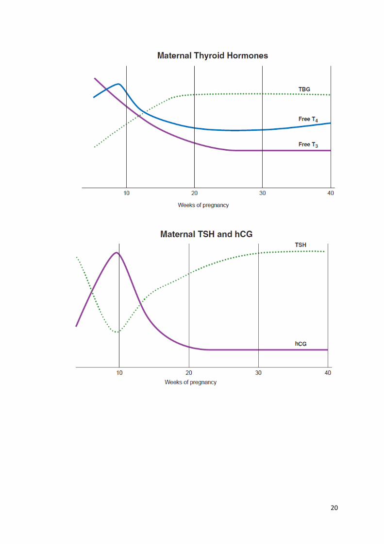
The increase in thyroid activity is attributed to the thyrotropic
substances secreted by the placenta: a chorionic thyrotropin and the
thyrotropic activity in human chorionic gonadotropin (hCG).
It has been calculated that hCG contains approximately 1/4,000th of
the thyrotropic activity of human TSH. In conditions with very elevated
HCG levels, the thyrotropic activity can be sufficient to produce
hyperthyroidism (gestational hyperthyroidism), and this can even be
encountered in normal pregnancy TBG levels reach a peak (twice
nonpregnant levels) at about 15 weeks, which is maintained throughout the
rest of pregnancy.
T4 undergoes a small increase in the first trimester, but T3 increases
more markedly. Because of the increase in TBG, free T4 and T3 levels then
decrease, although they remain within the normal range. There is an inverse
relationship between maternal circulation levels of TSH and hCG.60 TSH
reaches a nadir atthe same time that hCG reaches a peak at 10 weeks of
pregnancy. TSH levels then increase as hCG levels drop to their stable
levels throughout the rest of pregnancy.
Thus, the range of normal for TSH levels change with each
trimester. The lower limit of TSH in the first and second trimesters is 0.03
and 0.13 mU/L in the third trimester. The upper limit of normal in the
first trimester is 2.3 and 3.5 mU/L in the second and third trimesters.

19
These changes support a role for hCG stimulation of the maternal
thyroid gland, especially during early pregnancy, providing a small but
important increase in maternal thyroid hormones for the fetus until fetal
thyroid function is sufficient to serve fetal needs.
It is well recognized that patients who have conditions associated
with very high levels of hCG (trophoblastic disease, hCG-secreting cancers)
can develop hyperthyroidism. The thyroid stimulating activity of hCG is
explained by the molecular homology between hCG and TSH, and between
their receptors.
In normal pregnancies, placental transfer of TSH, T4, and T3 is
limited in both directions. Slight, but significant, transfer of T4 and T3 does
occur, however, when maternal levels are very high or when fetal levels are
substantially lower than the maternal levels.
Therefore, in the early weeks of pregnancy, before the fetal thyroid
gland becomes active, the fetal brain is dependent on the placental transport
of maternal T4. Both overt and subclinical maternal hypothyroidism are
associated with increased risks of miscarriage, preeclampsia, low birth
weight, premature delivery, and a decrease in intelligence in the children.
20

21
Epidemiological Parameters Relevant for Recurrent Pregnancy Loss
Occurrence
Using the traditional definition, the incidence of RPL is the number
of new women each year (or in another defined period) suffering their third
consecutive pregnancy loss, and the prevalence of RPL is the number of
women in a population who, at a specific time point, have had three or more
consecutive pregnancy losses. The incidence/prevalence is often expressed
as a rate of those individuals being at risk for the disorder.
The number in the denominator could be all women in the population,
women of fertile age or women who had attempted pregnancy at least two
or three times. Indeed, the estimateof the incidence/prevalence of RPL is
very uncertain since in most countries there is no nationwide registration of
miscarriages or RPL, and many early miscarriages will not be treated in
hospitals and are thus not registered.
There is no valid estimate of the incidence of RPL whereas there are
a few estimates of the prevalence rate of RPL. One of the most informative
studies of the prevalence rate of RPL was performed by Alberman,who
asked female doctors to report retrospectively about the outcome of their
previous pregnancies.
Nine out of 742 + 355 women (0.8%) who had had three or four
previous pregnancies reported three or more consecutive pregnancy losses.
22
This study must still be considered the best estimate of the prevalence of
RPL since the cohort was restricted to women who had attempted
pregnancy at least three times, and because it consisted of doctors it is
expected that misclassification of delayed menstruations, induced abortions,
and ectopic pregnancies as miscarriages will be small.
However, since the study is from before 1980 many early
miscarriages may not have been registered due to lack of highly sensitive
human chorionic gonadotropin tests and ultrasound examinations at that
time. Furthermore, female doctors may not reflect the background
population: on one side they may be healthier than other women, which
may lower the miscarriage risk, but on the other side, due to their long
education they are older than average when attempting pregnancy, which
increases the miscarriage risk.
Other estimates of the population prevalence of RPL are roughly in
accordance with that of Alberman. An RPL prevalence of 2.3% was found
in 432 randomly identified women in a multicenter study.4 In a group of
5901 Norwegian women with at least two pregnancies screened for
toxoplasma antibodies, 1.4% had experienced RPL.5 Data from a Danish
questionnaire-based study6 found, in a random sample of 493 women with
at least two intrauterine pregnancies, that 0.6% had had at least three
consecutive miscarriages, 0.8% at least three consecutive pregnancy losses
during all trimesters, and 1.8% had had at least three, not necessarily

23
consecutive, losses some time during pregnancy. Overall, these studies thus
find the prevalence of RPL to be between 0.6% and 2.3%.
Number of Previous Miscarriages
Almost all prospective studies of RPL patients show remarkable
consistency in finding an increasing risk of miscarriage as the number of
previous miscarriages increases. The chance of subsequent live birth in
untreated RPL patients with three, four, and five or more miscarriages has
been found to be 42–86%, 41–72%, and 23–51%, respectively.The
significant variability in the estimate of the subsequent risk of miscarriage
in RPL patients can probably be attributed to the time of ascertainment of
the pregnancies since the average age of the patients and the duration of
follow-up in the various studies were not different.
Thyroid Abnormalities and Pregnancy Loss
Hyperthyroidism
Hyperthyroidism occurs in approximately 0.1–0.4% of pregnancies.It
seems that excess production of thyroid hormone usually is not correlated
with infertility or RPL. Women with subclinical or mild hyperthyroidism
have evidence of ovulation when endometrial sampling is performed.
Pregnant women with untreated overt hyperthyroidism are at increased risk
for spontaneous miscarriage, congestive heart failure, thyroid storm,
24
preterm birth, pre-eclampsia, fetal growth restriction, and increased
perinatal morbidity and mortality.
Treatment of overt Graves’ hyperthyroidism in pregnancy to achieve
adequate metabolic control has been associated with improved pregnancy
outcomes.However, hyperthyroidism has not been reported commonly as an
independent cause of RPL. Only a recent retrospective study has suggested
that excess exogenous thyroid hormone is associated with an elevated rate
of fetal loss.
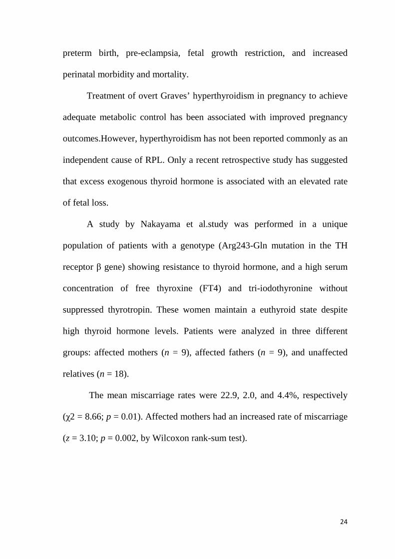
A study by Nakayama et al.study was performed in a unique
population of patients with a genotype (Arg243-Gln mutation in the TH
receptor β gene) showing resistance to thyroid hormone, and a high serum
concentration of free thyroxine (FT4) and tri-iodothyronine without
suppressed thyrotropin. These women maintain a euthyroid state despite
high thyroid hormone levels. Patients were analyzed in three different
groups: affected mothers (n = 9), affected fathers (n = 9), and unaffected
relatives (n = 18).
The mean miscarriage rates were 22.9, 2.0, and 4.4%, respectively
(χ2 = 8.66; p = 0.01). Affected mothers had an increased rate of miscarriage
(z = 3.10; p = 0.002, by Wilcoxon rank-sum test).

25
Hypothyroidism
The most common cause of hypothyroidism in pregnant women,
affecting approximately 0.5% of patients is chronic autoimmune thyroiditis
(Hashimoto’s thyroiditis).Other causes of hypothyroidism include endemic
iodine deficiency (ID), prior radioactive iodine therapy and thyroidectomy.
There seems to be no doubt that hypothyroidism is associated with
infertility.
Untreated hypothyroidism in pregnancy has consistently been shown
to be associated with an increased risk for adverse pregnancy complications,
as well as detrimental effects on fetal neurocognitive development.30
Specific adverse outcomes associated with maternal overt hypothyroidism
include increased risks for premature birth, low birth weight, and
miscarriage
Thyroid hormones have an impact on oocytes at the level of the
granulosa and luteal cells that interfere with normal ovulation.Low
thyroxine levels have a positive feedback on thyroid-releasinghormone
(TRH). Elevations in TRH have been associated with PRL elevation.It is
believed that elevated PRL alters the pulsatility of gonadotropin-releasing
hormone (GnRH) and interferes with normal ovulation.
Therefore, severe forms of hypothyroidism rarely complicate
pregnancy because they are associated with anovulation and infertility.

26
Even if an association exists between low thyroid function and pregnancy
loss, direct evidence for a causal role is missing.One postulated explanation
for this relationship is that LPD has been linked to thyroid hypofunction.
A study of thyroid function and pregnancy outcome in 2009
demonstrated a positive linear relationship between fetal loss and maternal
thyroid-stimulating hormone (TSH) levels assayed in healthy women,
without overt thyroid dysfunction.Surprisingly, in this study any association
was found between FT4 levels and subsequent risk of child loss in these
women.
We believe it is prudent to screen for thyroid disease and normalize
thyroid function prior to conception when function is found to be abnormal.
Even if there is no clear cause–effect relationship between hypothyroidism
and RPL, there is some evidence that subclinical hypothyroidism is
correlated with poor maternal outcome as well as prematurity and reduced
intelligence quotient in the offspring.
There is disagreement as to the suitable upper limit of normal serum
TSH in order to make the diagnosis of subclinical hypothyroidism. There is
a trend with the new TSH assays to decrease the upper limit of normal TSH
(range, 4.5 to 5.0 mU/L) to 2.5 mU/L. This upper limit is recommended by
the National Academy of Clinical Biochemistry guideline, and based on the
fact that 2.5 mU/L represents more than 2 standard deviations above
meticulously screened euthyroid volunteers.Clearly, this new upper limit
27
will significantly increase the number of patients diagnosed with subclinical
hypothyroidism, and its clinical benefit remains questionable.
Thyroid peroxidase antibody
Thyroid peroxidase is a poorly glycosylated membrane-bound
enzyme, responsible for iodine oxidation and iodination of thyrosyl
residues of the Tg molecule ` It had been termed microsomal antigen based
on its intracellular localization. Antibodies react against conformational
epitopes at the surface of the molecules and against linear epitopes .
Polyclonal antibodies from healthy individuals and patients are directed
against the same epitopes.
Anti-TPO antibodies from healthy subjects did not block TPO
activity or interfere with the blocking activity of anti-TPO antibodies from
AITD patients , while anti-TPO antibodies from AITD patients can fix
complement, destroy thyrocytes, and act as competitive inhibitors of
enzymatic activity . These antibodies can be of any class of IgG, although
some studies indicated a higher prevalence of IgG1 (70%) and IgG4
(66.1%) compared to IgG2 (35.1%) and IgG3 (19.6%).
Low levels of IgA antibodies have also been reported . Anti-TPO
antibodies are more common than anti-Tg antibodies and more indicative
for thyroid disease. Anti-TPO antibodies are inductors of oxidative stress
evidenced by decreased antioxidant potential, advanced glycosylation

28
products and oxygen metabolites in blood . However, their contribution to
thyroid damage compared to T cell and cytokine-mediated apoptosis is
minor . Anti-TPO antibodies are detected in 90–95% of AITD patients, 80%
of GD, and 10–15% of non-AITD patients.
While anti-TPO antibodies may act cytotoxic on thyrocytes in HT
they do not have an established role in GD. Anti-TPO antibodies are able to
cross the placenta barrier to variable extent, but the effect on the neonate is
unclear. Concerns on a potential negative effect on cognitive development
of the offspring have not been confirmed so far.
Symptoms can be mild, and patients might not seek medical advice. Even
when treatment has been initiated, titers of anti-TPO antibodies decrease
only slowly (e.g., over 5 years) upon treatment with levothyroxine, and anti-
TPO antibody titers remain in the pathological range. Normal anti-thyroid
antibody titers are lower for anti-TSHR antibodies than for anti-TPO and
anti-Tg antibodies.
Thyroid Autoimmunity and Pregnancy Loss
Autoimmune thyroid disease is the most common endocrine disorder
in women of reproductive age with an overall prevalence in women of 10 to
15%38 and among pregnant women, autoimmune thyroid disease has a
prevalence of 5 to 20%.39 In recent years many studies have found an
association between thyroid autoimmunity (TA) and recurrent abortions;
29
moreover it has been suggested that thyroid autoantibodies may be
employed as a marker for at-risk pregnancies.
Many studies have linked TA with recurrent miscarriages, although
the mechanism involved is unclear. Despite this, three mechanisms have
been postulated to explain the possible association between TA and early
pregnancy loss:
(1) the presence of thyroid autoantibodies reflects a generalized activation
of the immune system and a generally heightened autoimmune reactivity
against the feto-placental unit
(2) The presence of thyroid autoantibodies may act as an infertility factor
and may delay conception. Thus, when women with thyroid autoantibodies
do become pregnant, they are older and have a higher risk of miscarriage
(3) The presence of thyroid autoantibodies in euthyroid women may be
linked with a mild deficiency in thyroid hormone concentrations or a lower
capacity of the thyroid gland to adapt to the demands of the pregnancy state.
Indeed, the mean serum TSH values, while being within normal
range, were significantly higher in thyroid autoantibody positive women
compared to women with negative thyroid autoantibodies. This may reflect
lower thyroidal reserve during pregnancy when a greater amount of thyroid
hormones is demanded.However, the various hypotheses mentioned above
are not contradictory to each other, so it is possible that the mechanisms
explained act in concert.
30
Thyroxine administration seems to be effective in reducing the
number of miscarriages when given during the early stages of pregnancy,
because miscarriages with maternal thyroid autoimmunity generally occur
within the first trimester.Poppe et al. have proposed that serum TSH, free
FT4 and thyroid autoantibodies should be measured in early gestation.
When serum TSH is elevated or free FT4 is below normal, levothyroxine
(LT4) should be administered during pregnancy.
In women with thyroid autoantibodies and serum TSH <2 mU/L, LT4
treatment is not warranted; however, serum TSH and free FT4 should be
measured later in gestation, preferably at the end of the second trimester.
For women with thyroid autoantibodies and TSH between 2 and 4 mU/L in
early gestation, treatment with LT4 should be considered. It is important to
consider that serum TSH is downregulated during the first half of gestation
by hCG.47 However, further studies are required to understand if all women
with positive thyroid autoantibodies should be started on LT4 therapy
during their pregnancies to decrease miscarriage rate.

31
AIM OF THE STUDY
The aim of the study was to find out the association between Anti-
TPO antibody and recurrent miscarriages
To evaluate obstetric complications such as Pre Eclampsia and
Preterm labour in them
32
REVIEW OF LITERATURE
1. L. Mehran,1 M. Tohidi, F. Sarvghadi, H. Delshad, A. Amouzegar,O. P.
Soldin, and F. Azizi in a study of Management of Thyroid Peroxidase
Antibody Euthyroid Women in Pregnancy: Comparison of the American
Thyroid Association and the Endocrine Society Guidelines concluded that
The current available data regarding associations between thyroid
autoantibodies and spontaneous or recurrent pregnancy loss and preterm
delivery is convincing. However, the reduction of these complications by
treatment with LT4 supplementation is less robust. The evidence is not
conclusive enough to recommend screening for thyroid autoantibodies or
for the treatment of euthyroid women who are positive for thyroid
autoantibodies during pregnancy. Both sets of guidelines discussed here are
of high quality, and there does not seem to be contradiction or disagreement
between the recommendations of the American Thyroid Association (2011)
and of the Endocrine Society (2012) on the screening and management of
women with thyroid autoantibodies in pregnancy. They suggested that
either one of the two guidelines may be used by clinicians for appropriate
and up-to-date management of thyroid autoimmunity during pregnancy.
2. Alex Stagnaro-Green, MD; Sheila H. Roman, MD; Rhoda H. Cobin, MD;
et al in a study of Detection of At-Risk Pregnancy by Means of Highly
Sensitive Assays for Thyroid Autoantibodies screened 552 women who
33
presented to their obstetrician in the first trimester of pregnancy using
highly sensitive enzyme-linked immunosorbent assays for the presence of
thyroglobulin and thyroidperoxidase autoantibodies and found an incidence
of positivity of 19.6%. The tendency to secrete detectable levels of thyroid
autoantibodies was significantly correlated with an increased rate of
miscarriage. Thyroid autoantibody—positive women miscarried at a rate of
17%, compared with 8.4% for the autoantibody-negative women. Individual
levels of thyroglobulin and thyroidperoxidase autoantibodies were similarly
related to this increased miscarriage rate, with no evidence of autoantibody
specificity in the relationship. Furthermore, the increase in miscarriages
could not be explained by differences in thyroid hormone levels, the
presence of cardiolipin autoantibodies, maternal age, gestational age at the
time of maternal entry into the study, or previous obstetric history and
concluded that thyroid autoantibodies are an independent marker of "at-
risk" pregnancy.
3. MF Prummel and WM Wiersinga in a study of Thyroid autoimmunity and
miscarriage in European Journal of Endocrinology with aim to ascertain
the strength of the association between thyroid autoimmunity and
miscarriage, performed a meta-analysis of both case-control and
longitudinal studies performed since 1990 when this association was first
described. A clear association between the presence of thyroid antibodies
34
and miscarriage was found with an odds ratio (OR) of 2.73 (95 %
confidence interval (CI), 2.20-3.40) in eight case-control and ten
longitudinal (OR, 2.30; 95 % CI, 1.80-2.95) studies. This association may
be explained by a heightened autoimmune state affecting the fetal allograft,
of which thyroid antibodies are just a marker. Alternatively, the association
can be partly explained by the slightly higher age of women with
antibodies compared with those without (mean+/-S.D. age difference,
0.7+/-1.0 years; P<0.001). A third possibility is mild thyroid failure, as
thyroid-stimulating hormone (TSH) levels in antibody-positive but
euthyroid women are higher than in antibody-negative women: difference
0.81+/-0.58 mU/l (P=0.005). Randomized clinical trials with l-thyroxine
(aiming at TSH values between 0.4 and 2.0 mU/l) and with selenium (to
decrease antibodies against thyroid peroxidase) are clearly needed to
elucidate further the nature of this association.
4. Thyroid autoimmunity and the risk of miscarriage Alex Stagnaro-Green,
Daniel Glinoer in a study Thyroid autoimmunity and the risk of miscarriage
concluded that in approximately one-third of all pregnancies end in
miscarriage. The etiology of recurrent abortion remains unknown in
approximately 50% of all women. In the early 1990s it was discovered that
unselected euthyroid women who present with thyroid antibodies (thyroid
peroxidase and thyroglobulin) in the first trimester of pregnancy have a
two–four-fold increase in their miscarriage rates. Although the etiology of
35
miscarriage in thyroid antibody women remains unknown, recent data have
revealed a potential direct effect of thyroglobulin antibodies on pregnancy
loss in a murine model. Uncontrolled studies assessing the effect of
levothyroxine on decreasing the miscarriage rate in euthyroid antibody
positive women, have demonstrated a decreased miscarriage rate.
5. Kris Poppe, Brigitte Velkeniers& Daniel Glinoer in a study of The role of
thyroid autoimmunity in fertility and pregnancy found that Hypothyroidism
influences ovarian function by decreasing levels of sex-hormone-binding
globulin and increasing the secretion of prolactin. In women of reproductive
age, hypothyroidism can be reversed by thyroxine therapy to improve
fertility and avoid the need for use of assisted reproduction technologies.
For infertile women, preparation for medically assisted pregnancy
comprises controlled ovarian hyperstimulation that substantially increase
circulating estrogen concentrations, which in turn can severely impair
thyroid function. In women without thyroid autoimmunity these changes are
transient, but in those with thyroid autoimmunity estrogen stimulation might
lead to abnormal thyroid function throughout the remaining pregnancy
period. Prevalence of thyroid autoimmunity is significantly higher among
infertile women than among fertile women, especially among those whose
infertility is caused by endometriosis or ovarian dysfunction. Presence of
thyroid autoimmunity does not interfere with normal embryo implantation,
but the risk of early miscarriage is substantially raised. Subclinical and overt
36
forms of hypothyroidism are associated with increased risk of pregnancy-
related morbidity, for which thyroxine therapy can be beneficial. Systematic
screening for thyroid disorders in pregnant women remains controversial
but might be advantageous in women at high risk, particularly infertile
women.
6. Roberto Negro, Gianni Formoso, TizianaMangieri, Antonio Pezzarossa,
DavideDazzi, Haslinda Hassan in a study of Levothyroxine Treatment in
Euthyroid Pregnant Women with Autoimmune Thyroid Disease: Effects on
Obstetrical Complications conducted in a total of 984 pregnant women;
11.7% were thyroid peroxidase antibody positive (TPOAb+). The TPOAb+
patients were divided into two groups: group A (n = 57) was treated with
LT4, and group B (n = 58) was not treated. The 869 TPOAb− patients
(group C) served as a normal population control group. Rates of obstetrical
complications in treated and untreated groups were measured. They found
that, at baseline, TPOAb+ had higher TSH compared with TPOAb−; TSH
remained higher in group B compared with groups A and C throughout
gestation. Free T4 values were lower in group B than groups A and C after
30 wk and after parturition. Groups A and C showed a similar miscarriage
rate (3.5 and 2.4%, respectively), which was lower than group B (13.8%) [P
< 0.05; relative risk (RR), 1.72; 95% confidence interval (CI), 1.13–2.25;
and P < 0.01; RR = 4.95; 95% CI = 2.59–9.48, respectively]. Group B
displayed a 22.4% rate of premature deliveries, which was higher than
37
group A (7%) (P < 0.05; RR = 1.66; 95% CI = 1.18–2.34) and group C
(8.2%) (P < 0.01; RR = 12.18; 95% CI = 7.93–18.7). They concluded that
the Euthyroid pregnant women who are positive for TPOAb develop
impaired thyroid function, which is associated with an increased risk of
miscarriage and premature deliveries. Substitutive treatment with LT4 is
able to lower the chance of miscarriage and premature delivery.
7. R. Negro, G. Formoso, L. Coppola, G. Presicce, T. Mangieri, A. Pezzarossa,
D. Dazzi in a study of Euthyroid women with autoimmune disease
undergoing assisted reproduction technologies: The role of autoimmunity
and thyroid function to assess if patients with autoimmune thyroid disease
undergoing assisted reproduction technologies (ART) are afflicted by poor
pregnancy and/or delivery rate and if the outcome is conditioned by pre-
ART thyroid status. The study was retrospective (from January 2000 to
January 2005) and was carried out at the Division of Physiopathology of
Human Reproduction. Women who underwent ART were tested for TSH,
free T4 (FT4), thyroid peroxidase antibodies (TPOAb) before and during
pregnancy. A total of 416 euthyroid women were selected; 42 (10.1%) were
TPOAb (+). Women >35 yr were excluded. The endpoints were pregnancy
and delivery rates. Results: no differences in pregnancy and delivery rates
were observed between women with and without antibodies. In TPOAb (+),
women who failed to become pregnant or miscarried displayed higher TSH
values before ART (2.8 mlU/l) compared to the ones who delivered (1.6

38
mlU/l; p=0.032) and compared to TPOAb (−) (1.1 mlU/l; p=0.018).
Conclusions: in euthyroid women undergoing ART the pregnancy and
delivery rates are not affected by the presence of TPOAb. In TPOAb (+)
high-normal TSH values are associated with increased risk of unsuccessful
pregnancy or subsequent miscarriage.
8. F.H. Rushworth, M. Backos, R. Rai I.T. Chilcott, N. Baxter L. Regan in a
study of Prospective pregnancy outcome in untreated recurrent miscarriers
with thyroid autoantibodies with a purpose to determine the prevalence of
thyroid antibodies in women with recurrent miscarriage and to observe
whether their presence was predictive of future pregnancy outcome. A total
of 870 consecutive, non-pregnant women with a history of three or more
pregnancy losses and normal parental karyotypes were investigated for the
presence of thyroglobulin antibodies (TgAb) and for thyroid microsomal
antibodies (TmAb). Thyroid antibodies were found in 162 (19%) women.
TgAb only were found in eight women (5%); TmAb only in 98 (60%) and
both TgAb and TmAb were found in 56 (35%). Thirteen women had a
history of thyroid disease and a further 15 women were found to have
abnormal thyroid function. All 28 were excluded from the pregnancy
outcome study. Among the remaining 134 thyroid antibody positive
women, 36 women were not tested and normal thyroid stimulating hormone
results were obtained for 98. In the group proven euthyroid, 14 of 24
untreated pregnancies resulted in live births (58%). Among the 710 thyroid
39
antibody negative women, 47 of 81 untreated pregnancies resulted in live
births (58%). The future risk of pregnancy loss in women with unexplained
recurrent miscarriage is not affected by their thyroid antibody status.
9. Amir Iravani, Maryam Saeedi, JalilPakravesh, SepehrHamidi,
MehrshadAbbasi in Thyroid Autoimmunity and Recurrent Spontaneous
Abortion in Iran: A Case-Control Study with objective to determine the
association of thyroglobulin antibodies (TG-Ab) and thyroid peroxidase
antibodies (TPO-Ab) with recurrent spontaneous abortion in a euthyroid,
nonpregnant population of women in Iran. This case-control study
conducted between November 2003 and September 2006 in Tehran, Iran,
nonpregnant women with a history of 3 or more consecutive pregnancy
losses and age-matched, healthy parous women without a history of
reproductive problems were assessed. Thyroid function tests were
performed, which included assessment of thyroid-stimulating hormone,
triiodothyronine, thyroxine, and the presence of TG-Ab and TPO-Ab. A
total of 641 patients and 269 controls were included. Mean age (± SD) was
30.6 ± 6.4 years (range, 16-51 years) in the patient group and 30.05 ± 6.6
years (range, 18-48 years) in the control group. Thyroid antibodies were
present in 157 of 641 patients (24.5%) and in 34 of 269 controls (12.6%)
(P<.001). The presence of thyroid antibodies was significantly associated
with recurrent abortion independent of the impact of age with an odds ratio
of 2.24 (95% confidence interval, 1.5-3.35). They concluded that TG-Ab

40
and TPO-Ab were identified more frequently in women with recurrent
abortions compared with controls, and thyroid autoimmunity was
independently associated with a higher risk of recurrent abortion.
10. ShakilaThangaratinam, Alex Tan, Ellen Knox, Mark D Kilby,
ArriCoomarasamy in a study of Association between thyroid autoantibodies
and miscarriage and preterm birth: meta-analysis of evidence with an
objective to evaluate the association between thyroid autoantibodies and
miscarriage and preterm birth in women with normal thyroid function and
to assess the effect of treatment with levothyroxine on pregnancy outcomes
in this group of women. The study design was Systematic review and meta-
analysis. With Data sources Medline, Embase, Cochrane Library, and
SCISEARCH (inception-2011) without any language restrictions. They
used a combination of key words to generate two subsets of citations, one
indexing thyroid autoantibodies and the other indexing the outcomes of
miscarriage and preterm birth. The Study selection included Studies that
evaluated the association between thyroid autoantibodies and pregnancy
outcomes were selected in a two stage process. Two reviewers selected
studies that met the predefined and explicit criteria regarding population,
tests, and outcomes. Data synthesis Odds ratios from individual studies
were pooled separately for cohort and case-control studies with the random
effects model.

41
Results included 30 articles with 31 studies (19 cohort and 12 case-
control) involving 12 126 women assessed the association between thyroid
autoantibodies and miscarriage. Five studies with 12 566 women evaluated
the association with preterm birth. Of the 31 studies evaluating miscarriage,
28 showed a positive association between thyroid autoantibodies and
miscarriage. Meta-analysis of the cohort studies showed more than tripling
in the odds of miscarriage with the presence of thyroid autoantibodies (odds
ratio 3.90, 95% confidence interval 2.48 to 6.12; P<0.001). For case-control
studies the odds ratio for miscarriage was 1.80, 1.25 to 2.60; P=0.002).
There was a significant doubling in the odds of preterm birth with the
presence of thyroid autoantibodies (2.07, 1.17 to 3.68; P=0.01). Two
randomised studies evaluated the effect of treatment with levothyroxine on
miscarriage. Both showed a fall in miscarriage rates, and meta-analysis
showed a significant 52% relative risk reduction in miscarriages with
levothyroxine (relative risk 0.48, 0.25 to 0.92; P=0.03). One study reported
on the effect of levothyroxine on the rate of preterm birth, and noted a 69%
relative risk reduction (0.31, 0.11 to 0.90). They concluded that the presence
of maternal thyroid autoantibodies is strongly associated with miscarriage
and preterm delivery. There is evidence that treatment with levothyroxine
can attenuate the risks.

42
MATERIALS AND METHODS
RESERCH DESIGN
Case control observational study
STUDY SETTING
The study was conducted in the Institute of Social Obstetrics in madras
medical college between March2017 to March 2018 . The Study approved
by Ethical committee of the hospital
INCLUSION CRITERIA
Patients with history of recurrent miscarriages ( more than 3 in first
trimester)presenting to antenatal clinic in Madras Medical College.
EXCLUSION CRITERIA
Those with
1.anatomical uterine defects
2.Overt hypothyroid
3.Anti phospholipid antibody syndrome
4. other auto immune disorders

43
SAMPLE SIZE
100
METHOD
All patients provided written informed consent. 50 women who presented
with history of recurrent miscarriage were taken as cases &50 mothers
without such history were taken as controls.
Those with anatomic uterine defects, overt hypothyroidism,
antiphospholipids syndrome and other autoimmune diseases were excluded
from cases.
Under quality control and safety procedures for sample collection 10ml
venous blood sample was collected in vaccutainertubes .Serum samples
were sent for anti-TPO and TSH assay. Serum TSH levels were determined
using Micro particle Enzyme Immunoassay (MEIA) kits.
Normal reference range in pregnancy for TSH: 0.35-2.5µIU/ml (as per our
hospital reference value), values >2.5 µIU/ml were considered high for
pregnancy. Anti- TPO antibodies quantitative determination was done using
CLIA (Chemiluminescemt Immunoassay) kits . TPO levels >34 iu/ml were
considered abnormal and these women were considered TPO+VE. Cases\
were evaluated for obstetrics complications such as
preeclampsia and preterm labour.
44
RESULTS AND DISCUSSION
The collected data were analysed with IBM.SPSS statistics software
23.0 Version.To describe about the data descriptive statistics frequency
analysis, percentage analysis were used for categorical variables and the
mean & S.D were used for continuous variables. To find the significant
difference between the bivariate samples in Independent groups the
Unpaired sample t-test was used. To find the significance in categorical data
Chi-Square test was used similarly if the expected cell frequency is less than
5 in 2×2 tables then the Fisher's Exact was used. In all the above statistical
tools the probability value .05 is considered as significant level.
45
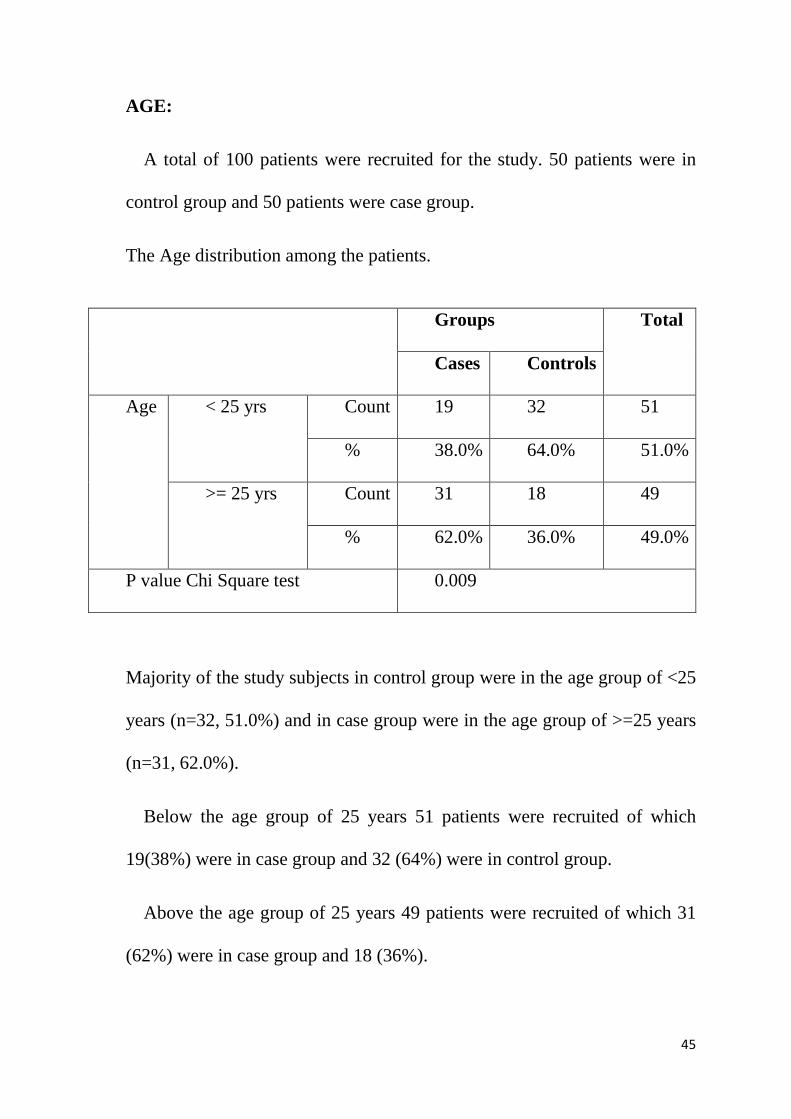
AGE:
A total of 100 patients were recruited for the study. 50 patients were in
control group and 50 patients were case group.
The Age distribution among the patients.
Majority of the study subjects in control group were in the age group of <25
years (n=32, 51.0%) and in case group were in the age group of >=25 years
(n=31, 62.0%).
Below the age group of 25 years 51 patients were recruited of which
19(38%) were in case group and 32 (64%) were in control group.
Above the age group of 25 years 49 patients were recruited of which 31
(62%) were in case group and 18 (36%).
Groups Total
Cases Controls
Age < 25 yrs Count 19 32 51
% 38.0% 64.0% 51.0%
>= 25 yrs Count 31 18 49
% 62.0% 36.0% 49.0%
P value Chi Square test 0.009
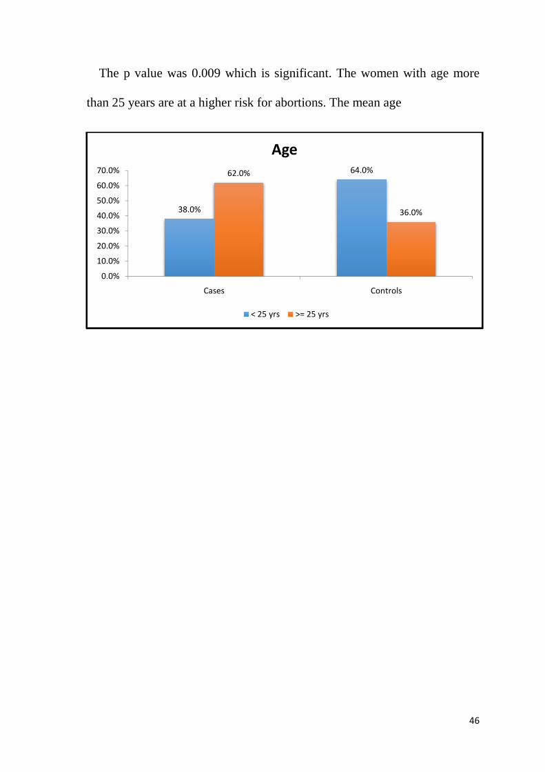
The p value was 0.009 which is significant. The women with age more
than 25 years are at a higher risk for abortions. The mean age
38.0%
0.0%
10.0%
20.0%
30.0%
40.0%
50.0%
60.0%
70.0%
s 0.009 which is significant. The women with age more
than 25 years are at a higher risk for abortions. The mean age
38.0%
64.0%62.0%
Cases Controls
Age
< 25 yrs >= 25 yrs
46
s 0.009 which is significant. The women with age more
than 25 years are at a higher risk for abortions. The mean age
36.0%
Controls

47
OBSTETRIC CODE:
Groups Total
Cases Controls
Obst
code
MULTI Count 50 24 74
% 100.0% 48.0% 74.0%
PRIMI Count 0 26 26
% 0.0% 52.0% 26.0%
Total Count 50 50 100
%
within
Groups
100.0% 100.0% 100.0%

In the control group of the 50 patients 24 (48%) were Multigravida and 26
(52%).
0%
10%
20%
30%
40%
50%
60%
70%
80%
90%
100%
Cases
In the control group of the 50 patients 24 (48%) were Multigravida and 26
Cases Controls
100.0%
48.0%
52.0%
Obstetric code
Multi Primi
48
In the control group of the 50 patients 24 (48%) were Multigravida and 26
49
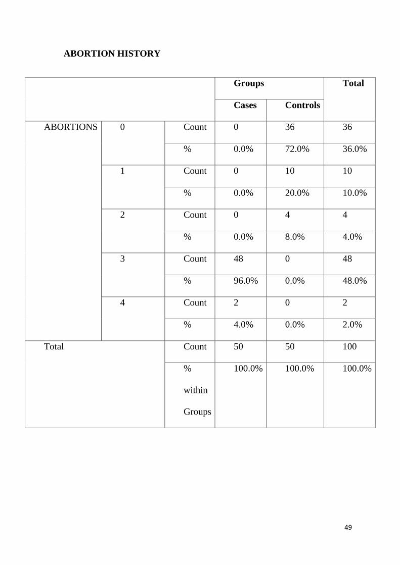
ABORTION HISTORY
Groups Total
Cases Controls
ABORTIONS 0 Count 0 36 36
% 0.0% 72.0% 36.0%
1 Count 0 10 10
% 0.0% 20.0% 10.0%
2 Count 0 4 4
% 0.0% 8.0% 4.0%
3 Count 48 0 48
% 96.0% 0.0% 48.0%
4 Count 2 0 2
% 4.0% 0.0% 2.0%
Total Count 50 50 100
%
within
Groups
100.0% 100.0% 100.0%
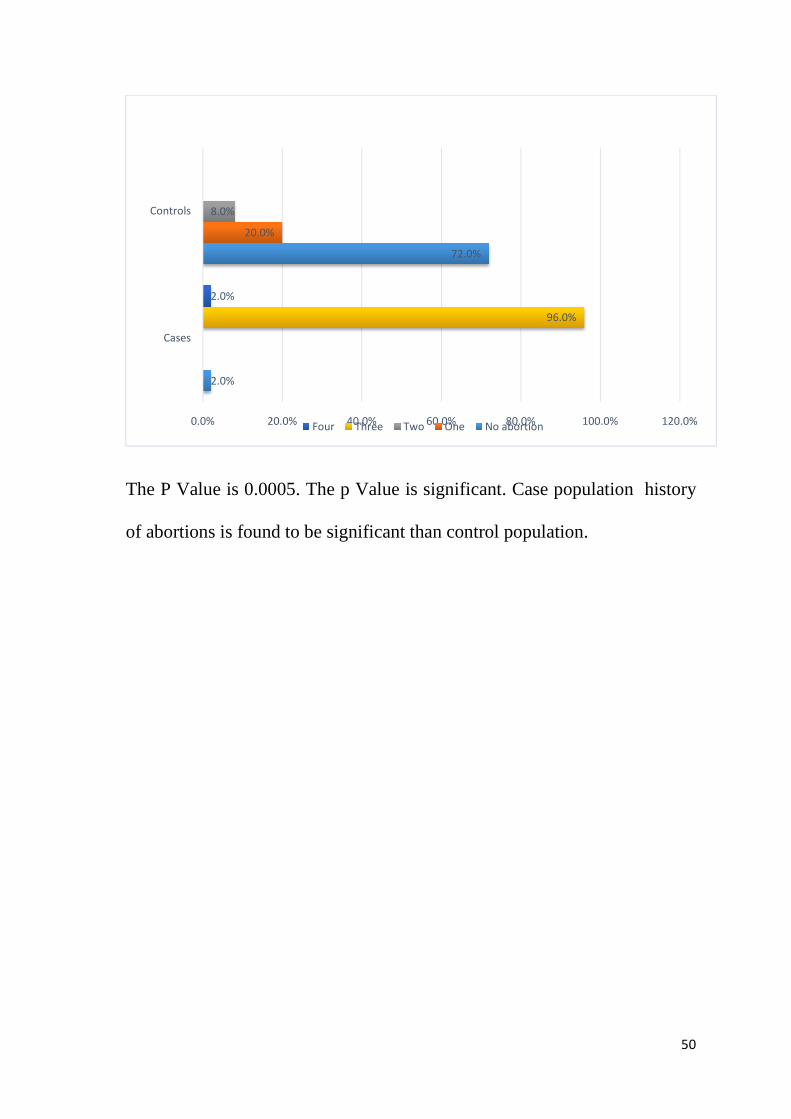
The P Value is 0.0005. The p Value is significant. Case population history
of abortions is found to be significant than control population.
2.0%
20.0%
8.0%
2.0%
0.0% 20.0%
Cases
Controls
The P Value is 0.0005. The p Value is significant. Case population history
of abortions is found to be significant than control population.
72.0%
96.0%
20.0% 40.0% 60.0% 80.0%Four Three Two One No abortion
50
The P Value is 0.0005. The p Value is significant. Case population history
of abortions is found to be significant than control population.
96.0%
100.0% 120.0%

51
GESTATIONAL AGE AT THE TIME OF DELIVERY
Groups Total
Cases Controls
GA Upto 36+6 Count 4 0 4
% 8.0% 0.0% 4.0%
> 36 to
37+6
Count 8 0 8
% 16.0% 0.0% 8.0%
>37 +
38+6
Count 16 3 19
% 32.0% 6.0% 19.0%
>38 +
39+6
Count 16 14 30
% 32.0% 28.0% 30.0%
> 39 Count 6 33 39
% 12.0% 66.0% 39.0%
Total Count 50 50 100
%
within
Groups
100.0% 100.0% 100.0%

In the case group 4 (8%) patients were in the preterm group.
0.0005. Patients with recurrent spontaneous abortion have a significant
chance of having preterm labour.
0%
20%
40%
60%
80%
100%
Upto 36+6
In the case group 4 (8%) patients were in the preterm group.
. Patients with recurrent spontaneous abortion have a significant
chance of having preterm labour.
Cases Controls
8.0%0.0%
16.0%
0.0%
32.0%
6.0%
32.0%
28.0%
12.0%
66.0%
Gestational age
> 36 to 37+6 >37 + 38+6 >38 + 39+6
52
In the case group 4 (8%) patients were in the preterm group. the p Value is
. Patients with recurrent spontaneous abortion have a significant
> 39

53
THE PLACE OF RESIDENCE
Groups Total
Cases Controls
RESIDENCE Rural Count 24 21 45
% 48.0% 42.0% 45.0%
Urban Count 26 29 55
% 52.0% 58.0% 55.0%
Total Count 50 50 100
%
within
Groups
100.0% 100.0% 100.0%
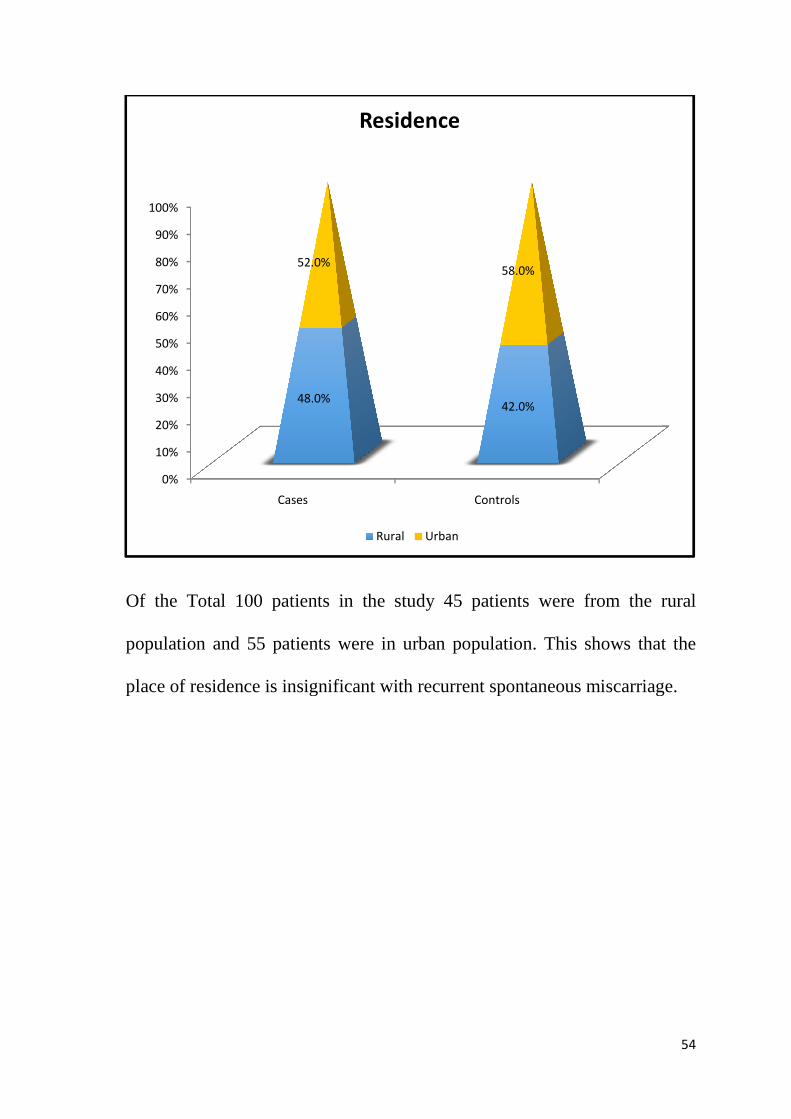
Of the Total 100 patients in the study 45 patients were from the rural
population and 55 patients were in urban population. This shows that the
place of residence is insignificant with recurrent spontaneous miscarriage.
0%
10%
20%
30%
40%
50%
60%
70%
80%
90%
100%
Cases
Of the Total 100 patients in the study 45 patients were from the rural
population and 55 patients were in urban population. This shows that the
place of residence is insignificant with recurrent spontaneous miscarriage.
Cases Controls
48.0%42.0%
52.0%58.0%
Residence
Rural Urban
54
Of the Total 100 patients in the study 45 patients were from the rural
population and 55 patients were in urban population. This shows that the
place of residence is insignificant with recurrent spontaneous miscarriage.

55
SOCIO ECONOMIC STATUS
Groups Total
Cases Controls
socio
economic
status
LM Count 13 17 30
% 26.0% 34.0% 30.0%
UL Count 27 21 48
% 54.0% 42.0% 48.0%
UM Count 10 12 22
% 20.0% 24.0% 22.0%
Total Count 50 50 100
% within
Groups
100.0% 100.0% 100.0%

Out of 100 patients 30% were in low middle
lowerclass , 22 % in upper middle class . Thus, socio economic status found
be insignificant in recurrent spontaneous miscarriage.
Bar diagram showing Socio economic status
0.0% 10.0%
LM
UL
UM
Out of 100 patients 30% were in low middle class, 48% were in upper
22 % in upper middle class . Thus, socio economic status found
be insignificant in recurrent spontaneous miscarriage.
Bar diagram showing Socio economic status
26.0%
20.0%
34.0%
42.0%
24.0%
20.0% 30.0% 40.0%
Socio Economic Status
Controls Cases
56
class, 48% were in upper
22 % in upper middle class . Thus, socio economic status found
54.0%
42.0%
50.0% 60.0%
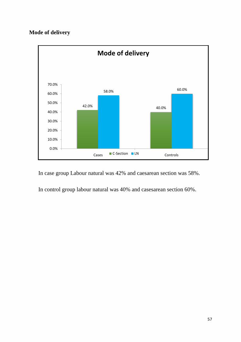
Mode of delivery
In case group Labour n
In control group labour n
42.0%
0.0%
10.0%
20.0%
30.0%
40.0%
50.0%
60.0%
70.0%
In case group Labour natural was 42% and caesarean section
ontrol group labour natural was 40% and casesarean section
42.0%40.0%
58.0%
Cases Controls
Mode of delivery
C-Section LN
57
atural was 42% and caesarean section was 58%.
l was 40% and casesarean section 60%.
60.0%
Controls
58
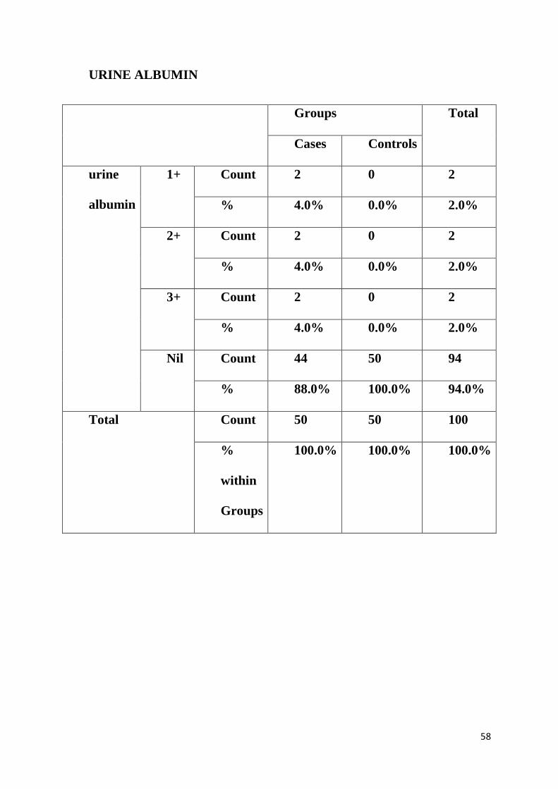
URINE ALBUMIN
Groups Total
Cases Controls
urine
albumin
1+ Count 2 0 2
% 4.0% 0.0% 2.0%
2+ Count 2 0 2
% 4.0% 0.0% 2.0%
3+ Count 2 0 2
% 4.0% 0.0% 2.0%
Nil Count 44 50 94
% 88.0% 100.0% 94.0%
Total Count 50 50 100
%
within
Groups
100.0% 100.0% 100.0%

59
Urine albumin was found to be more than 1+ in 12% in case group. among
the case group 4% were urine albumin 1+, 4 % were 2+, 4% were 3 +.
4.0
%
0.0
%
4.0
%
0.0
%
4.0
%
0.0
%
88
.0% 1
00
.0%
C A S E S C O N T R O L S
URINE ALBUMIN
1+ 2+ 3+ Nil
60
MATERNAL RISK FACTORS
Groups
Total Cases Controls
COMORBIDIT
IES
Nil Coun
t
47 50 97
% 94.0
%
100.0
%
97.0
%
Obesi
ty
Coun
t
3 0 3
% 6.0% 0.0% 3.0%
Total Coun
t
50 50 100
%
withi
n
Grou
ps
100.0
%
100.0
%
100.0
%
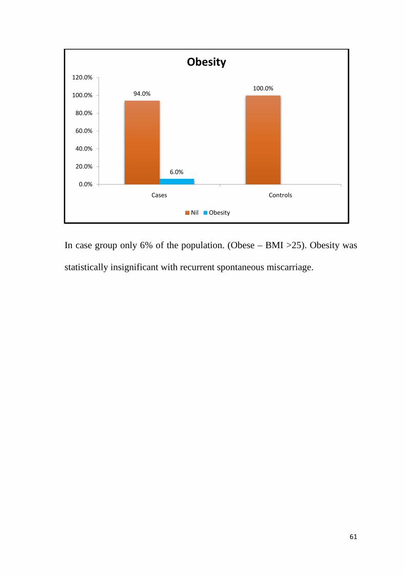
In case group only 6% of the population. (Obese
statistically insignificant with recurrent spontaneous miscarriage.
94.0%
0.0%
20.0%
40.0%
60.0%
80.0%
100.0%
120.0%
In case group only 6% of the population. (Obese – BMI >25). Obesity was
statistically insignificant with recurrent spontaneous miscarriage.
94.0%100.0%
6.0%
Cases Controls
Obesity
Nil Obesity
61
BMI >25). Obesity was
statistically insignificant with recurrent spontaneous miscarriage.
62
MATERNAL COMPLICATIONS:
Groups
Total Cases Controls
Maternal
complicati
on
No
complicati
ons
Coun
t
40 50 90
% 80.0
%
100.0
%
90.0
%
Preeclamps
ia
Coun
t
6 0 6
% 12.0
%
0.0% 6.0%
Preterm
labour
Coun
t
3 0 3
% 6.0% 0.0% 3.0%
PROM Coun
t
1 0 1
% 2.0% 0.0% 1.0%
Total Coun
t
50 50 100
%
withi
n
Grou
ps
100.0
%
100.0
%
100.0
%

63
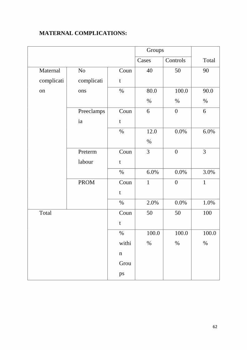
Preeclampsia was noted among 12% of mothers of with recurrent
spontaneous miscarriages,. Preterm labour was noted in 6% of the case
population and Preterm rupture of membranes was seen in 2% of the case
population. The p value was 0.011. which is significant. The incidence of
complication among women with recurrent spontaneous miscarriage is
higher than the general population.
80.0%
100.0%
12.0%6.0% 2.0%
0.0%
20.0%
40.0%
60.0%
80.0%
100.0%
120.0%
Cases Controls
Maternal complication
No complications Preeclampsia Preterm labour PROM

64
Value df
Asymp. Sig.
(2-sided)
Pearson Chi-Square 11.111a 3 .011
Likelihood Ratio 14.976 3 .002
N of Valid Cases 100
Pre -term labour
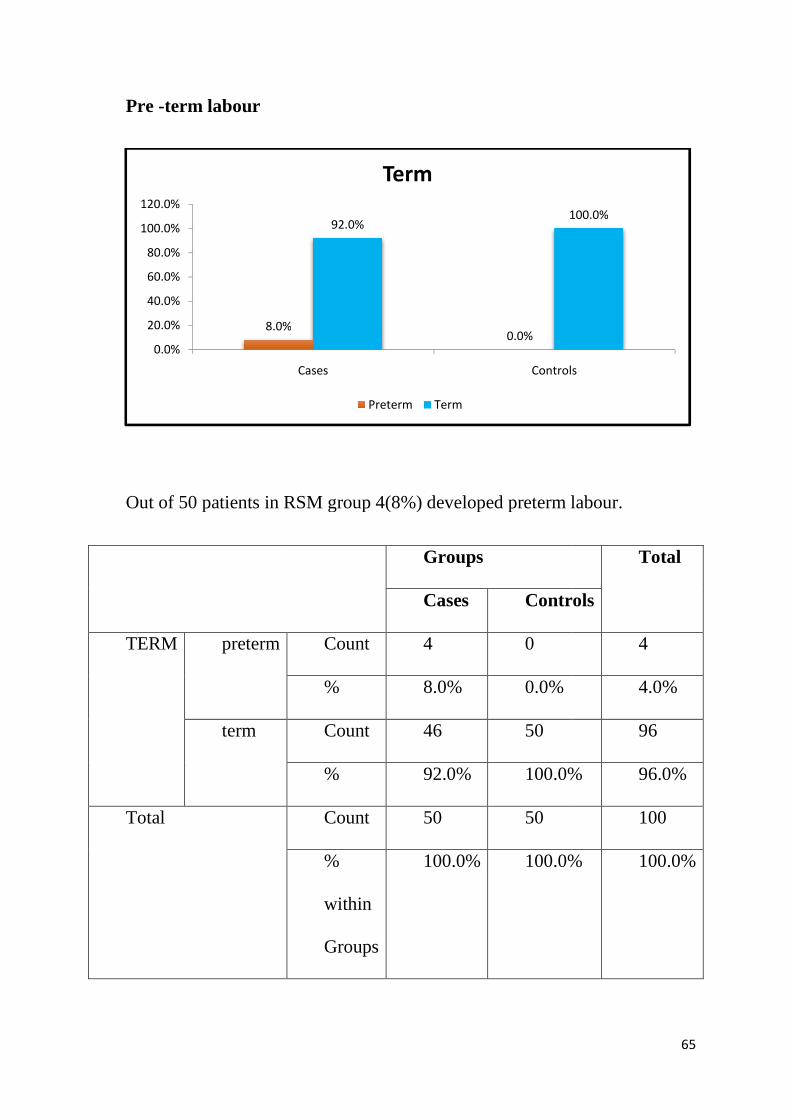
Out of 50 patients in RSM group 4(8%) developed preterm labour.
TERM preterm
term
Total
8.0%
0.0%
20.0%
40.0%
60.0%
80.0%
100.0%
120.0%
Out of 50 patients in RSM group 4(8%) developed preterm labour.
Groups
Cases Controls
Count 4 0
% 8.0% 0.0%
Count 46 50
% 92.0% 100.0%
Count 50 50
%
within
Groups
100.0% 100.0%
8.0%0.0%
92.0%100.0%
Cases Controls
Term
Preterm Term
65
Out of 50 patients in RSM group 4(8%) developed preterm labour.
Total
Controls
4
4.0%
96
100.0% 96.0%
100
100.0% 100.0%
100.0%
Controls
66
TSH
Groups
Total Cases Controls
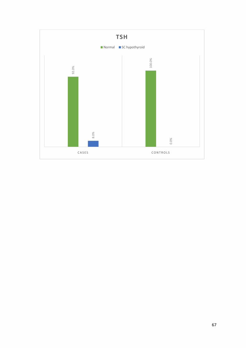
TSH Normal Count 46 50 96
% 92.0% 100.0% 96.0%
Sub clinical
hypothyroid
Count 4 0 4
% 8.0% 0.0% 4.0%
Total Count 50 50 100
%
within
Groups
100.0% 100.0% 100.0%
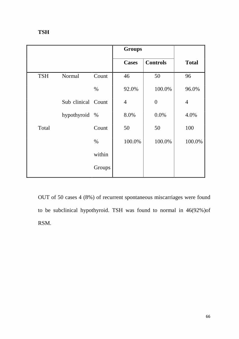
OUT of 50 cases 4 (8%) of recurrent spontaneous miscarriages were found
to be subclinical hypothyroid. TSH was found to normal in 46(92%)of
RSM.
67
92
.0% 1
00
.0%
8.0
%
0.0
%
C A S E S C O N T R O L S
TSH
Normal SC hypothyroid

68
ANTI TPO POSITIVITY:
Out of 50 patients in case group 14 (28%) were ANTI TPO positive and 36
(72%) were ANTI TPO negative.
Out of 50 patients in control group, 3(6%)were ANTI TPO POSITIVE and
47(94%)were ANTI TPO negative. P value 0.006 shows that ANTI TPO
POSITIVITY were significant in RSM group.
Groups Total
Cases Controls
ANTI TPO
POSITIVE
Negative Count 36 47 83
% 72.0% 94.0% 83.0%
Positive Count 14 3 17
% 28.0% 6.0% 17.0%
Total Count 50 50 100
%
within
Groups
100.0% 100.0% 100.0%

69
Value df
Asymp.
Sig.
(2-sided)
Exact
Sig.
(2-sided)
Exact
Sig.
(1-sided)
Pearson
Chi-Square
8.575a 1 .003
Continuity
Correctionb
7.087 1 .008
Likelihood
Ratio
9.185 1 .002
Fisher's
Exact Test
.006 .003
N of Valid
Cases
100
72
.0%
94
.0%
28
.0%
6.0
%
C A S E S C O N T R O L S
TPO POSITIVITY
Negative Positive
70
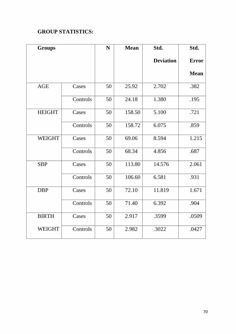
GROUP STATISTICS:
Groups N Mean Std.
Deviation
Std.
Error
Mean
AGE Cases 50 25.92 2.702 .382
Controls 50 24.18 1.380 .195
HEIGHT Cases 50 158.50 5.100 .721
Controls 50 158.72 6.075 .859
WEIGHT Cases 50 69.06 8.594 1.215
Controls 50 68.34 4.856 .687
SBP Cases 50 113.80 14.576 2.061
Controls 50 106.60 6.581 .931
DBP Cases 50 72.10 11.819 1.671
Controls 50 71.40 6.392 .904
BIRTH
WEIGHT
Cases 50 2.917 .3599 .0509
Controls 50 2.982 .3022 .0427

71
SUMMARY
The study population consisted of apparently healthy pregnant
women with a history of unexplained recurrent miscarriage during the first
trimester (5 – 13 weeks of gestation). Depending on the increased necessity
of the thyroid gland for normal development, growth and metabolic
homeostasis during in pregnancy and fetal life, changes associated with
pregnancy require an increased availability of thyroid hormones by 40% to
100% in order to meet the needs of mother and fetus during pregnancy.
The relation of the thyroid antibodies with miscarriage is an
important issue that has attracted the interest of many investigators. A
number of researches have been published concerning the relation of
thyroid autoimmunity and miscarriage which include healthy women,
women with recurrent miscarriage and those undergoing assisted
reproductive techniques. All these studies are not easily comparable due to
the different selection criteria employed for specific aims for each study, but
most studies have shown a significant positive association between the
presence of thyroid autoantibodies and miscarriage rate.
It was suggested that those autoantibodies, which can also be higher
in the euthyroid patients, may produce a threat for miscarriage in the
subsequent pregnancy. Thyroid peroxidase antibodies target the thyroid
peroxidase enzyme that assists in the production and metabolism of thyroid
72
hormone. Although they are widely seen in autoimmune thyroid disorders,
thyroid peroxidase antibodies are not necessarily a sign of disease. In up to
26 percent of healthy women, low levels of TPO antibodies are seen. In our
study the control group the TPO was positive in among 6 percent of the
study population.
In the normal healthy population, when TPO antibodies are
accompanied by a higher TSH level (> 2.0 mu/L); they suggest an increased
risk for developing Hashimoto’s thyroiditis. In spite of many studies
available regarding the physiological changes in the thyroid function during
pregnancy, it is uncertain if functional disorders of the thyroid gland play a
role in the aetiology of spontaneous miscarriages. However, this study was
about evaluating the thyroid peroxidase antibody in recurrent spontaneous
miscarriage, risk factors and the obstretric complication in the study
population.
In our study showed the mean age of was 25.92 in case population
and in control population was 24.18. In our that significant correlation the
age of the women and the incidence recurrent spontaneous miscarriages.
The mean height was 158.5 cm in cases and 158.72 cm in controls.
The weight 69.06 kg in cases and 68.34 kg in control group. The was no
significance about the of height and weight of the women with recurrent
spontaneous miscarriage
73
The residence of the women has no significance with recurrent
spontaneous miscarriage. In our study the case population resided in urban
was 52% and in rural 48% and in the control population the urban was 58%
and rural population was 42%.
There was no significance with socioecomic status of the women.
In the case group 12 (6) percentage developed preeclampsia. Among
these 6 women 2 women had severe preeclampsia. These 2 women
developed severe preeclampsia at 37 weeks of gestation and pregnancy was
terminated. Urine albumin was 3+ in 2 women. Urine albumin was 1+ and
2 + was seen in 4 women. Among the 6 women who developed
preeclampsia 4 patients were ANTI TPO positive.
Three women developed preterm labour. One women at 35Weeks and
other two at 36 all three women had vaginal delivery and babies had good
apgar. Among these 3 women who had preterm labour 2 patients had ANTI
TPO positive after ruling out other causes of Pre term labour.
Among case population one women developed PREMATURE
RUPTURE OF at MEMBRANES AT 38 weeks and delivered by caesarean
delivery with good apgar and the women was ANTI TPO POSITIVE.
In our study the incidence of subclinical hypothyriods was 4%.The
ANTI TPO positive in a total of 17, patients in the case population 14
74
patient were anti TPO positive and 3patients were ANTI TPO positive in
the control population. The p value was 0.006. The TPO positivity is
significant in the recurrent spontaneous miscarriage.
In our study the prevalence of TPO positivity was higher in women
with history of RSM than those with uneventful pregnancy history. Again
those with TPO positive have higher prevalence of subclinical
hypothyroidism.
Thyroid hormones are essential for the developing fetus. Hence a
pregnant woman needs thyroxine more than non-pregnant lady to provide
T4 to both herself and her developing fetus. The fetus's thyroid gland is not
fully functional until after 12 weeks of pregnancy. If the mother does not
have sufficient thyroid hormones, she may be at increased risk of
miscarriage. Since the majority of women are not sure that they are pregnant
until four to six weeks after the last menstrual period, they do not go to see
doctors and test their thyroid function until the first trimester is more than
half over.
It is advisable to suggest thyroid investigation to the pregnant women
with history of recurrent miscarriage as soon as possible after knowing they
are pregnant. Management of thyroid diseases during pregnancy requires
special considerations because pregnancy induces major changes in thyroid
function, and maternal thyroid disease can have adverse effects on the

75
pregnancy and the fetus. Care requires coordination among several
healthcare professionals.
Avoiding maternal and fetal thyroid dysfunction is of major
importance because of potential damage to fetal neural development, an
increased incidence of miscarriage, and preterm delivery.
76
CONCLUSION
Thyroid autoimmunity and subclinical hypothyroid can be considered
as risk marker for recurrent spontaneous miscarriage. Euthyroid women in
early stage of pregnancy with TPO antibody positive are still at risk of
developing hypothyroidism later. There serum TSH level needs to
monitored.
The TSH levels were found to be normal in miscarriage women
compared with healthy pregnant women. The feedback mechanism of
thyroid-pituitary glands is profound or not properly works in recurrent
miscarriage women.
The thyroid antibodies can only affect pregnant women when their serum
TSH level is relatively within the actual normal range.
The total thyroid hormones (TT4 and TT3) as well as decreased in
recurrent miscarriage women compared to the control subjects.
Most of women with recurrent miscarriage were euthyroid
In patients with recurrent miscarriage and if all the other work up for
recurrent pregnancy loss have turned out to be negative then tests for
detecting anti TPO should be considered even with normal TSH levels. And
if anti TPO antibodies turns out to be positive, treatment with Tablet
levothyroxine can be considered.
77
BIBLIOGRAPHY
1. Green-top Guidelines no.17, April 2011. Availablefrom:
https://www.rcog.org.uk/globalassets/documents/guidelines/gtg_17.pdf.
2. Mowbray JF, Underwood JL. Abstracts of Contributors' Individual Data
Submitted to theWorldwide Prospective Observation Study
onImmunotherapy for Treatment of Recurrent Spontaneous Abortion.
AJRI. 1994;32:261-74.
3. Casey BM, Dash JS, Wells CE. Subclinicalhypothyroidism and
pregnancy outcomes. ObstetGynecol. 2005;105(2):239.
4. Faussett MB, Branch DW. Autoimmunity andpregnancy loss.
SeminReprod Med. 2000;18:379-92.
5. Feki M, Omar S. Thyroid disorders in pregnancy:frequency and
association with selected diseases andobstetrical complications in
Tunisian women.Clinical Biochemistry. 2008;41(12):927-31.
6. Allan WC, Haddow JE, Palomaki GE. Maternalthyroid deficiency and
pregnancy complications:implications for population screening. Journal
ofMedical Screening. 2000;7(3):127-30.
7. Stagnaro-Green A, Roman SH, Cobin RH. Detectionof at risk pregnancy
by means of highly sensitiveassays for thyroid antibodies. J Am Med
Assoc.1990;264:1422-5.
78
8. Ticconi C, Giuliani E, Veglia M, Pietropolli A,Piccione E, Di Simone N.
Thyroid autoimmunity andrecurrent miscarriage. Am J
ReprodImmunol.2011;66(6):452-9.
9. Bagis T, Gokcel A, Saygili ES. Autoimmune thyroiddisease in pregnancy
and the postpartum period:relationship to spontaneous abortion.
Thyroid.2010;11:1049-53.
10. Abbassi-Ghanavati M, Casey B, Spong C. Pregnancyoutcomes in women
with thyroid peroxidaseantibodies. Obstetrics Gynaecology. 2010;116(2
Pt):381.
11. Negro R, Mangieri T, Coppola L, Presicce G.Levothyroxine treatment in
thyroid peroxidaseantibody-positive women undergoing
assistedreproduction technologies: a prospective study. HumReprod.
2005;20:1529-33

79
ANNEXURES:

80
PROFORMA
Name: Educational status:
Age:
Residence:
Socio economic status:
LMP:
DEE:
GA:
Obstetric history;
1st:
2nd:
3rd:
4th :
Present history:
h/o labour pain
h/o draining pv
h/o bleeding pv
h/o imminent symptoms

81
Past history:
Diabetes: Yes ( ) No ( )
Hypertension: Yes ( ) No ( )
Renal disease: Yes ( ) No ( )
Thyroid disorder: Yes ( )Hyper ( ) Hypo ( ). Other ( ) No ( )
Congenital abnormality: Yes ( ) No ( )
APLA positivity: Yes ( ) No ( )
Clinical examination:
Height General condition:
Weight: Pallor:
BMI: Pedal edema:
Temperature: Icterus:
BP:
PR:
RR:

82
Systemic examination:
CVS:
RS:
Per abdomen:
Per speculum:
Per Vaginal:
Investigations
Urine albumin
TSH
FT3
FT4
ANTI TPO
Other routine investigations:
Maternal complication
MODE OF DELIVERY
LN ( ) C- SECTION ( )

83
FOETAL OUTCOME
Alive/
Term/ preterm
Boy/girl
Apgar
Birth weight

84
INFORMATION SHEET
We are conducting a study on “ANTI-THYROID PEROXIDASE
POSITIVITY IN RECURRENT MISCARRIAGE AND ASSOCIATED
OBSTERICS COMPLICATIONS” among the patients in Institute of
Obstetrics and Gynaecology, Madras Medical College, Chennai and for that
your clinical details may be valuable to us.
We are selecting certain patients and if you are found eligible, we may be
using your clinical details in such a way so as to not affect your final report or
management.
The privacy of the patient in the research will be maintained throughout
the study. In the event of any publication or presentation resulting from the
research, no personally identifiable information will be shared.
Taking part in the study is voluntary. You are free to decide whether to
participate in the study or to withdraw at any time; your decision will not result
in any loss of benefits to which you are otherwise entitled.
The result of the special study may be intimated to you at the end of the
study period or during the study. If anything is found abnormal, which may aid
in management or treatment.
Signature of Investigator: Signature of Participant:
Date :

85
ேநாயாள� தகவ� தா�
ம���� ம���� க�சிைத� ஏ�ப�தலி� எதி���
ைதரா�� ெபரா�ஸிேட பாசி!"வ$!" ம�%� அத'
ெதாட��ைடய மக�ேபறிய� சி�க�க*
ேநாயாள-க.�கான தகவ� :
உ2கைள இ4த ஆரா�சிய$� ப26 ெகா*ள
அைழ�கிேறா�. நா2க* உ2க.�6 ெகா��6� இ4த
ப"வ9தி� உ*ள வ$வர2கைள� ெகா�� ந:2க* இ4த
ஆரா�சி�6 உ!படலாமா அ�ல; நிராக<�கலாமா
எ'பைத ந:2கேள ="� ெச�யலா�. ேம>� உ2கள-'
ச4ேதக2கைள?� எ2கள-ட� ேக!கலா�. ந:2க* எ2க*
ஆரா�சி�6 த6தி உ*ளவரா6� ப!ச9தி�, ெச'ைன
ம�9;வ க�@<ய$�நைடெப%� இ4த ஆரா�சிய$�
உ2கைள ப2ெக�9;�ெகா*ள ெச�ேவா�.
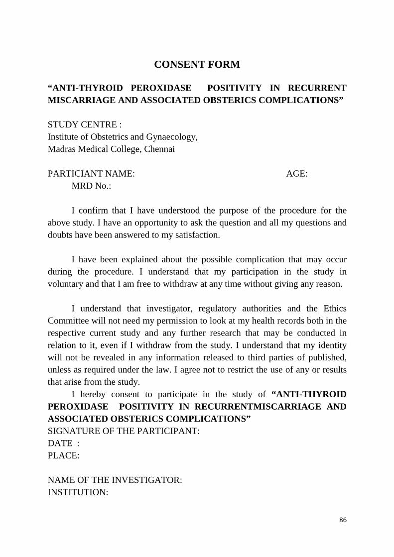
86
CONSENT FORM
“ANTI-THYROID PEROXIDASE POSITIVITY IN RECURRENT MISCARRIAGE AND ASSOCIATED OBSTERICS COMPLICATIONS” STUDY CENTRE : Institute of Obstetrics and Gynaecology, Madras Medical College, Chennai PARTICIANT NAME: AGE: MRD No.:
I confirm that I have understood the purpose of the procedure for the above study. I have an opportunity to ask the question and all my questions and doubts have been answered to my satisfaction.
I have been explained about the possible complication that may occur during the procedure. I understand that my participation in the study in voluntary and that I am free to withdraw at any time without giving any reason.
I understand that investigator, regulatory authorities and the Ethics Committee will not need my permission to look at my health records both in the respective current study and any further research that may be conducted in relation to it, even if I withdraw from the study. I understand that my identity will not be revealed in any information released to third parties of published, unless as required under the law. I agree not to restrict the use of any or results that arise from the study.
I hereby consent to participate in the study of “ANTI-THYROID PEROXIDASE POSITIVITY IN RECURRENTMISCARRIAGE AND ASSOCIATED OBSTERICS COMPLICATIONS” SIGNATURE OF THE PARTICIPANT: DATE : PLACE: NAME OF THE INVESTIGATOR: INSTITUTION:

87
ய ஒ��த� ப�வ�
ம���� ம���� க�சிைத� ஏ�ப�தலி� எதி���
ைதரா�� ெபரா�ஸிேட ஆ'"பா" பாசி!" வ$!"
ம�%� அத' ெதாட��ைடய மக�ேபறிய� சி�க�க*
ஆ�� நட9தப�� இட�: ெச'ைன ம�9;வ� க�@<,
ெச'ைன.
ப26ெப%பவ<' ெபய� :
ப26ெப%பவ<' வய; :
ப26ெப%பவ<' எ� :
இ4த ஆ�வ$� 6றி�ப$!�*ள ம�9;வ ஆ�வ$'
வ$வர2க* என�6 வ$ள�க�ப!ட;. நா' இAவா�வ$�
த'ன-ைசயாக ப2ேக�கிேற'. எ4த காரண9தினா>�
எ4த ச!ட சி�க>�6� உ!படாம� நா' இAவா�வ$�
இ�4; வ$லகி�ெகா*ளலா� எ'%� அறி4;
ெகா�ேட'.
இ4த ஆ�� ச�ப4தமாகேவா அைத சா�4த ேம>�
ஆ�� ேம�ெகா*.�ேபா;� இ4த ஆ�வ$� ப26
ெப%� ம�9;வ� எ'Cைடய ம�9;வ அறி�ைகைய
பா��பத�6 எ' அCமதி ேதைவய$�ைல எ'பைத
அறி4; ெகா*கிேற'. இ4த ஆ�வ$' Dல� கிைட�6�
="ைவ பய'ப�9தி�ெகா*ள ம%�கமா!ேட'.

88
இ4த ஆ�வ$� ப26ெகா*ள ஒ���ெகா*கிேற'.
இ4த ஆ�ைவ ேம�ெகா*.� ம�9;வ அண$�6
உ�ைம?ட' இ��ேப' எ'% உ%தியள-�கிேற'.
ப2ேக�பவ<' ைகெயா�ப� சா!சிகள-' ைகெயா�ப�
இட� : இட� :
ேததி : ேததி :
ப2ேக�பவ<' ெபய� ம�%� வ$லாச� :
ஆ�வாள<' ைகெயா�ப�
89
90
PLAGIRSM CERTIFICATE
This is to certify that this dissertation work titled “ANTI-THYROID
PEROXIDASE POSITIVITY IN RECURRENT MISCARRIAGE AND
ASSOCIATED OBSTERICS COMPLICATIONS” of the candidate
Dr.K.MANJULA, with registration Number 221616010 or the award of M.S
Degree in the branch of Obstetrics And Gynaecology . I personally verified the
urkund.com website for the purpose of plagiarism Check. I found that the
uploaded thesis file contains from introduction to conclusion pages and result
shows 6% percentage of plagiarism in the dissertation.
Guide & Supervisor.
Dr.K.KANMANI. M.D.,D.G.O.,
Professor
Institute of social obstetrics,
Madras Medical College,
Chennai.