MASSIVE TRANSFUSION IN RELATION TO OBSTETRIC ...
100
From DEPARTMENT OF CLINICAL SCIENCE, INTERVENTION AND TECHNOLOGY DIVISION OF OBSTETRICS AND GYNECOLOGY Karolinska Institutet, Stockholm, Sweden MASSIVE TRANSFUSION IN RELATION TO OBSTETRIC HEMORRHAGE WITH SPECIAL ATTENTION TO PLACENTA ACCRETA Lars Thurn Stockholm 2019
-
Upload
khangminh22 -
Category
Documents
-
view
0 -
download
0
Transcript of MASSIVE TRANSFUSION IN RELATION TO OBSTETRIC ...

From DEPARTMENT OF CLINICAL SCIENCE, INTERVENTION AND TECHNOLOGY
DIVISION OF OBSTETRICS AND GYNECOLOGY Karolinska Institutet, Stockholm, Sweden
MASSIVE TRANSFUSION IN RELATION TO OBSTETRIC HEMORRHAGE
WITH SPECIAL ATTENTION TO PLACENTA ACCRETA
Lars Thurn
Stockholm 2019

All previously published papers were reproduced with permission of the publishers. Published by Karolinska Institutet. Printed by E-print AB 2019 © Lars Thurn, 2019 ISBN 978-91-7831-467-6

Massive transfusion in relation to obstetric hemorrhage, with special attention to placenta accreta
THESIS FOR DOCTORAL DEGREE (Ph.D.) At Karolinska Institutet to be publicly defended in Lecture Hall B64,
Karolinska University Hospital, Huddinge
Friday, September 20th, 2019, at 9 am
By
Lars Thurn
Principal Supervisor: Pelle G Lindqvist M.D. Associate Professor Karolinska Institutet, Södersjukhuset Department of Clinical Science and Education Division of Obstetrics and Gynecology Co-supervisors: Agneta Wikman M.D. Associate Professor Karolinska Institutet Department of Laboratory Medicine Division of Clinical Immunology and Transfusion Medicine Prof. Magnus Westgren Karolinska Institutet Department of Clinical Science, Intervention and Technology Division of Obstetrics and Gynecology
Opponent: Prof. Bo Jacobsson Göteborg University Institute of Clinical Science Department of Obstetrics and Gynecology Examination Board: Ylva Vladic Stjernholm, M.D. Associate Professor Karolinska Institutet Department of Women’s and Children’s Health Division of Obstetrics and Gynecology Prof. Gösta Berlin Linköping University Department of Clinical Immunology and Transfusion medicine Prof. Marie Blomberg Linköping University Department of Clinical and Experimental Medicine Division of Children’s and Women’s Health


In loving memory of my mother Gerd.


ABSTRACT The overall purpose of this thesis was to assess risk factors, incidences, and complications of massive blood transfusions in relation to obstetric hemorrhage postpartum. Obstetric hemorrhage requiring blood transfusion postpartum has recently shown an increasing trend in many high resource countries. Massive transfusion, defined as more than 10 units of RBC within 24 hours is well described in surgery and trauma care, however little is known about its occurrence and risk factors in obstetric patients. Most blood transfusions are safe and necessary, but there are potential complications, including transfusion reactions, transfusion transmitted infections, and post transfusion thrombosis, which have to be taken into consideration when choosing between blood transfusion and other alternatives. The increasing rate of cesarean deliveries since the 1970’s, has contributed to complications in sequential pregnancies. One of the more severe complications is abnormally invasive placenta, a condition with a high risk of requiring massive blood transfusion and peripartum hysterectomy.
In Study 1, the incidence, risk factors, and rate of antenatal detection of abnormally invasive placenta in the Nordic countries were investigated. The study was conducted as a Nordic collaboration from 2009 to 2012, and included 605,000 deliveries. Cases of abnormally invasive placenta were reported on a monthly basis directly from maternity wards, and were complemented with data from the National Health Registries to confirm or to identify missing cases. In total, 205 cases of invasive placentas associated with a laparotomy were identified, corresponding to a prevalence of 3.4 per 10,000 deliveries. Major risk factors were placenta previa (OR = 290) and prior cesarean section (OR = 7). Only one third of the cases identified as invasive placentas were detected antenatally, and among those cases not detected, more than one third had had a prior cesarean section.
Study 2 was a retrospective population-based cohort study investigating risk factors, incidence, and trends over time for massive blood transfusion in women who gave birth in the County of Stockholm between 1990 and 2011. Data from the Medical Birth Registry was cross-linked to the Stockholm Transfusion Database. Massive transfusion was defined as transfusion of >10 units of red blood cells from time of partus through the next day. Altogether 517,874 pregnancies were included. The study found the incidence of massive transfusion to be 5.3 per 10,000 deliveries and showed an increasing trend over time. Major antenatal risk factors were abnormal placentation (OR = 41) and prior cesarean section (OR = 4).
Study 3 was a retrospective cohort study investigating whether postpartum hemorrhage and
red blood transfusion are significant and independent major risk factors for venous
thromboembolism postpartum. Women who gave birth between 1999 and 2002 in the
Stockholm region were included in the study. A time period before the implementation of
national thromboprophylaxis guidelines was chosen. Data from the Medical Birth Registry
was linked to the transfusion database and to the National Discharge Registry. Among 82,376
deliveries 56 cases of venous thromboembolism were identified. The study found transfusion
of red blood cells postpartum (OR = 5) - but not postpartum hemorrhage without blood
transfusion - to be a significant major risk factor for venous thromboembolism postpartum.
In Study 4 the aim was to assess the risk of transfusion reactions in women receiving postpartum blood transfusion. This populations based cohort study is based on the same cohort as Study 2. Data on pregnancies from the Medical Birth Registry was linked to the Stockholm Transfusion Database. Women with postpartum blood transfusion and a transfusion reaction within seven days from partus were identified. The study found a two-fold increased risk (OR = 2.0) of a transfusion reaction in women postpartum compared to non-pregnant women receiving a blood transfusion. Among all women who had a blood transfusion postpartum, women with preeclampsia were twice as likely to have a transfusion reaction.
In summary, abnormally invasive placenta occurs in 3.4 out of 10,000 deliveries and is the major risk factor for massive blood transfusion postpartum. A reduction in the rate of cesarean deliveries might be the best way to lower the incidence of both invasive placenta and massive blood transfusion postpartum. A focused ultrasound in pregnant women with a placenta previa or a low-lying placenta covering the scar of a previous cesarean section might improve antenatal detection of abnormally invasive placentas and allow better planning for delivery, thereby reducing maternal morbidity in those complicated pregnancies.
Postpartum blood transfusion and especially massive blood transfusion are independent major risk factors for postpartum thromboembolism. As such, they should be implemented in the Swedish thromboprophylactic guidelines during pregnancy. The risk of transfusion reactions in women during pregnancy seems to be increased, especially in pregnancies complicated by preeclampsia. Therefore, a heightened attention is recommended to women with preeclampsia when a blood transfusion is to be administrated.
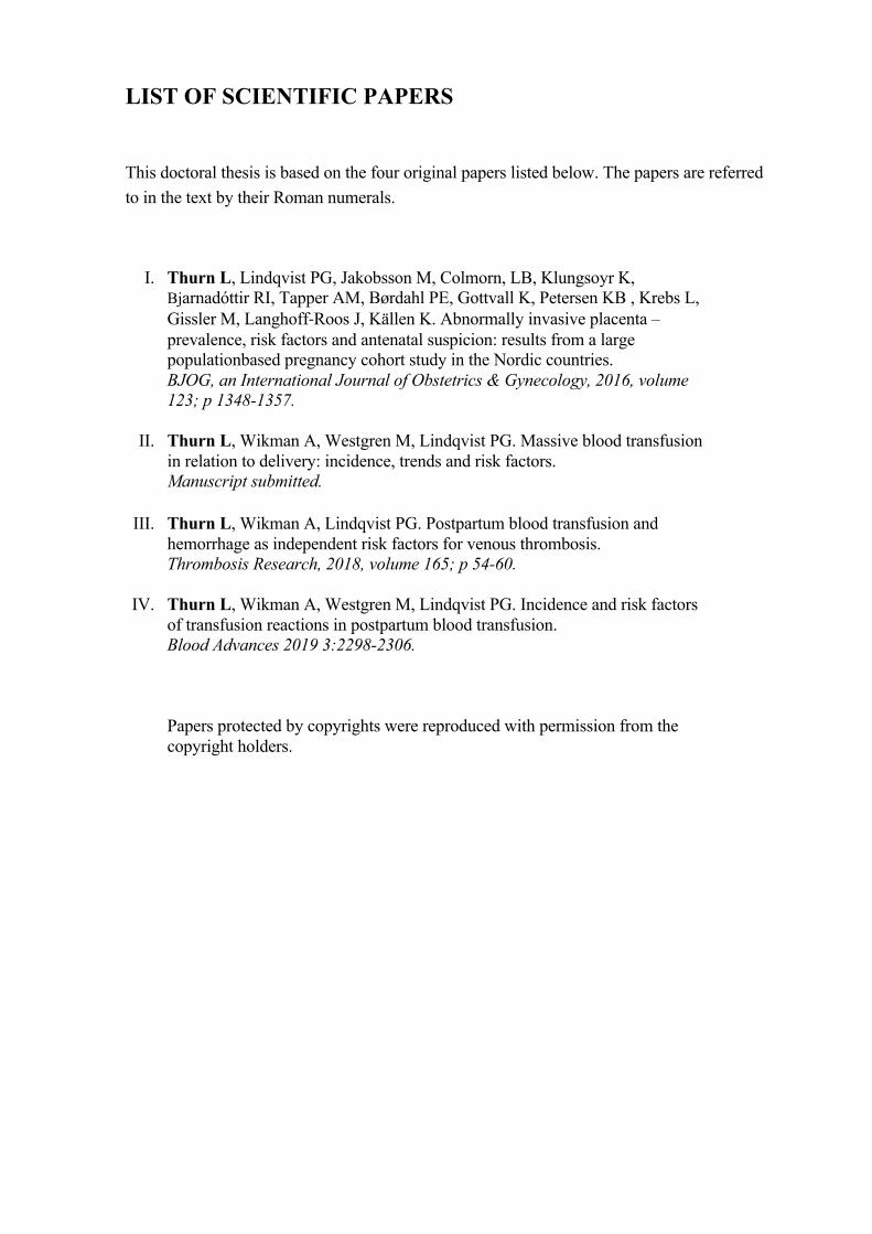
LIST OF SCIENTIFIC PAPERS
This doctoral thesis is based on the four original papers listed below. The papers are referred to in the text by their Roman numerals.
I. Thurn L, Lindqvist PG, Jakobsson M, Colmorn, LB, Klungsoyr K, Bjarnadóttir RI, Tapper AM, Børdahl PE, Gottvall K, Petersen KB , Krebs L, Gissler M, Langhoff-Roos J, Källen K. Abnormally invasive placenta –prevalence, risk factors and antenatal suspicion: results from a large populationbased pregnancy cohort study in the Nordic countries. BJOG, an International Journal of Obstetrics & Gynecology, 2016, volume 123; p 1348-1357.
II. Thurn L, Wikman A, Westgren M, Lindqvist PG. Massive blood transfusion in relation to delivery: incidence, trends and risk factors. Manuscript submitted.
III. Thurn L, Wikman A, Lindqvist PG. Postpartum blood transfusion and hemorrhage as independent risk factors for venous thrombosis. Thrombosis Research, 2018, volume 165; p 54-60.
IV. Thurn L, Wikman A, Westgren M, Lindqvist PG. Incidence and risk factors of transfusion reactions in postpartum blood transfusion. Blood Advances 2019 3:2298-2306.
Papers protected by copyrights were reproduced with permission from the copyright holders.

CONTENTS 1 Historical introduction .................................................................................................... 1 2 Previous cesarean deliveries and its consequences ........................................................ 3 3 Invasive placenta/placenta accreta .................................................................................. 5 4 Postpartum hemorrhage (PPH) ..................................................................................... 23 5 Massive blood transfusion (MT) .................................................................................. 25
5.1 Blood transfusion and MT in obstetric patients ................................................. 26 6 Hemostasis .................................................................................................................... 26
6.1.1 Primary hemostasis ................................................................................ 27 6.1.2 Secondary hemostasis ............................................................................ 28 6.1.3 Fibrinolysis ............................................................................................ 29
6.2 Physiology and hemostasis during pregnancy ................................................... 29 6.2.1 Physiology during pregnancy ................................................................ 29 6.2.2 Hemostasis during pregnancy ................................................................ 30 6.2.3 Thromboembolism in pregnancy ........................................................... 31
7 Transfusion complications ............................................................................................ 33 7.1.1 Transfusion reactions (TR) .................................................................... 33 7.1.2 Transfusion related acute lung injury (TRALI) .................................... 35 7.1.3 Transfusion-associated circulatory overload (TACO) .......................... 36 7.1.4 Amniotic fluid embolism (AFE) ........................................................... 36
8 Aims of the studies ........................................................................................................ 37 9 Methods ......................................................................................................................... 38
9.1 Data sources ........................................................................................................ 38 9.2 Study population and design .............................................................................. 39
9.2.1 Paper I .................................................................................................... 40 9.2.2 Paper II ................................................................................................... 41 9.2.3 Paper III .................................................................................................. 43 9.2.4 Paper IV ................................................................................................. 43
9.3 Statistical methods .............................................................................................. 44 9.3.1 Paper I .................................................................................................... 45 9.3.2 Papers II and IV ..................................................................................... 45 9.3.3 Paper III .................................................................................................. 45
9.4 Ethical considerations ......................................................................................... 46 10 Results and discussion: ................................................................................................ 48
10.1 Summary of main results/conclusions ............................................................... 48 10.2 Paper I Abnormally invasive placenta (AIP) ..................................................... 49
10.2.1 Prevalence .............................................................................................. 49 10.2.2 Risk factors ............................................................................................ 50 10.2.3 Antenatal detection ................................................................................ 51 10.2.4 Conclusions ............................................................................................ 51 10.2.5 Discussion .............................................................................................. 51
10.3 Paper II Massive transfusion (MT) in obstetric patients ................................... 53

10.3.1 Prevalence and trends ............................................................................ 53 10.3.2 Risk factors ............................................................................................. 54 10.3.3 Balanced transfusion (plasma/RBC ratio) ............................................. 54 10.3.4 Conclusions ............................................................................................ 55 10.3.5 Discussion .............................................................................................. 55
10.4 Paper III Blood transfusion and postpartum hemorrhage (PPH) as risk factor for venous thromboembolism (VTE) ...................................................... 57 10.4.1 Prevalence .............................................................................................. 57 10.4.2 Risk factors ............................................................................................. 58 10.4.3 Conclusions ............................................................................................ 60 10.4.4 Discussion .............................................................................................. 60
10.5 Paper IV Risk of transfusion reactions during pregnancy. ................................ 61 10.5.1 Incidence ................................................................................................ 61 10.5.2 Risk factors ............................................................................................. 62 10.5.3 Conclusions ............................................................................................ 62 10.5.4 Discussion .............................................................................................. 62
11 Main conclusions – clinical implications .................................................................... 64 12 Future perspectives ...................................................................................................... 66 13 Summary in Swedish - Populärvetenskaplig sammanfattning på svenska: ................ 69 14 Acknowledgements ...................................................................................................... 72 15 References .................................................................................................................... 74

LIST OF ABBREVIATIONS
AFE
AIP
PAS
Amniotic Fluid Embolism
Abnormally Invasive Placenta
Placenta Accreta Spectrum
AOR Adjusted Odds Ratio
BMI Body Mass Index
CI
CS
FFP
FIGO
ICD
IPR
L
MBR
MRI
MT
NOSS
OR
PAR
PPH
RBC
SD
SOFT
TACO
TR
TRALI
UKOSS
WHO
VTE
Confidence Interval
Cesarean Section
Fresh Frozen Plasma
The International Federation of Gynecology and Obstetrics
International Classification of Disease
Swedish National Inpatient Register
Liters
Medical Birth Register
Magnetic Resonance Imaging
Massive Transfusion
Nordic Obstetric Sureveilance System
Odds Ratio
Swedish Patient Register
Postpartum Hemorrhage
Red Blood Cells
Standard Deviation
Svensk Förening För Obstetrik och Gynekologi
Transfusion Associated Circulatory Overload
Transfusion Reaction
Transfusion Related Lung Injury
United Kingdom Obstetric Surveillance System
World Health Organization
Venous Thromboembolic Event

1
1 HISTORICAL INTRODUCTION Historical introduction to blood transfusion and invasive placenta
Blood has been considered a mysterious but vital fluid since the beginning of mankind. It is the very basis for life and as such has also played an important role in many religions, cultures and magic rituals. In Mayan and Aztec cultures bloodletting and human sacrifices were carried out in order to honor their gods. Drinking human blood as a mean to survive is described in Scottish folklore where a female blood sucking fairy (Baobahn Sith) lures wayfarers in the highlands. In fiction and movies we have vampires, such as Bram Stoker’s Dracula, dependent on blood from another individual to survive.
Although bloodletting was a popular treatment for a number of conditions from time of the Egyptians some 4000 years ago up to the middle of the nineteenth century, there was an early understanding that great blood loss was associated with death.1
The idea of transfusing blood as a treatment for illnesses (and madness) is old, but the knowledge of how to perform safe transfusions in humans took centuries to develop and many early attempts were unsuccessful and often fatal.
The first step toward safer blood transfusions was taken in 1628 when the British physician William Harvey described the circulatory system and showed that blood was being pumped, by the heart, through a single system of arteries and veins to the whole body.2 Prior to Harvey it was believed that the body contained two completely separate blood systems and that blood did not circulate at all, but was consumed at the same rate it was produced.
In 1825, almost two centuries later, came the first report of a successful human-to-human transfusion. It was performed by British obstetrician James Blundell and his friend Charles Waller on a woman with a life-threatening postpartum hemorrhage, with her husband as the blood donor Figure 1.3 While the procedure was successful, blood transfusions at that time were hazardous and associated with more than 50% mortality.4
The next significant step in the evolution of safer blood transfusions was the understanding of the ABO system described by the Austrian physician Karl Landsteiner in 1901.5 This discovery made it possible to avoid most acute hemolytic transfusion reactions, which probably was a common fatal complication.
Today postpartum hemorrhage (PPH) is still a major cause of maternal mortality worldwide. In low-resource countries 26% of all deaths in relation to delivery are due to bleeding and lack of blood transfusions, as was the case in London almost 200 years ago, when James Blundell and colleges pioneered in blood transfusions after massive postpartum hemorrhage.6
In high-resource countries death due to hemorrhage at delivery is rare (1:100,000 deliveries).7 Blood transfusions occur in approximately 3 in 100 deliveries, and there seems to be a progressively increasing trend.8-11

2
Figure 1. Illustration of a woman with a postpartum hemorrhage and the first human to human blood transfusion. © Photos/Alamy, printed with permission.
One important reason for major PPH and massive blood transfusion is abnormally invasive placenta (AIP), also referred to as the placenta accreta spectrum (PAS), a condition where there is abnormal adherence of the placenta to the myometrium. It requires complicated surgery and involves the risk of heavy bleeding at the time of delivery.12 The first cases of AIP were reported by Irving and Hertig in 1937.13 Since then we have seen a dramatic increase in of this condition, parallel with the rising rate of cesarean deliveries in many high resource countries.14 Improvements in prenatal diagnosis of AIP by ultrasound and in recent years by MRI, has allowed clinicians to make necessary preparations before delivery in an attempt to prevent massive hemorrhage. Since Tabsh et al. in 1982 reported the first case of a pregnancy with AIP that was diagnosed before delivery using ultrasound, great effort has been made to standardize ultrasound protocols and improve its efficacy as a screening method for AIP.15,16

3
2 PREVIOUS CESAREAN DELIVERIES AND ITS CONSEQUENCES
Cesarean section (CS) has been described as far back in history as ancient Hindu, Greek and Roman times. It has been suggested that Julius Caesar himself was born by cesarean section, and for this reason the procedure was named cesarean. However, the story is doubtful since Ceasar’s mother, Auriel, is said to have lived to hear of the Roman invasion of Britain many years later, whereas cesarean procedures at that time would only have been performed on women who were already dead or dying. The word cesaerean is more likely to come from the Latin verb caedare meaning to cut out.
According to Agneta Pleijel’s novel The Queen’s Surgeon, the first CS in Sweden was performed by the obstetrician Herman Schützer in 1758.17 At this time short-term complications after surgery and complicated deliveries were extremely high and many women died from hemorrhage or postoperative infections.18
In 1985 the World Health Organization (WHO) stated in its guidelines on appropriate technologies for birth that there is no justification or health benefits in having a CS rate below 10% or higher than 15%.19 However, there is no consensus on an optimal fixed rate of CSs, and WHO amended the statement in 2015 by adding that the goal is not a specific rate, but rather the provision of CS to pregnant women in need. During the past 50 years there has been a drastic rise in rate of cesarean deliveries worldwide. Between the years 2000 and 2015 the number of births by CS almost doubled from 16 million to 30 million, equivalent to an increase in the overall CS rate worldwide from 12% to 21% in 2015.20 Globally there are significant disparities between countries and regions with rates of CS varying from 0.6% to 60%, primarily due to differences in economy, resources, and health care systems.20 In Sweden and in the Nordic countries the rate of CSs in the early 1970s was approximately 5%; it has increased to about 18% in 2016 (Figure 2).21,22
Most deliveries by CS in Sweden and in other high resource countries are safe, but there are
both short and long-term complications that have to be considered. As always there should be
a balance between benefits and risk of doing harm.
Dominant short-term complications after CS, that have been previously described, are
hemorrhage, infection, hysterectomy and thrombosis.23-25 A cesarean delivery is reported to
increase the risk of severe postpartum hemorrhage by 2 to 4 times and a venous
thromboembolic event (VTE) by 3 to 5 times, compared to a spontaneous vaginal
delivery.23,25-28

4
Figure 2. Rate (%) of cesarean deliveries in the Nordic Countries 1975 to 2016. Used with permission from THL, The National Institute for Health and Welfare, Finland 2018.
However, interpretations of these risk estimations are difficult as the complications registered could be a result of the underlying indication for the CS instead of the surgical procedure itself. For example, preeclampsia with impaired hemostasis might be the indication behind a CS that in turn leads to a postpartum hemorrhage and a transfusion of blood products. All of the above mentioned risk factors are associated with each other and with the risk of an eventual VTE in the postpartum period. In the analysis of risk factors based on data from long-term studies one has to consider that both the rate of CS as well as characteristics and risk-profiles of pregnant women change over time. Thus, compared to the last decades, pregnant women today are older, have higher BMI, and are more often conceived through IVF, which has to be considered when evaluating prior CS as an independent risk factor in seeking to improve prophylactic guidelines for postpartum hemorrhage or thrombotic events. Since we lack controlled prospective studies, observational studies can give important information on rare pregnancy complications. Still, the effect of confounders has to be considered. This can be achieved by the use of stratifications and adequate adjustments in regression analysis.
Long-term maternal complications after CS mainly occur in regard to subsequent pregnancies. Severe complications refer to uterine rupture, placental abnormalities such as placenta previa and abnormally invasive placenta (AIP), with its possible consequences of an acute hysterectomy and risk of massive blood transfusion. In a Nordic population-based study from 2017, incidence of severe complications in a second delivery occurred in 1.1% of women who had a first cesarean delivery as compared to 0.2% in the general pregnant population.24 The risk of complications seems to be lower in women with a first emergency cesarean compared to women with a first elective cesarean.24,29 Surgical techniques using a one or two layer closure of the hysterotomy at CS has not been associated with a difference in risk of either uterine rupture or the risk of AIP in a subsequent pregnancy.30,31 The number of prior cesarean deliveries progressively increases the risk of complications (especially

5
abnormal placentation). For women who plan to have more than one child it would be beneficial in most cases to avoid CS in the first delivery.24,32,33 A study by Williams et al. in 2018 found that a previous cesarean delivery was associated with a small but significant increased risk of preterm birth in a subsequent pregnancy, which further emphasizes the advisability of reducing the rate of CSs, especially in the first pregnancy.34
This thesis focuses on incidences, trends, and risk factors (including prior CS) for AIP and massive blood transfusion postpartum. VTEs and transfusions reactions are potential consequences of massive blood transfusions and will be assessed in detail below.
3 INVASIVE PLACENTA/PLACENTA ACCRETA
Abnormal adherence and invasiveness of the placenta is a severe obstetric complication associated with catastrophic hemorrhage and a high risk of maternal morbidity and mortality. It is a condition that has only been reported in human pregnancies.35 When the normal detachment of the placenta fails, it causes uncontrolled bleeding that often requires complex surgery. Today, invasive placenta is a major cause of peripartum hysterectomy.36,37 As already mentioned the first series of cases were described by Irving et al. in1937 in a review of 18 clinical cases with “abnormal adherence of the afterbirth to the underlying uterine wall most likely caused by a partial or complete absence of the decidua basalis”.13 They concluded that attempting to manually extract the adherent placenta was extremely dangerous, and when this procedure was attempted two-thirds of the mothers died. Maternal mortality in cases of invasive placentas is much lower today, but still is reported to be as high as 7% in high resource countries.38 Abnormal placentation has not been studied to the same extent in low-income countries.
Terminology
The term “placenta accreta” refers to all variations of adherent placenta. The spectrum includes three different subtypes, depending on how deeply the trophoblasts penetrate into the myometrium: a) placenta accreta/creta represents the mildest form, where the villi reach the inner surface of the myometrium but do not invade; b) placenta increta represents cases where the villi invade deep into the myometrium but without engaging the serosa; and c) placenta percreta represents the most severe cases where the villi penetrate through the uterine serosa, as illustrated in Figure 3.16 Abnormally invasive placenta (AIP) is another commonly used, broader term for the condition, but often refers to the more invasive cases that include placenta increta and percreta.39,40 In recent guidelines the term Placenta Accreta Spectrum (PAS) has been suggested; it includes all histopathological subtypes of abnormal placentation.40

6
Figure 3. Spectrum of invasive placenta.16
S = serosa; M = myometrium; D = decidua; PC = placenta creta; PI = placenta increta; PP=placenta percreta. Copyright © American Journal of Obstetrics and Gynecology, printed with permission.
The lack of an international consensus on definition and the heterogeneity in the terminology over the past decades has made it difficult to directly compare studies, incidences, and other results. Definitions have been based on antenatal ultrasound, clinical findings, histopathological findings or a combination of the above.41 The diagnosis of PAS is originally made by histopathological findings, including an absent decidua and presence of myometrial fibers alongside chorionic villi in the basal plate.12 Using histopathology as the only means of diagnosing PAS can be questioned. For instance, myometrial fibers in the basal plate have been reported even in normal placentas.12 Today, conservative and uterine saving strategies in managing cases of PAS have become more frequent. In these cases hysterectomies are not performed, making histopathologic examinations unfeasible. The same is true of scar pregnancies, where there is a no decidua or myometrial tissue to eaxamine.42 Therefore, the clinical description of the invasiveness of the placenta has become more important in diagnosing PAS/AIP. A clinical grading system has been suggested in the International Federation of Gynecology and Obstetrics (FIGO) consensus guidelines on placenta accreta spectrum disorders (Figure 4).42
Placenta accreta is the most common subtype of PAS and represents approximately two-thirds of all cases. The most severe cases, placenta percreta, are rare and constitute about 5% of all PAS cases. Milder forms may include retained placentas.12,42 In some cases, retained placentas is due to a constricted cervix, not abnormal attachment, and should hence not be included among PAS cases. This might have occurred in some previous studies, resulting in an overestimation of the reported prevalence of PAS. In this thesis the term abnormally invasive placenta (AIP) will be used.

7
Figure 4. Clinical grading system of AIP, printed with permission, Int J Gynecol Obstet.42
Incidence
The worldwide incidence of AIP has increased dramatically over the past 50 years and is now reported to be between 2 and 90 per 10,000 births.43-48 To a great extent the wide range in incidence can be explained by differences in study design, the definition of AIP used, and to some extent differences in the population studied. The incidence has risen ten-fold from the 1970s. The increased rate of AIP is parallel to the rise in rate of cesarean deliveries.
In a study from the US in the 1980s, Read et al. found the incidence of AIP to be 1 in 4,000. It was a small study, including only 14 confirmed cases, but with a solid definition of placenta accreta based on histopathological findings after hysterectomy.49
Twenty years later, in another US population, Wu et al. found a much higher incidence of 1 in 533.45 However, the authors included both histopathological and clinical findings, which might partially explain the higher incidence. Their study retrospectively included cases from 1982 to 2002, and compared the last 10 years with the first. They found an increase in the CS rate from 12.5% to 23.5% and, parallel to this, an almost five-fold increase in AIP from 0.4 per 1000 births to 1.9 per 1,000 births. This strongly supports the association between AIP and the increase in CS rate.

8
In a 2017 Australian case-control study using the Australasian Maternity Outcomes Surveillance System (AMOSS), Farquhar et al. found an incidence of 0.5 per 1,000 deliveries using a wide definition of AIP including assessment by antenatal imaging, at surgery or by pathological examination.48
Using a similar obstetric surveillance system a study from the UK found a much lower incidence of AIP: 1.7 per 10,000 deliveries.47 A combination of strict clinical and pathological criteria was used to define AIP cases and designed to capture severe cases. Data was collected prospectively using a monthly reporting system (UKOSS). The lower incidence found might be because the majority of cases were placenta increta and percreta, excluding milder forms of the placenta accreta spectrum and cases with retention placentas. The higher incidence in the AMOSS study might also be due to reporting of cases based on only antenatal imaging. Cases detected antenatally may not necessarily have been AIP at time of delivery, which might result in an overestimation of the true rate of AIP.
In a more recent US study from 2016 they found a high prevalence of 37 per 10,000 deliveries.50 However, it relied exclusively on the International Classification of Disease codes (ICD) and had no strict clinical definition and no histopathological data, which might have resulted in a lack of specificity. In addition, the study included some cases with retention of placenta and membranes, which will result in a registration bias and an expected higher rate compared studies with more strict inclusion criteria. In the UK study the authors concluded that the rates of AIP during the study period did not parallel the increasing rate of prior CSs.47 This might be because of the time lag. The rate of AIP is estimated to lag the rate of CS by six years.51 The association between prior CS and AIP is well established and has been reported in several studies.32,46
The highest reported incidence of 90 per 10.000 deliveries is reported from Israel in 2002. However, they included several women diagnosed with placenta accreta in a previous pregnancy and had a low rate of hysterectomy (3.5%) among identified cases, indicating they had a very broad definition of AIP and most likely included cases with retention placenta.44
The challenge in optimal management at delivery lies mainly within the placenta incretas and percretas. True incidence of these subtypes of AIP is still uncertain.
Most studies on AIP are from countries with a less homogeneous population and a much higher rate of CS than in Sweden and the Nordic Countries. Therefore, previously reported incidence rates, risk profiles and complications are not necessarily the same in the Nordic Countries, which was a main reason for conducting the research in Paper I.
Consequences
The degree of invasiveness of the villi into the myometrium and the engagement of the bladder or parametrical tissues corresponds to the risk of massive bleeding and surgical

9
complications. The mean estimated blood loss at delivery in women with AIP is reported to be approximately 3 L, and blood transfusion is required in up to 90% of those cases.52 Surgical complications include damage to the urethras, cystotomies and, need for a reoperation.53 AIP has also become the main cause of peripartum hysterectomy on vital indication.54,55 In a prospective Nordic obstetric surveillance study (NOSS) from 2015 (including 211 cases of hysterectomy), and in a retrospective Australian study from 2018 (including 72 cases of hysterectomy), AIP accounted for 43% and 67%, respectively, of all emergency peripartum hysterectomies.37,56 Data from the 2018 World Maternal Antifibrinolytic (WOMAN) trail, included reports from Africa, Asia, Europe and the Americas, found that hemorrhage from abnormal placentation had the highest risk of hysterectomy (17%) compared to obstetric trauma (5%) and uterine atony (3%).57 The psychological trauma and long-term consequences of AIP may be severe but is scarcely studied. The reported mortality rate due to AIP varies widely. From 7 % in the early 1990’s to 0.7% in more recent reports.38,48 A summary of the most common complications from AIP is presented in Figure 5.
Figure 5. Complications associated with abnormally invasive placenta (AIP)42

10
Pathophysiology
The leading hypothesis as to why AIP occurs is that damage to the interface between the endometrium and myometrium causes an abnormal decidualisation, with a resulting deep infiltration of the trophoblasts. The most common histopathological findings of AIP are a) absence of decidua, b) chorionic villi directly adjacent to myometrial fibers, and/or c) presence of myometrial fibers in the basal plate on the placental side.
Placentation
To prepare implantation of the embryo, the endometrium undergoes decidualisation. This complex process begins in the mid-secretory phase of the menstrual cycle, when high levels of estrogen and progesterone transform the endothelium and the stroma cells into larger decidual cells to create an optimal environment for the attachment of the blastocyst.58 After implantation the decidua divides into three regions. Directly beneath the implantation site lies the decidua basalis or basal plate that forms the maternal part of the placenta. The rim around the embryo is the decidua capsularis, and the decidua parietalis covers the rest of the uterine cavity (Figure 6).59
Figure 6 Decidua- basalis, capsularis and parietalis. Copyright © Clinicalgate, 2015, used with permission.
From the embryo, cytotrophoblast cells on top of the anchoring villi proliferate and transform themselves into extravillous trophoblast cells (EVT) that invade the decidual stroma. In a normal pregnancy these EVT cells penetrate no further than the first third of the myometrium layer. Here they fuse and are formed the multinucleated trophoblast giant cells. It has been suggested that this fusion contributes to the EVT cells losing their invasive capability.60
Deciduaparietalis
Deciduacapsularis
Deciduaparietalis
Chorionvilli
Deciduabasalis
Chorionicplate
Chorioniccavity Amnio7ccavity
Figure 5 Development of the decidua

11
Figure 7 illustrates how a stem chorionic villus with its branches (placental cotyledon) penetrate deeper into the myometrium in an increta placentation (B) than in a normal placentation (A).
Figure 7 A = Normal placentation, B = Placenta Increta placentation, L = Lacuna (placental lake). Copyright © American Journal of Obstetrics and Gynecology. Printed with permission
The small spiral and radial arteries in the myometrium of non-pregnant women are rich in smooth muscle cells and sensitive to vasoactive substances. During pregnancy these arteries are remodulated by specific proteases from the EVT cells, causing them to lose their elasticity and their responsiveness to vasoactive substances, and hence become the high flow vascular system that is vital for the exchange of oxygen and nutritional substances between mother and fetus.61 In pregnancies with preeclampsia and fetal growth restriction this remodeling of spiral arteries fails. However, in women with AIP there seems to be no increased risk of placental insufficiency and fetal growth restriction.16 That may be because the placental defect in AIP is focal, as compared to growth restricted fetuses in preeclamptic patients where AIP involves the entire placenta.
Abnormal placentation
In a normal pregnancy, the basal part of the decidual plate, also called Nitabuch’s layer, is where the placenta separates from the myometrium Figure 8. The Nitabuch’s fibrinoid consists of an eosinophilic matrix and a deposit of maternal fibrin that prevents the trophoblasts from penetrating further into the myometrium.62 The placental separation is caused by the tearing action between the contracting movements of the myometrium and the non-contracting placenta. In the absence of the decidua or the Nitabuch’s layer, this natural separation of the placenta might not happen, resulting in damage to the large blood vessels that have supported the pregnancy and massive hemorrhage.

12
Figure 8. Decidua basalis with Nitabuch’s layer.
There have been several theories as to why AIP occurs. The primary hypotheses have been excessive extravillous trophoblasts, a primary defect in or absence of the decidua, abnormal vascular remodeling, or a combination of all these.60 It is no longer believed that AIP is caused by excessively invasive trophoblasts invading the myometrium. Instead, the main hypothesis is that AIP is primarily caused by a defect in the interface between the endometrium and the myometrium.63 This defect, often the result of a scar from a previous CS or other uterine surgery, prohibits normal decasualization and enables the chorion villi and trophoblasts to invade directly deep into the myometrium.63 Chorionic villi in the vascular space of the myometrium have been detected in patients with AIP, supporting this theory.64 A further explanation for the defective decidua might be that the blood circulation in the myometrium around a uterine scar is impaired by poorer vascularization, thereby leading to myometrial degeneration. This is supported by a 2013 study from Norway that found that women with a prior CS had a higher uterine resistance and reduced uteroplacental blood flow compared to women with a previous vaginal birth.65
The aggressive invasiveness of trophoblasts in interstitial or ectopic pregnancies, where there exists no endometrium, is a further indication that it is in fact the decidua that modulates placentation and the invasiveness of trophoblasts. The etiology and management of AIP cases differs from molar pregnancies, where the trophoblasts are more invasive.66
Although rare, AIP even exists among primiparous women. This might be caused by a superficial defect in the endometrium after such minor gynecological surgery as hysteroscopy or curettage, and probably represent less invasive cases in the spectrum of AIPs.67
Basalplate
Myometrium
Intervillousspace
ChorionicplateStemvillus
Syncy7otrophoblats
Anchoringvillus
Maternal
blood
cells
Fibrino
iddepo
sits
(Nitabu
ch’sla
yer)
Termin
alvilli
Fetalbloodvessels
Cytotrophoblats
Intermediatevillus

13
Risk factors
The single most important risk factor for AIP is placenta previa. In approximately 80% of all AIP pregnacies placenta previa is present.68-70 In a meta-analysis from 2013 the overall incidence of placenta previa was 5.2 per 1,000 deliveries worldwide, and 3.6 per 1,000 deliveries in Europe.71 Previa is strongly dependent on maternal age, IVF, and prior SC.72 Silver et al. found the risk of AIP in women with placenta previa and no previous SC to be 3%, but it rose dramatically for every prior cesarean delivery (Figure 9).32 The risk of AIP in women with placenta previa and one prior CS was 11% but after three prior CSs it reached 60%. The progressively increased risk of AIP by number of prior SC is supported by several authors.46,73 The risk would presumably be even greater in cases with a higher number of CSs; however, in most cases the treatment for AIP is hysterectomy, which eliminates the possibility of further pregnancies.
Figure 8 Increasing risk of AIP by number of prior CS. Modified from Silver et al.
There are indications that prior elective CSs are associated with a higher risk of AIP than emergency CSs.73 Whether this might be dependent on surgical technique or the placement of the incision is not completely known. The effect of suturing techniques, closing the uterus with a single or double layer or the impact of suture materials have all been debated but none seem to have a significant effect on the risk of AIP in a subsequent pregnancy.73,74
The presence of myometrial fibers in the delivered placenta is associated with AIP in the subsequent pregnancy. Deliveries complicated by placental retention or postpartum hemorrhage leading to manual removal of the placenta might be considered a risk factor for the future.75 The risk of AIP is also reported to increase after other uterine surgery such as
0
10
20
30
40
50
60
70
80
1 2 3 4 5
RiskofA
IP(%
)
Numberofcesareandeliveries
RiskofAIPbynumberofCS
Previa
Noprevia

14
myomectomies, hysteroscopic procedures, Asherman’s syndrome, increasing maternal age (> 35), increasing parity, smoking, and IVF.32,43-45,47,76
Diagnostics
Antenatal awareness of AIP has been shown to reduce complications.77 It is advantageous to identify women at high risk for AIP in order to prepare a detailed delivery plan involving a multidisciplinary surgical team at a center with appropriate medical resources.68,69
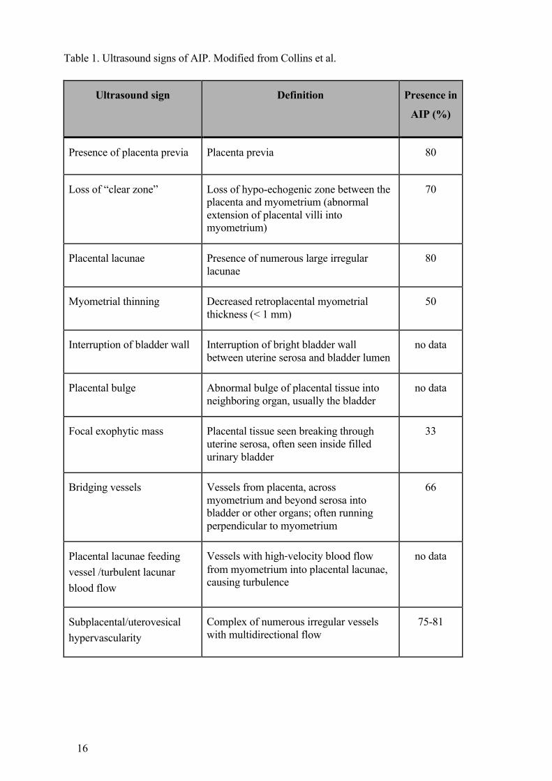
Clinical evaluation, ultrasound assessment, magnetic resonance imaging (MRI), and biomarkers all play a role in the antenatal diagnosis of AIP. Both ultrasound and MRI are technologies that may be used to identify AIP disorders (Figure 10 a and b).70,78 Ultrasound is today the primary diagnostic modality. Identification of AIP by ultrasound in the first trimester is possible, especially in cases with scar pregnancies, but most cases are diagnosed in the second and third trimester.78,79 Both gray-scale and color doppler signs are used in diagnosing AIP. Apart from presence of a placenta previa the typical signs are multiple lacunae and uterovesical hypervascularity. However, in difficult cases with high BMI or a posterior low laying placenta where deep penetration into parametrium is suspected, MRI may be beneficial.80 On the other hand, as a screening method, MRI is less practical since it has limited accessibility, is time-consuming and more costly. Therefore ultrasound is still the preferred diagnostic modality.
Figure 10 a,b. Images of abnormally invasive placenta in a twin pregnancy using (a) ultrasound and (b) MRI. One normal and one abnormal placenta (arrows).
The two different imaging modalities have been shown to be highly effective in diagnosing
AIP. In a recent study from France, the sensitivity and the specificity were reported to be 0.92
and 0.67 for ultrasound, and 0.84 and 0.78 for MRI, respectively.81 Meng et al. described
a b

15
similar results in another meta-analysis. The average sensitivity and specificity for ultrasound
was 83% and 82%, respectively. The figures for MRI were 95% and 88%, respectively, with
no significant difference in diagnostic value.82 In a third systematic review from 2013 that
included 23 studies, D'Antonio et al. found an average sensitivity of 91% and a specificity of
97% in using ultrasound for diagnosing AIP.83
Even though the above diagnostic results above are promising the performance of the
methods might be overestimated. This is suggested by recent large observational studies
where almost half of the AIP cases were undiagnosed at delivery.84,85 A majority of the
studies were performed in highly specialized centers on a high-risk population. Results are
likely to be operator dependant. The skill of interpreting ultrasound signs varies and
interobserver discrepancies are substantial.86
In order to improve overall performance in diagnosing AIP, there continues to be a great need
for standardization in interpreting antenatal ultrasound signs. Recently standardized
ultrasound descriptors for AIP have been suggested by The European Working Group on
Abnormally invasive placenta.87 The major signs and definition are summarized in Table
1.16,78,87 An extensive description of the pathophysiology behind the different signs has been
published by Jauniaux et al.16,88
Biomarkers
Several biomarkers associated with AIP have been proposed. Those biomarkers studied
include alpha-fetoprotein (AFP), human chorionic gonadotropin (HCG), and pregnancy
associated plasma protein-A (PAPP-A).12 More recently, higher levels of cell-free beta-HCG
mRNA, cell-free placental mRNA, and cell-free fetal DNA seen in pregnancies have been
reported as being associated with AIP.89,90 However, all of the biomarkers tested remain too
nonspecific, and therefore more prospective data is needed. Perhaps combining ultrasound
findings and biomarkers might be of clinical value in the future.
16
Table 1. Ultrasound signs of AIP. Modified from Collins et al.
Ultrasound sign Definition Presence in
AIP (%)
Presence of placenta previa Placenta previa 80
Loss of “clear zone” Loss of hypo-echogenic zone between the placenta and myometrium (abnormal extension of placental villi into myometrium)
70
Placental lacunae Presence of numerous large irregular lacunae
80
Myometrial thinning Decreased retroplacental myometrial thickness (< 1 mm)
50
Interruption of bladder wall Interruption of bright bladder wall between uterine serosa and bladder lumen
no data
Placental bulge Abnormal bulge of placental tissue into neighboring organ, usually the bladder
no data
Focal exophytic mass Placental tissue seen breaking through uterine serosa, often seen inside filled urinary bladder
33
Bridging vessels Vessels from placenta, across myometrium and beyond serosa into bladder or other organs; often running perpendicular to myometrium
66
Placental lacunae feeding vessel /turbulent lacunar blood flow
Vessels with high‐velocity blood flow from myometrium into placental lacunae, causing turbulence
no data
Subplacental/uterovesical hypervascularity
Complex of numerous irregular vessels with multidirectional flow
75-81

17
Management
The main risk associated with AIP is catastrophic hemorrhage and need for complex surgery.
Optimal management in surgical or expectant approaches are still under debate.
Hysterectomy is performed today in the majority of AIP cases (up to 90%), but several
uterine saving techniques have also been described.91
After the antenatal detection of an invasive placenta, recent guidelines suggest management
according to the following main categories:53,92,93
• Multidisciplinary team care (MDT)
• Counseling and providing information to the pregnant women about her condition
and the different options available
• Pre-delivery optimizing of hemoglobin
• Optimal timing of delivery
• Level of care
• Preoperative preparations
• Intraoperative considerations and management
• Choice of expectant or conservative management
• Follow-up, future pregnancies
Multidisciplinary team care (MDT)
The medical care of patients with AIP disorders is complex and the positive impact of an
MDT approach has been clearly demonstrated.68,69,94 Shamshirsaz found that even though the
group with MDT care had more cases of placenta percreta, they tended to require fewer blood
transfusions and less emergency deliveries.95 In 2017 Smulian reported significantly less
blood loss and administration of blood products, and Eller cited fewer cases that needed
reoperation in the group receiving MDT care compared to standard care.68,94 In agreement
with these results, a 2019 US study found less blood loss and lower rates of blood transfusion
in the group where AIP was suspected antenatally as compared to a group where it was not
expected and they attributed the better outcome to the MDT approach seen in the first
group.96 As these were all retrospective cohort studies and the selection of patients and degree
of invasiveness might be different in the various groups, which could potentially have had an
effect on the results. The exact composition of a multidisciplinary team may vary, but recent

18
guidelines recommend that they include the following key personnel; experienced
obstetricians, a pelvic surgeon (gynecologic oncologist), an interventional radiologist, a
urologist, an obstetric anesthesiologist, a neonatologist, specialized nursing staff, and access
to a blood bank with resources to handle massive transfusion protocols.
Timing of delivery
Data from several authors has indicated that a planned cesarean delivery, before the onset of
labor decreases the risk of maternal morbidity as compared to an emergent delivery.70,97
Among women with suspected AIP the risk of an unscheduled delivery, and hence minimal
preoperative planning, is significantly increased. Such women may have pre-partum bleeding
episodes, increased uterine activity, and preterm premature rupture of membranes.46,53 The
optimal gestational week for delivery of women with suspected AIP is not known and no
randomized controlled studies regarding timing of delivery exist. However most authors
recommend delivery between 34+0 and 36+6 weeks of gestation in high-risk cases (placenta
previa with episodes of bleeding or placenta percreta) and 36+0 to 37+0 weeks in more
uncomplicated pregnancies (no episodes of bleeding, placenta accreta/increta).53,93
Intraoperative considerations
A detailed description of the different surgical techniques used in handling AIP disorders
have been presented in recent guidelines.36,53,93 After delivery of the fetus and with the
placenta in utero, hysterectomy it is still the preferred primary surgical approach and is
recommended by most authors37,98,99 Total hysterectomy is often required because of cervical
engagement and bleeding from the lower uterine segment.92
Monitoring of blood loss, urine output, hemostasis, and ongoing communication between the
surgical and anesthesia teams is essential. Preoperative preparation for the transfusion of
large numbers of erythrocyte concentrates, plasma, and platelets should be made. A high
plasma/erythrocyte ratio (> 1) is recommended in trauma and surgical care but optimal ratios
have not been extensively studied in obstetric patients.100
The most common surgical complication in AIP patients is urinary tract injuries, reported to
be as high as 29%.101 The general use of ureteral stents or catheters to minimize the risk of
injuring the ureter is recommended by some but not all authorities.97 The procedure enables

19
evaluation of the invasiveness of the placenta through the bladder wall, and the stents provide
fast identification of the ureters during difficult surgery in cases involving massive
hemorrhage. In cases with a high suspicion of bladder invasion, ureteral stents are
recommended.69,102
Most authors and guidelines recommend a midline skin incision in cases where a
hysterectomy is planned or there is an anterior placenta that goes above the level of the
umbilicus.36,69,93,103
It is preferable that intra/preoperative ultrasound be used to identify the upper placental
margin in order to avoid the placenta when performing the uterine incision.93,104 If not
spontaneously delivered, the placenta should not be manually removed.94,97 The use of
prophylactic oxytocin is not clear, but is recommended in cases in cases of ongoing
hemorrhage. After suture of the fundal hysterotomy and confirmation of the AIP diagnosis, is
confirmed, options remaining are to a) perform a hysterectomy b) do a focal resection of the
wall with invasive placenta, c) or chose expectant/conservative management leaving the
placenta in situ.
The choice of surgical method depends on several factors. These include the desire for future
fertility, the invasiveness of the placenta, and the preferences of the surgical team. A detailed
comparison and description of the different surgical methods are beyond the scope of this
thesis except for certain aspects of hemostasis, which will be discussed below.
Devascularization
If hysterectomy or a focal resection is to be performed, prophylactic devascularization may
be performed in order to minimize blood loss. Available techniques are intravascular
techniques such as embolization, or temporary balloon occlusion. Extravascular techniques
are ligation or temporary clamping (vascular (“bulldog”) clamps) of blood vessels to the
uterus (uterine artery or the internal iliac artery).105-108 Temporary clamping of the internal
iliac artery by “bulldog” vessel clamps is shown in Figure 11. The major vessels involved are
the aorta, common iliac, internal iliac or the uterine arteries. The results are controversial as
are the use of these methods. Reduction of blood loss after endovascular balloon occlusion of
the aorta and iliac arteries has been demonstrated in several studies.106,109,110 A study in 2012
comparing occlusion of the abdominal aorta to occlusion of the internal iliac artery showed
less blood loss, reduced need for blood transfusion, and shorter insertion time in the aorta

20
occlusion group.111 Other authors could not find a positive effect of endovascular occlusion.
There are also reports of thromboembolic complications and rupture of the iliac
artery.112,113,114 Data on ligation or clamping of the internal iliac arteries is scant and
inconclusive.108,115 It may be that these procedures are useful only in more severe cases with a
percret placenta. However, to demonstrate this would require large multicenter controlled
trials.
Figure 11. Clamping of the internal iliac artery in surgery of invasive placenta.
Cell saver technology
Cell salvage is a technique in which blood from a bleeding patient is filtered and re-
transfused into the patient. It may reduce the need for allogeneic blood transfusion and is
used today in many medical facilities that handle AIP cases.116 If care is taken to avoid
suction of amniotic fluid or fetal blood, and in combination with the use of leucocyte filtering
techniques, the procedure is considered safe.117
MinimerablödningunderOP• Valavincision – nedremedellinje• Ureterkatetrar preop.Fridissekerasochslyngas• U-ljudperop - identifieraexaktplacentasövregräns
– Undvikskäraiplacenta,undvikuterotonika• Eftersectio Time Out II
• Stängavblodtillflödeinnanvidaredissektion:– Ligera a.ovarica bilattilluterus– Temporäravstängninga.iliaca interna(intravasaltelextravasalt)– Ev.Aortaballongpreop (”hybridsal”).Preoperativt– VbAortakompression– Vem/hur– Gummislangruntcx– Deflestablödningarvenösa!– Identifieravenösabäcken/parametrie- kärlsomsuccessivtligeras.

21
Tranexamic acid
Tranexamic acid is a fibrinolytic inhibitor that affects the breakdown of fibrin by plasmin. It
is recommended as a treatment in cases where there is ongoing hemorrhage and as
prophylaxis before CS in high risk-cases.
Two large placebo controlled studies have demonstrated that it reduces mortality in cases
with hemorrhage without increasing thromboembolic complications.
The CRASH-2 trial compared 1 g tranexamic acid administered intravenously within one
hour of admittance to placebo in trauma patients with ongoing hemorrhage and they found a
reduced mortality risk in the treatment group (relative risk (RR) 0.85).118
The WOMEN trial compared 1 g intravenous tranexamic acid given within 3 hours
postpartum to placebo in women with a postpartum hemorrhage (PPH) greater than 500 ml. It
demonstrated that the treatment significantly reduced mortality (RR = 0.69).119
However, as prophylaxis for PPH, tranexamic acid has yielded somewhat contradictory
results. A 2018 study by Sentilhes found no difference in the rate of PPH (blood loss > 500
ml) between 1 g of tranexamic acid or placebo given intravenously before planned vaginal
delivery.120 However, a meta-analysis of nine trials by Simonazzi concluded that tranexamic
acid given before elective cesarean delivery significantly reduced blood loss and the need for
blood transfusion.121 These findings were later supported by two placebo-controlled trials in
2016 and 2017 121,122
Conservative management
For women who wish to retain their uterus for future pregnancies hysterectomy is not a
decision of choice. Conservative management has also been proposed as an option to
decrease the risk of maternal morbidity by reducing hemorrhage and the need for blood
transfusion in severe cases.
The main strategies in these instances are a) conservative surgery with removal of the focal
area of invasive placenta and repair of the uterine wall, b) and leaving the placenta in situ
without hysterectomy or resection.

22
The different techniques for conservative surgery of AIP have similar main principles. They
include:123
1. Delivery of the fetus from an upper or fundal hysterotomy and not manually
removing the placenta
2. Devascularization around the uterus and the uterine−bladder interface either by intra-
abdominal ligation of blood vessels or intravascular occlusion
3. Resection of the invasive placenta, including the invaded myometrial tissue
4. Myometrial reconstruction
In a systematic review conducted in 2015 that included 177 cases of AIP, a uterus-preserving
surgical intervention was attempted in 76 cases. The success rate, defined as a subsequent
menstruation or pregnancy, was 63% (48/76), and the need for secondary hysterectomy was
30% (23/76).124 Shabana et al. demonstrated an even higher success rate of 91% in preserving
the uterus after focal resection by use of extensive pelvic devascularisation.125 However, this
promising result requires advanced pelvic and vascular surgical skills, that will probably limit
the widespread adoption of the method.
The Triple- P method (periopertive placental localization, pelvic devascularization, placental
non separation) described by Chandraharan in 2012 is a variation of the above procedure.
Intravascular balloons are used for pelvic devascularization before performing myometrial
excision and uterine wall repair. In this procedure the entire placenta is not removed and
some placental tissue is left in situ.126,127 The method has shown a lower rate of PPH and
peripartum hysterectomy but only in small series. Larger studies are needed to evaluate its
efficacy and risks.127
In addition, there are reports of a hemostatic technique where the cervix is inverted into the
uterus as a natural tamponade, and another using special multiple 8-compression
suturing.128,129 Both studies are showing positive results, but again, too few instances have
been recorded to draw any conclusions. All of the methods described share a common
dilemma in being greatly operator dependent, which makes reproducibility, comparison, and
generalization difficult.
Leaving the placenta after cesarean delivery without hysterectomy or focal resection.
The largest study to date using this approach is a 2010 French trial that retrospectively
included 167 AIP cases. It resulted in an overall success rate of 78% (131/167) with

23
preservation of the uterus.130 However, 18 women in the study had a primary hysterectomy
and 18 had a delayed hysterectomy. The rate of severe maternal complications was 6% and
included sepsis, thromboembolism, uterine necrosis, and secondary postpartum hemorrhage.
Spontaneous placental desorption occurred in 75% of the women over a median time of 13.5
weeks. One case of maternal mortality was also reported in connection with methotrexate.
Such an approach might be an option in selected cases, but informing women about the risks
of complications and the long follow-up time is essential.
Future fertility
Data on future pregnancies following successful conservative treatment is limited. While
pregnancy may be possible it will have a higher risk of complications, including postpartum
hemorrhage uterine rupture and recurrent AIP disorders. The overall risk of a recurrent AIP
disorder is reported to be as high as 30%.131
Methotrexate in treatment of AIP disorders
Methotrexat is a folate antagonist. In contrast to the situation in early pregnancy trophoblast
cell turnover is much lower later in pregnancy. Treatment-related complications such as
neutropenia and medullar aplasia have been reported even after a single dose of
methotrexate.132
Data on the efficacy and safety of methotrexate as a treatment for AIP disorders is
inconclusive. It is currently not recommended as adjuvant treatment in patients with AIP.88
4 POSTPARTUM HEMORRHAGE (PPH)
Postpartum hemorrhage (PPH) remains as one of the leading causes of maternal mortality
worldwide, causing an estimated 125,000 deaths annually.6 In Sweden and other high
resource countries maternal mortality due to bleeding has fallen dramatically over the past
100 years, and is now as low as 0.9 per 100,000 deliveries in some countries. 7 However,
recent studies have indicated an increasing trend toward higher rates of PPH with the
accompanying need for blood transfusions.10,133-135 The explanation for this is unclear but it
may be related to an increase in IVF pregnancies, multiple pregnancies, obesity, higher

24
maternal age, and the increasing frequencies of placental abnormalities such as placenta
previa and AIP. The rise in placental complications may be a consequence of the growing
rate of cesarean deliveries.10,136,137 This association has been described in countries with high
rates of CS, but has not been studied systematically in the Nordic countries, where there is a
comparatively low rate of CS. PPH in high resource countries is reported to occur in 1 to 15%
of all deliveries. In 2018 the overall rate of PPH in Sweden was 8%, according to data from
the Swedish Maternal Health Care Register (MHCR).138
The World Health Organization (WHO) defines primary PPH as a loss of blood greater than
500ml within 24 hours of delivery.139 In Sweden primary PPH is defined as a blood loss of >
1000ml within the above time period. This difference in definition is confusing but
understandable since a blood loss of 500 ml in a woman in poor health due to malnutrition or
chronic infection (e.g., malaria) might be fatal, whereas most women in high resource
countries the same blood loss would be of little consequence due to their better health status.
Different definitions of PPH make it difficult to compare results between studies. Another
cause of uncertainty is that blood loss is a subjective estimation. It is often overestimated after
cesarean and underestimated after vaginal deliveries.140 Weighing and measuring blood and
blood soaked materials have improved the estimation of blood loss. A superior method of
estimating the severity of blood loss is to record the number of transfused units of red blood
cells (RBC), plasma, and platelets. In Sweden, all transfused blood components have been
registered in a computerized system since the early 1980s. Coombs has suggested a clinical
definition of PPH as the “need for blood transfusion”. However this definition may also be
subjective due to variations in transfusion protocols and general attitudes towards blood
transfusion.141
Risk factors for PPH have been described by several authors and formalized in recent
guidelines.142,143 Atonic uterus is the major cause of PPH and is responsible for
approximately 65% of all cases.143,144 To prevent massive hemorrhage, early identification of
risk factors is essential. It has been reported that the majority of all maternal deaths due to
hemorrhage, were associated with suboptimal care and could have been avoided.145
Risk factors linked to massive blood transfusion postpartum may differ from those described
for ordinary PPH, as will be analysed in more detail below and in Paper II.

25
5 MASSIVE BLOOD TRANSFUSION (MT)
In order to maintain adequate circulation, tissue oxygenation, and hemostasis, massive
hemorrhage needs to be treated with massive transfusion. While no universally accepted
definition of MT exists, it often involves a volume or number of units of RBCs transfused
during a specific time frame. The most common definition is the transfusion of > 10 units of
RBC within 24 hours.146-148 However, this definition does not necessarily reflect acute
clinical cases. Studies using this definition might be “diluted” by including less acute
patients, and in the most severe cases patients may die before 10 units are transfused, and will
therefore be excluded.
Other proposed definitions are: 136,149,150
• blood loss of more than 50% of the total blood volume within 3 hours
• > 8 units of RBC within 24 hours
• > 5 units of RBC within 4 hours
• > 4 units within one hour
The three definitions above have the potential to better represent cases with true acute and
life-threatening hemorrhages. In order to better compare studies and results, there is need for
an international consensus on the best definition of massive transfusion that reflects the most
severe cases.
Patients who require MT have often been reported associated to trauma or military care. In
the US, up to 5% of clinical trauma and 10 % of military trauma patients require MT.151
However, a recent study from Sweden and Denmark found that MT (defined as > 10 units
RBC within 2 or 7 days) were most commonly used in cases involving major surgery (61%),
as compared to trauma or obstetric hemorrhage (15% and 2%, respectively). 152 Reports from
military and trauma care have indicated that protocols for MT and a high plasma/RBC ratio
(1:1) have improved outcomes and survival in patients with massive bleeding.100,153 Holcomb
found that 30 day survival significantly increased in patients with a ratio of > 1:2 compared to
<1:2.154 Whether these trauma-oriented protocols are fully applicable to obstetric situations
is not clear.

26
5.1 BLOOD TRANSFUSION AND MT IN OBSTETRIC PATIENTS
The successful decrease in the rate of blood transfusions reported in some medical disciplines
is not seen in obstetric care.133,155 Today, the overall frequency of blood transfusions due to
obstetric hemorrhage is reported to be 0.2 to 3.2%.8,9,156,157 Few publications have addressed
MT in obstetric care.8,136,158 The reported incidence of MT in obstetrics is between 2 and 9
per 10,000 births. Of the three published studies addressing MT postpartum, two have used
the definition: > 8 units of RBC within 24 hours of birth and one the definition of >10 units
of RBC within 24 hours.136,158 This heterogeneous use of definitions for MT is a dilemma that
is further discussed in Paper II.
To prevent unnecessary bleeding and coagulopathy it is important to take early hemostatic
action. Compared to other causes of hemorrhage, major obstetric bleeding more often leads to
early disseminated intravascular coagulopathy (DIC).159
Therefore, transfusion protocols involving early administration of FFP/plasma and platelets in
massive obstetric hemorrhage has been suggested. How these new transfusion strategies are
implemented and whether they improve severe maternal morbidity, is unclear. Green et al.
have suggested the need for targeted transfusions strategies, depending on the different causes
of massive obstetric hemorrhage.160 Their findings suggest that the hemostatic changes in
massive obstetric hemorrhage due to placental abnormalities result in lower levels of platelets
and fibrinogen, compared to the changes seen in trauma situations. On the other hand,
Gutierrez et al. found that a standardized MT protocol in obstetric care was associated with
favorable hematologic post-resuscitation.161 Finally, Tanaka et al., in a recent systematic
review of MT protocols in obstetrics, suggested that a FFP/RBC ratio > 1 was necessary for
optimal hemostatic treatment, concluding that a ratio > 1 was associated with higher survival
in women with severe obstetric hemorrhage.162 However, once again conclusive information
on the best obstetric transfusion protocol would require large, randomized, controlled,
multicenter trials.
6 HEMOSTASIS
Hemostasis is a complex, tightly regulated physiological system. Any imbalance can lead to
severe hemorrhage or thromboembolism. Knowledge of hemostasis is vital for the

27
understanding and effective treatment of many pregnancy complications that may affect the
coagulation system.
Hemostasis consist of three main steps:163
- Primary hemostasis
- Secondary hemostais (or plasmacoagulation)
- Fibrinolysis
6.1.1 Primary hemostasis
The primary hemostasis is the result of complex interactions between endothelial proteins and
platelets that lead to the formation of a primary platelet plug. It is initially triggered by an
endothelial injury to the vascular wall. In contrast to the antithrombotic endothelial lining, the
subendothelial layer contains highly thrombogenic substances such as collagen fibers,
laminin, vitronectin, and Von Willebrand factor (vWF).164,165 At the site of the damaged
endothelium these thrombogenic substances cause platelet adhesion. The activated platelets
change form and release the vasoconstrictive substances thromboxane A2 (TXA2) and
serotonin, which in turn cause vasospasm and reduce blood loss. Further platelet aggregation
and creation of the preliminary platelet plug is stimulated by secretion of calcium, adenosine
diphosphate (ADP), and TXA2 from the activated platelets (Figure 12).166 On undamaged
endothelium, prostacycline (PGI2) and nitric oxide (NO) act as inhibitors of further platelet
activation thereby limiting the thrombosis.167
Figure 12 Primary hemostasis.
Inters''um
Endothelialdamage
vWF
Bloodvessel
ThromboxaneA2
ADP
Plateletadhesion
Plateletaggrega'on
Collagenfibers
Endothellium
Subendothelium
Smoothmussel

28
6.1.2 Secondary hemostasis
Secondary hemostasis leads to the formation of fibrin by coagulation proteins. This process
can be divided into four phases:
- Initiation
- Amplification
- Propagation
- Termination
In the initiation phase, tissue factor (TF) on the cell surface at the site of injury is exposed to
the circulating blood and its components. Activated FVII binds to TF, forming a TF-VIIa
complex, which in turn activates FIX and FX. Activated FX (in a complex with FVa)
promotes the conversion of prothrombin to small amounts of thrombin.
In the next phase, thrombin not only catalyzes fibrinogen to fibrin but also acts as a promoter
in converting FXI to FXIa, which after activation of FIX promotes the further conversion of
FX to FXa. This positive feedback of conversion of prothrombin to thrombin in the
amplification phase was formerly known as the intrinsic pathway, but is today more
commonly described as the ”thrombin burst”.163
In the propagation phase, large quantities of thrombin lead to the production of sufficient
amounts of fibrin that can form a stabilized platelet plug. Finally, FXIIIa promotes the cross-
linking of loose fibrin polymers to create a stable and elastic clot. Secondary hemostasis is
summarized in Figure 13.
Figure 13. Secondary hemostasis
ModifiedwithpermissonfromFaribaBaghaei
SecondaryhemostasisCoagula<on
Cellinjury

29
During the termination phase the coagulation process is constrained by specific inhibitors, the
most important of which are antithrombin (AT), Protein C, Protein S, and tissue factor
inhibitor (TFPI).
6.1.3 Fibrinolysis
After the damaged blood vessel is healed, fibrinolysis begins and the clot is gradually
dissolved. In this process plasminogen is converted to plasmin that in turn resolves fibrin to
small protein fragments (D-dimers). Plasminogen is produced in the liver and circulates
freely in the plasma. It binds to fibrin and is activated by tissue plasminogen activator (t-PA)
and urokinase plasminogen activator on the surface of the fibrin clot. This ensures that the
fibrinolysis remains local to the affected area. The main inhibitors of fibrinolysis are
antiplasmin and plasminogen activator inhibitor type 1 (PAI-1), and, during pregnancy, also
PAI-2 (which inhibits tPA). PAI-2 is synthesized in the placenta and increases with
gestational age.168
6.2 PHYSIOLOGY AND HEMOSTASIS DURING PREGNANCY
6.2.1 Physiology during pregnancy
Major physiological changes occur during pregnancy in order to nurture the growing fetus
and prepare the mother for the forthcoming delivery, and a possible hemorrhage. These
changes involve all organs, but they resolve almost completely after a normal pregnancy.
Some of the most important adaptions to pregnancy are mentioned below. Changes in
hemostasis are described separately in more detail.
Cardiac output (CO) increases by 40% by means of increased stroke volume and a slightly
higher heart rate. It reaches its maximum around 20 to 28 weeks of gestation. This is partly to
compensate for a peripheral vasodilatation that causes vascular resistance to fall by 30% and
to the increased blood volume.169 The plasma volume rises progressively by about 50%
during pregnancy.170 This plasma expansion exceeds the increase in red blood cell mass,
causing a lower hemoglobin concentration and bringing about the physiological anemia seen
in pregnancy. Further changes during pregnancy are an increase in the metabolic rate by 15%,
and in the consumption of oxygen by 20%. To compensate, ventilation is increased by

30
50%.171 This slight form of hyperventilation causes a physiological compensated respiratory
alkalosis in normal pregnancies.171
6.2.2 Hemostasis during pregnancy
As mentioned above, coagulation is a complex, tightly regulated system. During pregnancy
this balance changes to a hypercoagulable state to prepare for potential bleeding at delivery.
From an evolutionary perspective, the effect might have been an important advantage for
survival.172 This hypercoagulability may partially explain the increased risk of
thromboembolism during pregnancy. It might also explain why some variants of
thrombophilia (such as factor V mutation or factor II mutation) only cause a slightly
increased risk of thrombosis in non-pregnant women, but they became significant risk factors
during pregnancy.
Most coagulation factors increase during pregnancy, especially FVII, FX and FXII. Table 2.
Fibrinogen increases by 50% to 3-6g/L.173 VWF is a protein that carries FVIII, and binds to
activated platelets increases significantly as early as the first trimester.174
There might be an acquired APC (activated protein C) resistance during pregnancy that is
most likely due to an increase in FVIII but also to a decrease in protein S.
Table 2. Coagulation factors and haemostatic changes in pregnancy.

31
The main anticoagulants, antithrombin and Protein S as well as tissue factor inhibitor (TFPI)
decreases, which favors coagulation. Finally, fibrinolysis is lowered by a rise in plasminogen
activator inhibitor-1 (PAI-1) and PAI-2 from the placenta.
6.2.3 Thromboembolism in pregnancy
There is a well-known increased risk of venous thromboembolism (VTE) during pregnancy
and in the postpartum period. The three main contributors to the formation of thrombosis,
venous stasis, endothelia injury and hypercoagulability (Virchow’s triad), are all present in
pregnancy Figure 14.
Figure 14. Virchow’s triad
VTE remains a major cause of maternal morbidity and is responsible for 3% of all maternal
mortality worldwide. In high resource countries pulmonary embolism (PE) accounts for 8%
to 17% of all maternal deaths.6 Pregnancy, including the postpartum period is associated with
an increased risk of VTE by a factor of 5 to 20, as compared to age-matched non-pregnant
women.28,175,176 In Sweden and other high resource countries the incidence of VTE during
pregnancy and the postpartum period is estimated at 12 to 17 per 10,000 pregnancies.28,175,177
The risk of VTE, especially PE, is highest in the first 6 weeks postpartum (Figure 15).25,178
Virchow’s triad
Thrombosis
Stasis
HypercoagulationVessel wall injury

32
Figure 15. Venous thromboembolic events during pregnancy and postpartum. (Used with
permission from ACOG, Jacobsen 2018)
Variations in the reported incidences may be due to differences in data sources, management
of VTE cases in outpatient care, misclassification (ICD coding), use of imaging and other
diagnostic modalities, as well as the extent of thromboprophylaxis in the cohort studied.
As a consequence of the increased risk of VTE, many countries have guidelines
recommending low-molecular-weight heparin (LMWH) as thromboprophylaxis during
pregnancy and postpartum. Sweden has a weighted scoring-based thromboprophylaxis
algorithm that includes major risk factors that are equivalent to an approximately five-fold
increased risk of VTE. By means of this algorithm, women with > 2 major risk factors
(representing approximately 4 % of all deliveries with a 25-fold increased risk) will be
recommended postpartum thromboprophylaxis. The efficacy of LMWH prophylaxis during
pregnancy has been shown to produce an 88% relative risk reduction.179 Similarly, there are
indications that maternal death caused by PE has decreased by almost 50% after
implementing thromboprophylactic guidelines in the UK.7
Prophylaxis with LMWH is generally safe but a heightened risk of PPH, hematomas, and
skin reactions have been reported.180 As always there must be a balance between potential
harm and benefit.
In order to identify the high-risk group of women who can benefit from LMWH, correct risk
factors must be identified.
Prior thrombosis and thrombophilia are two of the most important risk factors for VTE. Other
major risk factors are obesity, high maternal age, immobilization, rheumatoid disease, and

33
inflammatory bowel diseases. Among pregnancy-related risk factors, preeclampsia, multiple
pregnancy, placental abruption, cesarean delivery, and ovarian hyperstimulation syndrome
(OHSS) have been previously reported.28,176,181
Both PPH and blood transfusions have been suggested as risk factors for VTE postpartum. It
is still unclear whether either of these risk factors are strong enough by themselves to be
implemented in the Swedish risk scoring algorithm, that is, representing at least a five-fold
increased risk of VTE. To answer these questiones we conducted a study based on a cohort
that existed before general implementation of the thromboprophylactic guidelines.
7 TRANSFUSION COMPLICATIONS
Blood transfusions can be life saving and have become one of the most common medical
treatments in high resource countries today. In Sweden close to 600,000 transfusions of RBC,
plasma, or platelets were administered annually from 2011 to 2013. Modern blood
transfusions are safe, but complications do occur. Severe complications after ABO-
incompatible transfusions and the risk of transfusion transmitted infections (TTI) such as HIV
were major contributors to the development of national surveillance schemes for transfusions
that now exist in many countries. TTIs such as hepatitis B, C, and HIV have become rare due
to testing, but emerging infections, i.e., Zika and West Nile virus, are not universally tested
for and are still a concern.182
7.1.1 Transfusion reactions (TR)
Complications other than TTI are transfusion reactions (TR) and short- and long-term effects
of immunomodulation. TR can be classified based on a) pathophysiology or b) time between
transfusion of blood component and clinical sign of a reaction. Early reactions occur in close
relation to the transfusion; delayed reactions may produce symptoms days or months after the
transfusion. Reactions can be immunologic or non-immunologic, and may vary in severity
from mild to fatal.
Blood transfusions may also have a delayed effect on the recipient’s immune system. This
reaction, transfusion-related immunomodulation (TRIM), involves both immunosuppression
and hyperinflammation. It is thought to be associated with an increased risk of recurrence of
tumors and infections. However, many clinical aspects of TRIM are uncertain and its

34
pathophysiology is unclear.183 The different types of transfusion reactions are summarized in
Table 3 below.184,185
Table 3. Types of transfusion reactions
The true overall incidence of TR is not known (partly due to underreporting), but published
incidences lie between 1 and 37 per 1,000 blood transfusions.186-188 Most TRs are mild, with
common symptoms such as fever, chills, itching, and urticaria, that in most cases resolves
without any treatment. Mortality due to TRs are rare and occur in one in 100.000 transfusions
with TACO and TRALI as major contributors.189-191 However, there has been an decrease in
rate of TRALI after implementing the exclusion of female plasma donors and use of
leukodepleted blood components.190
HLA and other leucocyte antibodies are associated with increased risk of TR.192,193 During
pregnancy the levels of human leucocyte antigen (HLA) and granulocyte antibodies increase,
which may affect the risk of adverse TR.193,194 Also red blood cell antibodies are increased in
a pregnant population and can cause hemolytic transfusion reactions. Very few studies have
assessed TR in relation to pregnancy. In a 2014 study by Teofili et al. an association was
found between pregnancy-related hypertensive disorders in women with PPH and TRALI
(OR = 27).195 The retrospective study included 14 cases of possible TRALI in women
transfused with three or more units of RBC. The potential mechanism for this association is
Type of reaction Early/acutereactions
ImmunologicMildallergicAnaphylacticFebrilenon-hemolyticAcutehemolyticSepticTransfusionrelatedacutelunginjury(TRALI)
NonimmunologicTransfusion-associatedcirculatory(TACO)
DelayedreactionsImmunologic
DelayedhemolyticTransfusion-associatedgraft-versus-hostPost-transfusionpurpura(PTP)Immunomodulation
NonImmunologicIronoverload

35
far from clear, and the results remain to be confirmed by additional research, which was a
motivation for our study, “Incidence and risk factors of transfusion reactions in postpartum
blood transfusions” (Paper IV).
7.1.2 Transfusion related acute lung injury (TRALI)
TRALI is a clinical diagnosis and has symptoms similar to those of acute respiratory distress
syndrome (ARDS). They include increased respiratory rate and hypoxia caused by an
inflammatory lung injury and increased microvascular permeability. ARDS is associated with
sepsis, pneumonia, complicated surgery, and aspiration of gastric fluid in the transfusion
recipient. The risk of mortality is considerable, around 40%.196
TRALI is also characterized by acute respiratory distress and occur within 6 hours of a blood
transfusion. According to the Canadian Consensus Conference on TRALI, the diagnostic
critera criteria of diagnosis are the following:197
• 1) Acute lung injury, hypoxemia: SpO2 < 90% or PaO2/FiO2 < 300 mm Hg on room
air
• 2) No preexisting acute lung injury before transfusion
• 3) Occurs during or within 6 hours of transfusion
• 4) No temporal relationship to an alternative risk factor for acute lung injury (such as
pneumonia, sepsis, aspiration, or multiple trauma)
As with all TR, the incidence of TRALI is uncertain, and the previously reported incidence of
1 in 5,000 transfusions has decreased dramatically after introduction of leukoreduced blood
products and the exclusion of female plasma donors as mentioned above.155,198-200
The etiology of TRALI is not completely understood, but the leading theory consists of a
“two-hit model”.201 In the first hit, a clinical condition exists such as major surgery or
infection/sepsis leading to sequestration and priming of neutrophils on pulmonary
endothelium. In the second hit, these primed neutrophils are activated by a factor in the blood
being transfused, causing an inflammatory response that damages the endothelium and results
in pulmonary edema.202 Anti-HLA or anti-human neutrophil antigen (HNA) antibodies as
well as bioactive lipids and soluble mediators from stored platelets, are suggested to be
responsible for the activation of the neotrophils.203 The main source of anti-neutrophil and
anti-HLA antibodies is blood from women with prior pregnancies. Thus, the exclusion of
female plasma donors has reduced the incidence of TRALI.200

36
Pregnancies complicated by fetal growth restriction and preeclampsia are associated with
endothelial modulations that may, in theory, be a cause for the priming of neutrophils on the
pulmonary endothelium corresponding to the first hit of TRALI described above.
7.1.3 Transfusion-associated circulatory overload (TACO)
TACO is a result of hydrostatic transudate accumulation in the lungs. Its etiology is not clear
but is also described as a similar two-hit model. Clinical signs are characterized by acute
respiratory distress with hypoxia and pulmonary edema within 6 hours of a blood transfusion.
It was introduced as a transfusion reaction in 2006. Although its incidence is probably
greatly underestimated it is reported to affect between 1% to 8% of all patients receiving
blood transfusions.184,201,204 As with TRALI it is associated especially with plasma and
platelet transfusions, but with the important difference that TACO is a hydrostatic and not a
permeability edema. Therefore, patients with TACO respond to diuretic treatment in clinical
praxis. Still, TACO has a reported mortality rate of 4% and is associated with between 30%
and 44% of all transfusion related fatalities.205-207 As in the case of every TR, the first action
taken is to stop transfusion immediately, administer oxygen, and if TACO is suspected,
administer diuretics.
7.1.4 Amniotic fluid embolism (AFE)
AFE is a rare but serious complication of pregnancy. It is not considered a TR but has clinical
features similar to TRALI. Clinical characteristics include sudden dyspnea, low blood
pressure, cardiovascular collapse, and coagulopathy (disseminated intravascular coagulation).
The incidence in high resource countries is reported to be 1.7 to 6.1 per 100,000 births and a
case fatality rate of 13% to 30%.208-210 The diagnosis of AFE in surviving cases is probably
underreported, and so the true incidence is not known. The etiology is also not fully
understood and many theories have been proposed. One such theory holds that amniotic fluid
and fetal debris cause an anaphylactic/anaphylactoid (not immunoglobulin E mediated)
reaction.211 However, the expected mast cells degranulation seen in anaphylaxis has not been
found.212 In Paper IV we discuss the possibility that AFE, having both similar risk factors and
clinical signs as TRALI, might be the result of an incompatible feto-maternal transfusion
triggering an acute hemolytic TR and respiratory distress.213

37
8 AIMS OF THE STUDIES
The overall aim of this thesis was to explore prevalence, risk factors and complications of massive blood transfusions in relation to pregnancy, with a special focus on abnormally invasive placenta.
The specific aims of the individual studies were as follows:
I. To investigate the epidemiology, outcomes and risk factors for abnormally
invasive placenta in the Nordic countries and determine its rate of antenatal
detection.
II. To describe the epidemiology of massive transfusion (MT) postpartum regarding
incidence, risk factors, and changes over time; and to assess balanced transfusion
(plasma/RBC ratio) in relation to MT and peripartum hysterectomy post partum.
III. To explore whether blood transfusion and postpartum hemorrhage are significant
independent risk factors for venous thromboembolism, and whether they should
be included in the Swedish thromboprophylaxis algorithm during pregnancy.
IV. To assess whether postpartum blood transfusion is associated with an increased
risk of a transfusion reaction compared to transfusion in other recipients.

38
9 METHODS
Study designs
9.1 DATA SOURCES
Swedish Medical Birth Register (MBR)
The Swedish Medical Birth Register (MBR) is a unique database established in 1973. It
includes data of the mother, pregnancy, and the perinatal outcome. It covers more than 97 to
99% of all births in Sweden and has been validated in previous publications.214,215 The data is
standardized and prospectively collected from medical records. Demographic data of the
mother, including medical conditions and maternal diagnosis before and during the current
pregnancy, as well as medical interventions are registered using International Classification
of Diseases (ICD) 9 and 10 codes.
The Stockholm Transfusion Database
In the Stockholm Transfusion Database (ProSang®, Databyran AB, Stockholm, SE), all
transfusions of blood components have been registered in the County of Stockholm since the
beginning of 1980. The data includes personal identification number (PIN) of donor and
Table 4. Studies included
Study design Inclusion criteria Cases Statistical analysisStudy I Population-based Women who gave birth 205 Cross-tabulation,Accreta cohort in the Nordic countries, Chi2 test or
2009-2012 Fisher's exact testn = 605,362
Study II Population-based Women who gave birth 277 Chi2-test, Student's t-testMassive cohort in the Stocholm County, Multivariate regressiontransfusion 1990-2011 analysis
n = 517,854
Study III Population-based Women who gave birth 56 Cross-tabulation,VTE cohort in the Stockholm County, Multiple logistic regression
1999-2002 in multivariate analysisn = 82,376
Study IV Population-based Women with PP blood- 96 Chi2-test, Student's t-testTR cohort transfusion in Stockholm Cross-tabulation and
County 1990-2011 logistic regression inn = 11,842 multivariate analysis

39
recipient, blood group of donor and recipient, unique serial number of blood component, the
time the blood component was issued, and whether it was transfused or returned. According
to national regulations, all transfusions are reported and traceable within the system. The data
programme has been validated according to quality standards for IT-systems in health care.
The Swedish Discharge Register
The Swedish Discharge Register, also called The Swedish National Inpatient Register (IPR),
was founded in 1964, but complete coverage dates from 1987. The register includes
information on dates of admission and discharge and on diagnosis and interventions
according to the Swedish International Classification of Disease (ICD) system. More than
99% of all somatic discharges are registered in the IPR, which records nearly all inpatient
diagnoses and surgical procedures. The high coverage and quality of the data has been
validated in a 2010 study that found an overall positive predictive value (PPV) of diagnoses
in the IPR to be 85% to 95%. .216
Specific data forms and medical charts
In paper I, data on cases with abnormally invasive placenta (AIP) was collected by means of
a specific data-form (electronic or paper form) directly from the clinicians to a national
representative. In cases identified from MBRs data was collected retrospectively from
medical charts and inpatient registers. The data-forms were jointly developed by the
members of the Nordic Federation of Societies of Obstetrics and Gynecology study group.
The data included information on maternal characteristics, previous deliveries, previous
postpartum hemorrhage, previous surgery, antenatal suspicion of AIP, blood loss at delivery,
amount of blood transfusion, hemostatic treatments, surgical procedures and postoperative
complications.
9.2 STUDY POPULATION AND DESIGN
To determine causal relationships, prospective controlled studies are preferred. However, to
explore rare outcomes, prevalence, and risk factors, large retrospective observational studies
are often the best option. In observational studies both random and systematic errors have to

40
be addressed. Our studies involved very large sets of data, so the impact of random errors was
small. Systematic errors of importance would be risk of confounding, selection bias, and
information bias. In all of the studies we conducted, measures were taken to reduce risk of
confounding by stratifying data and by careful adjustments in the multivariate logistic
regression analysis. In Papers II to IV, the cohort studied was pregnant women in Stockholm
County. While this may be considered a selection bias the cohort represents approximately
one-third of all births in Sweden. Another possible selection bias was missing data, as seen in
the BMI variable. As mentioned in the related papers, data on BMI must be interpreted
carefully. The BMI variable was also excluded as an adjusting variable in the regression
analysis in Paper IV. Misclassification in coding a diagnosis is a possible information bias.
The MBR and the IPR have been previously validated and proven to be of high quality. In
Paper I, the number of AIPs in the MBR corresponded to the number of AIPs reported
directly by clinicians in Sweden. Any eventual misclassification is most likely to be same in
both cases, and in the background populations in Papers II to IV. In all the studies above,
odds ratio (OR) with 95% confidence intervals (CI) was used as a measure of risk estimation.
However, in studying risk factors for rare outcomes, as in the studies included in this thesis,
OR will become a proxy for relative risk (RR).
9.2.1 Paper I
This study was conducted between 2009 and 2012 as part of collaboration between the
Nordic countries called the Nordic Obstetric Surveillance Study (NOSS). It comprised a
population-based cohort study of abnormally invasive placenta (AIP), and included 91%
(605,362/ 666,306) of all pregnancies in the Nordic countries. The cohort base was made up
of the maternities registered in each country’s MBR during the study period. The national
coverage of participating clinics was 100% in Denmark, Finland, and Iceland; 80% in
Sweden; and 88% in Norway.
Clinicians in the maternity wards surveyed reported cases of AIP to a national coordinator on
a monthly basis. Regular reminders were sent to the participating clinics to urge full
compliance with the survey. Positive or negative answers had to be mailed back. In case of
positive findings, the local representative got informed consent from the cases and a more
detailed questionnaire was answered by online registry or on paper. The specific data forms
were developed by consensus in a working group of the Nordic Federation of Societies of
Obstetrics and Gynecology (NFOG). In Sweden, Finland, and Iceland all cases were

41
collected prospectively through the reporting system described. The total number of cases
was validated against the national MBRs. In Denmark and Norway the majority of the AIP
cases (70%) were identified from MBRs and hospital discharge registers. The clinicians
responsible verified the cases and reported the information on the data collection forms
above. The appropriate international classification of diseases (ICD-10) code (O43.2X) was
used.
Cases of AIP were defined as a) women with a cesarean delivery assessed by the attending
obstetrician to be AIP, or b) a vaginal delivery in which a laparotomy was needed due to AIP.
A total of 205 cases of AIP were identified and included in the analysis. Data collected from
Sweden and Finland were stored with the national NOSS representative in each country.
Denmark, Norway, and Iceland stored their data in a common database in Denmark. Only
aggregated background data from the MBRs were able to be shared in the NOSS studies due
to different ethics clearances.
9.2.2 Paper II
This is a population-based cohort study including women, whose pregnancy extended over
22+0 gestational weeks and who gave birth in the County of Stockholm between 1990 and
2011. Altogether 525,534 births were registered during the study period, representing about
approximately one-third of all deliveries in Sweden. Multiple pregnancies (n = 7,657) were
identified and treated as a single pregnancy. Three pregnancies were lacking all data from the
MBR and were therefore excluded. The final cohort included 517,854 pregnancies (Figure
16). MT was defined for the purpose of this study as the transfusion of >10 units of RBC
within time from partus through the next day. This definition is a modification of the usual
definition of MT, i.e., receiving 10 or more units of RBC within 24 hours. The change was
due to having the date, but not the exact time, of partus and blood transfusion in the database.

42
Figure 16. Flow chart of inclusion of pregnancies.
By use of the mother’s PIN, data on the mother and the pregnancy could be collected from
the MBR and linked to the Stockholm Transfusion Database. The data was merged at the
National Board of Health and Welfare (Socialstyrelsen). The final anonymized data included
information on maternal characteristics, previous cesarean deliveries, IVF pregnancy,
multiple pregnancy, date of delivery, diagnostic and procedural codes during pregnancy and
at delivery using ICD 9 and 10 codes, mode of delivery, as well as maternal blood group and
number of transfused blood components (RBC, plasma, and platelets).
Characteristics of women receiving between 1 and 9 units of RBC postpartum were
compared to women receiving massive transfusion (> 10 units of RBC) and to women having
no blood transfusion post partum. Risk factors were divided into two categories: a) those
apparent before delivery, and b) those evident at delivery.
Medical birth register (MBR) Newborns 1990-2011 in the
Stockholm area n = 525,534
(510,268 single births)
Data on: Maternal characteristics,
pregnancy, delivery, and the newborn
Women with massive transfusion (>10 units RBC
n = 277 (0.05% of deliveries)
Births with red blood cell transfusion
n = 9,450 (1.8%)
Multiple births 15,263 (2.9%) Second, third and fourth child
excluded n = 7,657 excluded
Transfusion database. Data on:
Red blood cell-, plasma-, platelet-transfusions from partus to the next
day.
Births with no blood transfusion from partus to the next day.
n = 508,424 (98.2%)
Included pregnancies n = 517,874
Data missing n = 3

43
To determine changes in rate of blood transfusions and rate of peripartum hysterectomy over
time, we compared the first 11 years of the study period (1990 to 2000) with the last 11 years
(2001to 2011).
9.2.3 Paper III
The study was designed as a retrospective cohort study. It included all women who gave birth
in the County of Stockholm between 1999 and 2002. We chose a time period before the
implementation of the national guidelines for thromboprophylaxis in pregnancy in order to
avoid ongoing and unregistered thromboprophylaxis as a confounding factor in assessing
venous thromboembolic events (VTE). At that time, only women with a prior VTE, estimated
to be 0.3% of all pregnancies, were considered for this prophylaxis.217 A total of 82,376
deliveries were included, and 56 cases of postpartum VTE were identified in the study.
By means of the unique PIN of the mothers, data from the MBR was linked to the transfusion
database (previously described in Paper II) and to the National Discharge Registry (NDR).
Women who are hospitalized at least overnight are registered in the NDR with the
corresponding diagnoses (ICD-10 codes). To locate cases with VTE at time of delivery the
ICD-10 codes O871, O873, and O882 were used. In the study we identified women with a
VTE diagnosis from date of delivery through the next six weeks.
Postpartum hemorrhage was defined according to the Swedish ICD-10 criteria as a blood loss
of >1000 ml within 24 hours from partus. Finally, women receiving a blood transfusion at the
time of delivery were identified by linking the MBR to the transfusion database. Outpatients
were not included for two main reasons: At the time the majority of the VTEs were diagnosed
by imaging methods such as ultrasound, phlebography or lung scintigraphy and treated in
hospital care. The risk for misclassification is reported to increase when outpatient cases are
included.218
To analyze PPH and blood transfusion postpartum as independent risk factors for VTE, we
used multiple logistic regression analysis in consecutive models.
9.2.4 Paper IV
This population-based cohort study is used the same cohort as Paper II and included all
women who gave birth in the County of Stockholm between 1990 and 2011. Data on

44
pregnancies and deliveries were linked to the Stockholm Transfusion Database. The merged
data included information on maternal characteristics, pregnancy data, blood group of donor
and recipient, the time of issue of blood component, number of transfused blood components,
and whether a TR occurred or not.
Altogether 517,854 pregnancies were included among which, 11,842 (2.3%) women with a
blood transfusion within 7 days of partus were identified. Multiple pregnancies were treated
as one case.
Blood components with a recorded TR in women with a postpartum blood transfusion were
identified. Maternal characteristics and pregnancy complications in women with a TR were
compared to women with a postpartum blood transfusion without a TR. Cases of TR were
also compared to the background population of non-pregnant women who had received blood
transfusion during the same time period.
In cases of multiple blood transfusions it was not always possible to identify the specific
blood component that caused the reaction. Therefore, we chose to analyze incidence of TR as
well as rates of TR per transfused blood component (RBC, plasma, and platelets).
The registration of transfusions and TR are regulated by The National Board of Health. After
2005, registration of severe TRs to a National Hemovigilance Register became mandatory.
However, since the beginning of 1980 TRs have been registered in the Stockholm County.
The clinicians responsible reported unexpected reactions that occurred in relation to a blood
transfusion. The involved blood product was returned to the blood bank where the TR was
recorded in the transfusion database. Almost 50% of all TR in this study are from 2005 to
2011.The change in registration policy however, should have equal effect on cases and
control group.
The TRs were registered as Yes or No data, since we did not have information of the different
types of TR.184
9.3 STATISTICAL METHODS
The statistical calculations in all studies were performed using IBM SPSS v22.0 (SPSS Inc.,
Chicago IL, US). Continuous data was checked for normal distribution (Shapiro-Wilk test
plots) and presented as means with standard deviation (SD). Variables not normally

45
distributed were presented as medians with interquartile range (IQR). Student’s t-test or
Mann-Whitney U test were used as appropriate. When possible, continuous variables were
categorized and Pearson’s Chi-square test or Fisher’s exact test was used to investigate
differences in proportion. Risk associations were presented as odds ratios (OR) with 95%
confidence intervals (CI). Logistic regression was used to control for potential confounders
and presented as adjusted risk estimates (OR with 95% CI). Statistical significance was
defined as P-values < 0.05.
9.3.1 Paper I
Frequencies and rates were given per 10,000 deliveries. Risk estimations, expressed as OR,
were calculated by cross tabulation with 95% CIs. In this study with a relatively rare
outcome, OR will be almost the same as relative risk (RR). Risk profiles for AIP were
compared with background population data obtained from the MBRs during the study period.
Dichotomous data was analyzed using Chi-square test or Fisher’s exact test where
appropriate. The different ethical permissions obtained permitted only aggregated data and
stratified analysis.
9.3.2 Papers II and IV
Normally distributed continuous data were presented as means with +/- SD and not normal
distributed data as medians with an IQR expressed as the 25th and 75th percentiles.
Continuous variables were categorized where possible. Discrete data was summarized into
frequencies and percentages. Risk estimations were expressed as crude and adjusted OR with
95% CI and were calculated using cross-tabulation and logistic regression. In the multivariate
analysis we included variables with p < 0.1 from the bivariate analysis.
Data on BMI was incomplete in almost one-third of the cases and was included in the
background data but excluded in the multivariate analysis.
9.3.3 Paper III
Continuous variables were, where possible, categorized and presented as frequencies and
percentages, and normally distributed continuous variables as means and SDs. Not normally
distributed variables were presented as medians with its corresponding IQRs.

46
Associations between a VTE and possible risk factors were calculated in the bivariate
analysis by using cross tabulations and presented as OR with 95% CI.
In the multivariate analysis, variables with a p-value < 0.1 from the bivariate analysis were
included. Absolute risks for main risk factors were presented from the analysis.
RBC transfusions and postpartum hemorrhage are dependent on each other in their
association with pregnancy complications (including VTE). For this reason these variables
should not be evaluated in the same adjusted analysis in order to avoid over or under
estimation of the true OR.
In order to eliminate as far as possible the interactions between the related variables, we
created appropriate dummy variables and used different logistic regression models in a step-
by-step manner.219 In each model we included a “dummy variable” in which the variables
investigated in the model were excluded. This variable was then used as a reference when
adding “step-by-step” variables that might be dependent on each other.
9.4 ETHICAL CONSIDERATIONS
All four studies included in this thesis were approved by appropriate national or regional
ethics committees. Study I was approved by the Swedish Central Ethics Board for Medical
Research; in Norway by Regional Ethics Committees; in Denmark by the Danish Data
Protection Agency; in Iceland by the Directorate of Health and the National Bioethics
Committee; and in Finland by the Ministry of Social Affairs and Health. Studies II to IV were
approved by the regional ethic committee in Stockholm.
Medical data registers contain sensitive personal information and the use of this data in
research might be considered an invasion of privacy. In all research it is important to weigh
the risk of harm against the benefits of the research. In the included research presented here
the benefits were considered to outweigh the risk of harm.
In very rare outcomes identification might be possible without personal identification number
(PIN), but no data was presented on an individual basis.
In Study I the different ethics approvals from the Nordic countries did not allow individual
data to be shared between the researchers. Therefore only aggregated background data was

47
used in the analysis. All the material presented is at the group level and without any
possibility of identifying any unique patient.
In studies II-IV data from the MBR and the Stockholm Transfusion Database was cross-
linked at the Swedish National Board of Health and Welfare and delivered as anonymized
data. All data was presented in aggregated form. All data was kept within the research group
of four researchers. Outcomes with very few numbers were excluded. Personal identification
was not possible.

48
10 RESULTS AND DISCUSSION:
10.1 SUMMARY OF MAIN RESULTS/CONCLUSIONS
Paper I
• The rate of abnormally invasive placenta (AIP) in the Nordic countries is 3.4 per
10,000 deliveries.
• Main risk factor for AIP is placenta previa. Prior CS is associated with a
progressively incresed risk of AIP and prior PPH was found to be a novel riskfactor.
• Antenatal detection in the Nordic countries is a low 30% and a focused ultrasound
assessment of women at high risk might allow improved predelivery preparations and
reduce morbidity.
Paper II
• The rate of massive transfusion (MT) postpartum is 5.3 per 10,000 deliveries.
• Placental abnormalities (placenta previa/AIP) is the major risk factor evident before
delivery (adjusted OR = 41).
• Uterine rupture, uterine atony and cesarean delivery are major risk factors evident at
delivery (adjusted OR = 38, 17, and 3 respectively).
• Increased awareness of riskfactors for MT and lowering the rate of CS might be the
best way to reduce rate of MT postpartum.
Paper III
• Blood transfusion is an independent risk factor for VTE postpartum, but postpartum
hemorrhage (PPH) alone is not.
• Blood transfusion should be implemented as a risk factor in the score-based
thromboprophylactic algorithm during pregnancy.
Paper IV
• Transfusion reactions are twice as common in women receiving bloodtransfusions
postpartum compared to not-pregnant women.
• Women with preeclampsia have the highest risk (OR = 2) of a TR during pregnancy.
• Blood products should only be administered when necessary and when alternative options
have been considered, especially in women with preeclampsia.

49
10.2 PAPER I ABNORMALLY INVASIVE PLACENTA (AIP)
10.2.1 Prevalence
Altogether 91% (605,362) of all deliveries in the Nordic countries during the study period
from 2009 to 2012 were included in the study. A total of 205 cases of AIP were identified
which corresponds to a prevalence of 3.4 per 10,000 deliveries. Only cases with CS or a
laparotomy were included in order for the prevalence to represent only severe clinical cases
that were assessed to be AIP. When cases with vaginal births without laparotomy were
included the prevalence increased to 4.6 per 10,000 deliveries but resulted in a much wider
spread in the individual rate of AIP among the participating countries suggesting differences
in the interpretation of the case definition.220 In Sweden, 29% (12/41) of the included AIP
cases underwent a laparotomy after vaginal delivery. Nine cases that were assessed to be AIP
after vaginal birth, but did not need a laparotomy, were excluded in the final cohort, Figure
17. One case diagnosed in gestational week 13 resulting in termination of pregnancy was also
excluded.
Figure 17 (a + b). Cases of AIP in Sweden a) including CS and all vaginal deliveries b)
including CS and vaginal deliveries with a laparotomy.
In the Nordic countries, hysterectomy was performed in an average of 47% of the cases, but
with a considerable spread (19% to 100%) between the countries. Mean blood loss was 4.6 L
and blood transfusion of more than 5 units of RBC occurred in 44% of all AIP cases,
suggesting that mostly severe cases of AIP were identified and included in the analysis.
Vaginal 18%
CS 58%
Vaginal + laparatomy
24%
a) Total AIP cases including vaginal births (Sweden n= 50)
CS 71%
Vaginal + laparotomy
29%
b) Total AIP cases with CS or laparotomy (Sweden n = 41)
50
10.2.2 Risk factors
The main risk factors for AIP were, as expected, placenta previ and prior cesarean delivery.
As the different ethical approvals we received only allowed aggregated background-data to
be shared between countries solely stratified analysis was performed. Risk of AIP increased
290-fold in the presence of placenta previa. The greatest increased risk (OR = 640) was seen
in women over 35 years and older who had a placenta previa and a previous CS. The absolute
risk of AIP in this high group was 10%. The number of prior CSs progressively increased the
risk of AIP from 7-fold after one CS to 56-fold after 3 or more CSs. This supports findings
from other authors that also demonstrate AIP to have a dose-dependent association with prior
SC.32,43,46 We identified a novel risk factor, prior postpartum hemorrhage, which was
associated with a more than 6-fold increased risk of AIP. The reason might be occurrence of
an abnormally adherent placenta in a previous pregnancy, resulting in manual or instrumental
evacuation that caused myometrial damage. Senthiles et al. has reported a recurrence risk of
AIP in a future pregnancy to be as high as 30%.131 High BMI (> 30) was more common
among cases with AIP than in the background population. The risk of a cesarean delivery is
increased in women with a high BMI, which may partly explain this relationship.79 However,
a considerable proportion of cases had no major risk factor and one-fifth of AIP cases were
primiparous and with no placenta previa.
Table 5. Background data from the Nordic countries and summary of outcomesDenmark Finland Iceland Norway Sweden Total
Data collection period 4/09-12/11 4/09-8/11 9/09-8/11 9/10- 8/12 9/09-8/11
Months of collection 33 29 24 24 24Parturients included 168,170 145,546 9,540 106,531 175,575 605,362Participating clinics 30 31 11 24 40 136Coverage parturients 100.00% 100.00% 100.00% 87.60% 79.30% 90.80%Cesarean section (%) 21.20% 16.20% 14.70% 16.60% 16.50% 17.80%
Outcome study populationPlacenta accreta (n) 85 52 1 26 41 205Antenatal awareness (%) 25% 42% 0% 8% 34% 29%Hysterectomy (%) 19% 62% 100% 54% 67% 47%Blood loss mean (L) 3.5 6.8 6 3.3 5 4.6Blood loss > 5L (%) 21% 37% 100% 27% 39% 30%Blood transfusion > 6 units (%) 32% 60% 100% 42% 49% 44%Maternal death (n) 0 0 0 0 0 0

51
10.2.3 Antenatal detection
Surprisingly only 29% of all AIP cases were detected antenatally. Finland had the highest rate
of antenatal detection, 42%; in Sweden 34% were suspected before delivery. All cases were
identified by means of ultrasound. There was no improved maternal outcome among women
with antenatal detection, which contradicts what other authors have reported. This may be
because only the more severe cases of AIP were detected antenatally. Unfortunately, we
could not differentiate the cases into accreta, increta, and percreta. Peripartum hysterectomy
was performed more often when AIP was suspected (65%) as compared to cases where AIP
was not suspected (45%), (p = 0.001).
Among cases with AIP that were not detected antenatally, one-third had a placenta previa and
40% a previous CS. In the Swedish cohort, vaginal bleeding before delivery was present in
42% (17/41) of women with AIP, and in this group AIP was suspected only 41% of the time
(7/17).
10.2.4 Conclusions
The overall prevalence of AIP in the Nordic countries was 3.4 per 10,000 deliveries, using a
strict clinical definition that included only cases with CS or laparotomy after vaginal birth.
Prior PPH was a novel major risk factor (OR = 6.5) that should be considered in risk
assessments for AIP. We found that placenta previa is the single most important risk factor,
with the number of prior CS progressively increasing the risk of AIP.
It is a concern that more than two thirds (70%) of all AIPs are not identified before delivery.
A screening policy, with a focused ultrasound on a well-identified risk group for AIP might
allow better planning and surgical preparation of the delivery, hence improve outcomes.
However, the most effective way to decrease the occurrence of future AIPs appears to be by
lowering the rate of cesarean deliveries.
10.2.5 Discussion
There is a wide spectrum of reported incidences of AIP, ranging from 2 to 90 per 10,000
deliveries. This can be accounted for because of a lack of uniformity in definitions of AIP,
varying study designs, and possible differences in maternal characteristics. The prevalence of
AIP in the Nordic countries was 3.4 per 10,000 deliveries, which is at the lower end of what
has been reported previously. This might reflect the fact that for the last few decades the

52
Nordic countries have had lower rates of cesarean deliveries than many other high resource
countries. Our study showed a difference in observed prevalence of AIP between the
participating countries ranging from 1.1 to 4.9 per 10,000 deliveries (Table 5). In Denmark
the prevalence of AIP was twice as high as Sweden, which might in part be explained by a
higher rate of prior cesarean deliveries in Denmark. Also, despite our efforts to minimize
inclusion bias (and to exclude vaginal births with potential cases of placental retention), there
might have been differences in interpreting the definition of AIP. However, the study was not
designed to explain differences between countries, since the frequencies of AIP were too
small to accomplish this. For example, Iceland contributed with only one case of AIP. A
limitation of our study might be the lack of histopathological results. However, our purpose
was to use a definition that reflected the most clinically severe cases. Histopathological
examinations are only possible in cases of hysterectomy, partial resections, or death. Today,
great effort is being made to preserve the uterus and fertility, which makes histopathological
examinations difficult and would exclude severe cases of AIP from the study. Also, there
have been reports of normal histopathological findings in cases with clinically verified cases
of placenta accreta at time of delivery.
A major finding was the surprisingly high proportion of cases (70%) not diagnosed
antenatally. Although our study did not show that early detection of AIP led to an improved
outcome several other studies have reported less complications, especially if patients were
referred to a specialized center.69 One reason for this discrepancy might be that only the worst
cases of AIP were detected antenatally, or that early detection did not lead to a referral to a
dedicated medical center with multidisciplinary team care. In order to improve the rate of
antenatal detection, well-defined criteria for an AIP are needed. Women with a placenta
previa (about 0.3% of the pregnant population) or women with a previous CS (about 10% of
the pregnant population in Sweden) and low-lying placenta over the uterine scar (about 0.5%
of the pregnant population) would adequately define a high-risk group.221 Antenatal detection
of AIP using ultrasound has demonstrated with high sensitivity and specificity (70% to 90%).
If a focused ultrasound addressing AIP were to be performed on this subgroup of women
during gestational weeks 24 to 28, antenatal detection of AIP would most likely increase and
allow pre-delivery planning with a multidisciplinary team. Such precautions might decrease
the rate of maternal morbidity, such as severe blood loss, excessive blood transfusion, and
surgical complications.
Assuming that ultrasound in assessing AIP has 80% sensitivity and 90% specificity on a
subgroup of pregnant women with a placenta previa and previous cesarean delivery with

53
placenta over the uterine scar (approximately 0.8% of pregnant population). A focused
ultrasound on these high-risk women would almost double the rate of AIPs detected before
delivery, compared to the detection rate in the present study. In the studied cohort constituting
of approximately 600,000 pregnancies, 4800 women would be assessed for AIP using
ultrasound. Theoretically, out of the expected 141 (205-64) cases of AIPs in this group, 113
(141 x 0.8) cases of AIP would be detected antenatally, compared the total of 60 (29%) cases
that was suspected before delivery in our study. An almost doubling (88%) of suspected AIP
cases.
The major strengths of this study are its large size (the largest of its kind at the time); its
design, which prospectively collected data directly from the responsible clinicians; and the
ability to use the medical birth registries for background data and verifying number of AIP
cases. There was a difference between the countries in data collection (described above),
which might have caused an underestimation of the prevalence of AIP. Due to ethics
constraints and restricted data access, we were limited to using aggregated data and could not
perform adjusted analysis. However, by employing stratified analysis, risk factors with odds
ratios and risk groups could be described.
10.3 PAPER II MASSIVE TRANSFUSION (MT) IN OBSTETRIC PATIENTS
10.3.1 Prevalence and trends
In total 517,874 pregnancies were included in the study and we found that among them a
blood transfusion of at least one unit of RBC was administered in 9450 women (1.8%) from
partus through the next day. MT occurred in 5.3 per 10,000 deliveries (277 women). The
median number of transfused RBC units was 2 and the highest number of transfused units
was 73. Among women receiving MT, plasma was transfused in almost all of the cases
(276/277), as compared to platelets which were administered in only 67% of cases with MT.
To discover changes in rate of RBC, plasma and massive transfusion over time the first 11
years was compared to the last 11 years of the study period. The prevalence of RBC
transfusion and MT both increased significantly by 40% and 30%, respectively (Table 6).
During the study period a total of 110 (2.1 per 10,000 deliveries) peripartum hysterectomies
were performed and a hysterectomy was performed in 17% of women with MT; (Table 6).
Notably, the rate of hysterectomies did not increase over time. However, the proportion of

54
MT among cases with hysterectomy did increase dramatically from 24% in the first eleven
years to 60% in the last eleven years (p < 0.001).
10.3.2 Risk factors
Risk factors were divided into risks those apparent before delivery and those evident only at
the time of delivery. Major risk factors detectable before delivery were the placental
complications; AIP and placenta previa. These complications combined showed a 41-fold
adjusted increased risk for MT. This is especially important, since only 30% of all cases of
AIP are identified antenatally, according to the study in Paper I.222 Other major antenatal risk
factors with at least a 3-fold increased risk were prior cesarean delivery,
preeclampsia/abruption, and age > 40 (adjusted OR = 4, 4, and 3, respectively).
Major risk factors for MT at delivery with a more than 3-fold increased risk, were uterine
rupture (adjusted OR = 38), atonic uterus (adjusted OR = 17), and cesarean delivery (adjusted
OR = 3).
10.3.3 Balanced transfusion (plasma/RBC ratio)
Balanced transfusion is described as a high plasma/RBC ratio or a high rate of plasma and
platelets transfusion. A recent review of MT protocols in obstetrics by Tanaka et al. found a
Table 6. Transfusion of blood components and hysterectomies over time comparing the first eleven years with the next eleven years
1990-2000 2001-2011 Total Trend P-valueEvent Cases (n) (%) Cases (n) (%) (n) (%)
Total deliveries 240,934 100.00% 276,940 100.00% 517,874 115% n/aDelivery with CS 27,336 11.35% 54,542 19.69% 81,878 174% < 0.001Previuos CS 15,798 6.56% 27,190 9.82% 42,988 150% < 0.001RBC transfusion 3,645 1.51% 5,805 2.10% 9,450 139% < 0.001Plasma transfusion 959 0.40% 1,331 0.48% 2,290 121% < 0.001Platelets transfusion 71 0.03% 340 0.12% 411 417% < 0.001RBC >10 units 110 0.05% 167 0.06% 277 132% 0.003Hysterectomies 50 0.02% 60 0.02% 110 104% n/sHysterectomies with MT 12 24.00% 36 60.00% 48 250% < 0.001Uterine rupture 104 0.04% 306 0.11% 410 256% < 0.001PPH* 15,414 6.40% 21,971 7.93% 37,385 124% < 0.001n/a = not appliable; CS = Cesaerean Section; RBC = Red blood Cell; n/s = not significantMT = Massive blood transfusion; PPH = Postpartum Hemorrhage* 1990-1996 PPH dignosed as estimated hemorrhage >500ml and from 1997 and forward >1000ml

55
plasma/RBC ratio > 1 associated with an improved maternal outcome.162 In our study we
found the overall plasma/RBS ratio to be 0.7 in women with MT. There was no difference
between the two decades we compared. Plasma transfusion occurred in all but one case of
MT. Only 15% had a plasma/RBC ratio above 1.0. The transfusion of platelets in cases with
MT increased from 40% in the first half of the study period to 80% in the second half. The
effect of the higher platelet rate on outcomes could not be evaluated further in this study.
10.3.4 Conclusions
The prevalence of MT postpartum is 5.3 per 10,000 deliveries and has shown an increasing
trend. Major risk factors that can be identified antenatally are abnormal placentation and prior
cesarean section (OR 41 and 4, respectively). Identifying women at risk and lowering the rate
of CS might be the most effective way to reduce the frequency of MT postpartum. In women
receiving MT, the plasma/RBC ratio was 0.7 and did not increase during the study period. A
higher plasma/RBC ratio (> 1.0) has been suggested as potentially improving outcomes in
cases with MT postpartum. Finally, the increase of MT in cases involving peripartum
hysterectomy is a concern that might be associated with higher frequency of placental
abnormalities and obstetricians having less surgical training. However, this theory, remains to
be explored in future studies.
10.3.5 Discussion
The reported incidences of MT postpartum vary widely and ranges from 2.3 to 9.1 per 10,000
deliveries. The use of different definitions and reporting systems makes direct comparisons
difficult. A prospective study from the UK reported the lowest incidence of 2.3 per 10,000
deliveries.136 However, they defined MT as transfusions of 8 or more units of RBC within 24
hours as compared to the definition of 10 or more units of RBC from partus to the next day in
our study. Both studies could link data directly to a high-quality transfusion register, which
represents major strength in comparison to voluntary reporting system used in other studies.8
In comparing results from two recent Dutch studies (2004 to 2006 and 2011 to 2012) they
could report a decreasing trend in the rate of MT (9.1 to 6.5 per 10,000 deliveries) using the
same definition as in the UK study.223 Interestingly the also reported an increase in the case
fatality rate from a low 0.9% to 2.3%. In our Study the transfusion rate of 8 or more units of
RBC postpartum was 8.5 per 10,000 deliveries. The reported difference in incidence of MT is

56
most likely due to a more liberal transfusion policy in the Netherlands and Sweden than in the
UK.158,223 Finally, our findings are in agreement with a 2013 US study that found rate of
massive transfusion (defined as transfusion of 10 or more units of RBC) in obstetric patients
to be 5.9 per 10,000 deliveries. The best clinical definition of MT is still unresolved. The
definition of MT should reflect clinically acute and life-threatening hemorrhages. Therefore a
more appropriate definition might be the transfusion of 4 or more units of RBC within 4
hours, a definition proposed by Morena et al.150
In order to prevent unnecessary bleeding and take early hemostatic action it is important to
identify risk groups for MT. It has been reported that 54% to 93% of maternal deaths due to
hemorrhage in the US was associated with suboptimal medical care.145 By separating risk
factors before and at delivery it becomes more evident in a clinical situation what should be
focused on in the risk assessment during pregnancy and at delivery. In this study we found
that the single most important risk factor was abnormal placentation, (OR=41) a condition
that is known to increase with the number of previous CSs.32 Uterine atony is still the major
cause of ordinary PPH. In our study we found uterine atony as a cause of MT in only 18% of
all cases, far less than the 65% reported for ordinary PPH.34 We conclude that uterine atony is
the major risk factor for PPH and placental complications are the major risk factor for MT.
Women with PPH and women receiving MT seem to have different risk profiles.
Rate of peripartum hysterectomy was 2.1 per 10,000 deliveries, which is lower than the
average rate reported from the 2015 Nordic Obstetric Surveillance Study (NOSS) (3.5 per
10,000 deliveries).37 NOSS described a broad span in incidence between countries, ranging
from 2.0 to 5.1 per 10,000 deliveries. The variations in incidence suggest differences in
policy towards peripartum hysterectomy. They also studied a later time period, 2009 to 2012,
which may include higher frequencies of women with prior CS and pregnancies complicated
with AIPs, which may partly explain their higher rate in peripartum hysterectomy.
Although the rate of peripartum hysterectomy did not increase over time during the study
period, the fact that the proportion among women with a hysterectomy who needed MT
increased by 150 % in the last decade of the study period is troublesome. The reason for this
is not evident. Hysterectomies might have become more complicated due to an increasing
rate of placental abnormalities such as AIPs and placenta previa.32,45,51 This is supported by a
Nordic study from 2015 that found AIP to be the major cause for acute peripartum
hysterectomy.37 Another reason could be that obstetricians have become more reluctant to
perform a hysterectomy because of a patient’s wish to preserve the uterus; or it might even be
due to a lack of surgical training among obstetrician because of early specialization in

57
gynecology or obstetrics as was suggested in a recent study from France.224 The UK study
reported a much higher rate of hysterectomies among women with MT, 45 %, compared to
17 % in our study, which is difficult to explain but might have contributed to their lower rate
of MT.136
Plasma is not an internationally standardized product; different preparations, such as fresh
frozen plasma (FFP), thawed plasma, never-frozen liquid plasma, and pathogen inactivated
plasma are used. In each of these preparations the methods and quality differ, e.g., the volume
of a plasma unit and the preparation method differs, and thawed and liquid plasma may be
stored for different periods of time. In addition, cryoprecipitate is used in many hemorrhage
protocols. It is, therefore important to be careful when interpreting the reported plasma/RBC
ratios from different studies. During the first 10 years of the study period liquid plasma,
stored up to 14 days, were most often administered, but today, FFP is the dominant plasma
product transfused in Sweden. Internationally, FFP is also most commonly used. FFP and
stored plasma have a volume of about 270ml. FFP contains a higher level of labile
coagulation proteins, such as factor VIII and FV, compared to stored plasma. SD plasma is a
pooled product from > 1000 donors. It is not used routinely in Sweden, but used on specific
indications. In the US and in the UK cryoprecipitate is in general use. Cryoprecipitate is
prepared by precipitation of the large coagulation factors in a plasma unit. These are
fibrinogen, factor VIII, factor XIII, and von Willebrand factor (vWF). Normally one
cryoprecipitate transfusion is made from a pool of 5 units of FFP. Most guidelines concerning
severe obstetric hemorrhage recommend that the plasma/RBC should be at least 1:1. Our
results show that only 15% of the cases with MT had a plasma/RBC ratio > 1 which suggests
that the bleeding protocols may be improved. The optimal rate of platelet transfusion is not
known, but in Sweden a 4/4/1 ratio of plasma/RBC/platelets is recommended in the bleeding
protocols.
10.4 PAPER III BLOOD TRANSFUSION AND POSTPARTUM HEMORRHAGE (PPH) AS RISK FACTOR FOR VENOUS THROMBOEMBOLISM (VTE)
10.4.1 Prevalence
A total of 82,376 women were included in this study, among whom 56 suffered a venous
thromboembolic event (VTE). A pulmonary embolism (PE) was identified in 21 (38%) of the

58
cases. The prevalence of postpartum VTEs of 0.7 per 1,000 deliveries is similar to what has
been reported from Norway by Jacobsen et al. (0.5 per 1,000), from Sweden by Lindqvist et
al. (0.6 per 1,000), and from the US by James et al. (0.9 per 1,000).25,28,176 The rate of PE was
0.25 per 1,000 in our study, compared to 0.22 per 1,000 reported by Jacobsen. The
Norwegian study was a register-based case-control study and included 600,000 pregnancies.
The characteristics of the pregnant populations in Norway and Sweden are likely to be
similar, and one would expect to find analogous prevalence of VTE.
10.4.2 Risk factors
In the initial bivariate analysis, BMI > 30, multiple pregnancy, preeclampsia, placental
complications, postpartum anemia, cesarean delivery, postpartum hemorrhage, red blood cell
transfusion, plasma transfusion, and platelets transfusion were associated with an increased
risk of VTE. In multivariate analysis, different adjustments significantly influence the
estimated ORs.
In all of the adjustments made in the analysis, cesarean delivery (OR = 3) and preeclampsia
(OR = 6) remained as significant and independent risk factors for VTE in the postpartum
period. These findings confirm the findings of several previous authors.27,225,226 Among
women with VTE in our study, 16% had RBC transfusion and 20% had PPH. There were no
significant differences between the RBC and the PPH groups regarding age > 40, BMI > 30,
IVF, cesarean delivery, or preeclampsia.
By performing different regression analysis in a consecutive manner, we systematically
evaluated a) RBC transfusion, and b) PPH as independent major risk factors for VTE.
In the first model assessing RBC transfusion, adjusting for age (> 40 years), BMI > 30,
multiple pregnancy, blood group non-O, CS, and placental retention, we found RBC
transfusion and preeclampsia/abruptio to be 5-fold and 7-fold increased risks (OR) for
postpartum VTE. The combination of these risk factors was related to a 15-fold increased
risk. Preeclampsia and placental abruption are measures of the same entity and were for this
reason combined into one variable (preeclampsia/abruption). If divided into separate
variables their risk might be underestimated and the analysis less stable due to few cases.
In the second model assessing PPH, adjusting for age (> 40 years), BMI > 30, multiple
pregnancy, blood group non-O, CS, and placental retention, we found PPH and
preeclampsia/abruptio to be 2-fold and 8-fold increased risks for VTE. However, the

59
combination of these two risk factors did not increase the risk of VTE more than
preeclampsia/abruptio alone. Thus the addition of PPH did not improve the risk estimate.
In the third model we showed that the risk of VTE increased 3-fold with transfusion of 1 to 3
units of RBC and 5-fold when > 3 units were transfused.
In the fourth model, representing the main outcome, assessing both PPH and RBC
transfusion for the risk of VTE, adjusting for age (> 40 years), BMI > 30, multiple pregnancy,
blood group non-O, CS, placental retention and preeclampsia/abruptio, we found that PPH
alone (without RBC transfusion) was not a significant risk factor for VTE, but transfusion of
RBC was. Thus, of the two possible risk factors measuring a similar cause, only RBC
transfusion was a valid risk factor for VTE.
In the fifth model assessing RBC and plasma transfusion we show that the combination of
RBC and plasma transfusion seems to lower the risk of VTE, as compared to RBC
transfusion alone.
In summary, we found that PPH was not an independent risk factor for VTE while RBC
transfusion was related to a five-fold increased risk (OR = 5), when confounding for placental
complications (preeclampsia/abruptio placenta). The risk increased by the number of
transfused units (Table 7). The estimated absolute risk of postpartum VTE in women with a
transfusion of > 3 units of RBC was almost ten times higher than the background risk (6.0
compared to 0.7 per 1,000 deliveries).
Table 7. Risk (OR) of VTE by number of transfused units of RBC.
Control VTEAmountofRBCTransfusion group group
n=82320 n=56 OR 95%CInone 80353 47 1 reference1-3unitsRBC 1340 5 3.1 1.1-8.4>3unitsRBC 627 4 4.9 1.6-15.2

60
10.4.3 Conclusions
A transfusion of RBC at delivery is an independent major risk factor for postpartum VTE and
thus should be included in thromboprophylactic guidelines during pregnancy. PPH without
RBC transfusion, on the other hand, was not an independent risk factor for VTE.
10.4.4 Discussion
The primary aim of this study was to assess blood transfusion and PPH as independent risk
factors for PPH. To evaluate the true risk of VTE, we required a study population that existed
before the introduction of thromboprophylaxis guidelines in Sweden, and we therefore chose
a study period from 1999 to 2002.
In the study we clearly show the impact of different adjustment models. If common
pregnancy complications, such as preeclampsia and abruptio placenta, were not adjusted for,
PPH alone (no blood transfusion) would still be considered a major risk factor for VTE.
RBC transfusions and the association of PPH with VTE have been investigated by previous
authors. They have all reported increased risk of PPH, but different ORs, depending on study
design and inclusion criteria.176,225,227,228 They did not, however, consider the confounding
effect of the RBC transfusion or, in some studies, the effect of cesarean delivery,
preeclampsia, and placental abruption on the risk of VTE.178,225 226 By performing the
systematic step-by-step logistic regression analysis described above, we were able to explain
that the major part of the heterogeneity in risk estimates (OR) depends on which factors are
included in the adjustments.
Blood group non-O is associated with higher levels of factor VIII and vWF factor, and it has
been suggested that individuals with these blood groups have an increased risk of VTE and
preeclampsia.229 However, blood group non-O showed a non-significant increased risk of
70% in the crude estimations and was therefore, not included in the adjusted analysis that
followed.

61
10.5 PAPER IV RISK OF TRANSFUSION REACTIONS DURING PREGNANCY.
10.5.1 Incidence The cohort studied included 517,854 pregnancies, the same as in Paper II. A total of 12,183
women were identified who received at least one unit of RBC, plasma, or platelets from the
time of partus through the next seven days. This corresponds to an incidence of 2.3% and is at
the higher end of what has been reported from other high resource countries.8,9,26
The risk of a TR was more than doubled in women with a blood transfusion postpartum,
compared to the background population consisting of age-matched non-pregnant women (OR
= 2.0, 95% CI 1.6 – 2.5). In total 96 women (0.8%) were identified with a TR postpartum,
compared to 3,436 (0.4%) in the background population.
There was no difference in the rate of female RBC or plasma donors (47.1% with no TR and
52.2% with a TR, p-value = 0.147) or in the proportion of plasma transfused in the two
groups.
A TR occurred in almost 2% (6/309) of women receiving MT (> 10 units of RBC within 2
days from partus). Surprisingly, the highest rate of TRs, (5,5%) was seen in women who
received just one unit of RBC, probably because further transfusions that were planned were
cancelled when an adverse reaction occurred. After the first unit was transfused, the risk of a
TR increased with the number of RBC units being transfused, and with the addition of plasma
and platelets. When a combination of RBC, plasma, and platelets were transfused, the
absolute risk for a TR was 4% (Figure 18).
Figure 18. Absolute risk of TR in women receiving blood transfusion postpartum.
0
0.5
1
1.5
2
2.5
3
3.5
4
4.5
RBC only
Plasma o
nly MT
RBC+Plasma+Plate
lets
Preecla
mpsia/ab
ruptio
% Absolute risk of a TR (%)

62
10.5.2 Risk factors
Significant risk factors among women with postpartum blood transfusion were preeclampsia,
induced labor, and premature delivery < 34 gestational weeks.
After adjustments in a multivariate logistic analysis, preeclampsia remained as the dominant
risk factor and was associated with a two-fold increased risk of a TR postpartum (OR = 2.0,
95% CI 1.2–3.5).
The increased risk due to induced labor or premature delivery was OR = 1.7 in either case.
Other significant risk factors from the initial bivariate analysis did not remain after adjusting
for confounders. In these variables the increased risk most likely stands for their association
with hemorrhage and blood transfusion, not TRs.
10.5.3 Conclusions
Pregnancy seems to be associated with an increased risk for TRs. The awareness of this risk
might contribute to considering alternative options to administering blood products to women
postpartum and an overall reduction of blood transfusions.
In women with preeclampsia and postpartum blood transfusion, particular attention to clinical
signs of a TR is recommended.
10.5.4 Discussion
Prevalence estimates of TR vary widely based on reporting systems and study design. An
active reporting system has higher reported rates than systems with passive reporting. 230
There is also a risk of comparing different form of rates as rates can be reported per patient,
per unit, or per hospital. Moreover, most TRs are underreported. Therefore, the true
prevalence of TR is not known, and comparisons between studies must be interpreted with
caution. Other authors have reported a prevalence between 0.1% and 4%, which renders the
background rate of 0.4% in our study acceptable.230,231
A major strength of our study is the ability to compare the rate of TR in postpartum women to
a large background population of non-pregnant women using the same system of registration
and definitions. We found that women with postpartum blood transfusion were associated
with a two-fold increased risk (OR = 2.0) of TR, compared to non-pregnant women. The two

63
groups were age-matched but the background population probably included severely ill
persons who might have had an affect on the rate of TRs. On the other hand, symptoms in
healthy young women were more unexpected and therefore might have been more likely
recorded than similar TRs in the background population.
Unfortunately, we were not able to divide the TRs into different pathophysiological subtypes.
In Sweden registration of transfusion of blood components are regulated by the Swedish
National Board of Health. In Stockholm County, TRs have been registered since1980 and it
has been mandatory since 2005 to report severe cases of adverse TRs to a National
Hemovigilance Register. This is probably the main reason for the increased rate of TR seen
during the last years of the study period. Almost half (47%) of all TRs in our study period
were registered between 2005 and 2011.
Few studies have investigated TR in relation to pregnancy. Among women with a postpartum
blood transfusion, we found that those with preeclampsia had a two-fold increased risk of TR.
This is in agreement with a study by Teofili et al. They found that women with gestational
hypertension had an increased risk of TRALI (OR = 28). However, they retrospectively
diagnosed possible TRALI from medical charts among women who had received 3 or more
units of RBC postpartum. This might have caused the higher OR. However, even though the
specific ORs cannot be directly compared, both studies found preeclampsia to be a major risk
factor for TRs, which should be considered in clinical praxis concerning blood transfusion
postpartum. The etiology of TRALI is far from clear, but the two-step theory with the initial
priming of neutrophils on the pulmonary endothelium by an illness could well apply to
preeclampsia. A pregnancy complicated by severe preeclampsia has an endothelial
dysfunction.232
We also speculate that the amniotic fluid embolism syndrome could, in fact, be a TR caused
by a fetomaternal transfusion with a similar mechanism as in TRALI. Induced labor seems to
be associated with TRs and with the risk of a fetomaternal transfusion, which is in
congruence with this theory.

64
11 MAIN CONCLUSIONS – CLINICAL IMPLICATIONS
AIP, massive postpartum blood transfusions, VTEs postpartum, and adverse TR are all
potential severe complications of pregnancy. As shown in the research supporting this thesis,
they are all directly or indirectly associated with one another. Together they stand for
approximately half of the maternal mortality in high resource countries.7 A major risk factor
for VTE and TR is MT, and the single most important risk factor for MT is abnormal
placentation. These complications also share an association with prior CS. A long-term
strategy that is highly likely to help reduce the rate of AIP and, as a consequence, MT is to
avoid unnecessary cesarean deliveries. A further critical strategy to reduce morbidity in
relation to pregnancy is to institute improved risk assessment and identification of certain
high-risk groups. As an example of this, the results from Study I on AIP, describing the very
low rate of antenatal detection (< 30%) of AIP in the Nordic countries, has motivated and
contributed to the Swedish national guidelines on diagnosing AIP (produced by the national
working group of obstetric ultrasound (Ultra-Arg) in 2018). This work has further led to
Swedish guidelines on management of AIP (to be published in 2020). A flowchart
summarizing suggested management of AIP is presented in Figure 18.
Figure 19. Management of AIP in Swedish (by permission of the Swedish National Working-
Group on Management of AIP)
Invasivplacenta/Accreta
Riskgrupper-iden.fiering(RUL):• Placentaprevia• Tidigaresec4o/uterusopera4on–placentaöver4digareärr.
Diagnos.kgraviditetsvecka24-28• Ingenmisstankepåplacentapreviaelaccreta• Högrisk/Lågrisk(förmassivblödning/hysterektomi)• Pa4en4nforma4on
Opera.on–HögriskPercreta/increta/totalprevia
• Högspecialiseratcentra• OPteammederfarenhetavaccreta• Blodprodukter(4:4:1),hemostasLM• Kirurgiskhemostasmetod• Hysterektomi/resek4on• Cellsalvage
Mul.disciplinärtTeam(MDT)• Diagnos4k/MRI• Preopera4vop4mering,Hb>110Planering:• Närförlösa?• Varförlösa?Vårdnivå? • Hurförlösa?Opera4onsmetod
Opera.on–Lägrerisk• Kanhandläggaspåregion
sjukhus• Ingateckenpådjupinväxt• Accretautantotalprevia
Uppföljning• Pa4ent• Lokalt(Sjukhus)• Na4onellt
Previautanaccreta• Kontrollgrav32+36
IngenpreviaelAccreta• Normalagraviditetskontroller
Ingenmisstankepåaccretaelprevia/lågtsi\ande
placenta

65
Blood transfusions can be lifesaving, and in developing countries lack of blood transfusions
cause maternal death. At the same time, all blood transfusion should be well motivated and
alternatives to blood transfusion should be considered whenever possible. Most blood
transfusions today are safe; but, as shown in Papers III and IV, there are concerns about VTE
and adverse TRs. Especially in women with preeclampsia, heightened attention to the risk of
TR is advisable.
As a member of the National Working Group for Hemostasis in Obstetrics and Gynecology, I
have participated in many discussions on how to improve the prophylaxis of pregnancy-
related VTEs. One frequently asked question was whether hemorrhage or blood transfusion
should be considered major risk factors. Previous studies have indicated an increased risk, but
whether this applied to all PPH or only to large hemorrhages following blood transfusion was
unclear.225,227 Such uncertainties motivated our study. In it we showed that PPH alone was
not an independent major risk factor for VTE. RBC transfusion, however, was a major risk
factor, bringing with it a 5-fold increased risk for VTE, and a risk which increased by the
number of transfused RBCs. Today blood transfusion is implemented as a major risk factor in
the Swedish thromboprophylactic algorithm during pregnancy. Remarkably, the transfusion
of plasma seems to lower the risk of VTE. In the study we speculate that early plasma
transfusion might retain the balance between coagulation and fibrinolysis, hence reduce the
risk of VTE.
The trend toward increasing blood transfusions is of concern. It is costly and might have both
short and long term consequences as described above.
Awareness of the increased risk of a TR might help the clinician opt for alternative
treatments. Especially in pregnancies complicated by preeclampsia. In women with a
combination of preeclampsia and certain erythrocyte antibodies (Anti-Lea), rare but severe
reactions have been described.233 An increased awareness of TRs could lead to reduced
morbidity by heightening early detection and treatment, i.e. stopping the transfusion,
administering oxygen, and also diuretics in cases of TACO.195,233 TRALI is a clinical
diagnosis with uncertain etiology that used to be one of the leading causes of transfusion
related mortality but after implementing the use of only male plasma-donors and
leukodepletion it is now much more scarce. Never the less, the inflammatory process plays an
important role in the development of TRALI. Recent research has indicated that an anti-
inflammatory cytokine, interleukin-10, might be a possible future treatment.234 Another anti-
inflammatory substance is ASA, commonly used as prophylaxis for severe preeclampsia.
Whether the anti-inflammatory drugs, such as ASA and hydroxychloroquine, could lower the

66
risk of TRs in women with preeclampsia has not been investigated; but it may be a topic for
future studies and could perhaps complement the ongoing Impact trial on preeclampsia in
Sweden.
12 FUTURE PERSPECTIVES
The purpose of the present research has been to contribute to the understanding and medical
care of rare obstetric complications. It is hoped that the papers included here have added to
the accumulated knowledge of AIP, massive blood transfusions and complications of blood
transfusions, in relation to delivery. They have also attempted to raise new questions and
indicate areas that need to be addressed by future research.
• Management of AIP changes over time. During the past decade, diagnostics have
improved and new surgical techniques have been introduced. National guidelines
suggesting more standardized care protocols using a multidisciplinary approach have
been published. How these changes in approaches to treatment of AIP have been
implemented and how they may have affected outcomes in Sweden and in the Nordic
countries is not fully known. The management of AIP today differs from clinic to
clinic, partly due to a lack of consensus on what constitutes best medical practice.
To further improve clinical management and be able to make recommendations on
ultimate levels of care and best surgical approaches, it would be of great value to
conduct a follow-up study on detection rate, management, and outcomes of women
with AIP in Sweden and the Nordic Countries. The study design of Paper I, which
used a prospective obstetric surveillance system together with data from the National
Birth Register, attempted to insure high quality data and a low level of missing cases.
To add data from the National Transfusion Database would have further increased the
data quality. Lessons learned from the previous study (Paper I) include maintaining
close contact with all involved centers on a regular basis, and assigning specific
clinicians the responsibility for reporting cases at each clinic. There should be
complete agreement on a strict definition that has clear clinical relevance. In
diagnosing AIP, the use of three-dimensional power doppler ultrasound should be
incorporated and further evaluated.235 A standardized protocol (clinical grading
system), as suggested by Collins et al., would be the most preferable method to ensure
a uniform definition of the AIP cases included and to separate the percretas from the

67
less severe forms of AIPs. Morevover, the percretas should be analyzed separately,
since they require special resources and have the highest risk of severe complications.
Further, ethics permission should be obtained to allow adjusted regression analysis to
go beyond aggregated data and stratified analysis. Finally, data on histopathological
findings should be included. Steps have already been taken in a Nordic collaboration
and in the Swedish working group to begin a follow-up study in the Nordic countries.
Another study of interest would an investigation of best surgical techniques and the
best method for devascularisation when performing a hysterectomy or local resection
of the uterine wall. Endovascular procedures have not been compared to extravascular
techniques in prospective randomized trials. The trend towards uterine-saving
techniques, such as local resection, or leaving part or the entire placenta in situ to give
women a chance for future pregnancies rather than performing a hysterectomy, has
been investigated in observational studies, but multicenter RCTs are needed to be
conclusive. Studies addressing these issues are needed in order to provide women
with reliable information and recommend evidence-based treatment.
• Amniotic fluid embolism (AFE) is a rare obstetric condition with an unclear etiology.
As described in Paper IV, its symptoms are similar to those of TRALI and of acute
hemolytic transfusion reactions. In fatal cases, arrhythmia, sudden hypotension,
disseminated intravascular coagulopathy, pulmonary edema, and cardiovascular
collapse have been reported. Hemolysis often leads to high potassium levels that may
contribute to disturbances in the heart rhythm and cause cardiac arrest. In Paper IV we
propose that AFE might be the result of an incompatible feto-maternal blood
transfusion. We have shown that preeclampsia and induction of labor are risk factors
for TR postpartum. The same risk factors are known to be associated with both feto-
maternal blood transfusion and AFE.236 Due to its rarity, AFE is difficult to study, and
retrospective observational studies would not answer questions of causality. A
suggested study design would be to prospectively identify possible cases of AFE by
use of a multicenter obstetric surveillance system, as described above, and then carry
out specific blood tests on both newborn and mother in suspected cases. Blood testing
should include blood group, fetal hemoglobin fraction, erythrocyte antibodies, HNA-
and HLA-antibodies, and potassium levels.

68
In theory, AFE cases with suspected hemolysis and acute high levels of potassium
could be treated with intravenous calcium (Calciumglukonat) to decrease the risk of
arrhythmia. However, to establish effectiveness on mortality large international RCTs
will be needed.
• Women with massive transfusion at delivery constitute a heterogeneous group. The
different causes of hemorrhage are likely to have differing impacts on future well-
being. Few, studies have been published regarding the long-time consequences of
MT. In 2011 Sentilhes et al. reported that women with severe PPH and uterine
preservation had adverse long-term psychological outcomes.237 After MT at delivery,
women may face extended periods of sick leave, unemployment, and are likely to be
prescribed antidepressant drugs. Those with a spared uterus might have fewer future
pregnancies compared to women without MT at delivery. These are issues among
women who received MT that have not been previously studied. A qualitative pilot
study has already begun to address this. After obtaining ethics permission, 50 out of
277 women with MT postpartum have been contacted by mail. The response has been
overwhelming. Several women have described how they suffer from depression or
have other health issues as a result of circumstances surrounding delivery. In their
initial written replies, many state that they felt neglected by the healthcare system and,
twenty years later, still live with many unanswered questions. They had expected that
their medical care would include better follow-up. The importance of the subject,
based on the response from these women, is a strong motivation to continue the study.
69
13 SUMMARY IN SWEDISH - POPULÄRVETENSKAPLIG SAMMANFATTNING PÅ SVENSKA:
Blödning i samband med förlossning är fortfarande den vanligaste orsaken till att kvinnor dör
i samband med graviditet. Ur ett globalt perspektiv står blödning för ¼ av den totala
maternella mortaliteten och drabbar enligt WHO mer än 100,000 gravida kvinnor varje år. I
Sverige och andra hög inkomstländer är mortalitet i anslutning till graviditet ovanligt, men
oroande är att andelen förlossningar med stor blödning och behov av blodtransfusion har ökat
de senaste åren. En av orsakerna till detta anses vara en stegrad kejsarsnitts frekvens. I
Sverige har andelen förlossningar med kejsarsnitt ökat från 5 % i början på 1970-talet till 18
% idag. Blodtransfusion är idag i de allra flesta fall en säker och nödvändigt åtgärd, med det
finns allvarliga komplikationer såsom transfusionsreaktioner och infektioner. Massiv
transfusion innebär att man ger ett stort antal enheter blod under en begränsad tid. Vi har
definierat detta som transfusion av 10 eller fler enheter inom 24 timmar. Dessa mycket
allvarliga tillstånd förekommer oftast inom kirurgi eller trauma sjukvård men även inom
förlossningsvården. En av orsakerna till stora blödningar med stort transfusions behov vid
förlossning är att moderkakan har vuxit fast på ett onormalt sätt i livmoderns vägg. Detta
tillstånd kallas placenta accreta och är starkt kopplat till förekomst av en föreliggande
moderkaka och antalet tidigare kejsarsnitt eller annan kirurgi på livmodern. Förekomsten av
placenta accreta varier kraftigt från land till land och rapporteras vara mellan 2 och 90 per
10,000 förlossningar. För att kunna ta hand om dessa förlossningar på ett optimalt sätt och
därmed minska risken för komplikationer krävs en noggrann planering och att tillståndet blir
upptäckt under pågående graviditet. Förekomsten av och hur väl omhändertagande av dessa
patienter fungerat i Sverige och i de Nordiska länderna har inte varit känt.
En annan allvarlig komplikation under graviditet och efter förlossning är blodproppar
(trombos) i venerna i benen, bäckenet och i lungan. Risken för blodpropp är ökad 10-20
gånger i anslutning till graviditet jämfört med icke-gravida kvinnor i motsvarande ålder. För
att förhindra att blodproppar uppstår ges idag blodförtunnande medicin till gravida kvinnor
med hög trombos risk. Kännedom om riskfaktorer är därför av stor vikt.
Denna avhandling består av fyra delarbeten som alla berör olika aspekter av blodtransfusion i
samband med förlossning. Syftet med arbetet har varit att öka kunskapen och identifiera
riskfaktorer för invasiv moderkaka, massiv blodtransfusion samt komplikationer till
blodtransfusion i anslutning till förlossning. Delarbete I och II berör orsaker och riskfaktorer

70
för stort transfusionsbehov medan delarbete III och IV berör potentiella komplikationer till
blodtransfusion.
I delarbete I studeras förekomst av och riskfaktorer för placenta accreta samt till vilken grad
dessa graviditeter kunde identifieras innan förlossningen startade. Studien, som var ett
Nordiskt samarbete, pågick under åren 2009 till 2012 och inkluderade 605,000 graviditeter,
vilket motsvarar 91% av alla förlossningar i de Nordiska länderna under denna tidsperiod.
Fall med placenta accreta rapporterades direkt från ansvariga klinker på
förlossningsenheterna till en nationell representant i respektive land. Bakgrunds data
hämtades från de Nationella födelseregistren från respektive land. Totalt identifierades 205
fall med placenta accreta, vilket motsvarar en förekomst på 3,4 per 10,000 förlossningar. För
graviditeter med placenta accreta var genomsnittsblödningen 5 L och 44% fick minst 6
enheter blod. Mest betydelsefulla riskfaktorer var föreliggande moderkaka (placenta previa)
och tidigare kejsarsnitt som ökade risken för placenta accreta med 290 respektive 7 gånger.
Överraskande nog var en majoritet av placenta accreta fallen (70 %) inte upptäckta före
förlossningsstart. Av dessa odiagnostiserade fall hade 40% en föreliggande moderkaka och
1/3 av mödrarna hade genomgått ett tidigare kejsarsnitt.
Slutsats: Graviditeter som kompliceras med placenta accreta orsakar stora blödningar och
förekommer vid 3.4 av 10,000 förlossningar i Norden. En minskning av andelen kejsarsnitt är
troligen den mest effektiva åtgärden för att minska andelen framtida graviditeter med
placenta accreta. En ultraljudsundersökning av gravida kvinnor med tidigare kejsarsnitt och
en lågt sittande moderkaka skulle förbättra andelen med placenta accreta som upptäcks innan
förlossning och därmed möjliggöra ett planerat omhändertagande och mindre risk för
komplikationer och sjuklighet för dessa kvinnor.
I delarbete II studerades riskfaktorer för och förekomst av massiv blodtransfusion i
anslutning till förlossning. I studien inkluderas alla kvinnor som fick barn i
Stockholmregionen under åren 1990 till 2011. Data från det medicinska födelseregistret
(MFR) länkades till Stockholms transfusionsdatabas. Cirka 517,000 kvinnor kunde
inkluderas i studien. Massiv blodtransfusion förekom i 5.3 per 10,000 förlossningar och
visade en utmed tiden ökande trend. Störst risk förelåg vid föreliggande eller invasiv
moderkaka (OR = 41) samt vid tidigare förlossning med kejsarsnitt (OR = 4).

71
Slutsats: Massiv blodtransfusion förkommer vid ca. 5 av 10,000 förlossningar och har en
ökande trend. Genom att innan -eller tidigt i förlossningsförloppet- identifiera de gravida
kvinnor som har riskfaktorer för stor blödning och genom att minska andelen förlossningar
med kejsarsnitt skulle troligen förekomsten av massiv blodtransfusion minska.
I delarbete III studerades om blodtransfusion och stor blödning efter förlossning är oberoende
riskfaktorer för tromboembolism (blodpropp). Vi samanställde data från MFR,
Patientregistret och Stockholms transfusionsdatabas på 82,376 graviditeter för åren 1999 till
2002 (innan införandet av blodproppsprofylax till gravida). Blodpropp i anslutning till
förlossning förekom hos 56 kvinnor. Förekomst av blodtransfusion efter förlossning innebar
en femfaldigt ökad risk för blodpropp medan blödningar över 1000ml utan blodtransfusion
inte innebar en riskökning för blodpropp. Risken ökade med antalet enheter blod som
transfunderades.
Slutsats: Blodtransfusion är en oberoende riskfaktor för blodpropp efter förlossning och bör
ingå i den nationella riktlinje som idag finns för blodproppsförebyggande behandling under
graviditet, medan risken för blodpropp efter stor blödning utan transfusionsbehov är låg.
I delarbete IV studerades risken för transfusionsreaktion (TR) vid blodtransfusion i anslutning
till förlossning. I denna studie användes samma population av gravida kvinnor som i
delarbete II. Information från MFR och Stockholms transfusions databas samanställdes.
Kvinnor med blodtransfusion och TR under den första veckan efter förlossning identifierades.
Bland 12,183 kvinnor med blodtransfusion noterades 96 med en TR. Gravida kvinnor som
erhöll blodtransfusion hade en dubbelt så hög risk för TR som icke-gravida jämnåriga
kvinnor. Bland gravida kvinnor innebar preeklampsi (tillstånd med förhöjt blodtryck under
graviditet) en fördubblad risk för TR när blodtransfusion gavs post partum. Induktion av
förlossning och prematur förlossning innebar en 70% ökad risk för att utveckla en TR vid
blodtransfusion direkt efter förlossning.
Slutsats: Risken för transfusionsreaktion är dubblerad under graviditet och en ökad
observans är speciellt indicerad när blodtransfusion ges till kvinnor med preeklampsi i
anslutning till förlossning.

72
14 ACKNOWLEDGEMENTS
This PhD project would not have been possible without huge support and understanding from my colleges, friends and family.
I would especially like to thank you:
Pelle Lindqvist, my main supervisor and friend who has patiently and skillfully guided me through the bumby roads of science. Always being positive and supportive but also, at times demanding and pushing me forward in the right direction whenever needed. During the years of this PhD project we have had many laughs and good times, which I hope will continue for many years to come.
Agneta Wikman, my co-supervisor. For sharing your vast knowledge and secrets of transfusion medicine. For your energy and tremendous support, especially when times were difficult. For all the amazingly quick replies to all my questions no matter where in the World you were.
Professor Magnus Westgren, my co-supervisor. For all your support and guidance. Your ability to sharply point out the important facts and focusing on the clinical implications was a great inspiration for me during my PhD project.
My co-authors from the NOSS collaboration, Lotte Colmorn, Kari Klungsoyr, Inga Bjarnadottir, Anna-Maija Tapper, Per Børdahl, Karin Gottvall, Kathrine Birch Petersen, Lone Krebs, Mika Gissler, Jens Langhoff-Roos and Karin Källén. For all the hard work, data sharing, stimulating meetings and useful input to the placenta accreta Paper.
All clinicians in the Nordic countries who contributed by reporting data on accreta patients.
My colleagues and friends in Hem-Arg (the working group on hemostatic disorders in Obstetrics and Gynecology). For all the exciting discussions and great work in wonderful places. Probably the best working group in the world.
Professor Margareta Hellgren, for sharing your immense knowledge and explaining the mysteries of hemostasis. You have been my greatest inspiration.
My dear colleagues at the Department of Obstetrics and Gynecology at Skåne University Hospital Lund and Malmö, for their patience and understanding. Special thanks to Jana Brodszki, Helena Strevens, Povilas Sladkevicius, Andreas Herbst and Dag Wide-Swensson. Together we really make the impossible possible.
Pia Teleman, head of the Department of Obstetrics and Gynecology at SUS and my boss, for supporting me and believing in me even when times were hard.

73
Per Buchhave my friend and colleague in Karlskrona who taught me everything there is to know about obstetrics and the importance of empathy, patience and the wisdom that evidence and golden practice in medicine is important but it is not the final or only truth.
Sune Pettersson for help with collecting and presenting data from the Stockholm transfusion database and putting up with all my ideas about new data searches and inputs of data.
The department of Clinical Science Intervention and Technology (CLINTEC) for accepting me as doctoral student and for providing all the support and guidance that I needed throughout this PhD project.
My father Carl for all the love and support throughout life.
My fantastic sister, Ann-Christine, for always being their both in good times and in bad times.
Felix, Anna and Sara my wonderful, wonderful, children. You make me proud every single day.
Finally, Pernilla, my wonderful wife, love and soulmate. You are my rock and guidance in life. I would be hopelessly lost without you.
Funding:
These studies were funded in part by grants from FOU, Södra sjukvårdsregionen, the NFOG (NordicFederation of Societies of Obstetrics and Gynaecology) foundation and by Clintec, Karolinska Institutet, Stockholm, Sweden.
74
15 REFERENCES
1. Parapia LA. History of bloodletting by phlebotomy. Br J Haematol. 2008;143(4):490-495.
2. Ribatti D. William Harvey and the discovery of the circulation of the blood. J Angiogenes Res. 2009;1:3.
3. Waller C. Case of Uterine Hemorrhage, in Which the Operation of Transfusion Was Successfully Performed. Lond Med Phys J. 1825;54(320):273-277.
4. Blundell J. Experiments on the Transfusion of Blood by the Syringe. Med Chir Trans. 1818;9(Pt 1):56-92.
5. Landsteiner K. Ueber Agglutinationserscheinungen normalen menschlichen Blutes. Wiener Klinische Wochenschrift. 1901(46):1132-1134.
6. Say L, Chou D, Gemmill A, et al. Global causes of maternal death: a WHO systematic analysis. Lancet Glob Health. 2014;2(6):e323-333.
7. Knight M, Tuffnell D, Kenyon S, et al. Saving Lives, Improving Mothers’ Care - Lessons learned to inform maternity care from the UK and Ireland Confidential Enquiries into Maternal Deaths and Morbidity 2014-16. Oxford, 2018.
8. Mhyre JM, Shilkrut A, Kuklina EV, et al. Massive blood transfusion during hospitalization for delivery in New York State, 1998-2007. Obstet Gynecol. 2013;122(6):1288-1294.
9. Balki M, Dhumne S, Kasodekar S, Seaward G, Carvalho JC. Blood transfusion for primary postpartum hemorrhage: a tertiary care hospital review. JOGC. 2008;30(11):1002-1007.
10. Knight M, Callaghan WM, Berg C, et al. Trends in postpartum hemorrhage in high resource countries: a review and recommendations from the International Postpartum Hemorrhage Collaborative Group. BMC pregnancy and childbirth. 2009;9:55.
11. van Stralen G, von Schmidt Auf Altenstadt JF, Bloemenkamp KW, van Roosmalen J, Hukkelhoven CW. Increasing incidence of postpartum hemorrhage: the Dutch piece of the puzzle. Acta Obstet Gynecol Scand. 2016;95(10):1104-1110.
12. Bartels HC, Postle JD, Downey P, Brennan DJ. Placenta Accreta Spectrum: A Review of Pathology, Molecular Biology, and Biomarkers. Dis Markers. 2018;2018:1507674.
13. Irving C. A study on placenta accreta. Surg Gynecol Obstet 1937(64):178-200.
14. Higgins MF, Monteith C, Foley M, O'Herlihy C. Real increasing incidence of hysterectomy for placenta accreta following previous caesarean section. Eur J Obstet Gynecol Reprod Bio. 2013;171(1):54-56.
15. Tabsh KM, Brinkman CR, 3rd, King W. Ultrasound diagnosis of placenta increta. J Clin Ultrasound. 1982;10(6):288-290.
16. Jauniaux E, Collins S, Burton GJ. Placenta accreta spectrum: pathophysiology and evidence-based anatomy for prenatal ultrasound imaging. Am J Obstet Gynecol. 2018;218(1):75-87.

75
17. Pleijel A. Drottningens chirurg. Nordsteds förlag. 2006.
18. Hogberg U, Brostrom G. The demography of maternal mortality--seven Swedish parishes in the 19th century. Int J Gynaecol. 1985;23(6):489-497.
19. World Health Organization. Appropriate technology for birth. Lancet. 1985;2(8452):436-437.
20. Boerma T, Ronsmans C, Melesse DY, et al. Global epidemiology of use of and disparities in caesarean sections. Lancet. 2018;392(10155):1341-1348.
21. The National Board of Health and Welfare. Statistics on Pregnancies, Deliveries and Newborn Infants 2016. Stockholm, 2018.
22. National institute for health and Welfare, Finland. Perinatal statistics in the Nordic countries. Helsinki, 2018.
23. Pallasmaa N, Ekblad U, Aitokallio-Tallberg A, et al. Cesarean delivery in Finland: maternal complications and obstetric risk factors. Acta Obstet Gynecol Scand. 2010;89(7):896-902.
24. Colmorn LB, Krebs L, Klungsoyr K, et al. Mode of first delivery and severe maternal complications in the subsequent pregnancy. Acta Obstet Gynecol Scand. 2017;96(9):1053-1062.
25. Jacobsen AF, Skjeldestad FE, Sandset PM. Incidence and risk patterns of venous thromboembolism in pregnancy and puerperium--a register-based case-control study. Am J Obstet Gynecol. 2008;198(2):233 e231-237.
26. Kramer MS, Berg C, Abenhaim H, et al. Incidence, risk factors, and temporal trends in severe postpartum hemorrhage. Am J Obstet Gynecol. 2013;209(5):449 e441-447.
27. Blondon M, Casini A, Hoppe KK, Boehlen F, Righini M, Smith NL. Risks of Venous Thromboembolism After Cesarean Sections: A Meta-Analysis. Chest. 2016;150(3):572-596.
28. Lindqvist P, Dahlback B, Marsal K. Thrombotic risk during pregnancy: a population study. Obstet Gynecol. 1999;94(4):595-599.
29. Kok N, Ruiter L, Hof M, et al. Risk of maternal and neonatal complications in subsequent pregnancy after planned caesarean section in a first birth, compared with emergency caesarean section: a nationwide comparative cohort study. BJOG. 2014;121(2):216-223.
30. Sumigama S, Sugiyama C, Kotani T, et al. Uterine sutures at prior caesarean section and placenta accreta in subsequent pregnancy: a case–control study. BJOG. 2014;121(7):866-875.
31. Hesselman S, Hogberg U, Ekholm-Selling K, Rassjo EB, Jonsson M. The risk of uterine rupture is not increased with single- compared with double-layer closure: a Swedish cohort study. BJOG. 2015;122(11):1535-1541.
32. Silver RM, Landon MB, Rouse DJ, et al. Maternal morbidity associated with multiple repeat cesarean deliveries. Obstet Gynecol. 2006;107(6):1226-1232.
33. Silver RM. Implications of the first cesarean: perinatal and future reproductive health and subsequent cesareans, placentation issues, uterine rupture risk, morbidity, and mortality. Semin Perinatol. 2012;36(5):315-323.

76
34. Williams CM, Asaolu I, Chavan NR, et al. Previous cesarean delivery associated with subsequent preterm birth in the United States. Eur J Obstet Gynecol Reprod Biol. 2018;229:88-93.
35. Chuong EB, Hannibal RL, Green SL, Baker JC. Evolutionary perspectives into placental biology and disease. Appl Transl Genom. 2013;2:64-69.
36. Silver RM, Branch DW. Placenta Accreta Spectrum. N Engl J Med. 2018;378(16):1529-1536.
37. Jakobsson M, Tapper AM, Colmorn LB, et al. Emergency peripartum hysterectomy: results from the prospective Nordic Obstetric Surveillance Study (NOSS). Acta Obstet Gynecol Scand. 2015;94(7):745-754.
38. O'Brien JM, Barton JR, Donaldson ES. The management of placenta percreta: conservative and operative strategies. Am J Obstet Gynecol. 1996;175(6):1632-1638.
39. Chantraine F, Langhoff-Roos J. Abnormally invasive placenta--AIP. Awareness and pro-active management is necessary. Acta Obstet Gynecol Scand. 2013;92(4):369-371.
40. Jauniaux E, Ayres-de-Campos D, Diagnosis FPA, Management Expert Consensus P. FIGO consensus guidelines on placenta accreta spectrum disorders: Introduction. Int J Gynaecology Obstet. 2018;140(3):261-264.
41. Jauniaux E, Collins SL, Jurkovic D, Burton GJ. Accreta placentation: a systematic review of prenatal ultrasound imaging and grading of villous invasiveness. Am J Obstet Gynecol. 2016;215(6):712-721.
42. Jauniaux E, Chantraine F, Silver RM, Langhoff-Roos J, Diagnosis FPA, Management Expert Consensus P. FIGO consensus guidelines on placenta accreta spectrum disorders: Epidemiology. Int J Gynaecol Obstet. 2018;140(3):265-273.
43. Miller DA, Chollet JA, Goodwin TM. Clinical risk factors for placenta previa-placenta accreta. Am J Obstet Gynecol. 1997;177(1):210-214.
44. Gielchinsky Y, Rojansky N, Fasouliotis SJ, Ezra Y. Placenta accreta--summary of 10 years: a survey of 310 cases. Placenta. 2002;23(2-3):210-214.
45. Wu S, Kocherginsky M, Hibbard JU. Abnormal placentation: twenty-year analysis. Am J Obstet Gynecol. 2005;192(5):1458-1461.
46. Bowman ZS, Eller AG, Bardsley TR, Greene T, Varner MW, Silver RM. Risk factors for placenta accreta: a large prospective cohort. Am J Perinatol. 2014;31(9):799-804.
47. Fitzpatrick KE, Sellers S, Spark P, Kurinczuk JJ, Brocklehurst P, Knight M. Incidence and risk factors for placenta accreta/increta/percreta in the UK: a national case-control study. PloS one. 2012;7(12):e52893.
48. Farquhar CM, Li Z, Lensen S, et al. Incidence, risk factors and perinatal outcomes for placenta accreta in Australia and New Zealand: a case-control study. BMJ Open. 2017;7(10):e017713.
49. Read JA, Cotton DB, Miller FC. Placenta accreta: changing clinical aspects and outcome. Obstet Gynecol. 1980;56(1):31-34.
50. Mogos MF, Salemi JL, Ashley M, Whiteman VE, Salihu HM. Recent trends in placenta accreta in the United States and its impact on maternal-fetal morbidity and

77
healthcare-associated costs, 1998-2011. J Matern Fetal Neonatal Med. 2016;29(7):1077-1082.
51. Solheim KN, Esakoff TF, Little SE, Cheng YW, Sparks TN, Caughey AB. The effect of cesarean delivery rates on the future incidence of placenta previa, placenta accreta, and maternal mortality. J Matern Fetal Neonatal Med. 2011;24(11):1341-1346.
52. Hudon L, Belfort MA, Broome DR. Diagnosis and management of placenta percreta: a review. Obstet Gynecol Surv. 1998;53(8):509-517.
53. Allen L, Jauniaux E, Hobson S, et al. FIGO consensus guidelines on placenta accreta spectrum disorders: Nonconservative surgical management. Int J Gynecol Obstet. 2018;140(3):281-290.
54. Kwee A, Bots ML, Visser GH, Bruinse HW. Emergency peripartum hysterectomy: A prospective study in The Netherlands. Eur J Obstet Gynecol Reprod Bio. 2006;124(2):187-192.
55. Machado LS. Emergency peripartum hysterectomy: Incidence, indications, risk factors and outcome. N Am J of Med Sci. 2011;3(8):358-361.
56. Arulpragasam K, Hyanes G, Epee-Bekima M. Emergency peripartum hysterectomy in a Western Australian population: Ten-year retrospective case-note analysis. Aust N Z J Obstet Gynaecol. 2018.
57. Huque S, Roberts I, Fawole B, Chaudhri R, Arulkumaran S, Shakur-Still H. Risk factors for peripartum hysterectomy among women with postpartum haemorrhage: analysis of data from the WOMAN trial. BMC pregnancy and childbirth. 2018;18(1):186.
58. Gellersen B, Brosens JJ. Cyclic decidualization of the human endometrium in reproductive health and failure. Endocr Rev. 2014;35(6):851-905.
59. Lowe J AP. Stevens & Lowe's Human Histology (Fourth Edition) Vol Chapter 17 Female Reproductive System2015.
60. Tantbirojn P, Crum CP, Parast MM. Pathophysiology of placenta creta: the role of decidua and extravillous trophoblast. Placenta. 2008;29(7):639-645.
61. Burton GJ, Woods AW, Jauniaux E, Kingdom JC. Rheological and physiological consequences of conversion of the maternal spiral arteries for uteroplacental blood flow during human pregnancy. Placenta. 2009;30(6):473-482.
62. Benirschke K. Pathology of the Human Placenta. 6 ed: Springer-Verlag Berlin Heidelberg; 2012.
63. Jauniaux E, Jurkovic D. Placenta accreta: pathogenesis of a 20th century iatrogenic uterine disease. Placenta. 2012;33(4):244-251.
64. Parra-Herran C, Djordjevic B. Histopathology of Placenta Creta: Chorionic Villi Intrusion into Myometrial Vascular Spaces and Extravillous Trophoblast Proliferation are Frequent and Specific Findings With Implications for Diagnosis and Pathogenesis. Int J Gynecol Pathol. 2016;35(6):497-508.
65. Flo K, Widnes C, Vartun A, Acharya G. Blood flow to the scarred gravid uterus at 22-24 weeks of gestation. BJOG. 2014;121(2):210-215.

78
66. Lurain JR. Gestational trophoblastic disease I: epidemiology, pathology, clinical presentation and diagnosis of gestational trophoblastic disease, and management of hydatidiform mole. Am J Obstet Gynecol. 2010;203(6):531-539.
67. Baldwin HJ, Patterson JA, Nippita TA, et al. Antecedents of Abnormally Invasive Placenta in Primiparous Women: Risk Associated With Gynecologic Procedures. Obstet Gynecol. 2018;131(2):227-233.
68. Eller AG, Bennett MA, Sharshiner M, et al. Maternal morbidity in cases of placenta accreta managed by a multidisciplinary care team compared with standard obstetric care. Obstet Gynecol. 2011;117(2 Pt 1):331-337.
69. Shamshirsaz AA, Fox KA, Salmanian B, et al. Maternal morbidity in patients with morbidly adherent placenta treated with and without a standardized multidisciplinary approach. Am J Obstet Gynecol. 2015;212(2):218 e211-219.
70. Warshak CR, Ramos GA, Eskander R, et al. Effect of predelivery diagnosis in 99 consecutive cases of placenta accreta. Obstet Gynecol. 2010;115(1):65-69.
71. Cresswell JA, Ronsmans C, Calvert C, Filippi V. Prevalence of placenta praevia by world region: a systematic review and meta-analysis. Trop Med Int Health. 2013;18(6):712-724.
72. Faiz AS, Ananth CV. Etiology and risk factors for placenta previa: an overview and meta-analysis of observational studies. J Matern Fetal Neonatal Med. 2003;13(3):175-190.
73. Kamara M, Henderson JJ, Doherty DA, Dickinson JE, Pennell CE. The risk of placenta accreta following primary elective caesarean delivery: a case-control study. BJOG. 2013;120(7):879-886.
74. Di Spiezio Sardo A, Saccone G, McCurdy R, Bujold E, Bifulco G, Berghella V. Risk of Cesarean scar defect following single- vs double-layer uterine closure: systematic review and meta-analysis of randomized controlled trials. Ultrasound Obstet Gynecol. 2017;50(5):578-583.
75. Linn RL, Miller ES, Lim G, Ernst LM. Adherent basal plate myometrial fibers in the delivered placenta as a risk factor for development of subsequent placenta accreta. Placenta. 2015;36(12):1419-1424.
76. Eshkoli T, Weintraub AY, Sergienko R, Sheiner E. Placenta accreta: risk factors, perinatal outcomes, and consequences for subsequent births. Am J Obstet Gynecol. 2013;208(3):219 e211-217.
77. Tikkanen M, Paavonen J, Loukovaara M, Stefanovic V. Antenatal diagnosis of placenta accreta leads to reduced blood loss. Acta Obstet Gynecol Scand. 2011;90(10):1140-1146.
78. Comstock CH, Bronsteen RA. The antenatal diagnosis of placenta accreta. BJOG. 2014;121(2):171-181; discussion 181-172.
79. Timor-Tritsch IE, Monteagudo A, Cali G, et al. Cesarean scar pregnancy and early placenta accreta share common histology. Ultrasound Obstet Gynecol. 2014;43(4):383-395.
80. Kocher MR, Sheafor DH, Bruner E, Newman C, Mateus Nino JF. Diagnosis of abnormally invasive posterior placentation: the role of MR imaging. Radiol Case Rep. 2017;12(2):295-299.

79
81. Daney de Marcillac F, Moliere S, Pinton A, et al. [Accuracy of placenta accreta prenatal diagnosis by ultrasound and MRI in a high-risk population]. J Gynecol Obstet Biol Reprod. 2016;45(2):198-206.
82. Meng X, Xie L, Song W. Comparing the diagnostic value of ultrasound and magnetic resonance imaging for placenta accreta: a systematic review and meta-analysis. Ultrasound Med Biol. 2013;39(11):1958-1965.
83. D'Antonio F, Iacovella C, Bhide A. Prenatal identification of invasive placentation using ultrasound: systematic review and meta-analysis. Ultrasound Obstet Gynecol. 2013;42(5):509-517.
84. Bailit JL, Grobman WA, Rice MM, et al. Morbidly adherent placenta treatments and outcomes. Obstet Gynecol. 2015;125(3):683-689.
85. Fitzpatrick K, Sellers S, Spark P, Kurinczuk J, Brocklehurst P, Knight M. The management and outcomes of placenta accreta, increta, and percreta in the UK: a population-based descriptive study. BJOG. 2014;121(1):62-71.
86. Bowman ZS, Eller AG, Kennedy AM, et al. Interobserver variability of sonography for prediction of placenta accreta. J Ultrasound Med. 2014;33(12):2153-2158.
87. Collins SL, Ashcroft A, Braun T, et al. Proposal for standardized ultrasound descriptors of abnormally invasive placenta (AIP). Ultrasound Obstet Gynecol. 2016;47(3):271-275.
88. Sentilhes L, Kayem G, Chandraharan E, et al. FIGO consensus guidelines on placenta accreta spectrum disorders: Conservative management. Int J Gynaecol Obstet. 2018;140(3):291-298.
89. Zhou J, Li J, Yan P, et al. Maternal plasma levels of cell-free beta-HCG mRNA as a prenatal diagnostic indicator of placenta accrete. Placenta. 2014;35(9):691-695.
90. El Behery MM, Rasha LE, El Alfy Y. Cell-free placental mRNA in maternal plasma to predict placental invasion in patients with placenta accreta. Int J Gynaecol Obstet. 2010;109(1):30-33.
91. Jauniaux E, Bhide A. Prenatal ultrasound diagnosis and outcome of placenta previa accreta after cesarean delivery: a systematic review and meta-analysis. Am J Obstet Gynecol. 2017;217(1):27-36.
92. Society of Gynecologic Oncology, American College of Obstetricians and Gynecologists and the Society for Maternal-Fetal Medicine, Cahill Ag, et al. Placenta Accreta Spectrum. Am J Obstet Gynecol. 2018;219(6):B2-B16.
93. Jauniaux E, Alfirevic Z, Bhide AG, et al. Placenta Praevia and Placenta Accreta: Diagnosis and Management: Green-top Guideline No. 27a. BJOG. 2019;126(1):e1-e48.
94. Smulian JC, Pascual AL, Hesham H, et al. Invasive placental disease: the impact of a multi-disciplinary team approach to management. J Matern Fetal Neonatal Med. 2017;30(12):1423-1427.
95. Shamshirsaz AA, Fox KA, Erfani H, et al. Multidisciplinary team learning in the management of the morbidly adherent placenta: outcome improvements over time. Am J Obstet Gynecol. 2017;216(6):612 e611-612 e615.

80
96. Erfani HF, K; Shah, S;Stewart, K. Unexpected Placenta Accreta Spectrum (PAS): Improved outcomes with Multidisciplinary Team Care. Am J Obstet Gynecol. 2019;220(1):127.
97. Eller AG, Porter TF, Soisson P, Silver RM. Optimal management strategies for placenta accreta. BJOG. 2009;116(5):648-654.
98. Practice CoO. Committee opinion no. 529: placenta accreta. Obstet Gynecol. 2012;120(1):207-211.
99. Cal M, Ayres-de-Campos D, Jauniaux E. International survey of practices used in the diagnosis and management of placenta accreta spectrum disorders. Int J Gynecol Obstet. 2018;140(3):307-311.
100. Murphy CH, Hess JR. Massive transfusion: red blood cell to plasma and platelet unit ratios for resuscitation of massive hemorrhage. Curr Opin Hematol. 2015;22(6):533-539.
101. Abbas F, Talati J, Wasti S, Akram S, Ghaffar S, Qureshi R. Placenta percreta with bladder invasion as a cause of life threatening hemorrhage. J Urol. 2000;164(4):1270-1274.
102. Norris BL, Everaerts W, Posma E, et al. The urologist's role in multidisciplinary management of placenta percreta. BJU international. 2016;117(6):961-965.
103. Camuzcuoglu A, Vural M, Hilali NG, et al. Surgical management of 58 patients with placenta praevia percreta. Wien Klin Wochenschr. 2016;128(9-10):360-366.
104. Walker MG, Pollard L, Talati C, et al. Obstetric and Anaesthesia Checklists for the Management of Morbidly Adherent Placenta. JOGC. 2016;38(11):1015-1023.
105. Panici PB, Anceschi M, Borgia ML, et al. Intraoperative aorta balloon occlusion: fertility preservation in patients with placenta previa accreta/increta. J Matern Fetal Neonatal Med. 2012;25(12):2512-2516.
106. Chou MM, Kung HF, Hwang JI, Chen WC, Tseng JJ. Temporary prophylactic intravascular balloon occlusion of the common iliac arteries before cesarean hysterectomy for controlling operative blood loss in abnormal placentation. Taiwan J Obstet Gynecol. 2015;54(5):493-498.
107. Salim R, Chulski A, Romano S, Garmi G, Rudin M, Shalev E. Precesarean Prophylactic Balloon Catheters for Suspected Placenta Accreta: A Randomized Controlled Trial. Obstet Gynecol. 2015;126(5):1022-1028.
108. Dursun P. Use of bulldog vascular clamps to reduce intraoperative bleeding during cesarean hysterectomy for placenta percreta. Int J Gynaecol Obstet. 2018;140(3):379-380.
109. Carnevale FC, Kondo MM, de Oliveira Sousa W, Jr., et al. Perioperative temporary occlusion of the internal iliac arteries as prophylaxis in cesarean section at risk of hemorrhage in placenta accreta. Cardiovasc Intervent Radiol. 2011;34(4):758-764.
110. Sun W, Duan S, Xin G, et al. Safety and efficacy of preoperative abdominal Aortic balloon occlusion in placenta increta and/or percreta. J Surg Res. 2018;222:75-84.
111. Wang YL, Duan XH, Han XW, et al. Comparison of temporary abdominal aortic occlusion with internal iliac artery occlusion for patients with placenta accreta - a non-randomised prospective study. Vasa. 2017;46(1):53-57.

81
112. Greer JW, Flanagan C, Bhavaraju A, et al. Right external iliac artery thrombus following the use of resuscitative endovascular balloon occlusion of the aorta for placenta accreta. J Surg Case Rep. 2018;2018(11):rjy313.
113. Gagnon J, Boucher L, Kaufman I, Brown R, Moore A. Iliac artery rupture related to balloon insertion for placenta accreta causing maternal hemorrhage and neonatal compromise. Can J Anaesth. 2013;60(12):1212-1217.
114. Bishop S, Butler K, Monaghan S, Chan K, Murphy G, Edozien L. Multiple complications following the use of prophylactic internal iliac artery balloon catheterisation in a patient with placenta percreta. Int J Obstet Anesth. 2011;20(1):70-73.
115. Iwata A, Murayama Y, Itakura A, Baba K, Seki H, Takeda S. Limitations of internal iliac artery ligation for the reduction of intraoperative hemorrhage during cesarean hysterectomy in cases of placenta previa accreta. J Obstet Gynaecol Res. 2010;36(2):254-259.
116. Elagamy A, Abdelaziz A, Ellaithy M. The use of cell salvage in women undergoing cesarean hysterectomy for abnormal placentation. Int J Obstet Anesth. 2013;22(4):289-293.
117. Goucher H, Wong CA, Patel SK, Toledo P. Cell Salvage in Obstetrics. Anesth Analg. 2015;121(2):465-468.
118. collaborators C-t, Shakur H, Roberts I, et al. Effects of tranexamic acid on death, vascular occlusive events, and blood transfusion in trauma patients with significant haemorrhage (CRASH-2): a randomised, placebo-controlled trial. Lancet. 2010;376(9734):23-32.
119. Collaborators WT. Effect of early tranexamic acid administration on mortality, hysterectomy, and other morbidities in women with post-partum haemorrhage (WOMAN): an international, randomised, double-blind, placebo-controlled trial. Lancet. 2017;389(10084):2105-2116.
120. Sentilhes L, Winer N, Azria E, et al. Tranexamic Acid for the Prevention of Blood Loss after Vaginal Delivery. N Engl J Med. 2018;379(8):731-742.
121. Simonazzi G, Bisulli M, Saccone G, Moro E, Marshall A, Berghella V. Tranexamic acid for preventing postpartum blood loss after cesarean delivery: a systematic review and meta-analysis of randomized controlled trials. Acta Obstet Gynecol Scand. 2016;95(1):28-37.
122. Lakshmi SD, Abraham R. Role of Prophylactic Tranexamic Acid in Reducing Blood Loss during Elective Caesarean Section: A Randomized Controlled Study. J Clin Diagn Res. 2016;10(12):QC17-QC21.
123. Palacios Jaraquemada JM, Pesaresi M, Nassif JC, Hermosid S. Anterior placenta percreta: surgical approach, hemostasis and uterine repair. Acta Obstet Gynecol Scand. 2004;83(8):738-744.
124. Mei J, Wang Y, Zou B, et al. Systematic review of uterus-preserving treatment modalities for abnormally invasive placenta. J Obstet Gynecol. 2015;35(8):777-782.
125. Shabana A, Fawzy M, Refaie W. Conservative management of placenta percreta: a stepwise approach. Arch Gynecol Obstet. 2015;291(5):993-998.

82
126. Chandraharan E, Rao S, Belli AM, Arulkumaran S. The Triple-P procedure as a conservative surgical alternative to peripartum hysterectomy for placenta percreta. Int J Gynecol Obstet. 2012;117(2):191-194.
127. Teixidor Vinas M, Belli AM, Arulkumaran S, Chandraharan E. Prevention of postpartum hemorrhage and hysterectomy in patients with morbidly adherent placenta: a cohort study comparing outcomes before and after introduction of the Triple-P procedure. Ultrasound Obstet Gynecol. 2015;46(3):350-355.
128. Shazly SA, Badee AY, Ali MK. The use of multiple 8 compression suturing as a novel procedure to preserve fertility in patients with placenta accreta: case series. Aust N Z J Obstet Gynaecol. 2012;52(4):395-399.
129. El Gelany SA, Abdelraheim AR, Mohammed MM, et al. The cervix as a natural tamponade in postpartum hemorrhage caused by placenta previa and placenta previa accreta: a prospective study. BMC pregnancy and childbirth. 2015;15:295.
130. Sentilhes L, Ambroselli C, Kayem G, et al. Maternal outcome after conservative treatment of placenta accreta. Obstet Gynecol. 2010;115(3):526-534.
131. Sentilhes L, Kayem G, Ambroselli C, et al. Fertility and pregnancy outcomes following conservative treatment for placenta accreta. Human Reprod. 2010;25(11):2803-2810.
132. Isaacs JD, Jr., McGehee RP, Cowan BD. Life-threatening neutropenia following methotrexate treatment of ectopic pregnancy: a report of two cases. Obstet Gynecol. 1996;88(4 Pt 2):694-696.
133. Lutomski JE, Byrne BM, Devane D, Greene RA. Increasing trends in atonic postpartum haemorrhage in Ireland: an 11-year population-based cohort study. BJOG. 2012;119(3):306-314.
134. Callaghan WM, Kuklina EV, Berg CJ. Trends in postpartum hemorrhage: United States, 1994-2006. Am J Obstet Gynecol. 2010;202(4):353 e351-356.
135. Bateman BT, Berman MF, Riley LE, Leffert LR. The epidemiology of postpartum hemorrhage in a large, nationwide sample of deliveries. Anesth Analg. 2010;110(5):1368-1373.
136. Green L, Knight M, Seeney FM, et al. The epidemiology and outcomes of women with postpartum haemorrhage requiring massive transfusion with eight or more units of red cells: a national cross-sectional study. BJOG. 2016;123(13):2164-2170.
137. Blomberg M. Maternal obesity and risk of postpartum hemorrhage. Obstet Gynecol. 2011;118(3):561-568.
138. Prendiville WJ, Elbourne D, McDonald S. Active versus expectant management in the third stage of labour. Cochrane Database Syst Rev. 2000(3):CD000007.
139. World Health Organization. Managing complications in pregnancy and childbirth. WHO, 2007.
140. Bose P, Regan F, Paterson-Brown S. Improving the accuracy of estimated blood loss at obstetric haemorrhage using clinical reconstructions. BJOG. 2006;113(8):919-924.
141. Combs CA, Murphy EL, Laros RK, Jr. Factors associated with postpartum hemorrhage with vaginal birth. Obstet Gynecol. 1991;77(1):69-76.

83
142. RCOG. Prevention and Management of Postpartum Haemorrhage: Green-top Guideline No. 52. BJOG. 2017;124(5):e106-e149.
143. Evensen A, Anderson JM, Fontaine P. Postpartum Hemorrhage: Prevention and Treatment. Am Fam Physician. 2017;95(7):442-449.
144. Magann EF, Evans S, Hutchinson M, Collins R, Howard BC, Morrison JC. Postpartum hemorrhage after vaginal birth: an analysis of risk factors. South Med J. 2005;98(4):419-422.
145. Bingham D, Jones R. Maternal death from obstetric hemorrhage. J Obstet Gynecol Neonatal Nurs. 2012;41(4):531-539.
146. Malone DL, Hess JR, Fingerhut A. Massive transfusion practices around the globe and a suggestion for a common massive transfusion protocol. J Trauma. 2006;60(6 Suppl):S91-96.
147. British Committee for Standards in H, Stainsby D, MacLennan S, Thomas D, Isaac J, Hamilton PJ. Guidelines on the management of massive blood loss. Br J Haematol. 2006;135(5):634-641.
148. Mitra B, Cameron PA, Gruen RL, Mori A, Fitzgerald M, Street A. The definition of massive transfusion in trauma: a critical variable in examining evidence for resuscitation. Eur J Emerg Med. 2011;18(3):137-142.
149. Como JJ, Dutton RP, Scalea TM, Edelman BB, Hess JR. Blood transfusion rates in the care of acute trauma. Transfusion. 2004;44(6):809-813.
150. Moren AM, Hamptom D, Diggs B, et al. Recursive partitioning identifies greater than 4 U of packed red blood cells per hour as an improved massive transfusion definition. J Trauma Acute Care Surg. 2015;79(6):920-924.
151. Jennings LK, Watson S. Massive Transfusion. Treasure Island FL: Stat Pearls, 2018.
152. Halmin M, Chiesa F, Vasan SK, et al. Epidemiology of Massive Transfusion: A Binational Study From Sweden and Denmark. Crit Care Med. 2016;44(3):468-477.
153. Young PP, Cotton BA, Goodnough LT. Massive transfusion protocols for patients with substantial hemorrhage. Transfus Med Rev. 2011;25(4):293-303.
154. Holcomb JB, Zarzabal LA, Michalek JE, et al. Increased platelet:RBC ratios are associated with improved survival after massive transfusion. J Trauma. 2011;71(2 Suppl 3):S318-328.
155. Whitaker B. The 2011 National Blood Collection and Utilization Survey Report. Rockville MD: US Dept. Health & Human Services, 2011.
156. Ekeroma AJ, Ansari A, Stirrat GM. Blood transfusion in obstetrics and gynaecology. Br J Obstet Gynaecol. 1997;104(3):278-284.
157. Merriam AA, Wright JD, Siddiq Z, et al. Risk for postpartum hemorrhage, transfusion, and hemorrhage-related morbidity at low, moderate, and high volume hospitals. J Matern Fetal Neonatal Med. 2018;31(8):1025-1034.
158. Ramler PI, van den Akker T, Henriquez D, Zwart JJ, van Roosmalen J. Incidence, management and outcome of women requiring massive transfusion after childbirth in the Netherlands: secondary analysis of a nationwide cohort study between 2004 and 2006. BMC pregnancy and childbirth. 2017;17(1):197.

84
159. Leslie SD, Toy PT. Laboratory hemostatic abnormalities in massively transfused patients given red blood cells and crystalloid. Am J Clin Pathol. 1991;96(6):770-773.
160. Green L, Knight M, Seeney F, et al. The haematological features and transfusion management of women who required massive transfusion for major obstetric haemorrhage in the UK: a population based study. Br J Haematology. 2016;172(4):616-624.
161. Gutierrez MC, Goodnough LT, Druzin M, Butwick AJ. Postpartum hemorrhage treated with a massive transfusion protocol at a tertiary obstetric center: a retrospective study. Int J Obstet Anesth. 2012;21(3):230-235.
162. Tanaka H, Matsunaga S, Yamashita T, et al. A systematic review of massive transfusion protocol in obstetrics. Taiwan J Obstet Gynecol. 2017;56(6):715-718.
163. Lippi G, Favaloro EJ. Hemostasis practice: state-of-the-art. J Lab Precis Med. 2018;3.
164. Cines DB, Pollak ES, Buck CA, et al. Endothelial cells in physiology and in the pathophysiology of vascular disorders. Blood. 1998;91(10):3527-3561.
165. O'Donnell JS, O'Sullivan JM, Preston RJS. Advances in understanding the molecular mechanisms that maintain normal haemostasis. Br J Haematol. 2019;0(0).
166. Heemskerk JW, Bevers EM, Lindhout T. Platelet activation and blood coagulation. Thromb Haemost. 2002;88(2):186-193.
167. Mitchell JA, Ali F, Bailey L, Moreno L, Harrington LS. Role of nitric oxide and prostacyclin as vasoactive hormones released by the endothelium. Exp Physiol. 2008;93(1):141-147.
168. Hellgren M. Hemostasis during normal pregnancy and puerperium. Semin Thromb Hemost. 2003;29(2):125-130.
169. Carlin A, Alfirevic Z. Physiological changes of pregnancy and monitoring. Best pract Res Clin Res Obstet Gynaecol. 2008;22(5):801-823.
170. Rodger M, Sheppard D, Gandara E, Tinmouth A. Haematological problems in obstetrics. Best pract Res Clin Res Obstet Gynaecol. 2015;29(5):671-684.
171. Soma-Pillay P, Nelson-Piercy C, Tolppanen H, Mebazaa A. Physiological changes in pregnancy. Cardiovasc J Afr. 2016;27(2):89-94.
172. Lindqvist PG, Svensson PJ, Dahlback B, Marsal K. Factor V Q506 mutation (activated protein C resistance) associated with reduced intrapartum blood loss--a possible evolutionary selection mechanism. Thromb Haemost. 1998;79(1):69-73.
173. Bremme KA. Haemostatic changes in pregnancy. Best Pract Res Clin Haematol. 2003;16(2):153-168.
174. Castaman G. Changes of von Willebrand Factor during Pregnancy in Women with and without von Willebrand Disease. Mediterr J Hematol Infect Dis. 2013;5(1):e2013052.
175. Heit JA, Kobbervig CE, James AH, Petterson TM, Bailey KR, Melton LJ, 3rd. Trends in the incidence of venous thromboembolism during pregnancy or postpartum: a 30-year population-based study. Ann Intern Med. 2005;143(10):697-706.

85
176. James AH, Jamison MG, Brancazio LR, Myers ER. Venous thromboembolism during pregnancy and the postpartum period: incidence, risk factors, and mortality. Am J Obstet Gynecol. 2006;194(5):1311-1315.
177. Liu S, Rouleau J, Joseph KS, et al. Epidemiology of pregnancy-associated venous thromboembolism: a population-based study in Canada. JOGC. 2009;31(7):611-620.
178. Sultan AA, Tata LJ, West J, et al. Risk factors for first venous thromboembolism around pregnancy: a population-based cohort study from the United Kingdom. Blood. 2013;121(19):3953-3961.
179. Lindqvist PG, Bremme K, Hellgren M, Working Group on Hemostatic Disorders SSoO, Gynecology. Efficacy of obstetric thromboprophylaxis and long-term risk of recurrence of venous thromboembolism. Acta Obstet Gynecol Scand. 2011;90(6):648-653.
180. Greer IA, Nelson-Piercy C. Low-molecular-weight heparins for thromboprophylaxis and treatment of venous thromboembolism in pregnancy: a systematic review of safety and efficacy. Blood. 2005;106(2):401-407.
181. Royal College of Obstetricians and Gynecologists. Reducing the Risk of Venous Thromboembolism during Pregnancy and the Puerperium. Green-top Guideline No. 37a, London, 2015.
182. Kuehnert MJ, Basavaraju SV, Moseley RR, et al. Screening of Blood Donations for Zika Virus Infection - Puerto Rico, April 3-June 11, 2016. MMWR Morb Mortal Wkly Rep. 2016;65(24):627-628.
183. Nester T. Transfusion-related immunomodulation. In: Transfusion Medicine and Hemostasis. Chapter 71. Amsterdam: Elsevier, 2019.
184. Delaney M, Wendel S, Bercovitz RS, et al. Transfusion reactions: prevention, diagnosis, and treatment. Lancet. 2016;388(10061):2825-2836.
185. Suddock JT, Crookston KP. Transfusion reactions. Treasure Island FL: Stat Pearls, 2018.
186. Hirayama F. Current understanding of allergic transfusion reactions: incidence, pathogenesis, laboratory tests, prevention and treatment. Br J Haematol. 2013;160(4):434-444.
187. Hendrickson JE, Roubinian NH, Chowdhury D, et al. Incidence of transfusion reactions: a multicenter study utilizing systematic active surveillance and expert adjudication. Transfusion. 2016;56(10):2587-2596.
188. Roubinian NH, Hendrickson JE, Triulzi DJ, et al. Incidence and clinical characteristics of transfusion-associated circulatory overload using an active surveillance algorithm. Vox Sang. 2017;112(1):56-63.
189. Bolton-Maggs PH. Bullet points from SHOT: key messages and recommendations from the Annual SHOT Report 2013. Transfus Med. 2014;24(4):197-203.
190. Bolton-Maggs PHB, Cohen H. Serious Hazards of Transfusion (SHOT) haemovigilance and progress is improving transfusion safety. Br H Haematol. 2013;163(3):303-314.
191. Bolton-Maggs PHB. Serious hazards of transfusion - conference report: celebration of 20 years of UK haemovigilance. Transfus Med. 2017;27(6):393-400.

86
192. Curtis BR, McFarland JG. Mechanisms of transfusion-related acute lung injury (TRALI): anti-leukocyte antibodies. Crit Care Med. 2006;34(5 Suppl):S118-123.
193. Triulzi DJ, Kleinman S, Kakaiya RM, et al. The effect of previous pregnancy and transfusion on HLA alloimmunization in blood donors: implications for a transfusion-related acute lung injury risk reduction strategy. Transfusion. 2009;49(9):1825-1835.
194. Bux J, Jung KD, Mueller-Eckhardt G, Mueller-Eckhardt C. [Granulocyte-specific and HLA antibodies in pregnancy: incidence and clinical value]. Beitr Infusionsther. 1992;30:446-449.
195. Teofili L, Bianchi M, Zanfini BA, et al. Acute lung injury complicating blood transfusion in post-partum hemorrhage: incidence and risk factors. Mediterr J Hematol Infect Dis. 2014;6(1):e2014069.
196. Bellani G, Laffey JG, Pham T, et al. Epidemiology, Patterns of Care, and Mortality for Patients With Acute Respiratory Distress Syndrome in Intensive Care Units in 50 Countries. JAMA. 2016;315(8):788-800.
197. Kleinman S, Caulfield T, Chan P, et al. Toward an understanding of transfusion-related acute lung injury: statement of a consensus panel. Transfusion. 2004;44(12):1774-1789.
198. Sachs UJ. Pathophysiology of TRALI: current concepts. Intensive Care Med. 2007;33 Suppl 1:S3-S11.
199. Sayah DM, Looney MR, Toy P. Transfusion reactions: newer concepts on the pathophysiology, incidence, treatment, and prevention of transfusion-related acute lung injury. Crit Care Clin. 2012;28(3):363-372, v.
200. Wiersum-Osselton JC, Middelburg RA, Beckers EA, et al. Male-only fresh-frozen plasma for transfusion-related acute lung injury prevention: before-and-after comparative cohort study. Transfusion. 2011;51(6):1278-1283.
201. Semple JW, Rebetz J, Kapur R. Transfusion-associated circulatory overload and transfusion-related acute lung injury. Blood. 2019;133(17):1840-1853.
202. Vlaar AP, Juffermans NP. Transfusion-related acute lung injury: a clinical review. Lancet. 2013;382(9896):984-994.
202. Khan SY, Kelher MR, Heal JM, et al. Soluble CD40 ligand accumulates in stored blood components, primes neutrophils through CD40, and is a potential cofactor in the development of transfusion-related acute lung injury. Blood. 2006;108(7):2455-2462.
204. Narick C, Triulzi DJ, Yazer MH. Transfusion-associated circulatory overload after plasma transfusion. Transfusion. 2012;52(1):160-165.
205. Popovsky MA. Transfusion and the lung: circulatory overload and acute lung injury. Vox Sang. 2004;87 Suppl 2:62-65.
206. Annual Serious Hazards of Transfusion (SHOT) report 2017. https://www.shotuk. org/wp-content/uploads/myimages/SHOT- Report-2017-WEB-Final-v4-25-9-18.pdf, 2017.
207. Fatalities Reported to Food and Drug Administration (FDA) Following Blood Collection and Transfusion; Annual summary for fiscal year 2016. https://www.fda.gov/media/111226/download, 2016
87
208. Fitzpatrick KE, Tuffnell D, Kurinczuk JJ, Knight M. Incidence, risk factors, management and outcomes of amniotic-fluid embolism: a population-based cohort and nested case-control study. BJOG. 2016;123(1):100-109.
209. Knight M, Berg C, Brocklehurst P, et al. Amniotic fluid embolism incidence, risk factors and outcomes: a review and recommendations. BMC pregnancy and childbirth. 2012;12:7.
210. Conde-Agudelo A, Romero R. Amniotic fluid embolism: an evidence-based review. Am J Obstet Gynecol. 2009;201(5):445 e441-413.
211. Kobayashi H. Amniotic Fluid Embolism: Anaphylactic Reactions With Idiosyncratic Adverse Response. Obstet Gynecol Surv. 2015;70(8):511-517.
212. Benson MD. Current concepts of immunology and diagnosis in amniotic fluid embolism. Clin Dev Immunol. 2012;2012:946576.
213. Lindqvist P, Ajne G, Swahn M, Westgren M. Can an incompatible foeto�maternal transfusion cause an acute maternal haemolytic transfusion reaction with fulminant disseminated intravascular coagulation? OA Case reports. 2013;15(2):140.
214. The National Board of Health Welfare . The Swedish Medical Birth Register - A Summary of Content and Quality. Stockholm, 2003.
215. Cnattingius S, Ericson A, Gunnarskog J, Kallen B. A quality study of a medical birth registry. Scand J Soc Med. 1990;18(2):143-148.
216. Ludvigsson JF, Andersson E, Ekbom A, et al. External review and validation of the Swedish national inpatient register. BMC public health. 2011;11:450.
217. Lindqvist PG, Olofsson P, Dahlback B. Use of selective factor V Leiden screening in pregnancy to identify candidates for anticoagulants. Obstet Gynecol. 2002;100(2):332-336.
218. Al-Ani F, Shariff S, Siqueira L, Seyam A, Lazo-Langner A. Identifying venous thromboembolism and major bleeding in emergency room discharges using administrative data. Thromb Res. 2015;136(6):1195-1198.
219. Hosmer DW, Lemeshow S. Confidence interval estimation of interaction. Epidemiology. 1992;3(5):452-456.
220. Colmorn L, Petersen K, Jakobsson M, et al. The Nordic Obstetric Surveillance study - NOSS A study of complete uterine rupture, abnormally invasive placenta, peripartum hysterectomy and severe blood loss at delivery. Acta Obstet Gynecol Scand. 2015; 94(7):734-744.
221. Committee on Obstetric P. Committee opinion no. 529: placenta accreta. Obstet Gynecol. 2012;120(1):207-211.
222. Thurn L, Lindqvist PG, Jakobsson M, et al. Abnormally invasive placenta-prevalence, risk factors and antenatal suspicion: results from a large population-based pregnancy cohort study in the Nordic countries. BJOG. 2016;123(8):1348-1355.
223. Ramler PI, van den Akker T, Henriquez DDCA, et al. Women receiving massive transfusion due to postpartum hemorrhage: A comparison over time between two nationwide cohort studies. Acta Obstet Gynecol Scand. 2019;98(6):795-804.

88
224. Bouet PE, Madar H, Froeliger A, et al. Surgical treatment of postpartum haemorrhage: national survey of French residents of obstetrics and gynecology. BMC pregnancy and childbirth. 2019;19(1):91.
225. Jacobsen AF, Skjeldestad FE, Sandset PM. Ante- and postnatal risk factors of venous thrombosis: a hospital-based case-control study. J Thromb Haemost. 2008;6(6):905-912.
226. Virkus RA, Lokkegaard E, Lidegaard O, et al. Risk factors for venous thromboembolism in 1.3 million pregnancies: a nationwide prospective cohort. PloS one. 2014;9(5):e96495.
227. Abbasi N, Balayla J, Laporta DP, Kezouh A, Abenhaim HA. Trends, risk factors and mortality among women with venous thromboembolism during labour and delivery: a population-based study of 8 million births. Arch Gynecol Obstet. 2014;289(2):275-284.
228. Chauleur C, Cochery-Nouvellon E, Mercier E, et al. Analysis of the venous thromboembolic risk associated with severe postpartum haemorrhage in the NOHA First cohort. Thromb Haemost. 2008;100(5):773-779.
229. Franchini M, Mengoli C, Lippi G. Relationship between ABO blood group and pregnancy complications: a systematic literature analysis. Blood Transfus. 2016;14(5):441-448.
230. Rogers MA, Rohde JM, Blumberg N. Haemovigilance of reactions associated with red blood cell transfusion: comparison across 17 Countries. Vox Sang. 2016;110(3):266-277.
231. Savage WJ. Transfusion Reactions. Hematol Oncol Clin North Am. 2016;30(3):619-634.
232. O'Brien M, Baczyk D, Kingdom JC. Endothelial Dysfunction in Severe Preeclampsia is Mediated by Soluble Factors, Rather than Extracellular Vesicles. Sci Rep. 2017;7(1):5887.
233. Marchese M. Postpartum acute hemolytic transfusion reactions associated with anti-Lea in two pregnancies complicated by preeclampsia. Immunohematology. 2017;33(3):114-118.
234. Kapur R, Kim M, Aslam R, et al. T regulatory cells and dendritic cells protect against transfusion-related acute lung injury via IL-10. Blood. 2017;129(18):2557-2569.
235. Collins SL, Stevenson GN, Al-Khan A, et al. Three-Dimensional Power Doppler Ultrasonography for Diagnosing Abnormally Invasive Placenta and Quantifying the Risk. Obstet Gynecol. 2015;126(3):645-653.
236. Troìa L, Al-Kouatly HB, McCurdy R, Konchak PS, Weiner S, Berghella V. The Recurrence Risk of Fetomaternal Hemorrhage. Fetal Diagn Ther. 2019;45(1):1-12.
237. Sentilhes L, Gromez A, Clavier E, Resch B, Descamps P, Marpeau L. Long-term psychological impact of severe postpartum hemorrhage. Acta Obstet Gynecol Scand. 2011;90(6):615-620.