Blood Banks and Transfusion Society of Turkey - Kan ...
400
-
Upload
khangminh22 -
Category
Documents
-
view
0 -
download
0
Transcript of Blood Banks and Transfusion Society of Turkey - Kan ...


Blood Banks and Transfusion Society of Turkey
Bağdat Caddesi Kumbaracılar Çıkmazı
Birlik Apt. B Blok, No.16 D.24
34724 Feneryolu - Kadıköy - İstanbul
Tel: +90 (216) 414 44 17 (pbx)
Fax: +90 (216) 414 44 19
Web: www.kmtd.org.tr
E-mail: [email protected]
Turkish Blood Foundation
Bağdat Cad. Kumbaracılar Çıkmazı Birlik Apt. B Blok
No:16/26 Feneryolu Kadıköy-İstanbul/TURKEY
Tel : +90 (216) 330 72 72 (pbx)
Fax: +90 (216) 336 41 43
Web: www.kan.org.tr
E-mail : [email protected]
Pre-prepared by
Mavi Kare Reklamcılık +90 (212) 274 74 10
Printed by
Yatay Ofset +90 (212) 576 52 57
All or portion of the written documents published in this book to be published again in any medium must be underTurkish Blood Banking & Transfusion Society of Turkey and Turkish Blood Foundation written permission.
COMMITTEES OF IXth BBTST CONGRESS
JOINT CONGRESS COMMITTEETURKISH MINISTRY OF HEALTH
TURKEY RED CRESCENTUNIVERSITY OF HEALTH SCIENCES
ASIAN ASSOCIATION OF TRANSFUSION MEDICINE (AATM)TURKISH BLOOD FOUNDATION
BLOOD BANKS & TRANSFUSION SOCIETY of TURKEY (BBTST)
ORGANIZATIONTURKISH BLOOD FOUNDATION
BLOOD BANKS & TRANSFUSION SOCIETY of TURKEY (BBTST)
HONORARY PRESIDENTMinister of Health; Dr. Mehmet MÜEZZINOGLU
HONORARY COMMITTEE Prof. Dr. Eyüp GÜMÜŞ
Hüseyin ÇELİK Prof. Dr. İrfan ŞENCAN
Prof. Dr. Nurullah OKUMUŞ Uzm. Dr. Arif KAPUAĞASI
Ahmet Lütfi AKARProf. Dr. Kaya KILIÇTURGAY
Prof. Dr. Tekin KANRAProf. Dr. Şükrü CİN
Prof. Dr. Okan TÖREProf. Dr. Türkiz GÜRSEL
PRESIDENTProf. Dr. Gürol EMEKDAŞ
SECRETARY GENERALUzm. Dr. Ramazan ULUHAN
NATIONAL ORGANIZING COMMITTEE MEMBERSUzm. Dr. F. Yüce AYHAN
Dr. S. Haldun BALProf. Dr. Mahmut BAYIK
Uzm. Dr. Rukiye BERKEMUzm. Dr. Hülya BİLGENUzm. Dr. İlhan BİRİNCİ
Yrd. Doç. Dr. Rıza Aytaç ÇETİNKAYADoç. Dr. Yasemin HEPER
Prof. Dr. İhsan KARADOĞANUzm. Dr. Eylem KARATAŞ
Dr. L. Tufan KUMAŞUzm. Dr. Reha MASATLIProf. Dr. Gülsüm ÖZETUzm. Dr. N. Banu PELİT
Dr. N. Nuri SOLAZProf. Dr. Meral SÖNMEZOĞLU
Uzm. Dr. Kamuran ŞANLIUzm. Dr. Berrin UZUN
Dr. Ayla YAVUZUzm. Bio. Mehmet YAY

COMMITTEES OF XIIth AATM CONGRESS
JOINT CONGRESS COMMITTEE TURKISH MINISTRY OF HEALTH
TURKEY RED CRESCENTUNIVERSITY OF HEALTH SCIENCES
ASIAN ASSOCIATION OF TRANSFUSION MEDICINE (AATM)TURKISH BLOOD FOUNDATION
BLOOD BANKS & TRANSFUSION SOCIETY of TURKEY (BBTST)
ORGANIZATIONTURKISH BLOOD FOUNDATION
BLOOD BANKS & TRANSFUSION SOCIETY of TURKEY (BBTST)
PRESIDENTDr. Farrukh HasanKarachi, Pakistan
IMMEDIATE PAST PRESIDENTDr. Ananda Gunasekera
Kandy, Sri Lanka
VICE - PRESIDENTSDr. Ahmad MasoudKabul, Afghanistan
Dr. Md. Badrul IslamSylhet, Bangladesh
Dr. Marukh GetshenThimphu, Bhutan
Dr. Anupam VermaLucknow, India
Dr. Ali Akbar PourfatullahTehran, Iran
Dr. Abdul Milza MuhsinMale', Maldives
Dr. Namjil ErdenebayarUlaanbaatar, Mongolia
Dr. Manita RajkarnikarKathmandu, Nepal
Dr. Anil DissanayakeColombo, Sri Lanka
Prof. Dr. Gürol Emekdaşİstanbul, Turkey
SECRETARY GENERALDr. Nabajyoti Choudhury
Delhi (NCR), India
JOINT SECRETARIESProf. Dr. Md. Mazharul Hoque
Dhaka, Bangladesh
Dr. Amit Agrawal (Publicity)New Delhi, India
Dr. Ankit Mathur (Scientific Affair)Bangalore, India
EDITOR-IN-CHIEFDr. C. Shivram
Bangalore, India- 4 -

- 5 -
SCIENTIFIC COMMITTEE ADRESS
Doç. Dr. Cafer Adıgüzel Medicalpark Göztepe Hastanesi E-5 Üzeri Merdivenköy, Kadıköy,İstanbul, Turkey
Dr. Amit Agrawal Consultant,Fortis Escorts Heart Institute, New Delhi, India
Dr. Ranjit Ajmani CEO, Plasmagen Biosciences, Plasma Fractionation, Bangalore, India
Uzm. Dr. Arzu Akçay Acıbadem Atakent Hastanesi Çocuk Kemik İliği Nakli Ünitesi,İstanbul,Turkey
Doç. Dr. Burak Akesen Uludağ Üniversitesi Tıp Fakültesi Ortopedi ve Travmatoloji AD, Bursa,Turkey
Hem. Şükriye Akkoyun Türk Kızılayı Kuzey Marmara Bölge Kan Merkezi, İstanbul, Turkey
Dr. Armağan Aksoy Türk Kızılayı Kan Hizmetleri Genel Müdürlüğü, Ankara, Turkey
Hem. Esra Alan Türk Kızılayı Kuzey Marmara Bölge Kan Merkezi, İstanbul, Turkey
Yrd. Doç. Dr. Güçhan Alanoğlu Süleyman Demirel Üniversitesi Tıp Fakültesi, İç Hastalıkları AD,Isparta, Turkey
Prof. Dr. Davut Albayrak Ondokuz Mayıs Üniversitesi Tıp Fakültesi, Kan Merkezi, Samsun,Turkey
Uzm. Dr. Hüsnü Altunay Medstar Antalya Hastanesi, Kanser Merkezi Hematoloji ve HücreselTedaviler Merkezi Laboratuvar Koordinatörü, Antalya, Turkey
Doç. Dr. İsmail Yaşar Avcı GATA Enfeksiyon Hastalıkları ve Klinik Mikrobiyoloji AD, Ankara,Turkey
Prof. Dr. Faruk Aydın Karadeniz Teknik Üniversitesi Farabi Tıp Fakültesi Mikrobiyoloji veKlinik Mikrobiyoloji AD, Trabzon, Turkey
Uzm. Dr. F. Yüce Ayhan Dr. Behçet Uz Çocuk Hastalıkları Cerrahisi E.A.H. Kan Merkezi, İzmir,Turkey
Prof. Dr. Selim Badur İstanbul Üniversitesi Çapa Tıp Fakültesi Mikrobiyoloji ve KlinikMikrobiyoloji AD, Çapa, İstanbul, Turkey
Dr. S. Haldun Bal Uludağ Üniversitesi Tıp Fakültesi, Dr. Raşit Durusoy Kan MerkeziGörükle, Bursa, Turkey
Prof. Dr. Zafer Başlar İstanbul Üniversitesi Cerrahpaşa Tıp Fakültesi, Kan Merkezi, İstanbul,Turkey
Kimya Müh. İlknur Batuk Kalite Akademi Eğitim Kurumu, İstanbul, Turkey
Prof. Dr. Mahmut Bayık Türk Kan Vakfı, İstanbul, Turkey
Prof. Dr. Mahmut Baykan Necmettin Erbakan Üniversitesi Meram Tıp Fakültesi, TıbbiMikrobiyoloji AD, Konya, Turkey
Uzm. Dr. Can Murat Beker Türk Kızılayı Ege Bölge Kan Merkezi, İzmir, Turkey
Uzm. Dr. Burcu Belen Gaziantep Çocuk Hastanesi Çocuk Hematoloji, Onkoloji Uzmanı,Gaziantep, Turkey
Uzm. Dr. Rukiye Berkem S.B. Ankara E.A.H. Mikrobiyoloji ve Klinik MikrobiyolojiLaboratuvarı, Ankara, Turkey
Uzm. Dr. Hülya Bilgen Medipol Mega Hastaneler Kompleksi Bağcılar, İstanbul, Turkey
Uzm. Dr. İlhan Birinci Türk Kızılayı Kuzey Marmara Bölge Kan Merkezi, İstanbul, Turkey
Prof. Dr. Suat Büket Özel Kent Hastanesi Kalp Damar Cerrahisi Bölümü Karşıyaka, İzmir,Turkey
Uzm. Dr. Nurhilal Büyükkurt Başkent Üniversitesi Tıp Fakültesi Hematoloji BD, Ankara, Turkey
Prof. Dr. Duran Canatan Antalya Genetik Hastalıklar Tanı Merkezi Müdürü Konyaaltı, Antalya,Turkey
XIIth AATM Congress - IXth BBTST Congress

Dr. Şenay Canpolat Türk Kızılayı Kan Hizmetleri Genel Müdürlüğü, Ankara, Turkey
Doç. Dr. Nurgül Ceran Haydarpaşa Numune E.A.H. Kan Merkezi, İstanbul, Turkey
Dr. Mahendra Singh Chauhan HOD – Dattaji Bhale Blood Bank, Dr Hedgewar Hospital, Aurangabad
Dr. Nabajyoti Choudhury Secretary General, Asian Association of Transfusion Medicine (AATM),Haryana, India
Prof. Dr. Şükrü Cin Ankara, Turkey
Prof. Dr. Ümran Çalışkan Necmettin Erbakan Üniversitesi Meram Tıp Fakültesi, Çocuk Sağlığı veHastalıkları AD, Hematoloji BD, Konya, Turkey
Prof. Dr. Türker Çetin Memorial Hastanesi Hematoloji Bölümü, Ankara, Turkey
Uzm. Dr. Fuat Çetinkaya Özel Marmara Tıp Merkezi, Göztepe, İstanbul, Turkey
Yrd. Doç. Dr. Rıza Aytaç Çetinkaya Gülhane Askeri Tıp Akademisi Haydarpaşa Eğitim Hastanesi, İstanbul,Tukey
Sağ. Mem. Gürcan Çoban S.B.Haseki E.A.H. Kan Bankası Birim Sorumlusu, Fatih, İstanbul,Turkey
Prof. Dr. Özgür Çoğulu Ege Üniversitesi Tıp Fakültesi, Çocuk Sağlığı ve Hastalıkları AD, İzmir,Turkey
Prof. Dr. Dilek Çolak Akdeniz Üniversitesi Tıp Fakültesi Tıbbi Mikrobiyoloji AD, Antalya,Turkey
Doç. Dr. Nuri Danışman Zekai Tahir Burak Kadın Sağlığı E.A.H. Perinatoloji Koodinatör Şef,Başhekim Yardımcısı, Ankara, Turkey
Uzm. Dr. Aysu Değirmenci Döşkaya Ege Üniversitesi Tıp Fakültesi Hastanesi Kan Merkezi Bornova, İzmir,Turkey
Bio. Reyhan Demir Patlar Acıbadem Labmed Klinik Laboratuvarları, İstanbul, Turkey
Uzm. Dr. Kadri Demirel Türk Kızılayı Kuzey Marmara Bölge Kan Merkezi, İstanbul, Turkey
Prof. Dr. İmdat Dilek S.B. Atatürk E.A.H. Kan Merkezi, Ankara, Turkey
Dr. Anil Disanayyake Director, National Blood Transfusion Service, Colombo, Sri Lanka
Hem. Güler Dişiaçık Türk Kızılayı Kuzey Marmara Bölge Kan Merkezi, İstanbul, Turkey
Uzm. Dr. Sibel Doğan Kaya Kartal Koşuyolu Yüksek İhtisas E.A.H. Kan Merkezi, İstanbul, Turkey
Dr. İsmail Hakkı Dündar Türk Kızılayı, Ege Bölge Kan Merkezi, İzmir, Turkey
Sibel Eldemir Türk Kızılayı Kan Hizmetleri Genel Müdürlüğü Kalite Koordinatörü,Ankara, Turkey
Prof. Dr. Gürol Emekdaş Biruni Üniversitesi Tıp Fakültesi Tıbbı Mikrobiyoloji AD Öğretim Üyesi,İstanbul, Turkey
Dr. Namjil Erdenebayar General Director, National Center For Transfusion Medicine, Ministryof Health and Sports, Mongolia
Doç. Dr. Ömer Erdeve Ankara Üniversitesi Tıp Fakültesi Cebeci Araştırma ve UygulamaHastanesi Çocuk Sağlığı ve Hastalıkları AD Neonatoloji Dikimevi,Ankara, Turkey
Prof. Dr. Cevdet Erdöl Sağlık Bilimleri Üniversitesi Rektörü, İstanbul, Turkey
Prof. Dr. Aynur Eren Topkaya Namık Kemal Üniversitesi Tıp Fakültesi Tıbbi Mikrobiyoloji AD,Tekirdağ, Turkey
Uzm. Dr. Canan Eren Marmara Üniversitesi E.A.H. Kan Merkezi Pendik, İstanbul, Turkey
Hem. Meltem Eren Başkent Üniversitesi Kan Merkezi, Üsküdar, İstanbul, Turkey
Dr. Tufan Ertop Türk Kızılayı Güney Anadolu Bölge Kan Merkezi Müdürü, Diyarbakır,Turkey
- 6 -
XIIth AATM Congress - IXth BBTST Congress

Uzm. Dr. Nigar Ertuğrul Örüç S.B. Dışkapı Yıldırım Beyazıt E.A.H. Kan Merkezi, Ankara, Turkey
Dr. Ünal Ertuğrul Kan Hizmetleri Genel Müdürlüğü, Bilimsel Teknolojik AraştırmalarMüdürlüğü, Ankara, Turkey
Prof. Dr. Bülent Eser Erciyes Üniversitesi Tıp Fakültesi Hastanesi, Kan Merkez, Kayseri,Turkey
Prof. Dr. Gamal Gabra Independent Consultant Haematology & Transfusion Medicine,Birmingham, United Kingdom
Dr. Mahrukh Getshen Transfusion Specialist, Blood Bank, JDW,NR Hospital, Thimphu,Bhutan
Dr. Rebecca Gerrad National Lead: Patient Blood Management Practitioner Team NHSBlood and Transplant , UK
Dr. Gökay Gök Türk Kızılayı Ege Bölge Kan Merkezi, İzmir, Turkey
Dr. Gajendra Gupta Head of Department, Department of Pathology and TransfusionMedicine,Santokba Durlabhji Memorial Hospital, Cum MedicalResearch Institute, Bhawani Singh Marg, Jaipur,India
Hem. İlknur Güçlü İstanbul Çekmece Bölgesi KHB Genel Sekreterliği, İstanbul, Turkey
Doç. Dr. Nil Güler Ondokuz Mayıs Üniversitesi Tıp Fakültesi Hematoloji Bölümü, Samsun,Turkey
Dr. Mehmet Güllüoğlu Türk Kızılayı Genel Müdürü Ankara, Turkey
Dr. Mustafa Nuri Günçıkan Kan Hizmetleri Genel Müdürlüğü, Bilimsel Teknolojik AraştırmalarMüdürlüğü, Ankara, Turkey
Dr.Farrukh Hasan President, Asian Association of Transfusion Medicine (AATM), Pakistan
Uzm. Dr. Medine Hasçuhadar Sağlık Hizmetleri Genel Müdürlüğü Organ, Doku Nakli ve DiyalizHizmetleri Daire Başkanlığı, Türkök Birimi, Ankara, Turkey
Uzm.Dr. Levent Hayat Türk Kızılayı Ege Bölge Kan Merkezi, İzmir, Turkey
Doç. Dr. Yasemin Heper Uludağ Üniversitesi Tıp Fakültesi, Dr. Raşit Durusoy Kan MerkeziGörükle, Bursa, Turkey
Dr. A. Serdar Hepgül Türk Kızılayı Kuzey Marmara Bölge Kan Merkezi, İstanbul, Turkey
Dr. Mehmet G. Hizarcı Medipol Mega Hastanesi Strateji ve İş Geliştirme Koordinatörü,İstanbul, Turkey
Uzm. Dr. Ece Gül İbrişim Dr. Zekai Tahir Burak Kadın Sağlığı E.A.H. Kan Merkezi, Ankara,Turkey
Uzm. Dr. Rana İçel Sucu S.B. Şişli Hamidiye E.A.H. Kan Merkezi, İstanbul, Turkey
Dt. Tuna İlbars T.C. SB. Tedavi Hizmetleri Genel Müdürlüğü Ağız ve Diş SağlığıDaire Başkanlığı, Ankara, Turkey
Dr. Ashadul Islam Training Manager at Globe Soft Drinks Ltd. and AST Beverage Ltd.Bangladesh
Dr. Susan T. Johnsons Assistant Director, Indian Immunohematology Initiative, Director,Clinical Education Blood Center of Wisconsin, Milwaukee, USA
Dr. Metin Kalender Türk Kızılayı Kan Hizmetleri Müdürlüğü, Ankara, Turkey
Dr. Naynesh Kamani VP, Center for Cellular Therapies and Research, AABB
Uzm. Dr. Arif Kapuağası Sağlık Bakanlığı Sağlık Hizmetleri Genel Müdür Yardımcısı, Ankara,Turkey
Uzm. Dr. Abdurrahman Kara Ankara Çocuk Hematoloji Onkoloji E.A.H. Ankara, Turkey
Prof. Dr. İhsan Karadoğan Medstar Antalya Hastanesi Hematoloji ve Hücresel TedavilerKoordinatörü, Antalya, Turkey
- 7 -
XIIth AATM Congress - IXth BBTST Congress
XIIth AATM Congress - IXth BBTST Congress
Hem. Özden Karakaya Türk Kızılayı Çapa Kan Bağış Merkezi Fatih, İstanbul, Turkey
Doç. Dr. Esra Karakoç S.B. Ankara E.A.H. Mikrobiyoloji ve Klinik MikrobiyolojiLaboratuvarı, Ankara, Turkey
Uzm. Dr. Eylem Karataş Manisa Merkez Efendi Devlet Hastanesi Kan Merkezi, Manisa, Turkey
Uzm. Dr. Bülent Kaya Kartal Dr. Lütfi Kırdar E.A.H. Cevizli, İstanbul, Turkey
Prof. Dr. Sabri Kemahlı Yeditepe Üniversitesi Tıp Fakültesi Hastanesi, İstanbul, Turkey
Uzm. Dr. Ebru Keskin Yılmaz Samsun İlkadım Kadın Doğum ve Çocuk Hastalıkları Hastanesi,Çocuk Hematoloji Polikliniği, İlkadım, Samsun, Turkey
Yük. Müh. Şeyda Keskin Türk Standartları Enstitüsü, Gebze, Kocaeli, Turkey
Hem. Serap Kınalı Bereketli Türk Kızılayı Kuzey Marmara Bölge Kan Merkezi, İstanbul, Turkey
Dr. Burak Kızanlık Türk Kızılayı Kuzey Marmara Bölge Kan Merkezi, İstanbul, Turkey
Dr. Gülhayat Koç Kızanlık Türk Kızılayı Kuzey Marmara Bölge Kan Merkezi, İstanbul, Turkey
Doç. Dr. Nafiz Koçak Gümüşsuyu Askeri Hastanesi Başhekimi, İstanbul, Turkey
Doç. Dr. Şükran Köse S.B. İzmir Tepecik E.A.H. Enfeksiyon Hastalıkları Kliniği İzmir, Turkey
Dr. L. Tufan Kumaş Uludağ Üniversitesi Tıp Fakültesi, Dr. Raşit Durusoy Kan MerkeziGörükle, Bursa, Turkey
Doç. Dr. Erdal Kurtoğlu Antalya E.A.H. Hematoloji Kliniği, Antalya, Turkey
Prof.Dr. R.N.Makroo Sr. Consultant and Director, Department of Transfusion Medicine andImmunohematology, Indraprastha Apollo Hospitals, New Delhi
Dr. Sadhana Mangwana Sr. Consultant and Head-Blood Transfusion Services, Sri Balaji ActionMedical Institute, New Delhi, India
Uzm. Dr. Reha Masatlı Dr. Siyami Ersek Göğüs Kalp Damar Cerrahisi E.A.H. EnfeksiyonHastalıkları Kliniği Haydarpaşa, İstanbul, Turkey
Dr. Ahmad Masoud National Director, Afghanistan National Blood Safety and TransfusionServices Ministry of Public Health, Kabul, Afghanistan
Dr. Ankit Mathur Consultant, Transfusion Medicine at Rotary ttk Blood Bank, Bangalore,India
Dr. Asuman Mersin Kökrek Medipol Mega Hastaneler Kompleksi Bağcılar, İstanbul, Turkey
Dr. Nidhi Mehta Consultant Transfusion Medicine, Kokilaben Dhirubhai AmbaniHospital and Medical Research Centre,Mumbai, Inda
Dr. Abdul Milza Muhsin Head, Medical Laboratory, IGMH, Male, Maldives
Prof. Dr. Zhu Ming Party Secretary, Shanghai(Red Cross) Blood Center Director, WHOCollaborating Center for Blood Transfusion Services Vice Chair, ChineseSociety of Blood Transfusion Chair, Blood Quality Committee, CSBT,China
Prof. Dr. Birsen Mutlu Kocaeli Üniversitesi Tıp Fakültesi, Kan Merkezi, Kocaeli, Turkey
Dr. Astrid Norgaard Medical Director of Patient Blood Management Section for TransfusionMedicine, Capital Region Blood Bank, Rigshospitalet CopenhagenUniversity Hospital, Denmark
Prof. Dr. Ercüment Ovalı Acıbadem Labcell Hücre Laboratuvarı ve Kordon Kanı Bankası,Üsküdar, İstanbul, Turkey
Uzm. Dr. Melda Özdamar Özel Anadolu Sağlık Merkezi, Kocaeli, Turkey
Prof. Dr. Gülsüm Özet S.B. Ankara Numune E.A.H. Hematoloji Kliniği, Ankara, Turkey
Dr. Abdullah Öztürk Sağlık Hizmetleri Genel Müdürlüğü, Sağlıkta Kalite ve AkreditasyonDaire Başkanı, Kolej, Ankara, Turkey
- 8 -

Prof. Dr. Gülyüz Öztürk Acıbadem Üniversitesi Atakent Hastanesi Pediatrik Hematoloji ve KitÜnitesi, Halkalı, İstanbul, Turkey
Uzm. Dr. Ertan Özyurt Dr. Siyami Ersek Göğüs Kalp Damar Cerrahisi E.A.H. Kan MerkeziHaydarpaşa, İstanbul, Turkey
Dr. Sangeeta Pathak Chairperson -North Zonal Council-ISBTI, Treasurer-ISBTI, GeneralSecretary - Delhi Chapter-ISBTI Senior Consultant & Head - BloodBank, Max Super Speciality Hospital, New Delhi, India
Dr. James T. Perkins Director, Indian Immunohematology Initiative Assistant Professor,University of Chicago Director of Blood Banks, NorthShore UniversityHealth System, USA
Dr. Ali Akbar Pourfathollah Iranian Blood Transfusion Organization, Tehran, Iran
Uzm. Dr. Nil Banu Pelit Acıbadem Sağlık Grubu Hastaneleri Kan Merkezi, İstanbul, Turkey
Dr. Manita Rajkarnikar Nepal Red Cross Society National Blood Transfusion Service, Nepal
Dr. Levent Sağdur Türk Kızılayı Kan Hizmetleri Genel Müdürlüğü, Ankara, Turkey
Prof. Dr. Serhan Sakarya Adnan Menderes Üniversitesi, Tıp Fakültesi Enfeksiyon Hastalıkları AD,Aydın, Turkey
Yrd. Doç. Dr. Ozan Salim Akdeniz Üniversitesi Hematoloji BD, Antalya, Turkey
Dr. Mehmet Bakır Saygan Türk Kızılayı Orta Anadolu Bölge Kan Merkezi, Ankara, Turkey
Dr. Harprit Singh India
Dr. N. Nuri Solaz Dodurga Mah. Susam Cad. No:48A D:6 Çankaya, Ankara, Turkey
Dr. Cynthia So-Osman Staff member at Dept of Clinical Transfusion Research, SanquinResearch, Leiden, The Netherlands
Prof. Dr. Meral Sönmezoğlu Yeditepe Üniversitesi Tıp Fakültesi, Kan Merkezi, İstanbul, Turkey
Doç. Dr. İbrahim Subaşı Marmara Üniversitesi Bankacılık ve Sigortacılık YüksekokuluSigortacılık Bölümü Hukuk Öğretim Üyesi, İstanbul, Turkey
Uzm. Dr. Kamuran Şanlı Kanuni Sultan Süleyman E.A.H. Küçükçekmece, İstanbul, Turkey
Prof. Dr. İrfan Şencan Sağlık Bakanlığı Müsteşar Yardımcılığı, Ankara, Turkey
Yrd. Doç. Dr. Alper Şener Onsekiz Mart Üniversitesi Araştırma ve Uygulama HastanesiEnfeksiyonHastalıkları, Kepez, Çanakkale, Turkey
Doç. Dr. Güneş Şenol Dr. Suat Seren Göğüs Hastaklıkları ve Cerrahisi E.A.H. Kan Merkezi,İzmir, Turkey
Hem. Melike Şentürk Türk Kızılayı Kuzey Marmara Bölge Kan Merkezi, İstanbul, Turkey
Doç. Dr. İshak Özel Tekin Bülent Ecevit Üniversitesi Tıp Fakültesi İmmünoloji AD, Zonguldak,Turkey
Prof. Dr. Naci Tiftik Mersin Üniversitesi Tıp Fakültesi İç Hastalıkları AD, Mersin, Turkey
Dr. Özlem Timur Türk Kızılayı Ankara Kan Bağış Merkezi, Dikimevi, Ankara, Turkey
Prof. Dr. Ayşen Timurağaoğlu Emsey Hospital Hematoloji Bölümü Pendik, İstanbul, Turkey
Prof. Dr. Fevzi Toraman Acıbadem Sağlık Grubu Acıbadem Kadıköy Hastanesi, Anesteziyolojive Reanimasyon Kliniği, İstanbul, Turkey
Prof. Dr. Okan Töre Osmangazi, Bursa, Turkey
Prof. Dr. Salih Türkoğlu Özel Anadolu Sağlık Merkezi Gebze, Kocaeli, Turkey
Uzm. Dr. Servet Uluer Biçeroğlu Ege Üniversitesi Tıp Fakültesi Kan Merkezi, Bornova, İzmir, Turkey
Uzm. Dr. Ramazan Uluhan S.B. Zeynep Kamil E.A.H. Mikrobiyoloji ve Klinik MikrobiyolojiLaboratuvarı, İstanbul, Turkey
XIIth AATM Congress - IXth BBTST Congress
- 9 -

Uzm. Dr. Berrin Uzun İzmir Atatürk E.A.H. Karabağlar, İzmir, Turkey
Doç. Dr. Ekrem Ünal Erciyes Üniversitesi Tıp Fakültesi Çocuk Hematoloji ve Onkoloji BD,Kayseri, Turkey
Dr. Graeme Woodfield Dept of Molecular Medicine and Pathology, University of Auckland,New Zealand
Uzm. Bio. Melek Yanaşık İstanbul Üniversitesi İstanbul Tıp Fakültesi, Kan Merkezi, İstanbul,Turkey
Dr. Ayla Yavuz Trabzon Numune E.A.H. Kan Merkezi, Trabzon, Turkey
Prof. Dr. Tevfik Yavuz Balıkesir Üniversitesi Tıp Fakültesi Mikrobiyoloji AD, Çağış Kampüsü,Balıkesir, Turkey
Uzm. Bio. Mehmet Yay Erciyes Üniversitesi Tıp Fakültesi, Kan Merkezi, Kayseri, Turkey
Prof. Dr. Rüçhan Yazan Sertöz Ege Üniversitesi Tıp Fakültesi Mikrobiyoloji ve Klinik MikrobiyolojiAD, İzmir, Turkey
Dr. Murat Yazıcı TKHK Sağlık Tesisleri Daire Başkanı, Ankara, Turkey
Dr. Turan Yazmalar Samsun Mehmet Aydın E.A.H. Samsun, Turkey
Prof. Dr. Şadi Yenen İstanbul Üniversitesi İstanbul Tıp Fakültesi, Mikrobiyoloji ve KlinikMikrobiyoloji AD, İstanbul, Turkey
Prof. Dr. İdil Yenicesu Gazi Üniversitesi Tıp Fakültesi Pediatrik Hematoloji BD, Ankara, Turkey
Dr. Rebecca Yeung Director Market Development, CERUS Asia Pacific
Hem. Gönül Yıldırım Türk Kızılayı Ege Bölge Kan Merkezi, İzmir, Turkey
Prof. Dr. Fatma Meriç Yılmaz S.B. Ankara Numune E.A.H. Tıbbi Biyokimya Kliniği, Ankara, Turkey
Hem. Kıymet Yılmaz Acıbadem Sağlık Grubu Hastaneleri Hemşirelik Hizmetleri GelişimDepartmanı, Eğitim ve Gelişim Hemşiresi, İstanbul, Turkey
Uzm. Dr. Sevinç Yılmaz Türkiye Yüksek İhtisas E.A.H. Sıhhıye, Ankara, Turkey
XIIth AATM Congress - IXth BBTST Congress
- 10 -

- 11 -
Esteemed Members of the Family of Blood Banking and Transfusion Medicine;
We are all excited, proud and happy to be celebrating the 20th anniversary of the establishment of BloodBanking and Transfusion Society of Turkey (BBTST) that was established in 1996 so as to contribute to the lackof education and knowledge in the field of Blood Banking and Transfusion Medicine in our country by realizing thisissue.
- 8 national congresses
- 18 national courses
- 106 national symposia
- 30 national training panels
- 1 international congress
- 5 international training panels
- 4 international workshops were organized in the last 20 years with your contribution and participation.
Contributions were provided for many legislative activities including, in particular, the Blood Law and theNational Guidelines on Blood and Blood Products and many publications on this subject were translated into ourlanguage.
BBTST has had a new achievement in its 20th year of establishment as it will organize the XIIth AnnualConference of Asian Association of Transfusion Medicine (AATM) together with our IXth National Congressof Blood Banking and Transfusion Centers of Turkey to be organized in 2016.
The joint congress is of special importance to share with international community the knowledge and experiencethat we have gained in our country in the field of Blood Banking and Transfusion in the last 20 years throughpresentations in the congress. Attending the courses of leading scientists in the field of Blood Banking andTransfusion Medicine throughout our congress will significantly contribute to us.
Since the date of establishment, BBTST has been in close cooperation and coordination with the Ministry ofHealth of the Republic of Turkey which is the National Health Authority of our country and the Turkish RedCrescent which is our national blood supply institution. In this congress, it is of great importance to share withinternational community the important developments that have been made by these 2 national institutions in the fieldof Blood Banking and Transfusion Medicine in recent years.
We hope to organize a congress that is beneficial for our country and international community and will leavebehind a good memory just like the VIIIrd European Regional Congress of International Society of BloodTransfusion organized by us in 2003.
Sincerely,
Dr. Ramazan Uluhan Prof. Dr. Gürol EmekdaşGeneral Secretary of BBTST President of BBTST


- 13 -
Editors
Ramazan Uluhan
N. Nuri Solaz
Yasemin Heper


- 15 -

- 16 -

- 17 -

- 18 -

- 19 -

- 20 -

- 21 -

- 22 -

- 23 -

- 24 -
CONTENTS Author Pages
Milestones in The Progress Journey of Blood Banking Gamal Gabra 26
Current Status in Afghanistan Ahmad Masoud *
Current Status in Bangladesh Ashadul Islam *
Current Status in Bhutan Makrukh Getshen 30
Current Status in Iran Ali Akbar Pourfathollah 35
Current Status in India Nidhi Mehta 40
Current Status in Mongolia Namjil Erdenebayar 45
Current Status in Maldives A B Milza *
Current Status in Nepal Manita Rajkarnikar 48
Current Status in Pakistan Farrukh Hasan 51
Current Status in Sri Lanka Indica de Silva *
Current Status in Turkey N. Nuri Solaz 54
Genetics of Blood Group Antigens Duran Canatan 57
How to Resolve Antibody Problems? Which Tests Do We Have? Davut Albayrak 64
Problems Related with Blood Grouping and Crossmatching Güçhan Alanoğlu 68
Pathogen Inactivation: Today & Future Rebecca Yeung 75
Different Algorithms for Blood Donor Screening Test R.N. Makroo 76
Which Type of Safety Precautions: Donor Selection,
Screening Procedures or Viral Inactivation? Nabajyoti Choudhury *
Rare Blood Phenotype: Ethinic Variation and Current Situation
of Rare Donor Registry in Asia Hülya Bilgen 79
Partial D and Weak D: Picking Up the Rh(esus) Pieces Susan T. Johnsons 81
Non-Invasive Prenatal Methods for Fetal RhD Genotyping Özgür Çoğulu 83
Molecular Technics in Blood Grouping, Serologic and Molecular Test
Discrepancies L. Tufan Kumaş 85
Türkök Project: From the View of Ministry of Health Medine Hasçuhadar 88
Türkök Project, Donor Registry Şenay Canpolat 90
Social Media in Donor Acquisition Metin Kalender 94
Mesenchymal Stromal Cells: Are They Ready for Clinical Use?
An Update Naynesh Kamani 96
AABB 2016: Meeting the Challenges in
Blood Banking and Transfusion Medicine? Sue Johnson *
CT Initiatives at AABB: Standards, Accreditation and Education Naynesh Kamani 97
What Type of Standards? National or Regional or International Ayla Yavuz 98
Quality Risk Management in Blood Banking and Transfusion Medicine Ayşe Esra Karakoç 107
Medical Graduate & Post Graduate Education Graeme Woodfield *
Education for Nurses & Technicians N. Nuri Solaz 109
Component Production: How Many, When & Why? Sadhana Mangwana 112
Plasma Collection and Usage (Preparation for Fractination) Harprit Singh *
Last Ten Years in Plasma Fractination Rajeet Singh Ajmani 118
The Implementation & Progress of Plasma Fractionation in Turkey Ünal Ertuğrul 122
Patient Blood Management: What & How? Fevzi Toraman 135
Haemovigilance: Just the Basics Mustafa Nuri Günçıkan 137
XIIth AATM Congress - IXth BBTST Congress

Implementation of National Haemovigilance System Meral Sönmezoğlu 150
Haemovigilance: Effective But Only Part of the Story James T. Perkins 153
Pooled Versus Apheresis Platelet Concentrates Mehmet Yay 157
Information Technologies for Apheresis Machines F. Yüce Ayhan 164
Therapeutic Efficacy of Granulocyte Apheresis Ekrem Ünal 167
Validation Process of Apheresis Instrumentation Servet Uluer Biçeroğlu 172
Cellular Manipulations in Haematopoietic Stem Cell Transplantation Ercüment Ovalı 176
Extracorporeal Photopheresis Gülyüz Öztürk *
Hb Triggers and Single-Unit Transfusion Astrid Norgaard 179
Use of Transfusion Alternatives in Elective Orthopaedic Surgery Cynthia So-Osman 180
Implementation of PBM in the UK Rebecca Gerrard *
Country Models Mahendra Singh Chauhan *
Blood Bank Regulatory Frame Work in Asian Countries Sangeeta Pathak 181
Cost, Price & Budgets in Blood Banks Amit Agrawal 186
Status of TTI Screening & Blood Safety in AATM Countries Anil Disanayake *
Yield of NAT: Data & NAT Algorithm in Turkey Levent Hayat 188
Does Testing Kit Sensitive Enough to Pick Up Local Variant of
Infection Markers? Ankit Mathur *
Way Forward to Bring Blood Safety From TTI Transmission Zhu Ming *
What are the Advantages and Disadvantages of
Universal Leukoreduction Berrin Uzun 192
Current Status of Bacterial Contamination of Blood Components İ. Yaşar Avcı 196
Trim: Importance and Preventive Strategies S. Haldun Bal 204
Massive Transfusion Nil Güler 211
Blood Transfusion Practice in Pregnancy and the Neonatal Period Ece Gül İbrişim 213
Blood Banks Working with Transplantation Clinics Nurhilal Büyükkurt 219
Oral Presentations 228
Poster Presentations 254
Index 388
* We could not receive this abstract from related speaker. Because of that this abstract had not been published
in Abstract Book.
XIIth AATM Congress - IXth BBTST Congress
- 25 -

- 26 -
The history of blood banking has been a long journey of change that brought us where we are now. It also con-
tinues to point at the progress that will take place in the future.
This review is not intended to indulge in scientific details, but rather an attempt to trace important corner stones
erected over the years into a solid build up of knowledge and information that we are witnessing in modern trans-
fusion practice.
The early “Blood bank” started as a side room next to the ward in clinical care facilities, where physicians col-
lected blood, from individually recruited volunteers to support patients requiring blood transfusion. Panels or lists of
blood donors were established early last century in several countries when the need for blood was established dur-
ing the 1st world war.
Changing skills and demography of transfusion practitioners
Transfusion medicine practitioners have, over one generation, gone through considerable changes on many fronts
and at several levels. New sophisticated “transfusion practitioners”, “transfusion safety officers” and “nurse practi-
tioners” have now replaced the “Blood Transfusion Orderly” of the Second World War.
Through training and quality management, a new breed of specialists in transfusion practice is gradually replac-
ing many of the functions of the early blood bank physician. Nurses are now capable to take responsibilities for
blood collection and donor care. They also participate in hospital clinical transfusion practice in a variety of ways
including prescribing blood and components, monitoring clinical bedside transfusion and reporting adverse effects
and reactions.
As a result of the introduction and development of wide ranging automated laboratory procedures the comple-
ment of laboratory scientific staffing has been reduced substantially. In parallel with centralisation of component
production a large number of drivers on blood transport vans have become indispensible for distribution between
blood establishment, hospital blood banks and regional stock holding units.
Transfusion infection risks
The use of PCR amplification and sequencing techniques have maximised the effectiveness of screening and test-
ing procedures. Introduction of very sensitive solid phase systems in screening tests has reduced transfusion-trans-
mitted infections and made transfusion much safer than ten years back.
Automated identification of specimens, products, staff, donors, patients and procedures improved standardisation
Gamal GABRA
MILESTONES IN THE PROGRESSJOURNEY OF BLOOD BANKING
and reliable documentation of most operational laboratory activities and clinical transfusion practice.
Robotic technology has been adapted to blood transfusion laboratory procedures and is now widely used for
delivery of samples and reagents for immunohaematology testing of blood donations for transfusion transmissible
infections.
The newer automated machines have proved to be robust and require less maintenance than the older models.
Long gone are the hand held pipettes and the extensive bench space needed in high output blood centres.
Epidemiological monitoring, research and horizon scanning for potential risk of transfusion transmissible infec-
tions continues as a basic strategy to maintain the safety of the blood supply worldwide. The spreading Zika epidem-
ic is currently monitored very closely to ensure that it does not affect the safety of transfusion.
Blood collection and processing
The use of glass bottles for blood collection and storage was associated with several complications including
clotting, haemolysis, air embolism, microbial contamination and pyrogenic febrile reactions. The development of
the plastic blood bag that started in the forties is a good example for the contribution of industry to the improvement
and safety of transfusion practice. An important clinical trial was conducted during the Korean War early in 1950
and blood bags were gradually introduced for use. FDA approval was obtained in 1963.
Development of cold chain storage equipment with wide range of temperatures was successfully introduced for
quality assured use in blood centres. The use of glycerol and other cryo-protectants allowed reaching below zero
levels for freezing and recovery of products including plasma, red cells, platelets and stem cells.
Automated “intelligent” whole blood processing systems simplified many labour intensive manual procedures
and achieved reliable standards for component preparations. The use of new apheresis machines allowed selective
multi-component collection from individual donors. An approach that created options for optimal use of donor
resources to maintain effective use of bloodstocks.
The early methods of pasteurisation of plasma for viral inactivation have over the years been superseded by sol-
vent detergent treatment and heat treatment of plasma derivatives. Filtration has also been successfully introduced
for viral reduction of infective agents including Nano filtration for prions that are responsible for vCJD.
Riboflavin and ultraviolet irradiation are increasingly used for viral inactivation of blood and blood products. This
process disrupts the DNA structures in white cells and reduces the infectivity of viral and parasitic agents in blood
components.
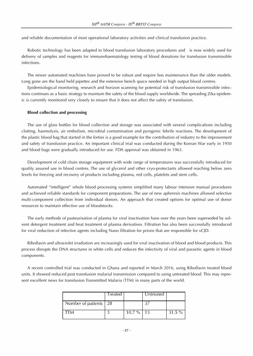
A recent controlled trial was conducted in Ghana and reported in March 2016, using Riboflavin treated blood
units. It showed reduced post transfusion malarial transmission compared to using untreated blood. This may repre-
sent excellent news for transfusion Transmitted Malaria (TTM) in many parts of the world.
XIIth AATM Congress - IXth BBTST Congress
- 27 -

Quality and governance
The extended clinical and manufacturing role of blood services forced them to become accountable for contin-
ued improvement of their operational standards. Most blood transfusion facilities adopted the principles of clinical
governance by creating an environment for good laboratory practice in testing and processing of blood and clinical
haemotherapy.
The role of computers to improve blood safety and traceability has been substantial. The use of electronic control
points at critical steps in the process (e.g.: release of final product; bedside verification of patient identification), can
be much enhanced.
Bar coding and the widely used ISBT128 has facilitated consistent standardised identification, storage, exchange
and retrieval of valuable data for blood transfusion operational activities. In addition a wide range of information
can be made available to support effective use of blood and more efficient operational management of blood trans-
fusion services in general.
Cellular therapy and other medical products of human origin
Preparation of red cells and platelets can now be considered routine activities of the past. The new era of stem
cell transplantation started with early reports from the USA and Japan of transfusion related graft versus host disease.
This clinical finding was tamed and developed further into controlled valuable therapeutic tools in stem cell trans-
plantation for life saving treatment of many serious conditions.
Bone marrow harvesting and stem cell separation by apheresis from circulation or by collection from cord blood
constitute an important main stay for modern management of haematological malignancies, genetic disorders and
other life threatening conditions.
The blood banking skills, accumulated over the years, constitute the scientific and operational foundation for
establishing a new array of medical products of human origin (MPHO) and making use during this future technolog-
ical and scientific journey of the high manufacturing standards and the ethical values that were elaborated and
developed during the previous phase of blood banking.
All these developments could not have been achieved without the joint effort of industry and the vision of the
men and women in blood banks and blood centres lately known in Europe as “Blood Establishments”. They worked
together in unison to improve the quality of therapeutic care for patients requiring blood, its components, derivatives
and the recently developed stem cell products, tissues and organs.
Role of professional groups
International transfusion medicine groups are able to see and understand the globally different cultures and
needs. By working together they provided the basis for cooperation, transfer of best practice and experience. This
was evident as a professional necessity at the international level by the two main international professional groups
headed by the AABB and the ISBT. They were established in 1947 and 1935 respectively.
International professional associations in general can enhance good practice and standards at regional and
national levels. One of the major contributions of these international groups was to establish the humanitarian and
XIIth AATM Congress - IXth BBTST Congress
- 28 -

ethical standards of blood donation and blood transfusion. These efforts were pioneered earlier by the Red Cross and
expressed in the resolution of the 17th international Red Cross Conference, in 1948 in Stockholm recommending
that:
1. Blood should be donated freely and supplied free of charge
2. Red Cross Societies should urge the organisation of national transfusion services
3. Red Cross Societies should cooperate with their respective governments
4. There should be cooperation with ISBT, particularly regarding standardisation of methods and equipment
The ISBT Code of Ethics formulated in 1980 has been a landmark internationally respected document. It was
revised later and adopted by the ISBT general assembly in July 12th 2000. It was later supported by a detailed WHO
technical declaration in November 2001.
Regional professional groups
Regional groups are Informal professionally lead interventions that can, in the medium term, reduce diversity and
drive improvement through sharing best practice and influencing national policies and decision-making. They are
primarily bottom-up, grass root interventions, driven by professionals to assure quality and safety in transfusion prac-
tice.
These interventions in return provide the international well-established organisations with opportunities to have
a more active and coordinated regional role to help establish safer transfusion services and promote good transfusion
practices at regional levels, at a stage where it is difficult to mandate a single approach to quality of transfusion prac-
tice, as what has happened in countries of the European Union.
They provide regional opportunities for continued education by conducting educational courses for those who
are not able or are intimidated to attend and participate in international activities and meetings. They also help to
overcome regional isolation of transfusion medicine practitioners. The European School of Transfusion Medicine
(ESTM) is one example that has already proved to be of value to assist transfusion medicine workers in East and
Central Europe.
Members of three regional groups are participating in this meeting. The Arab Transfusion Medicine Forum (ATMF),
the Anatolian Blood Days (ABD) and the Asian Association of Transfusion Medicine (AATM). It is the coming together
of transfusion medicine professionals from many countries, hosted by the Blood Banking and Transfusion Society of
Turkey (BBTST).
Our meeting is an example of international cooperation, at its best, that will no doubt encourage continued
advance on the journey that started years ago on the road of development and progress. We can now, together, start
to explore newer future horizons with a wider range of Medical Products of Human Origin (MPHO).
XIIth AATM Congress - IXth BBTST Congress
- 29 -

Mahrukh GETSHEN
Introduction:
Bhutan is one of the youngest democratic countries in the world. It is situated in the Eastern Himalayas with 60%
of the country under forest cover and mountainous terrain which poses challenges to road accessibility and delivery
of essential services like the health. It is divided into 3 regions and 20 districts for governance purpose. Health and
education services are provided free of cost by the Royal Government of Bhutan (RGoB) to all the citizens and
residents in the country.
The health system comprises of basic health units (BHUs), district hospitals, 2 regional referral hospitals and one
national JDW, Referral Hospital in the capital city.
Vital statistics:
Capital: Thimphu
Total area: 38,394 kms
Total population: 6, 91,141 (2005 national census)
Total hospital beds: 1078
Blood Transfusion Service (BTS) in Bhutan comprises of:
• Blood Safety Program(BSP) is one of the listed programs in the Health Care and Diagnostic Division,
Department of the Medical Services, MoH. It is run by a Program Officer and its functions are strategic planning and
work plan implementation; co-ordination with other health related programs and NBTS; project management;
monitoring and data management.
• National Blood Transfusion Service: a total of 27 government owned hospital based blood centers exist. The
National Blood Center (NBC) is headed by a transfusion specialist, and run by diploma and degree laboratory
technologists and general MLTs. The rest of centers are operated by laboratory technicians as a unit of the clinical
laboratory. Routine day to day activities are under the respective hospital administration to which blood center is a
part of. There are no private or NGO run blood centers.
• Though not part of the BTS, the National Public Health Reference laboratory housed at the new Royal Center
for Disease Control in the capital works in collaboration with BTS to provide with confirmatory testing of Transfusion
Transmissible Infections (TTIs), conducting National External Quality Assessment Schemes in TTIs and evaluation of
test kits for screening of blood for infectious disease markers.
CURRENT STATUS IN BHUTAN
- 30 -

Major changes in BTS observed since past 5 years:
1. Management:
• Blood Safety program has become an active program in the MoH.
• A full time focal point has been appointed as the Program Officer to manage and monitor program related
activities of BTS in the country.
• This has lead to improved efficiency and timely implementation of the program activities; improved
coordination between NBTS and Program.
• There has been marked improvement in the overall management of the program with less responsibility on the
part of Head of National Blood Centre to look into managerial part of the program and provide only technical
expertise and guidance to the Program.
2. Blood donations:
• The donor profile has markedly changed in last 5 years. The number of blood units collected from voluntary
donors has increased in last five years. Compared to 2010 where in VNRBD was 56%, in 2014it has increased to
71% VNRBD with less reliance on family /replacement donations.
• This has been possible due to the blood centers conducting mobile donation camps at regular intervals.
• The BSP and blood centers have been conducting various educational, motivational and advocacy programs.
As a result, many organizations-government and non-government have come forward to hold blood drives. Many
corporates have taken up blood donation as one of their Corporate Social Responsibilities. The highlight is the
sponsoring of the WBDD celebration by the Bank of Bhutan for the last three years. High schools and colleges have
been holding regular drives as part of their social activities. Religious monasteries have also been engaged in such
activities.
• The number of regular and repeat donors has increased especially at the NBC and 2 RRBCs with better
retention strategies.
3. Improvement in test methods:
• More and more blood centers are adhering to the national standards in immunohematolgy tests. More number
of centres has shifted to tube method from earlier slide method for ABO and Rh blood grouping. From the reports
of NEQAS in BGS in 2014, 80% of the centers were found to be carrying out by test tube method.
• Crossmatching is done by test tube method in 100 % blood centers.
• In the last 2 years, 2 regional blood centers have also started antibody detection test though antibody
identification and red cell phenotyping is carried out by NBC only.
• 2 regional blood centers have also started screening infectious markers using ELISA assays with increase in
throughput.
4. Blood component and clinical use of blood:
• The blood component preparation has begun in 2 regional centers and component therapy has been
encouraged.
• Increase in Random Donor Platelets preparation especially at NBC by almost 40 % due to increased
requirement and consumption. JDWNRH has observed a changing pattern of diseases requiring blood transfusions
to chronic, non communicable ones like cancers, chronic kidney and liver diseases, aplastic anemia etc. Also the
XIIth AATM Congress - IXth BBTST Congress
- 31 -

other component preparation(PRC ) has risen by 20 to 30%
National data 2014:
• Total blood collection: 9375 units
• HIV prevalence in donated units =0.07%
• HBV prevalence in donated units =0.76%
• HCV prevalence in donated units =0.29%
• Syphilis prevalence in donated units=0.9%
• Total number of blood and components transfused=10,067
• Number of Whole Blood units consumed: 3660 =36%
• Number of PRC units consumed: 4936 =49%
• Number random donor PC consumed: 854 =9%
• Number of FFP units consumed: 617 =6%
Update on National blood Center,Thimphu
Data of 2014 versus 2015
XIIth AATM Congress - IXth BBTST Congress
- 32 -

NBC’s collaborators:
• Pacific Paramedical Training Center, New Zealand • National Reference Laboratory , Australia • National Standards Bureau ,Thimphu• TTK Rotary Blood bank, India• Regional Testing Center, Medanta-The Medicity, India• National Institute of Biologicals, India
Significant Events at NBC in the year 2015
1. A total of 30(thirty) blood donation camps conducted in and around Thimphu in 2015.
2. World Blood donor Day observed on 14 June 2015 with the Theme “Thank You for Saving my Life” inpartnership with MoH and Bank Of Bhutan.
3. Few important blood donation camps conducted by various organizations in 2015 are: • Visit of the Lions Club International President to the NBB and blood donation camp organized by Lions Club
members of Druk Thimphu on 24th August.• Royal Bhutan Police celebrating their Raising Day by donating blood on29th September.• Trongsa Penlop Thuendrel Club constituted by the Alumni of Trongsa Poenlop Scholarship donated blood on
4th June to commemorate Her Majesty’s Birthday.• The women of the Kussung Women Association (RBG) Thimphu commemorated the Royal Wedding
Anniversary by donating blood on 13th October.• Blood donation camp at UN House to commemorate the UN Day on 24th October • Bhutan Australia Alumni Association members donated blood on 31st October.
4. Visit by an ISO expert Mr Hans Peter fielded in by Bhutan Standards Bureau to conduct an assessment oflaboratory and blood bank for future accreditation to ISO 15189 in April 2015.
5. Visit of the team of two senior officials in December 2015 from National Institute of Biological Noida andAIIMS Delhi, India to discuss with NBB & MoH to initiate the Hemo-Vigilance Program for Bhutan in collaborationwith Hemo-Vigilance Program of India.
6. A hands on training on Column Agglutination technology conducted by Ortho Clinical Diagnostics (OCD)team in mid December.
7. Ongoing training of interns, PG students and laboratory technologists and technicians
National Achievements in 2014:
• VNRBD improved from 63% in 2013 to 71% at country level and from 90% in 2013 to94% at NBC in 2014.
• NBC & NRL, Bhutan successfully conducted NEQAS in BGS and TTIs respectively with 95% participation of blood centers. Successful participation of NBC in IEQAS with PPTC New Zealand and NRL, Australia
XIIth AATM Congress - IXth BBTST Congress
- 33 -

• Blood Transfusion Advisory Committee constituted
• Blood and Blood Product Regulation of Bhutan endorsed by Bhutan’s Medicines Board.
• Bhutan selected for WHO/OFID project Phase II (2014-2016)
• NBC staff Ms Galem underwent a three weeks fellowship training in blood banking at TTK Rotary Blood BankBangalore, India sponsored by Asian Association of Transfusion Medicine.
• The mobile app “Dial 4 Blood” developed in collaboration with NBC and an IT team was declared the winner app during an open competition of app development organized by G2 C. Its function is for‘Crowd Sourcing blood’ in times of urgent blood needs of the blood banks in the country.
Currently its being pilot tested at NBC.
Activities supported by AATM
3 NBC staff underwent 2 to 4 weeks AATM fellowship in QMS at TTK Blood Center, Bangalore, India in last 3years.
1 of the 3 staff trained has been posted to the regional blood center and has been applying the knowledge andskills learnt during the fellowship program.
The other 2 are at NBC involved in documentation and quality control activities of the QAP at NBC. They haveshown higher degree of confidence in their day to day work and shouldering additional responsibilities in QAactivities.
Future Plans:
1. Review and revise the national Blood Policy, 20072. Finalize the national strategic plan for BTS.3. Create a website for BTS 4. National blood system strengthening with effective quality management, haemovigilance and robust data
management systems5. Upscale advocacy and general awareness activities to promote 100% VNRBD by 2020.6. Upgrade Immunohematology technology to Column Agglutination technology from tube method at NBC.7. Include participation of 2 regional blood centers in IEQAS.8. Continue NEQAS in BGS and TTIs.9. Enforce blood regulations by 2016.
Challenges:
• Increasing blood needs and demands for blood components at bigger centers• Manual documentation of processes and procedures which time consuming, leading to errors and inefficient
for proper data management • Need for a collaboration with a regional referral center for seeking advice for problematic immunohematologic
cases.
XIIth AATM Congress - IXth BBTST Congress
- 34 -

Introduction:
Iranian Blood Transfusion Organization (IBTO) is a centrally coordinated and managed Blood Transfusion Service
(BTS), which 100% of its blood establishments are community-based.
IBTO is managed administratively by Iranian government. Till 2015, all blood products were distributed free of
charge to all private and public hospitals and IBTO was fully supported by the government. In 2015, based on the
efforts made by IBTO through optimization of blood usage, most of the blood components incorporated into pricing
framework. According to the decision made by Iranian government, insures have to reimburse the costs related to
the processing. As a result, it is planned to obtain 30% of the costs of BTS 's operation from cost recovery system.
In 2014, 2,071,031 of blood donations were collected in the whole country. Given the fact that Iran's total
population was 78,143,644 million, the rate of blood donation per 1000 population was 26.6 in 2014.
Totally, there are 693 hospital blood banks in Iran. Currently, blood donation sites are 207 that 116 of them are
blood collection centres, 31 are blood collection and preparation centres and 60 are main blood centres . All blood
donation sites are governmental.
These fixed blood centers collect 80% of blood, and the rest is collected by the mobile teams in work places,
educational institutes and cities which are not served by fixed sites. Only one blood center located in the
headquarters performs stem cell collection and therapeutic procedures.
Iran is the first country in Eastern Mediterranean Region (EMR) which reached 100% Voluntary Non-remunerated
Blood Donation in 2007. The rate of regular blood donation was 51.5 in 2014.
CURRENT STATUS IN IRAN
Ali Akbar POURFATHOLLAH
- 35 -
Blood Components
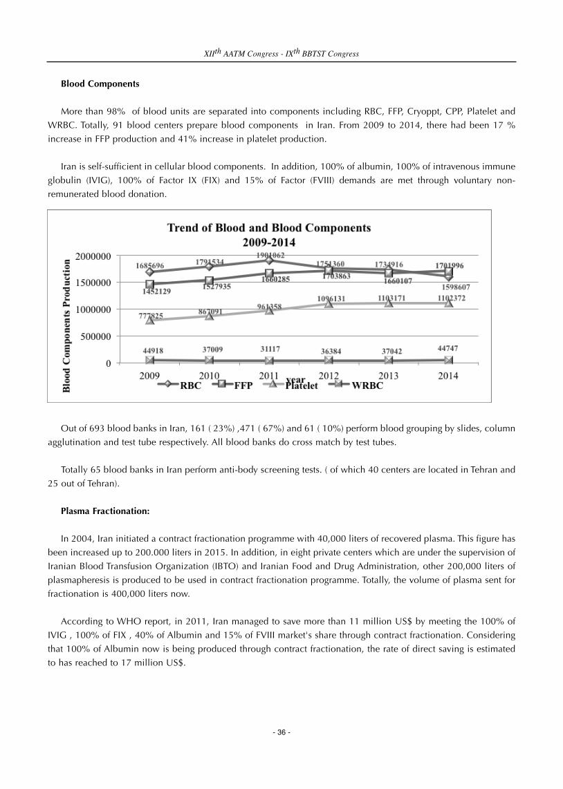
More than 98% of blood units are separated into components including RBC, FFP, Cryoppt, CPP, Platelet and
WRBC. Totally, 91 blood centers prepare blood components in Iran. From 2009 to 2014, there had been 17 %
increase in FFP production and 41% increase in platelet production.
Iran is self-sufficient in cellular blood components. In addition, 100% of albumin, 100% of intravenous immune
globulin (IVIG), 100% of Factor IX (FIX) and 15% of Factor (FVIII) demands are met through voluntary non-
remunerated blood donation.
Out of 693 blood banks in Iran, 161 ( 23%) ,471 ( 67%) and 61 ( 10%) perform blood grouping by slides, column
agglutination and test tube respectively. All blood banks do cross match by test tubes.
Totally 65 blood banks in Iran perform anti-body screening tests. ( of which 40 centers are located in Tehran and
25 out of Tehran).
Plasma Fractionation:
In 2004, Iran initiated a contract fractionation programme with 40,000 liters of recovered plasma. This figure has
been increased up to 200.000 liters in 2015. In addition, in eight private centers which are under the supervision of
Iranian Blood Transfusion Organization (IBTO) and Iranian Food and Drug Administration, other 200,000 liters of
plasmapheresis is produced to be used in contract fractionation programme. Totally, the volume of plasma sent for
fractionation is 400,000 liters now.
According to WHO report, in 2011, Iran managed to save more than 11 million US$ by meeting the 100% of
IVIG , 100% of FIX , 40% of Albumin and 15% of FVIII market's share through contract fractionation. Considering
that 100% of Albumin now is being produced through contract fractionation, the rate of direct saving is estimated
to has reached to 17 million US$.
XIIth AATM Congress - IXth BBTST Congress
- 36 -

Blood Safety
In Iran, 100% of donated blood is screened for HIV, HCV, HBV and Syphilis. However, only 20% of blood is
screened for HTLV I/II. Screening of donated blood for Hepatitis B surface antigen (HBs-Ag) has become mandatory
since the establishment of IBTO in 1974. Screening of blood units for HIV and HCV has started since 1989 and
1996, respectively.
The prevalence rate of HBV and HCV and HIV in blood donors was 0.133 %, 0.043% and 0.003% in 2014
respectively. The prevalence rate of Syphilis and HTLV I/II was 0.0019% and 0.067 in 2013. There had been 0.16%,
0.02% and 0.001% reduction in the prevalence rate of HBV, HCV and HIV from 2009 to 2014.
The prevalence of HIV and HCV among blood donors is almost 10 times lower than general population. (0·003
vs 0·03% and 0·043 vs 0·5%, respectively). This indicates the effectiveness of safety measures applied in IBTO.
In Iran, HIV, HCV , HBV and HTLV I/II tests are done by ELISA. IBTO uses rapid tests for Syphilis. NAT and
pathogen inactivation are not currently used in Iran. In previous years, we have done NAT test for selective donors
in pilot studies and its planned to run NAT tests in near future.
Proficiency tests (EQAS) are done in 34 blood centers in Iran. Totally, 91 blood centers are accredited by ISO.
Education:
One of the most important goals that IBTO pursues through its education and training programs is to make all
related staff informed and trained on the most recent specialized scientific activities and developments in the fields
of transfusion medicine and transfusion science.
IBTO regularly conducts education programmers for clinicians on rational use of blood. In 2014, in 167
hospitals that under the heamovigilance system, 9127 nurses (74%), 2001 physicians( 50%) and 752 technicians of
blood banks (80%) were trained on rational clinical use of blood.
IBTO also conducts refreshing course for doctors who deal with blood donors on annual basis.
In addition, High Institute for Research and Education on Transfusion Medicine educates students at MSc and
PhD level in the discipline of Hematology and Blood Banking.
XIIth AATM Congress - IXth BBTST Congress
- 37 -

The number of admitted MSc and PhD students in the discipline of Hematology and Blood Banking
Blood usage:
Since 2014, IBTO started to collect the number of blood units used in 612 hospitals which are under
haemovigilance system. From 2014 to 2015, there has been 40 % increase in the usage of RBC, 20% decrease in
Plasma products and 28 % increase in platelet usage.
Man- Power Development
In last five years, there has been considerable development in man-power in the country including:
1) Haemovigilance system
Iranian Blood transfusion Organization has implemented a Mandatory Transfusion Transmitted Injuries
Surveillance System (TTISS) to monitor adverse transfusion events (ATEs) since 2009. Till 2016, the system has been
established in 612 hospitals (about 73 %of Iranian hospitals) and more than 6332 adverse reactions have been
reported. The establishment of this system in all Iranian hospitals is on the agenda for the next coming years.
2) Institute for Research and Education on Transfusion Medicine (IRETM)
IRETM was established in 2008. The most important goals of IRETM that pursues through its research and
education activities are to make all the staff involved in technical affairs in all blood centres across the country
informed on the most recent improvements and scientific developments in the fields of transfusion medicine and
transfusion sciences. IRETM benefits the contribution of 37 full time faculty members formed within scientific groups
of Immunohematology, Hematology, Immunology, Microbiology, Biochemistry, Medical Biotechnology, and
Pathology. So far, more than 400 research projects have been approved and conducted by Research Department of
IRETM; a wide range of faculty members and IBTO experts and staff have been actively participating in the projects.
Add to it, the publication of 35 specialty books and publication of about 500 articles in scientific journals (ISI- and
PubMed-indexed).
XIIth AATM Congress - IXth BBTST Congress
- 38 -

3) Centre for Innovation
The Centre for Innovation of IBTO was established on 23 February 2015 . The Centre is a complete miniature of
a “transfusion chain” from vein to vein, with all the standard operating procedures (SOPs) and equipment used by
IBTO and hospitals. The Centre is the unique of its kind in the Middle East and Eastern Mediterranean region. The
main goal of the center is to be used as a training centre for both national and international applicants.
4) Iranian National Cord Blood Bank and Iranian Stem Cell Donor Registry
Iranian National Cord Blood Bank was established in May 2010. It is a public blood bank with the mission of
providing stem cells for patients. From November 2010 to 2012, UCBs were collected from 5 hospitals of Tehran.
All the collection, processing, testing, cryopreservation and storage procedures were done according to standard
operation procedures.
Iranian Stem Cell Donor Registry is a governmental organization that operates under the supervision of IBTO . It
is established in 2009 to help patients in need of haematopoietic stem cell Transplant. Currently 3600 blood donors
are registered in the center.
XIIth AATM Congress - IXth BBTST Congress
- 39 -

- 40 -
INTRODUCTION
The Republic of India is the seventh largest country by area, geographically located in South Asia, and the
second-most populated democratic country with over 1.2 billion people. Blood services were disorganized in earlier
days where the hospitals used to draw blood from donors and would issue the products tested/untested as per need
of the hour.
The 1st Blood Bank was established in March, 1942 at the All India Institute of Hygiene & Public Health,
Calcutta. In those days Blood was collected in glass bottles which were sterilized by autoclaving and ACD was used
as preservative and blood was stored for 3 weeks. The current scenario paints a picture which reflects the growth in
Transfusion Medicine. The Transfusion Medicine can be divided into hospital based and community-oriented,
standalone systems. There are a total of 2760 blood banks in India, out of which 69% are Hospital based centres
and 31% are community-oriented stand-alone blood banks. Out of the total blood banks, 42% is under government
control, 18.4% under private-setups, 12% are nonprofit organizations, and 1.4% under Red Cross.
The financial support has also changed with the change in administrative control. While 50% of BTS operation
is government supported, 32% of the operation is a shared responsibility of government and the private
organizations and 18% have reported to be on cost recovery basis. Regional Blood Transfusion Centres have been
established in different states of India which are supplying to 6-8 Blood storage centres. 1200 Blood Banks prepares
components. 680 – 700 perform apheresis and 25-30 performs stem cell collection & therapeutic procedures.
BLOOD DONATION
Social workers in various sectors maintain a list of willing prospective blood donors prior to the individual setup
of Blood banks. Pioneers of the field contributed largely in developing the voluntary blood services of our country,
which started with the organization of first National Seminar & Workshop, on Blood Donor Motivation held at
Calcutta in January 1985 with the participation of blood bankers and donor motivators from all over the country,
along with a few experts from abroad. The first National Guidebook on Blood Donor Motivation saw the light of the
day in June 1990.
Currently -10 million units of blood is collected annually. Voluntary blood donation (VBD), including family
blood donors, has reached to 84 % in 2013-14 from baseline of 54.4% while replacement donors constitute 16%
of the total number. 40-45%% of the collected blood comes from various blood drives that are organized by various
corporate setups and NGOs every year.
Currently, a growing need is for platelets and this change is reflected in increasing number in platelet donation.
CURRENT STATUS IN INDIA
Nidhi MEHTA

BLOOD BAG MANUFACTURE
Partnering with Sree Chitra Tirunal Institute for Medical Sciences and Technology (known then as the Chitra
Medical Centre), Penpol started production in its factory in Trivandrum, Kerala, on 26th March 1987, with TTK
Pharma as its sole sales agent. In 1989, the company achieved some significant milestones like sending out its first
export shipment, and setting up its R&D division. Currently there are multiple indigenous manufactures of Blood
bags like J Mitra. HLL etc.
INFECTIOUS DISEASE TESTING
ELISA machines and HIV test kits were supplied to 138 blood banks throughout the country since HIV test has
been made mandatory in 1988. The prevalence of Transfusion Transmitted Infections (TTIs) has been estimated as
follows: HIV - 0.3%; HBV - 1.1%; HCV - 0.4%; Syphilis - 0.4%.
Currently blood banks use Chromatographic strips, ELISA (différent generations) and Chemiluminiscence for TTI
testing. Only 4% Blood Banks are reported to use Nucleic Acid Technology as a testing protocol.
IMMUNOHAEMATOLOGY
The Blood Group Reference Centre (BGRC) was started in 1957 due to active interest shown by Indian Council
of Medical Research (ICMR) in the field of blood banking. The functions entrusted to BGRC were few but of national
importance.
• To train the people in methodology of blood grouping and blood banking.
• To prepare and supply standard blood grouping reagents.
• To work as a reference centre for the unsolved problems of cross matching.
• To prepare and maintain a list of rare bloods and conduct research in this unknown field in the country at that
time.
On 5th February 2008, ICMR decided to give the Institute a national status and renamed it as "National Institute
of Immunohaematology".
The rarest blood group in India is Bombay phenoyype which was discovered in 1952 amongst three unrelated
individuals in Mumbai by Bhende et al., an important event in the field of immune-hematology which changed the
scope of Blood Banking.
All Blood Banks in India are currently capable of performing blood grouping, out of which 40-50% use the test
tube method while 10-20% are capable of performing Column Agglutination. <5% of the blood banks are capable
of RBC antibody detection whereas <1% are capable to identify the detected antibodies.
ROLE OF GOVERNMENT AGENCY
National AIDS Committee was constituted in the Ministry of Health and Family Welfare, in 1986, following the
detection of the first AIDS case in the country.
In 1992 India’s first National AIDS Control Programme (1992-1999) was launched, and National AIDS Control
Organisation (NACO) was constituted to implement the programme.
XIIth AATM Congress - IXth BBTST Congress
- 41 -

1986-1992 (DGHS) Till 1992, the bulk of funds for AIDS-related projects were used:
* Improving blood testing facilities in blood banks
* 62 Surveillance facilities were set up.
* 154 Zonal Blood Testing Centres (ZBTC) were set up.
Many government agencies in field of Blood transfusion in India such as FDA, NACO, and NATIONAL BLOOD
TRANSFUSION COUNCIL are working together and their roles are complimentary in order to improve the quality
of Blood Transfusion Services. .
LICENSING OF BLOOD BANKS
On public interest litigation, the Supreme Court delivered a historic judgement on January 4, 1996.
1. Establish National Blood Transfusion Council – 23rd May 1996
2. Banning of Professional Blood Donors – 1st January 1998
3. Compulsory Licensing of Blood Banks
4. Framing of National Blood Policy – April 2002
World Health Organisation directed that replacement blood donor system be phased out and abolished by 2005.
Action plan for Blood safety was published in 2003
National Blood policy came in force in 2002 with following objectives
1. To reiterate firmly the Govt. commitment to provide safe and adequate quantity of blood, blood components
and blood products.
2. To make available adequate resources to develop and reorganise the blood transfusion services in the entire
country.
3. To make latest technology available for operating the blood transfusion services and ensure its functioning in
an updated manner.
4. To launch extensive awareness programmes for donor information, education, motivation, recruitment and
retention in order to ensure adequate availability of safe blood.
5. To encourage appropriate clinical use of blood and blood products.
6. To strengthen the manpower through human resource development.
7. To encourage Research & Development in the field of Transfusion Medicine and related technology. 8. To take
adequate regulatory and legislative steps for monitoring and evaluation of blood transfusion services and to
take steps to eliminate profiteering in blood banks.
EDUCATION AND TRAINING
In the early years Blood Bank meetings were clubbed with Association of Physicians of India which is a
professional body of consultant physicians formed in 1944 mainly to provide a common forum to the Physicians of
India to meet and to share experience and research observations in the field of Medicine. It's members are
physicians with postgraduate qualifications in different specialties. ISHBT was then constituted to have independent
meetings for Blood Transfusion and Hematology.
ISBTI is a Non Profit Voluntary Organization operating at National Level. Registered under the Societies
Registration Act XXI of 1860 by the Registrar of the Societies, Chandigarh on December 28, 1973, it functions as a
XIIth AATM Congress - IXth BBTST Congress
- 42 -

National body for blood transfusion chapter for scientific advancement and research including up gradation &
maintenance of safe blood transfusion services in the country.
ISTM the Indian Society of Transfusion Medicine was constituted in 2011 which serves as a platform for scientific
discussions, exchanges, deliberations, exchange of ideas, teaching and raining with regards to Transfusion Medicine
pertaining to Indian context.
Looking at the boom of growth in Indian Blood Banking, need for fresh eyes and leadership has become
prominent. For this reason alone, 17 centers have been identified across the country as training centers to impart
training on all aspects of Blood Transfusion Services involving Blood Bank Medical Officers, Technicians,
Counselors, Nurses, Clinicians, Donor Motivators and Programme Officers of SACS. The training programme will
aim to strengthen the inter- and intra-regional collaboration between NACO and its Collaborating Centers, national
Blood transfusion services, education and training instiutions and NGOSs in order to support and strengthen the
national capacity in education. Courses in Transfusion Medicine have started in various institutes:
Diploma in Immuno-Haematology and Blood Transfusion 1 centre 5 seats
MD-Transfusion Medicine 8 centres- 12
MD - Immuno Haematology & Blood Transfusion 27 centres 54 seats
HEMOVIGILANCE PROGRAMME IN INDIA
Launched on 10th December 2012 in 90 medical colleges, it is a centralized programme initiated to monitor
transfusion reactions and create awareness amongst health care professionals. Currently 279 centres, located in
blood banks, medical colleges/institutions, govt./ private hospitals are enrolled under HvPI and as of yet, 3394
Adverse Transfusion Reaction Reports have been reported. Haemovigilance Programme of India is one of the
Mandate’ s of NIB as per its bye-laws 3.4.1 as approved by the Governing Body of the Institute chaired by Secretary
(Health & F.W.)/ Chairman, Governing Body of NIB.
NATIONAL DONOR VIGILANCE PROGRAMME
Launched as a national level initiative at Science city, Kolkata on World Blood Donor Day – 14th June 2015 with
the aim to improve donor safety and satisfaction through monitoring, analysing and researching adverse events
Provide evidence based support for improvement in the Blood Donation Process in order to increase donation
frequency while reducing the number of adverse events.
NEWER TECHNOLOGIES
Blood Irradiation
Though irradiation of blood and cellular blood products is necessary for centres undertaking treatment of cancer
patients and bone marrow transplantations, less than ten per cent of healthcare institutions in India are having blood
irradiators. According to international standard for blood banking and transfusion services, blood and blood
products must be irradiated. However, the National Blood Policy does not mention about blood irradiation. Many
healthcare institutions cannot afford to install blood irradiator because they don’t have an economically feasible
project to install it. Proper planning of manpower, resources and logistics are required to install a blood irradiator.
XIIth AATM Congress - IXth BBTST Congress
- 43 -

NAT
NAT testing has been started in few centers in India, but it is not a mandatory screening test for TTIs as per Drug
and Cosmetics Act, 1940 and the rules therein. Major barriers in implementing routine NAT testing in India is its
high cost and lack of technical expertise in most of the blood centers.
Pathogen Inactivation
These PIT imply a proactive, more generalized approach against multiple new and (re-)emerging pathogens,
which perpetually challenge the safety of the blood supply and will become an serious alternative to a repetitive
implementation of new screening tests.
Photopheresis
Extracorporeal photopheresis is one of several secondary therapies which have shown promise in the clinical
setting. While the procedure itself has been around for over 20 years, our understanding of the mechanisms from
which therapeutic benefits are seen, and the population they are seen in, remains limited. ECP has reached our
country and in 2 corporate hospitals are in process of standardization and validation.
ACCREDITATION
Accreditations have proven to be a great source in Quality Check. There are 66 Blood Banks in India which has
been recognized and accreditated by NABH Accreditation Program for blood Banks.
References1. https://en.wikipedia.org/wiki/India2. http://www.mahasbtc.com/blood-transfusion-safety-and-regulatory-requirements3. http://www.naco.gov.in/NACO/About_NACO/
Acknowledgements:I would like to extend my gratitude and thanks to Dr. S R Joshi for his guidance and support. I would also like to extend my thanks to Dr Ishita Chakraborty for her contribution.
XIIth AATM Congress - IXth BBTST Congress
- 44 -

- 45 -
Mongolia located in the heart of Central Asia between Russian Federation and the Republic of China. 1,564,100
sqr km, 19th and the most sparsely populated independent largest country in the world and 3,027,582 people with
the density of 1.9 people/sqr km. The average altitude of 1,580 m above sea level and semi-desert and plains,
mountains in the west and southwest, Gobi desert in the south and southeast, taiga forests and lakes in the north.
The capital city name is Ulaanbaatar. More than half of Mongolia’s population lives in Ulaanbaatar.
The Ministry of Health and Sports, encouraging the Blood Service in order to approve and implement the
principles, guidelines related to the blood safety and ensuring professional methods for activities of implementing
the Mongolian Government policy. National blood service consisted of 26 hospital-based blood banks in 21 aimags
and 1 stand-alone (National Center for Transfusion Medicine) in Ulaanbaatar, are centrally coordinated and
managed by 100% government. The Blood banks which is a specialized professional service that deals with
producing safe and ample blood supplies to local and private hospitals throughout Mongolia as well as continually
managing, guiding and embracing all Hospital Blood banks. Blood Banks provide the hospitals in rural areas with
necessary blood and blood products and specialized management and guidance for soum level.
The National center for transfusion medicine /NCTM/ operates with in the term of the national blood policy and
Mongolian Health legislation to promote uniform implementation of standards and consistency in the quality and
safe blood and blood products. The NCTM is responsible for organizing, advising, reporting and uniform
implementation of standards and consistency, government policy, program, laws, and Orders of improving blood
and blood products supply, and blood safety. The NCTM guides and manages all 26 Blood Banks’ functions and
central organization of specialized training, production, research and development. Essential functions of the NCTM
includes strategic and operational planning, donor recruitment, blood collection and preparation, provision of
sufficient resource, coordination and management to ensure an adequate supply of blood and blood products and
safe clinical transfusion, one of the main functions is to conduct specialized training for blood service among the
hospital staff and medical doctors, and to conduct training ensuring safe blood procedures for pre- and post-
graduate students.
The Donor Law was approved in 2012 by the Great Khural (Parliament) of Mongolia. The Government policy on
“Improving provision and supply with safe blood and blood products” was approved in 2007 by the Great Khural.
The Plan of Action for 2008-2015 to implement the Government policy for improvement of provision with safe
blood and blood products was approved in 2008 by article 111 of Government Resolution
Objectives of the Plan of Action for 2008-2015 to implement the Government policy for improvement of
provision with safe blood and blood products was approved in 2008 by article 111 of Government Resolution to
provide equally safe, sufficient and quality blood and blood products to all hospitals, to provide safe and ample
blood supply in large quantity in times of major disaster, to expand donor activity along with international standard
and increase participation of health organization, government and non-government organizations, entities,
CURRENT STATUS IN MONGOLIA
Namjil ERDENEBAYAR

collections and population in training and promotional activity, to introduce advanced technology for screening,
processing, storing and transporting donor blood and blood products and to support improving blood safety and
blood supply of the National Center for Transfusion Medicine, Regional Diagnosing Center, Blood Banks beside the
aimag and the district's health organizations and collaboration with foreign and international organizations from
the Government. In the framework 41 activitiesin 5 objectives of the Plan of Action for 2008-2015successfully
implemented.
In Mongolia since 2004 celebrates World Blood Donor Day as tradition, NCTM and Mongolian Red Cross
Society co-work to celebrate WBDD under the annual theme of WHO. The activities from co-operation of NCTM
and Mongolian Red Cross Society is focusing on enlargement donor recruitment, activation and inspiration of
donors.
For statistical indicators, 2011-2015 collected 20700 to 30396 units of whole blood and components. In 2011
only stand-alone (NCTM) performed apheresis, by 2015 two hospital-based blood banks and one stand-alone were
performed apheresis. 2011- 2015 number of family replacement donors decreased 3.2 to 0.6 percent and regular
donors increased by 41.4 percent 2011- 2015. For distribution of 100 percent voluntary nonremunerated blood
donors in Mongolia. But in the registration 99.4 percent is voluntary nonremunerated and 0.6 percent is the family
replacement.Because, in some case, thatlack of rare blood groups suggesting to members of family of recipient
donate voluntary.
New donors increased by 60.7 percent and regular donors increased by 41.4 percent 2011- 2015. Blood
production increased by 26 percent 2011- 2015. Starts emergency resource of fresh frozen plasma preparing each
year. 14 types of blood products prepared by 2015. 2011-2015 whole blood products decreased from 0.2 percent
to 0.1 percent and components products increased up to 99.9 percent. In 2014 implemented apheresis and
cytapheresis technology by blood collection. 2011-2015, rational usage units of components increased from 54670
to 62822. All 27 blood banks testing blood grouping, cross matching by slide/tiles, test tube, and column
agglutination. And only stand-alone (NCTM) has red blood cell antibody detection and identification as of 2015. In
all Blood service testing 100 percent transfusion-transmitted infection (TTI) such as HIV, HBV, HCV by ELISA. In
2012 implemented new technology NAT in NCTM. By 2015 elimination with TTI such as HIV, HBV, HCV decreased
from 10.4 percent to 7.2 percent.
The NCTM involves in the External Quality Assurance Services (EQAS) for Donor Infectious Disease Testing
conducted by the National Reference Laboratories, Australia and the Reference Laboratory for Transfusion medicine
at the NCTM was established several years ago and has built intensive relationships with other national reference
laboratories. NCTM involving regularly in the EQAS program. Laboratories of 26 Blood Banks in aimags have been
involving in the EQAS program since 2010. The Linear bar coding electronic information system has been
introduced in the workflow (daily work) of the National Center for Transfusion Medicine since 2013.
Each year graduated 50-100 clinicians and other appropriate staffs by the short time training program about
blood safety and rational use of blood, who working at the clinic. And each year graduated about 14 doctors or
technicians by postgraduate degree for doctors and nurses/technicians by Transfusion Medicine. Regular refresher’s
course doctors and nurses/technicians per year (average no) 2-4 persons. In these years number of related surveys
conducted according to the topics donor recruitment, hemovigilance, TTI, production and blood bank
administration (for instance screening HTLV-1/2, CMV, and Occult HBV etc).
In the framework of renewing the legislation environment and ensuring the blood safety, planning to confirm new
XIIth AATM Congress - IXth BBTST Congress
- 46 -
National program for expanding the voluntary non-remunerated blood donations by the State Government of
Mongolia in 2016. In addition planning to expand foreign affair, especially highlight the regional cooperation for
sharing experiences and best practices for blood donor recruitment and retention in 2016. In the framework of
National program for improving the donor recruitment and retention will be developed the donor training and
advertising activities and will be increased the capability of the human resource.
In 2016 new Blood center’s building construction process ending and will be ready to serve in the 2nd quarter
of this year. With the new Blood center implementing many new technologies such as pathogen inactivation etc.
Further, finding the way to implement small batch sized plasma fractionation technology. “Asian Rare Blood Joint
Seminar” by the National Center for Transfusion Medicine, Mongolia (country chapter of AATM) and Shanghai Blood
Center (WHO Collaborating Center) will be hosted in Ulaanbaatar in August 2016. The objective of the seminar is
to understanding the challenges and barriers in provision of rare blood for patient and identification on strategies for
promoting the rare blood supply in Asia.
XIIth AATM Congress - IXth BBTST Congress
- 47 -
- 48 -
Background
Blood Transfusion Service of Nepal Red Cross Society was established in the year 1966 i.e. 3 years after the
inception of the Society itself. During the initial years the service was available only for the people of Kathmandu
but over the years blood banks have been established in 101 places of 68 districts of the country.
In the initial years the service was made possible through collection of blood from professional donors but since
1982 collection of blood was emphasized from voluntary non-remunerated donors only. In the meanwhile serious
efforts were made and are being made to collect blood from institutionalized sectors like colleges, universities,
industries, clubs, governmental and non governmental offices. Today, blood is collected mostly from these sectors
through routine motivational and collection campaigns but motivated individuals are also significant donors. The
blood collection mobile teams routinely visit these institutions in the valley and in the outskirts of Kathmandu Valley
to collect blood. Sometimes these teams also spread out to other adjoining districts for the same purpose. With the
expansion of health services, establishments of medical colleges, government and private hospitals and Nursing
homes the demand of blood is rapidly going up not only in the Valley but also in the districts.
Government of Nepal, in it's policy declaration of 1991, has mandated Nepal Red Cross Society as the sole
authority in conducting blood program in Nepal. Therefore, a great responsibility has fallen on the Society and to
prove its capability, it is systematically strengthening itself with resources available nationally and exploring resource
possibilities internationally. The Kathmandu-based Central Blood transfusion service , so far being the only referral
centre for the whole country, has been planning not only to upgrade the Centre but also in upgrading regional blood
centers of Biratnagar, Pokhara, Nepalgunj and Chitwan. It is also considering the possibilities of upgrading the
Dharan and Dhangadhi district blood centres in view of the establishments there of major hospitals and coverage
area. As it stands today, there are 23 district level blood banks, emergency units in 37 and 36 hospital units of the
blood service in the country.
Service Delivery
During this reporting period Nepal Red Cross had collected 217,160 units of blood throughout the country and
supplied 311,330 units of blood to the needy patients. .Blood collection is voluntary based and there is no
professional blood donation. 2200 units of blood were supplied in free of charge to earthquake affected patients.
Total 1000 units of blood and blood component were provided to Thalassemia society in subsidized charge.
Total1100 units of blood and blood components were provided to hemophilia society in subsidized charge. Blood
cold chain system is effectively implemented for the transportation of blood and blood product to maintain the
quality of blood.
CURRENT STATUS IN NEPAL
Manita RAJKARNIKAR

There was a National and International cooperation and coordination effort for enhancement of quality system
of blood collection, testing and processing. Project base support from Nepal government National Bureau of Blood
Transfusion Service, National Center for AIDS and STD control, Luxembourg Red Cross, Australian Embassy, Global
Advisory Panel , British Red Cross for the scaling up of quality system of blood transfusion service with the supply
of blood bank equipment to CBTS and district level blood centers. These supports are very much effective to upgrade
the quality service in blood centers. Total 3,428 mobile blood collection camps were organized by different
institutions/organizations. Total 1012 mobile camps were organized by different organizations/institutions in the
Kathmandu Valley with the technical support of CBTS. In Central Blood Transfusion Service 69,309 units of blood
were collected and supplied more than 100,000 units. Among that 9,706 units of blood were collected from female
donors and 59,603 units of blood from male donors.
Laboratory Investigations
Blood Service is providing safe blood and blood - products to the needy patients all collected blood is routinely
tested for HIV, HBsAg, HCV, Syphilis besides grouping, cross-matching and anti-D antibody identification and
titration in the Centers. Blood products like plasma, packed red cells, platelets, cryo-precipitates, platelet rich
plasma are also produced in Kathmandu, all regional centers and some of the district centers providing services to
the patients of hemophilia and other blood diseases. Due to the Blood Component production technology is
sensitive and expensive; this facility is available in limited blood centers but in near future some other districts are
also going to provide this service. After testing for Transfusion Transmissible Infections all reactive cases are further
processed for Blood Donor counseling ( pre and post donation) are done. This is targeted for the wellbeing of donor
health status and confirmatory detection of HIV, Hepatitis 'B' & 'C' and Syphilis.
Blood component facility
In 10 centers are with blood component facility and one hospital is having apheresis facility. Around 50% of
collected units are processed in blood components as Packed Red Cells, Plasma, platelets and cryo. Still around 50%
of collected units are used as whole blood. There is a plan to use more component products in near future with a
plan to expand component facility in more districts. In the country there is only one apheresis machine available in
one of the Hospital.
Blood collection and Male/ Female blood donor Ratio
The Nepal Red Cross Society, as stated above, was able to collect 217,160 units of blood during the period which
was an increment of about 8% over the collection of the previous year. Among that Male blood donors are 84.5 %
and female 15.5 %.
Trainings and orientation programs
Total 1,680 people were participated in blood donation motivation and interaction program held in 31 different
organizations in this period. Orientation programs were organized for doctors and paramedics, altogether 225 were
participated in 6 orientation program on clinical use of blood and blood products in different hospitals.
Annual meetings and collaboration for the trainings
An annual meeting held at Kathmandu for the review of national blood program. 58 representatives were
XIIth AATM Congress - IXth BBTST Congress
- 49 -

involved in the meeting from different regional, district and hospital blood centers. Total 42 technicians from
different blood centers were trained in quality management of blood transfusion service training, supported by WHO
and Nepal Government Ministry of Health and population,
Blood Group Percentage
Blood group percentage of blood donors are as follows:
Blood National In Kathmandu
Group % of Positive % of Negative % of Positive % of Negative
A 26.82 0.73 29.39 0.83
B 27.82 0.84 27.42 0.62
O 29.92 0.93 28.59 0.87
AB 12.68 0.26 11.94 0.34
Total 97.24 2.76 97.34 2.66
No. of HIV, Hepatitis 'B' & 'C' and Syphilis Detected in the Collected Blood during the Period
SN Centers HIV Hepatitis B Hepatitis C Syphilis
1. Central Blood Transfusion Centre 21 192 224 360
2. Regional Blood Transfusion Centre 13 151 56 115
3. District/Emergency Blood Transfusion Centre 27 227 119 260
4. Hospital Blood Transfusion Unit 7 47 23 25
Total 68 617 422 760
% of positivity in Kathmandu Valley 0.03 0.26 0.31 0.50
Total Positive Percentage nationwide 0.03 0.31 0.21 0.38
"Give Blood Save life"
XIIth AATM Congress - IXth BBTST Congress
- 50 -

- 51 -
Estimated population: 180 million
No. Blood Banks: 800 Blood Banks
Blood Collection per annum: around 1.2 million
Country requirement: 3.2 million bags / year
Low average age of donors: 22 years.
Voluntary non-remunerated donations: 300000 = 10%
Family/replacement donations: 2700000 = 90%
Paid donations: 0%
Female donation: 3.77%
Current System Architecture
1830 Blood banks (<1200 in Punjab Province), Mostly Hospital Blood Banks
20 % Public Sector (including Armed Forces)
Private Sector and NGO 80 % contribution in total,
Predominant in the provinces of Punjab and Sindh. There are mainly, Hospital based blood banks and stand-
alone blood banks.
These are diverse in terms of functions and capacities from donor mobilization only to the entire v2v transfusion
chain.
The major contributors to management of thalassaemia patients are BDOs which include university based
organizations . Major contributors to the voluntary blood donor pool in Pakistan through their donor motivation and
mobilization programmes.
National Blood Policy and Strategic Framework 2014-20
Developed in 2002 with support from WHO EMRO and GIZ. Revised in 2008 and 2014.NBP proposed to
establish a coordinated National Blood Transfusion Program consistent with the international recommendations to
ensure safe blood.
Future System Architecture
System Reform being implemented by the Safe Blood Transfusion Programme, Pakistan based on a centralized
model.
CURRENT STATUS IN PAKISTAN
Farrukh HASAN

In the current phase of the SBT project (completing Nov 2014), 10 RBCs and 60 affiliated HBBs would be
constructed/refurbished In the first phase, the model will contribute approximately 20% to the sector in terms of
blood supply Quality Management Systems Wide diversity Existence of ISO certified institutions on the one hand,
Lack of even basic quality control measures on the other hand.
Potential importance of Blood Transfusion Authorities (for mandatory licensing) and Pakistan NationalAccreditation Council (for accreditation).
Blood Processing and Testing prevalence is as follows.
HIV = 0.04%, HCV = 3.7%, HBV = 2.5%, Syphilis = 1.01% and Malaria = 0.3%
Most blood transfused as ‘Whole Blood’ (62%) till the widespread occurrence of the dengue epidemic.
Diversity in the methods and techniques for transfusion transmittable infectious disease testing.
Gold standard methods (e.g NAT testing) available in a few centres. Intermittent supply of screening kits(commonest scenario) compromises blood safety at the other end of the spectrum.
First CUB guidelines (Published in 1999)
Clinical governance-Hospital Transfusion Committees existing and functional in a few centres Current strategyRevision of CUB guidelines by a Task Force comprising all stakeholders (including clinicians, haematologists, CPSP,PMDC, PSH).
Establishment of Hospital Transfusion Committees for clinical governance: Terms of Reference developed,information sharing about existing HTCs through workshops and monthly SPTP newsletter, plan for their actualinstitution is under process.
Haemovigilance
Concept relatively new in Pakistan .Centralized haemovigilance does not exist.
Current strategy
Blood Transfusion Authorities would also be given the mandate to elaborate haemovigilance rules. HospitalTransfusion Committees would be responsible for transmitting Haemovigilance related information to BTAs.
'Pakistan Haemovigilance Network (PNH)‘ (to be supported by the INH): national baseline survey, reportingsystem PNH would encourage and assist in the formation of HTCs all over the country. The HTC will collecthaemovigilance data from their respective centers and report to PHN. PHN would compile Annual Reports, whichwould help in policy making, planning and blood safety improvement.
Management Information System
The concept of coherent, reliable data recording and management only beginning to be understood and
XIIth AATM Congress - IXth BBTST Congress
- 52 -

implemented In most cases, some data recorded manually but not utilized for management decisions. It is beingworked upon.
Current strategy: introduction of nation-wide BT MIS, functional brief for MIS developed.
Sustainability of Reforms There are development Plans for the future centralized systems.
Public-private partnerships
SAATM has been playing some limited role in bringing the Public / private sectors together and also promotingthe awareness, educational and service campaign.
Most of the hematologists country wide are its members and feel comfortable to sit on a common platform.
XIIth AATM Congress - IXth BBTST Congress
- 53 -

- 54 -
Based on 2015 statistics Turkey has 77 695 904 population. Annual population increase is 1.04 %, 70.5 % of the
population lives in cities. Almost half of the population is younger than 28.3 years. 66.5 % of the population is
between 15 - 64 years old. Depending on 2013 statistics average life expectancy is 73,7 years for men and 79.4
years for women.
First Blood Banking & Transfusion Medicine (BB&TM) activities started at Istanbul University Medical School at
early 1920 ies. Due to donor supply problems Turkish Red Crescent decided to be involved in Blood Banking
activities and declared this at 1953 during annual congress. First public blood banks were opened at 1957 by Turkish
Red Crescent in Ankara and Istanbul. First Blood law was issued at 1983 and private blood banks were closed, paid
donation was banned. This law was updated at 2007. A group of dedicated doctors who were involved in the field
of BB&TM decided to support BB&TM activities by a civilian initiative and established a national blood society;
“Blood Banks and Transfusion Society of Turkey (BBTST)” at 1996.
The main strategy of BBTST defined as below listed topics;
a) education of blood bank and clinical staff which is directly related on staff competence and improve BB&TM
quality management
b) establishing close collaboration with the national health authority
c) establishing official and academic education programs
d) collaborating among national and international organizations, agencies and institutions involved in the safety
of blood products and transfusion practice.
BBTST organised 18 national courses, 8 national congresses, and 200 symposiums 30 national education
sessions all around Turkey with the collaboration of MoH Since 1997. VIII European Congress of International
Society of Blood Transfusion was organised at 2003 in Istanbul by BBTST. BBTST has been organizing annual
international workshops under the name of “Anatolian Blood Days” since 2011 with the collaboration of Turkish
Blood Foundation.
Ministry of Health (MoH) is the “national authority” on BB%TM likewise other medical aspects in Turkey. Turkey
has had mixed Blood Banking since 2002 when MoH announced new “National Blood Policy” as “regionalisation”.
Although MoH appointed TRC as a main national blood supplier still there are “temporary blood banks” run by
university hospitals and MoH training hospitals. TRC collects almost 80% of national blood donation annually. Each
year around 2 500 000 units of blood are donated. Donor deferral rate is around 6,5 – 10 %. Around 10% of
collected bloods are destroyed due to positive serological test results and etc. Apheresis service has been available
in Turkey since late 1980 ies. Around 250 apheresis machines from all major manufacturers are in service and
annual platelet apheresis is around 75 000 units in Turkey. Since early 1990’s blood irradiation is available in Turkey
not only in major metropoles but also in other cities.
CURRENT STATUS IN TURKEY
N. Nuri SOLAZ

There are 368 blood facilities in Turkey (regional blood centers, blood donation centers and transfusion centers)
which collect blood. There are more than 1 500 institutions where blood transfusion is performed legally (hospitals,
special medical centers, etc.). Cost of blood products are annually issued by Ministry of Finance. Blood transfusion
costs are completely reimbursed by state owned Social Security Agency.
Blood donation is “voluntary non-remunerated” by law. Turkey has had first “National Blood and Blood Products
Guidelines” since 2009 and all BB&TM activities are run according to this national guidelines.
Each unit of donated blood should get below listed tests;
a) Blood group typing since 1921
b) VDRL since 1950’s
c) HBsAg since 1973
d) Anti-HIV ½ since 1983
e) Anti – HCV since 1996
TRC has started to screen each donated blood by NAT for HBV, HBC and HIV since 2014.
Frequencies of seropositive microbiological tests are as below table;
Test Type Frequency
VDRL 0,11 %
HBsAg 1,99 %
Anti-HIV½; 0,12 %
Anti – HCV 0,45%
Distribution of blood group typing in Turkey is as below table;
Blood Type Frequency
A Rh + 38%
A Rh - 5%
B Rh + 14%
B Rh - 2%
AB + 7%
AB - 1%
O + 29%
O - 4%
BB&TM is not an independent medical speciality in Turkey and there is no specialisation education on
BB&TM. A Master education program is available for medical doctors as a post graduate training on BB&TM. MoH
has accredited training courses for medical doctors and non MD medical staff since 2001.
Finally; blood transfusion is safe enough in Turkey because;
• Basic equipment for safe blood supply is available all around the country.
XIIth AATM Congress - IXth BBTST Congress
- 55 -

• Well trained basic manpower for blood supply is available all around the country.
• Automation, quality control, Haemovigiliance, etc. advanced topics of Blood
Banking is initiated all around the country.
XIIth AATM Congress - IXth BBTST Congress
- 56 -

- 57 -
Background: Blood group antigens present on the cell membrane of red blood cells (RBC), platelets andneutrophils can be defined either serologically or predicted based on the genotypes of genes encoding forantigens.(1,2) Use of molecular testing in clinical laboratories requires knowledge of the molecular basis of bloodgroup antigens and the availability of suitable genotyping methods. Many of the serologically determined RBCantigens are related to single nucleotide polymorphisms (SNPs) which result in a single amino acid change in RBCantigens. (3,4) Molecular events other than SNPs in the antigen coding region of the gene may also give rise to bloodgroup antigens or affect antigen expression. A wide variety of molecular events generate blood group antigendiversity. These include; missense mutation (the most common), non sense mutation, mutation of motifs involved intranscription, altenative splicing, deletion of a gene exon(s) or nucleotid(s), insertion of exon(s) ornucleotid(s),alternate initation, chromosome translocation, single crossover, gene conversion and other generearrangements. Some examples of blood groups are showed at Table 1.(1,5)
Blood groups antigen and genes: At present, thirty-five red cell blood group systems, seventeen human plateletantigens and eleven neutrophil antigens are known. Thirty-five red cell blood group systems and more than 300antigens have been described on the surface of human red cells within 44 genes and 1568 alleles.(1,6,7) Red cellgroups antigen genes have been summarized at Table 2.(8)
Platelet antigens and neutrophil antigens are summarized Table 3 and Table 4.(1)
The methods of genotyping: Since the first blood group gene cloning in the early 1990s, knowledge on themolecular basis of most red blood cell, platelet and neutrophil antigens brought the possibility of using nucleotide-based techniques to predict phenotype.(9)
Red cell antigen genotyping has many applications in blood donor testing, as well as in various diagnosticsettings. Genotyping results can therefore be used as a reliable adjunct to serologic testing and will becomeincreasingly used in red cell antigen assignment.The majority of genes encoding either proteins that carry bloodgroup antigens or transferases that attack blood group specified carbonhydrates have been cloned and molecularbases associated with many cell surface markers on red blood cells (RBCs), platelets and neuthropils have beendetermined. Thus, it is possible to perform molecular analysis for these inherited allels. Such analysis include allelespesific PCR amplification (AS-PCR) and PCR amplification followed by restriction enzyme digestion, restrictionfragment length polymorphism (RFLP).(1,9)
The methods of PCR were developed in 2000s, as real-time PCR(RT-PCR) assays for high-throughput humanblood antigen typing (10), single base extension in multiplex PCR blood group genotyping (11), PCR-ELISA forhigh-throughput blood group genotyping. (12)
Recently, several groups have developed multiplex-polymerase chain reaction SNaPshot assays to determinesingle nucleotide polymorphisms (SNPs) in blood group genes with the purpose of identifying clinically relevantantigens and rare alleles. (13) As an alternative to phenotyping, large-scale DNA-based assays, which are feasiblefor high-throughput donor red blood cell typing were developed for determination of blood group polymorphisms.(14)
GENETICS OF BLOOD GROUP ANTIGENS
Duran CANATAN

Development and validation of a fully automated platform for extended blood group genotyping. Targetsamplified by multiplex PCR were hybridized on the chip, and a revelation step allowed the simultaneousidentification of up to twenty four blood group antigens, leading to the determination of extended genotypes.(6)
A multiplex polymerase chain reaction assay was designed for typing thirthy five RBC antigens in six reactionmixes. The study shows that in practice, this high-throughput genotyping assay is feasible, fast and provides reliableresults. Compared to serological testing, this molecular approach is also very cost-efficient. (15)
Approaches to determination of a full profile of blood group genotypes are single nucleotide variant (SNV)mapping by DNA microarray and massively parallel sequencing, with respect to blood group genotyping. The mostfrequent genetic change associated with blood group antigens are SNVs. (7)
Indications of genotyping:(4)Indications for Non-RHD Genotyping
• Serological antigen typing/phenotyping cannot be determined due to chronic transfusion requirement (eg.Thalassemia major, Blackfan Diamond anemia).
• Serologically complex patients - with multiple or unidentified antibodies who require ongoing transfusionsupport.
• Patients with autoimmune hemolytic anemia and/or with a positive DAT (in spite of chemical treatment), andcirculating autoantibody.
• Patients with a suspected alloantibody against an antigen for which no commercial antisera is available (eg.Possible anti-Doa).
• Select patients with variable or null reactivity using serological methods when a variant allele is suspected(eg. Sickle cell anemia).
Indications for RHD Genotyping
• Prenatal patients with discrepant, weak or inconclusive serological RhD testing results where RHDgenotyping may modify their blood product or Rh Immune Globulin (RhIG) requirements. For example:prenatal patients with weak or discrepant RhD serology may be Weak D type 1, 2 or 3 and would not requireRhIG.
• Patients likely to require chronic transfusion, or with complex transfusion needs, where RHD genotypingmay modify their blood product requirements.
• Patients who likely require transfusion with an anti-D who appear serologically D positive.
Clinical Applications genotyping in transfusion practice:(1)Red cell blood groups:
1. Identification of fetus at risk for anemia of the neonate (hemolytic disease of the newborn)(HDN),2. Determination of presence or absence of blood group alleles in recently transfused patients,3. Determination of blood group alleles when patients’ RBCs are coated with immunglobulin,4. Resolution of A, B, and D typing discrepancies,5. Screening for antigen negative blood donors,6. RHD Zygosity,7. Situations where genotype and phenotype do not agree.
Platelet Blood groups:
1. Identification a fetüs at risk for alloimmune thrombocytopenia,2. Genotyping when appropriate antisera are not available,
XIIth AATM Congress - IXth BBTST Congress
- 58 -

3. Determination of genotypes in thrombocytopenic patients,4. Platelet panels for antibody investigations,5. Screening for antigen negative platelet donors,
Neutrophil blood groups:
1. Immune neutrophenia
2. Transfusion related acute lung injury(TRALI)
Table 1: Molecular events that give rise to blood group antigens and phenotypes(5)
Molecular events Blood Group Systems
Gene conversion or recombination events MNs, Rh, Ch/Rg
Duplication of an exon Gerbich
Deletion of a gene, exon, or nucleotide (s) ABO, Rh, MNS,Kel,Duffy, Dombrock
Insertion of a nucleotide (s) Rh, Colton
Single nucleotide substitutions Most blood group systems
Table 2:Red Blood Cell system genes(8)
ISBT System Gene Name Gene Name Gene Product and Associated Blood Name (Number) ISBT (HGNC)* Location Component Name Group Antigens [Null
[CD Number] phenotype]
ABO ABO 9q34.2 Glycosyltransferase, A; B; A,B; A1(001) (ABO) carbohydrate [Group O]
MNS MNS 4q31.21 Glycophorin A(GPA) M, N, S, s, U, He,Mi ͣͣ,Vw(002) (GYP*A [CD235a) + 38 more
GYP*B Glycophorin B (GPB) [En(a-); U-; MᵏMᵏ]GYP*E) [CD235b]
P1PK P1 22q13.2 Glycosyltransferase, P1, Pᵏ(003) (4GALT) carbohydrate
RH RH 1p36.11 RhD [CD240D] D, G, Tar (004) (RHD RhCE[CD240CE] C, E, c, e, V, Rh17
RHCE) +43 more [Rh null]
Lutheran LU 19q13.32 Lutheran glycoprotein; Luᵃ Lu ᵇ, Lu3, Lu4,(005) (LU) B-cell adhesion Auᵃ,Auᵇ +14more
molecule [CD239] [Recessive Lu (a-b-)]
XIIth AATM Congress - IXth BBTST Congress
- 59 -

Kell KEL 7q34 Kell glycoprotein K,k,Kpᵃ, Kpᵇ, Ku, Jsᵃ,Jsᵇ(006) (KEL) [CD238] +25 more
[K0 or K null]
Lewis LE 19p13.3 Fucosyltransferase, Leᵃ, Leᵇ, Leᵃᵇ, Leᵇʰ,(007) (FUT3) Carbohydrate ALeᵇ,BLeᵇ
(Adsorbed from plasma) [Le (a-b-)]
Duffy FY 1q23.2 Duffy glycoprotein Fyᵃ, Fyᵇ, Fy3, Fy5, Fy6(008) (DARC) [CD234] [Fy(a-b-)]
Kidd JK (SLC14A1) 18q12.3 Human urea transporter Jkᵃ, Jkᵇ, Jk3(009) (HUT) [Jk(a-b-)]
Kidd glycoprotein
Diego DI 17q21.31 Band 3, Anion Exchanger 1 Diᵃ, Diᵇ, Wrᵃ, Wrᵇ, Wdᵃ,(010) (SLC4A1) [CD233 Rbᵃ + 16 more
Yt YT 7q22 Acetylcholinesterase Ytᵃ, Ytᵇ(011) (ACHE)
Xg XG (XG) Xp22.33 Xg ͣ glycoprotein Xgᵃ(012) (MIC2) Yp11.3 CD99( MIC2 product) CD99
Scianna SC 1p34.2 ERMAP Sc1, Sc2, Sc3, Rd+3(013) (ERMAP) more
[Sc:-1,-2,-3]
Dombrock CO 12p12.3 Do glycoprotein; Doᵃ, Doᵇ, Gyᵃ, Hy, Joᵃ +2(014) (ART4) ART 4 [CD297] more
[Gy(a-)]
Colton CO 7p14 Aquaporin 1 (AQP1) Coᵃ, Coᵇ, Co3, Co4(015) (AQP1) [Co(a-b-)]
Landsteiner- LW 19p13.2 Lw glycoprotein İntracellular LWᵃ, LWᵃᵇ, LWᵇWiener (ICAM4) adhesion molecule 4 (ICAM4) [LW( a-b-)](016) [CD242]
Chido/Rodgers CH/RG (C4A, 6p21.32 Complement Ch1, Ch2, Rg1 +6 more(017) CAB) component: [Ch-Rg-]
C4A; C4B
H H 19q13.33 Fucosyltransferase, H(018) (FUT1) carbonydrate [CD173] [Bombay (0 h)]
Kx XK Xp21.1 XK glycoprotein Kx(019) (XK) [Mcleod phenotype]
XIIth AATM Congress - IXth BBTST Congress
- 60 -

Gerbich GE 2q14.3 Glycophorin C (GPC) Ge2, Ge3, Ge4 +8 more(020) (GYPC) [CD236] [Leach phenotype]
Glycophorin D (GPD)
Cromer CROM 1q32.2 DAF Crᵃ, Tcᵃ, Tcᵇ, Tcᶜ,Drᵃ,Esᵃ,(021) (CD55) [CD55] IFC +9 MORE
[Inab Phenotype]
Knops KN 1q32.2 CR1 Knᵃ, Knᵇ, McCᵃ, SIᵃ, Ykᵃ(022) (CR1) [CD35] +4 more
Indian IN 11p13 Hermes Antigen[CD44] Inᵃ, Inᵇ +2 more(023) (CD44)
Ok OK 19p13.3 Neurothelin,basigin Okᵃ, OKGV, OKGM(024) (BSG) [CD147]
Raph RAPH 11p15.5 CD151 MER2(025) (CD151) [Raph-]
JMH JMH 15q24.1 Semaphorin 7A JMH + 5 more(026) (SEMA7A) (CD108) [JMH-]
I IGNT 6p24.2 Glucosaminyltransferase, I(027) (GCNT2) carbonhydrate [I-or i adult]
Globoside GLOB 3q26.1 Transferase, P(028) (3GALT3 or carbonhydrate [P-]
GaINAcT1) (Gb4, globoside)
Gill GIL 9p13.3 Aquaporin 3 (AQP3) GILL(029) (AQP3) [GIL-]
Rh-associated RHAG 6p21-qter Rh-associated Duclos,Ol ͣ,DSLKᶧGlycoprotein glycoprotein [Rh null( regulator(030) [CD241] type)]³7
* ISBT: International Society Blood Transfusion HGNC: Human Gene Nomenclature Commitee. ERMAP:Erythroid Membrane Associated Protein
XIIth AATM Congress - IXth BBTST Congress
- 61 -

Table 3: Platelets system genes(1)
Platelet system Platelet system Gen Name Gen Name Alleles for molecular Classical ISBT* ISBT* HGM* protocols given
Bil/Zav HPA-5 GP1A ITGA2 GP1A1/GP1A2
Sil HPA-13 GP1A ITGA2 GP1A1/GP1A3
Ko HPA-2 GP1BA GP1BA GP1BA1/ GP1BA2
Ly HPA-12 GP1BB GP1BB GP1BB1/ GP1BB2
Bak/Lck HPA-3 GP2B ITGA2B GP2B1/ GP2B2
Max HPA-9 GP3A ITGB3 GP2B2/ GP2B3
PIA/Zw HPA-1 GP3A ITGB3 GP3A1/GP3A2
Pen/Yuk HPA-4 GP3A ITGB3 GP3A1/GP3A3
Ca/Tu HPA-6 GP3A ITGB3 GP3A1/GP3A4
Mo HPA-7 GP3A ITGB3 GP3A1/GP3A5
Sr HPA-8 GP3A ITGB3 GP3A1/GP3A6
La HPA-10 GP3A ITGB3 GP3A1/GP3A7
Gro HPA-11 GP3A ITGB3 GP3A1/GP3A8
Oc HPA-14 GP3A ITGB3 GP3A1/GP3A9
*ISBT: International Society Blood Transfusion HGM: Human Genom Mapping
Table 4: Neutrophil system genes(1)
Platelet system Platelet system Gen Name Gen Name Alleles for molecularClassical ISBT* ISBT* HGM* protocols given
NA NA NA FCGR3B NA1/NA2
NB NB NB None NB1/NB2
SH None None FCGR3B SH/ SH
Ond None None ITGAL Onda/Ondb
Marr None None ITGAM Marra/ Marrb
S S None None Sa/Sb
9a None None None
SL None None None
*ISBT: International Society Blood Transfusion HGM: Human Genom Mapping
XIIth AATM Congress - IXth BBTST Congress
- 62 -

References1. Demomme GA, Rios M and Reid ME: Molecular protocols in transfusion medicine. Academic Press. London.
UK 2000.2. Veldhuisen B, Van der Schoot CE, de Haas M. Blood group genotyping: from patient to high-throughput
donor screening. Vox Sang. 2009 Oct;97(3):198-206. doi: 10.1111/j.1423-0410.2009.01209.x. Epub 2009Jun 22.
3. David J. A. Red cell genotyping and the future of pretransfusion testing. Blood, 2009 114: 248-256.4. Goldman M, Hannaford K, Hannon J and Berardi,P .Molecular Immunohematology at Canadian Blood
Services: Red Cell Antigen Genotyping 2016.Canada.5. Castilho L and Júnior JP. Blood group genotyping. Rev. Bras. Hematol. Hemoter. 2004;26(2):135-140.6. Boccoz SA, Le Goff GC, Mandon CA, et al. Development and Validation of a Fully Automated Platform for
Extended Blood Group Genotyping. J Mol Diagn. 2016 Jan;18(1):144-52. doi:10.1016/j.jmoldx.2015.09.002. Epub 2015 Nov 29.
7. Jungbauer C, Hobel CM, Schwartz DW, Mayr WR. High-throughput multiplex PCR genotyping for 35 redblood cell antigens in blood donors. Vox Sang. 2012 Apr;102(3):234-42. doi: 10.1111/j.1423-0410.2011.01542.x. Epub 2011 Nov 21.
8. Zimring JC, Spitalnik SL: Molecular biology and immunology in transfusion medicine. In (Eds: Roback JD,Grossman BJ, Harris T, Hillyer CD) Technical Manual. American Association of Blood Banks (AABB); 17thedition (July 28, 2011) p:293-362.
9. St-Louis M. Molecular blood grouping of donors.Transfus Apher Sci. 2014 Apr;50(2):175-82. doi:10.1016/j.transci.2014.02.012. Epub 2014 Mar 6.
10. Araujo F. Real-time PCR assays for high-throughput blood group genotyping. Methods Mol Biol.2009;496:25-37. doi: 10.1007/978-1-59745-553-4_3.
11. Denomme GA.Single base extension in multiplex blood group genotyping. Methods Mol Biol. 2009;496:15-24. doi: 10.1007/978-1-59745-553-4_2.
12. St-Louis M. PCR-ELISA for high-throughput blood group genotyping. Methods Mol Biol. 2009;496:3-13. doi:10.1007/978-1-59745-553-4_1.
13. Latini FR, Castilho LM.An overview of the use of SNaPshot for predicting blood group antigens.Immunohematology. 2015;31(2):53-7.
14. Latini FR, Gazito D, Arnoni CP et al. A new strategy to identify rare blood donors: single polymerase chainreaction multiplex SNaPshot reaction for detection of 16 blood group alleles. Blood Transfus. 2014 Jan;12Suppl 1:s256-63. doi: 10.2450/2013.0242-12. Epub 2013 Apr 15.
15. Mc Bean RS, Hyland CA, Flower RL .Approaches to determination of a full profile of blood group genotypes:single nucleotide variant mapping and massively parallel sequencing. Comput Struct Biotechnol J. 2014 Sep23;11(19):147-51. doi: 10.1016/j.csbj.2014.09.009. eCollection 2014.
XIIth AATM Congress - IXth BBTST Congress
- 63 -

- 64 -
Antibodies to red blood cells (RBC) cause clinical problems and blood bank will find compatible blood for this
patient. They are on surface of RBC, in plasma or both. Antibody binds to antigen of RBC leads to agglutination,
hemolysis via complement activation and phagocytosis. Anti-body to recipient causes direct antiglobulin test (DAT)
positivity, antibody to donor RBC causes indirect antiglobulin test (IDAT) positivity and major cross match
incompatibility. DAT positivity present with hemolytic anemia. IDAT positivity causes hemolysis and ineffective
transfusion after erythrocyte transfusion (1,2). Firstly, problem was determined by tests and clinical findings and
history.to resolve antibody problems. History of Anti-E, anti-K, anti-D+anti-C, and anti-D+anti-E is important.
Previous records of patient helps to rapid evaluation and to decide the solution. After understanding problem, The
compatible blood may be obtained for transfusion and appropriate treatment modality may be chosen for patient.
Test and methods used to determine antibody problem and decide solution:
Direct antigolbulin test (Direct Coombs), indirect antiglobulin test (indirect Coombs), antibody identification test,
Blood subgroup identification by antibody or molecular, Elution, absorption (2,3).
Direct antiglobulin test (direct Coombs):
This test is used to detect IgG and/or complement adherence to red cells. Positive test shows that anemia is an
immune hemolytic anemia due to autoantibodies, drugs or delayed hemolytic transfusion reaction. The type of
antibody can be identified with the use of monospecific antibodies to immunoglobulin G (IgG) and C3d. If gG or
IgG plus C3d is positive, the antibody is usually a warm antibody . When the red cells are coated with C3d only,
the antibody is often but not always a cold antibody.
A positive DAT indicates that an immune reaction has taken place on the patient's RBC (either patient’s own RBC
or transfused RBC). Antibody source may be Auto vs. Alloimmune. History of Previous transfusion or IVIG and RHIG
use shows alloimmune source. But this did not exclude otouimmunity. Eluate will performed on patient cells after
cell separation. This eluate is incubated auto or allo RBCsamples known subgroups. Differences help to evaluation.
If DAT is positive, additional tests will be performed including determination unexpected red cell antibodies in
the serum and attached to the red cells. A patient with a positive DAT should be evaluated for hemolysis (blood
smear, LDH, indirect bilirubin, reticulocytes, haptoglobin, etc) due to autoimmune antibody or a drug-induced auto-
antibody. The most common cause of serious or fatal drug-induced hemolytic anemias is use of 2nd and 3rd
generation cephalosporins. The previous and current drug therapy of patients with a positive DAT should be carefully
evaluated . When possible, consideration should be given to substitute these antibiotics with others. Other drugs
such as penicillin, quinidine, can cause immune red cell hemolysis, but severity is rarely same with 2nd and 3rd
generation cephalosporins(4,5)
HOW TO RESOLVE ANTIBODY PROBLEMS?WHICH TESTS DO WE HAVE?
Davut ALBAYRAK

Autoimmune antibodies may be idiopathic or secondary to autoimmune diseases, infections, B-cell
malignancies, or due to drugs, eg procainamide, alpha methyldopa. The DAT is frequently positive in patients with
HIV infection. Autoimmune Ig G antibodies are usually benign, but occasionally cause mild to severe hemolytic
anemia requiring urgent red cell transfusions despite plasma incompatibility with all red cells. In general, patients
with warm reacting autoantibodies will not destroy transfused RBC faster than their own RBC and transfusion is safe
(there are reports of thrombotic episodes in the rare cases of HIV patients with severe autoimmune hemolytic anemia
who are transfused). In contrast, patients with high titer cold reactive autoantibodies such as titer > 1/500 may have
severe acute hemolysis upon transfusion.
The presence of the autoantibody may make difficult to identify or rule out the coexistence of significant red cell
alloantibodies in the plasma of patient. There is coexisting alloantibodies Up to 40% of patients with
autoantibodies. In contrast with autoantibodies, alloantibodies to RBC may cause severe hemolysis of transfused
red cells. when a patient has an autoantibody that may mask an alloantibody, transfusion should be undertaken only
with careful monitoring and the full cooperation of the Blood Bank .
Autoimmune IgM antibodies are typically cold reactive (6). If they are present in sufficiently high titer, this are
associated with severe hemolysis of the patient’s own red cells and transfused red cells, despite steroid treatment.
There are also rare cases of red cell agglutination in coronary arteries. Hemolysis during surgery may cause cardiac
problem in patients with symptomatic cold agglutinin disease or high titer cold agglutinins. The management of
these patients may include the use of RBC phenotype-matched blood, steroids, warmed blood and a warm room (for
patients with cold reactive autoantibody), IVIG, plasmapheresis or Rituximab. Other diagnostic possibilities to think
in a patient with a positive DAT include a delayed hemolytic transfusion reaction associated with recent blood
transfusion or transfusion of ABO incompatible plasma or platelet transfusion.
Antibody screening
Antibody screening uses indirect coombs test with RBC panels known subgroup antigens. Usually, two or three
cell panel is used. If it is positive, next step will be identification of target of antibody.
This test is recommended before transfusion, in pregnancy, multitransfused patients, ineffective transfusions, DAT
positivity after transfusion, Rh-negative women with Rh-positive partner, Rh-positive women known to be at
increased risk of antibody sensitization, such as previous history of antibody production, history of red cell
transfusion, or placental incompetence, mothers with hemolytic disease of the newborn.
Patients whose plasma gives a positive red cell antibody screen (an indirect Coombs test) require extra time to
obtain fully compatible blood. About 1/100 transfusions or pregnancies will result in the formation of an antibody
to a foreign red cell antigen. Many of these antibodies can cause a severe hemolytic reaction, while others are
clinically insignificant. They can be ignored. The Blood Bank should identify the antibody to group and decide its
clinical relevance. Clinically significant antibodies usually require a search for red cell units that lack the
sensitizing antigen. This takes extra time. If a patient with a positive antibody screen requires transfusion,
appropriate blood can be obtained after the antibody can be identified. The Blood Bank will supply the safest blood,
available and consistent with the patient’s needs. This may consist of incompatible by crossmatch. Least
incompatible units may be used for transfusion in autoimmune hemolytic anemia. In the above cases, the physician
must signify his/her understanding of the added risk of transfusion reaction in such cases by signing a form (7).
If the current antibody screen is negative and the patient has had a history of an alloantibody(s), an anamnestic
XIIth AATM Congress - IXth BBTST Congress
- 65 -

antibody response may occur. Response usually appears 2-4 day after transfusion with increasing titer. If it is
suspected from direct antiglobulin test (DAT)-negative immune hemolytic anemia, more sensitive methods were
used (8)
Antibody identification studies:
Antibody identification studies will be performed if the antibody screen is positive. Antibody screen use a RBC
panels with known subgroubs. Presence or absence antigens and positivity with individual cells helps decide which
antibodies are present. Additional knowledge may obtained with use of enzyme treatment with neuromidase
activityand cold incubation. Antibodies with specificity known to cause hemolytic disease are titrated to assist in
determining the need to monitor the severity clinical disese such as the newborn hemolytic disease.
The determined antibodies are classified by clinical significance: ABO ,Duffy, Rh, Kidd, Kell and SsU antibodies
are always important clinical potential. Lewis, Lutheran,MNS, Cartwright and P may sometimes cause imporatnt
important reaction. Leb,Xga,Chido/Rodgers Bg andYork HTLA is rarely associated with important reaction.
Molecular testing may be used to obtain more information on relevant antigen such as variant D gene (9).
Additional investigations:
Patient history includes these headings: Diagnosis, Pregnancy history, Medications / IV therapy, signs of immune
disease, previously identified antibodies ( especially, history of the antibodies, Anti-E, anti-K, anti-D+anti-C, and
anti-D+anti-E.), previous transfusion history, date of last transfusion, use of IVIG. If the patient was seen at another
hospital, it is when and where? may records be accessible and obtainable? Have you had any problems during
transfusion, where have you been transfused? Physician and Nurse may assist. If previous records is accessible, it
include patient demographics (age, sex, race), the observed serologic problem, test descriptions (method, strength
of reactivity, estimated prevalence of reactivity). a complete record of the transfusion histories of patients with
antibody sould be maintained to prevent haemolytic transfusion reaction in future transfusion due to the amanaestic
effect of these antibodies. Medication history should include use of all blood products. Commercial IVIG have
measurable levels of anti-A and -B (IgG class), as well as a variety of non-ABO antibodies.
In conclusion, an antibody is present in patient, this is important for treatment and transfusion selections.
Antibody is screened and then identified. Subgroup of patient is determined. Then compatible blood is searched.
Sufficient stock should be available for patient needs. It is very important for the blood bank to maintain the list of
the rare blood group donor pools in order to provide the suitable blood when necessary. If hospitals have not such
laboratory facilities, they should determine a referee blood center.
References1. Arndt PA, Leger RM, Garratty G. Serologic findings in autoimmune hemolytic anemia associated with
immunoglobulin M warm autoantibodies. Transfusion.2009;49(2):235-242.2. Daniels G. Other Blood Group. In: Robac JD, Combs MR, Grossman BJ, Hilyer CD (eds.) Technical Manual.
16th edition, Bethesda, MD, AABB press, USA. 2008;pp 411-4363. Brecher ME, editor. AABB technical manual. Bethesda: American Association of Blood Banks; 2005.4. Garbe E, Andersohn F, Bronder E, et al. Drug induced immune hemolytic anemia in the Berlin Case Control
Surveillance Study. Br J Haematol 2011;154:644-653.5. Garratty G. Immune hemolytic anemia associated with drug therapy. Blood Rev 2010;24:143-150.
XIIth AATM Congress - IXth BBTST Congress
- 66 -

6. Chandesris MO, Schleinitz N, Ferrera V, et al. Cold agglutinins, clinical presentation and significance;retrospective analysis of 58 patients. Rev Med Interne 2004;25(12):856-865
7. Petz LD. “Least incompatible” units for transfusion in autoimmune hemolytic anemia: should we eliminatethis meaningless term? A commentary for clinicians and transfusion medicine professionals. Transfusion2003;43(11):1503-1507.
8. Karafin MS, Denomme GA, Schanen M, Gottschall JL.Clinical and reference lab characteristics of patientswith suspected direct antiglobulin test (DAT)-negative immune hemolytic anemia.Immunohematology.2015;31(3):108-15.
9. Musa RH, Ahmed SA, Hashim H, Ayob Y, Asidin NH, Choo PY, Al-Joudi FS . Red cell phenotyping of bloodfrom donors at the National blood center of Malaysia. Asian J Transfus Sci 2012;6:3–9.
XIIth AATM Congress - IXth BBTST Congress
- 67 -
- 68 -
The ABO system is the most important of all blood groups in transfusion practice. It is the only blood group
system in which individuals have antibodies in their serum to antigens that are absent from their red blood cells
(RBC). This occurs without any exposure to RBCs through transfusion or pregnancy. For this reason they are called
natural antibodies. Due to the presence of these antibodies, transfusion of an incompatible ABO type may result in
immediate lysis of donor RBCs. This produces a very severe, if not fatal, transfusion reaction in the patient.
Pretransfusion testing is based on the testing to detect ABO incompatibility between a donor and potential
transfusion recipient1. Due to these antibodies, transfusion of ABO-incompatible blood may cause severe
intravascular hemolysis as well as the other manifestations of an acute hemolytic transfusion reaction2.
Forward grouping (front type) is defined as using known sources of commercial antisera (anti-A, anti-B) to detect
antigens on the individual’s RBCs. Reverse grouping (back type) is defined as detecting ABO antibodies in the
patient’s serum by using known reagent RBCs, namely A1 and B cells. ABO forward and reverse grouping tests must
be performed on all donors and patients. ABO grouping is the most frequently performed test in the blood bank.
There is always an inverse reciprocal relationship between the forward and reverse type; thus, one serves as a check
on the other. For example, if the individual has A antigens only on their red cells, there will be an ‘’expected’’
naturally occuring anti-B antibody in their serum since they lack the B antigen1(figure1).
ABO discrepancies
ABO discrepancies occur when unexpected reactions occur in the forward and reverse grouping. These can be
due to problems with the patient’s serum (reverse grouping), problems with the patient’s red cells (forward
grouping), or problems with both the serum and cells. The unexpected reaction can be due to an extra positive
reaction or a weak or missing reaction in the forward and reverse grouping. All ABO discrepancies must be resolved
prior to reporting a patient or donor ABO group1. When a discrepancy is encountered, the discrepant result must
be recorded, but interpretation of the ABO group must be delayed until the discrepancy resolved. If the specimen is
from a donor unit, the unit must not be released for transfusion until the discrepancy is resolved. When the blood is
from a potential recipient, it may be necessary to administer group O red cells of the appropriate Rh type before the
investigation is completed. It is important to obtain sufficient pretransfusion blood samples from the patient to
complete additional studies that may be required2. Red cell and serum test results may be discrepant because of
intrensek problems with red cells or serum, test related problems or technical errors. Discrepancies may be signaled
either because of negative results are obtained when positive results are expected, or positive results are found when
tests should have been negative2.
PROBLEMS RELATED WITH BLOOD GROUPING ANDCROSSMATCHING
Güçhan ALANOĞLU

Technical Errors
Technical errors can also cause ABO discrepancies. Table1
Table 1: Common causes of false-negative and false –positive results in ABO testing2
False –negative results False-positive results
Reagent or test serum not added to a tube Over centrifugation of tubes.
Hemolysis not identified as apositive reaction. Use of contaminated reagents, red cells, or saline
Inappropriate ratio of serum (or reagent) to red cells. Use of dirty glassware
Tests not centrifuged sufficiently Incorrect interpretation or recording of test results
Tests incubated at temperatures above 20-24 C
Incorrect interpretation or recording of test results
Table 2: Common sources of Technical Errors resulting in ABO discrepancies1
• Incorrect or inadequate identification of blood specimens, test tubes or slides.
• Cell suspension either too heavy or too light
• Clerical errors or incorrect recording of results.
• A mix-up samples
• Missed observation of hemolysis
• Failure to add reagents
• Failure to add samples
• Failure to follow manufacturer’s instructions
• Uncalibrated centrifuge
• Overcentrifugation or undercentrifugation
• Contaminated reagents
• Warming during centrifugation
Table 3: Overview of ABO discrepancies
Problems with red blood cell testing? Problems with Serum testing?
Extra antigens Extraantibodies
Acquired B phenotype A subgroups with anti-A1
B(A) phenotype Cold alloantibodies
polyagglutination Cold autoantibodies
Rouleaux Rouleaux
Missing or weak antigen Missing or weak antibody
ABO subgroup Newborn
Pathologic etiology Elderly population
Mixed field reaction Pathologic etiology
Transfusion of group O to A, B or AB
Bone marrow or stem cell transplantation
A3 phenotype
XIIth AATM Congress - IXth BBTST Congress
- 69 -

ABO discrepancies may be arbitrarily divided into four major categories
Group I Discrepancies
These are associated with unexpected reactions in the reverse grouping due to weakly reacting or missing
antibodies. Discrepancies in this group includes
I. Anti-A and anti-B agglutinins (IgM) produced by the infant can first be demonstrated at 3–6 months. Anti-A
and anti-B if present in cord sera are usually IgG and are of maternal origin. Reverse ABO grouping in infants is not
warranted before 6 months
II. Elderly patients: Earlier studies showed a progressive decrease in anti-A and -B agglutinin titers with age, with
low levels (titer 4 or less) being common in subjects aged 80 years or more.
III. Severe hypogammaglobulinemia: Anti A and Anti B are often present in very low concentration in patients
with inherited immunodeficiency and in rare X-linked Wiskott- Aldrich syndrome . Anti-A and anti-B may also be
present in very low concentration in patient immunosuppressed by therapy or disease and in patients undergoing
intensive plasma exchange.
IV. ABO incompatible HPC transplantation: ABO incompatible HPC transplantation with induction of tolerance
e.g. group A patient receiving group O bone marrow will have circulating group O red cell but will only produce
Anti B antibody.
V. In chimerism: That is, a person with dual population of cells from more than one zygote. Presence of two
populations of red cells of different ABO group may lead to absence of antibodies. Twin chimerism occurs when
hematopoietic stem cells migrate between vascular bridges which allow mixing of blood between two fetuses.
Chimeric twins have immune tolerance; they do not make against A ro B antigens that are absent from their own red
cells but present on cells of engrafted twins.
VI. Pediatric patients receiving long term parentral and entral nutrition which is sterile and free of bacteria. It is
believed that the immunizing source for such naturally occurring antibodies is gut and environmental bacteria which
have been shown to possess ABO like structures on their lipopolysaccharide coats.
Resolution of group I discrepancies
a. It can be resolved by enhancing weak or missing reaction by incubating the patient’s serum with reagent A1
and B cells at room temperature for 15-30 minutes
b. If there is still no reaction after centrifugation, serum cell mixture is incubated at 40 C for 15-30 minutes
Note: an autocontrol and O cell control must always be tested concurrently to detect reactivity of other
commonly occurring cold agglutinins e.g. anti I
Group II discrepancies
These are associated with unexpected reactions in forward grouping due to weakly reacting or missing antigen.
XIIth AATM Congress - IXth BBTST Congress
- 70 -

Discrepancies in this group includes
I. Subgroups of A or B: subgroups of A antigen ( Ax, Am, Ay, Ael) which are not agglutinated or weakly
agglutinated by most anti A. Subgroups of B antigen (Bx, Bm, Bel) ) which are not agglutinated or weakly
agglutinated by most anti B. Both can present as group II discrepancies.
II. Leukaemia may yield weakened A or B antigen: In acute leukaemia, the A antigen may be weakened.
Sometimes the blood appears to contain a mixture of group A and group O cells or of A1 and weak A. In other cases
the red cells react weakly with anti-A, even behaving like A3 or Am15. In a patient with erythroleukaemia, of group
B, 60% of the cells were not agglutinated by anti-B and appeared to be group O, but were really very weak B, when
separated from the normal B cells they would absorb anti-B.
III. Acquired B, B(A) and A(B) phenotypes: Acquired B results from the action of bacterial deacetylase, which
converts N-acetylgalactosamine to -galactosamine, which is very similar to galactose, the chief determinant of B.
The second type of acquired B that may be called the ‘passenger antigen’ type is caused by adsorption of B-like
bacterial products on to O or A cells but occurs only in vitro.
IV. Out of group transfusion or ABO mismatched hematopoietic progenitor stem cell transplantation:
ABOcompatible but not identical transfusion of red cell (e.g. group O red cells transfused to group A or B person)
results in artificially induced chimerism. ABO mismatched hematopoietic stem cell transplant (e.g. group O person
transplanted with group A or B marrow, group A or B person transplanted with group O marrow).
V. Neutralization of anti A and anti B typing reagent by high concentration of A or B soluble substances in serum
with serum or plasma suspended red cell
VI. Chimerism in fraternal twins, mosaicism arising from dispermy: Tetra-gametic or dispermic chimeras present
with chimerism in all tissues and are more frequently identified because of infertility and rarely because of mixed
populations of red cells
Resolution of group II discrepancies
a. Weaker reactions with antisera can be resolved by enhancing reaction of antigen with respective antisera by
incubating test mixture at room temperature for 15-30 minutes
b. Sub groups causing group discrepancies can be resolved by adsorption elution studies
c. Acquired B phenomenon can be resolved by lowering PH of monoclonal antisera. Anti B in the serum of
acquired B person does not agglutinate autologous red cells (autocontrol negative). Secretor status of person can
resolve acquired B, saliva of acquired B person contains A substance not B substance. Serum of acquired B person
contains A substance.
d. High concentration of A or B substance causing group discrepancies can be resolved by saline washing of
red cells
XIIth AATM Congress - IXth BBTST Congress
- 71 -
Group III discrepancies
These are associated with protein or plasma abnormalities, rouleaux formation and pseudoagglutination.
Discrepancies in this group includes
I. Elevated level of globulin from e.g. multiple myeloma, Waldenstorm macroglobulinemia, Hodgkin lymphoma.
II. Elevated level of fibrinogen.
III. Small fibrin clot in plasma or incompletely clotted serum can be mistaken for red cell agglutinates of reverse
grouping. Principal: patient’s sample with abnormal concentration of serum proteins, altered serum protein ratio, or
high molecular weight volume expanders can aggregate reagent red cells and can mimic agglutination. Rouleaux
are red cell aggregates that adhere along their flat surfaces, giving a stacked coin appearance microscopically.
Rouleaux will disperse when suspended in saline. True agglutination is stable in the presence of saline.
Group IV discrepancies
These discrepancies are because of miscellaneous problems. These can be due to-
I. Recent transfusion of out of group plasma containing component.
II. Cold alloantibodies (e.g. anti M) or autoantibodies (e.g. anti I), PH dependent autoantibodies, a reagent
dependent antibody (e.g. EDTA, paraben) leading to unexpected positive eaction.
III. Recent infusion of IvIg which can contain ABO isoagglutinins.
IV. Mix field agglutination with circulating red cell of more than one ABO type.
V. Polyagglutination (e.g. T activation) resulting from inherited or acquired abnormalities of red cell membrane
with exposure of auto cryptantigen. The T determinant is normally covered by N acetylneuraminic acid and can
therefore be described as a cryptantigen. The antigen can be exposed by the action of bacterial or viral
neuraminidases Anti-T and anti-Tn present in the serum of all subjects except infants, are presumably formed as a
reaction to T and Tn present in many Gram negative bacteria and vaccines. Very many organisms, including
pneumococci, streptococci, staphylococci,clostridia, E. coli, Vibrio cholerae and influenza viruses are capable of
producing this effect in vitro. T activation may occur in vivo. Usually, this polyagglutinability occurs as a transient
phenomenon, disappearing within a few weeks or months of the time when it is first observed In the past, T
activation was almost always detected by finding discrepancies between the results of testing red cells and sera in
the course of ABO grouping. Nowadays, monoclonal anti-A and -B are widely used and so T activation seldom
causes trouble in blood grouping.
Resolution of group IV discrepancies
a. Cold autoantibodies causing false positive reaction in forward grouping can be eliminated by washing of red
cell with warm saline. If warm saline fails to resolve, removal of autoantibody with acid glycine EDTA or
chloroquine can be used.
XIIth AATM Congress - IXth BBTST Congress
- 72 -

b. Autoagglutination causing false positive reaction in reverse grouping can be resolve by incubating at 37 c for
30-60 minutes. It can also be resolved by incubating red cells in presence of either dithiothreitol or 2-
mercaptoethanol.
c. Unexpected alloantibodies in the patient’s serum other than ABO isoagglutinins causing group discrepancy
is resolved by as soon as antibody is identified (e.g. anti M), reverse grouping should be repeated by A1 and B cell
that are negative for that antigen.
d. Unexpected ABO isoagglutinins (e.g. anti A1 in A2 or A2B) producing group discrepancies can be resolved
by repeating reverse grouping using at least 3 A1, A2, B, O cell along with autocontrol. Patient’s red cell can be
typed with anti A1 lectin from dolichos biflorus to determine subgroups of A antigen.
e. A reagent dependent antibody (anti acriflavine antibody against acriflavi used in Anti B) causing group
discrepancy should be resolved by washing person’s red cell with normal saline for at least 3 times.
Conclusion
A major reason for the clinical importance of the ABO blood group system is the obligatory presence of
isoagglutinins, potent natural antibodies directed against A and B antigens lacking on an individual’s own red cell
(RBC) membranes. Therefore, an individual’s ABO phenotype must be determined both by directly testing the
antigens on the RBCs and by testing the plasma for the presence of isoagglutinins. A discrepancy exists when the
results of red cell antigen grouping do not agree with serum grouping. When a discrepancy is encountered, results
must be recorded, but interpretation of the ABO type must be delayed until the discrepancy is resolved. If the
discrepant sample is from a potential transfusion recipient and there is a clinical urgency, it is better to issues group
O, Rh- compatible RBCs before the discrepancy is resolved.
Figure 1 Causes of Blood typing discrepancies
XIIth AATM Congress - IXth BBTST Congress
- 73 -

References1. Harmening DM. Modern Blood Banking and Transfusion Practices. The ABO Blood Group System Davis
Company 2012 6th edition. United States Of America. pp:119-148.2. Technical manual, ABO, H and Lewis Blood Groups and Structurally Related Antigens. 2003 14th edition.
United States Of America. pp: 271-313.3. Blaney KD, Howard PR. Concepts of Immunohematology. 2009 2nd edition. United States of America.
Pp:78-1244. http://www.austincc.edu/mlt/clin2/abo1.html5. http://ijtm.in/view_article.php.id=15
XIIth AATM Congress - IXth BBTST Congress
- 74 -

- 75 -
Current blood testing measures have greatly improved blood safety, particularly with regard to established
transfusion-transmitted infections (TTIs) such as HIV, HBV, and HCV. Yet, residual risks exist such as those due to
emerging pathogens and bacterial contamination. Examples of recent emerging pathogens are chikungunya and
dengue, pathogens for which there are no commercially available tests.
Pathogen Inactivation – Today and the Future
Pathogen inactivation is recognized by various centers and institutions, including AABB and FDA, as the
proactive strategy to reduce the risk of transfusion-transmitted infections. This talk will provide updates on pathogen
inactivation in light of industry events, including the heightened concern with regard to arboviruses like Zika,
dengue and chikungunya. Considerations that have driven the adoption of the INTERCEPT Blood system for
pathogen inactivation, such as therapeutic efficacy and safety of treated platelets and plasma components, will be
presented. The status of new developments such as red cell pathogen inactivation will also be discussed.
PATHOGEN INACTIVATION: TODAY & FUTURE
Rebecca YEUNG

- 76 -
Provision of safe blood for transfusion is the prime responsibility of every transfusion facility. The donor screening
algorithm include taking the elaborate history and testing for infectious markers for transfusion transmitted infections
(TTI) prevalent in a geographical area, in line with the local guidelines.
What is a screening algorithm?
• The specific tests and testing processes to be followed in each facility to ensure the suitability of each unit
of donated blood / blood component.
• It should be developed for each transfusion transmissible infection.
The design for such an algorithm would be decided upon by considering the specific infection marker to be
screened for, the expertise of the users, the infrastructure provided, the testing conditions and the quality systems.
These algorithms help to ensure consistency in application of screening tests, decision making regarding the
release of screened blood and donor notification for the transfusion transmitted diseases for which the consent of
the donor is to be taken in advance.
As per WHO, the tests followed in an algorithm should be highly sensitive and specific. It recommends two
approaches for blood screening based on the presence or absence of well-established quality systems in the
laboratories.
In conditions of absence of well-established quality systems, blood and blood components should be
immediately segregated and discarded once found to be reactive for a transfusion transmitted infection. The test on
an initially reactive donation may be repeated in duplicate, either using the same sample or a sample from the tubing
attached to the blood donation, and using the same assay in laboratories with well-established quality systems. If
either of the duplicate testing result shows a positive result, the unit is segregated and discarded, followed by
confirmatory testing for donor counselling.
Testing for HIV 1& 2/ Hepatitis C virus/ Hepatitis B virus
As per WHO, highly sensitive and specific antibody immunoassay or combined antigen-antibody immunoassays
should be used to minimise the risk of transfusion transmitted infection. Such tests should be able to detect subtypes
of an infectious agent, specific to the country or region. Rapid assays can be used in laboratories with small
throughput, in remote areas and emergency situations.
As per National Aids Control Organization, for screening of donated blood, Algorithm I needs to be followed
DIFFERENT ALGORITHMS FOR BLOOD DONORSCREENING TEST
R.N. MAKROO

wherein, blood is subjected once to a highly sensitive ELISA/Rapid testing for HIV. If negative, it is considered free
of HIV and if positive, is taken as HIV infected for all practical purposes.
For Hepatitis B, screening for anti-HBc is not recommended as a routine. Countries should determine the need
for anti-HBc screening based on the prevalence and incidence of HBV infection.
WHO algorithm for testing for Syphilis
Screening should be performed using a highly sensitive and specific test for treponemal antibodies, either with
treponemal pallidum hemagglutinating assay (TPHA) or enzyme immunoassay. In populations with a high incidence
of syphilis, screening should be performed using a non-treponemal assays like Venereal Disease Research Laboratory
(VDRL) or Rapid Plasma Reagin testing (RPR).
WHO algorithm for testing for Malaria
Algorithm varies with the endemicity of the disease in a region. In endemic regions, all donations should be
screened for parasitaemia using thick blood films or for evidence of malarial antigen using a highly sensitive enzyme
immunoassay. Whereas for non-endemic regions, all donors with a history of malaria/ malaria exposure risk should
be temporarily deferred until six months after symptoms or treatment have ceased and then may be re-instated as
donors if there is no evidence of malarial antibody using a highly sensitive enzyme immunoassay.
The Nucleic Acid Testing (NAT) algorithms
The risk of TTIs has reduced considerably with improvements in safe donor recruitment and strict adherence to
national strategies and algorithms for blood and blood component testing. Nucleic Acid testing (NAT) has been
recognized as an effective tool to improve safety of transfused blood and blood components. It is a highly sensitive
and specific test for detection of viral nucleic acids. It helps in interdicting window period donations and adds a
layer of safety to when coupled with serological tests. The algorithms followed for testing donations by NAT vary
greatly from country to country and also from within a country.
Few of the Asian countries, directly run the discriminatory assays based on initial reactive reports of NAT testing.
The drawback to this lies in that, the low viral load in cases of Hepatitis B infection can be missed by this algorithm.
In most of the blood centres in India, Malaysia, Egypt and Greece all the sero-positive donations are screened by
NAT and if this yields a negative result, is tested in duplicate to identify low viral loads and/or labelled as a
‘serological yield’. If a sero-negative donation is found to be reactive in NAT testing, it is repeated in triplicate and
labelled as a potential ‘NAT yield’ if either these repeat tests are positive. Testing in triplicate helps in high
recognition of NAT yields and avoids discriminatory runs on false reactive samples.
All Donors, either with concordant serology and NAT yield or a potential serological yield or a potential NAT
yield, are deferred permanently. Donors with non-repeat reactivity in NAT testing are eligible for further donation
but the index donation needs to be discarded. The algorithm followed at our centre has been elucidated in figure 1.
XIIth AATM Congress - IXth BBTST Congress
- 77 -

Figure 1: NAT algorithm followed at Department of Transfusion Medicine, Indraprastha Apollo Hospital,
XIIth AATM Congress - IXth BBTST Congress
- 78 -

- 79 -
A rare donor phenotype includes high-frequency-antigen-negative or multiple-common-antigen-negative or an
IgA-deficient donor (concentration less than 0.05 mg / dL determined on two separate samples). Such donors need
to be recognized, to overcome the most challenging situations of transfusion services, in providing antigen-negative
compatible blood to patients with clinically significant antibodies against high prevalence antigens.
The requirement for rare blood donations to meet the transfusion needs of patients with unusual blood group
antibodies or mixtures of antibodies was recognized in 1959 when the American Association of Blood Banks set up
a Rare Donor file ,France soon followed in 1965 .In the same year the International Society of Blood Transfusion
(ISBT) formed the World Health Organization International Donor Panel (WHO IDP) with the routine running of this
being assigned to the International Blood Group Reference Laboratory (IBGRL) now situated in Bristol, England. The
WHO IDP brings together data from various national sources, with the bulk of data coming from the UK, USA and
Japan.
The ISBT Rare Blood Donor Working Party (ISBT W/P) was appointed in 1985 by the ISBT to provide oversight of
any matters related to the supply of rare blood donations on an International basis.
In Asia Singapore ,Taiwan ,Iran ,Japan ,India ,China has rare blood group registrries. There are studies in Oman
also. The Sankalp India Foundation is a non-profit organisation, which has been running a rare blood group network
(www.bombaybloodgroup.org) with the voluntary participation of blood banks, individuals with Bombay blood
group (BBG), specialists in transfusion medicine and volunteers. The national network aims at making rare blood
available when needed, promoting use of alternatives to transfusion, as well as building a rare blood group donor
registry and preventing wastage of blood.
In their terms of reference, they were to
a. develop guidelines for standardisation of listing, labelling, shipping, testing and reimbursement for rare donor
blood;
b. provide ongoing information on matters related to rare blood, and
c. develop and expand the liaison with the IBGRL and thus assist blood services to be aware of, and contribute
to, the WHO IDP in Bristol, United Kingdom.
Members of the ISBT W/P meet regularly at ISBT meetings for administrative matters, conducting surveys, making
recommendations related to rare blood donation procedures and publishing relevant papers The Working Party has
also been active in promoting seminars on rare blood donors at National and International meetings in an effort to
educate and improve the flow of information on rare blood. Details on rare blood donors can also be obtained on
RARE BLOOD PHENOTYPE: ETHNIC VARIATION AND CURRENTSITUATION OF RARE DONOR REGISTRY IN ASIA
Hülya BILGEN
the ISBT web site.
The ISBT W/P has, at present, 15 well-qualified members representing all the major continents. It works in close
cooperation with the IBGRL, acting as a focus for rare blood donor information as well as carrying out advanced
investigations into blood group antigens and antibodies. A variety of topics related to rare blood donors are under
investigation by the Working Party.
In the future, it is anticipated that a wider range of countries will contribute to the International Donor Panel. It
is likely that with the many major population movements throughout the world and the potential consequent
development of rare antibodies or mixtures of antibodies, the need for the availability of rare blood donations will
remain for the foreseeable future.
Education is still a priority. The aim is to create a greater awareness of the availability of rare blood donors as
well as the availability of considerable technical help. The ISBT W/P intends to continue contributing to both
national and international meetings and to publish relevant information.
In conclusion, the requirement for rare blood is a clinical problem that may confront any of us at any time.
International co-operation between countries to provide often urgently needed rare blood is a worthwhile
programme and needs to be supported by all means available. The ISBT Working Party is committed to ensuring that
all such clinical needs are met effectively and efficiently. Asia should be a part of it.
References1. Mallory D., Malamut D., Sandler SG (1992); A decade of Rare Donor Services in the United States; Vox
Sang63 186–1902. Moullec J. (1966); The National Index of rare type donors; Transfusion (Paris); 9:163—166.3. Mourant AE (1965); The establishment of the International Panel of Rare Blood donors; Vox Sang 10:129—
132.4. Anstee D., Levene C., Mallory D., Overbeeke M., Poole J., Reid M., Smart E., Tani Y., Wendel S., Woodfield
G.. (1999) Rare Blood. An ISBT Working Party Report on Rare Blood Donors, Vox Sang 77:58—62.5. Novaretti M., Woodfield G. (2001). An International Survey on Rare Blood Activities, Transfusion Today, Sept
2001, Pages 6–7.6. Woodfield G., Giles C., Poole J. et al (1986); A further null phenotype (Sc-1,-2) in Papua New Guinea; Proc.
19th Congr Int Soc Blood Transfus, Sydney, p. 651.7. Booth PB, McLoughlin K. (1972); The Gerbich blood group system, especially in Melanesians. Vox Sang
22:73.
XIIth AATM Congress - IXth BBTST Congress
- 80 -

- 81 -
Detecting the presence or absence of the RhD antigen has challenged transfusion medicine specialists for
decades. It is not uncommon for laboratories to encounter discrepancies in RhD typing as compared to what is on
record or to detect weak or questionable results. Today, we have a better understanding of what causes the
discrepant typing results and genotyping to help confirm RhD status. Over 200 RHD alleles have been described
and this number will continue to grow (see, www.uni-ulm.de/~fwagner/RH/RB2/; also:
http://www.isbtweb.org/working-parties/red-cell-immunogenetics-and-blood-group-terminology/blood-group-
terminology/blood-group-allele-terminology/).
RhD antigen resides on a multi-pass, integral membrane protein. External loops of the protein are subject to
conformational/shape change. At least 30 different epitopes (antigenic determinants) to which an anti-D reagent may
bind have been described.
To add to the complexity in routine testing are individuals identified with weak expression of RhD antigen. The
first description of an unusual D variant was reported in 1946, as a new antigen termed Du. Many years later, Du
is no longer a valid description of this type. Instead, individuals with weak D antigen are those with a serologic
weak D phenotype.
In a Commentary published as a result of an inter organizational work group sponsored by AABB and the College
of American Pathologists (CAP), the serologic weak D phenotype was defined as “anti-D reagent giving no or weak
(≤2+) reactivity in initial testing, but agglutinating moderately or strongly with antihuman globulin”.1 A serologic
weak D phenotype is also detected when discordant typing results are obtained either at point of testing or upon
review of patient records. The frequency of serologic weak D phenotypes is reported to be 0.2 – 1% of the
Caucasian population.2 A few studies exist that support a higher incidence in other populations but further research
is needed.
The majority of serologic weak D phenotypes studied to date are Weak D Types with a single nucleotide
polymorphism which results in a single amino acid change in RHD reducing expression of the RhD protein. The
most common reported in a Caucasian population are Type 1, 2 and 3 who have reduced RhD antigen expression
but all normal D epitopes present. These individuals are not at risk of making anti-D and can be interpreted as RhD
positive.
Individuals with partial D are encountered less frequently. Partial D usually result from hybrid RHD/RHCE genes.
Individuals with partial D are missing D epitopes and are at risk of making anti-D if exposed to RhD positive blood
through pregnancy or transfusion.
D epitopes can also occur on the RhCE protein. These result from gene conversion events between RHD and
PARTIAL D AND WEAK D: PICKING UP THE Rh(esus) PIECES
Susan T. JOHNSONS

RHCE genes as well as single nucleotide polymorphisms. It is known that some reagent anti-D will be strongly
positive with individuals of the ceHAR or ceCF phenotypes while they lack the RhD protein.
Not only do RhD typing discrepancies occur due to variant RHD genes, discrepancies occur when testing with
different licensed anti-D reagents. All reagent anti-D are tested for specificity and sensitivity, however, because of
variation in RhD antigen expression each may detect a slightly different epitope(s) on the RhD protein.
The testing platform used to perform pretransfusion testing also influences detection of the RhD antigen. Test
tube, column agglutination methods (gel and glass beads) and solid phase automation all utilize standard
hemagglutination. The difference is in visualizing the positive versus negative result. Test tube methods require
manual, gentle resuspension of the RBCs while column agglutination requires observation and interpretation after
centrifugation. When using an automated solid phase method, Rh typing is performed using a microtiter plate where
the anti-D reagent is mixed with the individuals red cells. In both the automated gel and automated microplate
methods reactions are observed after centrifugation with an optical image reader. Differences in the way
hemagglutination is visualized may influence strength of a positive result.
Finally discrepant Rh typing results occur based on why an individual is being typed. Many laboratories whether
testing transfusion recipients or pregnant women do not require a weak D test (indirect antiglobulin test with anti-
D), for those typing negative or weakly positive (<2+). They interpret these individuals as Rh negative. Other
laboratories will perform a weak D test. For blood donors and newborns, it is required to have procedures to ensure
a serologic weak D phenotype will be detected and called Rh positive.
It is impossible to tell the difference between weak D and partial D types based on serologic typing. RHD
genotyping can be performed to differentiate the common weak D types 1, 2 and 3 from partial D. In June of 2015
the AABB and CAP issued a Joint Statement on Phasing-In RHD Genotyping for Pregnant Women and Other Females
of Childbearing Potential with a Serologic Weak D Phenotype based on the work in the previously mentioned
Commentary.1 http://www.aabb.org/advocacy/statements/Pages/statement150722.aspx. The statement: “RHD
genotyping is recommended whenever a weak D phenotype is detected by routine Rh blood typing of pregnant
women and other females of childbearing potential.” This was rated as strong recommendation, based on high-
quality evidence from observational studies (1A).3
Discrepant results in Rh typing are inevitable. It is up to transfusion medicine specialists to determine their best
course of action to interpret the results. Numerous reasons for variability seen in typing have been outlined. Finally,
guidance in interpretation of this important testing has been provided, however, one must keep in mind that RhD
typing challenges will remain despite our best efforts to improve methods and to improve our understanding of the
RhD protein and the molecular basis of the RHD and RHCE genes.
References1. Sandler SG, Flegel WA, Westhoff CM, Denomme GA, Delaney M, Keller MA, Johnson, ST, Katz L, Queenan
JT, Vassallo RR, Simon, CD. It’s time to phase in RHD genotyping for patients with a serologic weak Dphenotype. Transfusion. 2015;55:680-9.
2. Wagner FF, Gassner C, Müller TH, Schönitzer D, Schunter F, Flegel WA. Molecular basis of weak Dphenotypes. Blood 1999;93:385–93.
3. Guyatt GH, Oxman AD, Vist GE, Kunz R, Falck-Ytter Y, Alonso-Coello P, Schunemann HJ. GRADE: anemerging consensus on rating quality of evidence and strength of recommendations. BMJ Clin Res Ed2008;336:924–6.
XIIth AATM Congress - IXth BBTST Congress
- 82 -

- 83 -
Prenatal diagnosis/screening is evaluating foetus or embryo for health and well-being by using a number of
procedures which can be gathered under the titles of invasive and noninvasive methods. Noninvasive methods
involve ultrasound scanning and maternal serum biochemical analysis, and invasive methods involve invasive
sampling of fetal tissues by using techniques such as chorionic villus sampling or amniocentesis. For years those
methods have been the only available procedures to have information about the health of foetus. The major concern
in those tests has been the risk of harming foetus or mother, therefore studies have been focused on the safety of
those procedures.
Noninvasive prenatal testing (NIPT) in its current understanding is the genetic testing of fetal biological material
by using maternal blood sample. This technique provides information about foetus by using fetal cells or fetal DNA.
cffDNA immediately disappears from maternal circulation 1 day after delivery. Interestingly fetal cells may persist in
maternal blood for years. Cell-based technique is not used for this purpose because of a number of limitations,
however analysis of cell-free fetal DNA (cffDNA) in maternal blood allows assessment of fetal health status in respect
of different conditions including fetal rhesus D (RhD) status in RhD negative mothers (RhD genotyping), fetal sex
determination, fetal aneuploidy detection or the diagnosis of single gene disorders. Because alloimmunisation is a
crucial problem which leads to haemolytic disease in the newborn, with the detection of paternal allele in the
prenatal period, antenatal prophylaxis would be available to those D-negative women carrying a D-positive foetus.
Consequently, this prevents unnecessary anti-D treatments.
Maternal blood includes both maternal and fetal nucleic acids. cffDNA in maternal blood is derived from
apoptotic trophoblastic placental cells, fetal hematopoietic system and lysis of other fetal cells. It can be detected
in maternal blood from the 18th day of gestation. Fetal DNA is present with low concentrations in early pregnancy
and the concentration gradually increases as pregnancy progresses. There also exists variation in cffDNA
concentrations between individuals. The complete fetal genome is available in maternal blood therefore the
technique is based on the detection of fetal RhD specific DNA sequences (paternally inherited allele) which should
not present in RhD negative maternal blood. RhD genotyping of the foetus by using cffDNA in maternal plasma has
become available since 2001.
NIPT is based on numerous techniques, namely, PCR-based methods, microarrays or next generation sequencing.
RhD genotyping in maternal blood which is the first application of DNA-based NIPT can be performed by PCR-
based methods. This is a qualitative analysis which identifies the presence or absence of the RhD gene. Its sensitivity
is 99.5–99.8% and specificity=94.0–99.5%. The RhD gene is located on chromosome 1 and consists of 10 exons
which is contiguous to the RhCE gene. They form the Rh locus. On the other hand several RhD variants have been
described in RhD negative individuals such as pseudogene of RhD gene (RhDψ) and hybrid gene containing both
RhD and RhCE (RhD-CE-D). Due to the high complexity of the Rh system and the possibility of false results, at least
two exons are recommended for a safe analysis of RhD genotyping. The most commonly used exons are exons 4, 5,
NON-INVASIVE PRENATAL METHODS FOR FETAL RhDGENOTYPING
Özgür ÇOĞULU

7 and 10. Inclusion of exon 5 is mostly suggested to prevent false negative results.
In conclusion NIPT has become a routine application of genetic counselling in certain circumstances in clinical
practice worldwide and RhD genotyping has been one of the most common indication of NIPT.
References1. Bianchi DW, Flint AF, Pizzimenti MF, Knoll JH, Latt SA. Isolation of fetal DNA from nucleated erythrocytes
in maternal blood. Proc Natl Acad Sci U S A. 1990 May;87(9):3279-83.2. Lo YM, Lo ES, Watson N, Noakes L, Sargent IL, Thilaganathan B, Wainscoat JS.Two-way cell traffic between
mother and fetus: biologic and clinical implications. Blood. 1996 Dec 1;88(11):4390-5.3. Go AT, van Vugt JM, Oudejans CB.Non-invasive aneuploidy detection using free fetal DNA and RNA in
maternal plasma: recent progress and future possibilities. Proc Natl Acad Sci U S A. 2014 Jun10;111(23):8583-8.
4. Lo YM. Non-invasive prenatal diagnosis by massively parallel sequencing of maternal plasma DNA., OpenBiol. 2012 Jun;2(6):120086.
5. Wright CF, Burton H. The use of cell-free fetal nucleic acids in maternal blood for non-invasive prenataldiagnosis. Hum Reprod Update. 2009 Jan-Feb;15(1):139-51.
6. Norwitz ER, Levy B. Noninvasive prenatal testing: the future is now. Rev Obstet Gynecol. 2013;6(2):48-62.7. Chan KC, Clinical applications of the latest molecular diagnostics in noninvasive prenatal diagnosis. Top
Curr Chem. 2014;336:47-65.; 8. Lo YMD, Hjelm NM, Fidler C, Sargent IL, Murphy MF, Chamberlain PF, et al. Prenatal diagnosis of fetal RhD
status by molecular analysis of maternal plasma. N Engl J Med 1998;339:1734-1738.9. Grande M, Ordoñez E, Cirigliano V, Cid J, Grau E, Pericot A, Teixido I, Marin JL, Borrell A. Clinical
application of midtrimester non-invasive fetal RHD genotyping and identification of RHD variants in amixed-ethnic population. Prenat Diagn. 2013 Feb;33(2):173-8. Finning K,Martin P, Daniels G. A clinicalservice in the UK to predict fetal Rh (Rhesus) D blood group using free fetal DNA in maternal plasma. AnnN Y Acad Sci, 1022 (2004), pp. 119–123.
10. Ferres MA, Hui L, Bianchi DW. Antenatal non-invasive DNA testing: clinical experience and impact. Am JPerinatol, 31 (2014), pp. 577–582.
11. Ferrari M, Carrera P, Lampasona V, Galbiati S. New trend in non-invasive prenatal diagnosis. Clin Chim Acta.2015 Dec 7;451(Pt A):9-13.
XIIth AATM Congress - IXth BBTST Congress
- 84 -

- 85 -
Antibodies developed against Red Blood Cell (RBC) antigens is a major handicap in transfusion practices. Donor
or recipient RBC antigen typing (sometimes platelet or leucocyte antigen) and screening/identifying antibodies
against these antigens is vital for safe transfusion. Routinely used serologic method for blood grouping and
compatibility tests is hemagglutination. But in some cases molecular tests are used to solve the
immunohematological problems when serologic methods are insufficient. Molecular tests depend on the analysis of
deoxyribonucleic acid (DNA) or ribonucleic acid (RNA) molecules in the interested region of blood group genes.
The red cell membrane contains many surface proteins. Some of these surface proteins are polymorphic and
carry the different blood groups. Most blood group antigens are glycoproteins and their specificity is mostly
determined either by the oligosaccharide (e.g. ABO) or amino acid sequence (e.g. Rh). Each blood group system
represents either a single gene or a cluster of closely linked homologous genes. The 35 human blood group system
genes have been identified and sequenced and most of the polymorphisms are now known. Although many
mechanisms give rise to a blood group antigen or phenotype (Table 1), the majority of blood group antigens are a
consequence of single nucleotide polymorphism (SNP). SNPs encode amino acid substitutions in either a
glycosyltransferase (e.g. ABO) or extracellular domain of a red cell membrane protein (e.g. Kell, Duffy, Kidd).
Table 1: Molecular events that give rise to blood group antigens.
Molecular Mechanism Example for Blood Group
Single nucleotide change in a transcription site T > C in GATA of FY
Single nucleotide change in a splice site ag > aa in Jk(a-b-)
Deletion of an exon(s) Exon 2 of GYPC in Yus phenotype
Deletion of a gene(s) RHD in some D-negative people
Insertion of a nucleotide(s) 37-bp insert in RHDΨ in some D-negative
people
Insertion (duplication) of an exon(s) Exon 3 of GYPC in Ls(a+)
Alternative exon Exon 1 in I-negative people
Gene crossover, conversion, other recombination events Many hybrid genes in MNS and Rh systems
Alternative initiation (leaky translation) Glycophorin D
Absence/alteration of a required interacting protein RhAG in regulator Rhnull, and Rhmod
Presence of a modifying gene InLu in dominant Lu(a-b-)
Once the molecular basis of a blood group antigen has been known, the precise area of DNA can be analyzed
to predict the presence or absence of a blood group antigen. As the majority of genetically defined blood group
MOLECULAR TECHNICS IN BLOOD GROUPING,SEROLOGIC AND MOLECULAR TEST DISCREPANCIES
L. Tufan KUMAŞ

antigens are the consequence of SNPs, simple Polymerase Chain Reaction (PCR) based assays can be used to detect
a change in a gene encoding a blood group antigen. A wide range of DNA-based assays have been used for this
purpose: PCR-RFLP, PCR-SSP or PCR-ASP, real time PCR, DNA sequencing and pirosequencing and methods with
microarrays-based systems. The molecular methods for red cell genotyping can be divided into the following
categories: low to medium-throughput: PCR-RFLP, PCR-SSP or PCR-ASP, real time PCR, DNA sequencing and
pirosequencing and high-throughput with microarrays-based systems.
The main question about the intended use of molecular methods is when or where to use the RBC genotyping.
When? To solve the problems that could not be solved because of the limitations of serologic
immunohematology. These are:
• Technical limitations
o Subjective interpretation
o Labor intensive procedure requiring manual data entry
o Requires use of reliable antisera
o Cost of FDA-approved reagents is escalating
o Many antisera used are not FDA approved
o Antisera are often limited in volume, weakly reactive, or unavailable
o Source material is a biohazard serving as a potential reservoir of infectious disease
• Clinical limitations
o Typing of RBCs from patients who have been recently transfused
o Typing of RBCs from patients with autoantibodies
o Does not precisely determine RHD zygosity in D-positive individuals
o Small number of donors are typed for a small number of antigens, limiting antigen-negative or rare
donor registries
Where? Application and Implementation of Molecular Technologies:
• Donor center
o Genotype RBC products
• Product for special patient populations, such as sickle cell disease patients
• Products for patients with multiple alloantibodies
• RHD genotyping donors who are D-negative
o Reference laboratory
• Reagent RBCs for antibody detection
• Genotype to determine dosage of RBC antigens
• Resolution of typing discrepancies
• Genotype to predict presence or absence of an antigen when no antisera exists
• Determination if new antibody is an autoantibody or alloantibody
• Resolution of unusual serological findings
• Transfusion service
o Genotype patients
• Recently transfused patients
• Patients with autoantibodies
• D type of the patient to predict need for RhIg or D-negative products
o Providing genotyped matched products
• Patients with SCD
XIIth AATM Congress - IXth BBTST Congress
- 86 -

• Patients with thalassemia
• Patients with AIHA
• Chronically transfused patients
o Prenatal testing
• RHD type to predict need for RhIg
• Genotype fetal DNA to predict risk for HDFN
Abbreviations: SCD, sickle cell disease; AIHA, autoimmune hemolytic anemia; HDFN, hemolytic disease of the
fetus and newborn.
Although molecular methods in blood grouping provide so many advantages in Transfusion Medicine and they
are being more commonly used with the developing technology, there is still some questions about ABO testing.
Genotyping is not yet recommended for routine ABO blood group testing. According to “Guidelines for the Blood
Transfusion Services in the UK”; “Whereas ABO typing by serological means is straightforward and extremely
accurate, the genetics of ABO is complex, rendering ABO molecular typing by available methods unreliable. This is
particularly so in people of African origin, where hybrid ABO alleles are present. As it is never acceptable to obtain
a false ABO typing, prediction of ABO phenotype by molecular methods is not currently recommended.”.
References1. Kumaş LT, Heper Y. Kan Gruplarına Giriş. İstanbul: Nobel Medical Publishing; 2011.2. Technical Manuel, 17th edn. AABB, 2011.3. F. Monteiro, G. Tavares, M. Ferreira, A. Amorim, P. Bastos, C. Rocha, F. Araújo & L. M. Cunha-Ribeiro:
Technologies involved in molecular blood group genotyping. ISBT Science Series (2011) 6, 1–6.4. Christof Jungbauer: Routine use of DNA testing for red cell antigens in blood centres. Transfusion and
Apheresis Science 45 (2011) 61–68.5. Maryse St-Louis: Molecular blood grouping of donors. Transfusion and Apheresis Science 50 (2014) 175–
182.6. Willy A Flegel, Jerome L Gottschall, Gregory A Denomme: Integration of red cell genotyping into the blood
supply chain: a population-based study. Lancet Haematol 2015; 2: e282–88.7. Suneeti Sapatnekar and Priscilla I. Figueroa: How do we use molecular red blood cell antigen typing to
supplement pretransfusion testing? TRANSFUSION 2014;54:1452-1458.8. Christopher D. Hillyer, Beth H. Shaz, Anne M. Winkler, and Marion Reid: Integrating molecular technologies
for red blood cell typing and compatibility testing ınto blood centers and transfusion services. TransfusionMedicine Reviews, Vol 22, No 2 (April), 2008: pp 117-132.
XIIth AATM Congress - IXth BBTST Congress
- 87 -

- 88 -
TURKOK, is the name of the Stem Cell Coordination Center of Turkey, which was established for patients in need
of a hematopoetic stem cell transplant.
Bone Marrow Banking in Turkey has been carried out by two registries: TRIS was established in 1999 under the
auspices of Istanbul University Medical Faculty and TRAN was established in 2006 under the auspices of Ankara
University Medical Faculty. Despite their great efforts, they both could reach the number of 60 000 volunteer donors
until these days. If more volunteer donors sign up, the chances of finding an unrelated donor match will increase.
For this reason, TURKOK was launched on the 1sth April 2015 under the authority of The Ministry of Health of The
Republic of Turkey. In other words, TURKOK is responsible for facilitating the coordination between the transplant
centers, conducting the National Bone Marrow Bank which stores patients HLA data, and a great number of
volunteer donor’s HLA data, increasing the number of volunteer donors and making this system more effective and
efficient.
TURKOK has structured at three (multi-unit) different functions: volunteer stem cell donor recruitment, tissue
typing and national bone marrow banking.
Volunteer Stem Cell Donor Recruitment:
When TURKOK was a project, A Collaboration Agreement was signed on the 7th November 2013 between The
Ministry of Heath of Turkey and Turkish Red Crescent. This agreement states that Turkish Red Crescent is responsible
for providing volunteer donor recruitment, sample collection and submission and this agreement also says that an
accomplished task-based payment is used for these services. In order to reach larger donor rate at short notice, it is
aimed to draw advantage from past experience of Turkish Red Crescent about blood donation. 12 volunteer stem
cell donor centers was constructed throughout Turkey and they are located within regional blood donor centers of
Turkish Red Crescent. (they are located in these provinces: İstanbul, Eskişehir, Antalya, Ankara, Bursa, Düzce,
Adana, Kayseri, İzmir, Samsun, Gaziantep and Erzurum)
HLA Typing:
While TURKOK was a project, in order to procure HLA typing service, TURKOK selected external contractor
through call for tender, TURKOK signed contract with The Tissue Typing Laboratory on 1sth February 2014 and this
laboratory began HLA testing on the 25th October 2014. This contract states that the laboratory is responsible for
performing HLA testing and loading these results into the database of Turkok and this contract also says that an
accomplished task-based payment is used for these services. Turkish Red Crescent collects blood samples from the
donors and then submits these samples to the laboratory, furthermore Turkish Red Crescent uses ISBT 128 for
labelling of these samples. HLA typing is performed via Next Generation Sequencing (NGS) Technologies. The
TÜRKÖK PROJECT: FROM THE VIEW OF MINISTRY OF HEALTH
Medine HASÇUHADAR

laboratory reports, on average, about 1000-1200 results in one week time. The samples are typed at five HLA loci
(HLA-A, -B, -C and –DQ) and resolved to the 4 digit level.
National Bone Marrow Banking:
The database of TURKOK is formed by loading the HLA results which performed by TURKOK Tissue Typing
Laboratory. Also the patients’ list is formed by taking the details of patients, who are in need of an unrelated
hematopoetic stem cell transplant, from all transplant centers in Turkey. TURKOK’s formal framework by which job
tasks are divided five groups (Searching, Matching, Coordination, Administrative & Financial Affairs, Performance
and Quality): they are carried out these tasks in order; making search on the database, finding out best
match/matches, informing to relevant transplant center about search result, reaching to the volunteer donor,
collecting & processing the hematopoetic stem cell unit and shipment of this stem cell unit to the transplant center.
In other words, TURKOK is concerned about the coordination process between all relevant parties. In addition,
National Bone Marrow Bank manages both short term and long term post-transplant follow-up for donor and patient.
Furthermore, we applied to BMDW (Bone Marrow Donors Worldwide) as a registry. After our participation, we
can provide access to stem cell donors in the world, in other words we provide to maximize the chance of finding
stem cell donor for our patients, at the same time any patients in the world can have chance to find a stem cell
matches in the database of TURKOK.
XIIth AATM Congress - IXth BBTST Congress
- 89 -

- 90 -
TURKOK Project aims to establish Bone Marrow Bank for patients needing Hematopoietic stem cell
transplantation treatment in Turkey, to find candidate donors wishing to donate bone marrow or peripheral stem cell
voluntarily and to establish coordination between Apheresis Centers and Transplant Centers.
Turkish Ministry of Health and Turkish Red Crescent signed a cooperation protocol on 07.11.2013 and Turkish
Red Crescent has assumed the duty of “Stem Cell Donor Recruitment”.
The protocol aims at establishment of Red Crescent Stem Cell Coordination Center and Volunteer Donor Centers,
setting out personnel recruitment and training, donor automation system, campaign, public relations and
communication strategies, donor management, serology tests, transfer of tubes to TTL(Tissue Typing Laboratory) and
in case of matching, contacting the donor, receiving his/her acknowledgement, renewing serological tests and
transferring the donor to TURKOK.
Stem Cell Coordination Center (SCCC) established within organization of General Directorate of Turkish Red
Crescent Blood Services and Volunteer Donor Centers (VDC) established within organization of Regional Blood
Centers carry out donor recruitment activities.
Training and informing are paid maximum care in donor recruitment. That is why instructors specialized in their
fields have given “Stem Cell Medical-Technical Training to Volunteer Donor Recruitment Personnel to be employed
in stem cell donor recruitment activities, Blood Donor Planning Personnel and Regional Blood Center Directors. In
addition, the same personnel have taken “Communication-Persuasion Training” to show professional approach
during their talks with the potential donors.
Target mass in volunteer stem cell donor recruitment is firstly blood donors, apheresis donors and the youth. The
TÜRKÖK PROJECT, DONOR REGISTRY
Şenay CANPOLAT

samples from stem cell donors are accepted at 12 Volunteer Donor Centers at based blood taking units and stem
cell teams. Mobile blood donation teams do not accept stem cell donation. According to the protocol, donor
recruitment activities are not conducted for specific patients but covers all patients waiting for transplantation.
Stem Cell Module for stem cell donor registration and management has been incorporated into Hemonline
Automation system of Turkish Red Crescent Blood Banking Information Management System. ISBT 128 barcode
system is used in all forms and tubes used for Bone Marrow Bank data inputs.
“Standard Operation Procedures” (SOP) have been set out in Turkish Red Crescent Quality and Performance
Management System for stem cell donor recruitments and incorporated into Quality Management procedures.
Public spot with slogan of “Step forward –Save Life” and various visual materials have been provided for Stem
Cell Donor promotion activities. Information on Stem Cell Donation is available in details at www.kanver.org web
site of Turkish Red Crescent. Promotional activities are carried out from various social media accounts.
Upon reaching 100.000 stem cell donors specified under the protocol, targets were revised on 16.06.2015 and
new protocol was put into effect. The new target for 2016 is 80.000 donors.
DONOR RECRUITMENT
Healthy individuals of 18-50 years old, informed and giving consent can donate stem cell.
Stem cell donation process is initiated by taking sample of 10-20 ml of total 3 tubes. Following completion of
serological and blood grouping tests by Turkish Red Crescent, HLA samples of compatible ones are delivered to
Tissue Typing Laboratory of Turkish Ministry of Health. The results of tissue typing tests conducted by the laboratory
are transmitted to TÜRKOK Bone Marrow Bank (BMB).
In case of discovery of compatible donor in BMB system by Turkish Ministry of Health, Red Crescent Stem Cell
Coordination Center (SCCC) is informed for contacting the donor. Red Crescent reaches potential stem cell donor
and takes his/her consent for donation. After taking consent, the sample is taken again for serological tests (HbsAg,
Anti HCV, Anti HIV and syphilis). HLA sample is taken from those compatible for donation according to test results
and delivered to TTL, and samples from patient and donor are analyzed in comparison so as to minimize error rate.
Following approval from the physician of the patient upon such study, the donor is contacted to talk on date and
method of transplantation in coordination with Turkish Ministry of Health.
PATIENTS TO HAVE STEM CELL TREATMENT
Stem cell transplantation is applied for multiple myelom, lymphoma, various organ cancers, cases in which bone
marrow functions inadequately or fails to function, hereditary anemia, immune deficiencies, hereditary metabolic
diseases.
NUMBER OF STEM CELL DONORS
Upon donor recruitment activities initiated actively as of 13.08.2014, the number of donors by end of 2015
reached 99.999.
XIIth AATM Congress - IXth BBTST Congress
- 91 -
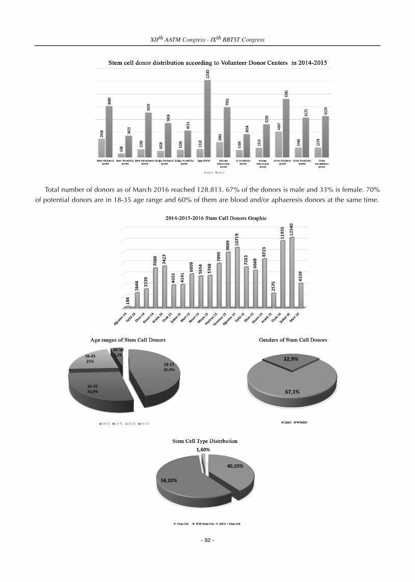
Total number of donors as of March 2016 reached 128.813. 67% of the donors is male and 33% is female. 70%
of potential donors are in 18-35 age range and 60% of them are blood and/or aphaeresis donors at the same time.
XIIth AATM Congress - IXth BBTST Congress
- 92 -

According to scanning studies conducted by Turkish Ministry of Health, 375 matching requests were informed
and 84% have given affirmative feedback. 58 individuals have decided not to be donors for various reasons before
or after matching.
Stem cell collection was made in 49 donors and transplantation to patients was completed. On the other hand,
13 donors are in the stage for planning transplantation.
XIIth AATM Congress - IXth BBTST Congress
- 93 -

- 94 -
When it is considered basically; the most important point of donor acquisition is that “it is located at the junction
of social awareness and personal sensitivity.” It requires a large communication network in its infrastructure. If we
look at the resulting strategy in general, when all points of communication come together; we monitor two different
effects together.
1. Top-down effect. Here advertising, promotion and public relations activities, carried out on a national basis
are highlighted.
2. Bottom-up effect. All points requiring face-to-face contact with people are included in this effect. All
platforms in which closest contact with blood donors is realized, such as a Blood Donation training
provided in an institution or the right information to be given by a phlebotomist when he meets a blood
donor, will create this effect.
When we approach social media in this context, social media takes its place in the steps of both the top-down
effect and the bottom-up effect. Therefore, it plays an important role in the rapid communication strategies. Social
media is the media system which allows the access to double-sided and simultaneous information sharing along
with bringing the Web 2.0 into use, rather than one-way information sharing. Besides, social media is the whole of
dialogues and information sharing realised on the internet by the people. In social networks, thanks to websites and
applications which enable people to share content and information with each other, everyone may have access to
the content that is inquired and interested. Also it allows for the production of contents without the need for different
sources of information. On the other hand, It provides an informal environment for people's education.
What should be the purpose of using social media? Generally speaking, it is evident that two main objectives are
required to achieve the desired point.
1. To give, propagate and sustain correct information. Blood donation activities should be announced. On the
other hand, informing should be carried out in areas on which an awareness raising is desired. Social media
is a non-formal educational tool. Relevant information studies realized especially following an information
request should be delivered to the target audience properly and in a short time by being spread rapidly.
2. To change a negative or incorrect information.
Since the impact from social media is instant by its nature, the main point is to what extent crisis management
is implemented properly. Then, how many types of reaction can be given to an impact coming from any channel?
An idea is developed against the incoming impact. In this case, a risk of increasing the power of knowledge
advocated by the counterparty exists. For example, “I'm not sure about the safety of blood, therefore I do not want
to give blood.” The effect may be grown more if the total opposite idea is told. “No, this is wrong because our system
SOCIAL MEDIA IN DONOR ACQUISITION
Metin KALENDER
is strong enough not to accept such errors.”
Parallel ideas may be developed against the incoming impact. In this case, it can be declared that such a risk
exists although the blood safety is tested in laboratories of international standards. However, in such a case, again
the power of incoming impact will increase. Because social media is a platform whose language is short, but its
effect may be long.
Sub-ideas may be developed against the incoming impact. The subject is moved to a different platform from the
point of incoming impact. “The most important point on blood safety is the window period of infection.” With this
information, scientific data on the safety of blood comes to the fore. Then the subject is treated in this context and
the process is continued. In this case, what we want takes place as expected, the power of incoming impact will
decrease and a different area will be opened for giving right information.
“Social media does not sleep.”
Reaction rate must be strong against the instant and inevitable rate of social media. An information, news or any
positive or negative demand should find instant answers with pre-planned processes. Answers should be quick,
short, simple and meaningful.
When we look at the pros and cons of social media, it can be seen that both of them begin at the same point.
First of all the real intention of the incoming impact should be read correctly. Because the nature of the information
content will determine whether its effects will be positive or negative. The positive side of social media is that
sharing is instant. However, the downside is still the same; instant sharing. For these reasons, the source of the
subject consists of the nature of intention sharing. The important point is the provision of necessary manipulations
which will ensure that the sharing will come to the desired point at the next step.
XIIth AATM Congress - IXth BBTST Congress
- 95 -

- 96 -
Mesenchymal stromal cells (also known as Mesenchymal stem cells or MSCs) were initially identified in cultures
of bone marrow a half century ago. Over the past two decades, methodologies for isolating and growing them in
vitro have been developed. We now know that MSCs are present in many body tissues and organs, have the
common characteristic of being adherent cells with the potential of differentiating into osteocytes, chondrocytes and
adipocytes and relatively unique cell surface characteristics. The bone marrow, adipose tissue and cord blood are
the most commonly used sources of MSCs. MSCs exert their function via multiple mechanisms of action including
the secretion of many bioactive molecules that have anti-inflammatory, tissue regenerative and immunosuppressive
properties. Both academic and industry laboratories have now used clinical grade MSCs for a variety of clinical
conditions including autoimmune diseases, graft-vs-host disease and tissue repair indications for many different
organ systems. There are several hundred phase I and II clinical trials being conducted with MSCs. Results of MSC
use in clinical trials for GVHD and stroke wil be discussed and challenges in bringing these cellular therapies to the
clinic will be discussed.
MESENCHYMAL STROMAL CELLS: ARE THEY READY FORCLINICAL USE? AN UPDATE
Naynesh KAMANI

- 97 -
AABB is an international not-for-profit professional association of over 7,000 individuals and organizations
working in the field of transfusion medicine and cellular therapy. Its mission is focused on patient and donor safety
and health. AABB was the first organization to develop standards for blood transfusion over six decades ago and
started to accredit facilities that collected and transfused these products against these standards. Standards have
now been developed for other therapies such as hematopoietic cell collection, processing, banking, release and
administration and facilities such as public and family cord blood banks and hematopoietic progenitor cell
processing laboratories voluntarily seek accreditation to demonstrate compliance with AABB cellular therapy
standards. The standards are revised on a two year cycle to remain current and compliant with federal and national
regulatory requirements. More recently, novel cell therapy standards have also been developed. In addition to
standard setting and accreditation activities, AABB has many educational offerings including the society journal,
TRANSFUSION, many textbooks in the field of transfusion medicine and cell therapy, audiocasts and webinars. An
on-line, self-paced CT certificate program is under development and will be launched later this year.
CT INITIATIVES AT AABB:STANDARDS, ACCREDITATION AND EDUCATION
Naynesh KAMANI

- 98 -
What is a standard?
A standard is a document that provides requirements, specifications, guidelines or characteristics that can be
used consistently to ensure that materials, products, processes and services are fit for their purpose.
ISO/IEC Guide 2 defines a Standard as a: “document, established by consensus and approved by a recognized
body, that provides, for common and repeated use, rules, guidelines or characteristics for activities or their results,
aimed at the achievement of the optimum degree of order in a given context. Standards should be based on the
consolidated results of science, technology and experience, and aimed at the promotion of optimum community
benefits.”
Standards are published documents setting out specifications and procedures designed to ensure that products,
services and systems are safe, reliable and consistently perform the way they were intended to. They establish a
common language that defines quality and safety criteria. Standards are practical and set achievable goals. They are
based on sound industrial, scientific and consumer experience and are regularly reviewed to ensure that they keep
pace with advances in technology.
Purpose of Standards
Standards are developed for a number of purposes, including: 1. Voluntary or mandatory applications: Standards that specify requirements to achieve at least the minimum
objectives of safety, quality or performance of a product or service.
2. Regulatory compliance: Standards that are used to specify minimum least-cost solutions to technicalrequirements expressing characteristics, performance and design criteria compatible with the function of legislation.
3. Contractual purpose: Standards that serve as purchasing specifications or technical conditions of contractbetween two parties.
4. Guidance: Standards that may be intended for educational purposes and which include recommendations, oradministrative or project management procedures. In general, these Standards will not be adopted in eitherlegislation or contract specifications.
Types of Standards
There are three categories of Standards that are generally recognised by the Standards community: International,Regional and National.
WHAT TYPE OF STANDARDS?NATIONAL OR REGIONAL OR INTERNATIONAL?
Ayla YAVUZ

According to the structure characters According to the application form According to the application area
1.Basic standards Voluntary Interoperability standards
2.Design standards Mandatory Industrial
Matter standards National
Product standards Regional
Test Method standards International
Service standards
Figure.1. Types of Standards
International Standards
International Standards are publicly available Standards that have been developed and approved by an
international SDO. ISO (International Organization for Standardization) and IEC (International Electrotechnical
Commission) are the two main international standardising bodies. Other international bodies include the ITU
(International Telecommunications Union) and the Codex Alimentarius Commission (set up by the Food and
Agriculture Organization of the United Nations and the World Health Organization (WHO) to develop food
Standards, guidelines and related texts such as codes of practice).
International Standards are used directly or are adopted by regional or national Standards bodies. ISO and IEC
encourage national adoption if International Standards. There are also ‘de facto’ international Standards that,
although they have not been developed by an international Standards organisation, have widespread acceptance.
These may also be adopted by international, regional or national Standards bodies.
Regional Standards
Regional Standards are publicly available Standards that have been developed and/or adopted by a regional
SDO; the most well-known of these are those that apply in the European Union (EU). These Standards are developed
by the European Committee for Standardization (CEN) and the European Committee for Electrotechnical
Standardization (CENELEC) and then adopted as national Standards by the member countries of the EU. EU
Standards are sometimes adopted by national Standards
National Standards
National Standards are publicly available Standards that have been developed, approved and/or adopted by a
national Standards body or other accredited organisation.
(AFNOR: Association Française de Normalisation, BSI: British Standards Institution, DIN: Deutsches Institut für
Normung, DS: Dansk Standardiseringsraad, NNI: Nederlands Normalisatie-Instituut, TSE: Turkish Standards
Institution, UNI: Ente Nazionale Italiano di Unificazione, etc.)
Functions of Standards
Product Standards: These specify characteristics (including dimensions), design, construction or composition of
XIIth AATM Congress - IXth BBTST Congress
- 99 -

a product to ensure acceptable performance, reliability, durability, finish, or other characteristics necessary to ensure
the product's suitability for the purpose envisaged by purchasers or users
Examples *Consumer products such as toys, motorcycle helmets, electrical appliances and bottled water
Design Standards: Design Standards are a basic element of nearly all engineering and building projects.
Examples *Steel structures, timber framed buildings, concrete structures, or boilers and other pressure vessels.
Code of Practice: Also called ‘Service Standards’. These Standards specify the practices or procedures for the
design, manufacture, installation, maintenance or utilisation of equipment, structures or products.
Examples *Llaying floor coverings, use of chainsaws, laundry practice, and information security.
Safety Standards: Provide guidance on safety in health, life and property matters.
Examples *Safety in the design, construction and/or operation of plant and equipment (e.g. milling machinery,
wood processing machinery), workplace health and safety (WH&S), and personal safety and health (e.g. swimming
pool fencing and hand operated electric tools).
Test Methods: Set out the steps to be followed to determine the properties of a product or component.
Examples .*Testing of physical properties (e.g. resistance to forces or linear dimensions when considering safety)
where acceptable test results are used in meeting requirements in other Standards,
Management System Standards: Also called ‘Process Standards’.These specify requirements to be fulfilled by
management systems or other processes.
Examples *ISO 9001 (Quality Management Systems) and ISO 14001 (Environmental Management Systems) as
well as other systems in the WH&S, food safety and climate change areas.
There are additional types of Standards including Interoperability Standards (IT), Terminology Standards, and
Standards on data to be provided.
Performance based and prescriptive Standards
Standards are often broadly classified as ‘Performance Based’ or ‘Prescriptive’.
Performance Based Standards have their requirements expressed in terms of performance, i.e. outcomes to be
achieved. This approach leaves freedom for the development of innovative technical methods to meet the
requirements of the Standard.
Prescriptive Standards express requirements in precise, often quantitative, terms. This leaves little opportunity to
depart from the specifications in the Standard.
Each Standard (International, Regional, or National) is uniquely identified by a letter/number combination termed
XIIth AATM Congress - IXth BBTST Congress
- 100 -

the ‘designation’.
• Prefix • Main number • Part numbers. • Interim• Year. • Suffix
Mandatory, Normative İnformative
Three terms that are consistently used with Standards are ‘mandatory’, ‘normative’ and ‘informative’.
Mandatory is a term used to describe a provision of a Standard to which it is necessary to conform in order to
be able to claim compliance with the Standard
Normative is a term used to describe an element of a Standard to which it is necessary to conform in order to be
able to claim compliance with the Standard.
Informative is a term used to describe an element (clause, note or appendix) of a Standard that gives additional
information, recommendations and/or guidelines, i.e. is of a non-mandatory nature.
The use of Shall, Must and Should
The word ‘shall’ is used to state that a requirement is strictly to be followed in order to conform to a Standard.
there can be no deviation from that requirement, other than where there is a specified tolerance.
In legislation and specifications it is common to use the word ‘must’ to express a requirement; however, the use
of ‘must’ is avoided in Standards.
Where Standards are adopted in legislation, the word ‘shall’ in the Standard is to be considered as equivalent to
‘must’ in the legislation.
The word ‘should’ (or ‘may’) introduces a suggestion or recommendation that is not a requirement. It is not
necessary that such recommendations or suggestions be followed in order to comply with the Standard. Similarly,
the antonyms ‘should not’ or ‘may not’ are only suggestions and are not required to be complied with.
Product status
Current, Available Superseded, Obsolescent, Redesignated, Superseded, Withdrawn
Patents
A Standard does not confer a monopoly on one section of industry to the detriment of another that can provide
an equally satisfactory article. Nonetheless, on exceptional occasions, technical reasons may justify inclusion in the
Standard of items or services covered by patent rights. There is no objection in principle to this, provided that certain
rules are adhered to.
International adoption Where an International Standard deals with the subject covered by a new project, such a
Standard is considered and evaluated for adoption as a national Standard by the committee concerned.
XIIth AATM Congress - IXth BBTST Congress
- 101 -

Standards in legislation
Standards are often cited (‘called up’) in State and Commonwealth legislation. When this happens, these
Standards become mandatory and can be subject to the scrutiny of the courts. Therefore, every attempt is made to
ensure Standards are written in a clear and concise manner, avoiding ambiguity and making it totally clear what has
to be done to comply with the Standard (and hence the regulation or contract that calls up the Standard).
STANDARDS FOR BLOOD BANK AND TRANSFUSION SERVICE
Blood transfusion is a multi-step process with risk of error in each process from selecting donors, collecting and
processing donations, testing of donor and patient samples, issue of compatible blood, to transfusing the patient. An
effective quality system provides a framework within which activities are established, performed in a quality-focused
way and continuously monitored to improve outcomes.
Key elements of quality systems include:
➢ Organizational management
➢ Standards
➢ Documentation
➢ Training
➢ Assessment
WHO
Regulatory Standards for Vaccines and Biologicals
The WHO Expert Committee on Biological Standardization is commissioned by WHO to establish detailed
recommendations and guidelines for the manufacturing, licensing, and control of blood products, cell regulators,
vaccines and related in vitro diagnostic tests.
EU
The European regulation on blood and blood components is declined in four directives: the Directive 2002/98/EC
known as "mother Directive" and three directives called "daughter Directives" 2004/33/EC, 2005/61/EC and
2005/62/EC. The primary piece of EU legislation related to blood is Directive 2002/98/EC. It lays down the general
framework for ensuring quality and safety for the collection, testing, processing storage and distribution of blood and
blood components.
AABB
Since 1957, AABB has been developing standards for blood banks and transfusion services. From the 1980s to
the present standards for Cellular Therapies, Perioperative Services, Relationship Testing, Immunohematology
Reference Laboratories, Molecular Testing and Patient Blood Management
Standards for Blood Banks and Transfusion Services, 30th edition
Standards for Cellular Therapy Services, 7th edition
Standards for Immunohematology Reference Laboratories, 9th edition
XIIth AATM Congress - IXth BBTST Congress
- 102 -

Standards for Molecular Testing for Red Cell Platelet and Neutrophil Antigens, 2nd edition
Standards for Perioperative Autologous Blood Collection and Administration, 6th edition
Standards for Relationship Testing Laboratories, 12th edition
ISBT
The International Society Of Blood Transfusion (ISBT) is a scientific society that was founded in 1935. Since that
time the ısbt has grown in to an international society where transfusion medicine professionals from across the globe
come together and do the one thing they do best: share knowledge to improve the safety of blood transfusion
worldwide.
ISBT Workıng Partıes: That focus on the study of specific topics.
(e.g.Workıng party on haemovıgılance)
ISBT 128 is a global standard for the identification, labeling, and information transfer of medical products of
human origin (including blood, cells, tissues, milk, and organ products) across international borders and disparate
health care systems.
ISO
The International Standard Organisation (ISO) 9000 standards are commonly accepted by blood establishments.
ISO 9001:2000 specifies requirements for a quality management system.
The standards given by ISO 9001 specify requirements for quality management systems that can be used for
internal application by organisations, or for certification, or for contractual purposes.
ISO 15189 Medical laboratories: Particular requirements for quality and competence specifies the quality
management system requirements particular to medical laboratories.
PHARMACEUTICAL INSPECTION CO-OPERATION SCHEME
PIC/S GMP Guıde For Blood Establishments
XIIth AATM Congress - IXth BBTST Congress
- 103 -
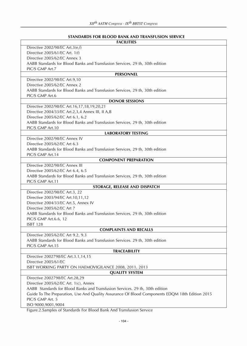
STANDARDS FOR BLOOD BANK AND TRANSFUSION SERVICEFACILITIES
Directive 2002/98/EC Art.3(e,f)Directive 2005/61/EC Art. 1(f)Directive 2005/62/EC Annex 3AABB Standards for Blood Banks and Transfusion Services, 29 th, 30th editionPIC/S GMP Art.7
PERSONNELDirective 2002/98/EC Art.9,10Directive 2005/62/EC Annex 2AABB Standards for Blood Banks and Transfusion Services, 29 th, 30th editionPIC/S GMP Art.6
DONOR SESSIONSDirective 2002/98/EC Art.16,17,18,19,20,21Directive 2004/33/EC Art.2,3,4 Annex III, II A,BDirective 2005/62/EC Art 6.1, 6.2AABB Standards for Blood Banks and Transfusion Services, 29 th, 30th editionPIC/S GMP Art.10
LABORATORY TESTINGDirective 2002/98/EC Annex IVDirective 2005/62/EC Art 6.3AABB Standards for Blood Banks and Transfusion Services, 29 th, 30th editionPIC/S GMP Art.14
COMPONENT PREPARATIONDirective 2002/98/EC Annex IIIDirective 2005/62/EC Art 6.4, 6.5AABB Standards for Blood Banks and Transfusion Services, 29 th, 30th editionPIC/S GMP Art.11
STORAGE, RELEASE AND DISPATCHDirective 2002/98/EC Art.3, 22Directive 2003/94/EC Art.10,11,12Directive 2004/33/EC Art.5, Annex IVDirective 2005/62/EC Art 7AABB Standards for Blood Banks and Transfusion Services, 29 th, 30th editionPIC/S GMP Art.6.6, 12ISBT 128
COMPLAINTS AND RECALLSDirective 2005/62/EC Art 9.2, 9.3AABB Standards for Blood Banks and Transfusion Services. 29 th, 30th editionPIC/S GMP Art.15
TRACEABILITYDirective 2002798/EC Art.3.1,14,15Directive 2005/61/EC ISBT WORKING PARTY ON HAEMOVIGILANCE 2008, 2011, 2013
QUALITY SYSTEMDirective 2002798/EC Art.28,29Directive 2005/62/EC Art. 1(c), AnnexAABB Standards for Blood Banks and Transfusion Services. 29 th, 30th editionGuide To The Preparation, Use And Quality Assurance Of Blood Components EDQM 18th Edition 2015PIC/S GMP Art. 5ISO 9000,9001,9004Figure.2.Samples of Standards For Blood Bank And Transfusıon Servıce
XIIth AATM Congress - IXth BBTST Congress
- 104 -

WHAT TYPE OF STANDARDS? NATIONAL OR REGIONAL OR INTERNATIONAL?
A national regulatory system may legally require all similar organizations to meet certain standards
Keeping in mind the guiding principle of consistency, the manual on ‘National Standards
for Blood Transfusion Service’ has been prepared on the basis of Good Laboratory Practice
and Good Manufacturing Practice, with the objective of ensuring quality and safety of
blood and blood products in the face of known and emerging threats to public health in
the country.
The existence of any relevant national legislation or regulations must be acknowledged and incorporated into the
framework for quality.
WHO Global Database on Blood Safety Summary Report 2011
WHO recommends that every country should put in place policies, systems and structures to ensure the safety,
quality, accessibility and timely availability of blood and blood products to meet the needs of all patients who
require transfusion. Support countries in developing and/or strengthening efficient and sustainable national blood
systems with national blood policies/plans, strategies, infrastructure, organization and management structure and
regulatory mechanisms, integrated within the national health care system.
Develop norms, standards, best practice guidelines, tools and materials on various steps of the blood transfusion
process from donor to patient to ensure blood safety.
In parallel, increased travel and improved communications will lead to a converging of global regulation and
standards.
References1. http://www.who.int2. http://ec.europa.eu3. http://www.isbtweb.org4. www.aabb.org5. Manual Common European Standards and Criteria for the Inspection of Blood Establishments Edition 1.0,
20106. Standards And Other Publıcatıons Standardısatıon Guıde 003 Standards Australia7. http://www.picscheme.org8. PIC/S GMP Guıde For Blood Establıshments Pharmaceutıcal Inspectıon Conventıon Pharmaceutıcal
Inspectıon Co-Operatıon Scheme Pe 005-3 25 September 2007 Pe 005-3 25 September 20079. Standard for Surveillance of Complications Related to Blood Donation, The International Haemovigilance
Network The AABB Donor Haemovigilance Working Group December 11, 2014
XIIth AATM Congress - IXth BBTST Congress
- 105 -
- 106 -
As it is stated in World Health Organization (WHO) documents, quality systems are the “key for ensuring the
availability of safe blood for all patients needing transfusion”(1). WHO defines quality as safety and availability of
blood. In blood banking and transfusion medicine quality and safety should be considered together.
The direct relationship between quality and safety in transfusion services has been shown in a number of studies.
In a study blood availability for elective surgery over 3 audited intervals was investigated and the incidence of
patients undergoing elective surgery without available crossmatched blood that had been requested was 1:333,
1:328, and 1:225 for pre-quality improvement, post-quality improvement, and subsequent postintervention audit
assessments, respectively. The relationship between operations, quality management, and risk management is
demonstrable in the transfusion services(2).
Blood is a medicinal product obtained from a biological starting material and blood banking today is composed
of a chain of processes. Quality of blood and blood components has risks in terms of collection, manufacturing and
throughout the life cycle of the component, as well as the transfusion. It is critical to maintain the quality of blood
and blood components starting from the initial steps of manufacturing as early as the selection of donors until the
final use of the component for the patient who is expected to benefit from it as it is stated in clinical studies.
Risk management has long been an integral part of quality systems. Quality risk management is defined as “a
systematic process for the assessment, control, communication and review of risks to the quality of a product across
its lifecycle. It can be applied both proactively and retrospectively(3).
Quality risk management requires a systematic approach. It is an obligation to comply with the requirements of
good manufacturing practice (GMP) and other quality systems like ISO(4). A quality system framework should have
a formal quality risk management approach with processes, methods, responsibilities and tools defined and in place.
An effective quality risk management is a proactive process that defines potential risks starting from the very first
process until the very last in development, manufacturing and use of the blood products. It enables the establishment
to improve decision making abilities when quality problems arise.
Risk is defined as the combination of the probability of occurance of harm and the severity of that harm. The
evaluation of the probability and the severity of harm show great variance. Perception of risk differs from one to
another and it is affected by public and political opinions (5). The impact of the loss of public confidence on safety
of blood would be dramatic. Thus the global safety of the blood supply system should be carefully monitored and
the public should be informed on the level of safety very carefully. In relation to blood banking and transfusion
medicine; patients, medical practitioners, government and manufacturers are the primary steakholders and it is the
safety of patient that is most important (3). The principles and the tools of quality risk management enables the
regulators and the manufacturers to make risk based decisions that are more effective and consistent.
QUALITY RISK MANAGEMENT IN BLOOD BANKING ANDTRANSFUSION MEDICINE
Ayse Esra KARAKOÇ

Harm is damage to the health of the recipient and it includes the damage arising from the loss of quality or the
availability of the component. Hazard is the potential source of harm. In order to manage risk, it is necessary to
define it first. Risk assessments reflect the assessment of hazards which represent a significant risk to the staff, to the
blood donor and to the patients receiving a blood or blood component transfusion. All of the people who could be
harmed by the hazard need to be considered; in transfusion medicine it will usually be the receiver of the
transfusion, the patient. Risk assessment includes risk identification, risk analysis and risk evaluation.
Two main principles of risk management are: 1. The methods used to evaluate the risk to quality should be based
on scientific knowledge and should be linked to patient safety and 2. The efforts, the procedures, the action plan to
protect from the defined risk should be relevant with the degree of the risk.
Each hazard should be assessed against a risk matrix for the severity and probability rating. Severity is rated from
1:negligable, 2:minor, 3:moderate, 4:serious, to 5:catastrophic and probability is rated from 1:rare, 2:unlikely,
3:possible, 4:likely, to 5:almost certain. Risk is rated with the risk matrix by multiplying the rates of severity and
probability and the rates of risks are graded as high, medium and low. According to this, control measures for each
hazard have to be identified and recorded. Further assessment is required if existing measures are not adequate to
control the risk with action plan of how to reduce or eliminate the risk. The risks have to be placed on a risk register
and should be recorded. The risks have to be reviewed on a regular basis(6). In Figure 1 and 2 below the sequence
and the interrelations and the overview of the processes in quality risk management are shown respectively.
Figure 1. The sequence and interrelations of quality risk management process (3)
XIIth AATM Congress - IXth BBTST Congress
- 107 -

Figure 2. An overview of risk management process (7)
The responsibilities are stated to be interdisciplinary in quality risk management. Quality unit, engineering,
business developers, regulators, operational, clinical and legal people are stated to be involved(3).
Concerning the blood banking and transfusion medicine, the establishments should learn from adverse events.
Errors, noncompliance to certain procedures, near accidents (identification errors, sample transpositions etc),
accidents give information to the blood bank on the insufficiency of the ongoing operations. Adverse events tell what
had happened. To analyse why it had happened helps to make the root cause analysis. The risk management efforts
can be directed to one or two most frequently occuring root causes.
To understand and to apply quality risk management is accepted as an essential requirement of 21st century
pharmaceutical industry that involves blood and blood products as well. It allows the processes and the equipment
to be designed more efficient and safer for the users, donors and patients(8).
References1. http://www.who.int/bloodsafety/quality/en/2. Goodnough LT. Operational, quality and risk management in the transfusion service: lessons learnt. Transfus
med rev 2012; 26(3): 252-61 3. European Commission, Enterprise and Industry Directorate General. EudraLex. The rules governing
medicinal products in the European Union. Volume 4. EU Guidelines to good manufacturing practicemedicinal products for human and veterinary use. Annex 20. Quality risk management. Brussels:2008
4. Technical Committee ISO/TC 212. The international organisation for standardization. ISO 15189:2012Medical laboratories-requirements for quality and competence, 2012
5. Smit Sibinga CT. Risk management: an important tool for improving quality. Transfus Clin Biol 2001;8(3):214-7
6. NHS. Risk assessment document for National Patient Safety Agency, safer practice notice 14,recommendations of York hospital transfusion committee. April 2007
7. ISO 14971-2007 Medical devices-application of risk management to medical devices8. Saghee MR, Sandle T. Embracing quality risk management: the new paradigm. Express pharma, 16-30
September 2010
XIIth AATM Congress - IXth BBTST Congress
- 108 -

- 109 -
Nurses and technicians have an essential role in Blood Banking & Transfusion Medicine (BBTM). Discussions on
education of this group has been going on decades and it will never be ended because of the progresses and changes
in the field of BBTM.
This group may have a role at 2 different parts of BBTM;
1) Blood banks or centers where blood is collected and prepared for transfusion
2) Hospitals or treatment centers where blood is transfused
Education of this group can be classified as;
1) Pre-graduate education
2) Post graduate education
2.a. Post graduate academic education
2.b. Post graduate continuous education
1) Pre-graduate education:
Pre-graduate education curriculum is the basic teaching of this group and there is still no common consensus on
it. There are numerous education model offers on this subject issued by different national and international bodies.
The most concrete and widely accepted one is “Recommendation Rec (2004)18 of the Committee of Ministers to
member states on teaching transfusion medicine to nurses” issued by Committee of Ministers on 15 December
2004 at the 909th meeting of the Ministers' Deputies).
One of the most recent activity on this topic was Anatolian Blood Days – II (ABD); an international workshop
organized by Turkish Blood Foundation (TBF) at November 25 – 27 2013 at Antalya / Turkey. During the meeting 30
transfusion professionals representing 17 countries gave presentations on their own services. They presented
evidence of inadequate training of nursing personnel in clinical transfusion that confirmed the need for work on this
subject. The participants in the meeting took into consideration the Rec (2004)18 of the Council of Europe on
teaching transfusion medicine to nurses.
This text has based on those above mentioned sources which are more in details and recently performed
depending on survey results and clear evidences.
After a plenary discussion at Anatolian Blood Days – II; the participants formed three working groups. Based on
the reports presented by each working group a draft consensus statement was prepared and discussed. The final
statement agreed by the participants is shown below.
1 The curriculum for education and training of nurses in the topic of clinical transfusion.
EDUCATION FOR NURSES & TECHNICIANS
N. Nuri SOLAZ

• Should be clearly defined and the method of delivery should be practically achievable.
• The latter requirement is especially important for in service training
• The core curriculum for nurses in training and for in-service nurses should be similar
• In service training should give special emphasis to practical procedures that affect patient safety.
• The pregraduate core curriculum should preferably be covered as a single block rather than distributed
among different parts of the course.
2 The core curriculum should cover the following topics
• Responsibilities of the nurse in clinical transfusion.
• Knowledge of regulations and guidelines on blood transfusion
• Compatibility of the blood component with the patient: ABO types, hemolytic transfusion reactions and
prevention. Prevention of Rh immunization
• Description of the main blood components, main indications, storage handling and administration
• Complications of blood transfusion
• Description of the clinical transfusion process
o Informed consent
o Request form, sampling and patient identification
o Receipt and visual inspection of the component unit
o Pre-transfusion identity checks to ensure that the blood component is the one intended for the patient
o Baseline observation of vital signs
o Administration of the blood component
o Monitoring vital signs
o Recognition of signs of acute transfusion reactions and initial management
o Finalization of transfusion (completion of transfusion record, discard of blood pack and giving set).
o Ensuring traceability of each unit transfused by completing the required documentation
3 With respect to the dedicated nurses in clinical transfusion, it was agreed that role requires a person with good
clinical experience and skills, preferably including experience in a clinical specialty in which transfusion is used.
This position might be named as “Transfusion Control Nurse” like “Infection Control Nurse”. The role of Transfusion
Nurse involves coordination, communication and the promotion of change and quality improvement, so a
successful appointee will have a good aptitude for management. Specific tasks of Transfusion Nurse are listed below;
• Leading the implementation of training in clinical transfusion
• Co coordinating Haemovigiliance reporting
• Promoting best clinical transfusion practice
• Co coordinating a program of transfusion audits
• Providing progress reports to the Hospital Transfusion Committee (HTC)
• Participating as a full member of the HTC
Structure of the training program such as total hours, grades, etc. may vary but dedicating necessary minimum
hours and combining education hours not more than 2 different grades are highly recommended.
2) Post graduate education
2.a. Post graduate academic education; can be done by academic institutions such as master or PhD programs.
2.b. Post graduate continuous education; might be organized by scientific societies and locally by academic or
national health system institutions. Selection criteria, location of training, selection procedure, and conditions of
XIIth AATM Congress - IXth BBTST Congress
- 110 -
service of nurses undergoing training, and the structure of the training program (hours, topics, diploma etc) all vary.
This education is widely used for the nurses who are going to work blood supplying institutions and transfusion
centers of the hospitals or clinics.
Regarding demands of modern medical treatments and technologies it will be wise to give a special postgraduate
basic / continuous education to the nurses who are dealing in clinical transfusion. This training might be a one-day
training with the below listed topics;
a) Actual legal and administrative conditions on BBTM
b) Blood components; preparation, storage, transportation, volume, etc.
c) Bedside transfusion safety
d) Transfusion complications
e) Haemovigiliance
Conclusion
To increase efficiency in blood transfusion medicine, physicians and nurses and other health workers who handle
blood or blood components should collaborate on development, evaluation and implementation. Documentation
regarding transfusions needs to be simplified and coordinated. Knowledgeable staff is an essential element of safe
systems.
Basic knowledge should never be assumed: mechanisms to monitor knowledge of key processes along with
ongoing feedback and remedial action are necessary to maximize performance. Nurse training curricula and formats
at all levels must reflect the requirements of modern transfusion medicine from novice to expert. Monitoring and
evaluation of the training activities are essential which should not be ignored
The Council of Europe’s Recommendation could contribute to the adoption and implementation of training
programs at a national level.
References1. Recommendation Rec (2004)18 of the Committee of Ministers to member states on teaching transfusion
medicine to nurses2. Final declaration of Second Anatolian Blood Days, November 25 -27 2013 convened by Turkish Blood
Foundation at Antalya / Turkey
XIIth AATM Congress - IXth BBTST Congress
- 111 -

- 112 -
Introduction
Blood transfusion saves lives and improves health, but many patients requiring transfusion do not have timely
access to safe blood. Blood transfusion is associated with both beneficial effects and adverse events in recipients.
The use of blood for transfusion has evolved in most parts of the world from the transfusion of whole blood to the
use of individual components: packed RBCs, platelet concentrates, and plasma. This approach allows the transfusion
of only those parts of the whole blood that the patient requires and facilitates the removal of unwanted elements
such as leukocytes. There have been six major paradigmatic shifts associated with transfusion medicine science and
technology-
Early 1900 - Karl Landsteiner discovers the ABO blood groups and humoral immunity; the main paradigm
explaining the effects of allogenic transfusion.
1940 - Edwin Cohn develops cold ethanol fractionation, the process of breaking down plasma into protein
components and products
1950 - Carl Walter and W.P. Murphy introduce the plastic bag for blood collection and component preparation.
1960 to 1980 - Development and implementation of effective screening methods to reduce pathogen
transmission.
1990 - Leukocyte reduction of blood components shown to be associated with improved clinical outcome, which
led to the implementation of universal leucoreduction (ULR) in many countries.
1995 to 2010 - Development of pathogen inactivation (reduction) technology to reduce the risk of pathogen
transmission by blood components.
Blood transfusion services play a central role in health systems by providing safe and adequate supplies of blood
and blood products for patients requiring transfusion. Blood products are defined as any therapeutic substances
derived from human blood, including whole blood, labile blood components (i.e. red blood cells, platelets and
plasma) and blood- or plasma-derived medicinal products (PDMPs) e.g. albumin, polyvalent and specific
immunoglobulin and blood clotting factors that are prepared in fractionation facilities from pools of thousands of
plasma units.
Availability and safety of blood and blood products remains a major concern in many countries around the
world. According to the World Health Organization (WHO), 1-3% of the world's population needs to be blood
COMPONENT PRODUCTION: HOW MANY, WHEN AND WHY
Sadhana MANGWANA

donors to maintain adequate blood supply. WHO data reveals that worldwide about 108 million blood donations
are collected every year. More than half of these are collected in high-income countries, home to 18% of the world’s
population. There is a marked difference in the level of access to blood between low- and high-income countries.
The median blood donation rate in high-income countries is 36.8 donations per 1000 population. This compares
with 11.7 donations in middle-income countries and 3.9 donations in low-income countries.
Component Production
Blood components can either be produced from a manually collected unit of whole blood, or they can be
collected as specific products using automated collection devices. In the manually collected units, there are options
for the strategies to choose for the post-donation processing of the whole blood.
i. Apheresis
Apheresis is the withdrawal of blood from a donor, removal of one or more blood components, and transfusion
of the remaining blood back into the donor. There are some distinct advantages of apheresis. It allows the collection
of specific products, and thus may help to better match inventory supply and demand with a concomitant reduction
in wastage due to outdating. Collection of plasma for transfusion or fractionation is greatly facilitated by apheresis
as the lack of red cell loss means the donation interval can be much shorter than whole blood donation. It is also
an important technology to produce specialty blood products that would be very difficult to obtain otherwise e.g.
Platelet- specific antigen matched and HLA-matched platelet products, as apheresis permits the collection of a
transfusion dose from a single donor. Blood components harvested, through Apheresis technology, are generally 3-
4 log leukoreduced, and provide better therapeutic benefits than the random donor products. More recently,
apheresis equipment is increasingly adopted for the collection of two units of red cells in a single procedure but
there are challenges for iron management in donors.
ii Whole blood donation
Blood collected in an anticoagulant can be stored and transfused to a patient in an unmodified state i.e. ‘whole
blood’ transfusion. In developing countries, or for military use, whole blood remains an important resource.
However, blood can be used more effectively if it is processed into components, such as red cell concentrates,
platelet concentrates, plasma and cryoprecipitate to meet the needs of more than one patient.
There are two main methods for the production of red cell concentrates, platelet concentrates, and transfusion
plasma. The first method, called the platelet rich plasma (PRP) method, in which two fractions are separated first, a
red cell concentrate and a mix of the platelets and plasma, then following a second centrifugation, separates the
plasma from the concentrated platelets. In second method, the buffy coat method, all the three elements are
separated in the first step: red cell concentrate, plasma, and the buffy coat. For platelet production, the buffy coats
of 4- 8 units are pooled, centrifuged and a platelet rich plasma is removed.
The main components are –
• Packed Red Blood Cells (PRBC)
• Platelet concentrates or Random Donor Platelets (RDP)
• FFP
• Cryoprecipitate
• Cryo- poor Plasma (CPP)
XIIth AATM Congress - IXth BBTST Congress
- 113 -

• Plasma-derived medicinal products (PDMPs) i.e. Albumin, Polyvalent and specific Immunoglobulin and
Blood clotting factors.
After blood collection, PRBC from whole blood units by buffy coat method are prepared and refrigerated within
8 hours of collection. Buffy coat method outweighs the benefits compared to the PRP method with lower overall
leukoreduction, increased platelet yield and reduced bacterial contamination rates. Buffy coat production of
platelets also positions the blood centre to use platelet additive solutions to reduce recipient reactions or pathogen
reduction technology, neither of which can readily be applied to PRP platelets.
In high-income countries (HIC), each unit of whole blood collected is separated into several therapeutic blood
components to both deliver the most effective treatment (component therapy) and to make most efficient use of
human resource. Access to safe and effective blood products is a major challenge in low and middle income
countries (LMIC) where local blood establishments may have very basic facilities and systems, where quality and
safety standards need to be established or strengthened, and where blood supplies may be insufficient to meet
medical needs. 95% of the blood collected in high-income countries is separated into components, 80% in middle-
income countries and 45% in low-income countries due to limited capacity to provide patients with the different
blood component.
Leucoreduction of blood Components
Amongst various strides in sample collection, processing and storage technologies, a comparatively recent
innovation is introduction of leucoreduction strategies. Freshly collected, whole blood unit contains roughly 109
leukocytes. Leukocyte reduction is defined as a blood processing step for reducing the leukocyte content of whole
blood, RBCs, or platelet units less than 5×10⁶ (3 log reduction 99.9%) with a minimum of 85 % red cell recovery
in 95 % of the units tested, as per the standards of the American Association of Blood Banks while European Council
guidelines require it to be less than 1×10⁶ residual WBCs per unit of component. Leucocytes reduction can be done
by cellulose acetate filters, red cell washing, centrifugation and buffy coat removal, freezing and deglycerolization
of red cells, and blood component collection through apheresis technology. Leucoreduction by leucofilters (third
generation) and components collected through apheresis devices meet the standards of leukocyte depletion, i.e. <
5 × 10⁶ WBC/unit of blood component while other methods achieve variable leukocyte depletion which is as
follows-
• Centrifugation and buffy coat removal — 108 WBCs (1 log leucodepletion)
• Washed red cell concentrate — 107 WBCs (1-2 log leucodepletion)
• Frozen deglycerolized red cells — 106-107 (2-3 log leucodepletion
Current generation of leukocyte filters (third and fourth) have leukocyte removal efficiency up to 99.99% as
compared to the first and second generation filters (90-96%).
Leukocyte reduction procedures can be done either pre-storage, at the time of collection or processing, post
processing (within the blood bank), or by the side of the patient (post storage) just prior to transfusion. Pre-storage
leukocyte reduction of cellular blood components removes donor WBCs before they undergo apoptosis or necrosis
and before they release breakdown products or cytokines. Clinical benefits of leucoreduction has been subdivided
on the basis whether benefit has been proven by evidenced based guidelines to be relevant clinically, likely relevant
clinically or clinical relevance is unproven.
XIIth AATM Congress - IXth BBTST Congress
- 114 -

Proven relevant clinically:
• Reduced frequency and severity of NHFTRs;
• Reduced risk of CMV transmission;
• Reduced risk of HLA-alloimmunization and platelet refractoriness.
• Reduced mortality and organ dysfunction in cardiovascular surgery patients.
Likely clinically relevant:
• Reduced infectious risk associated with immunomodulation (TRIM);
• Reduced direct risk of transfusion-transmission bacteria.
Unproven clinically:
• Avoidance of vCJD transmission.
• Avoidance of HTLV I/II, EBV etc.
• Reduced risk of GVHD.
• Reduced risk of TRALI.
Universal Leukocyte Reduction Strategies
There has been considerable debate on whether all cellular blood components should undergo leucoreduction
or selective leucoreduction protocol should be followed.
Selective pre-storage leucofiltration - The packed red cells are selectively leukofiltered prior to storage for
patients on regular transfusion therapy such as, thalassemia major patients, immunocompromised or chronically
transfused with skillful inventory management and active coordination between the transfusion physicians and the
blood bank.
Universal leukocyte reduction (ULR) is the routine application of the blood-processing step for reducing
leukocyte content to < 5 x 10⁶ WBC per unit to all units of whole blood, RBCs, and platelets prior to storage in a
country or in a blood transfusion service. Internationally, 19 countries namely France, Canada, Luxembourg, Austria,
Eire (Southern Ireland), Wales, Scotland, Switzerland, England, Northern Ireland, Malta, Spain (Portugal), New
Zealand, United Arab Emirates, Germany, Qatar, Holland, Norway and Finland have implemented ULR as part of
their blood safety policy. Several other countries are also currently moving toward implementing ULR, including
Sweden, Denmark, Italy, Belgium, Cyprus and Japan.
The adoption of ULR in the United States has been delayed primarily because of economic issues, leading to the
approach of selective leukocyte reduction protocols for patients thought to be at greatest risk for adverse effects
associated with the presence of leukocytes in blood components.
Plasma-derived medicinal products (PDMP)
It is the responsibility of individual governments to ensure sufficient and equitable supply of plasma-derived
medicinal products namely immunoglobulins and coagulation factors, which are needed to prevent and treat a
variety of serious conditions that occur worldwide. According to WHO, 43 countries (23 high-income, 18 middle-
XIIth AATM Congress - IXth BBTST Congress
- 115 -

income, 2 low-income) of the 156 reporting countries, reported producing all or part of the PDMP through the
fractionation (e.g. domestic or/and contract fractionation) of plasma collected in the country. 35 of the 43 countries
report plasma fractionation carried out within the country. 8 of the 43 countries report plasma sent for contract
fractionation in another country. Around 10 million litres plasma from 35 reporting countries (22 high-income
countries, 12 middle-income countries and 1 low-income country, covering a population of 2.76 billion) was
fractionated for the production of PDMP during the year. This includes around 50% plasma recovered from the
whole blood donations. World Health Assembly resolution (WHA63.12) has urged Member States to establish,
implement and support nationally-coordinated, efficiently-managed and sustainable blood and plasma programmes
according to the availability of resources, with the aim of achieving self-sufficiency.
International challenges of self-sufficiency in blood and blood products
• Lack of clear national policy and government commitment
• Increasing needs and demand
• Inadequate supply of blood and blood products
• Decreasing donor base
• Failure to identify blood as national resource
• Dependence on family/replacement donations
• Risks to donors’ health and safety
• Risk of transfusion-transmissible infections
• Limited capacity in production and processing
• Poor quality systems in developing countries
• Wastage of blood
• Wastage of plasma
• Inappropriate use of blood and blood products
• Influence of market force
Conclusion
Universal leucoreduction seems to be justified but in view of evidence and cost involved in universal
leucoreduction, it is not practically feasible to implement this policy especially in developing countries and under-
resourced countries. It is the commitment by national governments to be self-sufficient in safe blood and blood
products and a coordinated and integrated approach to policy development. Implementation of a policy for self-
sufficiency in blood and blood products generally follows a stepwise progression in scope, from whole blood
transfusions towards blood components for transfusion and further towards plasma fractionation, aligned to the state
of development of the national health system.
References1. Dhingra N. International challenges of self-sufficiency in blood products.Transfus Clin Biol. 2013 May ;
20(2):148-52.2. Blood safety and availability Fact sheet No. 279. Geneva: World Health Organization; June 2015.
http://www.who.int/mediacentre/factsheets/fs279/en/ index.html.3. World Health Assembly documentation. A63/20. Availability, safety and quality of blood products: report by
the Secretariat. Geneva: World Health Organization; 2011. http://apps.who.int/gb/ebwha/pdffiles/WHA63/A63_20-en.pdf
4. Devine DV, Serrano K. Preparation of blood products for transfusion: is there a best method? Biologicals.
XIIth AATM Congress - IXth BBTST Congress
- 116 -
2012 May;40(3):187-90.5. Bassuni WY, Blajchman MA, Al-Moshary MA. Why implement universal leukoreduction? Hematol Oncol
Stem Cell Ther. 2008 Apr-Jun;1(2):106-23.6. Sharma RR, Marwaha N. Leukoreduced blood components: Advantages and strategies for its implementation
in developing countries. Asian J Transfus Sci. 2010 Jan; 4(1): 3–8. 7. Bianchi M, Vaglio S, Pupella S, et al. Leucoreduction of blood components: an effective way to increase
blood safety? Blood Transfus 2015 Dec; 16:1-14.8. D'alessandro A. Leucoreduction of blood components: clinical and molecular evidence. Blood Transfus.
2015 Dec 16:1-2.
XIIth AATM Congress - IXth BBTST Congress
- 117 -

- 118 -
In the last 10 years plasma fractionation industry has undergone many changes from the point of view of -
plasma supply, safety profile, public health policies, new products, use of products for newer indications, industry
collaboration, regulatory reforms, technological development, improved rate of diagnosis, development of
recombinant plasma proteins and patients advocacy. The changes happened in the last one decade have brought
a significant improvement in the life of the patients, who are dependent on plasma protein therapy.
In 2005, worldwide about 22 million liters (7.69 recovered and 14.22 apheresis) of plasma was fractionated
while in 2012 it has increased to about 37 million liters (8.93 recovered and 27.85 apheresis) (1).
It clearly indicates an upward trend of apheresis plasma while recovered plasma collection has remained fairy
unchanged. Because of advancement and improvisation in clinical and surgical practices, usages of blood and its
component have gone down in many of the developing countries, which is reflected in the trend of availability of
recovered plasma. This also implies that a large number of patients, who use plasma protein therapy, are dependent
LAST TEN YEARS IN PLASMA FRACTIONATION
Ranjeet Singh AJMANI

on products coming from source plasma. About 60 % of apheresis plasma is collected in US while majority of
recovered plasma is collected in European region. Most of the Asian and Latin American countries do not have a
robust source plasma program and therefore are dependent on recovered plasma for production of plasma proteins
either indigenously or by toll fractionation. In 2005 the overall plasma product market was about $ 7 billion which
grew up to about $ 27 billion by 2014. Although many plasma fractionation plants have closed their operations or
shut down because of various reasons during this time, however there is an overall significant rise in global plasma
fractionation capacity. Mainly because of optimization of operational cost and other economic/business reasons
there has been a trend of setting up relatively large plasma fractionation units, in the last one decade. However this
increase in capacity has not changed the skewed pattern of availability of plasma products to the patients living in
developed and developing economies, it is because of the fact that most of fractionation capacity has either
increased in developed countries or China, which has very strong regulatory restriction on movement of plasma
products as well as it does not allow contract plasma fractionation. It has been reported that about 60 % of global
population living in developing countries have access to only 15 % plasma products (1).
Asia collects very limited quantity of plasma in proportion to its population and demand and therefore it remains
largely import dependent. This situation puts a large number of patients, with rare genetic disorders, living in
developing world at a very vulnerable position as the availability of products is dependent on external forces like
availability, regulatory restrictions and fluctuation of currency etc. Because of this, a large percentage of patients
living in developing countries remain un/under-diagnosed and face the challenges of consistent availability and
affordability of safe plasma products. It has been reported that about millions of liter of plasma is wasted globally
for variety of reasons like viral safety, quality of plasma, regulatory restrictions across the countries for contract
fractionation (2). Many lives can be saved if this plasma can be made available for fractionation and by improving
XIIth AATM Congress - IXth BBTST Congress
- 119 -

the quality of plasma and harmonization of regulatory system across the countries for contract fractionation.
In the last one decade various patient groups have played a very significant and active role in initiating a
dialogue with government /policy makers in advocating the rights of patients, which has helped early and improved
diagnosis, formation of national registries/chapters of various patient groups and allocation of more funds for plasma
products. Various plasma fractionating companies came forward and extended support to hemophiliac patients by
converting unutilized excess cryoprecipitate to manufacture factor concentrate and supply it to the patients of
developing world. This humanitarian effort was possible because of closed coordinated efforts between patient
organizations and the industry. The rate of diagnosis of Primary Immune Deficiency (PID) has improved in the last
one decade and this has lead to significant increase in the usages of Immunoglobulin (IVIG) across the world. Use
of IVIG for newer indications has also contributed in increased demand of IVIG. In the last one decade demand of
Albumin has increased very significantly and in many countries are facing acute shortage of its supply. Demand of
hyper immune proteins likes Anti-D, Tetanus, Rabies, CMV and Hepatitis B is still there however some of the hyper
immune proteins like mumps, pertussis, tick-borne encephalitis have become obsolete.
In the last one decade there were many viral outbreaks e.g. H1N1, Dengue, SARS, Bird Flue, Zika etc. in the
different parts of the world and this situation did pose a potential threat of viral transmission by plasma products
however there has not been any reported case of infection through plasma products. However outbreaks like these
kept industry on high alert and work towards better/newer viral inactivation protocols. These outbreaks have
encouraged industry to explore the possibility of manufacturing specific Immunoglobulin to treat these infections.
Considering criticality of the situation, World Health Organization also came with guidelines for empirical usages
of convalescent whole blood or plasma for treating the patients.
On the technology front, though Cohn Chromatography remains most widely used technology platform across
the globe but few newer options/concepts have also been introduced in this field claiming a significant increase in
yield, better product quality, operational ease, relatively less investment with an option of tailor made plants. Some
of these options are cascade technology, charged based separation etc. The products from these technology options
are yet to undergo clinical evaluation and regulatory approval. Introduction of newer technology platforms may
change the face of industry in coming years. Industry is also witnessing a trend of merger, acquisition and
consolidation.
In the last one decade organizations like Plasma Protein Therapeutic Association (PPTA), International Plasma
Fractionation Association (IPFA), World Federation of Hemophilia (WFH), World Health Organization (WHO),
International Patient Organization for Primary Immunodeficiencies ( IPOPI) etc. have contributed very significantly
in development and growth of this industry and patient care by setting up stage for open dialogue and discussion.
Social media has changed the dynamics of our lives in the last one decade. It has played a very pivotal role in
connecting various patient/patient groups who were isolated and fighting their battle more at individual/local level,
to share their concern/vision/challenges and have a collective & common voice, which has more impact on society
at large. Now these groups are able to share their resources and thoughts very freely and conveniently, which is
helping them to unite and fight for a common cause. Many books and publications, got more visibility in the last
decade and have increased level of awareness amongst patients, regulators, clinicians, corporate sector about
various aspects of plasma industry.
A lot needs to be done on the issue of self-sufficiency of plasma products country wise/region wise, as the disease
pattern, usages of plasma products, rate of diagnosis, public health policies are different in different parts of the
world (3). Although the last 10 years have been very significant for plasma fractionation industry but coming 10
XIIth AATM Congress - IXth BBTST Congress
- 120 -

years will still be more important with hope that patients across the globe would have better quality of life.
References1. The Worldwide Plasma Protein Market-2012. The Marketing Research Bureau, Inc. USA. 2. Thierry Burnouf, Jerard Seghatchian. Go no Go” in plasma fractionation in the world’s emerging
economies: Still a question asked 70 years after the COHN process was developed! Transfusion andApheresis Science 51 (2014) 113–119.
3. Ranjeet S. Ajmani. Global Perspective : Where is the Asian plasma therapeutics industry heading ?.Bioplasma-World Asia Conference: Hong Kong 2012.
XIIth AATM Congress - IXth BBTST Congress
- 121 -

- 122 -
TURKEY; Geography, Land, Population, Income
Turkey is a country in between Balkan Peninsula, Caucasians, North Africa and Middle East. Land area is 780.000
km2. 2016 population is 79 million, annual income was approximately 10.400 USD/pc in 2014.1
BLOOD SERVICES; Organisational Chart, Current Dual Structure, Progress
National Health Authority (Ministry of Health - MoH) legislate, licence and control all bodies and activities.
Leaded by MoH, Supreme Board of Blood prepares “The National Guide for Blood and Blood Products” which is
the main directive/guide in blood banking and transfusion medicine activities country-wide.2 “Blood Safety Project”
of MoH and Turkish Red Crescent (TRC) indicates that TRC is the appointed organisation to cover county wide blood
component demand by 2016. Till full coverage is achieved, some hospitals in relevent provinces are licenced to
receive donations.3 In 2015, TRC delivered 1.876.000 units of erytrocyte suspension to more than 1.100 hospitals,
covering 75% of country demand, mostly by voluntary donations, while 35 hospital blood centers covered the rest
mostly by replacement donations.3 TRC’s coverage ratio increases 5% in average, each year. In 2015 TRC delivered
927.000 units of fresh frozen plasma (FFP) for clinic use.3
Progress in blood services is summarized below. Some key steps of preparation for plasma fractionation are given
in bold characters.
1957 : First Modern Blood Banks by TRC
1983 : Law on Blood and Blood Products
1997 : Mandatory questioning form
2001 : Certification Program BB and TM employees by MoH
2005 : “Blood Safety Project” of MoH / TRC, TRC authorized as the main blood supplier
2006 : TRC started ISBT-128 barkoding system
ISO 9001:2000 acreditation for all TRC Blood Banks
2007 : TRC wide Information Management System
New Blood and Blood Products law
First international accreditation, (JCI, ISO 15189:2007), TRC Ankara RBC
2009 : First National Guide for Blood and Blood Products (revised in 2011)
Data bank for fractionation, cooperation with fractionators
2011 : Universal deferral database
2012 : Component identification and traceability system
2013 : Plasma supply plan for fractionation
2014 : Universal NAT Testing for HIV1/2-HBV-HCV, at TRC
Donations for National Stem-Cell Bank to TRC
2015 : Plasma Products Protocol - MoH, Social Security Inst. of MoLabor&SS , TRC
THE IMPLEMENTATION AND PROGRESS OF PLASMAFRACTIONATION IN TURKEY
Ünal ERTUĞRUL
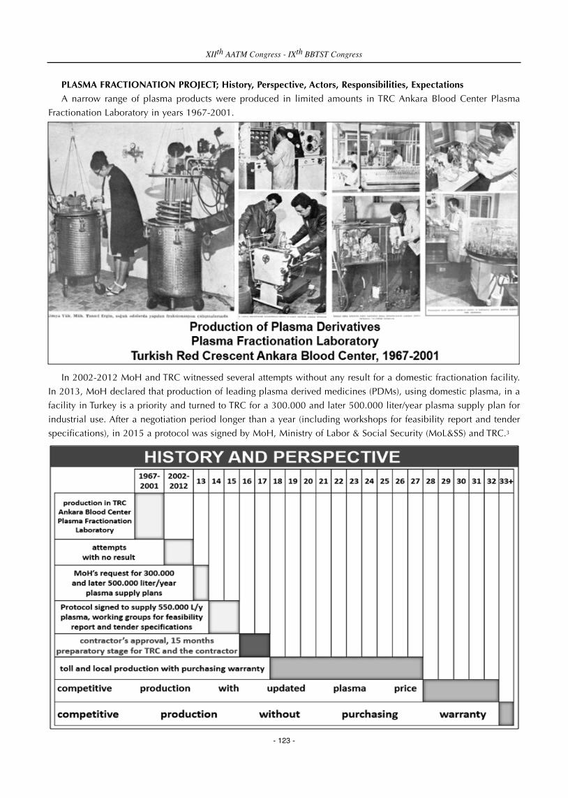
PLASMA FRACTIONATION PROJECT; History, Perspective, Actors, Responsibilities, Expectations
A narrow range of plasma products were produced in limited amounts in TRC Ankara Blood Center Plasma
Fractionation Laboratory in years 1967-2001.
In 2002-2012 MoH and TRC witnessed several attempts without any result for a domestic fractionation facility.
In 2013, MoH declared that production of leading plasma derived medicines (PDMs), using domestic plasma, in a
facility in Turkey is a priority and turned to TRC for a 300.000 and later 500.000 liter/year plasma supply plan for
industrial use. After a negotiation period longer than a year (including workshops for feasibility report and tender
specifications), in 2015 a protocol was signed by MoH, Ministry of Labor & Social Security (MoL&SS) and TRC.3
XIIth AATM Congress - IXth BBTST Congress
- 123 -

According to the Protocol actors of the project will be the MoH as the national authority, the Ministry of Labor
and Social Securiy’s (MoLabor&SS) Social Security Institution (SSI) putting in a tender, a local company as the
investor, a foreign manufacturer company to transfer technology and fractionate plasma, Turkish Red Crescent as
the plasma supplier.4
Responsibilities of the actors are summarized as follows;
• Social Security Institution
✔ Make tender, sign with contractor, mediate writing of additional protocols
✔ Pay for products
✔ Fund TRC in blood component production
• Ministry of Health
✔ Make legal changes, authorization/licensing/periodic inspection, quality control
✔ Order products to meet state hospitals need
✔ Fix clinic use of plasma to 750.000 / year level
✔ Support TRC’s PR campaigns to recruit plasmapheresis donors
• Turkish Red Crescent
✔ Share necessary data and cooperate with contractor
✔ Plan and finance physical and operational preparations
✔ Realize all steps of preparation in 15 months
✔ Provide relevant data for Plasma Master File (PMF)
✔ Recruite plasma donors, operate plasmapheresis centers
✔ Provide 550.000 liters of industrial plasma each year
• Contractor Consortium
✔ Conduct audits in RTC premises, lead corrective steps
XIIth AATM Congress - IXth BBTST Congress
- 124 -

✔ Plan and realize investments
✔ Set up the facility, cold-storage warehouse, plasmapheresis centers
✔ Run pilot production, prepare PMF, get approvals/licences
✔ Pay for plasma,
✔ Fractionate plasma abroad and in Turkey, transfer technology
Expectations of the actors are summarized as follows;
• Social Security Institution
✔ Pay less for at least 25 % of national consumption (in IU or gram)
✔ Fix the prices for at least 10 years
✔ Put this price as a leverage on the rest of the market
• Ministry of Health
✔ Secure product flow to some extend
✔ Bring in new technology, products, opportunities
✔ Import substitution, foreign exchange savings
✔ Positive political impact
• Turkish Red Crescent
✔ Develop new organizational and operational capacities
✔ Widen product and service range (PFF, plasma donor)
✔ Compliance with new international quality standards / partners
✔ Positive general impact on blood products / services quality
✔ Higher countrywide/worldwide organizational reputation and prestige
✔ Provide additional income out of excess plasma
• Contractor Consortium
✔ Privileged long term access to Turkey’s plasma for low price
✔ Purchase warranted market share in a big and growing market
✔ Good returns for investment due to financial, tax incentives
✔ Experience in overseas country operations
REQUIREMENTS; National, International
In addition to Protocol, National regulations, directives, guidelines, standards, recommendations of EU (and
EMA) will be the main references for plasma and medicines.4
National and international requirements that the actors have to comply with are given below.
XIIth AATM Congress - IXth BBTST Congress
- 125 -

WHAT IS DONE; Quality, Quantity
Blood Safety Project which induced improvements in blood donation management, rehabilitation of facilities and
product management, distribution and stock management (2005), use of ISBT-128 barkoding system (2006), ISO
9001:2000 acreditation (2006), information management system, first international accreditation (2007), universal
deferral database (2011), component identification and traceability system (2012), universal NAT Testing for HIV1/2-
HBV-HCV (2014) were some key steps for progress in blood banking as well as getting prepared for plasma
fractionation.
XIIth AATM Congress - IXth BBTST Congress
- 126 -

As part of Blood Safety Project TRC have already renewed some premises, equipments, devices and quality
documents, procedures and instructions as well. Considerable improvements have been achieved in donor
recruitment activities, public and donor group education, PR activities, donation, controlled transportation,
processing, labeling, pathogen safety measures, quality control, storage, logistic management, information
technologies and quality system.
XIIth AATM Congress - IXth BBTST Congress
- 127 -

Number of permenant and total personel has reached to 3.116, motor vehicles to 735 in 2016. Mobile donation
teams are supported with modified blood donation trucks.3
XIIth AATM Congress - IXth BBTST Congress
- 128 -

XIIth AATM Congress - IXth BBTST Congress
- 129 -

XIIth AATM Congress - IXth BBTST Congress
- 130 -

XIIth AATM Congress - IXth BBTST Congress
- 131 -

WHAT IS NEXT; Quality, Quantity
Due to the Protocol, TRC’s commitment is to supply 550.000 liters of plasma. Recovered and source plasma will
make the sum. Since TRC will be responsible from operating a dozen of plasmapheresis centers, recruiting the new
kind of donor (plasma donors) will be TRC’s new and tough challenge. A reasonable but steady increase in whole
blood donation figures (reaching 2,5 million in not more than a few years) is necessary to fulfill the “recovered”
side of the mission. This means; renewing some premises to run larger production, lab and storage capacities, also
constracting some regional cold storage houses, rent a number of refrigerated trucks, planning and conducting more
visits to the existing and potential blood donors, more mobile and fixed teams, more modified trucks, personnel,
equipment, validation, training, visits for supervision and inspection, adaptive changes for software etc. Plasma
protein level screening tests have to be put into practice. New quality documents, procedures, instructions as well.
XIIth AATM Congress - IXth BBTST Congress
- 132 -

Appropriate use of plasma in clinics enable countries to divert more of plasma to the industry. The well-known
ratio is 1/4 or for some instances 1/5 of the total plasma. Turkey’s erythrocyte suspension need is predicted to be 2,5-
3,0 million/year. For the worst-case scenario, consuming 750.000 in clinics can be realistic. However, both last few
years’ figures and the trend curve (given in the graphic below) sign consistent increase.3 Due to the Protocol, at
first to stabilize, then to reduce the consumption, MoH is expected to interfere the situation via conducting training
seminars for clinicians or encouraging specialists to follow the related annex of Turkey’s brand-new “National
Guide”.
XIIth AATM Congress - IXth BBTST Congress
- 133 -
References1. TÜİK web page; http://www.tuik.gov.tr/Start.do2. National Guide for Blood and Bood Products http://www.kanver.org/Upload/Dosya/ulusal_kan_rehberi.pdf3. Annual Report – TRC General Directorate of Blood Services, 20154. “Protocol for Production of Plasma Derived Medicines” signed by Social Security Institution, Ministry of
Health and Turkish Red Crescent, 20.08.2015
XIIth AATM Congress - IXth BBTST Congress
- 134 -

- 135 -
Allogenic transfusion is one of the oldest and most common applications in clinical practice and faced with many
problems like questionable safety and efficacy, increased cost and limited stocs. Knowing the early and late adverse
effects (bad outcomes) of using blood bank products clearly, requires to be more careful about blood transfusion
practice. Actually blood transfusion should be thought as an organ transplantation.
When we look at the economic burden of the countries for using blood bank products, it seems about 4,5 percent
of health care budget is spent on blood products. More importantly, the use of blood products without indication
induces the wrong consumption of limited sources. If it is not found a liquid that can be used instead of blood in
the coming years, there will be a big trouble for patients who really needs blood transfusion. Furthermore the
increasing and aging of the world population, increasing rate of diabetes mellitus, hypertension, hepatitis, AIDS and
other viral infections seen among people who can be donors, will negatively effect the donor pool.
If we continue to use blood products as we do today, it is obvious that we will be faced with a big trouble for
having bank blood products for our increasing world population in the coming years. So our knowledge about blood
transfusion should be updated and, patient-centered approach should replace the product-centered approach.
Patient blood management (PBM) is a system using patient-centered approach in clinical practice. İn PBM
approach, patients are evaluated prior to surgery and the patients with anemia (except urgent surgery) are treated to
correct anemia before surgery (optimization of haematological status of the patient). Furthermore using of
pharmacological agents to reduce blood loss, close monitoring of bleeding and coagulation system and, minimally
invasive surgery applications are recomended to reduce blood requirement during surgery (to ensure minimal
bleeding). PBM recommends a multidisciplinary approach to evaluate of each patient's own situation and doesn’t
determine a standardised hemoglobin threshold for transfusion requirement during perioperative period
(optimization of the physiological response to anemia). Thus PBM prevents making the transfusion decision by
single physician.
Unlike existing transfusion guidelines PBM; not only updates theoretical knowledge, but also takes an active role
in the clinical practice. In this way PBM allows to get some results like avoiding unnecessary transfusions, prevention
of adverse effects due to blood use (mortality-morbidity), ensuring the protection of stocks and, providing significant
economic benefits (Australian health ministry has reported that $ 50 million profit in 4 years after PBM application
and Austrian health ministry has put the project into practice as well).
How should we apply PBM:
PBM awareness should be created and, for this purpose the following procedures should be performed respect
vely.
PATIENT BLOOD MANAGEMENT: WHAT&HOW?
Fevzi TORAMAN

1. The initial PBM group should be established.
2. Education of the initial group about patient blood management should be provided.
3. For creating PBM awareness throughout the country, scientific programs should be created.
4. The support of the ministry of health and public insurance foundation should be received to reach larger
populations.
5. The cooperation with the relevant associations should be established and the bigger PBM groups should be
organised.
6. Mandatory training sessions (education meetings) should be held for 5 participants from each hospital.
7. Volunteer representatives should be chosen from the participants.
8. Each volunteer representative should prepare the transfusion schema of their hospital.
9. The doctors and clinics using blood products most common should be identified.
10. Hospital representatives should meet and educate these physicians and clinics about PBM.
11. These clinics and clinicians should be followed and the data obtained before and after PBM training should
be compared.
12. Hospital representatives should prepare a report every 3 months.
13. Based on these reports, the main PBM groups should have meeting every 6 months, and find financial,
logistic, technical and educational solutions.
14. The solutions should be unique to each hospital.
15. After 3 months of observation, if the problems persist the meetings attended by hospital administrators, PBM
representatives, ministry of health and public insurance foundation should be organised for each hospital.
16. Pilot hospitals should be determined.
17. Transfusion data especially the positive ones obtained PBM meetings should be shared with the others.
18. The awarding of the hospital using appropriate transfusion strategies should be provided by public insurance
foundation (like increasing the package price).
XIIth AATM Congress - IXth BBTST Congress
- 136 -

- 137 -
The word “hemovigilance” comes from the French and it means being “watchful” about “blood”. From 1980s
through early 1990s, a number of blood transfusions ended up in HIV/AIDS cases in some transfusion recipients in
France. In reaction to this unfortunate experience, a surveillance system for blood transfusion was implemented in
1994 and the term “hemovigilance” coined.
Hemovigilance can simply be defined as; “A set of surveillance procedures of the whole transfusion chain
intended to minimize adverse events or reactions in donors and recipients and to promote safe and effective use
of blood components”.
Basic Terminology:
-An adverse event is an undesirable and unintended occurrence in the blood transfusion chain (which consists
of the collection, testing, preparation, storage, distribution, ordering, issuing, and administration of blood and blood
components). It may or may not be the result of an error or an incident and it may or may not result in an adverse
reaction in a donor or recipient.
-An incident is a case in which the patient is transfused with a blood component that did not meet all the
requirements for a suitable transfusion for that patient, or that was intended for another patient. Incidents thus
comprise transfusion errors and deviations from standard operating procedures (SOPs) or hospital policies that have
lead to mistransfusions. It may or may not lead to an adverse reaction.
-A near-miss is an error or deviation from standard procedures or policies that is discovered before the start of
the transfusion and that could have led to a wrongful transfusion or to a reaction in a recipient.
-An adverse reaction is an undesirable response or effect in a patient or donor temporally associated with the
collection or administration of blood or blood component. It may, but need not, be the result of an incident.
“Adverse reactions in recipients” is synonymous with “Transfusion Reactions”.
NON INFECTIOUS TRANSFUSION REACTIONS
HEMOLYTIC TRANSFUSION REACTIONS
A hemolytic transfusion reaction is one in which symptoms and clinical or laboratory signs of increased red cell
destruction are produced by transfusion. Hemolysis can occur intravascularly or extravascularly and can be
immediate (acute) or delayed.
HAEMOVIGILANCE: JUST THE BASICS
Mustafa Nuri GÜNÇIKAN

Acute hemolytic transfusion reaction (AHTR)
An AHTR has its onset within 24 hours of a transfusion. Clinical or laboratory features of hemolysis are present.
Common signs of AHTR are: Fever; Chills/rigors; Facial flushing; Chest pain; Abdominal pain; Back/flank pain;
Nausea/vomiting; Diarrhea; Hypotension; Pallor; Jaundice; Oligoanuria; Diffuse bleeding; Dark urine.
Common laboratory features are: Hemoglobinemia; Hemoglobinuria; Decreased serum haptoglobin;
Unconjugated hyperbilirubinemia; Increased LDH and AST levels; Decreased hemoglobin levels.
Not all clinical or laboratory features are present in cases of AHTR. Blood group serology usually shows abnormal
results but absence of immunological findings does not exclude AHTR. AHTR may also be due to erythrocyte auto-
antibodies in the recipient or to non immunological factors like mechanical factors inducing hemolysis (malfunction
of a pump, of a blood warmer, use of hypotonic solutions, etc.).
Delayed hemolytic transfusion reaction (DHTR)
A DHTR usually manifests between 24 hours and 28 days after a transfusion and clinical or laboratory features
of hemolysis are present. Signs and symptoms are similar to AHTR but are usually less severe. DHTR may sometimes
manifest as an inadequate rise of post-transfusion hemoglobin level or unexplained fall in hemoglobin after a
transfusion. Blood group serology usually shows abnormal results.
Delayed serologic reaction (DSTR)
There is a DSTR when, after a transfusion, there is demonstration of clinically significant antibodies against red
blood cells which were previously absent (as far as is known) and when there are no clinical or laboratory features
of hemolysis. This term is synonymous with alloimmunization.
NON HEMOLYTIC TRANSFUSION REACTIONS
Febrile non hemolytic transfusion reaction (FNHTR)
There is a FNHTR in the presence of one or more of: fever (≥38 ° C oral or equivalent and a change of ≥1 ° C
from pretransfusion value); chills/rigors.
This may be accompanied by headache and nausea occurring during or within four hours following transfusion
without any other cause such as hemolytic transfusion reaction, bacterial contamination or underlying condition.
FNHTR could be present in absence of fever (if chills or rigors without fever). For international comparisons, only
the most serious cases od FNHTR should be accounte for: fever (≥39° C oral or equivalent and a change of ≥2 ° C
from pretransfusion value) and chills/rigors.
Allergic reaction
An allergic reaction may present only with mucocutaneous signs and symptoms: Morbilliform rash with pruritus;
Urticaria (hives); Localized angioedema; Edema of lips, tongue and uvula; Periorbital pruritus, erythema and edema;
Conjunctival edema occurring during or within 4 hours of transfusion.
In this form, it usually presents no immediate risk to life of patient and responds quickly to symptomatic
XIIth AATM Congress - IXth BBTST Congress
- 138 -

treatment like antihistamine or steroid medications. This type of allergic reaction is called ‘minor allergic reaction’
and would be graded as 1, i.e. non-severe.
An allergic reaction can also involve respiratory and/or cardiovascular systems and present like an anaphylactic
reaction. There is anaphylaxis when, in addition to mucocutaneous systems there is airway compromise or severe
hypotension requiring vasopressor treatment (or associated symptoms like hypotonia, syncope). The respiratory signs
and symptoms may be laryngeal (tightness in the throat, dysphagia, dysphonia, hoarseness, stridor) or pulmonary
(dyspnea, cough, wheezing/bronchospasm, hypoxemia). Such a reaction usually occurs during or very shortly after
transfusion. For the purpose of classification, this type of allergic reaction would be graded as 2 (severe), 3 (life-
threatening) or 4 (death) depending on the course and outcome of the reaction.
Transfusion associated graft-versus-host disease (TA-GVHD)
TA-GVHD is a clinical syndrome characterised by symptoms of fever, rash, liver dysfunction, diarrhea,
pancytopenia and findings of characteristic histological appearances on biopsy occurring 1-6 weeks following
transfusion with no other apparent cause. The diagnosis of TA-GVHD is further supported by the presence of
chimerism.
Post transfusion purpura (PTP)
PTP is characterized by thrombocytopenia arising 5-12 days following transfusion of cellular blood components
with findings of antibodies in the patient directed against the Human Platelet Antigen (HPA) system.
Transfusion-related acute lung injury (TRALI)
In patients with no evidence of acute lung injury (ALI) prior to transfusion, TRALI is diagnosed if a new ALI is
present:
- Acute onset;
- Hypoxemia, (Pa02 / Fi02 < 300 mm Hg), or
Oxygen saturation is < 90% on room air, or
Other clinical evidence;
- Bilateral infiltrates on frontal chest radiograph;
- No evidence of left atrial hypertension (i.e. circulatory overload);
- No temporal relationship to an alternative risk factor for ALI during or within 6 hours of completion of
transfusion.
Alternate risk factors for ALI are:
Direct Lung Injury;
- Aspiration
- Pneumonia
- Toxic inhalation
- Lung contusion
- Near drowning
Indirect Lung Injury;
- Severe sepsis
- Shock
- Multiple trauma
XIIth AATM Congress - IXth BBTST Congress
- 139 -

- Burn injury
- Acute pancreatitis
- Cardiopulmonary bypass
- Drug overdose.
It has been suggested by the Toronto TRALI Consensus Panel to add a category of Possible TRALI that would
have the same definition as TRALI except for the presence of a temporal relationship to an alternative risk factor
for ALI (as described above). In such a circumstance, TRALI should be indicated with a possible imputability to
transfusion.
TRALI is therefore a clinical syndrome and presence of anti-HLA or anti-HNA antibodies in recipient nor
confirmation of cognate antigens in donors are required for diagnosis.
Transfusion associated dyspnea (TAD)
TAD is characterized by respiratory distress within 24 hours of transfusion that does not meet the criteria of
TRALI, TACO, or allergic reaction. Respiratory distress should be the most prominent clinical feature and should not
be explained by the patient’s underlying condition or any other known cause.
Transfusion associated circulatory overload (TACO)
TACO is characterized by any 4 of the following:
- Acute respiratory distress
- Tachycardia
- Increased blood pressure
- Acute or worsening pulmonary edema on frontal chest radiograph
- Evidence of positive fluid balance occurring within 6 hours of completion of transfusion.
An elevated BNP is supportive of TACO.
Hypotensive transfusion reaction
This reaction is characterized by hypotension defined as a drop in systolic blood pressure of ≥ 30 mm Hg
occurring during or within one hour of completing transfusion and a systolic blood pressure ≤ 80 mm Hg. Most
reactions do occur very rapidly after the start of the transfusion (within minutes).
This reaction responds rapidly to cessation of transfusion and supportive treatment. This type of reaction appears
to occur more frequently in patients on ACE inhibitors. Hypotension is usually the sole manifestation but facial
flushing and gastrointestinal symptoms may occur. All other categories of adverse reactions presenting with
hypotension, especially allergic reactions, must have been excluded. The underlying condition of the patient must
also have been excluded as a possible explanation for the hypotension.
Other transfusion reactions
Haemosiderosis
Transfusion-associated haemosiderosis is being defined as a blood ferritin level of ≥ 1000 micrograms/l, with or
XIIth AATM Congress - IXth BBTST Congress
- 140 -

without organ dysfunction in the setting of repeated RBC transfusions.
Hyperkalemia
Any abnormally high potassium level (> 5 mml/l, or ≥1.5 mml/l net increase) within an hour of transfusion can
be classified as a transfusion- associated hyperkalemia.
Unclassifiable Complication of Transfusion (UCT)
Occurrence of an adverse effect or reaction temporally related to transfusion, which cannot be classified
according to an already defined Adverse Transfusion Effect and with no risk factor other than transfusion and no
other explaining cause.
The severity of an adverse reaction in a recipient is graded according to an international scale:
Grade 1 (Non-Severe):
- the recipient may have required medical intervention (e.g. symptomatic treatment) but lack of such would not
result in permanent damage or impairment of a body function.
Grade 2 (Severe):
- the recipient required in-patient hospitalization or prolongation of hospitalization directly attributable to the
event; and/or
- the adverse event resulted in persistent or significant disability or incapacity; or
- the adverse event necessitated medical or surgical intervention to preclude permanent damage or impairment
of a body function.
Grade 3 (Life-threatening):
- the recipient required major intervention following the transfusion (vasopressors, intubation, transfer to
intensive care) to prevent death.
Grade 4 (Death)*
- the recipient died following an adverse transfusion reaction.
* Grade 4 should be used only if death is possibly, probably or definitely related to transfusion. If the patient
died of another cause, the severity of the reaction should be graded as 1, 2 or 3.
The term “Imputability” denotes the likelihood that an adverse reaction in a recipient can be attributed to the
blood component transfused:*
Definite (certain): when there is conclusive evidence beyond reasonable doubt that the adverse event can be
attributed to the transfusion.
Probable (likely): when the evidence is clearly in favor of attributing the adverse event to the transfusion.
Possible: when the evidence is indeterminate for attributing the adverse event to the transfusion or an alternate
cause.
Unlikely (doubtful): when the evidence is clearly in favor of attributing the adverse event to causes other than
XIIth AATM Congress - IXth BBTST Congress
- 141 -

the transfusion.
Excluded: when there is conclusive evidence beyond reasonable doubt that the adverse event can be attributed
to causes other than the transfusion.
*Only possible, probable and definite cases should be used for international comparisons.
Transfusion-transmitted infections (TTIs)
Viral infection (TTVI)
Following investigation, the recipient has evidence of infection post-transfusion and no clinical or laboratory
evidence of infection prior to transfusion and either, at least one component received by the infected recipient was
donated by a donor who had evidence of the same infection, or at least one component received by the infected
recipient was shown to have been contaminated with the virus.
Bacterial infection (TTBI)
A TTBI should be clinically suspected if fever ˃39 °C or there is a change of ˃2 °C from pretransfusion value;
and rigors; and tachycardia ˃120 beats/min or a change of ˃40 beats/min from pretransfusion value or a rise or drop
of 30 mm Hg in systolic blood pressure within 4 hours of transfusion are present.
Possible TTBI:
- detection of bacteria by approved techniques in the transfused blood component but not in the recipient’s
blood; or
- detection of bacteria in the recipient’s blood following transfusion but not in the transfused blood component
and no other reasons are ascertainable for the positive blood culture.
Confirmed TTBI:
- detection of the same bacterial strain in the recipient’s blood and in the transfused blood product by
approved techniques.
Parasite infection (TTPI)
Detection of the same parasite in the recipient’s blood and parasite or specific antibodies in the donor’s blood.
INVESTIGATION OF TRANSFUSION TRANSMITTED INFECTIONS
In order to investigate a possible Transfusion Transmitted Infection, primarily, a complete list of the blood
components received by the patient and their dates of transfusion is required.
Additionally, the following information is sought from the hospital:
(1) Patient details
(a) full name
(b) address
(c) date of birth
(d) sex
(e) ethnic origin.
(2) Clinical details
(a) Physician, with specialty
(b) reason for transfusion
XIIth AATM Congress - IXth BBTST Congress
- 142 -

(c) underlying diagnosis
(d) current condition
(e) clinical evidence of post-transfusion infection.
(3) Laboratory results
(a) copies of laboratory reports for infectious markers
(b) any test results on samples prior to transfusion
(c) liver function test results (hepatitis cases).
With the primary information at hand, the donors of the blood components are identified and their records are
reviewed regarding the results of routine testing for index donations (the one the infected recipient received) and
any donations given subsequent to them. The situation is then assessed, considering the whole data, regarding
whether any further investigation is required and what will be investigated specifically. Then the need for additional
testing (and additional samples from the donors) is decided and you proceed as needed. Then the results are
reviewed. Finally, the conclusion is reported to clinician/hospital, etc.
OVERLAPPING SIGNS AND SYMPTOMS OF TRANSFUSION REACTIONS
SIGN OR SYMPTOM POSSIBLE REACTION MOST LIKELY > LESS LIKELY
BLOOD COMPONENT*
Fever, chills Febrile nonhemolytic Platelets (especially septic) >
Septic RBCs > Plasma
Acute hemolytic
TRALI
Urticaria, pruritus Allergic Plasma > Platelets > RBCs
Anaphylactic
Dyspnea TACO Any
TRALI
Anaphylactic
Hypotension Septic Platelets > plasma > RBCs
Hypotensive
Acute hemolytic
Anaphylactic
XIIth AATM Congress - IXth BBTST Congress
- 143 -

PREVENTIVE MEASURES FOR TRANSFUSION REACTIONS
TRANSFUSION REACTION PREVENTIVE MEASURES
Transfusion-transmitted Donor skin cleansing
bacterial infection Diversion pouch on donation line
Pathogen reduction
Correct storage conditions.
Transfusion-transmitted viral Donor selection
infection (HBV, HCV, HIV-1/2, other) Donation testing
Pathogen reduction.
Transfusion-transmitted Donor selection
parasitic infection (malaria, other) Donation testing
Pathogen reduction.
Hemolysis due to incorrect storage Quality assured clinical
transfusion process.
Transfusion Related Acute TRALI risk may be reduced with Fresh Frozen
Lung Injury (TRALI) Plasma (FFP) from male donors.
Graft-Versus-Host Disease Use of irradiated components for at-risk
patients; use of amotosalen treated platelets.
Transfusion Associated Avoid over-infusion; infusion should be at a
Circulatory Overload (TACO) rate not to exceed 2 to 4 mL/kg/hour, and the
rate should be lower (1 mL/kg/hour) for
patients at high risk of circulatory overload.
Febrile non-hemolytic TR Incidence may be reduced by
Leucodepletion.
ADVERSE REACTIONS OR COMPLICATIONS IN DONORS
Complications mainly with local symptoms
These complications are directly caused by the insertion of the needle. Some of these are mainly characterized
by occurrence of blood outside vessels, whereas others are mainly characterized by pain.
Complications mainly characterized by the occurrence of blood outside the vessels. Haematoma (bruise)
A haematoma is an accumulation of blood in the tissues outside the vessels. The symptoms are caused by blood
flowing out of damaged vessels and accumulating in the soft tissues. For apheresis procedures, haematomas may
also be caused by infiltration of the soft tissues by red cells during the return phase of the procedure. Large
haematomas, particularly those in deeper layers of the forearm, put pressure on surrounding tissues and may
contribute to other complications such as nerve irritation and injury and more rarely compartment syndrome. Signs
and symptoms: Bruising, discolouration, swelling and local pain. Accumulation of blood in deeper tissues may result
in more serious pain and pressure syndromes listed below.
Arterial puncture
Arterial puncture is a puncture of the brachial artery or of one of its branches by the needle used for bleeding the
donor. Because of the rapid blood flow, the risk of a large haematoma is increased and thereby risks of more serious
pain and pressure syndromes listed below. Signs and symptoms: A lighter red colour than usual of the collected
XIIth AATM Congress - IXth BBTST Congress
- 144 -

blood can be seen. The needle and tubing may appear to pulsate; the blood bag fills very quickly. There may be
weak pain localized to the elbow region.
Delayed bleeding (re-bleeding)
Leakage of blood from the venipuncture site after the initial bleeding has stopped. Re-bleeding may be related
to pressure not being applied to the correct location or for an adequate duration, or premature removal of the
bandage. After the donor has left the clinic, re-bleeding may be related to heavy lifting or strain to the donor’s arm.
Donors on certain medications, such as autologous donors on anticoagulants, may be at higher risk to rebleed. Signs
and symptoms: Spontaneous recommencement of bleeding from the venipuncture site, after pressure has been
applied and the initial dressing has been removed, or leaking through the dressing.
Complications mainly characterized by pain
Nerve injury / irritation
Injury or irritation of a nerve A nerve may be hit directly by the needle at insertion or withdrawal, or there may
be pressure on a nerve due to a haematoma or inflammation of the soft tissues. Include medically diagnosed cases,
as well as cases reported on the basis of documented ‘nerve’ type symptoms. Signs and symptoms: Radiating, often
‘electrical’ sharp pain moving away from the venepuncture site, and/or paraesthesias such as tingling, burning
sensations in the hand, wrist or shoulder area but away from the venepuncture site. Symptoms may arise
immediately when the needle is inserted or withdrawn. In cases associated with a haematoma, pain may not be
apparent at the time and may start when the haematoma has reached a sufficient size, some time after insertion of
the needle. Symptoms may be worse in certain positions or with certain arm motions. Rarely, weakness of the arm
may develop.
Symptoms resolving within 12 months: Symptoms usually resolve within days, but rarely may persist for months
or become permanent.
Symptoms lasting more than 12 months.
Other Painful arm
Pain in the arm is the primary symptom, without the characteristics of nerve irritation outlined above, or the
presence of a large hematoma or other defined complications that may be painful. Pain may be related to tissue
injury, possibly due to hematoma in the deeper tissues. Signs and symptoms: Pain in the arm, without
characteristics of nerve irritation. May be described as an ache or heaviness in the arm, similar to that experienced
after vaccination. Include all cases where arm pain is the main symptom, unless a diagnosis of nerve
injury/irritation is suspected in the presence of nerve type symptoms recognised by trained staff.
Localised infection/inflammation
Inflammation along the course of a vein, which may progress to localised infection several days after phlebotomy.
There may be clotting in the vein. Tissue damage and introduction of surface bacteria into the deeper tissues with
venepuncture. The superficial vein itself (thrombophlebitis) or the surrounding subcutaneous tissue (cellulitis) may
be predominantly affected. Signs and symptoms: Warmth, tenderness, local pain, redness and swelling at the site
of phlebotomy. The site and the vein may feel tender, firm, and warm to the touch. Fever may be present.
Thrombophlebitis: The redness, swelling, and tenderness extend along the course of the vein.
Cellulitis: The redness, swelling and tenderness affect the soft tissues, and are not localised to the course of the
vein.
XIIth AATM Congress - IXth BBTST Congress
- 145 -

Other major blood vessel injury
These rare, serious conditions must always be medically diagnosed.
Deep venous thrombosis (DVT)
Thrombosis of a deep vein in the donor’s phlebotomy arm. Superficial venous thrombosis may progress into the
deeper veins of the donor’s arm. DVT may also rarely occur without previous signs and symptoms of superficial
thrombosis. An additional risk factor for thrombosis, in particular, the use of oral contraceptives, may be present
in these donors. Symptoms and signs: Swelling and pain in the upper arm. May be accompanied by symptoms of
superficial inflammation and thrombosis.
Arteriovenous fistula
Acuired connection between the vein and artery due to venepuncture lacerations. A channel forms between the
lacerated vein and artery immediately postvenepuncture, or in the healing process. May be related to arterial
puncture. Signs and symptoms: Pulsating mass with a palpable thrill and associated bruit. The affected area may
be warm, and the distal part of the arm may be cool if significant shunting of blood is present. The distal veins may
be dilated and may pulsate.
Compartment syndrome Increased intracompartment pressure leading to muscle and soft tissue necrosis. Blood
may accumulate in the frontal deep areas of the forearm, closing small blood vessels and resulting in muscle and
nerve tissue necrosis. May be related to arterial puncture. Signs and symptoms: Painful arm, particularly on
movement; swelling, paresthesias and partial paralysis.
Brachial artery pseudoaneurysm
Collection of blood outside an artery, contained by adventitia or the surrounding tissues alone. After a traumatic
arterial puncture, blood may leak out of the artery and accumulate in the surrounding space. Signs and symptoms:
Pulsating mass in the arm. May be accompanied by pain and paraesthesias. May be preceded by a large hematoma
following arterial puncture.
Complications mainly with generalized symptoms
Vasovagal reactions
A vasovagal reaction (VVR) is a general feeling of discomfort and weakness with anxiety, dizziness and nausea,
which may progress to loss of consciousness (LOC: faint). It is the most common acute complication related to
blood donation. Both physiologic and psychological factors may be important. The reaction is generated by the
autonomic nervous system and further stimulated by psychological factors and the volume of blood removed,
relative to the donor’s total blood volume. Signs and symptoms: Usually several of the following: discomfort,
weakness, anxiety, light-headedness/dizziness, nausea, chills, sweating, vomiting, pallor, hyperventilation, rapid or
a slow pulse. Hypotension and loss of consciousness (LOC) may occur and can be accompanied by loss of bladder
or bowel control or convulsive movements.
Reactions may occur before phlebotomy (rare), during phlebotomy or immediately after phlebotomy, when the
donor stands up, in the refreshment area, or after the donor has left the collection site. Most reactions occur
within 12 hours of phlebotomy. Reactions accompanied by LOC carry a risk of injury, particularly if they occur
once the donor has left the collection site (delayed vasovagal reactions).
Without loss of consciousness (LOC) - the donor does not faint
With loss of consciousness (LOC) - the donor faints for a period of time
XIIth AATM Congress - IXth BBTST Congress
- 146 -

- LOC < 60 seconds - without other signs and symptoms
- LOC ≥ 60 seconds - or with complications of convulsive movements, urinary or faecal incontinence
- With injury - Injury caused by falls or accidents in donors with a vasovagal reaction
- Without injury
Location of reaction:
- On collection facility*- Symptoms occurred before donor has left the donation site
- Outside collection facility - Symptoms occurred after donor has left the donation site
*in area within which staff can observe the donor and be responsible for the care of donors with complications
Complications related to apheresis
Citrate reaction
Neuromuscular hyperactivity related to reduced ionized calcium levels. Infusion of citrate anticoagulant during
apheresis causes a fall in ionised calcium levels, leading to neuromuscular hyperactivity. If untreated, symptoms may
progress to tetany and severe cardiac arrhythmias, including cardiac arrest. Operator error with mix up of saline and
citrate bags may occur with some apheresis equipment, and lead to rapid citrate infusion. Symptoms and signs:
Numbness or tingling of lips, feelings of vibrations, numbness or tingling in the fingers, metallic taste, chills,
shivering, light-headedness, feeling of tightness, muscle twitching, rapid or slow pulse, shortness of breath.
Symptoms may progress to carpopedal spasms and vomiting, and in severe reactions, to generalised muscle
contractions (tetany), shock, irregular pulse and cardiac arrest.
Haemolysis
Donor red cells may be damaged, releasing haemoglobin. There may be malfunctioning valves, kinks or
obstruction of the tubing, incorrect installation of equipment, or other equipment failures affecting the
extracorporeal circuit. Incompatible replacement fluids, such as dextrose D5W, may be used in error. Signs and
symptoms: Pink or red plasma, blood in lines or filter may appear dark. The donor may notice pink or red urine
after collection.
Air embolism Air bubble introduced into the donor’s circulation. Mechanism: Air may enter into the lines due to
incomplete priming of lines, as a result of a machine malfunction or defective collection kits or through incorrect
manipulation by staff. Air in the donor’s pulmonary circulation may occlude the pulmonary arteries in the lung and
cause cardiopulmonary symptoms. Air may pass to the arterial circulation through an atrial septal defect, and reduce
blood flow to the brain. Signs and symptoms: Bubbling sound or feeling at the venipuncture site. Cough, dyspnea,
apprehension, sweating, chest pain, confusion, tachycardia, hypotension, nausea and vomiting.
Infiltration Intravenous solute (saline solution) enters the extravascular tissues during volume replacement
(generally only applicable to double red cell procedures). The needle is no longer positioned in the intravascular
space, so fluids enter the surrounding tissues. Signs and symptoms: Swelling of the tissues at the venipuncture site.
Allergic reactions
Allergy (local) Red or irritated skin at the venipuncture site. Reaction caused by allergens or irritants in solutions
used for disinfection of the arm (such as iodine or chlorhexidine) or in manufacture of the collection set. Irritation
may also occur due to application of the adhesive bandage (bandage adhesive dermatitis). An allergic reaction to
latex that may be in supplies such as gloves may also occur. Signs and symptoms: Itching and redness at the
venepuncture site, the bandage site, or the entire skin disinfection area. In a true allergic reaction, there may be a
raised rash or hives in these areas that may expand to cover a larger area of the arm. The reaction may occur soon
XIIth AATM Congress - IXth BBTST Congress
- 147 -

after donation or in the hours to days post-donation.
Generalised allergic reaction (anaphylactic reaction) Anaphylactic type reactions usually starting soon after the
procedure is begun and may progress rapidly to cardiac arrest. Extremely rare reactions, attributed to donor
sensitivity to ethylene oxide gas used to sterilize some collection kits. Signs and symptoms: Apprehension,
anxiousness, flushing, swelling of eyes, lips or tongue, cyanosis, cough, wheezing, dyspnea, chest tightness,
cramps, nausea, vomiting, diarrhoea, tachycardia, hypotension, and altered mentation.
Other serious complications related to blood donation
Major cardiovascular event (MCE)
Acute cardiac symptoms (other than myocardial infarction or cardiac arrest).
Myocardial infarction
Cardiac arrest
Transient Ischemic Attack
Cerebrovascular accident
Death.
Reporting is encouraged of MCE or death from any cause up to 24 hours after donation, with an assessment of
imputability. Only cases with definite, probable or possible imputability should be included in international
reporting. Major cardiovascular events, including death, may occur in the hours after attending the collection centre
for blood donation. This can occur without any relation to the donation (for deaths, this is described by the term
actuarial deaths).
Other complications
Other systemic reactions or complications that do not fit into the above, such as chest pain that may have been
investigated as angina, but was actually musculoskeletal, or transmission of infection to a donor through erroneous
reuse of equipment.
Grading of complication severity and imputability
Grading of severity
Life-threatening complications and long-term disability are thankfully extremely rare after blood donation.
Grading of severity for donor reactions does not easily fit into grading systems used for adverse reactions in patients.
Use of this grading system is therefore optional. The criteria for classification of a reaction as serious (severe) as
derived from these systems are:
Hospitalization: If it was attributable to the complication. The criterion of hospital admission is applicable if a
donor is kept in hospital overnight. Cases where a donor is seen, examined, and in some cases given treatment
(e.g. suturing, IV fluids, treatment of a fracture) but discharged home are not automatically classified as serious.
Intervention: To preclude permanent damage or impairment of a body function or to prevent death (life-
threatening)
Symptoms: Causing significant disability or incapacity following a complication of blood donation and
persisted for more than a year after the donation (Long term morbidity)
Death: If it follows a complication of blood donation and the death was possibly, probably or definitely related
to the donation.
Certain complications of donation are by their nature mild or severe.
XIIth AATM Congress - IXth BBTST Congress
- 148 -

Local reactions
Most local reactions (hematoma, arm pain syndromes) would not be considered severe. Severe consequences
are separate reaction types: deep venous thrombosis, arteriovenous fistula, and compartment syndrome. Nerve
injury may rarely result in long term donor signs and symptoms. This may be captured by the duration of symptoms
(optional split in nerve pain category).
Systemic reactions
Complications that are by their nature severe include generalised allergic (anaphylactic) reactions, and all
major cardiovascular events.
Grading of imputability
The strength of relation between donation and complication is:
Definite or certain: When there is conclusive evidence beyond reasonable doubt for the relation. Probable or
likely: When the evidence is clearly in favor of a relation.
Possible: When the evidence is indeterminate for attributing the complication to the donation or an alternative
cause.
Unlikely or doubtful: When the evidence is clearly in favor of attributing the complication to other causes.
Excluded: When there is conclusive evidence beyond reasonable doubt that the complication can be attributed
to causes other than the donation.
Imputability should only be reported for cardiovascular events leading to hospitalization or death post-
donation, and only cases with imputability of possible, probable or definite should be captured.
References1. Hemovigilance: An Effective Tool For Improving Transfusion Safety, Edited by Rene R.P. De Vries, Jean
Claude Faber, 2012, Wiley-Blackwell Publishing. ISBN: 978-0-470-65527-6.2. ISBT Working Party on Haemovigilance, July 2011, Proposed Standard Definitions for Surveillance of Non
Infectious Adverse Transfusion Reactions; http://www.ihn-org.com/wp-content/uploads/2011/06/ISBT-definitions-for-non-infectious-transfusion-reactions.pdf [accessed March 14, 2016].
3. Transfusion Microbiology, Edited by John A. J. Barbara, Fiona A. M. Regan and Marcela C. Contreras, 2008.Cambridge University Press. ISBN: 978-0-521-45393-6.
4. Rossi's Principles of Transfusion Medicine, 4th Edition, Edited by Toby L. Simon, E. L. Snyder, B. G. Solheim,Christopher, P. Stowell, Ronald, G. Strauss and Marian Petrides, 2009, Blackwell Publishing Ltd. ISBN: 978-1-405-17588-3.
5. Working Group on Donor Vigilance of the International Society of Blood Transfusion Working Party onHaemovigilance in collaboration with The International Haemovigilance Network, The AABB DonorHaemovigilance Working Group, December 11, 2014, Standard for Surveillance of Complications Relatedto Blood Donation; https://www.aabb.org/research/hemovigilance/Documents/Donor-Standard-Definitions.pdf [accessed March 14, 2016].
XIIth AATM Congress - IXth BBTST Congress
- 149 -

- 150 -
The transfusion of blood and blood products is a life-saving intervention. Major surgeries are not taking place
without blood, and acute bleeding, treatments of traumas and several chronic diseases all rely on the availability of
blood. However, there are risks of adverse events associated with the donation of blood and its components, and
with the transfusion of blood and blood products to patients.(1) Adverse events include all reactions, incidents, near
misses, errors, deviations from standard operating procedures and accidents associated with blood donation and
transfusion. Despite the useful information gained over transfusion reactions, the main risk remains human factors.
Learning from adverse events and identifying systems problems can drive the introduction of measures to enhance
the quality, safety, efficacy, and cost-effectiveness of blood and blood products as well as the donation and
transfusion processes.(2)
Blood components are living human tissue and their administration is not without risk. The safety of the blood
component that is delivered to the patient is thus of critical concern. To ensure the safety of the blood supply, there
must be systems in place that will evaluate, monitor and manage risk along the entire blood supply value chain.
Haemovigilance is a set of surveillance procedures covering the entire transfusion chain, from the donation and
processing of blood and its components, to their provision and transfusion to patients and their followup.
Haemovigilance includes the monitoring, reporting, investigation and analysis of adverse events related to the
donation, processing and transfusion of blood, as well as the development and implementation of recommendations
to prevent their occurrence or recurrence. The ultimate goal of haemovigilance is continuous quality improvement
of the transfusion chain through corrective and preventive actions to improve patient safety and outcomes, enhance
donor safety and reduce wastage. Haemovigilance should be fully integrated into the quality systems of all
institutions involved in the donation and provision of blood and blood products, including processing, inventory
management, storage and distribution, and in clinical transfusion. The organization of a haemovigilance system is
largely determined by the structure of the national blood system and the health system.(3) A system of
haemovigilance is dependent on the traceability of blood and blood products from donors to recipients and vice
versa, and on the monitoring, reporting, investigation and analysis of adverse events. The rigorous management of
information generated through this system is key to introducing amendments in blood policies and guidelines that
lead to changes in processes and practices in donation and transfusion. The establishment of a haemovigilance
system involves coordination and collaboration among all stakeholders, including the ministry of health, blood
transfusion services, hospitals, professional bodies, public health institutions and regulatory agencies, as well as
patient and donor group.
The evolution of Haemovigilance systems:
The 1980s were plagued by a new epidemic, AIDS (as it was then known) which shook the world. This was at a
time of medical conquest of Infectious Diseases with various immunization programmes and when some diseases
like Small Pox had been eradicated. Man had been to the moon, the pharmaceutical companies were producing a
IMPLEMENTATION OF NATIONAL HAEMOVIGILANCE SYSTEM
Meral SÖNMEZOĞLU

multitude of newer, more effective drugs, researchers were decoding the human genome, and longevity, the ultimate
medical dream was within reach. After regular blood and blood product users such as people with Haemophilia
became infected with the AIDS (HIV) virus, countries around the world took swift action in order to keep their blood
supply safe. The lessons learned from this epidemic and the related safety measures set the stage for the development
of nationwide monitoring systems that track adverse events and incidents associated with collections from blood
donors and transfusion to recipients.
On January 4 1993, a French law was enacted, establishing the world's first Haemovigilance system, which they
defined as; “a set of surveillance procedures from the collection of blood and its components to the follow up of
recipients aimed at collecting and assessing information on unexpected or undesirable effects resulting from the
therapeutic use of blood products on a National level”. The French programme places a 'rapporteur' in each
hospital, and that person is charged with implementing a system to track outcomes on all patients, which are then
coordinated and analysed nationally. It also includes the French territories, such as Guadeloupe, Martinique,
Guyana and Reunion and has 1400 Haemovigilance officers. Their extensive system led to the discovery and
reporting of a Chikungunya outbreak in Reunion, in 2005.
The British followed in 1996 by developing the Serious Hazards of Transfusion (SHOT) network, based on the
French system. Other countries in Europe and other parts of the world saw the value of having this type of system
and followed suit. Being voluntary, the SHOT programme has been very successful, providing reliable data and is
hence used as a benchmark for policy decisions on transfusion in other countries. For instance, the discovery that,
using male only plasma reduced the risk of TRALI has changed transfusion policy in some countries.
As several European nations implemented their own Haemovigilance systems in the 1990's, with a myriad of
varying definitions regarding the nature of adverse transfusion- related events, representatives of blood transfusion
and other health related organizations in Belgium, France, Luxembourg the Netherlands and Portugal joined forces
in 1998 to create the European Haemovigilance Network, with the goal of developing universal standards for
Haemovigilance and maintaining a common framework for ensuring the safety of blood and its components. They
also established a rapid alert system for reporting “clustered “ clinical signs that may point to a specific disease or
other problems in transfused blood or components. The European Haemovigilance network has expanded,
numbering 20 members by 2006, and extending beyond Europe to include Canada, Malta, New Zealand and
Singapore. The International Society for Blood Transfusion (ISBT) working party on Haemovigilance has 28 member
countries and contributes toward an effective international Haemovigilance programme.(4)
The word ‘haemovigilance’ (he ´movigilance in French) was coined in France in 1991 in analogy to the already
existing term ‘pharmacovigilance’. It is derived from the Greek word ‘haema’ = blood and the Latin word ‘vigilans’
= watchful (Fig. 1). The aim of haemovigilance is to detect and analyse all untoward effects of blood transfusion in
order to correct their cause and to prevent recurrence, thus improving the safety of blood transfusion.
Between 2003 and 2005, the European Commission published European Blood Directives that give mandatory
rules for ‘collection, testing, processing, storage and distribution of human blood and blood components’ [3,4,6,13].
The Directives dealing with haemovigilance require: 1. Full traceability of blood products from donor to recipient;
2. Notification of a. serious adverse events (SAE) that may have an influence on and b. serious adverse reactions (SAR)
that may be attributed to the quality and safety of blood and blood components.
WHO Global Strategic Plan, 2008–2015 released in 2007 indicated challenges for blood transfusion safety in
developing. The objective of the WHO programme on blood safety and availability is a reduction in avoidable
XIIth AATM Congress - IXth BBTST Congress
- 151 -

morbidity and mortality through improved access to safe blood and blood products and the safe and rational use of
blood transfusion, with a particular focus on life-saving emergency transfusions for reducing maternal and child
mortality, and on the prevention of HIV/AIDS, hepatitis B and C.
With the goal of ensuring universal access to safe blood, WHO has been at the forefront of the movement to
improve blood safety as mandated by successive World Health Assembly resolutions. Therefore, strengthened
national capacity for the implementation of strategic approaches and the development of effective systems to
achieve universal access to safe blood transfusion is necessary.(5)
Priority needs for blood transfusion safety in developing countries
– the building of national assessment and monitoring systems as part of the national blood transfusion service
– Support the development of effective national systems for the collection and management of data throughout
the transfusion chain
– Build and strengthen global, regional and national surveillance, vigilance and alert systems for blood safety
and availability, and adverse transfusion events.
–Strengthen the global, regional and national monitoring of process and outcome indicators on blood safety and
availability and measure progress.
Activities related are;
– Update and maintain global and regional indicators on blood safety and strengthen systems for national
monitoring and evaluation
– Improve the coverage and reliability of data from the WHO Global Database on Blood Safety and regional
databases
– Identify priority areas and countries requiring technical support in strengthening national systems for
monitoring and evaluation
– Advise on evidence-based, cost-effective strategies for blood safety and availability, based on information
obtained from indicators.
References1. am_National_Haemovigilance_ WHO System.2. Murphy MF, et al Transfusion practice and safety: current status and possibilities for improvement Vox
Sanguinis (2011) 100, 46–593. De Vries RRP et al Hemovigilance: an effective tool for improving transfusion practice. Vox Sanguinis (2011)
100, 60–674. Dhingra N Blood Safety in the Developing world and WHO initiatives. Vox Sang 2002; 83 (suppl 1): 173-
177.5. Globa l Consultation on Haemovigilance, Dubai, 2012. www.who.org
XIIth AATM Congress - IXth BBTST Congress
- 152 -

- 153 -
Hemovigilance is defined by the United States National Institutes of Health as "a set of surveillance procedures
covering the whole transfusion chain from the collection of blood and its components to the follow up of its
recipients, intended to collect and access information on unexpected or undesirable effects resulting from the
therapeutic use of labile blood products." That is certainly a good definition of formal hemovigilance systems which
began to be developed in some countries in the 1970's. However, informal collection of such "...information on
unexpected or undesirable effects..." was producing valuable insights even earlier. Important data on transfusion
safety is also gained by studies focused on individual types of adverse events rather than "...covering the whole
transfusion chain...." Whether formal or informal, comprehensive or focused, vigilance regarding adverse transfusion
events has led to profound improvements in transfusion safety.
For example, post-transfusion hepatitis (PTH) cases were first reported in the 1940s. Studies of "serum" and
"infectious" hepatitis over the following 3 decades culminated in discovery of the "Australia Antigen" or hepatitis B
surface antigen (HBsAg) test as well as the hepatitis A virus antibody test. Application of the HBsAg test to blood
donors allowed deferral of some who were infectious, but had two other major consequences. It aided in the
identification of paid blood donors as the most important source of PTH, and led to recognition that most PTH was
due to neither HAV nor HBV. Another 20 years of study was required to identify the virus causing such "non-A, non-
B hepatitis" and to develop a specific test for HCV, but the improvement in safety has been profound.
The much shorter time period between case reports of transfusion-associated AIDS (TA-AIDS) in 1982, isolation
of HIV, and development of the first effective HIV-antibody test in 1985 reflected the tremendous growth in
knowledge of biochemistry and microbiology between the 1940's and the 1980s. Similar sequences of recognition
of a transfusion-transmitted infection through more-or-less systematic 'hemovigilance', quantification of the risk
through more focused studies, and development of an effective donor test have followed, in affected countries, for
West Nile virus (WNV) and Chagas disease.
Bacterial contamination of blood components has also been addressed through event reporting (hemovigilance)
followed by focused study and implementation of closed system collection kits and more recently, by component
testing. Again, the risk has decreased tremendously. Other infectious risks of lower magnitude remain, and we will
continue to make progress in reducing these risks as targeted by hemovigilance systems.
Hemovigilance has been an essential part of risk identification and reduction with respect to non-infectious risks
of transfusion as well. In the United States the first hemovigilance program began in 1976 when the FDA required
hospitals to report all transfusion-associated fatalities. A 1980 publication summarizing the first 3 years of reports
revealed that half of all such deaths were due to clerical errors leading to ABO antibody-mediated immediate
hemolytic reactions (IHTRs). Subsequent reports refined these observations, helping to identify their "root causes".
The U.K hemovigilance or Serious Hazards of Transfusion (SHOT) system, which began in 1996, has been
HAEMOVIGILANCE; EFFECTIVE BUT ONLY PART OF THE STORY
James T. PERKINS

particularly useful in identifying how such errors occur. Application of these insights has reduced the risk as shown
in subsequent FDA and SHOT hemovigilance reports.
A final example of the power of hemovigilance is the story of transfusion-related acute lung injury (TRALI). TRALI
came to the attention of the transfusion medicine community in 1985 when Popovsky and Moore (Transfusion,
25;573-7) reported 36 cases of respiratory failure during or soon after transfusion and showed that many of the
responsible components had HLA antibodies directed against the recipient leukocytes. Hemovigilance such as that
conducted by the SHOT group highlighted the fact that plasma-containing components, FFP and platelets, were
disproportionately implicated and most often came from female donors. This led to the recommendation that female
donor plasma not be used for transfusion (SHOT Annual Report, 2000/2001), and since implementation of such
preventative measures TRALI fatalities have declined significantly.
So hemovigilance efforts have contributed to implementation of testing and other interventions that have greatly
reduced transfusion risks. And they have given us some idea of the residual magnitude of those risks. Can this help
us in making the difficult decision of whether or not to transfuse a specific patient? It is tempting to think that we
could enter those magnitudes into some simple risk versus benefit equation and out would pop our answer. But
there are at least two limitations that preclude this simple approach.
First, we have little information regarding the benefit side of the equation. In some situations the obvious need
for transfusion overrides consideration of the risks. Examples include patients in hemorrhagic shock or anemic
patients with severe symptoms such as angina or disabling shortness of breath. Others include bleeding patients with
severe thrombocytopenia due to chemotherapy or coagulopathy due to warfarin. If transfusion is successful we
observe resolution of the anemia symptoms or cessation of bleeding. But a majority of our transfusions are not
performed for such clear cut indications. Instead, we typically find ourselves transfusing to correct a number, most
often a hemoglobin level, a platelet count, or a clotting time. In these cases we believe the number predicts a
negative outcome such as those listed above, and we transfuse with the idea that we are preventing an adverse
outcome such as a myocardial infarction (MI) or bleeding. The benefit of such prophylactic transfusions is not visible;
if effective, nothing happens! In these cases we are balancing the risk of transfusing against the risk of NOT
transfusing, but unfortunately the risk of not transfusing is generally not known and typically overestimated.
A second problem is that hemovigilance focuses on specific events that are recognizably due to the transfusion.
But patients receiving transfusion are already ill. When confronted with fever or respiratory distress during or soon
after a transfusion we must consider whether it is due to the patient's interaction with the blood component itself,
or whether it’s due to the patient's underlying condition. This difficulty is acknowledged in hemovigilance systems
incorporating "imputability" criteria in an attempt to estimate the likelihood that the manifestations are due to the
transfusion. And consider events such as in-hospital infections. These inevitably occur, but if their incidence is
increased by transfusion, they cannot be tied to any one blood component as hemovigilance-defined events must
be.
The only solution to these two problems is an experimental trial comparing the outcomes of patient groups
transfused differently and controlled either by multivariate statistical analysis or by prospective randomization.
Thankfully more and more such data is available to help us make transfusion decisions.
A particularly powerful such trial by Hebert and coauthors appearing in 1999 (N Engl J Med, 340:409-17),
compared 838 ICU patients randomized either to receive RBCs according to a restrictive ([hgb] < 7 g/dL) or liberal
([hgb] < 10 g/dL) strategy. The primary outcome was 30-day mortality. Surprisingly the 'restrictive group' had slightly
XIIth AATM Congress - IXth BBTST Congress
- 154 -

better survival than the group that received blood more liberally, although this difference was not quite statistically
significant. But when the comparison was limited to patients less than 55 years old or those with lower disease
severity scores, the mortality difference was significant. Less blood yielded better survival! Adverse events were
tallied and compared between groups. The rate of pulmonary edema in the 'liberal group' was double that of the
restrictive group (10.7% vs. 5.3%, p<0.01), a comparison that did not depend on assigning these events to
hemovigilance categories such as TRALI or TACO (transfusion-associated circulatory overload). Interestingly the MI
rate was four times higher in the liberal group! This is not an outcome that could have been associated with
transfusion in a hemovigilance system.
We must ask ourselves whether these striking results are plausible. Namely, do other studies show such a
substantial deleterious effect of transfusion? In 2010 Carson reported a meta analysis of 17 such randomized trials
(AABB meeting, platform presentation). Five of the trials failed to achieve comparison groups with different levels of
transfusion. In every other study the restrictive transfusion group yielded equivalent or superior outcomes to the
liberal group, regardless of how the groups were defined. Another important paper that documents the deleterious
effects of transfusion as it is currently used is the review by Marik and Corwin (Crit Care Med, 2008:36;2667-74) of
45 observational studies that used multivariate analysis to investigate the relationship between transfusion and
mortality, infection, acute respiratory distress syndrome (ARDS), and multiple organ dysfunction syndrome (MODS)
in various patient populations. Such studies attempt to compare outcomes of transfused versus un-transfused patients
in a defined patient population, eliminating the effect of other prognostic variables using multivariate statistical
analysis. In 42 of the 45 reports the risks of transfusion appeared to have been greater than the benefits. Only one
study suggested that transfusion improved survival, in this case in elderly patients with a hematocrit below 30
admitted to the ICU for acute MI. Of note however, this study is deeply flawed since the hematocrit used was taken
on admission to ICU; the immediate pre-transfusion hematocrit was not available. Of 18 studies in which the effect
of RBC transfusion on mortality could be evaluated, 17 suggested that transfusion was independently associated with
mortality; in 22 of 22 evaluable studies transfusion predicted infections; and in 6 of 6 it was associated with ARDS.
In making the decision to transfuse we are often influenced by the patient's age and frailty. In orthopedic surgical
practice it may be argued that a higher hemoglobin level will help the patient participate in post-operative physical
therapy, thereby improving functional outcome. Carson and colleagues performed a trial in 2000 patients over age
50 (average age 81 years) who had risk factors for heart disease (40% had established coronary artery disease) and
were undergoing surgical repair of a hip fracture (N Engl J Med. 2011:365;2453-62). Patients were randomized to
restrictive (hgb < 8g/dL) or liberal (hgb < 10g/dL) transfusion of RBCs. In addition to the usual mortality outcome
measure, functional outcome (ability to walk 20 meters) was measured. In brief, liberal transfusion did not improve
functional outcome or survival.
The authors of the Hebert study cited above were similarly concerned whether their analysis might have missed
a positive effect of transfusion in patients with cardiovascular disease (CVD). Reanalysis of their data showed that
patients with CVD did not experience increased mortality with restrictive transfusion, and they had half the rate of
pulmonary edema as those transfused liberally. Only when the authors looked at the patients with acute ischemic
syndromes (acute MI or accelerating angina) did there appear to be a 5% survival disadvantage from restrictive
transfusion, but this was not statistically significant (Hebert. Crit Care Med, 2001:29; 224-39). Subsequently Rao and
colleagues (JAMA, 2004:292;1555-62) attempted to define the appropriate “transfusion trigger” for patients with
acute MI. In their analysis of a large database of studies of cardiac interventions transfusion was associated with
increased mortality or MI unless the patient's hematocrit was 25 or below.
Patients with upper gastrointestinal (UGI) bleeding are frequent candidates for transfusion. In a recent
XIIth AATM Congress - IXth BBTST Congress
- 155 -

randomized trial of such patients the restrictive transfusion group (hgb trigger <7g/dL) again experienced better
survival in spite of receiving half as much blood than the liberal group (Villanueva, N Engl J Med, 2013:368;11-21).
Liberally transfused patients started bleeding again 60% more often than those with restrictive transfusion,
particularly if liver disease was their underlying problem. The rate of adverse transfusion events was also higher in
the liberal group in whom TACO events were over four times as common.
Electronic medical records allow us to improve transfusion reaction surveillance by having the computer actively
search for transfusion-related events. This approach to hemovigilance is active, not passive and is not dependent on
practitioner knowledge. Collaborating investigators at two institutions in the USA use this approach to identify
transfusion-related pulmonary events (Murphy, Am J Med, 2013:126). Although the cases found are difficult to
classify as TRALI or TACO, selection of a set of relatively clear cut TACO cases allowed identification of its
underlying risk factors. These included transfusion for hemorrhagic shock, chronic renal and heart failure, and high
levels of concomitant fluid administration.
As discussed above, hemovigilance studies cannot assess the relationship between transfusion and hospital
acquired infection (HAI) while outcome studies show that promotion of infection is a major adverse consequence
of transfusion. Taylor and colleagues (Crit Care med, 2006:34;2302-8) performed a single institution, observational
cohort study of infection rates in 2,085 ICU patients, 21.5% of whom were transfused an average of 3.7 units of
RBCs. The rate of HAI was more than twice as high in the transfused than not-transfused patients (14.3% vs. 5.8%).
The association was dose-dependent, each additional unit of RBCs increasing the infection rate by 9.7%, which
makes a causal link more plausible. A more recent meta analysis of 18 randomized trials of restrictive versus liberal
transfusion that included infection as an outcome (Rohde et al. JAMA, 2014:211;1317-26) corroborated the
association of transfusion with HAI. The pooled infection rate in the restrictive group was 11.8% compared with
16.9% in the liberal group. This study showed that one HAI could be prevented for every 20 patients who were only
transfused for a hemoglobin <7g/dL. Patients in the ICU for sepsis particularly benefitted from restrictive transfusion.
Conclusions: Hemovigilance studies tell us that transfusion may cause specific adverse events and give us an idea
how commonly those events occur. Investigation into the causes of these events has frequently led to appropriate
corrective actions, the implementation of which has saved many lives. Nonetheless, outcome studies, both
randomized and observational, continue to demonstrate that patient survival could be improved in many clinical
scenarios if transfusion were used more sparingly. Both approaches are needed if transfusion practice is to be
optimized.
XIIth AATM Congress - IXth BBTST Congress
- 156 -

- 157 -
Platelet Products Available;
• RDP= Random donor platelet
• BCPP= Buffy coat pooled platelet
• SDP = Single donor platelet
Preparation and Administration
Platelets can be prepared as random-donor platelet concentrates from whole blood derived platelets or as
apheresis platelets from a single donor. In the whole blood harvest method, 500 mL of blood is collected and stored
in a citrate preservative at room temperature within eight hours, the blood is centrifuged with a slow spin and the
platelet-rich plasma (PRP) is separated into an attached empty satellite bag. This PRP is centrifuged again with a fast
spin and separated into one unit of platelet concentrate and one unit of plasma. Each unit of platelets contains 5.5
x 1010 platelets in 50 to 70 mL of plasma (to maintain the pH at >6.2) and 4 to 10 units of platelets are usually
pooled together in a single component bag.
Alternatively, platelets can be isolated from whole blood from the buffy coat layer, following centrifugation of
whole blood in specific bags that removes RBC and plasma through tubings in the bottom and top of the bag. The
platelet-enriched buffy coat is further processed (through centrifugation and/or leuko-reduction filters) to eliminate
WBCs and remaining RBCs. This method is currently employed in Europe and Canada and it permits storage of
whole blood at room temperature for up to 24 hours prior to platelet harvesting and provides some other potential
advantages.
Apheresis platelets or single donor platelets are obtained by performing apheresis on volunteer donors. During
this procedure, large volumes of whole blood are processed into an extracorporeal circuit and centrifuged to
separate the components. The red blood cells and a certain percentage of the plasma are returned to the donor. A
single donor on apheresis donates the equivalent of > 3.0 x 10¹¹ or six units of whole blood derived platelets
suspended in a volume of 200 to 400 mL of plasma. Single donor apheresis-derived platelets minimize the number
of donor exposures for the transfusion recipient and have become the primary source of platelets in the USA.
Platelet transfusions play an important role in prevention or treatment of bleeding in patients with
thrombocytopenia or severely impaired platelet function. Platelet concentrates available for transfusion are prepared
either from whole-blood donations or by platelet apheresis procedures. Both products are in widespread clinical
use. For example in Germany, about 40% of PC produced in the year 2007 were whole blood derived and 60% were
produced by apheresis. When comparing European Countries supposed to provide a similar transfusion service, the
respective use of apheresis PCs and pooled PC is very heterogeneous. In 2004, the use of pooled PCs ranged from
POOLED VERSUS APHERESIS PLATELET CONCENTRATES
Mehmet YAY

10% to 98%. Two methods of preparing PCs from whole blood are in use. The predominant method in Europe is the
buffy-coat (BCPP) method and in the United States the platelet rich plasma (PRP) method. For production of pooled
PCs from BC either the pooling kit method or the chain method can be used, also there are manual and automated
methods of pooling. Pooled platelets prepared by the BC method are usually stored in additive solution rather than
in plasma making more plasma available for clinical use and reducing the plasma-associated transfusion reactions.
The type of additive solution influences integrity, metabolism, recovery and function of platelets. The timing of
preparation of BC-derived platelets from either fresh or overnight-stored whole blood influences metabolic activity,
storage lesion and platelet count. Several studies demonstrated that PCs produced from whole blood by the BC
method after an overnight hold have laboratory characteristics suggestive of a higher quality than those concentrates
produced by the PRP method. As a result of the different preparation methods there is substantial variability of the
products with regard to number of pooled donations, platelet content, residual leukocyte count, plasma content,
type of additive solution and timing of the processing steps. There has been an ongoing debate whether one of these
types of platelet products has clear advantages over the others. In this review, we will summarize the existing
evidence, in particular comparing PCs from apheresis donation with PCs from whole-blood donation. Given the
differences between PCs produced by the PRP method and the BC method in terms of preparation, in vitro function
and adverse events, we try to discriminate between the different types of whole blood-derived PCs.
Quality control;
With platelet apheresis it is possible to collect enough from a single donor to constitute at least one transfusion
dose. It is possible to collect up to more than 8 · 1011 platelets per apheresis session. Usually these are divided into
two therapeutic units of about 3 · 1011 platelets. In new, still experimental approaches triple apheresis with
production of three therapeutic units is attempted. A recent study demonstrated that high-yield plateletpheresis
donation can correlate with reduced transfusion efficacy and that in-vivo studies are necessary to assess the quality
of the products. To obtain a therapeutic dose of platelets from whole blood donations requires pooling of the BC
from four to six donations. In our hands, the platelet content as well as the number of residual white blood cells and
erythrocytes is not different between a therapeutic unit of pooled PCs which are prepared with the chain method
from BCs or apheresis PCs which are mainly split products of double apheresis. Also others found similar platelet
content and mean platelet volume comparing BC-derived and apheresis PCs or similar platelet content in BC-
derived PCs and similar number of leukocytes.
Efficacy;
Post transfusion platelet increment is an important surrogate marker to assess the efficacy of platelet transfusions.
Several studies directly compared corrected count increments (CCI) after transfusion of apheresis or whole blood-
derived platelets]. In a systematic comparison on the basis of raw data from some of these studies, Heddle et al.
found a higher 1-h CCI with apheresis PC compared with all whole blood-derived PCs when PRP and BC-derived
PCs were combined. However, when whole blood-derived PCs were stratified into pooled PCs prepared by either
BC or PRP method, a significant difference remained between apheresis PCs and PRP pooled PCs, but there was no
significant difference between apheresis PCs and BC-derived pooled PCs. As with the 1-h CCI, there was a
significant difference between apheresis PCs and PRP pooled PC for the 18- to 24-h CCI, but apheresis PCs and BC-
derived pooled PCs were again not different. The ‘Trial to Reduce Alloimmunization to Platelets (TRAP) Study Group’
evaluated patient- and product related characteristics that influence post transfusion platelet response and interval
between platelet transfusions. Platelet factors that were associated with improved platelet response were giving AB0-
compatible platelets and platelets stored less than 48 h. Similarly, platelet factors that were associated with
significantly longer time to next transfusion were storage of 48 h or less and increasing platelet dose. In contrast,
XIIth AATM Congress - IXth BBTST Congress
- 158 -
type of platelet product (apheresis PCs, leuko-depleted pooled PCs), AB0 incompatibility or c-irradiation had no
influence on the transfusion intervals. Patient factors that improved platelet responses were splenectomy and
increasing patient age. In contrast, at least two prior pregnancies, male gender, splenomegaly, bleeding, fever,
increasing height and weight, lymphocytotoxic antibody positivity, an increasing number of platelet transfusions, or
receiving heparin or amphotericin were associated with decreased post-transfusion platelet responses.
Safety;
Febrile non-hemolytic transfusion reaction Febrile non-hemolytic transfusion (FNHTR) reactions are the most
common adverse reaction to platelet transfusions. Reactions to platelets are caused by leukocyte-derived cytokines
that accumulate in the component during storage. Therefore, only products with similar leukocyte content should
be compared. In a randomized comparison, no difference in the acute reactions were reported comparing pre-
storage leuko-reduced apheresis PCs and pre-storage leuko-reduced whole blood-derived PCs prepared by the PRP
method (13.3 vs. 11.4%). In a prospective randomized study from the UK, the probability of the occurrence of a
FNHTR following transfusion of PCs showed a significant decrease for apheresis PCs and pooled PCs produced by
the BC method (3.1% and 3.8% respectively) when compared with pooled PCs produced by the PRP method
(17.1%). Also the TRAP Study Group reported that apheresis PCs did not independently influence reaction rates.
Reaction rates were low with both apheresis PCs and filtered, pooled random-donor PCs which were prepared with
the PRP method (1.6% vs. 1.8%, resp) and significantly higher with pooled random-donor PCs which were not
leuko-depleted (2.5%). Use of platelet additive solution which results in reduction of remaining plasma from
individual donors leads to significant reduction of allergic transfusion adverse events. Most Blood Services have
implemented additive solution for BC-derived PCs. Also preparation of apheresis PCs can be adapted to use additive
solution. However, additive solution so far is not used to the same extent for apheresis PCs as it is already the case
for BC-derived PCs. But even if additive solution is used for apheresis PCs the remaining plasma volume is about
60–120 ml from one donor. In contrast, the plasma in BC-derived PCs originates from four to five donors. The volume
of plasma from the individual donor is only about 15–25 ml. In a systematic review, Heddle et al. found no
difference in acute reactions when leuko-reduced whole blood-derived pooled PCs (PRP and BC method) and
apheresis PCs were compared. Thus, quality of the product in terms of residual leukocytes, plasma content or storage
period and also patient factors seem to be more important for acute transfusion reactions than platelet product type
– at least as long as apheresis PCs and pooled BC-derived PCs are compared.
Bacterial and viral contamination;
A reservation against pooled PCs came from the theoretical argument of increased risk for transfusion-transmitted
infections (TTI) due to a ‘pooling’ of infectious agents. It has been argued that use of apheresis PCs leads to lower
donor exposure of the recipient. However, the current tests for detection of human immunodeficiency virus (HIV),
hepatitis B and C virus (HBV, HCV) have reduced the infectious risk for these viruses to a very low level in the range
below 1:4 · 106 for HIV and below 1:107 for HCV. Of course, if platelets of four or five donations are pooled, the
risk of infection is increased compared to a single unit. However, does this translate into increased risk for the
recipient when comparing apheresis PC and pooled, whole blood-derived PCs? As counterbalance to a ‘pooling’ of
infectious agents, one must take into account a ‘distribution’ effect by apheresis PCs. If one of the whole-blood
donors contributing to a therapeutic PC unit would be infected with a TTI, this would result in exactly one infectious
therapeutic PC unit. If an apheresis donor who is a regular donor or who donates by double- ⁄ triple-plateletpheresis
procedure is infected, this can result in several infectious therapeutic PC units. The majority of apheresis PC units
nowadays are prepared by a double- or triple-apheresis procedure. As a consequence up to three therapeutic units
per apheresis procedure can be produced. According to the guidelines which are in place in many countries the
XIIth AATM Congress - IXth BBTST Congress
- 159 -

maximum frequency of donation is higher for plateletpheresis compared with whole-blood donation. This translates
into higher average number of donations and shorter interval between donations. The probability of several
donations in the therapeutic window is higher in plateletpheresis donors and might lead to donation of more than
one infectious units in the window period. Given the higher risk for transfusion-transmitted infections in first-time
donors, only repeat donations should be used for preparation of pooled PCs. In case of pooling there might be a
change that if one of the donors is infectious for Hepatits B, another donor contributing to the pooled PC product
carries a sufficient amount of neutralizing antibodies. Asymptomatic donors with persistant hepatitis B infection who
are HBsAg-negative and pool PCR-negative might excape detection if no test for anti-HBc is performed.
Bacterial contamination of PCs is a longstanding problem in transfusion medicine. Prevalence data vary
considerably from one study to the next The incidence of reported clinically relevant reactions to contaminated
platelet products is much lower. Nevertheless, bacterial contamination is now considered as the most frequent
clinical relevant infectious risk of platelet transfusion. It was suggested to use generally apheresis PCs instead of
whole blood-derived pooled concentrates since higher bacterial contamination rates or higher incidence of septic
platelet transfusion reactions in pooled products has been reported when comparing sequential time periods.
Theoretically, one would assume that pooling of four or five BCs would increase the risk of bacterial contamination
by the same factor. However, using whole-blood donations which were spiked with bacteria it has been shown that
the preparation procedure for random BC-derived pooled PC can reduce titres by several log. In a prospective study
comparing contamination rates in more than 15 000 apheresis PCs and more than 37 000 pooled PCs by aerobic
and anaerobic cultures we found a significantly lower rate of potentially positive pooled PCs compared to apheresis
PCs and equal rate of confirmed positive apheresis and pooled PCs. Other studies arrived at similar results. When
comparing published data on bacterial contamination of platelet products one must take into account the strategies
which have been implemented in the recent years to reduce the risk of bacterial contamination: guidelines excluding
donors with risk of bacteraemia are in place; skin disinfection has been improved by implementation of skin
disinfectant standard operation protocols with repeated application and defined minimum resident time of
disinfectant prior to venipuncture; the diversion of the first 30–40 ml has been introduced in many centres. It has
been demonstrated that all these measures can be effective for reduction of bacterial contamination of blood
products.
Alloimmunization and platelet refractoriness
Several studies addressed whether transfusing only apheresis PCs can prevent alloimmunization as measured by
lymphocytotoxic antibodies and refractoriness as defined by poor post transfusion increments. In a metaanalysis
combining studies that used non-leukoreduced products, the overall relative risk for allosensitization was not
significantly different between apheresis PCs and pooled whole blood-derived PCs. In the TRAP trial patients who
were receiving induction chemotherapy for acute myeloid leukaemia were randomly assigned to receive one of four
types of platelets transfusions: unmodified, pooled PCs from random donors; filtered, pooled PCs from random
donors; ultraviolet B-irradiated, pooled PCs from random donors; or filtered platelets obtained by apheresis from
single random donors. All patients received transfusions of filtered, leukocyte reduced red cells. Of 530 patients with
no alloantibodies at base line, 13% of those in the non-leukodepleted, pooled PC group produced lymphocytotoxic
antibodies and their thrombocytopenia became refractory to platelet transfusions, as compared with 3% in the
leukodepleted pooled PC group, 5% in the UVB PC group, and 4% in the leukodepleted apheresis AP group. The
rate of alloimmunization was significantly lower in all groups as compared with non-leukodepleted pooled PCs.
However, there were no significant differences amongst the other groups. Of the product-related factors, c-
irradiation was associated with a significant increase in platelet refractory rates, whereas refractory rates were the
same regardless of the type of platelets transfused. Overall there is no evidence that apheresis PCs and pooled
random-donor PCs differ in the rate of alloimmunization as long as the products are leuko-depleted.
XIIth AATM Congress - IXth BBTST Congress
- 160 -

Logistic aspects and cost effective;
Preparation of apheresis PCs requires more additional resources in terms of staff, equipment and room which can
be attributed to plateletpheresis. This is reflected in higher cost of apheresis PCs. However, it is difficult to provide
exact figures on the difference because the accounting system, the attribution of costs and the pricing policy differs
substantially between institutions. In one study on cost-effectiveness it was concluded that the use of apheresis PCs
as opposed to pooled random donor platelets may not be a cost-effective method of reducing donor exposure in an
adult patient population.
Conclusion
There is a bunch of data, some are conflicting, on quality, safety and efficacy of PCs prepared by different
methods In summary, there is some advantage of pooled platelets prepared by the BC method over the PRP PCs.
However, in comparison between apheresis PCs and pooled BC-derived PCs the data suggest equivalence of the
products in non-allosensitized recipients. A clear advantage of apheresis PCs can only be demonstrated in
allosensitized patients with HLA- and or HPA-antibodies who receive antigen-compatible apheresis PCs. On this
basis it was recommended to base the product choice mainly on availability and medical indication. The
comparative studies were mainly based on surrogate parameters. We are missing sufficient comparative data on
prevention or treatment of bleeding or the interval between transfusions. Thus, from the point of clinical relevance
current evidence none of the products proofed superior. From the donor’s point of view, the additional risk of
producing pooled PCs from the whole-blood donation is zero. On the other hand, there are specific adverse events
of plateletpheresis which is undertaken solely to generate apheresis PCs and put the donor at risk. The frequency of
adverse events is very low, but not zero. The research on donor complications so far focussed on acute reactions.
Long-term effects, in particular taking into account late consequences of metabolic effects, are less investigated.
Therefore, to be on the safe side from the donor’s perspective we are in favour of using the abundance of platelets
available from whole-blood donation. On the basis of currently available data, we prefer pooled PCs derived from
whole-blood donation and prepared by the BC method unless a specific clinical condition such as neonatal immune
thrombocytyopenia or platelet refractoriness due to alloantibodies requires transfusion of matched PCs. However,
further studies are needed, in particular prospective trials comparing the product types for clinically relevant
endpoints. When deciding about the PC supply strategy, every Blood Service has to take into consideration its own
specific requirements and conditions, e.g. number of available whole-blood donations; number of refractory
patients, number of immuncompromised patients requiring CMV-negative products. The decision must balance
donor safety and optimal use of the blood donors gift with an optimal management of the patients. Furthermore the
comparison between the product types needs regular re-assessment since new developments, e.g. pathogen
inactivation, also have impact on the assessment of the various product types. From a risk management point of view
it is advisable that a Blood Service has both methods of preparation on hand.
References1. H. Schrezenmeier1,2 & E. Seifried1,3 Buffy-coat-derived pooled platelet concentrates and apheresis platelet
concentrates: which product type should be preferred? 2010. Vox Sanguinis; 2010;(99; 1-152. Henseler O, Heiden M, Haschberger B, Hesse J, Seitz R: Report on notifications pursuant to Section 21
German Transfusion Act for 2007. Bundesgesundheitsblatt Gesundheitsforschung Gesundheitsschutz 2009;52:715–731
3. Murphy S: Platelets from pooled buffy coats: an update.Transfusion 2005; 45:634–6394. Andreu G, Vasse J, Sandid I, Tardivel R, Semana G: Use of random versus apheresis platelet concentrates.
Transfus Clin Biol 2007; 14:514–521
XIIth AATM Congress - IXth BBTST Congress
- 161 -
5. Slichter SJ: Platelet transfusion therapy. Hematol Oncol Clin North Am 2007; 21:697–7296. Armstrong B, Hardwick J, Raman L, Smart E, Wilkinson RW: Introduction to Blood Transfusion Technology.
Vox Sang ISBT Science Series 2008; 3:165–1677. Ringwald J, Zimmermann R, Eckstein R: The new generation of platelet additive solution for storage at 22
degrees C: development and current experience. Transfus Med Rev 2006; 20:158–1648. Zhang JG, Carter CJ, Culibrk B, Devine DV, Levin E, Scammell K, Weiss S, Gyongyossy-Issa MI: Buffy-coat
platelet variables and metabolism during storage in additive solutions or plasma. Transfusion 2008; 48:847–856
9. Cardigan R, Sutherland J, Garwood M, Bashir S, Turner C, Smith K, Hancock V, Wiltshire M, Pergande C,Williamson LM: In vitro function of buffy coat-derived platelet concentrates stored for 9 days in CompoSol,PASII or 100% plasma in three different storage bags. Vox Sang 2008; 94:103–112
10. Dijkstra-Tiekstra MJ, Kuipers W, Setroikromo AC, de Wildt-Eggen J: Overnight or fresh buffy coat-derivedplatelet concentrates prepared with various platelet pooling systems. Transfusion 2008; 48:723–730
11. Levin E, Culibrk B, Gyongyossy-Issa MI, Weiss S, Scammell K, LeFresne W, Jenkins C, Devine DV:Implementation of buffy coat platelet component production: comparison to plateletrich plasma plateletproduction. Transfusion 2008; 48:2331– 2337
12. Picker SM, Radojska SM, Gathof BS: Prospective comparison of high-dose plateletpheresis with the latestapheresis systems on the same donors. Transfusion 2006; 46:1601–1608
13. Vassallo RR Jr, Wahab F, Giordano K, Murphy S: Improving technology for collecting platelets by apheresis:five-year experience in one blood center. Transfus Med Rev 2004;18:257–266
14. Richa E, Krueger P, Burgstaler EA, Bryant SC, Winters JL: The effect of double- and triple-apheresis plateletproduct donation on apheresis donor platelet and white blood cell counts.Transfusion 2008; 48:1325–1332
15. Turner VS, Hawker RJ, Mitchell SG, Seymour Mead AM: Paired in vivo and in vitro comparison of apheresisand ‘‘recovered’’ platelet concentrates stored for 5 days. J Clin Apher 1994; 9:189–194
16. Arnold DM, Heddle NM, Kulczycky M, Carruthers J, Sigouin C, Blajchman MA: In vivo recovery and survivalof apheresis and whole blood-derived platelets: a paired comparison in healthy volunteers. Transfusion2006; 46:257–264
17. Albanyan AM, Harrison P, Murphy MF: Markers of platelet activation and apoptosis during storage ofapheresis- and buffy coat-derived platelet concentrates for 7 days. Transfusion 2009; 49:108–117
18. Jilma-Stohlawetz P, Hergovich N, Homoncik M, Dzirlo L, Horvath M, Janisiw M, Panzer S, Jilma B: Impairedplatelet function among platelet donors. Thromb Haemost 2001; 86:880–886
19. Anderson NA, Gray S, Copplestone JA, Chan DC, Hamon M, Prentice AG, Johnson SA, Phillips M, van WG,Oakhill A, Abeyasekera S, Pamphilon DH: A prospective randomized study of three types of plateletconcentrates in patients with haematological malignancy: corrected platelet count increments and frequencyof nonhaemolytic febrile transfusion reactions. Transfus Med 1997; 7:33–39
20. Kluter H, Dorges L, Maass E, Wagner T, Bartels H, Kirchner H: In-vivo evaluation of random donor plateletconcentrates from pooled buffy coats. Ann Hematol 1996; 73:85–89
21. Gmur J, von FA, Osterwalder B, Honegger H, Hormann A, Sauter C, Deubelbeiss K, Berchtold W, MetaxasM, Scali G, Frick PG: Delayed alloimmunization using random single donor platelet transfusions: aprospective study in thrombocytopenic patients with acute leukemia. Blood 1983; 62:473–479
22. The Trial to Reduce Alloimmunization to Platelets Study Group: Leukocyte reduction and ultraviolet Birradiation of platelets to prevent alloimmunization and refractoriness to platelet transfusions. N Engl J Med1997; 337:1861–1869
23. Heddle NM, Blajchman MA, Meyer RM, Lipton JH, Walker IR, Sher GD, Constantini LA, Patterson B, RobertsRS, Thorpe KE, Levine MN: A randomized controlled trial comparing the frequency of acute reactions toplasma-removed platelets and prestorage WBC-reduced platelets. Transfusion 2002; 42:556–566
24. Dettke M, Hlousek M, Kurz M, Leitner G, Rosskopf K, Stiegler G, Stohlawetz P, Worel N, Hocker P, Panzer
XIIth AATM Congress - IXth BBTST Congress
- 162 -

S: Increase in endogenous thrombopoietin in healthy donors after automated plateletpheresis. Transfusion1998; 38:449–453
25. Tibbles HE, Navara CS, Hupke MA, Vassilev AO, Uckun FM: Thrombopoietin induces p-selectin expressionon platelets and subsequent platelet ⁄ leukocyte interactions. Biochem Biophys Res Commun 2002;292:987–991
26. Van OE, Wu YP, Pouwels JG, Ijsseldijk MJ, Sixma JJ, Akkerman JW, De Groot PG, Van WG: Thrombopoietinincreases platelet adhesion under flow and decreases rolling. Br J Haematol
27. 2003; 121:482–49028. Bunker JP, Bendixen HH, Murphy AJ: Hemodynamic effects of intravenously administered sodium citrate. N
Engl J Med 1962; 266:372–37729. Despotis GJ, Goodnough LT, Dynis M, Baorto D, Spitznagel E: Adverse events in platelet apheresis donors:
A multivariate analysis in a hospital-based program. Vox Sang 1999; 77:24–3230. Charney DI, Salmond R: Cardiac arrest after hypertonic citrate anticoagulation for chronic hemodialysis.
ASAIO Trans 1990; 36:M217–M21931. Leitman SF, Boltansky H, Alter HJ, Pearson FC, Kaliner MA: Allergic reactions in healthy plateletpheresis
donors caused by sensitization to ethylene oxide gas. N Engl J Med 1986;315:1192–119632. Muylle L, Baeten M, Avonts G, Peetermans ME: Anaphylactoid reaction in platelet-pheresis donor with IgE
antibodies to ethylene oxide. Lancet 1986; 2:122533. Popovsky MA: Safety of RBC apheresis and whole blood donation in allogeneic and autologous blood
donors. Transfus Apher Sci 2006; 34:205–21134. Lopez-Plaza I, Weissfeld J, Triulzi DJ. The cost-effectiveness of reducing donor exposures with single-donor
versus pooled random-donor platelets. Transfusion 1999;39:925-93235. Vamvakas EC. Relative safety of pooled whole blood-derived versus single-donor (apheresis) platelets in the
United States: a systematic review of disparate risks. Transfusion 2009;49:2743-275836. Pereira A. Economies of scale in blood banking: a study based on data envelopment analysis. Vox Sang 2006;
90: 308-31537. Schrezenmeier H, Wiesneth M, Reinhardt P, Sireis W,Klu¨ ter H, Seifried E: Adverse Events of Whole Blood
and Plateletpheresis Donations; Transf Med Hemother 2008; 35 (suppl 1), 3438. Franchini M, Gandini G, Gandini AR, Crocco I, de Gironcoli M, Bertuzzo D, Giuffrida AC, Lippi G,
Vassanelli A, Bressan F:Aprilli: Frequency of Adverse Events during Blood and Apheresis Donations: A Single-Center Study. Infus Ther Transfus Med 2002; 29:200–205
XIIth AATM Congress - IXth BBTST Congress
- 163 -

- 164 -
Operational necessity of the work in 24 hours a day, 7 days a week and the serious consequences of even minor
clerical errors on patient’s life are challenging for blood banking and transfusion medicine services. Evolving from
simple databases to fully integrated workflow applications in time made the information technology (IT) most
challenging aspect of the field (1,2).
In general, health information technology (HIT) is defined as hardware or software that is used to electronically
create, maintain, analyze, store, receive of information or otherwise aid in the diagnosis, cure, mitigation, treatment
or prevention of disease (3). It is also stated that it must not be an integral part of the device. Therefore, though the
relationship to HIT, information technologies for medical devices-including apheresis machines need to provide the
optimal conditions for medical device data systems.
Medical device data system (MDDS) is integration of the electronically display, storage and transfer of medical
device data. The system may include software, electronic or electrical hardware such as a physical communications
medium (including wireless hardware), modems, interfaces, and a communications protocol. It is expected that the
MDDS should neither control nor alter the functions of the connected device (4).
U.S. Food and Drug Administration (FDA) stipulates that the components of general IT infrastructure could not be
considered MDDS even though expressing the capabilities of display, storage, transfer or convertion of data. It is
necessary that the components of the system are specifically connected to the medical device for intended use (4).
In the field of blood banking and transfusion medicine, ITs were initially used for improving of transfusion safety.
The introduction of ITs into the field with barcodes, automated testing and automated processing equipments were
followed by fully integrated IT systems which increase the safety of the processes and the products and eventually
transformed the workflows (5, 6).
As concerning the both aspects of the transfusion medicine –patients and donors, ITs for apheresis machines need
to be distinctive and more comprehensive including donation based operations and therapeutic apheresis
procedures.
The competition between the companies that of the producers of apheresis machines forced them to develop
advanced systems endowed with information technologies and various data softwares were developed peculiar to
devices.
Haemonetics offers specialized software solutions for blood banks and hospitals. Cellular therapy units using the
devices of the company are improved transfusion services with the software, the EdgeCell™ which enables the
management of data in a centralized system from collection to utilization. To streamline the operations of collector
INFORMATION TECHNOLOGIES FOR APHERESIS MACHINES
F. Yüce AYHAN
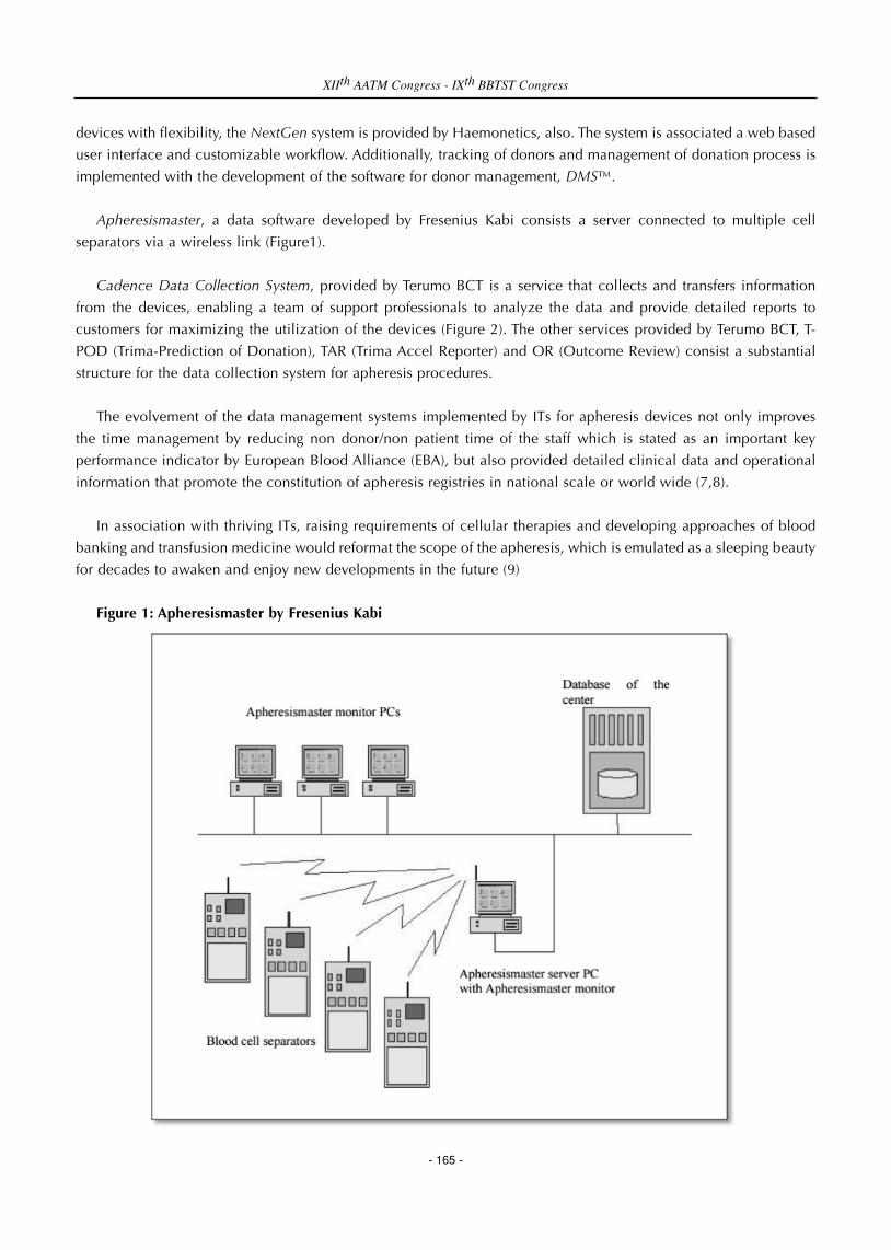
devices with flexibility, the NextGen system is provided by Haemonetics, also. The system is associated a web based
user interface and customizable workflow. Additionally, tracking of donors and management of donation process is
implemented with the development of the software for donor management, DMS™.
Apheresismaster, a data software developed by Fresenius Kabi consists a server connected to multiple cell
separators via a wireless link (Figure1).
Cadence Data Collection System, provided by Terumo BCT is a service that collects and transfers information
from the devices, enabling a team of support professionals to analyze the data and provide detailed reports to
customers for maximizing the utilization of the devices (Figure 2). The other services provided by Terumo BCT, T-
POD (Trima-Prediction of Donation), TAR (Trima Accel Reporter) and OR (Outcome Review) consist a substantial
structure for the data collection system for apheresis procedures.
The evolvement of the data management systems implemented by ITs for apheresis devices not only improves
the time management by reducing non donor/non patient time of the staff which is stated as an important key
performance indicator by European Blood Alliance (EBA), but also provided detailed clinical data and operational
information that promote the constitution of apheresis registries in national scale or world wide (7,8).
In association with thriving ITs, raising requirements of cellular therapies and developing approaches of blood
banking and transfusion medicine would reformat the scope of the apheresis, which is emulated as a sleeping beauty
for decades to awaken and enjoy new developments in the future (9)
Figure 1: Apheresismaster by Fresenius Kabi
XIIth AATM Congress - IXth BBTST Congress
- 165 -

Figure 2: The Cadence Data Collection Sytem by Terumo BCT
References1. Williamson LM, Devine DV. Challenges in the management of the blood supply. Lancet 2013; 381: 1866–
752. Sharma G, Parwani A, Raval JS, TriulziDJ, Benjamin RJ, Pantanowitz L. Contemporary issues in transfusion
medicine informatics. J Pathol Inform 2011; 2: 33. Magrabi F, Ong MS, Runciman W, Coiera E. Using FDA reports to inform a classification for health
information technology safety problems. J Am Med Inform Assoc 2012; 19:45-434. U.S. Food and Drug Administration. Medical Device Data Systems, Medical Image Storage Devices, and
Medical Image Communications Devices. Doc. no:1400021, 20155. Bessos H, Fraser R, Seghatchian J. What’shappening – Scotblood 2006 Advances in immunetolerance,
information technology, microarrays and pathogen inactivation in transfusion medicine. Trans Apher Sci2006; 35: 173–177
6. Colligan D, McGowan N, Seghatchian J. What’s happening – Scotblood2012: Transfusion/transplantmedicine in the 2nd decade of the 21st century. Trans Apher Sci 2013; 48: 415-420
7. Nyberg-Oksanen E, Morgan S, Sydenham V, Georgsen J et al. Managing donors and donation sessions toimprove donor service and session productivity. ISBT Science Series 2016; 11 (Suppl. 1): 277–284
8. Wu Y. Report Of The ASFA Apheresis Registry. ASFA&WAA Joint Conference (15th WAA Congress at the ASFA2014 Annual Meeting) San Francisco, USA 2014
9. Granata A, Drezet A, Faucher C et al. New developments in the use of apheresis to collect haematopoieticcells for cell transplantation and cell therapies.ISBT Science Series 2016; 11 (Suppl. 1):100–104
XIIth AATM Congress - IXth BBTST Congress
- 166 -

- 167 -
Background
Neutrophil granulocytes are the most abundant type of leukocytes and crucial in protection against bacterial and
fungal infection. A persisting reduction in neutrophil numbers is called neutropenia. The absolute neutrophil count
(ANC) in the peripheral blood is used for classification of neutropenia into mild (1.0– 1.5 x 10⁹/l), moderate (0.5–
1.0 x 10⁹/l), and severe (<0.5 x 10⁹/l). In clinical practice neutropenia is generally observed as febrile neutropenia,
a complication after intensive chemotherapy or hematopoietic stem cell transplantation (HSCT). On the other hand,
congenital neutropenia related to different monogenetic entities, such as ELANE, GFI1, HAX1, G6PC3, and GCSF3R
deficiency [1-3], and neutrophil functional disorders such as chronic granulomatous disease [4-6], leukocyte
adhesion defect [7], and myeloperoxidase deficiency [8] may be rarely seen.
Febrile neutropenia is a common emergency encountered in children receiving chemotherapy for malignancies
or HSCT. Left untreated, it can lead to acute and life-threatening invasive bacterial and fungal infections such as
brain abscess, cellulitis, pneumonia, sepsis, chronic stomatologic infections such as recurrent aphthosis,
paradontopathy with serious morbidity and mortality [9,10]. Febrile neutropenia is suspected in any patient on
chemotherapy who presents with fever. Prompt evaluation and management is essential for a better outcome. Initial
stabilization, prompt initiation of appropriate antibiotics and adequate supportive care are the cornerstone of
treatment. Although broad-spectrum antibiotics and antifungal therapies had become the mainstay treatment of
febrile neutropenia, they could not control infections in all cases and these patients require additional treatment
strategies.
Granulocyte transfusions can be used as supportive therapy in patients with life-threatening neutropenia caused
by bone marrow failure or in patients with neutrophil dysfunction. With the advent of potentially curative intensive
chemotherapy regimens used alone or in combination with stem cell transplantation, there has been an increase in
patients with fungal infections during periods of prolonged neutropenia and subsequently, a renewed interest in the
use of granulocyte transfusions to support these patients [6,9,11-15]. Despite the potential availability of this
component, there is limited published literature on the in vivo efficacy of whole blood derived granulocyte
concentrates [16-20].
Clinical indications for Granulocyte transfusion:
Therapeutic granulocyte transfusions may be indicated for patients with severe neutropenia who fulfill all of the
following criteria:
I. Severe neutropenia, defined as ANC <0.5 x 10⁹/L due to congenital or acquired bone marrow failure
syndromes.
II. Receiving active chemotheraphy in an attempt to achieve disease remission.
THERAPEUTIC EFFICACY OF GRANULOCYTE APHERESIS
Ekrem ÜNAL

III. Proven or highly probable fungal or bacterial infection that is unresponsive to appropriate antimicrobial
therapy as demonstrated by visible spreading lesions on skin, mucosa or radiological examination.
IV. In whom neutrophil recovery is expected (ANC>0.5x10⁹/l) in the near future and / or in whom definitive
therapy of curative potential is planned.
V. Therapeutic granulocyte transfusions may also be indicated for patients with a known congenital disorder of
neutrophil function regardless of neutrophil count with proven or highly probable fungal or bacterial infection
unresponsive to appropriate antimicrobial therapy, demonstrated by visible spreading lesions on skin, mucosa
or radiological examination.
The current guidelines indicated that the granulocyte transfusion should not be issued for therapeutic use in
patients with bone marrow failure where neutrophil recovery is not anticipated to recur spontaneously and no
further active treatment is planned, sepsis in the absence of either neutropenia or known neutrophil dysfunction,
fever of unknown origin [21].
Unstimulated vs stimulated granulocyte transfusion:
The use of a single injection of G-CSF alone or combined with a single oral dose of steroids has enabled the
collection of significantly greater yields of granulocytes by apheresis. Using this method, doses of granulocytes in
excess of 5 x 10¹⁰ cells can be produced for larger children and adults. The ability to collect greater numbers of
granulocytes has been a major factor influencing the rekindling of interest in the potential role of granulocyte
transfusions as additional therapy for patients with neutropenia and established infections. Studies with promising
but overall inconclusive results have been reported both in adults, and children.
There are a number of technical problems that make it difficult to collect adequate granulocyte doses for
transfusion. Normal donors do not have very high levels of circulating granulocytes in the peripheral blood and as
a result can donate doses of granulocytes that are only likely to be sufficient for very small children. Moreover
granulocytes are difficult to separate from other blood cells even if this has been facilitated by commercially
available long-chain starch solutions (sedimenting agents) such as hetastarch and pentastarch. The concerns grew
after multicentre randomised trials found that administration of hydroxyethyl starch in patients who had sepsis or
were critically ill was associated with a higher risk of kidney injury and bleeding, and more deaths in patients who
had sepsis, when compared with crystalloids. The trials prompted the German Federal Institute for Drugs and
Medical Devices to ask the European Medicines Agency (EMA) to review the use of HES in 2012 [22]. In stimulated
granulocyte regimen donations are collected from donors following the administration of G-CSF and the steroid
(dexamethasone) for transfusion to children or adults. The exposure of a healthy volunteer donor to any form of
medication with potential side effects does however present ethical and safety issues (this will be discussed in the
following phrases). The guidelines of European Committee (Partial Agreement) On Blood Transfusion (Cd-P-Ts)
Granulocyte preparations (Dosage, Storage, Release Transport, Compatibility testing, Irradiation)
Donors should be tested for the blood-transmissible diseases HIV, hepatitis B and C, HTLV-I/II and syphilis. The
hospital blood bank should be advised to carry out appropriate compatibility testing according to local practice. In
view of the high red cell content, granulocyte preparations should be compatibility tested in the same manner as
red cells. ABO and RhD matching is essential, as is the determination of irregular erythrocyte and HLA antibodies,
and the performance of erythrocyte cross matches and (in the case of HLA antibodies) granulocyte cross matches.
Major or minor ABO incompatibility does not form an absolute contra-indication, but does create a risk of acute or
delayed hemolytic transfusion reactions. In the case of major or minor incompatibility between the donor and the
XIIth AATM Congress - IXth BBTST Congress
- 168 -

patient, measures (tailored to antibody titres) should be implemented to reduce the number of red blood cells and
plasma if relevant. Reactive HLA and/or HNA antibodies between donor and patient should be considered as a
contra-indication for the donor involved.
Typically, granulocyte suspensions are prepared for a specific patient and administered immediately. Generally
storage is not recommended since the half life time of granulocyte suspensions are very short. The granulocyte
component has a short shelf-life, with infusion within 6 hours of collection being preferable. The maximum shelf-
life for granulocyte components is 24 hours. However the volume of the product may cause problem especially for
low weighted recipients causing volume overload. Oymak et al. [12] reported that splitted products can be used but
the ANC increment of primer product was higher than that of splitted product.
There is not any fixed component specifications have been agreed upon due to the large individual donor and
patient variation. However, regarding the number of granulocytes per component and in accordance with the
European Guidelines, a minimum of 1 x 10¹⁰ granulocytes per component is advised [23]. The initial dose of
granulocytes per kilogram of body weight for the patient is preferably > 8 x 10⁸/kg. Doses of at least 1 x 10¹⁰
granulocytes per transfusion appear to be required to treat or prevent infection [23].
Granulocytes are stored at 22+2ºC ideally without agitation. If granulocytes are agitated in error this does not
preclude their transfusion as there is limited evidence that agitation affects them functionally [24,25]. Granulocytes
must be irradiated to prevent transfusion associated graft versus host disease.
Safety
The guidelines of European Committee (Partial Agreement) On Blood Transfusion (Cd-P-Ts) reported that the
potential donor of granulocytes needs to receive medication, and sedimenting agents may be needed during the
apheresis procedure. Both of these have potentially severe side-effects that need to be communicated to the donor
as part of the informed consent process. In addition to the recognised complications of routine donor apheresis, the
following side-effects may occur.
• Hydroxyethyl starch (HES): acts as a volume expander. Donors who have received HES may experience
headaches or peripheral oedema because of expanded circulatory volume. HES may accumulate, which can result
in pruritus, and allergic reactions are possible.
• Corticosteroids: may cause, for example, hypertension, diabetes mellitus, cataracts, peptic ulcer, and
psychiatric problems.
• Granulocyte colony-stimulating factor (G-CSF): the most common short-term complication following G-CSF
administration in peripheral blood stem cell (PBSC) donors is bone pain; although, on very rare occasions, splenic
rupture or lung injury may occur. Concerns relating to the development of acute myeloid leukaemia
(AML)/myelodysplasia (MDS) after G-CSF administration are based primarily on reports of increased rates of these
disorders among women with breast cancer who received chemotherapy or patients with severe chronic neutropenia
who received G-CSF support. To date, however, registry data from Europe and the United States have not identified
any increased risk of AML/ MDS in over 100 000 healthy individuals who donated PBSCs and received G-CSF as
pre-treatment. The median follow-up of these studies is, however, less than 5 years. Therefore, if G-CSF is given to
a donor, a protocol for long-term follow-up should be in place (as advised by JACIE/FACT) [20, 26].
Adverse events such as febrile reactions, occasional severe pulmonary reactions and HLA (human leucocyte
antigen) alloimmunisation are well recognized complications in the recipients of granulocyte transfusions.
XIIth AATM Congress - IXth BBTST Congress
- 169 -

Efficiency of Granulocyte transfusions
There are controversital findings about the efficiency of granulocyte transfusions. Non randomized studies
showed benefits of granulocyte transfusions especially for patients with severe invasive fungal infections [6,9,11-15].
Currently, Estcourt et al. [18] reported an update of a Cochrane review which was first published in 2009. The
authors concluded that in people who are neutropenic due to myelosuppressive chemotherapy or a hematopoietic
stem cell transplant, there is low-grade evidence that prophylactic granulocyte transfusions decrease the risk of
bacteraemia or fungemia. There is low-grade evidence that the effect of prophylactic granulocyte transfusions may
be dose-dependent, a dose of at least 1.0 x 10¹⁰ per day being more effective at decreasing the risk of infection.
There is insufficient evidence to determine any difference in mortality rates due to infection, all-cause mortality, or
serious adverse events. However, Metzendorf et al. [19] recently underlined the tricks and pitfalls of taking well-
informed decisions or carry out sound research via different literature database.
Conclusion
The issue of efficacy of granulocytes (either therapeutically for refractory infection or as secondary prophylaxis
for high risk groups of patients with prior severe infection) is still very much an open question. Possible future
clinical studies of this component to address efficacy in patients with neutropenia will need to evaluate how best to
use the component alongside granulocytes for transfusion collected by apheresis from GCSF/steroid stimulated
donors. New studies to definitively address the issue of effectiveness are required
References1. Hauck F, Klein C. Pathogenic mechanisms and clinical implications of congenital neutropenia syndromes.
Curr Opin Allergy Clin Immunol. 2013 Dec;13(6):596-606. 2. Triot A, Järvinen PM, Arostegui JI, et al. Unal E, Klein C. Inherited biallelic CSF3R mutations in severe
congenital neutropenia. Blood. 2014 Jun 12;123(24):3811-7. 3. Patiroglu T, Gungor HE, Triot A, Unal E. Severe congenital neutropenia in two siblings related to HAX1
mutation without neurodevelopmental disorders. Genet Couns. 2013;24(3):253-8. 4. Patiroglu T, Gungor HE, Unal E, Koker Y. The clinical features and genetic mutations of chronic
granulomatous disease: results from a reference centre at middle Anatolia. Genet Couns. 2013;24(3):327-35.
5. Patiroglu T, Gungor HE, Lazaroski S, Unal E. Chronic granulomatous disease with markedly elevated IgElevels mimicking hyperimmunoglobulin E syndrome. Acta Microbiol Immunol Hung. 2013 Jun;60(2):155-62.
6. Patiroglu T, Unal E, Yikilmaz A, Koker MY, Ozturk MK. Atypical presentation of chronic granulomatousdisease in an adolescent boy with frontal lobe located Aspergillus abscess mimicking intracranial tumor.Childs Nerv Syst. 2010 Feb;26(2):149-54.
7. van de Vijver E, Maddalena A, et al, Unal E, Patiroglu T, Belohradsky BH, Schwartz K, Somech R, KuijpersTW, Roos D. Hematologically important mutations: leukocyte adhesion deficiency (first update). Blood CellsMol Dis. 2012 Jan 15;48(1):53-61.
8. Patıroğlu T, Eke Güngör H, Belohradsky JS, Unal E, Klein C. Myeloperoxidase deficiency: the secret underthe flag of unstained cell. Turk J Haematol. 2013 Jun;30(2):232-3.
9. Patiroglu T, Unal E, Karakukcu M, Ozdemir MA, Tucer B, Yikilmaz A, Deniz K. Multiple fungal brainabscesses in a child with acute lymphoblastic leukemia. Mycopathologia. 2012 Dec;174(5-6):505-9.
10. Kose D, Paksoy Y, Koksal Y, Unal E. Neurological complication of non Hodgkin lymphoma in childhood:experience from a single center in Turkey. Childs Nerv Syst. 2014 Apr;30(4):639-45.
11. Atay D, Ozturk G, Akcay A, Yanasik M, Anak S, Devecioglu O. Effect and safety of granulocyte transfusions
XIIth AATM Congress - IXth BBTST Congress
- 170 -

in pediatric patients with febrile neutropenia or defective granulocyte functions. J Pediatr Hematol Oncol.2011 Aug;33(6):e220-5.
12. Oymak Y, Ayhan Y, Karapinar TH, Devrim I, Ay Y, Sarihan H, Vergin C. Granulocyte transfusion experiencein pediatric neutropenic fever: Splitted product can be an alternative? Transfus Apher Sci. 2015Dec;53(3):348-52.
13. Bozkaya IO, Kara A, Yarali N, Cagli A, Turgut S, Tunc B. Numerous granulocyte transfusions to a patient withsevere aplastic anemia without severe complication. Transfus Apher Sci. 2013 Jun;48(3):371-3.
14. Oztürkmen S, Altuntaş F, Olcay L. Granulocyte transfusion therapy in paediatric patients with severeneutropenic infection. Transfus Apher Sci. 2013 Jun;48(3):381-5.
15. Averbuch D, Engelhard D, Pegoraro A, Cesaro S. Review on efficacy and complications of granulocytetransfusions in neutropenic patients. Curr Drug Targets. 2016 Jan 31. [Epub ahead of print] PubMed PMID:26844563.
16. Yoshihara S, Ikemoto J, Fujimori Y. Update on granulocyte transfusions: accumulation of promising data, butstill lack of decisive evidence. Curr Opin Hematol. 2016 Jan;23(1):55-60. doi:10.1097/MOH.0000000000000203.
17. Strauss RG. Neutrophil/granulocyte transfusions collected from G-CSF + dexamethasone-stimulated donors.Curr Opin Hematol. 2015 Nov;22(6):565-7.
18. Estcourt LJ, Stanworth S, Doree C, Blanco P, Hopewell S, Trivella M, Massey E. Granulocyte transfusions forpreventing infections in people with neutropenia or neutrophil dysfunction. Cochrane Database Syst Rev.2015 Jun 29;6:CD005341.
19. Metzendorf MI, Schulz M, Braun V. All Information Is Not Equal: Using the Literature Databases PubMed andThe Cochrane Library for Identifying the Evidence on Granulocyte Transfusion Therapy. Transfus MedHemother. 2014 Oct;41(5):364-74.
20. Bank I, Wiersum-Osselton JC, Van Walraven SM, Netelenbos T, Fechter M, Marijt-van der Kreek T, Bär BM,Van der Bom JG, Brand A. Donors' health state the year after peripheral haematopoietic progenitor cellcollection: A prospective follow-up study in related and unrelated donors compared to first-time plateletdonors. J Clin Apher. 2016 Jan 27. doi: 10.1002/jca.21444.
21. Elebute M, Massey E, et al. Clinical Guidelines for the use of Granulocyte Transfusions. [available from:http://www.bcshguidelines.com/index.html accessed 15 March 2016].
22. Hartog CS, Natanson C, Sun J, Klein HG, Reinhart K. Concerns over use of hydroxyethyl starch solutions.BMJ. 2014 Nov 10;349:g5981.
23. The guidelines of European Committee (Partial Agreement) On Blood Transfusion (Cd-P-Ts) EDQM 18thEdition 2015
24. Miyamoto M, Sasakawa S. Studies on granulocyte preservation. III. Effect of agitation on granulocyteconcentrates. Transfusion. 1987 Mar-Apr;27(2):165-6.
25. Seidel MG, Peters C, Wacker A, Northoff H, Moog R, Boehme A, Silling G, Grimminger W, Einsele H.Randomized phase III study of granulocyte transfusions in neutropenic patients. Bone Marrow Transplant.2008 Nov;42(10):679-84.
26. JACIE/FACT [Internet]. International Standards for Cellular Therapy Product Collection, Processing andAdministration. [available from: www.jacie.org, accessed 15 March 2016].
XIIth AATM Congress - IXth BBTST Congress
- 171 -

- 172 -
Quality management in blood banks relates to all the actions in order to ensure the quality and safety of blood
products. Quality programs in blood banks warrants the entire process starting from the safety of donors during
blood collection until the delivery of the final product. According to National Turkish Blood and Blood Products
Guide every blood service unit should have a validation policy with the following features.
1. Supports the quality system
2. Clearly explains the thing or things that needs to be validated according to the standards, guides and
regulations
3. Clearly identifies the validation process and the targets in the system
4. Should undertake that the critical processes and the systems are valid
Validation is the entire work done to ensure that the intended quality conditions are met and to prove that the
performance of the system is continious, accurate and precise. The use of a validated method warrants that the
procedure is accurate, specific, consistent, valid and reliable. Validation should be done when a new system
(informatic system or equipment) is implemented or when a process is carried out fort he first time. In the case of a
change in the current process validation is performed as a part of the change control. Validation also has targets such
as control demonstration, ensurance of compliance, data collection, providing information and determination of
future needs.
Validation protocol or validation master plan (VMP) is a document that specifies how the validation will be
conducted, the testing parameters to be used, product (result) characteristics and decision points to determine
acceptable test results. According to the European GMP directive: “All validation activities should be planned. The
key elements of a validation program should be clearly defined and documented in a VMP or equivalent
documents”. For validation of commercial instruments and systems the 4Q model can be used which has phases
such as design qualification (DQ), installation qualification (IQ), operational qualification (OQ), performance
qualification (PQ). The process is illustrated in Figure 1.
DQ is the documented collection of activities that define the functional and operational specifications of the
instrument and criteria for selection of the vendor, based on the intended purpose of the instrument. IQ demonstrates
that the instrument is properly installed in environmental conditions that meet the manufacturer’s specifications. OQ
demonstrates that the installed equipment operates as intended. It is the documented collection of activities
necessary to demonstrate that an instrument will function according to its operational specification in the selected.)
PQ demonstrates that the equipment performs as expected for its intended use in the facility and that the output
meets specifications. The criteria which indicates acceptable performance must be defined. PQ is the documented
collection of activities necessary to demonstrate that an instrument consistently performs according to the
specifications defined by the user, and is appropriate for the intended use. All activities related to validation process
VALIDATION PROCEDURE OF APHERESIS INSTRUMENTATION
Servet Uluer BİÇEROĞLU

should be documented in a validation report and the conclusions should also be defined. If unexpected results are
obtained during validation testing, it should also explain what changes will need to be made or what workarounds
will be implemented to mitigate the risks.
According to the Guide to the preparation, use and quality assurance of blood components released in 2015 by
the European Directorate for the Quality of Medicines & Health Care; ‘All equipment and technical devices must be
used in accordance with validated procedures (Directive 2005/62/EC Annex 6.4.1). The processing of blood
components must be carried out using appropriate and validated procedures, including measures to avoid the risk
of contamination and microbial growth in the prepared blood components (Directive 2005/62/EC Annex 6.4.2).’
According to the same guide all automated systems and processes should be validated. All platelet units collected
by apheresis should be screened for volume, platelet content, residual leucocyte content and the pH should be
measured at the end of shelf-life. The volume of all apheresis platelet units should be > 40 mL per 60 ×109 of
platelets. The platelet content standard unit should be minimum 2 × 10¹¹ per unit and one per cent of all units with
a minimum of 10 units per month should be screened. Residual leucocyte content should be < 0.3 × 10⁹ per unit
and one per cent of all units with a minimum of 10 units per month should be screened. The pH measured (+ 22
°C) at the end of the recommended shelf-life should be > 6.4 and one per cent of all units with a minimum of 4
units per month should be screened.
Plateletapheresis instrumentation validation is required to document that a new or modified instrument or
technique is capable of consistently producing acceptable products at the blood service. Although the instruments
have CE mark or FDA approved they need to be validation at each center. Validation is required when a new
instrument or technique (process) is used that could affect the quality of the product. According to the FDA, each
apheresis system and each type of product (single, double, triple) need to be validated separately. The parameters to
be validated are at the may vary among the blood services center. At least product volume, platelet yield and WBC
content (incase the product is leukoreduced) need to be validated, and 5-day pH needs to be monitored if products
are to be stored up to five days. FDA’s product performance qualification criteria for platelet component collection
is shown in Table-1.
XIIth AATM Congress - IXth BBTST Congress
- 173 -

Figure-1 Phases of validation
Design Qualification
• Compare user requirements with supplier specifications
• Supplier assessment
Installation Qualification
• Verify environment
• Verify arrival as purchased
• Check proper installation of hardware and software
Operational Qualification
• Test of operational functions
• Performance testing
• Test of security functions
Performance Qualification
• Test for specified application
• Preventive maintanance
• On-going performance test
XIIth AATM Congress - IXth BBTST Congress
- 174 -

Table 1. Product Performance Qualification Criteria for the Platelet Component Collection
1, 2 Process failures only; non-process failures should be excluded.3 Corrective actions for exceeding allowable process failures
• if you select a sample size of 11 and find one failure, 17 additional samples would need to be tested with
no additional failures.
• if you select a sample size of 60 and find one failure, 91 additional samples would need to be tested with
no additional failures. If you select a sample size of 93 and find two failures, 157 additional samples should
be tested with no failures. If you select a sample size of 124 and find three failures, 127 additional samples
should be tested with no failures.
* 95% confidence that greater than 75% of the components meet the standard.
** The sample size numbers can be used in a sampling plan that should be representative of products collected
on each machine type in each facility.
*** 95% confidence that greater than 95% of the components meet the standard.
**** Or per the container/automated blood cell separator device manufacturer’s specifications
***** The stratified recommended results should ensure that the individual transfusabl&e units will be
< 5.0 x 10⁶ even with a 25% error in equilibration of the volume for double and triple collections.
References1. Burgstaler EA, Pineda AA. Plateletapheresis: instrumentation validation. Transfusion Science 21;(1999):153-
161. 2. Guidance for Industry and FDA Review Staff. Collection of Platelets bu Automated Methods. December
2007. 3. European Directorate for the Quality of Medicines & Health Care. Guide to the preparation, use and quality
assurance of blood components Recommendation No. R (95) 15 18th edition 2015. 4. Ludwig Huber. Analytical Instrument Qualification and System Validation. Agilent Technologies, Germany,
January 1, 2009. Publication Number 5990-3288EN. 5. Strengers P.F.W. Quality management of blood collection. ISBT Science Series (2012); 7:12-15.
XIIth AATM Congress - IXth BBTST Congress
- 175 -
- 176 -
Hematopoietic stem cell transplantation (HSCT) has revolutionized the treatment of hematologic malignancies,
bringing substantial improvements to survival outcomes for many patients. However, infection, graft-versus-host dis-
ease (GVHD), and relapse are still the most challenging sequelae to address to improve the outcomes of all patients
after allogeneic transplantation.
History of HSCT began with bone transplant in the 1950s. Today, it has become a model of adoptive immunother-
apy using cellular manipulations. We see that the first cellular manipulation started with purification and cryopreser-
vation of collected bone marrow in 1970s. 1990s was the period that first peripheric stem cell collection was made
for HSCT. The second half of 1990s, we began to see T and B cell reduction from peripheric or bone marrow stem
cell suspensions. In 2000s, donor lymphocyte infusion (DLI), virus specific T (VST) cells and mesenchymal stem cells
(MSC) began to be implemented in stem cell transplants. Today, we see that cellular manipulations applied in stem
cell transplants increased and become more sophisticated. The cause of all this manipulations are to reduce the
basic transplant complications and to increase transplant success. Today, applied cellular manipulations and some
of their details are summarized below.
1. Cellular manipulations for GVHD prevention
a. CD34 selection,
b. CD3 or CD3 and CD19 depletion,
c. αβ-T cell depletion,
d. CD45RA depletion,
2. Cellular manipulations in GVHD treatment
a. MSC (autologous, allogeneic-donor origin/third party)
b. Donor regulatory T lymphocyte (T-reg) infusions
3. Cellular manipulations for augmentation of graft versus tumour effect(GVT),
a. DLI,
b. T/ ��-T cell depleted DLI,
c. Tumour specific T cell infusions,
d. High affinity T-cell receptors or chimeric antigen receptors (CAR) positive T cell infusions,
e. KIR mismatch NK cell infusions,
f. CAR positive NK92 cell line infusions,
4. Cellular manipulations for virus specific treatment
a. Virus specific T cells
b. CD45RA depleted DLI
CELLULAR MANIPULATIONS IN STEM CELL TRANSPLANTATION
Ercüment OVALI
Cellular manipulations for GVHD prevention
Alloreactivity in stem cell transplantation is a major player of rejection, engraftments, GVHD and GVT in HSCT.
In early period of cellular manipulations, pure CD34 positive cell transplantation was studied. Engraftment failure,
rejection and/or relapse were major problems in using CD34 positive only cell infusions in HSCT. Therefore, this
method was abandoned, especially in allogeneic transplants. Studies on T cell depletion protocols in second period
of cellular manipulations of HSCT showed that T cell depletion could control the GVHD. But, this protocol had the
same problems as engraftment, rejection and relapse also. Studies in the coming years showed that αβ-T-cell recep-
tor (1) and CD45RA positive T cells (2) have a major roles in GVHD while γδ T cell receptor, CD45RO positive T
cells and NK cells have important roles in GVT (3). Therefore, αβ-T cell reduction from stem cell product arose as
an option, particularly in haplo-identical HSCT. However, this model has some problems as extended immune sup-
pression, high rate of engraftment failure and especially increased viral infections.
A new method studied in recent years gives new opportunities in HSCT. According to method, CD45RA positive
cells (naïve T cells) are depleted from remaining stem cell product after CD34 positive selection (Because of some
CD34 positive cell are CD45RA positive). Remaining CD45RO positive T and CD34 positive cells are infused togeth-
er. Early results showed that it has optimal engraftment rates with low GVHD, low viral infection rate and no
increased relapse risk (2).
Cellular manipulations for GVHD treatment
MSC has strong regulator and immune suppressive effect on immune system. These features of MSC makes it an
important option for keeping aGVHD under control. Studies exhibit that response rate is about %50-70 in aGVHD
with MSC therapy (4).
Cellular manipulations for graft versus tumour effect (GVT) augmentation
In case of relapse, post-transplant DLI infusion is the first cellular therapy protocol currently used, but this pro-
tocol has low effectivity and high aGVHD risk. Therefore, αβ-T-cell receptor or CD45RA positive T cells depleted
DLIs are tried to control relapse without aGVHD risk. In recent years, a new cellular manipulation have been devel-
oped to control relapse as chimeric antigen specific receptor positive or high affinity T cell receptor positive T cell
infusions (5). These cells are formed with constructed gene transfer. Other promising cellular therapy products are
KIR mismatch NK or genetically modified NK cell lines (such as CAR positive NK92 cell line)(5).
Cellular manipulations in viral specific treatment
After HSCT, prolonged immunesuppression and viral infections, especially in cases where the reduction of T cells
is an important issue in the transplantation clinic. The Virus-specific T cells (VST) is a good option to treat viral infec-
tions. VST are isolated from donor or 3rd party donors and they are expanded to be prepared of VST suspensions. A
recent study showed that VSTs can control viral disease in %70 overall response rate (6).
All these challenges indicate that we leave the classic HSCT transplant soon. Future HSCT protocols will be the
safer and more effective with activation of cellular manipulations.
XIIth AATM Congress - IXth BBTST Congress
- 177 -

References1. Bertaina A, Merli P, Rutella S et al. HLA-haploidentical stem cell transplantation after removal of ab1 T and
B cells in children with nonmalignant disorders. Blood. 2014; 124(5): 822-826 2- Touzot F, Neven B, Dal-Cortivo L et al. CD45RA depletion in HLA-mismatched allogeneic hematopoietic
stem cell transplantation for primary combined immunodeficiency: A preliminary study. J Allergy ClinImmunol. 2015 May; 135(5): 1303-9.
3- Minculescu L, Sengeløv H. The role of gamma delta T cells in haematopoietic stem cell transplantation.Scand J Immunol. 2015 Jun; 81(6): 459-68.
4- McGuirk JP, Smith JR, Divine CL et al.Wharton's Jelly-Derived Mesenchymal Stromal Cells as a PromisingCellular Therapeutic Strategy for the Management of Graft-versus-Host Disease. Pharmaceuticals (Basel).2015 Apr 16; 8(2): 196-220.
5- Russell Cruz and Catherine M. Bollard. T-cell and natural killer cell therapies for hematologic malignanciesafter hematopoietic stem cell transplantation: enhancing the graft-versus-leukemia effect. Haematologica.2015; 100(6): 709-719.
6- Leen AM, Bollard CM, Mendizabal AM, et al. Multicenter study of banked third-party virus-specific T cellsto treat severe viral infections after hematopoietic stem cell transplantation. Blood. 2013 Jun 27; 121(26):5113-23.
XIIth AATM Congress - IXth BBTST Congress
- 178 -
- 179 -
Evidence from randomized clinical trials has shown that liberal red blood cell transfusion in the non-bleeding
may increase infection, circulatory overload and short term mortality in some patients. Liberal transfusion practice
has been the rule in Denmark, caused by too high pre-transfusion haemoglobin concentration (Hb trigger) combined
with a traditional dose of 2 red cell units. Single-unit dosing has been proven safe when applied together with a
restrictive Hb trigger in a number of randomized controlled trials. Transfusion of two or more RBC units in
succession is associated with an increased risk of pulmonary oedema or transfusion-associated circulatory overload
(TACO), especially in the elderly, females and patients with heart or kidney failure, or a positive fluid balance.
We established a Patient Blood Management programme to improve the quality of transfusion treatment. The goal
was to increase compliance with transfusion guidelines by decreasing Hb triggers and increasing single unit
transfusions, and thereby reducing unnecessary transfusion exposure in the 10 hospitals serving the 1.7 Million
citizens of Copenhagen. From 2009-11 the biggest hospital implemented restrictive transfusion practice and in
2011-13, 8 of the 9 remaining hospitals joined, 1 hospital declined to participate in the PBM intervention.
In all participating hospitals, transfusion above the guideline was reduced by 23-43 percent in participating vs.
13 percent in the non-PBM hospital. Transfusion compliance with the restrictive trigger increased with 26-100
percent in PBM hospitals vs. 16 percent in the non-PBM hospital. The percentage of single-unit transfusions
increased with 19-39 percent in PBM hospitals vs. 2 percent in the non-PBM hospital. Blood usage decreased with
15-24 percent per admission and 14-23 percent per operation in PBM hospitals vs. an increase of 6 percent and a
decrease of 2 percent respectively in the non-PBM hospital. At the regional level the percentage of patient transfused
with red cells was halved in elective hip surgery and reduced with 30 percent in CABG.
The implementation of restrictive transfusion practice increased the compliance with transfusion guidelines,
reduced the patients’ unnecessary exposure to blood transfusion and constituted the first phase of PBM in
Copenhagen.
HB TRIGGERS & SINGLE-UNIT TRANSFUSION:IMPLEMENTING EVIDENCE-BASED TRANSFUSION PRACTICE
Astrid NORGAARD

- 180 -
A randomised multicentre study was recently performed in the Netherlands on the integrated use of Patient BloodManagement interventions as erythropoietin (Epo), cell saver and/or postoperative drain re-infusion to evaluate theeffect on red blood cell (RBC) transfusion reduction and costs in 2442 adult elective hip-and knee surgery patientswhile applying a restrictive transfusion threshold. (1,2) Firstly, patients were stratified by the preoperativehemoglobin (Hb) level: low Hb (10- 13 g/dL) patients were randomized for Epo or no Epo; normal Hb patients(above 13 g/dL) were ineligible for Epo. Secondly, patients from both strata were randomized for autologous re-infusion by cell saver device or postoperative drain re-infusion device or for no blood salvage device. Outcomemeasures: RBC use and cost-effectiveness. Epo resulted in a significant 50% reduction of transfused patients (OR0.5, 95% CI: 0.35- 0.75) and a non-significant 29% mean reduction in RBC use (ratio 0.71, 0.42-1.13). Epo,however, increased costs by €785 per patient (262-1309), i.e. €7300 per avoided transfusion (1900-24000). In bothstrata, autologous blood re-infusion did not result in a RBC reduction and increased costs by €378 per patient (161-595).
It can be concluded, that in elective knee-and hip surgery, while using a restrictive transfusion trigger, autologousre-infusion by cell saver or postoperative drain re-infusion were no longer found effective in reducing RBC use andare consequently not cost-effective, while Epo, although it is effective in reducing RBC use, is not cost-effective dueto its high costs. Therefore, these interventions are no longer considered as appropriate transfusion alternatives inthis clinical setting. The finding that autologous blood re-infusions were no longer reducing RBCs was furtherexplored and explained in a subsequently performed meta-analysis, of which the results will be discussed as well.(3)
References1. Patient blood management in elective total hip- and knee-replacement surgery (Part 1): a randomized
controlled trial on erythropoietin and blood salvage as transfusion alternatives using a restrictive transfusionpolicy in erythropoietin-eligible patients. So-Osman C, Nelissen RG, Koopman-van Gemert AW, Kluyver E,Pöll RG, Onstenk R, Van Hilten JA, Jansen-Werkhoven TM, van den Hout WB, Brand R, Brand A.Anesthesiology. 2014 Apr;120(4):839-51. doi: 10.1097/ALN.0000000000000134.
2. Patient blood management in elective total hip- and knee-replacement surgery (part 2): a randomizedcontrolled trial on blood salvage as transfusion alternative using a restrictive transfusion policy in patientswith a preoperative hemoglobin above 13 g/dl. So-Osman C, Nelissen RG, Koopman-van Gemert AW,Kluyver E, Pöll RG, Onstenk R, Van Hilten JA, Jansen-Werkhoven TM, van den Hout WB, Brand R, Brand A.Anesthesiology. 2014 Apr;120(4):852-60. doi: 10.1097/ALN.0000000000000135.
3. Cell Salvage in Hip and Knee Arthroplasty: A Meta-Analysis of Randomized Controlled Trials. van Bodegom-Vos L, Voorn VM, So-Osman C, Vliet Vlieland TP, Dahan A, Koopman-van Gemert AW, Vehmeijer SB,Nelissen RG, Marang-van de Mheen PJ. J Bone Joint Surg Am. 2015 Jun 17;97(12):1012-21. doi:10.2106/JBJS.N.00315.
USE OF TRANSFUSION ALTERNATIVES INELECTIVE ORTHOPAEDIC SURGERY
Cynthia SO-OSMAN

- 181 -
The blood transfusion service (BTS) of a country is like its lifeline is an indispensable, potentially life-saving
medical intervention, and blood products such as clotting factors and some immunoglobulins are designated by
WHO as essential medicines. Regulatory issues and guidelines play a vital role in shaping the structure of this vital
service in any country because the inherent risks of blood and the complexity of providing adequate, timely and
equitable access to safe blood products require an organized national or regional blood regulatory system. To
achieve the goal of safe and adequate blood for all, WHO has advocated for the establishment and sustenance of
strong national regulatory authorities (NRAs) both in developed and developing countries. In in 2010, the World
Health Assembly urged Member States “to take all the necessary steps to update their national regulations on donor
assessment and deferral, the collection, testing, processing, storage, transportation and use of blood products, and
operation of regulatory authorities in order to ensure that regulatory control in the area of quality and safety of blood
products across the entire transfusion chain meets internationally recognized standards.”
In a hospital, all other medical services such as cardiac/renal care and even laboratory testing remain free of any
legal guidelines, the blood bank is always under the scanner of the local regulatory health authority.
The blood transfusion service (BTS) of countries in South Asia (barring a few) are plagued with certain common
problems such as
• Paucity of funds
• Shortage of voluntary donors
• Higher infection rates in donors
• Difficulty in implementation of uniform regulatory policies
• Fragmented services with private and government blood banks
Below is a brief
Singapore
More than 100,000 units of blood are required by patients in Singapore every year. Through a strategic
partnership with the Singapore Red Cross, the National Blood Programme works towards collecting sufficient blood
to fulfil this need. The Blood Services Group (BSG) of the Health Sciences Authority is responsible for collecting,
processing and distributing blood and blood components to all public and private hospitals in Singapore. The Blood
Services Group ensures that all Singaporeans have access to adequate and safe blood. The Group comprises the
following branches: Blood Resources, Blood Supply, Patient Services and Clinical Services.
Bhutan
The national blood transfusion service also termed as ‘blood bank service’ is one of the listed programs under
the Health Care and Diagnostic Division of Department of Medical Services, Ministry of Health. The national blood
BLOOD BANK REGULATORY FRAME WORKIN ASIAN COUNTRIES
Sangeeta PATHAK

policy has been endorsed by MoH in 2007 and the ten-year national blood plan was drafted in 2010 for finalization
and adoption. The national standards for BTS have been developed and endorsement by MoH recently in 2012-
2013. There are 27 blood banks in the country which collect about 8028 units out of which voluntary donation is
3686 (46%) and family relative donors are 4342 (54%). All blood banks are hospital based and managed by the
Royal Bhutan Government.
India
Blood has been treated as a 'drug' under the Drugs and Cosmetic Act (D and C Act), 1940 and Drugs and
Cosmetics Rules, 1945. In 1993, Supreme made it mandatory for all blood banks to be licensed licensing of blood
banks in the country mandatory and also directed that the National Blood Transfusion Council (NBTC) along with
State Blood Transfusion Councils (SBTC) be set up for monitoring the activity of blood banks in India. The Indian BTS
is highly fragmented with over 2500 blood banks, 9 Million blood donations and poses unique challenges. The
National Blood Policy (NBP) was published by the Government of India in 2002. The NBP reiterates government
commitment to safe blood and blood components and has well documented strategies, for making available
adequate resources, technology and training for improving transfusion services. It also outlines methods for donor
motivation and appropriate clinical use of blood by clinicians. Some improvements have been seen in India over
the last two decade with better qualified staff, rules laid down for minimum requirements in terms of space, staff and
equipment, public awareness of blood donation and also National Aids Control Organization (NACO) support for
blood safety.
Bangladesh
There are more than 131 blood banks under government sector and 45 blood banks in the private sector. Red
Crescent blood banks are another prominent force in the BTS in Bangladesh. To oversee regulation and development
of BTS, a high power committee named ′National Safe Blood Transfusion Council′ was formed in 2002. National
blood policy is at the stage of finalization by the government. As reported from the Safe Blood Transfusion
Programme 2012, currently more than 600,000 units of blood are required annually in Bangladesh and the
requirement is gradually increasing. Only 31% of the annual demand is fulfilled from voluntary blood donations,
while the rest comes from relatives or replacement donors. The use of blood component is only 17% which is
inadequate in implementation of specific use of blood products. Therefore, the improvement of the national blood
transfusion service is essential for addressing the challenges arising out of HIV, Hepatitis B & C, and other infectious
diseases.
South Korea
The Korean Red Cross (KRC) Blood services started in 1957. In 1981 the government entrusted the Blood services
to the KRC which is now actively involved in blood donation and donor recruitment. It is a centrally coordinated
and monitored blood transfusion service under the Ministry of Public Health. Korean Red Cross blood service is the
single largest domestic program, covering over 95% of South Korea’s total blood supply. The KRC Blood services
have a working relationship with government bodies, medical blood banks, and private blood center as a
components of blood service in Korea. The 16 blood centers, nearly 2.5 million blood donations, a blood transfusion
research institute, 3 NAT laboratories, a fractionation center are under the management of KRC Blood Service
Headquarters.
Indonesia
Indonesia has about 211 blood banks under Indonesian Red Cross (IRC) and another 150 hospital-based blood
banks under government control. Government-controlled blood banks are gradually becoming operational and IRC
blood banks take care of major part of the blood supply. IRC blood banks collect more than 2 million units per year
XIIth AATM Congress - IXth BBTST Congress
- 182 -

and 87% donation comes from voluntary source. About 70% of total collection is separated into components. IRC
blood banks distribute blood and components directly to the hospitals after doing cross matching test or the hospital
store and cross match as per their requirement. Indonesian BTS runs on cost recovery system; however, there is
government subsidy on different sectors.
Sri Lanka
National Blood Transfusion Service (NBTS) is established in Sri Lanka in 1950s and is an integral part of the
National Health Service. The main function of NBTS is to collect, process and deliver safe blood, blood components
and blood products through 19 cluster centres and 77 peripheral blood banks situated island wide. As of 2014, the
NBTS collected over 350,000 voluntary blood donations.
NBTS is a decentralized unit which comes under Ministry of Health, Sri Lanka. NBTS is the sole supplier of blood
and blood products to all state hospitals and some of the private hospitals which are registered under Ministry of
Health for supply of blood and blood products.Having its head quarters at National Blood Centre (NBC), NBTS has
96 blood banks island wide. The categorization of blood banks is as follows...
1. National Blood Centre - the head quarters.
2. Cluster Centres.
3. Peripheral Blood Banks.
Maldives
Maldivian Blood Services (MBS) is formed on 1st November 2012. "National Thalassaemia Centre (NTC)" and
"National Blood Transfusion Services (NBTS)" were merged to form Maldivian Blood Services with the hope and aim
to provide better service to patients suffering from hematological disorders like Thalassaemia and other
hemoglobinopathies and also to provide safe blood to those who are in need.. There are two multi-specialty
hospitals, one in government sector i.e. Indira Gandhi Memorial Hospital (IGMH) and another one in private sector.
IGMH has a basic level of modern blood bank with component preparation facility. Blood collection by IGMH is
about 300 units per month. There is another blood bank attached to National Thalassaemia Center (NTC) which
caters to only thalassaemia patients in the city as well as from other parts of the country. NTC collects about 500
units per months. Both the blood banks collect blood mainly from family replacement donors and directed donors
for their own patients. About 80% blood units are collected by this mechanism. There are few blood banks in other
islands of Maldives.
Myanmar
The Myanmar Red Cross Society (MRCS) initiated blood donation activities in 1961, and since then, has been
organizing regular blood donation activities. The MRCS supports the ‘national blood and blood product law’
(enacted in January 2003) to save patients lives through blood transfusion of quality assured blood and blood
products There is a nationally coordinated BTS managed by government. National Blood Center(NBC) at Yangon is
the technical hub for development of the BTS in the countryThe Blood Policy is in the stage of finalization and
subsequent implementation. Voluntary blood donation in the country is about 85%. National Blood Center
coordinates with Red Cross and other organizations for voluntary blood donation. It distributes components only in
the capital Yangon and more than 20,000 blood units are distributed per year.
Nepal
Blood Transfusion Service of Nepal Red Cross Society was established in the year 1966 i.e. 3 years after the
inception of the Society itself. The Central Blood Transfusion Service Center (CBTSC) in Kathmandu is responsible
for management of services in the Kathmandu valley, and for supervising and monitoring the technical standards of
the district centers, providing guidance to ensure the collection and supply of safe blood. Thus, the CBTSC not only
XIIth AATM Congress - IXth BBTST Congress
- 183 -

serves the blood transfusion needs of the people in Kathmandu valley, but also provides technical and other supports
to the blood service centres across the country. In the year 2008 nationwide 1,78652 units of blood and blood
products were provided to the patients from different blood centers. There are about 70 blood centers in 50 districts.
There is one Central Blood Transfusion service in the capital Kathmandu and four regional BTS (Pokhra, Nepalganj,
Biratnagar, Citwan). There are 21 district blood transfusion services; 19 emergency BTS and 25 hospital-based blood
transfusion services.
Malaysia
Blood services in Malaysia started in the 1950s, based on voluntary non-remunerated blood donation. Since
then, the provision of safe and quality blood and blood products through the national blood service has grown under
the Ministry of Health of Malaysia. In 2011, there were 627,518 whole blood donations to the MOH Blood Services
and 17,068 apheresis donations of plasma and platelets. Whole blood, red blood cells and other blood components
are provided free of charge for all patients in the public hospitals in Malaysia. Private hospitals pay service charge
which is heavily subsidized by the government.
Thailand
Thailand has one of the best BTS in this region. It is nationally coordinated and BTS is managed by the Thai Red
Cross Society. National Blood center is in the capital Bangkok and there are 12 other regional centers in provinces.
National blood policy was enacted about 15 years back and it was revised in 2010 to incorporate necessary
amendments. The National Center collected about 539,094 units in 2009. More than 93% of total collected blood
in the country is separated into components.
Pakistan
The total blood collection is more than 1.6 million. The BTS is not nationally coordinated and proper regulatory
mechanism is not available. As per unofficial sources, there are 450 hospital-based blood banks and 2357 private
blood banks. Many private blood banks maintain questionable quality standard and ethics. About 90% blood is
collected from family replacement donors and out of which 10% comes thorough ′unsafe′ paid donors. Disease
burden among blood donor is high and needs screening of all units with sensitive tests. 20-40% blood collected is
not even screened for disease markers especially in non-regulated private sectors. There are 13 regional blood
centers and under which 78 hospital-based blood banks are operating mainly in public sector. Two policies are
designed, one at national and another in ground level.
Vietnam
Through the International Development Association (IDA) funding and World Bank Group expertise, Vietnam is
now providing more and better quality blood in over 32 provinces through a modernized transfusion system. A new
network of transfusion centers is nationally coordinated to provide blood products free of the HIV/AIDS virus and
targeted for the right clinical uses. The World Bank is supporting Vietnam’s National Blood Transfusion Program,
which is aimed at increasing blood supplies through a higher number of voluntary non-remunerated blood donors.
Beyond building the country’s first blood centers with IDA support, the government has developed a national blood
policy and is working with other donors to build blood centers in other parts of the country. It has also recently
inaugurated the National Institute of Hematology and Blood Transfusion in Hanoi—a new facility under the Ministry
of Health—outfitted with modern medical technologies to diagnose and treat all blood-related diseases. It also has
the capacity to stock 150,000 units of blood per year.
References1. http://www.hsa.gov.sg/content/hsa/en/Blood_Services/About_Blood_Services_Group.html
XIIth AATM Congress - IXth BBTST Congress
- 184 -
2. http://www.who.int/bloodproducts/NationalBloodRegSystems.pdf3. http://www.who.int/bloodsafety/transfusion_services/BhutanComprehensiveAssessmentStudyBTServices.pdf4. Action Plan for Blood Safety. Available from http://www.hsph.harvard.edu/population/aids/india.aids. 07.pdf5. http://www.searo.who.int/bangladesh/areas/wbd_day_2013/en/6. http://www.redcross.or.kr/eng/eng_activity/activity_blood.do7. http://mbs.gov.mv8. http://www.ncbi.nlm.nih.gov/pmc/articles/PMC2798779/9. http://www.nrcs.org/program/blood-services10. http://medind.nic.in/aae/t11/i2/aaet11i2p117.htm11. http://english.redcross.or.th/news/information12. http://web.worldbank.org/WBSITE/EXTERNAL/PROJECTS/0,,contentMDK:22709621~pagePK:41367~
piPK:279616~theSitePK:40941,00.html
XIIth AATM Congress - IXth BBTST Congress
- 185 -

- 186 -
Blood Banking in present era is lifeline of health care. Every technology requires cost input so is blood products.
Era has changed from glass bottles to plastic bags, reusable material to disposables, slide to robust testing technology
and much more.
Developing of blood transfusion services is complex,costly and time consuming strategy.Blood Banking is under
“NOT FOR PROFIT” model and under control of Drug Authorities and respective government organizations,Eg-
NACO in India..At one side,technology and blood safety requires expenses,other side we have to work on Not For
Profit model and committed to provide safe blood at reasonable cost.
Working on costing involves all expenses direct or indirect,including any sort of freebies or support from
Government or grant from Non-Government Organizations.This sort of working is easier in stand alone blood banks
but difficult in Government set up where financial grant in various forms received and in hospital set up where
services among various departments are intermingled and difficult to work out actual cost involvement.In
addition,depends upon whether set up is dealing with whole blood/component,total no. of donations,type of
component made,usage of components and discard etc.
COST, PRICE & BUDGETS IN BLOOD BANKS
Amit AGRAWAL

While deciding for price of particular blood component,in addition to cost consideration should be given to rates
as fixed by Government agencies and/or through survey as done by centres in nearby area/same country or nearby
or far off countries.
Budgeting is the most important aspect in any industry. Any organization can survive only if budgeting is done
in proper fashion taking into consideration priorites, hidden expenses and futuristic vision.Budget in various heads
need to be decided based on capex purchase,maintenance,recruitment,market inflation,unexpected expenses etc.
versus income/grant/freebies or any other support expected.
References1. http://www.hsa.gov.sg/content/hsa/en/Blood_Services/About_Blood_Services_Group.html2. Action Plan for Blood Safety. Available from http://www.hsph.harvard.edu/population/aids/india.aids. 07.pdf3. Costing Blood Transfusion Services,World Health Organization 1998
XIIth AATM Congress - IXth BBTST Congress
- 187 -

- 188 -
Although serological tests are main screening methods in blood banking, importance of nucleic acid
amplification tests (NAT) are increasing day to day. It has been shown that the major residual transfusion risk arises
from donations taken from recently infected donors in the pre-seroconversion phase of infection. Routine screening
of blood and blood products by serological tests and NAT shortens seronegative “window period” of HBV, HCV and
HIV infections and detect variant viruses result in risk of transfusion transmitted infections (TTI) decreases .
Turkish Red Crescent is main blood and blood products supplier in Turkey. Totally 1937932 units whole blood
were donated to Turkish Red Crescent in 2015. All donated whole bloods and blood products has been screened by
NAT in Turkish Red Crescent’s laboratories since November 2014.
Cobas s201 system MP6 (Mini Pool 6) (Roche Diagnostic Inc. Switzerland) has been used by four Turkish Red
Crescent’s laboratories in Turkey. The Cobas s201 system is used for testing donor pools and resolution testing of
reactive pools to identify the reactive individual donor specimens. Cobas s201 system consist of three main
components: Hamilton Microlab Star Pipettor, Cobas AmpliPrep Instrument and TaqMan Analyzer. This system use
Cobas TaqScreen MPX test v2.0 test kit. The test kit is approved by FDA and CE-IVD. This kit is qualitative multiplex
(MPX) test that enables the simultaneous detection and discrimination of HIV RNA (HIV-1 group M, group O and
HIV-2), HCV RNA and HBV DNA. Reactive test results are confirmed by quantitative RT-PCR method with Abbott
m2000rt system (Abbott Molecular Inc. USA). This system use Abbott Real Time HBV, HCV and HIV kits (Abbott
Molecular Inc. USA).
The first step of the NAT algorithm of Turkish Red Crescent’s laboratories is pooling the donor plasma samples.
Pool of the six plasma samples is screened by multiplex RT-PCR method. Test results are transferred by Cobas s201
system to laboratory information system (LIS) and evaluated conjunction with serologic test results. If test results
detected as “non-reactive” blood products released from quarantine units. If NAT result of pooled plasma sample
detected as “reactive” resolution procedure started and every donor plasma sample in the pool individually screened
by NAT (ID-NAT). If ID-NAT and/or serologic test results detected as “reactive” related blood products are disposed
and infected blood donor is deferred permanently. Reactive donor samples are sent confirmation laboratory either
serologic and NAT reactivity.
NAT algorithm of Turkish Red Crescent’s laboratories is summarized in Figure-1.
YIELD OF NAT: DATA AND NAT ALGORITHM IN TURKEY
Levent HAYAT
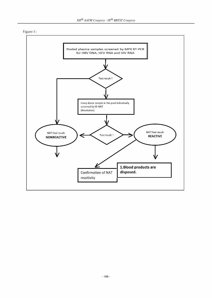
Figure-1:
XIIth AATM Congress - IXth BBTST Congress
- 189 -

Confirmation of NAT reactivity algorithm is summarized in Figure-2.
Figure-2:
The samples of 1436555 donations between November 2014 - August 2015 were screened by Turkish Red
Crescent’s laboratories. In this period, 7068 samples for HBV, 1774 samples for HCV and 2273 samples for HIV
were detected as reactive by serologically and/or NAT screening.
Serologic tests and NAT results of HBV, HCV and HIV are summarized in Table 1.
Table-1
Test name Number of reactive results % Total number
Only HBsAg 1702 24,1
HBV HBsAg + HBV DNA 4526 64 7068 Only HBV DNA 840 11,9
Only anti-HCV 1613 91
HCV anti-HCV + HCV RNA 144 8,1 1774 Only HCV RNA17 0,9
Only anti-HIV ½ 2165 95,3
HIV1/2 anti-HIV ½ + HIV RNA 92 4,1 2773 Only HIV RNA 16 0,6
XIIth AATM Congress - IXth BBTST Congress
- 190 -

Serologically nonreactive / NAT reactive donor screening results of Turkish Red Crescent laboratories are
summarized in Table-2.
Table-2
HBV HCV HIV 1/2 Total
N 840 17 16 873
% 0,058 0,012 0,011 0,061 (873/1436555)
In November 2014 – August 2015 period Turkish Red Crescent laboratories’ NAT yields were detected as 1/3288,
1/358700, 1/717149 for HBV, HCV and HIV respectively.
Main advantage of the NAT is able to detect serologically nonreactive donor. Totally 873 (%0,061) screening
results were detected as serologically nonreactive / NAT reactive in aforementioned time period. NAT is not decrease
importance of serologic screening tests and these tests are not used for each other. However, NAT screening has
significantly reduce seronegative “window period” of TTI agents and residual risk especially for HBV.
References1. Roth WK. Nucleic acid testing: general view in, Transfusion Microbiology. Barbara JAJ, Regan FAM,
Contreras MC Cambridge University Press 2008, 193-201.2. Saygan Mehmet Bakır. Türk Kızılayı’nda NAT uygulamaları ve NAT verilerinin paylaşılması. Sözel sunum.
VIII. Ulusal Kan Merkezleri ve Transfüzyon Tıbbı Kongresi 14-18 Aralık 2015 Antalya.
XIIth AATM Congress - IXth BBTST Congress
- 191 -

- 192 -
Leukoreduction is a process in which the white cells, ordinarily present in collected blood components, are
intentionally reduced in number. Through the use of centrifugation or filtration, 99.995% leukocyte reduction can
be accomplished. Leukoreduction may occur in two forms: prestorage filtration or poststorage filtration. The most
common form used in Europe and the USA is prestorage leukoreduction, which removes leukocytes before they can
contribute to the storage lesion (RBCs) or transfusion reactions (platelets/RBCs). The process allows the opportunity
for better quality control, has not been associated with acute hypotensive episodes, and eliminates the need for
transfusion services to manage fitler inventories and for nursing staff to maintain multiple blood administration
protocols.
However, Prestorage leukoreduction, at the present time, is more costly than poststorage leukoreduction.
Poststorage filtration may be beneficial because the filter may remove undesired substances that accumulate during
storage.
There are some accepted and some controversial advantages associated with universal leukoreduction (Table 1).
First, it is widely accepted that leukoreduction is an effective technology for reducing recurring transfusion reactions.
It is likely that the use of selective leukoreduced blood for these recipients eliminated many of the recurring
nonhemolytic transfusion reactions. It has been argued that the application of universal leukoreduction would result
in a substantial fall in reported transfusion reactions for RBCs. However, populations of patients who historically
have received nonleukoreduced blood (eg, surgical patients, patients with acute gastrointestinal bleeding) account
for a very small percentage of the total number of reported transfusion reactions. Because of this selection bias, when
an institution changes from selective leukoreduction to universal leukoreduction, only a small decrease (perhaps not
statistically significant) will be observed in the reported transfusion reaction rates, although formal studies designed
to detect milder reactions may show a significant decrease.
The assertion, therefore, that universal leukoreduction will have a dramatic impact on reported transfusion
reaction rates is likely exaggerated, and institutions converting from selective leukoreduction (which, for practical
purposes, reduces many of the transfusion reactions) to universal leukoreduction should have a low expectation that
there will be a material difference in reported transfusion reaction rates. In contrast, leukoreduced products certainly
will result in the reduction in primary HLA alloimmunization. A policy of selective leukoreduction for patients at
risk for transfusion complications resulting from anti–class 1 HLA antibodies probably mitigates most of this adverse
effect. Thus, the application of universal leukoreduction to prevent primary HLA alloimmunization in recipients for
whom no immediate or short-term benefit is anticipated is difficult to justify. However, younger recipients of blood
transfusion who become HLA alloimmunized by blood transfusion and require multiple platelet transfusions in the
future may constitute a delayed complication of non–leukoreduced blood transfusion, which is not realized until
many years after the initial transfusion event.
WHAT ARE THE ADVANTAGES AND DISADVANTAGES OFUNIVERSAL LEUKOREDUCTION
Berrin UZUN

Leukoreduction is a known effective strategy for reducing the transmission of cell-associated viruses. The most
prominent among these viruses are the herpesviruses, particularly cytomegalovirus (CMV or human herpesvirus
[HHV]-5). There also should be reduction in the transmission of other herpesviruses, such as Epstein-Barr virus
(HHV-4) and HHV-6 and HHV-8, although the clinical implications of this are largely unknown. Again, a policy of
selective leukoreduction probably achieves most of the same benefit of preventing primary CMV disease in
recipients at risk for this complication. However, the clinical implications of transmitting CMV infection by blood
transfusion to people who are not at risk for CMV disease are unclear. Conventional thinking is that this is
innocuous. At least on theoretical grounds, avoiding CMV infection by blood transfusion would be desirable, as a
current blood transfusion recipient may develop a medical condition at some time in the future for which CMV
reactivation could cause substantial morbidity, eg, a renal allograft recipient in which the occurrence of CMV
disease could be a serious, life-threatening postoperative complication. Of considerable importance (which has
received little attention) is that leukoreduction likely would be an effective strategy for preventing the transfusion-
associated transmission of some retroviruses, such as HTLV-I and HTLV-II, which are known to be transmitted only
by the transfusion of cellular blood products. This is an important principle in that a processing step (leukoreduction)
could obviate the need for a testing step (enzyme-linked immunosorbent assay for anti–HTLV-I and anti–HTLV-II).
There also is the question of improved RBC potency with prestorage leukoreduction. This effect on improving
quality (approximately 2%) tends to offset to some extent (although not totally) the reduction in quantitative potency
owing to the filtration process itself (approximately 5%-10% RBC loss). With the recent introduction, however, of
500 mL collections, the residual potency of many blood products collected is higher than in the era before universal
prestorage leukoreduction, even after filtration. It is unlikely, therefore, that filtration-associated potency loss will
result in a noticeable increase in total RBC units transfused.
There are additional possible benefits associated with universal leukoreduction. Leukoreduction filters bind
Trypanosoma cruzi promastigotes and may reduce the risk of transfusion-associated Chagas disease. In the case of
RBCs, bacterial sepsis from Yersinia enterocolitica may be reduced by prestorage leukoreduction of the RBC product.
Some cases of transfusion-related acute lung injury have been attributed to the presence of allogeneic leukocytes in
the stored RBC product, although most cases are due to the passive transfusion of anti-HLA antibodies, which react
with recipient neutrophils.
There are other possible areas that are controversial but have considerable implications for the benefits of
universal leukoreduction. First, there is literature on the value of leukoreduction (mostly poststorage) for preventing
postoperative complications in patients undergoing surgery. This is one of the more contentious and difficult areas.
There also is reported evidence of improvement in morbidity and mortality in cardiac surgery. Two reports have
been published, one involving a large series of patients in whom the mortality rate was 50% less in patients receiving
leukoreduced blood (regardless of whether it was prestorage or poststorage leukoreduced). The findings of this large
study suggest that leukoreduction should be applied for patients undergoing cardiac surgery in order to improve
patient outcome.
There are some useful practical aspects to be considered once a policy of universal prestorage leukoreduction is
adopted vs a policy of selective (poststorage) leukoreduction. From the transfusion service perspective, the
timeconsuming need to search for CMV-seronegative blood products can be eliminated. Inventory of multiple filters
for RBCs and platelets can be eliminated. Inadequate inventory control may cause these filters to be unavailable at
particular times, creating urgent situations and logistic difficulties. Furthermore, some patients (eg, in the operating
room) require transfusion of RBCs at a more rapid rate than is customary with elective RBC transfusions. Under these
XIIth AATM Congress - IXth BBTST Congress
- 193 -

circumstances, different filters capable of achieving leukoreduction at rapid RBC infusion rates need to be in stock.
Although it is difficult to estimate the amount of time and resources consumed in all of these processes, in the
aggregate, they are likely to be quite substantial. The application of a policy of universal leukoreduction has some
practical advantages for blood administration, whereby this process essentially can be standardized, and further
modifications of this process incorporating the use of bedside leukoreduction filters for subpopulations of patients
are no longer necessary. This eliminates the need for nurses to be trained specifically in the tech technology of using
bedside filters for defined (selected) transfusion recipients. Also, procedure manuals do not need to reference
specific filters, avoiding documentation difficulties filters is a perennial problem (particularly with platelets), causing
considerable frustration for patients and nurse transfusionists alike. Application of universal prestorage
leukoreduction is sometimes seen as improving the efficiency of transfusion, particularly for outpatients, not only in
terms of a reduction in transfusion reactions but also in improved patient throughput. Little attention has been given
these practical benefits of a policy of universal leukocyte reduction. associated with vendor changes. Furthermore,
clogging of filters is a perennial problem (particularly with platelets),30 causing considerable frustration for patients
and nurse transfusionists alike. Application of universal prestorage leukoreduction is sometimes seen as improving
the efficiency of transfusion, particularly for outpatients, not only in terms of a reduction in transfusion reactions but
also in improved patient throughput. Little attention has been given these practical benefits of a policy of universal
leukocyte reduction.
Disadvantages of Leukoreduction
The major disadvantages associated with leukocyte reduction are those of cost and logistics (Table 2). Logistics
and cost are clearly interrelated, since incurring extra expenditures and resource consumption is, when properly
applied, an effective means of overcoming logistic difficulties. As stated previously, there is some loss in product
potency associated with leukoreduction technology, although, at the present time, for RBCs this is unlikely to cause
an increased demand for blood transfusion because of the concurrent increase in the volume of whole blood
collections that offsets this loss in product potency. With regard to the loss in platelet potency, a substantial number
of platelets already are being leukoreduced in the institutions that use a policy of selective leukoreduction. In the
remaining platelet transfusion situations, eg, cardiac surgery and trauma, there is already considerable uncertainty
about the therapeutic dose of platelets, such that a 10% to 15% quantitative reduction is unlikely to be noticeable
clinically. As in the case of RBCs, the increase in whole blood collection will increase the platelet potency of whole
blood–derived platelets by 11%, negating the filtration effect.
Table 1: Advantages of Universal Leukocyte Reduction
• Reduction in total number of transfusion reactions
• Reduction in primary HLA alloimmunization
• Reduction or elimination in the transfusion transmission of CMV, HTLV-I, and other cell-associated viruses
such as EBV, HHV-6 and HHV-8
• Improvement in RBC quality
• Possible reduction in
Parasite transmission (Toxoplasma gondii)
Bacterial sepsis from RBCs
Prion disease transmission by blood transfusion
Transfusion-related acute lung injury
• Possible reduction in transfusion-associated morbidity and mortality
Postoperative infections in abdominal surgery
XIIth AATM Congress - IXth BBTST Congress
- 194 -

Morbidity or mortality in cardiac surgery
Organ dysfunction in patients in the ICU
• Simplifies inventory management; avoids errors; reduces transfusion service workload
• Simplifies blood administration; no requirement to maintain nursing procedures for special filter use,
training, filter failure: improves efficiency of nursing time
CMV, cytomegalovirus; EBV, Epstein-Barr virus; HHV, human herpesvirus; HTLV, human T-lymphotropic virus; ICU, intensive care unit.
Table 2: Disadvantages of Universal Leukocyte Reduction
• Cost
• Logistics
2% loss of red blood cells
Platelet loss of 11% due to trapping
References1. Shapiro MJ. To filter blood or universal leukoreduction: what is the answer? Critical Care 2004, 8 (Suppl
2):S27-S30 (DOI 10.1186/cc2453)2. XVIII. Ulusal Kan Merkezleri ve Transfüzyon Tıbbi Kursu. Temel Kurs Kitabı3. Sweeney JD. Universal Leukoreduction of Cellular Blood Components in 2001? Am J Clin Pathol
2001;115:666-673.4. Damla Dergisi. Sorun Söyleyelim. Soru: Kan torbasının Işınlanması , lökosit filtresi kullanım endikasyonunu
ortadan kaldırır mı?5. Dzik WH. Leukoreduction of blood components. Current Opions in Hematology 2002,9:521-526.6. Sharma DC, Rai S, Gupta S, Jain B. Universal Leukoreduction Decreases the Incidence of Febrile
Nonhemolytic Transfusion Reactions to Cellular Blood Components: A 5 Year Study. International BloodResearch & Reviews 2(6): 279-288, 2014.
XIIth AATM Congress - IXth BBTST Congress
- 195 -

- 196 -
Improvements in blood banking (e.g. in donor selection, leukocyte depletion, introduction of new-generation
ELISAs, mini-pool nucleic acid testings and pre-donation sampling), have reduced the resudual risk for the
transmission of clinically relevant viral agents to far below that of bacterial ones(1–3).
The residual virali nfectionrisk is less than 1:1000000. In contrast, the risk for bacterial infections in general has
remained stable and ranged between 1:2000 and1:3000, and the incidence for fatal septic reactions ranges between
1:100000and 1:500000 (4).
Contaminated blood units may contain a numbers of virulent bacteria as well as endotoxins that are considered
to be fatal to the recipient (5,6).
All blood components can be contaminated, but platelet concentrates due to their storage at room temperature
have the main focus. However, transfusion-related septic reactions have also been reported for red cell concentrates.
The bacterial contamination of blood products represents an on-going challenge in transfusion medicine,
although increased scientific research has been performed and many developments have been achieved with in the
last decade.
There are three major distiguishing aspects for bacteria from viral agents: The potential sources of bacterial
contamination of blood collected for transfusion are skin flora introduced at the time of phlebotomy and from
bacteria in the donor’s bloodstream (7-9).
Improved donor arm disinfection is important to reduce the bacterial concentration prior to puncture.
Independent of the disinfection fluid, diversion of the first 10–40 ml after blood puncture reduces the residual
bacterial infection risk by approximately 50%. Diversion has been a general procedure to improve blood safety due
to bacterial infections (10,11).
The second major point is that bacteria are growing pathogens. Very few viable microorganism (estimated at only
10–100 bacteria per bag or 0-3 CFU/ml), can grow over time and achieve transfusion-relevant concentrations prior
to transfusion (4,10,11).
The last one is that each bacterium hase different behaviours in response to environmental conditions. Some of
them need strongly aerobic or anaerobic growth conditions (4).
Most patients transfused with contaminated blood products do not show immediate clinical signs. Active
surveillance suggests patient risk 10- to 40-fold higher than passive hemovigilance especially in neutropenic
CURRENT STATUS OF BACTERIAL CONTAMINATION OF BLOODCOMPONENTS
İsmail Yaşar AVCI
patients, passive hemovigilance fails to reliably detect such reactions, and that commonly used clinical diagnostic
criteria are not useful for the recognition of transfusion reactions associated with exposure to bacterially
contaminated platelets. (12,13).
Among the bacteria confronting in of contaminated blood transmission are gram negative bacteria including
Klebsiella pneumoniae, Escherichia coli, Yersinia enterocolitica, Pseudomonas fluorescence, Pseudomonas
aeruginosa and gram positive bacteria including Bacillus and Staphylococcus. Greatest contamination in red blood
cell units is mostly created by Yersinia enterocolitica. Among other organism associated to red blood cells
contamination are Serratia marcescens, Serratia liquefaciens, Pseudomonas aeruginosa and various species of
Enterobacter (14).
TRANSMISSION FROM ASYMPTOMATİC BACTEREMİC DONOR
The major source of bacterial contamination in blood prod¬ucts is the donor arm (15,16), and to a much lesser
extent donor bacteremia (17) contaminated collection equipment, (18) contamina¬tion of the blood (product)
during processing (19,20) and finally procedures around transfusion in the hospital.
In the majority of cases, bacteremic donors will be sick enough to avoid donation. However, bacteremia can be
asymptomatic during the incubation period and low-grade in chronic infections or can be transient, eg, after dental
procedures (21,22).
Yersinia enterocolitica is one of the most important bacteria in asymptomatic healthy blood donors as a result of
a low-grade infection. Indeed, half of the Yersinia cases reported were asymptomatic. Transfusion of red cells
con¬taminated with Y. enterocolitica after long storage is most risky. It is one of the few human patho¬gens able to
grow at 4°C (psychrophilic) (23).
For platelet concentrates, contamination with Y. enterocolitica does not seem to be a problem, related to the
siderophilic character of Yersinia. After a peak in the mid-1990s, probably related to the increased use of leukocyte-
depleted blood products, the incidence of transfusion-related Yersinia infections seems diminished (24-26).
Many bacteria may be involved in asymptomatic bacteremia. Transfusion transmission of Salmonella enterica and
Staphylococcus aureus from asymptomatic bacteremic donor was also reported (27). Gram-negative bacteria usually
cause the most severe septic reactions in the recipient due to the presence of lipopolysaccharides or endotoxin, even
in killed bacteria (28).
DISINFECTION of VENIPUNCTURED SKIN
The skin-derived organisms (resident and transient) account for over 90% of reported platelet concen¬trate- and
70% of reported erythrocyte concentrate-associated bacterial transmissions (29-31). Effective disinfection of the skin
will result in reduction of bioburden during skin penetration upon venipuncture.
A single-step 2% chlorhexidine swab disinfection technique is superior to other technique (32) but the result
depends also on the quality of the skin. Dimpled skin or skin with scars or eczema will prevent proper disinfection
of the surface. It has also to be kept in mind that surface disinfection will not reach microorganisms in the deeper
layers of the skin or those present as biofilms(27).
XIIth AATM Congress - IXth BBTST Congress
- 197 -

DIVERSION OF FIRST BLOOD VOLUME
Most of the microorganisms found in blood bag were most probably derived from the skin of the donor, either
resident or transient. Therefore, it was suggested to avoid use of the first volume of collected blood. During
venipuncture, a so-called skin plug containing bacteria was punched by the needle. If the first volume of blood,
containing this skin plug with bacteria, would be removed, this would reduce the bacterial load considerably(27).
Studies showed that removal of the first collected volume resulted in a lower degree of bacterial contamination
and by incorpo¬rating a small pouch with a Y-piece into the collection line of standard blood-collection systems,
the diverted volume could be collected in a closed system and used for testing (10,11).
SCREENING for BACTERIAL CONTAMINATION
Platelet concentrates are most likely to be contaminated, as these products are stored at room temperature, under
aerated conditions with high concentrations of glucose and other nutrients for bacteria. For single-donor platelet
concentrates, screening of platelet concen¬trates for the presence of bacteria means indirectly screening of red
blood cells, but apheresis platelets also must be screened.
Early Testing
Blood culture can be considered the method of choice, despite the disadvantage of being time-consuming. For
most of the bacteria related to bacterial transmissions with high clinical impact, in automated culture systems, the
cultures flag positive within the first 24–48 hours of culture.
Early culture testing resulted in a 50%–75% decrease in septic transfusion reactions (STRs), especially those due
to Gram-negative bacterial contamina¬tion (10,33). Despite the use of early testing, some platelet concen¬trates
remain contaminated with bacteria and sometimes result in STRs in recipients. This may due to false-negative testing
results. For several bacterial spe¬cies (Staphylococcus epidermidis, Streptococcus pyogenes, Klebsiella pneumonia,
and Escherichia coli), with a spiking of ∼10 CFU per unit of platelets (actually 0.01–0.23 CFU/mL), the bacteria were
able to grow to very high levels in platelet concentrates during storage (34).
Late, Rapid Testing
Platelet units with bacteria concentrations of<105 CFU/mL are much less likely to cause severe STRs than units
with higher bacteria concentrations (35). So late testing can also be allowed to be less sensitive, as the level of
bacteria shown to be harmful to patients is around 103 CFU/mL or higher. However, units with a low bacteria
concentration might still be responsible for STRs at a later moment, especially for patients with vascular implants.
There are few number of assay currently licensed in the US for secondary screening of platelets. Some are Pan
Genera Detection (PGD) test from Verax Biomedical (Marlborough, MA, USA) and the BacTx® from Immunetics Inc.
(Boston, MA, USA) (27).
Alternative methods for rapid testing are flow cytometry-based methods, nucleic acid testing (polymerase chain
reac¬tion=PCR),and delayed sampling before culturing in BacT/Alert. Standard flow cytometry did not meet the
criteria for sensitivity, and although BacT/Alert was able to meet these criteria. The flow-cytometry method called
BactiFlow® (BioMérieux), was found to be sensitive enough and took about 2–3 hours. Also, 16S deoxyribonucleic
XIIth AATM Congress - IXth BBTST Congress
- 198 -

acid testing with PCR was found to be sensitive enough for late testing. that BactiFlow and 21S RNA reverse-
transcription PCR were shown sensitive enough to be used for late testing (36-39).
RESUDUEL RISK AFTER OF SCREENING
One in 1,500 units was contaminated with bacteria that were not detected by early automated culturing. Based
on clinical data, the STR rate following transfusion of whole blood-derived platelets screened for bacteria from 2007
to 2011 was estimated to be one in 100,000 (40).
PATHOGEN INACTIVATION TECHNIQUES
Pathogen inactivation techniques were mainly focused on viruses, either well-known viruses like HIV and
hepatitis C or new viruses thought to threaten the blood supply. The available methods for pathogen reduction,
currently restricted to platelet concentrates and plasma, have been shown to be very effective in killing most bacteria
spe¬cies(41). But the current methods for pathogen inactivation have a negative effect on the quality of platelets,
although this is not always reflected in clinical effectiveness (42).
STORAGE-TIME REDUCTION
Fresher units have a lower risk for STRs than units stored for up to 5 days, with a clear increase in risk from day
3 of storage and most fatalities related to units stored for 5 days (30).
An option to decrease STRs could be to screen platelets with an early culture method and to transfuse platelets
as early as possible in their shelf life. Units stored longer than 4 days, a late and rapid testing should be implemented
to reduce the risk that highly contaminated units would be transfused.
APHERESIS VERSUS POOLED PLATELETS
No significant difference in risk between pooled or apheresis platelets (27).
WHAT DO GUIDES SAY?
Turkish National Guide For Preperation, Use And Quality Assurance Of Blood And Blood Components:
Bacterial quality control tests may be employed on all blood components. For this purpose bacterial cultures of
platelets are most often used. If bacterial cultures of platelet concentrates will be employed as a general
contamination indicator, bacterial culture shall be performed at the suitable time and under appropriate conditions.
Sampling can be done after 48 hours for single unit platelet concentrates derived from whole blood. If a sample
will be taken from a pooled or aphaeresis platelet concentrate, it might be taken at any time within the first five days
following the donation.
With agitation, the maximum storage time is 5 days. Can be extended up to 7 days when a method is used in
order to determine or reduce bacterial contamination.
In order to provide the quality control that aseptic conditions are observed during blood collection practice,
XIIth AATM Congress - IXth BBTST Congress
- 199 -

blood service unit must determine the ratio of bacterial contamination in platelet concentrates. To this end, 5 % of
the issued platelet units (including the expired units) shall be sampled to make bacterial cultures.
In the event that donated units in the blood service center are not processed to obtain platelet concentrates, then
blood components (whole blood or erythrocyte concentrates) corresponding to 1 % of the number of all blood
donations shall be cultured.
In case rate of contamination exceeds 0.4% in random platelet concentrates and 0.2% in other blood
components, potential sources shall be searched; corrective and preventive measures shall be carried out; all blood
collection and product processing procedures shall be re-validated (43).
European Guide to the Preparation, Use And Quality Assurance Of Blood Components
As a quality control for aseptic collection of blood components, blood collection centres should determine the
rate of bacterial contamination in platelets at least yearly by culturing 1500 or more units (about 30 units per week
or 5% of units released after 48 hours of collection, whichever is larger).
Routine pre-release bacteriological testing of all platelets to establish a criterion for issue of platelets as “culture-
negative to date” obviatesrecommendations in “Quality control of aseptic collection and processingof blood
components”. Sampling of platelets for the purpose ofestablishing a release criterion based on a negative result of
bacterial cultures requires that the integrity of the closed system should be maintained.
The quality of platelets is preserved if the pH stays continuously above 6.4 under storage period used.
The impact of transport conditions on the quality of platelet components should be validated by quality control
tests, e.g. swirling tests and pH measurements of components at the end of storage period.
The maximum storage time for Platelets, Rec, Pool is five days. Storage may be extended to 7 days in conjunction
with appropriate detection or reduction of bacterial contamination (44).
CDC, AABB and FDA Recommandations:
CDC and FDA recommends the implementation of measures to improve the safety of platelet transfusions from
bacterial contamination and has approved multiple novel technologies to that end, including 2 rapid diagnostic tests
that are performed on the day of transfusion to detect high concentrations of bacteria (105 CFUs per mL) and a
pathogen reduction process that is performed soon after collection and is capable of inactivating most but not all
bacteria, viruses, protozoa, and leukocytes.
FDA mandate the use of approved technologies to reduce the risk of bacterial contamination, as a means to
ensure universal implementation.
The AABB emphasizes the need to recognize and provide a timely response to suspected STRs and to protect
other patients from receiving contaminated co-components (45).
Red Book -Guidelines for the Blood Transfusion Services:
XIIth AATM Congress - IXth BBTST Congress
- 200 -

Bacterial sampling after skin cleansing with 70% isopropyl alcohol/2% chlorhexidine gluconate will reveal
bacteria at a rate of no greater than 2 cfu per standard contact plate.
A minimum of 20 mL of the first part of every blood donation should be diverted into a side-arm pouch.
There should be a means of detecting bacterial contamination of platelet components, using validated methods.
The key requirements of a detection system are (i) effective sample size, (ii) a rapid test result or automated 24-hour
readout with alarm notification and (iii) reliable detection of bacteria at a level indicating emerging risk to recipient
(46).
References1. Busch MP, Glynn SA, Stramer SL, et al. A new strategy for estimating risks of transfusion-transmitted viral
infections based on rates of detection of recently infected donors. Transfusion 2005; 45:254–264.2. Roth WK, Busch MP, Schuller A, et al. International survey on NAT testing of blood donations: expanding
implementation and yield from 1999 to 2009. Vox Sang 2012; 102:82–90.3. Hourfar MK, Jork C, Schottstedt V, et al. Experience of German Red Cross blood donor services with nucleic
acid testing: results of screening more than 30 million blood donations for human immunodeficiency virus-1, hepatitis C virus, and hepatitis B virus. Transfusion 2008; 48:1558–1566.
4. Schmidt M. Bacterial contamination of blood products ISBT Science Series 2013; 8: 177–180.5. Kuehnert MJ, RothVR, Haley NR,et al. Transfusion transmitted bacterial infection in the United States, 1998
through 2000. Transfusion, 2001; 41(12): 1493–1499.6. DreierJ, StormerM, Kleesiek K. Real-time polymerase chain reaction in transfusion medicine: applications for
detection of bacterial contamination in blood products. Transfusion Medicine Reviews, 2007; 21(3): 237–254.
7. Vasconcelos E, Seghatchian J. Bacterial contamination inblood components and preventative strategies: anoverview.Transfusion and Apheresis Science”2004; 31(2)155–163.
8. Sugai Y, SugaiK, Fuse A. Current status of bacterial contaminationof autologous blood for transfusion.Transfusionand Apheresis Science, 2001; 24(3):255–259.
9. Kleinman S, Chan P, Robillard P. Risks associatedwith transfusion of cellular blood components in Canada.Transfusion Medicine Reviews, 2003; 17(2):120–162.
10. de Korte D, Curvers J, De Kort WL, et al. Effects of skin disinfection method, deviation bag, and bacterialscreening on clinical safety of platelet transfusions in the Netherlands. Transfusion 2006; 46:476–485
11. de Korte D, Marcelis JH, Verhoeven AJ, et al. Diversion of first blood volume results in a reduction ofbacterial contamination for whole-blood collections. Vox Sang 2002; 83:13–16.
12. Hong H, Xiao W, Lazarus HM, et al. Detection of septic transfusion reactions to platelet transfusions byactive and passive surveillance. Blood. 2016;127(4):496-502.
13. Benjamin RJ. Transfusion-related sepsis: A silent epidemic. Blood 2016;127(4):380-381.14. Latifi M, Khosro Issazadeh K, Majid MR, Pahlaviani K. Cibtech Journal of Zoology.2015;4(1): 52-56.15. Walther-Wenke G, Schrezenmeier H, Deitenbeck R, et al. Screening of platelet concentrates for bacterial
contamination: spectrum of bacteria detected, proportion of transfused units, and clinical follow-up. AnnHematol. 2010;89:83–91.
16. de Korte D, Marcelis JH, Soeterboek AM. Determination of the degree of bacterial contamination of whole-blood collections using an automated microbe-detection system. Transfusion. 2001;41:815–818.
17. Rhame FS, Root RK, MacLowry JD, et al. Salmonella septicemia from platelet transfusions. Study of anout¬break traced to a hematogenous carrier of Salmonella cholerae-suis. Ann Intern Med. 1973;78:633–641.
18. Heltberg O, Skov F, Gerner-Smidt P, et al. Nosocomial epidemic of Serratia marcescens septicemia ascribedto contaminated blood transfusion bags. Transfusion. 1993;33:221–227.
XIIth AATM Congress - IXth BBTST Congress
- 201 -

19. Goldman M, Blajchman MA. Blood product-associated bacterial sepsis. Transfus Med Rev. 1991;5:73–83. 20. Bland LA, Villarino ME, Arduino MJ, et al. Bacteriologic and endo¬toxin analysis of salvaged blood used in
autologous transfusions during cardiac operations. J Thorac Cardiovasc Surg. 1992;103:582–588. 21. Ness PM, Perkins HA. Transient bacteremia after dental procedures and other minor manipulations.
Transfusion. 1980;20:82–85. 22. Hockett RN, Loesche WJ, Sodeman TM. Bacteraemia in asymptomatic human subjects. Arch Oral Biol.
1977;22:91–98.23. Tipple MA, Bland LA, Murphy JJ, et al. Sepsis associated with transfu¬sion of red cells contaminated with
Yersinia enterocolitica. Transfusion. 1990;30:207–213.24. Guinet F, Carniel E, Leclercq A. Transfusion-transmitted Yersinia enterocolitica sepsis. Clin Infect Dis.
2011;53:583–591.25. Prentice M. Transfusing Yersinia enterocolitica. BMJ. 1992;305: 663–664.26. [No authors listed]. From the Centers for Disease Control and Prevention. Red blood cell transfusions
contaminated with Yersinia enterocolitica – United States, 1991–1996, and initiation of a national study todetect bacteria-associated transfusion reactions. JAMA. 1997;278:196–197.
27. de Korte D, Marcelis JH. Platelet concentrates: reducing the risk of transfusion-transmitted bacterialinfections. International Journal of Clinical Transfusion Medicine 2014:2 29–37.
28. Martich GD, Boujoukos AJ, Suffredini AF. Response of man to endotoxin. Immunobiology. 1993;187:403–416.
29. Perez P, Salmi LR, Follea G, et al. Determinants of transfusion-associated bacterial contamination: results ofthe French BACTHEM Case-Control Study. Transfusion. 2001;41:862–872.
30. Roth VR, Kuehnert MJ, Haley NR, et al. Evaluation of a reporting system for bacterial contamination of bloodcomponents in the United States. Transfusion. 2001;41:1486–1492.
31. Walther-Wenke G, Wirsing von Konig CH, Däubener W, et al. Monitoring bacterial contamination of bloodcomponents in Germany: effect of contamination reduction measures. Vox Sang. 2011;100:359–366.
32. McDonald CP, Lowe P, Roy A, et al. Evaluation of donor arm disinfec¬tion techniques. Vox Sang.2001;80:135–141.
33. McDonald CP. Interventions implemented to reduce the risk of transmis¬sion of bacteria by transfusion inthe English National Blood Service. Transfus Med Hemother. 2011;38:255–258.
34. Stormer M, Arroyo A, Brachert J, et al. Establishment of the first international repository for transfusion-relevant bacteria reference strains: ISBT working party transfusion-transmitted infectious diseases (WP-TTID),subgroup on bacteria. Vox Sang. 2012;102:22–31.
35. Jacobs MR, Good CE, Lazarus HM, Yomtovian RA. Relationship between bacterial load, species virulence,and transfusion reaction with transfusion of bacterially contaminated platelets. Clin Infect Dis.2008;46:1214–1220.
36. Sireis W, Rüster B, Daiss C, et al. Extension of platelet shelf life from 4 to 5 days by implementation of a newscreening strategy in Germany. Vox Sang. 2011;101:191–199.
37. Vollmer T, Dreier J, Schottstedt V, et al. Detection of bacterial con¬tamination in platelet concentrates by asensitive flow cytometric assay (BactiFlow): a multicentre validation study. Transfus Med. 2012;22: 262–271.
38. Schmidt M, Sireis W, Seifried E. Implementation of bacterial detection methods into blood donor screening– overview of different technologies. Transfus Med Hemother. 2011;38:259–265.
39. Vollmer T, Kleesiek K, Dreier J. Detection of bacterial contamination in platelet concentrates using flowcytometry and real-time PCR methods. Methods Mol Biol. 2013;943:91–103.
40. Eder AF, Kennedy JM, Dy BA, et al. Limiting and detecting bacterial contamination of apheresis platelets:inlet-line diversion and increased culture volume improve component safety. Transfusion. 2009;49: 1554–1563.
41. Prowse CV. Component pathogen inactivation: a critical review. Vox Sang. 2013;104:183–199.42. Kerkhoffs JL, van Putten WL, Novotny VM, et al. Clinical effectiveness of leucoreduced, pooled donor
XIIth AATM Congress - IXth BBTST Congress
- 202 -
platelet concentrates, stored in plasma or additive solution with and without pathogen reduction. Br JHaematol. 2010;150:209–217.
43. Turkish National Guide For Preperation, Use And Quality Assurance Of Blood and Blood Components(Ulusal Kan ve Kan Bileşenleri Hazırlama, Kullanım ve Kalite Güvencesi Rehberi), 2015 Available at:http://sbu.saglik.gov.tr/Ekutuphane/ Acces date: Mar, 14th 2016.
44. Guide to the preparation, use and quality assurance of blood components,2015 Available at:https://www.edqm.eu/en/blood-transfusion-guides-1608.html Acces date: Mar, 14th 2016.
45. Bacterial contamination of Platelets. Available at: http://www.cdc.gov/bloodsafety/bbp/bacterial-contamination-of-platelets.html Acces date: Mar, 14th 2016.
46. Red Book: Guidelines for the Blood Transfusion Services Available at:http://www.transfusionguidelines.org/red-book/chapter-9-microbiology-tests-for-donors-and-donations-general-specifications-for-laboratory-test-procedures/9-5-recommended-standards-for-the-reduction-of-bacterial-contamination-of-blood-components Acces date: Mar, 14th 2016.
XIIth AATM Congress - IXth BBTST Congress
- 203 -

- 204 -
Transfusion is life saving. But it has serious and life threatening complications. These can be classified as
infectious, immunological and non-immunological complications. One of the immunological complications is
transfusion related immunomodulation (TRIM). TRIM describes various benefical or deleterious clinical effects that
occured by some alterations in the immun status of the transfusion recipient after allogeneic blood transfusion (ABT).
These clinical effects have been summarized as the followings (1-2):
Benefical effects:
• Enhanced survival of renal allograft
• Reduced recurrence rate of Crohn’s disease
• Decreased rate of recurrenced spontaneous abortion
Deleterious effects:
• Increased recurrence rate of resected malignancies
• Increased incidence of postoperative bacterial infections
• Icreased short term mortality (up to 3 month)
• Activation of endogenous CMV or HIV infection
TRIM was firstly reported at 1973 in patients who underwent renal transplantation. It was observed that
pretransplant ABT could improve allograft survival (3). But this benefical effect has not been corroborated by
randomized controlled trials. However, ABT has been accepted as a standart procedure before renal transplantation
until the AIDS pandemia. In course of time many studies have shown other effects of TRIM (4-14).
It is not clear, how TRIM phenomenon occur? Until today, studies about TRIM haven’t provided any significant
proof of the presence and any explanation for main mechanisms of TRIM (15). A lot of parameters were investigated
in these studies like allogeneic leukocytes and dendritic cells (16-17), soluble Fas ligand (sFasL) (18-19), soluble
class I HLA molecules (sHLA-1) (19-20), microchimerism (21-22), biologic response modifiers like histamine,
eosinophilic cationic protein, myeloperoxidase (23), cytokines (19, 24-26), bioactive lipids (27-28), supernatant of
red blood cell concenrate (RBCC) (29-30), storage time of blood component (31-32) and many other potential
agents. A set of alterations in recipient’s immun system after ABT have been postulated through these studies.
Decreased helper T (Th) lymphocyte account, natural killer (NK) cell function, ratio of CD4/CD8, antigen (Ag)
presentation, lymphocyte blastogenesis, late type hipersensitivity reaction, cytokine production,
monocyte/macrophage phagocytic function and increased anti-idiotypic and anti-clonotypic antibody (Ab)
production are some of these postulated mechanisms (33-34).
All these postulated mechanisms and paratmeters are summarized in a review. TRIM orginates from three
main sources (1):
TRIMIMPORTANCE AND PREVENTIVE STRATEGIES
Salih Haldun BAL
1. İmmunologically active and intact allogeneic white blood cells (WBC): Allogeneic WBCs or mononuclear
cells (MNC) exist in blood components. If the donor cells are not cleared by a special process, they can pass to
recipient circulation through ABT and trigger TRIM. Many studies have shown that the donor MNCs could contribute
to the TRIM development (16, 35). Allogeneic MNCs may mediate TRIM by various mechanisms. For example, they
can manipulate the recipient’s lymphocyte clones that are spesific to itself to deletion (36-37) or to anergy (38), also
they can lead to microchimerism (39-40) and immunosuppression through apoptotic MNC transfer (41-43).
2. Soluble, leukocyte derived mediators accumulating in the supernatant fluid of stored RBCCs: The soluble
and leukocyte derived mediators that accumulates in the supernatant at storage time of RBCCs are very various.
Although these mediators are usually secreted from donor’s MNCs, they may be secreted from other blood cells. For
example, free Hb is derived from red blood cells and lipid mediators from many others. Various studies has shown
that RBCC’s supernatants and soluble mediators may contribute to TRIM (23, 29, 44-46). It was shown that they
could lead to immunosuppression in the ABT recipient (29, 47-48).
3. Soluble HLA peptides circulating in allogeneic plasma: sHLA-I has been found significantly elevated in
supernatant of 30 days old stored RBCCs (44). Elevated sHLA-I levels are corelated with long storage time and
residual leukocyte in RBCC. sHLA-I may induce TRIM by various mechanisms as clonal deletion (49) and
immunosuppression (44, 47).
As seen above, it is possible to suggest that allogeneic MNCs and derivatives are the most important sources of
the TRIM phenomenon. In spite of many studies that support this view (14, 50-53), some studies support the opposite
(54-62). Therefore MNCs and derivatives are not considered as the only source of TRIM. If only these three sources
that are derived from MNCs lead to TRIM, prevention of it’s development would be possible with predeposit or
universal leukoreduction. Some different mediators which are not derived from leukocytes like lipopolysaccharides,
non-polar lipids, lysophosphatidylcholines (63), eicosanoid (64), free iron (65-68) may also induce TRIM. In addition
a lot of different variables may trigger TRIM too. For instance, some properties of red blood cells such as
microparticles (69), RBCCs storage time (70-75) and transfused product number (14, 53) were shown as other
sources of immunomodulatuary effects.
The incidence of TRIM is not well known. But it can be harmful for the patient like other transfusion
complications. This unwanted effect may be hazardous with not only it’s direct effect to the patient, but also
extended hospitalization time and increased expenses of treathment. TRIM should be prevented if it’s possible. But
there aren’t any well known efficient preventive strategies. The potential reasons of TRIM can be classified like the
followings:
➢ Leukocytes
➢ Mediators and molecules, derived from leukocytes
➢ Mediators and molecules, derived from red blood cells
➢ Mediators and molecules, derived from neither leukocytes nor red blood cells
➢ Prolonged storaged time
➢ Transfused product number
When these reasons are evaluated, leukocytes and mediators/molecules derived from them seems to be the most
important source of TRIM. Therefore leukoreduction might be the most efficient preventive strategy. But its prevenive
effect occasionally can fail due to other potential reasons mentioned above.
Efficiency of leukoreduction on preventing the TRIM is variable. To date, a lot of studies, reviews and meta
XIIth AATM Congress - IXth BBTST Congress
- 205 -

analyses have been published. While some of them have shown the preventive effect of leukoreduction on the
hazardous impacts of ABT (14, 45, 50, 53, 76-78), some of them have shown contrast results (1, 79-81). Mediators
and molecules that derived from other cells than leukocytes which exist in the supernatant fluid of RBCC can limit
the leukoreduction’s effectiveness. All these mediators and molecules may be actual reasons of TRIM. But the most
important thing is that there isn’t any sufficient method to take away the all these mediators and molecules from
RBCC’s supernatant. Therefore other preventive policies have to be improved. Prolonged storage time of RBCCs may
another cause of TRIM due to critical accumulation of mediators in the supernatant. For example, as a new
preventive strategy, old blood components might not be used for transfusion. But this strategy doesn’t seem to be
practicable, because this could lead to a great disposal of blood components because of a shortened shelf life.
Moreover, to prevent unnecessary ABTs would be another strategy, not only for preventing TRIM, but also for other
transfusion reactions.
Finally, we know that TRIM is an impotant transfusion reaction. Although it’s mechanisms and reasons are still
unkown, it is hypothesed that allogeneic leukocytes and various mediators/molecules in the RBCC might induce
TRIM. Efficient preventive strategies still do not exist. In spite of universal leukoreduction seems to be the most
preventive implementation, it has some limitations. Hence, avoiding unnecessary ABTs is one of the most important
issue in transfusion medicine.
References1. Vamvakas EC, Blajchman MA. Transfusion-related immunomodulation (TRIM): an update. Blood Reviews.
2007 Nov;21(6):327-48.2. Bilgin YM, Brand A. Transfusion-related immunomodulation: a second hit in an inflammatory cascade? Vox
Sanguinis. 2008 Nov;95(4):261-71.3. Opelz G, Sengar DP, Mickey MR, Terasaki PI. Effect of blood transfusions on subsequent kidney transplants.
Transplantation Proceedings. 1973 Mar;5(1):253-9.4. Gantt CL. Red blood cells for cancer patients. Lancet. 1981 Aug 15;2(8242):363.5. Amato A, Pescatori M. Perioperative blood transfusions for the recurrence of colorectal cancer. Cochrane
Database of Systematic Reviews. 2006(1):CD005033.6. Blumberg N, Heal JM. Effects of transfusion on immune function. Cancer recurrence and infection. Arch
Pathol Lab Med. 1994 Apr;118(4):371-9.7. Heiss MM, Mempel W, Delanoff C, Jauch KW, Gabka C, Mempel M, et al. Blood transfusion-modulated
tumor recurrence: first results of a randomized study of autologous versus allogeneic blood transfusion incolorectal cancer surgery. Journal of Clinical Oncology. 1994 Sep;12(9):1859-67.
8. Mowbray JF, Gibbings C, Liddell H, Reginald PW, Underwood JL, Beard RW. Controlled trial of treatment ofrecurrent spontaneous abortion by immunisation with paternal cells. Lancet. 1985 Apr 27;1(8435):941-3.
9. Peters WR, Fry RD, Fleshman JW, Kodner IJ. Multiple blood transfusions reduce the recurrence rate ofCrohn's disease. Diseases of the Colon & Rectum. 1989 Sep;32(9):749-53.
10. Blumberg N, Heal JM. Effects of transfusion on immune function. Cancer recurrence and infection. Archivesof Pathology & Laboratory Medicine. 1994 Apr;118(4):371-9.
11. Chang H, Hall GA, Geerts WH, Greenwood C, McLeod RS, Sher GD. Allogeneic red blood cell transfusionis an independent risk factor for the development of postoperative bacterial infection. Vox Sanguinis.2000;78(1):13-8.
12. Houbiers JG, van de Velde CJ, van de Watering LM, Hermans J, Schreuder S, Bijnen AB, et al. Transfusion ofred cells is associated with increased incidence of bacterial infection after colorectal surgery: a prospectivestudy. Transfusion. 1997 Feb;37(2):126-34.
13. Sloand E, Kumar P, Klein HG, Merritt S, Sacher R. Transfusion of blood components to persons infected withhuman immunodeficiency virus type 1: relationship to opportunistic infection. Transfusion. 1994
XIIth AATM Congress - IXth BBTST Congress
- 206 -

Jan;34(1):48-53.14. van de Watering LM, Hermans J, Houbiers JG, van den Broek PJ, Bouter H, Boer F, et al. Beneficial effects
of leukocyte depletion of transfused blood on postoperative complications in patients undergoing cardiacsurgery: a randomized clinical trial. Circulation. 1998 Feb 17;97(6):562-8.
15. Vamvakas EC, Blajchman MA. Deleterious clinical effects of transfusion-associated immunomodulation: factor fiction? Blood. 2001 Mar 1;97(5):1180-95.
16. Bordin JO, Heddle NM, Blajchman MA. Biologic effects of leukocytes present in transfused cellular bloodproducts. Blood. 1994 Sep 15;84(6):1703-21.
17. Clark DA, Gorczynski RM, Blajchman MA. Transfusion-related immunomodulation due to peripheral blooddendritic cells expressing the CD200 tolerance signaling molecule and alloantigen. Transfusion. 2008May;48(5):814-21.
18. Puppo F, Contini P, Ghio M, Brenci S, Scudeletti M, Filaci G, et al. Soluble human MHC class I moleculesinduce soluble Fas ligand secretion and trigger apoptosis in activated CD8(+) Fas (CD95)(+) T lymphocytes.International Immunology. 2000 Feb;12(2):195-203.
19. Ghio M, Contini P, Negrini S, Mazzei C, Zocchi MR, Poggi A. Down regulation of human natural killer cell-mediated cytolysis induced by blood transfusion: role of transforming growth factor-beta(1), soluble Fasligand, and soluble Class I human leukocyte antigen. Transfusion. 2011 Jul;51(7):1567-73.
20. Magee CC, Sayegh MH. Peptide-mediated immunosuppression. Current Opinion in Immunology. 1997Oct;9(5):669-75.
21. Beko KR, 2nd, Tran HO, Hewitt CW, Black KS, Patel MP, Ramsamooj R, et al. Mechanisms of prior bloodtransfusion-cyclosporine-induced tolerance: a potential role for immune-cellular chimerism. TransplantationProceedings. 1991 Feb;23(1 Pt 1):147-8.
22. Dzik WH. Mononuclear cell microchimerism and the immunomodulatory effect of transfusion. Transfusion.1994 Nov-Dec;34(11):1007-12.
23. Nielsen HJ, Reimert CM, Pedersen AN, Brunner N, Edvardsen L, Dybkjaer E, et al. Time-dependent,spontaneous release of white cell- and platelet-derived bioactive substances from stored human blood.Transfusion. 1996 Nov-Dec;36(11-12):960-5.
24. Ghio M, Ottonello L, Contini P, Amelotti M, Mazzei C, Indiveri F, et al. Transforming growth factor-beta1 insupernatants from stored red blood cells inhibits neutrophil locomotion. Blood. 2003 Aug 1;102(3):1100-7.
25. Hodge G, Markus C, Nairn J, Hodge S. Effect of blood storage conditions on leucocyte intracellular cytokineproduction. Cytokine. 2005 Oct 7;32(1):7-11.
26. Kristiansson M, Soop M, Shanwell A, Sundqvist KG. Prestorage versus bedside white blood cell filtration ofred blood cell concentrates: effects on the content of cytokines and soluble tumor necrosis factor receptors.Journal of Trauma and Acute Care Surgery. 1996 Mar;40(3):379-83.
27. Silliman CC, Voelkel NF, Allard JD, Elzi DJ, Tuder RM, Johnson JL, et al. Plasma and lipids from stored packedred blood cells cause acute lung injury in an animal model. Journal of Clinical Investigation. 1998 Apr1;101(7):1458-67.
28. Silliman CC, Moore EE, Kelher MR, Khan SY, Gellar L, Elzi DJ. Identification of lipids that accumulate duringthe routine storage of prestorage leukoreduced red blood cells and cause acute lung injury. Transfusion.2011 Dec;51(12):2549-54.
29. Baumgartner JM, Silliman CC, Moore EE, Banerjee A, McCarter MD. Stored red blood cell transfusioninduces regulatory T cells. Journal of the American College of Surgeons. 2009 Jan;208(1):110-9.
30. Grimshaw K, Sahler J, Spinelli SL, Phipps RP, Blumberg N. New frontiers in transfusion biology:identification and significance of mediators of morbidity and mortality in stored red blood cells. Transfusion.2011 Apr;51(4):874-80.
31. Mynster T, Dybkjoer E, Kronborg G, Nielsen HJ. Immunomodulating effect of blood transfusion: is storagetime important? Vox Sanguinis. 1998;74(3):176-81.
XIIth AATM Congress - IXth BBTST Congress
- 207 -

32. Adamson JW. New blood, old blood, or no blood? The New England Journal of Medicine. 2008 Mar20;358(12):1295-6.
33. Blajchman MA, Bordin JO. Mechanisms of transfusion-associated immunosuppression. Current Opinion inHematology. 1994 Nov;1(6):457-61.
34. Brunson ME, Alexander JW. Mechanisms of transfusion-induced immunosuppression. Transfusion. 1990Sep;30(7):651-8.
35. Blajchman MA, Bardossy L, Carmen R, Sastry A, Singal DP. Allogeneic blood transfusion-inducedenhancement of tumor growth: two animal models showing amelioration by leukodepletion and passivetransfer using spleen cells. Blood. 1993 Apr 1;81(7):1880-2.
36. Terasaki PI. The beneficial transfusion effect on kidney graft survival attributed to clonal deletion.Transplantation. 1984 Feb;37(2):119-25.
37. ABBAS AK, LICHTMAN AH, PILLAI, S. Cellular and molecular immunology. 6. baskı ed. Philadelphia:Saunders Elsevier; 2007.
38. Mincheff MS, Meryman HT, Kapoor V, Alsop P, Wotzel M. Blood transfusion and immunomodulation: apossible mechanism. Vox Sanguinis. 1993;65(1):18-24.
39. Gafter U, Kalechman Y, Sredni B. Blood transfusion enhances production of T-helper-2 cytokines andtransforming growth factor beta in humans. Clinical Science (London). 1996 Oct;91(4):519-23.
40. Hu A, Li Q, Shi H, Tai Q, Wu L, Xiong J, et al. Donor-derived bone marrow transfusion produces mixedchimerism and promotes a Th2 shift in Th1/Th2 balance in rat heterotopic small bowel transplantation.Digestive and Liver Disease. 2012 Dec;44(12):988-94.
41. Dzik WH. Apoptosis, TGF beta and transfusion-related immunosuppression: Biologic versus clinical effects.Transfusion and Apheresis Science. 2003 Oct;29(2):127-9.
42. Cheng J, Zhou L, Qin YS, Wang Y, Xie HY, Feng XW, et al. Donor apoptotic lymphocyte transfusion-inducedliver allograft tolerance by up-regulation of CD4(+)CD25(+) regulatory T cells in peripheral blood.Transplantation Proceedings. 2009 Nov;41(9):3893-7.
43. Feng JF, Chen F, Liu H, Liu J. Induction of immune tolerance by pre-infusion of apoptotic lymphocytesderived from peripheral blood of donor rats before liver transplantation. Minerva Chirurgica. 2013Apr;68(2):183-9.
44. Ghio M, Contini P, Mazzei C, Brenci S, Barberis G, Filaci G, et al. Soluble HLA class I, HLA class II, and Fasligand in blood components: a possible key to explain the immunomodulatory effects of allogeneic bloodtransfusions. Blood. 1999 Mar 1;93(5):1770-7.
45. Hodge GL, Hodge SJ, Nairn J, Tippett E, Holmes M, Reynolds PN. Poststorage leuko-depleted plasma inhibitsT-cell proliferation and Th1 response in vitro: characterization of TGFbeta-1 as an importantimmunomodulatory component in stored blood. Transplantation. 2005 Jul 15;80(1):95-101.
46. Karam O, Tucci M, Toledano BJ, Robitaille N, Cousineau J, Thibault L, et al. Length of storage and in vitroimmunomodulation induced by prestorage leukoreduced red blood cells. Transfusion. 2009Nov;49(11):2326-34.
47. Puppo F, Ghio M, Contini P, Mazzei C, Indiveri F. Fas, Fas ligand,and transfusion immunomodulation.Transfusion. 2001 Mar;41(3):416-8.
48. Ashkenazi A, Dixit VM. Death receptors: signaling and modulation. Science. 1998 Aug 28;281(5381):1305-8.
49. Naji A. Induction of tolerance by intrathymic inoculation of alloantigen. Current Opinion in Immunology.1996 Oct;8(5):704-9.
50. Blumberg N, Zhao H, Wang H, Messing S, Heal JM, Lyman GH. The intention-to-treat principle in clinicaltrials and meta-analyses of leukoreduced blood transfusions in surgical patients. Transfusion. 2007Apr;47(4):573-81.
51. Opelz G, Vanrenterghem Y, Kirste G, Gray DW, Horsburgh T, Lachance JG, et al. Prospective evaluation of
XIIth AATM Congress - IXth BBTST Congress
- 208 -

pretransplant blood transfusions in cadaver kidney recipients. Transplantation. 1997 Apr 15;63(7):964-7.52. Wallis JP, Chapman CE, Orr KE, Clark SC, Forty JR. Effect of WBC reduction of transfused RBCs on
postoperative infection rates in cardiac surgery. Transfusion. 2002 Sep;42(9):1127-34.53. Bilgin YM, van de Watering LM, Eijsman L, Versteegh MI, Brand R, van Oers MH, et al. Double-blind,
randomized controlled trial on the effect of leukocyte-depleted erythrocyte transfusions in cardiac valvesurgery. Circulation. 2004 Jun 8;109(22):2755-60.
54. Skanberg J, Lundholm K, Haglind E. Effects of blood transfusion with leucocyte depletion on length ofhospital stay, respiratory assistance and survival after curative surgery for colorectal cancer. ActaOncologica. 2007;46(8):1123-30.
55. van de Watering LM, Brand A, Houbiers JG, Klein Kranenbarg WM, Hermans J, van de Velde C. Perioperativeblood transfusions, with or without allogeneic leucocytes, relate to survival, not to cancer recurrence. BritishJournal of Surgery. 2001 Feb;88(2):267-72.
56. Lange MM, van Hilten JA, van de Watering LM, Bijnen BA, Roumen RM, Putter H, et al. Leucocyte depletionof perioperative blood transfusion does not affect long-term survival and recurrence in patients withgastrointestinal cancer. British Journal of Surgery. 2009 Jul;96(7):734-40.
57. Vamvakas EC. White-blood-cell-containing allogeneic blood transfusion and postoperative infection ormortality: an updated meta-analysis. Vox Sanguinis. 2007 Apr;92(3):224-32.
58. Nathens AB, Nester TA, Rubenfeld GD, Nirula R, Gernsheimer TB. The effects of leukoreduced bloodtransfusion on infection risk following injury: a randomized controlled trial. Shock. 2006 Oct;26(4):342-7.
59. Vamvakas EC. White-blood-cell-containing allogeneic blood transfusion and postoperative infection ormortality: an updated meta-analysis. Vox Sang. 2007 Apr;92(3):224-32.
60. van Hilten JA, van de Watering LM, van Bockel JH, van de Velde CJ, Kievit J, Brand R, et al. Effects oftransfusion with red cells filtered to remove leucocytes: randomised controlled trial in patients undergoingmajor surgery. British Medical Journal. 2004 May 29;328(7451):1281.
61. Hiesse C, Busson M, Buisson C, Farahmand H, Bierling P, Benbunan M, et al. Multicenter trial of one HLA-DR-matched or mismatched blood transfusion prior to cadaveric renal transplantation. Kidney International.2001 Jul;60(1):341-9.
62. Collier AC, Kalish LA, Busch MP, Gernsheimer T, Assmann SF, Lane TA, et al. Leukocyte-reduced red bloodcell transfusions in patients with anemia and human immunodeficiency virus infection: the Viral ActivationTransfusion Study: a randomized controlled trial. The Journal of the American Medical Association. 2001Mar 28;285(12):1592-601.
63. Silliman CC, Clay KL, Thurman GW, Johnson CA, Ambruso DR. Partial characterization of lipids that developduring the routine storage of blood and prime the neutrophil NADPH oxidase. Journal of Laboratory andClinical Medicine. 1994 Nov;124(5):684-94.
64. Jacobi KE, Wanke C, Jacobi A, Weisbach V, Hemmerling TM. Determination of eicosanoid and cytokineproduction in salvaged blood, stored red blood cell concentrates, and whole blood. Journal of ClinicalAnesthesia. 2000 Mar;12(2):94-9.
65. Walker EM, Jr., Walker SM. Effects of iron overload on the immune system. Annals of Clinical & LaboratoryScience. 2000 Oct;30(4):354-65.
66. Schaible UE, Kaufmann SH. Iron and microbial infection. Nature Reviews Microbiology. 2004Dec;2(12):946-53.
67. Theurl I, Fritsche G, Ludwiczek S, Garimorth K, Bellmann-Weiler R, Weiss G. The macrophage: a cellularfactory at the interphase between iron and immunity for the control of infections. Biometals. 2005Aug;18(4):359-67.
68. Cairo G, Pietrangelo A. Iron regulatory proteins in pathobiology. Biochemical Journal. 2000 Dec 1;352 Pt2:241-50.
69. Sadallah S, Eken C, Schifferli JA. Erythrocyte-derived ectosomes have immunosuppressive properties. Journalof Leukocyte Biology. 2008 Nov;84(5):1316-25.
XIIth AATM Congress - IXth BBTST Congress
- 209 -

70. Atzil S, Arad M, Glasner A, Abiri N, Avraham R, Greenfeld K, et al. Blood transfusion promotes cancerprogression: a critical role for aged erythrocytes. Anesthesiology. 2008 Dec;109(6):989-97.
71. Hod EA, Zhang N, Sokol SA, Wojczyk BS, Francis RO, Ansaldi D, et al. Transfusion of red blood cells afterprolonged storage produces harmful effects that are mediated by iron and inflammation. Blood. 2010 May27;115(21):4284-92.
72. Koch CG, Li L, Sessler DI, Figueroa P, Hoeltge GA, Mihaljevic T, et al. Duration of red-cell storage andcomplications after cardiac surgery. The New England Journal of Medicine. 2008 Mar 20;358(12):1229-39.
73. Offner PJ, Moore EE, Biffl WL, Johnson JL, Silliman CC. Increased rate of infection associated with transfusionof old blood after severe injury. Archives of Surgery. 2002 Jun;137(6):711-6; discussion 6-7.
74. Zallen G, Offner PJ, Moore EE, Blackwell J, Ciesla DJ, Gabriel J, et al. Age of transfused blood is anindependent risk factor for postinjury multiple organ failure. The American Journal of Surgery. 1999Dec;178(6):570-2.
75. Purdy FR, Tweeddale MG, Merrick PM. Association of mortality with age of blood transfused in septic ICUpatients. Canadian Journal of Anesthesia. 1997 Dec;44(12):1256-61.
76. Phelan HA, Eastman AL, Aldy K, Carroll EA, Nakonezny PA, Jan T, et al. Prestorage leukoreduction abrogatesthe detrimental effect of aging on packed red cells transfused after trauma: a prospective cohort study. TheAmerican Journal of Surgery. 2012 Feb;203(2):198-204.
77. Friese RS, Sperry JL, Phelan HA, Gentilello LM. The use of leukoreduced red blood cell products is associatedwith fewer infectious complications in trauma patients. The American Journal of Surgery. 2008 Jul;196(1):56-61.
78. Hebert PC, Fergusson D, Blajchman MA, Wells GA, Kmetic A, Coyle D, et al. Clinical outcomes followinginstitution of the Canadian universal leukoreduction program for red blood cell transfusions. The Journal ofthe American Medical Association. 2003 Apr 16;289(15):1941-9.
79. Phelan HA, Sperry JL, Friese RS. Leukoreduction before red blood cell transfusion has no impact on mortalityin trauma patients. Journal of Surgical Research. 2007 Mar;138(1):32-6.
80. Vamvakas EC, Blajchman MA. Prestorage versus poststorage white cell reduction for the prevention of thedeleterious immunomodulatory effects of allogeneic blood transfusion. Transfusion Medicine Reviews. 2000Jan;14(1):23-33.
81. Vamvakas EC. Meta-analysis of randomized controlled trials investigating the risk of postoperative infectionin association with white blood cell-containing allogeneic blood transfusion: the effects of the type oftransfused red blood cell product and surgical setting. Transfusion Medicine Reviews. 2002 Oct;16(4):304-14.
XIIth AATM Congress - IXth BBTST Congress
- 210 -

- 211 -
Traditionally, massive Transfusion (MT) is described as the transfusion of more than 10 U per 24 hours. But this
definition is insufficient because excludes patients died before 24 hours (1).
Approximately 10% of all injured trauma patients are transfused one or more units of blood and 30% of them
need a MT (1). Models which describe the protocol for MT are mostly retrospective and based on the data collected
from chart review. The Prospective Observational Multicenter Major Trauma Transfusion (PROMMTT) study gives us
to a model to predict MT (1). According to this study current definition of MT may not catch the patients with
significant mortality. A dynamic rate-based MT definition as the greater than 4U/h transfusion allows us early
identification of patients with a significant mortality risk (1).
Up to one third of severely injured patients coming in the emergency room have a problem with hemostasis. It
begins very soon after severe injury (1). Severe traumatic blood loss leads to the acute coagulopathy of trauma shock
or trauma-induced coagulopathy (2). Because of that there is an opinion to start transfusion before admitting to
hospital. Blood products for emergency status are type O negative uncrossed packed red blood cell, AB-negative
plasma, and single donor platelets. It needs to keep several thawed universal donor (AB) plasma units (4–6 units) at
all times (2).
The Pragmatic, Randomized Optimal Platelets and Plasma Ratios (PROPPR) and the study of Holcomb in 2008
demonstrated the benefit of transfusing higher plasma and platelet ratios in massively transfused trauma patients (7).
Significantly less severe bleeding and overall higher survival at 3h has been demonstrated in ratios of fresh frozen
plasma (FFP) : platelet concentrate :RBC (1:1:1) group compared with the 1 : 1 : 2 arm. Findings supported to no
increase in complications and 4% reduction in mortality (2,3).
Trauma-induced coagulopathy is caused by multiple factors, such as anemia, hemodilution, hypothermia,
acidosis, shock, and serious trauma itself (4). Early ‘haemostatic resuscitation’ is lifesaving (5). Damage control
resuscitation (DCR) and damage control surgery (DCS), hemostatic resuscitation are essential in patients with severe
hemorrhagic shock in the trauma patient (2). Hemostatic resuscitation aims to minimize dilutional coagulopathy (2).
Low platelet and FFP, high amount of Erythrocyte Suspension cause dilutional coagulopathy.
Hemostatic resuscitation needs to be guided by laboratory study. However current laboratory tests such as PT,
PTT are not enough to define the invivo real coagulation strength, fibrinolysis and platelet function (6). There isn’t
any test to determine the endothelial function (2).
Hemostatic analysis with viscoelastical hemostatic assays which include both TEG and thromboelastometry can
identify deficits in clotting factors, initiation of clot, clot strength, and excessive clot breakdown. The rapid TEG uses
tissue factor in addition to kaolin to speed the initiation of the clotting process (7). It may guide to give tranexamic
MASSIVE TRANSFUSION
Nil GÜLER
acid therapy if the patient is within 3h of injury (2).
AB plasma can be transfused because it doesn’t carry anti-A and anti-B when the blood group is unknown and
transfusion has to be switched to ABO-identical plasma once the blood group has been determined. In the absence
of AB plasma, transfusion of A plasma seems safe. O plasma isn’t appropriate because carries both anti-A and anti-
B (8).
Conclusion: Firstly we have to aware that, massive transfusion can be harmful if hemostasis principles ignored.
These patients can be hypothermic, hypotensive and with trauma induced hyper fibrinolytic status. Prehospital blood
product transfusion and appropriate hemostasis testing may improve outcomes.
References1. Moren AM, Hamptom D, Diggs B, Kiraly L, Fox EE, Holcomb JB, Rahbar MH, Brasel KJ, Cohen MJ, Bulger
EM, Schreiber MA; PROMMTT Study Group. Recursive partitioning identifies greater than 4 U of packed redblood cells per hour as an improved massive transfusion definition. J Trauma Acute Care Surg. 2015Dec;79(6):920-4
2. Stephens CT, Gumbert S, Holcomb JB. Trauma-associated bleeding: management of massive transfusion.Curr Opin Anaesthesiol. 2016 Apr;29(2):250-5.
3. Holcomb JB, Wade CE, Michalek JE, Chisholm GB, Zarzabal LA, Schreiber MA, Gonzalez EA, Pomper GJ,Perkins JG, Spinella PC, Williams KL, Park MS. Increased plasma and platelet to red blood cell ratiosimproves outcome in 466 massively transfused civilian trauma patients. Ann Surg. 2008 Sep;248(3):447-58
4. Gando S, Hayakawa M. Pathophysiology of Trauma-Induced Coagulopathy and Management of CriticalBleeding Requiring Massive Transfusion. Semin Thromb Hemost. 2016 Mar;42(2):155-65
5. Green L, Knight M, Seeney F, Hopkinson C, Collins PW, Collis RE, Simpson NA, Weeks A, Stanworth SJ. Thehaematological features and transfusion management of women who required massive transfusion for majorobstetric haemorrhage in the UK:a population based study. Br J Haematol. 2016 Feb;172(4):616-24
6. Levi M, Hunt BJ. A critical appraisal of point-of-care coagulation testing in critically ill patients. J ThrombHaemost. 2015 Nov;13(11):1960-7
7. Holcomb JB, Minei KM, Scerbo ML, Radwan ZA, Wade CE, Kozar RA, Gill BS, Albarado R, McNutt MK,Khan S, Adams PR, McCarthy JJ, Cotton BA. Admission rapid thrombelastography can replace conventionalcoagulation tests in the emergency department: experience with 1974 consecutive trauma patients. AnnSurg. 2012Sep;256(3):476-86
8. Balvers K, Saleh S, Zeerleder SS, Klinkspoor JH, Goslings JC, Juffermans NP. Are there any alternatives fortransfusion of AB plasma as universal donor in an emergency release setting? Transfusion. 2016 Feb 25. doi:10.1111/trf.13527.
XIIth AATM Congress - IXth BBTST Congress
- 212 -

- 213 -
BLOOD TRANSFUSION IN PREGNANCY
Various hematological changes occur in the mother in pregnancy. There is a 40-50% increase in blood volume
due to the salt and water retaining effect of increased estrogen while the increase rate in erythrocyte and plasma
remains at 30%. This increase in blood volume causes vascular hypertrophy. The hematocrit level in pregnant
women can decrease to 30-32%.
Generally, the number of leukocytes is normal during pregnancy but the number of neutrophils may increase.
Myocytes can rarely be seen in the peripheral blood. Significant leukocytosis is present during childbirth and the
puerperium.
The platelet count may reduce moderately in pregnancy. As the platelet size decreases, the mean platelet volume
and platelet distribution with increases. Platelet half-life may be shortened. Changes in coagulation factors are also
observed in pregnancy. Increased fibrinogen, factor II, VII, VIII, IX and X levels are observed while there is a
moderate decrease of factor XI and factor XIII levels.
Anemia in pregnancy can be acquired or hereditary. It may appear as iron deficiency anemia (IDA), thalassemia,
anemia due acute blood loss, sickle cell anemia, inflammation- or malignancy-associated anemia, other
hemoglobinopathies, megaloblastic anemia, hemolytic anemias, and aplastic or hypoplastic anemia.
Pregnancy and bleeding diathesis
Thrombocytopenia
Thrombocytopenia (platelet count <150.000/mm3) is seen in 6-10% of pregnant women.A platelet
count<50.000/mm3 is found in 0.1% of pregnant women. The platelet count is decreased significantly (10%) in the
3rd trimester in pregnancy. Thrombocytopenia in pregnancy can be due to;
1- Gestational (incidental) thrombocytopenia: 74%
2- Hypertensive diseases of pregnancy: 21%
3- Immune thrombocytopenic purpura (ITP): 4%
4- Other conditions: 1% (acquired hemolytic anemia, transfusions, coagulopathy during abruptio placenta,
megaloblastic anemia, septicemia, SLE, antiphospholipid syndrome (APS), medications, viral infections, aplastic
anemia, excessive radiation, and about 6% of pregnant women using cocaine).
Pregnancy and thrombosis
There is a predisposition to clotting during pregnancy. The thromboembolus incidence during pregnancy is
BLOOD TRANSFUSION PRACTICE IN PREGNANCY AND THENEONATAL PERIOD
Ece Gül İBRİŞİM

7/100.000. A blood component transfusion indication can arise in conditions related to the hematologic changes
occurring in pregnancy. The decision for such a transfusion will be made in accordance with the general transfusion
criteriaby considering the benefit and harms that may occur.
Rh Immunization; This is another condition that closely affects the prognosis of the fetus and neonate during
pregnancy and that requires follow-up and treatment. While hemolytic disease due to Rh incompatibility was an
important cause of perinatal mortality and morbidity in the past, its incidence has decreased rapidly with the
developments in follow-up and treatment within the last 50 years and is now1-6 per 1000 births. Rh
alloimmunization related fetal anemia is still seen despite increased anti-D immunoglobulin use. If an Rh(-)mother
is not provided prophylaxis, her risk of becoming immunized with an Rh(+) ABO group fetus is around 16%. Of
these cases, 1-2% emerge antepartum, 7% as an anamnestic response within 6 months after birth, and 7% as an
anamnestic response in the early periods of the second pregnancy. The risk of isoimmunization decreases to 0.2%
in mothers who have received prophylaxis with Rh Ig.
In case of a negative indirect Coombs test (ICT) at the initial check of a pregnant woman with blood
incompatibility (Rh/Rh incompatibility), ICT should be repeated at four-week intervals from the 20th week due to
the low possibility of Rh isoimmunization development in the antenatal period. ICT negative pregnant women
should first receive prophylaxis with 300 micrograms of anti-D gamma globulin in the 28th week. The aim of
prophylaxis during this period is to compensate for the fetomaternal bleeding that may occur during the 12-week
period until birth. The most important period for a pregnant woman who has not been immunized is the birth itself.
Direct Coombs test (DCT) from the cord blood of the infant and the infant blood type should be studied following
the birth. If the DCT is negative and the infant blood type is Rh(+), anti-D immunoglobulin administration should be
repeated. If the blood type of the infant is positive, anti-D immunoglobulin injection should be repeated within the
first 72 hours after birth. This prevents antibody formationin the mother and prevents the next pregnancy from being
affected by these antibodies. No problem occurs in the first pregnancy with Rh incompatibility. In case of ICT test
positivity in pregnant women, titration should be performed. No risk is present in the intrauterine period for the fetus
with titers of 1/16 or less. In this case, it is enough to repeat ICT at 2- to 4-week intervals. There is no need to
intervene during pregnancy unless the ICT titer is over 1/16. No prophylaxis will be required since only
isoimmunization will develop. In case of a titer of 1/16 or above, advanced investigations such as amniocentesis,
cordocentesis and USG should be started in order to investigate the severity of the impact. If the condition is serious,
changing the infant's blood in the mother (intrauterine transfusion) can be required.
The other common causes of fetal anemia are maternal parvovirus infection, chronic fetomaternal hemorrhage,
erythrocyte function disorders, and twin to twin transfusion syndrome.
Intrauterine transfusion is required in cases where the fetal hematocrit level decreases to below 30%. Intrauterine
transfusion can be performed between the 18th and 35th pregnancy week and can be intravascular, intraperitoneal
or combined. Since the hematocrit of transfused blood decreases about 1% a day, repeat transfusions are required
every 10-14 days. Although the general complication rate is 3.1% and the general survival rate 89% after intrauterine
transfusion, these figures can change according to the gestational week, the center where the procedure is
implemented, the experience of the implementing person, and the presence of hydrops in the fetus at the first
presentation. About 70% of fetuses with hydrops and 92% of those without hydrops at the time of initial IUT survive.
Therefore, determining fetal hematocrit by performing percutaneous umbilical blood sampling before hydrops
develops in fetuses suspected of having anemia and initiating simultaneous intrauterine transfusion if required are
vital.
XIIth AATM Congress - IXth BBTST Congress
- 214 -

Erythrocyte suspensions for intrauterine transfusion: These should have a blood type of 0 Rh(-), collected with
CPD, have a Htc of 70-80%,and be compatible with maternal plasma during cross-match, filtered, irradiated, and
obtained a maximum of 5 days ago. Post-delivery blood transfusion is often required for fetuses receiving
intrauterine transfusion.
NEONATAL PERIOD TRANSFUSION PRACTICE
Neonates constitute one of the patient groups with the highest transfusion requirement. The transfusion
requirements and blood components required are different than in adults due to their specific situation. These
differences can be listed as follows;
- The growth and maturation of the organs are continuing and they are adapting from the womb to the outside
world.
- The expected life duration is longer than in adults in terms of the late effects of the transfusion.
- The blood volume-bag volume relationship is different. They require less blood components than the volume of
a standard blood bag.
- Their blood type antibodies are not fully developed.
- The frequency and the volume of blood drawn for tests often causes a transfusion requirement for the blood
loss.
- The risk of infections caused by viruses such as CMV is high in premature infants.
- Transfusion indications may differ.
- Transfusion threshold values differ.
The blood components we use in neonatal transfusion are;
WHOLE BLOOD: HDN exchange blood for exchange transfusion should be as fresh as possible (not older than
5 days)
ERYTHROCYTE Susp.: Transfusion is performed in small amounts for chronic anemias of neonates related to the
frequent drawing of blood samples and low Hb.
THROMBOCYTE Susp.
FFP
Granulocyte suspensions
NEONATAL BLOOD EXCHANGE REASONS are:
Hemolytic Disease of the Newborn (HDN)
Sepsis
Metabolic Disease
DIC disorders.
The main indication of blood transfusion in a neonate is the prevention of neurologic complications caused by
rapidly increasing indirect bilirubin levels. This situation can emerge due to the inability of the immature liver to
metabolize hemoglobin breakdown products. The underlying cause is usually hemolysis due to antibodies that have
developed against the erythrocytes of the neonate. Various blood type antigens can cause hemolytic disease of the
newborn (Kell, Duffy, Kidd, MNSs and Diego) but Rh is the predominant one. Hemolytic Disease of the Newborn
(HDN) related to fetomaternal ABO incompatibilityis one of the commonest indications of blood transfusion in
neonates. It develops due to an Anti-A and Anti-B IgG fraction when the blood type is A in the infant and O in the
mother. The direct antiglobulin test (DAT) of the neonate can be positive. It usually has a mild course since there is
less antigen A and B expression. A diagnosis of ABO incompatibility-related HDN is usually made in infants who
XIIth AATM Congress - IXth BBTST Congress
- 215 -

are not significantly anemic at birth but develop jaundice within the first 24hours. ABO incompatibility does not
lead to in utero findings and never causes hydrops.
The most effective method in removing bilirubin is double volume transfusion (about 170 ml/kg).If bilirubin levels
rise to dangerous levels again, a double volume transfusion should be repeated.
The Characteristics of the Blood Used in Blood Transfusion: This blood is usually mixed by the blood bank with
erythrocytes to decrease the plasma hematocrit level to 50-60%. Group O erythrocytes and group AB plasma are
mixed and used for blood group incompatibilities (these should be of the O blood type, or the ABO blood type that
is compatible with the mother and neonate's plasma). Physiological saline or albumin can be added instead of
plasma unless there is ABO incompatibility. If there is no incompatibility, the blood transfusion should be with the
same blood type as the child's. Blood that is negative for the antigen that caused the subgroup incompatibility is
used. It should not contain the erythrocyte antigen that has the maternal antibodies and should not be older than 5
days. If whole blood will be used in blood exchange, it should be fresh whole blood (not exceeding 24 hours). It
should be taken with CPD, be CMV negative or leukocyte filtered, heated, irradiated, and used within 24 hours of
the irradiation.
Volume. The volume should be 80- 160 ml/kg for full-term and 100- 200 ml/ kg for premature babies. While 75%
of antibody-coated erythrocytes are removed with a single volume transfusion, 90% of antibody coated erythrocytes
and 50% of bilirubin are removed with a double volume transfusion. Transfusion is conducted in 15 ml increments
in full-term infants and at smaller volumes in smaller infants.
PRETERM infants: 80% of this group and especially those with a birth weight under 1500 grams will need a
transfusion. They require erythrocyte suspensions as a large number of repeated volumes (10- 20 ml/kg) within the
first few weeks of life. They will often encounter more than one blood donor. An inadequate erythropoietin response,
short survival of fetal erythrocytes, and the large amount of losses from phlebotomy cause anemia in this group. The
transfusion possibility is inversely related to gestational age and birth weight and directly related to phlebotomy
losses.
The aims of neonatal blood transfusion should include minimizing the risk of encountering different donors and
decreasing the number of transfusions. The basic logic of the transfusion decision is that the absolute benefit for the
patient should overweight the unavoidable harm. Therefore, it should be possible to prepare the erythrocyte
concentrate used for transfusion in neonates in small volumes or use it by dividing it into several parts. This
procedure decreases waste of blood. The number of donors the infant encounters is also reduced and transfusion
complications are therefore reduced. The cost is reduced and administration of more blood to the baby than was
calculated is prevented.
Characteristics of an erythrocyte suspensionto be used in a small volume transfusion:
The transfusion should be of an ABO blood type compatible with the mother and neonate's plasma, should be
the Rh D blood type compatible with the newborn or Rh D (-), should not be older than 35 days with SAG-M, should
not be older than 21 days with CPD, and should be irradiated and used within 24 hours of irradiation. Htc should
be 50- 60% and the transfusion rate should be monitored. A transfusion rate of 5 ml/kg/hour is considered safe. A
transfusion volume of 20 ml/kg instead of 10 ml/kg is recommended.
XIIth AATM Congress - IXth BBTST Congress
- 216 -

What are the thresholds often recommended for erythrocyte transfusion?
Within the initial 24 hours Hb: 12 g/dl
Neonatal ICU, initial week cumulative blood loss About 10% of blood volume
Infant requiring intensive care Hb: 12 g/dl
Acute blood loss About 10% of blood volume
Late anemia, stable patient,chronic oxygen dependency Hb: 11 g/dl
The Principles of Fresh Frozen Plasma Transfusion in the Neonatal Period
The use of plasma transfusions in neonatal intensive care units is controversial. There is no mortality decreasing
effect, neurologic development supporting effect, intraventricular hemorrhage preventing effect, or cardiovascular
supporting effect. they also cannot be used as a volume expander, to comb at infections, or for immunological
support.
Clotting duration in term infants is longer than in adults due to inadequate synthesis in premature infants, even
without any pathology. Coagulation parameter values should therefore be interpreted by comparison with the
normal values of the age group. A 15 ml/kg/dose of ABO compatible fresh frozen plasma (FFP) transfusion is
recommended in neonates with significant coagulopathy (PT and aPTT values 1.5 times higher than normal) when
a risk of hemorrhage is present (preterm, intubated or history of periventricular hemorrhage)or in neonates with
significant coagulopathyand those about to undergo a serious invasive intervention. FFP is not used to treat
polycythemiaunless there is comorbid coagulopathy.
The Principles of Thrombocyte Transfusion in the Neonatal Period:
75% of sick neonates have temporary thrombocytopenia within the first two days of their life. Thrombocytes start
to increase around the 4th day. They usually reach normal levels around the 10th day (86%).
If the number of thrombocytes is <100x109 L and bleeding is present
If the number of thrombocytes is<50x109/L and an invasive intervention will be performed
If the number of thrombocytes is<20x109 /L and the patient is clinically stable
If the number of thrombocytes is<100x109 /L and the patient is not clinically stable
In the presence of hemorrhage in any qualitative thrombocyte disorder or during an invasive intervention
regardless of platelet count.
What is the recommended treatment strategy in neonatal alloimmune thrombocytopenia?
Antigen-negative thrombocytes usually obtained from the mother or rarely from another allogeneic donor are
used together with a high dose of IVIG for neonatal alloimmune thrombocytopenia.
A platelet count above 30x109 /L is recommended in this patient group.
The Principles of Granulocyte Transfusion in the Neonatal Period
This transfusion is used in presence of a neutrophil count <3x10⁹/L in the first week of life or<1x10⁹/L in the later
period and when a severe bacterial or fungal infection does not respond to antibiotic or antifungal treatment.
XIIth AATM Congress - IXth BBTST Congress
- 217 -

Granulocyte concentrate transfusions are administered on a daily basis. They are continued until the infection
improves or the neutrophil count becomes>1x10⁹ /L. The product must be ABO compatible, cross-match certified,
and irradiated.
References1. Transfusion Therapy Clinical Principles and Practise 3 rd edition, chap 072. Widness JA. Pathophysiology of Anemia During the Neonatal Period, Including Anemia of Prematurity.
Neoreviews. 2008 Nov 1;9(11):e520.3. Strauss RG. Anaemia of prematurity: pathophysiology and treatment. Blood Rev. 2010 Nov;24(6):221-5.4. Rabe H, Reynolds G, Diaz-Rossello J. A systematic review and meta-analysis of a brief delay in clamping the
umbilical cord of preterm infants. Neonatology 2008;93.138-44 Aher SM, Ohlsson A. Early versus lateerythropoietin for preventing red blood cell
5. transfusion in preterm and/or low birth weight infants. Cochrane Database Syst Rev. 2012 Oct17;10:CD004865
6. Whittaker PG, Macphail S, Lind T. Serial hematologic changes and pregnancy outcome. Obstet Gynecol1996; 88: 33-39.
7. Cunningham FG, Leveno KL, Bloom SL, et al. Maternal adaptations to pregnancy. In: F. Gary Cunningham,Kenneth L. Leveno, Steven L. Bloom, John C. Hauth, Larry C. Gilstrap III, Katharine D. Wenstrom eds.Williams Obstetrics. 21st ed. United States of America: McGraw-Hill Companies, Inc. 2001. p.120-142.
8. Segal JB, Streiff MB, Hofmann LV, Thornton K, Bass EB. Management of venous thromboembolism: asystematic review for a practice guideline. Ann Intern Med 2007; 146: 211-222.
9. S. Kahyaoglu, N Danışman,Intrauterine Fetal Blood Transfusion: At Which Circumstances, When and HowOften Shoul be Performed?AcurrentReview of the literature. Türkiye Klinikleri J Gynecol Obst2013;23(3):186-92
10. Harkness UF, SpinnatoJM. Prevention and Management of RhD isoimmunization. Clin. Perinatol 2004;721-42
11. S. Sağol,F. Akercan,P.solmaz.Management of the pregnancies complicated with Rh alloimmunization: reportcase Ege Tıp dergisi44(2)119-122
XIIth AATM Congress - IXth BBTST Congress
- 218 -
- 219 -
• Role of Transfusion Medicine in Transplantation:
Hematopoietic progenitor cell (HPC) transplantations are used to treat hematologic malignancies, solid tumors,
aplastic anemia/marrow failure states, and a continuously expanding list of autoimmune and inherited metabolic
and immunodeficiency diseases. The increase in the number of HPC transplantations performed has impacted
transfusion services, not only in hospitals where transplantations are performed, but also in hospitals that do not
perform transplantations but may become involved in managing patients before or after the procedure. Outcomes
of the HPC transplantation may be compromised with transfusion because of;
The alloimmunization from human leukocyte antigens (HLAs) with transfused products,
The immunohematologic consequences of ABO-mismatched transplantations. 1, 2
The impact of immunosuppression associated with the HPC procedure.3
Transfusion support may differ in the three phases of transplantation pre, peri and post transplantation.
Transplantation clinics must consider some issues with their blood bank as ABO issues, irradiation requirement,
alloimmunization risks, and other product modifications at the time of pretansplantation period (Figure 1).4
On the other hand transfusion medicine holds a place of primer importance in solid organ transplant surgeries.
The dependency on ABO and HLA matching has decreased considerably with the use of modern
immunosuppressant drugs and transplant techniques. The greatest advance in clinical implementation of ABO-
incompatible transplants came about through desensitization and isoagglutinin elimination techniques with
immunoadsorption and anti-CD20 antibodies becoming the norm.
BLOOD BANKS WORKING WITH TRANSPLANTATION CINICS
Nurhilal BÜYÜKKURT

Figure 1. Transfusion-related considerations for each transplantation period
Before Transplantation; As soon as a patient is identified as a candidate for an HPC transplantation, the patient’s
physician should communicate this information to the blood transfusion service. HPC transplantation candidates
may require special blood components, such as leukocyte-reduced cellular, cytomegalovirus (CMV)-seronegative,
and/or -irradiated components.3,5-7 Optimal use of resources may be compromised before transplantation because
of the acuity of the patient’s condition or the scarcity of resources.
The number and choice of blood products transfused during an organ transplant surgery is highly variable and it
depends on the center and the organ to be transplanted. The perioperative team of doctors; surgeons and
anesthesiologists, plays the determining role in the utilization of blood products.3 The usage of various blood
components in solid organ transplants is shown in table 1.8
Table 1. Blood products are used in solid organ transplantation
XIIth AATM Congress - IXth BBTST Congress
- 220 -

Leukocyte-reduced erythrocyte and platelet components:
Reducing the exposure to leukocytes may also benefit potential HPC transplantation candidates (and other
selected immunocompromised patients) by decreasing both the incidence of alloimmunization to HLA antigens and
the risk of transfusion-transmitted CMV infections. In the multi-institutional Trial to Reduce Alloimmunization to
Platelets study(5) a significant number of patients (17%-20%) developed HLA antibodies, although all of their
erythrocyte and platelet components had been leukocyte reduced by stringently controlled leukocyte filtration.
Results from studies in transfused solid organ transplantation patients showed a comparable incidence of HLA
antibodies before and after implementation of leukocyte reduction, showing the incomplete effect of current
methods of leukocyte reduction for preventing HLA alloimmunization. While the lack of 100% effectiveness in
preventing HLA alloimmunization, authors still recommend transfusing only leukocyte-reduced erythrocyte and
platelet components to pre-HPC transplantation patients because it is one of the few tools available to potentially
reduce the risk of platelet transfusion refractoriness.
Cytomegalovirus infection:
Prevention of CMV transmission both with transfusions and transplantation components is important for
successful transplantation.9,10 CMV-seropositive rates among blood donors in different cities across the United
States range from 40% to 80%,11,12 in our country this rates is higher than United States. The current American
Association of Blood Banks’ Technical Manual13 states that a blood component with less than 5x10⁶ residual
leukocytes may significantly reduce, if not prevent, transfusion-transmitted CMV in transplantation recipients; much
of these data were obtained from neonatal and hematologic malignancy patients and anecdotally applied to HPC
transplantation recipients. As a result, most transfusion services in the United States now prevent transfusion-
transmitted CMV infections by supplying leukocyte-reduced erythrocyte and platelet components. As a result, most
transfusion services now prevent transfusion-transmitted CMV infections by supplying leukocyte-reduced
erythrocyte and platelet components.
The incidence of CMV infections is significantly reduced in potential HPC transplantation recipients who are
CMV-seronegative and who are exposed only to CMV-seronegative blood products (including a CMV-seronegative
HPC graft). Avoiding antiviral CMV treatment for select HPC recipients prevents exposure to therapy, which can
suppress hematopoiesis or be nephrotoxic, and may improve HPC transplantation survival time.9,14 Leukocyte
reduction filters, although helpful, have not convincingly demonstrated efficacy as a substitute for CMV-seronegative
blood products. Access to CMV-seronegative blood products is problematic in many countries, and urgent
transfusions should not be delayed until CMV-seronegative blood products are available. Although no formal
recommendations exist for how long CMV-seronegative patients should use CMV-seronegative blood product, a
prudent course may be to continue use until immunosuppressant is withdrawn.
Gamma irradiation:
Donor lymphocytes may be detected in recipients’ circulation for months to years after the transfusion of freshly
collected erythrocyte or platelet components (microchimerism).15 The lymphocytes may engraft in a susceptible
immunocompromised host and cause transfusion-associated graft-versus-host disease (TA-GVHD).16 As a
consequence of immunosuppressive treatment, both autologous and allogeneic HPC transplantation recipients are
at increased risk for the development of TA-GVHD.17 Limited data suggest that patients with cancer, bone marrow–
failure states, and other diagnoses, such as inborn errors of metabolism and hemoglobinopathies, who may be
treated by HPC transplantation, are also at increased risk. 16, 17, 18 Although TA-GVHD is uncommon, the
XIIth AATM Congress - IXth BBTST Congress
- 221 -

mortality rate is 88%. Clinical symptoms of TA-GVHD occur between 4 and 30 days after transfusion and may
include fever, maculopapular rash, bloody diarrhea, and/or pancytopenia.21 Gamma irradiation induces chemical
crosslink in the DNA of irradiated donor lymphocytes, preventing their proliferation. The recommended dose is 2500
cGy. Gamma irradiation of all cellular blood components is also critical and standard practice in the
peritransplantation and post transplantation periods.19
Peritransplantation;
The peritransplantation period begins with administration of the immunosuppressive preparative regimen,
includes the HPC graft infusion, and extends until engraftment occurs. During this period, all pretransplantation
considerations pertaining to blood transfusions remain in effect. Optimally, donor and recipient ABO/Rh (D) types
will be serologically compatible, if not identical. However, ABO/Rh (D) mismatches do not exclude a candidate
HPC donor. The outcomes of ABO/Rh (D)-mismatched transplantations have been mixed, with some studies
associating mismatching with delayed engraftment of erythrocyte or platelet precursors and an increased incidence
of acute GVHD and veno-occlusive disease (VOD); however, ABO/RH (D)-mismatched transplantations have usually
not been associated with graft failure.2 The results of 2 retrospective studies evaluating approximately 200 to 300
HPC transplantation recipients each showed a decrease in survival time with either major or minor ABO-
mismatched grafts2, but results of other studies have shown no deleterious impact of donor recipient ABO/Rh(D)
mismatching on survival rates.20 Changing transfusion practice to avoid transfusion of ABO incompatible plasma to
HPC transplantation recipients eliminates the effect of ABO donor-recipient incompatibility on survival rates.
If a major ABO incompatibility (ie, donor’s erythrocytes are incompatible with recipient’s plasma) exists, efforts
should be made to minimize erythrocyte content in the HPC graft, even at the expense of a slightly lower HPC
content. To minimize risk of exposure to incompatible erythrocyte volume with the collected product when there is
major ABO incompatibility between donor and recipients, hematocrit should be kept to less than 2% during
aphaeresis collection. If a minor ABO incompatibility (donor’s plasma is incompatible with recipient’s erythrocytes)
exists, plasma may require reduction in the bone marrow product. The volume of plasma that is removed depends
on the titer of the offending antibody(s) and the ratio of plasma-to-recipient erythrocyte volume. To minimize risk,
many centers will plasma-deplete all minor erythrocyte-incompatible marrow products. HPC products collected by
aphaeresis are already plasma- as well as erythrocyte-depleted. For ABO major-minor donor-recipient (bidirectional)
mismatches, the bone marrow–derived HPC product must be depleted of erythrocytes and plasma, either through
machine mononuclear cell concentration or by density gradient separation.
Because allogeneic HPC transplantations do not require blood group matching between donor and recipients,
unique serologic challenges exist for transfusion services.21 Transfusion services should establish specific guidelines
for transfusions that are ABO/Rh (D) compatible with both the donor and the recipient (Table 2). Because transfused
erythrocytes may circulate for days to weeks, special consideration for selecting the ABO/Rh(D)- compatible blood
components should be instituted as early as possible.
When units of rare erythrocytes and other blood products are required, transfusion services and clinicians must
work together to prioritize the competing needs of various patients.22
XIIth AATM Congress - IXth BBTST Congress
- 222 -

Table 2. Guidelines for selecting ABO blood group for erythrocyte- and plasma-containing components for
patients undergoing HPC transplantation with ablative conditioning*
After transplantation;
In the immediate post transplantation period, considerations for peritransplantation remain in effect, but
additional requirements may apply.
Immune hemolysis:
Potential immunohematologic complications after an ABO mismatched allogeneic HPC transplantation include
immediate hemolysis, delayed hemolysis, delayed erythrocyte engraftment, and pure red-cell aplasia. Immune
hemolysis immediately after HPC transplantation infusion is from recipient-derived antierythrocyte antibodies (eg,
major ABO mismatch), whereas delayed hemolysis is probably derived from donor blood-group antibodies (eg,
minor ABO mismatch). Table 3 provides a guide to the differential diagnosis of hemolysis in patients after HPC
transplantation.4
XIIth AATM Congress - IXth BBTST Congress
- 223 -
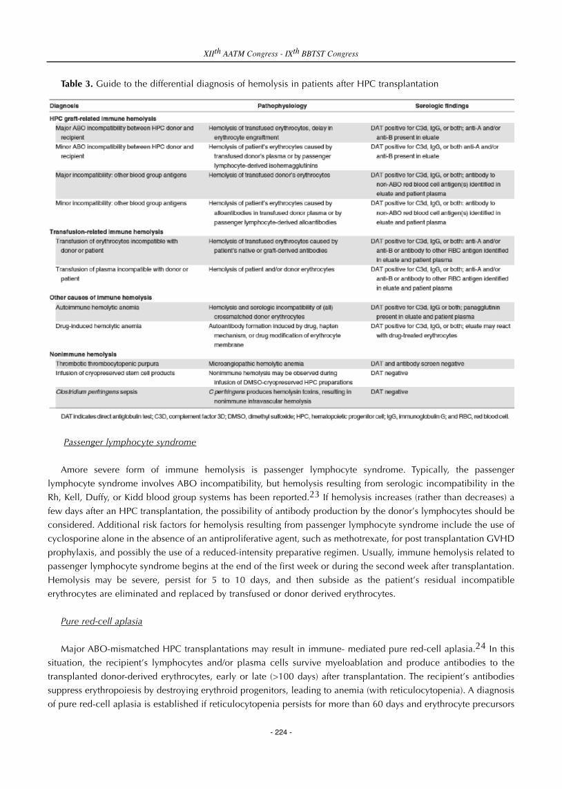
Table 3. Guide to the differential diagnosis of hemolysis in patients after HPC transplantation
Passenger lymphocyte syndrome
Amore severe form of immune hemolysis is passenger lymphocyte syndrome. Typically, the passenger
lymphocyte syndrome involves ABO incompatibility, but hemolysis resulting from serologic incompatibility in the
Rh, Kell, Duffy, or Kidd blood group systems has been reported.23 If hemolysis increases (rather than decreases) a
few days after an HPC transplantation, the possibility of antibody production by the donor’s lymphocytes should be
considered. Additional risk factors for hemolysis resulting from passenger lymphocyte syndrome include the use of
cyclosporine alone in the absence of an antiproliferative agent, such as methotrexate, for post transplantation GVHD
prophylaxis, and possibly the use of a reduced-intensity preparative regimen. Usually, immune hemolysis related to
passenger lymphocyte syndrome begins at the end of the first week or during the second week after transplantation.
Hemolysis may be severe, persist for 5 to 10 days, and then subside as the patient’s residual incompatible
erythrocytes are eliminated and replaced by transfused or donor derived erythrocytes.
Pure red-cell aplasia
Major ABO-mismatched HPC transplantations may result in immune- mediated pure red-cell aplasia.24 In this
situation, the recipient’s lymphocytes and/or plasma cells survive myeloablation and produce antibodies to the
transplanted donor-derived erythrocytes, early or late (>100 days) after transplantation. The recipient’s antibodies
suppress erythropoiesis by destroying erythroid progenitors, leading to anemia (with reticulocytopenia). A diagnosis
of pure red-cell aplasia is established if reticulocytopenia persists for more than 60 days and erythrocyte precursors
XIIth AATM Congress - IXth BBTST Congress
- 224 -

are absent in the bone marrow aspirate. Treatments for red-cell aplasia not associated with parvovirus B19 infections
include plasma exchange to remove the offending hemagglutinins and decreasing the myelosuppressive treatment
to induce a graft-versus-host hematopoietic and lymphocyte effect.
Autoimmune hemolytic anemia
Autoimmune hemolytic anemia (AHA) occasionally develops in HPC transplantation recipients, occurring 2
months to 3 years after transplantation. Although reports are mostly anecdotal, late AHA may be more frequent in T
cell–depleted transplantation recipients.
Postengraftment:
Engraftment syndrome and diffuse alveolar hemorrhage(DAH); HPC patients with engraftment are at risk for
pulmonary complications similar to transfusion-related lung injury. This engraftment syndrome often presents with
fever and hypoxia at initial white blood cell recovery and may progress to DAH. If the lung injury is caused by donor-
derived antibodies, future platelet products may need to be more closely HLA-matched or washed.4
Graft- versus- host disease; Acute GVHD is a major, and frequent, complication of allogeneic HPC
transplantations. HPC recipients with acute GVHD may develop cytopenias of 1 to 3 cell lines and an immune-
based .hemolytic anemia. The patients with severe liver GVHD may also develop the clotting disturbances
associated with end-stage liver disease. All of these patients may require transfusion support.4
Hemorrhohagic cystitis; Bladder irrigation and platelet transfusion are the staple of therapy, but if B-K virus is
found and the patient has no GVHD, immune suppression tapering should be considered.
Veno-occlusive disease; Incompatible isoagglutinin to ABH blood-group antigens on hepatic sinusoidal
endothelial cells may cause toxic injury initiating a sequence of biologic events that may lead to circulatory
compromise of centrilobular hepatocytes, fibrosis, and obstruction of blood flow, resulting in VOD, a major cause
of post transplantation mortality.4
Erythrocyte (blood group) chimerism; Blood group chimerism is an intrinsic characteristic of HPC
transplantation. Transfusion services have reported varied incidences of ABO-grouping discrepancies (so-called
forward- or reverse-typing discrepancies) or mixed field-agglutination reactions, depending on whether the donor
and recipient are ABO identical. Many nonmyeloablative, as well as T cell–depleted, transplantation recipients
remain in the microchimeric state for a prolonged time period after neutrophil recovery. Once the patient’s blood
becomes full-donor chimera, with disappearance of recipientderived antierythrocyte antibodies, the use of blood
products consistent with donor ABO typing is logical. Transfusion services should monitor for recurrence of recipient
ABO-group erythrocytes after non–ABO-identical allogeneic HPC engraftment.
Infusion of donor lymphocytes; Little has been published about transfusion issues related to lymphocyte infusion.
Often, for patients with relapse of the primary disease, the red cells have returned to recipient typing, and donor
red-cell typing emerges after the antitumor response. Changes in the status of HPC engraftment will be observed by
the transfusion service at the onset of mixed-field agglutination in ABO-blood grouping.
Granulocyte transfusion; Whereas transfusions of granulocyte concentrates are not widely used for management
of infections in neutropenic HPC recipients and remain controversial. Granulocyte transfusions have a risk of
XIIth AATM Congress - IXth BBTST Congress
- 225 -

transmitting CMV and, usually, potential donors who are CMV-seropositive are excluded. Granulocyte transfusions
increase the risk for development of HLA antibodies; if this occur, subsequent granulocyte concentrates should be
obtained from HLA-matched donors. Finally, all granulocyte concentrates must be gamma-irradiated before
administration to prevent transfusion-associated GVHD.4
Long term:
Long-term management of post–HPC transplantation patients requires ready access to pertinent blood
transfusion–related medical records. However, often patients return to communities distant from the transplantation
center where their most current medical reports are unavailable. Although most hospital transfusion services have
computerized records that specify special requirements for their own previously treated patients, these records often
do not include special requirements for gamma irradiation, CMV-seronegative components, or group O erythrocytes
for HPC patients who require an urgent transfusion. To ensure optimal matching of erythrocytes for transfusion, as
well as provision of special transfusion needs for their patients, transplantation physicians have an obligation to
communicate to their patients the importance of providing information about their HPC transplantation to local
healthcare providers, including transfusion services, and to consider wearing transfusion requirement identification
on their bodies in case of an emergency.
References1. Remberger M, Watz E, Ringde´n O, Mattsson J, Shanwell A, Wikman A. Major ABO blood group mismatch
increases the risk for graft failure after unrelated donor hematopoietic stem cell transplantation. Biol BloodMarrow Transplant. 2007; 13.675-682
2. Resnick IB, Tsirigotis PD, Shapira MY, et al. ABO incompatibility is associated with increased nonrelapse andGVHD related mortality in patients with malignancies treated with a reduced intensity regimen: a singlecenter experience of 221 patients. Biol Blood Marrow Transplant. 2008;14: 409-417.
3. Klumpp TR. Immunohematologic complications of bone marrow transplantation. Bone Marrow Transplant.1991;8:159-170.
4. James L. Gajewski, Viviana V. Johnson,S. Gerald Sandler, Antoine Sayegh, and Thomas R. Klumpp. Areviewof transfusion practice before, during, and after hematopoietic progenitor cell transplantation.Blood2008;112:3036-3047
5. Leukocyte reduction and ultraviolet B irradiation of platelets to prevent alloimmunization and refractorinessto platelet transfusions. The Trial to Reduce Alloimmunization to Platelets Study Group. N Engl J Med.1997;337:1861-1869.
6. Killick SB, Win N, Marsh JC, et al. Pilot study of HLA alloimmunization after transfusion withprestorageleucodepleted blood products in aplastic anaemia. Br J Haematol. 1997;97:677-684
7. Kaminski ER, Hows JM, Goldman JM, Batchelor JR. Pretransfused patients with severe aplastic anaemiaexhibit high numbers of cytotoxic T lymphocyte lymphocyte precursors probably directed at non-HLAantigens. Br J Haematol. 1990;76:401-405.
8. Brig R.S. Sarkara, Col J. Philip , Dy Comdt Pramod Yadav.Transfusion medicine and solid organ transplant-Update and review of some curent issue.MJAVİ 2013;69:162-167
9. Broeckh M, Nichols WG, Papanicolaou G, Rubin R, Wingard JR, Zaia JA. Cytomegalovirus in hematopoieticstem cell transplants: current status, known challenges, and future strategies. BiolBlood Marrow Transplant.2003;9:543-558.
10. Rubie H, Attal M, Campardou AM, et al. Risk factors for cytomegalovirus infection in BMT recipientstransfused exclusively with seronegative blood products. Bone Marrow Transplant. 1993;11:209-214.
11. Goodrich JM, Bowden RA, Fisher L, Keller C, Schoch G, Meyers JD. Ganciclovir prophylaxis to preventcytomegalovirus disease after allogeneic bone marrow transplant. Ann Intern Med. 1993;118:173-178.
XIIth AATM Congress - IXth BBTST Congress
- 226 -

12. Winston DJ, Ho WG, Bartoni K, et al. Ganciclovir prophylaxis of cytomegalovirus infection and diseaseinallogeneic bone marrow transplant recipients: results of a placebo-controlled, double-blind trial. Ann InternMed. 1993;118:179-184.
13. American Association of Blood Banks. Technical Manual, 15th ed. Bethesda, MD: American Association ofBlood Banks; 2005
14. Bass EB, Powe NR, Goodman SN, et al. Efficacy of immune globulin in preventing complications ofbonemarrow transplantation: a meta-analysis.Bone Marrow Transplant. 1993;12: 273-282.
15. Lee TH, Paglieroni T, Ohto H, et al. Survival of donor leukocyte subpopulations in immunocompetenttransfusion recipients: frequent long-term microchimerism in severe trauma patients. Blood.1999;93: 3127-3139
16. Petz LD, Calhoun L, Yam P, et al. Transfusion associatedgraft-versus-host disease in immunocompetentpatients: report of a fatal case associated with transfusion of blood from a second degreerelative, and asurvey of predisposing factors. Transfusion.1993;33:742-750
17. Remlinger K, Buckner CD, Clift RA, Ramberg R, Hansen JA. Fatal graft-versus-host disease and probablegraft-versus-graft reaction due to an unirradiated granulocyte transfusion after allogeneic bone marrowtransplantation. Transplant Proc.1983;15:1725-1728.
18. Williamson L, Wimperis JZ, Wood ME, Woodcock B. Fludarabine treatment and transfusion associated graftversus host disease [letter]. Lancet.1996;348:472-473
19. Silva MA, ed. Standards for Blood Banks and Transfusion Services, 23rd ed. Bethesda, MD: AmericanAssociation of Blood Banks; 2005.
20. Klumpp TR, Herman JH, Ulicny J, Emmons RV, Martin ME, Mangan KF. Lack of effect of donor recipient ABOmismatching on outcome following allogeneic hematopoietic stem cell transplantation. Bone MarrowTransplant. 2006;38:615-620.
21. Seebach JD, Stussi G, Passweg JR, et al. ABO blood group barrier in allogeneic bone marrow transplantationrevisited. Biol Blood Marrow Transplant. 2005;11:1006-1013.
22. Senzel L, Boulad F, Wuest D, Reid ME. Transfusion policy: when to stop the use of extremely rare blood foran allogeneic hematopoietic progenitor cell transplant recipient with a history of red cell alloimmunization.Transfusion. 2007;47:781-787.
23. Leo A, Mytilineos J, Voso MT, et al. Passenger lymphocyte syndrome with severe hemolytic anemia due toan anti-Jk(a) after allogeneic PBPC transplantation. Transfusion. 2000;40:632-636.
24. Gmur JP, Burger J, Schaffner A, et al. Pure redcell aplasia of long duration complicating major ABO-incompatible bone marrow transplantation. Blood. 1990;75:290-295.
XIIth AATM Congress - IXth BBTST Congress
- 227 -

OralPresentations

OP-01
WHAT SHOULD WE TEACH MEDICAL STUDENTS?
David Graeme Woodfield
The University of Auckland, New Zealand
Adequate teaching of transfusion medicine in medical schools is essential if future doctors are to make the best use
of donated blood. Such teaching is far from uniform either within or between countries. Frequently, the blood services
are not well integrated with training facilities with a result that teaching is sometimes carried out by non-specialists.
Furthermore, the time allocated to teaching in the various aspects of transfusion is commonly very limited and students
may graduate without ever being seriously examined in the subject.
Given the recent evidence based clinical information on transfusion, it is clearly appropriate to revise our training
schedules in medical schools. This requires an approach that starts earlier in medical training, covering not only the
theory of immunohaematology but also the clinical aspects of transfusion, ending in a compulsory specific
examination. Teaching is best performed by specialists in Transfusion medicine, who should also have clinical expertise.
More use needs to be made of self-learning programmes rather than didactic lectures. There is however still a small
place for lectures, as well as some teaching of practical transfusion procedures. All medical students should have
exposure to the blood donation system and attachments to the Blood service for some students should be encouraged.
The nett result may well result in better informed medical graduates who can utilize the gift of blood more
appropriately, carry out new research, and raise the status of transfusion medicine.
It is suggested that as part of the programme to use donor blood efficiently and to safeguard patients from harm,
every country should review its education of medical students and allocate a greater proportion of curriculum time to
the teaching of transfusion medicine.
OP-02
ISOLATION OF AUTOLOGOUS RED BLOOD CELLS USING HYPOTONIC SALINE IN TRANSFUSED
β-THALASSEMIA PATIENTS
Ankita Sheladiya, Kinjal Mendapara, Sanmukh R. Joshi
Lok Samarpan Regional Blood Centre, Surat, India.
OBJECTIVES: The patients with β-thalassemia receive regular transfusions so have a mixture of donors’ RBCs in
circulation.
AIM: To eliminate the donors’ RBCs from the specimen for cell typing on such patients using hypotonic saline.
BACKGROUND: RBCs in β-thalassemia are resistant to osmotic pressure as compared to those from normal
individuals. Exposure of RBCs to hypotonic saline would lyse the donors’ RBCs leaving behind the patient’s native
RBCs.
XIIth AATM Congress - IXth BBTST Congress
- 229 -

METHODS/MATERIALS: one hundred μL of once washed RBCs from transfused patients were added to 40 ml of
0.3% hypotonic saline with intermittent mixing for 5 minutes. The content was centrifuged and the supernatant was
discarded. The sediment RBCs, expected to be the patients’ native RBCs, were washed with and re-suspended in normal
saline to be used for cell typing. Serological techniques were employed as per reactivity nature of anti-sera, e.g. saline
tube test or column agglutinin technique. Blood group antigens, viz. C, c, M, N, P, and Le(b) were included in this
study. Corresponding anti-sera used were of commercial origin or detected in our laboratory. Cell typing on 3 sets of
RBCs, viz. the patient’s post-transfusion, patients’ isolated “native” and corresponding donors’, was run in parallel.
RESULTS: Post transfusion specimen of RBCs from all the patients tested showed mixed-field agglutination. Upon
removal of the donors’ RBCs by hypotonic saline, 6 patients were typed negative for the M and P, the antigens being
present only on corresponding donors’ RBCs. Likewise, after removal of the donors’ RBCs, 8 patients were distinctly
typed positive for C, c, M and P, for the antigens being absent on respective donors’ red cells.
CONCLUSION: Autologous RBCs from the transfused β-thalassemia patients can be separated using hypotonic
saline. The method is simple, rapid and affordable by every blood bank.
Thalassemia patient's RBCs treated with hypotonic saline. Patient's autologous RBCs at the bottom.
OP-03
PREVALENCE OF PARVOVIRUS B19 IN HEALTHY BLOOD DONORS FROM GIRESUN PROVIENCE, TURKEY
Şahin Direkel1, Emel Uzunoğlu Karagöz1, Seda Tezcan Ülger2, Ömer Emecen3, Büşra Kır4
1Department of Medical Microbiology, Faculty of Medicine, Giresun University, Giresun, Turkey2Department of Medical Microbiology, Faculty of Medicine, Mersin University, Mersin, Turkey3Department of Blood Bank, Prof. Dr. A. Ilhan Ozdemir Goverment Hospital, Giresun, Turkey4Department of Medical Microbiology, Health Sciences Institute, Giresun University, Giresun, Turkey
AIM: Parvovirus B19 is transmitted by mainly respiratory tract secretions, blood transfusion, organ transplantation
and transplacental route. Several transfusion-transmitted infections caused by Parvovirus B19 have been reported.
Specific etiologic diagnosisis possible with serological tests or detection of viral nucleic acid via polymerase chain
XIIth AATM Congress - IXth BBTST Congress
- 230 -

reaction. The aim of this study was to determine the prevalence of Parvovirus B19 among healthy blood donors from
Giresun provience.
METHODS: In this study, the plasma samples of 560 healthy blood donors were investigated for the presence of
Parvovirus B19 DNA by polymerase chain reaction method. Briefly, ten samples were pooled in a tube and phenol-
chloroform extractions method was used for DNA isolation. Inhouse PCR was performed for the nucleic acid
amplification according to the methods previously described. PCR products were screened by the gel electrophoresis
via1% agarose gel contain ethidium bromide. If a positive result was detected in an individual tube, PCR method was
repeated within that group.
RESULTS: The incidence of B19 viremia in healthy blood donors in this study was 3/560 (0.54%). Gel
electrophoresis for parvovirus B19 DNA of healthy blood donor samples was shown in the figure 1.
CONCLUSION: According to our results were commend to screen B19 DNA in blood products to prevent
transfusion mediated viral infections especially for immunocompromised patients and pregnant women. Further donor-
recipient studies, determining transfusion-transmitted B19 infections, are needed from different blood centers.
Gel electrophoresis for parvovirus B19 DNA of healthy blood donor samples
M: marker, column 6 and 8 positive (~350 bp) for parvovirus B19 DNA.
OP-04
REACTIVE BLOOD DONOR NOTIFICATION; RESPONSES AND PERCEPTIONS: AN EXPERIENCE FROM A
DEVELOPING COUNTRY
Naila Raza1, Hina Qureshi2, Ambrina Qureshi3
1Depatment of Hematology, Liaquat National Hospital and Medical College, Karachi, Pakistan.2Department of Pathology,The Cancer foundation, Karachi, Pakistan.3Department of Community Dentistry, Dow University of Health Sciences,Karachi.
BACKGROUND: Donor notification of transfusion transmittable infections (TTIs) is important to prevent the spread
XIIth AATM Congress - IXth BBTST Congress
- 231 -
of disease. Response of reactive donors to seek confirmation and treatment is a direct reflection of knowledge and
attitudes towards transfusion transmittable infections (TTIs).
MATERIALS-METHODS: We evaluated reactive donations in one year, from August 2014 to July 2015 at the blood
bank of Liaquat National Hospital, Karachi. The donors were considered sero-positive for HBV, HCV, Syphilis and HIV1
and 2 on positive screening tests done by Chemi-luminescene Immunoassay (CIA) method and Nucleic Acid Tests
(NAT). MP-ICT was used to determine the presence of Malaria. All reactive donors were contacted on their provided
contact numbers. Failure to notify with reasons and notification response at initial and subsequent follow-ups was
judged for repeat and first time reactive donor groups further stratified into primary, secondary and tertiary
qualifications. A cross-sectional analytical study, using non-probability purposive sampling was conducted on 350
potential blood donors fulfilling the criteria of donor selection who were interviewed using pre-tested questionnaire
regarding demographic information, TTIs awareness, transmission routes, financial implications and disease
sensitization. SPSS version 21was used to calculate mean (standard deviation) and frequency / percentages for
quantitative and qualitative variables respectively. Pearson’s Chi-square test (p<0.05) was used to assess relationship of
knowledge and attitude scores with demographic variables. Adjusted Odds Ratio (95% CI) was calculated determining
socio demographic variables with reference to their knowledge and attitude.
RESULTS: From August 2014 to July 2015, out of 16660 donations 940 donors (5.57%) were rejected on positive
screening tests for anti HCV, HBsAg, anti HIV1 & 2, Syphilis or Malaria. Repeat donors (69.5%) with primary to
secondary qualifications constituted the bulk of reactive donors. The notification rate was 54.25% and donor response
was 28.68%.The survey results showed that overall awareness about grave outcomes of diseases was not up to the
mark; 9.52% for primary, 16.66% for secondary and 3.57% for tertiary qualifications. 48% of the respondents were
ignorant of disease spread through blood transfusion. 96.6% of donors did not know the financial impact of treatment
and 69.7% were unable to afford it when informed. Similarly almost all the donors (94.9%) were not vaccinated against
hepatitis B. Multivariate logistic regression analysis for knowledge and attitude scores each after adjustment for age,
education and employment status showed that participants with secondary education as compared to those with
primary education (OR=0.372, 95% CI: 0.203–0.681, p value < 0.01) had significantly less odds of being adequately
knowledgeable whereas higher education was associated with higher odds of knowledge score (OR=4.044, 95% CI:
1.567–10.435, p value < 0.01). Age and employment status were found to have a non significant impact on knowledge
score. Attitude score after adjustment for age, education and employment status showed that participants who have
secondary education as compared to those who have primary education (OR=2.019, 95% CI: 1.190-3.426, p value <
0.01) had higher odds and significant impact for adequate attitude score.
CONCLUSION: There is a need of pre-donation counseling to sensitize donors about TTIs in a resource limited
country where treatment costs are high and out of reach for majority donors. Comprehensive efforts at structural,
healthcare provider and individual level are needed to control spread of TTIs.
XIIth AATM Congress - IXth BBTST Congress
- 232 -

Reactive Donors Notification Outcome (Aug2014-Jul 2015)
XIIth AATM Congress - IXth BBTST Congress
- 233 -
REPEAT DONORS PRIMARY (n=235)
SECONDARY (n=277)
TERTIARY (n=146)
TOTAL (n=654)
DONORS NOT NOTIFIED 107(45.53%) 107(38.62%) 76(52.05%) 290(44.34%)
Not contacted from Blood Bank 2 10 7 19(6.55%)
No Contact Number 5 1 2 08(2.75%)
Wrong Contact Number 5 5 5 15(5.17%)
Cell phone off/busy/no response 95(88.78%) 101(94.39%) 62(81.57%) 258(88.96%)
DONORS NOTIFIED 132(20.18%) 153(23.39%) 71(10.85%) 356(54.43%)
Donor response(1st visit) 34(25.75%) 47(30.71%) 19(26.76%) 100(15.29%)
Donor response(2nd visit) 04(3.03%) 07(4.57%) 03(4.22%) 14(3.93%)
1ST TIME DONORS PRIMARY (n=150)
SECONDARY (n=97)
TERTIARY (n=39)
TOTAL (n=286)
DONORS NOT NOTIFIED 68(45.33%) 52(53.60%) 19(48.71%) 139(48.60%)
Not contacted from Blood Bank 3 5 2 10(7.19%)
No Contact Number 10 3 0 13(9.35%)
Wrong Contact Number 8 3 0 11(7.91%)
Cell phone off/busy/no response 47(69.11%) 41(78.84%) 17(89.47%) 105(75.53%)
DONORS NOTIFIED 80(53%) 46(47.42%) 19(48.71%) 145(50.69%)
Donor response(1st visit) 19(23.75%) 14(30.43%) 09(47.36%) 42(28.96%)
Donor response(2nd visit) 02(2.5%) 03(6.52%) 01(5.26%) 06(4.13%)

Description of Knowledge and Attitude regarding Blood Donation
XIIth AATM Congress - IXth BBTST Congress
- 234 -
DONOR KNOWLEDGE ASSESSMENT Score:>10 Score:<10
Frequency % Frequency %
GENERAL INFORMATION ABOUT TTIs 1. Name common transfusion transmittable infections. 92 26.3 258 73.7
2. Name major complications of TTIs 62 17.7 288 82.3
3. Name the common symptoms of TTIs. 38 10.9 312 89.1
4. Name treatment options for TTIs. 284 18.9 66 81.1
5. What is the estimated cost of TTIs? 12 3.4 338 96.6
MODES OF DISEASE TRANSMISSION 1. Through blood transfusions 182 52 168 48
2. Sharing of food/utensils of affected patients. 210 60 140 40
3. Drinking un-boiled water. 210 60 140 40
4. Getting shaves from barbers using straight razors. 266 76 84 24
5. From affected patient to spouse. 280 80 70 20
6. From affected mother to unborn child. 280 80 70 20
7. Having multiple partners or extramarital relations with sex workers. 292 83.4 58 16.6
8. Using unsterilized syringes. 292 83.4 58 16.6
9. Using unsterilized surgical instruments. 300 85.7 50 14.3
10. Through contact/touch. 294 84 56 16
ATTITUDE VARIABLES 1. Can you be a carrier of any of these infections? 164 46.9 186 53.1
2. Would you like to be informed about positive screening results? 320 91.4 30 8.6
3. Is the treatment of TTIs possible? 234 66.9 116 33.1
4. Can you afford the treatment? 106 30.3 244 69.7
5. Are you vaccinated against hepatitis B? 18 5.1 332 94.9

Percentage Knowledge and Attitude Score of Blood Donation by Socio-Demographic Variables
Level of significance at < 0.05
Factors Predicting Adequate Knowledge and Attitude of Blood Donation
1.* Adjusted for age, educational status and employment status 2.¥Level of significance at < 0.05
XIIth AATM Congress - IXth BBTST Congress
- 235 -
Socio Demographic Variables
% Knowledge
Level of Significance P-value
% Attitude
Level of Significance P-value
Adequate (n=280)
Inadequate (n=70)
Adequate (n=166)
Inadequate (n=184)
Age (years) 0.266 0.355
21-28 76.4 70 72.9 77.2 31-34 23.6 30 27.1 22.8 Educational Status <0.001* <0.01*
Primary 22.9 57.1 38.6 21.7 Secondary 36.4 8.6 25.3 35.9 Tertiary 40.7 34.3 36.1 42.4 Employment Status 0.086 0.550
Unemployed 16.8 25.7 19.9 17.4 Employed 83.2 74.3 80.1 82.6
Socio-demographic Variables
Adequacy of %Knowledge
Adequacy of % Attitude
Adjusted OR* (95% CI)
P-value¥
Adjusted OR* (95% CI)
P-value¥
Age (years) 21-28 Reference Reference 31-34 1.55 (0.82-2.91) 0.171 0.78 (0.47-1.29) 0.344
Educational Status <0.01 Primary Reference <0.01 Reference Secondary 0.37 (0.20-0.68) 2.01 (1.19-3.42) <0.01
Tertiary 4.04 (1.56-10.43) 0.79 (0.47-1.33) 0.384
Employment Status Unemployed Reference Reference Employed 0.65 (0.33-1.29) 0.222 1.05 (0.60-1.84) 0.854

OP-05
BLOOD PRODUCTS PROVISION AND DISCARD RATES AT THE BLOOD TRANSFUSION CENTER OF THE
KARTAL KOŞUYOLU HIGH SPECIALIZATION TRAINING AND RESEARCH HOSPITAL
Sibel Doğan Kaya, Nur Uyar, Hamit Kaya, Tülay Karabürk, Tuğba Gevrek, Arif Şenel, Aslıhan Özmen, Bülent Arınc
Kartal Koşuyolu High Specialization Training and Research Hospital
AIM: Provision of blood components is carried out by Regional Blood Centers according to “the Guidelines for the
Blood and Blood Products” published in the Official Gazette of Turkey dated 4th December 2008. However, transfusion
centers have also been granted the authority to take blood from donors in emergency situations. Since donation, testing,
storing and distribution of blood by regional transfusion centers are demanding processes, effective use of blood
products is very important. The purpose of this study is to determine and demonstrate the rates of blood products
demanded from, provided and discarded by the transfusion center at our hospital.
METHOD: The amounts of the blood products demanded from, provided or discarded by the transfusion center of
our hospital between the dates of 1st January 2012 and 31st December 2015 were examined retrospectively.
FINDINGS: Out of 154016 blood products demanded from our hospital’s transfusion center between 1st January
2012 and 31st December 2015, a total of 38970 products (25.6% of the demand) were provided by the Red Crescent's
Northern Marmara Regional Blood
Center, while 113313 products (74.4% of the demand) were provided by donors having applied to our center.
The rate of provision is the highest by 41.4% for fresh frozen plasma, and the lowest for CRYO by 0.4%. The
provision rate is 40.6% for erythrocyte suspension, 12.6% for platelet suspension, and 5.1% for whole blood.
Out of a total of 152283 blood products, 6922 (4.5%) were discarded by our hospital’s transfusion center within
the time range.
3619 of the discarded products were fresh frozen plasma (52.5% of the discarded products), while 2578 were
erythrocyte suspension (37.2%), 666 were platelet suspension (9.5%) and 59 were whole blood (0.08%).
CONCLUSION: Due to the difficulty in providing blood products, lowering the discard rates to the minimum
should be aimed at. Our study revealed the fact that the most discarded product was fresh frozen plasma. Consequently,
critical stock level at hospitals as well as cooperation with the organization of the Red Crescent in emergency should
be provided. It is also important that the blood products demanded from hospitals and those never used and discarded
be regularly recorded, and that educational programs in that matter be organized.
XIIth AATM Congress - IXth BBTST Congress
- 236 -

FIGURE 1
OP-06
THE PREVENTATIVE MEASURE OF ERYTHROCYTE ALLOIMMUNIZATION IN PATIENTS REQUIRING
CHRONIC BLOOD TRANSFUSIONS: RESULT OF A REFERENCE CENTER IN CENTER ANATOLIA, TURKEY
Mehmet Yay1, Ekrem Ünal2, Bülent Eser1, Alper Özcan2, Göksel Budak1, Mustafa Çakas1
1Department of Blood Centers; Erciyes University, Kayseri, Turkey2Department of Pediatric Hematology Oncology; Erciyes University; Kayseri, Turkey
INTRODUCTION: The compatibility tests (CTs) are obligatory tests that must be done before a blood transfusion.
In Turkey, the cross matching tests are the only legal obligatory test that had to be performed between these tests.
However, cross matching tests are the final stage of CTs including ABO typing, antibody screening and antibody
identifications tests. Recent guidelines advise not to perform cross matching tests even in these screening tests are done
correctly. In general, the overall positivity rate of antibody screening were reported approximately 0.5-1%. We aimed
to share our experience with reduction of erythrocyte alloimmunization in patients requiring chronic blood
transfusions.
MATERIALS-METHODS: Laboratory and medical records of donors, and patients receiving erythrocyte transfusion
from Erciyes University Blood Bank were evaluated, respectively. Historical improvements were noted; and results of
different time periods were compared.
RESULTS: Routine antibody screening tests were started after 2006. Antibody identification tests were performed in
137565 patients from 2006 to 2012. Gel centrifugation method (DiaMed ID microtyping system) was used for antibody
identification. Antibodies were identified and defined in 380 (0.27%) patients. Among these alloimmunized patients
XIIth AATM Congress - IXth BBTST Congress
- 237 -
Years Erythrocyte Suspension
Fresh Frozen Plasma Whole Blood Platelet
Suspension CRYO Total
2012
procurement::19176 destruction:753 (%3.9)
procurement:17155 destruction:1353 (%7,9)
procurement:2852 destruction:21 (%0,7)
procurement:3622 destruction:188(%5,1)
procurement:0 destruction:0
procurement:42805 destruction:2315 (%5,4)
2013
procurement:14280 destruction:638 (%4,5)
procurement:17364 destruction:718 (%4,1)
procurement:1949 destruction:15 (%0,8)
procurement:5874 destruction:338 (%5,8)
procurement:14 destruction:0
procurement:39481 destruction:1709 (%4,3)
2014
procurement:13235 destruction:643 (%4,9)
procurement:13191 destruction:781 (%5,9)
procurement:1845 destruction:15 (%0,8)
procurement:2339 destruction:84 (%3,6)
procurement:81 destruction:0
procurement:30691 destruction:1523 (%4,9)
2015
procurement:14930 destruction:544 (%3,6)
procurement:15361 destruction:767 (%5)
procurement:1161 destruction:8 (%0,7)
procurement:7423 destruction:56 (%0,8)
procurement:431 destruction:0
procurement:39306 destruction:1375 (%3,5)

285 (%75) patients have hematological disease, 95 (25%) of them were patients with surgical disease. The 317 (83.4%)
of detected antibody was Rh subgroup, and Kell; 63 (16.6%) of them was others (MNS, KIDD, etc). These results showed
that the majority of alloimmunazition originated from more antigenic Rh and Kell systems. After 2013 all of our blood
donors were routinely screened for ABO + D Reverse typing, and Rh + K (C, c, E, e, K). The antigen typing was done
by Neo microplate system, Immucor. The distribution of 60.413 donors in this period showen that at the table. In
addition to these laboratory improvements, for prevention of erythrocyte alloimmunization in patients requiring chronic
blood transfusions, blood groups, the result of antibody screening-identification tests, and Rh sub group, Kell
identifications of patients and donors were electronically recorded between 2013, and 2015. The computer system of
donor and recipient antigen typing does not only serve to find suitable donor from previously recorded data but also
allow inviting the known donors to donate blood in the absence of suitable blood product in the stock. By this system
we only detected antibodies in patients with surgerical or gynecological problems who have history of previous
pregnancy or transfusion story. Furthermore we did not encounter any new allo-immunization in patients with
hematological disorders such as thalassemia, sickle cell anemia, and recipients of hematopoietic stem cell
transplantation between 2013, and 2015.
CONCLUSION: Erythrocyte alloimmunization is one of the important complications of transfusion especially for
patients requiring chronic blood transfusions. Our experienced showed that the laboratory and software improvement
was resulted in no alloimmunization for 5212 units of erythrocyte suspension for 58 patients with thalassemia, sickle
cell anemia; whom the historical erythrocyte alloimmunization was found to be 6 in 11254 transfusions in the same
group.
Distribution of 60.413 donors (C,c,E,e,K)
OP-07
EFFICACY AND SAFETY OF THERAPEUTIC ERYTHROCYTAPHERESIS PROCEDURE PRIOR TO TOTAL HIP
REPLACEMENT SURGERY IN PATIENTS WITH SICKLE CELL DISEASE: A CASE SERIES
Nidhi Mehta, Rajesh Sawant, Ishita Chakravarty, Manoj Gupta
Kokilaben Dhirubhai Ambani Hospital and medical research Institute, Mumbai, India
BACKGROUND: Patients with sickle cell disease have abnormal red blood cells (RBCs) and are at risk for
perioperative complications including acute chest syndrome, renal failure, pain episodes, and stroke. RBC exchange
transfusion can, without increasing the whole-blood viscosity, quickly replace abnormal erythrocytes with normal and
XIIth AATM Congress - IXth BBTST Congress
- 238 -
Antijen Positive Negative
C 43.657 (%72.3) 16.756 (%27.7)
c 45.964 (%76.1) 14.449(%23.9)
E 17.341(%29) 43.072(%71)
e 58.539(%97.1) 1.774(%2.9)
K 3.826(%6.4) 56.585(93.6)

raise the hematocrit resulting in improved delivery of oxygen to hypoxic tissues.We intend to review procedural
parameters and clinical efficacy of therapeutic erythrocytapheresis procedures carried out in a tertiary care multi-super-
speciality hospital.
CASE SUMMARY: Therapeutic erythrocytapheresis (TE) procedures carried out prior to surgery in sickle cell disease
patients were reviewed for procedural parameters, any adverse effects, clinical efficacy and clinical outcome. The
treatment plan was to reduce the number of defective red blood cells, maintain or alter the patient’s hematocrit and
control the fluid balance. Two female and one male patient, with baseline mean HbS of 64 % and range (46-82%) were
treated with TE procedure using an automated continuos flow cell separator. Critical factors like type of vascular access
(central jugular line), anticoagulant to be used (ACD), volume of red cell to be removed, replacement fluid to be used
(Leucodepleted, cross-match compatible and Rh and Kell antigen matched RBC units with hematocrit of > 60%) were
decided before starting the procedure.The goal was to keep the percentage of HbS <30% and the total hematocrit at
around 30% to suppress HbS production. Blood volume processed was mean 3953 ml (2317-5332 ml), mean 1341 ml
red cells were removed and the same volume was replaced. Average hematocrit of replacement fluid was 60 %. 100%
fluid balance was maintained in all three patients. 334 ml ACD (188-417) was used for anticoagulation and procedure
was completed in mean 73 (46-93) minutes. No major or minor procedure related adverse effects were observed. A
single volume TE procedure resulted in reduction of the patients HbS level to the desired level of <30% (21% -24%).
The planned surgery was done without any complications related to the sickle cell disease in all three patients.
DISCUSSION: Red cell exchange transfusions remain an effective but possibly underutilized therapy in the acute
& chronic treatment of sickle cell disease. TE with automated cell separators resulted in rapid correction of anaemia, a
rapid decrease in defective red cells and improvement in patients clinical condition enabling the surgical procedure
without complications related to the disease. We could achieve better hemodynamic control with no rise in viscosity
and Hb was maintained around 10 gms%.
CONCLUSION: The advent and ready availability of automated apheresis equipment may alter significantly the
approach to management of patients with sickle cell disease. Currently available apheresis equipment calculates the
replacement RBC volume to achieve the desired HbS, hematocrit levels and allows performance of RBC exchange in
a very reasonable amount of time, with relative ease, safety and efficiency.
OP-08
A CASE OF TRANSFUSION RELATED ACUTE LUNG INJURY (TRALI)
Senem Maral1, Sule Mine Bakanay1, Seval Izdes2, Imdat Dilek1
1Department of Hematology, Ataturk Research and Treatment Hospital,Ankara,Turkey2Department of Intensive Care Medicine,Ataturk Research and Treatment Hospital,Ankara,Turkey
BACKGROUND: Transfusion related acute lung injury is recognized as rare but potentially mortal adverse
transfusion reaction. It is presented with dispnea, hypoxia and noncardiogenic pulmonary edema, typically occurs in
first 6 hours of transfusion. It is known as leukocyte antibody mediated reaction, commonly against human neutrophil
antigens and human leukocyte antigens. The diagnosis and management of TRALI is challenging.
CASE: A 43 year-old man was hospitalized for treatment of Hodgkin Lymphoma stage IVB. He had disease
associated B symptoms and normal pulmonary examination on admission. He presented with elevated transaminases
XIIth AATM Congress - IXth BBTST Congress
- 239 -

and jaundice as a result of hepatic involvement. Laboratory investigation also revealed anemia and coagulopathy
related to liver dysfunction. He was transfused with 2 packs of RBC followed with 4 packs of fresh frozen plasma (FFP).
Thirty minutes after the last pack of FFP, he developed severe respiratory failure and hypotension. There were no signs
of circulatory overload. His O2 saturation dropped to 79% on pulse oximetry. Since high pressure nasal oxygen
supplementation could not correct the hypoxia, endotracheal intubation was immediately done and the patient was put
on mechanical ventilation with 100% oxygen. Urgent chest X-ray showed bilateral pulmonary infiltrates consistent with
acute respiratory distress syndrome(ARDS). (Figure1.) Dexamethasone 40 mg/day was started at the time of intubation
and continued for 4 days. The patient rapidly improved clinically and the chest X-ray taken 24 hours after the event
showed marked resolution. (Figure2.) The patient was extubated and transferred out of the intensive care unit two days
later.
CONCLUSION: Transfusion related acute lung injury is an infrequent and serious adverse transfusion reaction with
fatal course. Treatment is based on recognising on time and providing adequate oxygen supplementation. High dose
dexamethasone started within the 24 hours’ of diagnosis can be life saving in some cases.
Figure 1
Figure 2
XIIth AATM Congress - IXth BBTST Congress
- 240 -

OP-09
THE APPROACH OF THE PHYSICIANS TO FRESH FROZEN PLASMA TRANSFUSIONS
Nil Banu Pelit1, Kamuran Şanlı2, Elçin Akduman Alaşehir3, Güçhan Alanoğlu4, Erşan Kaya4, Esra Kesen1, Şirin
Efe1, Yusuf Soytürk5, Özlem Unay Demirel5, Canan Eren6, Gülsen Şener7, Alpay Arı8, Ümit Savaşçı9, Aslıhan
Demirel10, Gülay Konya10, Çiğdem Kader11, Tuğçe Bozkurt11, Onur Kaplan12, Ali Gürbüz12, Meltem Eren12, Figen
Atalay12, Muhittin Serdar1
1Acıbadem Labmed Clinical Laboratories2Istanbul Kanuni Sultan Süleyman Training and Research Hospital3Maltepe University Faculty of Medicine4Süleyman Demirel University Research and Education Hospital5Göztepe Medical Park Hospital6Marmara University Pendik Training and Research Hospital7Mehmet Akif Ersoy Cardio-Vascular-Thoracic Surgery Training and Research Hospital8Izmir Bozyaka Training and Research Hospital9Gulhane Military Medical Faculty Training Hospital10Kadıköy Florence Nightingale Hospital11Yozgat Bozok University Faculty of Medicine12Başkent University Istanbul Practice and Research Center
AIM: Fresh Frozen Plasma (FFP) is accepted as a raw material for fractionation industry. It is used in rare indications,
such as factor deficiencies, thrombotic thrombocytopenic purpura, etc. However, in transfusion practices there are
some acquired habits from mentors. The purpose of this study is to understand the approach of the physicians to FFP
transfusions.
MATERIAL METHODS: In this multicenter study, 73 physicians having different specialities from 13 hospitals were
asked to answer a questionnaire. There were 9 questions in questionnaire. They are: Q-1 What are the indications of
FFP transfusions? Q-2 Do you request laboratory tests before FFP transfusions? Q-3 If yes, which tests are? Q-4 Do you
follow the efficiency of FFP transfusion? Q-5 If yes, how? Q-6 How do you decide the dosage of FFP? Q-7 Which
plasma component do you prefer? Q-8 If you don't know the patients ABO blood group, which ABO group of FFP do
you choose? Q-9 If you don't know the patients Rh blood group, which Rh group of FFP do you choose? Statistical
analyses were done by chi square test.
RESULTS: Among the 73 physicians subject to the survey are; 58 males and 15 females. The average age is 42 (25-
65;median value: 41). Among 13 centers; 4 were research and training hospitals (32 physicians), 3 were state university
hospitals, 2 were university hospitals established by foundations (20 physicians) and 4 were private hospitals (21
physicians). 3 Research and training hospitals and 1 state university hospital were regional blood centers while the
other 9 were transfusion centers. For question 1; 37 (50,7%) physicians selected the ‘’nutritional, volume expander and
supportive treatment’’ options. While 11 (15, 1%) physicians stated that there is no need for any follow up before
transfusion, 7 (9,6%) physicians stated that there is no need for follow up after transfusion. 27 (37%) physicians selected
the platelet count among the test options for FFP transfusion indications. The number of physicians stated the FFP
dosage as 10-15 ml/kg were 49 (67, 1%). 20 (27,4%) physicians replied as AB to 8th question; 30 (41,1%) stated that
RH was not important in 9th question. There was no statistically significant difference when the results were evaluated
according to the physician’s age, gender, employer institution, speciality, institution that granted the specialization and
XIIth AATM Congress - IXth BBTST Congress
- 241 -

the blood center of physician’s hospital.
CONCLUSION: It’s a fact that inaccurate indications widely exist in FFP usage. This study shows that there is lack
of knowledge and misuse in our country too with regard to this matter. We believe that trainings on the clinical use of
blood products should be increased and indications on the FFP usage should be emphasized more often.
OP-10
COMPARISON OF VIABILITIES, MICROPARTICLE LEVELS AND THE HEMOSTATIC ACTIVITIES OF THE
PLATELETS CRYOPRESERVED VIA THE NEW METHOD WITH DMSO AND WITHOUT DMSO
Rıza Aytaç Çetinkaya1, Ibrahim Eker3, Soner Yılmaz2, Aytekin Ünlü4, Ercan Yenilmez1, Aysel Pekel5, Orhan
Gürsel3, Sebahattin Yılmaz2, Cengizhan Açıkel6, Ismail Yaşar Avcı7
1Department of Infectious Diseases and Clinical Microbiology, GATA Haydarpasa Training and Research Hospital,
Istanbul, Turkey.2Department of Blood Training Center and Blood Bank of Gülhane Military Medical Academy, Ankara, Turkey.3Department of Pediatrics, Division of Hematology, Gulhane Military Medical Academy, Ankara, Turkey.4Department of General Surgery, Division of War Surgery, Gulhane Military Medical Academy, Ankara, Turkey.5Division of Immunology and Allergic Diseases, Gulhane Military Medical Academy and School of Medicine,
Ankara, Turkey.6Department of Epidemiology,Gulhane Military Medical Academy, Etlik, Ankara, Turkey.7Department of Infectious Diseases and Clinical Microbiology, Gulhane Military Medical Academy, Etlik, Ankara,
Turkey.
INTRODUCTION: The accumulating literature knowledge suggests that procoagulant phenotypes of cryopreserved
platelets may increase the efficacy of goal directed transfusions. However, concerns also exist on their prophylactic
transfusion in patients without active bleeding for two main reasons: It may increase thrombotic complications and it
may increase dimethyl sulfoxide (DMSO) toxicity with recurrent transfusions. The principle of the new cryopreservation
method is the removal of the supernatant (DMSO or 0.9 percent NaCl) before freezing, a modification that eliminates
the need for post-thaw washing and simplified the cryopreservation procedure. Frozen of platelets via the new method
without DMSO can prevent DMSO toxicity. As such the purpose of our study was to explore whether the new
cryopreservation method of platelets without DMSO can be an alternative for the use of cryopreserved platelets on
prophylactic transfusion.
METHODS: Apheresis platelet concentrates from 20 donors (APCs) and single donor random platelets (RDPs) from
20 donors have been stored for a day at room temperature with agitation and divided into two equal volumes. One
group of the APCs and RDPs were cryopreserved via the new method with %6 dimethyl sulfoxide and the other group
with 0.9 percent NaCl only, without DMSO. After the removal of the supernatant via centrifugation at 22 °C and 1250x
g for 10 minutes before freezing a platelet pellet of 20-25 mL was obtained and the bag was put into a cardboard
freezing box and stored at -80 °C. Cryopreserved APCs were kept at - 80 °C for one day and then kept in 37 °C medium
10 minutes. Thawed and pelleted platelets were diluted with 20 mL of autologous plasma. Specimens were analyzed
using platelet thrombin generation tests to test hemostatic activities and flow cytometric analyses to test viabilities of
platelets and to determine platelet microparticle levels (PMPs).
RESULTS: The mean platelet counts of APCs and RDPs were 1195,2 ± 153,5 x 10³/µL and 1008 ± 304,9 x 10³/µL,
XIIth AATM Congress - IXth BBTST Congress
- 242 -
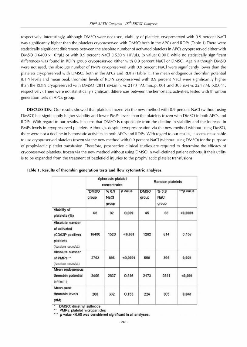
respectively. Interestingly, although DMSO were not used, viability of platelets cryopreserved with 0.9 percent NaCl
was significantly higher than the platelets cryopreserved with DMSO both in the APCs and RDPs (Table 1).There were
statistically significant differences between the absolute number of activated platelets in APCs cryopreserved either with
DMSO (16400 x 10³/µL) or with 0.9 percent NaCl (1520 x 10³/µL), (p value: 0,001) while no statistically significant
differences was found in RDPs group cryopreserved either with 0.9 percent NaCl or DMSO. Again although DMSO
were not used, the absolute number of PMPs cryopreserved with 0.9 percent NaCl were significantly lower than the
platelets cryopreserved with DMSO, both in the APCs and RDPs (Table 1). The mean endogenous thrombin potential
(ETP) levels and mean peak thrombin levels of RDPs cryopreserved with 0.9 percent NaCl were significantly higher
than the RDPs cryopreserved with DMSO (2811 nM.min. vs 2173 nM.min.,p: 001 and 305 nM vs 224 nM, p:0,041,
respectively). There were not statistically significant differences between the hemostatic activities, tested with thrombin
generation tests in APCs group.
DISCUSSION: Our results showed that platelets frozen via the new method with 0.9 percent NaCl (without using
DMSO) has significantly higher viability and lower PMPs levels than the platelets frozen with DMSO in both APCs and
RDPs. With regard to our results, it seems that DMSO is responsible from the decline in viability and the increase in
PMPs levels in cryopreserved platelets. Although, despite cryopreservation via the new method without using DMSO,
there were not a decline in hemostatic activities in both APCs and RDPs. With regard to our results, it seems reasonable
to use cryopreserved platelets frozen via the new method with 0.9 percent NaCl (without using DMSO) for the purpose
of prophylactic platelet transfusion. Therefore, prospective clinical studies are required to determine the efficacy of
cryopreserved platelets, frozen via the new method without using DMSO in well-defined patient cohorts, if their utility
is to be expanded from the treatment of battlefield injuries to the prophylactic platelet transfusions.
Table 1. Results of thrombin generation tests and flow cytometric analyses.
XIIth AATM Congress - IXth BBTST Congress
- 243 -

OP-11
SIGNIFICANCE OF NUCLEIC ACID TESTING IN WINDOW PERIOD DONATIONS: REVISITING
TRANSFUSION SAFETY IN HIGH PREVALENCE-LOW RESOURCE SETTINGS
Sadhana Mangwana, Neha Bedi
Sri Balaji Action Medical Institute, Paschim Vihar, New Delhi
INTRODUCTION: Safe blood transfusion to the people is essential requirement of health care delivery system
which can be done through integrated strategy for elimination of transfusion transmitted infection (TTI). Safe blood can
be ensured through collection of blood from voluntary, non-remunerated blood donor and screening of all donations
for viral markers by highly sensitive tests.
Despite the mandatory screening of blood with newest, very sensitive serological test, considerable risk remains for
transfusion transmission of virus due to window period infections, occult infections, viral variants or delayed
seroconversion.
AIM: Aim of the study was to analyze the efficacy of Mini pool Nucleic Acid Amplification Testing (MP-NAT) as
additional donor screening program, seroprevalence of TTI and its role in improving blood safety in the high prevalence
population.
MATERIAL AND METHODS: Study was performed at a tertiary-care, accreditated hospital from June 2013 to
December 2015. Donor samples were collected at the time of blood donation for serological and MP-NAT Testing. All
negative cases for anti-HIV, anti-HCV and HBsAg by ELISA were subjected to MP-NAT in pools of six on Roche’s Cobas
Taq Screen MPX assay v2.0 on Cobas System s 201 to detect HIV-1 (groups M and O RNA), HIV-2 RNA, HCV-RNA
and HBV DNA.
RESULTS: Total of 20470 donations were received during period of 31 months of which 19932 (97.37%) were males
and 538 (2.63%) females. Whole blood donations were 16997 (83.03%) and 3473 (16.97%) apheresis. 779 (3.80%)
were voluntary donors and 19691 (96.20%) replacement donors. Out of 16997 donations, 446 (2.61%) were
seroreactive of which 139 (0.68%) were HBsAg, 60 (0.29%) anti-HCV, 36 (0.18%) anti-HIV and 211 (1.03%) anti-TP
(syphilis) seroreactive donors.
Out of 16551 sero-negative donors subjected to MP-NAT testing, 17 (0.10%) were NAT reactive with NAT yield (1
in 974). Out of 17 NAT yield, 12 (0.07%) were HBV (1 in 1379), 4 (0.024%) HCV (1 in 4138) and 1 (0.006%) HIV
NAT reactive (1 in 16551). All NAT yield donors were males, replacement donors.NAT positive donors’ age ranged from
19 – 49 years.
CONCLUSION: New technology like NAT has improvised the blood safety by detecting the virus in the pre-sero
conversion window period and thereby providing the sensitivity that is much higher compared to the newest generation
serological tests. Since viremia precedes seroconversion by several days to weeks, Nucleic Acid Amplification test
technologies have the potential to detect viremia earlier than routine screening tests based on seroconversion.Variation
in our yield rate from western studies could be due to high prevalence of HBV and HCV in the population as India is
considered to be in the intermediate level of HBV endemicity with over 40 million HBV carriers and lesser number of
voluntary donations. In countries with high incidence of infection with significant number of window period donations,
XIIth AATM Congress - IXth BBTST Congress
- 244 -

NAT can serve as a valuable tool to achieve the goal of zero risk of blood. With clearer benefits of NAT outweighing
cost in providing medical care to the infected person, it is recommended that NAT should be made mandatory by
policymakers as a part of TTI testing along with other serological testing in high prevalence, resource constrained
countries like India.
OP-12
RESULTS OF DETERMINING OCCULT HEPATITIS B VIRUS INFECTION AMONG BLOOD DONORS
Tserendejid Magvansuren, Erdenebayar Namjil
National center for transfusion medicine Mongolia
BACKGROUND: Hepatitis B virus (HBV) remains a major public health problem worldwide. Implementation of
hepatitis B surface antigen (HBsAg) in routine screening of blood donors in the early 1970s has greatly enhanced
transfusion safety. The incidence of transfusion-transmitted hepatitis B has been steadily reduced over the last four
decades. However, it was demonstrated that HBV transmission by blood components negative for HBsAg can still occur
and HBV transmission remains the most frequent transfusion-transmitted viral infection; thus, the term occult hepatitis
B virus infection (OBI) was introduced. OBI is simply defined as serologically undetectable hepatitis B surface antigen
(HBsAg-ve), despite the presence of circulating HBV DNA with or without the presence of HBV antibodies.
AIM: To determine the prevalence of occult hepatitis B among blood donors and evaluate the presence of HBV DNA
in HBsAg negative plasma samples.
MATERIALS-METHODS: It includes 16700 samples which donated in NCTM in Ulaanbaatar in 2013. We used to
“triplex” PCR assay that included the detect of hepatitis B virus HBV-DNA in addition HCV-RNA and HIV1/2-RNA for
whom with absence of serological markers of infection. The studies used molecular biology methods were performed
with the help of equipment (ROCHE COBAS S 201) and technology based on Real Time PCR (pool size: 6 donation)
Then we choose HBsAg negative, DNA positive samples and determined, anti-HBc and anti-HBs by serological
methods, of ELISA Wantai HBc and HBs 3.0 tests. After 6 months we re-call HBV DNA positive donors and determined
HBsAg, HBV DNA, anti-HBs, anti-HBc.
RESULTS: The 14948 samples were detected serological negative in the total of 16700 samples. PCR test results
show 35 (0.23%) positive by HBV-DNA. 26 (74%) of the 35 DNA positive blood donors were alone anti-HBc positive
and 3 (9%) were anti-HBs, anti-HBc positive. 6(17%) were seronegative. Of the 35 OBI cases, 28 (80%) were detected
the first time they were screened for HBV DNA while 7 (20%) gave one more HBV PCR-nonreactive results before
detection. Callback studies we determined 2 cases were pre-HBsAg window period.
CONCLUSION: The prevalence of HBV DNA positive in HBsAg negative blood donors is found 0.23%. HBV NAT
needs either extreme sensitivity or to be performed on individual donations to eliminate HBV DNA-containing units.
XIIth AATM Congress - IXth BBTST Congress
- 245 -

OP-13
IMPLEMENTATION OF PATIENT BLOOD MANAGEMENT IN THE UK
Rebecca Anne Margaret Gerrard
National Health Service Blood And Transplant, England
AIM: The aim of Patient Blood Management (PBM) in the UK is to build on the success of the previous government
‘Better Blood Transfusion’ initiatives, but with an emphasis on improving patient outcomes through blood avoidance
and the use of alternatives to transfusion where clinically indicated. The main aim is for blood use in England to be
<27/1000 head of population and costs associated with blood avoidance reduced by further £18 million by 2018.
MATERIALS: In England, there are two national teams who work for the blood service but support hospitals to drive
the PBM initiative:
• The Consultants Team: 10 Consultant Haematologists who have joint posts with large hospitals.
• The Practitioner Team: 13 senior nurses and scientists and a small Education and Audit Team.
METHODS: The PBM Teams have a 3 year Strategic Workplan that outlines the way forward for working in
collaboration with the National Blood Transfusion Committee and all the hospitals in England to improve patient care,
save money and help sustain the blood supply with a strong emphasis on education, innovation, integration and
evidence based clinical practice
The national team works closely with the Transfusion Practitioners in the hospitals and provides support and
educational tools for them.
They also liaise with the other UK countries to work on joint initiatives, benchmark themselves and share examples
of best practice in transfusion.
RESULTS: In 2015, some “proof of concept pilots” were initiated with several hospitals to develop evidence that a
targeted approach to implementing PBM produces faster improvements in transfusion practice and reduces costs.
Following the success of these pilots, we aim to implement this model on a national level and carry out further pilots
targeting different aspects of PBM.
We also need to understand where and why blood is being used. We aim to establish a clinical benchmarking
database collecting data from hospitals to compare practice against key performance indicators. This data will provide
real time intelligence on how blood is being used, identify where we need to target PBM initiatives and help inform
the demand planning process.
CONCLUSION: PBM is better for patients: national and regional audits show continued inappropriate and
excessive use of blood components (>20% of transfusions) with insufficient take-up of transfusion alternatives and poor
consent processes. PBM aims to improve practice by putting patients at the heart of blood management.
PBM is better for the blood supply: it supports long-term sustainability of the blood supply and protects patients with
rarer blood groups.
XIIth AATM Congress - IXth BBTST Congress
- 246 -

OP-15
THE LEVELS OF CD55 AND CD59 IN THALASSEMIA MAJOR PATIENTS WITH AND WITHOUT SPLENECTOMY
Ayşegül Uğur Kurtoğlu1, Belkıs Koçtekin1, Erdal Kurtoğlu2, Mustafa Yıldız3, Selen Bozkurt4
1Department of Biochemistry, Antalya Education and Research Hospital, Antalya, Turkey2Department of Hematology, Antalya Education and Research Hospital, Antalya, Turkey.3Department of Medical Oncology, Antalya Education and Research Hospital, Antalya, Turkey4Department of Biostatistics Medical Informatics, Faculty of Medicine, Akdeniz University, Antalya, Turkey
OBJECTIVE: The direct effects of Beta thalassemia major (β-TM ) on other organs and tissues are due to the
deleterious effects of the profound anemia, the byproducts of hemolysis, and the intramedullary and extramedullary
expansion of erythroid progenitors. Massive splenomegaly develops early in the course of β-TM due to increased red
cell destruction and the presence of splenic extramedullary hematopoiesis. If the spleen is surgically removed, a minor
infection can potentially develop into a life-threatening infection known as sepsis. Studies about the etiopatogenesis of
hemolysis β-TM are very low in number. Complement-mediated erythrocyte destruction can cause both intravascular
and extravascular hemolysis. Complement regulatory proteins protect cells from such effects of complement system.
Intravascular hemolysis is normally blocked by CD59, which prevents the final stage of complement assembly,
production of the membrane attack complex that forms a pore in the target cell. Extravascular hemolysis is normally
blocked by CD55, which prevents assembly of the C3 and C5 convertases upstream of MAC formation in the
complement cascade. We aimed to perform quantitative analysis of membrane-bound complement regulators, CD55
and CD59, on peripheral red blood cells by flow cytometry on β-TM patients.
MATERIALS AND METHOD: The study looks at a total of 45 patients with beta-thalassaemia major, divided into
two groups according to the presence (Group 1: 22 β-TM with splenectomy) or absence (Group 2: 23 β-TM without
splenectomy) of the spleen. Seventeen healthy volunteers (Group 3: controls) were included in the present study. The
EDTA blood sample was processed within one hour. One hundred μL of blood and 10 μL of monoclonal antibodies
against to CD55 (PE Mouse Anti-Human), CD59 (FITC Mouse Anti-Human) were transferred into a polystyrene tube.
The cells were analysed in the flow cytometer (FACS Canto II, Becton-Dickinson).
RESULTS: The overall mean percentage of CD55 positive RBCs of group 1 (50.72 ± 18.67 %) was significantly lower
than group 2 (64.36 ± 13.98 %) and group 3 (89.03 ± 11.75 %) (p<0.01). The overall mean percentage CD59 positive
RBCs of patients was no significantly different group 1 than group 2 and group 3.
CONCLUSION: Level of CD55 is lower in β-TM patients than healthy population, but CD59 level are not different
in two groups. These two findings indicate the presence of extravascular hemolysis in these patients. We think that there
is a possible genetic defect in these patients leading to partial deficiency in CD55 levels. Erythrocytes expressing CD55
in normal range are transfused to patients in every 1-2 weeks in routine transfusion programs. We suggest that finding
of very low levels of CD55 in splenectomized patients having transfusion in every 3-4 weeks is due to much more large
transfusion intervals. We think that CD55 levels are lower than these levels in non-transfused patients, and this may be
a very useful indicator to decide the frequency of transfusions. More studies are needed in this field.
XIIth AATM Congress - IXth BBTST Congress
- 247 -

OP-16
UNIDENTIFIED FOREIGN OBJECTS IN BLOOD BAGS: {ARE WE ALL HALLUCINATING?}
Mustafa Nuri Günçi ̇kan1, Ferit Sönmez1, Ismet Gürsel Karahacıoğlu1, Armağan Aksoy1, Nurettin Hafızoğlu1,
Mehmet Güllüoğlu2, Fatma Meriç Yılmaz3
1Turkish Red Crescent Blood Services General Directorate, Ankara, Turkey2Turkish Red Crescent General Directorate, Ankara, Turkey3Department of Medical Biochemistry, Yıldırım Beyazıt University Medical School, Ankara, Turkey
BACKGROUND: Any foreign object in a blood bag is obviously a threat to the safety of blood and creates a case
to be managed according to scientific principles. We aimed to write a descriptive report of how Turkish Red Crescent
managed a crisis in which a number of blood component bags with unidentified foreign objects were noticed by the
Regional Blood Center.
METHODS: This study is a retrospective case report showing the course of action taken by Turkish Red Crescent
when this crisis unfolded.
RESULTS: The crisis started with the index report (dated May 21st, 2015) of foreign objects being noticed in a
number of blood bags (Red Cells, Buffy Coat Removed, in Additive Solution) at the Regional Blood Center. The Regional
Blood Center had started to look for the reasons why the number of blood bags which were returned by the hospitals
due to presence of clot was unusually high. In return, Turkish Red Crescent Blood Services General Directorate was
contacted as for the nature of this object and what had to be done, next. Reactively, The Blood Bag Company was
notified of this potentially harmful situation and requested to start their inquiry. All Regional Blood Centers were
warned not to use blood bags with the same lot numbers, either for issuing to hospitals or component production. Some
blood bags with foreign object were submitted to the company for their inquiry. All Regional Blood Centers were also
ordered to contact hospitals for withholding those unused blood components with the relevant lot numbers. The
Ministry of Health was also notified. The Infrared Spectrum Analysis Report from the company stated the "object"
consisted of blood proteins and no plastic or foreign substances. The report from Ankara University stated the object as
"polymeric" and the one from TÜBITAK as "of peptide origin". The final report from the company, with the addition of
Electron Microscopy, stated this object consisted of activated platelets, fibrin and denaturated proteins as in a "blood
clot". After the recommendations of National Blood and Blood Products Commission of the Ministry of Health, actual
donation times and the amount of fibrinogen in FFPs of the same lot numbers were checked and found to be in normal
limits. The Regional Blood Centers were notified, on June 8th, that any blood components, in addition to empty blood
bags, on withhold at the blood center itself or the hospital, would be started to be used again, after visual control of
two persons. The manual for the blood bags were revised as to reflect the root cause analysis result. A number of
components had to be disposed due to different reasons in this whole crisis.
CONCLUSION: Blood bag problems have been experienced elsewhere before and for this crisis, a parallel
approach, both in terms of the inquiry and the preventive and corrective actions taken, has been taken. The principle
of doing no harm was satisfied at all times. No patient was harmed due to any foreign object in the blood bag. There
was no shortage of any component as a result of the measures taken by Turkish Red Crescent because there was an
adequate stock of blood components at all times. So with lessons learned and experience gained, we can say that this
crisis was succesfully managed.
XIIth AATM Congress - IXth BBTST Congress
- 248 -

OP-17
FORWARD-REVERSE INCOMPATIBILITIES IN ABO BLOOD GROUP TYPING
Derya Çağlayan Serin1, Berrin Uzun2, Nilgün Aşkaroğulları3, Fahri Yüce Ayhan3
1Blood Bank, M.Enver Şenerdem Torbalı State Hospital, Izmir, Turkey2Blood Bank, Katip Celebi University, Ataturk Training and Research Hospital, Izmir, Turkey3Blood Bank, Dr. Behcet Uz Children's Hospital, Izmir, Turkey
INTRODUCTION: ABO blood group is determined with red blood cell surface antigens and detection of serum
antibodies against these antigens, and being consistent with each other. During the immunohematological tests, weak
agglutination in forward typing and lack of the expected antibody or detection of unexpected antibody in reverse typing
should remind the ABO variations or presence of auto/alloantibodies. In case of forward-reverse nonconformity, further
investigation should be considered. The aim of this study, emphasizing the importance of forward-reverse
nonconformities encountered during the determination of blood groups in district hospitals with limited capacity and
discussing the results.
MATERIALS-METHODS: Three patient samples which blood group could not be determined due to forward-reverse
incompatibility were evaluated in Torbalı (Izmir, Turkey) State Hospital Transfusion Center. Blood group examination
was performed with gel centrifugation method (Across Gel, DiaPro Medical Products, Turkey) manually by using A, B,
AB, DVI +, DVI-, Ctl, N/A1, N/B configurated cards. Patient samples were sent to Dr. Behçet Uz Children’s Hospital
Transfusion Center for further investigation. For the determination of A1 and H antigens, anti-A1 and anti-H commercial
antisera (ALBAclone, Alba Biosicence, the United Kingdom) were used. Reverse groupings were retyped by using a set
of A1, A2, B, O red cells(Across Gel, DiaPro Medical Products, Turkey) which was performed at room temperature (RT)
and +4°C.
RESULTS: All patients with nonconformity of forward and reverse typing were female. One of the patients for whom
transfusion planned, was 37 years old and admitted with ectopic pregnancy and bleeding, the other was 61 years old
and had gonarthrosis. The third case was 31 years old pregnant with cesarean birth plan.
The immunohaematological test results were presented in the tables 1 and 2. While it was considered that the ABO
subgroups might be responsible for the nonconformity of forward reverse grouping in two cases, anti-M
alloimmunization was detected in the third case.
CONCLUSION: Due to nonconformity of forward and reverse groupings, results were reported as undetermined
and cross-matching with group O red cell concentrates was performed. It should be considered to further investigation
of discrepancies between forward and reverse grouping to improve the transfusion safety even in the transfusion centers
with limited capasities.
Forward Grouping Results
XIIth AATM Congress - IXth BBTST Congress
- 249 -
Case A antigen B antigen AB antigen A1 antigen H antigen
1 +1 Negative +2 Negative +4
2 +3 +4 +4 Negative Negative
3 Negative +4 +4 Negative Negative

Reverse Grouping Results
RT: Room temperature
Reported Blood Groups
OP-18
VIRTUAL BLOOD CENTERS
Serhan Akar1, Reyhan Demir2, Nil Banu Pelit3
1Acıbadem Internatıonal Hospital, Istanbul Turkey2Acıbadem Hospital Group, Istanbul Turkey3Acıbadem Healthcare Group Blood Coordinator, Istanbul Turkey
AIM: Social media has been broadly defined to refer to “ the many relatively inexpensive and widely accessible
electronic tools that enable anyone to public and access information, collaborate on a common effort or build
relationships”. Social media, which can be used for many different purposes, has become a significant platform where
patient relatives use to reach the donors. The hugeness of the member number of a Web-site, which is dintinguished
incidentally while searching about blood donation organization, leaded us to research the Web-sites and pages that
were performed with the same purpose.
MATERIALS-METHODS: This study was carried on during the whole year, 2015. The Web-sites organizing blood
donations in Turkey were reviewed on Facebook, Twitter, Google and Android systems, using keywords; blood
donation, blood donors, find donors, etc. Interviews were made with the administrators of some websites; whereas the
other sites were signed in to evaluate the principles.
RESULTS: In this study, it was observed that the websites which were about “blood donation organizations” were
formed in two groups; as open and closed. Open groups (Tüm kan gönüllüleri, kan hayattır, kan arıyorum yardımlaşma
derneği, kan trombosit bağışı, Türkiye kan bankası, acil kan aranıyor, etc) reached all of the target receivers. Closed
XIIth AATM Congress - IXth BBTST Congress
- 250 -
A1 cells A1 cells A2 cells A2 cells B cells B cells 0 cells 0 cells
Case RT +4 °C RT +4 °C RT +4 °C RT +4 °C
1 Negative N/A Negative N/A +3 N/A Negative N/A
2 Negative Negative Negative Negative Negative Negative Negative Negative
3 +4 +4 N/A N/A 2+ 2+ Negative Negative
Case 1 Non A1, A subgroup-possibly Ax
Case 2 A2B
Case 3 B (Anti-M antibody positive)
groups aimed to reach certain crowd of people by filtering location (like Antalya kan gönüllüleri), occupation/political
groups (like Istanbul Barosu), blood groups (like Kan bankası 0 negatifler), etc. It was observed that, there are lots of
different websites with varying numbers from 122 to 138800 voluntary members. They reported the most important
problem was the lack of the voluntary staff. It was mentioned that, they had difficulties in calling or being called
continuously, application of excessive donors and disregarding that the need is over, because of the communication
problems. It is understood that, virtual blood centers have the advantages of reaching out to too many volunteers in a
short time, having instant feed-back, eliminating the distance between the volunteer and the center and urgent supply
of the needs. However, the disadvantages such as; the contact between the relatives and donors, the lack of control
mechanisms and standardized procedures, misinformation given by the administrators to donors, ineffective
organization of the members are also noted.
CONCLUSION: People can access a lot of independent web pages with wrong and/or missing information just to
satisfy their needs on the Web. In daily life, virtual media is actively used by numerous numbers of donors/donor
candidates. These people, considered to represent an important and powerful population, should be controlled and
organized by a national programme and a centralized management. It is emphasized that, the Turkish Red Crescent,
collaborating plenty of institution, Foundation and association, should contact with there virtual formations, to develop
the infrastructures for an organised, efficient and national acquisition, to block the abuse and to increase the number
of conscious and voluntary donors.
OP-19
FREQUENCY OF RED BLOOD CELL ALLOANTIBODIES IN OUR TRANSFUSION CENTER
Nihal Öztürk Şahin, Esra Kesen, Yeşim Uysal, Seda Öztürk, Reyhan Patlar, Nil Banu Pelit
Acibadem Labmed Clinical Laboratories
AIM: There is no data about the frequency of red blood cell (RBC) alloantibodies in Turkey. In this study, we
analyzed our antibody identification test results.
MATERIAL & METHOD: In this retrospective study, antibody identification test results of 264 patients, between Jan
2010 and Jan 2016, were investigated. The test was done by gel centrifugation method (BIORAD, NaCl enzyme test
and cold agglutinins & Coombs cards with Diapanel I-XI; Diapanel I-XIP test cells). The records were investigated
according to sex, age, clinics where the patients were treated and direct antiglobulin test results.
RESULTS: Distribution of sex, age and related clinics of 264 patients are listed in Table 1. Two hundred and twenty
of the subjects in 264 were positive (83.3%). In these 220 positive test results, 99 (45%) were single RBC alloantibody,
16 (7,3%) were double RBC antibodies and 105 (47,7%) were nonspecific. Nonspecific positivity was later grouped as
pan-reactive antibodies (62) and unspecified antibodies (43) by using panel cells. In 33 out of 105 samples of
nonspecific positivity group, DAT was performed and 25 were positive. Our laboratory suggested antibody
identification tests for 185 samples, due to ABO discrepancy (23), crossmatch incompatibility (45) and positivity in
antibody screening tests (116). One of 185 tests belonged to a mother who had a newborn with positive DAT result.
Most of the RBC alloantibodies was against to Rh blood group antigens, 60 in 99 as single, 13 in 16 as double antigens.
Anti-D antibody was identified in 34, but there were history of passive immunization by anti-D immunoglobulin in 10.
The second frequent antibody was anti-M (18 as single, 2 as double antibodies). Anti-M antibodies found in 13 patients
were related to ABO discrepancy and in 4 patients they were related to crossmatch incompatibility.
XIIth AATM Congress - IXth BBTST Congress
- 251 -

CONCLUSION: The data demostrated that alloantibodies were mainly against Rh blood group antigens. It was
interesting to find naturally occured Anti-M as the most frequent RBC alloantibodies. We suppose that this anti-M
finding is related to microorganisms such as bacteria, virus etc. As a result, we suggest that investigations should be
performed in all ABO discrepancies and crossmatch incompatibilities.
Table 1. Distribution of age, sex & clinics of the patients
OP-20
THE USE OF THE ELECTRONIC CROSSMATCH IN A PRIVATE HOSPITAL, KOCAELI, TURKEY
Melda Özdamar1, Fuat Çetinkaya2, Ismail Vardar1, Ömer Buğra Ergene1, Salih Türkoğlu1
1Anadolu Medical Center, Kocaeli, Turkey2Invitro Laboratory
BACKGROUND AND OBJECTIVES: Electronic crossmatch (EXM) test needs a well-designed computer program
with the use of donor and recipients’ previous immunohaemotologic test results. The standard operating procedure
(SOP) and the data of Anadolu Medical Center blood bank were presented.
MATERIAL-METHODS: Electronic crossmatch, erythrocyte suspensions (ES) transfusion and whole blood (WB)
transfusion were evaluated for the years 2014 to 2015. EXM is only permitted when a valid pre-transfusion sample has
been tested for ABO/Rh typing and there are no clinically significant antibodies detectable in the current sample
without history of clinically significant antibodies. For this purpose, transfusion practice trainings were given to all the
nurses. The blood bank technicians have been trained by the IT specialists. ES and WB release have been allowed
during 72 hours when all the related records are complete in the blood bank computer program. The correct fill in of
the transfusion follow-up forms and their turn back to the blood bank (BB) in a high ratio as > % 95 has been targeted
XIIth AATM Congress - IXth BBTST Congress
- 252 -
Gender Age Groups Obst&Gyn Hemato&Onc Surgery Int
Med ICU & Anest Other Total
Female (194) 0- 19 0 7 1 1 0 0 9
Female (194) 20- 40 112 0 4 2 2 11 131
Female (194) 41- 60 2 6 4 4 3 4 23
Female (194) > 60 0 9 7 7 2 6 31
Male (70) 0- 19 0 9 2 1 2 0 14
Male (70) 20- 40 0 2 5 1 0 4 12
Male (70) 41- 60 0 2 8 1 1 1 13
Male (70) > 60 0 14 4 9 2 2 31
Total 114 49 35 26 12 28 264

which has been further validated.
A comprehensive, validated (according to AABB standards) electronic data management system is in place in the
BB. A gel agglutination system (Across Gel®, DiaPro, Turkey) is used in the laboratory. A total of 11254 ES and 15 WB
transfusions and the orders and the tests that were done in these years were analyzed.
RESULTS: The total of CXM, indirect coombs (IC) tests and transfused ES were 11599, 9574 and 6896 respectively.
Ordered ES and released (transfused) ES were 25009 and 11254 respectively. They are shown at the table 1 and 2. Any
major hemolytic transfusion reaction related to ABO/Rh incompatibility in this period of time has not been reported to
the BB. The cost of EXM has been found as 20000 tL (average) lower per year compared to serological CXM (in term
of test cost).
CONCLUSION: In the “National Blood and Blood Products guideline” (Ankara, 2011) EXM is mentioned but its
use is yet not general in Turkey. Electronic crossmatch method has been approved by Anadolu Medical Center (ASM)
Transfusion Committee in the use of the standarts of AABB. In most European countries and USA, there may be different
requirements for the use of the EXM, the electronic cross-match is much more widely used since 1998¹. When the
proper restrictions for its use are applied, very few problems have been encountered. EXM is therefore considered to
be safe in the ASM hospital settings. It saves time and is inexpensive than serological CXM. It seems therefore safe to
predict that its use will further increase in Turkey.
REFERENCES:
1- The use of the electronic (computer) cross-match, H. W. Reesink et al; Vox Sanguinis (2013) 104, 350–364
Table 1
Blood product orders and transfusion
Table-2
BB test numbers
XIIth AATM Congress - IXth BBTST Congress
- 253 -
Tests No
Total CXM 11599
IC CXM 6896
AHG CXM 2025
Blood product Order Transfusion
ES 25009 11254
WB 16 15

PosterPresentations

PP-001
EVALUATION OF THE RELATIVES OF INPATIENTS WITH CRIMEAN-CONGO HEMORRHAGIC FEVER (SINGLE
CENTER EXPERIENCE)
Erdoğan Doğan1, Gülgün Sevimligül2, Meriç Kayma Cihan3, Selda Özüm1, Erdal Karakaş1, Yasin Doğan1, Filiz
Çiçekli1, Abdurrahman Cihat Cığır1, Aynur Engin4, Hatice Terzi5, Mehmet Şencan5
1Laboratory of The Blood Transfusion Center, Cumhuriyet University School of Medicine, Sivas, Turkey.2Nursing Research and Project Development Department, Cumhuriyet University Hospital, Sivas, Turkey.3Department of Pediatrics, Cumhuriyet University School of Medicine, Sivas, Turkey.4Department of Infectious Diseases and Clinical Bacteriology, Cumhuriyet University, Faculty of Medicine, Turkey.5Department of Hematology, Cumhuriyet University School of Medicine, Sivas, Turkey.
INTRODUCTION: Crimean Congo Hemorrhagic Fever (CCHF) is a serious disease with a mortality rate of 3-30 %
caused by a virus defined in the Nairovirus species of Bunyaviridae family. Thrombocytopenia and disseminated
intravascular coagulation (DIC) are the major problems in CCHF (1). The incidence of DIC among the CCHF patients
is about 40-50 % (1,2). On other hand, because of there is no specific, antiviral treatment against CCHF, supportive
treatments like intravenous hydration, erythrocyte, thrombocyte and fresh frozen plasma transfusion, respiratory and
circulatory supports and parenteral nutrition are lifesaving.
Seven days after the onset of complaints IgM and IgG antibodies can be detected by ELISA and IFA tests. Specific Ig
M level is reduced to undetectable levels after 4 months from infection, but Ig G levels can be detected for 5 years (3).
Infection is caused by contact with infected body fluids of patients without prevention precautions, such as contact with
bare hands. So relatives of patients and health workers are under the risk of infection.
AIM: In this study we aimed to evaluate CCHF Ig M, Ig G levels and complete blood counts (CBC) of first degree
relatives of 66 patients who were hospitalized for CCHF.
MATERIALS-METHODS: Between the years 2010-2011 at Infectious Diseases Service of Cumhuriyet University
Faculty of Medicine, Sivas, Turkey, first degree relatives of 60 adult and 6 pediatric patients hospitalized for CCHF were
questioned for age, gender, proximity to the patient, tick exposure, fever and weakness. Complete blood counts and
CCHF Ig G and Ig M were examined.
RESULTS: There were 61/66 men (92.42 %) and 5/66 women (7.58 %) in the patient’s relative group. Mean age was
42.12 years (19-63). Patients were from rural areas (villages and towns) and city centers of Tokat and Yozgat cities of
Turkey. Forty eight (72.72 %) patients were living in the villages, 15/66 (22.72 %) were living in the towns and 3/66
(4.54 %) were living in the city center. Individuals from villages were dealing with animal breeding and farming.
Provincial and district residents stated that they went to the village from time to time.
At the relatives of the patients there were no fever, fatigue, headache and bleeding and tick exposure in the last four
months. Mean levels of hemoglobin was 15.1±1.0 gr/dl, hematochrit was 46.2±2.5 %, white blood cell count was 7.8
x109/L and platelet count was 274.1±66x109/L. Eight (8/66, 12.12 %) men were positive for CCHF Ig G but their Ig M
were negative. Five (62.5 %) of these Ig G positive relatives, have consanguinity as parent and son. Other 3 (37.5 %)
were husband-wife and uncle-nephew. All women relatives in our study were negative for CCHF Ig M and Ig G.
XIIth AATM Congress - IXth BBTST Congress
- 255 -

CONCLUSION: In our study, 12 % of the relatives of CCHF inpatients were positive for CCHF Ig G. It is understood
that these individuals had asymptomatic disease. Of course, it does not possible to arrive at a definitive judgment about
way of infection of these individuals living in endemic regions. Considering that 80% of CCHF virus infected cases have
sub clinic infection, people living in endemic areas, dealing with animal breeding may developed antibodies as a result
of contacts in various ways.
PP-002
SINGLE-DONOR PLATELET APHERESIS: COMPARISON OF TRIMA ACCEL AND HAEMONETICS MSC PLUS
CELL SEPARATORS
Özlem Unay Demirel1, Filiz Sağlam2, Seyda Iğnak3, Demet Yalçın4, Yusuf Soytürk5
1Department of Medical Biochemistry, Bahcesehir University School of Medicine, Istanbul, Turkey2Department of Medical Microbiology, Bahcesehir University School of Medicine, Istanbul, Turkey3Department of Medical Biology, Bahcesehir University School of Medicine, Istanbul, Turkey4Department of Infectious DIseases and Medical Microbiology, Medical Park Hospital, Göztepe, Istanbul,Turkey5Transfusion Center, Medical Park Hospital, Göztepe, Istanbul,Turkey
BACKGROUND: Platelet transfusion is an important supportive therapy for the patients with hematologic
malignancies. Platelet products obtained by aphaeresis are preferred because of lower risk of alloimmunization and
transmission of infections. There are variety of plateletpheresis instruments available in the market, all with good
effectiveness. In this study, we aimed to compare two plasmapheresis instruments (Trima Accel and Haemonetics MSC
Plus) with respect to process time, processed blood, product volume and platelet yield.
MATERIALS-METHOD: In a prospective observational study, platelet products were prepared for the use in the
supportive treatment of pediatric blood donor receivers. One of the plasmapheresis instruments was randomly chosen
and process time, processed blood, product volume and platelet yield were recorded. When the data of totally 103
products were collected (Trima access, n=65 and Haemonetics MSC Plus, n=38) the means with the standard
deviations were calculated and statistically compared using t-test on SPSS. The significance level was accepted as 0.05.
RESULTS: The process time was significantly shorter in Trima (58.7±8.0 vs 65.4±7.9 min, p<0.001); platelet yield
was significantly higher in Trima (4.2±1.5 vs 3.4±1.2 x1000/μL, p=0.01). There was not significant difference in
processed blood volume (2989±632.5 vs 2906±497.5, p=0.484) and product volume (349.5±104.1 vs 311.6±91.6)
between two devices.
CONCLUSION: Our study shows Trima to be more efficient in platelet collection, although both of the devices
reach the target yield(3x1000/μL).
XIIth AATM Congress - IXth BBTST Congress
- 256 -

PP-003
EVALUATION OF PLATELET APHERESIS USE OF KONYA EDUCATION AND RESEARCH HOSPITAL BETWEEN
2012-2015 YEAR
Kamile Yücel, Adnan Aydoğdu, Şerife Ataş
Konya Education and Research Hospital Transfusion Center, Konya, Turkey
OBJECTIVE: Apheresis platelet infusions are used for bleeding and prevention. In this study, the use of apheresis
platelets and destruction rates were investigated in our hospital transfusion center.
MATERIALS and METHODS: In our hospital tranfusion center the amount of apheresis platelet which are used and
destroyed between 2012-2015 were analyzed.
RESULTS: In total number 1853, 850 of apheresis platelets where obtained from our tranfusion center and 1003
from the Red Crescent Blood Center. 77 units apheresis platelets were destroyed due to expiry term. The apheresis
platelets that most commonly used service is medical oncology.
CONCLUSION: We thinking the rate of destruction will decrease if getting apheresis platelets are doing on the time
of using. Utilization rate by apheresis platelets is increasing yearly.
PP-004
THE RELATIONSHIP WITH MEAN PLATELET VOLUME AND COLLECTED PLATELET COUNT APHERESIS ON
DONORS
Selin Merih Urlu, Funda Ceran, Gülten Korkmaz, Duygu Nurdan Avcı, Mesude Falay, Tülay Yılmaz, Meltem
Demirbaş, Simten Dağdaş, Gülsüm Özet
Ankara Numune Training And Research Hospital
INTRODUCTION-AIM: Apheresis is a process that includes these stages: taking blood from the patient or donor,
the separation of whole blood to components with cell separator, collection of one or both of the separated components
in another place and giving back the residue of blood to the patient or donor. Platelet apheresis is the most common
donated apheresis process. In this process the donor’s platelet count should be between 150.000-500.000X10⁹/L.
Mean platelet volume is a marker of platelet functions. Large platelets are more active, they have dense granules
containing more rothrombotic factors and adhere more easily.
In this study we researched mean platelet volume and collected platelet count apheresis on donors applied to the
Ankara Training and Research Hospital.
MATERIAL-METHOD: 210 apheresis platelet donors’, applied Ankara Numune Education and Training Hospital in
6 months, count of platelets in the first application, mean platelet volume and count of platelet collected by
haemonetics MCS device was analyzed retrospectively. Available data were obtained through the file and system
maintained in blood banks. One apheresis collected donors were PLT: 150.000-300.000, kilo: <70 KG and double
apheresis collected donors were: kilo: >70 KG, PLT: ≥300.000.
XIIth AATM Congress - IXth BBTST Congress
- 257 -

FINDINGS: 196 male, 4 female platelet donors applied to the Ankara Numune Training and Research Hospital
Blood Center. Donors’ average age were 40±20. In the first application 39 donors’ platelet count were 150.000-
200.000, 70 donors’ platelet count 200.000-250.000, 59 donors’ platelet count were 250.000-300.000, 26 donors’
platelet count were 300.000-350.000 and 6 donors’ platelet count were 350.000-400.000. Double apheresis is
collected from 44 donors and single apheresis is collected from 156 donors. Compared to the platelet counts and
collected apheresis significant correlation was obtained: P:0,00<0,01. But compared to the collected apheresis count
and MPV could not obtain a meaningful
RESULT: P:0,296>0,01).
DISCUSSION: Economical using the apheresis platelets’ blood products are preferred to prevent alloimmunisation,
treatment of patients with platelet refractory, directed donation (neonatal thrombocytopenia). Double or triple dose
apheresis platelets that collected from suitable donors is the preferred method for removing the a greater number of
patients’ need for transfusion. Average platelet volume is a parameter which is easily accessible used in a lot of study.
We also analyzed the relationship between MPV and collected apheresis from the platelet donors. Results show that
there is a significant relationship between platelet count and collected apheresis count, however there is not a
significant relationship between collected apheresis count and MPV.
PP-005
EVALUATION AND COMPARISON OF IN VITRO HEMOSTATIC FUNCTIONS OF APHERESIS VS RANDOM-
DONOR PLATELET CONCENTRATES VIA THROMBIN GENERATION TEST
Ibrahim Eker1, Rıza Aytaç Çetinkaya2, Soner Yılmaz3, Aytekin Ünlü4, Ercan Yenilmez2, Tuğba Kula Atik5, Bayhan
Bektöre5, Berksan Şimşek6, Ümit Savaşçı3, Ertuğrul Yazıcı3, Orhan Gürsel1, Sebahattin Yılmaz3, Ismail Yaşar Avcı7
1Department of Pediatrics, Division of Hematology, Gulhane Military Medical Academy, Ankara, Turkey
2Department of Infectious Diseases and Clinical Microbiology, GATA Haydarpasa Training and Research Hospital,
Istanbul, Turkey3Department of Blood Training Center and Blood Bank of Gülhane Military Medical Academy, Ankara, Turkey.4Department of General Surgery, Division of War Surgery, Gulhane Military Medical Academy, Ankara, Turkey.5Department of Blood Bank, GATA Haydarpasa Training and Research Hospital, Istanbul, Turkey6Department of Clinical Microbiology, Kasımpasa Military Hospital, Istanbul, Turkey.7Department of Infectious Diseases and Clinical Microbiology, Gulhane Military Medical Academy, Etlik, Ankara,
Turkey.
INTRODUCTION: Platelets can be prepared from single donor via apheresis (APCs), or from whole blood (random
platelets, RDPs) via the buffy coat method. The clinical efficacy of these types of platelets remains under debate.
Thrombin generation test (TGT) is a global assay that measures the overall tendency of a plasma sample to form
thrombin after initiation of coagulation. Currently thrombin generation test is accepted as the most appropriate test for
the global assessment of in vivo homeostasis in an ex vivo environment. Because platelet concentrates are also a type
of platelet rich plasma, we evaluated the hemostatic effects of APCs and RDPs via TGT to compare their thrombin
generation capacity.
METHODS: Platelets were obtained from whole blood (random platelets) using apheresis procedure (Trima,
Caridian BCT, Inc., Lakewood, CO). All of the blood donors met the apheresis donor selection criteria of National
XIIth AATM Congress - IXth BBTST Congress
- 258 -

Blood and Blood Products Guide. All blood donors also underwent microbiological screening tests. Apheresis platelet
concentrates and RDPs were collected from 20 donors for each product. After the storage of the products for 24 hours,
TGT was performed by CAT ® (Thrombinoscope BV, Maastricht, The Netherlands) device. In the current TGT, we used
platelet rich plasma reagent (Thrombinoscope BV), which only contains tissue factor (1pmol/L) for assessing the
presence of phospholipid in the sample. A 80 µl sample from each separate APC and RDP was taken. Each samples
were transferred to three microtitered plates (Immulon 2HB, Thermo Electron Corporation, Milford) that contained 20
µl of platelet rich plasma reactant and 20 µl of thrombin calibrator. After the incubation of the mixture at 37°C for 15
minutes, 20 µl was taken from the mixture and 20 µl of fluo-buffer® was added and the reaction was monitored with
a fluorometer. Using the Thrombinoscope® program, thrombogram curve was recorded and endogenous thrombin
potential (ETP), peak height, lag time and time to reach the peak thrombin level were measured (Figure 1). The area
under the curve, which shows the total thrombin generation capacity of the sample, was recorded as nmol/L x mins.
The peak height, which indicates the highest generated thrombin value, was recorded as nmol/L. Lag time and time to
peak were recorded as seconds.
RESULTS: The mean platelet counts of APCs and RDPs were 1195,2 ± 153,5 x 10³/µL and 1008 ± 304,9 x 10³/µL,
respectively and there were no statistically significant differences between them. Endogenous thrombin potential, peak
height, lag time and time to reach the peak thrombin level results were adjusted according to platelet levels of the
concentrates and compared statistically. There were not statistically significant differences among the parameters of
thrombin generation tests, between APCs and RDPs samples (Table 1).
CONCLUSION: Our results demonstrated that there is no statistically significant differences between in vitro
hemostatic functions of apheresis vs random-donor platelet concentrates stored for 24 hours. However, definitive
conclusions regarding the hemostatic functions of APCs and RDPs can be drawn by conducting large scale prospective
clinical trials, adjusted for potentially confounding variables.
Figure 1. Thrombin generation and whole blood viscoelastic assays
From Young et al. Thrombin generation and whole blood viscoelastic assays in the management of hemophilia:
current state of art and future perspectives. Blood; March 14, 2013: 121 (11).
XIIth AATM Congress - IXth BBTST Congress
- 259 -

Table 1: Thrombin generation test results of APCs and RDPs
PP-006
ASSESSMENT OF THE BLOOD PRODUCTS PRODUCED IN BLOOD CENTER OF DICLE UNIVERSITY,
FACULTY OF MEDICINE AND OBTAINED FROM RED CRESCENT BLOOD CENTER OF THE REGION
Tuncer Özekinci, Saim Dayan, Çiğdem Erberk
Blood Center, Dicle University, Faculty of Medicine, Diyarbakır, Turkey
OBJECTIVE: Dicle University, Medical Faculty Hospitals are the only hospitals providing services as Blood Center
of the Region in Southeastern Anatolian Region. Blood product needs of our patients are met by our blood center as
well as Turkish Red Crescent Southeastern Anatolian Region Blood Center (RBC). The present study aimed to investigate
the production and sufficiency of the blood products used in our hospital.
METHOD: The blood products produced in our blood center and obtained from Turkish Red Crescent between
January, 1, 2011 and December, 31, 2015 were examined retrospectively. Between the aforesaid dates, different
products were obtained from blood of eligible donors collected into 3-system bags. Apheresis platelet was obtained
through Haemonetics apheresis device.
FINDINGS: The blood products produced in our blood center and obtained from Turkish Red Crescent between
January, 1, 2011 and December, 31, 2015 were presented in Table 1.
Erythrocyte suspension, random platelet and fresh frozen plasma were obtained only from Turkish Red Crescent
RBC. Erythrocyte suspension, random platelet and fresh frozen plasma which were used in our hospital were obtained
from Turkish Red Crescent RBC by rates of 8.5% (7033/82474), 28% (6274/22396), 19.8% (16345/82474), respectively.
XIIth AATM Congress - IXth BBTST Congress
- 260 -

CONCLUSION: A significant portion of blood products required in our hospital is obtained from our center. within
the dates above, random platelet was mostly supplied from Turkish Red Crescent. It is concluded that our center is
sufficient with trained technical personnel and developed infrastructure.
Table 1: The blood products produced in our center and obtained from Turkish Red Crescent RBC by years.
PP-007
USE OF BLOOD COMPONENTS IN CARDIOVASCULER SURGICAL PROCEDURES IN 2 YEARS CAN WE
REDUCE THE USE OF WHOLE BLOOD?
Züleyha Ertaş, Aydek Sultan Özdemir, Fatma Orhan, Esat Koç, Ahmet Meral, Meltem Demirbaş, Gülizar Yurtal, Tülay
Yılmaz, Pınar Güler, Yasemin Çiftçi, Zeliha Arslan, Semra Telli, Funda Ceran, Mehmet Ali Uçar, Simten Dağdaş, Gülsüm
Özet
Ankara Numune Training And Research Hospital
In this presentation, the percentage use of blood components and the comparative distribution of blood component
groups which were used in the Ankara Numune Training and Research Hospital in 2014-2015 are given.
The blood and its components ( whole blood suspensions, RBC suspension, apheresis-derived platelets(AD-PLT’s),
fresh frozen plasma(FFP)) which are routinely prepared before the cardiovascular surgical cases are most used in the
process of operations and post operations. In 2014, 727 RBC suspensions, 1533 FFP, 133 Platelet suspension, 2 AD-
PLT’s and 162 whole blood suspensions were transfused to 905 patients. 2557 unites of bloods and its components
were used in total. In 2015, 855 RBC suspensions, 1702 FFP’s, 162 Platelet suspensions, 6 apheresis-derived platelets
(AD-PLT’s) and 141 whole blood suspensions were transfused to 1041 patents. In total, 2866 units of blood components
XIIth AATM Congress - IXth BBTST Congress
- 261 -
2011 2011 2012 2012 2013 2013 2014 2014 2015 2015
Number of products obtained from D.Unv. Medical Faculty RBC
Number of products obtained from Turkish Red Crescent RBC
Number of products obtained from D.Unv. Medical Faculty RBC
Number of products obtained from Turkish Red Crescent RBC
Number of products obtained from D.Unv. Medical Faculty RBC
Number of products obtained from Turkish Red Crescent RBC
Number of products obtained from D.Unv. Medical Faculty RBC
Number of products obtained from Turkish Red Crescent RBC
Number of products obtained from D.Unv. Medical Faculty RBC
Number of products obtained from Turkish Red Crescent RBC
Whole Blood 1199 - 614 - 343 - 498 - 259 -
Erythrocyte 13689 2604 14933 614 15899 1181 15377 1651 15543 983
Washed Erythtocyte 32 - 30 - 37 - 40 - 34 -
Random Platelet 1973 929 3001 1567 4180 798 689 2174 6279 806
Platelet by apheresis 2174 - 2259 - 2318 - 2865 - 2672 -
FFP 13689 2604 14933 4349 15899 6171 15377 2259 15543 662
Cryoprecipitates - - 7 - - - - - 94 -

were used. No significant difference between 2014 and 2015 in terms of the number of units of blood components was
observed.
Our center has been operating as a Transfusion Center according to the contract made with the Kızılay Regional
Blood Transfusion Center since 2014. Though blood products are primarily supplied by Kızılay Blood Centre, whole
blood suspensions and apheresis-derived platelets are collected and prepared by our center in the case of any needs
especially for the surgical operations and post-op.
Hence, our findings show that no units of whole-blood suspension is used in 2014 and in 2015, only 3 units of
whole-blood suspensions are used, except from Cardiovascular Surgical Clinic. Transfusion Commitee has been
intensively working in order to reduce the use of whole blood suspensions.
COMPARISON
PP-008
KONYA EDUCATION AND RESEARCH HOSPITAL TRANSFUSION CENTER DESTRUCTION RATE AND CAUSE
BLOOD PRODUCTS
Adnan Aydoğdu, Kamile Yücel, Feti Demir
Konya Education and Research Hospital Transfusion Center, Konya, Turkey
OBJECTIVE: Blood components, aren’t used for various reason and disposed. The aim of our work, is to examine
blood destruction rates and causes in transfusion center.
MATERIALS AND METHODS: Between 01.01.2015- 01.02.2016, received blood product, destroyed blood
products and disposed of reasons were analyzed retrospectively.
XIIth AATM Congress - IXth BBTST Congress
- 262 -

RESULTS: 1051 (% 6,2) of the 17010 total blood components was destroyed. Blood components that destroyed is
% 1 blood components, % 2 fresh frozen plasma, % 4,3 random platelets, % 15 apheresis platelets, % 2,5 whole blood.
CONCLUSION: The high rate of platelets destruction is particularly noteworthy. The biggest reason for this situation
is that the platelets of storage time less than the oter compounds. If the entering blood isn't used in 72 hours in the
blood center,it is foreseen that the rate of destruction will be decreased by ensuring the use of other patients.
PP-009
A NEW ALTERNATIVE FOR PREHOSPITAL HEMORRHAGIC SHOCK RESUSCITATION: LYOPHILIZED PLASMA
Soner Yılmaz1, Ibrahim Eker3, Rıza Aytaç Çetinkaya2, Ercan Yenilmez2, Aytekin Ünlü4, Bayhan Bektöre5, Tuğba
Kula Atik5, Turan Bayhan6, Berksan Şimşek7, Burcu Eser8, Ertuğrul Yazıcı1, Orhan Gürsel3, Nazif Zeybek4, Sebahattin
Yılmaz1, Mehmet Ali Saraçlı9, Ismail Yaşar Avcı10
1Department of Blood Training Center and Blood Bank of Gülhane Military Medical Academy, Ankara, Turkey.2Department of Infectious Diseases and Clinical Microbiology, GATA Haydarpasa Training and Research Hospital,
Istanbul, Turkey3Department of Pediatrics, Division of Hematology, Gulhane Military Medical Academy, Ankara, Turkey.4Department of General Surgery, Division of War Surgery, Gulhane Military Medical Academy, Ankara, Turkey.5Department of Blood Bank, GATA Haydarpasa Training and Research Hospital, Istanbul, Turkey6Department of Pediatric Hematology Unit Hacettepe University Faculty of Medicine, Ihsan Dogramaci Children's
Hospital, Ankara, Turkey.7Department of Clinical Microbiology, Kasımpasa Military Hospital, Istanbul, Turkey.8Department of Research Center FAVOR Laboratory, Gulhane Military Medical Academy, Ankara, Turkey.9Department of Clinical Microbiology, Gulhane Military Medical Academy, Etlik, Ankara, Turkey.10Department of Infectious Diseases and Clinical Microbiology, Gulhane Military Medical Academy, Etlik, Ankara,
Turkey.
INTRODUCTION: Point-of-injury plasma transfusion has several significant advantages over currently used
resuscitation fluids. Lyophilized Plasma (LyP), also known as Freeze-dried plasma (FDP), is a powdery plasma which
can be stored at ambient temperatures and ready for transfusion within a few minutes of dissolution. Currently, FDP
has been actively used by the German, French and Israel Military. We aimed to share our experience from the
processing and the in-vitro hemostatic functions of FDP.
METHODS: The study and the protocol (200 mL plasma containing approximately 36 mL of ACD-A anticoagulant)
was approved by the Institutional Review Board.
Plasma supply: Six fresh donor plasma units were obtained from six voluntary male donors in Gulhane Military
Medical Academy of Blood Center.
Freeze-drying and storage: Freeze-drying was performed on Christ Gamma 2-20 Lyophilizer (Germany). Twenty
milliliter aliquots of plasma were transferred to plastic petri dishes. Samples were frozen to - 40°C at a rate of 18°C/min
and were placed on the pre-cooled shelf of the lyophilizer. Primary drying was carried out at 10°C under a pressure of
0.3 mBar for 55 hours.Then, secondary drying was carried out at 15°C under a pressure of 0.03 mBar for 22 hours.
XIIth AATM Congress - IXth BBTST Congress
- 263 -

Rehydration: Freeze-dried plasma was rehydrated using 20 mL of pure reagent grade water after resting at room
temperature for 15 days. FDP was reconstituted in less than 15 min
Coagulation assays: Before and after the process, prothrombin time (PT), International Normalized Ratio (INR),
activated partial thromboplastin time (aPTT), thrombin time (TT), coagulation factors (I, II, V, VII, VIII, IX, X, XI, XII, von-
Willebrand Factor), percentage activity of protein C, protein S, antithrombin-III and antiplasmin were were performed
on donor plasma samples, using the STA-R (Diagnostica Stago,NJ) automated coagulation device. Total protein,
albumin (Beckman Coulter Inc, USA) and immunoglobulins (Ig G, A, M, D) (BN ProSpec®,Siemens,Germany), were
also performed according to the manufacturers’ instructions.
Clotting time (CT) and clot formation time (CFT), maximum clot firmness (MCF) and maximum lysis (ML) percentage
of MCF was calculated by thromboelastometry device (ROTEM®). Except for the thromboelastometry analyses, all
coagulation tests were funded by Ankalab® Laboratories in Ankara.
RESULTS: All of the test results of the 6 donors before and after Lyp is seen in Table 1 and Table 2. Because of the
low number of the samples statistical analyses were not performed. Median values of the tests performed were seen in
Table 3 and Table 4. Activity of all factors, anticoagulant proteins, total protein ann albümin levels were also diminished
outside the reference limits in FLyP, except von Willebrand Factor levels, which were also diminished but within the
reference limits like Ig levels. Median prothrombin time (12,5 s vs. 52,5s), activated partial thromboplastin time (30 s
vs. 300 s) and thrombin time(19 s vs. 80 s) were increased outside the reference limits in LyP. The authors observed an
evident increase in the clotting and clot formation time, but there was not an evident change in maximum clot firmness
and maximum lysis in the thromboelastometric parameters.
CONCLUSIONS: Although these are preliminary results and the study population is very low, our study results
account for a significant decrease of in vitro hemostatic functions of LyP. However, while the gobal capacity to induce
in vitro clot formation is prolonged also with thromboelastometric tests, the global capacity to stabilize the in vitro
formated clot seems to be preserved. The clinical relevance of these decreased factors is not known. Our study has the
limitations of in vitro models: it lacked the contribution of in vivo factors like platelets, other blood cells, endothelial
factors and vascular factors. Also it is a pioneering and preliminary study about Lyp in Turkey and needs to be
developed with other studies. Consequently, our findings could not be translated directly to the clinical setting and
could not be accepted as failed. Nowadays, LyPs are produced commercially and usage of them started actively in
German, French and Israel Defense Forces after succesfull clinical trials. To provide the point-of-injury plasma
transfusion with LyP plasma also in Turkey, we appreciate that this process shall most effectively be developed and
executed by the cooperation of Turkish Red Crescent, Turkish Blood Banks and Turkish Armed Forces.
XIIth AATM Congress - IXth BBTST Congress
- 264 -

Figure 1. Freeze-drying and storage
Freeze-drying was performed on Christ Gamma 2-20 Lyophilizer (Germany). Twenty milliliter aliquots of plasma
were transferred to plastic petri dishes. Samples were frozen to - 40°C at a rate of 18°C/min and were placed on the
pre-cooled shelf of the lyophilizer. Primary drying was carried out at 10°C under a pressure of 0.3 mBar for 55
hours.Then, secondary drying was carried out at 15°C under a pressure of 0.03 mBar for 22 hours. Rehydration:
Freeze-dried plasma was rehydrated using 20 ml of pure reagent grade water after resting at room temperature for
15 days.
Figure 2.Thromboelastometry (ROTEM®) analyses
Clotting time (CT) and clot formation time (CFT), maximum clot firmness (MCF) and maximum lysis (ML)
percentage of MCF was calculated by thromboelastometry.
XIIth AATM Congress - IXth BBTST Congress
- 265 -

Table 1. Test results of coagulation parameters.
XIIth AATM Congress - IXth BBTST Congress
- 266 -
Before the lyophilization process After the lyophilization process
Median (Minimum-Maximum) Median (Minimum-Maximum)
Total protein (g/dL) 6,2 (6,0-6,9) 5,1 (5,1-5,2)
Albumin (g/dL) 4,0 (3.7-4.3) 3.3 (3,0-3,3)
Protein S activity (%) 92(74-109) 67 (58-78)
Anti Plazmin 106,5 (98-122) 78 (26,8-87)
Anti Thromb.3 activity 79,5 (75-84) 60,5 (56-67)
IgA (IU/mL) 141,5(105-218) 139 (94.3-198)
IgG (IU/mL) 753 (594-1140) 644 (537-987)
IgM (IU/mL) 84 (45,6-116) 77,6 (56,3-107)
IgE (IU/mL) 30,8 (7,4-38,5) 21,4 (4,3-26,7)
PTZ (s) 12,5 (12,2-13,1) 52,5 (46,5 -61,3)
INR 0,9 (0,9-0,9) 5,7 (4,9-6,9)
APTT (s) 30 (27-33) 300 (123-300)
VWF antigen (%) 109 (71-138) 85 (49-109)
Thrombin time(s) 19 (18,9-19,9) 80 (55,7-90,7)
Faktör I (%) 250 (212-363) 128 (97-230)
Faktör II (%) 102 (91-116) 44,5 (40-52)
Faktör V (%) 98,5 (90-122) 8 (7-10)
Faktör VII (%) 99 (71-124) 32,5 (24-39)
Faktör X (%) 87,5 (74-101) 21,5 ( 13-25)
Faktör VIII (%) 109 (72-141) 9,5 (5-11)
Faktör IX (%) 109 (99-140) 39,5 (32-46)
Faktör XI (%) 1434 (120-149) 56 (42-78)
Faktör XII (%) 110 (91-137) 28 (20-38)
Protein C activity (%) 114,5 (99-127) 86,5 (73-99)
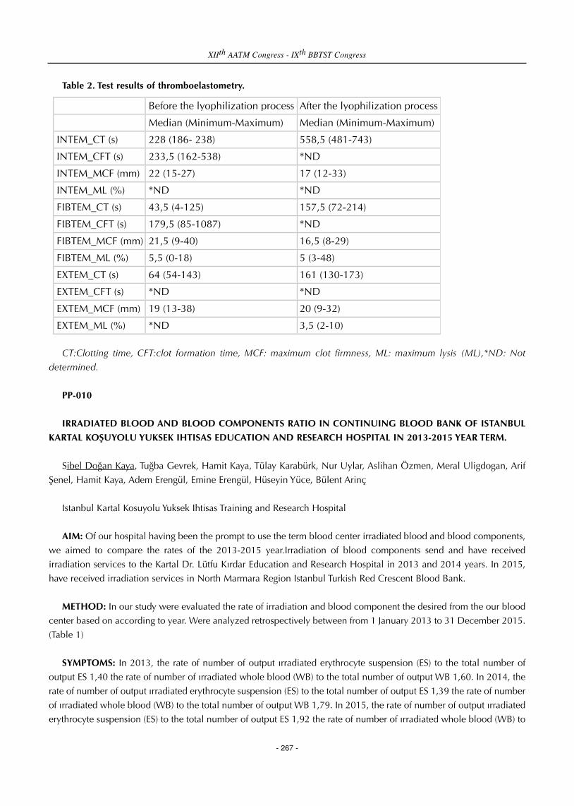
Table 2. Test results of thromboelastometry.
CT:Clotting time, CFT:clot formation time, MCF: maximum clot firmness, ML: maximum lysis (ML),*ND: Not
determined.
PP-010
IRRADIATED BLOOD AND BLOOD COMPONENTS RATIO IN CONTINUING BLOOD BANK OF ISTANBUL
KARTAL KOŞUYOLU YUKSEK IHTISAS EDUCATION AND RESEARCH HOSPITAL IN 2013-2015 YEAR TERM.
Sibel Doğan Kaya, Tuğba Gevrek, Hamit Kaya, Tülay Karabürk, Nur Uylar, Aslihan Özmen, Meral Uligdogan, Arif
Şenel, Hamit Kaya, Adem Erengül, Emine Erengül, Hüseyin Yüce, Bülent Arinç
Istanbul Kartal Kosuyolu Yuksek Ihtisas Training and Research Hospital
AIM: Of our hospital having been the prompt to use the term blood center irradiated blood and blood components,
we aimed to compare the rates of the 2013-2015 year.Irradiation of blood components send and have received
irradiation services to the Kartal Dr. Lütfu Kırdar Education and Research Hospital in 2013 and 2014 years. In 2015,
have received irradiation services in North Marmara Region Istanbul Turkish Red Crescent Blood Bank.
METHOD: In our study were evaluated the rate of irradiation and blood component the desired from the our blood
center based on according to year. Were analyzed retrospectively between from 1 January 2013 to 31 December 2015.
(Table 1)
SYMPTOMS: In 2013, the rate of number of output ırradiated erythrocyte suspension (ES) to the total number of
output ES 1,40 the rate of number of ırradiated whole blood (WB) to the total number of output WB 1,60. In 2014, the
rate of number of output ırradiated erythrocyte suspension (ES) to the total number of output ES 1,39 the rate of number
of ırradiated whole blood (WB) to the total number of output WB 1,79. In 2015, the rate of number of output ırradiated
erythrocyte suspension (ES) to the total number of output ES 1,92 the rate of number of ırradiated whole blood (WB) to
XIIth AATM Congress - IXth BBTST Congress
- 267 -
Before the lyophilization process After the lyophilization process
Median (Minimum-Maximum) Median (Minimum-Maximum)
INTEM_CT (s) 228 (186- 238) 558,5 (481-743)
INTEM_CFT (s) 233,5 (162-538) *ND
INTEM_MCF (mm) 22 (15-27) 17 (12-33)
INTEM_ML (%) *ND *ND
FIBTEM_CT (s) 43,5 (4-125) 157,5 (72-214)
FIBTEM_CFT (s) 179,5 (85-1087) *ND
FIBTEM_MCF (mm) 21,5 (9-40) 16,5 (8-29)
FIBTEM_ML (%) 5,5 (0-18) 5 (3-48)
EXTEM_CT (s) 64 (54-143) 161 (130-173)
EXTEM_CFT (s) *ND *ND
EXTEM_MCF (mm) 19 (13-38) 20 (9-32)
EXTEM_ML (%) *ND 3,5 (2-10)

the total number of output WB 3,12.
RESULT: The transfusion of blood and blood constituents saves lives of many people; however, it can induce fatal
Graft-Versus-Host disease. The only way to prevent this disease, which is characterized by an increase in the damage
of donor’s T-lymphocytes that target organs of the receiver, is the radiation of the blood and blood constituents with X
and gamma rays.
Figure.1
PP-011
THE ANALYSIS OF THE USAGE OF PLATELET SUSPENSIONS
Berrin Uzun1, Hayri Güvel1, Serdar Güngör2, Özcan Sengel1
1Izmir Katip Celebi University, Ataturk Training and Research Hospital, Transfusion Center2Izmir Katip Celebi University, Ataturk Training and Research Hospital, Medical Microbiology
BACKGROUND: Izmir Katip Celebi University and Ataturk Training and Research Hospital with 1100 beds is a
university research and training hospital. Since the second half of 2013, the blood and blood products supply actively
began to be provided from Regional Blood Center of Turkish Red Crescent (TRC).
AIMS: This study was performed in order to emphasize the importance of blood and blood products diversity by
examining distributions of the usage of platelet suspension products in three- year period.
METHODS: In 2013 blood and blood products were both produced in our hospital and as well as provided from
TRC. In 2014 and 2015 all of the blood and blood products fully were supplied by TRC. In the hospital, the whole
blood drawn from avaible donors was divided into components by removing buffy coat with the blood bag systems
(Quadruple Top&Bottom CPD/SAG-M 450 ml SAP 5D; Kansuk, Turkey). The pooled platelet suspensions were obtained
using Platelet Pooling Bag with Filter (Kansuk, Turkey). Apheresis platelet suspensions were obtained using automated
Haemonetics apheresis device (United States of America). All the products were provided as buffy coat removed from
TRC.
RESULTS: In 2013 totally 7504, in 2014 3602 and in 2015 3461 units of platelet suspension were used. The types
and the distribution of the platelet suspensions are given in the table.
XIIth AATM Congress - IXth BBTST Congress
- 268 -
Years
Total ES Out put
total ES Out put
number of patients to be applied irradiated ES Suspension
Irradiated ES Suspension output
rate
Total number of patients to be given Whole Blood
Total Whole Blood output
Number of patients to be given irradiated Whole Blood
Irradiated Whole Blood output
Rate
2013 2719 13655 9 191 1,4 1138 1935 7 31 1,6
2014 2933 14372 8 200 139 1079 1840 6 33 1,79
2015 3017 15075 9 290 1,92 686 1154 8 36 3,12

Table: The types and the distribution of the platelet suspensions.
CONCLUSION: Totally the the platelet rich plasma transfusion was significantly decreased in years. Platelet
suspension utilization rate was decreased by half according to the time when the platelet suspension was prepared in
our center. The product usage especially is proportional to facilitate its ability to supply. The platelet rich plasma
production must be made from all the whole blood despite the increasing workload. Because this application would
be provided solutions to meet the patients needs. In addition, the pooled platelet production was not seen that done
yet despite last years.
PP-012
THE ANALYSIS OF THE USAGE OF CRYOPRECIPITATE AND FRESH FROZEN PLASMA
Berrin Uzun1, Hayri Güvel1, Serdar Güngör2, Özcan Sengel1
1Izmir Katip Celebi University, Ataturk Training and Research Hospital, Transfusion Center2Izmir Katip Celebi University, Ataturk Training and Research Hospital, Medical Microbiology
BACKGROUND: Izmir Katip Celebi University and Ataturk Training and Research Hospital with 1100 beds is a
university research and training hospital. Since the second half of 2013, the blood and blood products supply actively
began to be provided from Regional Blood Center of Turkish Red Crescent (TRC).
AIMS: This study was performed in order to emphasize the importance of blood and blood products diversity by
examining distributions of the usage of cryoprecipitate products and fresh frozen plasma in three- year period including
transitional.
METHODS: In 2013 blood and blood products were both produced in our hospital and as well as provided from
TRC. In 2014 and 2015 all of the blood and blood products fully were supplied by TRC. In the hospital, the whole
blood drawn from avaible donors was divided into components by removing buffy coat with the blood bag systems
(Quadruple Top&Bottom CPD/SAG-M 450 ml SAP 5D; Kansuk, Turkey). Cryoprecipitate products were obtained using
transfer Bag 300 ml (Kansuk, Turkey) and using sterile hose joining device and blades (Terumo, Japonya) in our hospital.
All the products were provided as buffy coat removed from TRC.
XIIth AATM Congress - IXth BBTST Congress
- 269 -
2013 2014 2015 USED PLATELET SUSPENSİONS
Number (n)
Percentage (%)
Number (n)
Percentage (%)
Number (n)
Percentage (%)
Platelet Rich Plasma (PRP) 5055 67.36 1752 48.64 1734 50.10 RPR- Irradiated 1128 15.03 553 15.35 245 7.08
Pooling RPR 107 1.43 - - - - Apheresis Platelets 1074 14.31 676 18.77 894 25.83 Apheresis Platelets-
Irradiated 140
1.87
621
17.24
588
16.99 Total 7504 100.00 3602 100.00 3461 100.00

RESULTS: The distribution of the cryoprecipitate products and fresh frozen plasma according to years are given in
the table.
Table: The changes of used cryoprecipitate and fresh frozen plasma according to the year.
CONCLUSION: The use of cryoprecipitate was decreased each passing year. This reduction has been explained by
the non-produced blood products cannot be used. It is an important problem that must be solved to not keeping of
stock of cryoprecipitate in TRC. The usage of fresh frozen plasma was substantially reduced in 2015 while it was same
in 2013 and 2014. The decreased usage has been linked to the plasmapheresis cannot be done in the hospital in 2015
year.
PP-013
FIVE YEARS OF HEMOVIGILANCE REPORTS OF COMPLICATIONS OF THE BLOOD DONATION REPORTED
AT A TERTIARY CARE CENTRE IN KARACHI
Bipin Nepal, Saleem Khan, Muhammad Akbar Agha
Dr. Ishrat ul Ebad Khan Institute of Blood Diseases
BACKGROUND: There is a minor chance of risk among blood donors. Even though blood donors are usually
screened for the presence of risk factors, sometimes blood donations can put a person at panic.
OBJECTIVE: The safety of the blood supply depends on the actions to protect both; blood transfusion recipient and
the blood donor. Hemovigilance practice of learning of complications of blood donation and protecting them from
such complications is the best way to minimize the risk to blood donor.
STUDY DESIGN AND METHODS: Comprehensive blood donor hemovigilance program was studied at Dr. Ishrat
ul Ebad Khan Institute of blood diseases, Karachi from 2010 to 2015. Outlines of reported and communicated
complications were collected after whole blood donation. Analysis was done by general logistic regression.
RESULTS: Complications after 30,000 Whole blood donation procedures calculated 1620 total. (54 per 1,000
donations). The majority of the complications were faint and pre-faint reaction with light headedness (58.6 %), Sore
arm (24 %), Bruises and hematoma (14.4 %). Minor complications were Agitation/sweating (2 %) and arterial puncture
(1 %). Markers of the complications were age, sex, race, weight, blood pressure and donation status. All associated
independently after whole blood donation. Age and first-time status were associated with a significantly higher risk of
complications with 18-22 years old at higher risk compared to 23 to 50 years old. First-time donor were at higher risk
compared to repeat donor.
CONCLUSION: The results of this study are helpful in identifying and understanding the promoter to complication
of blood donation. Donor age and status were strong predictors of complications. The remedies and specific areas of
care should be provided.
XIIth AATM Congress - IXth BBTST Congress
- 270 -

PP-014
THE FIRST DATA OF HAEMOVIGILANCE SYSTEM IN A DAYCARE OF A TERTIARY HOSPITAL INBANGLADESH
Sonia Shormin Miah, Md Ashadul Islam
Department of Transfusion Medicine, Bangabandhu Sheikh Mujib Medical University (BSMMU), Dhaka,Bangladesh
BACKGROUND: This study is the first report of transfusion errors and adverse reactions recorded in Bangladesh. It'sa new approach to collect, collate, and analyze the data to speak of the issues associated with blood transfusion in theday care. It's also an alert for the physicians of developing country who are only tagged for whole blood transfusion.
AIMS: This study is aimed to perceive the frequency of adverse reaction and the errors associated with its barrier.
METHODS: Total 9,984 patients were included. Clinical and laboratory data were collected,analyzed and intendedto gather information on near miss and clerical errors.
RESULT: 936 adverse reactions were reported, equivalent to 0.093 reactions/9,984 units of transfused bloodcomponents. 1.06% adverse events reported; acute were 0.94 %. Among them febrile reactions were 0.22% due totransfusion of RCC plus FWB and 0.20% were due to PC. Allergic reaction (urticarial and anaphylactic reactions) were0.52% and 0.21% were due to platelet transfusion from RD, 0.31% were due to transfusion of FFP. Additional failurewere 0.09% due to excessive lipemic plasma and 0.03% were tearing of plasma bags. Red cell alloimmunization were11.11% to 12.12%. Clerical and near miss errors come 3.5% to 4.1% and 3 reports of mismatch blood transfusion, 2is about misidentification of the patient group due to severe myelosuppresion owing to leukaemia and 1 is forsubgroup. However, it's not always possible to analyze the relevance of all the transfusion and the outcome of thepatient.
CONCLUSION: Now, a developing country like Bangladesh has to face and notify the errors and to solve them inparallel. It may be slow in moving ahead but the team is aware to guard and share this with all the departments whichare also reliant on transfusion.
PP-015
BLOOD TRANSFUSION IN DEVELOPING COUNTRIES: HEMOVIGILANCE OR ULTRA-HEMOVIGILANCE
Syed Muhammad Irfan1, Hasan Abbas Zaheer2, Sadia Sultan1
1Department of Hematology and Blood Bank, Liaquat National Hospital, Karachi, Pakistan2Department of Blood Bank, Pakistan Institute of Medical sciences, Islamabad, Pakistan
BACKGROUND: Blood transfusion saves lives and improves health. Developing and underdeveloped countries,however, face tremendous shortage of life the saving fluid. These countries comprise bulk of the world population (75-80%). These are the countries with poor socio-economic systems, political instabilities, high illiteracy and morerecently extremism/terrorism etc. The main WHO regions in this regard are EMRO, SEAR, Africa and some countriesfrom European and American region.
XIIth AATM Congress - IXth BBTST Congress
- 271 -

INTRODUCTION: There is marked difference in availability and level of access of blood in advanced countries and
under resource countries. Of the 110 million donations collected globally, about half of them is collected in high
income countries which is home to just 1/4th of world population. Donation rate in low income countries is just
3.9/1000 population compared to high income countries (36.8/1000). Replacement and directed donations remain the
main mode of donation in countries with limited resources. In our part of the world picture is dismal and SERO
countries face the deficit of 8-10 million units annually. BTS is generally fragmented and whole blood is still used in >
60% places and lot of use of blood is unjustified. Hemovigilance is almost non- existent and transfusion committes are
seen at few centers only. Thailand, Siri-lanka, Turkey and Indonesia are probably having the best BTS in the region.
Ultra-hemovigilance: Health is directly linked to overall socio-economic status of the country. Providing adequate
safe blood should be a part of every country’s national health care policy. This, however, is not possible without socio-
economic uplift. Trying to attain everything in transfusion systems appears a futile practice in resource constraint
countries. We need to prioritize areas of great concern and optimize them within the given resources. Unlike
hemovigilance, which is there to prevent unwanted events related to transfusion, ultra-hemovigilance is a strategedy to
increase the availability of blood particularly in resource constraint world; to educate the youngsters for regular
donation, fractionate the blood into desired components, justified use of the component required, Establishment of
transfusion committees in hospitals, consideration of alternatives to blood and educating the community to best avoid
the use of blood.
SUMMARIZING: Blood is a common wealth and developing countries can attain adequacy in availability and
actual transfusion through ultra-hemovigilance. It addresses the people, practitioners and patients at very basic level so
that availability and usage of blood could be optimized in under resource countries.
PP-016
AN AUDIT OF WRONG BLOOD TRANSFUSIONS AND INTERVENTION WITH BEDSIDE ABO TYPING AND
TIME OUT
Bushra Moiz, Farheen Karim
Department of Pathology and Laboratory Medicine, Aga Khan University, Karachi, Pakistan
BACKGROUND: Blood transfusion is a life-saving option but is associated with various hazards. One of them is
wrong blood transfusion due to ABO mismatch which is potentially life threatening. Hemovigilance of tranfusion
processess is required through a team of colloborated work to minimize adverse effects of blood transfusion. Hospital
transfusion committees are instrumental in monitroing the policies and practices for achieving hemovigilance.
OBJECTIVES: To audit frequency and causes of wrong transfusion of red cell units during 2010 to 2016 and to
develop strategies for minimizing recurrence of such hazards.
SUBJECTS AND METHODS: All patients who received red cell blood transfusion in Aga Khan University during
January 1, 2010 to December 301 2015 were studied. Standard used was in-house with zero tolerance policy for wrong
transfusions.The total number of red cell transfusions was estimated. Wrong transfusion and the reasons for the same
were determined. Mortality ratio was also evaluated. This was discussed in hospital transfusion committee and multiple
interventions were discussed. Based on these, time out and bedside ABO typing was initiated. Nurses training was
considered for determining ABO group.ABO group card was saved in medical chart after performing the test. Using
XIIth AATM Congress - IXth BBTST Congress
- 272 -

barcoding for patient identification was also programed. This has yet to be initiated.
RESULTS: During the study period, 133888 red cell units were transfused. Eight wrong transfusions and three deaths
were observed. Wrong transfusion and mortality ratio was 1:16000 and mortality ratio was 1:67000. Final bedside error
and wrong addressograph were identified as the main reasons. During March to September 2015, 1400 nurses were
trained for bedside ABO typing through 50 nurse coordinators. From October 1 to January 2016, 2003 bedside ABO
typing have been performed. Time out before transfusion was also initiated. We observed 100% concordance between
bedside and laboratory ABO grouping. However, 21 medical records checked for ABO cards showed issues like lack
of proper filing, biosafety issues and incomplete recording of date and time for the test. In these four months, no wrong
blood transfusion was reported.
CONCLUSIONS: Our limited experience showed time out and bedside ABO typing as effective interventions for
minimizing wrong red cell transfusions. Training of nurses and maintaining cards properly in medical charts were
challenging.
RECOMMENDATIONS: We need a follow-up audit to monitor competency of nurses and to identify issues in
performing bedside ABO typing by nurses
ABO card
This is the pouch with ABO card and consumables with instructions to follow by nurses. This was sent with each
red cell unit dispense from blood bank.
XIIth AATM Congress - IXth BBTST Congress
- 273 -

Nurses doing bedside ABO typing
Two nurses verify blood unit identification and one performing bedside ABO typing
PP-017
REPORT OF UNWANTED SERIOUS ADVERS EFFECT AND SERIOUS ADVERS EVENT RESULTS FOR THE LAST
SEVEN YEARS AT OUR TRANSFUSION CENTER
Şükran Köse1, Gürsel Ersan1, Fatma Liv2, Tanju Yılmazer3
1Tepecik Education and Research Hospital, Department of Infectious Diseases and Clinical Microbiology,
Izmir,Turkey2Tepecik Education and Research Hospital,Transfusion Center,Izmir,Turkey3Tepecik Education and Research Hospital, Department Family Medicine Clinic, Izmir,Turkey
OBJECTIVE: Hemovigilance describes entire procedures by which unwanted serious advers effect and events and
the epidemiological followup of blood donors and blood receivers is carried out. At this study, data is collected and
evaluated in order to prevent the repetition of unwanted serious advers effect and advers events caused by procedures
done at our transfusion center.
MATERIALS-METHODS: Early unwanted serious effects can be stated as; hemolysis, nonhemolitic febril reaction,
rash, erythemia, urticaria, anaphylactic shock, bacterial contamination, transfusion-related acute lung injury. Delayed
serious unwanted effects on the other hand might be stated as; hemolysis, transfusion-associated graft vs. host disease,
post-transfusion purpurea, ALT elevation.Alloimmunization development against erythrocyte, platelet antigents or HLA
and virus,parasite,pyrion contamination are evaluated as other serious effects. Meanwhile unwanted serious events are
described as eventsthat might cause death or life threat,permanent and important morbidity or loss of working power
or need for hospitalization or lengthening of hospitalization period.Mistakes made in ABO type defining and wrong
labeling of blood samples are assessed as examples of this type of events. As a subgrup; ‘’Events avoided at the last
XIIth AATM Congress - IXth BBTST Congress
- 274 -

moment’’ and ‘’Transfusion mistakes without a serious event’’ have also been studied.
Check- forms for unwanted serious events have been filled at wards possibility levels has been set. The terminology
was noted in such a way that if data is not sufficient to analyze the situation; inevaluable, if the suspected serious effect
was proved to be caused by any reason apart from transfusion or despite the fact that there aren’t any certain evidence
but the serious effect is thought to be due to another reason; impossible, if the evidence is not sufficient to attribute the
unwanted serious effect to blood and blood products or not sufficient to link it to other reasons; possible, if the serious
effect can be attributed to blood or blood products; probable and if the unwanted serious effect can be related to blood
or blood products beyond reasonable suspicion; certain possibility levels were identified.
RESULTS: In our transfusion center, 192348 units blood components output was made between the dates of
01.01.2009 and 31.12.2015. In the meantime, unwanted serious effects 157 ( % 0.08 )in total, events avoided at the
last moment and transfusion mistakes without a serious event was found at the rate of 86( %0.04 ) Unwanted serious
events has not seen. Unwanted serious effects are determined in and after 2010 by the check-forms. It has been
determined as 55 % of it inevaluable, 20 % of it possible, 21% probable, 4 % certain. According to years and blood
products, unwanted serious effects can be seen on the first chart. According to years and reasons, unwanted serious
event and its subgroup can be seen on the second chart.
CONCLUSION: Attention should be paid to prevent unwanted effects and repetition of events in regards of some
mistakes on the transfusion process. Surveillance of unwanted serious effects and events contribute the safety of
transfusion.
XIIth AATM Congress - IXth BBTST Congress
- 275 -
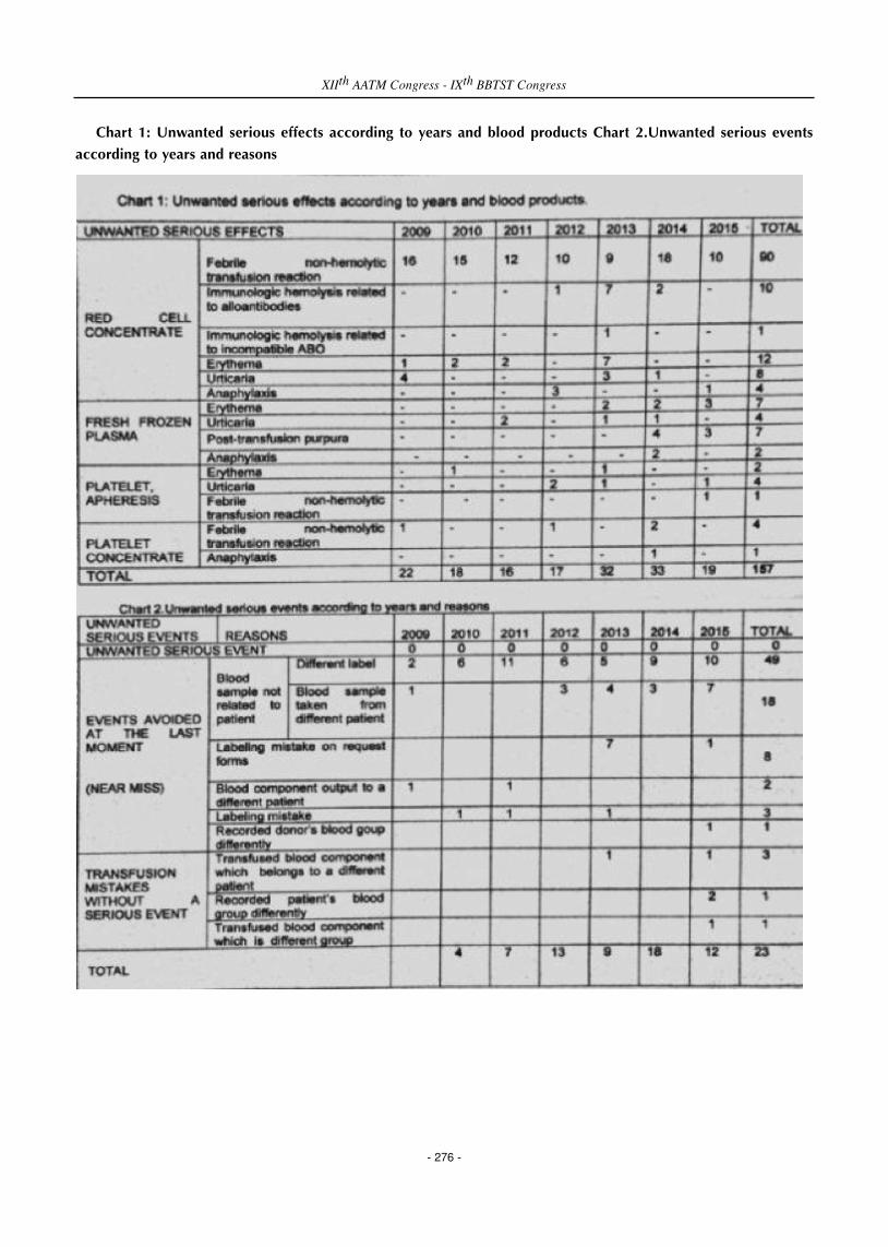
Chart 1: Unwanted serious effects according to years and blood products Chart 2.Unwanted serious events
according to years and reasons
XIIth AATM Congress - IXth BBTST Congress
- 276 -

PP-018
THE FREQUENCY OF ACUTE TRANSFUSION REACTIONS DUE TO USAGE OF BLOOD PRODUCTS
Nesrin Gareayaghi, Nesrin Gürlük, Rana Sucu
Sisli Hamidiye Etfal Training and Research Hospital,Istanbul
OBJECTIVE: Blood transfusion can be described as a special tissue transplantation. Although acute transfusion
reactions are considered as the reactions which develop during or 24 hours following the transfusion, most of the
reactions occur during or couple hours following the transfusion. This being the reason, patients should be monitored
closely for the first 15 minutes to an hour after the transfusion. In-line leukocyte filtered blood bags are used in our
clinic. It is known that blood products devoid of leukocytes reduce the transmission of CMV, occurence of febrile non-
hemolytic transfusion reactions and alloimmunization to an extent. In this study, we examined the frequency of acute
reactions due to usage of blood products throughout a year in our hospital.
MATERIALS-METHODS: In our hospital, transfusions are monitored by our transfusion control nurses. If it is
suspected that the reactions during the transfusion occurred because of the transfusion, the process is terminated by
the nurses or the doctors and a blood sample from the patient and the blood product baggiven to the patient are sent
to the blood transfusion center for an examination. Reasons to the reactions are investigated with blood type, cross-
match, Rh sub-type and indirect coombs tests. In our study, the inputs are elicited retrospectively from our transfusion
follow-up forms.
RESULTS: The results are stated in the table below.
CONCLUSION: The reactions that are mostly seen in patients include rash, erythema, trembling and itchiness.
Fever, tachycardia, hypotension, nausea and vomiting are following these findings. The reactions are treated with
antihistamines and antipyretics. None of these reactions are found to be life treating. Even though febrile non-hemolytic
transfusion reaction is the most common one, due to our usage of in-line leukocyte filtered blood bags, it is reduced
significantly. Blood banks serve to provide blood securely. It is vital to reduce the complications and intervene promptly
if a complication takes place. All these given, it is essential that the health care provider who is maintaining the
transfusion is careful, educated and talented.
Results Table
XIIth AATM Congress - IXth BBTST Congress
- 277 -
Types of product Number of products The number of reactions(%) FNHTR (%)
Whole blood 79 0(%0.00) 0(%0.00)
Red blood cell suspension 16325 15(%0.09) 3(%20)
Fresh frozen plasma 8150 16(%0.20) 2(%16)
Random platelets 2074 0(%0.00) 0(%0.00)
Aferez platelets 1429 3(%0.20) 0(%0.00)
Cryoprecipitate 420 0(%0.00) 0.(%0.00)
Washed red blood cells 19 0(%0.00) 0(%0.00)
Total 28496 34(%0.12) 5(%14.7)

PP-019
ISTANBUL CEKMECE DISTRICT PUBLIC HOSPITALS ASSOCIATION OF THE GENERAL SECRETARIAT OF THE
HOSPITAL TRANSFUSION FOLLOW TRACEABILITY ASSESSMENT SCOPE OF NURSING ACTIVITIES
Ilknur Güçlü, Figen Demir, Abdulbaki Kumbasar
Istanbul Bölgesi Çekmece Kamu Hastaneleri Birliği Genel Sekreterliği Tıbbi Hizmetler Başkanlığı
INTRODUCTION – AIM: Each one from donors with the traceability of the preconditions for the creation of
Hemovijilans network unit of blood or blood components until its final destination and is defined as the ability to follow
the direction opposite.
Prepared component of blood outside the blood service units in order to ensure data security to ensure that the
patient has to be given to the professional to do this task.
Following the April 20, 2015 in our hospital transfusion Nursing (Nursing Hemovijilans) service was introduced with
a staff of 12 people and was assessed 8 months of data.
MEANS METHOD: Transfusion to ensure the standard of follow-up data submissions were prepared to nurses. In 8
months in our 8 hospitals patients on behalf of all blood components made out, was evaluated on Form Follow
transfusion.
RESULTS: Seen in our 8 hospitals at 39 344 number of blood transfusions evaluated the eight-month period of
service units to a total of 43253 units of the number of products made out on behalf of patients. Our service units of
blood products derived from the study of legal documents before and after the transfusion, patients were allowed to be
filled, these documents were examined and the most common deficiencies and errors were detected. Looking into the
red blood cells examples;
In April, the number of forms that can not be considered 336 Transfusion follow-December, the number of forms is
not reached to form Follow transfusion was found to be 43.
CONCLUSION: Hemovijilan are about 60 000 years our blood component due to the use of public hospitals was
provided to obtain secure data for traceability, one of the priorities. Hospitals and clinics are determined based on the
number of blood use, and most often the faults and deficiencies, corrective and preventive actions are planned.
PP-020
DOES ABO AND RH INCOMPETIBLE APHERESIS PLATELETS TRANSFUSION RESULTS INCREASE IN
TRANSFUSION RELATED REACTIONS
Fatma Atasayar1, Burak Deveci1, Uğur Serçebay1, Ismail Koyuncu1, Levent Dönmez2, Naile Mısırlıoğlu1,
Hüsnü Altunay1, Ihsan Karadoğan1
1Medstar Antalya Hospital, Antalya, Turkey2Department of Public Health, Akdeniz University, Antalya, Turkey
XIIth AATM Congress - IXth BBTST Congress
- 278 -

This paper is aimed to investigate weather ABO and Rh incompatibility increases the transfusion related reactions
or not during apheresis platelets transfusion at our hospital? Results are matched by two proportion T Test.
Medstar Antalya Hospital is a special center for the Hematological and Oncological Diseases. 5248 apheresis
platelets transfused to 3098 patient 01.01.2015 – 31.12.2015 at our center. At this period random platelets usage is
zero. ABO and Rh compatibility is primerly chosen during platelets transfusion but if not other group transfusion
preferred.
4249 fullmatched, 479 ABO incompatible, 520 Rh incompatible platelets tranfusion done at this period. 22
transfusion related reactions developed at 18 patient. ABO incompatible 2 patient 3 reaction, Rh incompatible 1 patient
1 reaction. Itching, urticaria like mild transfusion reactions seen all patients, antihistaminic and corticosteroid treatment
reversed the symptoms
For the first patient 8 full compatible, 7 Rh incompatible, 2 ABO incompatible 17 apheresis platelets tranfusions
done. 1st and 5th transfusions of the patient are ABO incompatible 6th- 10th and 16th-17th transfusions are Rh
incompatible. 2 ABO incompatible transfusions are both reacted. Second patient 1 ABO incompatible, 9 ABO
compatible totally 10 transfusion done. Incompatible transfusion result with reaction. Third patient 128 full compatible,
7 Rh incompatible, 3 ABO incompatible transfusion done. 139th transfusion reacted. After that 19 compatible and 2
Rh incompatible transfusion done. Totally 160 transfusion ( Table 1).
Transfusion related reactions of our hospital is 0.41%, compatible transfusion 0.42%, ABO incompatible transfusion
0.62% ( P=0.7927 ), RH incompatible 0.19% ( P=0.6752 ).
Not statisticaly significant difference determined.
Both ABO and Rh Incompatible Transfusion Reactions At Medstar Antalya Hospital 2015
PP-021
CROSS UNMATCHED ERTYROCYTE SUSPENSION TRANSFUSION EFFECTS TO TRANSFUSION REACTIONS
Burak Deveci1, Fatma Atasayar1, Haydar Veske1, Tarık Kayoz1, Levent Dönmez2, Naile Mısırlıoğlu1, Hüsnü
Altunay1, Ihsan Karadoğan1
1Medstar Antalya Hospital, Antalya, Turkey2Department of Public Health, Akdeniz University, Antalya, Turkey
This paper investigate cross unmatched Erithrocyte Transfusion Reactions in our hospital. Results are matched by
two proportion T Test.
XIIth AATM Congress - IXth BBTST Congress
- 279 -

Medstar Antalya Hospital is a special center for the Hematological and Oncological Diseases. At the center 8505
erithrocyte suspension (ES) used between 01/01/2015-31/12/2015 for 4763 patients. 8363 cros matched ES transfused
to 4741 patient, 142 cross unmatched ES transfused to 22 patients.
14 Transfusion Reactions occured just two of them are cross unmatched. Cross unmatched transfusions and
transfusion related reactions shown in Table 1. Cross unmatched transfusion without transfusion related reactions are
shown at Tablo 2.
Transfusion related mild reactions like fever, tachycardia, dizziness, rush seen all patients. Corticosteroid and
antihistaminic treatments are reversed these mild symptoms.
Transfusion related reactions of our hospital 0,16%, cross matched transfusions 0,14% cross unmatched 1,4%
(P=0,0079 ) are found statistically significant.
Cross unmatched transfusions should be carefully fallowed.
Table 1: Cross Unmatched Transfusions and Transfusion Related Reactions
Table 2: Cross unmatched Transfusion Without Transfusion Related Reactions
XIIth AATM Congress - IXth BBTST Congress
- 280 -

PP-022
WHAT’S THE FILLING QUALITY OF TRANSFUSION OBSERVATION FORMS
Fatma Atasayar1, Burak Deveci1, Emine Yiğit1, Yeşim Temel1, Aslıhan Divarcı1, Levent Dönmez2, Naile
Mısırlıoğlu1, Hüsnü Altunay1, Ihsan Karadoğan1
1Medstar Antalya Hospital, Antalya, Turkey2Department of Public Health, Akdeniz University, Antalya, Turkey
Aim of this paper is to dedicate filling quality of transfusion observation forms in our hospital using at services,
operating room and reanimation. 15389 transfusion 01.01.2015-31.12.2015 period at Medstar Antalya Hospital, all
these observation forms are screened. Results are matched by single proportion T Test. After transfusion finished the
Observation Forms put in schedule at service, transfusion nurse control the form and take a copy for herself everyday.
Mistakes shown to service nurse, after correction the mistake registered by the Transfusion Nurse. Ten services of our
hospital, intinsive care unit and operating room are included the research. Number of forms and true fullfilling forms
statistics documented monthly at Table 1. Statistical percentages of true form filling of the services documented monthly
at Table 2.
The percentace of filling the forms has been detected as 85.1% for the entire hospital. When the clinics were
examined separately, the percentage of filling the forms was significantly low in intensive care unit, operating theater
and 8th floor ward. The percentage was significantly high in 10th and 12th floor ward. The rest of the wards came up
to the average.
While the percentage was statistically significantly low in January and February. The low results that are seen at the
beginning of the year has been achieved to desirable results with the education.
Table 1: Medstar Antalya Hospital Transfusion Observation Form Trueness Monthly 2015/Total
XIIth AATM Congress - IXth BBTST Congress
- 281 -

Table 2: Trueness Percentages According Months At Medstar Antalya Hospital-2015
PP-023
SUCCESSFUL DONOR LYMPHOCYTE HARVEST AND INFUSION (DLI) WITH LIMITED SUCCESS IN
OUTCOME: A CASE REPORT
Dinesh Arora1, Aseem K Tiwari1, Nitin Sood2, Ravi C Dara1, Geet Aggarwal1
1Medanta Department of Transfusion Medicine, Gurgaon, Haryana, India2Medanta Division of Medical Oncology and Haematology, Gurgaon, Haryana, India
AIMS AND PURPOSE: Allogenic stem cell transplantation is the only known curative therapeutic option for CMML
patients and there is even less information available related to the use of Donor Lymphocyte Infusion (DLI) in these
patients to prevent post transplant relapse of the disease. Here we report a case of CMML who underwent
hematopoietic progenitor cell – apheresis (HPC-A) transplant; had falling donor chimerism six months after the
transplant. Escalating doses of DLI was administered to prevent disease relapse.
MATERIAL-METHOD: A 49 year old female of African origin, who was negative for JAK2 and bcr-abl PCR, was
diagnosed with myeloproliferative chronic myelomonocytic leukemia (MPN-CMML) on flowcytometery and
cytogenetic analysis of bone marrow aspirate. After an elaborate discussion about various treatment options available,
allogenic hematopoietic stem cell transplant (HSCT) was decided as a course of action. Patient’s sister, a 10/10 HLA
match, was selected as Peripheral Blood Stem Cell (PBSC) donor and patient was started on reduced intensity
conditioning regimen (RIC) [1].
Patient was administered a dose of 5 million CD34+cells per kg on day Zero. Neutrophil and platelet engrafted on
day +16 and day +13 respectively. Post-transplant donor chimerism decreased from 90% on day +55 to 4.49% on
day+187. DLI [2, 3, 4] was an option to treat relapse by virtue of Graft Vs Tumor (GVT) effect and therefore after
discussion with patient, escalating DLI therapy was planned. Patient was administered infusions of DLI on day+209,
day+320 and day+407 with three consecutive harvests of donor lymphocytes. One, five and ten million donor
lymphocytes per kg body weight of patient was successfully harvested with no discomfort or adverse reaction to the
donor. The infusion of these donor lymphocytes was uneventful. There was no contraindication to DLI in form of signs
or symptoms of GvHD. However, after 0% donor chimerism on day+488, patient was labelled as post HPSCT relapse-
XIIth AATM Congress - IXth BBTST Congress
- 282 -

case of CMML (Graph). Though the patient could be managed without aggressive chemotherapy or re-transplant for
almost a year, there was disease relapse. However, Relapse ultimately occurs in 10 to 40 percent of allogenic HPSCT
patients and discontinuation of immunosuppression followed by immunotherapy.
CONCLUSION: With this case study we conclude that DLI harvest can be successfully harvested in our country
and it is safe for both the donor as well as patient.
1. Martino R, Caballero MD, Canals C, Simon JA, Solano C et al. Allogeneic peripheral blood stem cell
transplantation with reduced-intensity conditioning: results of a prospective multicentre study. Br J Haematology 2001
Dec; 115(3): 653-9.
2. Deol A, Lum LG. Role of donor lymphocyte infusions in relapsed hematological malignancies after stem cell
transplantation revisited. Cancer Treat Rev 2010; 36:528.
3. Kolb HJ, Schattenberg A, Goldman JM, et al. Graft-versus-leukemia effect of donor lymphocyte transfusions in
marrow grafted patients. Blood 1995; 86:2041.
4. Collins RH Jr, Shpilberg O, Drobyski WR, et al. Donor leukocyte infusions in 140 patients with relapsed
malignancy after allogeneic bone marrow transplantation. J Clin Oncol 1997; 15:433.
Graph 1
Graphical presentation of Transplant day (day 0), percentage of donor chimerism in follow up days, Days on
which DLI was administered
XIIth AATM Congress - IXth BBTST Congress
- 283 -

PP-024
TÜRKÖK DONOR CENTERS STEM CELL DONOR COUNTS AND RATES
Mustafa Eren Uzun1, Damla Dernek1, Cansu Yıldırımkaya1, Şenay Canpolat1, Savaş Koç1, Metin Kalender1, Levent
Sağdur1, Armağan Aksoy1, Nurettin Hafi̇zoğlu1, Mehmet Güllüoğlu1, Fatma Meriç Yi ̇lmaz2
1Turkish Red Crescent Society, Ankara2Board Member of Turkish Red Crescent, Yıldırım Beyazıt University, Ankara
Main objective of this operation is to find potential donors wishing to donate peripheral stem cell or bone marrow
to TÜRKÖK Bone Marrow Bank which was founded for the patients in need of homoeopathical stem cell transplant.
Moreover, the procedures of transplantation operations and reaching the donors through the scanning tests-carried
out by Turkish Ministry of Health-at TÜRKÖK are both included as the main objectives of TÜRKÖK project. In 2015,
43 succesful transplantation operations were carried out of 239 matches.
Considering the rate of success in the field of Stem Cell Donation, the operations conducted by the Ministry of
Health and Turkish Red Crescent as part of TÜRKÖK Project have made Turkey one of the top countries in the field of
Stem Cell Donation. Furthermore, stem cell transplantations carried through by TÜRKÖK Bone Marrow Bank will
contribute to the decrease of the need for foreign help for homoeopathical stem cell transplantation.
Stem Cell Donor Distribution According to Monthly in 2014-2015
XIIth AATM Congress - IXth BBTST Congress
- 284 -
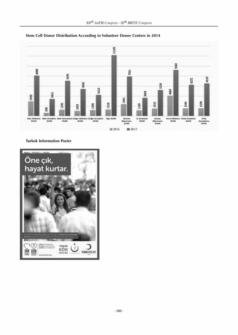
Stem Cell Donor Distribution According to Volunteer Donor Centers in 2014
Turkok Information Poster
XIIth AATM Congress - IXth BBTST Congress
- 285 -

Volunteer Donor Center Matching Graphic in 2014
PP-025
COMPARISON OF ANTIBODY TITERS USING CONVENTIONAL TUBE TECHNIQUE VERSUS COLUMN
AGGLUTINATION TECHNIQUE FOR ABOI RENAL TRANSPLANT
Amardeep Pathak
Department of Transfusion Medicine, Rajiv Gandhi Cancer Institute & Reserach Centre, New delhi, India
INTRODUCTION: Measurement of alloantibody titer to a red cell antigen is an essential semi-quantitative tool in
ABO blood group incompatible (ABOi) renal transplants. Titers are usually determined by serial double-fold dilution
method. Different centers have their ‘cut-off’ titers pre-transplant, which they target with plasma exchange or antibody
adsorption.
AIM: To compare and correlate the alloantibody titers against red cell antigen by conventional Tube technique (CTT)
and column agglutination tecnique (CAT) in ABO blood group incompatible renal transplants (ABOiRT).
MATERIAL & METHOD: During work up of ABOi renal transplants, the ‘cut-off’ titer of 1:8 by the CTT was targeted
for renal transplant. All patients underwent series of alloantibody titer estimation against donor antigens at baseline and
then after every second plasma exchange or after every session of antibody adsorption. Antibody titers of sequential
recipient’s serum were done in parallel by both tube and gel methods.
RESULTS: A total of 67 samples were processed in parallel for anti-A/B antibodies by both tube and gel methods.
The mean titer by CTT was 38.5 + 96.6 and by the CAT was 96.4 + 225. The samples correlated well with Spearman
rho correlation coefficient of 0.94 (p = 0.01). However, the Lin’s concordance coefficient was 0.58. The sensitivity of
gel method was greater than that of tube method, with the gel results being approximately two and half fold higher (one
more dilution) than that of tube method.
XIIth AATM Congress - IXth BBTST Congress
- 286 -

CONCLUSION: One should be careful in interpreting the results of anti A/B titers in ABOiRT depending upon the
method used for titer estimation. Gel card for anti A/B titer estimation in an ABO incompatible kidney transplant is
more sensitive and easy method. The ‘cut-off’ titers would be at least a dilution higher, if estimated by Gel columns.
Alloantibody titers in ABOi Renal Transplant
PP-026
SIGNIFICANCE OF ANTIBODY SCREENING AND IDENTIFICATION IN PRE TRANSFUSION AND ANTENATAL
TESTING-A RETROSPECTIVE STUDY
Rani Premkumar
Dr Rani Premkumar Sakra World Hospital Bangalore
BACKGROUND: Genetic disparity of RBC Antigen between donor and recipient is responsible for RBC
alloimmunisation.Though RBC transfusion is a lifesaving therapy in most of the patients, risk of alloimmunisation is
always a concern for patients receiving multiple transfusion. Pregnancy also carries the risk of alloimmunisation. Very
few studies on alloimmunisation are done on the general Indian Hospital patients.
AIM: The study was aimed at assuming the frequency and type of unexpected red cell antibodies in both patients
going to receive transfusion and antenatal cases at a multispecialty tertiary care hospital in Bangalore.
METHODS: It is a retrospective study. Antibody screening was carried out in 1912 patients including inpatients and
antenatal mothers from January to December 2014.All positive cases were subjected to antibody identification. In
patients receiving transfusion antigen negative red cells were cross matched and given. Antenatal cases were followed
up every month with antibody titre till the time of delivery.
RESULTS: It is a retrospective study in which evaluation of 1912 cases (870:45.5% males and 1042:54.5%females)
done.. Antibody screening was positive in 19 patients (0.99%). In the serum samples of 37 patients only autoantibodies
were identified, 4 cases revealed autoantibody also with underlying alloantibody. The total alloimmunisation rate was
0.99%, alloimmunisation in antenatal females was 0.15%.Among the antenatal females anti D was the most common
(4 cases) Anti E and Anti K one each.
INTERPRETATION AND CONCLUSION: Since clinically significant antibodies are frequently detected in our
patient population, antibody screening and identification is mandatory to ensure safe transfusion practice. Since
antibody against Rh, Kell and Le group antigens are more common and clinically significant, provision of Rh and Kell
matched cells may be of protective value.
Clinically significant unexpected antibodies are capable of causing hemolytic transfusion reactions secondary to
accelerated destruction of a significant proportion of transfused red blood cells. Therefore, screening for unexpected
XIIth AATM Congress - IXth BBTST Congress
- 287 -
IgM IgG (CTT) IgG (CAT) pearson Correlation
Mean 24.7 plus-minus 77 96.4 plus-minus 225 38.5 plus-minus 96.6 0.849
Range 0-512 0-1024 0-521

antibodies should be part of all pretransfusion testing, with antibody identification in the event of a positive result.
Antenatal detection of the non-anti-D causes of HDN requires Red cell antibody screening. If RCAS is positive, the
following steps are to be taken. Antibody Identification should be done to identify the antibody. The spouse has to be
screened for the presence of offending antigen and the pediatrician has to be alerted about delivery of a potentially
sensitized infant. The blood bank should find a suitable antigen-negative donor for transfusion to baby and mother.
PP-027
ANALYSIS OF ANTIBODY SCREENING & IDENTIFICATION USING A FULLY AUTOMATED SOLID PHASE
METHOD
Sangeeta Pathak, Surjit Singh, Tamojit Chakraborty, Anurag Gupta, Satish Kaushik, Ruchi Dubey
Department of Transfusion Medicine, Max Super Speciality Hospital, Saket, New Delhi, India
BACKGROUND: There are several methods & technology available to detect irregular antibodies in patient or
Blood donor but they are in limitation to detect its clinically significance & specificity. In our facility we have
implemented Immucor Gailieo-NEO (Fully automated immunohematology analyzer) for antibody screen testing &
automated antibody identification by solid phase method. This is an analysis of the antibody types & number of
antibodies detected, to determine the effectiveness of solid phase antibody identification for routine antibody screening
for patient & donors.
AIM: To establish routine antibody screening and identification on a fully automated solid phase method, and to
minimize the risk of immunization or provide safest blood to immunized patient seeking for blood transfusion.
METERIAL & METHOD: One year study was carried on total no. of 35680 samples (Both patient & donors), which
were tested for 3 cell antibody screening by solid phase method using Immucor Capture-R Ready Screen 3. Out of
them, 38 patients requiring blood transfusion with a positive antibody screen, was further tested for antibody
identification using Immucor Capture-R Ready-ID Panels. Wherever necessary, additional testing was carried out by
coombs method using Immucor capture-R Panocell-10, Ficin Treated Panocell-10 & Panocell-20. For advance
immunohematology work-up i.e. elution & adsorption, Immucor’s Elukit & WARM reagents were used. Multiple panels
and additional reagents were only used for any unresolved or auto antibody case. Extended panels were used to rule
out antibodies specificities.
RESULTS: Out of 38 samples 63% of total identified antibodies were Rh & Kell type (anti-E-34.2%, anti-c-13%, anti-
D-5.3%, anti-C-2.6, anti-e-2.6%, and anti-K-5.3%), 21% of warm auto antibody, 6% of other clinically significance
antibodies (i.e. anti-Fya, anti-S) and 10% of non-clinically significant antibodies (i.e. anti-M, anti-Leb).
SUMMARY/CONCLUSION: Antibody Identification work-up is stills a big task for major Blood Bank facility in
India. To minimize the immunization risk and provide safe blood to an immunized patient as soon as possible is a big
challenge by routine testing. We have identified about 79% of cases by using a fully automated method. Liquid panels
were used for completion of 21% of samples. Most of the clinical significant antibodies can be detected and identify
on solid phase automation using Capture R technology without any additional resources. These data demonstrate that
although this method automates the antibody identification process; however one fourth of the samples require
multiple panels for resolution.
XIIth AATM Congress - IXth BBTST Congress
- 288 -

PP-028
ABO DISCREPANCIES AND ERRORS IN A ROUTINE BLOOD BANK
Bipin Nepal, Nadeem Nusrat, Muhammad Akbar Agha
Dr. Ishrat ul Ebad Khan Institute of Blood Diseases
INTRODUCTION: - ABO discrepancy exist when red cell antigen grouping result do not correlate with serum
grouping result.
OBJECTIVES: - The study is done to identify the incidence and type of ABO discrepancies and errors leading to
wrong results.
MATERIAL-METHODS: All the data of ABO grouping from June 2012 till June 2013 was collected at Ishrat ul Ebad
Khan institute of blood disease, Dow university of health sciences, Karachi, Pakistan.
ABO discrepancies, pre-analytical, analytical & post-analytical errors in routine blood grouping were noted and
recorded on a Performa designed for the study and results were tabulated.
RESULTS: -30 discrepancies (0.3 %), 18 (0.18 %) pre & post analytical errors were identified from a total of 10,000
blood group tests performed during the study period. The commonest of all was ABO discrepancy due to sub groups n
26 (86.6 %) followed by auto-antibodies n 4 (13.3 % )
CONCLUSION: ABO discrepancy is not an infrequent finding in routine blood banking while pre-analytical,
analytical & post-analytical errors are also seen in routine practice and are small but very important source of mistakes
which can lead to serious hazards of transfusion. Measures should be taken to identify and resolve these discrepancies
while ensuring proper collection, labeling, analysis and reporting of results for safe blood transfusion.
PP-029
RED CELL ALLOIMMUNIZATION IN MULTI-TRANSFUSED PATIENTS: A BI-CENTRIC STUDY IN INDIA
Amit Agrawal1, Ankit Mathur2
1Blood Bank,Fortis Escorts Heart Institute,New Delhi2Rotary-ttk Blood Bank,Bangalore
BACKGROUND: It is well known that alloimmunization to red cell (RBC) antigens resulting from the genetic
disparities between donor and recipient is one of the risks of blood transfusion. The antibody screening cells are used
to detect unexpected antibodies.
The risk of alloimmunization is higher in patients who have received multiple blood transfusions like thalassemia,
other hematological disorders, Renal failure patients on dialysis who receive blood transfusions and females with bad
obstreitic history.
XIIth AATM Congress - IXth BBTST Congress
- 289 -

The antibody screening cells are used to detect unexpected antibodies.If antibody screening is positive, additional
tests are carried out to specifically identify antibody using the antibody identification panel and red cell antigen typing.
Antibody screening test using 2-3 cells panel is not a mandatory pre transfusion testing in India and is performed
routinely in limited blood centers.
MATERIAL-METHODS: This bi-centric study was carried out at two regional blood transfusion centers,one in North
India(referred as Centre-I) which collects 25000 units in a year & another from South India(referred as Centre-II) which
collects 35000 units in a year.Aim of the study was to look at prevalence of antibodies in multi-transfused patients who
have higher risk of alloimmunization like patients who have received multiple blood transfusions like thalassemia, other
hematological disorders, Renal failure patients on dialysis who receive blood transfusions and females with bad
obstreitic history.
This study was conducted during 1st February, 2012 to 31st March, 2012 at both the centers in parallel. The pre
transfusion testing protocol followed at both the centers was Blood typing, Indirect anti globulin test (IAT) & cross
match. The antibody screening using 3 cells is not a routine practice at both the centers.Total 4569 cases were analyzed
and 258 patients were selected for antibody screening & identification.
The patients included in the study were thalassemia, other hematological disorders, renal failure patients on dialysis
who receive blood transfusions and females with bad obstetrics history. These samples underwent initial antibody
screening using commercial 3 cell panel. The samples with positive antibody screening were tested for antibody
identification using 11 cell panel. The antibody screening cells and antibody identification cell panel used were
procured from Diagast (DIAGAST251-Avennue Eugene Avinee-BP 9-59374 LOOS, FRANCE) and test was performed
on Qwalys3, fully automatic machine working on Erthrocyte magnetization technology and software to interpretate the
results.
RESULTS: Out of total 4569 patients, 258 multi-transfused patients were studied. Among these 7 patients (2.71%)
were found alloimmunized. The risk of alloimmunization was 2.90% in Thalassemia, 0% in chronic renal failure
patients, 3.77% pregnant females with bad obstetric history & 2.78% other multi- transfused patients like cancer.
DISCUSSION: Multi- transfused patients are always at higher risk of alloimmunization & this creates difficulty in
their pre transfusion testing. Regular monitoring through antibody screening & transfusion of blood matched for minor
erythrocyte antigens are recommended in these patients.
Rate of RBC alloimmunization & antibodies
XIIth AATM Congress - IXth BBTST Congress
- 290 -
Centre Total pre transfusion testing done
No of cross matches
No of cases selected for study(n)
No of positive antibody screening
Rate(95% confidence interval)
I 2315 3760 154 02 (1.2%) 1.3(0-3.1%)
II 2254 3667 104 05 (4.8%) 4.8(0.7-8.9%)

PP-030
DISTRIBUTION OF BLOOD GROUPS IN THE TWO YEAR PERIOD IN THE GIRESUN PROF. DR. A. ILHAN
OZDEMIR GOVERNMENT HOSPITAL
Şahin Direkel1, Cihangir Akdemir1, Ömer Emecen2, Gamze Kaçmaz3
1Department of Medical Microbiology, Faculty of Medicine, Giresun University, Giresun, Turkey2Department of Blood Bank, Prof. Dr. A. Ilhan Ozdemir Goverment Hospital, Giresun, Turkey3Department of Medical Microbiology, Health Sciences Institute, Giresun University, Giresun, Turkey
AIM: Determination of the frequency of blood groups in a particular region facilitates timely provision of blood and
blood products. In this study, we aimed to find out the rates of ABO and Rh blood groups in Giresun province.
METHODS: This retrospective study was conducted by investigating the records of 13150 person who admitted to
Giresun Prof. Dr. A. Ilhan Ozdemir Government Hospital, Blood Bank between 1st January, 2014 and 31st December,
2015. ABO and Rh blood groups were examined by gel centrifugation methods.
RESULTS: In this study, the frequency of blood groups A, O, B, AB were found 41.81%, 38.00%, 14.27%, 5.92%
respectively. Rh (+) positivity was determined in 11.401 cases (86.70%) and Rh (-) negativity was determined in 1749
cases (13.30%). The frequency of Rh intra-group was found Rh (+) 87.47% and Rh (-) 12.53% in blood group A, Rh (+)
86.35% and Rh (-) 13.65% in blood group O, Rh (+) 85.68% and Rh (-) 14.32% in blood group B, Rh (+) 85.99% and
Rh (-) 14.01% in blood group AB. Gender distribution of their blood group determination was 58.79% men and
41.21% women.
CONCLUSION: ABO and Rh blood groups distribution in our region was similar to general rates of Turkey. There
was a positive correlation between frequency of ABO blood group type and Rh positivity rate in each group. We believe
that present study will contribute to the literature in terms of database of blood groups.
Distribution of blood groups in Giresun Prof. Dr. A. Ilhan Ozdemir Government Hospital, Blood Bank in the
two year period
XIIth AATM Congress - IXth BBTST Congress
- 291 -
Blood group Male Percentage Female Percentage Total
O Rh (+) 2532 58.7 1782 41.3 4314
O Rh (-) 401 58.8 281 41.2 682
A Rh (+) 2828 58.8 1981 41.2 4809
A Rh (-) 389 56.5 300 43.5 689
B Rh (+) 919 57.1 690 42.9 1609
B Rh (-) 175 65.1 94 34.9 269
AB Rh (+) 410 61.3 259 38.7 669
AB Rh (-) 77 70.6 32 29.4 109
Total 7731 5419 13150

PP-031
PARTIAL D STUDIES IN TURKISH RED CRESCENT NORTHERN MARMARA REGION BLOOD CENTER
LABORATORIES
Kadri Demirel1, İlhan Birinci1, Erdoğan Koşan1, Yalçın Aksu1, Dilek Ün1, İsmet Karahacıoğlu2, Armağan Aksoy2,
Nurettin Hafızoğlu2, Mehmet Güllüoğlu2, Fatma Meriç Yılmaz2
1Turkish Red Crescent Northern Marmara Region Blood Center/Istanbul2Directorate General of Turkish Red Crescent Blood Services/Ankara
AIM: Anti-D is one of the antibodies which have a clinical importance for erythrocyte surface antigens. In the
National Guide of Blood and Blood Products, it is pointed out that standards identifying category D VI in patients
should not be used whereas standards identifying category D VI in blood donors should be used. Accordingly, the cards
which identify Weak D and DVI are routinely used in our labs. In this study, the blood donors whose weak D reaction
was detected one positive were also examined in terms of partial D.
MATERIAL AND METHOD: BioRad gel centrifugation kit is routinely used for typing of ABO/Rh in our blood
donors. The Rh (-) negative ones out of these are studied by BioRad kit which can detect Weak D/D VI. Particularly, D
variants of the blood donors who could not be ultimately decided to be negative/positive during their previous
donations and the ones whose Weak D result was +1 or indefinite were attempted to be identified using BioRad,
Extended Partial RhD Typing Set and 12 different series of Anti-D antibody. During 11 months of period we conducted
the study (01 April 2015 – 01 March 2016), blood groups of 1.053.085 blood donors in total were studied. On 144.381
blood donors with Rh (-) negative out of these, weak D/D VI was studied and 914 Weak D was detected.(Table 1.) On
149 blood donors whom we were unable to decide, Partial D's were studied. The obtained results are presented in table
2.
RESULTS: Even it is theoretically known that Weak D individuals can not generate Anti-D antibody whereas partial
D individuals are able to generate Antibody, it is accepted that some weak D phenotypes can produce anti-D as well
as provide Anti-D production. D antigenic structure in the donated blood should be revealed. As result of not approving
the 149 ±, +1 Weak D, D IV samples in our study as positive with respect to Weak D and studying by Extended Partial
RhD Typing Set and 12 different Anti-D antibody series, 68 samples were identified true negatives. These 68 samples
which were not evaluated as wrong positive, were able to be delivered to the needers as Rh (-) negative. Weak D and
D VI were detected in 14 of the remaining 81 samples, and 67 samples were revealed as partial D. As we consider all
those circumstances; we think that within the laboratories such as Northern Marmara Region Blood Center laboratories
that study a large number of blood groups, kits should be available where D variant can be examined and this would
help in such type of circumstances which might arise at the decision stage.
XIIth AATM Congress - IXth BBTST Congress
- 292 -

Table 1. Number of blood donors studied during 11 months period between 01 April 2015 – 01 March 2016
Table 2. Partial D Evaluation table
PP-032
INDETERMINATED ABO BLOOD GROUP ANTIGENS IN NEONATES
Filiz Sağlam1, Özlem Unay Demirel2, Demet Yalçın5, Seyda Iğnak4, Yusuf Soytürk3
1Department of Medical Microbiology, Bahcesehir University School of Medicine, Istanbul, Turkey2Department of Medical Biochemistry, Bahcesehir University School of Medicine Medicalpark Goztepe Hospital,
Istanbul, Turkey3Transfusion Center, Medicalpark Goztepe Hospital, Istanbul, Turkey4Department of Medical Biology, Bahcesehir University School of Medicine, Istanbul, Turkey5Infectious diseases and medical microbiology, Medicalpark Goztepe Hospital, Istanbul, Turkey
INTRODUCTION: Neonatal blood type grouping is one of the first laboratory tests to be performed after birth in
Turkey. This test is used routinely to determine the blood group antigens in neonates. Blood group antigens are not fully
XIIth AATM Congress - IXth BBTST Congress
- 293 -
Dates of 01 April 2015– 01 March 2016 Blood Donor
% Value
Number of Total Blood Groups Studied 1.053.085
Number of the ones on whom Rh (-) was detected and weak D was studied
144.381 13.71%
Number of the ones on whom Weak D was identified 914 0.63%
Number of the ones on whom Partial D was studied 149 0.10% 149 0.10%
Obtained results Identified sample
D VI detected 2 samples
Weak D detected 12 samples
Rh (-) Negative detected 68 samples
DAR-E detected 4 samples
DFR detected 60 samples
D IV 1 samples
D III 2 samples

formed and isohemaglutinins are not present in the serum at the birth. Mother’s antibodies are dominant in the first six
months of life. Weaker agglutination reactions are observed with newborn infants' red blood cells (RBCs) compared to
the mature RBCs of adults due to the number and strength of A and B antigen sites being less. In this study, we aimed
to find out the ratio of indeterminate blood groups in the neonates.
METHOD: This study was done in Goztepe Medical Park Hospital (Istanbul, Turkey) transfusion center where
neonatal blood group typing is performed by micro-column gel typing system using forward grouping only (Diana
Processor, Hemakim, Turkey). If the clear result is obtained, it is reported with the note” In babies, we recommend the
blood type grouping to be repeated after 6 months of age.”; if the result is not clear, it is reported as “ABO antigens
undetermined”. The data of blood group types in infants under 6 months old was obtained retrospectively from the
laboratory information system. We evaluated all blood grouping results of the target patients (n= 6961) between 2011
January and February 2016.
RESULTS: In total, 537 indeterminate results among 6961 neonates (7.71%) were found. According to the years, 77
indeterminate cases among 1627 patients (4.7%) were found in 2011, 88 in 1307 (6.7%), 135 in 1397 (9.7%), 105 in
1325 (7.9%), 120 in 1118 (10.7%) and 12 in 187 (6.4%) were the cases determined for 2012, 2013, 2014, 2015 and
2016 respectively.
CONCLUSION: In newborns, 5-10 % of the cases have ABO antigens that are not detected by standard gel
centrifugation method.
PP-033
NEONATAL HEMOLYTIC ANEMIA DUE TO ALLOIMMUNIZATION OF MINOR GROUP INCOMPATIBILITY OF
ANTI-E
Alper Ozcan1, Can Acıpayam2, Mehmet Yay4, Musa Karakukcu1, Turkan Patıroglu1, Mehmet Akıf Ozdemir1,
Bulent Eser3, Ekrem Unal1
1Department of Pediatric Hematology Oncology, Erciyes University, Kayseri, Turkey2Department of Pediatric Hematology Oncology, Sutcu Imam University, Kahramanmaraş, Turkey3Department of Hematology Oncology, Erciyes University, Turkey4Blood Center, Erciyes University, Kayseri, Turkey
INTRODUCTION: Blood group incompatibility is the most common cause of hemolytic anemia in the early
newborn period. Because of using anti-D gammaglobulin in the last four decades the rate of minor blood group
incompatibility has relatively increases in the etiology of newborn hemolytic anemia. In this article, we report a patient
with symptoms of anemia who developed hemolytic disease of the newborn due to anti-e alloimmunization.
CASE: A female newborn having the B, Rh positive blood type was admitted for jaundice on the 36th day after being
born. At the time of admission, the patient had jaundice with mild hepatosplenomegaly. The laboratory tests were as
follows: the blood type of the baby: B Rh(+), the blood type of the mother: BRh (+), direct Coombs test: ++++. Indirect
coombs was (+). Hemoglobin: 10.2 gr/dL, hematocrit: 32%, leukocyte: 12440 /mm3, platelets: 17400/mm3 and
reticulocyte: 6%. Peripheral smear revealed 55% neutrophils, 33% lymphocytes, 12% normoblasts and fragmented
erythrocytes. The subgroups typing, antibody detecting/identification tests of the mother, and the newborn were studied;
anti-e antibody was found in the mother. Intravenous immunoglobulin (1 g/kg) and metilprednisolone was administered
XIIth AATM Congress - IXth BBTST Congress
- 294 -

to the patient. On the 7th day of treatment, the metilprednisolone treatment was decreased and stopped without any
complications.
DISCUSSION: The most common causes of pathological neonatal jaundice are hemolytic diseases due to blood
incompatibilities including ABO, Rh and subgroup incompatibilities. The clinical picture with minor blood group
incompatibility may vary from subclinical hemolysis to active hemolysis and newborn jaundice. The best known ones
among these antigens which are responsible of 3% of the cases of neonatal hemolytic disease include Kell, Duffy,
Diego, Kidd, MNS, C and E and they may lead to a hemolysis picture in the neonatal period similar to Rh disease.
CONCLUSION: It should be kept in mind that minor blood groups may lead to neonatal jaundice and minor blood
groups should be tested in newborns with pathologic jaundice with unknown etiology and in newborns with hemolytic
disease.
PP-034
WEAK D DETECTION RATES IN A BLOOD CENTER FROM BLOOD DONORS
Bayhan Bektöre1, Tuğba Kula Atik1, Ercan Yenilmez2, Berksan Şimşek3, Ilker Murat Şen4, Gökhan Öziz1, Rıza
Aytaç Çetinkaya2
1Department of Blood Bank, GATA Haydarpasa Training and Research Hospital, Istanbul, Turkey2Department of Infectious Diseases and Clinical Microbiology, GATA Haydarpasa Training and Research Hospital,
Istanbul, Turkey3Department of Clinical Microbiology, Kasımpasa Military Hospital, Istanbul, Turkey.4Department of Infectious Diseases and Clinical Microbiology, Tatvan Military Hospital, Bitlis, Turkey.
AIM: According to current National Blood and Blood Components Guideline blood donors have to be tested with
a Weak D test when they are negative for DVI+ test. In this retrospective study we aimed to find out the Weak D
detection rates between 01.09.2015 and 30.12.2015 from blood donors in Gulhane Military Academy, Haydarpasa
Training Hospital, Blood Center.
METHOD: DVI+ test was performed with gel centrifugation method using “DiaClon ABO/D + Reverse Grouping
for Donors” Cards (Biorad, Switzerland) and Weak D test was performed again with gel centrifugation method using
Coombs Anti-IgG cards (Biorad, Switzerland) and ID-DiaClon Anti-D serum (Biorad, Switzerland).
RESULTS: 68 blood donors were negative for DVI+ test among 615 voluntary blood donations during four months.
Three (0.4%) of the 68 DVI+ test negative blood donors were positive for weak D test. The rate of positive weak D
positive donors among all donors was found as 0.4% (3/615).
CONCLUSION: D antigen is a complex erythrocyte antigen possessing the highest antigenicity after A and B
antigens. Weak D positive erythrocytes have D antigens with a normal intact antigenic structure but in fewer numbers.
They have all the D epitopes but their genomic expression is low. In this study, Weak D detection rate among healthy
donors was found as 4.4%. People with this rare D antigen type should be accepted as Rh positive when they are
donors and Rh negative when they are patients.
XIIth AATM Congress - IXth BBTST Congress
- 295 -
PP-035
DISTRIBUTION OF BLOOD GROUPS AMONG BLOOD DONORS IN A BLOOD CENTER OF A TRAINING
HOSPITAL
Tuğba Kula Atik1, Bayhan Bektöre1, Ercan Yenilmez2, Berksan Şimşek3, Ilker Murat Şen4, Rıza Aytaç Çetinkaya2
1Department of Blood Bank, GATA Haydarpasa Training and Research Hospital, Istanbul, Turkey2Department of Infectious Diseases and Clinical Microbiology, GATA Haydarpasa Training and Research Hospital,
Istanbul, Turkey3Department of Clinical Microbiology, Kasımpasa Military Hospital, Istanbul, Turkey.4Department of Infectious Diseases and Clinical Microbiology, Tatvan Military Hospital, Bitlis, Turkey.
AIM: In this retrospective study we aimed to evaluate the rates of ABO and Rh blood groups and to compare with
national data in voluntary blood donors between 01.01.2011 and 01.01.2016 in Gulhane Military Medical Academy,
Haydarpasa Training Hospital, Blood Center.
METHOD: Medical records of a total number of 11.650 blood donors were evaluated for ABO and Rh blood groups
during a five year period. Blood typing was performed with gel centrifugation method using “DiaClon ABO/D + Reverse
Grouping for Donors” card (Biorad, Switzerland) based on detection of forward A, B and DVI+ (Rh) antigens and
reverse A1 and B antibodies.
RESULTS: Among the total study group of 11.160 blood donors, 4.590 (44.3%) have Blood Group A, 3.570 (31.9%)
have blood group O, 1.785 (15.9%) have blood group B and 855 (7.6%) have blood group AB. Rh antigen was detected
in 9.620 (86.2%) of the study population and the remaining 1.540 (13.7%) were Rh negative.
CONCLUSION: The rate of blood group A is between 39-47%, blood group O is between 29-36%, blood group B
is between 15-19% and blood group AB between is %5.5-14 in national studies. Rh negativity rate is reported between
8-16% and Rh positivity rate is reported between 84-92% again in those studies. Our data obtained from voluntary
donors of our center is consistent with national data. Detection of blood group distribution rates of donors locally for
each center might be useful in determination of critical stock levels.
Distribution of Blood Groups among Blood Donors
XIIth AATM Congress - IXth BBTST Congress
- 296 -

PP-036
NEWBORN BLOOD GROUP DETECTION RATES
Bayhan Bektöre1, Tuğba Kula Atik1, Ercan Yenilmez2, Berksan Şimşek3, Rıza Aytaç Çetinkaya2
1Department of Blood Bank, GATA Haydarpasa Training and Research Hospital, Istanbul, Turkey2Department of Infectious Diseases and Clinical Microbiology, GATA Haydarpasa Training and Research Hospital,
Istanbul, Turkey3Department of Clinical Microbiology, Kasımpasa Military Hospital, Istanbul, Turkey.
AIM: Blood group typing of newborns is among the responsibilities of a Blood Center. But sometimes it can be quite
challenging and confusing and requires re-testing a few months later. In this retrospective study we aimed to find out
the reported blood group typing rates and their distribution in newborns requested from Obstetrics and Gynecology
Service between 01.09.2015 and 30.12.2015 in Gulhane Military Academy, Haydarpasa Training Hospital, Blood
Center.
METHOD: Medical records of a total number of 1570 newborns were evaluated for ABO and Rh blood groups
during a five year period in our blood center. Blood typing was performed with gel centrifugation method using
“DiaClon ABO/Rh for Newborns DVI-” card (Biorad, Switzerland) based on detection of forward A, B, AB and DVI-
(Rh) antigens and direct antiglobulin test (DAT).
RESULTS: Among the total study group of 1.570 newborns, 165 (10.5%) of them were undetermined for both ABO
antigens and Rh antigen. In 195 (12.4%) of the newborns Rh group typing could be done but ABO antigens were
undetermined due to double population. 150 (76.9%) of these newborns were Rh positive and 45 (%23) of them were
Rh negative. In 1.210 of the newborns blood groups could be determined. And among these 1.210 newborns, 520
(%42.9) have blood group A, 363 (%30) have blood group O, 175 (%14.4) have Blood group B and 84 (%6.9) have
blood group AB. Rh antigen was detected in 1.028 (85%) of them and the remaining 181 (14.9%) were Rh negative.
15 (0.9%) of the newborns among 1.570 were DAT positive. ABO and Rh antigens were undetermined in all of these
15 DAT positive newborns.
CONCLUSION: In newborns blood group antigen levels are low when compared to adults and the presence of
antibodies from the mother in the newborn prevents the usage of reverse grouping. Because of these reasons blood
group typing can be problematic in newborns. In 360 (22.9%) of the newborns ABO and/or blood typing could not be
detected due to double population and re-testing after three months is recommended.
PP-037
DISTRIBUTION OF BLOOD GROUPS AMONG PATIENTS IN A BLOOD CENTER OF A TRAINING HOSPITAL
DURING FIVE YEARS PERIOD
Bayhan Bektöre1, Tuğba Kula Atik1, Ercan Yenilmez2, Berksan Şimşek3, Rıza Aytaç Çetinkaya2
1Department of Blood Bank, GATA Haydarpasa Training and Research Hospital, Istanbul, Turkey2Department of Infectious Diseases and Clinical Microbiology, GATA Haydarpasa Training and Research Hospital,
Istanbul, Turkey3Department of Clinical Microbiology, Kasımpasa Military Hospital, Istanbul, Turkey.
XIIth AATM Congress - IXth BBTST Congress
- 297 -

AIM: In this retrospective study we aimed to evaluate the rates of ABO and Rh blood groups between 01.01.2011
and 01.01.2016, in patients with blood grouping requests from various clinics in Gulhane Military Medical Academy,
Haydarpasa Training Hospital, Blood Center.
METHOD: Medical records of a total number of 4.295 patients were evaluated for ABO and Rh blood groups and
for the clinic requesting the test during a five year period. Blood typing was performed with gel centrifugation method
using “DiaClon ABO/Rh for patients” card (Biorad, Switzerland) based on detection of forward A, B, AB, CDE (Rh) and
DVI- (Rh) antigens.
RESULTS: Among the total study group of 4.295 patients, 1.295 (44.8%) have Blood Group A, 1.460 (33.9%) have
blood group O, 625 (14.5%) have blood group B and 285 (6.6%) have blood group AB. Rh antigen was detected in
3.730 (86.8%) of the study population and the remaining 565 (13.1%) were Rh negative. Distribution of test numbers
according to clinics was as follows: Obstetrics and Gynecology; 1.295 (44.8%), Cardiovascular Surgery; 700 (16.2%),
Pediatrics; 515 (%11.9), Internal Medicine; 385 (8.9%), Infectious Diseases; 330 (%7.6), other internal branches; 225
(5.2%), other surgical branches; 215 (5%).
CONCLUSION: Among the total number of 4.295 requests, 44.8% of them were from Obstetrics and Gynecology.
Our data obtained from patients of our center is consistent with national data. Detection of blood group distribution
rates of patients locally for each center might be useful in determination of critical stock levels and help to improve the
management of blood and blood components.
PP-038
FORWARD AND REVERSE BLOOD TYPING DISCREPANCIES IN A BLOOD CENTER
Bayhan Bektöre1, Tuğba Kula Atik1, Yusuf Hançerli2, Ercan Yenilmez3, Rıza Aytaç Çetinkaya3
1Department of Blood Bank, GATA Haydarpasa Training and Research Hospital, Istanbul, Turkey2Department of Internal Medicine, GATA Haydarpasa Training and Research Hospital, Istanbul, Turkey3Department of Infectious Diseases and Clinical Microbiology, GATA Haydarpasa Training and Research Hospital,
Istanbul, Turkey
AIM: There are two ways of ABO blood group typing. In the first one A and B antigens on erithrocytes are evaluated
and it is called Forward typing. In the second one Anti-A and Anti-B antibodies in serum or plasma are evaluated and
it is called reverse typing. These two methods are used in combination and generally they are expected to be consistent
with each other. But sometimes discrepancies may occur between forward and reverse blood typing results. In this
study we aimed to evaluate forward and reverse discrepancies between 01.01.2016 and 01.03.2016 in our Blood
Center.
METHOD: Forward and reverse discrepancy was detected in six patients during two months period. Blood typing
was performed with gel centrifugation method using “DiaClon ABO/D + Reverse Grouping ” card (Bio-Rad,
Switzerland) based on detection of forward A, B and DVI- (Rh) antigens and reverse A1 and B antibodies. After the
observation of the discrepancy samples are re-tested with same system at room temperature and +4°C. Patients are
evaluated for C3, C4, IgA, IgM and IgG levels for a possible hypogammaglobulinemia. All patients were tested for
Direct Antiglobulin test (DAT) with Anti Human Globulin.
XIIth AATM Congress - IXth BBTST Congress
- 298 -

RESULTS: Demographic Data and Test Results of the patients are presented in the table
CONCLUSION: Forward and reverse discrepancies while ABO group typing can be seen in newborns, elderly
patients and immunocompromised patients due to low or weak antibodies. Elderly patients have low antibody levels
and in hypogammaglobulinemia seen in immunocompromised patients antibody production can be severed.
Demographic Data and Test Results of the patients
DAT: Direct Antiglobulin Test, IMICU: Internal Medicine Intensive Care Unit
PP-039
PROBLEMS OF IRON
David Graeme Woodfield
Dept.of Molecular Medicne and Pathology, Faculty of Medical and Health Sciences, University of Auckland, New
Zealand
Maintaining iron levels in blood donors is of major importance to all blood services. With an increasing reliance of
blood donor services on regular repeat blood donors, the loss of iron from the donor into the blood donation may
become significant, sometimes to the extent of depleting donor iron stores. In countries with low per capita incomes,
and with populations that may be dominantly vegetarian, have inadequate diets, or educational and financial poverty,
a lowering of iron levels by blood donation may be serious enough to result in clinical anaemia. It is even a problem
in some developed countries, reducing the pool of active blood donors.
Usually the inexpensive copper sulphate method for estimating haemoglobin levels is commonly used in less
developed countries. Even when this is performed according to all directions, it is not a good indicator of iron levels.
The requirement for blood donations can encourage blood collection from a potential donor sometimes when the test
is only marginally successful.
If we want to provide the best care for our volunteer blood donors, we should develop procedures to minimise and
even treat iatrogenic iron depletion. Iron stores are best estimated using the relatively inexpensive serum ferritin assay
and more use of this test in repeat donors and those who have marginal haemoglobins or who fail the haemoglobin
test is advocated. At a basic level we need to increase the information on methods to increase the iron intake to our
donors, explain aspects of diet and the need for medical supervision if iron levels are low. The problem is particularly
XIIth AATM Congress - IXth BBTST Congress
- 299 -
Patient Number Gender Age Clinic C3 C4 IgA IgM IgG DAT
1 Male 89 IMICU Normal Normal High Normal High Negative
2 Male 65 Oncology Normal Normal Low Normal Low Negative
3 Female 83 IMICU Normal Normal Normal Normal Normal Negative
4 Male 74 Anesthesiology Low Low Normal Normal High Negative
5 Male 79 Thoracic Surgery Normal Normal Normal Low Normal Negative
6 Female 80 Neurology Normal Normal Normal Low Normal Negative

relevant to women blood donors as they normally have other causes of iron loss and thus reduced iron stores. A case
can be made for the blood service to provide iron therapy to selected donors if other causes for lowered levels of iron
have been eliminated. If we can do this, it will facilitate the supply of regular blood donors and protect their health.
It is proposed that all countries in the region review or create policies aimed at reducing the frequency of iron
depletion in blood donors as part of their overall package of blood donor care. Blood service physicians should be alert
to the problems of iron reduction or depletion in their donor population and provide follow-up care to all those who
volunteer for blood donation. It may require a more inclusive system of blood donor care by blood service physicians.
PP-040
EXTERNAL QUALITY ASSESSMENT IN TURKISH RED CRESCENT LABORATORIES
Kadri ̇ Demi ̇rel1, İlhan Birinci1, Mehmet Bakır Saygan2, Süleyman Seven3, Sibel Eldemir4, İsmet Karahacıoğlu4,
Armağan Aksoy4, Nurettin Hafızoğlu4, Mehmet Güllüoğlu4, Fatma Meriç Yılmaz4
1Turkish Red Crescent Northern Marmara Region Blood Center/Istanbul2Turkish Red Crescent Central Anatolia Regional Blood Center/Ankara3Turkish Red Crescent Eastern Anatolia Regional Blood Center/Erzurum4Directorate General of Turkish Red Crescent Blood Services/Ankara
AIM: Reliability of our blood products are increased by the routine check of their tests studied in Turkish Red
Crescent laboratories through external quality control programs.
MATERIAL and METHOD: For each MicroEIA plaque studied in our serology laboratories, one piece of Run Control
serum is utilized. Run Controlls were validated by being studied in our laboratories and the results obtained were
defined. Run Control plaques, which are out of the ranges determined after validation, are deemed invalid and repeated
again. For each cell and line studied in our NAT laboratories, one piece of Run Control is utilized at the beginning of
the day. Tests for the cells, UDEC results of which are positive, are approved.
Samples of external quality control of our serology laboratory are made by UK NEQAS (England), BIO-DEV (Italy)
control program. Our laboratories are included in "CD-P-TS coded B-PTS019, B-PTS020, B-PTS021, B-PTS022 frame
programme regarding labotaratory work prepared by European Union Biological Standardization Department, OMCL
Network & HealthCare (DBO) Blood Transfusion Division". External quality controls of our NAT laboratories are made
by SISTRA (Italy) control program.
CONCLUSION: All external quality control programmes carried out in Turkish Red Crescent Laboratories are
checked by laboratory experts, and registered if they are compliant or not. In non-compliant results, process
improvement is initiated. Results are presented in Table 3.
Various kits are used in blood centers of our country. When the kits used and laboratory conditions are taken into
consideration, it is anticipated that standardization of the studied tests and their check by external quality control
programmes shall positively affect the work carried out by the laboratory and make contribution to prevent infections
which might transmit by transfusion as well as prevent labour and financial losses.
XIIth AATM Congress - IXth BBTST Congress
- 300 -
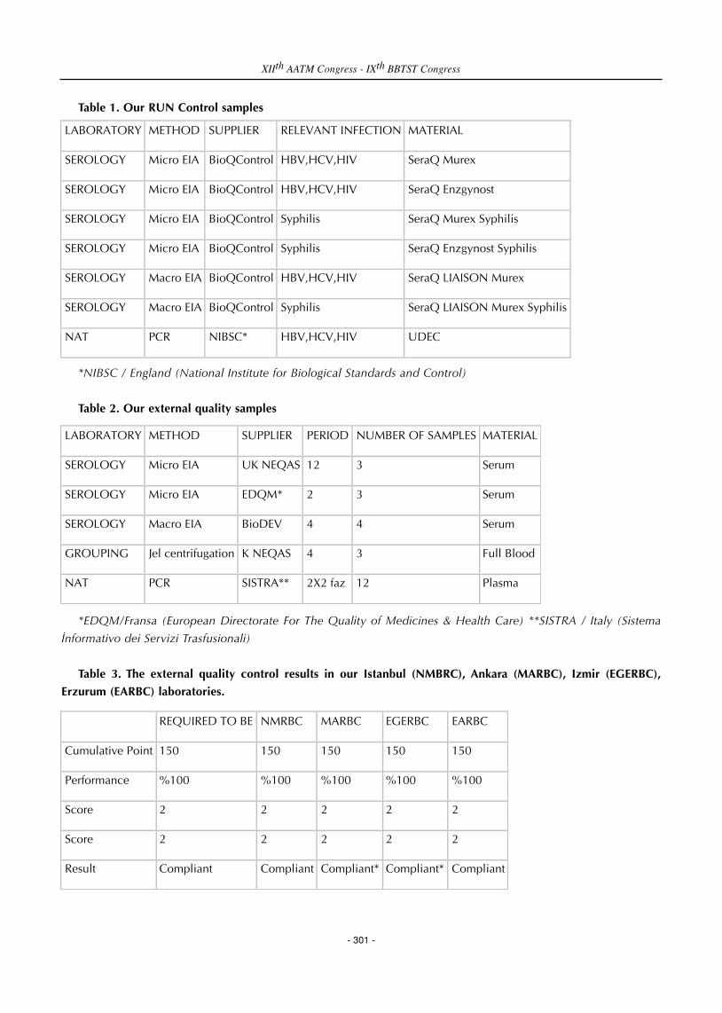
Table 1. Our RUN Control samples
*NIBSC / England (National Institute for Biological Standards and Control)
Table 2. Our external quality samples
*EDQM/Fransa (European Directorate For The Quality of Medicines & Health Care) **SISTRA / Italy (Sistema
İnformativo dei Servizi Trasfusionali)
Table 3. The external quality control results in our Istanbul (NMBRC), Ankara (MARBC), Izmir (EGERBC),
Erzurum (EARBC) laboratories.
XIIth AATM Congress - IXth BBTST Congress
- 301 -
LABORATORY METHOD SUPPLIER RELEVANT INFECTION MATERIAL
SEROLOGY Micro EIA BioQControl HBV,HCV,HIV SeraQ Murex
SEROLOGY Micro EIA BioQControl HBV,HCV,HIV SeraQ Enzgynost
SEROLOGY Micro EIA BioQControl Syphilis SeraQ Murex Syphilis
SEROLOGY Micro EIA BioQControl Syphilis SeraQ Enzgynost Syphilis
SEROLOGY Macro EIA BioQControl HBV,HCV,HIV SeraQ LIAISON Murex
SEROLOGY Macro EIA BioQControl Syphilis SeraQ LIAISON Murex Syphilis
NAT PCR NIBSC* HBV,HCV,HIV UDEC
LABORATORY METHOD SUPPLIER PERIOD NUMBER OF SAMPLES MATERIAL
SEROLOGY Micro EIA UK NEQAS 12 3 Serum
SEROLOGY Micro EIA EDQM* 2 3 Serum
SEROLOGY Macro EIA BioDEV 4 4 Serum
GROUPING Jel centrifugation K NEQAS 4 3 Full Blood
NAT PCR SISTRA** 2X2 faz 12 Plasma
REQUIRED TO BE NMRBC MARBC EGERBC EARBC
Cumulative Point 150 150 150 150 150
Performance %100 %100 %100 %100 %100
Score 2 2 2 2 2
Score 2 2 2 2 2
Result Compliant Compliant Compliant* Compliant* Compliant

Table 4. Results with “*” are explained in Table 4.
PP-041
EXAMINING THE RESULTS OF THE ERRORS REPORTED BY ANTALYA TRAINING AND RESEARCH HOSPITAL
TRANSFUSION CENTER TO WESTERN MEDITERRANEAN REGIONAL BLOOD CENTER
Belkıs Salman Koçtekin, Özcan Eren
Antalya Training and Research Hospital Transfusion Center
AIM: The quality control studies of the blood components supplied by Turkish Red Crescent are periodically
conducted in accordance with the national legislation, and the accredited quality management systems with the
method and amounts defined in National Blood and Blood Products Guide. The error reporting mechanism between
Transfusion Centers (TC) and Regional Blood Centers (RBC) initiates with the error reporting over Error Reporting Form
and results in Retrospective Blood Component Analysis Report. With this system it is aimed to detect the errors, to
identify the sources of the errors and to prevent the repetition of the errors.
The aim of this study is to examine the error reports conducted by Antalya Training and Research Hospital
Transfusion Center to Western Mediterranean Regional Blood Center between 2013-2015 and to contribute to the
determination of these errors’ causes and the improvements for the measures to be taken.
METHOD: The error reports made by Antalya Training and Research Hospital Transfusion Center between 2013-
2015 and the results reported by the Western Mediterranean Regional Blood Center are examined retrospectively.
The data are obtained from Error Reporting Forms and Western Mediterranean Regional Blood Center Quality
Control Laboratory examination reports.
RESULTS: All error reports conducted regarding the erythrocyte suspension (ES) from blood products.
In the years 2013, 2014 and 2015, samples received respectively 23836, 23933 and 23138, the ES relevant error
XIIth AATM Congress - IXth BBTST Congress
- 302 -
MARBC EGERBC
Non-compliance
Non-compliance was found once only in NAT external quality control.
Non-compliance was found once only in NAT external quality control.
Reason HIV RNA false positivity HIV RNA false positivity
Occured at In NAT Phase-1, we identified HCV RNA positivity in the 6th Sample as well as HIV RNA test as positive.
In NAT Phase-2, we identified HCV RNA positivity in the 2nd Sample as well as HIV RNA test as positive.
Activity Performed
According to the criteria, the non-compliance is at "low" level and since no performance improvement is required, only the process was reviewed.
According to the criteria, the non-compliance is at "low" level and since no performance improvement is required, only the process was reviewed.
Other Not available Not available
reports are grouped under two headings such as clotted blood and direct antiglobulin test (DAT) positivity.
As a result of the examination conducted by Western Mediterranean Regional Blood Center Quality Control
Laboratory, for the year 2013, 12 of 23836 products (0.05%) are reported with clotted blood, and 7 of these reports
(58.34%) are confirmed. For the year 2014, 7 of 23933 products (0.03%) are reported with clotted blood, and 3 of
these reports (42.86%) are confirmed. For the year 2015, 8 of 23138 products (0.04%) are reported with clotted blood,
and 4 of these reports (50%) are confirmed.
DAT positive error reporting is conducted for 9 ES (0.03%) and each result (100%) is reported as DAT positive. DAT
positive error reporting is not conducted for the years 2014 and 2015.
CONCLUSION: Considering the confirmed error counts, our study shows that there are some improvements
needed for the preparation of blood components and laboratory testing phases.
PP-042
ANALYSIS OF BLOOD DONATION CENTER MEDICAL STAFF APPROACH AGAINST RISK AND DANGER
FACED IN WORK ENVIRONMENT BY SOCIO-DEMOGRAPHIC FEATURES
Emine Aluç1, Güler Dişiaçık1, Sevgi Ünlü1, Ebru Çorbacı1, Ilhan Birinci1, Nurettin Hafızoğlu2, Mehmet
Güllüoğlu2, Fatma Meriç Yılmaz2
1Turkish Red Northern Marmara Regional Blood Center, Istanbul, Turkey2Directorate General of Turkish Red Crescent Blood Services/Ankara
INTRODUCTION: Work environment sinclude risks with health and safety hazards. The dangers and risks include
occupational diseases and work accidents that can directly affect the health of the person. Dangers are threats which
occurs against human health. Risk is a concept which is related with probability and it is defined as the probability of
occurrence of a negative event in a dangerous situation.
OBJECTIVE: This study was made in Northern Marmara Regional Blood Center (NMRBC) and subordinates (Kartal
Blood Donation Center and Zeynep Kamil Blood Donation Center), for determination of clinical staff approach
according to their socio demographic characteristics, against the dangers that they face during blood donation
processes.
METHOD: The sample data of the survey was collected from NMRBC medical staff between 01.03.2016 and
07.03.2016. In the study questionnaire was used to collect data. The survey consists of four separate descriptor table
that contains statistical information.
60 employees participated in the survey; Socio-demographic characteristics of employees are shown in Table 1
FINDINGS: When the answers of the participants has been considered it was seen that 58 % wash their hands
before working and 73 % of those who wash their hands also dry them.
It was determined that the thirty-three percent of respondents use hand sanitizer besides male participants preferred
hand sanitizer more than hand washing.
XIIth AATM Congress - IXth BBTST Congress
- 303 -

The majority of the medical staff (85%) were found to give importance to the use of gloves but they don't have the
same sensitivity in the use of protective lab coats and glasses.
Participants' sensitivity of working in body forcing positions was found low.58 percent of medical staff haven’t
experienced an occupational accident however group that had an occupational accident are mostly below 25 years
old and all of them are associate degree graduate and single.
When basic security (physical and biological) measures asked, 5% of respondents reported finding adequate
measures.
Participants have been asked about the educational needs of occupational health and safety; despite 70% of them
have been trained before, wanted to repeat the education. (personal hygiene, diseases transmitted through blood and
prevention methods, blood and body fluid contacting precaution instructions, biosafety instructions and management
procedure of medical waste)
When Employees' Approach Against Psychosocial Risk is analyzed; physical conditions affect business productivity,
staff assignments are suitable for the job done and workload is not too heavy, but it was determined that business
productivity decreases if there is insufficient rest period.
Flexible working hours negatively affects 73% of the medical staff.
CONCLUSION: Occupational safety culture should be created in healthcare business. In order to adopt the new
hires to security culture, healthcare management should give more serious information about the risks and
dangers.Also; adapting the working environment to the health conditions, elimination of some danger possibility,
regulation of working hours, suitable working order for physiological characteristics, ensuring compliance of the tools
with the person and work should be the main objective.
Table 1
XIIth AATM Congress - IXth BBTST Congress
- 304 -
PP-043
FREQUENCY OF ABO AND RHESUS BLOOD GROUPS IN PREOPERATIVE PATIENTS OF CABG SURGERY IN
LOCAL POPULATION OF DISTRICT RAWALPINDI (PAKISTAN).
Muhammad Noorulamin1, Rida Tanveer2, Agha Babaur Hussain1, Asad Mehmood Raja2, Raja Muhammad
Saqlain2, Pakeeza Arzoo Shaiq2, Ghazala Kaukab Raja2
1Rawalpindi Institute of Cardiology, Rawalpindi, Pakistan2PMAS. Arid Agriculture University, Rawalpindi, Pakistan
INTRODUCTION: Blood transfusion has been a vital piece of coronary artery bypass graft surgery (CABG) since its
starting. Transfusion rates in cardiovascular surgery stay high in spite of real advances in CABG surgery. Cardiovascular
Disease (CVD) is one of the most critical problems now a days. 15 million deaths are caused due to CVD every year
and most of the people who die are below 65 years of age. Pakistani population also has one of the highest risks of
coronary heart diseases (CHD) in the world. In Pakistan, 30-40% of all deaths are due to CVD. as it takes nearly
200,000 lives in Pakistan every year.
OBJECTIVES: The purpose of this study is to demonstrate the distribution of ABO and Rh blood groups in a large
population of patients undergoing for CABG. To identify the Blood group most common in patients going for CABG
surgery which may help in blood & blood products stock management.
MATERIALS-METHODS: The study was carried out at Rawalpindi Institute of Cardiology (RIC) to collect data of
coronary heart disease patients on the basis of ABO blood group and Rh factor. This study included 633 patients with
the minimum age of 31 to the maximum age of 75 having CAD and were about to go for Coronary artery bypass graft
surgery. (Table1) Standard tube agglutination method was adopted for the identification of ABO and rhesus blood group
by using commercially available antiseras A, B and D, along with appropriate positive and negative controls. All
statistical analysis was performed on SPSS software version 21.0.
RESULTS: A target sample population of 633 patients aging from 31-75 years were enrolled in this study. The mean
SD age of participants was (52.8) ±10.9 years out of which 434 (68.6%) were male while 199 (31.4%) were females.
According to our research CVD prevalence between both sexes was largest among the subjects ≥41-60 years of age.
Of the 633 patients which were included in the analysis showed phenotypes A, B, O and AB as 26.7%, 34.1%, 31.3%
and 7.9% respectively. Present study shows that out of 633 patients, 581 (91.8%) were RH positive while only 52
(8.2%) were RH negative. (Table 2)
CONCLUSION: The data suggest that blood group "'B'' is most common in patient going for CABG surgery. The
data also suggest that phenotype ''B'' might be associated with Coronary heart disease but there is need to confirm
effect of blood group on the development of cardiac diseases. With respect to this study it is proposed that blood banks
should maintain stock of Blood Group B and Rh Positive as compare to other ABO blood groups.
XIIth AATM Congress - IXth BBTST Congress
- 305 -
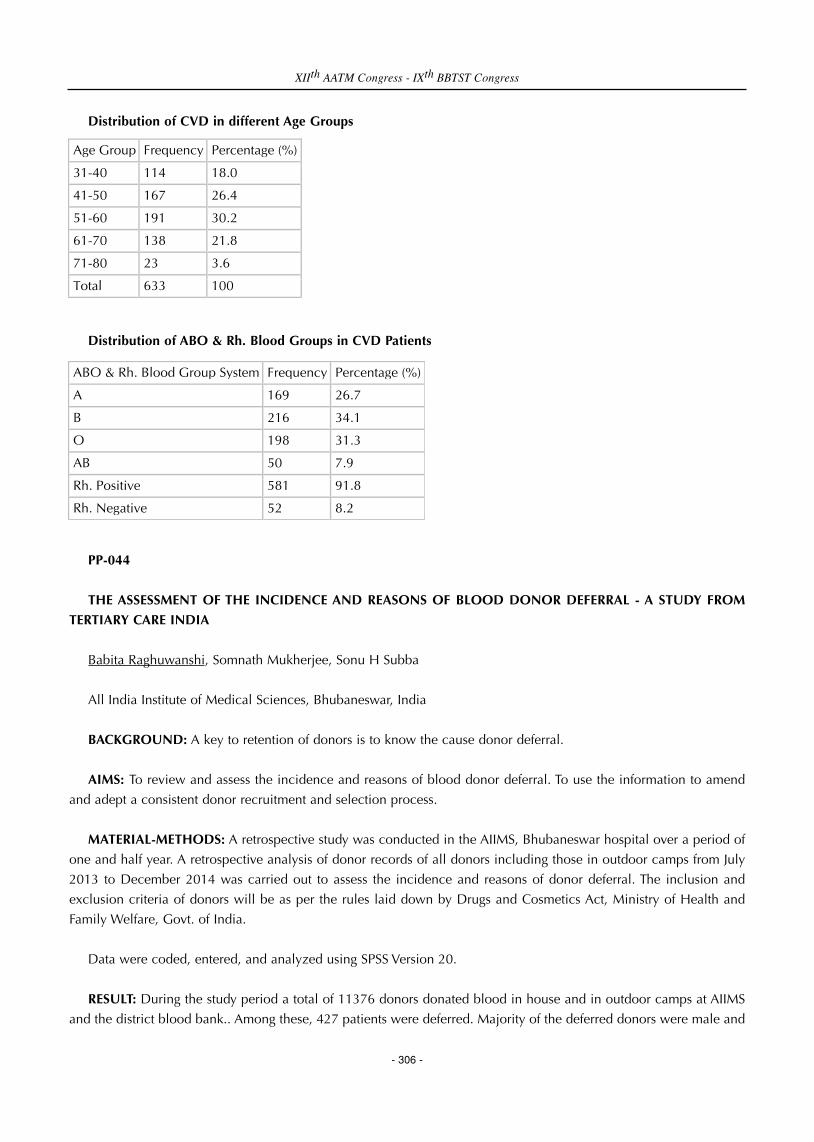
Distribution of CVD in different Age Groups
Distribution of ABO & Rh. Blood Groups in CVD Patients
PP-044
THE ASSESSMENT OF THE INCIDENCE AND REASONS OF BLOOD DONOR DEFERRAL - A STUDY FROM
TERTIARY CARE INDIA
Babita Raghuwanshi, Somnath Mukherjee, Sonu H Subba
All India Institute of Medical Sciences, Bhubaneswar, India
BACKGROUND: A key to retention of donors is to know the cause donor deferral.
AIMS: To review and assess the incidence and reasons of blood donor deferral. To use the information to amend
and adept a consistent donor recruitment and selection process.
MATERIAL-METHODS: A retrospective study was conducted in the AIIMS, Bhubaneswar hospital over a period of
one and half year. A retrospective analysis of donor records of all donors including those in outdoor camps from July
2013 to December 2014 was carried out to assess the incidence and reasons of donor deferral. The inclusion and
exclusion criteria of donors will be as per the rules laid down by Drugs and Cosmetics Act, Ministry of Health and
Family Welfare, Govt. of India.
Data were coded, entered, and analyzed using SPSS Version 20.
RESULT: During the study period a total of 11376 donors donated blood in house and in outdoor camps at AIIMS
and the district blood bank.. Among these, 427 patients were deferred. Majority of the deferred donors were male and
XIIth AATM Congress - IXth BBTST Congress
- 306 -
Age Group Frequency Percentage (%)
31-40 114 18.0
41-50 167 26.4
51-60 191 30.2
61-70 138 21.8
71-80 23 3.6
Total 633 100
ABO & Rh. Blood Group System Frequency Percentage (%)
A 169 26.7
B 216 34.1
O 198 31.3
AB 50 7.9
Rh. Positive 581 91.8
Rh. Negative 52 8.2
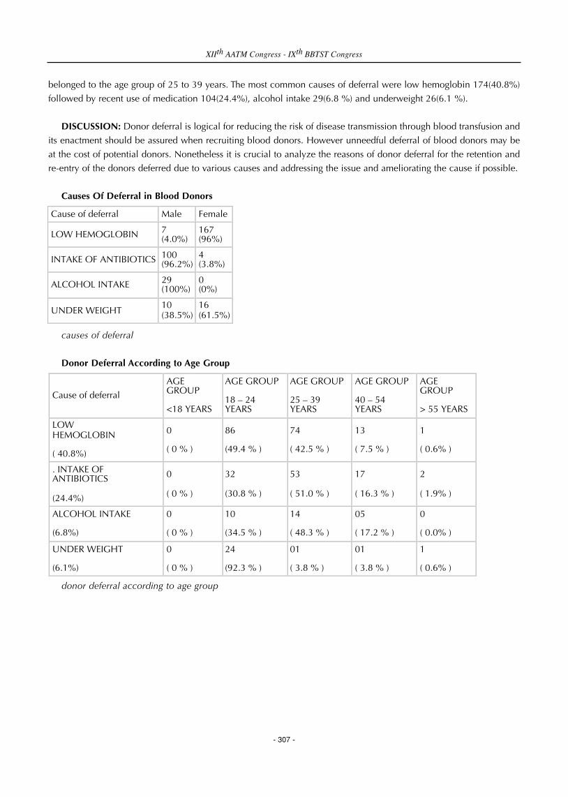
belonged to the age group of 25 to 39 years. The most common causes of deferral were low hemoglobin 174(40.8%)
followed by recent use of medication 104(24.4%), alcohol intake 29(6.8 %) and underweight 26(6.1 %).
DISCUSSION: Donor deferral is logical for reducing the risk of disease transmission through blood transfusion and
its enactment should be assured when recruiting blood donors. However unneedful deferral of blood donors may be
at the cost of potential donors. Nonetheless it is crucial to analyze the reasons of donor deferral for the retention and
re-entry of the donors deferred due to various causes and addressing the issue and ameliorating the cause if possible.
Causes Of Deferral in Blood Donors
causes of deferral
Donor Deferral According to Age Group
donor deferral according to age group
XIIth AATM Congress - IXth BBTST Congress
- 307 -
Cause of deferral Male Female
LOW HEMOGLOBIN 7 (4.0%)
167 (96%)
INTAKE OF ANTIBIOTICS 100 (96.2%)
4 (3.8%)
ALCOHOL INTAKE 29 (100%)
0 (0%)
UNDER WEIGHT 10 (38.5%)
16 (61.5%)
Cause of deferral
AGE GROUP <18 YEARS
AGE GROUP 18 – 24 YEARS
AGE GROUP 25 – 39 YEARS
AGE GROUP 40 – 54 YEARS
AGE GROUP > 55 YEARS
LOW HEMOGLOBIN ( 40.8%)
0 ( 0 % )
86 (49.4 % )
74 ( 42.5 % )
13 ( 7.5 % )
1 ( 0.6% )
. INTAKE OF ANTIBIOTICS (24.4%)
0 ( 0 % )
32 (30.8 % )
53 ( 51.0 % )
17 ( 16.3 % )
2 ( 1.9% )
ALCOHOL INTAKE (6.8%)
0 ( 0 % )
10 (34.5 % )
14 ( 48.3 % )
05 ( 17.2 % )
0 ( 0.0% )
UNDER WEIGHT (6.1%)
0 ( 0 % )
24 (92.3 % )
01 ( 3.8 % )
01 ( 3.8 % )
1 ( 0.6% )

PP-045
MANAGEMENT OF BLOOD SUPPLY TO THE HOSPITALS THAT THE CASUALTIES HAVE TRANSPORTED AFTER
A TERRORIST ATTACK
Murat Güler1, Levent Sağdur2, Armağan Aksoy2, Nurettin Hafızoğlu2, Mehmet Güllüoğlu3
1Middle Anatolian Regional Blood Center, TRC, Ankara, Turkey2General Directorate of Blood Services, TRC, Ankara, Turkey3General Dirocterate of Turkish Red Crescent,TRC, Ankara, Turkey
The quantities of blood supplies, distribution of blood groups, and elapsed time until delivery to hospitals were
collected retrospectively and evaluated.
Twenty-nine citizens have lost their lives and numerous others were injured and hospitalized after the terrorist attack
on 17th of February, 2016 at 18:32 in Ankara.
There were a total of 2023 units of erythrocyte suspension, of which 1107 units were ready to use, and 916 were
at quarantine.
The informations of hospitals that casualties have been transported were obtained from 112 emergency coordination
center, and those hospitals were contacted immediately after the blast. The routine demands other than emergency
needs of other hospitals were stopped, and blood and blood products were transferred to the injured citizens
immediately.
After the attack, 350 units of ES in 90 minutes, and a total of 379 units of ES until 00:00 same night has been
transferred to the hospitals. Total number of products was 517 units, when other blood products were accounted.
High numbers of O Rh(-) ES were requested from the hospitals due to high numbers of injured people. Seventy-five
units of O Rh(-) ES, which are reserved in the stocks of Middle Anatolian Regional Blood Center for exceptional
circumstances like this, were circulated immediately to the hospitals.
Until 00:00 of the attack day, a total of 589 units of erythrocyte suspension were transferred from the 17 Regional
Blood Centers that operated under the body of Turkish Red Crescent, which 299 units of them were negative blood
groups and AB(+) blood type. Eventually, the reserved stock against extraordinary occasions, particularly the O Rh(-)
reserve, has been re-established.
The number of ESs in the inventory of Middle Anatolian Regional Blood Center at 09:30 a.m. on the day after the
attack was 2396 units.
The number of reserved O Rh(-) ES in the stocks of Turkish Red Crescent Regional Blood Centers for the
circumstances like terrorist attacks has been increased and stock plans were revised after the second half of 2015.
Stocked amounts of O Rh(-) ESs were sufficient, but some problems have encountered in transportation of the
supplies to the hospitals after the terrorist attack on 10th of October, 2015, at the meeting in front of the Ankara central
train station. The priority of the demands from the hospitals could not be evaluated adequately due to the hurry to
XIIth AATM Congress - IXth BBTST Congress
- 308 -

respond to all demands, and Ankara Numune Hospital, which the majority of the casualties were transferred, could not
took the blood supply proportional to its need. Nevertheless, the internal stocks of the hospitals have covered the
demand and no problems have emerged.
By the lessons learned from this case, the demands from the hospitals after the attack on February 17th, 2016 were
first confirmed from the 112 coordination center, and blood supply plans were based on the information of patients
that transported to each of the hospitals, to reply all the demands effectively and adequately.
Number of blood products according to blood groups, which were delivered to hospitals in 90 minutes after
the blast
Numbers of ES (erythrocyte suspensions), that transferred from other Regional Blood Centers to Middle
Anatolian Regional Blood Center
XIIth AATM Congress - IXth BBTST Congress
- 309 -
ES PLT FFP TOTAL
O Rh(-) 70 70
O Rh(+) 65 20 85
A Rh(-) 14 14
A Rh(+) 130 12 142
AB Rh(-) 1 20 21
AB Rh(+) 30 5 20 55
B Rh(-) 5 5
B Rh(+) 35 20 55
TOTAL 350 57 40 447
ES
O Rh(-) 80
O Rh(+) 150
A Rh(-) 65
A Rh(+) 130
AB Rh(-) 25
AB Rh(+) 95
B Rh(-) 34
B Rh(+) 10
TOTAL 589

PP-046
ROLE OF MONITORING BLOOD SERVICES IN IMPROVEMENT OF PROVISION SAFE BLOOD - INDIAN
EXPERIENCES
Harprit Singh1, R S Gupta2
1National AIDS Control Organisation2National Blood Transfusion Council
AIM: Efficiency and effectiveness in National Blood Services is of vital importance. National Blood Transfusion
Council, India (NBTC) was established in 1996 with vision of improving the blood transfusion services in the country.
Monitoring & Evaluation are necessary to ensure efficiency and effective uses of measure. Programmatic monitoring
and evaluation of blood services should be performed to ensure that health activities are implemented as planned and
to assess whether desired results are being achieved.
MATERIAL-METHODS: Major parameters of blood services in the country before and 20 years after establishment
of National Blood Transfusion Council in the country were measured against the desirable.
CASE STUDY: Monitoring mechanism is complex in the country with Federal Government. Blood Transfusion
Services is managed by State (provincial government). National Blood Transfusion Council (NBTC) - defines the policy
consisting of objectives, goals, devise strategy. Blood Transfusion Services at National AIDS Control Organization
(NACO), prepares annual action plan and allocate necessary resources consistent with NBTC. State (provincial) govt.
counterparts of NBTC and NACO designs how can they are getting desired output from the inputs that are being
utilized. Blood Bank Facilities define actual supervision, operations and ensures that planned activities are being
carried out as per schedule. With continuous monitoring following results have been achieved in last 2 decades;
RESULTS: These improvements have been evaluated with various time frames involving short term, midterm and
long term strategies. As evident from table, continuous monitoring & evaluation improve blood transfusion services
availability has increased and access of safe blood and blood component is evident. Governmental commitment is
evident by establishing National Blood Transfusion Council. Government has allocated timely resources, among
competing priorities, for current and future program of Blood Transfusion services. Continuous monitoring and
evaluation of program has resulted in more relevant, more efficient, safe and more effective blood transfusion services.
Regulatory amendments have paralleled to growth and technological advancements in the country.
DISCUSSION: The purpose of monitoring is to ensure that programs are implemented as planned. Therefore,
Monitoring of blood program provides concurrent feedback on the progress of predefined activities/ norms of
performance. Level of performance becomes measurable and it can be compared with established standards or norms.
It helps in identifying deviations and explains the reasons for the deviation for taking necessary corrective action. Thus,
monitoring is part of planning cycle which starts with preparation of action plan. Inadequacy in planning will result in
inadequacy in monitoring. Monitoring of blood program is at different levels aligned with policies and implementation
strategies.
XIIth AATM Congress - IXth BBTST Congress
- 310 -

Monitoring Mechanism of Blood Transfusion Services in India
Status of blood transfusion services before and after establishment of National Blood Transfusion Council
XIIth AATM Congress - IXth BBTST Congress
- 311 -
S.No. Parameters Before NBTC (1996) 2015
1 Whole Blood Collection Approx. 1 Million 10.8 Million
2 Total No. of Blood Banks < 500 Around 2700
3 Blood Component Separation Units <50 1163
4 Apheresis center None 503
5 Infection Screening as per Norms Not routinely done 100%
6 Licensing of Blood Banks Not Mandatory 100%
7 Availability of plasma for plasma derived medicinal products in an Year
<10 thousand Liters > 3 lac liters

PP-047
THE EVALUATION OF THE USE OF BLOOD PRODUCTS AND THE DISTRIBUTION OF THEM ACCORDING
TO BLOOD GROUPS IN KONYA DR FARUK SÜKAN GYNECOLOGY, OBSTETRICS AND CHILDREN HOSPITAL
Nadire Seval Gündem1, Erkan Ataş2, Alper Gözükara3
1Medical Microbiology, Dr Faruk Sükan Gynecology, Obstetrics and Children Hospital, Konya, Turkey2Pediatrics, Dr Faruk Sükan Gynecology, Obstetrics and Children Hospital, Konya, Turkey3Blood Transfusion Center, Dr Faruk Sükan Gynecology, Obstetrics and Children Hospital, Konya, Turkey
OBJECTIVE: Blood and blood products are essential components in the medical management of patients in almost
every field of clinical practice. In this study, it is aimed to determine the number and the rate of used blood products
and the distribution of them according to blood groups in our hospital.
MATERIALS AND METHODS: Between September 2015-February 2016, the data of blood products obtained from
Kızılay Regional Blood Center was investigated retrospectively. The number and the rate of obtained, used and
eliminated blood products were determined. Blood groups were detected by using gel centrifugation method with
commercial diagnostic kits on the immunohematology automated analyser (Autovue Innova, Ortho Clinical
Diagnostics, USA ) according to manufacturer’s instructions.
RESULTS: A total of 893 blood products were obtained from Kızılay Regional Blood Center for clinical practice in
our hospital. The most used blood product was detected as erythrocyte suspension with the rate of 67.7%. Platelet
suspension was the most eliminated blood product with the rate of 73.1%. While A Rh + and 0 Rh + were found as
the most common blood groups among erytrocyte suspension and fresh frozen plasma, AB Rh + was prevalent for
platelet suspension. AB Rh – and 0 Rh – were the most rare blood groups for all blood products as reported in other
studies carried out in our country.
CONCLUSION: The determination of the rate of used and eliminated blood products is important for detecting and
updating of critical stock level. Blood transfusion centers in hospitals should have good practice in all aspects of blood
grouping, compatibility testing, the storage and transportation of blood and blood products to clinics. If blood
transfusion therapy is appropriately used, it can improve the quality of life and prevent morbidity and mortality.
Table 1: Distribution of blood products according to utilization, elimination and obtaining from Kızılay
Regional Blood Center
*KIZILAY REGIONAL BLOOD CENTER
XIIth AATM Congress - IXth BBTST Congress
- 312 -
BLOOD PRODUCTS OBTAINED FROM KRBC* USED ELIMINATED
n % n % n %
Erythrocyte suspension 542 60.7 506 67.7 36 24.9
Platelet suspension 160 17.9 54 7.2 106 73.1
Fresh frozen plasma 191 21.4 188 25.1 3 2
TOTAL 893 100 748 100 145 100

Table 2: Distribution of blood products according to blood groups
PP-048
RESEARCHING TEMPORARY REASONS OF VOLUNTEER BLOOD DONOR DEFERRALS WHOM APPLIED TO
BLOOD CENTER OF ISTANBUL KARTAL KOSUYOLU YUKSEK IHTISAS TRAINING AND RESEARCH HOSPITAL
Sibel Doğan Kaya, Arif Şenel, Hamit Kaya, Adem Erengül, Nur Uylar, Tulay Karabürk, Aslıhan Özmen, Meral
Ulığdogan, Emine Erengul, Hüseyin Yüce, Tuğba Gevrek, Bülent Arınç
Istanbul Kartal Kosuyolu Yuksek Ihtisas Training and Research Hospital
BACKGROUND: The only source of blood and its components is human, and there is no alternative modality when
it can not be provided. In developed countries, while the volunteer blood donation’s rate for general population is
reaching 5%, this ratio is about 1,5% in Turkey due to deferral of donors because of various reasons.
MATERIAL-METHODS: In the present retrospective study 34128 volunteer blood donors admitted to Istanbul Kartal
Kosuyolu Yuksek Ihtisas Training and Research Hospital between 01 January 2014–01 March 2016 were examined in
terms of number of donors deferred temporarily from blood donation and the causes of deferral. The causes of
temporary deferral are classified into 12 main groups.
RESULTS: The highest blood donation was taken in March 2015 (1693 blood donations, 4,96%) and March 2014
(1608 blood donations, 4,71%), while the lowest blood donation was taken in December 2015 (815 blood donations,
2,38%) and Semterber 2014 (922 blood donations, 4,70%). 7,8% of donors (2662/34128) were deferred temporarily
from blood donation. The three uppermost reasons for temporary deferral were anomalies of white blood cells (19,4%)
infectıons (27,8%), and problems of tooth treatment (%6,89)
CONCLUSIONS: The temporary deferral rate of our center is 19.2%, and the anomalies of blood count and
varioushealth problems are the leading causes. Early diagnosis and treatment of these causes may increase already low
blood donation rates to desired levels.
XIIth AATM Congress - IXth BBTST Congress
- 313 -
BLOOD GROUPS Erythrocyte suspension Platelet suspension Fresh frozen plasma TOTAL
n % n % n % n %
A Rh + 155 28.6 9 5.7 58 30.4 222 24.9
A Rh - 38 7 4 2.5 12 6.3 54 6
B Rh+ 94 17.4 4 2.5 28 14.7 126 14.1
B Rh - 26 4.8 2 1.2 5 2.6 33 3.7
AB Rh+ 60 11 127 79.3 24 12.6 211 23.6
AB Rh - 14 2.6 1 0.7 2 1 17 2
0 Rh+ 134 24.8 13 8.1 62 32.4 209 23.4
0 Rh - 21 3.8 0 0 0 0 21 2.3
TOTAL 542 100 160 100 191 100 893 100

FIGURE 1
PP-049
A COMPARISON OF THE APHERESIS PLATELET DATA BETWEEN YEARS 2012-2015
Canan Eren
Deparment of Blood Bank, Marmara University Pendik Training and Research Hospital, Istanbul, Turkey
AIM: The evaluation of the effect of blood donations hours on apheresis platelet donations numbers.
MATERIAL- METHOD: The produced apheresis platelet counts between 2012-2015 were analyzed and compared
with the times of accept blood donations in Marmara University Pendik Training and Research Hospital.
RESULTS: In 2012, platelet apheresis donation is only possible between 09:00 and 12:00 a.m. on weekdays and
during this period total produced apheresis platelet number was 228. In 2013, platelet apheresis donation accept time
was between 09:00 a.m and 17:00 p.m. and total produced apheresis platelet number was 1861. In 2014, with the
start of acceptance apheresis platelet donation 24 hours, 7 days as from month of March, the total produced apheresis
platelet number was 3704. In 2015, throughout the year with the accept of apheresis platelet donations 24 hours, 7
days, the total produced number of apheresis platelet reached 4182.
CONCLUSION: With the opening apheresis platelet donation time 24 hours, 7 days led to the a significant increase
in the number of apheresis platelet produced. This situation has provided grate satisfaction and relief for the doctors,
the patients and their relatives.
XIIth AATM Congress - IXth BBTST Congress
- 314 -
WHY RED TOTAL
INFECTION 702 ( % 29,88 )
SURGERY ENDOSCOPY 197 ( % 6,85 )
PINPRICK 18 ( % 0,76 )
TATTOO 37 ( % 1,57 )
DENTISTRY 162 ( % 6,89 )
VACCINE 69 ( % 2,93 )
DRUG 23 ( %0,97 )
HYPER-HYPO TENSION 60 ( %0,55 )
LOW HEMOGLOBIN 456 ( %1,96 )
RISKY SEX 17 ( %0,72 )
AGE 53 ( % 2,25 )
KILO 48 ( % 2,04 )
DRUG USE 306 ( % 13,02 )
OTHER 507 ( % 21,58 )

Comparison of the apheresis platelet numbers between 2012-2015
The distribution of produced apheresis platelet numbers according to years
XIIth AATM Congress - IXth BBTST Congress
- 315 -
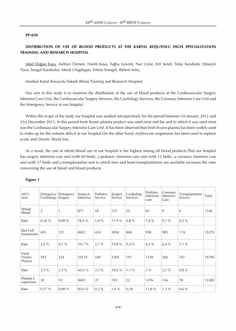
PP-050
DISTRIBUTION OF USE OF BLOOD PRODUCTS AT THE KARTAL KOŞUYOLU HIGH SPECIALIZATION
TRAINING AND RESEARCH HOSPITAL
Sibel Doğan Kaya, Aslıhan Özmen, Hami̇t Kaya, Tuğba Gevrek, Nur Uylar, Ari̇f Senel, Tülay Karabürk, Hüseyi̇n
Yüce, Songul Karabulut, Meral Ulugdogan, Emi ̇ne Erengül, Bülent Ari̇nç
Istanbul Kartal Kosuyolu Yuksek Ihtisas Training and Research Hospital
Our aim in this study is to examine the distribution of the use of blood products at the Cardiovascular Surgery
Intensive Care Unit, the Cardiovascular Surgery Services, the Cardiology Services, the Coronary Intensive Care Unit and
the Emergency Service at our hospital.
Within the scope of the study our hospital was studied retrospectively for the period between 1st January 2012 and
31st December 2015. In this period fresh frozen plasma product was used most and the unit in which it was used most
was the Cardiovascular Surgery Intensive Care Unit. It has been observed that fresh frozen plasma has been widely used
to make up for the volume deficit in our hospital.On the other hand, erythrocyte suspension has been used to replace
acute and chronic blood loss.
As a result, the rate of whole blood use in our hospital is the highest among all blood products.That our hospital
has surgery intensive care unıt (with 60 beds), a pediatric intensive care unit (with 12 beds), a coronary intensive care
unıt (with 27 beds) and a transplantation unit in which liver and heart transplantations are available increases the rates
concerning the use of blood and blood products.
Figure 1
XIIth AATM Congress - IXth BBTST Congress
- 316 -
2015 year
Emergency Cardiology
Emergency Surgery
Surgıcal Intensive
Pediatrıc Service
Surgery Service
Cardiology Services
Pediatrıc Intensive care
Coronary Intensive Care
Transplantatıon Service Total
Whole Blood 3 1 877 18 137 10 85 9 6 1146
Rate 0,26 % 0,08 % 76,5 % 1,8 % 11,9 % 0,8 % 7,4 % 0,7 % 0,5 %
Red Cell Suspension 401 121 8422 424 3050 840 958 985 174 15375
Rate 2,6 % 0,7 % 54,7 % 2,7 % 19,8 % 5,4 % 6,2 % 6,4 % 1,1 %
Fresh Frozen Plasma
593 234 10174 549 3304 191 1238 366 147 16796
Rate 3,5 % 1,3 % 60,5 % 3,2 % 19,6 % 1,1 % 2 % 2,1 % 0,8 %
Plalelet S uspension 20 10 9697 37 193 22 1376 156 78 11589
Rate 0,17 % 0,08 % 83,6 % 0,3 % 1,6 % 0,18 11,8 % 1,3 % 0,6 %

PP-051
EVALUATION OF THE DONOR QUESTIONNAIRE FORMS IN GIRESUN PROF. DR. A. ILHAN OZDEMIR
GOVERNMENT HOSPITAL, BLOOD BANK IN TWO YEAR PERIOD
Şahin Direkel1, Nejla Cebeci Güler1, Ömer Emecen2, Saliha Kesti3
1Department of Medical Microbiology, Faculty of Medicine, Giresun University, Giresun, Turkey2Department of Blood Bank, Prof. Dr. A. Ilhan Ozdemir Goverment Hospital, Giresun, Turkey3Department of Medical Microbiology, Health Sciences Institute, Giresun University, Giresun, Turkey
AIM: The blood donor history questionnaires was designed to aid blood establishments in evaluating a prospective
donor's history relative to current known blood safety risks. In Turkey, blood donors must complete a donor history
questionnaire (DHQ). In this study, we aimed to evaluate donor questionnaire forms who admitted to Giresun Prof. Dr.
A. Ilhan Ozdemir Government Hospital, Blood Bank in Giresun province.
METHODS: This retrospective study was conducted by investigating the records of 607 blood donor history
questionnaires who admitted for donation to Giresun Prof. Dr. A. Ilhan Ozdemir Government Hospital, Blood Bank
between 1st January, 2014 and 31st December, 2015.
RESULTS: Total 607 people (34 women, 573 men) were apply to our hospital for blood donation. These blood donor
history questionnaire form was evaluated. Total 494 (81.33%) donation according to blood donor history
questionnaires were accepted. But 113 (18.67) donation were unaccepted. Reasons for rejection were such as low
weight, influenza, diarrhea, hepatitis, low hemoglobin, high WBC, antibiotic use, diabetes, 18 in their young age. Rate
of donation accepted or unaccepted according to blood donor history questionnaire in different age groups was shown
in the table.
CONCLUSION: Blood and blood products needs of our hospitals are supplied from regional blood center of the
Turkish Red Crescent. If, enough blood is not found in the hands of the Turkish Red Crescent, we supplies our own
blood and blood products needs in the our blood transfusion center. Blood donors can fill sometimes giving no answer
to all questions without reading the questions. They can sometimes give misleading answers to questions. The removal
of some questions blood donor history questionnaire and a revised according to international standards is necessary.
Such as "Have you been abroad in the last 3 years?"
Rate of donation accepted or unaccepted according to blood donor history questionnaire
XIIth AATM Congress - IXth BBTST Congress
- 317 -
Accepted women
Accepted men
Unaccepted women
Unaccepted men
Accepted total
Unaccepted total
18-25 age 9 88 1 21 97 22
26-30 age 1 59 1 13 60 14
31-35 age 2 87 3 24 89 27
36-40 age 2 83 3 22 85 25
41-55 age 7 145 3 19 152 22
56-60 age 2 8 0 3 10 3
Over 60 years 0 1 0 0 1 0
Total 23 471 11 102 494 113

PP-052
CONSEQUENCES AND RESULTS OF DECREASING RESERVE PERIOD OF ERYTHROCYTE SUSPENSIONS FOR
PATIENTS FROM 7 TO 3 DAYS
Pınar Güler1, Ilknur Avcı1, Hatice Nerdret Saatçıoğlu1, Funda Ceran2, Gülsüm Özet2
1Transfusion Center, Ankara Numune Research and Training Hospital, Ankara, Turkey2Hematology Department, Ankara Numune Research and Training Hospital, Ankara, Turkey
INTRODUCTION: The aim of this study is to compare the disposal rates of erythrocyte suspension (ES) due to
expiration, in 2010 and 2015.
FINDINGS: Monthly used erythrocyte suspension count, and amount of disposals in 2010 and 2015 were obtained
from database. Also, cross-match counts were also obtained from the database.
In the practice of Ankara Numune Research and Training Hospital Blood Center in 2010, cross-match was studied
for each ES, and individually reserved for patient for 7 days. But, this duration decreased to 3 days since 5th month of
2014. By this change, cross-match rates were significantly increased (p<0.005) and disposal of ESs significantly
decreased (p<0.05), without a significant difference in the average number of ES used.
CONCLUSION: This retrospective research revealed that decreasing the reservation period of erythrocyte
suspensions for patients resulted in decreased number of disposals. As a consequence, cross-match numbers have
increased, but disposal rate decreased from 2.57% in 2010 to 1.45% in 2015.
ES count, cross match rate anddisposal rate in 2010
XIIth AATM Congress - IXth BBTST Congress
- 318 -
2010 ES n Cross match n Cross match ratio ES Disposal n ES Disposal %
January 1356 3109 2.29 29 2.14
Fabruary 1268 2558 2.02 57 4.50
March 1097 2071 1.89 44 4.01
April 1156 2320 2.01 42 3.63
May 1213 2441 2.01 65 5.36
June 1279 2867 2.24 54 4.22
July 1431 2781 1.94 35 2.45
August 1260 2987 2.37 11 0.87
September 1433 2769 1.93 11 0.77
October 1604 3056 1.91 7 0.44
November 1238 2314 1.87 7 0.57
December 1278 2682 2.10 24 1.88
AVERAGE 1301 2662 2.04 32 2.57

ES count, cross match rate anddisposal rate in 2015
PP-053
FEAR OF BLOOD DONATION
Şahin Direkel1, Büşra Kır2
1Department of Medical Microbiology, Faculty of Medicine, Giresun University, Giresun, Turkey2Department of Medical Microbiology, Health Sciences Institute, Giresun University, Giresun, Turkey
AIM: Blood is a very critical component in human life. Blood transfusion has a significant role in today's healthcare
operations. The blood donor system in Turkey depends mainly on voluntary donors. Volunteered blood donation occurs
from 1.5-2% of the population in Turkey. This ratio is very high (5-10%) in developed countries. Compared to
worldwide applications, Turkey has not been reached yet to a desired level. In the present study, we aimed to evaluate
the factors influencing fears of blood donation among the Giresun University students.
METHODS: We applied a questionnaire to students who are eligible to donate blood. The group consisting of 371
students (234 male, 137 female) in different academic departments of the University was performed questionnaire
work. The average age was found 20.4 years.
RESULTS: “Why are you afraid from blood donation” to question students were answered as 14.3 % large blood
collection needles, 7 % see the blood, 12.7% transmission of the blood diseases, 6.2% meet positive test results for
HIV, hepatitis B and C, and syphilis, 17.5% faint during blood donation, 2.7% because of my parents who don't allow
blood donation, 11.6% loss of blood after blood donation, 4.4% weight loss after blood donation, 18.3% because of
not feeling healthy enough for blood donation, 8.6% incompetence and bad behavior of health staff, 3.2% weight gain
after blood donation, 0.5% because of religious causes, 17.5% because of feeling psychologically not ready for blood
donation, 11.1% rupture of the blood vessel during donation, 16.7 % other reasons. According to the questionnaire,
118 students have donated blood before, but 253 students were not at any blood donation.
XIIth AATM Congress - IXth BBTST Congress
- 319 -
2015 ES n Cross match n Cross match ratio ES Disposal n ES Disposal %
January 1126 3667 3.262.94 15 1.33
February 1061 3118 2.94 16 1.51
March 1107 3248 2.93 11 0.99
April 1147 1997 1.74 27 2.35
May 1154 2235 1.94 32 2.77
June 1233 3480 2.82 27 2.19
July 1374 3082 2.24 21 1.53
August 1368 3501 2.56 8 0.58
September 1058 2586 2.44 6 0.57
October 1509 3419 2.27 15 0.99
November 1139 3370 2.96 20 1.76
December 1141 3217 2.82 9 0.79
AVERAGE 1201 3076 2.56 17 1.45

CONCLUSION: Firstly public awareness for the importance of the voluntary blood donation should be increased.
It's necessary to inform all the students about the need to ensure a supply of safe blood to the health system in order
to encourage them to become the voluntary and regular blood donors of the future. The reasons for why afraid from
blood donation of healthy individuals should be determined and found the solutions to this problem.
BLOOD DONATION QUESTIONNAIRE
PP-054
STUDY ON REASONS OF REJECTION FOR DONORS APPLIED TO OUR BLOOD CENTER FOR YEAR 2015
Hatice Erdoğan, Tunay Gül
Blood Transfusion Center of Haseki Training and Research Hospital, Istanbul, Turkey
AIM: By investigation the reasons of rejection for donors applied to our blood center, to take attention to missing
and failing points in donor questioning.
METHOD: In this study; donor questioning forms for the last year and our statistical informations have investigated
retrospectively.
XIIth AATM Congress - IXth BBTST Congress
- 320 -

EVIDENCES: The reasons of rejection for donors for year 2015 are shown in Table 1 below.
CONCLUSION: Our rejection rate has found as 8,48%. In the first place, the reason of rejection is caused by low
Hgb as of 39,50% and 86% of this number created by women donors. The major part of 23,12% Health problems are
composed of hypertension, diabetes and passed viral infections. Other reasons are composed of drug abuse, tattoo,
acupuncture, overseas travel and detention. The prevention of unnecessary donor rejection is planned by reviewing the
rejection reasons according to the last updated guide.
The reasons of rejection for donors for year 2015
Evidence: shown in Table1 below
PP-055
EVALUATION OF KNOWLEDGE LEVEL IN OUR SOCIETY FOR BLOOD DONATION
Fügen Kuruçay, İlhan Birinci, Nurettin Hafızoğlu, Mehmet Güllüoğlu, Fatma Meriç Yılmaz
Turkish Red Crescent Regional Blood Center North Marmara, Kartal, Istanbul, Turkey
AIM; The foundation of blood banking is a safe blood donor. Ensuring safe blood donation in the community ispossible with increasing the number of regular, voluntary and conscious blood donors. Starting from the fact that thereare not enough voluntary, regular and conscious blood donors in our country yet, with the survey to assess people'sattitude, behavior and knowledge towards blood donation, it is intended to contribute to recovery and training activitiesof blood donors.
METHOD; In December 2014, with people who came to the blood donation campaign in Burak Bora AnatolianHigh School, found in Kartal district of Istanbul province, a face-to-face survey with 10 questions was completed. Thepurpose and content of the study done was explained to all participants. Blood donor information brochures were givento the total of 624 people surveyed at the end of the survey.
XIIth AATM Congress - IXth BBTST Congress
- 321 -
Reasons of rejection Number Percentage %
Low Hgb 332 39.50
Health problems 194 23.12
Serology positive 111 13.20
Others 99 11.79
Inappropriate age 60 7.15
Risky sexual intercourse 25 2.97
Use of drug 18 2.14
Total rejection 839 8.48
Total donor 9890

FINDINGS;
Table 1
Gender
73% of our survey respondents were male, 27% female.
Table 3
EDUCATIONAL STATUS
82% of people who participated in our survey reported that they have a high school and over level education. Still
studying are classified according to their most recent school graduates.
Table 6
FREQUENCY OF YOUR BLOOD DONATION
55% of people surveyed said they came to donate blood for the first time.
Table 8
WHO CAN NOT DONATE BLOOD?
%11 level of knowledge about the conditions that prevent the blood donation, has been identified.
RESULT; 55% of people surveyed consists of people who applied for the first time as blood donors. 97% of them
consider the blood donation campaigns to be an effective way in our society for getting blood donors. Despite coming
to a blood donation campaign as volunteers, people surveyed were found not to have sufficient knowledge about blood
donation.
Considering %82 of respondents have the high school and over level of education;
- To increase the number and regularity of blood donors, realizing blood donation campaigns which are announced
effectively
- Raising awareness and consciousness using media and press and social media tools
- Within a national program, blood donation information training should be given at all levels starting from primary
school to high school, college and apprenticeship training schools
- Considering the target audience (aged 18 – 65 years), the implementation of appropriate training modules for all
levels to the areas where people live and work as a whole.
- Training must be the first step of the corporate blood donation campaign
- Implementation of education, encouragement and motivation programs to get the people who donated blood for
the first time as regular blood donors
- Based on information that we have average 11% of knowledge about disability to blood donation, which were
specified in 8 main title, giving information about the risks and the importance of blood for the person to be donated
blood transfusion to those who apply to donate blood
- In addition to raising public awareness of blood donation, working to raise awareness of the joy of helping other
people, is a need to be made.
One that gets educational awareness can be gained as voluntary blood donor, and also can give accurate
information about the risks associated with.
Only in this way, the most active ring will be completed to reach the safe blood.
XIIth AATM Congress - IXth BBTST Congress
- 322 -

Table 1
GENDER Male / Female 73% of our survey respondents were male, 27% female. (Table 1)
Table 2
BLOOD TYPE DISTRIBUTION All of the people who participated in our survey know their blood type. With the ratio
of %36, O Rh positive is the most, and with the ratio of %1, AB Negative is the least reported blood type. (Table 2)
Table 3
EDUCATIONAL STATUS Primary – Secondary School / High School / College – Universty 82% of people who
participated in our survey reported that they have a high school and over level education. Still studying are classified
according to their most recent school graduates. (Table 3)
XIIth AATM Congress - IXth BBTST Congress
- 323 -

Table 4
AGE RANGE 30% of people who participated in the survey are aged 18-25 years; 33% of them are 25-40 years
and %37 are at the range of 40– 65 years. (Table 4)
Table 5
DO YOU REGULARLY MAKE BLOOD DONATION? 25% of people surveyed said they are regular blood donors.
People who donate blood twice or more in a year are considered to be regular blood donors. (Table 5)
Table 6
FREQUENCY OF YOUR BLOOD DONATION First Time - 2 or 3 Times - Once or twice in a year - Once in 3 or 6
months Irregular Regular Regular 55% of people surveyed said they came to donate blood for the first time. (Table 6)
XIIth AATM Congress - IXth BBTST Congress
- 324 -
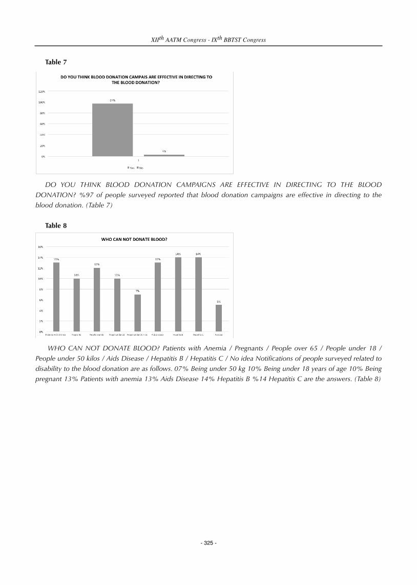
Table 7
DO YOU THINK BLOOD DONATION CAMPAIGNS ARE EFFECTIVE IN DIRECTING TO THE BLOOD
DONATION? %97 of people surveyed reported that blood donation campaigns are effective in directing to the
blood donation. (Table 7)
Table 8
WHO CAN NOT DONATE BLOOD? Patients with Anemia / Pregnants / People over 65 / People under 18 /
People under 50 kilos / Aids Disease / Hepatitis B / Hepatitis C / No idea Notifications of people surveyed related to
disability to the blood donation are as follows. 07% Being under 50 kg 10% Being under 18 years of age 10% Being
pregnant 13% Patients with anemia 13% Aids Disease 14% Hepatitis B %14 Hepatitis C are the answers. (Table 8)
XIIth AATM Congress - IXth BBTST Congress
- 325 -

Table 9
WHEN YOU DONATE BLOOD, HOW MANY PEOPLE’S LIVES DO YOU SAVE? 1 person / 3 people / Doesn’t
know 72% of people who participated in our survey reported having no information on blood products obtained
after blood donation. (Table 9)
Table 10
IS BLOOD DONATION BENEFICAL TO THE BLOOD DONOR? Can reduce cholesterol / Renews blood cells /
The type of our blood is indicated / We pass health checks / The feeling of being helpful to the other people make
you happy 82% of people surveyed think there is a benefit to the donor of the blood donation. 18% reported that
there are only spiritual dimensions of blood donation. (Table 10)
PP-056
ANALYSIS OF DISCARDING BLOOD AND BLOOD COMPONENTS IN A HEMATOLOGY AND ONCOLOGY
HOSPITAL IN TWO YEARS PERIOD
Nur Efe Iris1, Abdullah Ayyıldız1, Aylin Ordu1, Reyhan Diz Küçükkaya2
1Department of Infectious Diseases and Clinical Microbiology, Istanbul Bilim University, Turkey2Department of Hematology, Istanbul Bilim University, Turkey
OBJECTIVE: There are no substitutes for human blood. Thus proper utilization of blood is necessary with minimal
wasting.In the present study, we aim to find out the rates of discarding whole blood and components in our blood bank.
XIIth AATM Congress - IXth BBTST Congress
- 326 -

MATERIALS-METHODS: A retrospective study was carried out in the blood bank of Istanbul Bilim University
Avrupa Florence Nightingale Hospital,from 1st of January 2014 to 1st of January 2016.Total numbers of blood and
blood components and discarded blood and components were analysed in two years period.
RESULTS: It was observed that a total of 8599 blood supplied, out of these 389 (4,5%) were discarded. The most
common blood components discarded were platelets (6,4 %),followed by fresh frozen plasma (3,7% ), packed red cells
(2,9%), and cryoprecipitate (2,6% ).
CONCLUSION: Platelet concentrates with a short shelf life were discarded the most when not utilized. Our study
revealed that the discard rate of whole blood bags was 4,5% and the most common components discarded were
platelets with the rate of 6,4%, due to short shelf life as the literature.
PP-057
SIGNIFICANCE INCREASE IN THE NUMBER OF RBC-SUSPENSIONS PROVIDED BY KIZILAY BLOOD CENTRE
AND DECREASE IN THE DESTRUCTION RATE DUE TO CHANGE IN THE DURATION OF RESERVES IN ANKARA
NUMUNE TRAINING AND RESEARCH HOSPITAL IN 3 YEARS
Semra Telli, Hatice Nedret Saatçioğlu, Esat Koç, Fatma Orhan, Aydek Sultan Özdemir, Meltem Demirbaş, Pınar
Güler, Züleyha Ertaş, Tülay Yılmaz, Yasemin Çiftçi, Gülizar Yurtal, Ahmet Meral, Ilknur Avcı, Zeliha Arslan, Funda
Ceran, Gülsüm Özet
Ankara Numune Training And Research Hospital, Depertmant of Hematology
BACKGROUND: Since March of 2013, our Blood Bank has been serving as a Blood Transfusion Centre where itprovided 12319 RBC-suspensions out of 15608 used, and the rest were provided by Kızılay Blood Bank. The numberof destructed RBC-suspensions was 415 out of 16023 provided in total. Hence, the research showed that the rate atwhich Kızılay Blood Centre provided RBC- suspensions turned out to be 23% and the destruction rate was realized as2.59 %. In the year of 2015, the number of RBC-suspensions used in the hospital was 14210. 2387 of which weresupplied by our Blood Bank whereas the remaining 12411 were supplied by Kızılay Blood Centre. 207 out of 14417RBC –suspensions were eradicated. Considering in percentage term, it is observed that rate of RBC-suspensionssupplied by Kızılay Blood Centre and of destruction were 86 % and 1.43 % respectively.
This significant decrease in the eradication rate is the results of the detailed researchs and the studies carried on withthe help of our Transfusion Commitee,which supported putting in practice the decisions taken earlier on. In earlierconventions, after practicing cross match tests RBC suspensions were waited in reserves for 7days, while in the newapplication this were restricted to 3 days.
METHOD: The findings of this study is the outcomes of retrospective analysis of data recorded by the BloodTransfusion Centre of the Ankara Numune Training and Research Hospital.
CONCLUSION: 1) Eventhough we have not been able to reach a 100% rate of the RBC-suspensions supplied byKızılay Blood Bank, it has gone up significantly from 23% in 2013 to 86% in 2015.
2) Due to reducing the waiting time of RBC- in reserves from 7 days to 3 days, the destruction rate has fallensubstantially. Considering 5% as an acceptable rate of destruction, the efforts of our centre attracts attentions owing tothe efficient use of the blood components.
XIIth AATM Congress - IXth BBTST Congress
- 327 -

PP-058
THE DISTRUBUTION OF COMPONENT USE ACCORDING TO CLINICS: SINGLE CENTER EXPERIENCE
Sema Akıncı1, Kamile Sılay3, Aysun Yıkılmaz2, Senem Maral2, Şule Mine Bakanay2, Imdat Dilek2
1Department of Hematology, Ataturk Research and Training Hospital, Ankara, Turkey2Department of Hematology, Yildirim Beyazit University, Ankara, Turkey3Department of Geriatrics, Yildirim Beyazit University, Ankara, Turkey
AIM: The aim of this study to investigate the distribution and usage of blood components according to departments
in our hospital during 2015.
METHODS: The components type and number that belong to 2015 was evaluated respectively.
RESULTS: In the last one year total 13868 units blood components including 6980 unit red blood cell (ES), 2479
units random thrombocytes ( rTS), 20 units apheresis thrombocytes (aTS), 3777 units fresh frozen plasma (FFP) and 512
units cryoprecipitate (cryo) has been used. The erythrocyte suspension has been used in following departments;
emergency service (22%), orthopedics (14.1%), intensive care unit (11.5%), hematology (6.7%), general surgery,
oncology, urology, obstetrics and gynecology and internal medicine (4.2%). The least ES usage belongs to ear-nose-
throat and endocrinology services. While fresh frozen plasma has been used in emergency service (30%), intensive care
unit (29.5%), oncology (11.2%), and hematology ( 5.6%), it has not been used at all in ear-nose-throat and
endocrinology services. The thrombocyte suspension need according to departments is hematology (50%), oncology
(17.3%), intensive care unit (16.1%) and emergency service (11.8%). No thrombocyte suspension has been used in
endocrinology, ear-nose-throat, hemodialysis, plastic surgery, cardiyology, orthopedics and neurosurgery in the last one
year. Cryoprecipitate has been mostly used in intensive care unit ( 51.6%) and hematology services (20%). It has been
used approximately 4% in oncology, general surgery, internal medicine and cardiovascular surgery departments. The
rate of aferesis thrombocyte number is 20 which has been used in our hospital.
DISCUSSION: According to our data whole blood cel transfusion has not been used in our hospital. As a policy
random thrombocyte suspension has been preferred over apheresis thrombocytes. Apheresis thrombocyte usage is very
low. While in some departments ES transfusion rate is high, in the others TS transfusion rate is high. This finding reveals
that the indications of component transfusion education should be given and repeated as needed.
PP-059
THREE YEAR EXPERIENCE: TRANSITION FROM REGIONAL BLOOD CENTER TO TRANSFUSION CENTER
Sema Akıncı1, Kamile Sılay3, Aysun Yıkılmaz2, Senem Maral2, Şule Bakanay2, Imdat Dilek2
1Department of Hematology, Ataturk Research and Training Hospital, Ankara, Turkey2Department of Hematology, Yildirim Beyazit University, Ankara, Turkey3Department of Geriatrics, Yildirim Beyazit University, Ankara, Turkey
AIM: The transfusion center, previously known as regional blood center, has been serving in the last three years.
The center receives components from Central Anatolia Regional Blood Center, Kizilay, Ankara. The aim of this abstract
XIIth AATM Congress - IXth BBTST Congress
- 328 -

is to share our experience, the problems that we have been faced and propose solutions regarding these problems.
RESULTS: There have been some problems following the transition of Blood Center to Transfusion Center which has
been resolved. The difficulties while receiving components has been resolved with good communication. Our hospital
with 500 beds, 3rd step health center and annual component requirement is around 20.000 units. As seen on table,
73% to 93% of our component request has been received from Regional Blood Center. While the lowest rate belongs
to erythrocyte suspension (73%), fresh frozen plasma has the highest rate (93%). All the blood products have been
supplied by Regional Blood Center since transfusion center does not get any donations.. If the first request was not
supplied by the blood center then the component was requested again. This explains why supply rate does not met with
the demand rate on table.
DISCUSSION: The components are picked up twice daily from regional blood center. In case of emergency;
additional pickups are arranged. Even though some problems occur during this transaction, they are solvable with good
communication. If regional blood center has difficulties to sent component, transfusion center arranges donations or
sometimes mobile blood donor bus is sent to hospital.
CONCLUSION: This three year experience proved that the new system is adequate and we do not have any more
hesitations.
Table: Transfusion Center component request rate from Regional Blood Center and the supplication rate
PP-060
EVALUATION OF THE FACTORS EFFECTING THE PRODUCTIVITY OF THE BLOOD PRODUCTS BY
ANALYSING THE BLOOD PRODUCTS PRODUCES, OBTAINED AND USED IN BETWEEN 2012 – 2015
Canan Eren
Department of Blood Bank, Marmara University Pendik Training and Research Hospital, Istanbul, Turkey
PURPOSE: In the present study factors effecting the productivity of the blood products produced in our blood center
and blood products obtained from the Red Crescent in between 2012-2015 were analyzed and evaluated.
MATERIALS AND METHOD: Blood products produced in Marmara University Pendik Education and Research
Hospital Blood Center in between 2012-2015, blood products obtained from the Red Crescent and used blood
products were evaluated. Factors effecting the increase in the numbers for production were evaluated. Whole blood
taken from appropriate donors to T&B system blood bags were decomposed by buffy coat removal. Erythrocyte
suspension (ES), Plasma and Randomize platelet (RPLT) were obtained. Apheresis platelet (APLT) were produced by
XIIth AATM Congress - IXth BBTST Congress
- 329 -
type of components requested component amount (n) provided component amount (n)
erythrocyte suspension 35809 26195 (73%)
thrombocyte suspension 12893 11235 (87%)
fresh frozen plasma 20455 19100 (93%)
apheresis thrombocyte 89 67 (75%)
cryopresipitate 3794 3049 (81%)

using automated haemonatics.
RESULTS: In 2012 ES produced in our center were 6500 and obtained from the Red Crescent were 9000 while ES
increased to 17991 and demand from the Red Crescent decreased to 110 in 2015. In 2012 total number of 19.670
RPLT were obtained from the Red Crescent while there were no production in our facility. In 2015 RPLT produced in
our center were 9278 while demand from the Red Crescent decreased to 3533. In 2012 APLT produced in our center
were 228 while total number 1200 were obtained from the Red Crescent. In 2015 our center’s APLT production
quantity reached to 4182 while demand from the Red Crescent decreased to zero. In 2012 total number of fresh frozen
plasma (FFP) were 6500 while supply level from the Red Crescent was 7253. In 2015 FFP produced in our center were
16.173 while the demand from the Red Crescent was decreased to 1211. (Graphs 1, 2, 3, 4).
CONCLUSION: While our center were dependent greatly to the Red Crescent in 2012 now produces blood and
blood products by itself by increasing its performance as increasing the donoring times by considering the donoring
durations, improvement on the physical conditions, increasing the total number of employees and renewal of the
equipment.
The distribution of produced apheresis platelet in the blood bank and obtained from the Red Crescent
according to years
XIIth AATM Congress - IXth BBTST Congress
- 330 -

The distribution of produced ES from the blood bank and obtained from the Red Crescent according to years
The distribution of produced FFP in the blood bank and obtained from the Red Crescent according to years
XIIth AATM Congress - IXth BBTST Congress
- 331 -

The distribution of produced RPLT in the blood bank and obtained from the Red Crescent according to years
PP-061
COUNTRY NEED FOR BLOOD IN 2015
Şükrü Çağlak1, Derviş Ülger1, Hüseyin Çoker1, Levent Sağdur1, Armağan Aksoy1, Nurettin Hafizoğlu1, Mehmet
Güllüoğlu2, Fatma Meriç Yilmaz3
1General Directorate of Blood Services, Turkish Red Crescent, Ankara, Turkey2General Directorate, Turkish Red Crescent, Ankara,Turkey3Public Hospitals Institution of Turkey, Ankara, Turkey
AIM: To determine the country need for blood in 2015.
METHOD: Calculations have been performed using data gathered from “The Blood Component Identification and
Traceability System”. Country need for blood is calculated as the sum of; (1) the number of products (Erytrocyte
Suspension + Apheresis PLT + Apheresis Erytrocyte Suspension + Apheresis Granulocyte Suspension + whole blood)
Turkish Red Crescent delivered to hospital transfusion centers and hospitals which have Temporary Regional Blood
Centers and (2) the number of the same products produced by the hospitals themselves.
RESULTS: In 2015 1.143 hospitals have used blood and blood components country-wide. Blood need has been
realized as approximately 2.600.000 units. Out of this total number Istanbul’s share was 20%, Ankara’s share 9% and
Izmir’s was 8%.
DISCUSSION: When the likely number of illegally produced and transferred products are added to the need
gathered using the Blood Component Identification and Traceability System, country-wide blood need in 2015 is
estimated as 2.850.000 units.
XIIth AATM Congress - IXth BBTST Congress
- 332 -

PP-062
EVALUATION OF REJECTION REASONS AMONG BLOOD DONATION VOLUNTEERS IN A BLOOD CENTER
OF A TRAINING HOSPITAL
Tuğba Kula Atik1, Bayhan Bektöre1, Ercan Yenilmez2, Gökhan Öziz1, Rıza Aytaç Çetinkaya2
1Department of Blood Bank, GATA Haydarpasa Training and Research Hospital, Istanbul, Turkey2Department of Infectious Diseases and Clinical Microbiology, GATA Haydarpasa Training and Research Hospital,
Istanbul, Turkey
AIM: Blood transfusion is a life saving process when it is done for appropriate indications and donors are the sole
source of blood. Healthy donors are the first step to improve blood safety. Donors have to be chosen carefully. It is
important to select healthy donors but it is also important not to lose them unnecessarily while making this selection.
In this study we aimed to evaluate the reasons of rejection among blood donation volunteers between 01.01.2010 and
01.01.2015 in Gulhane Military Medical Academy, Haydarpasa Training Hospital, Blood Center.
METHOD: A total number of 12.326 blood donation volunteers are evaluated during five years period according to
National Blood and Blood Components Guideline.
RESULTS: 480 (3.8%) of the volunteers were rejected due to various reasons. Reasons of rejection are presented in
the table.
CONCLUSION: The most common reason of donor rejection was leukocytosis (30.8%). 148 volunteers were
rejected due to leukocytosis. Adverse donor reactions were in the second place with a rate of 16.8% and previous
temporary rejection was in the third place with a rate of 8.7%. Our donor rejection rates are lower than the rates
reported in other national studies. Most of our blood donation volunteers are healthy young adults that passed a
detailed medical examination for their current occupation. And this may be the reason for lower rejection rates
Reasons of RejectionNumber of Rejections
XIIth AATM Congress - IXth BBTST Congress
- 333 -
Reasons of Rejection Number of Rejections
Leukocytosis 148
Adverse Donor Reaction 81
Previous Temporary Rejection 42
Low Hemoglobulin 39
Treponema pallidum Positivity 35
Previous Permanent Rejection 34
Anti-HCV Positivity 31
Period from previous donation too short 26
HbsAg Positivity 22
Anti-HIV Positivity 15
Low trombocyte levels 7
Total 480

PP-063
ANNUAL DISTRIBUTION OF TRANSFUSED BLOOD COMPONENTS IN A TRANSFUSION CENTER
Berrin Uzun1, Hayri Güvel1, Serdar Güngör2, Özcan Sengel1, Aslı Gamze Şener2
1Izmir Katip Celebi University, Ataturk Training and Research Hospital, Transfusion Center2Izmir Katip Celebi University, Ataturk Training and Research Hospital, Medical Microbiology
BACKGROUND: Estimation of critical stock levels is obligatory for transfusion centers in Turkey. Besides, it is
significant to document the total amount and distribution of transfused components to plan the activities and goals of
the center.
AIMS: In this study, it is aimed to document the annual distribution of transfused blood components provided by
Regional Blood Center of Turkish Red Crescent (TRC).
METHODS: The amount and distribution of blood components used for transfusion in Izmir Katip Celebi University,
Ataturk Training and Research Hospital Transfusion Center were retrospectively investigated for the year 2015.
RESULTS: The total number of blood components transfused was 34809 units for the yaer 2015. Distribution of the
components were given in the table.
CONCLUSION: Estimation of critical stock levels is significant for stock management of transfusion centers in daily
practice. But documentation of the total amount and distribution of transfused components is important both for
planning the requirement of transfusion centers and the goals of regional blood centers. As a facility of one of the largest
hospitals in Izmir, Turkey, our transfusion center demands an extensive variety of blood components from the Regional
Blood Center of TRC. The documentation of data for transfused blood components promotes the cooperation of
transfusion center and regional blood center for the stock management.
Annual distribution of transfused blood components
XIIth AATM Congress - IXth BBTST Congress
- 334 -

PP-064
STOCK MANAGEMENT PRACTICES IN TURKISH RED CRESCENT BLOOD SERVICES -{2015 EXPERIENCE}
Ayça Hande Kundakci1, Derviş Ülger1, Levent Sağdur1, Armağan Aksoy1, Nurettin Hafizoğlu1, Mehmet
Güllüoğlu2, Fatma Meriç Yilmaz3
1General Directorate of Blood Services, Turkish Red Crescent (Ankara)2General Directorate, Turkish Red Crescent (Ankara)3General Directorate, Turkish Red Crescent (Ankara), Yıldırım Beyazıt University (Ankara)
AIM: The aim of this study is to introduce the New Stock Management System within the scope of Distribution and
Stock Management Project of Turkish Red Crescent (TRC) in general terms, and, analysis of stock level’s increase and
decrease periods.
METHOD: TRC’s 2015 stock level figures were filtered from Hemonline Stock Management Module database,
Blood Component Identification and Traceability System database and TRC General Directorate of Blood Services daily
stock reports. The study is based upon the comparison of TRC’s average erytrocyte suspension (ES) stock figures of each
365 days with total ES demand of hospitals country-wide.
RESULTS: Minimum ES stock level for 2015 was 33.000 units. In March, April, May donors’ tendency to donate was
higher and ES supply/ES demand ratio was satisfactorily high. However, obviously due to the lower donation attitude
in winter, during Ramadan and religious holidays, stocks were lower than the minimum stock level. As a result, ES
supply/demand ratio was low.
DISCUSSION: It is understood that the findings related to the periodical level of the stock can be taken as a
reference point in terms of both the TRC stock levels and hospital stock levels in determination and creation of the stock
turnover level, optimum stock level and minimum stock level. Practices carried out based on the stock levels such as
call for blood donation campaigns, transfers between Regional Blood Centers (RBCs) and/or temporarily stopping the
invitation applications for blood donations (SMS delivery, etc) are important for minimum stock level protection and
avoiding possible ES destructions due to expiration date.
PP-065
EVALUATION OF DESTRUCTION RATES OF BLOOD AND BLOOD COMPONENTS USED IN A MATERNITY
AND CHILDREN’S HOSPITAL
Hülya Eksan1, Elif Altunöz2, Hüseyin Büyük2, Zühal Bolca3, Ramazan Uluhan4
1Zeynep Kamil Maternity and Paediatrics Training and Research Hospital, Training Coordinator, Istanbul2Zeynep Kamil Maternity and Paediatrics Training and Research Hospital, Transfusion Centre3Zeynep Kamil Maternity and Paediatrics Training and Research Hospital, Transfusion Centre Microbiology and
Clinical Microbiology Laboratory4Zeynep Kamil Maternity and Paediatrics,Training and Research Hospital,Medical Microbiology Laboratory,
Istanbul
XIIth AATM Congress - IXth BBTST Congress
- 335 -

AIM: To assess on a yearly basis the destruction rate of blood and blood components used at the transfusion centre
of a Maternity and Pediatrics Training and Research Hospital and initiate the necessary corrective actions based on the
destruction rates.
METHOD: Retrospective study of blood and blood components provided for our hospital between January 2013
and December 2015 based on hospital data entry forms and Hospital Information Management System.
FINDINGS: Between January 2013 and December 2015 our hospital transfusion centre was provided 14,949 units
of blood and blood components total. Erythrocyte suspension, thrombocyte suspension, all the fresh frozen plasma was
obtained from Red Crescent Blood Centre. When we look at the supply of whole blood in 2013, all was provided by
Red Crescent Blood Centre. In 2014, 22units were obtained from Red Crescent Blood Centre and 3 units were obtained
at our hospital. In 2015, 43 of them were obtained at the hospital. 510 units of the total of 14,949 units of blood was
destroyed and the destruction rates of blood and blood components was found to be 3%. Destruction rates by year are
shown in the table.
RESULTS: When blood and blood components destruction rates by year were assessed, it was observed that the
destruction rate in 2014 increased compared to previous years. It is believed ths stems from the fact that the hospital
entered a period of renovation, there has been a change in the number of patients and staff and the use of random
thrombocytes. In 2015, a reduction in the destruction rate was aimed for and this objective has been reached thanks
to the implementation of the decisions taken at the hospital Blood Transfusion Committee, receiving of requests based
on requirements, raising of awareness among the entire staff by increasing in service training of staff.
Blood Components
XIIth AATM Congress - IXth BBTST Congress
- 336 -
Erythrocyte Suspension
Whole Blood
Thrombocyte Suspension TDP
Years Number taken
Total number of destroyed units
% Number Taken
Total number of destroyed units
% Number Taken
Total number of destroyed units
% Number taken
Total number of destroyed units
%
% of Annual number of destroyed units
2013 3227 67 2.07 8 2 25 629 72 11.44 1334 5 0.37 2.80
2014 2553 83 3.25 25 18 72 610 104 17 1420 7 0.49 4.60
2015 2448 62 2.53 43 4 9.3 994 81 8.14 1658 5 0.30 2.95
TOTAL 8228 212 2.57 76 24 31.57 2.233 257 11.5 4412 17 0.38 3.41

PP-066
TRENDS AND PREVALENCE OF TRANSFUSION TRANSMISSIBLE INFECTIONS IN BLOOD DONORS IN A
TERTIARY LEVEL CARDIAC HOSPITAL IN PAKISTAN: A 02 YEAR EXPERIENCE.
Muhammad Noorulamin, Salman Faisal, Faheem Asghar, Hassan Raza Ahsan, Muhammad Hammad Ali, Agha
Babur Hussain
Department of Pathology & Blood Bank, Rawalpindi Institute of Cardiology, Pakistan
BACKGROUND: The Safe Blood Transfusion Pakistan (SBTP) and World Health Organization (WHO) suggest that
the screening of blood donors for the main transfusion-transmissible infections (TTIs): human immunodeficiency
infection (HIV), hepatitis B Virus (HBV), hepatitis C virus (HCV), syphilis and malaria are compulsory. The study goals
were to observe the prevalence of these TTIs among apparent healthy blood donors at the tertiary level cardiac center.
PLACE OF STUDY: A retrospective cross sectional study was conducted at Department of Pathology & Blood Bank
of Rawalpindi institute of cardiology, a tertiary level cardiac hospital in Pakistan
MATERIALS & METHODS: Blood units collected from physically healthy blood donors during January 2014 to
December 2015 were screened for anti HIV 1 and 2, anti HCV and HBsAg by enhanced chemiluminescence assay on
VITROS® ECiQ immunodiagnostics system and Syphilis and Malaria Parasite by Immunochromatographic Technique
(ICT).
RESULTS: Of the 6053 consenting blood donors (98.7% replacement donors), were infected with HCV, HBV, HIV,
Syphilis and Malaria were 1.71% (n=104), 1.32% (n=80), 0.16% (n=10), 1.47% (n=89) and 0.0% respectively. In 2014,
seroprevalence of HCV, Syphilis and HIV were 2.02% (n=67), 1.26% (n=42) and 0.12% (n=4) respectively, while in
2015, the frequency of infected donors with HCV, Syphilis and HIV were 1.35% (n=37), 1.71% (n=47) and 0.21%
(n=06) respectively (1). The annual rates indicated decreasing trends in case of HCV infection, but in case of Syphilis
and HIV, there was a linear increase. There were no positive donor suffering with Malaria reported because of donor
History form was properly filled by blood bank staff.
CONCLUSIONS: Taking into account the seroprevalence study, we presume that screening of blood and blood
component should be done by dual testing methodology like enhanced chemiluminescence technology and NAT. Our
study raises genuine concerns with respect to the Syphilis and HIV pervasiveness in our country. We archived higher-
than-anticipated syphilis and HIV seroprevalence rates in apparent healthy donors. As per our outcomes, the national
arrangement for syphilis and HIV control in Pakistan must be changed. It could be avoided by promoting the culture
of Voluntary blood donation, screening of blood and its components by highly sensitive markers and through
continuous awareness campaign.
Prevalence of Transfusion transmissible infections in Blood Donors
XIIth AATM Congress - IXth BBTST Congress
- 337 -
Year Total Donation Reactive HCV Reactive HBV Reactive Syphilis Reactive HIV
2014 3312 67 41 42 04
2015 2741 37 39 47 06
Total 6053 104 (1.71%) 80 (1.32%) 89 (1.47%) 10 (0.16%)

PP-067
A TEN-YEAR EVALUATION OF THE SCREENING TESTS OF HEALTHY BLOOD DONORS: DATA FROM
MALATYA
Selda Özbayraktar1, Rabia Gün1, Tayfur Demiray2, Mehmet Köroğlu2, Osman Içel3, Fadime Inci3, Nazif Akel3,
Mustafa Özgül4, Mahmut Saraç3, Irfan Kuku4
1Sakarya University, Institute of Health Science, Sakarya, TURKEY2Sakarya University Education and Research Hospital, Transfusion Centre, Sakarya, TURKEY3Malatya State Hospital Transfusion Centre, Malatya, TURKEY4Inonu University, Turgut Ozal Medical Centre, Transfusion Centre, Malatya, TURKEY
AIM: Screening tests are one of the key points for preventing the transmission of infectious diseases though the
blood transfusion process. In our country, donors have to be screened for HBsAg, anti-HCV, anti HIV1/2 and anti-
syphilis. In this retrospective study, we aimed to determine the seroprevelance rates of the HBsAg, anti-HCV, anti
HIV1/2 and anti-syphilis test and seropositivity rates for 10-year period.
MATERIALS AND METHODS: During the ten-year study period (2005-2015), the total numbers of 42622 healthy
volunteer blood donors were evaluated at the transfusion centres of the Malatya State Hospital and Inonu University
Turgut Ozal Medical Centre. HBsAg, anti-HCV and anti HIV1/2 of the donors were determined by using the
chemiluminescence microparticle immunoassay (CMIA) method. For screening of syphilis VDRL or RPR method and
CMIA were used.
RESULTS: The overall seropositivity rates for the HBsAg, anti-HCV, anti HIV1/2 and anti-syphilis were determined
as 0.96%, 0.33%, 0.02% and 0.23%, respectively. The seroprevalence rates for the years 2005, 2010 and 2015 were
given in table.
DISCUSSION / CONCLUSIONS: In this study, we determined that seropositivity rates of HBsAg, anti-HCV, anti
HIV1/2 and anti-syphilis were decreasing except for HBsAg and anti-HCV in 2010. In highly developed countries risk
of transmission of the infections via blood donations is dramatically decreased in recent years. To achieve such success
national strategies and national guidelines should be implanted in developing countries such as use of improved
counselling forms and new generation screening tests to avoid life-threatening complications of the blood transfusions.
Table. Seropositivity rates among blood donors for the years 2005, 2010 and 2015
XIIth AATM Congress - IXth BBTST Congress
- 338 -

PP-068
EVALUATION OF REPEATED MICROBIOLOGICAL TESTS DUE TO INSTRUMENT ERRORS
Tuğba Kula Atik1, Bayhan Bektöre1, Ercan Yenilmez2, Rıza Aytaç Çetinkaya2
1Department of Blood Bank, GATA Haydarpasa Training and Research Hospital, Istanbul, Turkey2Department of Infectious Diseases and Clinical Microbiology, GATA Haydarpasa Training and Research Hospital,
Istanbul, Turkey
AIM: According to National Blood and Blood Components Guideline; Blood Centers have to test the donated blood
with HBsAg, Anti-HCV, Anti-HIV 1/2 and Treponema pallidum antibody tests for the detection of transfusion
transmissible diseases. In this study, we aim to evaluate the number of microbiological test repeats due to instrument
errors between October 2015 and March 2016 in Gulhane Military Medical Academy, Haydarpasa Training Hospital,
Blood Center.
METHOD: Microbiological Tests for HbsAg, Anti-HCV, Anti-HIV 1/2 and T. pallidum detection are performed with
chemiluminescent micro particle assay method in Architect i1000SR instrument (Abbott, USA). 756 blood donors were
evaluated with all these four parameters during five months period.
RESULTS: During this timeline 14 HBsAg, 7 Anti-HCV, 6 Anti-HIV and 5 T. pallidum tests were repeated two times
due to instrument error. All of the errors were solved after re-centrifugation of blood samples.
CONCLUSION: Sometimes even with the most technologically advanced test systems errors may occurs. This
problem can easily be solved by re-centrifugation of blood samples. Sometimes remaining clot particles and bubbles
can plug the aspirator. Centrifugation process is very important to prevent this errors. With sufficient centrifugation and
proper instrument care these errors could be prevented and re-testing costs can be reduced.
PP-069
EVALUATION OF MICROBIOLOGICAL TEST RESULTS IN A BLOOD CENTER OF A TRAINING HOSPITAL
Bayhan Bektöre1, Tuğba Kula Atik1, Ercan Yenilmez2, Rıza Aytaç Çetinkaya2
1Department of Blood Bank, GATA Haydarpasa Training and Research Hospital, Istanbul, Turkey2Department of Infectious Diseases and Clinical Microbiology, GATA Haydarpasa Training and Research Hospital,
Istanbul, Turkey
AIM: Maintaining blood safety is the major concern of transfusion medicine. WHO defines the “safe blood” asblood supply free of harmful substances or infectious agents, and properly typed and cross-matched to insureserological compatibility between donors and recipients. Microbiological tests required for “safe blood” may vary fromcountry to country. According to current national guideline HBsAg, Anti-HCV, Anti-HIV 1/2 and Treponema pallidumantibody tests are mandatory. In this retrospective study we aimed to evaluate HBsAg, Anti-HCV, Anti-HIV 1/2 andTreponema pallidum seroprevalance between 01.01.2010 and 01.01.2015 in Gulhane Military Medical Academy,Haydarpasa Training Hospital, Blood Center.
METHOD: A total number of 11.846 donated bloods were tested for HBsAg, Anti-HCV, Anti-HIV 1/2 and T.pallidum screening with chemiluminescent micro particle assay method in Architect i1000SR instrument (Abbott, USA).
XIIth AATM Congress - IXth BBTST Congress
- 339 -

RESULTS: Among total number of 11.846 donors, 41(%0.34) of them were HBsAg positive, 49 (%0.41) of themwere Anti-HCV positive, 9 (%0.07) of them were Anti-HIV positive and 28 (% 0.23) of them were T. Pallidum antibodypositive.
CONCLUSION: According to our microbiological test results of our Blood Center, Anti-HCV positivity and therebyHepatitis C was the most encountered infectious agent. Recently by the introduction of nucleic acid amplification testsdetection time from exposure is getting shorter. Even with these tests there is still risk of false negative results. Selectionof blood donors by medical evaluation and proper use of donor inquiry form is still of utmost importance.
PP-070
COAGULATION PROFILE AND ANALYSIS OF OUTCOME OF BLOOD COMPONENT THERAPY IN SNAKEBITE VICTIMS
Aboobacker Mohamed Rafi, Susheela Jacob Innah
Department of Transfusion Medicine,Jubilee Mission Medical College & Research nstitute,Thrissur,Kerala,India
BACKGROUND: Bites from poisonous snakes cause substantial human morbidity and mortality in tropical andsubtropical countries. Hemotoxic envenoming is quite common in many parts of India, but only limited data exist onits accurate incidence in the country, and clinical effects. Herein we present findings from an observational study oncoagulation profile and outcomes of blood component therapy in patients with hemotoxic snake bites.
AIMS: To assess the coagulation profile and outcome of blood component therapy in snake bite victims
METHODS: We undertook an observational study between January 2013 and June 2014, on patients admitted tothe medical ICU of our tertiary hospital, with a history of snake bite and satisfying the inclusion criteria. The patientsunderwent detailed evaluation of envenoming by clinical and laboratory investigations. The patients were screened bya standard, 20-minute Whole Blood Clotting Time (WBCT) test (repeated four times at 30 min. intervals) andprolongation of at least one test result indicated hemotoxic envenoming. Routine blood counts, Bleeding Time (BT),Prothrombin Time (PT), International Normalized Ratio (INR), activated Partial Thromboplastin Time (aPTT), fibrinogenand D-dimer levels were also monitored. Samples of plasma separated from acitrated blood sample collected at theoccurrence of deranged WBCT were flash frozen and assayed for coagulation factors. Blood component therapy andits outcomes were also monitored in the patients.
RESULTS: Among 595 patients reporting with snake bites, 445 were adults. Absence of signs of envenomingsuggested non-venomous bites 282 patients. One hundred and sixty three patients manifested features of envenoming,and received Anti-Snake Venom (ASV). Species identification of the biting snake was not possible in 50% of cases.Among the rest, Russel’s viper and Pit viper accounted for 66 and 9 cases, respectively. Only the cases where the bitingspecies could be identified were studied further.
Delayed treatment resulted in severe local and systemic manifestations in patients. Hematological manifestationsdeveloped following bites from Russel’s viper, pit viper and unidentified species as well. Acute Renal Failure (ARF) dueto Disseminated Intravascular Coagulation (DIC) and capillary leak syndrome developed only in patients with bitesfrom Russels viper, and necessitated hemodialysis.
Coagulopathies occurred in most patients with bites from Russel’s viper and pit viper. Patients bitten by Russel’sviper manifested severe deficiency of Factor V and Factor X, while it did not occur in pit viper bites. Qualitative testingfor Factor XIII revealed its deficiency specifically in cases of Russel’s viper bite.
XIIth AATM Congress - IXth BBTST Congress
- 340 -

Most patients with hemotoxic envenomation received transfusion of fresh frozen plasma (FFP), 24 hours afteradministration of ASV. Severe thrombocytopenia necessitated the administration of platelet concentrate in sevenpatients.pPacked Red Blood Cells (PRBC) was administered in 4 patients with anemia. Blood component therapysignificantly increased hospital stay of the patients. No statistically significant complications or mortality occurred aftertransfusion therapy.
SUMMARY/CONCLUSION: Rural areas had higher incidence of snake bites. Though BT and CT were sufficient toidentify hemotoxicity, costlier and time-consuming assessment of PT, aPTT, serum fibrinogen and D-dimers had moresensitivity. Hematological manifestations occurred following bites from Russel’s viper and pit viper, while the formerproduced major bleeding diatheses and a higher incidence of DIC and ARF. The public need to be made aware of theneed for early identification and appropriate treatment of snake bites
PP-071
THERAPEUTIC PLASMA EXCHANGE MODALITIES IN ABO INCOMPATIBLE LIVING DONOR RENALTRANSPLANT (ABOI LDRT)
Anil Khetarpal1, Bhavna Arora1, Aman Gupta2
1Department of Transfusion Medicine, Artemis Hospitals, Gurgaon, Haryana, India2Sri Ramchandra Medical College, Chennai, India
AIMS: The authors have evaluated the various Therapeutic Plasma Exchange (TPE) modalities in ABOi LDRT inreducing the pre operative titers of ABO antibodies to an acceptable titer of 16 or below
METHODS: 3 cases of ABOi LDRT from February to December 15 were included in the study. The patient’sdemographic and other details are as mentioned (Table I). All the patients were diagnosed as CKD stage 5 for renaltransplant. Recipient’s baseline antibody titers were established using column agglutination technology (CAT; OrthoClinical Diagnostics). HLA crossmatch was negative. Immunosuppression was commenced with Rituximab. Afterinformed consent, TPE procedures were performed using cascade (CP) and conventional plasmapheresis (C) alone orin combination over a period of few days exchanging 1-1.5 plasma volume during each procedure. CP consisted ofseparating patient’s plasma using a plastic disposable kit (PL1, Fresenius Kabi, Germany) on the apheresis equipmentCOM.TEC (Fresenius Kabi, Germany) and passing it through pore size based filter column 2A 20 (Evaflux, KawasumiLaboratories, Japan). Each CP and C procedures were followed by 5 gm IVIG. ABO antibody titers were performed aftereach procedure using CAT (Table II). The patients were transplanted when the titers dropped to 16 or below. Triple drugimmunosuppression was also commenced prior to surgery. Follow up post transplant titers are as mentioned (Table III).
RESULTS: The various TPE modalities resulted in gradual reduction in ABO titres. The patient A, where CP wasexclusively used and Patient B where both CP and C were used, a delayed decrease in titres was observed with CP asseen in table II. The conventional TPE procedures using FFP/Albumin as replacement fluid in patient C however showeda rapid and satisfactory decline in titres requiring only 2 procedures as compared to 9 and 8 procedures required bypatient A and B respectively. Use of Albumin is considered superior to FFP due to allergic reactions with the latter asobserved in our patients, B and C. Follow up post transplant titers remained below 16 indicating a stable graft function.
CONCLUSION: TPE is an effective desensitization modality in ABOi LDRT. Though CP is more selective inremoving high molecular weight substances mainly immunoglobulins, the procedure however is considered lesseffective than conventional TPE in the number of procedures required to bring down the antibody titers.
XIIth AATM Congress - IXth BBTST Congress
- 341 -

Table I
Demographic and other details
Table II
Pre Transplant Titers *P= Procedure day **(C) = Conventional ***(CP) = Cascade
Table III
Post Transplant Titers *D= Post operative day
XIIth AATM Congress - IXth BBTST Congress
- 342 -
Patient Age/Sex Blood Group of Patient
Blood Group of Donor
Number of TPE procedure Pre Transplant TPE Modality
A 63/M O POS AB POS 9 Cascade PP
B 21/F O POS B POS 8 Conventional & Cascade
C 24/M A POS AB POS 2 Conventional
Patient Titer Baseline P1* P2* P3* P4* P5* P6* P7* P8* P9*
A Anti A (IgG) 256 128 128 64 32 32 32 16 16 16
A Anti B (IgG) 128 64 64 32 8 8 8 8 8 8
B Anti B (IgG) 256 256 (C)**
128 (CP)***
64 (CP)
64 (CP)
64 (CP)
32 (C)
16 (C)
8 (C) -
B Anti B (IgM) 64 16 8 8 4 4 4 2 2 -
C Anti B (IgG) 32 4 4 - - - - -- - -
C Anti B (IgM) 64 8 8 - - - -- - - -
Patient Titer D1* D2* D3* D4* D5* D6* D7* D8* D9* D10*
A Anti A (IgG) 8 8 8 8 8 8 16 16 16 8
A Anti B (IgG) 2 2 2 2 2 2 2 2 2 2
B Anti B (IgG) 8 8 8 16 8 8 8 8 16 8
B Anti B (IgM) 2 2 2 2 2 2 2 2 2 2
C Anti B (IgG) 4 2 2 2 2 2 2 2 2 2
C Anti B (IgM) 8 4 4 4 4 2 2 2 2 2

PP-072
DETERMINATION OF WEAK D ANTIGEN IN RH-D NEGATIVE PAKISTANI BLOOD DONORS A MULTICENTERSTUDY
Muhammad Saeed1, Maqsood Ahmad2, Tariq Hamid Rahmani3
1Punjab Institute of Cardiology Lahore2Institute of Blood Transfusion Services3Allama Iqbal Medical College Lahore
OBJECTIVE: Determination of Rh-D Status have a significant importance in the field of transfusion medicine.Because weak Rh-D phenotype is capable of causing alloimunization in Rh-D negative individuals. Moleculargenetics of Rh-D gene revealed that weak D antigen is a Rh-D phenotype that possess less numbers of complete Dantigen on surface of red cells. Present study was conducted to determine the Prevalence of weak D (Du) phenotypeamong Rh-negative Pakistani blood donors. So that recommendations can be formulated at the district level forconsidering weak D testing as mandatory procedure for RH negative subjects.it will reduce the risks associated withRH Alloimmunization.
METHODS: ABO/Rh typing was performed by manual hema-aglutination testing techniques by usingcommercially available monoclonal anti-sera. The specimens without agglutination with anti-D were further analyzedon surface of red cells by using indirect anti-globulin test for detection of weak-D antigen in blood.
RESULTS: Out of total 55,874 healthy donors 5,123 (9.16%) were Rh negative. Among these Rh negative samples,5,076 samples were negative even after AHG phase of testing. Only 47 (0.91%) was weak D positive, in Rh negativesamples.
CONCLUSION: we conclude that every RH negative individual should process for weak D antigen detection byindirect anti globulin technique. Because, it may not detected by immediate spin tube method.
PP-073
ALLIANCE OF BLOOD GROUP ALONG WITH CORONARY ARTERY DISEASE IN PATIENTS WITH CORONARYARTERY BYPASS GRAFT SURGERY
Md Ashadul Islam1, Sonia Shormin Miah2
1Professor Md. Ashadul Islam, Professor & Chairman, Department Of Transfusion Medicine, Bangabandhu ShekihMujib Medical University, Dhaka, Bangladesh.
2Dr. Sonia Shormin Miah, Medical Officer, Department of Transfusion Medicine, Bangabandhu Shekih MujibMedical University, Dhaka, Bangladesh.
BACKGROUND & AIMS: Blood pays a vital premise in different populations. The frequency of blood group inBangladesh is parallel with that of India. Pakistan and other South Asian countries. This study has conducted todetermine the frequency of blood group in patients with coronary artery disease evaluated in a tertiary level hospitalin Bangladesh. This is designed to investigate the distribution of ABO blood group and its association with the risk ofvarious diseases. It is focused to determine the most regular blood groups for the high incidence of coronary arterialdisease in the blood group carriers without any association with other co morbidity like smoking, obesity, diabetesmellitus, high triglyceride etc. To establish a possible correlation with ABO blood group among the Coronary ArteryDisease (CAD) in well documented patients and in patients awaiting for Coronary Artery Bypass Graft (CABG) surgery.
MATERIAL-METHODS: A cross sectional observational study was made in a tertiary level hospital (NationalInstitute of Cardiovascular Disease) from March ’14 to September ’14. Total 52 patients were preferred to establish therelationship of ABO blood groups with coronary artery disease.
XIIth AATM Congress - IXth BBTST Congress
- 343 -

RESULTS: This study is consisted with 52 patients. Among them, 38 (78.08%) were male and 14 (26.92%) were
female. Mean age 47.21 ± 8.00 and 46.74 ± 11.98 years respectively. Furthermore, blood group O was (34.62%),
group B was (26.92%) and group A was (25.00%). This result also demonstrates that the prevalence of coronary artery
disease in blood group O is higher than all other blood groups and it is male predominant.
CONCLUSION: Thus is conclusion reveals that the O phenotype have a positive association which substantially
increased a risk for coronary arterial disease. Though the entire disease occurs due to thrombotic occlusion at the site
of a ruptured or erosive atherosclerotic plug which is the main cause of death in adults. This learning may also alarm
the patients who are of group O. At present, the mechanisms underlying this observation are unknown. Therefore, more
work will be needed. Moreover, the prevalence of blood group O shows a well documented correlation with advancing
of the entire disease process. Though group B is higher in Bangladeshi population but the risks reveals more in group
O patients. This also advocated that coronary artery disease risks are allied with group O phenotype that seems to be
a independent of conventional cardiovascular risk factors therefore assumed in this observation and can be
extrapolated in Bangladeshi population.
PP-074
ASSESSMENT OF RETURN OF THE TRANSFUSION FOLLOW-UP FORMS IN THE BLOOD CENTER OF
ISTANBUL MEHMET AKIF ERSOY EDUCATION AND RESEARCH HOSPITAL OF THORACIC AND
CARDIOVASCULAR SURGERY
Gülsen Şener1, Tülin Bayrak2, Sevil Çaylak1, Rabia Yurtseven1
1Blood Center of Mehmet Akif Ersoy Education and Research Hospital of Thoracic and Cardiovascular Surgery2Ordu University, Faculty of Medicine, Department of Biochemistry
OBJECTIVE: Studying of the distribution of proportions of the follow-up forms sent to our blood center following
transfusion to the number of the blood and blood products which are transfused after sending to our hospital’s units
from our blood center by months, performing the transfusion procedures in accordance with the national guidelines
and recording all the transfusion procedures by filling in the follow-up forms, and providing new data are the objectives
of our study.
METHOD: Pre-operative blood products preparation form and specimen of the patient requested by the hospital’s
units are prepared and reserved. While the product which is reserved by the request from operating room, intensive
care unit and inpatient clinics is sent over hospital information system together with a copy of the request form for
blood and blood products issued in two copies, the other copy is kept in our center for following up the blood products.
One copy of the transfusion follow-up form which is revised in accordance with the national guideline, appropriately
filled-in and signed by two clinic nurses is delivered to our center. The forms are filed by the transfusion follow-up nurse
depending on the units by stapling them together with blood request forms. Improvement studies are conducted by
informing the relevant units after the monthly assessment meetings by following up the returns of the transfusion follow-
up forms by units. Accordingly, the number and distribution by units of the transfusion follow-up forms which were
returned to our center after identifying their conformity by performing the blood group and cross-match tests by means
of the micro-plaque method from the blood specimens with EDTA from 01.04.15 to 01.02.16 from our blood center
and sent to our hospital’s units, i.e. transfused blood and blood products were studied retrospectively.
FINDINGS: While exit of 16708 blood products in total was made from 01.04.2015 to 31.01.2016 from our
XIIth AATM Congress - IXth BBTST Congress
- 344 -
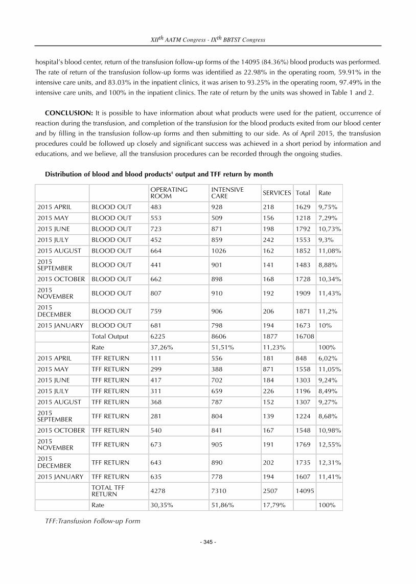
hospital’s blood center, return of the transfusion follow-up forms of the 14095 (84.36%) blood products was performed.
The rate of return of the transfusion follow-up forms was identified as 22.98% in the operating room, 59.91% in the
intensive care units, and 83.03% in the inpatient clinics, it was arisen to 93.25% in the operating room, 97.49% in the
intensive care units, and 100% in the inpatient clinics. The rate of return by the units was showed in Table 1 and 2.
CONCLUSION: It is possible to have information about what products were used for the patient, occurrence of
reaction during the transfusion, and completion of the transfusion for the blood products exited from our blood center
and by filling in the transfusion follow-up forms and then submitting to our side. As of April 2015, the transfusion
procedures could be followed up closely and significant success was achieved in a short period by information and
educations, and we believe, all the transfusion procedures can be recorded through the ongoing studies.
Distribution of blood and blood products' output and TFF return by month
TFF:Transfusion Follow-up Form
XIIth AATM Congress - IXth BBTST Congress
- 345 -
OPERATING ROOM
INTENSIVE CARE SERVICES Total Rate
2015 APRIL BLOOD OUT 483 928 218 1629 9,75%
2015 MAY BLOOD OUT 553 509 156 1218 7,29%
2015 JUNE BLOOD OUT 723 871 198 1792 10,73%
2015 JULY BLOOD OUT 452 859 242 1553 9,3%
2015 AUGUST BLOOD OUT 664 1026 162 1852 11,08%
2015 SEPTEMBER BLOOD OUT 441 901 141 1483 8,88%
2015 OCTOBER BLOOD OUT 662 898 168 1728 10,34%
2015 NOVEMBER BLOOD OUT 807 910 192 1909 11,43%
2015 DECEMBER BLOOD OUT 759 906 206 1871 11,2%
2015 JANUARY BLOOD OUT 681 798 194 1673 10%
Total Output 6225 8606 1877 16708
Rate 37,26% 51,51% 11,23% 100%
2015 APRIL TFF RETURN 111 556 181 848 6,02%
2015 MAY TFF RETURN 299 388 871 1558 11,05%
2015 JUNE TFF RETURN 417 702 184 1303 9,24%
2015 JULY TFF RETURN 311 659 226 1196 8,49%
2015 AUGUST TFF RETURN 368 787 152 1307 9,27%
2015 SEPTEMBER TFF RETURN 281 804 139 1224 8,68%
2015 OCTOBER TFF RETURN 540 841 167 1548 10,98%
2015 NOVEMBER TFF RETURN 673 905 191 1769 12,55%
2015 DECEMBER TFF RETURN 643 890 202 1735 12,31%
2015 JANUARY TFF RETURN 635 778 194 1607 11,41%
TOTAL TFF RETURN 4278 7310 2507 14095
Rate 30,35% 51,86% 17,79% 100%

Percentage of TFF RETURN(On a monthly basis)
PP-075
DISTRIBUTIONAL EVALUATION OF BLOOD PRODUCTS USE BASED ON THE BLOOD PRODUCT USE BY
SERVICE, INTENSIVE CARE UNIT AND NUMBER OF OPERATIONS IN ISTANBUL MEHMET AKIF ERSOY
THORACIC AND CARDIOVASCULAR SURGERY HOSPITAL
Gülsen Şener1, Tülin Bayrak2, Hakan Türk1, Hayriye Ak Yıldırım3
1Blood Center of Mehmet Akif Ersoy Education and Research Hospital of Thoracic and Cardiovascular Surgery2Ordu University, Faculty of Medicine, Department of Biochemistry3Biochemistry of Mehmet Akif Ersoy Education and Research Hospital of Thoracic and Cardiovascular Surgery
OBJECTIVE: Istanbul Mehmet Akif Ersoy Thoracic and Cardiovascular Surgery Hospital is the only hospital
cardiovascular surgery of the European side of Istanbul.Providement of data about the possible need and the use of
blood and investigation of the amount of blood product use by Operating rooms, Intensive Care Unit and inpatient
services as well as statistical determination of relationship of blood product use with operation and number of lying
patients are intended.
METHODS: The relation between the amount of blood and blood products used, number of inpatients and their
distribution according to the clinics, the number of operations and their distribution regarding their sort and the amount
of blood products used for patients in the duration between 01.04.2015-31.01.2016 was analyzed retrospectively
RESULTS: Between the dates 01.04.2015-31.01.2016, 13 606 patients who received inpatient services (service
9045 (47.21%), intensive care unit 4561 (23.81%) 5552 (28.98%) operations were carried out (2439 class, 572B, 1536
C 104D, 151, 134 robotics, 561-day operation) Within this period, the number of blood products sent to the relevant
units by Blood Center is 16708 units.Maximum use of the product occurred in the intensive care unit ( 8606, 51.51
%),then respectively service (1877, 11:23 % ) and use in the operating room ( 6225, 37.26 %).The month we had most
patients in our hospital is November 2015 and 1909 ( 11:43 %) patients were operated. The month we had the lowest
number of patients is May 2015 and 1218 patients (7.29 %) were operated.The.month has the highest blood use is
December 2015 ( 12:21 %), the least was September 2015 ( 7.98% ).Blood product use rate according to the
departments and number of operations are presented in Table 1.Blood use per operation is about 1, in intensive care is
2, while in the service is below 1.
CONCLUSION: The use of blood products obtained from our blood center has the maximum value in intensive
care,and the minimum in the services.It is found out that the blood use is uncorrelated to number of patients and the
XIIth AATM Congress - IXth BBTST Congress
- 346 -
2015 APRIL
2015 MAY
2015 JUNE
2015 JULY
2015 AUGUST
2015 SEPTEMBER
2015 OCTOBER
2015 NOVEMBER
2015 DECEMBER
2015 JANUARY
TFF RETURN
TFF RETURN
TFF RETURN
TFF RETURN
TFF RETURN
TFF RETURN
TFF RETURN
TFF RETURN
TFF RETURN
TFF RETURN
OPERATING ROOM 22,98% 54,07% 57,68% 68,81% 55,42% 63,72% 81,57% 83,4% 84,72% 93,25%
INTENSIVE CARE 59,91% 76,23% 80,6% 76,72% 76,71% 89,23% 93,65% 99,45% 98,23% 97,49%
SERVICES 83,03% 94,87% 92,93% 93,39% 93,83% 98,58% 99,4% 99,48% 98,06% 100%
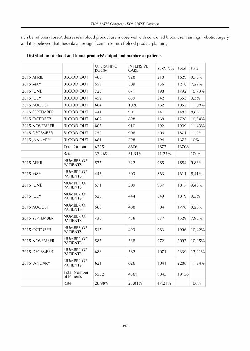
number of operations.A decrease in blood product use is observed with controlled blood use, trainings, robotic surgery
and it is believed that these data are significant in terms of blood product planning.
Distribution of blood and blood products' output and number of patients
XIIth AATM Congress - IXth BBTST Congress
- 347 -
OPERATING ROOM
INTENSIVE CARE SERVICES Total Rate
2015 APRIL BLOOD OUT 483 928 218 1629 9,75%
2015 MAY BLOOD OUT 553 509 156 1218 7,29%
2015 JUNE BLOOD OUT 723 871 198 1792 10,73%
2015 JULY BLOOD OUT 452 859 242 1553 9,3%
2015 AUGUST BLOOD OUT 664 1026 162 1852 11,08%
2015 SEPTEMBER BLOOD OUT 441 901 141 1483 8,88%
2015 OCTOBER BLOOD OUT 662 898 168 1728 10,34%
2015 NOVEMBER BLOOD OUT 807 910 192 1909 11,43%
2015 DECEMBER BLOOD OUT 759 906 206 1871 11,2%
2015 JANUARY BLOOD OUT 681 798 194 1673 10%
Total Output 6225 8606 1877 16708
Rate 37,26% 51,51% 11,23% 100%
2015 APRIL NUMBER OF PATIENTS 577 322 985 1884 9,83%
2015 MAY NUMBER OF PATIENTS
445 303 863 1611 8,41%
2015 JUNE NUMBER OF PATIENTS 571 309 937 1817 9,48%
2015 JULY NUMBER OF PATIENTS 526 444 849 1819 9,5%
2015 AUGUST NUMBER OF PATIENTS 586 488 704 1778 9,28%
2015 SEPTEMBER NUMBER OF PATIENTS
436 456 637 1529 7,98%
2015 OCTOBER NUMBER OF PATIENTS 517 493 986 1996 10,42%
2015 NOVEMBER NUMBER OF PATIENTS 587 538 972 2097 10,95%
2015 DECEMBER NUMBER OF PATIENTS 686 582 1071 2339 12,21%
2015 JANUARY NUMBER OF PATIENTS 621 626 1041 2288 11,94%
Total Number of Patients 5552 4561 9045 19158
Rate 28,98% 23,81% 47,21% 100%

PP-076
LABORATORY STUDIES IN THALASSEMIA PATIENTS BEFORE TRANSFUSION
Asım Sabıtogulları, Ahmet Meral, İlknur Avcı, Hatıce Nedret Saatcıoglu, Aydek Sultan Ozdemır, Fatma Orhan, Haluk
Ipek, Bakı Baskan, Nejmı Arı, Melek Reyhan, Dursun Ayverdıata, Orhan Tuncer, Meltem Demırbas, Funda Ceren,
Sımten Dagdas, Gulsum Ozet
Ankara Numune Eğitim Ve Araştırma Hastanesi , Kan Bankası
INTRODUCTION: Blood transfusion is a form of treatment in thalassemia patients that requires continuity.
Thalassemia diagnosed people who undergoing frequently transfusion patients. To follow-up these patients, it is
necessary to ensure the transfusion with compatible subset of the erythoricite suspension by identifying sub-groups of
these patient’s Rh and other groups.
PRACTISE: Preparation of blood products demanded by clinic for thalassemia patients, blood group and Rh sub-
groups typing obtained by microplate method primarily and saved to HIMS.(Hospital Information Management System)
Maximum of 5 (five) day erythrocytes suspensions compatible with blood group patients, Rh typing sub-groups by
working with the microplate method and erythrocyte suspensions are determined with the same sub-groups of patients
needed for erythrocyte suspensions. Blood samples of patients with Rh sub-group matching red blood cell suspension
crossmatch tests in compliance, erythrocyte suspensions are reserved on patients identified as suitable. Reserved
erythrocyte suspensions are performed by leukocyte filtration process to get ready for transfusion.
RESULT: In order to provide healthy transfusion for Thalessamia patients, it is convenient to follow by the same
center of these patients. As a result of cause the formation different antibodies Rh sub-groups can not be determined
erythrocyte suspensions have transfused to the patients, also caused problems in handling crossmatch testing blood
compatible in the next transfusion and transfusion requirements intervals will shorter for patients.
SUGGESTION: It is important in terms of the treatment process to establish a data bank which includes the country
and follow-up to be done through this data bank for follow-up the thalassemia patients and to prevent the improper
blood transfusions with non-compatible Rh sub-groups.
Rh Sub-Groups Study In Thalassemıa Patıents
XIIth AATM Congress - IXth BBTST Congress
- 348 -

PP-077
EVALUATION THE USE OF BLOOD PRODUCTS IN OUR HOSPITAL
Kamuran Şanlı, Miyase Kılıç
Istanbul Kanuni Sultan Suleyman Education and Research Hospital, ISTANBUL.TURKEY
AIM: We aim to evaluate the amount and distribution of blood and blood products according the departments in
our hospital.
MATERIAL-METHODS: Retrospective examination of data registry including request and use of blood and blood
products by departments between 1January-31 December 2015 was performed.
RESULTS: During one year period an amount of 34.014 blood products have been used. Of them was 15.893 (46.72
%) erythrocyte suspension, 7.058 Ü (20.75 %) fresh frozen plasma, 10.195 (29.97 %) platelet suspension. The rest of
3% included apheresis platelet, granulocyte apheresis, whole blood and cryoprecipitate. Table 1 shows the rates of
blood products used by the departments. Whereas table 2 reflects the proportion of blood products used.
CONCLUSION: Blood transfusion is needed mostly because of chronic anemia and rarely in the case of bleeding.
In general indication for blood product supply are like follows, erythrocyte suspension for the treatment of signs due
to severe hypoxemia, platelet suspension in thrombocytopenia with a count under 50000/mm3, fresh frozen plasma in
the case of multiple coagulation factor deficiencies, massive erythrocyte transfusion, bleeding diathesis during the
course of disseminated intravascular coagulopathy. Products used among departments were like follows, in
proportional quantity for erythrocyte suspension was like pediatric hematology (27.21 %), gynecology and obstetrics
(19.12 %), adult emergency (11.51 %). Platelet suspension use was like pediatric hematology (54.28 %), pediatric
intensive care unit (12.96 %), pediatric clinic (12.27 %). Fresh frozen plasma use was like neonatal intensive care unit
(35.08 %), adult intensive care unit (20.02 %), gynecology and obstetrics clinic (12.99 %) (table1).
It was remarkable that adult emergency unite used erythrocyte suspension on third frequency with a rate of (11.51
%). Adult emergency unite has a critical role and effects directly blood banking logistic activity and blood component
storage regulation. Monitoring and preventing misuse of blood components on adult emergency unite is an essential
strategy for blood bank inventory management.
An erythrocytes/ fresh frozen plasma utilization rate of 2 or over is reported to be appropriate in a centre where
blood components are used. Our data reflects a rate of 2.51 which suggests an appopriate use in our hospital. On the
other hand the use of fresh frozen plasma on certain departments was relatively rather high (table2). These units were
pediatric intensive care unit, adult intensive care unit and neonatal intensive care unit. A wider indication assesment
is thought to be the cause of this data. Anyway, evaluation should be done to make sure that blood products are used
on true indications in the light of Blood and Blood Products Guidelines and evidence-based medicine. The main
objective of treatment with blood transfusion is to provide maximum benefit with minimum harm to the patient.
Blood transfusion indication should be guided by main principles of evidence-based medicine and current
knowledge.
XIIth AATM Congress - IXth BBTST Congress
- 349 -

Graph of used of blood
RED CELL SUSPENSION AND FRESH FROZEN PLASMA BY CLINICAL USE OF COMPARISON
XIIth AATM Congress - IXth BBTST Congress
- 350 -

Table 1 - According to an annual distribution of clinical blood use
Table 2 - Evaluation of plasma by comparing the concentrates of the erythrocyte concentrates in clinical
XIIth AATM Congress - IXth BBTST Congress
- 351 -
PP-078
MAY WHOLE BLOOD CONSUMPTION IN CARDIAC SURGERY BE LOWERED TO ZERO?
Oral Koçyiğit1, Osman Eren Karpuzoğlu2, Tansel Türkoğlu1, Nil Banu Pelit3, Yasemin Çiçek1, Ayşe Yolaçan1
1Acibadem Kayseri Hospital, Kayseri, Turkey2Istanbul Medicine Hospital, Istanbul, Turkey3Acıbadem Healthcare Group Blood Coordinator, Istanbul, Turkey
AIM: Blood transfusions are life saving precedures but also have serious complication risks. These risks and
hardening supply due to limited resources causes limitations in blood tranfusions all over the world. Our aim was to
evaluate the results of our modifications on blood consumption in patients subjected to cardiac surgery which
speciality known with relatively higher rate of blood transfusions.
METHODS AND RESULTS: Patients who have had open cardiac surgery (n=1099) between 01.01.2011-
12.31.2015 in our institution were enrolled in the study. Yearly number of operations, blood and blood products used
were detailed in Table 1.
Some precautions were performed to lower the rate of whole blood consumption to levels recommended in
guidelines. Division visits, committee meetings, doctor breifings, quality management and educations were carried out.
Whole blood transfusion indications were reviewed with cardiovascular surgery team. Indications and
contraindications were detailed, risks/benefits were discussed with the whole team and the decisions were taken on
concensus. Surgical techniques on blood preservation were revised according to the guidelines which in turn lead to
a significant lowering especially on whole blood consumption.
DISCUSSION AND CONCLUSION: As seen in our study, effective education, inter disciplinary coordination and
blood preservation surgical techniques may help reducing whole blood consumption to zero Current transfusion
practices according to rules and international guidelines may have significant benefits on medical, financial and social
results. Responsive supervision of the whole procedure beginning from the indication to post transfusion follow up
mainly by doctors and the dedicated effords of the all members of the medical team causes improvement in the results.
Whole blood is only for one patient but if it's processed into products, may be used for 4 different patients. As the
one and only source of this precious liquid is humans, blood should be transfused with extreme caution and
ungenerously. Costs of blood transfusion is high by itself and with related complications (infections, reactions, acute
renal failure, immune modulation etc.) costs have potential for a financial disaster. Whole blood was the preferred
choice mostly because of availability. Large scale hospitals with major surgical units like cardiovascular surgery should
have the facilities and legal permissions to process blood and blood products to lower the rate of whole blood
comsumption nationwide. As a conclusion, education and informative actions with interdisciplinary integration of
guidelines may help reducing the blood transfusions to internationally accepted levels.
XIIth AATM Congress - IXth BBTST Congress
- 352 -

Table.1
CVS: Cardiovascular Surgery, ES: Erythrocyte suspension, FFP:Freshly Frozen Plasm, PLT: Platelet
PP-079
BLOOD MANAGEMENT APPLICATIONS OF SCRUP SURGERY NURSES
Ertan Özyurt1, Emine Uğurlu2, Satı Birbudak3, Rabia Yorgancı4
1Department of Microbiology Dr. Siyami Ersek thoracic and cardiovascular surgery training and research
hospital,istanbul Turkey2Department of Biochemistry laboratory Dr. Siyami Ersek thoracic and cardiovascular surgery training and research
hospital,istanbul Turkey3Department of research and development Dr. Siyami Ersek thoracic and cardiovascular surgery training and
research hospital,istanbul Turkey4Deparment of Stock Dr. Siyami Ersek thoracic and cardiovascular surgery training and research hospital,istanbul
Turkey
Blood transfusion is widely used during procedures of cardiac surgeries, especially cardio pulmonary bypass (CPB).
Cardiac surgeries consume 10% to 15% of national blood source and due to the significant increase in complexity of
cardiac surgery procedures, this portion progressively increases according to the existing findings. Open cardiac
surgeries have been performed for forty years in our country.
At least 10, sometimes 20, 30 units of blood per surgery had to be presented at the first years of these surgeries.The
major causes of these were technical inability of the heart lung machines and blood usage was considered as an
innocent operation. Blood usage has decreased to 8-10 units due to growth technology in the mid of 1980s.
After the mid of 1990s, information about disadvantages of blood usage started to be announced. Despite of this,
applications about blood usage decrease did not exist in scrup surgery nurses task descriptions. In this study, we
investigated scrup nurse (sterile) sircule nurse (roving) inoperative responsibilities and required nursing functionalities
XIIth AATM Congress - IXth BBTST Congress
- 353 -
2011 2012 2013 2014 2015
CVS OPERATIONS 213 210 196 259 221
WHOLE BLOOD (Total) 40 12 1 1 0
WHOLE BLOOD (Average) 0,19 0,06 0,01 0,00 0,00
ES (Total) 277 242 121 259 259
ES (Average) 1,30 1,15 0,62 1,00 1,17
FFP (Total) 369 373 131 311 234
FFP (Average) 1,73 1,78 0,67 1,20 1,06
PLT- RANDOM(Total) 64 66 0 39 24
PLT -RANDOM(Average) 0,30 0,31 0,00 0,15 0,11
PLT- Apheresis(Total) 0 0 0 1 0
PLT -Apheresis (Average) 0,00 0,00 0,00 0,00 0,00
in blood usage and bleeding control.
After 2000s, techniques to reduce blood usage in cardiac surgeries were improved and blood usage was questioned
by perfusion surgery teams and anaesthesia teams.
Nurses who are indispensable of the surgeries can take a great role in reducing blood usage such as good control
of bleeding, leading the team in choosing the safen extraction technique, to perform tissue closure operation faster. Leg
bandage and protection of the leg after safen removal in reducing blood loss are part of nursing roles. In cardiovascular
clinics, each surgery team and nurse, who does not have blood protecton and blood usage procedure, perform different
applications. Reducing blood usage and bleeding subjects should exist in curriculum in nursing education and in
service education.
PP-080
BLOOD USAGE AND DISTRIBUTION TO THE CLINICS IN HASEKI TRAINING AND RESEARCH HOSPITAL
FOR YEAR 2015
Hatice Erdoğan, Tunay Gül
Blood Transfusion Center of Haseki Training and Research Hospital, Istanbul, Turkey
AIM: We intended to study on consumption and distribution rates of Haseki Training and Research Hospital (HTRH)
blood and blood products according to the clinics.
METHOD: In this study; blood obtainment for the last year (from our blood center and Red Crescent), usage
according to the clinics and erythrocyte extermination data have calculated retrospectively from the hospital blood
center’s computer aided operation system.
EVIDENCES: Totally 9162* donations realized in our hospital’s blood center. Transfusions realized for totally 11931
patients in our hospital’s clinics. The transfused products and their distribution according to the services are shown in
Table 1 below. Number of erythrocyte exterminations and reasons are shown in table 2.
CONCLUSION: As of the most product consumption in the departments of HTRH are seen as in the first place
Internal Diseases, the second place General Intensive Care and the third place Cardiovascular Surgery. Since
Hematology Department is in the Internal Diseases Department, it affects this result. Because of our Blood Transfusion
Center had Authorized Blood Center feature in year 2015; 13,2% of our total products and 8,4% of erythrocyte
suspensions were supplied by Red Crescent. The number of Erythrocyte exterminations are appropriate according to
the numbers planned yearly in quality standards.
XIIth AATM Congress - IXth BBTST Congress
- 354 -
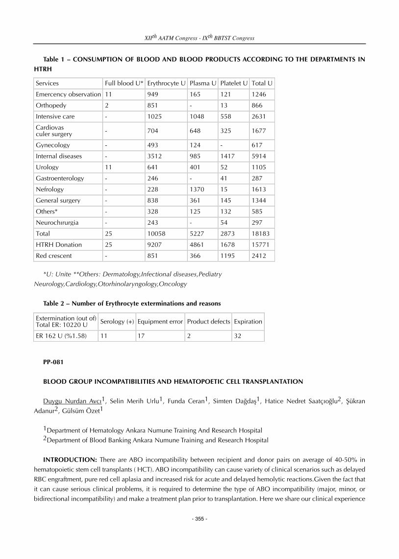
Table 1 – CONSUMPTION OF BLOOD AND BLOOD PRODUCTS ACCORDING TO THE DEPARTMENTS IN
HTRH
*U: Unite **Others: Dermatology,Infectional diseases,Pediatry
Neurology,Cardiology,Otorhinolaryngology,Oncology
Table 2 – Number of Erythrocyte exterminations and reasons
PP-081
BLOOD GROUP INCOMPATIBILITIES AND HEMATOPOETIC CELL TRANSPLANTATION
Duygu Nurdan Avcı1, Selin Merih Urlu1, Funda Ceran1, Simten Dağdaş1, Hatice Nedret Saatçıoğlu2, Şükran
Adanur2, Gülsüm Özet1
1Department of Hematology Ankara Numune Training And Research Hospital2Department of Blood Banking Ankara Numune Training and Research Hospital
INTRODUCTION: There are ABO incompatibility between recipient and donor pairs on average of 40-50% in
hematopoietic stem cell transplants ( HCT). ABO incompatibility can cause variety of clinical scenarios such as delayed
RBC engraftment, pure red cell aplasia and increased risk for acute and delayed hemolytic reactions.Given the fact that
it can cause serious clinical problems, it is required to determine the type of ABO incompatibility (major, minor, or
bidirectional incompatibility) and make a treatment plan prior to transplantation. Here we share our clinical experience
XIIth AATM Congress - IXth BBTST Congress
- 355 -
Services Full blood U* Erythrocyte U Plasma U Platelet U Total U
Emercency observation 11 949 165 121 1246
Orthopedy 2 851 - 13 866
Intensive care - 1025 1048 558 2631
Cardiovas culer surgery - 704 648 325 1677
Gynecology - 493 124 - 617
Internal diseases - 3512 985 1417 5914
Urology 11 641 401 52 1105
Gastroenterology - 246 - 41 287
Nefrology - 228 1370 15 1613
General surgery - 838 361 145 1344
Others* - 328 125 132 585
Neurochırurgia - 243 - 54 297
Total 25 10058 5227 2873 18183
HTRH Donation 25 9207 4861 1678 15771
Red crescent - 851 366 1195 2412
Extermination (out of) Total ER: 10220 U Serology (+) Equipment error Product defects Expiration
ER 162 U (%1.58) 11 17 2 32

in HCT with ABO incompatibility.
RESULTS: We reviewed HCT patients with ABO incompatibility between 2014-2015 in our clinic. We had 6 cases
of ABO incompatibility. Two of them (33.3% ) had major incompatibility while four ( 66.6 %) had minor
incompatibility. We studied isohemagglutinin titration in all our cases. In two of these patients who had major
incompatibility with a titration of 1/128 and above, plasmapheresis was done prior to allogeneic HCT to reduce anti-
donor A/B antibodies in recipient plasma. The isohemagglutinin titers were followed after stem cell transplantation in
every 15 days. Plasma reduction was applied in 4 cases with minor incompatibility. At the same time, isohemagglutinin
titration, direct and indirect Coombs, and blood groups were studied. We found that, time to RBC engraftment and RBC
transfusion requirement was higher in patients with minor and major incompatibility compared to other HCT cases.
We prefered O type blood whenever erythrocyte transfusion was needed.
DISCUSSION: Even though blood group compatibility is not a must for allogeneic stem cell transplantation, blood
group incompatibility can cause serious clinical problems when it is not evaluated properly. Different clinical
presentations like delayed RBC engraftment, hemolytic reactions and pure red cell aplasia can be observed. It is crucial
to run, properly evaluate and repeat tests for type of ABO incompatibility, isohemagglutinin titers, and direct/ indirect
coombs. Incompatibility type, blood type of the donor and recipient are the main determinants in blood product
transfusions. By sharing our clinical experience in allogeneic HCT with ABO incompatibility, we want to emphasize
the importance of follow-up of these patients.
PP-082
A RESEARCH ON KNOWLEDGE LEVEL OF THE NURSES FOR TRANSFUSION PROCEDURES
Saim Dayan, Tuncer Özekinci, Şükran Kaplan
Blood Center, Faculty of Medicine, Dicle University,Diyarbakır,Turkey
OBJECTIVE: A significant part of complications of blood transfusion is known to be caused by knowledge deficiency
of the implementers. In the present study, knowledge level of the nurses who perform transfusion on blood transfusion
and efficiency of the training were measured.
METHOD: Knowledge level of 61 nurses working in Dicle University Hospitals about blood transfusion procedures
was measured. Rates of correct answers were compared according to the school graduated, working period, age groups
and transfusion implementation subjects. A training about transfusion implementations was given to the nurses,
knowledge levels of the nurses were measured again and knowledge level before and after the training was measured.
Pooled sample t test and χ² test were used for data assessment.
FINDINGS: A questionnaire including 10 questions was applied to the nurses who participated into the research.
Rates for correct answers were 60.52% /115/190) for Medical Vocational High School graduates, 67% (67/100) for 2-
year Vocational School of Higher Education graduates, 60.93% (195/320) for 4-year Vocational School of Higher
Education. (p>0.05)
When the results were assessed according to working period, rates of correct answers were 60.95% /128/210) for
the nurses who work less than 5 years and 62.25% (249/400) for the nurses who work more than 5 years. (p>0.05)
XIIth AATM Congress - IXth BBTST Congress
- 356 -

When the results were assessed according to age group, rates of correct answers were 59.72% (215/360) for the
nurses who were younger than 30 years and 64.8% (162/250) for the nurses who are older than 30 years. (p>0.05)
By the questionnaire, questions measuring the knowledge level on transfusion reactions, transfusion periods for
blood products, storage conditions for blood products and transfusion procedures were asked to the nurses. The rates
of correct answers about reactions developing after transfusion of a wrong blood type and temperature degrees that
whole blood and erythrocyte suspensions are stored were lower (21.31% and 41%, respectively). Higher rates of
correct answers were detected about the topics that erythrocyte suspensions to be transfused under elective conditions
are not required to be heated up to body temperature and in which intervals that vital functions would be written on
transfusion monitoring form during transfusion (81.96 and 98.36, respectively).
The total knowledge level before training was found 61.96% (378/610) whereas an increase to 96.55% (589/610)
was detected following intervention by training. (p<0.001)
CONCLUSION: In the present research, no significant difference was detected between the schools graduated,
working period as a nurse and ages for transfusion procedures. Rates of correct answers about transfusion procedure
were found significantly lower on some topics and significantly higher on some topics. A significant increase on
knowledge level was detected after intervention through training. From this point of view, training the personnel within
certain intervals is suggested to reduce the complications of transfusion.
PP-083
RETROSPECTIVE ANALYSIS OF UTILIZATION OF BLOOD COMPONENTS IN A HEMATOLOGY ONCOLOGY
HOSPITAL IN TWO YEARS PERIOD
Nur Efe Iris1, Abdullah Ayyıldız1, Aslıhan Demirel1, Safiye Koçulu1, Esin Çevik1, Esat Namal2
1Department of Infectious Diseases and Clinical Microbiology, Istanbul Bilim University, Turkey2Department of Oncology, Istanbul Bilim University, Turkey
OBJECTIVE: More blood components are required in Hematology- Oncology centers than in most other
disciplines.Usually all of the components are necessary for patients with malignancy.We aimed to investigate the rates
of the blood components which were utilized in our hospital.
MATERIALS-METHODS: A retrospective analysis of the utilization of blood components in the Istanbul Bilim
University Avrupa Florence Nightingale Hospital was performed two years period between 1st of January 2014- 1st of
January 2016.
RESULTS: It was observed that a total of 8210 blood components were used in our hospital during two years
period.The most common blood components utilized were random donor platelets ( 52.9%), followed by packed red
cells (30.2%), apheresis platelets (11.3%), fresh frozen plasma (3.8%) and cryoprecipitate (1.8%)
CONCLUSION: Thrombocytopenia frequently occurs in patients with hematologic malignancy or solid organ
tumors, resulting from the underlying disease processes or a complication of cancer treatments.Platelet transfusions are
used to treat people who have active bleeding or at a high risk of bleeding. In our hospital the most utilized component
of blood was random donor platelets in accordance with our patients properties.
XIIth AATM Congress - IXth BBTST Congress
- 357 -

PP-084
EVALUATION OF THE TRANSFUSION CHECKLISTS IN THE REGIONAL BLOOD CENTER OF ISTANBUL
MEHMET AKIF ERSOY EDUCATION AND RESEARCH HOSPITAL OF THORACIC AND CARDIOVASCULAR
SURGERY
Gülsen Şener1, Murat Koşer2, Tülin Bayrak3, Rabia Yurtseven1, Hakan Türk1
1Blood Center of Mehmet Akif Ersoy Education and Research Hospital of Thoracic and Cardiovascular Surgery,
Istanbul2Silivri Department of Corrections State Hospital, Central Laboratory, Istanbul3Ordu University, Faculty of Medicine, Department of Biochemistry
OBJECTIVE: The aim of this study is to evaluate transfusion forms for improving traceability and transfusion safety
in compliance with the national guidelines.
METHOD: Transfusion checklists appropriately filled-in and signed by two clinic nurses are delivered to our
Regional Blood Center. These forms and blood request forms are filed together depending on the units by transfusion
nurse. Transfusion checklists of blood products transfused between 1 April 2015 and 1 January 2016 were studied
retrospectively.
FINDINGS: Only 12.488 (83.06 %) checklist of 15.035 transfusions are returned to Blood Center between
01.04.2015 to 3.01.2016. 2547 transfusion checklists forms of blood products are unfilled. 12488 of transfusion
checklists forms were examined and errors were analyzed. Incomplete filling details are listed in Table1.
CONCLUSION: Medical records of transfused patients must be accurate. The first step is to use and fill in
transfusion checklists completely. Since april 2015 the presence of Transfusion Nurse made possible to monitor
transfusions more closely in our hospital. Due to our data, it is more easy to give feedback and continuously educate
the staff. An improvement in transfusion safety is only possible, when traceability and compliance reaches very high
levels.
The numbers and percentage of incomplete filling of transfusion forms
XIIth AATM Congress - IXth BBTST Congress
- 358 -
Incomplete filling of: Numbers Percent (%)
Demographic details of patients 55 0,44%
Ward / Unit 1902 15,23%
Blood group type 144 1,15%
Required blood component 254 2,03%
Expiry date of product 1458 11,67%
Component check 536 4,31%
Pretransfusion check 546 4,37%
Planned Time of transfusion 4422 35,41%
Pretransfusion checkers (staff ID) 625 5%
Time and duration of transfusion 1700 13,61%
Transfusion follow 3267 26,16%
Name or signature of staff performing transfusion 745 5,97%
Transfused volume 1309 10,48%
Adverse effects 786 6,29%

PP-085
DESTRUCTION RATE OF HUMAN BLOOD AND BLOOD COMPONENTS IN OUR HOSPITAL BLOODTRANSFUSION CENTER
Nuray Arı1, Pervin Özlem Balcı1, Funda Koçal Can1, Gülşen Özveren1, Hatice Çağlar2
1Tokat State Hospital, Blood Transfusion Center, Tokat2Turkey Training and Research Hospital, Department of Medical Microbiology Laboratory, Ankara
AIM: Determining the rate of destruction was destroyed blood and blood products for various reasons. It was aimedto make the necessary efforts to reduce these rates.
METHOD: It was retrospectively data between the dates 01.01.2015-31.12.2015 of Tokat State Hospital BloodTransfusion Center. Erythrocyte suspension, platelet suspension, fresh frozen plasma and cryoprecipitate was obtainedby Samsun Red Crescent Society Regional Blood Center. In an emergency, we were taken full blood by our BloodTransfusion Center.
FINDINGS: Total 13471 piece blood and blood products used for patients in the 2015, There off 45 (%0.33) manyproducts are various reasons was destroyed. Because off the full postmature 10(%0.07) many erythrocyte and fullblood, because the bag explosion 22(%0.16) many fresh frozen plazma, due to return to service 11(%0.08) manyerythrocytes, platelets and whole blood was destroyed (t
RESULT: The blood request must be made for Patients who confirmed the decision to transfuse In order to preventunnecessary destruction of blood and blood products should be done Plan to pass in front of the destruction causedby the filling miata. "last in, last out’’policy of strictly enforced in blood storage.
Destruction rates for 2015
PP-086
LEAST INCOMPATIBLE RED BLOOD CELL UNIT USING; HOW SAFETY DECISION?
Senem Maral, Sule Mine Bakanay, Sema Akinci, Aysun Senturk Yikilmaz, Imdat Dilek
Department of Hematology, Ataturk Research and Treatment Hospital, Ankara,Turkey
INTRODUCTION: Haemolytic transfusion reactions caused by alloantibodies canbe troubesome in patients withautoimmune haemolytic anaemia( AIHA).Transfusion is essential when anemia is life treatening.However, finding acompatibile red blood cell (RBC) unit is a complicated process in patients with alloantibodies.Mostly, leastincompatibile RBC unit is selected for transfusion.
XIIth AATM Congress - IXth BBTST Congress
- 359 -
Fresh frozen plasma Erythrocyte suspension Platelet suspension Full blood Total
Miata stuffed - 8 - 2 10
Bag explosion 22 - - - 22
Return to service - 7 2 2 11
Incorrect products - 2 - - 2
Total 22 15 2 4 45

The purpose of this study is to demonstrate validity and safety of using least incompatible cross-matched group
specific blood by invivo transfusion.
METHODS: In two years time, a total of 27.109 RBC units were cross-matched in our transfusion centre. Cross-
match incompatibility was found only in nine patients whose direct antiglobuline test (DAT) positive.
Indications of transfusion were; anemia with life threatening severity and one preoperative preparation. Least
incompatible RBC units were chosen and transfused to patients.Patients were moniterized closely for any signs or
sympthoms of acute hemolysis. Before transfusion low dose dexamethazone and antihistaminics were administered.
Totally, 48 units of least incompatible RBC packs were transfused to patients. All transfusions were observed for acute
haemolysis signs like as dispnea, flank pain, hypotension, headache. Laboratory tests showing hemolysis such as total-
indirect bilurubin, lactat dehidrogenase and complete blood count to ensure sufficient increment of hemoglobine were
obtained after transfusion.
CONCLUSION: None of the patients experienced acute hemolytic transfusion reaction. Laboratory evaluation of
hemolysis parameters did not reveal any significant increase in hemolysis. As a conclusion; whenever transfusion is
required in patients with AIHA, ıt should not be cancelled because of cross-match incompatibility.Least incompatible
RBC units can be safely transfused.
Table 1. Laboratory results before transfusion
Table 2. Laboratory results after transfusion
XIIth AATM Congress - IXth BBTST Congress
- 360 -
Patient no Gender Age hb (g/dl) Hct T.bil(U/L) D.bil(U/L) LDH(U/L)
1 Female 58 7.5 20 20 12 1763
2 Male 78 6.1 18 0.8 0.4 249
3 Male 59 3.9 11 4.4 0.6 946
4 Female 71 5.4 18 3.0 0.9 345
5 Female 63 4.5 12 2.6 0.6 1247
6 Female 61 6.4 20 2.6 0.7 802
7 Male 74 6.0 17 0.8 0.3 164
8 Female 68 5.4 17 3.3 1.3 915
9 Female 72 3.8 9 4.4 0.6 508
Patient no RBC unit transfused hb(g/dl) hct T Bil(U/L) D Bil(U/L) LDH(U/L)
1 4 8.9 24 6.2 4.8 1342
2 8 10 30 NA NA NA
3 12 8.8 26 3.4 1.0 881
4 2 8.9 28 2.9 0.9 358
5 4 9.9 20 1.2 0.3 1230
6 2 9.6 30 2.4 0.8 757
7 8 9.4 29 NA NA NA
8 3 8 21 1.8 0.7 944
9 4 11.8 36 2.0 0.6 542

PP-087
A SURVEY OF PHYSICIANS’ KNOWLEDGE AND AWARENESS ABOUT TRANSFUSION
Gürsel Ersan1, Şükran Köse1, Fatma Liv2, Timur Köse3
1Tepecik Education and Research HospitalDepartment of Infectious Diseases and Clinical Microbiology,
Izmir,Turkey2Tepecik Education and Research Hospital, Transfusion Center,Izmir,Turkey3Ege Universty Medical School,Department of Bioistatistics and Medical Informatics
OBJECTIVE: To determine the level of knowledge and awareness about transfusion process to contribution of
transfusion safety for the physicians at the clinics whose transfusions are mostly made.
MATERIALS-METHODS: Izmir Tepecik Education and Research Hospital is a tertiary healthcare service using blood
and blood components frequently. (More than 15000 unit red cell concentrate in a year)A questionnaire study was
organized between the dates of 01.10.2015 and 30.10.2015 with 22 questions to the physicians who has been working
and willing to attend at the transfusion frequently was made clinics in our hospital. The questions were about blood
components, compatibility tests, practice of transfusion and transfusion reactions. The information note including the
answers has been delivered to the clinics with the same topic title.
Defining analysis has been done on the survey forms. IBM SPSS version 20.0 was used as a package.
RESULTS: 150 physicians attended to the survey ( 49,5 % of the physicians who attended to the survey at the
clinics.)
It was seen that, 62 % of participants was from surgical clinics and 38 % of it was from internal clinics.
In terms of the statistics of tranfusions center, 58,66% transfusions were given at the clinics those attended to the
survey. In totall, the average success grade is found 64,2. In regards of the topic title average points respectively,
transfusion reactions ( 74.75 ), transfusion practice ( 61.2 ), compatibility tests before transfusions ( 56. 5 ), information
for the blood component ( 55.3 ) was found.
CONCLUSION: Attending to this type of surveys helps to improve knowledge and awareness on the transfusions
process for nurses in charge and technicians beside of physcians. For this, in service training activities can be organised.
All of this works would help positively to the transfusion safety.
XIIth AATM Congress - IXth BBTST Congress
- 361 -
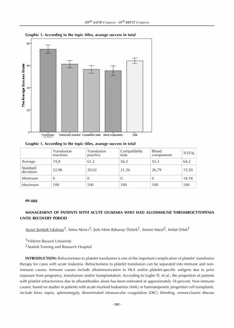
Graphic 1. According to the topic titles, avarage success in total
Graphic 1. According to the topic titles, avarage success in total
PP-088
MANAGEMENT OF PATIENTS WITH ACUTE LEUKEMIA WHO HAD ALLOIMMUNE THROMBOCYTOPENIA
UNTIL RECOVERY PERIOD
Aysun Şentürk Yıkılmaz1, Sema Akıncı2, Şule Mine Bakanay Öztürk1, Senem Maral2, Imdat Dilek1
1Yıldırım Beyazıt University2Atatürk Training and Research Hospital
INTRODUCTION: Refractoriness to platelet transfusion is one of the important complication of platelet transfusion
therapy for cases with acute leukemia. Refractoriness to platelet transfusion can be separated into immune and non-
immune causes. Immune causes include alloimmunization to HLA and/or platelet-specific antigens due to prior
exposure from pregnancy, transfusions and/or transplantation. According to Legler TJ. et al.; the proportion of patients
with platelet refractoriness due to alloantibodies alone has been estimated at approximately 18 percent. Non-immune
causes, based on studies in patients with acute myeloid leukaemia (AML) or haematopoietic progenitor cell transplants,
include fever, sepsis, splenomegaly, disseminated intravascular coagulation (DIC), bleeding, venoocclusive disease
XIIth AATM Congress - IXth BBTST Congress
- 362 -
Transfusion reactions
Transfusion practice
Compatibility tests
Blood components TOTAL
Average 74,8 61,2 56,5 55,3 64,2
Standard deviation 22,96 20,02 21,36 26,79 15,50
Minimum 0 0 0 0 18,18
Maximum 100 100 100 100 100

(VOD), graftversus-host disease (GVHD) and medications. Here, we describe to cases developing platelet
alloimmunization in patient followed by acute leukemia, as retrospectively.
MATERIALS AND METHODS: In our clinical data includes From 2010 to 2016 years, 9 patients in 118 acute
leukemia cases had platelet alloımimunızatıon. We used to a corrected count increment (CCI) for measuring response
to platelet transfusion. The corrected count increment (CCI) has also been used to measure the response to platelet
transfusion based on a platelet count obtained 10 minutes to one hour post-transfusion. CCI = [PPI x BSA (m2)] x 1011/
number of platelets transfused PPI is post-transfusion platelet count minus pre-transfusion platelet countBSA is body
surface area measured in square metersOur approach is consistent with that of the American Society of Clinical
Oncology, which defines refractoriness to platelet transfusion as a CCI of <5000/microL on at least two sequential
occasions.
RESULTS: In this group of patients was identified demographic characteristics such as the type of leukemia, age,
gender. Allloimmünization occurred in 7 patients during induction and in 2 patients during the consolidation.
Alloimmunization has improved 12th day average since the beginning of the chemotherapy. Number of the platelet
transfusions are indicated until platelet refracterness in Table 1. All patients are treated with frequent intervals platelet
transfusions (4-6 hours) plus 400 mg/kg day IVIG, we specified which day of platelets recovery and IVIG treatment
response, in table 2. The mean duration of platelet transfusion support was 12 ( min 6- max 18) days.
CONCLUSION: However, once it is determined that a patient is alloimmunized, HLA-matched platelets improve
platelet count increments. Other strategies for managing refractoriness were also attempted and generally met with
limited success. A variety of drug therapies have been attempted based, at least in part, upon their efficacy in
autoimmune thrombocytopenia. Intravenous immune globulin (IVIG) has been proposed as a strategy for immune
modulation, but the results have generally been unimpressive. The supportive therapy should be performed HLA-
matched platelets in cases with platelet alloimmunization. However, in clinical practice, the administration can be
done in limited cases. Because, HLA-matched donor is not possible for every patient. Data on the IVIG administration
for alloimmune thrombocytopenia treatment are not sufficient, but it should be emphasized as an alternative option.
Consequently, as in our cases, IVIG therapy plus frequent intermittent transfusions seems to be usefull.
Table.1
dermographic data
XIIth AATM Congress - IXth BBTST Congress
- 363 -
patient age (years) gender Leukemia
Type
The initial count of platelets
Time of hospitalization(day)
Time of death (month)
Cytotoxic treatment
1 41 F AML 75000 36 Ida-ara c
2 36 M AML 17000 21 10 Ida-ara c
3 70 M AML 24000 40 9 ATRA
4 30 M AML 40000 20 1 Ida-ara c
5 66 M AML 76000 16 1 Ida-ara c
6 46 F AML 35000 28 20 Ida-ara c
7 39 F AML 25300 26 4 Ida-ara c
8 36 F AML 12000 24 Ida-ara c
9 42 F ALL 75000 36 CALGB

Table.2
Data of platelets transfusion
PP-089
THE CRYOPRECIPITATE REPLACEMENT FOR THE ACUTE PROMYELOCYTIC LEUKEMIA CASES WHO
PRESENTED WITH HYPOFIBRINOGENEMIA
Aysun Şentürk Yıkılmaz1, Sema Akıncı2, Şule Mine Bakanay Öztürk1, Senem Maral2, Imdat Dilek1
1Yıldırım Beyazıt University2Atatürk Training and Research Hospital
Acute promyelocyticleukemia (APL) is a subtype of acute myeloid leukemia (AML) characterized by distinctive
morphology of blast cells, a life-threatening coagulopathy and a specific balanced reciprocal translocation t(15;17)
which fuses the PML (promyelocyte) gene on chromosome 15 to the RAR (retinoic acid receptor) gene on chromosome
17. The dominant clinical manifestation of the disease was indeed severe and life-threatening bleeding, which proved
to be the major cause of early mortality in every report. Disseminated intravascular coagulation (DIC) is frequently
present at diagnosis or occurs soon after the initiation of cytotoxic chemotherapy in patients with APL. This
complication constitutes a medical emergency, since, if left untreated, it can cause pulmonary or cerebrovascular
hemorrhage in up to 40 percent of patients and a 10 to 20 percent incidence of early hemorrhagic death. Treatment of
the coagulopathy (DIC) associated with APL may be difficult and should be managed expectantly. Coagulation
parameters, including fibrinogen, D-dimer, PT, aPTT, and platelet counts, should be monitored closely. Transfusions of
platelets and cryoprecipitate or fresh frozen plasma are used to maintain the platelet count above 20,000 to
30,000/microL and the plasma fibrinogen concentration above 150 mg/dL. Higher platelet counts of 30,000/microL
may be beneficial to control overt bleeding. Inaddition, anecdotal evidence supports attempting to maintain the platelet
count above 50,000/microL during periods of DIC, as reflected by falling fibrinogen or high D-dimer levels. Here, we
examined cryoprecipitate replacement count and efficacy in APL patients which was presented with
hypofibrinogenemia. Patients who had cryoprecipitate replacement was compared with patients without replacement.
XIIth AATM Congress - IXth BBTST Congress
- 364 -
Patient Alloimmünization time(day of chemotherapy)
Number of the day Plt> 50 000 after IVIG
Number of the day plt>100000 after chemotherapy (recovery day)
Number of platelets transfusion until alloimmünization
1 14 18 26 68
2 12 10 24 70
3 15 8 31 36
4 1 22 22 70
5 15 16. DAY EX 16. DAY EX 69
6 5 10 22 4
7 17 5 26 36
8 15 (First concolidation) 6 23 109
9 14 12 26 68

MATERIAL AND METHODS: Between the years 2009-2015, it was screened for patients admitted with acute
promyelocytic leukemia. A total of 18 patients with APL were identified in the study, Fibrinogen levels of cases were
monitored every day following initial diagnosis. In patients with hypofibrigonemia, ıf there is bleeding, fibrinogen level
were removed of 150 mg / dL whereas there isn’t bleeding, fibrinogen level were removed of 100 mg / dL.
RESULTS: Five APL cases was presented with hypofibrinogenemia in initial diagnosis. Cryoprecipitate was made
replacement in these cases. Among the five patients admitted with hypofibrinogenemia, three patient had the
intermediate risk group characteristics, two patient had the high risk group characteristics. However, in the 13 cases in
without hypofibrinogenemia, six patient had the low risk group characteristics. For five cases, Total of cryoprecipitate
replacement were made 143 ünits. There were not new cases who developed hypofibrinogenemia after initial
diagnosis. Support of cryoprecipitate was made between the first and eighteenth days.
CONCLUSION: APL cases are more prone to disseminated intravascular coagulation. Especially, if patients present
with hypofibrinogenemia, patients should be supported by cryoprecipitate until promyelocytes has been cleared up
from the bone marrow. Our clinic, we had cases who had performed with cryoprecipitate replacement until the 18th
day of treatment. As a result, the clinical course was no difference the groups that received cryoprecipitate between the
groups that are not support for cryoprecipitate.
PP-091
THE INCOMPATIBLE CROSSMATCH BLOOD TRANSFUSION
Mehmet Ali Uçar, Asım Sabitoğulları, Hatice Nedret Saatçioğulları, Funda Ceran, Simten Dağdaş, Aydek Sultan
Özdemir, Fatma Orhan, Ahmet Meral, Gülsüm Özet
Ankara Numune Education And Research Hospital, Departmant Of Hematology
INTRODUCTION: The aim of blood transfusion is to provide an appropriate immunologic and serologic blood
components to a patient. Initial examination includes ABO and Rh compliance in the patient and donor blood sample.
Crossmatching and antibody screening tests are performed to detect the presence of antibodies in patients plasma. If
there are allo-antibodies in patients plasma, it is very difficult to find proper blood product and loss of time is a major
problem. Crossmatching is the last control in compatibility of the transfusion. The crossmatching refers to the test that
is performed prior to a blood transfusion in order to determine if the donor's blood is compatible with the blood of an
intended recipient. We aimed in this article to report the crossmatching incompatible transfusions and the incompatible
blood transfusion results.
RESULTS: Microplate method was used for the determination of blood type, crossmatch and indirect antiglobulin
test. Gel-filtration method was used for direct antiglobulin test. Brand of neo were used as instruments for all tests. The
incompatible crossmatch resutls of patients were investigated in our transfusion center in 2015. A total of 3984 patients
were treated with blood transfusion. Crossmatching in 6640 donors. The crossmatch incompatible transfusions gave in
9 patients (5 female, 4 male). The average age was 59.4 years. Five patients had hematologic malignancies. Two patients
with acute life threatening bleeding transfused O Rh (-) blood group. Sub-group compatible blood transfusion was given
to all other patients. Antibody screening and identification was done all patients except for emergency cases. All
patients had previously received blood transfusion. Acute hemolytic reactions can be seen after incompatible
crossmatch blood transfusion. Therefore vital signs of patients (bloodpressure, pulse, respiratory, fever), laboratory
XIIth AATM Congress - IXth BBTST Congress
- 365 -

findings of hemolysis ( haptoglobulin, indirectbilirubin, lacticdehydrogenase, hemoglobinuria), urine output
monitoring followed carefully. Hemoglobinuria measurements were performed every five minutes in a urine. Immune
hemolysis was not observed in any patient.
DISCUSSION: The incompatible crossmatch is a major problem for both the blood bank and the patient's
physician. The major concerns are, difficulies in finding the appropriate blood products, hemolytic reaction that may
occur after the application of blood products and time issues. Subgroup compatible blood transfusion might given to
patients with incompatible crossmatch but it should be closely monitored for hemolysis. In case of emergency O Rh (-
) blood replacement can given if there is no time for the determination of blood group.
Table 1
: futures of patients
PP-092
THE ANALYSIS OF THE USAGE OF RED BLOOD CELL COMPONENT
Berrin Uzun1, Hayri Güvel1, Serdar Güngör2, Özcan Sengel1, Aslı Gamze Şener2
1Izmir Katip Celebi University, Ataturk Training and Research Hospital, Transfusion Center2Izmir Katip Celebi University, Ataturk Training and Research Hospital, Medical Microbiology
BACKGROUND: Izmir Katip Celebi University, Ataturk Training and Research Hospital is a large entity with 1100
bed capacity. As a consequence of changing policy for blood supply at national level, blood components for transfusion
are provided by the Regional Blood Center of Turkish Red Crescent (TRC) since second half of 2013.
AIMS: In this study, it is aimed to analyze the distribution of red blood cell concentrates transfused between 2013-
2015 in Izmir Katip Celebi University, Ataturk Training and Research Hospital.
XIIth AATM Congress - IXth BBTST Congress
- 366 -
patients age sex blood type
Direct antiglobulin test Ig-G
Direct antiglobulin test C3-d
Indirect antiglobulin test Disease
1 72 k A (+) (++) (++) (+++) myelodysplastic syndrome
2 64 e 0 (+) (+++) (-) (+++) chronic lymphocytic leukemia
3 65 k A (+) (+++) (-) (++++) Knee surgery
4 64 e A (-) (+) (-) (+++) myelodysplastic syndrome
5 68 e O (+) (++) (-) (+++) multiple myeloma
6 81 e O(+) (-) (-) (+++) Upper gastrointestinal bleeding
7 42 k A (+) (+++) (-) (+++) Chronic renal failure
8 38 k B (+) (+++) (++) (++++) systemic lupus erythematosus
9 41 k B (+) (+++) (++++) (-) myelodysplastic syndrome

METHODS: In 2013 blood and blood products were both produced in our hospital and as well as provided from
TRC. In 2014 and 2015 all of the blood and blood products fully were supplied by TRC. In the hospital, the whole
blood taken from avaible donors to donate blood was divided into components by removing buffy coat with blood bag
systems (Quadruple Top&Bottom CPD/SAG-M 450 ml SAP 5D; Kansuk, Turkey). All products were provided as buffy
coat removed from TRC.
RESULTS: In 2013 totally 23953, in 2014 22336 and in 2015 to 22127 units of red blood cell components were
used. The types and the distribution of the red blood cell components are given in the table.
Table: The types and the distribution of the red blood cell components.
CONCLUSION: Totally the usage of the erythrocyte suspensions was decreased in years but varieties of processed
erythrocyte suspensions were increased. The variety of used products especially is proportional to ease of its ability to
supply. For supplying optimal blood products, The Red Crescent Regional Blood Centers would be producing benefits
to planning according to the data of the transfusion centers of the hospitals in the supply of specialized materials.
PP-093
ZEYNEP KAMIL MATERNITY AND PAEDICATRICS TRAINING AND RESEARCH HOSPITAL ATTENDANCE
RATES OF TRAINING GIVEN TO DOCTORS ON SAFE TRANSFUSION AND ITS IMPORTANCE
Hülya Eksan1, Ramazan Uluhan2
1Zeynep Kamil Maternity and Paediatrics Training and Research Hospital, Training Coordinator, Istanbul2Zeynep Kamil Maternity and Paediatrics Training and Research Hospital, Medical Microbiology Laboratory,
Istanbul
AIM: Blood transfusion is defined as a tissue transplantation. In clinical practice, doctors must determine the right
XIIth AATM Congress - IXth BBTST Congress
- 367 -

indication for transfusion, correct blood and blood components must be used, possible adverse reactions must be
recognised at an early stage and early intervention must be applied and early notifications and feedback must be
provided. Education is also important in lowering the blood destruction rate. Therefore, it is envisaged to reach 90%
participation rate in this training.
METHOD: Efforts are made to enable physicians working at Zeynep Kamil Maternity and Paediatrics Training and
Research Hospital to participate in training regarding safe transfusion, blood and blood components etc. In order to
find the retrospective rate of participation in the aforementioned training program, participant evaluation forms were
studied for the December- January 2015 period in order to assess the quality of the training given and the number of
physicians participating in the program.
FINDINGS: It is critically important to use the correct components when it is medically necessary to use such
components for a patient. To create awareness regarding safe transfusion practices, it is envisaged to facilitate a greater
number of physicians’ access to in service training in 2015 in order to raise awareness regarding blood transfusion. In
service training plans have been designed so as not to encounter any problems during the program. Training is planned
to take place in 12 components and necessary coordination has been effected with the relevant units when making the
participant lists so as not to experience any problems during the training. During the year 214 out of 230 physicians
received training and the participation rate was determined as 93%.It was observed that the 7% of the physicians who
were unable to attend the training were either on unpaid leave, on pre or post maternal and being on job rotation. The
results of the end of training evaluation forms filled in by participants regarding the evaluation of the trainer and the
training was found to be 94.7% satisfactory.
RESULT: Following the in service training, making requests based on the appropriate component, and complete
filling in of forms for transfusion will contribute to a change in outlook to transfusion implementations and enable
participants to believe that the transfusion implementation is as important as organ donation. It was observed that there
was a reduction in the rate of blood components destruction compared to the previous year. Safe transfusion is
important as regards transfusion results. These training programs will contribute to safe transfusion implementations.
PP-094
TRANSFUSION TRANSMISSIBLE INFECTIONS IN BLOOD DONORS FROM NORTHERN PAKISTAN: THREE
YEARS EXPERIENCE (2010-2012)
Saif Ullah Khan Niazi1, Asad Mahmood1, Maqbool Alam1, Eijaz Ghani2
1Armed Forces Institute of Transfusion (AFIT), Rawalpindi2Armed Forces Institute of Pathology (AFIP), Rawalpindi
AIM and PURPOSE: To determine seroprevalence of transfusion transmissible infections (TTIs) in blood donors from
northern Pakistan and to provide data for future monitoring of the trend in seroprevalence of these infections.
MATERIALS-METHODS: The study was conducted at Armed Forces Institute of Transfusion (AFIT), Rawalpindi, from
January 2010 to December 2013. All the blood donors who donated blood at AFIT during the study period were
included in the study. Prior to blood donation at the institute, all the donors were subjected to a preset, structured
questionnaire to determine their eligibility for donation as per the criteria set by the institute. Serum samples from each
donor were collected in serum separator tubes and were tested each day as one single batch, within 24 hours of
XIIth AATM Congress - IXth BBTST Congress
- 368 -

collection. These serum samples were screened for HBV, HCV, HIV and syphilis (HBsAg, anti-HCV, HIV antigen-
antibody combination assay and syphilis antibody, respectively). The repeatedly reactive samples were considered as
true reactive and the blood products of these donors were separated from inventory and incinerated.
RESULTS: A total 160552 blood donors were screened during the study period. The mean age of the donors was 29
±10.2 years (Range: 18 - 60 years). Out of these, 158144 (98.5%) were male donors and 2408 (1.5%) were female
donors. 7385 (4.6 %) donors were volunteer and 153167 (95.4%) donors the replacement donors. The seroprevalence
of TTIs in the donors for HBV, HCV, HIV and syphilis was 2385 (1.48%), 4194 (2.61%), 26 (0.02%) and 1520 (0.95%),
respectively. The seroprevalence of HBV was higher and statistically significant (p value <0.05) in Gp-II (31-45 years)
and the seroprevalence of both HCV and Syphilis was higher and statistically significant (p value <0.05) in both GP-II
(31-45 years) and GP-III (46 years and above) when compared with overall seroprevalence of the respective infections
in all age groups.
CONCLUSION: This study highlights that the seroprevalence of hepatitis B and hepatitis C is decreasing in our
blood donors, but still it is an important risk factor for spread of these infections. The seroprevalence of HIV is rising
gradually in the blood donors. The study will also help in monitoring future trend of syphilis infection.
Table.1 Gender based seroprevalence of TTIs in blood donors
Table.2 Age based seroprevalence of TTIs in blood donors
PP-095
FREQUENCY OF REACTIVE BLOOD DONORS IN DR. ISHRAT UL EBAD KHAN ISTITUTE OF BLOOD
DISEASES
Bipin Nepal, Sadam Hussain, Nouman Mughal
Dr. Ishrat ul Ebad Khan Institute of Blood Diseases
Human Immunodeficiency Virus (HIV), hepatitis C virus (HCV), and hepatitis B virus (HBV) are blood-borne viruses
and share transmission routes among at-risk populations, specifically injection drug use and remote blood transfusions
before modern donor screening for these pathogens, making co-infection common. Morbidity and mortality from
XIIth AATM Congress - IXth BBTST Congress
- 369 -
Gender of donors No of donors HBsAg anti-HCV anti-HIV/Ag anti-syphilis abs
Male 158144 2376 (1.50%) 4166 (2.63%) 26 (0.02%) 1503 (0.95%)
Female 2408 9 (0.37%) 28 (1.16%) 0 (0.00%) 17 (0.70%)
Total 160552 2385 (1.48%) 4194 (2.61%) 26 (0.02%) 1520 (0.95%)
Age group of donors (Years) No of donors HBsAg anti-HCV anti-HIV/Ag anti-Syphilis abs
Gp-I (18-30) 108567 1491 (1.37%) 2549 (2.35%) 18 (0.02%) 896 (0.82%)
Gp-II (31-45) 48143 825 (1.71%) 1517 (3.15%) 8 (0.02%) 561 (1.16%)
Gp-III (46 and above) 3842 69 (1.80%) 128 (3.33%) 0 (0.00%) 63 (1.64%)

infection with HCV in HIV-infected patients are increasing and have become a major challenge in the management of
such patients. In recent years, number of patients infected with HBV or HCV or HIV or co-infected with either of the
two viruses, has increased tremendously in Karachi population. IDUs (intravenous drug users), MSM (Men who have
Sex with Men) and individuals having unsafe sex are among the people who are identified as groups at higher risk of
contracting these infections than others. But these studies does not give an exact picture of prevalence and frequency
of these infection in Karachi’s population as focus of most of these studies were individuals already involved in
behaviors (intravenous drug use and unsafe sex) regarded as high risk behaviors. In order to that we screened 6996
blood donors, who visited Dr Ishrat-ul-Ebad Khan Institute of blood diseases (DIEKIBD) from January 2013 to December
2014, for HIV, HBV and HCV. We found that out of 6996 donors; 214 were positive for HBV (3%), 231 for HCV (3.3%)
and 26 blood donors were found positive for HIV (0.37%).
PP-096
NAT TESTING IN BLOOD DONOR SAMPLES-A WAY AHEAD
Amit Agrawal
Blood Bank, Fortis Escorts Heart Inst.,New Delhi
BACKGROUND: Blood banking in present era has seen many developments but even today the safety of blood and
blood components is of great concern worldwide. Enzyme linked immunosorbent assay (ELISA)/chemiluminiscene
(CLIA) relies on detection of serological markers that may not appear until upto 3 months of infection leaving window
period increasing risk of transmission of infection. Nucleic Acid Testing (NAT) decreases window period by early
detection. Over the past few decades, NAT has become a routine part of blood donor infectious screening in developed
countries and progressively in some developing countries.
NAT yield refers to samples which are seronegative but positive on NAT when repeated in duplicate and then
discriminatory. Serological yield is defined as sample repeatedly positive (may be false positive) on Chemi but negative
by NAT. With the implementation of NAT in countries around the world, there is growing pressure on the transfusion
services in India to adopt NAT testing. India has more than 2500 licensed Blood Centres including both the private and
the Government blood centres. The Transfusion Services in India are fragmented, variably regulated and the quality
standards are variably implemented.
This study was undertaken to assess impact of implementation of NAT testing in resources limited country like India
with a population of around 1.23 billion and a high prevalence rate of HIV (0.29%), HBV (2-8%) and HCV (≈ 2%) in
general population based on parameters of NAT yield and serological yield.
AIM: To calculate NAT yield on blood samples tested
MATERIALS & METHODS: As a cost effective measure, based on Hub and spoke model >10 blood banks across
Delhi/NCR are procured for testing at single place. Sufficient care is taken to maintain confidentiality and samples/
donor information are coded.
STUDY PERIOD: 2010 till 2015(6 years)
RESULTS: A total of 0.24 million(approx.) donor samples to date have been tested from various centres without any
XIIth AATM Congress - IXth BBTST Congress
- 370 -

lapses. Discriminated NAT exclusive reactive infectious units have been 186 (1 per 1313 units), with 146 HBV, 14 HCV,
0 HIV-1 and 2 HBV-HCV coinfections.
CONCLUSION: In this modern era, one blood unit is bifurcated to three, so transmission of TTI infection in case of
NAT yield becomes 3 times. NAT combines the advantages of direct detection of the viral genetic material with
sensitivity several orders of magnitude higher than that of traditional methods. Also, with the number of Regular Repeat
Voluntary Donors being limited in India, there is a higher percentage of reactive samples, for which NAT testing is
useful.
Advantages of centralization and consolidation:
- Optimization of resources
- Standardization of GMP/ GLP
- Integration of logistics and laboratory expertise
PP-097
PREVALENCE OF HBSAG, ANTI-HCV, ANTI-HIV AND VDRL AMONG THE BLOOD DONORS AS AGE AND
GENDER BETWEEN THE YEARS OF 2010 AND 2015 AT THE BLOOD CENTERS OF MEDICAL FACULTY OF
ANKARA UNIVERSITY
Yesim Ozer, Hakan Ersoy, Fadime Sen, Pervin Topcuoglu, Onder Arslan
Cebeci Hospital Serpil Akdağ and Ibni Sina Hospital Blood Banks, Ankara University,Ankara,Turkey
Blood and blood components are the products obtained from voluntary donors. Blood transfusion is life-saving
therapy, but it is a risky approach in transmitting some infectious diseases. Blood donors must be questioned, and
screened for the infectious disease to prevent the transmission of infection to the recipient by blood transfusions.
Infectious agents transmitted by the transfusion of the blood and the blood products constitute a serious risk factors not
only for the recipient's health but also public health. Therefore, it is important to investigate the sero-prevalence of
infectious agents via blood to determine the risk factors of the transmission and development of infection and reduce
these risks. In our study, we evaluated the test results in the donors who came to the Blood Centers of Ankara University
in last 5 years for the blood donation. We investigated the prevalence of HBsAg, anti- HCV, anti - HIV and syphilis
among the blood donors as age and gender. A total of 140 037 donors were screened of which 129 766 were men and
10 270 were women. As the results of screening tests the positivity were 656 donors for HBsAg (0.46 %), 575 donors
for anti-HCV (0.39 %), 66 donors for anti-HIV (0.047 %), and 1489 donors for Syphilis. The prevalence of anti-HCV
and HBsAg were significantly different as the donors’ genders (p=0.003, p=0.002, respectively). There were no
statistically significant differences in the prevalence of anti- HIV and syphilis as the genders (p= 0.691 and p=0.397,
respectively). When we evaluated the results as the donors’ age, anti-HCV and anti-HIV were similar prevalence among
the blood donors (p=0.121 and p=0.15, respectively). But the sero-prevalence of HbsAg and Syphilis were found in a
difference as the age groups (p<0.001 and p<0.001, respectively). According to the verification test results, the
positivity was detected as 21 for anti-HCV (0.01 %), 2 for anti-HIV (0.0001 %), and 54 for Sypilis (0.03 %). The
determination of donors with positive test has very important to evaluate early treatment possibilities for the donor and
public healthy. The costs of follow-up and controlling these diseases are very high, so epidemiological studies should
be more comprehensive and continuous.
XIIth AATM Congress - IXth BBTST Congress
- 371 -

COMPARE OF TEST RESULTS
PP-098
SEROPOSITIVE/NAT NEGATIVE HIV INFECTION: A CASE REPORT
Kadri Demirel1, İlhan Birinci1, Erdoğan Koşan1, Canan Cemile Keskin1, Yalçın Aksu1, İsmet Karahacıoğlu2,
Armağan Aksoy2, Nurettin Hafızoğlu2, Mehmet Güllüoğlu2, Fatma Meriç Yılmaz2
1Turkish Red Crescent Northern Marmara Region Blood Center/Istanbul2Directorate General of Turkish Red Crescent Blood Services/Ankara
AIM: Samples of Blood Donors arriving at Turkish Red Crescent Northern Marmara Region Blood Center
Laboratories are subjected to Blood group, Micro EIA and Mini Pool 6 – Nükleik Asit Test (MP6-NAT) tests according
to ISBT Barcode numbers. The samples having positive MP6-NAT test results are subjected to resolution. The resolution
result is evaluated as the test result, blood and blood products of the reactive sample are disposed of, the sample is sent
to Confirmation laboratory. Again, the samples that result negative in Micro EIA are repeated 2 times more. If at least
one of the repeated tests results positive; it is evaluated as repeatedly reactive, blood and blood products are disposed
of and sent to sample Confirmation laboratory for conclusion.
MATERIAL AND METHOD: Samples of the Blood donor with T005415098169 Barcode number, who donated
blood to Kadıköy Square team by Zeynep Kamil Blood Donation Center on the date of 5 December 2015 Saturday,
arrived at our Laboratory on the same date at 22:30 hour. According to our routine test algorithms, it was observed as
A Rh (+) positive, HBsAg, Anti HCV, T.pallidium antibody Nonreactive, Anti HIV ½ reactive, MP6-NAT Nonreactive
when put into process (Table. 1).
Anti HIV ½ was repeated 2 times more and reactivity was identified as to continue. Since MP6-NAT is non-reactive,
the ID-NAT test made to sample was also found non-reactive Table. 1).
It was delivered to the sample Confirmation laboratory. Anti-HIV imBlot test was studied by the INNO-LIA HIV I/II
kit and Anti-HIV reactivity was confirmed (Table. 2). Again in accordance with our algorithms, it was sent to the sample
Ministry of Health laboratories for WB test. For identity verification, new blood sample was demanded from the blood
donor. WB positive result returned from Ministry of Health laboratories, then Form D86 B of the person was filled in
XIIth AATM Congress - IXth BBTST Congress
- 372 -

and sent to Provincial Directorate of Health.
Although Anti-HIV positivity was confirmed, non-reactive result of MP- NAT ve ID-NAT test has atttacted our
interest. As we made a literature review to evaluate the case, we realized that HIV RNA becomes negative in HIV
positive cases receiving treatment and HIV-DNA tests should be studied in order to make Confirmation. When we tried
to reach the blood donor, the address and phone numbers provided turned out to be false. The Blood Donor could not
be reached. As we made a research afterwards by code from the Ministry of Health, it was revealed that the Blood
Donor was registered as HIV positive in 2013 and receiving treatment.
CONCLUSION: It is another research subject how the person's psychological condition is after receiving the
treatment. But we have seen that we could encounter such type of cases in reliable blood transfusion studies. Therefore,
we believe that it is very important that anyone who is confirmed as Anti-HIV positive in any way should be subjected
to Permanent Deferral by entering them into blood donor system. The people, whose transfusion transmitted infection
test results are confirmed, should be included in Permanent Deferral list in Blood Donor system.
Table 1. Results obtained from the sample with T005415098169 Barcode number according to our algorithms
INNO-LIA HIV I/II Score
Table 2. Results obtained from the kit with 401538 lot number on the date of 07.12.2015
XIIth AATM Congress - IXth BBTST Congress
- 373 -
Line Sgp120 Gp41 P31 P24 P17 Sgp105 Gp36
Rating 2+ 3+ 3+ 2+ 1+ - -
Date of Study Method Appliance Used O.D. S/CO, Cut-Off
06.12.2015 Micro EIA Tecan 15 1.666 6.763 0.246
06.12.2015 Micro EIA Tecan 14 3.281 14.518 0.226
06.12.2015 Micro EIA Tecan 13 3.108 13.263 0.234
06.12.2015 Macro EIA Liaison XL HIV Ab 124 Reactive HIV Ag 0.237 Nonreactive
08.12.2015 Macro EIA Liaison XL HIV Ab 118 Reactive HIV Ag 0.267 Nonreactive
06.12.2015 MP-NAT Roche Cobas S201 Nonreactive
07.12.2015 ID-NAT Roche Cobas S201 Nonreactive

PP-099
HBSAG, ANTI-HCV AND ANTI-HIV SEROPREVALENCE OF BLOOD DONORS IN DIYARBAKIR
Saim Dayan, Tuncer Özekinci
Blood Center, Faculty of Medicine, Dicle University, Diyarbakır,Turkey
OBJECTIVE: Infection caused by transfusion of blood and blood products is an important health problem. The blood
donors should be investigate in terms of infectious agents which may spread by blood transfusion at regional and
national level to be able to obtain a reliable blood and blood product.
METHOD: HBsAg, Anti-HCV and Anti-HIV analysis results of blood donors referring to Dicle University, Faculty of
Medicine, Blood Center between January, 1, 2011 and December, 31, 2015 were assessed retrospectively. χ² test was
used for variables. Results with a P value <0.05 were accepted significant.
FINDINGS: Number of blood donors referring within such period was 92,393. HBsAg, Anti-HCV and Anti-HIV was
detected in 1607 (1.73%), 255 (0.75%) and 2 (0.0021%) donors, respectively.
The donors included 89,353 (96.71%) males and 3,040 (3.29%) females. In male donors, number of HBsAg-positive
donors was 1,583 (1.77%) whereas number of HBsAG-positive female donors was 24 (0.79%).
HBsAg positivity rates were detected 5.70% in 2011, 5.95% in 2012, 3.81% in 2013, 3.07% in 2014 and 2.03%
in 2015. (p<0.001)
When HBsAg positivity was analyzed depending on the age range, positive HBsAg rates were 0.77% between 18
and 24 years of age, 1.89% between 25 and 49 years of age and 4.11% for the donors older than 49 years of age.
(p<0.001)
Anti-HCV positivity rates were detected 0.35% in 2011, 0.34% in 2012, 0.29% in 2013, 0.23% in 2014 and 0.16%
in 2015. (p<0.001)
Within a 5-year period, Anti-HIV was confirmed in two donors (0.0021%) only.
CONCLUSION: When state of the blood donors in the region was investigated, a significant difference was
observed between male and female gender. Number of female donors is quite low. Lower number of female donors is
considered to be associated with cultural characteristics of the region. However, carrier rates for HBsAg were found
significantly lower in female donors than male donors. A significant decrease is observed in positivity rates for HBsAg
and Anti-HCV, according to the years. Such decrease is associated with vaccination, gradual recovery of hygiene
conditions and raising awareness of the community about hepatitis. When age characteristics of the donors were taken
into account, rates of positivity in HBsAg and Anti-HCV were detected significantly lower than older donors.
XIIth AATM Congress - IXth BBTST Congress
- 374 -
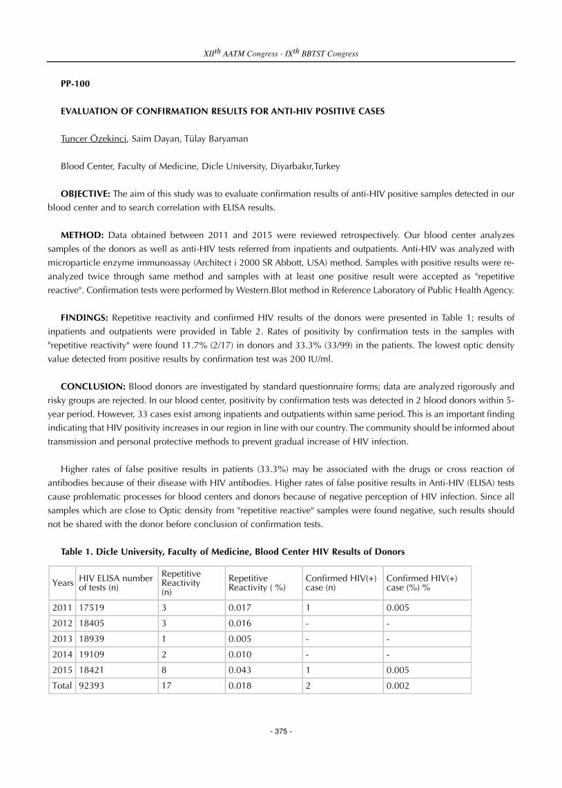
PP-100
EVALUATION OF CONFIRMATION RESULTS FOR ANTI-HIV POSITIVE CASES
Tuncer Özekinci, Saim Dayan, Tülay Baryaman
Blood Center, Faculty of Medicine, Dicle University, Diyarbakır,Turkey
OBJECTIVE: The aim of this study was to evaluate confirmation results of anti-HIV positive samples detected in our
blood center and to search correlation with ELISA results.
METHOD: Data obtained between 2011 and 2015 were reviewed retrospectively. Our blood center analyzes
samples of the donors as well as anti-HIV tests referred from inpatients and outpatients. Anti-HIV was analyzed with
microparticle enzyme immunoassay (Architect i 2000 SR Abbott, USA) method. Samples with positive results were re-
analyzed twice through same method and samples with at least one positive result were accepted as "repetitive
reactive". Confirmation tests were performed by Western.Blot method in Reference Laboratory of Public Health Agency.
FINDINGS: Repetitive reactivity and confirmed HIV results of the donors were presented in Table 1; results of
inpatients and outpatients were provided in Table 2. Rates of positivity by confirmation tests in the samples with
"repetitive reactivity" were found 11.7% (2/17) in donors and 33.3% (33/99) in the patients. The lowest optic density
value detected from positive results by confirmation test was 200 IU/ml.
CONCLUSION: Blood donors are investigated by standard questionnaire forms; data are analyzed rigorously and
risky groups are rejected. In our blood center, positivity by confirmation tests was detected in 2 blood donors within 5-
year period. However, 33 cases exist among inpatients and outpatients within same period. This is an important finding
indicating that HIV positivity increases in our region in line with our country. The community should be informed about
transmission and personal protective methods to prevent gradual increase of HIV infection.
Higher rates of false positive results in patients (33.3%) may be associated with the drugs or cross reaction of
antibodies because of their disease with HIV antibodies. Higher rates of false positive results in Anti-HIV (ELISA) tests
cause problematic processes for blood centers and donors because of negative perception of HIV infection. Since all
samples which are close to Optic density from "repetitive reactive" samples were found negative, such results should
not be shared with the donor before conclusion of confirmation tests.
Table 1. Dicle University, Faculty of Medicine, Blood Center HIV Results of Donors
XIIth AATM Congress - IXth BBTST Congress
- 375 -
Years HIV ELISA number of tests (n)
Repetitive Reactivity (n)
Repetitive Reactivity ( %)
Confirmed HIV(+) case (n)
Confirmed HIV(+) case (%) %
2011 17519 3 0.017 1 0.005
2012 18405 3 0.016 - -
2013 18939 1 0.005 - -
2014 19109 2 0.010 - -
2015 18421 8 0.043 1 0.005
Total 92393 17 0.018 2 0.002

Table 2. Dicle University, Faculty of Medicine, HIV Results of Inpatients and Outpatients
PP-101
RELATIONSHIP BETWEEN ABO/RH BLOOD GROUPS AND URINARY TRACT INFECTION
Özlem Genç1, Evrim Aksu2
1Dumlupınar University Faculty of Medicine, Department of Medical Microbiology2Dumlupınar University Kütahya Evliya Çelebi Training and Research Hospital, Department of Microbiology
Laboratory
AIM: The ABO blood group system is the most important blood type system in human blood transfusion. Found on
platelets, epithelium, and cells other than erythrocytes. There are many studies demonstrated a correlation between
blood group antigens and susceptibility diseases such as bacteria, fungi, parasites and viruses. Interactions of some
parasites, viruses and bacteria with human cells have been shown to depend on the presence of certain blood group
antigens, especially carbohydrate antigens. The objective of our study is to assess the susceptibility of ABO/Rh blood
groups to the isolated bacteria and fungi in urinary tract infections (UTI) in our hospital.
MATERIAL-METHODS: Study was designed retrospectively. Seven hundred fifty two patients with acute UTI and
225 patients with recurrent UTI totally 977 patients were enrolled in the study between the years 2014-2015. Urine
culture results and ABO/Rh groups were obtained from hospital documented system for each patient. Blood groups
were studied in these three years with gel centrifugation (Grifols, Wadiana, Spain) method. Urinary tract infection was
defined in the presence of a positive urine culture (>104 CFU/ml of a urinary pathogen). The identification tests were
performed on the automated BD Phoenix system (BD Diagnostics, Sparks, MD).
RESULTS: Demographic data of two patients group has been shown in Table-1. Both groups were similar in terms
of demographic data (mean age, gender and blood group distribution). The most common blood type was identified as
group A in both patients group, 42% and 46% respectively. The distribution of the blood groups in people with urinary
tract infections is similar to blood group ratio in Turkey. The distribution of different blood group and the main
bacterial/fungal pathogens isolated from 752 patients was determined as following, as shown in the Table-2. The most
common bacterial/fungal agents in the acute urinary tract infection among UTI group were Escherichia coli, Klebsilla
pneumoniae, Non-albicans Candida, Enterococcus faecalis, Enterococcus faecium respectively. E.faecalis was second
place after E.coli in the Rh (-) group. K.pneumoniae was detected in blood group AB and in group Rh (+) more than
the other groups. Rh (-) group was more sensitive to E.coli. K.oxytoca was more isolated in urine of patients with blood
group O than other ABO groups.
XIIth AATM Congress - IXth BBTST Congress
- 376 -
Years HIV ELISA number of tests (n
Repetitive Reactivity(n)
Repetitive Reactivity(%)
Confirmed HIV(+) case (n)
Confirmed HIV(+) case (%)
2011 8734 18 0.206 2 0.022
2012 10682 17 0.159 4 0.037
2013 13861 15 0.108 6 0.043
2014 13595 19 0.139 13 0.095
2015 12509 30 0.239 8 0.063
Total 59381 99 0.166 33 0.055

CONCLUSION: The distribution of blood groups in patient with acute and recurrent UTI’s was found similar to the
distribution of blood groups in our country. The most common bacterial/fungal agents in the urinary tract infection were
E.coli. Isolating of different bacteria and fungi from patients with acute urinary tract infection reveals that it is required
more studies to improve the relationship between blood groups and urinary tract infections.
Table-1. Demographic data of two patients group
Table-2. Different blood group and main bacterial/fungal pathogens in acute UTI group
XIIth AATM Congress - IXth BBTST Congress
- 377 -
Parameters Acute UTI group (n = 752)
Recurrent UTI group (n = 225)
Gender Male, n (%) Female, n (%)
294 (39%) 458 (60.9%)
94 (41.7%) 131 (58.2%)
Mean age + STD (years) 47.8 + 31.47 53.8 + 30.61
Group A 354 (42.5%) 104 (46.2%)
Group O 220 (29.2%) 65 (28.8%)
Group B 119 (15.8%) 34 (15.1%)
Group AB 59 (7.8%) 22 (9.7%)
Rh (+) group 675 (89.7%) 199 (88.4%)
Rh (-) group 77 (10.2%) 26 / (11.5%)
Bacteria Group A (n = 354)
Group O (n = 220)
Group B (n = 119)
Group AB (n = 59)
Rh (+) group (n = 675)
Rh (-) group (n = 77)
E.coli 150 (42.3%) 102 (46.3%) 57 (47.8%) 26 (44%) 293 (43.4) 42 (54.5%)
K.pneumoniae 43 (12.1%) 28 (12.7%) 15 (12.6%) 12 (20.3%) 93 (13.7%) 5 (6.4%)
Non- albicans Candida 39 (11%) 16 (7.2%) 11 (9.2 %) 6 (10.1%) 67 (9.9%) 5 (6.4%)
E.faecalis 30 (8.4%) 12 (5.4%) 6 (5%) 5 (8.4%) 43 (6.3%) 10 (12.9%)
C.albicans 21 (5.9%) 5 (2.2%) 4 (3.3%) 1 (1.6%) 30 (4.4%) 1 (1.2%)
E.faecium 16 (4.5%) 6 (2.7%) 5 (4.2%) 0 26 (3.8%) 1 (1.2%)
Two bacterial /fungal isolates 15 (4.2%) 10 (4.5%) 2 (1.6%) 3 (5%) 28 (4.1%) 2 (2.5%)
E.cloacae 13 (3.6%) 9 (4%) 1 (0.8%) 3 (5%) 23 (3.4%) 3 (3.8%)
K.oxytoca 5 (1.4%) 10 (4.5%) 1 (0.8%) 0 14 (2%) 2 (2.5%)
Others 22 (6.2%) 22 (10%) 17 (14.2%) 3(5%) 58 (8.5%) 8 (10.3%)

PP-102
THE PREVALENCE AND TREND OF TRANSFUSION TRANSMITTED INFECTIONS IN BLOOD DONORS IN
KOSOVO DURING 2006-2015
Bukurije Sami Zhubi1, Mimoza Agron Zhubi2, Hysen Avdyl Sadriu1, Idriz Selatin Merovci3, Ilirijane Sami Raça
Bunjaku4
1National Blood Transfusion Center of Kosovo in Prishtina2Institute of Biochemistry, Clinical University Center of Kosovo in Prishtina3Department of Pathopysiology, Faculty of Medicine, University of Prishtina4Clinic of Pulmology, Clinical University Center of Kosovo in Prishtina
The aim & purpose of the paper was the investigation of the prevalence and trends of Transfusion Transmitted
Infections (TTI) among blood donors in Kosovo.
MATERIALS & METHODS: TTI screening of 221900 donations between 2006 and 2015 at National Blood Centre
of Kosovo were analysed. The seroprevalence of hepatitis B virus (HBV), hepatitis C virus (HCV), human
immunodeficiency virus (HIV), and Syphilis infections were expressed in percentages for the entire study group as well
as groups by demographic characteristics and by type of blood donation.
RESULTS: From total number of tested blood donors, 2889 (2.124%, 95% CI 1.363–2.885%) that were donated in
Blood Centre of Pristina (BCP) and had serological evidence of infection of TTI and 2916 (3.798%, 95% CI 2.826–
4.770%) showed evidence of TTIs in blood donors at Regional Blood Centres (RBC). Total prevalence of TTI was 2.62%
(5805 Blood Donors).
The mean seroprevalence of HBV, HCV, HIV, and syphilis infections was 2.368% (95% CI 1.77–2.96%), 0.3% (95%
CI 0.17–0.42%), 0.0009% (95% CI -0.004–0.002%), and 0.05% (95% CI 0.028–0.07%) respectively. Trend analysis for
the total prevalence of TTIs in blood donors showed a significant decrease, from 4.269% to 1.513%. The trend
prevalence of HBV in blood donors in BCP (3.84 to 0.89%) and RBC (5.68 to 1.96%) during the period of time 2006-
2015 was represented by gradual decrease. Also the prevalence of HCV is relatively low with a tendency of gradual
reduction by year (BCP 0.55 to 0.049% respectively RBC 1.52 to 0.22%). HIV prevalence is very low only 3 cases
during 10 years investigation. The prevalence of TTIs is higher in men than in women (2.42% respectively 0.99%). The
TTIs prevalence was found to be lower in Voluntary Blood Donations compared to Family Blood Donations (1.4%
respectively 2.98%, p value =0.05).
CONCLUSION: Based on our results we can say that during 2006-2015, the prevalence of TTIs shows a significant
negative trend despite increasing number of voluntary blood donations. TTI prevalence was significantly lower among
blood donors at the blood transfusion centre than in donated blood at regional centres.
XIIth AATM Congress - IXth BBTST Congress
- 378 -
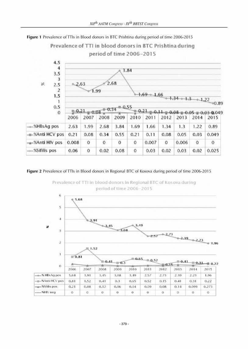
Figure 1 Prevalence of TTIs in Blood donors in BTC Prishtina during period of time 2006-2015
Figure 2 Prevalence of TTIs in Blood donors in Regional BTC of Kosova during period of time 2006-2015
XIIth AATM Congress - IXth BBTST Congress
- 379 -
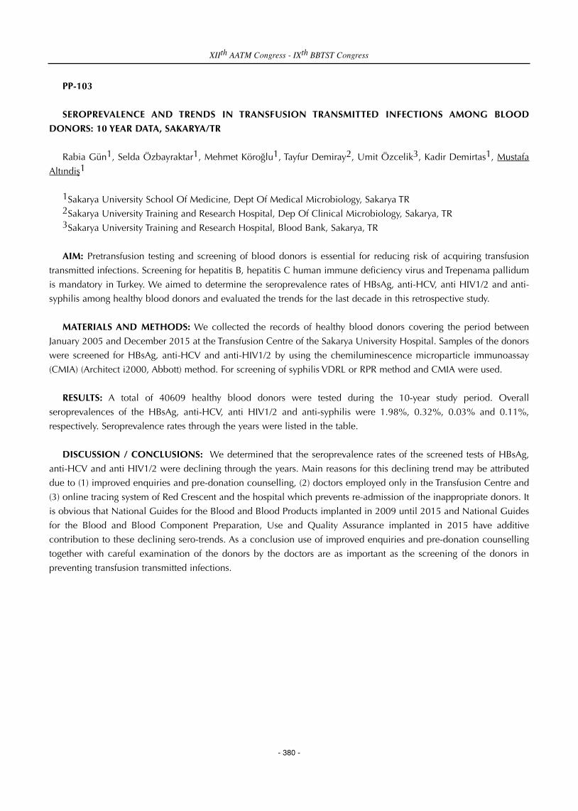
PP-103
SEROPREVALENCE AND TRENDS IN TRANSFUSION TRANSMITTED INFECTIONS AMONG BLOOD
DONORS: 10 YEAR DATA, SAKARYA/TR
Rabia Gün1, Selda Özbayraktar1, Mehmet Köroğlu1, Tayfur Demiray2, Umit Özcelik3, Kadir Demirtas1, Mustafa
Altındiş1
1Sakarya University School Of Medicine, Dept Of Medical Microbiology, Sakarya TR2Sakarya University Training and Research Hospital, Dep Of Clinical Microbiology, Sakarya, TR3Sakarya University Training and Research Hospital, Blood Bank, Sakarya, TR
AIM: Pretransfusion testing and screening of blood donors is essential for reducing risk of acquiring transfusion
transmitted infections. Screening for hepatitis B, hepatitis C human immune deficiency virus and Trepenama pallidum
is mandatory in Turkey. We aimed to determine the seroprevalence rates of HBsAg, anti-HCV, anti HIV1/2 and anti-
syphilis among healthy blood donors and evaluated the trends for the last decade in this retrospective study.
MATERIALS AND METHODS: We collected the records of healthy blood donors covering the period between
January 2005 and December 2015 at the Transfusion Centre of the Sakarya University Hospital. Samples of the donors
were screened for HBsAg, anti-HCV and anti-HIV1/2 by using the chemiluminescence microparticle immunoassay
(CMIA) (Architect i2000, Abbott) method. For screening of syphilis VDRL or RPR method and CMIA were used.
RESULTS: A total of 40609 healthy blood donors were tested during the 10-year study period. Overall
seroprevalences of the HBsAg, anti-HCV, anti HIV1/2 and anti-syphilis were 1.98%, 0.32%, 0.03% and 0.11%,
respectively. Seroprevalence rates through the years were listed in the table.
DISCUSSION / CONCLUSIONS: We determined that the seroprevalence rates of the screened tests of HBsAg,
anti-HCV and anti HIV1/2 were declining through the years. Main reasons for this declining trend may be attributed
due to (1) improved enquiries and pre-donation counselling, (2) doctors employed only in the Transfusion Centre and
(3) online tracing system of Red Crescent and the hospital which prevents re-admission of the inappropriate donors. It
is obvious that National Guides for the Blood and Blood Products implanted in 2009 until 2015 and National Guides
for the Blood and Blood Component Preparation, Use and Quality Assurance implanted in 2015 have additive
contribution to these declining sero-trends. As a conclusion use of improved enquiries and pre-donation counselling
together with careful examination of the donors by the doctors are as important as the screening of the donors in
preventing transfusion transmitted infections.
XIIth AATM Congress - IXth BBTST Congress
- 380 -

Seropositivity rates for the screening tests through the years
PP-104
SEROPREVELANCE OF HEPATITIS B, C AND HIV AMONG TALASEMIA PATIENTS IN TEPECIK EDUCATION
AND RESEARCH HOSPITAL
Şükran Köse, Ayşe Özkan, Hazal Albayrak, Didem Deştioğlu, Bengü Gireniz Tatar
Izmir Tepecik Eğitim ve Araştırma Hastanesi
Thalassemia patients need to get multiple blood transfussion during life time. That increases the risk of enfections
caused by blood transfussions, such as hepatitis B, hepatitis C and HIV.
According to datas from Turkish Hematologia Association, there are 1.300.000 thalasemia minor patients and 4.500
thalasemia major patients in our country. We had aimed to determinate seroprevalance of Hepatitis B,C and HIV on
adult and pediatric patients whom follow up our hematologia clinics.
METHOD: Our study consists of patients’ informations who applied our hematologia clinic between years 2007-
2016, includes age, sex, transfussion story and viral test results. We had studied retrospectively on that datas. We used
ELISA(Liaison, Diasorin, Italy) method to study HBsAg, AntiHBs, AntiHCv, AntiHIV.
RESULTS: We had detected 284 thalasemia major patients in our hospital. 141(%49.6) of them are female and
average age is 45. There is at least one blood transfussion story in their lifetime. 4 (%1.4) patients of them have possitive
XIIth AATM Congress - IXth BBTST Congress
- 381 -

HbsAg result and 3 (%1.05) patients of them have possitive AntiHCV results, and only 1 (%0.3) patients of them have
possitive AntiHIV results. Negative AntiHbs test results determined on 58 (%20.4) patients. We couldn’t add
informations of AntiHBc IgG possitivity.
CONCLUSSION: Thalassemia patients are in high risk groups for infections of hepatitis B,C and HIV. According to
our study, we detected that there is no difference of seroprevalance between thalassemia patients and healthy
community. On the result of this study; We aim to take attentions of health-worker to vaccination for AntiHbs negative
thalasemia patient, and importance of close examinations.
1.Information of talasemia patients
PP-105
WORK HEALTH AND SAFETY RISK ASSESSMENT FOR DIFFERENT OCCUPATIONAL GROUPS WORKING IN
BLOOD TRANSFUSION CENTER, FACULTY OF MERAM MEDICINE NECMETTIN ERBAKAN UNIVERSITY
Mehmet Ay1, Mehmet Ali Canbulat2, Bahadır Feyzioğlu2, Mahmut Baykan2
1Blood Transfusion Center, Meram Medicine of Faculty, Necmettin Erbakan University, Konya, Turkey2Department of Microbiology, Meram Medicine of Faculty, Necmettin Erbakan University, Konya, Turkey
OBJECTIVE: Determination of occupational health and safety risks that faced in the work environment of employees
in the Blood Transfusion Center in the Faculty of Meram Medicine, Necmettin Erbakan University.
METHODS: Risk analysis results have been expressed with graphics. Employees in the Blood Transfusion Center
have been assessed in terms of four different vocational categories and interprofessional risk differences have been
revealed. Risk analysis have been made by occupational health and safety specialist.
RESULTS: Employees are at risk with most blood and blood products. Hazards will caused without distinction
interprofessional to employees of physical risk factors is lesser. Encounter with communication and psychosocial risks
of employees is demonstrated varies between occupational groups. Ergonomic risks is make up different health and
safety risks are according to the quality of the work performed.
CONCLUSIONS: Identification of the risks faced by workers in work life and the implementation of preventive
actions will has been decrease significantly occupational accidents,occupational diseases and deaths.. Measures to be
XIIth AATM Congress - IXth BBTST Congress
- 382 -
GENDER n(%)
Female 141
Male 143
SEROLOGICAL MARKER HBsAg positive 4 (1.4)
Antı-HBs positive 226 (79.5)
Antı- HBs negative 58 (20.5)
Anti- HCV 3 (1.05)
Antı-HIV 1 (0.3)

taken against the identified risk significantly will have been reduced the effects of an accident or occurring
occupational disease.
PP-106
HISTORICAL DEVELOPMENT OF BLOOD BANKING IN MILITARY MEDICINE IN TURKEY
Engin Kurt1, Gürol Emekdaş2
1Gülhane Military Medical Faculty, Department of Medical Ethics and Medical History, Ankara, Turkey2Biruni University School of Medicine, Department of Medical Microbiology; Blood Centers of Turkey and
Transfusion Association, Istanbul, Turkey
Blood banking, which is one of the most important elements of the healthcare system, has developed through
history and reached the present state. “Blood”, which is especially required during wars, has held a significant place
during the development process.
Within the scope of blood banking in the world, it is stated that a blood transfusion center has been established for
the first time 1921, under the name of “Blood Transfusion Service” in London. Then, the small scale enterprises
established in the United States of America (USA) in 1928 have left their places to large and organized structures named
“Blood Banks” in 1938. It is stated that the first blood bank has been opened in 1937 at the Chicago Cook County
Hospital in USA.
On this subject, İsmail Kâzım Gürkan, one of the intern medical lieutenants of Gülhane in Turkey, has published a
brochure regarding blood donation for the first time. Prof. Dr. İsmail Kâzım Gürkan, who has become a faculty member
at the Istanbul University Cerrahpaşa School of Medicine in the following years, has emphasized the need to collect
blood and store it in order to be used when needed, with respect to blood banking.
In the letter, dated December 6, 1948, sent to the Turkish Red Crescent Society by the Ankara School of Medicine
Deanship, in line with the highly important decision and request of the Military School of Medicine Board of Professors;
it is stated, “We are delighted to notify that Blood Donation Center and Blood Bank is established at the Military School
of Medicine in Ankara, with the authorization of the Ministry of Defense Health Affairs Department Presidency, in order
to meet the blood needs of military and civilian patients in peace and the injured during war” and it is emphasized that
blood banking has started before December 1948 in military medicine. In this respect, Mr. Bedii Nuri Şehsuvaroğlu
also states that a Blood Bank was available at the Cebeci Hospital since 1945.
For the development of blood banking in Gülhane, Dr. Kazım KURTAR has been sent to the USA by the Ministry of
National Defense for two years, between April 07, 1956 and May 08, 1958, in order to advance his knowledge and
experience.
Blood supply at the Gülhane Military Medical Academy School of Medicine Blood Bank has been conducted in
accordance with the “Continual Instructions for Emergency and Collective Blood Supply” issued by the Garrison
Command and the Gülhane Military Medical Academy Command’s “Continual Instructions to Meet Blood Demands”.
Blood has been collected routinely, within a certain order, by the Blood Bank blood collection team considering the
“TT 8-255 Instructions for the Operation Methods of Military Blood Centers” issued on December 24, 1966, and the
principles of the World Health Organization in this respect.
XIIth AATM Congress - IXth BBTST Congress
- 383 -

The Gülhane Blood Center has been last restructured in 1967, by the resolution of the board of professors dated
August 17, 1967 and numbered 33-67, and continues its activities as the Gülhane Periodical Regional Blood Center as
per the Blood and Blood Products Law, published in the Official Journal dated 02.05.2007 and numbered 26510,
within the periods defined by the Ministry of Health.
PP-107
RELATIONSHIP BETWEEN TURKISH ARMED FORCES AND TURKISH RED CRESCENT IN TURKISH BLOOD
BANKING
Engin Kurt1, Gürol Emekdaş2, Armağan AKSOY3
1Gülhane Military Medical Faculty, Department of Medical Ethics and Medical History, Ankara, Turkey2Biruni University School of Medicine, Department of Medical Microbiology; Blood Centers of Turkey and
Transfusion Association, Istanbul, Turkey3General Directorship of Red Crescent Blood Services, Ankara, Turkey
It is known that the contribution of Soldiers serving in the Turkish Armed Forces (TAF) holds an important place
during the historical process in the supply of blood that is of vital significance for human life.
In the Commission Report on Blood Transfusion of the Red Crescent society dated 1948, it has been stated that the
best donors would be strong and sturdy individuals of 20-40 years of age and individuals of such characteristics could
be found as many as needed among soldiers, with regards to donor supply. It is also expressed in the same report that
it is also possible to give such individuals a leave for 15-20 days in order to take 250-500 gr of blood.
It is quite apparent that the TAF has received great support from the Turkish Red Crescent during the efforts of the
TAF to establish the first blood bank. Any regulations had not been issued regarding the establishment and maintenance
of an organization that will arrange for the blood transfusion activities during that period. At the same time, it is also
advised that it was not quite possible to find blood that would be adequate for the poor patients because there was no
general heal insurance. Transfusion could only be applied to wealthy individuals in return for a large amounts as 20
TL on the average (according to the monetary value in 1940). Therefore, a donation of 5000 TL has been made, so as
to be under the control of the floating capital of the Military Medical Academy, in order to pay for the cost of blood to
be collected from paid donors so that blood could also be given to poor patients. In addition, in the report that the
Turkish Red Crescent Society General Central Board has presented at the 1949 congress, it has been stated that 5000
TL has been donated during 1948 to the Blood Bank and Blood Transfusion Center established at the Military Medical
Academy in Ankara, in order to be spent when needed. Then, in the report presented during the 1950 congress, it has
been stated that 2500 TL more has been allocated to the Blood Bank and Blood Collection Center, and in the 1951
congress, it has been stated that 5000 TL more has been allocated, in addition to the earlier aids.
When the statistical data obtained regarding blood donation and donor supply are examined, it is observed that the
major supporters of the Red Crescent during the initial years have been the rank and file (Table-1). Meanwhile, it is
stated that almost all of the donors in 1957 were soldiers. Although soldiers constitute the majority of the donors during
the initial years, it is stated that 95% of the collected blood was used by civilians and 5% by soldiers.
XIIth AATM Congress - IXth BBTST Congress
- 384 -
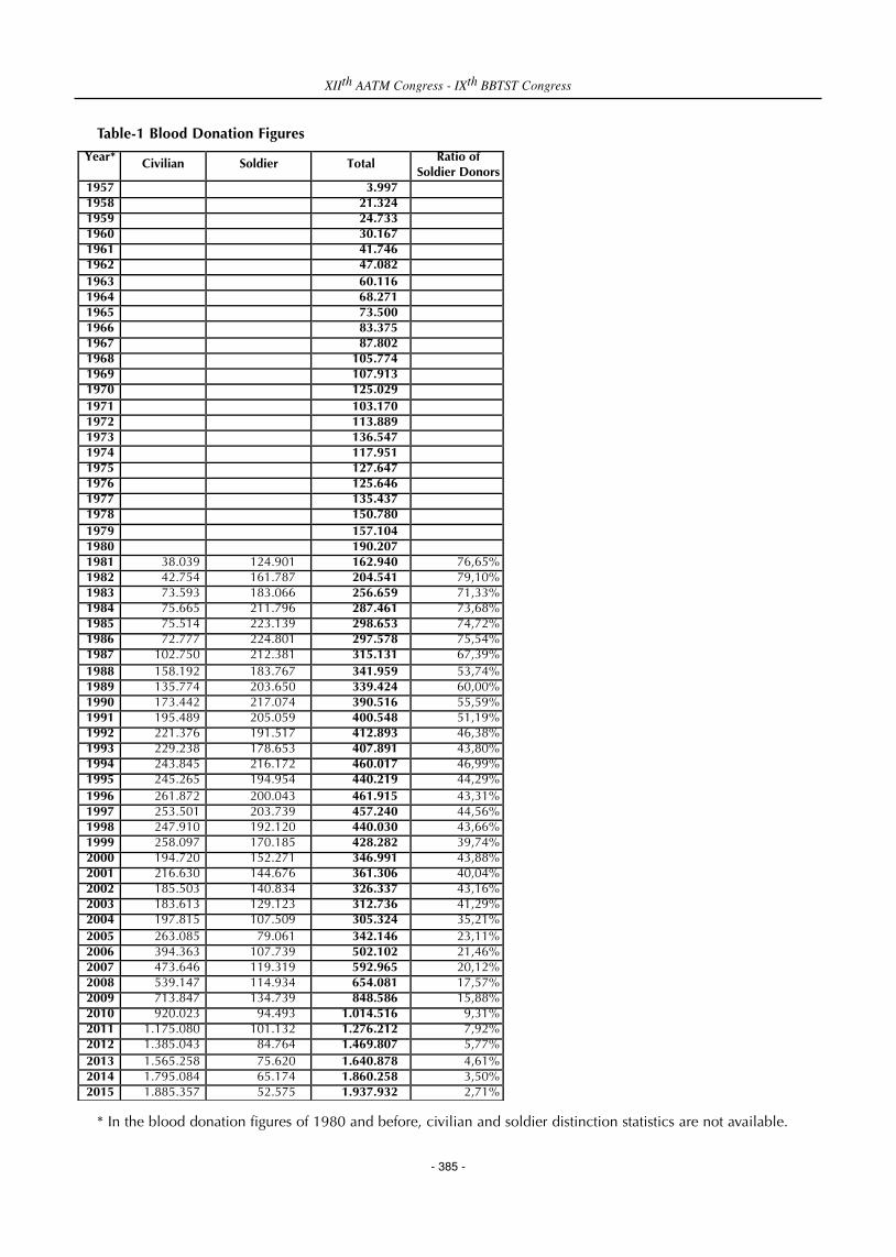
Table-1 Blood Donation Figures
* In the blood donation figures of 1980 and before, civilian and soldier distinction statistics are not available.
XIIth AATM Congress - IXth BBTST Congress
- 385 -
Year*
Civilian Soldier Total Ratio of Soldier Donors
1957 3.997 1958 21.324 1959 24.733 1960 30.167 1961 41.746 1962 47.082 1963 60.116 1964 68.271 1965 73.500 1966 83.375 1967 87.802 1968 105.774 1969 107.913 1970 125.029 1971 103.170 1972 113.889 1973 136.547 1974 117.951 1975 127.647 1976 125.646 1977 135.437 1978 150.780 1979 157.104 1980 190.207 1981 38.039 124.901 162.940 76,65% 1982 42.754 161.787 204.541 79,10% 1983 73.593 183.066 256.659 71,33% 1984 75.665 211.796 287.461 73,68% 1985 75.514 223.139 298.653 74,72% 1986 72.777 224.801 297.578 75,54% 1987 102.750 212.381 315.131 67,39% 1988 158.192 183.767 341.959 53,74% 1989 135.774 203.650 339.424 60,00% 1990 173.442 217.074 390.516 55,59% 1991 195.489 205.059 400.548 51,19% 1992 221.376 191.517 412.893 46,38% 1993 229.238 178.653 407.891 43,80% 1994 243.845 216.172 460.017 46,99% 1995 245.265 194.954 440.219 44,29% 1996 261.872 200.043 461.915 43,31% 1997 253.501 203.739 457.240 44,56% 1998 247.910 192.120 440.030 43,66% 1999 258.097 170.185 428.282 39,74% 2000 194.720 152.271 346.991 43,88% 2001 216.630 144.676 361.306 40,04% 2002 185.503 140.834 326.337 43,16% 2003 183.613 129.123 312.736 41,29% 2004 197.815 107.509 305.324 35,21% 2005 263.085 79.061 342.146 23,11% 2006 394.363 107.739 502.102 21,46% 2007 473.646 119.319 592.965 20,12% 2008 539.147 114.934 654.081 17,57% 2009 713.847 134.739 848.586 15,88% 2010 920.023 94.493 1.014.516 9,31% 2011 1.175.080 101.132 1.276.212 7,92% 2012 1.385.043 84.764 1.469.807 5,77% 2013 1.565.258 75.620 1.640.878 4,61% 2014 1.795.084 65.174 1.860.258 3,50% 2015 1.885.357 52.575 1.937.932 2,71%

In the TAF, which was one of the major supporters of the Red Crescent with respect to blood donation and donor
supply during the initial years, it is observed that the Esteemed Chief of General Staff General Cemal TURAL,
Commander of Naval Forces Admiral Necdet URAN, National Security Board General Secretary General Kemalettin
GÖKAKIN, Commander of the 1st Army General Memduh TAĞMAÇ, Commander of the 2nd Army Lieutenant General
Nazmi KARAKOÇ, Commander of the 3rd Army Fikret ESEN and the other commanders have supported these initiatives
by making written statements within the scope of the “Red Crescent Blood Program Activities” in 1967, as well as
getting involved in activities aimed to increase the motivations of the soldiers under their command.
PP-108
TRANSFUSION PRACTICES AND DEVELOPMENT IN TURKEY
Engin Kurt1, Gürol Emekdaş2
1Gülhane Military Medical Faculty, Department of Medical Ethics and Medical History, Ankara, Turkey2Biruni University School of Medicine, Department of Medical Microbiology; Blood Centers of Turkey and
Transfusion Association, Istanbul, Turkey
Blood has been mystical and fascinating for mankind since the antique ages. In 1615, Libavius has described a
transfusion method that is similar to the present method. Meanwhile, positive developments have started after William
Harvey has discovered blood circulation in 1916 and incorporated it into medicine.
There is no other treatment that is able to treat the symptoms caused by acute blood loss, which leads among
transfusion indications, better than blood transfusion. Therefore, transfusion of blood or blood products holds a
significant place in our day. Mehmet Yardımcı, Fethi Tezok and Halit Kamgözen, who have presented the “Report on
the Fifth International Transfusion Congress” in 1955, have stated that blood transfusion is a matter that concerns all
world countries, and it has been expressed in the papers of all countries who participated in the congress that blood
has lifesaving and curative effects in peace and especially during war.
Nihat Artunkal expresses that while blood transfusion in the war field in works written prior to World War I is merely
a delusion and was impossible to apply in practice, it is understood from the written works that transfusion has been
accomplished with success by the French Army during the aforementioned war. Meanwhile, during World War II, the
British and the American armies have sent the bloods they had previously prepared in their countries to the war
surgeons at the farthest point of the front during the war and ensured transfusion.
Meanwhile, transfusion activities in Turkey have been started by Ord. Prof. Dr. Burhanettin Toker in 1921 in
Cerrahpaşa, and by Operator Dr. Ömer Vasfi Aybar in 1923 at the Ankara Numune Hospital. Toker states that he has
made transfusion with the “Oehlecker” instrument in 1927, and subsequently the American-made “Persi” instrument.
Blood transfusion, which was used for treatment purposes during World War I, has been applied on many cases for
preventive porpoises (treatment preventing the development of diseases) and numerous times as preoperative in cases
that were not suitable for surgery during World War II. It is expressed by Burhaneddin Osman Tugan that blood
transfusion has started to be applied at Gülhane as of 1935, with the transfusion instrument (Blood Transfusion
Instrument of Dr. Jouvelet and Henyr) donated by the Turkish Ambassador Mr. Fethi in London.
In the 1947 Instructions of the Gülhane Military Medical Academy, under the sub-heading “Military Coursed to be
XIIth AATM Congress - IXth BBTST Congress
- 386 -

taught at the Military Medical Academy” of heading XXIV, it has been stated that blood and plasma transfusions will
be taught within the “War Surgery” course, in various health troops as revival measures and practices.
In the “Commission Report on Blood Transfusion” of the Red Crescent Society dated 1948, it has been stated that
the number of donors would be increased over time by announcements and propagandas to be made to the public, it
will be possible to collect blood by recording the data of the donors and store it at (+)5-6 degrees at transfusion centers,
and use it when needed or ensure direct transfusion by sending to the bed of the patient when needed.
Today, the transfusion practices are conducted in line with the “Blood and Blood Products Law”, published in the
Official Journal dated 02.05.2007 and numbered 26510, the “Blood and Blood Products Regulation” that has been
prepared as based on this law and published in the Official Journal dated b04.12.2008 and numbered 27074, and the
techniques set forth in the National Blood Guide.
XIIth AATM Congress - IXth BBTST Congress
- 387 -

INDEX
Acıpayam, Can PP-033
Açıkel, Cengizhan OPP-10
Adanur, Şükran PP-081
Aggarwal, Geet PP-023
Agha, Muhammad Akbar PP-013, PP-028
Agrawal, Amit PP-029, PP-096
Ahmad, Maqsood PP-072
Ahsan, Hassan Raza PP-066
Ak Yıldırım, Hayriye PP-075
Akar, Serhan OPP-18
Akdemir, Cihangir PP-030
Akduman Alaşehir, Elçin OPP-09
Akel, Nazif PP-067
Akıncı, Sema PP-058, PP-059, PP-086, PP-088, PP-089
Aksoy, Armağan PP-024, PP-031, PP-040, PP-045, PP-061, PP-064, PP-098, OPP-16
Aksu, Evrim PP-101
Aksu, Yalçın PP-031, PP-098
Alam, Maqbool PP-094
Alanoğlu, Güçhan OPP-09
Albayrak, Hazal PP-104
Ali, Muhammad Hammad PP-066
Altındiş, Mustafa PP-103
Altunay, Hüsnü PP-020, PP-021, PP-022
Altunöz, Elif PP-065
Aluç, Emine PP-042
Arı, Alpay OPP-09
Arı, Nejmı PP-076
Arı, Nuray PP-085
Arınç, Bülent PP-010, PP-048, PP-050, OPP-05
Arora, Bhavna PP-071
Arora, Dinesh PP-023
Arslan, Önder PP-097
Arslan, Zeliha PP-007, PP-057
XIIth AATM Congress - IXth BBTST Congress
- 389 -

Asghar, Faheem PP-066
Aşkaroğulları, Nilgün OPP-17
Atalay, Figen OPP-09
Atasayar, Fatma PP-020, PP-021, PP-022
Ataş, Erkan PP-047
Ataş, Şerife PP-003
Avcı, Duygu Nurdan PP-004, PP-081
Avcı, İlknur PP-052, PP-057, PP-076
Avcı, İsmail Yaşar PP-005, PP-009, OPP-10
Ay, Mehmet PP-105
Aydoğdu, Adnan PP-003, PP-008
Ayhan, Fahri Yüce OPP-17
Ayverdıata, Dursun PP-076
Ayyıldız, Abdullah PP-056, PP-083
Bakanay, Öztürk Şule Mine PP-058, PP-059, PP-086, PP-088, PP-089, OPP-08
Balcı, Pervin Özlem PP-085
Baryaman, Tülay PP-100
Baskan, Bakı PP-076
Bayhan, Turan PP-009
Baykan, Mahmut PP-105
Bayrak, Tülin PP-074, PP-075, PP-084
Bedi, Neha OPP-11
Bektöre, Bayhan PP-005, PP-009, PP-034, PP-035, PP-036, PP-037, PP-038, PP-062, PP-068,
PP-069
Birbudak, Satı PP-079
Birinci, İlhan PP-031, PP-040, PP-042, PP-055, PP-098
Bolca, Zühal PP-065
Bozkurt, Selen OPP-15
Bozkurt, Tuğçe OPP-09
Budak, Göksel OPP-06
Büyük, Hüseyin PP-065
Can, Funda Koçal PP-085
Canbulat, Mehmet Ali PP-105
XIIth AATM Congress - IXth BBTST Congress
- 390 -
Canpolat, Şenay PP-024
Ceran, Funda PP-004, PP-007, PP-052, PP-057, PP-076, PP-081, PP-091
Chakraborty, Tamojit PP-027
Chakravarty, Ishita OPP-07
Cığır, Abdurrahman Cihat PP-001
Çağlak, Şükrü PP-061
Çağlar, Hatice PP-085
Çağlayan, Serin Derya OPP-17
Çakas, Mustafa OPP-06
Çaylak, Sevil PP-074
Çetinkaya, Fuat OPP-20
Çetinkaya, Rıza Aytaç PP-005, PP-009, PP-034, PP-035, PP-036, PP-037, PP-038, PP-062, PP-068,
PP-069, OPP-10
Çevik, Esin PP-083
Çiçek, Yasemin PP-078
Çiçekli, Filiz PP-001
Çiftçi, Yasemin PP-007, PP-057
Çoker, Hüseyin PP-061
Çorbacı, Ebru PP-042
Dağdaş, Simten PP-004, PP-007, PP-076, PP-081, PP-091
Dara, Ravi C. PP-023
Dayan, Saim PP-006, PP-082, PP-099, PP-100
Demırbaş, Meltem PP-076
Demir, Feti PP-008
Demir, Figen PP-019
Demiray, Tayfur PP-067, PP-103
Demirbaş, Meltem PP-004, PP-007, PP-057
Demirel, Aslıhan PP-083, OPP-09
Demirel, Özlem Unay PP-002, OPP-09
Demi̇rel, Kadri̇ PP-031, PP-040, PP-098
Demirtaş, Kadir PP-103
Dernek, Damla PP-024
Deştioğlu, Didem PP-104
XIIth AATM Congress - IXth BBTST Congress
- 391 -
Deveci, Burak PP-020, PP-021, PP-022
Dilek, İmdat PP-058, PP-059, PP-086, PP-088, PP-089, OPP-08
Direkel, Şahin PP-030, PP-051, PP-053, OPP-03
Dişiaçık, Güler PP-042
Divarcı, Aslıhan PP-022
Doğan, Erdoğan PP-001
Doğan, Yasin PP-001
Doğan, Kaya Sibel PP-010, PP-048, PP-050, OPP-05
Dönmez, Levent PP-020, PP-021, PP-022
Dubey, Ruchi PP-027
Efe, Şirin OPP-09
Eker, İbrahim PP-005, PP-009, OPP-10
Eksan, Hülya PP-065, PP-093
Eldemir, Sibel PP-040
Emecen, Ömer PP-030, PP-051, OPP-03
Emekdaş, Gürol PP-106, PP-107, PP-108
Engin, Aynur PP-001
Erberk, Çiğdem PP-006
Erdoğan, Hatice PP-054, PP-080
Eren, Canan PP-049, PP-060, OPP-09
Eren, Meltem OPP-09
Eren, Özcan PP-041
Erengül, Emine PP-010, PP-048, PP-050
Erengül, Adem PP-010, PP-048
Ergene, Ömer Buğra OPP-20
Ersan, Gürsel PP-017, PP-087
Ersoy, Hakan PP-097
Ertaş, Züleyha PP-007, PP-057
Eser, Bülent PP-033, OPP-06
Eser, Burcu PP-009
Faisal, Salman PP-066
Falay, Mesude PP-004
Feyzioğlu, Bahadır PP-105
XIIth AATM Congress - IXth BBTST Congress
- 392 -
Gareayaghi, Nesrin PP-018
Genç, Özlem PP-101
Gerrard, Rebecca Anne Margaret OPP-13
Gevrek, Tuğba PP-010, PP-048, PP-050, OPP-05
Ghani, Eijaz PP-094
Gireniz, Tatar Bengü PP-104
Gözükara, Alper PP-047
Gupta, Aman PP-071
Gupta, Anurag PP-027
Gupta, Manoj OPP-07
Gupta, R S PP-046
Güçlü, İlknur PP-019
Gül, Tunay PP-054, PP-080
Güler Murat PP-045
Güler, Nejla Cebeci PP-051
Güler, Pınar PP-007, PP-052, PP-057
Güllüoğlu, Mehmet PP-024, PP-031, PP-040, PP-042, PP-045, PP-055, PP-061, PP-064, PP-098,
OPP-16
Gün, Rabia PP-067, PP-103
Günçıkan, Mustafa Nuri OPP-16
Gündem, Nadire Seval PP-047
Güngör, Serdar PP-011, PP-012, PP-063, PP-092
Gürbüz, Ali OPP-09
Gürlük, Nesrin PP-018
Gürsel, Orhan PP-005, PP-009, OPP-10
Güvel, Hayri PP-011, PP-012, PP-063, PP-092
Hafızoğlu, Nurettin PP-024, PP-031, PP-040, PP-042, PP-045, PP-055, PP-061, PP-064, PP-098,
OPP-16
Hançerli, Yusuf PP-038
Hussain, Agha Babaur PP-043, PP-066
Hussain, Sadam PP-095
Innah, Susheela Jacob PP-070
Ipek, Haluk PP-076
XIIth AATM Congress - IXth BBTST Congress
- 393 -

Islam, Md Ashadul PP-014, PP-073
Izdes, Seval OPP-08
İçel, Osman PP-067
İğnak, Seyda PP-002, PP-032
İnci, Fadime PP-067
İris, Nur Efe PP-056, PP-083
Joshi, Sanmukh R. OPP-02
Kaçmaz, Gamze PP-030
Kader, Çiğdem OPP-09
Kalender, Metin PP-024
Kaplan, Onur OPP-09
Kaplan, Şükran PP-082
Karabulut, Songul PP-050
Karabürk, Tülay PP-010, PP-048, PP-050, OPP-05
Karadoğan, İhsan PP-020, PP-021, PP-022
Karagöz, Emel Uzunoğlu OPP-03
Karahacıoğlu, İsmet Gürsel PP-031, PP-040, PP-098, OPP-16
Karakaş, Erdal PP-001
Karakukcu, Musa PP-033
Karim, Farheen PP-016
Karpuzoğlu, Osman Eren PP-078
Kaushik, Satish PP-027
Kaya, Erşan OPP-09
Kaya, Hamit PP-010, PP-048, PP-050, OPP-05
Kayma, Cihan Meriç PP-001
Kayoz, Tarık PP-021
Kesen, Esra OPP-09, OPP-19
Keskin, Canan Cemile PP-098
Kesti, Saliha PP-051
Khan, Saleem PP-013
Khetarpal, Anil PP-071
Kır, Büşra PP-053, OPP-03
Kiliç, Miyase PP-077
XIIth AATM Congress - IXth BBTST Congress
- 394 -

Koç, Esat PP-007, PP-057
Koç, Savaş PP-024
Koçtekin, Belkıs OPP-15
Koçulu, Safiye PP-083
Koçyi̇ğit, Oral PP-078
Konya, Gülay OPP-09
Korkmaz, Gülten PP-004
Koşan, Erdoğan PP-031, PP-098
Koşer, Murat PP-084
Koyuncu, İsmail PP-020
Köroğlu, Mehmet PP-067, PP-103
Köse, Şükran PP-017, PP-087, PP-104
Köse, Timur PP-087
Kuku, İrfan PP-067
Kula Atik, Tuğba PP-005, PP-009, PP-034, PP-035, PP-036, PP-037, PP-038, PP-062, PP-068,
PP-069
Kumbasar, Abdulbaki PP-019
Kundakcı, Ayça Hande PP-064
Kurt, Engin PP-106, PP-107, PP-108
Kurtoğlu, Erdal OPP-15
Kuruçay, Fügen PP-055
Küçükkaya, Reyhan Diz PP-056
Liv, Fatma PP-017, PP-087
Magvansuren, Tserendejid OPP-12
Mahmood, Asad PP-094
Mangwana, Sadhana OPP-11
Maral, Senem PP-058, PP-059, PP-086, PP-088, PP-089, OPP-08
Mathur, Ankit PP-029
Mehta, Nidhi OPP-07
Mendapara, Kinjal OPP-02
Meral, Ahmet PP-007, PP-057, PP-076, PP-091
Meriç Yılmaz, Fatma PP-042
Merovci, Idriz Selatin PP-102
XIIth AATM Congress - IXth BBTST Congress
- 395 -

Mısırlıoğlu, Naile PP-020, PP-021, PP-022
Miah, Sonia Shormin PP-014, PP-073
Moiz, Bushra PP-016
Mughal, Nouman PP-095
Mukherjee, Somnath PP-044
Namal, Esat PP-083
Namjil, Erdenebayar OPP-12
Nepal, Bipin PP-013, PP-028, PP-095
Niazi Saif Ullah, Khan PP-094
Noorulamin, Muhammad PP-043, PP-066
Nusrat, Nadeem PP-028
Ordu, Aylin PP-056
Orhan, Fatma PP-007, PP-057, PP-076, PP-091
Özcan, Alper PP-033, OPP-06
Özdemir, Mehmet Akıf PP-033
Özer, Yesim PP-097
Özbayraktar, Selda PP-067, PP-103
Özcelik, Umit PP-103
Özdamar, Melda OPP-20
Özdemir Aydek, Sultan PP-007, PP-057, PP-091
Özekinci, Tuncer PP-006, PP-076, PP-082, PP-099, PP-100
Özet, Gülsüm PP-004, PP-007, PP-052, PP-057, PP-076, PP-081, PP-091
Özgül, Mustafa PP-067
Öziz, Gökhan PP-034, PP-062
Özkan, Ayşe PP-104
Özmen, Aslihan PP-010, PP-048, PP-050, OPP-05
Öztürk, Seda OPP-19
Öztürk Şahin, Nihal OPP-19
Özüm, Selda PP-001
Özveren, Gülşen PP-085
Özyurt, Ertan PP-079
Pathak, Amardeep PP-025
Pathak, Sangeeta PP-027
XIIth AATM Congress - IXth BBTST Congress
- 396 -

Patıroglu, Turkan PP-033
Patlar, Reyhan Demir OPP-18, OPP-19
Pekel, Aysel OPP-10
Pelit, Nil Banu PP-078, OPP-09, OPP-18, OPP-19
Premkumar, Rani PP-026
Qureshi, Ambrina OPP-04
Qureshi, Hina OPP-04
Raça Bunjaku Ilirijane, Sami PP-102
Rafi Aboobacker, Mohamed PP-070
Raghuwanshi, Babita PP-044
Rahmani, Tariq Hamid PP-072
Raja, Asad Mehmood PP-043
Raja, Ghazala Kaukab PP-043
Raza, Naila OPP-04
Reyhan, Melek PP-076
Saatçioğlu, Hatice Nedret PP-052, PP-057, PP-076, PP-081, PP-091
Sabıtogulları, Asım PP-076, PP-091
Sadriu, Hysen Avdyl PP-102
Saeed, Muhammad PP-072
Sağdur, Levent PP-024, PP-045, PP-061, PP-064
Sağlam, Filiz PP-002, PP-032
Salman Koçtekin, Belkıs PP-041
Saqlain, Raja Muhammad PP-043
Saraç, Mahmut PP-067
Saraçlı, Mehmet Ali PP-009
Savaşçı, Ümit PP-005, OPP-09
Sawant, Rajesh OPP-07
Saygan, Mehmet Bakır PP-040
Sen, Fadime PP-097
Sengel, Özcan PP-011, PP-012, PP-063, PP-092
Serçebay, Uğur PP-020
Serdar, Muhittin OPP-09
Seven, Süleyman PP-040
XIIth AATM Congress - IXth BBTST Congress
- 397 -

Sevimligül, Gülgün PP-001
Shaiq, Pakeeza Arzoo PP-043
Sheladiya, Ankita OPP-02
Sılay, Kamile PP-058, PP-059
Singh, Harprit PP-046
Singh, Surjit PP-027
Sood, Nitin PP-023
Soytürk, Yusuf PP-002, PP-032, OPP-09
Sönmez, Ferit OPP-16
Subba, Sonu H. PP-044
Sucu, Rana PP-018
Şanli, Kamuran PP-077, OPP-09
Şen, İlker Murat PP-034, PP-035
Şencan, Mehmet PP-001
Şenel, Arif PP-010, PP-048, PP-050, OPP-05
Şener, Aslı Gamze PP-063,PP-092
Şener, Gülsen PP-074, PP-075, PP-084, OPP-09
Şentürk Yıkılmaz, Aysun PP-088, PP-089
Şimşek, Berksan PP-005, PP-009, PP-034, PP-035, PP-036, PP-037
Tanveer, Rida PP-043
Telli, Semra PP-007, PP-057
Temel, Yeşim PP-022
Terzi, Hatice PP-001
Tiwari, Aseem K. PP-023
Topcuoglu, Pervin PP-097
Tuncer, Orhan PP-076
Türk, Hakan PP-075, PP-084
Türkoğlu, Salih OPP-20
Türkoğlu, Tansel PP-078
Uçar, Mehmet Ali PP-007, PP-091
Uğur Kurtoğlu, Ayşegül OPP-15
Uğurlu, Emine PP-079
Ulugdogan, Meral PP-010, PP-048, PP-050
XIIth AATM Congress - IXth BBTST Congress
- 398 -

Uluhan, Ramazan PP-065, PP-093
Unay, Demirel Özlem PP-032
Urlu, Selin Merih PP-004, PP-081
Uylar, Nur PP-010, PP-048, PP-050, OPP-05
Uysal, Yeşim OPP-19
Uzun, Berrin PP-011, PP-012, PP-063, PP-092, OPP-17
Uzun, Mustafa Eren PP-024
Ülger, Derviş PP-061, PP-064
Ülger, Seda Tezcan OPP-03
Ün, Dilek PP-031
Ünal, Ekrem PP-033, OPP-06
Ünlü, Aytekin PP-005, PP-009, OPP-10
Ünlü, Sevgi PP-042
Vardar, İsmail OPP-20
Veske, Haydar PP-021
Woodfield, David Graeme PP-039, OPP-01
Yalçın, Demet PP-002, PP-032
Yay, Mehmet PP-033, OPP-06
Yazıcı, Ertuğrul PP-005, PP-009
Yenilmez, Ercan PP-005, PP-009, PP-034, PP-035, PP-036, PP-037, PP-038, PP-062, PP-068,
PP-069, OPP-10
Yıkılmaz, Aysun PP-058, PP-059
Yıldırımkaya, Cansu PP-024
Yıldız, Mustafa OPP-15
Yılmaz, Fatma Meriç PP-024, PP-031, PP-040, PP-055, PP-061, PP-064, PP-098, OPP-16
Yılmaz, Sebahattin PP-005, PP-009, OPP-10
Yılmaz, Soner PP-005, PP-009, OPP-10
Yılmaz, Tülay PP-004, PP-007, PP-057
Yılmazer, Tanju PP-017
Yiğit, Emine PP-022
Yikilmaz, Aysun Şentürk PP-086
Yolaçan, Ayşe PP-078
Yorgancı, Rabia PP-079
XIIth AATM Congress - IXth BBTST Congress
- 399 -

Yurtal, Gülizar PP-007, P057
Yurtseven, Rabia PP-074, PP-084
Yüce, Hüseyin PP-010, PP-048, PP-050
Yücel, Kamile PP-003, PP-008
Zeybek, Nazif PP-009
Zhubi, Bukurije Sami PP-102
Zhubi, Mimoza Agron PP-102
XIIth AATM Congress - IXth BBTST Congress
- 400 -