Measuring bleeding in platelet transfusion trials - Oxford ...
45
1 The challenges of measuring bleeding outcomes in clinical trials of platelet transfusions Short title: Measuring bleeding in platelet transfusion trials Authors: LJ Estcourt 1 , N Heddle 2 , R Kaufman 3 , J McCullough 4 , MF Murphy 1 , S Slichter 5 , EM Wood 6 , SJ Stanworth 1 , On behalf of the BEST (Biomedical Excellence for Safer Transfusion) Collaborative 1. NHS Blood and Transplant/Oxford University Hospitals NHS Trust, and the NIHR Biomedical Research Centre, John Radcliffe Hospital, Oxford, UK. 2. Department of Medicine, McMaster University, Hamilton, ON Canada 3. Harvard Medical School Blood Bank, Amory 260 Brigham and Women's Hospital, 75 Francis Street, Boston, MA 02115 4. University of Minnesota, 420 Delaware Street SE, Minneapolis, MN 55455 5. Puget Sound Blood Center and University of Washington School of Medicine, Seattle, Washington, US 6. Australian Red Cross Blood Service and Department of Clinical Haematology, Monash University, Melbourne, Australia Correspondence: Dr Lise J Estcourt, NHS Blood and Transplant, Level 2, John Radcliffe Hospital, Oxford, OX3 9BQ, United Kingdom; [email protected] Funding This study was supported by the BEST Collaborative Conflict of Interest The authors declare that they have no conflicts of interest relevant to the manuscript submitted to Transfusion. Several authors are PIs on included trials Word count Abstract: 249 Text: 4,452 Tables and figures: 6 Tables 1 Figure On-line appendices : 4 References: 46
-
Upload
khangminh22 -
Category
Documents
-
view
0 -
download
0
Transcript of Measuring bleeding in platelet transfusion trials - Oxford ...

1
The challenges of measuring bleeding outcomes in clinical trials of platelet
transfusions
Short title: Measuring bleeding in platelet transfusion trials
Authors: LJ Estcourt1, N Heddle2, R Kaufman3, J McCullough4, MF Murphy1, S Slichter5, EM Wood6, SJ Stanworth1, On
behalf of the BEST (Biomedical Excellence for Safer Transfusion) Collaborative
1. NHS Blood and Transplant/Oxford University Hospitals NHS Trust, and the NIHR Biomedical Research Centre, John Radcliffe Hospital, Oxford, UK.
2. Department of Medicine, McMaster University, Hamilton, ON Canada 3. Harvard Medical School Blood Bank, Amory 260 Brigham and Women's Hospital, 75 Francis Street, Boston, MA
02115 4. University of Minnesota, 420 Delaware Street SE, Minneapolis, MN 55455 5. Puget Sound Blood Center and University of Washington School of Medicine, Seattle, Washington, US 6. Australian Red Cross Blood Service and Department of Clinical Haematology, Monash University, Melbourne,
Australia
Correspondence: Dr Lise J Estcourt, NHS Blood and Transplant, Level 2, John Radcliffe Hospital, Oxford, OX3 9BQ, United
Kingdom; [email protected]
Funding This study was supported by the BEST Collaborative
Conflict of Interest
The authors declare that they have no conflicts of interest relevant to the manuscript submitted to Transfusion. Several
authors are PIs on included trials
Word count Abstract: 249 Text: 4,452
Tables and figures: 6 Tables 1 Figure
On-line appendices : 4
References: 46
2
Abstract
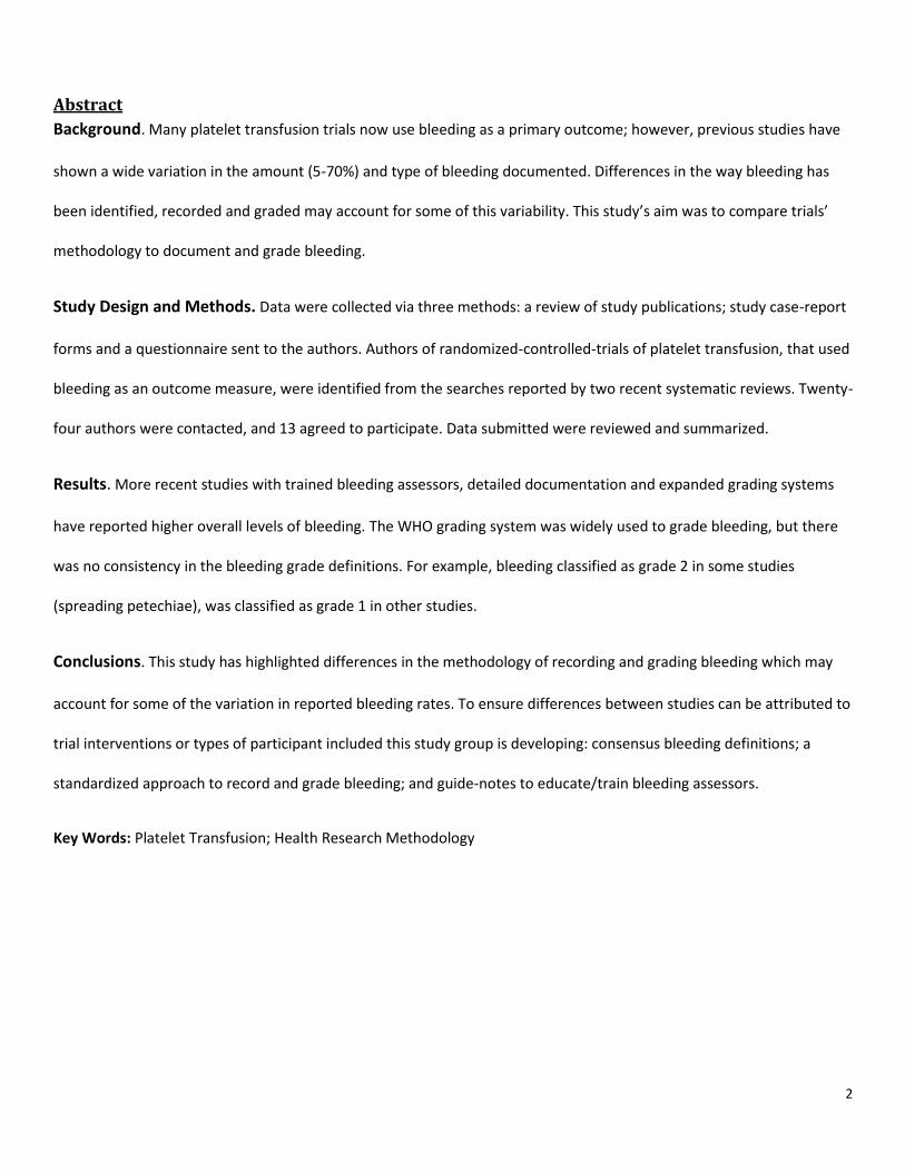
Background. Many platelet transfusion trials now use bleeding as a primary outcome; however, previous studies have
shown a wide variation in the amount (5-70%) and type of bleeding documented. Differences in the way bleeding has
been identified, recorded and graded may account for some of this variability. This study’s aim was to compare trials’
methodology to document and grade bleeding.
Study Design and Methods. Data were collected via three methods: a review of study publications; study case-report
forms and a questionnaire sent to the authors. Authors of randomized-controlled-trials of platelet transfusion, that used
bleeding as an outcome measure, were identified from the searches reported by two recent systematic reviews. Twenty-
four authors were contacted, and 13 agreed to participate. Data submitted were reviewed and summarized.
Results. More recent studies with trained bleeding assessors, detailed documentation and expanded grading systems
have reported higher overall levels of bleeding. The WHO grading system was widely used to grade bleeding, but there
was no consistency in the bleeding grade definitions. For example, bleeding classified as grade 2 in some studies
(spreading petechiae), was classified as grade 1 in other studies.
Conclusions. This study has highlighted differences in the methodology of recording and grading bleeding which may
account for some of the variation in reported bleeding rates. To ensure differences between studies can be attributed to
trial interventions or types of participant included this study group is developing: consensus bleeding definitions; a
standardized approach to record and grade bleeding; and guide-notes to educate/train bleeding assessors.
Key Words: Platelet Transfusion; Health Research Methodology

3
Introduction
Many recent clinical trials of platelet transfusion therapy have used bleeding as a primary endpoint1-9. There
are two important considerations when bleeding is used as an outcome measure: the documentation of
signs and symptoms of bleeding; and, the translation of this information into a clinically meaningful score or
grade. This is fundamental to the robustness of results reported in these trials, and valid comparisons
between studies can only be drawn if similar outcomes are being compared. Therefore, if bleeding is to be
used as a main outcome measure, it is important that it is defined and documented in a consistent and
standardized way.
However, the assessment of bleeding involves an element of subjectivity, and the literature indicates wide
variability in the methods by which bleeding has been assessed and documented in clinical trials of platelet
transfusion. Taken together, these factors may be responsible, in part, for differences in the reported
baseline bleeding rates between studies, which have varied in randomized-controlled trials from 5%10 to
70%5. Different trials have taken different approaches in an attempt to minimize this bias, such as the use of
a standardized tool for assessing bleeding6; training of staff3; or grading of bleeding4-6 11.
Although variability in the documentation and grading of bleeding is known to exist, there has not been a
systematic summary of the approaches used to document and grade bleeding in platelet transfusion trials.
Such a summary would help the transfusion medicine community to understand current practices and guide
recommendations for a consistent and standardized approach when bleeding is used as an outcome in
platelet transfusion trials. The ultimate goal of this project, undertaken by members of the Biomedical
Excellence for Safer Transfusion (BEST) Collaborative, was to develop recommendations and/or a
standardized case report form (CRF) to assist primary investigators when bleeding is used as an outcome in
future clinical trials and to facilitate comparison of outcomes between studies for reviewers and readers.
These recommendations/CRF should minimize some of the potential problems associated with the reliability

4
of this important clinical endpoint. As the first step to achieve this goal, Information was sought from the
lead investigators of clinical trials of platelet transfusions, with the specific aims to:
1) describe the methods used in the trial protocol to document bleeding; and,
2) describe the methods for grading bleeding, including the assignment of bleeding grades and validation of
the process.
Materials and Methods
Identification of studies
To identify clinical platelet studies we used the search strategies (Appendix 1) for a recently updated
Cochrane review of prophylactic platelet transfusions12 and that reported by the BEST Collaborative for their
systematic assessment of quality of reporting for platelet transfusion studies13. The search was limited to
randomized controlled trials (RCTs) in humans that reported bleeding as a primary or secondary outcome.
The primary authors of all identified trials were contacted and were invited to participate in the study by
sharing copies of their bleeding assessment tool case report forms (CRFs) together with any protocol,
standard operating procedure, or guidance notes describing the procedures for bleeding assessment and
documentation. The primary author was contacted up to three times. If the primary author did not respond
a second author was also contacted up to three times.
Data collection
Data for analysis were taken from three sources. Publications associated with each study were reviewed.
The CRFs and any guidance notes were reviewed to assess how the study teams documented signs,
symptoms and site of bleeding, severity (grade) of bleeding, and treatment of any bleeding episodes. This
was supplemented by information from a questionnaire (Appendix 2 – online only) developed by the BEST
Collaborative Project Group and piloted by three of the study authors. The questionnaire was circulated to
all authors who had agreed to participate and had provided copies of their CRFs. The questionnaire asked
about how study data were collected, the methods used for standardization and training, and how bleeding
was graded.

5
Summary data elements were identified by the investigators, and one investigator (LE) reviewed all
submitted material including the primary study manuscripts and captured the relevant data summarizing it
in table format. The information from each study was then sent to the study author for verification of
accuracy and completion of missing data.
Analysis
The overall analysis was descriptive with results presented as percentages for categorical data. Missing data
were reflected by variation in denominators. The study team reviewed all data summaries and developed
recommendations via consensus.
Results
Overview
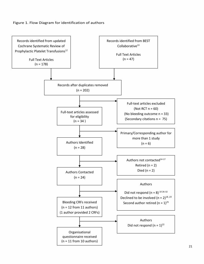
The results of the search strategy are shown in Figure 1. Four of the authors, who had published studies in
the 1970s and 80s, had retired or died14-17. Fifteen primary authors responded, of these, 13 authors1-8 20-24
agreed to participate. Nine secondary authors10 25-32 were contacted, one responded, but had just retired25.
Two sets of two authors3 6 21 24 worked in close collaboration and used the same CRFs and one author33 34 had
two different CRFs for separate studies.
Baseline Characteristics of identified studies (Tables 1 & 2)
Of the 11 authors who returned CRFs (Figure 1), eight were the primary authors of multi-center randomized
controlled trials and three of these studies were multi-national. Studies were based in Australia, Canada,
France, Germany, Italy, Netherlands, Norway, Spain, Sweden, UK and USA. Study size ranged from 82 to
1351 participants. The majority of the CRFs (9/12) and questionnaires (9/11) were from studies performed
from 2000 onwards.
Methods of Recording Bleeding (Table A Appendix 3)
Based on the 11 questionnaires returned, the bleeding assessment consisted of at least a formalized
bleeding assessment and a clinical examination of the patient. In all but two cases (9/11) this was done on a
daily basis. One study performed the assessment twice a day8, and the other study23 performed the

6
assessment twice daily on the day of any platelet transfusion and once daily on the day following the
platelet transfusion. In 71-3 5 6 9 34 of the 11 studies the bleeding assessment was performed at the same time
each day.
In 6 of the 11 studies2 3 5 6 8 9 33, the bleeding assessment was performed by trained research nurses or other
research investigators at least part of the time. In 4 out of 11 studies the bleeding assessor was reported to
be blinded to treatment allocation3 5 23 35. In only one study was the effectiveness of this blinding assessed3,
where research nurses were asked to detect study units from a panel of 10 platelet components (a mixture
of study and conventional units) and then complete a questionnaire.
Site and Severity of bleeding (Tables 3 & 4)
Information on the CRFs in all studies was collected using a mixture of check boxes (yes/no) and open-ended
questions (which may be more difficult to analyze). In all of the studies conducted during the last decade
(9/12)2 3 5-9 22 23 33 36, the CRFs consisted of tick box or Yes/No responses although (2/9)7 23 had a significant
number (> 25%) of open-ended questions.
There was significant variability among CRFs in the amount of detail related to the site and severity of
bleeding (Table 3). Definitions appeared to vary most between studies in relation to bleeding into the skin
and subcutaneous tissue. Pathology37 and dermatology38 textbooks differentiate between petechiae,
purpura and ecchymoses according to the size of the hemorrhage into the skin (petechiae < 2mm, purpura
2mm to 10mm and ecchymoses > 10mm) and all are forms of hematoma. However, the definitions of these
variables in many of the studies differed from each other and did not conform with textbook definitions. The
term purpura was often used to define any type of skin bleeding larger than petechiae. Three studies
distinguished between purpura that were less than or greater than one inch3 5 34. One study distinguished
between hematomas that were less than or greater than 1cm23. Two studies2 6 distinguished between
purpura and ecchymoses with the distinction between the two being 2cm6 or 1 inch2.
All studies provided suggested definitions for different severity scores of bleeding at some anatomical sites,
but the details varied between studies (Table 3). For example, all studies asked about bleeding from the

7
mouth, but three did not ask about the duration of bleeding4 22 33. Two studies asked about red blood cells
(RBCs) in CSF (cerebro-spinal fluid) only seen on microscopy5 34, and one study5 asked about RBCs in other
body cavity fluid on microscopic examination. Ten studies2 3 5-9 22 23 33 34 assessed bleeding severity by the
need for RBC transfusion to treat bleeding above routine requirements (83.3%; 10/12). Fifty per cent (6/12)
of the studies3 5 6 23 34 36 required information on whether bleeding was associated with hemodynamic
instability. Twenty-five per cent (3/12) of the studies documented whether bleeding was associated with a
significant fall in hemoglobin (Hb)5 7 34. Two studies2 34 also defined severity of bleeding by need for
interventions other than RBC transfusion e.g. other medications or procedures (Table 4).
None of the platelet threshold studies1 4 22 (Table 2) classified skin bleeding as clinically significant bleeding,
and all these studies had a lower rate of bleeding than the platelet dose studies2 5 7 and studies assessing
pathogen-reduction technologies3 33.
Methods of achieving consistency in the assessment of bleeding
Eight2-6 8 23 34 of the 11 studies reported the training of bleeding outcome assessors, prior to commencement
of the study. Four3 6 8 23 of the studies provided further training once the study had started, but only two3 6 of
these studies indicated that training occurred on a regular basis. Six of the studies2-6 8 reported more than
one trained bleeding assessor at each site and four2 3 5 6 of these mentioned that trained assessors were
present at weekends or that there was a back-up assessor to cover sick leave.
Seven2-6 23 34 of the studies reported providing guidance notes to the bleeding assessors in addition to the
actual CRFs. However there was great variability in the level of detail provided in these notes for the five
studies that shared this information. Two6 23 provided practical information on how to complete the CRFs
and two6 34 included ‘easy to read’ definitions of the different types of bleeding. For two studies2 3 the notes
were expanded versions of the World Health Organization (WHO) grading criteria.
Duplicate bleeding assessments were performed in three1 6 8 of the studies. This was performed in 50% of
cases for one study1, approximately 10% of cases for the second study6 and only in cases of severe or life-
threatening bleeding for the third study8. Whether the second bleeding assessor was blinded from the

8
results of the first bleeding assessor was not asked in the questionnaire. In all three cases the results of
these assessments were fed back to the assessors, as a form of on-going education. The results of two of
these studies6 8 9 have not yet been published. Two studies reported other specific methods of decreasing
inter-observer variability. These included dummy bleeding assessment scenarios during training days6 and
clinical investigator oversight and final review of all daily hemostatic assessment forms3. (In this study, all
CRFs were monitored against primary source documents by Contract Research Organization (CRO) research
monitors; discrepant data were queried and reviewed by the clinical investigator at each site; and the
primary clinical investigator at each site rendered the final assessment of daily bleeding grade.)
All of the studies2 3 5 that have reported bleeding rates over 50% had specially trained research nurses.
Grading of Bleeding
Six of the 11 authors used the WHO grading system39 in their most recent platelet transfusion studies2 5 6 21 23
34. Three of the authors reported using the Rebulla system4 or a variant of this scale7 22. One study used a
modified scoring system that included elements of the WHO and Rebulla classifications8. One study1 used a
system devised by Ajani et al 40, but this has not been used in more recent platelet transfusion studies.
Different grading systems can lead to different baseline levels of bleeding. This can be seen in data from the
SPRINT study (Table 5). Patients were graded daily using the WHO grading system but the trained study
personnel also reported any bleeding as a side-effect using the Common Toxicity Criteria for Adverse Events
(CTCAE) system. Although the overall incidence of bleeding was similar between the two grading systems41,
which reflects no difference in the number of bleeds being reported using the two systems, there were
significant differences in how they were graded. There were fewer grade 2, 3 or 4 bleeds using the CTCAE
system, which may be explained by the observation that occult blood in the urine could be graded as WHO
grade 2 and that, apart from bleeding into the skin, to classify bleeding as CTCAE grade 2 it required an
intervention of some sort.
WHO grading system 39 (Table B Appendix 3)

9
The WHO system is now used in the majority of studies. The original grading system39 classified petechiae as
grade 1; mild blood loss as grade 2; gross blood loss as grade 3 and debilitating blood loss as grade 4. None
of the terms were defined further. All of the authors who defined significant bleeding classified it as grade 2
or above. All of the authors who defined life-threatening bleeding classified it as grade 4. But, all of the
authors who were using the WHO grading system had refined it from the original formulation39 in different
ways. This led to variability in the way the same bleed would be graded between different studies.
Some of these differences could lead to the same bleed being categorized as grade 2 in one study and grade
1 or no bleeding in another study. For example, 3/6 studies2 3 23 defined occult blood >1+ in the urine as
WHO grade 2 bleeding. Three of six studies3 6 23 defined grade 2 vaginal bleeding as bleeding saturating more
than 2 pads per day, whereas the other 3 studies2 5 34 defined abnormal vaginal bleeding more than spotting
as grade 2 bleeding. Three of six studies5 6 34 defined epistaxis that lasted more than 30 minutes as grade 2
bleeding whereas the other 50%2 3 23 only regarded bleeding that lasted an hour as grade 2 bleeding. There
was also variability in the way that petechiae, purpura, hematomas and ecchymoses were defined between
studies. Two of the studies defined diffuse petechiae as WHO grade 2 bleeding3 6. Retinal bleeding that did
not cause visual compromise was classified as grade 1 in one study3 and grade 2 in the other five studies2 5 6
23 34. Finally, the definition of hemodynamic compromise varied between studies: two studies5 34 categorized
a drop in systolic or diastolic blood pressure of 30mmHg as a grade 3 bleed; whereas, two further studies3 6
regarded this as a grade 4 bleed.
RBC transfusion requirements
Ten of the 11 authors1-6 8 9 22 23 33 34 used RBC requirements to grade bleeding, and four of these authors1 4 8 9 34
reported a standardized transfusion policy operating in the study (only one34 of the six authors who used the
WHO grading system reported a standardized transfusion policy). Without a standardized policy the same
type of bleed could be graded as grade 2 or 3 depending on the transfusion decision by the treating
physician/research center.

10
Conversion of bleeding data into a bleeding grade (Table C Appendix 3)
The majority of studies assigned a bleeding grade manually (9/11). Three studies (3/11) assigned the grade
using a validated computer algorithm5 6 34 and two5 6 of these used this single system of grading. In one of
these studies6 validation of the algorithm was performed by comparing 100 patients via both manual and
computer methods, the other study5 did not report the method of validation. The third study34 performed
both manual and computer algorithm methods of grading bleeding. Of those that graded bleeding manually
only two studies4 23 did not check the grading using a second person or method. All of the studies that
checked their results via a second method or person resolved any disagreement by adjudication. This
adjudication could have involved: debate between study personnel (four studies1 8 33 34); referral to the Chief
Investigator/Principal Investigator (two studies3 8); or referral to an independent third party/parties (two
studies2 7).
Discussion
The methods used to assess, document and grade bleeding in platelet transfusion trials display considerable
heterogeneity, yet the outcome of bleeding is increasingly defined as a primary outcome. The variability
extended from the nature of the staff acting as bleeding assessors (e.g. trained and blinded research
nurses), type of bleeding data recorded, through to the different methods for assigning bleeding grade (e.g.
computer algorithm vs. manual). A key question is to what extent these different methodological
approaches contribute to the variability in the frequencies of bleeding reported between studies. Although
no formal analysis of the effect of the methodological differences on the reported bleeding outcomes were
possible in this study, some general trends in the variations of bleeding outcomes can be identified, which
have implications for researchers and their readers.
Variability in bleeding frequencies between studies was most apparent for minor and moderate bleeds
(WHO grade 1 & 2 bleeding) (Table 2). This raises questions about how different research groups have
11
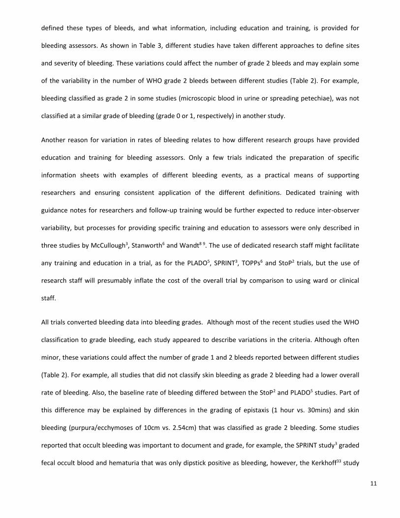
defined these types of bleeds, and what information, including education and training, is provided for
bleeding assessors. As shown in Table 3, different studies have taken different approaches to define sites
and severity of bleeding. These variations could affect the number of grade 2 bleeds and may explain some
of the variability in the number of WHO grade 2 bleeds between different studies (Table 2). For example,
bleeding classified as grade 2 in some studies (microscopic blood in urine or spreading petechiae), was not
classified at a similar grade of bleeding (grade 0 or 1, respectively) in another study.
Another reason for variation in rates of bleeding relates to how different research groups have provided
education and training for bleeding assessors. Only a few trials indicated the preparation of specific
information sheets with examples of different bleeding events, as a practical means of supporting
researchers and ensuring consistent application of the different definitions. Dedicated training with
guidance notes for researchers and follow-up training would be further expected to reduce inter-observer
variability, but processes for providing specific training and education to assessors were only described in
three studies by McCullough3, Stanworth6 and Wandt8 9. The use of dedicated research staff might facilitate
any training and education in a trial, as for the PLADO5, SPRINT3, TOPPs6 and StoP2 trials, but the use of
research staff will presumably inflate the cost of the overall trial by comparison to using ward or clinical
staff.
All trials converted bleeding data into bleeding grades. Although most of the recent studies used the WHO
classification to grade bleeding, each study appeared to describe variations in the criteria. Although often
minor, these variations could affect the number of grade 1 and 2 bleeds reported between different studies
(Table 2). For example, all studies that did not classify skin bleeding as grade 2 bleeding had a lower overall
rate of bleeding. Also, the baseline rate of bleeding differed between the StoP2 and PLADO5 studies. Part of
this difference may be explained by differences in the grading of epistaxis (1 hour vs. 30mins) and skin
bleeding (purpura/ecchymoses of 10cm vs. 2.54cm) that was classified as grade 2 bleeding. Some studies
reported that occult bleeding was important to document and grade, for example, the SPRINT study3 graded
fecal occult blood and hematuria that was only dipstick positive as bleeding, however, the Kerkhoff33 study
12
did not. In the SPRINT3 study the most common type of bleeding was genitourinary (32.1%), whereas other
studies that did not document microscopic blood loss had a much lower rate of genitourinary bleeding. For
example, in studies that included skin bleeding as grade 2 bleeding muco-cutaneous bleeding was the most
common (Wandt8 9), whereas gastrointestinal blood loss was the most common type in those that excluded
skin bleeding (Rebulla4;Tinmouth7).
Most studies reported using a manual method of bleeding adjudication for assigning grade, but few data are
available about how this works in practice and experience from the recent StoP trial2 suggests real
difficulties achieving consensus. Computer algorithms may provide a more consistent method of assigning
bleeding grade, but without details on validation of these methods, it is unclear exactly whether computer
algorithms deliver more accurate grading than manual methods, although reproducibility is likely to be
higher than manual methods.
Only a few studies included the need for specific interventions as a guide to severity. The use of
interventions to manage bleeding is one of the first decisions to be made by clinicians when faced by a
patient with bleeding. However, a difficulty with using this approach might be the desire to minimize
interventions in these patients who are often profoundly neutropenic (e.g. endoscopy/colonoscopy), and
the inevitable variation in access to interventions and which may also change with time e.g. use of
recombinant factor VIIa where the evidence for overall lack of effectiveness has become clearer42.
Implications for researchers
The analysis of methodologies in this study has raised a number of points which should help trialists, in the
future, at the study design stage, when training staff and when reporting results. Whilst it seems obvious
that study protocols should provide clear definitions of all relevant bleeds, wider agreement and consensus
on the definitions of bleeding events and which bleeding events are clinically meaningful would help
improve the consistency of reporting, and support direct comparison of results between studies. Some of
these key areas for agreement could be fairly easily considered by the international community. For

13
example, some protocols suggest that all petechiae are WHO grade 1, and clinically insignificant; but it
seems likely that many clinical hematologists when faced with a patient with spreading or generalized
petechiae and severe thrombocytopenia would treat the patient with a platelet transfusion. It also seems
difficult to suggest that nose bleeding up to 1 hour is of no importance or concern, particularly to the
patient. However, some studies always grade this type of bleeding as WHO 1 and therefore clinically
insignificant.
Some studies have collected information on occult bleeding. But if these types of bleeds are considered
important to collect, which is perhaps open to discussion, then specific questions on the CRFs need to
capture this information, to minimize any risk of under-reporting (this has been seen in other settings, for
example the systematic under-reporting of acute lung injury43).
These issues also raise the question as to whose perception should be considered if standardization is
attempted: the physicians, the patients, or both? There has been little reported work evaluating how
patients feel about the different types of bleeding, particularly more minor or moderate muco-cutaneous
bleeding. These examples indicate the need for research to explore and understand differences between the
clinician and the patient’s perception of bleeding events. Patient perception of bleeding is especially
important in those studies that rely on patients recording bleeding outcomes in the outpatient setting, away
from medical oversight.
The methods of data collection need to be considered in a trial protocol, including the role of bleeding
assessors. The method of training of staff or researchers undertaking bleeding assessments has been poorly
documented in many trials. Assessment of bleeding will always be subjective, to a degree, and therefore
ways to improve consistency within and between studies is crucial, and trial protocols should describe the
training programs and strategies taken to support consistency in the recording of bleeding. Achieving
blinding in platelet transfusion studies can be challenging, unless the platelet components are identical, due
to problems with blinding the medical staff caring for the patient. The only blinding that is likely to be readily
achievable is that of the bleeding assessor, if they are independent from the care of the patient. However,

14
the bleeding assessor is also usually the person collecting the clinical and laboratory transfusion data, and
therefore studies comparing prophylactic versus therapeutic policies or platelet transfusion thresholds
would require the data for bleeding and transfusion to be collected by separate individuals to maintain
blinding. This would have major resource implications.
The methodology for defining and assigning bleeding grades is important, and any differences from previous
trials should be indicated. The WHO score was developed as a tool for adverse event reporting in cancer
patients, and although widely used, it has never been validated for specific use in clinical trials of
transfusion. However, its widespread use and acceptance may provide a degree of post-hoc validation.
Indeed, one study has shown good agreement (90%; 136/151 days) between self-assessment and medical
grading of bleeding44, if the presence or absence of microscopic hematuria was excluded. If it is to continue
to serve as the international consensus scoring tool for this purpose agreement on the exact format of the
WHO grading score should be considered by the international research community, as this would remove
one level of uncertainty when comparing results between studies. For example, consensus on the need to
collect data on occult types of bleeding or these could be identified separately e.g. grade 1Occult or grade
2Occult. An alternative would be to develop and use a new type of bleeding scale which has recently been
reported, although with any new scale it would take time to assess its clinical acceptability45. At a minimum,
if bleeding is a main outcome measure46, all trials of platelet transfusion should include specific information
on bleeding grade definitions and educational/training support.
Limitations of this study
Although this study aimed to review all randomized-controlled platelet transfusion trials, it was limited by
only a proportion of authors responding to a request for the CRFs or completion of the questionnaire,
despite repeated requests. Despite this caveat, information was obtained from the majority of the major
researchers in this field and therefore this study represents a comprehensive picture of the current
methodologies.

15
The analysis in this study was descriptive; and this study was not designed to quantify risk factors
responsible for variability in bleeding. Estimates of how much the differences in grading could affect
bleeding rates, could only be undertaken if individual patient data were available, and recorded in sufficient
detail for different grading systems to be compared.
Summary
This review has identified important differences in how the recording of signs and symptoms of bleeding is
documented and how bleeding grades are assigned between studies. Consensus on optimal methodology
could include: standardized case report forms that all investigators could use to record signs and symptoms
of bleeding as well as interventions to treat bleeding, and a grading scale that is clinically relevant and can
be reproducibly applied (Table 6). These steps form part of an on-going program of research by this project
group.

16
Acknowledgements
Collaborators
The authors would like to thank all study authors who supplied CRFs and completed the questionnaire: Heckman, K
(Heckman study); Rebulla, P (IPTAS study & Trigger study); Corash, L (Lozano study & SPRINT study); Fletcher, D;
Goodrich, R (MIRACLE study); Wandt H (Nuerenberg study); Corson, J; Slichter, S (PLADO study); McClelland, S; Powter,
G; Stanworth, S (PPiP study & TOPPs study); Kerkhoffs, JL; van de Watering, L (PrePAReS study & TriPlate study); Barty,
R; Heddle, N (SToP study); Tinmouth, A (Tinmouth study)
All authors who supplied CRFs: Zumberg, M (Zumberg study).
L. Corash for provision of unpublished material from SPRINT study.
Other members of the BEST Project group: Hervig, T; Lozano, M; Tinmouth, A; van de Watering, L; Williamson, L who
provided input on the design of the study.
Ruth Strachan for her assistance with translating CRFs.
Authorship
Contribution: All authors contributed to the writing of the manuscript; L.J.E, N.H and S.J.S. designed the research; L.J.E
collected, analyzed and interpreted the data; J.M., N.H., S.J.S, and S.S. provided study CRFs and completed the
questionnaire.
Conflict of Interest: The authors declare that they have no conflicts of interest relevant to the manuscript submitted to
Transfusion. Several authors are PIs on included trials.
A complete list of the members of the BEST Collaborative Project Group and the BEST Collaborative appears as a data
supplement (Appendix 2) to the online version of this article.
Correspondence: Dr Lise J Estcourt, NHS Blood and Transplant, Level 2, John Radcliffe Hospital, Oxford, OX3 9BQ, United
Kingdom; [email protected]

17
Funding This study was supported by the BEST Collaborative
Appendices 1 to 4: (Online only)

18
References
1. Heckman KD, Weiner GJ, Davis CS, Strauss RG, Jones MP, Burns CP. Randomized study of prophylactic platelet transfusion threshold during induction therapy for adult acute leukemia: 10,000/microL versus 20,000/microL. J Clin Oncol 1997;15(3):1143-9.
2. Heddle N, Cook R, Tinmouth A, Kouroukis C, Hervig T, Klapper E, et al. A randomized controlled trial comparing standard and low dose strategies for transfusion of platelets (SToP) to patients with thrombocytopenia. Blood 2009;113(7):1564-73.
3. McCullough J. Therapeutic efficacy and safety of platelets treated with a photochemical process for pathogen inactivation: the SPRINT Trial. Blood 2004;104(5):1534-41.
4. Rebulla P, Finazzi G, Marangoni F, Avvisati G, Gugliotta L, Tognoni G, et al. The threshold for prophylactic platelet transfusions in adults with acute myeloid leukemia. Gruppo Italiano Malattie Ematologiche Maligne dell'Adulto. N Engl J Med 1997;337(26):1870-5.
5. Slichter SJ, Kaufman RM, Assmann SF, McCullough J, Triulzi DJ, Strauss RG, et al. Dose of prophylactic platelet transfusions and prevention of hemorrhage. N Engl J Med 2010;362(7):600-13.
6. Stanworth SJ, Dyer C, Choo L, Bakrania L, Copplestone A, Llewelyn C, et al. Do all patients with hematologic malignancies and severe thrombocytopenia need prophylactic platelet transfusions? Background, rationale, and design of a clinical trial (trial of platelet prophylaxis) to assess the effectiveness of prophylactic platelet transfusions. Transfus Med Rev 2010;24(3):163-71.
7. Tinmouth A, Tannock IF, Crump M, Tomlinson G, Brandwein J, Minden M, et al. Low-dose prophylactic platelet transfusions in recipients of an autologous peripheral blood progenitor cell transplant and patients with acute leukemia: a randomized controlled trial with a sequential Bayesian design. Transfusion 2004;44(12):1711-9.
8. Wandt H, Wendelin K, Schaefer-Eckart K, Thalheimer R, Schubert MS, Conradi R, et al. A therapeutic platelet transfusion strategy without routine prophylactic transfusion is feasible and safe and reduces platelet transfusion numbers significantly: preliminary analysis of a randomized study in patients after high dose chemotherapy and autologous peripheral blood stem cell transplantation. Blood 2008;112 (ASH Annual Meeting Abstracts):Abstract 286.
9. Wandt H, Schaefer-Eckart K, Frank M, Birkmann J, Wilhelm M. A therapeutic platelet transfusion strategy is safe and feasible in patients after autologous peripheral blood stem cell transplantation. Bone Marrow Transplant 2006;37(4):387-92.
10. Sensebe L. The efficiency of transfusing high doses of platelets in hematologic patients with thrombocytopenia: results of a prospective, randomized, open, blinded end point (PROBE) study. Blood 2005;105(2):862-64.
11. Heddle NM, Wu C, Vassallo R, Carey P, Arnold D, Lozano M, et al. Adjudicating bleeding events in a platelet dose study: impact on outcome results and challenges. Transfusion 2011;51(11):2304-10.
12. Estcourt LJ, Stanworth SJ, Doree C, Hopewell S, Murphy MF, Tinmouth A, et al. Prophylactic platelet transfusion for the prevention of haemorrhage after chemotherapy and stem cell transplantation. Cochrane Database of Systematic Reviews 2012(5).
13. Delaney M, Meyer E, Cserti-Gazdewich C, Haspel RL, Lin Y, Morris A, et al. A systematic assessment of the quality of reporting for platelet transfusion studies. Transfusion 2010;50(10):2135-44.
14. Higby DJ, Cohen E, Holland JF, Sinks L. The prophylactic treatment of thrombocytopenic leukemic patients with platelets: a double blind study. Transfusion 1974;14(5):440-6.
15. Murphy S, Litwin S, Herring LM, Koch P, Remischovsky J, Donaldson MH, et al. Indications for platelet transfusion in children with acute leukaemia. Am J Hematol 1982;12(4):347-56.
16. Roy AJ, Jaffe N, Djerassi I. Prophylactic Platelet Transfusions in Children with Acute Leukaemia: A Dose Response Study. Transfusion 1973;13(5):283-90.
17. Solomon J, Bofenkamp T, Fahey JL, Chillar RK, Beutel E. Platelet prophylaxis in acute non-lymphoblastic leukaemia. Lancet 1978;8058:267.
18. Diedrich B, Remberger M, Shanwell A, Svahn BM, Ringden O. A prospective randomized trial of a prophylactic platelet transfusion trigger of 10 x 10(9) per L versus 30 x 10(9) per L in allogeneic hematopoietic progenitor cell transplant recipients. Transfusion 2005;45(7):1064-72.
19. Sintnicolaas K, van de Velden K, Sizoo W, Haije WG, Abels J, Lowenberg B. Comparison of 'prophylactic' and 'therapeutic' single-donor platelet transfusions in patients with acute leukaemia. Br J Haematol 1982;50:684-5.
20. Kerkhoffs JL, Eikenboom JC, Schipperus MS, van Wordragen-Vlaswinkel RJ, Brand R, Harvey MS, et al. A multicenter randomized study of the efficacy of transfusions with platelets stored in platelet additive solution II versus plasma. Blood 2006;108(9):3210-5.
21. Lozano M, Knutson F, Tardivel R, Cid J, Maymó RM, Löf H, et al. A multi-centre study of therapeutic efficacy and safety of platelet components treated with amotosalen and ultraviolet A pathogen inactivation stored for 6 or 7 d prior to transfusion. Br J Haematol 2011;153(3):393-401.

19
22. Zumberg MS, del Rosario ML, Nejame CF, Pollock BH, Garzarella L, Kao KJ, et al. A prospective randomized trial of prophylactic platelet transfusion and bleeding incidence in hematopoietic stem cell transplant recipients: 10,000/µL versus 20,000/µL trigger. Biol Blood Marrow Transplant 2002;8(10):569-76.
23. Cazenave J-P, Follea G, Bardiaux L, Boiron J-M, Lafeuillade M, Debost M, et al. A randomized controlled clinical trial evaluating the performance and safety of platelets treated with MIRASOL pathogen reduction technology. Transfusion 2010;50(11):2362-75.
24. MacLennan S. Comparison of platelets stored for 2 - 5 versus 6 - 7 days in preventing and treating haemorrhage in thrombocytopenic patients: a randomised controlled trial ISRCTN, 2007.
25. Steffens I, Harrison JF, Taylor CPF. A dose response study of platelet transfusion: comparison between triple dose apheresis platelet transfusion and three split standard transfusions. Haematologica 2002;87(Supp 1):7th Congress of the European Hematology Association (EHA), Florence, Italy, June 2002.
26. Simonsen AC, Johansson PI, Conlan MG, Jacquet M, Lin JS, Junge K, et al. Transfusion of 7-day-old amotosalen photochemically treated buffy-coat platelets to patients with thrombocytopenia: a pilot study. Transfusion 2006;46(3):424-33.
27. van Rhenen D, Gulliksson H, Cazenave JP, Pamphilon D, Ljungman P, Kluter H, et al. Transfusion of pooled buffy coat platelet components prepared with photochemical pathogen inactivation treatment: the euroSPRITE trial. Blood 2003;101(6):2426-33.
28. Goodnough LT, Kuter DJ, McCullough J, Slichter SJ, DiPersio J, Romo J, et al. Prophylactic platelet transfusions from healthy apheresis platelet donors undergoing treatment with thrombopoietin. Blood 2001;98(5):1346-51.
29. Janetzko K, Cazenave JP, Kluter H, Kientz D, Michel M, Beris P, et al. Therapeutic efficacy and safety of photochemically treated apheresis platelets processed with an optimized integrated set. Transfusion 2005;45(9):1443-52.
30. Agliastro R, De Francisci G, Bonaccoroso R, Spicola D, Ziino O, Arico M, et al. Clinical study in pediatric hemato-oncology patients:efficacy of pathogen inactivated buffy coat platelets versus aphaeresis platelets. Transfusion 2006;46(9s):117A.
31. Bentley M, Taylor K, Wright S, Kelly C, Taylor D, Rodwell R. RH-thrombopoietin-derived autologous cryopreserved platelet support for PBPC transplantation. Blood 2000;96(11):425a.
32. Harrup RK, JT, Kiss J, Daniels B. Randomised blinded comparison of buffy coat plasma or T-sol supported platelet transfusions. Haematology Society of Australia and New Zealand Annual Scientific Meeting. Hobart; Tasmania 1999;Abstract.
33. Kerkhoffs JL, van Putten WL, Novotny VM, Te Boekhorst PA, Schipperus MR, Zwaginga JJ, et al. Clinical effectiveness of leucoreduced, pooled donor platelet concentrates, stored in plasma or additive solution with and without pathogen reduction. Br J Haematol 2010;150(2):209-17.
34. Brand A. Clinical effectiveness of standard versus pathogen-reduced buffy coat-derived platelet concentrates in plasma in acute myeloid leukemia patients. 13 Nov 2009 ed: Nederlands Trial Register, 2009:NTR 2106.
35. Arnold DM, Crowther MA, Cook RJ, Sigouin C, Heddle NM, Molnar L, et al. Utilization of platelet transfusions in the intensive care unit: indications, transfusion triggers, and platelet count responses. Transfusion 2006;46(8):1286-91.
36. Rebulla P, Grazzini G, Liumbruno G, Aprili G, Formisano S, Girelli G, et al. Pathogen inactivated platelets and prevention of immunological adverse reactions: The Italian Platelet Technology Assessment Study (IPTAS), 2009. http://www.bloodtransfusion.it/articoli/47/en/Doi%200013.pdf. [Accessed 21st November 2011].
37. Kumar V, Abbas AK, Aster J. Robbins & Cotran Pathologic Basis of Disease. 8th ed. Philadelphia: Saunders, 2009. 38. Burns T, Breathnach S, Cox N, Griffiths C, editors. Rook's Textbook of Dermatology. Oxford: Wiley-Blackwell, 2010. 39. WHO. WHO Handbook for Reporting Results of Cancer Treatment. 48 ed. Geneva: World Health Organisation, 1979. 40. Ajani J, Welsh S, Raber M. Comprehensive criteria for assessing therapy-induced toxicity. Cancer Invest 1990;8:141-53. 41. Snyder E, McCullough J, Slichter SJ, Strauss RG, Lopez-Plaza I, Lin JS, et al. Clinical safety of platelets photochemically treated with
amotosalen HCl and ultraviolet A light for pathogen inactivation: the SPRINT trial. Transfusion 2005;45(12):1864-75. 42. Simpson E, Lin Y, Stanworth S, Birchall J, Doree C, Hyde C. Recombinant factor VIIa for the prevention and treatment of bleeding
in patients without haemophilia. Cochrane Database of Systematic Reviews 2012(3). 43. Corash L, Lin JS, Sherman CD, Eiden J. Determination of acute lung injury after repeated platelet transfusions. Blood
2011;117(3):1014-20. 44. Stanworth SJ, Dyer C, Casbard A, Murphy MF. Feasibility and usefulness of self-assessment of bleeding in patients with
haematological malignancies, and the association between platelet count and bleeding. Vox Sang 2006;91(1):63-9. 45. Webert KE, Arnold DM, Lui Y, Carruthers J, Arnold E, Heddle NM. A new tool to assess bleeding severity in patients with
chemotherapy-induced thrombocytopenia. Transfusion 2012. On-line publication ahead of print. 46. Heddle NM, Arnold DM, Webert KE. Time to rethink clinically important outcomes in platelet transfusion trials. Transfusion
2011;51(2):430-34.

20
List of Tables
Table 1. Baseline characteristics of included studies
Table 2. Bleeding rates
Table 3. Site and severity of bleeding documented
Table 4. Procedures/interventions
Table 5. Comparison of CTCAE and WHO grades (Data from SPRINT5 Trial – including previously unpublished data)
Table 6. Suggestions to increase consistency in future studies that use bleeding as a primary outcome measure
List of Figures
Figure 1. Flow diagram for identification of authors
21
Figure 1. Flow Diagram for identification of authors
Authors
Did not respond (n = 1)22
Organisational questionnaire received
(n = 11 from 10 authors)
Authors Contacted
(n = 24)
Authors not contacted14-17
Retired (n = 2)
Died (n = 2)
Bleeding CRFs received
(n = 12 from 11 authors)
(1 author provided 2 CRFs)
Authors
Did not respond (n = 8) 10 26-32
Declined to be involved (n = 2)18 ,19
Second author retired (n = 1)25
Used same CRF as another author
(n = 2)
Full-text articles excluded
(Not RCT n = 60)
(No bleeding outcome n = 33)
(Secondary citations n = 75)
Authors Identified
(n = 28)
Primary/Corresponding author for
more than 1 study
(n = 6)
Records identified from updated
Cochrane Systematic Review of
Prophylactic Platelet Transfusions12
Full Text Articles (n = 178)
Records identified from BEST
Collaborative13
Full Text Articles (n = 47)
Records after duplicates removed
(n = 202)
Full-text articles assessed for eligibility
(n = 34 )

22

23
Table 1. Baseline characteristics of included studies
Authors who
provided
data
Study Type of study Study
period
Country Number of
participants
randomized
Intervention CRF
Sent
Questionnaire
returned
Study
results
published
Platelet Threshold Studies
Heckman Heckman et al 19971 Single centre Parallel RCT Apr 1991 to
Nov 1995 USA 82
Prophylactic platelet transfusions
with different transfusion triggers
Y Y Y
Rebulla Trigger4 Multicentre Parallel RCT Mar 1994 to
Mar 1997 Italy 276
Prophylactic platelet transfusions
with different transfusion triggers N Y Y
Zumberg Zumberg et al 200122 Single centre Parallel RCT Jul 1997 to
Dec 1999 USA 159
Prophylactic platelet transfusions
with different transfusion triggers Y N Y
Platelet Dose Studies
Heddle SToP2 Multicentre Parallel RCT Oct 2003 to
Jun 2007
Canada,
Norway & USA 129
Low dose versus standard dose
platelet transfusions Y Y Y
Slichter PLADO5 Multicentre Parallel RCT Jul 2004 to
Dec 2007 USA 1351
Low dose versus standard dose
versus high dose platelet transfusions Y Y Y
Tinmouth Tinmouth et al 20047
Single centre Bayesian
approach study
Feb 2001 to
Mar 2002 Canada 111
Low dose versus standard dose
platelet transfusions Y Y Y
Pathogen reduced platelet component studies
Corash/ SPRINT3*† Multicentre Parallel RCT Jul 1999 to
Feb 2001 USA 671
Pathogen reduced platelets versus
standard apheresis components Y Y Y

24
McCullough Lozano et al 201121*† Multicentre Parallel RCT
Oct 2005 to
Jul 2009
France, Spain,
Sweden & UK 242
Pathogen reduced platelets versus
standard platelet components NA NA Y
Goodrich MIRACLE23ǂ Multicentre Parallel RCT Dec 2005 to
Sep 2007 France 118
Pathogen reduced platelets versus
standard platelet components Y Y Y
Kerkhoffs
TriPlate33† Multicentre Parallel RCT Mar 2007 to
Jan 2009 Netherlands 295
Pathogen reduced platelets versus
standard platelet components Y Y Y
PrePAReS34† Multicentre Parallel RCT Started Nov
2010 Netherlands In progress
Pathogen reduced platelets versus
standard platelet components Y Y N
Rebulla IPTAS36†ǂ Multicentre Parallel RCT Started Dec
2008 Italy In progress
Pathogen reduced platelets versus
standard platelet components Y N N
Platelet age studies
MacLennan PPIP24§ Multicentre Crossover RCT Started Sep
2007 UK In progress
2- 5 day versus 6-7 day old platelet
transfusions NA NA N
Therapeutic only versus prophylactic platelet transfusions
Stanworth TOPPs6 Multicentre Parallel RCT 2006 to Aug
2011 Australia & UK 600
Prophylactic versus therapeutic only
platelet transfusions Y Y N
Wandt Nuerenberg trial8 9 Parallel RCT 2006 to 2010 Germany 400 Prophylactic versus therapeutic only
platelet transfusions Y Y N
Parallel RCT = patients are randomized to intervention or control.
Crossover RCT = patients receive both the intervention and the control. Randomized to which one they receive first. Bayesian approach study = differs from the standard frequentist approach to analysis. It starts with the researchers’ a priori belief about the risk ratio and uses the study data to modify that opinion. * Study has the same methodology as the SPRINT study on which CRFs and questionnaire were returned †Cerus pathogen-reduced platelet components (UVA in presence of amotosalen, S-59, in Intercept Blood System)
ǂCaridian pathogen-reduced platelet components (UVA in presence of riboflavin, B2 in Mirasol Pathogen Reduction Technology) § Study has the same methodology as the TOPPs study 12 on which CRFs and questionnaire were returned RCT = randomized controlled trial; N = No; NK = not known; NA = not applicable; Y = Yes.

25
Table 2. Bleeding rates
First authors Intervention Number of
patients in each
arm
Percentage of patients
with any bleeding
Percentage of patients with significant
bleeding/ WHO grade 2 or above
Percentage of patients
with WHO grade 3 or 4
bleeding or its equivalent
Platelet threshold studies
Diedrich 200518 < 10 x 109/l 79 - 17.7 3.8
< 30 x 109/l 87 - 14.9 6.9
Rebulla 19974 < 10 x 109/l 135 - 21.4 11.1*
< 20 x 109/l 120 - 20.0 9.2*
Heckman 19971 < 10 x 109/l 37 94.6 45.9 -*
< 20 x 109/l 41 90.2 17.1 -*
Zumberg 200222 < 10 x 109/l 78 94.9 26.9 -*
< 20 x 109/l 81 97.5 25.9 -*
Platelet dose studies
Sensebe 200410 Standard dose 48 18.8 4.2 -
High dose 48 10.4 6.3 -
Tinmouth 20047 Low dose 56 - 10.7 -*
Standard dose 55 - 7.3 -*
Heddle 20092 Low dose 58 91.4 51.7 13.8
Standard dose 61 78.7 49.2 9.8
Slichter 20105 Low dose 417 - 71.0 12.0
Standard dose 423 - 69.0 9.0

26
High dose 432 - 69.9 10.0
Pathogen reduced platelet studies
Janetzko 200529 p-Rx; apheresis 22 63.6 - -
Standard apheresis 21 71.4 - -
Kerkhoffs 201033
p-Rx; PAS III; BC 85 31.8 12.9 5.9*
PAS III; BC 94 14.9 4.3 0*
Plasma; BC 99 19.2 7.1 1.0*
McCullough 20043 p-Rx; apheresis 318 89.6 - 4.1
Standard apheresis 327 84.7 - 6.1
Variability in bleeding rates in all RCTs of platelet transfusions that performed daily bleeding assessments for the duration of the study. Studies sub-categorized into platelet threshold
studies, platelet dose studies and pathogen-reduced platelet studies. Studies are arranged within each group, with the lowest baseline bleeding rate at the top and highest rate at the
bottom. Authors from the studies10 18 29 in italics did not participate in this study.
p-Rx = pathogen reduced platelet components
PAS III = platelet additive solution III
BC = buffy coat
* Study did not use the WHO grading system

27
Table 3. Site and severity of bleeding documented Documented in the 12 case report forms (CRFs) *The denominator is the number of CRFs that reported bleeding at that anatomical site
Site of bleeding
Documented on CRF
No. of CRFs Specific types of
bleed at each
anatomical site
No. of CRFs* Severity of bleeding documented on CRFs that documented
bleeding at an anatomical site
No. of CRFs*
Mouth 12/12 - 12/12 Mouth Duration 9/12
Intervention 3/12
GI 12/12
Melena 12/12
GI
Number of separate bleeding
occasions 2/12
Hematemesis 12/12 Intervention 3/12
Hematochezia 9/12
CNS 11/12 - 11/11 CNS Neurological symptoms/signs 10/11
Intervention 3/11
Urogenital 11/12
Hematuria 11/11
Urogenital
Severity of hematuria 9/11
Vaginal
10/11
Severity of vaginal bleeding 9/11
Intervention 3/11
Nose 11/12 - 11/11 Nose Duration 9/11
Intervention 3/11
Eye 10/12
Retinal 10/10
Eye
Visual impairment 9/10
Conjunctival 3/10
Ophthalmology review 3/10
Vitreous 1/10

28
Pulmonary
(Hemoptysis) 10/12 - 10/10 Pulmonary (Hemoptysis) Intervention 4/10
Skin 10/12
Petechiae 9/10
Skin
Spread 5/10
Purpura 8/10 Size 8/10
Ecchymoses 4/10 Number 4/10
Insertion site 9/12 - 9/9 Insertion site - -
Musculo-skeletal 8/12 Hematoma 8/8
Musculo-skeletal - -
Joint bleed 3/8
Body cavities 5/12 - 5/5 Body cavities Severity of bleeding 5/5
Intervention 2/5
Associated with surgery 4/12 - 4/4 Associated with surgery - -
Other (please specify) 7/12 - 7/7 Other (please specify) - -
29
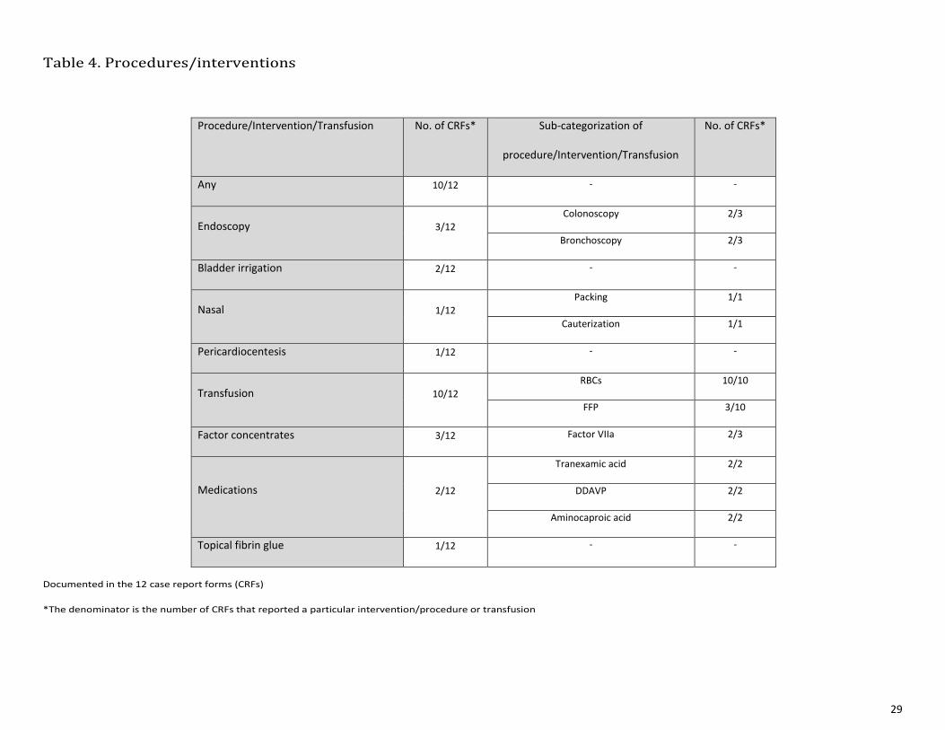
Table 4. Procedures/interventions Documented in the 12 case report forms (CRFs)
*The denominator is the number of CRFs that reported a particular intervention/procedure or transfusion
Procedure/Intervention/Transfusion No. of CRFs* Sub-categorization of
procedure/Intervention/Transfusion
No. of CRFs*
Any 10/12 - -
Endoscopy 3/12 Colonoscopy 2/3
Bronchoscopy 2/3
Bladder irrigation 2/12 - -
Nasal 1/12
Packing 1/1
Cauterization 1/1
Pericardiocentesis 1/12 - -
Transfusion 10/12
RBCs 10/10
FFP 3/10
Factor concentrates 3/12 Factor VIIa 2/3
Medications 2/12
Tranexamic acid 2/2
DDAVP 2/2
Aminocaproic acid 2/2
Topical fibrin glue 1/12 - -

30
Table 5. Comparison of CTCAE and WHO grades (Data from SPRINT3 Trial – including previously unpublished data)
CTCAE = Common Toxicity Criteria for Adverse Events; WHO = World Health Organization
* Maximum bleeding grade the patient experienced during the study
P-Rx (N = 318) Control (N = 327)
CTCAE*
N (%)
WHO*
N (%)
CTCAE*
N (%)
WHO*
N (%)
Any Bleeding 285 (90) 296 (93.1) 277 (85) 295 (90.2)
Grade 1 147 (46) 110 (34.6) 164 (50) 105 (32.1)
Grade 2 65 (20) 173 (54.4) 48 (15) 170 (52.0)
Grade 3 63 (20) 12 (3.8) 57 (17) 14 (4.3)
Grade 4 10 (3.1) 1 (0.3) 8 (2.4) 6 (1.8)

31
Table 6. Suggestions to increase consistency in future studies that use bleeding as a primary outcome measure
1. Studies should use methods to minimize inter-observer variability of bleeding
assessors
Methods of doing this will vary depending on study resources but could include:
Training of staff prior to and during study
Dual bleeding assessments and feedback
Dummy bleeding scenarios
2. Develop international consensus on a minimum data set required if bleeding is to be
used as the primary outcome of the study
In the future standard case report forms could be developed
3. Develop consistency in the way bleeding is reported so that studies can be compared
4. If a particular grading system is used, international agreement is required on the
criteria to allocate bleeding to a specific grade
5. If skin bleeding is to be categorized, need international agreement on definitions for
petechiae, purpura, ecchymoses and hematomas

32
Appendix 1: Search Strategy for identification of authors
Search Strategy for the Cochrane Review12
The search was not limited by language or publication date
1 MEDLINE search strategy (1996 to Jan 2002)
1. Platelet Transfusion.mh. 2. platelet$ adj10 (substitute$ or transfusion$ or prophyla$).tw. 3. 1 or 2 4. haemorrhage.mh. 5. platelet$.tw. 6. 4 and 5 7. exp Blood Transfusion/ 8. 5 and 7 9. 3 or 6 or 8 10. randomised controlled trial.pt. 11. controlled clinical trial.pt. 12. randomised controlled trials/ 13. random allocation/ 14. double blind method/ 15. single blind method/ 16. clinical trial.pt. 17. exp clinical trials/ 18. (clinic$ adj25 trial$).ti, ab. 19. cross-over studies/ 20. (crossover or cross-over or cross over).tw. 21. ((singl$ or doubl$ or trebl$ or tripl$) adj25 (blind$ or mask$)).ti, ab. 22. placebos/ 23. placebo$.ti, ab. 24. random$.ti, ab. 25. research design/ 26. or/10-25 27. 9 and 26 28. animal/ not (animal/ and human/) 29. 27 not 28
2 MEDLINE (Ovid) search strategy (Jan 2002-March 2011)
1. BLOOD PLATELETS/ 2. (platelet* or thrombocyte*).tw. 3. 1 or 2 4. exp BLOOD TRANSFUSION/ 5. transfus*.tw. 6. 4 or 5 7. 3 and 6 8. PLATELET TRANSFUSION/ 9. ((platelet* or thrombocyte*) adj5 (transfus* or infus* or administ* or requir*)).tw. 10. or/7-9 11. (prophylactic* or prophylax* or prevent*).tw. 12. 10 and 11 13. RANDOMIZED CONTROLLED TRIAL.pt. 14. CONTROLLED CLINICAL TRIAL.pt. 15. exp CLINICAL TRIAL/ 16. MULTICENTER STUDY.pt. 17. CLINICAL TRIALS AS TOPIC/ 18. CLINICAL TRIALS PHASE III AS TOPIC/ 19. CLINICAL TRIALS PHASE IV AS TOPIC/ 20. exp CONTROLLED CLINICAL TRIALS AS TOPIC/ 21. RANDOM ALLOCATION/ 22. DOUBLE BLIND METHOD/ 23. SINGLE BLIND METHOD/ 24. CROSSOVER STUDIES/ 25. PLACEBOS/ 26. or/13-25 27. (controlled adj3 (trial* or stud*)).ti,ab. 28. (blind* or mask*).ti,ab. 29. (placebo* or random* or factorial*).ti,ab. 30. (crossover or (cross adj over)).ti,ab. 31. aleatori*.ti,ab. 32. (treatment adj arm*).ti,ab. 33. ((phase adj iii) or (phase adj three) or (phase adj '3')).ti,ab. 34. (latin adj square).ti,ab. 35. or/27-34 36. 26 or 35

33
37. ANIMALS/ NOT (HUMANS/ AND ANIMALS/) 38. 36 not 37 39. 12 AND 38
3 EMBASE (Ovid) search strategy (1980 to Jan 2002)
1. random$.ti,ab. 2. factorial$.ti,ab. 3. (crossover$ or crossover$ or crossover$).ti,ab. 4. placebo$.ti,ab. 5. (double$ adj blind$).ti,ab. 6. (singl$ adj blind$).ti,ab. 7. assign$.ti,ab. 8. allocat$.ti,ab. 9. volunteer$.ti,ab. 10. CROSSOVER PROCEDURE.sh. 11. DOUBLE-BLIND PROCEDURE.sh. 12. RANDOMIZED CONTROLLED TRIAL.sh. 13. SINGLE-BLIND PROCEDURE.sh 14. versus.ti,ab,sh. 15. factorial.ti,ab. 16. latin square design.sh. 17. latine square.mp. 18. aleatoric.ab. 19. aleatory.ti,ab. 20. aleatorized.ab. 21. aleatorily.ab. 22. multicenter.ti,ab. 23. multicenter study.sh. 24. multicentered.ti,ab. 25. multicenters.ti,ab. 26. multicenterstudy.ti,ab. 27. multicenterstudie.ti. 28. multicenterstudies.ab. 29. multicentre.ti,ab. 30. multicentred.ti,ab. 31. multicentral.ti,ab. 32. multicentres.ti,ab. 33. or/1-32 34. ANIMAL/or NONHUMAN/ or ANIMAL EXPERIMENT 35. HUMAN 36. 35 and 34 37. 34 not 36 38. 33 not 37 39. THROMBOCYTE TRANSFUSION/ 40. 38 and 39
4 EMBASE (Ovid) search strategy (Jan 2002-March 2011)
1. THROMBOCYTE/ 2. (platelet* or thrombocyte*).tw. 3. 1 or 2 4. exp BLOOD TRANSFUSION/ 5. transfus*.tw. 6. 4 or 5 7. 3 and 6 8. THROMBOCYTE TRANSFUSION/ 9. ((platelet* or thrombocyte*) adj5 (transfus* or infus* or administ* or requir*)).tw. 10. or/7-9 11. (prophylactic* or prophylax* or prevent*).tw. 12. 10 and 11 11. random*.ti,ab. 12. factorial*.ti,ab. 13. (crossover* OR cross over* OR cross-over*).ti,ab. 14. placebo*.ti,ab. 15. (double* adj blind*).ti,ab. 16. (singl* adj blind*).ti,ab. 17. (assign* or allocat*).ti,ab. 18. (latin square or aleator*).ti.ab. 19. volunteer*.ti,ab. 20. CROSSOVER PROCEDURE/ 21. DOUBLE BLIND PROCEDURE/ 22. RANDOMIZED CONTROLLED TRIAL/ 23. SINGLE BLIND PROCEDURE/ 24. or/11-23 25. exp ANIMAL/ OR NONHUMAN/ OR exp ANIMAL EXPERIMENT/ 26. exp HUMAN/ 27. 25 NOT 26

34
28. 24 NOT 27 29. 12 AND 28
5 CENTRAL search strategy (Issue 2, 2011)
#1 MeSH descriptor Blood Platelets explode all trees #2 platelet* or thrombocyte* #3 (#1 OR #2) #4 MeSH descriptor Blood Transfusion explode all trees #5 transfus* #6 (#4 OR #5) #7 (#3 AND #6) #8 MeSH descriptor Platelet Transfusion explode all trees #9 (platelet* or thrombocyte*) NEAR/5 (transfus* or infus* or administ* or requir*) #10 (#7 OR #8 OR #9) #11 prophylactic* or prophylax* or prevent* #12 (#10 AND #11)
6 CINAHL (NHS Evidence) search strategy (Jan 2002-March 2011)
1. BLOOD PLATELETS/ 2. (platelet* or thrombocyte*).ti,ab 3. 1 or 2 4. exp BLOOD TRANSFUSION/ 5. transfus*.ti,ab 6. 4 or 5 7. 3 and 6 8. PLATELET TRANSFUSION/ 9. ((platelet* adj5 transfus*) or (platelet* adj5 infus*) or (platelet* adj5 administ*) or (platelet* adj5 requir*)).ti,ab 10. ((thrombocyte* adj5 transfus*) or (thrombocyte* adj5 infus*) or (thrombocyte* adj5 administ*) or (thrombocyte* adj5 requir*)).ti,ab 11. 7 or 8 or 9 or 10 12. (prophylactic* or prophylax* or prevent*).ti,ab 13. 11 and 12 14. "CLINICAL TRIAL"/ 15. ((controlled adj trial*) OR (clinical adj trial*)).ti,ab 16. ((singl* adj blind*) OR (doubl* adj blind*) OR (trebl* adj blind*) OR (singl* adj mask*) OR (doubl* adj mask*) OR (tripl* adj mask*)).ti,ab randomi*.ti,ab 17. RANDOM ASSIGNMENT/ 18. ("phase III" OR "phase 3" OR "phase three").ti,ab 19. (random* adj1 allocat*).ti,ab 20. (random* adj1 assign*).ti,ab 21. PLACEBOS/ 22. 14 OR 15 OR 16 OR 17 OR 18 OR 19 OR 20 OR 21 23. 13 AND 22
7 Free text search strategy for other databases
(platelet* OR thrombocyte*) AND (transfus* OR infus* OR administ* OR requir*) AND (prophylactic* OR prophylaxis OR prevent
OR prevention OR preventing)
Search Strategy for the BEST Review13
MEDLINE (1996-October 2008) using “platelet transfusion” as the key search term.
The search was limited to those published in the English language, involving humans, and to core clinical journals.
There was consensus to include additional journals with specific relevance to transfusion medicine that were not included in the core clinical journals by
MEDLINE. Clinically relevant journals added because of specific relevance to trnasfusion medicine: Transfusion, Transfusion Medicine, Vox Sanguinis,
British Journal of Haematology, Blood, Journal of American Medical Association, New England Journal of Medicine, Lancet, Circulation, Critical Care
Medicine, Journal of Thrombosis and Haemostasis, and Bone Marrow Transplantation.

35
Appendix 2: Organisational Form for Bleeding Assessment
Study Name.....................................................................Date of study.............................................
Bleeding Assessments (i.e. method of collecting data by bedside or from patient notes)
a) How was bleeding assessed?
From Patient Notes/Chart YES NO
By formalized bleeding assessment YES NO
Examination of patient YES NO
Patient questionnaire YES NO
Nurse questionnaire YES NO
b) How frequently was bleeding assessed? (e.g. 8 hourly; daily, etc.)
c) Was the bleeding assessment performed at a similar time each day? YES NO
If YES, when?
d) Who performed the bleeding assessment?
Medical staff routinely involved with patient care YES NO
Nurses routinely involved with patient care YES NO
Trained research staff (nurse, coordinator, assistant) YES NO
e) Were the bleeding assessors blinded to the trial treatment that the patient
was receiving?
YES NO
f) Was the effectiveness of the blinding assessed?
YES NO
If YES, how was the assessment done?

36
g) Did the bleeding assessors receive specific training prior to performing
bleeding assessments?
YES NO
If YES, what training did they receive (please specify)?
h) Did the bleeding assessors receive further training during the period the study
was open?
YES NO
If YES, what training did they receive and how frequently did it occur (please specify)?
i) Were duplicate bleeding assessments performed?
YES NO
If YES, what percentage of bleeding assessments were performed in duplicate?
If YES, were results fed-back to bleeding assessors to enhance consistency
between bleeding assessors?
YES NO
j) Were guidance notes provided to the bleeding assessors to assist completion
of the bleeding assessment form?
YES NO
If YES, did this include definitions of different types of bleeding?
YES NO
k) If there was more than one bleeding assessor at a trial site, was there a
formalized hand-over system to transfer information between bleeding
assessors?
YES NO
If YES, what was the system (please specify)?
l) Were any other methods used to decrease inter-observer variability? YES NO
If YES, what were they (please specify)?
m) What resources were available in the study to support the undertaking of bleeding assessment at
hospitals e.g. how many staff were appointed to perform the bleeding assessment?

37
Grading System for bleeding
a) Which grading system was used for the adjudication of bleeding, to convert the
results from the bleeding assessment form into a grade?
WHO (Original 1979 formulation) YES NO
Rebulla (1998) YES NO
CTCAE version 3.0 YES NO
Other (please specify) YES NO
b) Had the grading system selected been modified by the study authors in any
way?
YES NO
If YES, please state in what way it had been modified?
c) Was “significant bleeding” defined by the study authors e.g. WHO grade 2 or
above?
YES NO
If YES, please state the definition?
d) Was life-threatening bleeding defined by the study authors? YES NO
If YES, please state the definition?
e) If the grading system reported in the study used red cell transfusions to
partially define the severity of bleeding, was a protocol for red cell transfusion
agreed and used at all sites?
YES NO
Converting the Bleeding Assessment into a Bleeding Grade
a) How was the bleeding assessment initially converted into a bleeding grade?
i)
Manual assignment of grading YES NO
If YES, was the person assigning the grade blinded to the
intervention?
YES NO
Was the bleeding assessment converted into a bleeding grade by the
person who did the original bleeding assessment/ data collection?
YES NO
If YES, was this grading performed at the bedside? YES NO
If this was a multicentre study was the bleeding assessment
converted into a bleeding grade centrally, away from the local
participating site e.g. by central coordinating site
YES NO

38
ii) Computer algorithm YES NO
If YES, was this algorithm validated prior to its use in the study e.g.
by comparison to manual grading
YES NO
b) Was the initial assignment of bleeding grade checked via a second
method/person?
YES NO
If YES, what method was used?
i) Manual assignment of grading YES NO
If YES, was this the same person who assigned the initial bleeding
grade (i.e. same person assigning grade at two different times?
YES NO
1) If NO, was the individual blinded to the intervention? YES NO
2) If NO, was this individual the same person who performed the
bleeding assessment?
YES NO
ii) Computer algorithm YES NO
If YES, was this algorithm validated prior to its use in the study e.g.
by comparison to manual grading
YES NO
If YES, was this second method performed independently of/blinded to the
initial method?
YES NO
If YES, and this was a multicentre study, was the bleeding assessment
converted into a bleeding grade centrally, away from the local
participating site?
YES NO
c) Was adjudication performed if there was disagreement between the allocation
of bleeding grades according to the first and second person/method?
YES NO
If YES, how was this disagreement resolved (please specify)?

39

40
Table A. Assessment of bleeding
First author Study Assessment of bleeding Frequency
of
assessment
Same
time
each
day
Type of bleeding assessors Blinding
of
bleeding
assessors
Effectiveness
of blinding
assessed
Patient
notes/chart
Formalized
bleeding
assessment
Examination
of patient
Patient
questionnaire
Nurse
questionnaire
Medical
staff
Nurses Trained
research
nurses
Other
research
investigators*
Cazenave MIRACLE23 Y Y Y N N
Twice daily on
day of Tx,
once daily on
day after Tx
N Y Y N N Y N
Heckman Heckman et
al 19971 Y Y Y N N Daily Y Y Y N N N -
Heddle SToP2 Y Y Y N N Daily Y N N Y Y Y N
McCullough SPRINT3 Y Y Y Y Y Daily Y N N Y N Y Y†
Kerkhoffs
HOVON-
Triplate33 Y Y Y N N Daily N Y N N N N -
PrePAReS34 Y Y Y Y Y Daily Y N N N Y N -
Rebulla Trigger4 Y Y Y NR NR Daily N Y N N N N -
Slichter PLADO5 Y Y Y Y Y Daily Y N N Y N Y N
Stanworth TOPPs6 Y Y Y Y Y Daily Y Y N Y Y N -
Tinmouth Tinmouth et
al 20047 Y Y Y Y N Daily N Y N N N N -
Wandt Nuerenberg
trial8 9 Y Y Y Y Y Twice daily Y Y Y N Y N -

41
Results from the 11 questionnaires
Y = Yes; N = No; NR = Not reported; Tx = transfusion
* We did not ask for further details on who these investigators were
† Research nurses asked to detect study units from a panel of 10 platelet components (mixture of study and conventional units).

42
Table B: Major differences in WHO grading between studies Information from questionnaire, guidance notes sent with CRF and published article N = Not defined/present in the study’s grading system * Unexpected bleeding out of normal cycle OR bleeding heavier than normal OR breakthrough bleeding (patient on hormonal therapy) more than spotting
First author Study Occult
blood in
stool
Microscopic blood in
urine
Grade 2 skin bleeding Retinal
bleeding
without
visual
compromise
Grade 2 vaginal
bleeding
Grade 2
epistaxis/
bleeding
from
mouth
Hemodynamic instability
Grade 1 Grade 2 Petechiae Purpura Ecchymoses Definition Grade
Cazenave MIRACLE23 Grade 1 1+ > 1+ N > 1 cm N Grade 2 > 2 saturated
pads/day > 1 hr N -
Kerkhoffs PrePAReS34 Grade 1 Positive N N > 1 inch N Grade 2 Abnormal vaginal
bleeding* > 30 mins
30-50mmHg fall
>50mmHg fall/ 50% fall
in BP
3
4
Heddle SToP2 Grade 1 1+ > 1+ N N > 10 cm Grade 2 abnormal vaginal
bleeding*
> 1 hr or
packing N -
McCullough SPRINT3 Grade 1 1+ > 1+ Generalized > 1 inch N Grade 1 > 2 saturated
pads/day > 1 hr > 30mmHg fall 4
Slichter PLADO5 N N N N > 1 inch N Grade 2
Abnormal vaginal
bleeding >
spotting
> 30 mins
30- 50mmHg fall
>50mmHg fall/> 50%
fall
3
4
Stanworth TOPPs6 N N N Diffuse > 5
> 10cm or
multiple >
2cm
Grade 2
unexpected
vaginal bleeding
saturating 2
pads/24 hrs
> 30 mins > 30mmHg fall 4

43
Table C: Conversion to bleeding grade from data
First author Study Initial assignment
of bleeding grade
Bleeding assessor
& grader of
bleeding the same
person*
Person who graded
bleeding blinded to
the intervention*
Bleeding grade
checked by second
person/method
Method of
checking
Person who
graded bleeding
blinded to the
intervention
Person who
graded bleeding
was blinded to
the initial grade
Was adjudication†
performed if
differences in
bleeding grade
Cazenave MIRACLE23 Manual Y Y N - - - -
Heckman Heckman et al
19971 Manual Y N Y Manual N NR Y
Heddle SToP2 Manual Y Y Y Manual Y Y Y
Kerkhoffs
Triplate33 Manual Y N Y Manual N N Y
PrePAReS34 Manual N Y Y Computer
algorithm† - - Y
McCullough SPRINT3 Manual Y Y Y Manual Y N Y
Rebulla Trigger4 Manual Y N N - - - -
Slichter PLADO5
Computer
algorithmǂ
- - N - - - -
Stanworth TOPPs6
Computer
algorithmǂ - - N - - - -
Tinmouth Tinmouth et al
20047 Manual Y N Y Manual Y Y Y
Wandt Nuerenberg trial8 9 Manual N N Y Manual N NR Y
Results from the 11 questionnaires

44
Y = Yes; N = No; NR = Not reported; - = Not applicable
* Question was only answered if method of assessing bleeding was manual
† This was resolved mainly by discussion between the two graders or between the graders and Principal Investigator/Chief Investigator
ǂ Validated prior to commencement of the study
45
Appendix 3 :BEST Collaborative Members
Chair Past Chair and Treasurer
Larry Dumont Lorna Williamson
Honorary Members
Georges Andreu
James P. AuBuchon
Morris Blajchman
Anneke Brand
Marcela Contreras
Neelam Dhingra
Janny de Wildt-Eggen
Hermann Eichler
Andrew Heaton
Margarethe Heiden
Riitta Kekomaki
Harvey Klein
Maurice Masse
Wolfgang Mayr
Jeffrey McCullough
Gary Moroff
Paul Ness
Derwood Pamphilon
Ruby Pietersz
Chris Prowse
Martin Ras
Paolo Rebulla
Jerard Seghatchian
Girolamo Sirchia
Cees Smit Sibinga
Irena Sniecinski
Joseph Sweeney
Shigeru Takamoto
Jaro Vostal
Girish Vyas
Wolfram Walker
Silvano Wendel
Sam Wortham
Cellular
Therapy
Clinical
Studies
Conventional
Components
Transfusion
Safety
Team Leaders
Zbigniew Szczepiorkowski
David McKenna
Nancy Heddle
Alan Tinmouth
John Hess
Pieter van der Meer
Michael Murphy
Mark Fung
Scientific Members
JoAnna Reems
Ronald Sacher
Dominic Wall
Tor Hervig
Miguel Lozano
Andreas Greinacher
Leo van de Watering
Rebecca Cardigan
Dana Devine
Hans Gulliksson
Sherrill Slichter
Sunny Dzik
Richard Haspel
Richard Kaufman
Simon Stanworth
Associate Scientific Members
David Stroncek
Henk Garritsen
Minoko Takanashi
Daniel Hollyman
Donald Arnold
Jonathan Waters
Alyssa Ziman
Meghan Delaney
Jose Cancelas
Dirk de Korte
Rosemary Sparrow
Ralph Vassallo
Neil Beckman
Jay Brooks
Joan Cid
Mark Yazer