Urinary Incontinence in the Developing World: The Obstetric ...
Upload
khangminh22Category
view
5download
0
FOR HEALTH CARE PROVIDERS
A Pocket Guide for Clinical Management of Obstetric and Neonatal Emergencies in Africa
FOR HEALTH CARE PROVIDERS

A Pocket Guide for Clinical Management of Obstetric and Neonatal Emergencies in Africa
FOR HEALTH CARE PROVIDERS
2nd Edition

Clinical Management of Obstetric and Neonatal Emergencies in Africa
ISBN: 978-929023467-8
© WHO Regional Office for Africa 2022
Some rights reserved. This work is available under the Creative Commons Attribution-NonCommercial-ShareAlike 3.0 IGO licence (CC BY-NC-SA 3.0 IGO; https://creativecommons.org/licenses/by-nc-sa/3.0/igo).
Under the terms of this licence, you may copy, redistribute and adapt the work for non-commercial purposes, provided the work is appropriately cited, as indicated below. In any use of this work, there should be no suggestion that WHO endorses any specific organization, products or services. The use of the WHO logo is not permitted. If you adapt the work, then you must license your work under the same or equivalent Creative Commons licence. If you create a translation of this work, you should add the following disclaimer along with the suggested citation: “This translation was not created by the World Health Organization (WHO). WHO is not responsible for the content or accuracy of this translation. The original English edition shall be the binding
and authentic edition”.
Any mediation relating to disputes arising under the licence shall be conducted in accordance with the mediation rules of the World Intellectual Property Organization.
Suggested citation. Recommendations for Clinical Practice of Emergency Obstetrical and Neonatal Care in Africa. Brazzaville: WHO Regional Office for Africa; 2022. Licence:
CC BY-NC-SA 3.0 IGO.
Cataloguing-in-Publication (CIP) data. CIP data are available at http://apps.who.int/iris
Sales, rights and licensing. To purchase WHO publications, see http://apps.who.int/bookorders. To submit requests for commercial use and queries
on rights and licensing, see http://www.who.int/about/licensing.
Third-party materials. If you wish to reuse material from this work that is attributed to a third party, such as tables, figures or images, it is your responsibility to determine whether permission is needed for that reuse and to obtain permission from the copyright holder. The risk of claims resulting from infringement of any third party-
owned component in the work rests solely with the user.
General disclaimers. The designations employed and the presentation of the material in this publication do not imply the expression of any opinion whatsoever on the part

of WHO concerning the legal status of any country, territory, city or area or of its authorities, or concerning the delimitation of its frontiers or boundaries. Dotted and dashed lines on maps represent approximate border lines for which there may not yet be full agreement.
The mention of specific companies or of certain manufacturers’ products does not imply that they are endorsed or recommended by WHO in preference to others of a similar nature that are not mentioned. Errors and omissions excepted; the names of
proprietary products are distinguished by initial capital letters.
All reasonable precautions have been taken by WHO to verify the information contained in this publication. However, the published material is being distributed without warranty of any kind, either expressed or implied. The responsibility for the interpretation and use of the material lies with the reader. In no event shall WHO be liable for damages arising from its use.
Designed in Brazzaville, Congo

iii
CONTENTS
FOREWORD ......................................................................... xi
ABBREVIATIONS ................................................................ xiv
ACKNOWLEDGEMENTS .................................................... xvii
INTRODUCTION ................................................................... 1
CHAPTER 1: GENERAL PRINCIPLES FOR OPTIMAL MANAGEMENT OF OBSTETRIC AND NEWBORN EMERGENCIES ............................ 5
Definition ............................................................................ 5
Ensure good documentation............................................... 7
Resuscitate the patient and manage her specific complication ....................................................................... 8
In all cases with haemorrhage manage the patient’s blood status................................................................................... 8
CHAPTER 2: COMPLICATIONS IN EARLY PREGNANCY .... 13
Ectopic pregnancy ............................................................. 14
Diagnosis ........................................................................... 14
Management of ectopic pregnancy .................................. 16
Abortion ............................................................................ 19
Definition .......................................................................... 19

iv
Problem ............................................................................. 20
Diagnosis ........................................................................... 20
Threatened abortion ......................................................... 21
Inevitable abortion............................................................ 21
Incomplete abortion ......................................................... 22
Septic abortion .................................................................. 22
Complete abortion ............................................................ 23
Missed abortion ................................................................ 23
Molar pregnancy/abortion ............................................... 24
Management of abortion .................................................. 24
General guidelines ............................................................ 24
Threatened abortion ......................................................... 25
Inevitable abortion and incomplete abortion ................... 26
Septic abortion.................................................................. 30
Complete abortion ............................................................ 31
Missed abortion ................................................................ 31
Molar pregnancy/abortion ............................................... 34
Managing other abortion-related complications and injuries .............................................................................. 35
CHAPTER 3 HYPERTENSION IN PREGNANCY ................. 37
Definition .......................................................................... 37

v
Problem ............................................................................. 37
Diagnosis of hypertension................................................. 38
Classification of hypertension ........................................... 39
Pregnancy-induced hypertension ..................................... 39
Chronic hypertension ........................................................ 39
Eclampsia .......................................................................... 42
Management hypertension in pregnancy ......................... 43
Mild pre-eclampsia ........................................................... 44
Severe pre-eclampsia and eclampsia ................................ 46
CHAPTER 4 OBSTETRIC HAEMORRHAGE ....................... 65
Definition .......................................................................... 65
Problem ............................................................................. 65
Placenta praevia ................................................................ 66
Abruptio placentae ........................................................... 73
Coagulation disorders ....................................................... 78
Postpartum haemorrhage ................................................. 80
Retained placental tissue .................................................. 85
Tears of the cervix, vagina or perineum ........................... 85
Inversion of the uterus ...................................................... 86
Secondary postpartum haemorrhage ............................... 88

vi
CHAPTER 5 DYSTOCIA/ABNORMAL LABOUR ................. 90
1. Prolonged (active first stage) labour ................... 91
2. Cephalopelvic disproportion ............................... 98
3. Obstructed (prolonged latent stage) labour ....... 99
4. Transverse lie .................................................... 102
5. Shoulder dystocia .............................................. 103
6. Face presentation ............................................. 108
7. Brow presentation ............................................ 110
8. Labour and delivery in a scarred uterus ........... 111
9. Ruptured uterus ................................................ 113
10. Preterm labour .................................................. 119
11. Fetal distress (non-reassuring fetal heart status) ...................................................... 122
CHAPTER 6: FEVER DURING PREGNANCY AND POSTPARTUM PERIOD .............................. 124
1. Malaria .............................................................. 124
2. Urinary tract infection....................................... 130
3. Endometritis ..................................................... 132
4. Pelvic abscess .................................................... 134
5. Mastitis ............................................................. 136
6. Breast abscess ................................................... 137

vii
CHAPTER 7: CARE OF THE NEWBORN .......................... 139
1. Essential care of the newborn .......................... 139
2. Standard procedures in essential newborn care (ENC) .......................................... 140
3. Perinatal asphyxia ............................................. 144
4. Neonatal infection ............................................ 152
5. Prematurity ....................................................... 157
6. Neonatal jaundice ............................................. 160
7. Seizures in the newborn ................................... 162
8. Neonatal hypoglycaemia .................................. 166
9. Disorders of temperature control: hypothermia and hyperthermia ....................... 168
10. Respiratory distress .......................................... 170
11. Neonatal apnoea............................................... 174

viii
Appendices on Emergency Obstetrical and Newborn Care
1 Organization of the referral system ....................... 179
2 Shock ...................................................................... 182
3 Para-cervical block .................................................. 186
4 Manual vacuum aspiration (MVA) .......................... 188
5 Digital evacuation of uterus ................................... 191
6 Uterine curettage ................................................... 193
7 Induction of labour ................................................. 195
8 Eclampsia treatment packs and dosage preparation ............................................................. 203
9 Blood transfusion in obstetricS .............................. 205
10 Active management of the third stage of labour ... 208
11 Non-pneumatic antishock garment (NASG) ........... 212
12 Uterine balloon tamponade ................................... 216
13 WHO labour care guide (who “next generation partograph”) ........................................................... 217
14 Assisted vacuum delivery ....................................... 218
15 Hand hygiene .......................................................... 222
16 Respiratory hygiene ................................................ 224
17 Key messages on optimal breastfeeding

ix
of the newborn ....................................................... 225
18 Endotracheal tube placement ................................ 227
19 Umbilical vein catheterization ................................ 229
20 Newborn antibiotic doses....................................... 230
21 Feeding/fluid management: newborn ≥ 1.5kg ....... 231
22 Kangaroo mother care for low-birth-weight babies ......................................... 235
List of Figures and Tables Figure 2.1 Diagnosis and treatment of ectopic pregnancy ....... 18
Figure 3.1 Diagnosing hypertensive disease in pregnancy ..... 40
Figure 3.2 Management of eclampsia ..................................... 49
Figure 3.3 Controlling and preventing fits with magnesium sulphate IM regimen .............................................. 52
Figure 3.4 Controlling BP in hypertensive emergencies ......... 55
Figure 4.1 Management of placenta praevia .......................... 72
Figure 4.2 Management of abruptio placentae ....................... 77
Figure 4.3 Prevention and management of primary PPH ........ 87
Figure 5.1 Management of prolonged latent phase labour ...... 95
Figure 5.2 Flowchart for management of ruptured uterus ..... 118
Figure 7.1 Birth and cord cutting ............................................. 141
Figure 7.2 Newborn ventilation with bag and mask ............... 148
Figure 7.3 What about Meconium ........................................... 148

x
Figure 7.4 Infant chest compression: Hand-encircling technique ................................................................ 149
Figure 7.5 Neonatal resuscitation flowchart ........................... 152
Figure 7.6 Neonatal Sepsis flow chart .................................... 155
Figure 7.7 Management of neonatal seizures ........................ 165
Figure 7.8 Continuous positive airway pressure (CPAP)....... 172
Figure7.9 Causes of Neonatal Apnoea.................................. 176
Table 2.1 Misoprostol protocol for managing incomplete abortion ................................................. 28
Table 2.2 Medical management of missed abortion ............... 32
Table 3.1 Key principles in managing severe pre-eclampsia and eclampsia ......................................................... 48
Table 3.2 Onsets, peak actions and durations of effect of the antihypertensive medications ........................ 57
Table 7.1 The APGAR Score ................................................. 144
Table 7.2 Duration of treatment for neonatal sepsis ............. 156
Table 7.3 Physical features at different gestational ages ..................................................... 159
Table 7.4 Diagnosis and management .................................. 168

xi
FOREWORD In September 2015, governments and the international community committed to substantially improve the health and well-being of women and children by December 2030. Among the 17 Sustainable Development Goals (SDGs) adopted by the United Nations Member States is SDG 3 with 17 targets that aims to “Ensure health and promote well-being for all and at all ages”. It includes target 3.1 on reducing maternal mortality and target 3.2 on ending all preventable deaths of children under 5 years of age. The reduction of maternal and perinatal mortality is central to the attainment of the SDG 3 targets. The aim is to reduce the global maternal mortality to 70 per 100 000 live births and neonatal mortality to at most 12 per 1000 live births by 2030. These goals are ambitious but achievable. Although some progress has been made in Africa with the maternal mortality ratio (MMR) declining by 38% from 2000 to 2017, the Region still accounts for about 66% of all maternal deaths globally, with 196 000 deaths reported in 2017 (UN estimates 2000–2017). The main causes of maternal death include postpartum haemorrhage, pregnancy-related hypertensive disorders, infection, unsafe abortion and obstructed labour (UN MMEIG 2019). The first 28 days of life, i.e., the neonatal period, are the most vulnerable time for a child’s survival. Children face the highest risk

xii
of dying in their first month of life, with the 2019 average global neonatal mortality rate (NMR) at 17 deaths per 1000 live births. About a third of all neonatal deaths occur within the first day after birth and close to three-quarters within the first week of life. Sub-Saharan Africa has the highest NMR, which is estimated at 27. The major causes of neonatal mortality are prematurity at birth, birth asphyxia and neonatal sepsis (UNICEF 2020). The medical interventions required to prevent maternal and newborn deaths exist and are well known but their dissemination is often weak. Therefore, health workers lack up-to-date information and practical guidance for their day-to-day use, resulting in poor quality of care. To address this, the World Health Organization (WHO), in collaboration with Société africaine de gynécologie et d’obstétrique and the United Nations Population Fund (UNFPA), developed the “Recommendations for clinical practices on emergency obstetric and neonatal care in Africa (RCP)”. This manual was adapted from WHO’s “Managing complications in pregnancy and childbirth: a guide for midwives and doctors”. It aims to enhance the knowledge and skills of health facility staff to manage obstetric and newborn problems. The guidance in this manual is adaptable to local conditions and enables health care providers to manage cases better, considering the main causes of maternal and newborn mortality in the Region. The French version of the RCP has been disseminated widely and is already in use in francophone countries. It is our hope that

xiii
health providers in anglophone countries will find this English version useful in improving the quality of care provided during childbirth and in the immediate postnatal period. We believe that the use of this manual will facilitate the provision of quality maternal and newborn health care and move us closer to ending preventable maternal and neonatal mortality in the African Region.
Dr Matshidiso Moeti WHO Regional Director for Africa Brazzaville, Congo

xiv
ABBREVIATIONS
AMTSL active management of third stage of labour
ANC antenatal care
BEmOC basic emergency obstetric care
BEmONC basic emergency obstetrical and neonatal care
βhCG beta human chorionic gonadotrophin
BP blood pressure
CEmOC comprehensive emergency obstetric care
CEmONC comprehensive emergency obstetrical and neonatal care
cm centimetre
CO cardiac output
CPD cephalopelvic disproportion
CNS central nervous system
CRP C reactive protein
CSF cerebrospinal fluid
DIC disseminated intravascular coagulation
dL decilitre
ENC essential neonatal care
FBC full blood count
FHR fetal heart rate
g gram

xv
Hb haemoglobin
HELLP hemolysis, elevated liver enzymes, low platelet count
HIV human immunodeficiency virus
HR heart rate
IM intramuscular
IU international unit
IUFD Intra-uterine fetal demise
IV intravenous
kg kilogram
mcg microgram
mg milligram
ml millilitre
mm millimetre
mm Hg millimetres of mercury
MVA manual vacuum aspiration
NS normal saline
O2 oxygen
OD once a day
qid 4 times daily
RCP recommendations for clinical practice
stat immediately
STI sexually transmitted infection

xvi
SVR systemic vascular resistance
tds 3 times daily
UNFPA United Nations Population Fund
UNICEF United Nations Children’s Fund
WHO World Health Organization

xvii
ACKNOWLEDGEMENTS The initial team that contributed to the 2013 English version of the “Recommendations for clinical practice of emergency obstetrical and neonatal care in Africa” included Ms Roselyn Akinyi Koech, Examination Officer, Nursing Council of Kenya; Dr Romano Byaruhanga, Obstetrician Gynaecologist, Uganda Martyrs University; Dr Sylvia Deganus, Obstetrician Gynaecologist, Ghana; Dr Teguete Ibrahima, Obstetrician Gynaecologist, Mali; Dr GW Jaldesa, Obstetrician Gynaecologist, University of Nairobi, Kenya; Dr Oladosu Ojengbede, Obstetrician Gynaecologist, University of Ibadan, Nigeria; Dr Bogale Worku, Paediatrician and Head Department of Paediatric and Child Health, Addis Ababa University,Ethiopia; Dr Joyce Lavussa, Obstetrician Gynaecologist, WHO Kenya; Dr Triphonie Nkurunziza, Obstetrician Gynaecologist, WHO Regional Office for Africa. Dr Kasonde Mwinga, Director Universal Health Coverage and Life course, WHO AFRO who coordinated the effort.
We would like to thank the members of the Société africaine de gynécologie et d’obstétrique who contributed to the original French document, members of the East Central and Southern Africa College of Obstetrics and Gynaecology (ECSACOG), members of paediatric associations and midwives who contributed to the revision of the original document based on emerging evidence and guidance to produce this version in English.

xviii
WHO staff: Dr Triphonie Nkurunziza, Dr Assumpta Muriithi, Dr Fatim Tall, Dr Leopold Ouedraogo, Dr Ghislaine Conombo, Dr Hayfa Elamin, Dr Chilanga Asmani, Dr Azmach Hadush, Dr Nancy Kidula, Dr Maurice Bucagu, Dr Geneviève Saki-Nekouressi and Dr Gilles Landrivon. UNICEF staff: Dr Alain Prual, Dr Virginie Amoin Konan Epse Kouadio and Dr Bruno Aholoukpe UNFPA staff: Dr Joseph Vyankandonera, Dr Argentina Matavzi, Dr Pauline Abou-Kone and Dr Dougrou Sosthene CONSULTANTS: Dr Sylvia Deganus (obstetrician/ gynaecologist), Ghana and Dr Florence Murila (paediatrician/ neonatologist), Kenya. We count on the support of all for the dissemination of this pocketbook.

1
INTRODUCTION Sub-Saharan Africa has the highest maternal mortality in the world. According to estimates by the United Nations Maternal Mortality Estimation Inter-Agency Group (UN MMEIG)1 in September 2017, while the African Region had recorded a significant decline in maternal mortality rate (MMR) of 37.8% between 2000 and 2017, 66% of the 295 000 maternal deaths reported globally occurred in sub-Saharan Africa. The African Region is also noted to have an extremely high MMR, estimated at 542 per 100 000 livebirths, with an average annual rate of reduction of 2.9%. Newborn deaths constitute 47% of all mortality among children under 5 years of age and accounted for 2.5 million deaths in 2018. Two million stillbirths are estimated to have occurred in 2017 with 50% as intrapartum stillbirths. Of note is that 98% of the global newborn deaths and stillbirths occur in sub-Saharan Africa and Asia. Furthermore, 1.3 million of the newborns surviving each year have major disabilities. Most disabilities are preventable, and disability is a sensitive marker of the quality of maternal and newborn care. It is estimated that 3 million of the mothers and newborns lost each year and stillbirths could be saved with universal coverage of quality maternal and newborn care (Every newborn progress report 2019).
1 A MMEIG: WHO, the United Nations Children (UNICEF), the United Nations Population Fund (UNFPA),
the World Bank Group and the United Nations Population Division (UNPD) of the Department of Economic and Social Affairs

2
In line with the Global Strategy for Women’s, Children’s and Adolescents’ Health 2016–2030: Implementation Framework for the African Region endorsed at the Sixty-sixth session of WHO Regional Committee for Africa (RC 66), several initiatives and strategies have been introduced by the WHO Regional Office for Africa to galvanize country action towards the reduction of maternal and perinatal mortality, with the goal of accelerating the attainment of the SDG targets. One of the key strategies is to provide skilled attendants at birth. Of note is that many health workers in maternity units lack up-to-date information on evidence-based interventions and the skills required to provide maternity care. To address this gap, WHO, in collaboration, with the Société africaine de gynécologie et d’obstétrique(SAGO) and the United Nations Population Fund (UNFPA), developed the “Recommendations for clinical practices on emergency obstetric and neonatal care in Africa” (the RCP manual). The aim of the RCP manual was to enhance the skills of health facility staff in emergency obstetrical and neonatal care. The recommendations in that manual are adaptable to local conditions and enable health care workers to provide contextualized care focusing on the main causes of maternal and newborn mortality in the Region, including quality management of obstetrical and neonatal emergencies, harmonizing the protocols within countries and establishing the relevant quality criteria. The French version of the manual was launched and disseminated to francophone countries during the Congress of the Société africaine de gynécologie et d’obstétrique in November

3
2010 in Libreville, Gabon. Owing to the positive feedback from the users, WHO decided to translate the manual into English to make it available to anglophone countries in the Region. In 2013, WHO organized an expert consultation to review the manual and the accompanying job aids and translate them into English. The modifications to those products from the consultation were:
• A brief chapter on the general principles for the optimal management of obstetric and newborn emergencies was added as the introduction.
• A topic on pelvic abscess was added to chapter 6 on fever during pregnancy and the postpartum period, while the section on ovary infection was removed.
• Topics on brow presentation and cephalopelvic disproportion were added to chapter 5 on dystocia.
• In chapter 7 on newborn care, the topics on perinatal asphyxia and neonatal distress were merged, while fetal distress was renamed “non-reassuring fetal status”. The content was expanded.
In addition, three topics were added as the appendices: kangaroo mother care for the preterm baby, key messages on optimal breastfeeding of the newborn, and the para-cervical bloc procedure.

4
In 2019 it was found necessary to incorporate the WHO recommendations and tools on antenatal, intrapartum and postnatal care, which had been updated, into the existing manual. An obstetrician and a neonatologist were tasked to update the chapter with the prevailing and up-to-date evidence. Their input was validated by an expert group comprising WHO technical officers and key maternal and newborn health partners including experts from obstetrics and gynaecology, paediatric and midwifery professional associations. This version, titled “A pocket guide for clinical management of obstetric and neonatal emergencies in Africa”, contains seven chapters: general principles for optimal management of obstetric and newborn emergencies, complications in early pregnancy, hypertension in pregnancy, obstetric haemorrhage, dystocia/abnormal labour, fever during pregnancy and postpartum period, and care of the newborn (expanded). The content of each of these topics is presented in this sequence: definition, problem, diagnosis, and management. The various flowcharts have been modified and simplified to be amenable to low-resource settings such as those found in the African Region. Job aids and wall charts have also been developed to complement this guide. This pocket-size practical guide is meant for health professionals such as doctors, nurses, and midwives providing care to women and their babies during pregnancy, childbirth, and the postnatal period.

5
CHAPTER 1: GENERAL PRINCIPLES FOR OPTIMAL MANAGEMENT OF OBSTETRIC AND NEWBORN EMERGENCIES
Definition
Obstetric and newborn emergencies are health problems that are life threatening for pregnant women and newborns. They require prompt and focused interventions to save the lives of the mother and/or her baby. These emergencies may arise at any time during pregnancy, labour, birth or soon after birth. In the African Region the common obstetric and newborn emergencies include:
• severe pre-eclampsia/eclampsia
• bleeding during pregnancy due to abortion, placenta praevia or placenta abruption
• bleeding after childbirth (postpartum haemorrhage)
• prolonged and obstructed labour
• infections such as severe malaria
• sepsis due to genital tract infections
• neonatal asphyxia
• stillbirth and intrauterine death
Note: In all emergencies provide respectful maternal care.

6
It is important for care providers to remember that pregnancy can be a time of stress and anxiety, particularly for women facing pregnancy complications and labour pain. Women presenting at a health care facility during these periods may have communication, emotional and behavioural challenges. A woman’s care experiences with her health care provider can either empower and comfort her and lead to positive care experiences or inflict lasting damage and emotional trauma leading to negative experiences. Such negative experiences can become powerful deterrents to the future use of the services by the woman or her community. It is the responsibility of the entire care team to handle pregnant women respectfully and put them at ease. Respectful maternity care includes:
• Respecting the woman’s dignity and right to privacy;
• Respecting the woman’s right to information and informed consent;
• Respecting the woman’s right to decline any treatment or procedure offered;
• Respecting the woman’s choices and preferences, including for companionship during maternity care, procedures and treatment;
• Protecting the woman’s privacy rights and protections with respect to her health information, including how her health

7
information is used and to whom it is disclosed by health care providers;
• Being sensitive and responsive to the woman’s needs;
• Being non-judgmental about the decisions that the woman and her family make regarding her care.
Respectful maternity care is a universal human right due to every childbearing woman everywhere.
Requirements for optimal management of obstetric and newborn emergencies Ensure good documentation
The patient’s notes should be documented well and must reflect:
• The history of the onset and progression of the patient’s emergency;
• The general state of the patient at admission and/or at the time of the onset of the complication;
• The patient’s vital signs, i.e., blood pressure, pulse, respiratory rate, temperature, urine output and level of consciousness at the time of admission or onset of the complication;
• All the vital signs monitored closely for the indication of the patient’s deterioration or improvement;
• All the treatments administered, with their time and date and signature of the care provider;

8
• The signatures/names of the attending persons.
Resuscitate the patient and manage her specific complication
• An intravenous (IV) access line must be established on the patient and secured.
• The most experienced medical staff must be involved in the management of the complication as soon as possible and preferably within 10 minutes of the diagnosis.
• The most senior medical staff on call must take responsibility for defining the management plan for the patient.
• Infection prevention practices should be strictly adhered to at all times.
• The diagnosis and management plan should be communicated to the patient and/or her relatives.
In all cases with haemorrhage manage the patient’s blood status
• Blood must be taken for grouping and cross-matching as soon as possible and for baseline haemoglobin level checking/full blood count (FBC).
• The amount of blood lost should always be noted.
• Coagulation must be evaluated in all cases with massive blood loss.

9
If the patient is still pregnant or in labour take care of the fetus’s health as well
• Fetal well-being should be established and monitored as recommended, including the fetal heart rate.
• If the patient is in labour, a labour care guide, i.e. a next generation partograph, must be completed, reviewed and acted upon.
For the newborn’s care good documentation is essential
• The birth weight must be recorded when the newborn’s condition is stable.
• Feeding and thermal care charts must be maintained.
• The mother/family should be involved in the care of the baby.
When referring the patient ensure that all arrangements have been made (refer to Appendix 1)
• Critically assess the patient to validate the need for her referral.
• Provide first aid to the patient for her complications and stabilize her for the transfer.
• Counsel the patient and her support persons about the reasons for the transfer and their expected roles.
• Communicate with the referral centre to enable them to prepare to receive the patient.

10
• Ensure that all referral documents and notes are complete and sent along with the patient.
Provide care and support for women and/or families with unfavourable outcomes and grief
Obstetric complications sometimes result in the loss of the pregnancy (abortion), stillbirth, birth of a sick or abnormal baby, and/or maternal death. All these outcomes can lead to profound emotional distress for the woman and/or her family.
Disbelief, denial, despair, sense of failure, anger, anxiety, apprehension and profound sadness are all normal reactions associated with grief. It is important to recognize that the grieving process manifests in many ways and there is no right way to grieve. No two people grieve or express their grief in the same way or for the same time. The social situations of the woman and/or her family, her support systems, her cultural background and past pregnancy experiences are all factors that influence her grief process and its outcomes.
Denying women the opportunity to mourn the loss of their babies has been shown to be associated with long-term psychological injury. Grief situations can in some instances lead to gender-based violence, divorce and the breakdown of a family. Affected women and their families sometimes require prolonged counselling and support. Care providers must therefore appreciate this, recognize grief in their clients and provide the appropriate care and support.

11
The general principles in providing care and support in grief situations include:
• Appreciating the severity and depth of the loss for the affected individuals;
• Offering sympathy, understanding and support;
• Supporting the mother, father and/or family as they identify and express their feelings.
• The important actions that care providers can undertake to also support families and aid the grieving process include:
• At the time of the tragic event, assigning a skilled care provider with the right communication skills to break the bad news;
• In instances involving the baby, encouraging the woman or the couple to see, hold or spend as much time as they wish with their baby;
• Where possible, accommodating women who have suffered the loss of their baby separately from women who have given birth to healthy infants;
• Providing access to supportive individuals, groups and professionals where necessary;
• Facilitating the woman and/or her family to meet the cultural, administrative and legal requirements relating to the death, for example in regard to traditional practices, corpse handling, death registration etc.;
• Where relevant, arranging a discussion with the woman and

12
her partner to sensitively discuss the event and the possible preventive measures for the future, if this is relevant, without blaming the woman or the family.
Grief management in some instances can start before the occurrence of the tragic event or unfavourable outcome. The pre-event actions that can aid the grieving process for the woman and/or her family are, where feasible, preparing the woman/her family for the possibility of the disturbing or unexpected outcome and allowing her or her family to see the efforts made by the caregivers to revive the baby or the mother.
Immediate empathic care and continuing support conducted in a very professional manner for all clients needing such care ensures that women and/or their families have healthy grieving.

13
CHAPTER 2: COMPLICATIONS IN EARLY PREGNANCY
During the first 6 months of a pregnancy, the main obstetric complications often present with the symptoms of abdominal pain and/or vaginal bleeding. In the first and second trimesters of a pregnancy these are symptoms of complications such as unsafe abortion, miscarriage or ectopic pregnancy, which are major causes of maternal morbidity and mortality. If they are not detected and managed early, they can compromise the life and obstetrical future of the woman.
The abdominal pains are felt in the pelvis and/or abdomen. When they occur without associated vaginal bleeding, efforts must be made to exclude ectopic pregnancy and non-pregnancy causes such as acute appendicitis, twisting/torsion of an ovarian cyst, urinary tract infections etc. Vaginal bleeding can present with varying levels of bleeding, from slight to very heavy. Any level of vaginal bleeding in a pregnant woman is a danger sign.
The steps for recognizing and managing specific complications associated with pain or bleeding in early pregnancy are presented below.

14
Ectopic pregnancy
Diagnosis
If a patient presents with the following symptoms and signs, suspect her pregnancy to be ectopic: Symptoms
• amenorrhoea
• abdominal pains
• fainting and/or sudden collapse
• irregular vaginal bleeding in small quantities occurring before/after the expected date of her next menstrual period
Signs
• pallor of mucous membranes and conjunctivae
• sweating or cold extremities
• rapid pulse
• low blood pressure
• tender lower abdomen
• closed cervix, cervical excitation pain and tenderness in adnexa
• a tender adnexal mass

15
At the level of a basic emergency obstetric care (BEmOC) facility, if you suspect that the patient has an ectopic pregnancy, refer her, but first:
• Insert an IV line on her and take blood samples for haemoglobin level determination and blood grouping and cross-matching;
• Start the normal saline/Ringer’s lactate infusion. Stabilize the patient if she is in shock (see Appendix 2).
• Take and document her vital signs.
Where comprehensive emergency obstetric care (CEmOC) services exist, confirmatory diagnostic investigations can be performed including:
• urine or serum βhCG (beta human chorionic gonadotrophin) test
• pelvic ultrasound
• paracentesis
• culdocentesis
• diagnostic laparoscopy
• exploratory laparotomy

16
Management of ectopic pregnancy
An ectopic pregnancy is a surgical emergency case and must be treated at a CEmOC facility. The general guidelines for managing it are shown in Figure 2.1.
• Counsel the patient and/or her family on the diagnosis and obtain her or their informed consent.
• Perform the surgery immediately without waiting for the results of any additional diagnostic tests requested or the blood test, especially if the patient is in a poor condition or is deteriorating.
• Initiate treatment for shock (refer to Appendix 2).
• Set up an IV line with a wide bore canula on the patient and start the normal saline/Ringer’s lactate solution infusion.
• Take blood samples for haemoglobin level determination and blood grouping and cross-matching.
The time from diagnosis to surgical treatment should be less than 30 minutes.
Intraoperative actions
• Quickly identify the site and extent of the patient’s ectopic pregnancy and clamp the bleeding points to achieve haemostasis.
• Check the state of her contralateral tube, ovaries, uterus and pelvic cavity.
• Perform surgery on the patient as indicated, specifically a salpingectomy of the affected tube.

17
• If the patient’s bleeding has been heavy, provide her blood transfusion.
• If blood availability is limited, auto-transfusion may be provided.
• Document the details of the surgery appropriately.
• Before discharging the patient from the hospital:
• Inform her of the intraoperative findings, the procedures performed and their implications for her future fertility.
• Counsel her and offer her a contraception method of her choice.
• Correct her anaemia with the administration of iron, which must have 60 mg of elemental iron to be taken twice a day for at least 3 months.

18
Figure 2.1: Diagnosis and treatment of ectopic pregnancy
Suspected ruptured ectopic pregnancy
Symptoms Signs
Amenorrhoea Pallor of conjunctiva, sweating, cold extremities
Lower abdominal pain Rapid pulse, low blood pressure
Faintness/sudden collapse Tender lower abdomen
Irregular vaginal bleeding Closed cervix, cervical excitation
At BEmOC (primary care) facility
Give first aid:
Insert an IV line on the patient
and start normal saline/Ringer’s
lactate infusion
Take blood samples for
haemoglobin, grouping and
cross-matching
Refer the patient immediately to a
higher level facility where CEmOC
exists.
Accompany the patient.
At CEmOC facility
Confirm diagnosis Urine or serum BhCG test
Pelvic ultrasound
Paracentesis
Culdocentesis
Diagnostic laparoscopy
Provide treatment
Exploratory laparotomy
The time from diagnosis to surgical
treatment should be less than 30
minutes.
Perform laparotomy immediately without
waiting for results of additional tests if
the patient is in poor condition or is
deteriorating.

19
Abortion
Definition
Abortion is the loss of a pregnancy before the fetus is viable. It is generally accepted in many countries in Africa that a 28-week-old fetus that does not need resuscitation is viable. According to WHO, fetal viability is possible from 20 weeks of gestation. Countries have generally adopted fetal viability ages of between 20 and 28 weeks depending on their internal availability of services to successfully support early preterm care.
Types of abortion
Abortions can be spontaneous, i.e., miscarriage, or deliberately provoked, i.e., induced abortion. They usually present with the following symptoms:
• Amenorrhea or delayed menses and/or history of confirmed pregnancy,
• Intermittent lower abdominal pain or cramps,
• Vaginal bleeding.
During an abortion the severity of the symptoms of pain and/or bleeding varies as the process progresses, and the products of conception are expelled from the uterus. Therefore, abortions are classified as threatened, inevitable, incomplete, complete, and missed, based on the phases of the process in which women present themselves at health facilities.

20
Abortions can become complicated by infection to become what are known as septic abortions. This risk is particularly high in what are known as unsafe abortions, which are induced abortions performed by unskilled personnel using unapproved methods and in unhygienic circumstances.
A rare and unique type of nonviable pregnancy that also presents with features of an abortion is molar pregnancy/ abortion.
The type of abortion and the related complications it presents will determine how a woman will ultimately be managed.
Problem
If a miscarriage or another kind of abortion and its complications such as bleeding or infection are not recognized early and managed effectively they can endanger the life and obstetrical future of a woman. Women presenting with such pregnancy-related complications should be promptly attended to and be referred to and managed at CEmOC centres.
Diagnosis
The symptoms and signs of the various types of abortions are outlined below.

21
Threatened abortion
• vaginal bleeding in low levels
• closed cervix
• uterine size that is the expected size for the amenorrhoea period
The investigation of a threatened abortion will involve a pregnancy test, which should be positive, and an ultrasound showing a viable fetus.
Inevitable abortion
• vaginal bleeding at increasing levels
• lower abdominal pain/cramps
• open cervical os
• uterine size that is the expected size for the amenorrhoea period
• non-expulsion of the products of conception
The investigation of an inevitable abortion will involve a pregnancy test, which should be positive, and an ultrasound, which may show a viable or a non-viable fetus.

22
Incomplete abortion
• vaginal bleeding at increasing levels with or without clots
• lower abdominal pain
• open cervical os
• uterine size that does not match the expected size for the amenorrhoea period
• partial expulsion of the products of conception
The investigation of an incomplete abortion will require an ultrasound, which should show the retained products of conception in the uterus.
Septic abortion
• fever
• abdominal pain/cramps associated with persistent rebound tenderness and guarding
• persistent vaginal bleeding
• foul smelling/purulent vaginal discharge
• open cervical os
• a likely bulky uterus that is painful on movement
• tenderness in adnexa
• bulging of posterior fornix from abscess formation

23
Note: Because of stigma associated with pregnancy interference, obtaining the correct history
of the case may be difficult even when interference is suspected.
Complete abortion
• history of expulsion of products of conception or large clots
• minimal vaginal bleeding
• some abdominal discomfort
• closed cervical os
The investigation of a complete abortion will involve an ultrasound, which should show an empty uterus.
Missed abortion
• history of scanty vaginal bleeding that may have stopped
• symptoms of pregnancy that may have cleared
• closed cervix
• uterus size that matches the expected size for the amenorrhoea period or is smaller
The investigation of a missed abortion will require a pregnancy test the results of which may be positive or negative and an ultrasound showing a nonviable fetus.

24
Molar pregnancy/abortion
• excessive nausea and vomiting
• lower abdominal pain
• intermittent or profuse vaginal bleeding
• often an uterine size is larger than expected for the amenorrhoea period
• uterus that feels soft
• expulsion of vesicles
• likely open cervical os
• non-palpable fetal parts
• non-detectable fetal heart
• possible presence of ovarian cysts
For the investigation of molar pregnancy the pregnancy test will be positive even in several dilutions, the serum βhCG will be markedly elevated and the ultrasound will show a snow storm appearance in the uterus and enlarged ovarian cysts.
Management of abortion
General guidelines
• Quickly assess the general state of the patient, particularly the vital signs, i.e. the pulse, blood pressure, respiratory rate and temperature.

25
• Check for signs of shock such as:
− altered mental state, presence of anxiety and confusion, loss of consciousness etc.
− sweating
− severe pallor of the mucous membranes and conjunctivae
− cold extremities
− rapid and thready pulse of 110 beats per minute or higher
− low blood pressure with a systolic BP of less than 90 mm Hg
− rapid respiration of 30 breaths per minute or higher
− low urinary output of less than 30 ml/h
Note: Manage shock promptly and aggressively if there is severe bleeding with imminent or present shock (see Appendix 2).
• Determine the type of abortion for the case and offer its specific treatment.
Threatened abortion
Generally, no medical treatment is required.
• If the patient is in pain, treat her with an appropriate analgesia (1 g of paracetamol per 24 hours as needed without exceeding 3 g in that period).
• Advise the patient to avoid strenuous physical activity.
• Advise the patient to avoid sexual intercourse.
• Confirm the pregnancy viability by ultrasound.

26
• If the bleeding stops and the pregnancy is viable, refer the patient for antenatal care.
• If the pregnancy is not viable, evacuate it medically or surgically or refer the patient to an appropriate facility.
• If the bleeding resumes or persists, re-examine the patient, reconfirm the viability of the pregnancy and manage the patient accordingly.
Treatment with progestogens may be effective in reducing the rate of miscarriage in women who have threatened abortion.
Note: Tocolytics and haemostatics have no proven benefits in the management of threatened abortions.
Inevitable abortion and incomplete abortion
• Provide pain relief with treatments such as ibuprofen, paracetamol and diclofenac. For surgical procedures provide paracervical analgesia (see Appendix 3).
• Provide her a prophylactic antibiotic cover.
• Evacuate the uterus using surgical evacuation or medical or expectant management, which are all reasonable options.
•
• To determine the best course of action, one must take the following factors into consideration:
• haemodynamic stability of the patient

27
• skill set of the available providers and staff
• health facility setting, e.g., the materials, supplies and medications available
• patient’s preference, which should be sought after her counselling on the options.
If the patient is bleeding while awaiting evacuation of her pregnancy, give her 400 µg of misoprostol sublingually or 0.2 mg ergometrine intramuscularly if it is available, which should be repeated every 15 minutes where necessary.
Surgical evacuation of the uterus (see Appendices 4–6)
• If the pregnancy is less than 12 weeks, evacuate the uterus by manual vacuum aspiration (see Appendix 4).
• If the pregnancy is older than 12 weeks and abortion is inevitable, wait for the spontaneous expulsion of the fetus/products of conception. Augment the process if necessary. Give 40 IU of oxytocin intravenously in 1 L of normal saline or Ringer’s lactate solution running at 40 drops per minute.
Note: Oxytocin is not effective in augmentation of abortion if the pregnancy is less
than 16 weeks old.
• If some products of conception are retained after the fetus’s expulsion, complete the evacuation using digital or instrument curettage or using ovum forceps with the patient under

28
anaesthesia (see Appendices 5 & 6).
Medical management of incomplete abortion
If the patient is haemodynamically stable, her pregnancy is not suspected to be ectopic and she prefers the misoprostol regimen, proceed with her treatment based on the medical protocol shown in Table 2.1.
Table 2.1: Misoprostol protocol for managing incomplete
abortion
Notes:
Route of administration – Bu (buccal) = in the cheek, PO = orally, PV = vaginal, SL (sublingually) = under the tongue
*Repeat doses of misoprostol can be considered when needed for the success of the abortion process. The “Medical management of abortion guideline” does not include a recommendation for the maximum number of doses of misoprostol. Several studies limit the doses to five. For most women, the complete expulsion of the fetus is achieved before they take five doses of the treatment, but some studies have achieved a higher total success rate with more than five doses and without safety issues.
• For incomplete/inevitable abortion, women should be treated based on their uterine size rather than their last menstrual period date.
• If misoprostol is not available, oxytocin can be used for medical
Gestation Misoprostol dose/regimen Less than 14 weeks
600 µg PO (once)* or 400 µg SL (once) *or 400–800 µg PV (once).* Avoid this if the patient is bleeding or has signs of infection.
14 weeks or older 400 µg (Buc, PO, SL or PV) every 3 hours to achieve complete evacuation*

29
management of incomplete abortion of pregnancies older than 16 weeks.
• If the expulsion of the products of conception is not achieved, proceed to surgical evacuation.
Expectant management of incomplete abortion
• If the patient is haemodynamically stable and prefers to avoid surgical or medical intervention await the spontaneous expulsion of the products of conception. If this does not occur within a reasonable timeframe, which depends on the woman’s tolerance for discomfort and her haemodynamic status, proceed with either medical management or surgical evacuation of the fetus.
Post-abortion/miscarriage care
• Provide the patient with post-abortion counselling with information on her present state, hygiene measures to take, her future fertility status and family planning options.
• Provide her with a contraception method of her choice if she desires one.
• Identify if the patient needs other reproductive health services, for example sexually transmitted infection (STI) and cervical cancer screening, breast self-examination training and gynaecological consultation for recurrent abortions.

30
Septic abortion
• Start administering to the patient a combination of broad-spectrum parenteral antibiotics such as 1 g of amoxicillin given intravenously every 6 hours, plus gentamicin at 5 mg/kg given intravenously every 24 hours (given twice daily), plus 500 mg of metronidazole IV infusion every 8 hours before initiating any uterine evacuation. The duration of the antibiotic therapy must be determined by the patient’s clinical condition.
• The uterine evacuation procedure should be carried out 6–24 hours after the start of the antibiotic therapy and by an experienced doctor owing to the associated high risk of uterine perforation.
• In cases of severe infection, i.e. septic shock or sepsis, wait up to 24 hours before the evacuation procedure.
• Offer the patient post-abortion counselling with information on her present state, the hygiene measures to take, her subsequent fertility and family planning.
• Provide the patient with a method of contraception of her choice if she desires it.
• Identify the patient’s need for other reproductive health services such as STI and cervical cancer screening, breast self-examination training and gynaecological consultation for recurrent abortions.

31
Complete abortion
• Observe the patient and look for continuing bleeding.
• Institute an antibiotic therapy if there is a risk of infection (see endometritis).
• Offer post-abortion counselling, providing the patient with information on her present state, hygiene measures to take, subsequent fertility situation, family planning options etc.
• Provide the patient with a contraception method of her choice if she desires one.
• Identify if the patient needs other reproductive health services such as STI and cervical cancer screening, breast self-examination training, gynaecological consultation for recurrent abortions etc.
Missed abortion
• Take the patient’s history, examine her and investigate her state of health e.g., her fasting blood sugar and malaria status to identify possible preventable causes of pregnancy loss.
• Check her for anaemia.
• Evaluate the patient for coagulopathy if the pregnancy is older than 12 weeks.
• Provide prophylactic antibiotic cover to the patient.
• Counsel the patient for the surgical evacuation of the uterus by manual vacuum aspiration (MVA) or dilation and evacuation or by the use of medication.

32
Table 2.2: Medical management of missed abortion
Gestation Combination regime Misoprostol only regime
<14 weeks
200 mg of mifepristone stat (1–2 days) then 800 µg of misoprostol (Buc, PV or SL)
800 µg (Buc, PV, or SL) repeat dose if needed every 4-6 hours (x2 doses)*
>14- <28 weeks Intra-uterine fetal demise (IUFD)
200 mg of mifepristone stat (1–2 days) then 400 µg of misoprostol ( PV or SL) every 4-6 hours*
400 µg (PV or SL) every 4-6 hourly hours*
IUFD 27–28 weeks
- 100 µg misoprostol SL (preferred) Buc or PV every 4–6hours*
Notes:
Stat = immediately
Route of administration: Bu (buccal) = in the cheek, PO = orally, PV = vaginally, SL (sublingually) = under the tongue
Buc and PV routes result in a longer duration of action and greater efficacy compared with PO. The SL route has rapid absorption and high efficacy, but the greater maximum concentration results in more adverse effects.
• Repeat doses of misoprostol can be considered when needed to achieve success in the abortion process (see Table 2.2).
• Use caution and clinical judgement to decide the maximum number of doses of misoprostol for individuals with prior uterine incision, especially in cases with advanced gestational age.
• Consider the availability of facilities for emergency management in case of uterine rupture.
Surgical management of missed abortion
If the pregnancy is 12 weeks old or younger:
• Evacuate the uterus using MVA (see Appendix 4).

33
• Ripen the cervix using misoprostol 400 μg sublingually 1-2 hours before the procedure or bucally /vaginally 2-3 hours before the procedure.
If the pregnancy is older than 12 weeks manage the patient at CEmOC facilities.
• Ripen the cervix using misoprostol as follows:
− For a 13–19 week pregnancy use 400 μg misoprostol vaginally 3–4 hours before the procedure.
− If the pregnancy is older than 19 weeks misoprostol administration needs to be combined with other modalities to empty the uterus by dilatation and evacuation if skilled personnel are available to do it (see Appendix 6).
− If the pregnancy is older than 16 weeks, 40 IU of oxytocin in 1 L of normal saline or Ringer’s lactate solution running at 40 drops per minute may be used to facilitate delivery of the baby.
Note: Oxytocin is not effective as an augmentation agent when the pregnancy is
younger than 16 weeks.

34
Post-abortion care
• Provide counselling for grief management and information on post-abortion self-care, future fertility situation and family planning.
• Provide the patient with contraceptive method of her choice if she desires one.
• Identify the patient’s need for other health care services, for example for diabetes, hypertension, STI and cervical cancer screening; breast self-examination training and gynaecological consultation for recurrent abortions.
Molar pregnancy/abortion
Molar abortion should be managed at CEmOC facilities and the patient referred for gynaecological consultation and follow-up.
• Take the necessary measures to perform an immediate uterine evacuation on the patient, preferably using suction curettage and with the patient under oxytocin infusion. Insert an IV line on the patient to deliver 10 IU of oxytocin in 500 ml of normal saline or Ringer’s lactate solution running at 60 drops/min.
• If you are using the MVA procedure, have at least 3–4 syringes ready.
• Perform a post-evacuation ultrasound to assess the completion of the expulsion of the products of conception.
• Provide the patient with a hormonal contraceptive, preferably a

35
combined oral contraceptive, to use for at least one year if she desires it.
• Ensure that clinical and biological follow-ups occur and urine and pregnancy tests/serum βhCG are performed monthly for at least one year.
• The subsequent management of the patient will be based on her clinical evaluation and the βhCG levels.
Managing other abortion-related complications and injuries
Unsafe induced abortion is often associated with other severe complications and injuries. Where pelvic or intra-abdominal injuries are suspected, the patient should be referred to a CEmOC centre without delay. On admission of the patient:
• Carry out a uterine evacuation where necessary.
• Repair the injuries according to the appropriate techniques.
• Administer the appropriate antibiotic therapy (see endometritis).
• Perform vaginal irrigation in case caustic intravaginal substances were used.
• In case the patient had ingested poisonous substances, provide appropriate care, including resuscitation with IV fluids, gastric lavage and treatment with antidotes if indicated.
• Offer post-abortion counselling, providing information on her

36
present state, the hygiene measures to take, her subsequent fertility situation and family planning.
• Counsel the patient and provide her with a method of contraception of her choice if she desires one.
• Identify the patient’s need for other reproductive health services, for example STI and cervix cancer screening, breast self-examination training and gynaecological consultation for recurrent abortions.

37
CHAPTER 3: HYPERTENSION IN PREGNANCY
Definition
Hypertension or high blood pressure (BP) in pregnancy is defined as diastolic BP of 90 mm Hg or higher with or without systolic BP of 140 mm Hg or higher.
If the pre-pregnancy or early pregnancy (before 20 weeks of gestation) BP of a pregnant woman is known, then an increase during her pregnancy of 15 mm Hg in diastolic BP and/or 30 mm Hg in systolic BP over her baseline BP indicates hypertension.
Problem
Hypertension is present in at least 10% of pregnant women in sub-Saharan Africa. In its severe form, it may cause maternal or fetal death. Its most severe complication, eclampsia, is the third most common cause of maternal death.
Note: The BP of all pregnant women should be checked when they are booked at a health facility, during all their subsequent antenatal care visits, when they are in labour and during their puerperium period.

38
Diagnosis of hypertension
High blood pressure is diagnosed if the diastolic BP, taken twice with the readings 4–6 hours apart, is 90 mm Hg or higher, with or without the systolic BP at 140 mm Hg or higher.
Signs and symptoms
Usually hypertension is asymptomatic. Headaches, visual problems, convulsions and coma are signs of severe disease that generally appears in severe pre-eclampsia and eclampsia and when present they constitute an emergency.
Taking blood pressure • The pregnant woman should rest for at least 5 minutes before
her BP is taken.
• She should be in the sitting position with her legs uncrossed and her left arm completely bare and without her sleeve folded. Do not put the cuff over her clothes.
• Her elbow should be at the level of her heart and placed on an arm rest.
• Use a correctly calibrated BP monitor with an appropriately sized cuff for her arm.
• If you are using a sphygmomanometer, you must record the diastolic BP at the mercury level at which the radial/humeral pulse (Korotkoff) sounds disappear. This reading has the greatest prognostic value for a pregnant woman.

39
Classification of hypertension
There are two main types of hypertensive diseases in pregnancy:
• pregnancy-induced hypertension
• chronic pre-existing hypertension
Pregnancy-induced hypertension
This is hypertension that occurs beyond 20 weeks of pregnancy, during labour or within the 42 days following delivery. It comprises a spectrum of diseases and is classified as follows:
• Hypertension without proteinuria, or gestational hypertension;
• Hypertension with proteinuria, which is also known as pre-eclampsia. Pre-eclampsia can be mild with the diastolic BP at below 110 mm Hg or severe with the diastolic BP at above 110 mm Hg;
• Eclampsia, which is pre-eclampsia with convulsions.
Chronic hypertension
Chronic hypertension is the hypertension a patient has prior to her pregnancy or that is detected before 20 weeks of her pregnancy. It may become complicated with proteinuria during late pregnancy, in which case it is termed superimposed pre-eclampsia.

40
Figure 3.1: Diagnosing hypertensive disease in pregnancy
Chronic
hypertension
Chronic
hypertension with
superimposed pre-
eclampsia
Mild pre-
eclampsia
Severe pre-
eclampsia
<20 weeks gestation >20 weeks gestation
Pregnant woman with blood pressure >140/90 mm Hg
New or increasing
proteinuria or
increasing blood
pressure or HELLP
syndrome
With
proteinuria
Without
proteinuria
Gestational
hypertension Pre-
eclampsia
None or
stable
proteinuria
With
seizures
Eclampsia
41
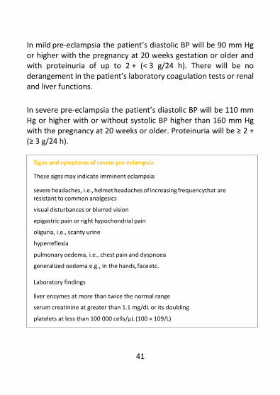
In mild pre-eclampsia the patient’s diastolic BP will be 90 mm Hg or higher with the pregnancy at 20 weeks gestation or older and with proteinuria of up to 2 + (< 3 g/24 h). There will be no derangement in the patient’s laboratory coagulation tests or renal and liver functions.
In severe pre-eclampsia the patient’s diastolic BP will be 110 mm Hg or higher with or without systolic BP higher than 160 mm Hg with the pregnancy at 20 weeks or older. Proteinuria will be ≥ 2 + (≥ 3 g/24 h).
Signs and symptoms of severe pre-eclampsia
These signs may indicate imminent eclampsia:
• severe headaches, i.e., helmet headaches of increasing frequency that are resistant to common analgesics
• visual disturbances or blurred vision
• epigastric pain or right hypochondrial pain
• oliguria, i.e., scanty urine
• hyperreflexia
• pulmonary oedema, i.e., chest pain and dyspnoea
• generalized oedema e.g., in the hands, face etc.
Laboratory findings
• liver enzymes at more than twice the normal range
• serum creatinine at greater than 1.1 mg/dL or its doubling
• platelets at less than 100 000 cells/µL (100 × 109/L)

42
Eclampsia
Eclampsia is tonic-clonic convulsion crisis occurring in a patient with pre-eclampsia and/or postictal coma.
Differential diagnosis of severe pre-eclampsia/eclampsia
For all cases presenting with a severe headache, rule out:
• Malaria, which presents with fever, shivering, muscular and joint pains and a positive thick smear;
• Migraine, which presents with vomiting;
• Meningitis, which presents with neck pain and stiffness and fever.
For all cases presenting with convulsions, rule out:
• Epilepsy, which is excluded by reviewing previous history of convulsions and the normal blood pressure of the patient;
• Cerebral malaria, which presents with fever, shivering, headaches, anaemia, jaundice and coma;
• Meningitis, which presents with headache, stiff neck, fever and photophobia;
• Tetanus, which presents with trismus, arched back, board-like rigidity and spasms of the face, neck and trunk.
Other complications of severe pre-eclampsia
Pre-eclampsia affects all organ systems in the body and has other common complications that include:

43
• abruptio placentae
• pulmonary oedema
• renal failure
• intracerebral haemorrhage
• hemolysis, elevated liver enzymes and low platelet count (HELLP syndrome)
• disseminated intravascular coagulopathy
Management hypertension in pregnancy
All patients with hypertension in pregnancy should be identified and managed preferably at a CEmONC facility.
Delays in diagnosing the condition early often result in patients presenting with severe disease and other complications that could have been prevented.
Mild hypertensive conditions can sometimes rapidly progress to severe disease within a few hours, hence close monitoring of the patient and prompt intervention when indicated are important principles of patient management.
Because pre-eclampsia presents as a spectrum of diseases, its signs and symptoms vary and some of its features may be more marked than others in some patients. For example, a patient may present

44
with normal blood pressure but have marked proteinuria. If there is doubt of the diagnosis, close monitoring of the patient is warranted, and she should be managed as for pre-eclampsia until proven otherwise.
Mild pre-eclampsia
If the pregnancy is less than 37 weeks:
• Admit the patient for at least 24 hours for laboratory investigations, BP monitoring and fetal well-being assessment.
• If the level of proteinuria increases, treat the illness as severe pre-eclampsia.
• If it is not possible to follow up with the patient in outpatient care, admit her in a ward for monitoring until delivery of her baby.
• If the level of proteinuria remains unchanged, you may follow up with the patient in the outpatient clinic at least once weekly or more frequently if feasible.
• If the diastolic BP returns to normal or remains stable, continue to follow up with the patient weekly on outpatient basis until the pregnancy is 37 weeks.
• Encourage the patient to self-monitor daily, checking her BP, urine protein and fetal activity levels at home or at a nearby clinic if feasible.
• Counsel the patient and her support persons about the danger signs and symptoms of worsening disease.

45
At each follow-up visit:
• Ask the patient about the symptoms of worsening pre-eclampsia.
• Enquire about her urine output.
• Check for high BP, urine protein and oedema.
• Check the fetus for fetal heart rate (FHR), growth and well-being.
• If possible, check the patient’s FBC, urea, creatinine, uric acid and liver function.
Admit the patient and deliver the baby if there is deterioration in the patient’s or fetus’s condition:
• If the gestational age is less than 34 weeks, provide the patient with corticosteroids for 48 hours to improve the fetus’s lung maturity prior to delivery.
• If the pregnancy is 37 weeks or more, deliver the baby.
• If there is no contraindication to vaginal delivery and the fetal well-being is reassuring:
− Assess the cervix using the Bishop score and if the score is favourable (≥ 6) and the fetal heart rate is normal, induce labour with misoprostol or oxytocin (see Appendix 7).
− If the cervix is not favourable, ripen it with prostaglandins or Foleys catheter.
− Deliver the baby by caesarean section if the fetal well-being is non-reassuring and the Bishop score is unfavourable.

46
Severe pre-eclampsia and eclampsia
The principles of the management of severe pre-eclampsia and eclampsia are generally the same. The general care guidelines are shown in Table 3.1 and Figure 3.2. The situations when a pregnancy must be ended include:
• If the patient has eclampsia, her condition must be stabilized and delivery expedited to be conducted within 12 hours of the start of her convulsive crises.
• If the pre-eclampsia is severe, delivery of the baby is needed following the appearance of symptoms or when one or more of the following indications emerge:
− Inability to control the maternal blood pressure despite administering three or more classes of antihypertensives in appropriate doses;
− Patient’s pulse oximetry at lower than 90%;
− Progressive deterioration in patient’s liver function, creatinine, haemolysis, or platelet count;
− Ongoing neurological features in the patient such as severe intractable headache, repeated visual scotomata or eclampsia;
− Placental abruption;
− Non-reassuring fetal heart rate or stillbirth.
• If the mother’s condition is stable, delivery may be delayed for 24–48 hours to allow for fetal lung maturation using steroids where indicated.

47
• In tertiary facility settings where the patient’s condition can be monitored very closely, e.g., the BP is checked every 6 hours, the expectant mother’s management can be undertaken in selected cases until the pregnancy is 34 weeks.

48
Table 3.1: Key principles in managing severe pre-eclampsia and eclampsia
1 Prevent and/or
control fits
Drugs of choice:
• 1st choice: Magnesium sulfate
• 2nd choice: Diazepam (valium)
2 Control blood
pressure
Monitor blood pressure closely. The drugs of choice are:
• Hydralazine IV, especially for unconscious patients
• Nifedipine PO (useful for conscious patient)
• Labetalol IV and PO
• Methyldopa PO (not useful in emergencies but used for
sustained BP control after the emergency is over)
3 Expedite delivery
Assess the patient for the safest and quickest route of delivery:
vaginal delivery or caesarean section
• For eclampsia, the patient must be delivered within 12 hours
• For severe pre-eclampsia, delivery must be within 24–48 hours if the patient cannot be closely monitored.
4
Monitor patient
for vital organ
failure
Kidneys: Renal failure is a common complication. Monitor urine
output carefully using an intake and output chart, urea,
creatinine, uric acid and urine for albumin/culture and
sensitivity.
Blood: Check the haemoglobin, platelet count and coagulation
profile.
Liver: Look for liver function derangements.
Heart: Check for cardiac failure.
Lungs: Check for pulmonary oedema, airway oedema and
deoxygenation (SpO2).
CNS/eyes: Check for blindness and stroke.
5 Prevent infection Administer a prophylactic broad-spectrum antibiotic. Examples
are ampicillin plus metronidazole or ceftriaxone.

49
Figure 3.2: Management of eclampsia
NURSING MEDICAL TREATMENT
Nurse preferably in an intensive care setting on a bed with protective guard rails
Keep patient’s airways clear of secretions by positioning her in the left lateral position or suction as necessary Place an indwelling urinary catheter
Monitor patient’s fluid input/output*
Insert a secure IV access line on the patient
Do blood sampling for:
• blood group
• FBC + platelets
• coagulation profile
• urea, creatinine, electrolytes
• liver function test
Do a urine test for
• culture, sensitivity
Monitor every 5 minutes and then every 15 minutes the patient’s: *
• pulse
• BP
• respiration rate and oxygen
saturation
* Record diligently all observations oncharts
Give anti-convulsive drugs: *
Magnesium sulphate: 4 g (20%) intravenously in 15 minutes, plus 5 g (50%) in a deep intramuscular injection into each buttock for a total 10 gOR only if magnesium sulphate is not available use diazepam (valium) in a 10 mg IV dose, then set up a drip of 40 mg in 500 ml of dextrose/saline to transfuse over 4–6 hours. Note: valium has depressive effects on the fetus.
• Give antihypertensive medication. Aim to achieve diastolic BP between 90 and 100 mm Hg and systolic BP lower than 160 mm Hg. Use 5-10 mg hydralazine intravenously ‘stat’ over 5 minutes. Check BP in 20 minutes and if response is inadequate repeat with 10 mg intravenously every 30 minutes as needed (maximum 20 mg per dose).
OR use 20 mg labetalol intravenously stat over 2 minutes, check BP in 10 minutes. If response is inadequate give 40 mg intravenously. Dose can be doubled every 10 minutes if indicated until max dose 300 mg is reached.
OR if patient is conscious or stable, give 10 mg of short acting nifedipine orally stat. Check BP in 20 minutes and if response is inadequate use repeat doses 10–20 mg every 30 inutes as needed (maximum 30 mg). OR use 200 mg of labetalol stat orally. Repeat the dose after 1 hour if treatment goal is not achieved (maximum should be 1200 mg in 24 hours). Provide intravenous fluid therapy: * Use dextrose/saline at a dose of 2−3 L/24 h for at least 48 hours guided by fluid balance chart. Correct any electrolyte imbalances

50
Eclampsia management if the patient is unconscious or convulsing
• Urgently mobilize the entire medical team.
• Place the patient in the left lateral position to avoid her inhaling secretions, gastric liquid or blood.
• Ensure that her upper airways are free and prevent her from biting her tongue. You may use the Guédel airway.
• Set up an IV access line on the patient and give her IV fluids, i.e. normal saline, Ringer’s lactate solution or dextrose saline.
• Stop the convulsions starting with the magnesium sulphate protocol, and if it is unavailable give 10 mg diazepam slowly intravenously (see Figure 3.3).
• Provide the patient with oxygen if necessary via a mask at 6 L per minute.
• Establish an indwelling urethral catheter on the patient.
OBSTETRICAL TREATMENT
Labour going on:
No fetal distress:
Allow spontaneous vaginal
delivery
If there is fetal distress:
Perform caesarean section or
assisted delivery with vacuum/
forceps
No labour
Consider the following:
gestational age of pregnancy
maternal condition
fetal condition (FHR)
available facilities
Decide best route of delivery
▪ caesarean section ▪ induced delivery

51
• Rapidly assess the general state of the patient and her vital signs including pulse, BP, breathing and temperature, as well as check for stiff neck and coma score.
• Check the patient’s urine for proteinuria. If the patient is not breathing or is gasping:
• Verify that her airways are well cleared and intubate her where necessary or use a face mask.
• Ventilate her using a self-inflating ambu bag at 4–6 L of oxygen per minute.
• Insert a nasogastric tube through the patient’s nose.
• Monitor the patient closely focusing on the respiratory rate, BP, pulse, temperature, urine output, fits and state of consciousness. Use a monitoring sheet.
• Collect the patient’s information to trace the history of the current disease and for medical records.
Note: If the cause of the convulsions is still not determined, treat the illness as eclampsia until proven otherwise.

52
Figure 3.3: Controlling and preventing fits with magnesium sulphate IM regimen
(a) Loading dose
(b) Followed by Maintenance Doses
(c) If breakthrough fits occur
NOTE: The intramuscular regimen of magnesium sulphate is easier to administer and simpler to monitor than using the intravenous regimen.
Maintenance
treatment doses
Give 5 g magnesium
sulphate intramuscularly
(IM)
Give deep intramuscular injection
of 2.5 g magnesium sulphate in
each buttock OR 5 g in alternate
buttocks every 4 ours.
Continue for 24 hours after last
convulsion or delivery of the baby
If more fits occur between
doses at any time
Give 2−4 g of magnesium sulphate
intravenously as a 20% solution,
slowly over 10–15 minutes
10 g intramuscular
magnesium sulphate
Give a deep
intramuscular
injection of 5 g in
each buttock
4 g intravenous
magnesium sulphate
Give as 20% solution
Slow intravenous injection
given over a period of not
less than 5 minutes,
preferably 10−15 minutes Give the loading
dose

53
For how to prepare magnesium sulphate doses, see Appendix 8.
Clinical monitoring during the administration of magnesium sulphate
Monitor the patient closely every 15−30 minutes. Give her the next IM dose of magnesium sulphate only if:
• her respiratory rate is greater than 16 per minute
• her urine output is greater than 25 ml per hour
• she has patellar reflexes
Clinical guidelines for the management of complications of magnesium sulphate
• If the patient experiences respiratory arrest:
− Intubate and ventilate her immediately.
− Stop the magnesium sulphate treatment.
− Give her 1 g (10 ml) of calcium gluconate intravenously. This is the antidote for magnesium toxicity.
− Continue ventilating her until she resumes normal, spontaneous respiration.
WATCH OUT for complications of magnesium sulphate!
Magnesium sulphate has potential hazards such as the following:
respirator arrest respiratory depression absent patellar reflexes renal failure

54
• If the patient has respiratory depression, i.e., she has fewer than 16 breaths per minute:
− Give her oxygen by a mask.
− Give her 1 g of calcium gluconate intravenously.
− Stop the magnesium sulphate treatment.
− Maintain her airway unobstructed.
• If the patient does not have patellar reflexes, monitor her respiratory rate closely. If her respiration is normal, stop the magnesium sulphate doses until the reflexes return. If her respiration is depressed:
− Give her oxygen by a mask.
− Give her 1 g of calcium gluconate intravenously.
− Stop the magnesium sulphate treatment.
− Maintain her airway unobstructed.
• If the patient’s urine output is less than 100 ml over 4 hours, i.e., less than 25 ml per hour:
− If she has no other signs of magnesium toxicity reduce the next dose of magnesium sulphate to 2.5 g.
− If there are other signs of magnesium toxicity, manage her as described under the appropriate section above.
− Review the overall management of the patient with particular attention to fluid balance and blood loss.
Once a patient’s reflexes have returned, magnesium sulphate can be restarted at a reduced dose if the patient is still at a risk of further convulsions, e.g., from poorly controlled BP. Magnesium sulphate can be discontinued if the patient is stable, blood pressure is controlled well, the patient has been delivered and she has not had convulsions over 24 hours.

55
Controlling high blood pressure
If the patient’s diastolic BP remains above 110 mm Hg, give her antihypertensive medication following the protocols shown in Figure 3.4 to maintain the diastolic BP at 90–100 mm Hg in order to prevent complications.
Figure 3.4: Controlling BP in hypertensive emergencies
Hydralazine protocol (for unconscious patients)
Nifedipine protocol (for conscious patients)
Give 5–10 mg hydralazine intravenously
slowly over 5–10 minutes Give the loading dose
Check the BP every 20 minutes (before the next treatment dose) and record the BP
Give the repeat dose (10 mg) every 30 minutes until diastolic BP of 90 mm Hg or
lower to 100 mm Hg is achieved. (maximum 20mg)
Maintenance dose
Give 10 mg oral (short-acting)
nifedipine* Give the loading dose
Check the BP every 20 minutes (before administering the next treatment dose)
If the expected level is not reached, give 10–20 mg of oral (short-acting) nifedipine* every 30 minutes until the diastolic BP is 90 mg Hg
or lower to 100 mm Hg. The maximum amount of nifedipine should be 30 mg.
Maintenance dose

56
Note: Do not puncture the nifedipine capsule as this may lead to faster drug action and this
can crash the BP and compromise fetal survival.
Labetalol protocols (IV and PO)
Maintaining blood pressure control
The dosage of antihypertensive drugs should be carefully tailored to the patient’s response.
• Check and note the patient’s BP before administering subsequent treatment doses.
• The oral antihypertensives methyldopa, labetalol and long-acting nifedipines such as adalat retard may be used to maintain the BP after its initial control with rapid acting or IV antihypertensive drugs.
Give 20 mg labetalol intravenously slowly over 5 minutes OR
200 mg orally if the patient is conscious
Loading dose
Check BP every 10–15 minutes (before each subsequent) IV dose
If the desired BP level is not reached:
Maintenance dose
Avoid labetalol in
asthmatics
Other antihypertensive treatment options should be considered if the BP is not lowered within the acute treatment phase of 90 minutes with the short-acting drugs, e.g., after 30 mg of short-acting nifedipine.

57
• When administering a combination of antihypertensive medications note their respective onsets of action, peak action and duration of effect in order to avoid potentiating effects and crashing of blood pressure below the recommended threshold levels (see Table 3.2).
• Note that blood pressure tends to drop after the delivery of a baby and the mother may require lower drug dosages then.
Table 3.2: Onsets, peak actions and durations of effect of the antihypertensive medications
Drug Dosage Onset of action Peak action Duration
Hydralazine IV 5–10 mg 15–20 min 60 min 3–6 h
Nifedipine (SA) 10 mg 5–10 min 30 min 3 h
Nifedipine (LA) 20–30 mg 30 min 4 h 12–24 h
Labetalol IV 20 mg 2–5 min 15 min 3–6 h
Labetalol PO 200 mg 20–60 min 1–4 h 8–12 h
Methyl Dopa 250 mg–2 g 40 min 3–6 h 12-24 h
Notes: PO = orally, SA = Short Acting, LA=Long Acting, IV=intravenous
Intravenous fluid management
• Carefully monitor IV fluid or electrolyte administration as the risks of pulmonary oedema and oliguria are high in patients with severe pre-eclampsia or eclampsia.
• Administer IV fluids judiciously; the fluid deficit is usually about 600 ml.
• Use crystalloids, i.e., normal saline, Ringer’s lactate or dextrose saline. Avoid plain dextrose as it can aggravate oedema.

58
• Give IV fluids at the rate of 1 L every 6–8 hours, that is 3 ml per minute or 30 drops per minute. Do not give the patient more than 125 ml per hour of either IV or oral fluids unless it is indicated. If an IV fluid bolus is indicated especially before the induction anaesthesia is used, it must be administered cautiously.
• Monitor, document and balance the fluid input and output very closely:
− use an indwelling bladder catheter if possible
− consider all fluids given with medications, e.g., IV injections etc.
• Monitor the patient’s lung fields and oxygen saturation closely.
• Monitor electrolyte levels at least once weekly if the patient is on IV fluid therapy. Check these more frequently if this is feasible.
Inpatient monitoring of the mother and baby
Patients must be monitored closely and preferably in intensive care settings until they are stable.
• Monitor the patient’s level of proteinuria and urine output daily.
• Check her for signs of pulmonary oedema daily.
• Do laboratory investigations to check her urea, creatinine, uric acid, proteinuria of 24 hours, liver function, platelets, haemoglobin (Hb) and the coagulation profile at least once weekly or more frequently if feasible or if it is indicted.
• Monitor fetal well-being daily, focusing on FHR, kick count, ultrasound results, biophysical profile, etc.

59
Delivery care and management
• If the cervix condition is favourable, i.e., the Bishop score is 6 or higher, and the fetus is alive and has no sign of distress, induce labour for the patient using misoprostol or oxytocin (see Appendix 7).
• If the cervix condition is unfavourable or if vaginal delivery is not foreseeable within 12 hours in cases of eclampsia or within 24 hours in cases of severe pre-eclampsia, perform a caesarean section.
• If a non-reassuring fetal heart pattern or fetal distress is noted, e.g. bradycardia with less than 100 beats per minute or tachycardia with more than 180 beats per minute, perform a caesarean section.
• If there are contraindications to performing a caesarean section on the patient, e.g., if the risk for a negative outcome is high, if it is impossible to use anaesthesia, or if the fetus is dead or is not viable, envisage vaginal delivery.
• If the condition of the cervix is not favourable, ripen it with prostaglandins or a Foley’s catheter, and then induce labour.

60
Preparing for a caesarean section
• Check the patient’s FBC and coagulation status.
• Ensure that blood and fresh frozen plasma are available because the risk of coagulopathy is high.
• Ensure that an anaesthetic review is done before surgery to decide on the best anaesthetic method for the case:
− Spinal or epidural anaesthesia can be given safely if the patient is conscious, seizure free and has stable vital signs and no signs of raised intracranial pressure.
− General anaesthesia is recommended for unconscious patients or patients with evidence of increased intracranial pressure.
Postpartum care
• Continue the antihypertensive treatment as long as the diastolic BP is 110 mm Hg or higher.
• Maintain the IV access line and the IV fluid therapy in the form of Ringer’s lactate, normal saline or dextrose saline for at least 48 hours in the immediate postpartum period.
• Maintain an input/output chart until the patient is discharged. The urine output should be more than 30 ml per hour.
Management of other related complications
Refer the patient to a specialist or ask for a specialist review if the patient has:
• Persistent oliguria for 48 hours after delivery,
• Coagulation disorders or the HELLP syndrome,

61
• Prolonged coma that lasts more than 24 hours after convulsions,
• Uncontrolled blood pressure.
Patients with the following related pre-eclampsia complications should be managed in specialist centres with intensive care facilities.
HELLP syndrome
The HELLP syndrome tends to occur towards the end of the second or at the beginning of the third trimester of pregnancy and is associated with hemolysis, rise in liver transaminases and thrombocytopenia (low platelet levels).
The signs and symptoms of HELLP syndrome include:
• hypertension
• proteinuria
• epigastric pain
• upper abdominal tenderness
• nausea and vomiting
• jaundice
• Patients with the HELLP syndrome are at an increased risk of bleeding, severe anaemia and hepatorenal failure.
• The appearance of epigastric pain should alert you to the possibility of a sub-capsular haematoma of the liver, which may rupture.

62
• Platelet levels below 50 000/dL increase the patient’s risk of haemorrhage during and after she delivers the baby.
• Ensure the availability of blood products such as packed cells and fresh frozen plasma or platelets.
Acute pulmonary oedema
• Position the patient in a semi-reclining posture for nursing.
• Provide the patient with oxygen therapy at 3−6 L per minute using non-invasive artificial ventilation to keep the SpO2 level at higher than 95%.
• Give the patient IV furosemide at 40−80 mg immediately, review the response and repeat this dosage as required.
• Monitor the patient’s respiratory rate and level of oxygenation.
Acute kidney failure
• Diagnose renal failure if the patient has oliguria, i.e. urine output of less than 30 ml per hour associated with a rise in urea and creatinine levels.
• Provide the patient with IV fluids and give her the 20–40 mg furosemide IV challenge.
• If there is no response or it is inadequate, refer or transfer the patient to a specialized unit.
Gestational HBP without proteinuria or oedema
• Inform the patient and her family about the danger signs indicating pre-eclampsia or eclampsia.

63
• Admit her for at least 24 hours for laboratory investigations, BP monitoring and assessment of fetal well-being.
• If the BP is high and or is rising or pre-eclampsia develops, manage her care as for severe pre-eclampsia, i.e. using nifedipine, labetalol or alpha methyldopa.
• If there is evidence of intrauterine growth restriction or fetal distress, deliver the baby.
• If the BP remains stable and there is no sign of proteinuria, continue monitoring the patient weekly on an outpatient basis until the pregnancy is full term and then deliver the baby.
Care at discharge and follow-up
• Emphasize to the patient the importance of regular medical follow-up, and before her discharge link her up for follow-up care at the primary care services.
• Counsel the patient on:
− The importance of continuing with BP monitoring and antihypertensive drug regimens as prescribed;
− The risk of the recurrence of hypertension in future pregnancies and the preventive interventions that may reduce those risks;
− The importance of initiating antenatal care early in all her future pregnancies and avoiding unplanned pregnancies.
− Family planning and postpartum contraception options.
− Her increased risk in the future for cardiovascular disease such as chronic hypertension and stroke.

64
Prevention of pre-eclampsia and eclampsia in subsequent pregnancies
• Consider a woman to be at a high risk of developing pre-eclampsia if she has one high‐risk factor from among hypertensive disease in a previous pregnancy, chronic hypertension, chronic renal disease, diabetes mellitus and an autoimmune disease or any two moderate‐risk factors from among multiple pregnancies, age of 40 years or more, a BMI of 35 kg/m2 or higher, family history of pre-eclampsia and an interpregnancy interval that is greater than 10 years.
• Prevent pre-eclampsia and its complications in women at a high risk of it as follows:
− Where a woman’s dietary calcium intake is low, i.e. less than 800 mg/day, provide calcium supplementation during her pregnancy at 1.5–2 g of elemental calcium per day.
− Provide low-dose acetylsalicylic acid (aspirin) at 75–150 mg daily at night.
− Initiate treatment early, i.e. at 12 weeks of gestation and continue until 36 weeks of gestation, when delivery occurs, or when pre-eclampsia is diagnosed.

65
CHAPTER 4 OBSTETRIC HAEMORRHAGE
Definition
Obstetric haemorrhage is vaginal bleeding occurring in the later part of a pregnancy, during labour or after childbirth. It can be classified broadly into:
• Antepartum haemorrhage, which occurs after the second trimester and before the delivery of the baby;
• Intrapartum haemorrhage, which occurs during labour and before birth;
• Postpartum haemorrhage, which comes after childbirth and up to 6 weeks postdelivery.
Problem
An obstetric haemorrhage incident constitutes an obstetrical emergency requiring immediate and appropriate care and treatment. It is often unforeseeable, yet it is the major cause of maternal mortality in most countries in sub-Saharan Africa.
Antepartum and intrapartum haemorrhage can be caused by:
• placenta praevia
• abruptio placentae
• uterine rupture (see Chapter 5: Dystocia)
• vasa praevia, but this is rare

66
• cervical lesions e.g., from polyps or cervical cancer Postpartum haemorrhage can be caused by:
• uterine atony
• retention of placental tissue
• lacerations of the soft tissues of the perineum, vagina, cervix or uterus
• clotting failure or coagulopathy
Placenta praevia
Definition
Placenta praevia is the total or partial implantation of the placenta on the lower part of the uterus.
Problem
Placenta praevia is the most frequent cause of haemorrhage in the late part of a pregnancy. In its serious forms, it leads to complications for the pregnant woman and the fetus that could result in death. It is therefore an obstetrical emergency condition. Diagnosis
During pregnancy and labour, placenta praevia may present with several symptoms and signs.

67
Symptom
• Bright red vaginal bleeding occurring suddenly and painlessly. Bleeding tends to reoccur late in the pregnancy, provoked sometimes by sexual intercourse.
Signs
• shock and anaemia corresponding to the apparent blood loss
• soft and non-tender uterus
• malpresentation and/or a high presenting part
The fetal heart sound and rate are usually normal.
NOTE: Once you suspect that the patient has placenta praevia, you should not perform a vaginal
exam on her.
Investigation
An ultrasound scan will show a placenta implanted in the lower segment of the uterus with the distance between the lower edge of the placenta and the internal cervix opening as less than 5 cm. DO NOT delay care and treatment waiting for an ultrasound report.

68
Management
General measures
For immediate care:
• Insert an intravenous line on the patient and take blood samples for grouping and cross-matching.
• Start infusing the patient with normal saline or Ringer’s lactate solution to compensate for the blood loss and to stabilize the patient’s hemodynamic state.
• Establish a urinary bladder catheter on the patient to monitor her urine output.
• Monitor and document her vital signs every 15 minutes, i.e. her pulse, BP, respiration rate, temperature and state of consciousness.
• If the patient is in shock, treat her following the guidelines in Appendix 2.
NOTE: If your unit is a primary level facility, refer the patient immediately to a CEmOC unit and accompany her there.
Obstetrical care before labour
• If the patient is haemorrhaging profusely and has haemodynamic instability, perform a caesarean section irrespective of the fetus’s gestational age.
• If the bleeding has stopped and the patient is stable, admit her in the ward, manage her expectantly until the pregnancy gestation is 37 weeks, and plan to deliver the baby by caesarean section.

69
• Ensure that blood is always available in the blood bank.
• Correct the patient’s anaemia if she has it.
• If the patient’s haemorrhage is minimal:
− Hospitalize her and monitor her pregnancy until delivery of the baby.
− Correct her anaemia.
− Ensure there is blood available in case the patient needs transfusion.
− If the patient is less than 37 weeks into the pregnancy, manage her expectantly until she is full term.
− If the pregnancy is 37 weeks or older, plan to deliver the baby immediately.
− If an ultrasound is not available or its report is inconclusive and the pregnancy is 37 weeks or older, examine the patient in the theatre under a double set-up to exclude placenta praevia.
Deliver the baby by caesarean section if the patient has major placenta praevia, i.e. types II to IV.
The double set-up
• An IV line is set up and is running on the patient.
• The surgical team is scrubbed and ready.
• A high level and disinfected vaginal speculum is used to visualize the cervix.
• A digital vaginal exam is carefully performed to check for the presence of a soft, spongy mass in the lower segment, which would be felt at the vaginal fornix.

70
During labour
• If the patient is bleeding profusely and has haemodynamic instability, perform a caesarean section on her irrespective of the fetal gestational age.
• For a patient with minimal haemorrhage:
• If an ultrasound scan has identified major placenta praevia in the patient, perform a caesarean section on her.
• If an ultrasound is not available, examine the patient in the theatre under a double set-up to exclude placenta praevia (see box above). Perform a caesarean section on her if the placenta praevia level is greater than types II to IV.
• If the double set-up examination in the theatre considers the placenta praevia to be type I (marginally inserted in lower segment) and delivery is imminent (cervix dilation is greater than 8 cm, the head is three-fifth descended and the membrane is intact), you may perform an artificial rupture of the membranes, augment labour and deliver the baby under careful observation. Other treatment measures
• Transfuse the patient if her haemoglobin level is below 7 g/dL and/or she has signs of haemodynamic instability such as tachycardia, tachypnoea, low BP and change in mental state (refer to Appendix 8).
• Start the patient on the antibiotic therapy.
• Prescribe for the patient 120 mg of iron and 400 µg of folic acid to

71
take daily until the anaemia is corrected, which is in at least 3 months.
• If the newborn requires resuscitation or is preterm, manage him or her appropriately (see Chapter 7).

72
Figure 4.1: Management of placenta praevia
Suspected placenta praevia
Symptoms Signs
- Vaginal bleeding - Anaemia - Bright red blood - Shock - Painless uterus - Soft, non-tender uterus - Bleeding may be provoked by contact - Malpresentation - Normal FHR
Do NOT perform a vaginal examination
At the primary level (BEmOC) - Establish an IV line on the patient - Take blood for Hb, grouping and cross-matching - Resuscitate the patient with IV fluids
- Refer the patient for CEmOC
At the CEmOC unit - Perform an ultrasound scan - Resuscitate the patient with IV fluids/ blood - Collect blood for Hb, grouping and cross-matching - Provide the final treatment depending on:
- the patient’s degree of bleeding - the gestational age of the pregnancy - the type of placenta praevia

73
Abruptio placentae
Definition
Abruptio placentae is the premature detachment of a normally situated placenta before childbirth.
Problem
Abruptio placentae is a cause of intrauterine fetal death and fresh stillbirth and can lead to massive obstetrical haemorrhage leading to shock, coagulation disorders, kidney failure and maternal death. Diagnosis
Abruptio placentae presents with the following symptoms and signs: Symptoms
• vaginal bleeding
• abdominal pains that are usually severe
• probable history of trauma
• probable history or features of pre-eclampsia
Signs
• shock and anaemia that is out of proportion with the apparent blood loss
• uterine overdistension that is greater than the expected

74
symphysiofundal height
• presenting part that may or may not be engaged
• fetal parts with palpating difficulty
• uterine tenderness
• increased uterine tone (wooden like sensation)
• uterine irritability or contractions
• usually normal presentation
• abnormal or absent FHR
• likelihood of bleeding tendencies having developed (coagulopathy)
Investigation
An ultrasound may reveal a retroplacental haematoma. Note: The diagnosis of abruptio placentae is usually made clinically.
General measures
Note: If the patient is first seen at a primary level health unit, she must be referred immediately to
a unit with CEmOC.
Immediate care
• Set up an intravenous line on the patient.
• Collect blood for grouping and cross-matching.
• Start infusing the patient with normal saline or Ringer’s lactate solution to compensate for lost blood and stabilize her

75
hemodynamic state.
• Insert a urinary bladder catheter on the patient to monitor her urine output.
• Monitor and document her vital signs, i.e. pulse, BP, respiration rate, SpO2, temperature and state of consciousness, as well as the fetal heart rate (if present) every 15 minutes.
Note: When abruptio placentae occurs in a patient with severe pre-eclampsia, her BP readings may be normal even when the she is in shock.
Obstetrical care
If the fetus is alive:
• Perform a caesarean section if the patient is not in labour or in active labour and the cervical dilation is less than 8 cm.
• If delivery is imminent for the patient, i.e., her cervical dilatation is greater than 8 cm, vaginal delivery is advised with augmentation of labour.
• If the fetus is dead:
• Stabilize the patient with intravenous fluids, fresh frozen plasma and blood.
• Rupture the patient’s membranes and augment her labour with oxytocin.
• Provide the patient with IM analgesia pethidine at a dose of 1 mg/kg.
• If the patient’s delivery is not foreseen within 6 hours or if blood

76
reserves are not readily available, perform a caesarean section on the patient.
Note: Most patients with abruptio placentae will deliver the baby within 6 hours of the initiation of treatment.

77
Figure 4.2: Management of abruptio placentae
Suspected abruptio placentae
If the fetus is alive: Aim for vaginal delivery if the cervical dilatation is greater than 8 cm, and patient is stable. Perform a caesarean section if fetal distress or patient .is ubstable.
Symptoms Signs Vaginal bleeding Shock Abdominal pain Anaemia Trauma Tender, hard abdomen Pre-eclampsia SFH that is greater than for a fetus of that age Abnormal/no FHR
At the primary care level • Place an IV line on the patient • Take blood sample for Hb, grouping and cross-matching • Resuscitate the patient with IV fluids • Refer and accompany the patient to a CEmOC
At the CEmOC level • Resuscitate the patient with fluids • Collect blood for Hb, grouping and cross-matching • Transfuse the patient with blood and fresh frozen
plasma • Check Clotting profile, bedside clotting time • Check fetal heart rate
If the fetus is dead: Rupture the membranes and augment labour with oxytocin. PerformCesarean section if patient is unstable and far from delivery

78
Coagulation disorders
Definition
Disseminated intravascular coagulation (DIC) is a syndrome with abnormal coagulation and fibrinolysis and is also known as consumption coagulopathy. It is a disorder marked by a reduction in the concentration of platelets and clotting factors due to the exhaustion of these factors in the peripheral blood. It occurs in association with obstetric complications such as:
• abruptio placentae
• eclampsia or pre-eclampsia
• postpartum haemorrhage
• sepsis
• ruptured uterus
• hypovolemic shock or massive blood transfusion
• amniotic fluid embolism
• intrauterine death
• trophoblastic disease such as choriocarcinoma
Problem
DIC is one of the main causes of the massive obstetrical
haemorrhage following the obstetrical complications listed above.

79
Diagnosis
• Consider a patient to have a coagulation disorder if she suffers persistent obstetrical haemorrhage, i.e., her blood fluid has few or no clots despite the discovery and treatment of the causes of her bleeding.
• Make a quick diagnosis of a coagulation disorder with a bedside coagulation test:
− Take 2 ml of blood from a vein on the patient in a clean and dry glass test tube.
− Hold the tube in a clenched fist to keep it warm.
− Suspect DIC if no clot is formed in the blood within 5–10 minutes.
• Perform laboratory tests for:
− blood grouping and cross-matching
− haemoglobin level
− hematocrit, platelet and fibrinogen levels
− prothrombin time Management
General measures
• Treat the patient for underlying obstetrical complications and shock.
• If she has placenta praevia, refer her to a CEmOC unit (see Appendix 1).

80
Note: DO NOT give colloids or dextran to the patient as these interfere with clotting. Resuscitate the patient with normal saline or Ringer’s lactate solution.
Specific measures
• Give the patient 1 g of tranexamic acid (TXA) intravenously immediately and slowly and repeat after 30 minutes if necessary. It is most effective if administered within 3 hours of the onset of bleeding.
• Transfuse the patient with recently donated whole blood (see Appendix 9).
• Transfuse the patient with fresh frozen plasma at 15 ml/kg body weight.
• Transfuse the patient with cryoprecipitate to replace the fibrinogen lost through bleeding.
• Transfuse the patient with platelet concentrates if her platelet level is below 20 000 platelets per ml.
Postpartum haemorrhage
Note: PPH is an obstetrical emergency that can lead to death within 2 hours of onset if no action is taken to handle it.
Definition
Postpartum haemorrhage (PPH) is vaginal bleeding in excess of 500 ml after childbirth or any blood loss that has the potential to result in cardiovascular instability. It can be classified as:
• Primary (immediate) postpartum haemorrhage, which is excessive bleeding that occurs within the first 24 hours of delivery. About 70% of the immediate PPH cases are due to uterine atony. Atony

81
of the uterus is defined as the failure of the uterus to contract adequately after a child is born.
• Secondary (late) postpartum haemorrhage, which is excessive bleeding occurring between 24 hours after the delivery of a baby and 6 weeks postpartum. Most late PPH is due to the retention of the products of conception or infection, or a combination of the two.
Traditionally PPH has been defined as blood loss in excess of 500 ml for vaginal deliveries and 1000 ml for caesarean section deliveries. The common causes of PPH can be recognized with a 4T mnemonic:
• tone – uterine atony
• tissue – retained placenta or clots
• trauma – uterine, cervical or vaginal tears or injury
• thrombin – pre-existing or acquired coagulopathy
Problem
Postpartum haemorrhage is the leading cause of maternal mortality in sub-Saharan Africa.
Diagnosis
Symptoms and signs
• increased bleeding after childbirth. This is primary PPH.
• tachycardia and low blood pressure

82
• a deteriorating level of consciousness
• uterine distension/flabby uterus
• tears in the perineum, vagina, cervix and uterus
• presence of retained products of conception
• bleeding tendencies Management
Note: All women who give birth must be offered Active Management of the Third Stage of Labour (AMSTL)including uterotonics during the third stage of labour to prevent PPH. (See Appendix 10)
General measures
If a patient has increased blood loss after delivery, suspect PPH:
• Call for help; PPH requires team management.
• Massage the uterus to stimulate a contraction and expelling of blood or blood clots.
• Give the patient 10 IU of oxytocin intramuscularly.
• Start an IV line on the patient with a wide bore needle and collect blood samples for blood grouping and cross-matching.
• Infuse the patient with IV fluids (see treatment for shock in Appendix 2).
• Provide an anti-shock garment if it is available (see Appendix 11).
• Establish a catheter in the patient’s bladder.
• Perform a rapid evaluation of the general condition of the patient,

83
including checking her vital signs of pulse, BP, respiration, SpO2 and temperature.
• Check for the possible causes of PPH:
− assess if the placenta is completely expelled
− examine the cervix, vagina and perineum for tears
− assess the uterine tone and contractility again Uterine atony
• Continue to message the uterus.
• Administer oxytocin at a dosage of 20−40 IU in 1 L of normal saline or Ringer’s lactate solution and infuse at 60 drops per minute.
• Continue the oxytocin treatment at a dose of 40 drops per minute for at least 6 hours.
OR
• Use carbetocin if its available, administering 100 µg as a single IV dose over 1 minute or as intramuscular bolus injection. Note that carbetocin has a longer duration of action (1 hour) than oxytocin.
• If oxytocin or carbetocin is not available:
• Give the patient 0.2 mg of ergometrine intravenously or intramuscularly. Repeat this after 15 minutes and then continue with same dosage every 4 hours for up to a total of 1 mg.
OR
• Give 800 µg of misoprostol sublingually immediately. Repeat

84
misoprostol if necessary after 30 minutes but using 200–800 µg, for a maximum of 1600 µg.
• If bleeding continues despite taking the management steps described above:
• Give the patient 1 g of tranexamic acid (TXA) intravenously immediately and slowly and repeat this after 30 minutes if necessary (NB: Give the patient TXA within 3 hours of giving birth). If bleeding still persists:
• Perform a bi-manual compression of the uterus.
OR Perform external aortic compression.
• Consider the use of uterine balloon tamponade (see Appendix 12). If bleeding continues despite conducting all the management procedures above:
• Perform a laparotomy on the patient.
• Carry out a uterine and ovarian artery ligation.
• Insert a B-Lynch suture into the uterus.
• Carry out a subtotal abdominal hysterectomy.
• Administer a broad-spectrum antibiotic, for example 2 g of amoxicillin and metronidazole with gentamycin.
Note: Ensure blood is available and transfuse the patient if she is haemodynamically unstable or has a Hb level lower than 7 g/dL.

85
Retained placental tissue
Retention of the placental tissue can take several forms. The entire placenta may stay imbedded in the uterus or this might be just a part of it (partial placenta retention).
• Repeat the dose of 10 IU of oxytocin either intravenously or intramuscularly with controlled cord traction.
• Insert a catheter into the patient’s bladder.
• Manually remove the placenta in the theatre with the patient under analgesia.
• Carry out evacuation of the retained products of conception in the theatre.
Tears of the cervix, vagina or perineum
• Examine the patient and determine the extent and location of the tears, preferably doing this in the theatre and under anaesthesia.
• If the summit of the tear of the cervix is accessible, stitch the lesion through the vagina.
• If a haematoma is present but is not bleeding or increasing in size, keep the patient under observation.
• If the haematoma increases in volume, evacuate the clot and identify and stitch the bleeding vessel(s).
• Administer analgesic and antibiotic medication to the patient.
• If the summit of the tear extends beyond the cervix, perform a laparotomy to identify the upper limit of the tear and then repair it accordingly.
NOTE: There may be multiple tears.

86
Inversion of the uterus
• Attempt manual reduction.
• Administer 10 UI of oxytocin intravenously.
• If these processes fail, perform a uterus laparotomy on the patient.

87
Figure 4.3: Prevention and management of primary PPH
Postpartum haemorrhage
• Vaginal bleeding > 500 ml
• Haemodynamic instability with less blood loss
• Blood loss following caesarean section >1000 ml
Prevent PPH by routinely doing AMTSL (Appendix 10)
• Give the patient an uterotonic agent (10 IU of oxytocin) within 1 minute of birth or other uterotonic (e.g. misoprostol) if oxytocic not available.
• Perform controlled cord traction.
• Initiate breastfeeding (for suckling effect)
If there is increased blood loss, suspect PPH:
• Assess the airway, breathing and circulation (ABC).
• If there is uterine atony, massage the uterus.
• Insert an IV line on the patient and treat her for shock (Appendix 2).
• Administer oxytocics: give 20 IU of oxytocin in 500 ml normal saline and infuse it at 60 drops per minute OR administer 100 µg of carbetocin intravenously or intramuscularly stat OR 0.2 mg of ergometrine intravenously or intramuscularly. Repeat this after 15 minutes OR use 800 µg of misoprostol sublingually.
• Empty the patient’s bladder and monitor her urine output.
• Refer the patient to a CEmOC unit if the bleeding persists or for transfusion.
• Use an anti-shock garment if it is available (Appendix 11).
• Insert a condom tamponade in the uterus if you have skills for this (Appendix 12).
• At the CEmOC unit you may need to perform an examination under anesthesia, a laparotomy, B-Lynch sutures, aortic compression, internal iliac artery procedure.
Retained placenta
Manual removal of the
placenta
Lacerations
Examine the perineum, vagina and cervix
and repair them under analgesia
Uterine rupture
Refer the patient to a CEmOC unit for
laparotomy (repair/subtotal) or
hysterectomy
Coagulopathy
Insert an IV line and
administer 1 g of TXA
slowly intravenously.
Transfuse patient with
frozen fresh plasma and
blood.

88
Secondary postpartum haemorrhage
Definition
Secondary postpartum haemorrhage is bleeding that occurs 24 hours after delivering a baby and within 42 days postpartum. It may be caused by:
• endometritis (see Chapter 6, section 3)
• partial placenta retention or retention of a cotyledon (see retained placental tissue in Figure 4.3)
• coagulation disorders (see page 79)
• wound dehiscence, e.g. caesarean section tears
Problem
Secondary postpartum haemorrhage commonly occurs after a new mother has returned home and it is a cause of maternal mortality.
Diagnosis
• Haemorrhage of more than 500 ml occurring after 24 hours of delivery or persistent or reappearing bleeding within 42 days after delivery.
• Check the Hb level, perform blood grouping and cross-matching, and check the clotting profile.
• Do an ultrasound to check for retained products of conception.

89
Management
• Resuscitate the patient and identify and treat the cause.
• For immediate care: establish an IV line on the patient and start infusing her with normal saline or Ringer’s lactate solution to treat shock and stabilize the haemodynamic state.
• Monitor and document the vital signs of pulse, BP, respiration rate, temperature and state of consciousness every 15 minutes and the amount of blood loss.
• Insert a bladder catheter to monitor the patient’s urine output.
• The subsequent treatment will depend on the cause of the haemorrhage. Usually there is need to evacuate the retained products of conception.

90
CHAPTER 5: DYSTOCIA/ABNORMAL LABOUR Definition
Dystocia refers to any abnormality in the progress of labour. It may be due to mechanical reasons or to poor uterine activity. Problem
Dystocia may lead to cases such as:
• Fetal distress that could lead to the death of the fetus
• Uterine rupture, in the case of a mechanical dystocia with high risk of maternal death
• Obstetrical fistula
• Sepsis in mother and newborn.
This chapter discusses
• Prolonged (first and second stage) labour
• Cephalopelvic disproportion
• Obstructed labour
• Transverse lie
• Shoulder dystocia
• Cord prolapse
• Breech presentation in labour
• Face presentation
• Brow presentation
• Labour and delivery in a scarred uterus

91
• Ruptured uterus
• Preterm labour
• Fetal distress (non-reassuring fetal heart status).
1. Prolonged (active first stage) labour
Definitions
▪ The first stage of labour comprises the latent and active phases. The current guidelines define the latent phase as the period characterized by painful uterine contractions and variable changes of the cervix, including some degree of effacement and slower progression of dilatation up to 5 cm.
▪ The active phase is the period characterized by regular painful uterine contractions, a substantial degree of cervical effacement and more rapid cervical dilatation from 5 cm until full dilatation, for first and subsequent labours.
▪ The standard duration of the latent phase of labour has not been established and can vary widely from one woman to another. Duration of active phase (from 5 cm until full cervical dilatation) however, usually does not extend beyond 12 hours in first labour, and usually does not extend beyond 10 hours in subsequent labours.
▪ The second stage of labour is the period between full cervical dilatation and birth of the baby, during which the woman has an involuntary urge to bear down, as a result of expulsive uterine contractions. In first labours, birth is usually completed within 3 hours, whereas in subsequent labours, birth is usually

92
completed within 2 hours.
▪ Durations of these phases of labour beyond these stated periods may be considered as unusual and call for careful patient review.
Problem
Prolonged labour contributes to high maternal and perinatal mortality and morbidity. These consequences can be prevented by close monitoring of the progress of labour and maternal and fetal well-being during labour. Diagnosis
Women with suspected slow labour progress should be carefully evaluated for the underlying cause (e.g., cephalo-pelvic disproportion) and to identify any developing risks or complications. The decision to intervene must not be taken based on duration alone. Prolonged labour can be recognized by:
• Active phase of labour longer than 12 hours in a nullipara and 10 hours in a multipara, commencing from a cervical dilatation plotting of 5 cm
• Second stage labour lasting longer than 3 hours in a nullipara and 2 hours in a multipara
• Cervical dilatation and fetal head descent plot showing arrest/stagnation over time
• Clinical evidence of deterioration in maternal physical

93
condition (BP, pulse, temperature, state of hydration, urine output)
• Symptoms showing that the mother’s emotional and psychological needs are not being met (e.g., poor cooperation, distress)
• Clinical evidence of increased risk for adverse fetal outcomes (e.g., non-reassuring fetal heart rates, caput succedaneum, moulding, meconium staining of amniotic fluid).
The WHO Labour Care Guide 2020 should be used to monitor labour (see Appendix 13)
Management
“Prolonged” latent phase
Note: The standard duration of the latent phase of labour has not been established. Labour may not naturally accelerate until the cervical dilatation threshold of 5 cm is reached (see WHO intrapartum care recommendations 2018). If cervical dilatation is < 5 cm:
• Observe mother in a unit outside of birthing area (within labour or maternity ward
• Reassure her that the duration of labour is highly variable and depends on individual physiological process and pregnancy characteristics
• If fetal and maternal conditions are reassuring, continue routine labour observations to assess maternal and fetal well-being (at least four hourly) until 5 cm cervical dilatation threshold is reached (active phase)

94
• Encourage the woman to use relaxation techniques, including progressive muscle relaxation, breathing, music, and mindfulness
• Provide pain relief depending on her preferences, and according to national protocols (e.g., massage or application of warm packs, fentanyl, diamorphine and pethidine
• Do not make use of medical interventions such as oxytocin augmentation or caesarean section to accelerate labour and birth before this threshold
• If at BEmOC, refer patient to CEmOC, if maternal or fetal condition is not reassuring or if there is deterioration and/or if any of the following complications are identified
o cephalopelvic disproportion o abnormal lie (transverse) o malpresentation (brow, breech, face) o non-reassuring fetal heart rate (FHR)/fetal distress o maternal emotional and psychological distress o patient has been in labour for more than 12 hours
(especially if membranes are ruptured).
NOTE: It is important to distinguish between latent labour and false labour, which is
characterized by irregular and infrequent uterine contractions, not resulting in
progressive effacement and dilatation of the cervix.

95
Figure 5.1: Management of prolonged latent phase of labour
▪ Membranes ruptured
▪ Abnormalities of theFHR) and/or
▪ Abnormality of theamniotic fluid
▪ Start antibiotics
▪ If at BEmOC refer to
▪ CEmOC
▪ At CEmOC perform cesarean section
Uterine stop
Monitor progress of active phase of labour
Refer to CEmONC or perform cesarean section
False Labour
Uterine contractions have occurred for more than 8 hours with cervical dilatation still less than 5 cm
Cervix is dilating but slowly FHR is normalAmniotic fluid is clear Maternal emotional and psychological needs are being met
Membranes intact No change in cervicaldilatation FHR is normal
Re-examine for maternal and fetal well-being and cervical dilatation
▪ Provide ntibiotics
▪ Re-evaluate for underlying cause of slow progress
No underlying problem exists Uterus contractions persist and labour progresses and cervical dilatation reaches 5 cm
Underlying cause exists e.g., CPD, malpresentation, malposition, inadequate uterine action and/or Maternal emotional and psychological status deteriorates
Provide pain relief Assess for other causes of abdominal pain e.g., malaria, UTI, etc. and treat if necessary Monitor: 4 hours

96
Prolonged active phase
• If labour is progressing slowly explain the situation to the woman.
• Monitor and document progress of all the labour parameters: including cervical dilatation, descent of presenting part, fetal heart rate, caput succedaneum, moulding, status of amniotic fluid, fetal descent, maternal temperature, blood pressure and urinary output.
• Assess regularly if her emotional and psychological needs are being met.
• Provide pain relief according to woman’s preferences as described above.
• Re-evaluate the patent for possible causes such as cephalopelvic disproportion, malpresentations, abnormal lie (e.g., transverse) and poor uterine action (two contractions or fewer in 10 mins, lasting less than 40 seconds each).
If at BEmOC, refer patient to CEmOC especially if:
• cephalopelvic disproportion
• abnormal lie (transverse, oblique)
• malpresentations /malposition (brow, breech, face)
• non-reassuring FHR/fetal distress
• maternal emotional and psychological distress
• patient has been in labour for more than 12 hours (especially if membranes are ruptured).

97
Inadequate uterine activity
• If poor progress of cervical dilatation and the amniotic sac is intact: in the absence of malpresentation, cephalopelvic disproportion, obstructing pelvic tumours, or fetal distress: o If poor uterine activity, rupture membranes and observe
for one hour. o If no progress and at BEmOC, refer to CEmOC.
• If at CEmOC and unfavourable response after one hour of ruptured membranes, augment labour with oxytocin infusion.
o Set up an oxytocin regime (2.5−5 UI in 500 ml of dextrose saline depending on parity) starting with 10 drops per minute (2.5 IU / minute) and increasing by 10 drops every 30 minutes (maximum rate of 60 drops per minute)
o Aim to achieve three contractions in 10 minutes, each lasting more than 40 seconds.
• Observe progress for one hour, if at the end of the hour of oxytocin infusion there is normal progress, observe for spontaneous vaginal delivery o Otherwise, perform a caesarean section.
• If at CEmOC the amniotic sac is already ruptured and there is stagnation of cervical dilatation: in the absence of cephalopelvic disproportion, malpresentation, fetal distress or obstructing pelvic tumours: o If the woman has poor uterine activity, augment labour
with oxytocin infusion as above. o If after four hours there is no improvement in progress of

98
labour, perform caesarean section.
Prolonged second stage of labour
Inform the woman that the duration of the second stage varies from one woman to another (3 hours in nullipara and 2 hours in multipara) and reassure her.
• If cervix is fully dilated, the amniotic sac is ruptured, and head is not engaged, reassess for fetal size, presentation, position and pelvic adequacy: o If fetal size is favourable, pelvis is adequate, FHR normal
and vertex presentation, monitor progress (descent of head) for one hour.
o If head descends to station 0 and there is poor maternal effort or fetal distress, perform assisted vacuum delivery (Appendix 14).
o If the head is not engaged after one hour, perform caesarean section.
o If cephalopelvic disproportion, malpresentation, malposition and fetal distress are identified, manage these as described below:
2. Cephalopelvic disproportion Definition
It is when the head of the fetus is too large in comparison to the maternal pelvic cavity.

99
Problem
• This means that it is difficult or impossible for the fetus to pass safely through the pelvis.
• Labour in a patient with cephalopelvic disproportion (CPD) may not progress and may become arrested and/or obstructed with serious consequences for the mother and baby.
• It can lead to uterine rupture, sepsis, haemorrhage and maternal death.
• The baby can be born severely asphyxiated, injured or dead. Diagnosis
The best way to confirm whether the pelvis is truly adequate for the size of the baby is trial of labour. Suspect CPD if:
• Labour is not progressing (arrest or delayed cervical dilatation)
• There is arrest or failure of descent of the head in a normal
• vertex presentation
• There is severe moulding (>2+) and caput
• Palpation and symphysiofundal height suggest big baby.
Management
Deliver by caesarean section.
3. Obstructed (prolonged latent stage) labour Definition
It is failure of the presenting part to descend through the pelvis despite strong contractions - because of mechanical factors with

100
prolongation of labour and deterioration in maternal and fetal conditions. Problem
• This condition is preventable and results from poor labour management or mismanagement of labour.
• If not recognized and addressed early, the outcome for the mother and/or baby can be severe and life-threatening.
• Complications in the mother include uterine rupture, sepsis, foot drop, obstetric fistula and death. In the baby, death, severe birth asphyxia, head injuries and sepsis may occur.
Causes include:
• Cephalopelvic disproportion
• Malpresentations and abnormal lie (brow, transverse, face, breech)
• Fetal abnormalities (hydrocephalus, locked twins)
• Obstructing pelvic tumours/strictures (e.g., fibroids, stenotic lesions of cervix/vagina).
Diagnosis
Signs/symptoms
• Secondary arrest in progress of cervical dilatation and descent of the presenting part as depicted on the labour care guide chart (WHO next-generation partograph Appendix 13)
• Large caput
• Third degree moulding

101
• Cervix poorly applied to presenting part
• Oedematous cervix and vulva
• Foul-smelling meconium-stained liquor
• Ballooning of lower uterine segment and formation of retraction band (Bandl’s ring)
• Maternal distress/deteriorating maternal condition (dehydration, ketosis, fever, exhaustion)
• Fetal distress (FHR >180/min or <100/min, thick meconium-stained liquor) or demise.
Management
Resuscitate patient Insert IV line (wide bore), take blood for grouping and cross-matching
• Rehydrate: Give IC fluids 1–3 litres/6 hours (normal saline/Ringer’s lactate solution/dextrose saline)
• Pass indwelling bladder catheter (this may sometimes be difficult)
• Start broad spectrum antibiotics (ampicillin, metronidazole, gentamicin).
If at BEmOC, refer to CEmOC At CEmOC, deliver baby.
If baby is alive:
• Aim to deliver with minimum trauma to mother and baby
• Perform caesarean section.

102
If baby is dead:
• If cephalic presentation, cervix is fully dilated, head is fully engaged (2/5 or more), you may perform destructive surgery (craniotomy) only if you have the skills
• Otherwise perform caesarean section NOTE: Do not give oxytocin infusion. Do not perform fundal pressure
• Continue IV fluid resuscitation
• Provide blood transfusion if indicated/correct anaemia
• Continue IV broad spectrum antibiotics until 72 hours after disappearance of fever
• Continuous bladder drainage for 10−14 days to prevent fistula development.
4. Transverse lie Definition
It is when the fetus is lying horizontally with its spine perpendicular to that of the mother. Problem
• This is a major cause of dystocia. Its misdiagnosis or delayed management can lead to obstruction, uterine rupture and maternal and fetal death.
• It may occur in association with other abnormalities such as placenta praevia, multiple gestation and presence of pelvic tumours.

103
Diagnosis
• Palpation reveals the head of the fetus in one flank of the mother’s abdomen and the breech in the opposite flank.
• Ultrasound will confirm the diagnosis. Management
If at BEmOC, refer patient to CEmOC
• If the fetus is alive: o Gestational age > 28 weeks: perform caesarean section
delivery o Gestational age ≤ 28 weeks: vaginal delivery may be
possible under close monitoring.
• If the fetus is dead: o Perform caesarean delivery in primipara o In multipara: if highly skilled and experienced, perform
internal podalic version and breech extraction under general anaesthesia when cervical dilatation is complete - if membranes are intact, and fetal macrosomia is not suspected. Otherwise, perform caesarean delivery.
NOTE: In case of transverse lie of the second twin (only if highly skilled and experienced), rupture the membranes and perform internal podalic version and breech extraction. Otherwise perform caesarean delivery.
5. Shoulder dystocia Definition
It is when there is failure to deliver shoulders after the head of the

104
baby has been delivered. Problem
• This complication is difficult to predict.
• It generally occurs in cases of fetal macrosomia. Diabetic mothers with large babies are at higher risk.
• It requires prompt and efficient action on the part of the birth attendant and the team.
• It can lead to serious complications in the fetus (fractures, neurological injuries and death) and in the mother (traumatic injuries of the genital track and/or of the bladder/rectum).
Diagnosis
• The delivered head remains tightly retracted on the vulva
• The fetal chin presses the perineum
• The traction applied on the head is not able to release the shoulder trapped above the pubic symphysis.
Management
• Call for help
• Perform a large episiotomy to increase room to work. Position patient in lithotomy with the hips and knees hyperflexed, legs in knee-to-chest position, abducted and rotated outward - McRoberts position - (this may be done with two assistants holding the legs in this position). This expands the pelvic diameter and may release the shoulder. If it fails:

105
• Exert suprapubic pressure to rotate and reduce the biacromial diameter and help release the trapped shoulder. If this fails:
• Deliver posterior arm as follows: insert the hand into the vagina, slide hand across the foetal chest and grasp the posterior arm at wrist, flex elbow and sweep forearm across the chest to deliver it. If this fails:
• Perform rotational manoeuvres while maintaining downward traction as follows: place two fingers behind anterior shoulder and apply downward pressure and/or two fingers in front of posterior shoulder and apply upward pressure to rotate the presenting part clockwise. You may do one or the other or both. If this method also fails:
• Ask the patient to roll onto her hands and knees and apply downward traction to deliver the posterior shoulder. If this fails, you may conduct the following manoeuvres of last resort: o Deliberate clavicle fracture especially if baby is dead, and
if you are experienced and skilled. This reduces the biacromial diameter and enables delivery of baby.
o Push back the head (Zavanelli manoeuvre) and perform a caesarean section.
• If unable to perform any of the manoeuvres, refer. NOTE: If baby is delivered alive, quickly resuscitate, examine the newborn baby and refer as
appropriate.
Management
• Criteria for considering vaginal breech delivery include: o Availability of skilled birth attendant o Estimated fetal weight is 2.5−3.5 kg

106
o No existing contraindications to vaginal delivery (e.g., placenta praevia)
o Absence of uterine scar (e.g., previous caesarean section) o Well flexed fetal head o Frank or complete breech o Continuous fetal monitoring - if possible o Absence of fetal anomaly is confirmed o Facility is available for caesarean section.
• Perform immediate caesarean section in event of: o Lack of skilled attendant o Presence of other contraindications, e.g., praevia, IUGR o Clinically inadequate pelvis is suspected (especially
primigravida) o Footling or kneeling breech o Large baby (>3500g) o Growth restricted baby (<2500g) o Hyperextended head o Previous caesarean scar.
• Monitor progress of breech labour: o Monitor progress closely with partograph o Remember that descent of the presenting breech must be
assessed by levels of the fetal sacrum vaginally o Perform caesarean section if progress of labour is poor.
Assisting vaginal breech delivery
• Position in semi-reclining position
• Give episiotomy when perineum is distended by breech

107
• Hands off: allow baby to deliver to navel level
• Free legs one at a time and pull down loop of cord
• Using towel to grasp hip and thighs, apply gentle traction, keeping back of baby up until clavicles can be seen delivered
• Deliver arm (Løvset’s manoeuvre) if extended
• Allow baby to hang until nape of neck is visualized
• Deliver head by suprapubic pressure, forceps or Mauriceu-Smellie-Veit or Burns Marshall techniques (Ritgen manoeuvre via rectum)
• Resuscitate the baby if necessary. Delivering the trapped after- coming head:
If fetus is alive: Attempt delivery of the head using the following manoeuvres:
• Suprapubic pressure
• Forceps or Mauriceu-Smellie-Veit, or Burns-Marshall techniques (Ritgen manoeuvre via rectum).
If fetus is dead:
• At BEmOC, refer to CEmOC
• At CEmOC the following techniques may be employed: o Attach a one kg weight to baby and allow to hang: Head
may deliver spontaneously after a few hours following collapse of the skull
o If highly experienced and skilled: drain cerebrospinal fluid using spinal needle via foramen magnum if nape of neck is visible. Extreme care should be taken to protect bladder
o May also perform destructive operation with or without

108
caesarean section delivery as a last resort. In case of breech presentation of the second twin
• Rupture membranes if intact
• Perform breech extraction of baby if cervix is fully dilated (this must be performed only by skilled attendant)
• Otherwise perform caesarean section.
6. Face presentation Definition
It is when the head of the fetus is hyperextended so that the occiput is in contact with the fetal back. The presenting portion of the fetus is the fetal face between the orbital ridges and the chin (mentum), which serves as the reference point when describing the position of the head. Problem
• In the mentoanterior position, the presenting diameter is submentobregmatic (9.5 cm) and can therefore allow descent and delivery of the head, but progress of labour may be slow.
• In the mentoposterior position however, the fully extended head is blocked by the sacrum, preventing descent and leading to labour arrest.

109
Diagnosis
• The vertex with suture lines and fontanelles are not felt; instead, facial features such as the mouth, chin, nose and eyes are easily felt on vaginal examination especially when cervical dilation is 3 cm and above.
• Chin is easily felt and may be in the mentoanterior, mentoposterior or mentotransverse positions if there is arrest in rotation.
Management
• Unfavourable factors for vaginal delivery include: o Big baby o Contracted pelvis o Mentoposterior position.
• If these occur deliver by caesarean section
• In the absence of these unfavourable factors, monitor progress of labour at CEmOC
• If chin rotates to mentoanterior position (face to pubis), vaginal delivery is likely
• Deliver by caesarean section if poor progress of labour or delayed second stage.
NOTE: Do not apply vacuum extractor on face presentation!

110
7. Brow presentation Definition
It is when the head is partially extended in the pelvis and presents with the brow of the baby in the lead. It is a rare presentation. Problem
In this presentation, the head enters the pelvis with its largest diameter: the mentobregmatic diameter. This predisposes to entrapment of the head in the pelvis and obstruction. Vaginal delivery of a full-term fetus of normal weight in brow presentation is often impossible. Diagnosis
The anterior fontanelle is easily felt but the posterior cannot be reached. Orbital ridges and nose may be easily felt in the opposite side of the head, but the mouth and chin cannot be felt. Management
• These patients must only be managed at CEmOC
• At BEmOC, refer to CEmOC for caesarean section as soon as recognized.
NOTE: On rare occasions, vaginal delivery may occur in a small fetus when the brow converts to face presentation through full extension

111
8. Labour and delivery in a scarred uterus Definition
• It is any labour in a woman with a scarred uterus. Problem
There is high risk of uterine rupture from scar dehiscence, with possible fatal consequences for the mother and/or the fetus. Diagnosis
• Presence of abdominal scar and history of uterine surgery such as previous caesarean section, hysterotomy, previous uterine rupture/perforation, myomectomy, cornual resection due to ectopic gestation.
• Obtain details about date, type/extent of operation for better evaluation of uterine scar.
Management
NOTE: All women with a scarred uterus (e.g., previous caesarean section delivery, myomectomy, etc.) must deliver in CEmOC health facilities.
• Assess carefully for possible trial of scar or caesarean section
• Inquire about the indications for abdominal surgery/operations on the uterus
• Inquire about aftermaths of operations (wound infection, burst abdomen)
• Determine time lapse since the last operation (risk is greater if less than one year)

112
• Examine for uterine tenderness, fetal size, presentation and FHR
• Perform ultrasound, if in early labour, to estimate fetal weight and exclude other abnormalities (placenta praevia, etc.).
Favourable factors for trial of scar include:
• Previous surgery not done for recurring condition, e.g., cephalopelvic disproportion
• At least one-year time lapse since uterine surgery
• Not more than one previous uterine scar
• No evidence of scar dehiscence, pain, tenderness, or PV bleeding)
• No other obstetric contraindications for vaginal delivery (e.g., placenta praevia, abnormal lies or malpresentations)
• Estimated fetal weight is 2.5−3.5 kg
• Good fetal condition (normal FHR, clear liquor)
• No evidence of cephalopelvic disproportion (head is engaged, no moulding/caput, etc.).
• Satisfactory progress of labour (cervical dilation, descent, contractions, FHR).
Conduct trial of scar by closely monitoring progress of labour, using the labour care guide (appendix V) if no contraindications exist. NOTE: Do not induce or augment labour with oxytocics (oxytocin, misoprostol). NOTE: If poor progress of labour or other abnormality (rising pulse, low BP) occurs during labour, perform emergency caesarean section.

113
NOTE: Routine exploration of the uterus after vaginal delivery is not advised. It should be reserved for symptomatic patients (bleeding, rising pulse, low BP) or patients with suspicion of uterine rupture and must be done with rigorous aseptic technique.
9. Ruptured uterus Definition
It is a condition in which there is a laceration, tear or scar dehiscence of the uterus during late pregnancy or in labour. Rupture may be partial or complete. Problem
• This is an obstetrical emergency, as it endangers the life of the mother and the baby.
Absolute contraindications to trial of scar
• Cephalopelvic disproportion is suspected
• Estimated fetal weight > 4 Kg
• Malpresentation, e.g., oblique lie
• Abnormal lie of baby
• Malposition (Face, brow)
• Uterine scar is less than one year old
• Multiple/repeat uterine scars
• Previous history of ruptured uterus and repair
• Other concurrent indications for cesarean section exist e.g., placenta praevia

114
• It generally results from poor monitoring of labour, improper use of uterotonic agents, as a complication of obstetrical procedures (shoulder dystocia manoeuvres, forceps delivery, internal podalic version and manual removal of placenta).
• Predisposing factors include previous uterine scar, obstructed labour and high parity.
Diagnosis
Uterine rupture presents with the following:
Symptoms
• Vaginal bleeding may occur
• Severe, lower abdominal pain, occurring suddenly, persist between contraction, but may reduce or disappear after the rupture
• Loss of fetal movements
• Shoulder tip pain or increased pain on inspiration (sign of haemoperitoneum).
Signs
• Sudden deterioration and shock (restlessness, rapid pulse, low BP)
• Pallor of conjunctiva and mucous membranes (anaemia)
• Evidence of shock (tachycardia, decreased blood pressure)
• Hour-glass uterus (Bandl’s ring)
• Abdominal distension with presence of free peritoneal fluid
• Tender abdomen
• Fetal parts easily palpable under the skin of the abdomen

115
(fetus out of the uterus cavity)
• Dislodged presenting part
• No fetal heart detected. Differential diagnosis includes: abruptio placenta, acute abdomen, bowel obstruction Investigation
• Take blood for grouping/cross matching and haemoglobin assessment, bedside clotting time.
Management (Also see Figure 5.2)
• Resuscitate patient
• Insert two IV lines (wide bore), take blood for grouping and crossmatching
• Give IV fluids (normal saline, Ringer’s lactate solution) 1−3 litres/6 hours
• Give oxygen 3 litres/minute
• Start broad spectrum antibiotics (ampicillin, metronidazole, gentamicin)
• Insert indwelling urinary bladder catheter
• Monitor vital signs closely every 15 minutes
• If at BEmOC, refer to CEmOC and accompany the patient. At CEmOC, perform surgical repair
• Perform laparotomy
• Continue IV fluid resuscitation

116
• Provide blood transfusion
• Continue IV broad spectrum antibiotics (e.g., amoxicillin 1 g IV every 6 hours, gentamycin 80 mg IV twice daily, metronidazole 500 mg in drip every 12 hours).
• Continuous bladder drainage for 10−14 days is recommended.
Surgical management
• At laparotomy, perform quickest and safest operative surgical procedure. Options include:
o Uterine repair with or without tubal ligation o Sub-total hysterectomy o Total hysterectomy o Repair uterine rupture if borders are not necrotized. o Perform sub-total hysterectomy if it is impossible to
repair the tear (necrosis) o Perform total hysterectomy if the lesions extend to the
cervix and vagina. Discharge and follow-up
Before discharging the patient
• Inform her about the procedure done and the implications for her future fertility and future obstetrical management
• Give the woman a home-based record on the procedure done, its implications and advice on future obstetrics care.
• Counsel and propose a method of contraception of her choice, where indicated

117
• Correct the anaemia with iron supplementation
• Monitor for fistula development.

118
Figure 5.2 Flowchart for management of ruptured uterus
Diagnose ruptured uterus
Check for shock
Resuscitate and prepare for surgery
Set up IV line/IV fluids
Take blood for grouping and crossmatching
Blood transfusion
Oxygen01
Catheterize bladder
Refer to CEmONC or consult
Repair uterus + tubal
ligation
Small fresh scar; uterine
scar dehiscence only,
desires more children
Extensive linear
rupture
No infection
Extensive rupture of uterus
Family completed or not
completed
Presence of infection
Perform Laparotomy
Hysterectomy or Repair
uterus + tubal ligation Repair uterus
Counsel on plans for
next pregnancy and
delivery Routine postoperative
care
Routine postoperative
care

119
10. Preterm labour Definition
It is initiation of labour before the completion of 37 weeks of gestation.
Problem
• Premature birth is the most common cause of perinatal morbidity and mortality in developing countries.
• Knowing that the “best incubator is the mother’s womb,” premature birth must be avoided as far as possible through the prevention of causes of premature delivery.
Diagnosis
• Vaginal occurrence of the following before 37 weeks’ gestation:
• Palpable uterine contractions increasing in frequency and intensity
• Blood-stained mucus discharge (show) or watery discharge
• Cervical dilatation and effacement
• Light bleeding. Prevention
The following interventions are recommended:
• Quality and timely ANC including early ultrasound before 24 weeks to confirm gestation of fetus
• Screening and treatment of reproductive tract infection
• Screening and treatment of urinary tract infection
• Better nutrition in pregnancy

120
• Smoking cessation
• Family planning. Management
If at BEmONC, refer to CEmONC
• CEmOC site should have facilities for adequate childbirth and preterm care (including resuscitation, thermal care, feeding support, infection treatment and safe oxygen use).
Monitor for:
• Active fetal movements and regular fetal heart rate
• Vaginal examination to document cervical dilatation or effacement and status of membranes
• Uterine contractions
• Occurrence of vaginal bleeding
• Temperature, pulse, and blood pressure.
Investigation (mother)
• Urinalysis
• Blood film for malaria parasite
• Complete blood count
• Blood group and rhesus
• Uterine ultrasound (to confirm presentation, viability, gestational age and placental location).
Management of the labour
The focus of the management of preterm labour is to delay the delivery in order to gain time to administer corticosteroids.

121
• Complete bed rest
• Tocolysis. Attempt tocolysis if: o Gestation is less than 37 weeks o Cervix is less than 3 centimetres dilated o There is no chorioamnionitis, pre-eclampsia or active
bleeding o There is no fetal distress.
If the pregnancy is 24–34 weeks’ gestation and preterm birth is imminent:
• Give antenatal corticosteroids to the mother to improve fetal lung maturity and chances of neonatal survival
o Give betamethasone 12 mg IM daily for two days or o dexamethasone 6 mg IM twice a day for two days o Do not give steroids if there is chorioamnionitis.
• Give tocolytic (e.g., nifedipine, magnesium sulphate)
o Nifedipine: 20 mg PO followed by 10–20 mg every 4–8 hours for up to 48 hours until contractions cease or transfer to CEmONC is completed.
o Magnesium sulphate: 4 g IV over 10–15 minutes, followed by, 5 g IM every 4 hours for 24 hours (Given if gestation is <32 weeks and imminent birth (neuroprotective for the baby).
• Inform the mother about the risks of premature birth.
• Inform the newborn care unit to prepare to receive the preterm baby
• At primary health facility level (BEmONC), give pre-referral tocolytics (if available) before sending the mother to the CEmONC health institution with neonatal care facilities

122
Do not give tocolysis for more than 48 hours. Do not give combinations of tocolytic agents.
• Antibiotics: only give if: o Woman has confirmed Group B streptococcal
colonization o Amniotic membranes are ruptured (preterm premature
rupture of the membranes (PPROM)) o Clinical signs of infection.
• Dosage: oral erythromycin 250 mg every six hours for 10 days (or until birth).
Do not use amoxicillin plus clavulanic acid (co-amoxiclav) in case of PPROM; it increases the risk of necrotizing enterocolitis.
11. Fetal distress (non-reassuring fetal heart status) Definition
It is an abnormal fetal heart rate, characterized by less than 100 beats per minute or more than 180 beats per minute with or without thick meconium-stained amniotic fluid. Problem
• Non-reassuring fetal status (fetal distress) demands emergency intervention to avoid fetal death or consequences of serious fetal distress.
• The underlying risk factors should be identified and addressed properly.

123
Diagnosis
• Irregular fetal heart rate of below 100 or above 180 beats per minute
• Meconium-stained amniotic fluid
• Reduced or increased fetal movement. Management
• Place the mother in left lateral or propped up position
• Monitor both maternal and fetal conditions
• Administer oxygen by face mask or nasal cannula
• Take blood for blood grouping and crossmatching
• If the mother was on oxytocin infusion, stop immediately
• Reassess the progress of labour using vaginal examination
• Rapid delivery depending on the situation
• Prepare for neonatal resuscitation.
NOTE: If the health facility does not have appropriate care for a premature newborn, refer promptly to a higher level (CEmONC)

124
CHAPTER 6: FEVER DURING PREGNANCY AND POSTPARTUM PERIOD
Introduction
Pregnant women are susceptible to a variety of infections associated with febrile morbidity (temperature 38ᵒC or more) and can lead to mortality of both the fetus and the mother. This chapter deals with some of the common infections witnessed in the African Region: malaria, urinary tract infection, endometritis, severe wound infections, pelvic abscess, mastitis, and breast abscess. Other infections that are less frequent are HIV infection, appendicitis, cholecystitis, necrotizing fasciitis.
1. Malaria
Definition
Malaria is a parasitic disease transmitted by the Anopheles mosquito; the most serious form is caused by Plasmodium falciparum. Problem
• Malaria can cause intrauterine fetal death, intrauterine growth restriction (IUGR), stillbirths, anaemia, fever episodes in pregnancy, etc.
• Severe malaria is associated with complications to the mother, maternal death and congenital malaria in the newborn. It is the main cause of morbidity in the pregnant woman in the African Region.

125
• Resistance to chloroquine has led to the modification of the different national protocols and recommendation of artemisinin-based combination therapy (ACT).
Caution!
Any fever episode in the pregnant woman in sub-Saharan Africa must be investigated and considered as malaria until proven otherwise. Because of the high prevalence and adverse effects associated with malaria in pregnancy, preventive measures are recommended during pregnancy. These include:
• Use of insecticide-treated nets (ITN),
• Use of intermittent preventive treatment (IPT)
• Prompt case management of malaria illness. Diagnosis
Clinical diagnosis
Malaria can present as either:
• Uncomplicated malaria or
• Severe malaria Signs and symptoms of uncomplicated malaria in pregnancy (no vital organ involvement).
• Fever (temperature of 37.5° C or more)
• Shivering/chills/rigours
• Headache
• Muscle/joint pains
• Loss of appetite

126
• Bitter taste in mouth
• Nausea and vomiting
• Abdominal pain associated with uterine contractions
• Frequent or reduced fetal movements
In severe malaria, there are clinical or laboratory signs of vital organ dysfunction in addition to the above. They include the following:
Investigations
• Blood smear, thin and thick
• Rapid diagnostic tests (RDTs)
• Blood sugar, haemoglobin, liver function test, urine analysis, renal function test
• Ultrasound scan of fetus.
Signs and symptoms of severe malaria • Dark coloured urine
• Drowsiness or coma
• Mental confusion • Seizures/convulsions
• Jaundice • Inability to stand or support oneself /prostration • Persistent vomiting
• Temperature over 39° C and above • Anaemia/pallor • Poor urine output
• Difficulty in breathing
• Absence of fetal activity and fetal heartbeat.

127
Note: Do not delay treatment if tests for malaria are not immediately available
Differential diagnosis
• Acute pyelonephritis
• Typhoid fever
• Meningitis, encephalitis
• Acute hepatitis
• Septicaemia
• Acute appendicitis
• Chorioamnionitis/intrauterine infections
• Eclampsia
• Viral haemorrhagic fever (e.g., Lassa fever, Ebola).
Management
Uncomplicated malaria
• Refer to national guidelines for treatment of malaria in pregnancy if available.
• Artemisinin-based oral combination therapy (ACT), such as artesunate-lumefantrine, artesunate/mefloquine, artesunate/amodiaquine combination, are recommended for use only in the second or third trimesters of pregnancy.
1st trimester
First episode:
• Quinine 10 mg/kg booking body weight orally every eight hours, plus clindamycin 300 mg every six hours for seven days.
• If clindamycin is not available, treat with quinine monotherapy.

128
• An artemisinin-based combination therapy (ACT) can be used if quinine is not available, or if quinine plus clindamycin fails, or if adherence to seven-day treatment with quinine cannot be guaranteed.
Symptomatic treatment
• Paracetamol 1 g three times a day for 5 days
• Antiemetic treatment may be given if necessary. 2nd trimester
First episode: oral artemisinin-based combination therapy is recommended. Based on national protocol and assuming a body weight of 50kg or more, treatment options and doses are:
• artesunate (200 mg) PLUS amodiaquine (540 mg) once daily for three days
• artesunate (200 mg) PLUS mefloquine (440 mg) once daily for three days
• dihydroartemisinin (160mg) PLUS piperaquine (1280 mg) once daily for three days
• artesunate (200 mg) once daily for three days PLUS sulfadoxine/pyrimethamine (1500/75 mg) single dose only on day one.
Note: Quinine is associated with an increased risk of hypoglycaemia in late pregnancy. It should be used, in combination with clindamycin, only if effective alternatives are not available.
Subsequent episodes: artesunate + clindamycin or quinine with clindamycin

129
Severe malaria
• Pregnant women with severe malaria are prone to hypo-glycaemia, pulmonary oedema, anaemia and coma.
• Fetal death and premature labour are common.
• They should be managed at a CEmONC facility.
• Parenteral antimalarial drugs should be given in full doses without delay.
All trimesters
Parenteral artesunate
• Is the treatment of choice in all trimesters. It causes more rapid parasitic depletion and does not cause hypoglycaemia
• Loading dose: artesunate 2.4 mg/kg body weight IV/IM every 12 hours for at least 24 hours.
Maintenance dose: artesunate 1.2 mg/kg body weight IV as a single bolus once daily beginning on the second day of treatment.
• Continue maintenance dosing until the woman is conscious and/or able to swallow; then give a complete oral treatment with ACT for three days.
Or
• Quinine: should be given either IV or IM until patient can swallow.
Note: If at BEmONC, give the pre-referral dose of quinine IM over anterior aspect of thigh and refer to CEmONC.

130
IV Administration
• Quinine hydrochloride, 10 mg/kg body weight (max. 600 mg) IV 8 hourly in 5−10 ml/kg of 5% dextrose over 4 hours; repeat 8 hourly
• If required for over 48 hours, reduce the dose to 5−7 mg/kg to avoid toxicity
• Change to oral medication when patient can swallow oral quinine with clindamycin tablets for 7 days or ACT for 3 days.
If parenteral artesunate or quinine is not available, give intramuscular artemether if patient is not able to swallow. Manage associate complications!
Supportive measures
• Intravenous line with fluids preferably 5% dextrose
• Indwelling bladder catheter
• Oxygen therapy
• Antipyretics (paracetamol is drug of choice)
• Anticonvulsive medication if convulsions occur
• Blood transfusion if Hb <7 gm/dl
• Treatment of hypoglycaemia.
2. Urinary tract infection Definition
It is an infection of the urinary organs, which can manifest itself as urethritis, cystitis, or pyelonephritis. It may be asymptomatic, such as asymptomatic bacteriuria, or overt with clinical symptoms and signs.

131
Problem
The most serious form of UTI is pyelonephritis, which can lead to high fever during pregnancy, preterm birth, abortion or intrauterine fetal death and renal complications in the mother. Diagnosis
• Symptoms and signs
• Fever
• Increased frequency and urgency of urination with dysuria
• Suprapubic and lower abdominal pain
• Loin/flank pains on either side
• Nausea, vomiting. Investigation
• Dipstick leucocyte esterase
• Urine microscopy
• Urine culture and sensitivity tests
• Full blood count/complete blood count. Management
Start treatment with broad spectrum antibiotics and change to recommended antibiotic when culture and sensitivity report are available. Cystitis
• Amoxicillin: 500 mg oral tds (3 times daily) for 5–7 days
• Amoxicillin/clavulanate 500/125mg oral tds for 5–7 days
• Cotrimoxazole: 160 mg/800 mg oral twice a day for 3 days (avoid in first trimester)

132
• Nitrofurantoin 100mg every 8 hours for 3 days (avoid in first and third trimesters).
Pyelonephritis
All cases with pyelonephritis should preferably be managed at CEmONC.
• Ceftriaxone 1–2g every 12 hours IV/IM OR
• Amoxicillin: 2 g every 6–8 hours IV /IM OR
• Amoxicillin/clavulanic acid 1.2 g IV every 8 hours PLUS
• Gentamycin: 5 mg/kg (booking body weight) IV bolus/IV infusion/IM every 24 hours (maximum 500mg)
• Continue parenteral antibiotic until afebrile for 48 hours, then give amoxicillin 1 g tds or amoxiclav 500/125 mg for 7–10 days or adapt the antibiotic treatment according to the results of the culture and sensitivity.
Supportive measures
• Intravenous fluids: 3 litres in 24 hours (Normal saline/ Ringer’s lactate/Dextrose saline)
• Analgesia: paracetamol 1 g tds for 5 days.
3. Endometritis Definition
It is infection of the inner lining of the uterus. It can occur after abortion or childbirth. Problem
It is often associated with poor infection prevention practices

133
during labour and delivery. It may progress to serious or fatal complications. It may result in long-term morbidity such as infertility. Diagnosis
Symptoms and signs
• Fever, shivering
• Vaginal bleeding
• Purulent and foul-smelling lochia
• Abdominal pain
• Abdominal distension
• Abdominal tenderness, guarding
• Poor uterine involution.
Investigation
• FBC (full blood count)/CBC
• Vaginal and endocervical swabs for bacteriology (gram stain, culture and sensitivity)
• Ultrasound of abdomen (to exclude retained products, pelvic abscess).
Management
• Use a combination of broad-spectrum antibiotics against aerobic and anaerobic organisms as per national protocols. If in doubt, use the following: o Clindamycin 600mg every 8 hours IV plus gentamycin 5
mg/kg body weight IV every 24 hours
• If clindamycin is not available: give o Ceftriaxone 1-2g 12 hourly IV/IM plus gentamycin as

134
above; OR o Amoxicillin 2 g IV every 6 hours plus gentamycin (as above)
• Maintain on IV antibiotics for at least 72 hours and re-evaluate. o If temperature has decreased, continue with oral
medication. Adapt the antibiotic treatment according to the results of the culture and sensitivity. Duration of treatment: 10−14 days
o If temperature is still elevated and signs persist, revise diagnosis.
• If retained products are suspected, perform exploration and evacuation of uterus under anaesthesia
• Correct anaemia with iron folate supplementation with 120mg elemental iron and 400mcg folic acid/blood transfusion if Hb <7g/dl.
NOTE: To reduce the risks of endometritis, strictly apply all measures of infection prevention.
4. Pelvic abscess Definition
It is collection of pus in the pelvic region following inflammation of the pelvic organs, resulting in abscess formation. Problem
It is often associated with poor infection prevention practices during pregnancy, labour and delivery. It may progress to serious or fatal complications. It commonly follows endometritis and can lead to septic shock and ultimately maternal death.

135
Diagnosis
• Symptoms and signs
• Fever
• General malaise
• Anorexia
• Foul smelling lochia
• Abdominal distension and pain
• Uterine tenderness and subinvolution of the uterus
• Tenderness in adnexa and bulging of pouch of Douglas
• As the infection spreads beyond the uterus, parametrial
• tenderness and signs of pelvic and generalized peritonitis may develop.
Investigations
Ultrasound scan, FBC, pus and blood for culture and sensitivity, electrolytes, liver function tests, renal function tests. Management
This is a combination of medical and surgical treatment. The abscess must be drained immediately after initiation of antibiotic treatment. Medical treatment
• Use a combination of broad-spectrum antibiotics against aerobic and anaerobic organisms as per national protocols.
• Give antibiotics for at least 24 hours before surgical intervention.
• Continue parenteral treatments until fever has settled for 48 hours then change to oral.

136
If in doubt, use the following
• Amoxicillin 2 g every 6 hours IV plus gentamycin 5mg/kg daily IV/IM plus metronidazole 500 mg every 8 hours IV
• When fever decreases for 48 hours change to oral medication for 10−14 days. Adapt the antibiotic treatment according to the results of the culture and sensitivity.
• Correct anaemia with iron folate supplementation 120 mg elemental iron and 400 mcg folic acid daily
• Give blood transfusion if Hb < 7g/dl. Surgical treatment
• Drain abscess, usually by performing a laparotomy
• If uterus is necrotic, perform hysterectomy
• If abscess is fluctuant and bulging at the pouch of Douglas/cul de sac, perform colpotomy and place a soft corrugated drain through the incision.
5. Mastitis
Definition
It is infection of the breasts. Problem
It is a common problem in the puerperium which can affect feeding of the newborn baby. The pain may be uncomfortable to the mother and compel her to stop breastfeeding.

137
Diagnosis
Symptoms and signs
• Fever
• Breast pain
• Breast swelling
• Warmth and redness around the breast
• Difficulty in breastfeeding
• Tenderness
• Tender enlarged axillary lymph nodes on affected side. Investigation: Full blood count/complete blood count Management
• Support the breast (encourage use of supportive brassier)
• Continue breastfeeding
• Cloxacillin 500 mg 6 hourly or ampicillin 250mg + cloxacillin 250mg 6 hourly for 7 to 10 days or erythromycin: 500 mg tds for 10 days
• Paracetamol 1 g tds or ibuprofen 200–400mg 6–8 hourly for 5 days
• Apply cold compresses between feedings to reduce swelling and pain.
Note: Breastfeeding should not be interrupted.
6. Breast abscess Definition
It is presence of a collection of pus in the breast.

138
Problem
A complication during breastfeeding that often follows mastitis. Diagnosis
• Symptom and signs
• Fever
• Throbbing pain
• Warmth over area
• Fluctuant swelling in breast
• Fistula formation with drainage of pus through an abnormal opening.
Management
Medical treatment
• Support the breast with a binder or well-fitting bra
• Continue breastfeeding on the other breast
• Ampicillin/cloxacillin or cloxacillin 500 mg qid for 7–10 days or erythromycin: 500 mg tds for 10 days
• Paracetamol 1g tds or ibuprofen 200-400 mg tds for 5 days
• Apply cold compresses to the breast between breastfeeds. Surgical treatment
• Drain the abscess by incision and drainage or via ultrasound guided needle aspiration (which may need to be repeated).
• Send sample of pus for microscopy culture and sensitivity.

139
CHAPTER 7: CARE OF THE NEWBORN
1. Essential care of the newborn Definition
It is care given to all newborn infants at birth to optimize their chances of survival. Problem
The standard procedures for providing essential newborn care are not commonly practised. This has resulted in serious consequences of unacceptably high levels of neonatal morbidity and mortality in the first 24 hours of life (e.g., asphyxia, hypothermia, hypoglycaemia, infection). General principles of caring for the sick newborn
Caring for the sick newborn starts from the pre-delivery period as preparations are made to receive the baby, and continues in the immediate postnatal period. During the first critical hour (golden hour) after birth, it includes the following components:
• Antenatal counselling and team briefing
• Delayed cord clamping
• Prevention of hypothermia
• Support to respiratory system
• Support to cardiovascular system
• Early nutritional care
• Prevention of hypoglycaemia
• Initiation of breast feeding
• Infection prevention (See Appendices 15 & 16)

140
• Laboratory investigation if needed
• Close monitoring and documentation
• Communication with family.
2. Standard procedures in essential newborn care (ENC) Step 1: Dry and stimulate
• Immediately dry the whole body including the head and limbs
• Keep the newborn warm by placing on the abdomen of the mother, skin to skin
• Stimulate by rubbing the back
• Remove the wet towel and replace with a dry one. Step 2: Assess breathing
• Check if the baby is crying/breathing while drying and stimulating it
• If the baby is not breathing and/or is gasping, call for help and immediately start resuscitation.
Note: Normal breathing rate in a newborn baby is 30 to 60 breaths per minute The newborn should not have any chest indrawing or grunting Small babies (less than 2.5 kg at birth or born before 37 weeks’ gestation) may have some
mild chest indrawing.
Step 3: Cord care (see Figure 7.1)
• If the newborn is not breathing clamp/tie and cut the cord immediately and take the newborn to a place where you can start resuscitation
• If the newborn cries/breathes well, wait for cord pulsations to cease or until approximately 1−3 minutes after birth

141
• Place one clamp/tie two centimetres from the baby’s abdomen
• Place the second clamp/tie two centimetres from the first clamp/tie
• Cut the cord between the two clamps/ties with sterile scissors or surgical blade, under a piece of gauze, to avoid splashing of blood.
Figure 7.1: Birth and cord cutting
Step 4: Keep the newborn warm
• Keep the newborn warm by placing it in skin-to-skin contact on the mother’s chest
• Cover the baby’s head with clean cloth or a hat
• Cover both the baby and the mother with clean cloth or blanket.

142
Step 5: Initiate breastfeeding (refer to Appendix 17)
• Initiate early breastfeeding within the first hour
• Counsel the mother for correct positioning
• Early breastfeeding reduces the risk of postpartum haemorrhage for the mother
• Colostrum (the “first milk”) has many benefits for the baby, especially anti-infective properties
• Follow the appropriate method of feeding for the HIV-infected mother, based on the most up-to-date national guidelines and informed choice
Step 6: Administer eye drops/eye ointment
• Wash your hands with soap and water
• Clean eyes immediately after birth with swab soaked in sterile water, using a separate swab for each eye
• Clean from medial to lateral side
• Give tetracycline eye ointment/drops within one hour of birth, usually after initiating breastfeeding.
Step 7: Administer vitamin K intramuscularly (IM)
• Give 1 mg for babies with gestational age of 34 weeks or above
• Give 0.5 mg for premature babies less than 34 weeks’ gestation.

143
Step 8: Place the newborn’s identification bands on the wrist and ankle
Step 9: Weigh the newborn when it is stable and warm
• Note: The newborn should never be left unattended on the scale
• Note: Defer the bath for at least 24 hours.
• Record the newborn’s weight in the appropriate register
• Inform the mother of the newborn’s weight and sex.
Step 10: Record all observations and treatment provided in the registers/appropriate chart/cards
• Clean the newborn of an HIV-infected mother as recommended
• Organize transport if necessary.
Interpretation of Apgar scores
The Apgar score test is generally done at one minute and five minutes after birth and may be repeated at 10 and 20 minutes of life if the score remains low. Scores 3 and below are generally regarded as critically low, 4 to 6 fairly low and 7 to 10 generally normal. See Table 7.1.
Note that the Apgar score is not used as a basis to initiate newborn resuscitation. You must start resuscitation during the first one minute if the newborn is not breathing or is breathing poorly.

144
Table 7.1: The Apgar score
(The score ranges between 0 and10 using five parameters)
Parameter Score of 0 Score of 1 Score of 2 Component
Skin colour/ complexion
Blue or pale all over
Blue at extremities Body pink (acrocyanosis)
No cyanosis Body and extremities pink
Appearance
Pulse rate Absent <100 ≥100 Pulse
Reflex irritability
No response to stimulation
Grimace/feeble cry when stimulated
Cry or pull away when stimulated
Grimace
Muscle tone None Some flexion Flexed arms and legs that resist extension
Activity
Breathing Absent Weak, irregular, gasping
Strong, lusty cry Respiration
Common perinatal problems Neonatal emergencies in delivery room
3. Perinatal asphyxia
Definition
It is the failure of the baby to initiate and maintain spontaneous and adequate breathing at birth. It can start before or after the baby is born. Problem
It is one of the most common causes of neonatal mortality in developing countries. A whole variety of conditions can predispose

145
a baby to asphyxia. However, it is important to realize that you may not be able to predict it, as half of the babies with asphyxia do not have any risk factor. Diagnosis
Newborn is not breathing or is gasping.
The newborn may exhibit one or more of the following clinical findings:
• Depression of respiration (respiratory rate below 30 breaths per minute)
• Tachypnea (respirations above 60 breaths per minute)
• Bradycardia (heart rate below 100 beats per minute)
• Poor muscle tone
• Cyanosis (blue discoloration of the skin and mucous membranes)
• Seizure in the first 24 hours of life due to hypoxic ischaemic brain insult.
Note: Many of these symptoms may also occur in other conditions, such as infection or hypoglycaemia (low blood sugar), or if the baby’s respiratory efforts have been depressed by medications, such as narcotics or general anaesthetic agents which were given to the mother before birth.
Management
Resuscitation of the newborn
All equipment necessary for newborn resuscitation must be available and operational at every delivery. Ideally, every birth should be attended by at least one person skilled in neonatal resuscitation.

146
There should be a dedicated space for neonatal resuscitation with equipment checked, functional and ready.
The basic newborn resuscitation equipment consists of:
• Flat surface
• Heat source
• Dry warm towels
• Bulb syringe/suction and catheter
• Bag and mask (size 0 and 1 for term and preterm babies respectively)
• Disposable gloves. Other equipment used during advanced neonatal resuscitation include:
• Source of oxygen
• Oropharyngeal airways
• Laryngoscopes with straight blades, 0 and 1
• Nasogastric tubes
• Tracheal tubes sizes 2.5 to 4.0 mm
• Umbilical catheterization equipment
• Adhesive tape. Newborn resuscitation at primary health care facility
Within one minute of birth, a baby should be breathing well or should be ventilated with a bag and mask. The golden minute concept identifies the steps that a birth attendant must take immediately after birth to evaluate the baby and stimulate breathing.

147
Initial steps
• Dry and stimulate the baby so that it will breathe
• Put the newborn under the radiant warmer
• Position the newborn supine with neck slightly extended
• Position yourself at the side or head of the newborn to begin ventilation (see Figure 7.2)
• Clear the mouth and nose with bulb syringe or suction machine only if there is meconium (See Figure 7.3)
• Evaluate baby’s respirations, heart rate and colour
• Ventilate with appropriate size mask and self-inflating Ambu bag at a rate of 40−60 breaths/minute using room air
• If baby is not improving after 90 seconds, ventilate with oxygen
• If the resuscitation is successful, continue giving essential newborn care
• Monitor breathing continuously for six hours
• If the baby remains weak or is having irregular breathing after 20 minutes of resuscitation, refer urgently to a centre with advanced neonatal care (CEmONC)
• Stop resuscitation: o if no heart rate for 10 minutes o If heart rate remains < 60/min for 20 minutes.
If the baby is still not breathing, clear airway and start ventilating within one minute after birth.

148
Figure 7.2: Newborn ventilation with bag and mask Counting out loud to maintain a rate of 40–60 breaths per minute
Breathe.............................Two.........................Three........................Breathe.........................Two.........................Three......................... (squeeze) (release...........) (squeeze) (release............................)
Counting out loud to maintain a rate of 40 to 60 breaths per minute
Note that the room air can be used to ventilate the newborn if oxygen is not available.
Figure 7.3: What about meconium

149
Advanced newborn resuscitation at CEmONC facility
• Follow the procedures listed for primary care facility
• Provide chest compressions if the heart rate falls below 60 beats per minute as you continue with assisted ventilation
• Refer to Appendix 18 (endotracheal tube placement) and Appendix 19 (umbilical catheterization).
Technique for chest compression
• Hold the newborn’s thorax with both hands, the thumbs placed one beside the other under the inter-mamillary line (see Figure 7.4)
• Compress the chest at the rate of 120 beats per minute (Three compressions/one (1) ventilation)
• Evaluate the newborn every 30 seconds during resuscitation. Figure 7.4: Infant chest compression: hand-encircling technique
Refer to figure 7.5 on newborn resuscitation flow chart

150
Drugs
If the newborn is not responding to effective ventilation and chest compression, then there is a need to administer drugs as you continue with the assisted ventilation and chest compressions. Adrenaline (epinephrine)
• Adrenaline (epinephrine) increases coronary artery perfusion during resuscitation, enhancing oxygen delivery to the heart.
• Give 10 mcg/kg (0.1 ml/kg 1:10000) adrenaline (epinephrine) intravenously or through endotracheal tube.
• Further doses of 10–30 micrograms/kg (0.1–0.3 ml 1:10000) may be tried at three to five-minute intervals if there is no response.
Bicarbonate
Any baby who is in terminal apnoea will have significant metabolic acidosis. Acidosis depresses cardiac function.
• Bicarbonate 1−2 mmol/kg (2 ml/kg of 4.2% solution) may be used to raise the pH and enhance the effects of oxygen and epinephrine.
• Bicarbonate use remains controversial. It should only be used in the absence of discernible cardiac output, despite all resuscitative efforts, or in profound and unresponsive bradycardia.
Dextrose
• Hypoglycaemia is a potential problem for all stressed or asphyxiated babies
• It is treated using a slow bolus of 5 ml/kg of 10% dextrose

151
intravenously
• Provide intravenous dextrose infusion of 10% dextrose at a rate of 100 ml/kg/day.
Fluid
• Fluid can be used if there is known or suspected blood loss or may be secondary to loss of vascular tone following asphyxia
• Volume expansion, initially with 10 ml/kg, may be appropriate. Normal saline can be used.
Alternatively,
• If blood loss is acute and severe, non-crossmatched O-negative blood should be given immediately.

152
Figure7.5: Neonatal resuscitation flowchart
4. Neonatal infection Definition
This is infection — usually bacterial — occurring during the first 28 days of life. Neonatal infection encompasses neonatal sepsis, congenital pneumonia and meningitis. It is also referred to as serious bacterial infection.
NEONATAL RESUSCITATION: FLOW CHART
➢Dry the Infant immediately with clean cloth➢Keep warm by skin to skin contact and covered
Look for: Breathing or crying, Good muscle tone or vigorous movements
➢Stimulate by rubbing back 2 to 3 times➢Suction only if had meconium stained liqour or
the mouth or nose is full of secreations
CALL FOR HELP
➢ Transfer to newborn Resuscitation area➢ Position Head/Neck slightly extended➢ Start positive pressure ventilation with mask➢ And self inflating bag within I minute of birth➢Make sure the chest is moving adequately
Check the heart rate (HR) with stethoscope
▪HR 60-100/min
➢Take ventilation Corrective steps➢Continue to Ventilate at 40 breaths per min➢Consider Higher oxgenconentration➢Suction if Necessary➢Reassess every 1-2 mins
▪HR > 100/min
➢Continue to ventilate at 40 breathes per min➢Every 1-2 min stop to se if breathing spontaneously➢Stop Ventilating when respiratory rate is > 30 breathes per min➢Give post resuscitation care
Routine Care
Routine care and closely observe breathing
Observe closely if continues to Breathe well
➢ Chest compression until Hr >100/min➢ Give higher oxygen concentration
▪ If HR remains < 60/min Consider:➢ Other ventilatory support➢ IV ardrenaline➢ Refer where possible
▪ If No HR for > 10 mins or remains < 60 min for 20 mins
➢ Discontinue
YES
NO
Breathing
Breathing well
Not Breathing or gasping
After 30-60 secs
If HR > 60/min
If HR <60 /min
A
B
C
Positive pressure ventilation should be initiated with air for infants with gestation >32 weeks. For very preterm infants it is preferable to start with 30% oxygen if possible
A and B are Basic Resuscitation Steps
If HR > 100

153
Timing of infections
• Early onset neonatal infections (0 to 3 days): these are related to risk factors in pregnancy and during labour/ delivery
• Late onset neonatal infections (4 to 28 days): such infections are acquired mostly from environmental factors at home or at health facilities (nosocomial infections).
Problem
• Neonatal infection is a major cause of neonatal mortality. Newborns, and especially, low-birth-weight babies, are extremely susceptible to infections because their immune system is not yet fully developed.
• Infection of the newborn occurs because the mother may have an infection, or the delivery may have occurred in an unhygienic environment.
Diagnosis
Risk factors
• Maternal fever
• Maternal urinary tract infection
• Premature rupture of membranes
• Chorioamnionitis. Clinical signs/danger signs
• Poor sucking/not sucking
• Lethargy/inactivity/decreased movement
• Fever or hypothermia
• Breathing difficulties including grunting or severe chest

154
indrawing or fast breathing
• Convulsions
• Frequent vomiting or green vomitus/abdominal distention
• Severe jaundice. Investigations
• Complete blood count
• Urine culture
• CSF analysis and culture
• Blood culture. Management of neonatal infection (See Figure 7.6)
• Apply standard precautions and hygiene rules during pregnancy, delivery and newborn baby care
• Ensure testing and correct treatment of infections in the pregnant woman
• Always treat bacterial infection of the newborn baby with parenteral antibiotics. Combinations of aminoglycoside and penicillin are used
• (See the treatment charts in Appendix 20 and Table 7.1).

155
Figure 7.6: Flow chart - management of neonatal sepsis

156
Table 7.2: Duration of treatment for neonatal sepsis
Antibiotic prophylaxis
• Antibiotic prophylaxis (benzyl penicillin and gentamicin standard dose) should be given as soon as possible after birth to all newborns (term and pre-term) with any one of the following risk factors:
o Prolonged rupture of membranes (PROM) >18 hours o A mother with fever (temperature > 38⁰ C) o Suspected or confirmed chorioamnionitis o Mother being treated for sepsis at any time during labour
or in the last 24 hours before and after birth
• Treatment should be given for 48–72 hours (at least 4 doses of penicillin + 2 doses of gentamicin) and may be stopped if the

157
baby has remained entirely well during this period
• Where possible, initiate laboratory investigations immediately but DO NOT withhold antibiotics
• If there are no obvious risk factors, DO NOT initiate antibiotics treatment.
A well-baby born preterm < 37 weeks or low birth weight with low risk factors does not require antibiotic treatment.
Maintenance fluid (also refer to Appendix 21)
Term newborn: Give 10% dextrose infusion 60−80 ml/kg/ day Preterm: Give 10% dextrose infusion 80−100 ml/kg/day No electrolytes in fluids, should be given in the first 24 hours. Other neonatal emergencies
5. Prematurity Definition
Birth occurring before 37 completed weeks of gestation. Problem
• The immaturity of all the organs leads to high morbidity and mortality
• Inadequate resources to manage premature births (equipment prevalence of about 15% in sub-Saharan Africa)
• One of the most common causes of neonatal mortality in Africa
• Qualified staff.

158
Diagnosis
Birth before 37 completed weeks of gestation
Common clinical signs of prematurity include:
• Abnormal breathing patterns (shallow, irregular pauses in breathing (apnoea))
• Breathing problems due to immature lungs (neonatal respiratory distress syndrome) or pneumonia
• Lower muscle tone and less activity than full-term infants
• Feeding problems due to difficulty in sucking or coordinating swallowing and breathing
• Less body fat
• Birth weight < 2.5 kg.
The gestational age of the newborn may be estimated by observation and examination of the physical features. See Table 7.3 below:

159
Table 7.3: Physical features at different gestational ages
Feature Very preterm Preterm Term
Lanugo None Abundant Mostly bald
Creases None Few creases near toes Creases over entire
on soles sole
Genitalia Smooth empty Scrotum has few Scrotum has many
scrotum; testes creases; testes high in canal creases; testes in
undescended scrotum
Protruding labia Labia minora equal to majora Majora cover minora
minora
Breasts Faint flat areolae Nipple, minimal or no Breast tissue
breast tissue >10 mm diameter
Ears Flat soft pinna without recoil Springy flat pinna Edge curved with
cartilage; firm recoil
Skin over Thin skin, visible veins Thin skin, veins less Thick skin, dry,
abdomen visible wrinkled, cracked, or peeling
Posture Limbs straight Frog posture Full flexion
Management of the premature newborn
• Keep the newborn warm
• Kangaroo mother care (see Appendix 22), warm room, incubator
• Prevent hypoglycaemia with early and appropriate feeding techniques
• Breastfeeding o Expressed breast milk feeding using a cup or nasogastric
tube
• Support breathing with oxygen only when indicated
• Look for possible bacterial infection and treat with appropriate

160
antibiotics
• Support and counsel the mother on care of a premature baby.
Note: Systematic hand washing is recommended before and after manipulation of premature babies (see Appendix 15)
6. Neonatal jaundice
Definition
It is a yellow coloration of the skin and mucous membranes due to high bilirubin level in the serum. Problem
• Jaundice occurs in 50% of full-term newborn babies and 80% of premature babies
• It is due to the breakdown of fetal haemoglobin and immature hepatic metabolic pathways
• Bilirubin encephalopathy (kernicterus) is one of the causes of severe brain damage in the newborn period.
Diagnosis
Physiological jaundice
• Jaundice appearing after 24 hours of life
• Bilirubin not exceeding 12 mg/dl in term and 15mg/dl in preterm baby
• Bilirubin clearing in 7 days in term, and 15 days in preterm baby.

161
Pathological jaundice
• Jaundice appearing in the first 24 hours of life and staying
• Increases in the level of total bilirubin by more than 5 mg/ dl per 24 hours
• Bilirubin persisting beyond 7 days in term, and 15 days in preterm baby
• Bilirubin peak exceeding 12 mg in term and 15 mg in preterm baby
• Direct bilirubin more than 2.0 mg/dl.
Integrated management of newborn and childhood illness (IMNCI) classification of jaundice
1. Jaundice a. Only skin or eye yellow.
2. Severe jaundice a. Within the first 24 hours b. Jaundice after 14 days c. Jaundice involving soles and palms.
Laboratory investigation
• Blood group and rhesus for the mother and the newborn
• Haematocrit and white blood cell count of the newborn
• Direct and indirect bilirubin of the newborn
• Direct and indirect Coombs tests. Note: Exclude sepsis.

162
Management of jaundice
• Treat the underlying cause
• Give 30% extra fluid
• Phototherapy (refer to Appendix 23)
• Double exchange transfusion with crossmatched blood. Note: Newborn with severe jaundice should be referred to a health facility with advanced neonatal care.
7. Seizures in the newborn Definition
Insult to the neonatal brain manifesting with sudden disordered motor and autonomic signs, and may consist of twitching, abnormal posturing, eye deviation or blinking, lip smacking, high-pitched crying, irregular respiration or heartbeat and apnoea. Problem
Seizures occur in many neonatal conditions and can lead to permanent neurological deficits.

163
Etiology
Hypoxic ischaemic brain
injury
Perinatal asphyxia, cerebral infarction
Intracranial haemorrhage Intraventricular haemorrhage, parenchymal
subdural and subarachnoid haemorrhage
Central nervous system
(CNS) infections
Meningitis and others
Acute metabolic disorders Hypoglycaemia/hypocalcaemia/
hyponatraemia/hypomagnesaemia
Epilepsy syndrome Pyridoxine (Vit B6) dependent/deficient
seizures, severe neonatal epileptic
encephalopathies
Inborn errors of metabolism Galactosaemia, amino-acidurias
Drug withdrawal Mostly drugs of abuse
Diagnosis
A high index of suspicion based on the history. Treatment
• Treat the underlying cause
• If there are more than three seizure episodes per hour or a single episode lasting for more than five minutes or a seizure associated with prolonged low oxygen saturation, the baby should be treated with anticonvulsants as follows:
1. Phenobarbitone: as first choice (a) Give loading dose of 20mg/kg IV over 10–20 minutes.
Repeat at 10mg/kg 30 minutes later if seizures continue.

164
(b) If seizures recur, repeat at 10mg/kg (to a maximum of 40mg/kg within 24 hours), followed by a daily maintenance dose of 5mg/kg/day as a single dose.
2. Phenytoin sodium: If seizures persist: (a) Give loading dose 20mg/kg IV over 20 minutes. (b) Maintenance dose of 3–4 mg/kg/day as a single dose.
Other secondary drugs
3. Lorazepam (If phenobarbitone or phenytoin do not stop the seizure): (a) as a bolus dose of 0.05–0.1 mg/kg IV in persistent
seizures. Causes deep sedation and secretions (b) look for and manage any electrolyte derangements.
Supportive care
• Oxygen administration, nutrition, fluid and electrolyte maintenance, environmental temperature (especially in asphyxia and preterm).
• Weaning off anticonvulsants
o If multiple drugs have been used, phenobarbitone is the last one to be weaned off.
• Plan for long-term follow up depending on condition and associated problems.

165
Figure 7.7: Management of neonatal seizures (convulsions)
Neonatal seizures
• Clear airways • Stop convulsions:
- IM phenobarbitone • Refer to hospital
• Assess airways
- Maternal history
- Examination of baby
- Lab investigations
• Treat specific cause
Hypoglycaemia Hypothermia Meningitis Neonatal jaundice Birth asphyxia
• 10% glucose IV
(2ml/kg)
• 10% glucose
via Nasogastric
tube
• Frequent
breastfeeding
or EBM
• Place baby
in
incubator
OR
• Wrap baby
in warm
blanket
• Continue
feeding
• Give
appropriate
antibiotics as
per national
guidelines
• Continue
breast feed
• Investigation
• Phototherapy
• Exchange
blood
• Transfusion
• Counsel parents on conditions/prognosis
• Long-term follow-up
• Ensure adequate control of convulsions in all cases
• Give
phenobarbitone
20mg/kg IM stat
• 5mg/kg/day
orally until
convulsions stop
• Continue feeding

166
8. Neonatal hypoglycaemia
Blood glucose
• Normal blood sugar level for newborns: 2.2–5.5 mmol/L
Hypoglycaemia
Definition
• Serum blood glucose < 2.6mmol/l. Note: Serum glucose level varies in the first week of life.
Problem
• Hypoglycaemia has a variety of causes.
• It can lead to neurological sequelae and death. Diagnosis
Clinical features
• Asymptomatic: hypoglycaemia detected by screening infants at risk.
• Symptomatic: floppiness, lethargy, poor feeding, jitteriness, seizures, coma.
Risk factors
• Prematurity
• Intrauterine growth restriction
• Sick term neonate, especially with neonatal infections
• Birth asphyxia
• Infant of diabetic mother and large for dates neonates

167
• Septicaemia
• Hypothermia
• Hypoxic ischaemic encephalopathy.
Prevention
• Initiate breastfeeding within the first hour of birth. Give adequate enteral feeds to prevent and/or manage hypoglycaemia.
Management
• Screen at-risk babies at birth/admission then at 2, 6, 12 and 24 hours; and thereafter, 6–8 hours depending on condition and blood glucose levels.
• Asymptomatic: Give feed 10–20 ml/kg, repeat blood glucose level after 1 hour. If level still <2.2mmol/L start IV 10% dextrose as for the symptomatic. If >2.2mmol/L continue with feeds at 2–3 hour intervals.
• Symptomatic: If glucose < 2.2 mmol/L, give 2 ml/kg of 10% dextrose over 5 minutes followed by continuous infusion of 10% dextrose at 6–8mg/kg/min. Check blood sugar after 30–60 minutes. If blood sugar is within normal range, monitor 6 hourly.
• Initiate feeds according to condition and weight. (Refer to Appendix 21).

168
9. Disorders of temperature control: hypothermia and hyperthermia
Definition
Hypothermia is body temperature below 36.0 °C while hyperthermia is body temperature above 37.5 °C. Problem
• Hyperthermia usually occurs in neonates who are given inadequate fluids or are overheated. It can lead to dehydration, weight loss and hypernatraemia with neurological presentation.
• Hypothermia usually occurs in neonates who receive adequate warmth. It can lead to hypoglycaemia, respiratory distress and coagulation defects.
Table 7.4: Diagnosis and management
Temperature range Sign Intervention Comments
35.5 to 36.4 °C (Cold stress)
• Irritable/lethargic • Poor feeding/refusal to
feed • Tachycardia • Tachypnoea • Acrocyanosis • Poor weight gain if
chronic
• Immediate warming by: ➢ Skin to skin care, if
possible, or ➢ Warm mattress or ➢ Incubator
• Feed by tube • Monitor: ➢ Blood glucose hourly ➢ Temperature hourly
Very easily overlooked as the body temperature is close to normal
32 °C to 35.5 °C (Moderate hypothermia)
• As above plus: • Lethargy/weak cry • Respiratory distress • Mottled and/or pale • Apnoea • Bradycardia
• As above plus: • Correct hypoglycaemia • Give oxygen • Start IV fluids • Feed by tube • Correct any acid/base
derangement • Immediate rewarming ➢ Radiant servocontrol
Rewarming: Debate between fast versus slow rewarming. Individualize - be guided by baby’s response

169
Temperature range Sign Intervention Comments
preferred or ➢ Warm mattress
• Give antibiotics • Monitor: ➢ Blood glucose hourly ➢ Blood pressure
(hypotension) Temperature every 30 min, aiming for an increase of 0.5 °C every hour.
Below 32°C (Severe hypothermia)
• As above plus: • Profound lethargy/no
movement • Oedema • Sclerema • Severe bradycardia • May appear “dead”
URGENT RESUSCITATION NEEDED PROCEDURE AS ABOVE plus • Take extreme care • Delay feeding until
rewarmed • If using a servo-controlled
incubator set skin temperature at 36.5 °C and ensure skin probe is fixed securely
• Take temperature after 30 minutes and then hourly until normal
• Continually reassess for emergency signs
• The baby is at risk of cardiorespiratory failure.
Above 37.5 °C (Hyperthermia)
• Baby appears flushed • Tachycardia • Tachypnea • Hypotension • Restlessness/
irritability • Poor feeding • Dehydration • Apnoea • Shock
• Check environmental temperature
• Remove any excess clothing
• Take temperature every 30 minutes till you achieve the appropriate temperature for the gestation and age.

170
10. Respiratory distress Definition
• Tachypnoea > 60 breaths/min, grunting, chest retraction with or without cyanosis.
Problem
• Respiratory distress is common in neonates. It is one of the most common causes of morbidity and mortality
in preterm neonates.
Causes of respiratory distress
Pulmonary
• Respiratory distress syndrome (surfactant deficiency)
• Pneumonia
• Aspiration syndromes (meconium, feeds)
• Pulmonary haemorrhage
Airway
• Laryngomalacia
• Choanal atresia
• Micrognathia
CNS
• Seizures
• Birth asphyxia
• Intracranial haemorrhage
Metabolic
• Hypoglycaemia
• Electrolyte derangements
• Temperature instability
Cardiac • Congenital heart diseases, especially those with cardiac
failure.

171
Management
• Treat the underlying cause
• Start antibiotics
• Start IV fluids
• Give oxygen. Prescribing oxygen
Continuous positive airway pressure (CPAP)
• This is a method of delivering air/oxygen at sustained low pressure that makes breathing easier for a baby in respiratory distress.
• It is a form of positive airway pressure ventilator, which applies mild air pressure on a continuous basis to keep airways open.
Continuous positive airway pressure
(For maximum benefit start as soon as symptoms are identified)

172
Figure 7.8: Continuous Positive Airway Pressure
(For Silverman Anderson score see Appendix 24)

173
Additional care (for babies on CPAP)
• Insert an orogastric tube for stomach decompression and feeding. (Leave the tube open to allow gas to escape).
• After taking a sepsis screen including blood culture, start antibiotics.
• Initiate enteral feeding by day - two to three feeds a day (Appendix 21).
• Give expressed breast milk by orogastric tube.
• If baby is not tolerating full feed, give trophic feeds
• When oxygen is no longer needed, allow the baby to begin breastfeeding.
• If the baby cannot breastfeed, give expressed breast milk using a cup or tube.
Observation and documentation:
Baby
• Hourly temperature, heart rate, respiratory rate, SpO2
• Prongs - check patency every 1–2 hours
• Document any changes in the infant's condition - infant position and comfort
• Abdominal distension
• Thermal environment
• Medications
• Feed/fluid balance
• Procedures and investigations
• Parental interaction.

174
Equipment: hourly
• Fraction of inspired oxygen (FiO2)
• CPAP settings and adjust as needed
• Gas flow rate
• Water level in humidifying chamber
• Humidifier and circuit temperature
• Activity of bubbles in bubble CPAP system. Weaning baby from CPAP
• As the score reduces and baby improves, gradually reduce FiO2 by 5%.
• When FiO2 is <30% discontinue CPAP to nasal prongs.
• Continue monitoring the respiratory distress syndrome (RDS) score to ensure baby does not require reintroduction of CPAP
• Continue monitoring O2 saturations until baby no longer needs it.
11. Neonatal apnoea Definition
Apnoea is a cessation of breathing for more than 20 seconds. Problem
Apnoea is a common problem in neonates and when recurrent, has a poor prognosis. Etiology/risk factors
• Intraventricular haemorrhage, drugs, seizures, asphyxia prematurity

175
• Sepsis and meningitis
• Hypotension and hypertension, hypoxaemia
• Anaemia
• Metabolic disorders - hypoglycaemia
• Unstable thermal environment (especially warming)
• Antepartum administration of magnesium sulfate or opiates to the mother
• Administration of opiates or general anaesthesia to the infant
• Necrotizing enterocolitis (NEC)
• Congenital anomalies of the upper airway. Diagnosis
• Clinical presentation
• Cessation of breathing
• Cyanosis
• Bradycardia.

176
Figure 7.9: Causes of Neonatal Apnoea
Investigation
• Complete blood count
• Blood culture
• Blood glucose
• Blood gas
• Electrolytes-K, Cl, Na, Ca.
Apnoea
Infections (neonatal sepsis, necrotizing
enterocolitis)
Decreased O2 delivery (hypoxaemia, anaemia, shock,
congenital heart diseases)
Drugs(maternal,
fetal)
CNS problems (asphyxia/ oedema,
haemorrhage, seizures,
malformations)
Metabolic disorders (hypocalcaemia, hypoglycaemia, hyponatraemia,
hypernatraemia, dehydraton, hyperammonaemia)
Thermal instability
Physiological factors (prematurity,
obstructive airway-neck flexion, sleep feeding)

177
Management
Preventing prematurity apnoea (all preterm <34 weeks’ gestation)
• Monitor preterm babies by pulse oximetry to detect apnoea and bradycardia.
• Give preferably caffeine citrate per oral 20mg/kg loading dose followed by 2.5–5mg/kg (12 to 24 hours later) once a day as the maintenance dose.
• When caffeine is not available use aminophylline 6 mg/kg loading dose per rectal or IV then continue with 2.5 mg /kg 12 hourly IV or per rectal as maintenance dosage.
• Use early CPAP in preterm babies with repeated apnoea.
• Control environmental temperature.
• Maintain nasal patency.
• Maintain head and neck in neutral position. Treatment
• Stimulation
• Bag and mask ventilation (if apnoea not responding to stimulation)
• Administer oxygen (by nasal prongs, CPAP or high flow oxygen) as needed o If no response, may need ventilation
• Slow IV caffeine citrate loading dose o If caffeine not available, use IV or rectal aminophylline
(only in preterm)
• Correct the underlying disorder.

178
Monitoring after resuscitation
• Monitor the following: o Heart rate o Respiratory rate o Oxygen saturation
• Initially every 15 minutes for the first hour.
• If stable, monitor every 30 minutes during the second hour.
• As the baby improves, continue with routine monitoring. Feeding (enteral)
• One apnoeic attack - continue feeding.
• Repeated attacks stop feeds for 6 hours. If stable, resume feeds at reduced feed volume or give smaller volume every one to two hours.
• If baby does not tolerate enteral feeds, has apnoea, bradycardia and respiratory distress:
o Give IV fluids
• Treatment care if aspirates are brown.

179
Appendices on emergency obstetrical and newborn care
Appendix 1: Organization of the referral system Aim of referrals
The aim of referral in obstetrics and paediatrics is to achieve better outcomes in the mother and baby through consultation, care and specialized services in health institutions with better resources. Components of referral
The referral system has several components that must be addressed to make it effective and useful. The skilled birth attendant must perform the following: a. Problem identification - identify and analyse the obstetric
and/or newborn problem in need of referral. Delayed referral can lead to further complications
b. Problem diagnosis - diagnose accurately before referral for better outcomes
c. Problem assessment - critical assessment of the patient’s condition helps to determine the seriousness of the problem
d. Referral counselling - counselling is necessary for the woman, the family and significant others
e. Communication - effective communication is important f. Transfer - before transfer, ensure all necessary information
and items are available for use in transit and when the mother/newborn reaches the referral facility
g. Facility feedback - the receiving point needs to give feedback to the referring health facility.

180
Referral requirements
Transport Preferably an ambulance. Alternative means of transport may be sought with a functional vehicle and a driver. Delay in transport can be catastrophic.
Communication Includes telephones, radio calls and mobile phones, to relay messages. Improve and build on existing communication systems to ease difficulties.
Qualified personnel
Identify qualified staff to accompany the mother. Must adapt to patient’s changing condition and take prompt action.
Stationery and equipment
Referral notes, records, partographs, care plans, results of investigations, resuscitation sets, stretchers, transit utility equipment, blood pressure machine, and fetoscope must be available and functional.
Emergency support systems
Emergency backup system and personnel, extra equipment, oxygen, emergency drugs, intravenous infusions, etc.
There are many ways in which the community can participate in supporting its referral system. This includes:
• Providing financial resources
• Providing/organizing transportation
• Providing physical support by being personally present
• Promoting the values of referral in the community

181
• Giving feedback on the referrals which have occurred Accepting the patient back to the community to complete treatment.

182
Appendix 2: Shock Absolute emergency – no delay in the treatment
Definition
• Shock is a life-threatening physiologic state characterized by failure of the circulatory system to maintain adequate perfusion of the internal organs. This leads to decreased delivery of oxygen to the tissues.
• Prolonged oxygen deprivation causes cellular hypoxia and derangement of critical biochemical processes. If not addressed early, these effects rapidly become irreversible leading to end-organ damage, multisystem organ failure and death.
• In obstetrics, shock usually follows massive haemorrhage or severe infection or trauma.
Types of shock
1. Hypovolemic shock is a life-threatening condition that occurs when there is sudden and significant loss of over 20% of the body’s fluid or blood supply. Consequently, the blood volume is reduced, and the heart has insufficient blood to pump throughout the body. The commonest cause of hypovolemic shock in pregnancy is obstetric haemorrhage. The reduced preload diminishes stroke volume, resulting in decreased cardiac output (CO). The systemic vascular resistance (SVR) is typically increased in an effort to compensate for the diminished CO and maintain perfusion to vital organs.

183
2. Cardiogenic shock is a consequence of cardiac pump failure, resulting in decreased CO. The SVR is typically increased in an effort to compensate for the diminished CO.
3. Distributive (vasodilatory) shock is a consequence of severely decreased SVR. The CO is typically increased in an effort to compensate for the diminished SVR.
4. Combined shock typically occurs in septic shock, with a hypovolemic component due to decreased oral intake, insensible losses, vomiting, or diarrhoea; a cardiogenic component due to sepsis-related myocardial dysfunction; and a distributive component due to activation of inflammatory and anti-inflammatory cascades and their effects on vascular permeability and vasodilation.
Signs of shock
• Rapid and thready pulse >110 beats/min
• Low blood pressure < 90/60 mm Hg
• Pallor of mucous membranes
• Sweating mainly over forehead
• Rapid breathing > 30 breaths/min
• Air hunger
• Visual blurring
• Urine output < 30 ml/hour
• Cold clammy skin and cyanosed extremities
• Nausea, vomiting, intense thirst
• Anxiety, agitation, delirium drowsiness, even coma.
Note: Care and treatment of a patient in shock should never be delayed, even when additional tests need to be done.

184
Management and treatment of shock
Absolute emergency – no delay in treatment
At primary level (BEmOC)
SHOUT FOR HELP MOBILIZE ALL AVAILABLE PERSONNEL
• Clear the airways (if necessary)
• Insert an IV line using a large bore gauge 16 or 18 cannula
• Draw blood for haemoglobin, blood grouping and cross matching
• Infuse IV fluids rapidly (normal saline or Ringer’s lactate solution) initially at rate of 1 litre in 15 minutes. Give at least 2 litres in the first hour
• Catheterize the bladder and monitor fluid input and urine output
• Measure vital signs (pulse, blood pressure) and blood loss every 15 minutes
• Raise the foot end of the bed to increase blood flow to the heart (except in case of cardiogenic shock)
• Apply anti-shock garment if available
• Refer and move with patient to CEmOC level.
At CEmOC level
MOBILIZE URGENTLY ALL AVAILABLE STAFF
• Monitor vital signs
• Place the patient in lateral decubitus to limit the risk of inhalation in case of vomiting and clear the respiratory routes

185
• Cover the patient to avoid cold, raise her legs to facilitate venous return
• Insert two intravenous lines using large bore16 or 18 G cannulas and take blood samples for the emergency tests (blood group, haematocrit, haemoglobin level, C-reactive protein, compatibility test); if impossible, perform venous cutdown to gain intravenous access
• Rapidly transfuse at least two litres physiological salted serum or Ringer’s lactate solution in the first hour, administering the first litre within 15–20 minutes
• In case of shock from haemorrhage, the flow of the drip must be faster in order to replace the estimated volume of blood lost by two to three times
• Administer 6–8 litres of oxygen per minute, using a mask or a nasal cannula
• Continue to monitor the vital signs and blood loss every 15 minutes
• Monitor the urine output
• Identify and treat cause according to protocol.

186
Appendix 3: Para-cervical block Definition
It is the application of local anaesthetic to the cervix to facilitate painless intrauterine manipulations. Indication
Dilatation and curettage, MVA Note: Make sure patient has no allergies to lignocaine or related drugs. Materials
• Lignocaine 1% (without adrenaline)
• 10 cc syringes
• Needles 3.5cm (22 gauge or 25 gauge)
• Antiseptic solution and swabs.
Technique
• Prepare 20 ml of 0.5% lignocaine solution (by drawing 10 ml of 1% lignocaine and 10 ml water for injection)
• Clean cervix (See Appendix C: MVA)
• Use tenaculum to grasp cervix. Apply slight traction to help identify the area between the smooth cervical epithelium and the vaginal tissue
• First inject 1 ml of lignocaine to site of cervix grasped by the tenaculum
Note: Before injecting the drug, draw back plunger to be sure that no vessel has been penetrated. If blood is returned in the syringe

187
with aspiration, remove the needle. Recheck the position carefully and try again. Never inject if blood is aspirated! The woman can suffer convulsions and death if intravenous injection of lignocaine occurs.
• Next inject 2 ml of lignocaine solution just under the epithelium not deeper than 3mm at 3, 5, 7 and 9 o’clock sites on cervix. (Optional injection sites are 2 and 10 o’clock).
• At end of the injection, wait for at least two minutes before beginning intrauterine procedure. Check for effectiveness of anaesthesia by pinching cervix. If the woman feels pain, wait for two more minutes.

188
Appendix 4: Manual vacuum aspiration (mva) Materials and equipment
Ensure that all equipment is available and working. Set up for MVA includes:
• MVA kit (syringe and Carman’s cannula set)
• Tenaculum forceps
• Sims/Cusco speculum
• Uterine sound
• Set of sterile Hegar’s dilators
• Antiseptic solution
• Sterile gloves
• Sterile swabs. Preparation of the patient
• Inform, reassure, explain procedure to the patient and obtain consent
• Have the patient empty her bladder
• Position in lithotomy/dorsal position and perform vulvo-perineal cleaning
• Administer analgesics/anaesthesia (para-cervical or general anaesthesia if needed).
Preparation of the operator
• Wear protective clothing: apron, goggles, etc.
• Perform surgical washing of hands and put on gloves.

189
Performing MVA procedure
• Clean the perineum/vulva/vagina of the woman with antiseptic solution
• Cover with the sterile drapes exposing only vulva
• Perform bimanual examination (if not previously done)
• Place vaginal speculum
• Using tenaculum, grasp the anterior lip of the cervix and hold horizontally
• Gently sound uterine cavity
• Dilate the cervix with Hegar’s dilators. If necessary, select appropriate size cannula based on size of uterus
• Gently insert the cannula into the uterus until it hits fundus then retract slightly
• Create vacuum in syringe and attach syringe to cannula
• Start the suction mechanism by releasing valves
• Evacuate uterus by gently moving cannula to and fro while rotating cannula aperture/opening circumferentially
• Continue suction until the aspiration yields no more products and there is appearance of red foam in the aspirator, a gritty sensation and increased resistance to cannula movements as the empty uterus grips the cannula, all which indicate uterine emptiness
• Remove the cannula and tenaculum (one at a time)
• Observe the patient for active vaginal bleeding o If no bleeding occurs, clean cervix and vagina
• Remove the speculum
• Place a sterile sanitary towel
• Nurse the patient in a comfortable position
• Administer uterotonic and an antibiotic

190
• Examine products and where indicated, forward for histology
• Process all instruments as per infection prevention protocols.
NOTE: MVA is performed only for gestations 14 weeks or less

191
Appendix 5 Digital evacuation of uterus Materials
• Sterile gloves
• Antiseptic lotion
• Sterile pads
• Sterile drapes
• Protective wear: apron, gloves
• Bedpan. Preparation of the patient
• Inform, reassure, explain procedure and obtain consent
• Have the patient empty her bladder
• Position in lithotomy/dorsal position and perform the vulvo-perineal cleaning
• Administer analgesics/para-cervical anaesthesia (or general anaesthesia if needed).
Preparation of the operator
• Wear protective clothing: apron, goggles
• Perform surgical washing of hands and put on sterile gloves. Procedure
• Perform vulvo-perineal cleaning on the patient
• Place the sterile aperture drape
• Introduce speculum and disinfect the vagina and cervix
• Using left hand, fix fundus of uterus
• Introduce one or two fingers into the uterine cavity, depending on the opening of the cervix

192
• Use digits to first dislodge retained products from all walls of the uterus circumferentially. Remove products with aid of ovum forceps if necessary. Ensure cavity is completely empty
• Remove the fingers once the uterine cavity is freed of all products and clots and there is no further bleeding
• Perform vulvo-vaginal cleaning
• Place sterile sanitary towel on vulva
• Administer uterotonic
• Nurse patient in comfortable position
• Start the antibiotic therapy, if not already done
• Process all instruments as per infection prevention protocols.

193
Appendix 6: Uterine curettage
Equipment and materials
• Curettage set including o Set of sterile Hegar’s dilators o Sterile speculum (Cusco/Sims/Auvards) o Tenaculum forceps o Sterile sharp and blunt curette(s)
• Uterine sound
• Antiseptic solution
• Clean pads
• Sterile gloves
• Local anaesthetic, needle, syringes
• Sterile drape.
Preparation of patient and operator
• Preparation is similar to that of MVA - See Appendix 4
Procedure
• Clean perineum, vulva, and vagina of the patient with antiseptic solution
• Place the sterile drapes
• Perform bimanual pelvic examination if not previously done
• Introduce speculum
• Using tenaculum forceps, grasp the anterior lip of the cervix and hold horizontally
• Provide para-cervical block
• With left hand fix fundus of uterus
• Sound uterine cavity

194
• Dilate the cervix with Hegar’s dilators, if necessary
• Gently insert the curette into the uterus until it hits the fundus of the uterus then retract slightly
• Perform the curettage by gently scraping entire uterine cavity by moving curette to and fro
• Continue until there is no further yield of products - a gritty sensation is felt and/or appearance of red foam at the cervical opening, which indicates that the uterus is empty
• Remove the curette and then the tenaculum forceps (one at a time)
• Check for active vaginal bleeding. If there is active bleeding, this may indicate that uterus is not empty
• If no bleeding, clean the cervix and vagina
• Remove the speculum
• Place sanitary towel over vulva
• Nurse the patient in a comfortable position
• Administer an uterotonic and antibiotic treatment
• Send the products to histology if indicated
• Process all instruments as per infection prevention protocols.

195
Appendix 7: Induction of labour
Definition
Induction of labour (IOL) describes the artificial stimulation of the onset of labour. The decision to induce labour should not be taken lightly due to the potentially increased rate of operative delivery and associated reduced maternal satisfaction. The following steps and conditions should be met before performing IOL: 1. The indication is right
Check to be sure that the reason for ending the induction of labour is justified and documented. Common indications include fetal demise, fetal malformation, post-dates, PROM, severe hypertension, uncontrolled diabetes.
2. The patient’s condition is right and there are no
contraindications
Review the patient’s medical and obstetric history and examine the patient to confirm findings. Contraindications to induction of labour are shown in the table below:

196
Absolute contraindications Relative contraindications
Placenta or vasa praevia Abnormal fetal heart rate patterns
Transverse fetal lie Breech presentation
Prolapsed umbilical cord Maternal heart disease (mild)
Prior classic uterine incision or myomectomy that entered the uterine cavity
Multifetal pregnancy
Pelvic structural abnormality Polyhydramnios
Obstructing pelvic masses, e.g., lower segment myoma, large condylomas (warts)
Presenting part above the pelvic inlet
Active genital herpes infection Severe maternal hypertension
Invasive cervical cancer
Fetal macrosomia or suspected CPD
Fetal distress or unsatisfactory FHR
Note: Patient refusal/lack of consent for induction of labour even when there is an obstetric indication is a contraindication to induction of labour.
However, appropriate counselling of the mother with documentation of the information provided regarding the indications, risks, benefits and alternatives to induction of labour should be done.

197
3. The facility has adequate functional systems in place for patient monitoring and timely intervention
• All staff on duty (midwives and doctors) in the ward must be informed about the decision for IOL, and specific staff identified to be directly responsible for monitoring patient.
• Patients being induced must be monitored closely as follows: o FHR: Every 15 mins for the first hour, if stable and normal,
then every 30 mins o Contractions, every 30 mins o Cervical dilatation and descent, every 4 hours o Maternal vital signs - BP, pulse, temperature, urine
output, fetal blood sampling, etc.
• Do not forget underlying reason for induction, which includes hypertension. The maternal signs for those conditions must also be monitored accordingly (at least every 4 hours if normal)
• Where the parameters are or become abnormal, the patient should be reviewed promptly by the doctor and a decision made on whether to continue or discontinue IOL.
Assessing cervical status
• The status of the cervix prior to IOL is an important predictor of the probability of its success.
Women receiving oxytocin, misoprostol or other prostaglandins
should never be left unattended

198
• This is assessed using the Bishop score. A score of 6 or more is considered as favourable
• Cervical ripening may be undertaken using misoprostol, Foleys catheter or laminaria.
Inducing labour/contractions
Cervical parameter SCORE
0 1 2 3
Position of cervix Posterior Mid-position
Anterior -
Consistency of cervix Firm Medium Soft -
Station of presenting part (relative to ischial spines)
-3
-2
-/0
+1/+2
Cervical dilatation 0 1–2 cm 3–4cm 5–6cm
Effacement <30% 31 -50 % 51-80% 80%
Cervical length (modified Bishop score)
4 cm 2-4cm 1-2 cm <1cm

199
Misoprostol regimes (Gestation > 27 weeks)
Purpose Gestation Dose and route
Fetal death 27–28 weeks
>28 weeks
100μg PV*/ SL / BUCC every 4 hours 25μg PV every 4- hours or 25μg PO every 2 hours
Induction of labour (live baby)
>28 weeks 25μg PV* every 6 hours or 25μg PO every 2 hours
PV – vaginal administration, SL – sublingual (under the tongue), PO – oral, BUCC – buccal (in the cheek), * Avoid PV (vaginal route) if bleeding and/or signs of infection. Administering low doses of misoprostol
Misoprostol is frequently available as a 200μg tablet. If the 25μg dose tablet is not available, for the recommended oral doses for induction of labour (25 μg 2-hourly), it is advised that rather than breaking the 200μg tablet into eight pieces, the tablet should be dissolved into 200 ml of water and 25 ml of that solution be administered as single doses. Oxytocin regimen
• Oxytocin is not very effective in ripening the cervix and therefore patients must have favourable Bishop scores (> 6) prior to IOL with oxytocin infusion.
• Cervical ripening may be undertaken using misoprostol, Foleys catheter or laminaria prior to the oxytocin administration. Where misoprostol is used, it is important to observe the patient for onset of contractions to

200
determine if oxytocin is still required.
• Oxytocin for IOL is administered as an infusion, with the dose titrated against patient response. The dose administered is increased progressively, approximately every 30 minutes until optimal contractions occur.
• Dosage: 2.5−5.0 UI in 500 ml of dextrose saline depending on parity. Start with 10 drops per minute (2.5 IU/minute) and increase by 10 drops every 30 minutes (maximum rate of 60 drops per minute).
• Observe progress for one hour, if at the end of the hour of oxytocin infusion there is normal progress, observe for spontaneous vaginal delivery.
Potential risks of oxytocin induction include:
• Hyponatraemia (oxytocin is an antidiuretic hormone (ADH) analogue)
• Tachycardia
• Hypotension
• Fetal distress
• Uterine hyperstimulation. Pain management
This is an important component of labour management and a determinant of IOL outcome. Patient care plan must include analgesia for when painful contractions start. Pain management
Aim is to achieve three contractions in 10 minutes, each lasting more than 40 seconds
201
options include:
• Reassurance/“verbocaine” - have a support person with patient if possible
• Allow her to labour in any position of her choice, if there are no contraindications
• Prescribe pethidine/morphine (if baby is alive and if dilatation is less than 5 cm (if baby is dead, pethidine should be provided for pain control until delivery).
Monitor for complications
• Fetal distress: abnormally high or low fetal heart rate, meconium-stained liquor
• Uterine hyperstimulation is defined as either occurrence of uterine contractions lasting more than 60 seconds, or occurrence of more than four contractions within 10 minutes, regardless of the state of the fetus
• Ruptured uterus: signs include cessation of contractions, generalized abdominal tenderness, loss of uterine contour, easily palpable fetal parts, presence of free peritoneal fluid, bleeding per vagina, absent FHR or fetal distress.
• Maternal distress
• Poor progress of labour
When to consider induction as failed
• Failed induction is defined as labour not starting after one full cycle of treatment.
Stop induction immediately if these complications occur.
Resuscitate and perform emergency caesarean section.

202
• If this occurs the clinician should: o reassess the woman’s condition o assess fetal well-being with electronic fetal monitoring o provide support and make decisions in accordance with
the woman’s wishes and clinical circumstances.
Options following failed induction of labour include a further attempt to induce labour after consultation with the patient or performing a caesarean section.

203
Appendix 8: Eclampsia treatment packs and dosage preparation
• To make administration of magnesium sulphate easier, it is
advisable to put together complete treatment packs of magnesium sulphate.
• Each pack must include enough supplies for the loading dose, maintenance therapy for 24 hours and treatment of at least one recurrent convulsion.
Contents of magnesium sulfate regimen packs
Intravenous infusion
500 ml normal saline. A giving set Intravenous cannula and tape (to secure cannula), swabs (to clean skin)
Magnesium sulphate
4g (for loading dose) 5 doses x 5g (for maintenance therapy) 5g (for recurrent convulsion)
Calcium gluconate 1g (antidote for magnesium toxicity)
Syringe and needles for administering calcium gluconate, magnesium sulphate, etc.
Patient monitoring charts
For mag sulphate administration and vitals
Preparing magnesium sulphate dosages
• Available preparations of magnesium sulphate commonly come as 50% solution.
• Intramuscular (IM) doses of magnesium sulphate are to be administered as a 50% solution, while IV doses must be

204
administered as a 20% solution.
• A 50% solution (for IM administration) means that every 10 ml contains 5g of magnesium sulphate. o If you want 5g dose, draw 10 ml of 50% solution o If you want 4g dose, draw 4/5 x 10 ml = 8 ml of 50%
solution.
• A 20% solution (for IV administration) means that every 10 ml contains 2g of magnesium sulphate. o If you want 4 g loading IV dosage as a 20% solution, and
you have a 50% solution, draw 4 g as 8 ml of the 50% solution, and then add another 12 ml of water. Now your original 4 g is available in 20 ml water, meaning 2 g is in 10 ml (20% solution)
o If you want only 2 g IV dosage as 20% solution and you have a 50% solution, draw 2/5 x 10 ml = 4 ml. Add 6 ml of water to make 10 ml. Now 2g in 10 ml = 20% solution.

205
Appendix 9: Blood transfusion in obstetrics Blood transfusion involves the transfer of blood or blood components from the donor to the recipient. Blood transfusion can be a life-saving process. Indications of transfusion
• Replace blood that has been lost due to severe bleeding
• Improve haemoglobin levels in severe anaemia. Conditions requiring blood transfusion
• Severe anaemia
• Ectopic pregnancy
• Antepartum haemorrhage
• Intrapartum haemorrhage
• Postpartum haemorrhage
• A dysfunction of a specific component of the blood (red blood cells, platelets or coagulation factors).
Complications of blood transfusion
1. Transfusion reactions
a. Acute haemolysis
• Clinical signs: anxiety, agitation, chest pains, low back pains, headaches, dyspnoea, shivering, fever.
Action:
• Stop transfusion
• Send the blood being transfused and a sample from the patient

206
to the laboratory to reverify compatibility
• Treat the hypotension (vasopressors)
• Consider the administration of corticosteroids
• Preserve kidney function by maintaining abundant diuresis (furosemide, mannitol)
• Remain vigilant about eventual disseminated intra-vascular coagulation (DIVC6).
b. Non-haemolytic transfusion reactions
• Clinical signs: anxiety, pruritus, moderate dyspnoea Action:
• Stop the transfusion
• If urticarial: o slow down the transfusion o give anti-pyretic and corticosteroids
• If history of post-transfusion fever and allergic reactions, give anti-pyretic drugs and antihistamine before the transfusion.
2. Metabolic complications of blood transfusion
Hypocalcaemia
• Usually due to the fixing of calcium by the citrate used as anticoagulant action: After two units of blood, inject a vial of calcium IV slowly.
Transfusion-related infections
• Hepatitis B or C

207
• HIV
• Bacterial infections
• Malaria
• Syphilis.
Bedside b lood c ompatibility t est Procedure:
▪ Wash hands very well or wear gloves ▪ Get a sheet (or Bristol paper) and a needle cap ready ▪ Place two drops of the patient’s serum, from previously collected blood, in a dry
tube ▪ Add a drop of the blood to be transfused ▪ Mix together ▪ Have a good source of light ▪ Roll in the hand to detect possible agglutination (antigens and antibodies
complex) ▪ Leave to rest for five minutes and examine again.
Possible outcome of the test:
✓ Agglutination (incompatibility):
Action: Do not connect. Return the bottle to the blood bank
✓ No visible agglutination:
Action: Blood is compatible. Connect and monitor the patient
✓ Inconclusive:
Action: Repeat the test
Note on the Bristol paper:
▪ Surname and first name of the patient ▪ Number of the transfused bottle ▪ Group of the transfused bottle ▪ Name of the doctor ▪ Date of transfusion ▪ Leave to dry and attach the Bristol to the file ▪ Note in the patient’s file the expiry date on the bottle ▪ Monitor the patient after the start of the transfusion for possible reaction
(pruritus localized or generalized urticaria, pains, shivering).

208
Appendix 10: Active management of the third stage of labour Active management of the third stage of labour (AMTSL) consists of interventions designed to prevent postpartum haemorrhage (PPH) by actively delivering the placenta and averting uterine atony. The components include:
• Administration of an uterotonic agent
• Controlled cord traction
• Uterine massage after delivery of the placenta, as appropriate. Technique
1. Injection of uterotonic (This is the most critical intervention in AMTSL)
• The use of uterotonics for the prevention of PPH during the third stage of labour is recommended for all births.
• Immediately after the delivery of the baby, palpate the abdomen to rule out the presence of another baby. After ensuring that there is no other fetus, inject 10 units oxytocin IM within one minute of birth.
• Oxytocin (10 IU, IV/IM) is the recommended uterotonic drug for the prevention of PPH, including in settings with multiple uterotonic options and in caesarean section.
• Oxytocin is preferred over other uterotonic drugs because it is effective within 2–3 minutes after injection, it has minimal side effects and can be used in all women.

209
• If oxytocin is not available, other uterotonics can be used as shown in the table below:
Trained community health workers and lay health workers can administer misoprostol (400 µg or 600 µg PO)
Are skilled health personnel who can administer injectable uterotonics available?
Is oxytocin available?
Oxytocin is not available,
or its quality cannot be
guaranteed
Is oxytocin of sufficient quality?
Heat-stable carbetocin (100
µg, IM/IV), in contexts where
its cost is comparable to other
effective uterotonics.
Ergometrine / methylergometrine (200 µg, IM/IV), in contexts where hypertensive disorders can be safely excluded prior to its use
Misoprostol (400 µg or 600 µg PO)
Fixed-dose combination of
oxytocin and ergometrine, in
contexts where hypertensive
disorders can be safely excluded
prior to its use. Use oxytocin (10 IU, IV or IM)
Yes
Yes
Yes
No
No
No
OR
Or
Or

210
Controlled traction on the cord (CCT)
• Clamp the cord close to the perineum (once pulsation stops in a healthy newborn) and hold the clamped cord and end of forceps in one hand.
CCT should only be done by a skilled birth attendant
• Place the other hand just above the woman’s pubic bone and stabilize the uterus by applying countertraction during CCT. This prevents inversion of the uterus.
• Keep slight tension on the cord and wait for a strong uterine contraction (2−3 minutes).
• When the uterine contraction occurs, encourage the mother to push and very gently pull downward on the cord to deliver the placenta. Continue to apply countertraction to the uterus with the other hand.
• If the placenta does not descend within 30−40 seconds of controlled cord traction, do not continue to pull on the cord: o Gently hold the cord and wait until the uterus is well
contracted again o With the next contraction, repeat controlled cord
traction with countertraction. 2. Uterine massage
• Immediately massage the uterus through the abdominal wall until the uterus contracts
• Repeat the massaging every 15 minutes for two hours.

211
3. Additional measures
• Initiate breastfeeding and skin-to-skin care for the newborn
• Check genital tract and repair any tears or episiotomy
• Ensure strict monitoring in the postpartum period: o Pulse, blood pressure o Uterine tone o Vaginal bleeding o State of the patient.

212
Appendix 11: Non-pneumatic antishock garment (NASG)
The NASG is a lightweight neoprene garment that is made up of five segments that close tightly with velcro. The NASG applies pressure to the lower body and abdomen, thereby stabilizing vital signs and resolving hypovolemic shock. When fitted correctly, the reusable NASG forces blood to the essential organs - heart, lungs, and brain. If available, apply the NASG as a temporizing measure until appropriate care can be available, and to allow you to transport the patient to a higher-level facility. Application of the NASG
• Place NASG under the woman with top edge at level of lowest rib
• Close segment 1 tightly around each ankle; check for snap sound
• Close segment 2 tightly around each calf, check for snap sound, leave the knee free so that the leg can bend
• Close segment 3 tightly around each thigh, check for snap sound, leave knee free so that the leg can bend
• Close segment 4 around pelvis with lower edge at level of pubic bone
• Close segment 5 with pressure ball over the umbilicus
source: S. Miller; P. Hensleigh

213
• Finish closing NASG using segment 6.
(Source: Pathfinder: Wallchart applying NASG)
• Segments 1, 2 and 3 can be applied by two persons simultaneously
• Segments 4, 5 and 6 should only be applied by one person
• Ensure that the woman can breathe normally with segment 6 in place.
Removing the NASG:
• Remove the NASG only when the woman has been stable for 2 hours (bleeding <50 ml/hr. pulse,100/min; BP >90/60, and is conscious.
• The NASG should only be removed by clinicians who have been trained to do so and at a facility where the woman can be closely monitored. Leave IV running until after removal of NASG.
• Take pulse and BP before opening the first segment. Confirm

214
that both are stable. Wear gloves.
• Simultaneously remove segment 1 from around the ankles. Wait for 15 minutes for the blood to redistribute then take pulse and BP. If stable: o simultaneously remove segment 2 from around both
calves. Wait for 15 minutes. Take pulse and BP. If no change:
o simultaneously remove segment 3 from around both thighs. Wait for 15 minutes. Take pulse and BP. If no change:
o remove segment 4 from around the pelvis. Wait for 15 minutes. Take pulse and BP. If no change:
o simultaneously remove segments 5 and 6 from around the abdomen. Wait for 15 minutes before allowing the woman to sit up.

215
(Source: Pathfinder: Wallchart removing the NASG
• Caution: If BP falls by 20mm/hg or pulse increases by 20 bpm (rule of 20) at any point in the removal process or vaginal bleeding increases, rapidly replace all segments, consider the need for more IV fluids or blood transfusion and determine source of bleeding.

216
Appendix 12: Uterine balloon tamponade

217
Appendix 13: WHO labour care guide (who “next generation partograph”)
Regular assessment of labour events is required to ensure well-being of women and their babies during labour.
The WHO Labour Care Guide 2020 is the next generation partograph. Instructions for use are in found in https://www.who.int/docs/default-source/reproductive-health/maternal-health/who-labour-care-guide.pdf?sfvrsn=bd7fe865_10

218
Appendix 14: Assisted vacuum delivery This is a procedure for assisting or hastening vaginal delivery by applying a vacuum pump and traction to the fetal head. Indications
• Fetal distress
• Maternal exhaustion
• Prolonged second stage of labour. Requirements for vacuum delivery
• The presentation must be cephalic, with well flexed head
• Head must be engaged (descent 2/5 or less above pelvic brim or station of more than +1)
• Term fetus
• Cervix must be fully dilated
• Amniotic membranes must be ruptured
• No suspicion of cephalopelvic disproportion. Contraindications
• Preterm baby
• Malpresentations/malposition: brow, face, breech
• Obvious cephalopelvic disproportion
• Fetal head not engaged. Complications of vacuum delivery

219
Fetal
• Lacerations of the fetal scalp Caput succedaneum
• Cephalo-haematoma/sub-galeal haematoma
• Intracerebral haemorrhage
• Fracture of the cranial vault. Maternal
• Vaginal and cervical tears
• Urethral and bladder injuries. Equipment and materials
• Ventouse set including:
• Fetal vacuum cup set (different sizes)
• Vacuum unit with pressure gauge
• Antiseptic lotion
• Sterile gloves
• Sterile drapes
• Local anaesthetic drug, needles and syringes
• Episiotomy set. Preparation of the patient
• Get an assistant/call for help if alone
• Inform, reassure, explain procedure and obtain consent
• Have the patient empty her bladder or catheterize
• Position in lithotomy/dorsal position and perform vulvo-perineal cleaning
• Administer local anaesthetic at episiotomy site.

220
Preparation of the operator
• Wear protective clothing: apron, goggles
• Perform surgical washing of hands and put on sterile gloves
• Check if vacuum equipment and connections are properly functioning.
Technique
• Clean vulva/vagina and perineum of the woman
• Apply sterile drapes
• Examine to confirm position of head
• Separate labia, insert and apply the largest cup that can easily fit the fetal head with centre of cup over vertex and 3 cm anterior to the posterior fontanelle, centring the sagittal suture (flexion point). This will promote maximum flexion of head, maximum traction, descent, and minimize detachment of the cup. Avoid placing cup over fontanelles.
• An episiotomy is not necessary at this time, but it may sometimes be required for proper placement of cup if perineum is too tight; otherwise perform episiotomy only when head stretches perineum if necessary.
• Check rim of cup to ensure maternal soft tissues are not caught between cup and fetal head.
• Operate the pump to create vacuum of up to 0.2 kg/cm2 negative pressure and recheck cup application for maternal soft tissue entrapments.
• Increase vacuum to maximum of 0.8 kg/cm2. Recheck application.
• Exert traction along the pelvic axis and perpendicular to the

221
axis of cup rim with each contraction. Encourage mother to push with each contraction.
• Between contractions/pulls, check for the following: o Cup rim for maternal soft tissue entrapment o Fetal heart rate
• Release the vacuum when head is delivered
• Continue with delivery of rest of baby routinely
• Repair episiotomy or tears as necessary. Do not:
Use cup to actively rotate fetal head. Normal rotation will occur with traction
Continue to pull in between contractions and expulsive efforts Continue to apply vacuum for more than 30 minutes. Consider failure and perform caesarean section if:
• Head fails to advance with each pull
• Fetal head is undelivered after three pulls or after 30 minutes
• Cup slips off head three times at proper direction of pull and good cup application.
Post delivery
• Inspect mother for any perineal, cervical or anal lacerations or tears and repair
• Carefully examine the neonate for any signs of trauma
• Document procedure in patient’s case notes.

222
Appendix 15: Hand hygiene
Source: https://www.who.int/gpsc/5may/Hand_Hygiene_When_and_How_Leaflet.pdf?ua=1
Wash hands when visibly soiled Do not use hand rub when hands are visibly soiled Hand washing stations should be placed in each patient care

223
area of the healthcare facility to ensure easy access for healthcare workers and patients.
WHO’s five moments of hand hygiene

224
Appendix 16: Respiratory hygiene This is a precaution that prevents transmission of all respiratory infections in a health care setting. Respiratory hygiene and hand hygiene go together Steps for respiratory hygiene
• Cover your nose and mouth when you cough or sneeze. Do so by using your inner elbow, inside your shirt, or by using a tissue.
• If a tissue is used, discard the tissue immediately into the bin.
• Always face away from others when coughing/sneezing.
• Wash your hands after coughing/sneezing/handling respiratory secretion.

225
Appendix 17: Key messages on optimal breastfeeding of the newborn
1. Give colostrum, it will protect the baby from illness
(a) This first yellow milk (colostrum) will help to expel baby’s first dark stool.
(b) Colostrum contains many important factors which will protect the newborn from disease.
2. Put baby on the breast immediately after birth, even before
the placenta is expelled, to stimulate production of milk. (a) Immediate breastfeeding within one hour of birth will help
to expel the placenta and reduce postpartum bleeding.
3. Pre-lacteal feeds (such as sugar water, water, butter, other) are not necessary and may interfere with establishing good breastfeeding practices or cause diarrhoea during the first days of the baby’s life.
4. Feed the newborn only breast milk for the first six months, not even giving water, to help the baby to grow healthy and strong.
(a) Feeding the baby only breast milk provides the best nourishment and will protect her/him from diseases such as diarrhoea and respiratory infections.
(b) If the baby takes water or other liquids, the baby’s appetite for breast milk may decrease, meaning she/he sucks less on the breast leading to poor growth.
(c) Even during very hot weather, breast milk will fully satisfy the baby’s thirst for liquids during the first six months.

226
Follow the national policy on feeding of the HIV-exposed baby.

227
Appendix 18 Endotracheal tube placement Endotracheal intubation may be indicated at several points during neonatal resuscitation:
• Initial endotracheal suctioning of non-vigorous meconium-stained newborns
• If bag valve mask ventilation is ineffective or prolonged
• For special resuscitation circumstances, such as congenital diaphragmatic hernia or extremely low birth weight.
Size of the endotracheal catheter depends on the weight of the newborn < 2500 grams: No. 2.5 2500–3500 grams: No. 3 >3500 grams: No. 3.5
The length of the tube inserted at the level of the nose is determined by marking 7 cm + 1 cm/kg. (For example, if the newborn weighs 3 kg the length of the endotracheal tube is 7+3 which is 10 cm). Procedure of endotracheal intubation
(a) Observe standard precautions of infection prevention
(b) Place newborn’s head in the slightly extended ‘sniffing’ position. Do not over-extend the head
(c) Pass laryngoscope blade gently along the side of the mouth
(d) Gently pull tongue and epiglottis forward by lifting the blade
(e) Pull the laryngoscope back gradually, if the vocal cords and

228
epiglottis do not come into view
(f) Remove tube if the infant remains bradycardic for more than 30 seconds during the procedure and intubation is not near complete
(g) Ventilate the infant by bag and mask until heart rate, colour and oxygen saturation are within normal limits
(h) Insert the endotracheal tube only under direct visualization of the vocal cord and epiglottis and secure the tube
(i) Applying cricoid pressure may be helpful
(j) Listen to the chest areas to verify the position of the tube in the trachea.

229
Appendix 19 Umbilical vein catheterization Procedure Sterilize the umbilical cord stump and surrounding abdomen with
a bactericidal solution. Sterile drapes should be placed
• Identify two thick-walled small arteries and one thin-walled larger vein. o A 3.5 F (French) gauge catheter is used for preterm
newborns, and a 5 F catheter is used for full-term newborns
• Grasp the catheter 1 cm from its distal tip with the forceps and gently insert it, aiming the tip toward the right shoulder
• Advance the catheter only 1−2 cm beyond the point at which good blood return is obtained
• Do not force the advancement
• Secure the catheter with a suture through the cord, marker tape, and a tape bridge.

230
Appendix 20 Newborn antibiotic doses

231
Appendix 21: Feeding/fluid management: newborn ≥ 1.5kg

232
Feeding/fluid management: for sick newborns ≥ 1.5 kg who cannot be fed

233
Feeding/fluid management (sick newborns) newborn < 1.5kg

234
Feeding/fluid management (well newborns) newborn < 1.5kg

235
Appendix 22 Kangaroo mother care for low-birth-weight babies
Definition of Kangaroo mother care (KMC)
KMC is defined as early, prolonged, skin-to-skin contact between a mother and a preterm baby. It is a high impact intervention which can be universally available and is a biologically sound method of care for preterm babies. KMC can be practised at the health facility and at home. It is usually continued until the baby reaches at least 40 weeks’ gestation (expected date of delivery) or no longer tolerates the position.
Who can provide Kangaroo mother care? Any family member including the father, grandmothers, sisters, aunts, and even friends can provide KMC as long as they understand the method and are willing to practise it. Components of KMC
• Kangaroo positioning (skin-to-skin contact)
• Nutrition (feeding on breast milk)
• Early discharge
• Community support. KMC can be practised:
Continuously, virtually 24 hours a day, or intermittently, only for certain periods of the day.

236
When to start
• The timing of initiating KMC depends mainly on the condition and status of the baby and the mother.
• It also depends on the willingness of the mother.
• KMC should be started when the small preterm or low- birth-weight baby is stable.
• For sick babies, intermittent KMC can be provided, as it helps in faster stabilization of the baby.
Benefits of KMC
• To the baby: Thermal control, better weight gain, facilitates breastfeeding
• To the mother: Empowers the mother, promotes breastfeeding, improves bonding
• To the health facility: Less cost, less dependence on incubator, shorter hospital stay.
Steps in positioning the baby for KMC
(i) Dress the baby in socks, a nappy/diaper, and a cap. (ii) Place the baby between the mother’s breasts. (iii) Secure the baby on the mother’s chest with a cloth. (iv) Put a blanket or a shawl on top for additional warmth. (v) Instruct the mother to put on a top that opens at the front
to allow the face, chest, abdomen, arms and legs of the baby to remain in continuous skin-to-skin contact with the mother’s chest and abdomen.
(vi) Instruct the mother to keep the baby upright when walking or sitting.
(vii) Advise the mother to have the baby in continuous skin-to-

237
skin contact 24 hours a day (or less in the case of intermittent KMC).
(viii) Advise the mother to sleep in a half-sitting position in order to maintain the baby in a vertical position.

238
Appendix 23: Levels of bilirubin for phototherapy and exchange transfusion if 37 weeks or more
Phototherapy

239
Levels of bilirubin for phototherapy and exchange transfusion < 36 weeks

240
Appendix 24 Silverman Anderson score and its interpretation
Interpretation
Score 1-3 = Mild respiratory distress - O2 by nasal prongs Score 4-6 = Moderate respiratory distress - CPAP Score > 6 = Impending respiratory failure - mechanical ventilation
NB. This scoring must be modified or interpreted with caution once the baby is on CPAP as the grunting and the nasal flaring are abolished.

Copyright © 2022 FDOKUMEN