Genetic Dissection and Functional Studies in Hirschsprung ...
Upload
independentCategory
view
2download
0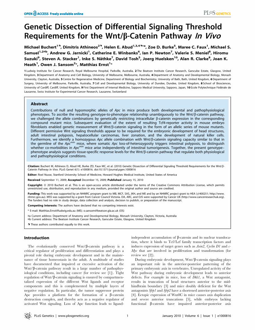
Genetic Dissection of Differential Signaling ThresholdRequirements for the Wnt/b-Catenin Pathway In VivoMichael Buchert1., Dimitris Athineos2., Helen E. Abud1,3,4.¤a, Zoe D. Burke5, Maree C. Faux1, Michael S.
Samuel1,2¤b, Andrew G. Jarnicki1, Catherine E. Winbanks6, Ian P. Newton7, Valerie S. Meniel8, Hiromu
Suzuki9, Steven A. Stacker1, Inke S. Nathke7, David Tosh5, Joerg Huelsken10, Alan R. Clarke8, Joan K.
Heath1, Owen J. Sansom2*, Matthias Ernst1*
1 Ludwig Institute for Cancer Research, Royal Melbourne Hospital, Parkville, Australia, 2 The Beatson Institute Cancer Research, Garscube Estate, Glasgow, United
Kingdom, 3 Department of Anatomy and Cell Biology, University of Melbourne, Melbourne, Australia, 4 Department of Anatomy and Developmental Biology, Monash
University, Clayton, Australia, 5 Centre for Regenerative Medicine, Department of Biology and Biochemistry, University of Bath, Bath, United Kingdom, 6 Department of
Surgery, University of Melbourne, Parkville, Australia, 7 Cell and Developmental Biology, University of Dundee, Dundee, United Kingdom, 8 School of Biosciences,
University of Cardiff, Cardiff, United Kingdom, 9 First Department of Internal Medicine, Sapporo Medical University, Sapporo, Japan, 10 Ecole Polytechnique Federale de
Lausanne, Swiss Institute for Experimental Cancer Research, Lausanne, Switzerland
Abstract
Contributions of null and hypomorphic alleles of Apc in mice produce both developmental and pathophysiologicalphenotypes. To ascribe the resulting genotype-to-phenotype relationship unambiguously to the Wnt/b-catenin pathway,we challenged the allele combinations by genetically restricting intracellular b-catenin expression in the correspondingcompound mutant mice. Subsequent evaluation of the extent of resulting Tcf4-reporter activity in mouse embryofibroblasts enabled genetic measurement of Wnt/b-catenin signaling in the form of an allelic series of mouse mutants.Different permissive Wnt signaling thresholds appear to be required for the embryonic development of head structures,adult intestinal polyposis, hepatocellular carcinomas, liver zonation, and the development of natural killer cells.Furthermore, we identify a homozygous Apc allele combination with Wnt/b-catenin signaling capacity similar to that inthe germline of the Apcmin mice, where somatic Apc loss-of-heterozygosity triggers intestinal polyposis, to distinguishwhether co-morbidities in Apcmin mice arise independently of intestinal tumorigenesis. Together, the present genotype–phenotype analysis suggests tissue-specific response levels for the Wnt/b-catenin pathway that regulate both physiologicaland pathophysiological conditions.
Citation: Buchert M, Athineos D, Abud HE, Burke ZD, Faux MC, et al. (2010) Genetic Dissection of Differential Signaling Threshold Requirements for the Wnt/b-Catenin Pathway In Vivo. PLoS Genet 6(1): e1000816. doi:10.1371/journal.pgen.1000816
Editor: Roel Nusse, Stanford University School of Medicine, Howard Hughes Medical Institute, United States of America
Received September 11, 2009; Accepted December 15, 2009; Published January 15, 2010
Copyright: � 2010 Buchert et al. This is an open-access article distributed under the terms of the Creative Commons Attribution License, which permitsunrestricted use, distribution, and reproduction in any medium, provided the original author and source are credited.
Funding: This work was supported by an NHMRC program grant to MB, MCF, SAS, JKH, and ME and by an NHMRC project grant to HEA (#400251; http://www.nhmrc.gov.au). MSS was supported by a grant from Cancer Council Victoria. DA, ARC, and OJS were supported by Cancer UK (http://www.cancerresearchuk.org).The funders had no role in study design, data collection and analysis, decision to publish, or preparation of the manuscript.
Competing Interests: The authors have declared that no competing interests exist.
* E-mail: [email protected] (ME); [email protected] (OS)
¤a Current address: Department of Anatomy and Developmental Biology, Monash University, Clayton, Victoria, Australia¤b Current address: The Beatson Institute Cancer Research, Garscube Estate, Glasgow, United Kingdom
. These authors contributed equally to this work.
Introduction
The evolutionarily conserved Wnt/b-catenin pathway is a
critical regulator of proliferation and differentiation and plays a
pivotal role during embryonic development and in the mainte-
nance of tissue homeostasis in the adult. A multitude of studies
have documented that impaired or excessive activation of the
Wnt/b-catenin pathway result in a large number of pathophys-
iological conditions, including cancer (for review see [1]). Tight
regulation of Wnt/b-catenin signaling is ensured by compartmen-
talized expression of the different Wnt ligands and receptor
components and this is complemented by multiple layers of
negative regulation. In particular, the tumor suppressor protein
Apc provides a platform for the formation of a b-catenin
destruction complex, and thereby acts as a negative regulator of
activated Wnt signaling. Loss of Apc function leads to ligand-
independent accumulation of b-catenin and its nuclear transloca-
tion, where it binds to Tcf/Lef family transcription factors and
induces expression of target genes such as Axin2, Cyclin D1 and c-
Myc that are involved in proliferation and transformation (for
review see [2]).
During embryonic development, Wnt/b-catenin signaling plays
an important role in the anterior-posterior patterning of the
primary embryonic axis in vertebrates. Unregulated activity of the
Wnt pathway during embryonic development leads to anterior
defects. For example in mice, loss of Dkk1, a Wnt antagonist,
results in truncation of head structures anterior to the mid-
hindbrain boundary [3] and mice doubly deficient for the Wnt
antagonists Sfrp1 and Sfrp2 have a shortened anterior-posterior axis
[4]. Ectopic expression of Wnt8C in mice causes axis duplication
and severe anterior truncations [5], while embryos lacking
functional b-catenin have impaired anterior-posterior axis
PLoS Genetics | www.plosgenetics.org 1 January 2010 | Volume 6 | Issue 1 | e1000816
formation [6]. Embryos homozygous for the mutant Apcmin allele,
which results in truncation of the full-length 2843 amino acid
protein at residue 850 and in heterozygous mice leads to an
intestinal phenotype akin to familial adenomatous polyposis (FAP)
in humans, fail to develop past the gastrulation stage due to
proximalisation of the epiblast and ectopic activation of several
posterior mesendodermal genes [7,8]. While these observations
establish indispensable roles for components of the Wnt pathway
in patterning the anterior-posterior axis, recent genetic rescue
studies have helped to define signaling threshold requirement(s) for
head morphogenesis [9].
Mutations in components of the destruction complex (APC,
AXIN, GSK3b etc) are implicated in tumorigenesis and result in
aberrant, ligand-independent activation of the WNT/b-CATENIN
pathway. For instance truncating nonsense mutations in APC, loss of
heterozygosity (LOH) or promoter hypermethylation are most
prominently associated with aberrant WNT signaling that is
characteristic of more than 90% of sporadic forms of colorectal
cancer in humans [10–12]. Meanwhile epigenetic and genetic
impairment mutations that reduce expression of wild-type AXIN2/
CONDUCTIN [13,14] or amino-terminal missense mutations in
CTNNB1 (b-CATENIN) [15] are most commonly associated with
aberrant WNT signaling in cancers of the liver (hepatocellular
carcinoma and hepatoblastoma), stomach, kidney (Wilms tumor)
and ovaries. It remains unclear why in humans the intestinal
epithelium is most sensitive to cancer-associated somatic mutations
in APC rather than to those in other components of the WNT
signaling cascade, and to what extent this may be due to the loss of
interaction between APC and actin-regulatory proteins and
microtubules that affect cell migration, orientation, polarity, division
and apoptosis, rather than the proliferation/differentiation gener-
ally associated with WNT/b-CATENIN signaling (for review see
[16]). However, at least in the mouse the C–terminal domains of
Apc are dispensable for its tumor suppressing functions [17]. In
addition, phenotypic changes observed after the conditional
deletion of Apc including those on apoptosis, migration, differen-
tiation and proliferation are rescued by concomitant deletion of the
Wnt/b-catenin target gene Myc [18].
Signaling threshold levels in vivo have been assessed by various
approaches, including administration of (ant-) agonistic com-
pounds, the (inducible) over-expression of transgenes and the
creation of haploinsufficiency through the combination of knock-
out and hypomorphic alleles. Elegant combinations of different
hypomorphic Apc alleles, for instance, have demonstrated that
within the context of intestinal tumorigenesis, there is a clear
correlation between gene dosage and phenotype severity [17,19].
In particular, these studies implied an inverse correlation between
the level of Apc protein expression and activation of the Wnt/b-
catenin pathway, and in turn, proliferation and differentiation of
epithelium along the crypt-villus axis as well as cell renewal in the
stem cell compartment [20]. Here we genetically identify
differences in signaling threshold levels that determine physiolog-
ical and pathological outcomes during embryonic development
and various aspects of tissue homeostasis in adult tissue. Using
combinations of epistatically related hypomorphic alleles of
components of the Wnt/b-catenin signaling cascade, we identify
tissue-specific signaling threshold levels for anterior specification
during embryogenesis, intestinal and hepatic homeostasis in the
adult. Our observations add further support to the ‘‘just-right’’
model [21] of Wnt/b-catenin signaling activation where distinct
dosages are required to perturb the self-renewal of stem cell
populations and lead to neoplastic transformation in the intestine
and liver.
Results/Discussion
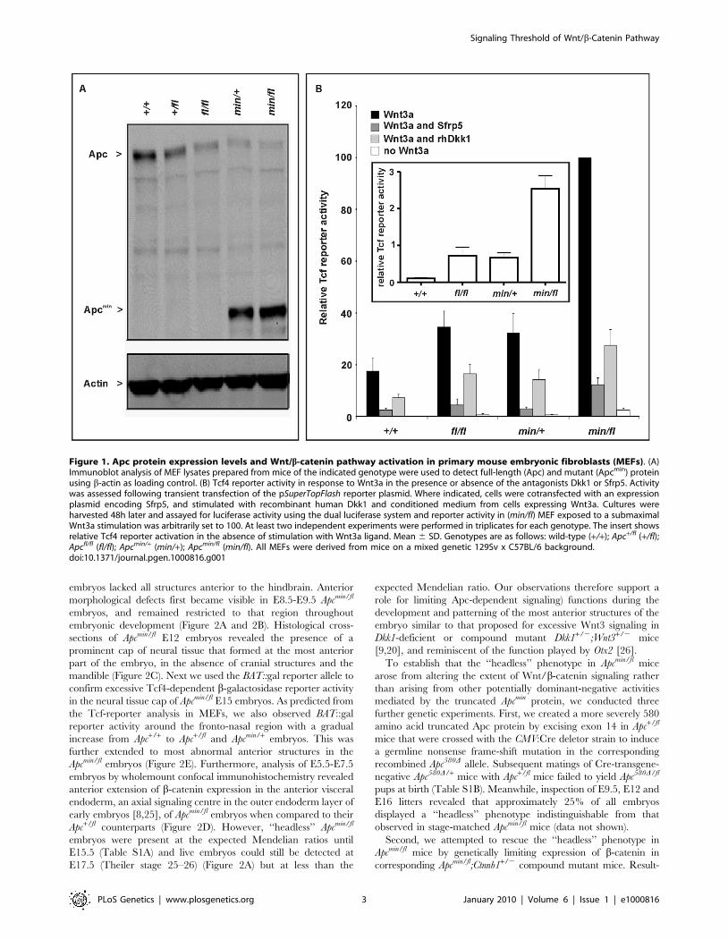
Genetic modulation of full-length Apc expression inmouse embryonic fibroblasts
In order to modulate the activity of the Wnt/b-catenin pathway
in the mouse, we took advantage of the Apcmin [22] and Apcfl [23]
alleles. The premature stop codon encoded by the Apcmin allele
encodes a truncated 850 amino acid Apc protein, which lacks the
15- and 20 aa repeats and Axin binding repeats required for b-
catenin regulation [24], while the unrecombined Apcfl allele results
in attenuated expression levels of wild-type Apc mRNA [23]. We
used Western blot analysis of lysates from mouse embryo
fibroblasts (MEFs) to quantitate expression of full-length Apc
protein and the capacity to augment Wnt3a-dependent signaling
in cells from the corresponding Apc allele combinations. We
observed an inverse relationship in the hierarchy of allele
combinations between full-length Apc protein expression
(Figure 1A), and signaling activity of the Wnt/b-catenin pathway
recorded with a Tcf4 reporter plasmid (Figure 1B). Owing to the
presence of residual amounts of full-length Apc protein, the two
soluble Wnt antagonists Sfrp5 and Dkk1 were able to suppress
Wnt3a-mediated reporter activation in cells of all tested allele
combinations. However, in the presence of Wnt3a, pSUPERTop-
Flash reporter activity was inhibited less effectively by Sfrp5 and
Dkk1 in cells with impaired expression of full-length Apc protein
(Figure 1B). Therefore, genetic modulation of the expression levels
of full-length Apc protein enables experimental manipulation of
Wnt/b-catenin pathway activation for a given concentration of
Wnt ligand or its soluble antagonists.
To assess whether the outcome of incremental modulation of
Wnt/b-catenin signaling by genetic means in MEFs would impact
differentially during development and in adult tissue homeostasis
in vivo, we set out to generate adult mutant mice with genotypes
comprising different combinations of Apc alleles. Surprisingly, we
were unable to obtain Apcmin/fl mice at term from crossing
heterozygous Apc+/fl with Apcmin/+ mice. Since homozygous Apcmin,
but not Apcfl, mice die in utero due to gastrulation defects [7], we
genotyped 117 embryos at E12 and found that all 30 Apcmin/fl
Author Summary
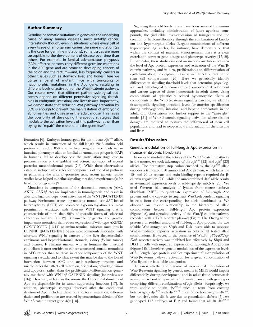
Germline or somatic mutations in genes are the underlyingcause of many human diseases, most notably cancer.Interestingly though, even in situations where every cell ofevery tissue of an organism carries the same mutation (asis the case for germline mutations), some tissues are moresusceptible to the development of disease over time thanothers. For example, in familial adenomatous polyposis(FAP), affected persons carry different germline mutationsin the APC gene and are prone to developing cancers ofthe colon and the rectum—and, less frequently, cancers inother tissues such as stomach, liver, and bones. Here weutilize a panel of mutant mice with truncating orhypomorphic mutations in the Apc gene, resulting indifferent levels of activation of the Wnt/b-catenin pathway.Our results reveal that different pathophysiological out-comes depend on different permissive signaling thresh-olds in embryonic, intestinal, and liver tissues. Importantly,we demonstrate that reducing Wnt pathway activation by50% is enough to prevent the manifestation of embryonicabnormalities and disease in the adult mouse. This raisesthe possibility of developing therapeutic strategies thatmodulate the activation levels of this pathway rather thantrying to ‘‘repair’’ the mutation in the gene itself.
Signaling Threshold of Wnt/b-Catenin Pathway
PLoS Genetics | www.plosgenetics.org 2 January 2010 | Volume 6 | Issue 1 | e1000816
embryos lacked all structures anterior to the hindbrain. Anterior
morphological defects first became visible in E8.5-E9.5 Apcmin/fl
embryos, and remained restricted to that region throughout
embryonic development (Figure 2A and 2B). Histological cross-
sections of Apcmin/fl E12 embryos revealed the presence of a
prominent cap of neural tissue that formed at the most anterior
part of the embryo, in the absence of cranial structures and the
mandible (Figure 2C). Next we used the BAT::gal reporter allele to
confirm excessive Tcf4-dependent b-galactosidase reporter activity
in the neural tissue cap of Apcmin/fl E15 embryos. As predicted from
the Tcf-reporter analysis in MEFs, we also observed BAT::gal
reporter activity around the fronto-nasal region with a gradual
increase from Apc+/+ to Apc+/fl and Apcmin/+ embryos. This was
further extended to most abnormal anterior structures in the
Apcmin/fl embryos (Figure 2E). Furthermore, analysis of E5.5-E7.5
embryos by wholemount confocal immunohistochemistry revealed
anterior extension of b-catenin expression in the anterior visceral
endoderm, an axial signaling centre in the outer endoderm layer of
early embryos [8,25], of Apcmin/fl embryos when compared to their
Apc+/fl counterparts (Figure 2D). However, ‘‘headless’’ Apcmin/fl
embryos were present at the expected Mendelian ratios until
E15.5 (Table S1A) and live embryos could still be detected at
E17.5 (Theiler stage 25–26) (Figure 2A) but at less than the
expected Mendelian ratio. Our observations therefore support a
role for limiting Apc-dependent signaling) functions during the
development and patterning of the most anterior structures of the
embryo similar to that proposed for excessive Wnt3 signaling in
Dkk1-deficient or compound mutant Dkk1+/2;Wnt3+/2 mice
[9,20], and reminiscent of the function played by Otx2 [26].
To establish that the ‘‘headless’’ phenotype in Apcmin/fl mice
arose from altering the extent of Wnt/b-catenin signaling rather
than arising from other potentially dominant-negative activities
mediated by the truncated Apcmin protein, we conducted three
further genetic experiments. First, we created a more severely 580
amino acid truncated Apc protein by excising exon 14 in Apc+/fl
mice that were crossed with the CMV:Cre deletor strain to induce
a germline nonsense frame-shift mutation in the corresponding
recombined Apc580D allele. Subsequent matings of Cre-transgene-
negative Apc580D/+ mice with Apc+/fl mice failed to yield Apc580D/fl
pups at birth (Table S1B). Meanwhile, inspection of E9.5, E12 and
E16 litters revealed that approximately 25% of all embryos
displayed a ‘‘headless’’ phenotype indistinguishable from that
observed in stage-matched Apcmin/fl mice (data not shown).
Second, we attempted to rescue the ‘‘headless’’ phenotype in
Apcmin/fl mice by genetically limiting expression of b-catenin in
corresponding Apcmin/fl;Ctnnb1+/2 compound mutant mice. Result-
Figure 1. Apc protein expression levels and Wnt/b-catenin pathway activation in primary mouse embryonic fibroblasts (MEFs). (A)Immunoblot analysis of MEF lysates prepared from mice of the indicated genotype were used to detect full-length (Apc) and mutant (Apcmin) proteinusing b-actin as loading control. (B) Tcf4 reporter activity in response to Wnt3a in the presence or absence of the antagonists Dkk1 or Sfrp5. Activitywas assessed following transient transfection of the pSuperTopFlash reporter plasmid. Where indicated, cells were cotransfected with an expressionplasmid encoding Sfrp5, and stimulated with recombinant human Dkk1 and conditioned medium from cells expressing Wnt3a. Cultures wereharvested 48h later and assayed for luciferase activity using the dual luciferase system and reporter activity in (min/fl) MEF exposed to a submaximalWnt3a stimulation was arbitrarily set to 100. At least two independent experiments were performed in triplicates for each genotype. The insert showsrelative Tcf4 reporter activation in the absence of stimulation with Wnt3a ligand. Mean 6 SD. Genotypes are as follows: wild-type (+/+); Apc+/fl (+/fl);Apcfl/fl (fl/fl); Apcmin/+ (min/+); Apcmin/fl (min/fl). All MEFs were derived from mice on a mixed genetic 129Sv x C57BL/6 background.doi:10.1371/journal.pgen.1000816.g001
Signaling Threshold of Wnt/b-Catenin Pathway
PLoS Genetics | www.plosgenetics.org 3 January 2010 | Volume 6 | Issue 1 | e1000816
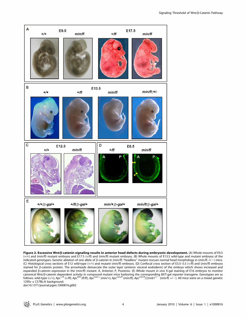
Figure 2. Excessive Wnt/b-catenin signaling results in anterior head defects during embryonic development. (A) Whole mounts of E9.5(+/+) and (min/fl) mutant embryos and E17.5 (+/fl) and (min/fl) mutant embryos. (B) Whole mounts of E13.5 wild-type and mutant embryos of theindicated genotypes. Genetic ablation of one allele of b-catenin in (min/fl) ‘‘headless’’ mutant rescues normal head morphology in (min/fl; +/2) mice.(C) Histological cross sections of E12 wild-type (+/+) and mutant (min/fl) embryos. (D) Confocal cross section of E3.5–5.5 (+/fl) and (min/fl) embryosstained for b-catenin protein. The arrowheads demarcate the outer layer (anterior visceral endoderm) of the embryo which shows increased andexpanded b-catenin expression in the (min/fl) mutant. A, Anterior; P, Posterior. (E) Whole mount in vivo X-gal staining of E16 embryos to monitorcanonical Wnt/b-catenin dependent activity in compound mutant mice harboring the corresponding BAT::gal reporter transgene. Genotypes are asfollows: wild-type (+/+); Apc+/fl (+/fl); Apcfl/fl (fl/fl); Apcmin/+ (min/+); Apcmin/fl (min/fl); Apcmin/fl;Ctnnb1+/2 (min/fl; +/2). All mice were on a mixed genetic129Sv x C57BL/6 background.doi:10.1371/journal.pgen.1000816.g002
Signaling Threshold of Wnt/b-Catenin Pathway
PLoS Genetics | www.plosgenetics.org 4 January 2010 | Volume 6 | Issue 1 | e1000816
ing Apcmin/fl;Ctnnb1+/2 MEFs revealed an approximately 50%
reduction of Wnt/b-catenin signaling when compared to their
Apcmin/fl;Ctnnb1+/+ counterparts (see below). When mating Apcfl/fl;
Ctnnb1+/2 with Apcmin/+;Ctnnb1+/+ mice, we recovered Apc+/fl;
Ctnnb1+/+, Apc+/fl;Ctnnb1+/2 and Apcmin/fl;Ctnnb1+/2 mice at weaning
age at a similar ratio, while among E13.5 embryos, all four possible
genotypes were represented at comparable frequencies (Table S1C
and Figure S1). Importantly, Apcmin/fl;Ctnnb1+/2 mice developed
normally into fecund adults (Figure 2B and data not shown),
suggesting that limiting Wnt/b-catenin signaling corrected
the development of detrimental phenotypes observed in Apcmin/fl
mice.
Since the atypical Wnt receptor component Ryk has recently
been suggested to amplify Wnt signaling during cortical neuro-
genesis through b-catenin-dependent as well as independent
pathways [27], we also tested whether the ‘‘headless’’ phenotype
was promoted by Ryk activity. However, and in contrast to b-
catenin, the embryonic lethality of Apcmin/fl mice was not rescued
by genetically limiting the expression of the atypical tyrosine
kinase Ryk, because we failed to recover either Apcmin/fl;Ryk+/2 or
Apcmin/fl;Ryk-/- compound mutant mice at weaning (Table S1D),
suggesting that Ryk expression was not contributing to the Wnt/b-
catenin induced phenotype.
Collectively, our observations extend previous reports that
identified a Wnt signaling gradient along the anterior-posterior axis
and a requirement for Dkk1 and other Wnt antagonists at the
anterior end to prevent posteriorization [3–6,28,29]. In particular,
our experiments clarify genetically that the tight signaling require-
ments for head morphogenesis previously attributed to Apc or the
extracellular components Dkk1 [3], Sfrp [4,30], Wnt3a [9] and
Wnt8a [5] occur exclusively through the Wnt/b-catenin pathway.
Unlike Apcmin/fl embryos, Apcmin/min embryos die around the time
of gastrulation [7], consistent with our observation that Apc580D/min
MEFs, which serve as a model for unavailable Apcmin/min
counterparts, reveal higher Tcf4 reporter activity than Apcmin/fl
MEFs (see below). Since the morphological defects in E4.75
Apcmin/min embryos correlate with excessive nuclear b-catenin in the
epiblast and primitive ectoderm [8], we also examined the effect of
genetically limiting b-catenin in these embryos. Unlike the
phenotypic rescue observed in Apcmin/fl;Ctnnb1+/2 mice, we
detected Apcmin/min;Ctnnb1+/2 embryos only at E4.5 and E5.5 but
not at later stages (E6 and E7). This finding is reminiscent of the
time points of embryonic death of Apcmin/min embryos [7] and
suggested that reduction of Wnt/b-catenin signaling was insuffi-
cient to rescue their death immediately after gastrulation (data not
shown). Therefore, higher threshold levels of Wnt/b-catenin
signaling selectively inhibit development at an earlier stage (i.e.
gastrulation) and genetic reduction of Wnt/b-catenin signaling
through ablation of one Ctnnb1 allele reduces signaling only below
the threshold that is tolerated during later stages of development.
However, we cannot formally exclude other essential function(s) of
the full-length Apc protein, which could be provided by residual
full-length protein encoded by the Apcfl allele, and which may be
required around the time of gastrulation.
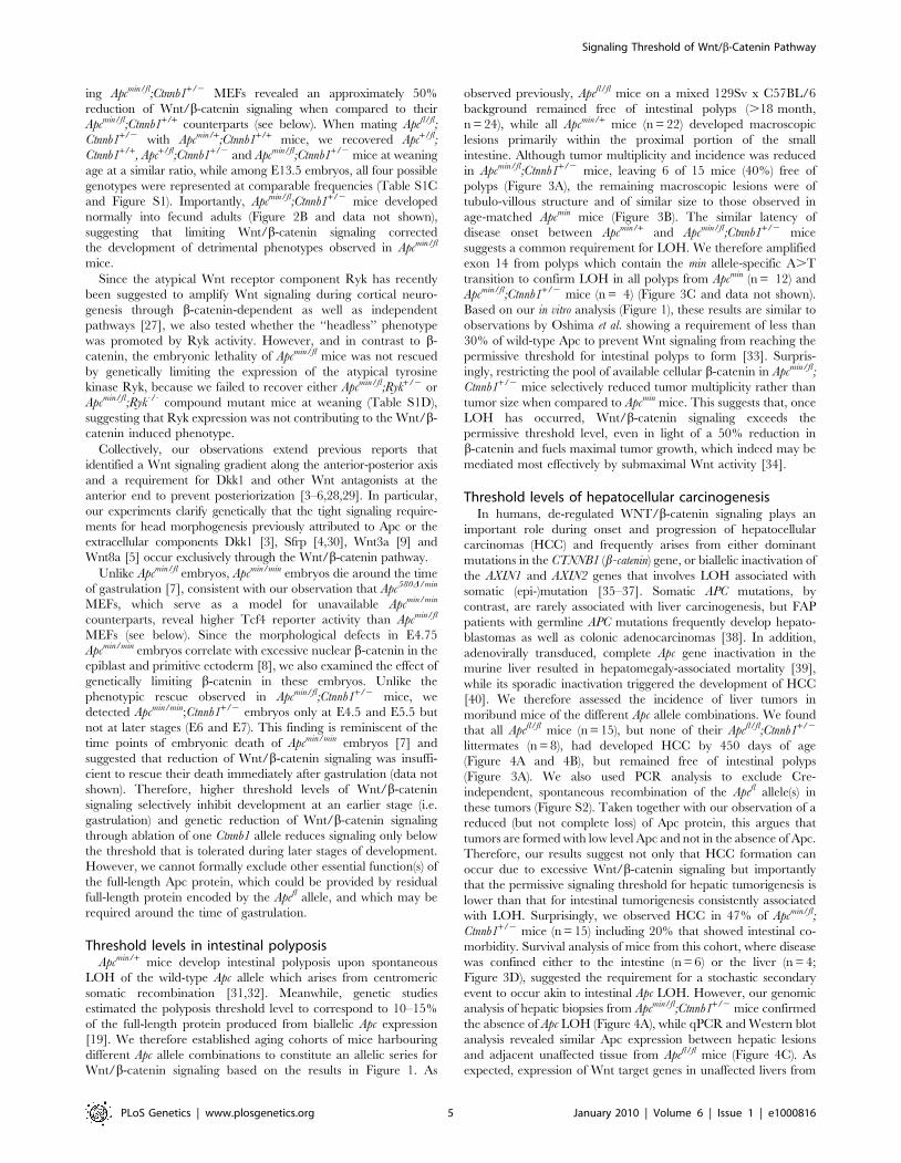
Threshold levels in intestinal polyposisApcmin/+ mice develop intestinal polyposis upon spontaneous
LOH of the wild-type Apc allele which arises from centromeric
somatic recombination [31,32]. Meanwhile, genetic studies
estimated the polyposis threshold level to correspond to 10–15%
of the full-length protein produced from biallelic Apc expression
[19]. We therefore established aging cohorts of mice harbouring
different Apc allele combinations to constitute an allelic series for
Wnt/b-catenin signaling based on the results in Figure 1. As
observed previously, Apcfl/fl mice on a mixed 129Sv x C57BL/6
background remained free of intestinal polyps (.18 month,
n = 24), while all Apcmin/+ mice (n = 22) developed macroscopic
lesions primarily within the proximal portion of the small
intestine. Although tumor multiplicity and incidence was reduced
in Apcmin/fl;Ctnnb1+/2 mice, leaving 6 of 15 mice (40%) free of
polyps (Figure 3A), the remaining macroscopic lesions were of
tubulo-villous structure and of similar size to those observed in
age-matched Apcmin mice (Figure 3B). The similar latency of
disease onset between Apcmin/+ and Apcmin/fl;Ctnnb1+/2 mice
suggests a common requirement for LOH. We therefore amplified
exon 14 from polyps which contain the min allele-specific A.T
transition to confirm LOH in all polyps from Apcmin (n = 12) and
Apcmin/fl;Ctnnb1+/2 mice (n = 4) (Figure 3C and data not shown).
Based on our in vitro analysis (Figure 1), these results are similar to
observations by Oshima et al. showing a requirement of less than
30% of wild-type Apc to prevent Wnt signaling from reaching the
permissive threshold for intestinal polyps to form [33]. Surpris-
ingly, restricting the pool of available cellular b-catenin in Apcmin/fl;
Ctnnb1+/2 mice selectively reduced tumor multiplicity rather than
tumor size when compared to Apcmin mice. This suggests that, once
LOH has occurred, Wnt/b-catenin signaling exceeds the
permissive threshold level, even in light of a 50% reduction in
b-catenin and fuels maximal tumor growth, which indeed may be
mediated most effectively by submaximal Wnt activity [34].
Threshold levels of hepatocellular carcinogenesisIn humans, de-regulated WNT/b-catenin signaling plays an
important role during onset and progression of hepatocellular
carcinomas (HCC) and frequently arises from either dominant
mutations in the CTNNB1 (b-catenin) gene, or biallelic inactivation of
the AXIN1 and AXIN2 genes that involves LOH associated with
somatic (epi-)mutation [35–37]. Somatic APC mutations, by
contrast, are rarely associated with liver carcinogenesis, but FAP
patients with germline APC mutations frequently develop hepato-
blastomas as well as colonic adenocarcinomas [38]. In addition,
adenovirally transduced, complete Apc gene inactivation in the
murine liver resulted in hepatomegaly-associated mortality [39],
while its sporadic inactivation triggered the development of HCC
[40]. We therefore assessed the incidence of liver tumors in
moribund mice of the different Apc allele combinations. We found
that all Apcfl/fl mice (n = 15), but none of their Apcfl/fl;Ctnnb1+/2
littermates (n = 8), had developed HCC by 450 days of age
(Figure 4A and 4B), but remained free of intestinal polyps
(Figure 3A). We also used PCR analysis to exclude Cre-
independent, spontaneous recombination of the Apcfl allele(s) in
these tumors (Figure S2). Taken together with our observation of a
reduced (but not complete loss) of Apc protein, this argues that
tumors are formed with low level Apc and not in the absence of Apc.
Therefore, our results suggest not only that HCC formation can
occur due to excessive Wnt/b-catenin signaling but importantly
that the permissive signaling threshold for hepatic tumorigenesis is
lower than that for intestinal tumorigenesis consistently associated
with LOH. Surprisingly, we observed HCC in 47% of Apcmin/fl;
Ctnnb1+/2 mice (n = 15) including 20% that showed intestinal co-
morbidity. Survival analysis of mice from this cohort, where disease
was confined either to the intestine (n = 6) or the liver (n = 4;
Figure 3D), suggested the requirement for a stochastic secondary
event to occur akin to intestinal Apc LOH. However, our genomic
analysis of hepatic biopsies from Apcmin/fl;Ctnnb1+/2 mice confirmed
the absence of Apc LOH (Figure 4A), while qPCR and Western blot
analysis revealed similar Apc expression between hepatic lesions
and adjacent unaffected tissue from Apcfl/fl mice (Figure 4C). As
expected, expression of Wnt target genes in unaffected livers from
Signaling Threshold of Wnt/b-Catenin Pathway
PLoS Genetics | www.plosgenetics.org 5 January 2010 | Volume 6 | Issue 1 | e1000816
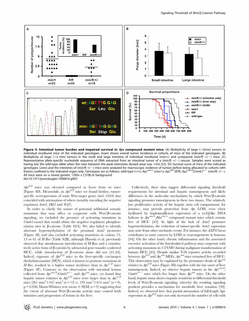
Apcfl/fl mice was elevated compared to livers from wt mice
(Figure 4D). Meanwhile, in Apcfl/fl mice we found further, tumor-
specific overexpression of some Wnt-target genes (incl. Cd44) that
coincided with attenuation of others (notably encoding the negative
regulators Axin2, Dkk2 and Wif1).
In order to clarify the nature of potential additional somatic
mutations that may affect or cooperate with Wnt/b-catenin
signaling, we excluded the presence of activating mutations in
Ctnnb1-exon3 that would ablate the negative regulatory phosphor-
ylation sites in b-catenin (Table S2A). We also failed to identify
aberrant hypermethylation of the proximal Axin2 promoter
(Figure 4E) and also excluded activating mutations in codons 12,
13 or 61 of H-Ras (Table S2B), although Harada et al. previously
observed that simultaneous introduction of H-Ras and a constitu-
tively active form of b-catenin by adenoviral gene transfer conferred
HCC, while introduction of b-catenin alone did not [41,42].
Indeed, exposure of Apcfl/fl mice to the liver-specific carcinogen
diethylnitrosamine (DEN), which is known to promote mutations in
H-Ras, resulted in a higher tumor incidence than in Apc+/fl mice
(Figure 4F). Contrary to the observation with intestinal lesions
collected from Apcmin/fl;Ctnnb1+/2 and Apcmin mice, we found that
hepatic tumor volumes in Apcfl/fl mice were larger than in Apc+/fl
mice (261 mm36167 mm3 (n = 12) vs. 193 mm36414 mm3 (n = 9),
p = 0.036; Mann-Whitney test; mean 6 SEM; n = 9) suggesting that
the extent of aberrant Wnt/b-catenin activity may control both
initiation and progression of lesions in the liver.
Collectively, these data suggest differential signaling threshold
requirements for intestinal and hepatic tumorigenesis and likely
differences in the molecular mechanisms by which Wnt/b-catenin
signaling promotes tumorigenesis in these two tissues. The relatively
low proliferative activity of the hepatic stem cell compartment, for
instance, may provide protection from Apc LOH, even when
facilitated by haploinsufficient expression of a recQ-like DNA
helicase in Apcmin/+;BlmCin/+ compound mutant mice which remain
free of HCC [43]. In light of the lack of Axin2 promoter
hypermethylation, the reduction of tumor-specific Axin2 expression
may arise from other stochastic events. For instance, the AXIN2 locus
contributes to some cancers by LOH or rearrangements in humans
[44]. On the other hand, chronic inflammation and the associated
excessive activation of the Interleukin-6 pathway may cooperate with
activating mutations in CTNNB1 during malignant transformation of
human HCC [45]. Despite similar Tcf4 reporter activity recorded
between Apcfl/fl and Apcmin MEFs, Apcmin mice remained free of HCC.
This observation may be explained by the premature death of Apcmin
relative to Apcfl/fl mice (Figure 3D) together with the late onset of liver
tumorigenesis. Indeed, we observe hepatic tumors in the Apcmin/fl;
Ctnnb1+/2 mice which live longer than Apcmin mice. On the other
hand, hepatic tissue shows exquisite sensitivity to differential threshold
levels of Wnt/b-catenin signaling, whereby the resulting signaling
gradient provides a mechanism for metabolic liver zonation [39].
Indeed, we observed here that partial attenuation of full-length Apc
expression in Apcfl/fl mice not only increased the number of cells with
Figure 3. Intestinal tumor burden and impaired survival in Apc compound mutant mice. (A) Multiplicity of large (.2mm) tumors inindividual moribund mice of the indicated genotypes. Insert shows overall tumor incidence in cohorts of mice of the indicated genotypes. (B)Multiplicity of large (.2 mm) tumors in the small and large intestine of individual moribund (min/+) and compound (min/fl; +/2) mice. (C)Representative allele-specific nucleotide sequence of DNA extracted from an intestinal tumor of a (min/fl; +/2) mouse. Samples were scored ashaving lost the wild-type allele when the ratio between the peak intensities (boxed area) was #0.6 [52]. (D) Survival curve of mice of the indicatedgenotypes. Livers and the intestines of (min/fl; +/2) mice were analyzed for macroscopic evidence of tumors before being allocated to cohorts withlesions confined to the indicated organ only. Genotypes are as follows: wild-type (+/+); Apcmin/+ (min/+); Apcfl/fl (fl/fl); Apcmin/fl;Ctnnb1+/2 (min/fl; +/2).All mice were on a mixed genetic 129Sv x C57BL/6 background.doi:10.1371/journal.pgen.1000816.g003
Signaling Threshold of Wnt/b-Catenin Pathway
PLoS Genetics | www.plosgenetics.org 6 January 2010 | Volume 6 | Issue 1 | e1000816
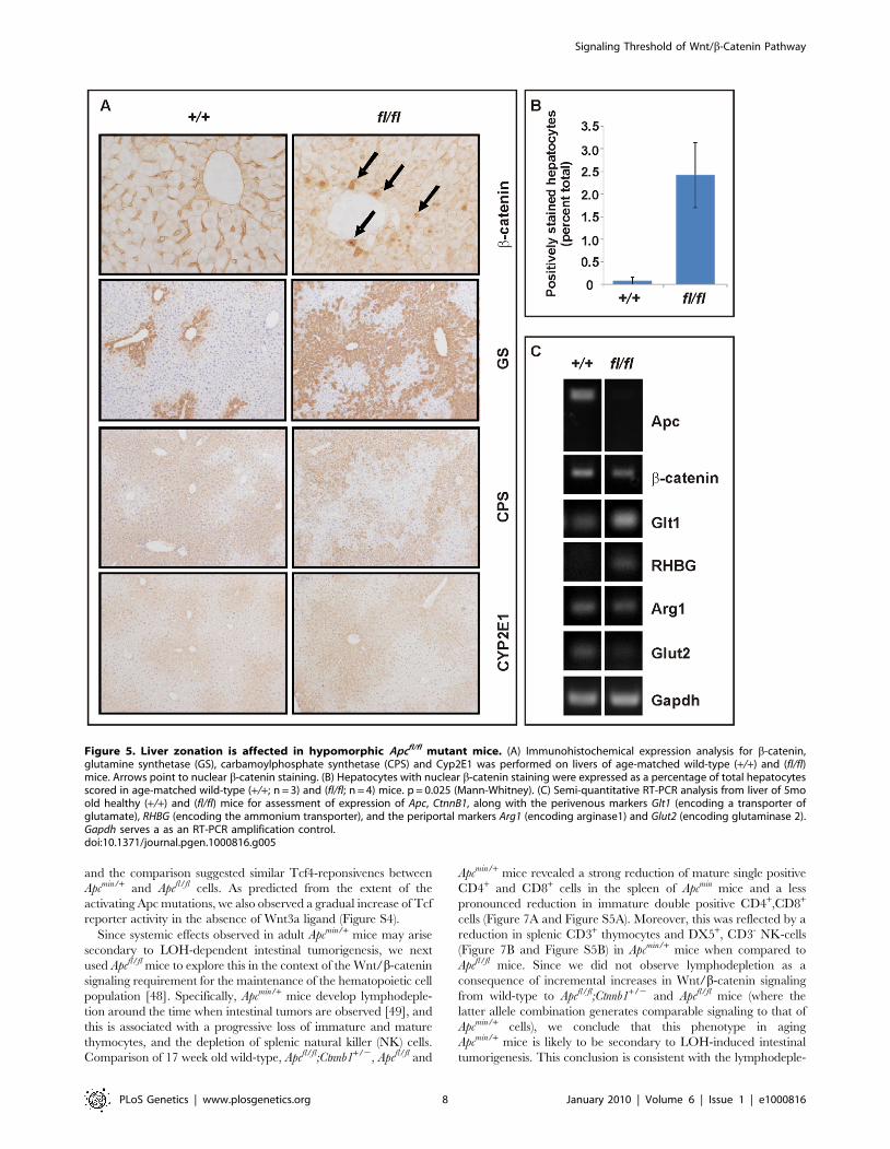
nuclear b-catenin (Figure 5A and 5B), but also altered expression of
Wnt target genes and liver zonation. In particular, and in agreement
with our previous findings [46], we observed that attenuation of full-
length Apc favored expansion of a perivenous gene expression
program (incl. GS, Glt1 and RHBG) at the expense of a periportal
signature (incl. CPS, Arg1 and Glut2) (Figure 5A and 5C). Our
observation that aberrant Wnt signaling in Apcfl/fl mice in the absence
of additional somatic mutations in H-Ras bias towards tumors with
perivenous characteristics is consistent with the finding that H-Ras
mutated HCCs favor a periportal gene expression program [47].
Reconciling tissue-specific phenotypes against differentlevels of Wnt/b-catenin signaling
To gain biochemical insights into the extent to which Wnt
signaling thresholds are related to the tumorigenic response in mice,
we generated MEFs of genotypes similar to those of cells having
undergone Apc LOH in Apcmin mice. In particular, we inactivated the
latent Apcfl allele by Cre-mediated recombination in MEFs following
infection with an AdCre-GFP adenovirus that expressed the Cre-
recombinase as a GFP-fusion protein (Figure S3). Western blot
analysis confirmed expression of the 580 amino acid truncated
protein encoded by the recombined Apc580D allele, in the presence of
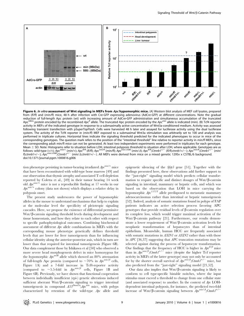
the 850 amino acid Apcmin protein (Figure 6A). To prevent our
analysis from being affected by potential ‘‘plateau effects’’, we
stimulated MEFs with submaximal concentrations of Wnt3a and
found a ,3-fold increase in Tcf-reporter activity between cells
harboring the unrecombined Apcfl or recombined Apc580D allele,
respectively (Figure 6B, compare Apcmin D vs. Apcmin/fl and Apc min/D;
Ctnnb1+/2 vs. Apcmin/fl;Ctnnb1+/2). Furthermore, we confirmed that
ablation of one Ctnnb1 allele reduced reporter activity by
approximately 50% (compare Apcmin/D vs. Apcmin/D;Ctnnb1+/2;
Apcmin/fl vs. Apcmin/fl;Ctnnb1+/2 and Apc fl/fl vs. Apcfl/fl;Ctnnb1+/2),
Figure 4. Liver phenotype in Apc mutant mice. (A) Hepatocellular carcinoma (HCC) of moribund Apcfl/fl and representative haematoxilin-eosinstained cross section with the dotted line indicating the boundary between normal (N) and tumoral (T) tissue. Representative allele-specificnucleotide sequence of DNA extracted from a liver tumor of a (min/fl; +/2) mouse demonstrating allelic balance between of the Apcmin and thefloxed wt allele (boxed area). (B) Incidence of HCC in mice of the indicated genotypes. (C) Western blot and qPCR analysis of full-length Apc proteinand Apc mRNA in normal (N) and tumoral (T) liver tissue of Apcfl/fl and Apc+/+ mice. Cell lysates of HeLa cells transfected with a plasmid encoding full-length wild-type Apc serves as an antibody specificity control. The abundance of de-phosphorylated, active (De-PO4) and total b-catenin protein inthe same tissue extracts are shown with b-actin serving as a loading control. kDa, protein size marker in kilo Daltons. (D) Comparative qPCR analysis ofrepresentative Wnt target gene expression between normal and tumoral liver tissue collected from moribund Apcfl/fl mice (right panel). A comparableanalysis was also performed on liver tissue from healthy 5mo old Apcfl/fl and wild-type mice (left panel). Mean 6 SD with n$3 mice per group.* P,0.05. (E) Bisulfite sequencing of the CpG island within the Axin2 promoter from adjacent normal (N) and tumor liver tissue (T1, T2, T3) from Apcfl/fl
mice. Each vertical line refers to a CpG dinucleotide at the indicated position relative to the transcriptional start site. Following bisulfite-treatment,DNA was subcloned and sequenced. Horizontal lines represent individual sequences with open and full circles denoting unmethylated andmethylated CpG residues, respectively. (F) Boxplot diagram comparing liver tumor multiplicity in +/fl mice (n = 9) and fl/fl mice (n = 12) 6 to 8 monthsafter treatment with DEN. p = 0.0003 (Mann-Whitney). Genotypes are as follows: wild-type (+/+); Apc+/fl (+/fl); Apcfl/fl (fl/fl); Apcfl/fl;Ctnnb1+/2 (fl/fl; +/2);Apcmin/fl;Ctnnb1+/2 (min/fl; +/2). All mice were on a mixed 129Sv x C57BL/6 background.doi:10.1371/journal.pgen.1000816.g004
Signaling Threshold of Wnt/b-Catenin Pathway
PLoS Genetics | www.plosgenetics.org 7 January 2010 | Volume 6 | Issue 1 | e1000816
and the comparison suggested similar Tcf4-reponsivenes between
Apcmin/+ and Apcfl/fl cells. As predicted from the extent of the
activating Apc mutations, we also observed a gradual increase of Tcf
reporter activity in the absence of Wnt3a ligand (Figure S4).
Since systemic effects observed in adult Apcmin/+ mice may arise
secondary to LOH-dependent intestinal tumorigenesis, we next
used Apcfl/fl mice to explore this in the context of the Wnt/b-catenin
signaling requirement for the maintenance of the hematopoietic cell
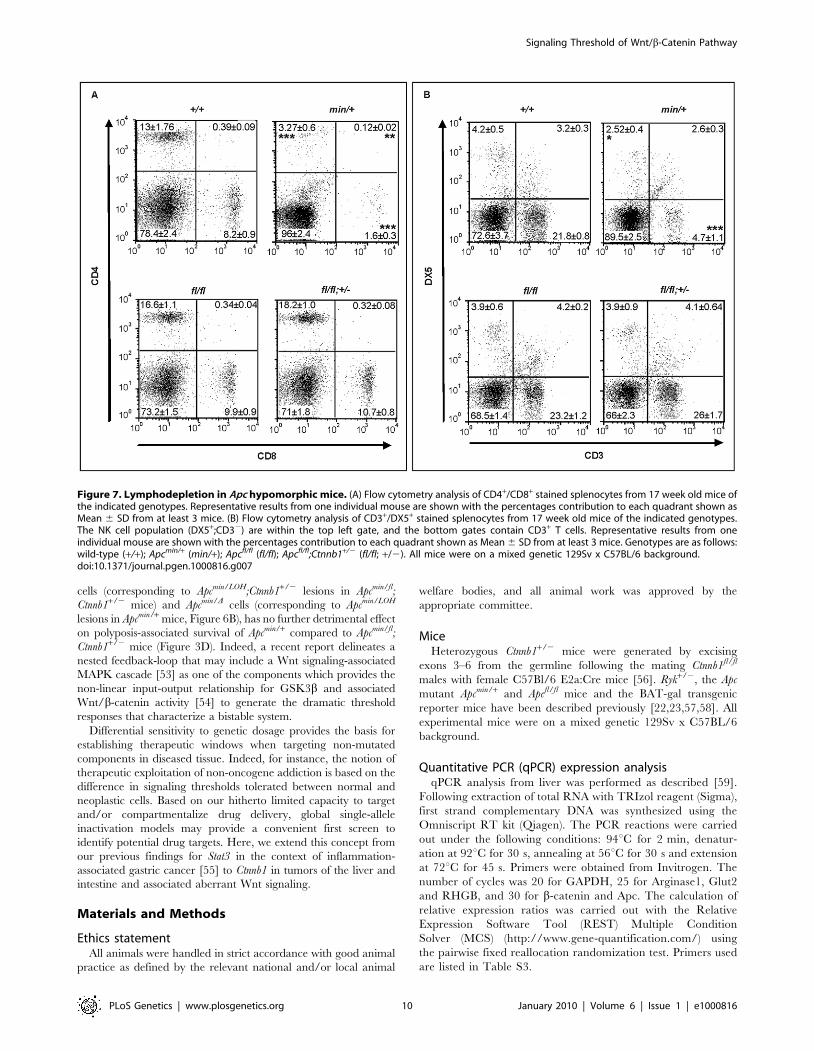
population [48]. Specifically, Apcmin/+ mice develop lymphodeple-
tion around the time when intestinal tumors are observed [49], and
this is associated with a progressive loss of immature and mature
thymocytes, and the depletion of splenic natural killer (NK) cells.
Comparison of 17 week old wild-type, Apcfl/fl;Ctnnb1+/2, Apcfl/fl and
Apcmin/+ mice revealed a strong reduction of mature single positive
CD4+ and CD8+ cells in the spleen of Apcmin mice and a less
pronounced reduction in immature double positive CD4+,CD8+
cells (Figure 7A and Figure S5A). Moreover, this was reflected by a
reduction in splenic CD3+ thymocytes and DX5+, CD3- NK-cells
(Figure 7B and Figure S5B) in Apcmin/+ mice when compared to
Apcfl/fl mice. Since we did not observe lymphodepletion as a
consequence of incremental increases in Wnt/b-catenin signaling
from wild-type to Apcfl/fl;Ctnnb1+/2 and Apcfl/fl mice (where the
latter allele combination generates comparable signaling to that of
Apcmin/+ cells), we conclude that this phenotype in aging
Apcmin/+ mice is likely to be secondary to LOH-induced intestinal
tumorigenesis. This conclusion is consistent with the lymphodeple-
Figure 5. Liver zonation is affected in hypomorphic Apcfl/fl mutant mice. (A) Immunohistochemical expression analysis for b-catenin,glutamine synthetase (GS), carbamoylphosphate synthetase (CPS) and Cyp2E1 was performed on livers of age-matched wild-type (+/+) and (fl/fl)mice. Arrows point to nuclear b-catenin staining. (B) Hepatocytes with nuclear b-catenin staining were expressed as a percentage of total hepatocytesscored in age-matched wild-type (+/+; n = 3) and (fl/fl; n = 4) mice. p = 0.025 (Mann-Whitney). (C) Semi-quantitative RT-PCR analysis from liver of 5moold healthy (+/+) and (fl/fl) mice for assessment of expression of Apc, CtnnB1, along with the perivenous markers Glt1 (encoding a transporter ofglutamate), RHBG (encoding the ammonium transporter), and the periportal markers Arg1 (encoding arginase1) and Glut2 (encoding glutaminase 2).Gapdh serves a as an RT-PCR amplification control.doi:10.1371/journal.pgen.1000816.g005
Signaling Threshold of Wnt/b-Catenin Pathway
PLoS Genetics | www.plosgenetics.org 8 January 2010 | Volume 6 | Issue 1 | e1000816
tion phenotype persisting in tumor-bearing irradiated Apcmin/+ mice
that have been reconstituted with wild-type bone marrow [49] and
our observation that thymic atrophy and associated T-cell depletion
reported by Coletta et al., [49] in their tumor bearing 14 week
old Apcmin/+ mice is not a reproducible finding at 17 weeks in our
Apcmin/+ colony (data not shown) which displays a relative delay in
polyposis onset.
The present study underscores the power of hypomorphic
alleles in the mouse to understand mechanisms that help to explain
at the molecular level the specificity of pleiotropic signaling
cascades. Here, we propose the existence of differential permissive
Wnt/b-catenin signaling threshold levels during development and
tissue homeostasis, and how they relate to each other with respect
to specific pathophysiological outcomes. Combining biochemical
assessment of different Apc allele combinations in MEFs with the
corresponding mouse phenotype genetically defines threshold
levels that are lower for liver tumorigenesis than for influencing
cellular identity along the anterior-posterior axis, which in turn are
lower than that required for intestinal tumorigenesis (Figure 6B).
Our data complement those by Ishikawa et al [50] who observed a
more severe head morphogenesis defect in mice homozygous for
the hypomorphic ApcneoR allele which showed an 80% attenuation
of full-length Apc protein (compared to ,70% in Apcmin/fl cells,
Figure 1A) and a 7-fold increase in Tcf4-reporter activity
(compared to ,5.5-fold in Apcmin/fl cells, Figure 1B and
Figure 6B). Previously, we have shown that functional cooperation
between individually insufficient (epi-) genetic alterations induced
sufficient aberrant Wnt/b-catenin signaling to trigger intestinal
tumorigenesis in compound A33Dnmt3a;Apcmin mice, with polyps
characterized by retention of the wild-type Apc allele and
epigenetic silencing of the Sfrp5 gene [51]. Together with the
findings presented here, these observations add further support to
the ‘‘just-right’’ signaling model which predicts cellular transfor-
mation to require specific and distinct dosages of Wnt/b-catenin
signaling in intestinal, mammary or hepatic cells, and which was
based on the observation that LOH in mice carrying the
hypomorphic Apc1572T allele predisposed to metastatic mammary
adenocarcinomas rather than intestinal or hepatic tumorigenesis
[52]. Indeed, analysis of somatic mutations found in polyps of FAP
patients indicates an active selection process favoring APC
genotypes that provide residual levels of b-catenin regulation over
its complete loss, which would trigger maximal activation of the
Wnt/b-catenin pathway [21]. Furthermore, our results demon-
strate a lower requirement of Wnt/b-catenin activation levels for
neoplastic transformation of hepatocytes than of intestinal
epithelium. Meanwhile, human HCC are frequently associated
with somatic mutations in AXIN1 or AXIN2 rather than with those
in APC [36,37] suggesting that APC truncation mutations may be
selected against during the process of hepatocyte transformation.
Our findings that the frequency of HCC is higher in Apcfl/fl mice
than in Apcmin/fl;Ctnnb1+/2 mice (despite the higher Tcf reporter
activity in MEFs of the latter genotype) may not only be accounted
for by the shorter overall survival of Apcmin/fl;Ctnnb1+/2 mice, but
also predicted from the ‘‘just-right’’ signaling model [21,52].
Our data also implies that Wnt/b-catenin signaling is likely to
conform to cell type-specific bistable switches, where the input
stimulus must exceed a threshold to change from one cellular state
(and associated response) to another. In the context of Apc LOH-
dependent intestinal polyposis, for instance, the predicted two-fold
increase of Wnt/b-catenin signaling between Apcmin/D;Ctnnb1+/2
Figure 6. In vitro assessment of Wnt signaling in MEFs from Apc hypomorphic mice. (A) Western blot analysis of MEF cell lysates, preparedfrom (fl/fl) and (min/fl) mice, 48 h after infection with Cre-GFP expressing adenovirus (AdCre-GFP) at different concentrations. Note the gradualreduction of full-length Apc protein (wt) with increasing amount of AdCre-GFP administration and simultaneous accumulation of the truncatedApc580D protein encoded by the recombined Apcfl allele. The truncated Apc protein encoded by the Apcmin allele is indicated (min). (B) Tcf4 reporteractivity in MEFs of the indicated genotype in response to a submaximally active concentration of Wnt3a-conditioned medium. Activity was assessedfollowing transient transfection with pSuperTopFlash. Cells were harvested 48 h later and assayed for luciferase activity using the dual luciferasesystem. The activity of the Tcf4 reporter in (min/fl) MEF exposed to a submaximal Wnt3a stimulation was arbitrarily set to 100 and analysis wasperformed in triplicate cultures. Horizontal lines indicate the signaling threshold predicted for the indicated phenotypes to occur in mice of thecorresponding genotypes. The question mark refers to the position of the ‘‘intestinal threshold’’ line relative to reporter activity in min/fl MEFs, sincethe corresponding adult min/fl mice can not be generated. At least two independent experiments were performed in triplicates for each genotype.Mean 6 SD. Note: Histograms refer to situation before LOH, intestinal polyposis threshold to situation after LOH, where applicable. Genotypes are asfollows: wild-type (+/+); Apcmin/+ (min/+); Apcfl/fl (fl/fl); Apcmin/fl (min/fl); Apcmin/D580 (min/D); Apcfl/fl;Ctnnb1+/2 (fl/fl;ctnnb1+/2); Apcmin/fl;Ctnnb1+/2 (min/fl;ctnnb1+/2); Apcmin/D580;Ctnnb1+/2 (min/D;ctnnb1+/2). All MEFs were derived from mice on a mixed genetic 129Sv x C57BL/6 background.doi:10.1371/journal.pgen.1000816.g006
Signaling Threshold of Wnt/b-Catenin Pathway
PLoS Genetics | www.plosgenetics.org 9 January 2010 | Volume 6 | Issue 1 | e1000816
cells (corresponding to Apcmin/LOH;Ctnnb1+/2 lesions in Apcmin/fl;
Ctnnb1+/2 mice) and Apcmin/D cells (corresponding to Apcmin/LOH
lesions in Apcmin/+ mice, Figure 6B), has no further detrimental effect
on polyposis-associated survival of Apcmin/+ compared to Apcmin/fl;
Ctnnb1+/2 mice (Figure 3D). Indeed, a recent report delineates a
nested feedback-loop that may include a Wnt signaling-associated
MAPK cascade [53] as one of the components which provides the
non-linear input-output relationship for GSK3b and associated
Wnt/b-catenin activity [54] to generate the dramatic threshold
responses that characterize a bistable system.
Differential sensitivity to genetic dosage provides the basis for
establishing therapeutic windows when targeting non-mutated
components in diseased tissue. Indeed, for instance, the notion of
therapeutic exploitation of non-oncogene addiction is based on the
difference in signaling thresholds tolerated between normal and
neoplastic cells. Based on our hitherto limited capacity to target
and/or compartmentalize drug delivery, global single-allele
inactivation models may provide a convenient first screen to
identify potential drug targets. Here, we extend this concept from
our previous findings for Stat3 in the context of inflammation-
associated gastric cancer [55] to Ctnnb1 in tumors of the liver and
intestine and associated aberrant Wnt signaling.
Materials and Methods
Ethics statementAll animals were handled in strict accordance with good animal
practice as defined by the relevant national and/or local animal
welfare bodies, and all animal work was approved by the
appropriate committee.
MiceHeterozygous Ctnnb1+/2 mice were generated by excising
exons 3–6 from the germline following the mating Ctnnb1fl/fl
males with female C57Bl/6 E2a:Cre mice [56]. Ryk+/2, the Apc
mutant Apcmin/+ and Apcfl/fl mice and the BAT-gal transgenic
reporter mice have been described previously [22,23,57,58]. All
experimental mice were on a mixed genetic 129Sv x C57BL/6
background.
Quantitative PCR (qPCR) expression analysisqPCR analysis from liver was performed as described [59].
Following extraction of total RNA with TRIzol reagent (Sigma),
first strand complementary DNA was synthesized using the
Omniscript RT kit (Qiagen). The PCR reactions were carried
out under the following conditions: 94uC for 2 min, denatur-
ation at 92uC for 30 s, annealing at 56uC for 30 s and extension
at 72uC for 45 s. Primers were obtained from Invitrogen. The
number of cycles was 20 for GAPDH, 25 for Arginase1, Glut2
and RHGB, and 30 for b-catenin and Apc. The calculation of
relative expression ratios was carried out with the Relative
Expression Software Tool (REST) Multiple Condition
Solver (MCS) (http://www.gene-quantification.com/) using
the pairwise fixed reallocation randomization test. Primers used
are listed in Table S3.
Figure 7. Lymphodepletion in Apc hypomorphic mice. (A) Flow cytometry analysis of CD4+/CD8+ stained splenocytes from 17 week old mice ofthe indicated genotypes. Representative results from one individual mouse are shown with the percentages contribution to each quadrant shown asMean 6 SD from at least 3 mice. (B) Flow cytometry analysis of CD3+/DX5+ stained splenocytes from 17 week old mice of the indicated genotypes.The NK cell population (DX5+;CD32) are within the top left gate, and the bottom gates contain CD3+ T cells. Representative results from oneindividual mouse are shown with the percentages contribution to each quadrant shown as Mean 6 SD from at least 3 mice. Genotypes are as follows:wild-type (+/+); Apcmin/+ (min/+); Apcfl/fl (fl/fl); Apcfl/fl;Ctnnb1+/2 (fl/fl; +/2). All mice were on a mixed genetic 129Sv x C57BL/6 background.doi:10.1371/journal.pgen.1000816.g007
Signaling Threshold of Wnt/b-Catenin Pathway
PLoS Genetics | www.plosgenetics.org 10 January 2010 | Volume 6 | Issue 1 | e1000816
Tissue fixation, embedding, and processingDissected liver tissue was fixed for 1 h in 4% paraformaldehyde
or overnight in 10% formalin (Sigma) at 4uC depending on the
antibody used (see below). After fixation, tissue samples were
transferred to 70% ethanol and embedded in paraffin wax.
Immunohistochemical analysis of adultSamples were prepared as described previously [46]. Immuno-
peroxidase staining for GS, CPS I and CYP2E1 (4% PFA) and b-
catenin (formalin) was carried out as follows. Sections were
dewaxed in Histoclear for 7 min. Sections were washed in PBS
and blocked for 30 min in 2% Roche blocking buffer (Roche)
before addition of the following antibodies: anti-mouse GS (1:400;
BD Transduction Laboratories), anti rabbit CPS (1:1,000; a kind
gift of Wouter Lamers), and CYP2E1 (1:500; a kind gift of Magnus
Ingelman-Sundberg) in blocking buffer overnight at 4uC. Immu-
nostaining for b-catenin (1:50; BD Transduction Laboratories) was
carried out as previously described [60]. Excess primary antibody
was removed by washing 3 times in PBS for 10 min each. Sections
were incubated with the DAKO Envision peroxidase-labeled anti-
mouse or rabbit secondary antibody polymer for 30 min. The
DAB substrate–chromogen mixture was added to the sections and
allowed to develop for 10 min. The reaction was terminated in
dH2O and the sections counterstained with hematoxylin where
appropriate. Specimens were observed using a Leica DMRB
microscope. Image collection from the Leica was made with a
Spot camera and images collated into figures in Photoshop.
Cell culture and transfectionsMouse embryo fibroblasts (MEFs) were derived from E13
embryos and propagated in DMEM supplemented with 10% FBS.
The day before transfection, cells were seeded at 56104 cells/well
into 24-well plates. Wnt3a-conditioned medium was a gift from
Liz Vincan (Peter MacCallum Cancer Institute, Melbourne) and
Nicole Church (JPSL, Ludwig Institute for Cancer Research,
Melbourne) and the recombinant human Dkk1- was from R&D
Systems (#1090-Dk). Transfections were carried out using either
FuGENE 6 transfection reagent (Roche) or nucleofector (Amaxa),
200 ng pSuperTOPflash, 4 ng pRL-CMV and 200 ng of pCMV-
HA-SFRP5 expression construct. Two days later, cultures were
processed using the Dual-Luciferase Reporter Assay kit (Promega)
and luminescence was measured using a Lumistar Galaxy
luminometer (Dynatech Laboratories).
Induction of liver carcinogenesisMice were injected intraperitoneally with a single dose of
diethylnitrosamine (DEN) (10 mg/ml) at 40 mg/kg at 14 days of
age. Mice were sacrificed 6–8 months later and livers were scored
for the presence of macroscopic tumors.
Flow cytometrySingle cell suspensions from spleens were prepared by passing
organs through a 40 mm mesh. Cell suspensions were treated with
NH4Cl to lyse red blood cells, and then nonspecific binding was
blocked by incubating with mouse Fc block (2.4G2). The cells were
incubated for 30 min at RT with the relevant fluorochrome-
conjugated antibodies to CD3 (clone 2C11), CD4 (GK1.5), CD8
(53–6.7) and DX5 (#558295). All antibodies and Fc Block for flow
cytometry were purchased from BD Biosciences, San Jose, CA.
Expression of surface markers on cells was detected using a
FACSCalibur flow cytometer (BD Biosciences) and analyzed using
the FlowJo software (Tree Star, Inc.) Forward scatter/side scatter
(FCS/SSC) gating was used to exclude debris and doublets and
dead cells were gated out on the basis of PI positivity measured on
the FL-3 channel.
LacZ staining for embryosEmbryos are killed by submerging in ice-cold PBS for a few
minutes and fixed by rocking for 45 min in ice-cold 4% PFA in
PBS. Specimens are washed 365 min in PBS and subsequently
incubated o/n at 30uC in X-gal staining solution. After washing in
PBS for a few minutes, stained embryos were photographed.
Western blottingCells were lysed using Triton-X based lysis buffer (30 mM
Hepes), 150 mM NaCl, 1% Triton-X-100, 2 mM MgCl2), with
Complete EDTA-free protease and phosphatase inhibitor cocktail
(Roche). This was followed by centrifugation at 13000 g for 5 min
at 4uC and denaturing at 95uC for 5 min. Protein concentration
was determined using a BIO-RAD assay kit. Proteins were then
separated by SDS-PAGE (Invitrogen), blotted onto nitrocellulose
and incubated with the appropriate antibody overnight. After
incubation with the secondary antibody, proteins were visualized
using ECL chemiluminescence detection kit (GE Healthcare). For
detection of APC, cell lysates were prepared by resuspending cells
in ice-cold Lysis buffer [20 mM HEPES, pH 7.4, 150 mM NaCl,
5 mM EDTA, 1% TritonX-100, 1% deoxycholate and Complete
EDTA-free protease inhibitor cocktail] and incubation on ice for
15 min. Lysates were clarified by microcentrifugation at 16,060 g
for 30 min at 4uC. Total cell lysates were then analysed by SDS-
PAGE (3–8% NuPAGE) and detected using the Odyssey infrared
imaging system (Odyssey). Quantification of Western blots was
performed by using Image J pixel analysis (NIH Image software).
Data from Western blots is presented as band density normalized
to the loading control, and is representative of three independent
experiments. Anti-Active-b-Catenin (anti-ABC), clone 8E7, was
from Upstate (#05-665), rabbit polyclonal antibody to the N-
terminus of APC (H-290) was obtained from Santa Cruz
Biotechnology (Santa Cruz) and anti-mouse Actin (AC-40) was
from Sigma-Aldrich.
Apc LOH determination, promoter methylation analysisParts of exon 16 containing the Min allele specific T.A
substitution was PCR-amplified and the gel-purified amplicons
were sequenced on an ABIprism377 DNA sequencer (Applied
Biosystems). Apc (ex16) forward primer 59-TCACCGGAGTAAG-
CAGAGACAC-39, reverse primer 59-TTTGGCATAAGGCATA-
GAGCAT-39. Bisulfite treatment of genomic DNA and methylation
specific PCR was carried out as described [61].
Production of adenoviruses and adenoviral infectionAdenovirus expressing Cre Recombinase fused to enhanced
green fluorescent protein (GFP; Cre-GFP) was produced by cloning
a cDNA encoding Cre-GFP into pShuttle, the adenoviral transfer
vector (Q-BIOgene). Linearised plasmid was then co-transformed
into Escherichia coli with pAdEasy1 (Ad5DE1/DE3) (Q-BIOgene).
The pAdCreGFP was linearised and transfected into Q-HEK293A
cells (Q-BIOgene) using the calcium phosphate method (Promega).
10 days after transfection, adenoviral infected cells were collected
and the adenovirus was released by three rounds of freeze/thawing,
and amplification in Q-HEK293A cells, as described in the protocol
(Q-BIOgene). For Tcf4 reporter assays MEFs were plated at 56104
cells/well and were transfected with pSuperTOPflash, and pRenilla-
luc. After 24 h, cells were infected with either Ad-LacZ (control
virus) or Ad-CreGFP (20 ml/well, TCID50 1.9956108/ml). 48 h
after infection, cells were lysed and assayed for luciferase activity.
Signaling Threshold of Wnt/b-Catenin Pathway
PLoS Genetics | www.plosgenetics.org 11 January 2010 | Volume 6 | Issue 1 | e1000816
For Western blot analysis, MEFs were plated at 1.56105 cells/well
in 6 well plates and infected with AdCreGFP (20 and 50 ml/well,
TCID50 1.9956108/ml) or Ad-LacZ for 48 h. For microscopy,
MEFs were plated on glass coverslips, infected with virus, and after
48 h, infected cells were washed twice with PBS and fixed in 4%
formaldehyde/PBS for 5 min. DIC and fluorescent images were
produced using a Nikon 90i microscope.
Statistical analysisStatistical significance was determined by unpaired t-test or,
where indicated, using Mann-Whitney analysis.
Supporting Information
Figure S1 Whole mounts of a representative E13.5 litter derived
from mating Apcfl/fl;Ctnnb1+/+ with Apcmin/+;Ctnnb1+/2 mice. N = to-
tal number of embryos recovered for the indicated genotypes.
Genotypes are as follows: Apc+/fl (+/fl); Apcmin/fl (min/fl); Apcmin/fl;
Ctnnb1+/2 (min/fl;ctnnb1+/2); Apc+/fl;Ctnnb1+/2 (+/fl;ctnnb1+/2).
Found at: doi:10.1371/journal.pgen.1000816.s001 (2.48 MB TIF)
Figure S2 No spontaneous recombination in the liver of Apcfl/fl;
mice in the absence of Cre recombinase. DNA agarose gel of PCR
products amplified from DNA derived from normal liver, hepatic
tumors or tails from Apcfl/fl mice on either a Cre-deficient (Cre2) or
Cre-proficient (Cre+) background. The 314 bp and the 250 bp
products are indicative of unrecombined and loxP-recombined
Apcfl alleles, respectively. L, DNA size ladder; Nrec, non
recombined; rec, recombined
Found at: doi:10.1371/journal.pgen.1000816.s002 (0.73 MB TIF)
Figure S3 Fluorescence analysis of Apcmin/fl MEFs following
infection with AdCre-GFP reveals wide-spread nuclear expression
of the Cre-GFP fusion protein.
Found at: doi:10.1371/journal.pgen.1000816.s003 (2.28 MB TIF)
Figure S4 Relative Tcf4 reporter activation in the absence of
Wnt3a ligand. Relative Tcf4 reporter activation in MEFs of the
indicated genotypes in the absence of Wnt3a ligand. At least two
independent experiments were performed in triplicates for each
genotype. Mean 6 SD. Genotypes are as follows: wild-type (+/+);
Apcfl/fl;Ctnnb1+/2 (fl/fl;ctnnb1+/2); Apcfl/fl (fl/fl); Apcmin/+ (min/+);
Apcmin/fl (min/fl); Apcmin/D580 (min/D); Apcmin/fl;Ctnnb1+/2 (min/fl;
ctnnb1+/2); Apcmin/D580;Ctnnb1+/2 (min/D;ctnnb1+/2). All MEFs
were derived from mice on a mixed genetic 129Sv x C57BL/6
background.
Found at: doi:10.1371/journal.pgen.1000816.s004 (3.62 MB TIF)
Figure S5 No lymphodepletion in Apc hypomorphic mice. The
percentage of single positive CD4+, CD8+ cells as well as
CD4+;CD8+ double positive splenocytes (A) and CD3+ cells and
DX5+ natural killer cells (B) in mice of the indicated genotypes.
Shown are Mean 6 SD, n = 3 per genotype, * p,0.05,
** p,0.005, and *** p,0.0001. Genotypes are as follows: wild-
type (+/+); Apcfl/fl;Ctnnb1+/2 (fl/fl;+/2); Apcfl/fl (fl/fl); Apcmin/+
(min/+). All cells were derived from mice on a mixed genetic 129Sv
x C57BL/6 background.
Found at: doi:10.1371/journal.pgen.1000816.s005 (2.20 MB TIF)
Table S1 Listing of analyzed mouse matings. Number of live
embryos (E7.5–E17.5) (A–C) and pups at weaning age (P21) (D)
from matings as indicated.
Found at: doi:10.1371/journal.pgen.1000816.s006 (0.06 MB
RTF)
Table S2 Mutational analysis of Ctnnb1 and H-Ras using DNA
sequencing. Representative DNA sequencing trails covering
Ctnnb1 exon3 (A) and H-Ras (B) of DNA isolated from hepatic
tumor lesions (T) or adjacent normal liver tissue (N) of Apcfl/fl mice.
The negative regulatory phosphorylation sites Ser (33, 37, 45) and
Thr (41) in b-catenin and the oncogenic hot spot in H-Ras
affecting codons 12,13 and 61 are indicated in bold.
Found at: doi:10.1371/journal.pgen.1000816.s007 (0.05 MB
RTF)
Table S3 List of primers used for quantitative PCR analysis and
Apc LOH determination.
Found at: doi:10.1371/journal.pgen.1000816.s008 (0.06 MB
RTF)
Acknowledgments
We would like to thank Karen R. Reed, Franca Casagranda, Natasha
Forrest, Melanie Condron, Therese Lundgren-May, Dianne Grail, Valery
Feakes, and the members of Ludwig Institute’s Animal Facility for excellent
technical assistance and Tony Burgess for critically reading the manuscript.
Liz Vincan (Peter MacCallum Cancer Institute, Melbourne) and Nicole
Church (JPSL, Ludwig Institute for Cancer Research, Melbourne) are
thanked for the gift of Wnt3a-conditioned medium.
Author Contributions
Conceived and designed the experiments: HEA ARC JKH OJS ME.
Performed the experiments: MB DA HEA ZDB MCF MSS AGJ CEW
IPN VSM HS. Analyzed the data: MB DA HEA ZDB MCF MSS AGJ
JKH OJS ME. Contributed reagents/materials/analysis tools: MCF SAS
ISN DT JH. Wrote the paper: MB ME.
References
1. Klaus A, Birchmeier W (2008) Wnt signalling and its impact on development
and cancer. Nat Rev Cancer 8: 387–398.
2. Clevers H (2006) Wnt/beta-catenin signaling in development and disease. Cell
127: 469–480.
3. Mukhopadhyay M, Shtrom S, Rodriguez-Esteban C, Chen L, Tsukui T, et al.
(2001) Dickkopf1 is required for embryonic head induction and limb
morphogenesis in the mouse. Dev Cell 1: 423–434.
4. Satoh W, Gotoh T, Tsunematsu Y, Aizawa S, Shimono A (2006) Sfrp1 and
Sfrp2 regulate anteroposterior axis elongation and somite segmentation during
mouse embryogenesis. Development 133: 989–999.
5. Popperl H, Schmidt C, Wilson V, Hume CR, Dodd J, et al. (1997) Misexpression
of Cwnt8C in the mouse induces an ectopic embryonic axis and causes a
truncation of the anterior neuroectoderm. Development 124: 2997–3005.
6. Huelsken J, Vogel R, Brinkmann V, Erdmann B, Birchmeier C, et al. (2000)
Requirement for beta-catenin in anterior-posterior axis formation in mice. J Cell
Biol 148: 567–578.
7. Moser AR, Shoemaker AR, Connelly CS, Clipson L, Gould KA, et al. (1995)
Homozygosity for the Min allele of Apc results in disruption of mouse
development prior to gastrulation. Dev Dyn 203: 422–433.
8. Chazaud C, Rossant J (2006) Disruption of early proximodistal patterning and
AVE formation in Apc mutants. Development 133: 3379–3387.
9. Lewis SL, Khoo PL, De Young RA, Steiner K, Wilcock C, et al. (2008) Dkk1
and Wnt3 interact to control head morphogenesis in the mouse. Development
135: 1791–1801.
10. Miyaki M, Konishi M, Kikuchi-Yanoshita R, Enomoto M, Igari T, et al. (1994)
Characteristics of somatic mutation of the adenomatous polyposis coli gene in
colorectal tumors. Cancer Res 54: 3011–3020.
11. Miyoshi Y, Ando H, Nagase H, Nishisho I, Horii A, et al. (1992) Germ-line
mutations of the APC gene in 53 familial adenomatous polyposis patients. Proc
Natl Acad Sci U S A 89: 4452–4456.
12. Powell SM, Zilz N, Beazer-Barclay Y, Bryan TM, Hamilton SR, et al. (1992)
APC mutations occur early during colorectal tumorigenesis. Nature 359:
235–237.
13. Lammi L, Arte S, Somer M, Jarvinen H, Lahermo P, et al. (2004) Mutations in
AXIN2 cause familial tooth agenesis and predispose to colorectal cancer.
Am J Hum Genet 74: 1043–1050.
14. Liu W, Dong X, Mai M, Seelan RS, Taniguchi K, et al. (2000) Mutations in
AXIN2 cause colorectal cancer with defective mismatch repair by activating
beta-catenin/TCF signalling. Nat Genet 26: 146–147.
15. Polakis P (2000) Wnt signaling and cancer. Genes Dev 14: 1837–1851.
16. McCartney BM, Nathke IS (2008) Cell regulation by the Apc protein Apc as
master regulator of epithelia. Curr Opin Cell Biol 20: 186–193.
Signaling Threshold of Wnt/b-Catenin Pathway
PLoS Genetics | www.plosgenetics.org 12 January 2010 | Volume 6 | Issue 1 | e1000816
17. Smits R, Kielman MF, Breukel C, Zurcher C, Neufeld K, et al. (1999)
Apc1638T: a mouse model delineating critical domains of the adenomatouspolyposis coli protein involved in tumorigenesis and development. Genes Dev
13: 1309–1321.
18. Sansom OJ, Meniel VS, Muncan V, Phesse TJ, Wilkins JA, et al. (2007) Mycdeletion rescues Apc deficiency in the small intestine. Nature 446: 676–679.
19. Li Q, Ishikawa TO, Oshima M, Taketo MM (2005) The threshold level ofadenomatous polyposis coli protein for mouse intestinal tumorigenesis. Cancer
Res 65: 8622–8627.
20. Kielman MF, Rindapaa M, Gaspar C, van Poppel N, Breukel C, et al. (2002)Apc modulates embryonic stem-cell differentiation by controlling the dosage of
beta-catenin signaling. Nat Genet 32: 594–605.21. Albuquerque C, Breukel C, van der Luijt R, Fidalgo P, Lage P, et al. (2002) The
‘just-right’ signaling model: APC somatic mutations are selected based on aspecific level of activation of the beta-catenin signaling cascade. Hum Mol Genet
11: 1549–1560.
22. Moser AR, Pitot HC, Dove WF (1990) A dominant mutation that predisposes tomultiple intestinal neoplasia in the mouse. Science 247: 322–324.
23. Shibata H, Toyama K, Shioya H, Ito M, Hirota M, et al. (1997) Rapidcolorectal adenoma formation initiated by conditional targeting of the Apc gene.
Science 278: 120–123.
24. Munemitsu S, Albert I, Souza B, Rubinfeld B, Polakis P (1995) Regulation ofintracellular beta-catenin levels by the adenomatous polyposis coli (APC) tumor-
suppressor protein. Proc Natl Acad Sci U S A 92: 3046–3050.25. Kimura-Yoshida C, Nakano H, Okamura D, Nakao K, Yonemura S, et al.
(2005) Canonical Wnt signaling and its antagonist regulate anterior-posterioraxis polarization by guiding cell migration in mouse visceral endoderm. Dev Cell
9: 639–650.
26. Matsuo I, Suda Y, Yoshida M, Ueki T, Kimura C, et al. (1997) Otx and Emxfunctions in patterning of the vertebrate rostral head. Cold Spring Harb Symp
Quant Biol 62: 545–553.27. Zhong W (2008) Going nuclear is again a winning (Wnt) strategy. Dev Cell 15:
635–636.
28. Gurley KA, Rink JC, Sanchez Alvarado A (2008) Beta-catenin defines headversus tail identity during planarian regeneration and homeostasis. Science 319:
323–327.29. Lewis SL, Khoo PL, Andrea De Young R, Bildsoe H, Wakamiya M, et al. (2007)
Genetic interaction of Gsc and Dkk1 in head morphogenesis of the mouse. MechDev 124: 157–165.
30. Hoang BH, Thomas JT, Abdul-Karim FW, Correia KM, Conlon RA, et al.
(1998) Expression pattern of two Frizzled-related genes, Frzb-1 and Sfrp-1,during mouse embryogenesis suggests a role for modulating action of Wnt family
members. Dev Dyn 212: 364–372.31. Luongo C, Moser AR, Gledhill S, Dove WF (1994) Loss of Apc+ in intestinal
adenomas from Min mice. Cancer Res 54: 5947–5952.
32. Shoemaker AR, Luongo C, Moser AR, Marton LJ, Dove WF (1997) Somaticmutational mechanisms involved in intestinal tumor formation in Min mice.
Cancer Res 57: 1999–2006.33. Oshima M, Oshima H, Kobayashi M, Tsutsumi M, Taketo MM (1995)
Evidence against dominant negative mechanisms of intestinal polyp formationby Apc gene mutations. Cancer Res 55: 2719–2722.
34. Pollard P, Deheragoda M, Segditsas S, Lewis A, Rowan A, et al. (2009) The Apc
1322T mouse develops severe polyposis associated with submaximal nuclearbeta-catenin expression. Gastroenterology 136: 2204-2213 e2201-2213.
35. de La Coste A, Romagnolo B, Billuart P, Renard CA, Buendia MA, et al. (1998)Somatic mutations of the beta-catenin gene are frequent in mouse and human
hepatocellular carcinomas. Proc Natl Acad Sci U S A 95: 8847–8851.
36. Satoh S, Daigo Y, Furukawa Y, Kato T, Miwa N, et al. (2000) AXIN1 mutationsin hepatocellular carcinomas, and growth suppression in cancer cells by virus-
mediated transfer of AXIN1. Nat Genet 24: 245–250.37. Taniguchi K, Roberts LR, Aderca IN, Dong X, Qian C, et al. (2002) Mutational
spectrum of beta-catenin, AXIN1, and AXIN2 in hepatocellular carcinomas and
hepatoblastomas. Oncogene 21: 4863–4871.38. Hirschman BA, Pollock BH, Tomlinson GE (2005) The spectrum of APC
mutations in children with hepatoblastoma from familial adenomatous polyposiskindreds. J Pediatr 147: 263–266.
39. Benhamouche S, Decaens T, Godard C, Chambrey R, Rickman DS, et al.(2006) Apc tumor suppressor gene is the ‘‘zonation-keeper’’ of mouse liver. Dev
Cell 10: 759–770.
40. Colnot S, Decaens T, Niwa-Kawakita M, Godard C, Hamard G, et al. (2004)
Liver-targeted disruption of Apc in mice activates beta-catenin signaling and
leads to hepatocellular carcinomas. Proc Natl Acad Sci U S A 101:
17216–17221.
41. Harada N, Miyoshi H, Murai N, Oshima H, Tamai Y, et al. (2002) Lack of
tumorigenesis in the mouse liver after adenovirus-mediated expression of a
dominant stable mutant of beta-catenin. Cancer Res 62: 1971–1977.
42. Harada N, Oshima H, Katoh M, Tamai Y, Oshima M, et al. (2004)
Hepatocarcinogenesis in mice with beta-catenin and Ha-ras gene mutations.
Cancer Res 64: 48–54.
43. Goss KH, Risinger MA, Kordich JJ, Sanz MM, Straughen JE, et al. (2002)
Enhanced tumor formation in mice heterozygous for Blm mutation. Science
297: 2051–2053.
44. Hughes TA, Brady HJ (2005) Expression of axin2 is regulated by the alternative
59-untranslated regions of its mRNA. J Biol Chem 280: 8581–8588.
45. Rebouissou S, Amessou M, Couchy G, Poussin K, Imbeaud S, et al. (2009)
Frequent in-frame somatic deletions activate gp130 in inflammatory hepatocel-
lular tumours. Nature 457: 200–204.
46. Burke ZD, Reed KR, Phesse TJ, Sansom OJ, Clarke AR, et al. (2009) Liver
zonation occurs through a beta-catenin-dependent, c-Myc-independent mech-
anism. Gastroenterology 136: 2316-2324 e2311-2313.
47. Braeuning A, Ittrich C, Kohle C, Buchmann A, Schwarz M (2007) Zonal gene
expression in mouse liver resembles expression patterns of Ha-ras and beta-
catenin mutated hepatomas. Drug Metab Dispos 35: 503–507.
48. Reya T, Duncan AW, Ailles L, Domen J, Scherer DC, et al. (2003) A role for
Wnt signalling in self-renewal of haematopoietic stem cells. Nature 423:
409–414.
49. Coletta PL, Muller AM, Jones EA, Muhl B, Holwell S, et al. (2004)
Lymphodepletion in the ApcMin/+ mouse model of intestinal tumorigenesis.
Blood 103: 1050–1058.
50. Ishikawa TO, Tamai Y, Li Q, Oshima M, Taketo MM (2003) Requirement for
tumor suppressor Apc in the morphogenesis of anterior and ventral mouse
embryo. Dev Biol 253: 230–246.
51. Samuel MS, Suzuki H, Buchert M, Putoczki TL, Tebbutt NC, et al. (2009)
Elevated Dnmt3a Activity Promotes Polyposis in Apc(Min) Mice by Relaxing
Extracellular Restraints on Wnt Signaling. Gastroenterology.
52. Gaspar C, Franken P, Molenaar L, Breukel C, van der Valk M, et al. (2009) A
targeted constitutive mutation in the APC tumor suppressor gene underlies
mammary but not intestinal tumorigenesis. PLoS Genet 5: e1000547.
doi:10.1371/journal.pgen.1000547.
53. Ishitani T, Kishida S, Hyodo-Miura J, Ueno N, Yasuda J, et al. (2003) The
TAK1-NLK mitogen-activated protein kinase cascade functions in the Wnt-5a/
Ca(2+) pathway to antagonize Wnt/beta-catenin signaling. Mol Cell Biol 23:
131–139.
54. Justman QA, Serber Z, Ferrell JE Jr, El-Samad H, Shokat KM (2009) Tuning
the activation threshold of a kinase network by nested feedback loops. Science
324: 509–512.
55. Jenkins BJ, Grail D, Nheu T, Najdovska M, Wang B, et al. (2005)
Hyperactivation of Stat3 in gp130 mutant mice promotes gastric hyperprolifera-
tion and desensitizes TGF-beta signaling. Nat Med 11: 845–852.
56. Huelsken J, Vogel R, Erdmann B, Cotsarelis G, Birchmeier W (2001) beta-
Catenin controls hair follicle morphogenesis and stem cell differentiation in the
skin. Cell 105: 533–545.
57. Halford MM, Armes J, Buchert M, Meskenaite V, Grail D, et al. (2000) Ryk-
deficient mice exhibit craniofacial defects associated with perturbed Eph
receptor crosstalk. Nat Genet 25: 414–418.
58. Maretto S, Cordenonsi M, Dupont S, Braghetta P, Broccoli V, et al. (2003)
Mapping Wnt/beta-catenin signaling during mouse development and in
colorectal tumors. Proc Natl Acad Sci U S A 100: 3299–3304.
59. Burke ZD, Shen CN, Ralphs KL, Tosh D (2006) Characterization of liver
function in transdifferentiated hepatocytes. J Cell Physiol 206: 147–159.
60. Sansom OJ, Reed KR, Hayes AJ, Ireland H, Brinkmann H, et al. (2004) Loss of
Apc in vivo immediately perturbs Wnt signaling, differentiation, and migration.
Genes Dev 18: 1385–1390.
61. Frommer M, McDonald LE, Millar DS, Collis CM, Watt F, et al. (1992) A
genomic sequencing protocol that yields a positive display of 5-methylcytosine
residues in individual DNA strands. Proc Natl Acad Sci U S A 89: 1827–1831.
Signaling Threshold of Wnt/b-Catenin Pathway
PLoS Genetics | www.plosgenetics.org 13 January 2010 | Volume 6 | Issue 1 | e1000816
Copyright © 2022 FDOKUMEN