A polarized human endometrial cell line that binds and transports polymeric IgA
Upload
kanpuruniversityCategory
view
1download
0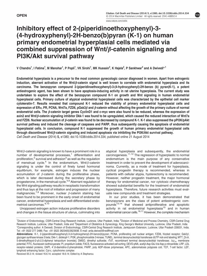
OPEN
Inhibitory effect of 2-(piperidinoethoxyphenyl)-3-(4-hydroxyphenyl)-2H-benzo(b)pyran (K-1) on humanprimary endometrial hyperplasial cells mediated viacombined suppression of Wnt/b-catenin signaling andPI3K/Akt survival pathway
V Chandra1, I Fatima1, M Manohar1, P Popli1, VK Sirohi1, MK Hussain2, K Hajela2, P Sankhwar3 and A Dwivedi*,1
Endometrial hyperplasia is a precursor to the most common gynecologic cancer diagnosed in women. Apart from estrogenicinduction, aberrant activation of the Wnt/b-catenin signal is well known to correlate with endometrial hyperplasia and itscarcinoma. The benzopyran compound 2-(piperidinoethoxyphenyl)-3-(4-hydroxyphenyl)-2H-benzo (b) pyran(K-1), a potentantiestrogenic agent, has been shown to have apoptosis-inducing activity in rat uterine hyperplasia. The current study wasundertaken to explore the effect of the benzopyran compound K-1 on growth and Wnt signaling in human endometrialhyperplasial cells. Primary culture of atypical endometrial hyperplasial cells was characterized by the epithelial cell markercytokeratin-7. Results revealed that compound K-1 reduced the viability of primary endometrial hyperplasial cells andexpression of ERa, PR, PCNA, Wnt7a, FZD6, pGsk3b and b-catenin without affecting the growth of the primary culture of normalendometrial cells. The b-catenin target genes CyclinD1 and c-myc were also found to be reduced, whereas the expression ofaxin2 and Wnt/b-catenin signaling inhibitor Dkk-1 was found to be upregulated, which caused the reduced interaction of Wnt7aand FZD6. Nuclear accumulation of b-catenin was found to be decreased by compound K-1. K-1 also suppressed the pPI3K/pAktsurvival pathway and induced the cleavage of caspases and PARP, thus subsequently causing the apoptosis of endometrialhyperplasial cells. In conclusion, compound K-1 suppressed the growth of human primary endometrial hyperplasial cellsthrough discontinued Wnt/b-catenin signaling and induced apoptosis via inhibiting the PI3K/Akt survival pathway.Cell Death and Disease (2014) 5, e1380; doi:10.1038/cddis.2014.334; published online 21 August 2014
Wnt/b-catenin signaling is known to have a prominent role in anumber of developmental processes,1 differentiation andproliferation,2 survival and adhesion3 as well as the regulationof menstrual cycle.4 In the endometrium, Wnt/b-cateninsignaling is under the control of finely tuned hormonalequilibrium, for example, estrogen induces the nuclearaccumulation of b-catenin during the proliferative phase,which is later decreased during the secretary phase byprogesterone, in the menstrual cycle.5,6 Aberrant regulation ofthe Wnt signaling pathway results in neoplastic transformationand thus lays at the root of initiation and progression of manymalignancies.7,8 Moreover, nuclear b-catenin staining hasbeen found to be prominent in the early onset of endometrialcancer, endometrial hyperplasia and well-differentiated endo-metriod carcinomas.9,10
Unopposed estrogen action induces proliferative disordersand changes in the tissue structure of uterus, culminating into
atypical hyperplasia and subsequently, the endometrialcarcinogenesis.11,12 The regression of hyperplastic to normalendometrium is the main purpose of any conservativetreatment in order to prevent the development of adenocarci-noma. Currently, as a mode of treatment for hyperplasia,cyclical progestin therapy is recommended, whereas inpatients with cellular atypia, hysterectomy is recommended.However, neither progestin treatment, the major hormonaltherapy for endometrial cancer, nor cytotoxic chemotherapyshowed substantial benefits for the treatment of endometrialhyperplasia. Therefore, future research activities must eval-uate new compounds and treatment strategies.
In our prior studies, it has been demonstrated thatbenzopyrans are the class of potent antiestrogenic com-pounds13,14 that showed antiproliferative and apoptoticactivity in rat endometrial hyperplasia15 and in humanendometrial cancer cells.16,17 However, the complete mechanism
1Division of Endocrinology, CSIR-Central Drug Research Institute, Lucknow, Uttar Pradesh, India; 2Division of Medicinal and Process Chemistry, CSIR-Central DrugResearch Institute, Lucknow, Uttar Pradesh, India and 3Department of Obstetrics & Gynecology, King George’s Medical University, Lucknow, Uttar Pradesh, India*Corresponding author: A Dwivedi, Division of Endocrinology, CSIR-Central Drug Research Institute, Jankipuram Extension, Lucknow, Uttar Pradesh 226031, India.Tel: +91 0522 277 2486; Fax: +91 0522 2623405/2623938; E-mail: [email protected]
Received 05.3.14; revised 16.6.14; accepted 18.6.14; Edited by A Stephanou
Abbreviations: K-1, 2-(piperidinoethoxyphenyl)-3-(4-hydroxyphenyl)-2H-benzo(b)pyran; PCNA, proliferating cell nuclear antigen; FZD6, frizzled receptor; Gsk3b,glycogen synthase kinase 3 beta; Dkk-1, dickkopf-1; PI3K, phosphoinositide 3-kinase; TUNEL, terminal deoxynucleotidyl transferase dUTP nick end labeling; MTT, [3-(4,5-dimethylthiazol-2-yl)-2,5-diphenyl tetrazolium bromide]; DMSO, dimethyl sulfoxide; rTdT, recombinant terminal deoxynucleotidyl transferase; Dcm, membranepotential; FITC, fluorescein isothiocyanate; PI, propidium iodide; FACS, fluorescence activated cell sorting; DEVD-pNA, acetyl-Asp-Glu-Val-Asp p-nitroanilide; LRP, LDLreceptor-related proteins; DAPI, 40,6-diamidino-2-phenylindole; PARP, poly ADP-ribose polymerase; APC, adenomatosis polyposis coli; TCF, T-cell factor; LEF,lymphoid enhancing factor; Cyt-c, cytochrome complex
Citation: Cell Death and Disease (2014) 5, e1380; doi:10.1038/cddis.2014.334& 2014 Macmillan Publishers Limited All rights reserved 2041-4889/14
www.nature.com/cddis

of action of 2-(piperidinoethoxyphenyl)-3-(4-hydroxyphenyl)-2H-benzo(b)pyran (K-1), responsible for inhibition of endo-metrial hyperplasia, remains to be explored in humanendometrial hyperplasial cells. Because estrogen is one ofthe factor responsible for inducing Wnt/b-catenin signalingthat may be responsible for inducing proliferation andhyperplasia formation in endometrium,4,5 we further analyzedwhether the benzopyran compound (K-1) modulates Wnt/b-catenin signaling in human endometrial hyperplasial cellsfrom proliferation (Wnt-On) to differentiation (Wnt-Off).Accordingly, in the current study, we explored the antiproli-ferative efficacy of K-1 on primary culture cells derived fromhuman endometrial hyperplasial tissue and investigated theWnt/b-catenin pathway and its downstream signaling. Wehave also studied the effect of K-1 on cell survival pathway tosubstantiate the above conjecture.
Results
K-1 inhibits endometrial hyperplasial cell proliferation.Firstly, the effect of K-1 on cell viability was examined by3-(4, 5-dimethylthiazol-2-yl)-2,5-diphenyl tetrazolium bromide(MTT) assay. Treatment of primary endometrial hyperplasialcells with K-1 showed decreased cell viability in a concentra-tion-dependent manner with IC50 of B5 mM (Po0.001), in theabsence or presence of estradiol (Figure 1c). K-1 wasineffective in normal endometrial primary culture cells butsuppressed the estradiol-induced proliferation in normalendometrial cells (Figure 1b). This showed that K-1 has anantiproliferative effect on primary endometrial hyperplasialcells without affecting the normal endometrial cells.
K-1 alters the expression of proliferation markers andWnt/b-catenin signaling markers in primary endometrialhyperplasial cells. To study the proliferation and Wnt/b-catenin signaling pathway, we analyzed proliferation markers(ERa, PR), downstream targets and integral parts of theWnt/b-catenin signaling (Wnt7a, FZD6, pGsk3b, Gsk3b andaxin2). K-1 was shown to downregulate the expression ofERa, PR, Wnt7a and FZD6 whereas upregulate the expres-sion of axin2 in a dose-dependent manner (Figure 2a). Thedensitometric analysis revealed that K-1 reduced theexpression of ERa byB76%, PR by B86%, Wnt7a byB77%, FZD6 by B55% and upregulated the expression ofaxin2 by 1.7 times at a concentration of 7.5 mM. Gsk3b, animportant downstream mediator of Wnt signaling thatregulates b-catenin phosphorylation and ubiqutinization,was found to be activated by K-1 via its dephosphorylation(Po0.001). These data suggest that K-1 significantly down-regulates the Wnt/b-catenin signaling in hyperplasial cells.
K-1 downregulates the expression of b-catenin and itsnuclear localization. K-1 was found to reduce the expres-sion of b-catenin and its downstream regulators (Figure 2b).The densitometric analysis showed that K-1 reduced theexpression of b-catenin by B65%, CyclinD1 by B75%,c-myc by B70% and PCNA by B81% at a concentration of7.5mM. Simultaneously, K-1 also reduced the nuclearlocalization of b-catenin (Figure 3), which might also beresponsible for suppression of b-catenin activation.
Effect of K-1 on Wnt/FZD6 binding and Dkk-1 expression.Co-immunoprecipitation studies were performed to analyzewhether K-1 inhibits Wnt7a and FZD6 interaction and
Figure 1 K-1 (2-[piperidinoethoxyphenyl]-3-[4-hydroxyphenyl]- 2H-benzo(b)pyran) inhibits endometrial hyperplasial cell proliferation. (a) Chemical structure ofcompound K-1, (b and c) cellular growth pattern of normal primary endometrial cellsand primary endometrial hyperplasial cells. Cells were treated with variousconcentrations of compound K-1 (1, 2.5, 5, 7.5 and 10mM) in the absence orpresence of estradiol (10 nM) for 48 h. Cell viability was measured using MTT assay.The percentage of viable cells was calculated as the ratio of treated cells to thecontrol cells. Data are expressed as mean (of three different experiments on normalsamples and five different experiments on hyperplasial samples);±S.E.M.,P values are a-Po0.001, b-Po0.01, c-Po0.05 and d-P40.05 versus controland e-Po0.001, f-Po0.01, g-Po0.05 and h-P40.05 versus estradiol
Inhibition of Wnt signaling in endometrial hyperplasiaV Chandra et al
2
Cell Death and Disease

modulates Wnt/b-catenin signaling. Results indicated thatK-1 decreased the interaction of Wnt7a/ FZD6 complex in adose-dependent manner (Figure 4a). The densitometricanalysis showed B78% reduction in Wnt7a/FZD6 bindingby K-1, which might be responsible for the deactivation ofWnt/b-catenin signaling.
Further, the expression of Dkk-1, a well-proven inhibitorof Wnt/b-catenin signaling, was found to be upregulated(1.8-fold) by K-1 (Figure 4b).
K-1 suppresses the estradiol-induced proliferation markersin normal endometrial cells. To analyze whether K-1 acts
through estrogen-mediated signaling to interfere with Wntsignaling, the effect of K-1 on estradiol-induced expression ofERa, PR, PCNA, Wnt7a, pPI3K and PI3K biomarkers innormal endometrial cells were studied. The densitometricanalysis showed that K-1 significantly reduced the estradiol-induced expression of ERa by B37%, PR by B42%, PCNAby B39%, Wnt7a by B49% and pPI3K/PI3K by B58% at aconcentration of 7.5mM (Figure 5b). Interestingly, K-1 did notcause any effect on the above-mentioned biomarkers innormal endometrial cells when incubated with K-1 alone(Figure 5a).
K1 interferes with the PI3K/Akt survival pathway inendometrial hyperplasial cells. We also studied the effectof K-1 on the PI3K/Akt pathway, an important cell survivaland proliferation pathway in hyperplasia development.Phosphorylation status of PI3K (at tyr485) and Akt(at ser473) was significantly downregulated by K-1. Thedensitometric analysis of immunoblots showed that the K-1decreased the intracellular levels of phosphorylated PI3K byB80% which in turn decreased the activation of Akt byB65% at a concentration of 7.5 mM of K-1 (Figure 4b).
K-1 triggers apoptosis in primary endometrial hyperplasialcells. To check whether the loss in cell viability on treatmentwith the K-1 is due to the induction of apoptosis, we analyzedannexin-V/PI-stained cells by flow cytometry, caspase-3activity and terminal deoxynucleotidyl transferase dUTP nickend labeling (TUNEL) assay. In the annexin-V/PI assay, K-1significantly increased the percentage of apoptotic cellsin a concentration-dependent manner and at 7.5 mM; theapoptotic cell fraction was approximately fivefold higher ascompared with that of control (Po0.001) (Figure 6a). In thecaspase-3 activity assay, it was found to be increased bythreefold at 7.5 mM of K-1 (Po0.001) (Figure 6b). Also,visually, a higher number of TUNEL-positive cells wereobserved in K-1-treated groups (Figure 7a).
Further, the mitochondrial transmembrane potential (cm) ofhyperplasial cells was analyzed and results showed asignificant drop (Po0.001) in Dcm in the presence of K-1(Figure 7b). These results indicated that the apoptoticsignaling pathway activated by K-1 is likely to be mediatedvia the mitochondrial pathway (Figure 6b). The role of K-1 ininducing apoptosis was analyzed by expression of cleavedcaspase-9 and 3 and cleaved PARP (Figure 7c). Densito-metric results showed that K-1 significantly upregulated theexpression of caspase-9 and 3 and cleaved PARP. The levelsof the pro- and antiapoptotic proteins (Bax and Bcl-2) werealso found to be altered in K-1-treated hyperplasial cells. K-1elicited a dose-dependent reduction in Bcl-2 expression(Po0.001) and an increase in the Bax up to 1.8-fold(Po0.001) (Figure 7c).
Discussion
Endometrial hyperplasia represents a precancerous,non-physiological and noninvasive proliferation of theendometrium18 and eventually that may lead to endometrialcarcinoma. In case of enhanced or unopposed estrogensignaling, constitutive activation of Wnt/b-catenin signaling
Figure 2 Effect of compound K-1 on the expression of (a) proliferation markersand Wnt/b-catenin signaling markers, and (b) its downstream effectors. Endometrialhyperplasial cells were treated with vehicle or compound K-1 at variousconcentrations (2.5, 5 and 7.5mM) for 48 h. Thirty-five micrograms of whole celllysate protein in each lane was probed for the expression of ERa, PR, Wnt7a, FZD6,axin2, pGsk3b(ser9), Gsk3b, b-catenin, CyclinD1, c-myc and PCNA using specificantibodies. b-actin was used as a control to correct for loading. Representative blotsare shown (upper panels) and densitometric quantitation of protein expressionlevels are shown as fold changes (lower panels). Data are expressed as mean ofthree different experiments;±S.E.M., P values are a-Po0.001, b-Po0.01,c-Po0.05 and d-P40.05 versus control
Inhibition of Wnt signaling in endometrial hyperplasiaV Chandra et al
3
Cell Death and Disease

triggers endometrial hyperplasia, which may further developinto endometrial cancer.10 Among various known Wnts, theWnt7a ligand is able to directly stimulate canonical Wntsignaling and induce proliferation in endometrium.19,20 Wehave previously shown that K-1 shows inhibitory activityagainst endometrial hyperplasia in a rat model.14 In thecurrent study, we have established a human primary atypicalendometrial hyperplasia cell culture for studying its effect onthe human tissue. Initially, we compared the growth pattern ofnormal endometrial cells and hyperplasial cells, and it wasinteresting to observe that the compound significantlyinhibited the growth of hyperplasial cells but not of normalendometrial cells. However, K-1 inhibited the estrogen-induced growth of normal endometrial cells. Under similarconditions, PCNA levels also remained below the baselinelevels in K-1-treated cells. The difference in PCNA levelscaused by the compound in the presence of E2 indicated thesuppression of estrogen action caused by K-1 in these cells(Figures 1 and 5). Because in hyperplasial endometrial cells,the unoppposed estrogen action is supposed to be respon-sible for cellular growth, we hypothesize that the differential
and specific effect of K-1 on hyperplasial cells may be due toits effect on estrogen-regulated signaling pathways. Also, inendometrium, estrogen has been shown to induce the Wntsignaling pathway, whereas progesterone has been shown tosuppress the same.5 Therefore, we further tried to analyzewhether K-1 has any effect on Wnt signaling using humanendometrial primary culture. We have observed that K-1suppresses the endometrial primary culture growth viadownregulating Wnt/b-catenin signaling.
The regulation of Wnt/b-catenin pathway is governed byWnt ligand and Frizzled receptor itself and various otherdownstream markers such as axin2, b-catenin, adenomatosispolyposis coli (APC) and Gsk3b. Cytoplasmic stabilizedb-catenin migrates to the nucleus, where it associates withTCF/LEF transcription factors, leading to the transcription ofb-catenin target genes.21,22 We found the reduced expressionof b-catenin as well as its nuclear accumulation as observedby confocal microscopy in K-1-treated cells. Subsequently,the downstream effectors CyclinD1 and c-myc23 were foundto be downregulated in treated cells. Further, the reducedexpression of Wnt7a ligand and FZD6, and the increased
Figure 3 Demonstration of nuclear b-catenin accumulation in primary endometrial hyperplasial cells by confocal microscopy. Cells were treated with vehicle, 2.5, 5 and7.5mM of the compound K-1 for 24 h. Cells were fixed, permeabilized, incubated with b-catenin antibody overnight, and subsequently incubated with FITC-conjugated anti-rabbit antibody for 2 h. The preparations were washed and counterstained with DAPI (Sigma-Aldrich). Representative micrographs demonstrating the distribution of b-cateninare shown. Cell images were grasped using a confocal fluorescence microscope
Inhibition of Wnt signaling in endometrial hyperplasiaV Chandra et al
4
Cell Death and Disease

expression of axin2 accompanied by a reduced expression ofphosphorylated Gsk3b in human hyperplasial primary cellculture under the influence of K-1 suggested the attenuation ofWnt/b-catenin signaling.
One of the known Wnt/b-catenin inhibitors, Dkk-1, is aproapoptotic gene,24 which has been demonstrated to interactwith LRP5/6 and to inhibit Wnt/b-catenin signaling viadisrupting the binding of the Wnt/FZD ligand–receptor
Figure 5 K-1 suppresses estradiol-induced proliferation markers and Wnt7a.Western blot analysis was done to see the effect of compound K-1 on primaryculture of normal endometrial cells (a) in the absence of estradiol and (b) in thepresence of estradiol. The expression of proliferation markers (ERa, PR andPCNA), Wnt7a and cell survival marker (pPI3K-tyr485/PI3K) was analyzed in thepresence of estradiol (10 nM). Cells were treated with vehicle or E2, K-1,K-1þE2, for 48 h. Thirty-five micrograms of whole cell lysate protein in each lanewas probed for the expression of ERa, PR, PCNA.Wnt7a, pPI3K(tyr485) and PI3Kusing specific antibodies. b-actin (upper panel) was used as a control to correct forloading. Representative blots are shown and densitometric quantitation of proteinexpression levels are shown as fold changes (lower panel). Data are expressed asmean of three different experiments;±S.E.M., P values are a-Po0.001,b-Po0.01, c-Po0.05 and d-P40.05 versus control and e-Po0.001, f-Po0.01,g-Po0.05 and h-P40.05 versus estradiol
Figure 4 Effect of K-1 on Wnt/FZD6 binding, Dkk-1 expression and markers ofcell survival pathway in endometrial hyperplasial cells. (a) Interaction betweenWnt7a ligand and FZD6 was determined by co-immunoprecipitation. Cells weretreated with vehicle or compound at various concentrations, for 48 h. Cell lysateswere immunoprecipitated with anti-FZD6 and subsequently immunoblotted withanti-Wnt7a. NC is the negative control in which cell lysate was incubated withnon-immune serum instead of anti-FZD6. Representative blots are shown in theupper panel and densitometric quantitation of protein expression levels areshown as fold changes. Data are expressed as mean of three differentexperiments;±S.E.M., P values are a-Po0.001, b-Po0.01, c-Po0.05and d-P40.05 versus control. (b) Phosphorylation status of PI3K, Akt andexpression of Dkk-1 as determined by western blot analysis in endometrialhyperplasial cells. Cells were treated with vehicle, 2.5, 5 and 7.5 mM of thecompound for 48 h. Thirty-five micrograms of whole cell lysate protein in eachlane was probed for the expression of pPI3K(tyr485), PI3K, pAkt(ser473),Akt and Dkk-1 using specific antibodies. b-actin was used as a control tocorrect for loading. Representative blots are shown in the upper panel anddensitometric quantitation of protein expression levels are shown as foldchanges. Data are expressed as mean of three different experiments;±S.E.M.,P values are a-Po0.001, b-Po0.01, c-Po0.05 and d-P40.05 versuscontrol
Inhibition of Wnt signaling in endometrial hyperplasiaV Chandra et al
5
Cell Death and Disease
complex.25 Interestingly, K-1 caused the induced expressionof Dkk-1 and inhibited Wnt/ FZD ligand–receptor binding asdemonstrated by co-immunoprecipitation experiment. Evi-dences have been given that progesterone induction of Dkk-1and Foxo-1 results in the inhibition of Wnt signaling in humanendometrium.6 It appears that our compound behaves likeprogesterone in modulating the Wnt signaling in endometrial
cells although it is known to bind to estrogen receptor (ER). Itcompetes with estradiol for binding to both hERa and hERbindicating its interaction with both ER subtypes.17 We haveearlier shown that K-1 inhibits classical and non-classicalestrogen signaling in uterus.15,16 Because Wnt signalingpathway is known to be regulated via estrogen, we exploredfurther whether K-1 is able to suppress the estrogen-inducedWnt signaling in human endometrial cells. Interestingly, theprofound effect of K-1 on Wnt7a expression was observed inhyperplasial cells and also the phosphorylated PI3K wasreduced in these cells when incubated with K-1. In experi-ments on normal cells, no significant effect on Wnt7a andpPI3k was observed, but when cells were treated with E2, thesignificant inhibition in these proteins was observed. Thisindicates that K-1 acts specifically on E2-induced Wntexpression. In absence of E2, the basal levels of Wnt werenot changed. The probable reason may be the inhibition of Eaction in endometrial hyperplasial cells which might havehigher expression levels of ER. Besides, in hyperplasial cells,the aberrant endogenously synthesized E may be responsiblefor higher ER expression. Because K-1 is already known tointeract with ERs, and antagonize the action of E,14,17 it mightcause the inhibition of estrogen-induced signaling mechanismin normal endometrial cells when exogenous E was given.
Further, it is likely that compound is responsible forsuppression of Wnt signaling due to interference withER-mediated signaling in endometrial hyperplasia.4 Thus,the difference in K-1 action in endometrial hyperplasial cells orestradiol-treated normal endometrial cells as compared withnormal endometrial cells may be because of the change inexpression of ERs, ERa/ERb ratio26 and/or the expression ofcoactivators or corepressors.27 These differences maymodulate the role of K-1 in different cell types leading tocell-specific effects.
ERa has been observed to be associated with importantgrowth factor pathway such as the PI3K pathway, thusindirectly cross-talking with canonical Wnt signaling.24 InPI3K/Akt survival pathway, phosphorylation of PI3K and itsdownstream target Akt enhances proliferation and survival ofcells through suppression of apoptosis. Akt phosphorylationcauses many modulations in a cell to make it cancerous.Deactivation of Gsk3b by Akt phosphorylation is one of thosemodulations.25 Akt also activates the expression of theantiapoptotic gene Bcl-2, which negatively controls theexpression of proapoptotic gene Bax,26,27 and subsequentlyinhibits the mitochondria-induced apoptotic signaling cas-cade. Here, we demonstrated that K-1 was involved ininhibiting the phosphorylation of PI3K and Akt. Significantdownregulation of the expression of Bcl-2 and the upregula-tion of Bax observed may be due to reduced Akt activitythat may enhance Cyt-c release from mitochondria byreducing its membrane potential in K-1-treated cells. Further,mitochondria-induced apoptotic signaling cascade was sup-ported by the presence of cleaved fragments of caspase-9and 3 and PARP. These findings were supported by caspase-3activation, TUNEL-staining and annexin-V/PI staining.
In summary, the present study has demonstratedthat K-1 hinders the Wnt/b-catenin signaling in endometrialhyperplasial cells, Wnt7a/ FZD6 binding and subsequentsignaling. It induced axin2 and Wnt/b-catenin inhibitor Dkk-1.
Figure 6 K-1 induces apoptosis and activates caspase-3 in primary endometrialhyperplasial cells. (a) Analysis of apoptosis in primary endometrial hyperplasial cellstreated with compound K-1 by flow cytometric analysis of annexin-V/PI-stained cellsafter 48-h culture. AVþ /PI� -intact cells; AV� /PIþ -nonviable/necrotic cells;AVþ /PI� and AVþ /PIþ -apoptotic cells. Representative images are shown inthe upper panel and the percentage of cell fraction with S.E.M. was calculatedbased on three independent experiments. P values are a-Po0.001, b-Po0.01,c-Po0.05 and d-P4.05 versus control. (b) Induction of caspase-3 proteolyticactivity was checked in primary endometrial hyperplasial cells after treatment ofcompound K-1 (2.5, 5 and 7.5mM). Proteolytic activity was measured by cleavage ofthe caspase-3 substrate DEVD-pNA as described in ‘Materials and Methods’.Cell lysates were prepared after 48 h. Results are expressed as mean±S.E.M.,n¼ 3.± S.E.M., P values area -Po0.001, b-Po0.01, c-Po0.05 and d-P40.05versus control
Inhibition of Wnt signaling in endometrial hyperplasiaV Chandra et al
6
Cell Death and Disease

Simultaneously, K-1 also downregulated the phosphorylationof PI3K/Akt, which enhanced apoptosis in primary endome-trial hyperplasial cells. Hypothetically, two mechanisms maybe involved in the regulation of Wnt signaling under theinfluence of these effects; compound K-1 either enhancesDkk-1 and antagonizes the estrogen-induced Wnt signalingvia ER (through ERE) or it might induce Foxo-1 expression,which acts as transcription factor for Wnt-regulated geneexpression, although the latter possibility needs to beanalyzed further. Altogether, these data suggest that K-1significantly down regulates the Wnt/b-catenin signalingwhich might be considered as one of the mechanismsresponsible for specific anti-proliferative activity of K-1 inhuman endometrial hyperplasial cells. Thus, the benzopyranderivative K-1 can serve as a future therapeutic agent fortreatment of endometrial hyperplasia over the currentlyavailable therapy.
Materials and MethodsCompound. K-1 (Figure 1a) was synthesized as described earlier.28,29
Antibodies and reagents. All culture media and other reagents werepurchased from Sigma-Aldrich (St. Louis, MO, USA). Anti-cytokeratin7, -ERa,-PR, -PCNA, -pGsk3b (ser9), -Gsk3b, -c-myc, -b-catenin, -CyclinD1, -Wnt7a,-FZD6, -Dkk-1, -pPI3K(tyr 485), -PI3K, -Akt, -b-actin antibodies, peroxidase- andfluorescein isothiocyanate (FITC)-conjugated secondary antibodies were procured
from Santa Cruz (Dallas, TX, USA). Antibodies for axin2, pAkt(ser473), cleavedcaspase-3 and 9, cleaved PARP, Bax and Bcl-2 were purchased from CellSignalling Technology, Life Sciences (Boston, MA, USA).
TUNEL detection kit was obtained from Roche (Basel, Switzerland). Immuno-BlotPVDF membrane was purchased from Millipore (Billerica, MA, USA). ECL reagentand ECL Hyperfilm were purchased from GE Healthcare (Little Chalfont, UK).
Endometrial tissue collection. Endometrial hyperplasia samples (fivedifferent cases of atypical hyperplasia) were collected from patients with abnormaluterine bleeding (age: 25–40 years) from the Department of Obstetrics andGynecology, King George’s Medical University, Lucknow, Uttar Pradesh, India.Normal endometrial samples (three different cases) were collected from thepatients undergoing hysterectomy for uterine prolapse reasons. An informedwritten consent was obtained from each patient and the study was approved bythe local Human Ethics Committee.
Histopathological examination such as gland-to-stroma ratio, gland’s size andshape, cytologic atypia, nuclear/cytoplasmic ratio, hyperchromatosis by simplestaining and expression of ER, progesterone receptor (PR) and proliferation marker(Ki67) by immune-histochemistry were carried out by expert gynecologists andpathologists from Department of Obstetrics and Gynecology and Department ofPathology, King George’s Medical University, who confirmed the category ofsamples as hyperplasia to be of atypical type and after the evaluation, tissuesamples were taken for further studies.
Primary culture of endometrial cells. Cell isolation was based on themethods as described by Genc et al.30 Briefly, tissues were collected in MEM,minced and incubated with 1 mg/ml collagenase and DNase (2 mg/ml) for 2 h at37 1C with periodic mixing. Digested tissue was mechanically dissociated andresuspended in 2 ml of fresh MEM. The cells were filtered through 18-mesh sterile
Figure 7 Effect of K-1 on TUNEL staining, expression of markers of apoptosis in primary endometrial hyperplasial cells. (a) Primary endometrial hyperplasial cells weretreated with vehicle, 2.5, 5 and 7.5mM of the compound K-1 for 24 h. Cells were fixed, permeabilized and the procedure was followed as described in Materials and Methods.Representative figures stained for TUNEL showing a large number of TUNEL-positive cells (greenish yellow) in K-1-treated groups in a concentration-dependent manner. Cellimages were grasped using a Nikon fluorescence microscope at � 40. (b) Cells were treated with vehicle, or compound K-1 at 2.5, 5 and 7.5mM concentrations, for 24 h.Mitochondrial membrane potential was measured by normalization of the 590 : 530 nm JC-1 emission ratios and then normalized to untreated cells. Results are expressed asmean±S.E.M., n¼ 3.± S.E.M., P values are a-Po0.001, b-Po0.01, c-Po0.05 and d-P40.05 versus control. (c) Cells were treated with the indicated concentrations ofcompound for 48 h, and 35mg whole cell lysate in each lane was probed for the expression of Bcl-2,Bax, cleaved caspase3/9 and cleaved PARP using specific antibodies.b-actin was used as a control to correct for loading. Representative blots are shown (left panel) and densitometric quantitation of protein expression levels are shown as foldchanges (right panel). Data are expressed as mean of three different experiments;±S.E.M., P values are a-Po0.001, b-Po0.01, c-Po0.05 and d-P40.05 versus control
Inhibition of Wnt signaling in endometrial hyperplasiaV Chandra et al
7
Cell Death and Disease

gauze, centrifuged and washed twice with MEM containing 10% fetal bovineserum (FBS), 1 mM sodium pyruvate, 2 mM L-glutamine and 2% of antibiotic-antimycotic solution. Then cells were transferred into plastic culture flasks (75 cm2)(Corning Incorporated, Corning, NY, USA) and incubated at 37 1C with saturatinghumidity and 5% CO2. Prior to experiments, cells were cultured in phenol red-freeMEM supplemented with 10% charcoal-stripped FBS and 1% antibiotic-antimycoticsolution.
The endometrial hyperplasial cells were characterized by examining theexpression of cytokeratin-7, an epithelial marker, by immunofluorescence(Supplementary Figure S1).
Equal numbers of cells were used each time in separate studies.
Cell viability assay. For determination of cell viability, MTT assay wasperformed. The primary culture of endometrial hyperplasial cells and normalendometrial cells were seeded (2.5� 103 cells/well) into 96-well plate and treatedwith K-1 (1, 2.5, 5, 7.5 and 10mM) in the absence or presence of estradiol (10 nM)for 48 h. Then MTT (0.5 mg/ml) (Sigma-Aldrich) was added and incubated for 2 hat 37 1C. After removing supernatants, cells were incubated with 100ml of dimethylsulfoxide (DMSO). The OD was read with Microquant (Bio Tek, Winooski, VT,USA) at 540 nm. The IC50 value of the compound was determined by Compusynsoftware. The experiments were performed three times with five replicates in caseof hyperplasial endometrial primary cells and with three replicates in case ofnormal endometrial primary cells.
Western blot analysis. Endometrial primary culture cells were treated withvehicle or various concentrations of K-1 or estradiol for 48 h. After each treatment,cells were lysed in lysis buffer (Sigma-Aldrich) supplemented with a proteaseinhibitor cocktail. Equal amounts of protein were separated by gel electrophoresisand transferred to Immuno-Blot PVDF membrane. The membrane was blockedwith 5% skimmed milk and incubated with primary antibody overnight at 4 1C.The membranes were then incubated with secondary antibody for 1 h. Antibodybinding was detected using enhanced chemiluminescence detection system(GE Healthcare). After developing, the membrane was stripped and re-probed withb-actin, Gsk3b, PI3K and Akt antibodies. Each experiment was repeatedthree times. Quantitation of band intensity was performed by densitometryusing Quantity-One software (v.4.5.1) and a Gel-Doc2000 imaging system(Bio-Rad, Hercules, CA, USA).
Immunofluorescence imaging by confocal microscopy. Primaryendometrial hyperplasia cells were grown on coverslips in 12-well plate andtreated with vehicle, or 2.5, 5 and 7.5mM of K-1 for 24 h. Cells were then fixed inmethanol and acetone (1 : 1) and followed by permeabilization with 0.1% TritonX-100. Cells were washed with PBS and blocked with 1% BSA and incubated withb-catenin antibody overnight followed by 1 h incubation with fluorescence-taggedsecondary antibody. Following incubation, cells were counterstained with 40,6-diamidino-2-phenylindole (DAPI) for 5 min. Images were captured at � 100 andanalyzed using LSM-Image-Examiner Software to detect fluorescence and DAPIemissions. Cells not exposed to primary antibodies served as negative controls.
Co-immunoprecipitation assay. Interaction between Wnt7a ligand andFZD6 proteins was studied by co-immunoprecipitation of the complex followed byimmunoblotting. Protein A-Sepharose beads were used and the desired proteinwas immunoprecipitated according to manufacturer’s instructions (Sigma-Aldrich).Briefly, 2mg of anti-FZD6 was added to 500mg of cell lysate treated with indicatedconcentrations of K-1 and incubated for overnight at 4 1C. For negative control,cell lysate was incubated with corresponding non-immune serum. Then, 100ml ofbeads suspension was added and samples were incubated for 1 h at 4 1C.Immunoprecipitated complexes were collected by centrifugation at 3000� g for2 min at 4 1C and washed three times with RIPA buffer. Immunoprecipitates wereresuspended in Laemmli sample buffer and heated for 5 min at 95 1C. Thesupernatants were collected by centrifugation at 12 000� g for 30 s at roomtemperature. Equal volumes of immunoprecipitated proteins were run on 8% SDS-PAGE and transferred to PVDF membrane. The proteins were probed with anti-Wnt7a, followed by the corresponding secondary antibody. Bands were detectedand analyzed by densitometry using Quantity-One Software (v. 4.5.1) and a Gel-Doc2000 (Bio-Rad) imaging system.
Terminal deoxynucleotidyltransferase-mediated nick endlabeling assay. TUNEL staining was performed by using a ‘Dead End’
fluorometric TUNEL kit (Roche, Upper Bavaria, Germany) as per manufacturer’sprotocol. Briefly, slides were equilibrated with equilibration buffer and wereincubated for 30 min at 37 1C with recombinant terminal deoxynucleotidyltransferase (rTdT) incubation buffer. The negative control slide was incubatedwithout the rTdT enzyme. Sections were then rinsed in buffer to stop the reaction,followed by PBS washing and examined under light microscope (Nikon Eclipse80i, Shinagawa-Ku, Tokyo, Japan). Images were captured using an NIS-ElementsF-3.0 camera (Nikon, Tokyo, Japan).
Measurement of mitochondrial membrane potential. Dcm wasdetermined using JC-1 (cationic mitochondrial vital dye) as a probe accordingto the method of Dey and Moraes.31 Briefly, treated cells were collectedand incubated for 20 min with 5 mM JC-1 at 37 1C, washed and resuspendedin media. The Dcm was measured at 590 nm for J-aggregates and at530 nm for J-monomer. The ratio of 590/530 nm was considered as the relativeDcm value. The experiments were performed thrice with three replicatesin each.
Annexin-V/propidium iodide labeling and flow cytometry assayfor apoptosis. Cells (2� 105cells/ml) were cultured in 6-well plates andtreated with K-1 (2.5, 5 and 7.5mM) for 48 h. Adherent and non-adherent cellswere probed with FITC-conjugated annexin-V and PI. The staining profiles weredetermined with FACScan and Cell-Quest software. The experiments wereperformed thrice with three replicates in each.
Caspase-3 colorimetric assay. Caspase-3 activity was determined usingthe colorimetric Caspase-3 assay kit (Sigma-Aldrich). Briefly, cells were trypsinizedand centrifuged for 5 min at 600 g at 4 1C. Then, cell pellets were resuspended inice-cold cell lysis buffer and incubated on ice for 20 min. Supernatants collectedafter centrifugation were incubated with 1 mM caspase-3 substrate (DEVD-pNA)for 2 h at 37 1C and the OD was measured at 405 nm. The experiments wereperformed three times with three replicates in each.
Statistical analysis. Results were expressed as mean ±S.E.M. for at leastthree separate determinations for each experiment. Statistical significance wasdetermined by ANOVA and Newman Keul’s test, and the levels of probability werenoted.
Conflict of InterestThe authors declare no conflict of interest.
Acknowledgements. We thank Mr AL Vishwakarma and Dr K Mitra,SAIF-facility, CDRI for help in flow cytometric analysis and confocal microscopy.The study was supported by grants from Indian Council of Medical Research, India.VC is the recipient of senior research fellowship from Indian Council of MedicalResearch. The CDRI communication number is 8736.
1. Luu HH, Zhang R, Haydon RC, Rayburn E, Kang Q, Si W et al. Wnt/beta-cateninsignaling pathway as a novel cancer drug target. Curr Cancer Drug Targets 2004; 4:653–671.
2. Rider V, Isuzugawa K, Twarog M, Jones S, Cameron B, Imakawa K et al.Progesterone initiates Wnt-beta-catenin signaling but estradiol is required for nuclearactivation and synchronous proliferation of rat uterine stromal cells. J Endocrinol 2006;191: 537–548.
3. Tarapore RS, Siddiqui IA, Mukhtar H. Modulation of Wnt/beta-catenin signaling pathway bybioactive food components. Carcinogenesis 2012; 33: 483–491.
4. Wang Y, van der Zee M, Fodde R, Blok LJ. Wnt/Beta-catenin and sex hormone signaling inendometrial homeostasis and cancer. Oncotarget 2010; 1: 674–684.
5. van der Horst PH, Wang Y, van der Zee M, Burger CW, Blok LJ. Interaction between sexhormones and WNT/�-catenin signal transduction in endometrial physiology and disease.Mol Cell Endocrinol 2012; 358: 176–184.
6. Wang Y, Hanifi-Moghaddam P, Hanekamp EE, Kloosterboer HJ, Franken P, Veldscholte Jet al. Progesterone inhibition of Wnt/beta-catenin signaling in normal endometrium andendometrial cancer. Clin Cancer Res 2009; 15: 5784–5793.
7. Ilyas M. Wnt signaling and the mechanistic basis of tumour development. J Pathol 2005;205: 130–144.
8. Thanendrarajan S, Kim Y, Schmidt-Wolf IGH. Understanding and targeting theWnt/b-Catenin signaling pathway in chronic leukemia. Leuk Res Treatment 2011;2011: 329572.
Inhibition of Wnt signaling in endometrial hyperplasiaV Chandra et al
8
Cell Death and Disease
9. Saegusa M, Okayasu I. Frequent nuclear beta-catenin accumulation and associated
mutations in endometrioid-type endometrial and ovarian carcinomas with squamous
differentiation. J Pathol 2001; 194: 59–67.10. Scholten AN, Creutzberg CL, van den Broek LJ, Noordijk EM, Smit VT. Nuclear
beta-catenin is a molecular feature of type I endometrial carcinoma. J Pathol 2003; 201:
460–465.11. Emons G, Fleckenstein G, Hinney B, Huschmand A, Heyl W. Hormonal interactions in
endometrial cancer. Endocr Relat Cancer 2000; 7: 227–242.12. Akhmedkhanov A, Zeleniuch-Jacquotte A, Toniolo P. Role of exogenous and endogenous
hormones in endometrial cancer: review of the evidence and research perspectives.
Ann N Y Acad Sci 2001; 943: 296–315.13. Dhar JD, Setty BS, Duran S, Kapil RS. Biological profile of 2-[4-(2-N-piperidinoethoxy)
phenyl]-3-phenyl (2H) benzo (b) pyran-a potent anti-implantation agent in rat.
Contraception 1991; 44: 461–472.14. Kharkwal G, Fatima I, Kitchlu S, Singh B, Hajela K, Dwivedi A. Anti-implantation effect of 2-
[piperidinoethoxyphenyl]-3-[4-hydroxyphenyl]-2H-benzo(b)pyran, a potent antiestrogenic
agent in rats. Fertil Steril 2011; 95: 1322–1327.15. Chandra V, Fatima I, Saxena R, Kitchlu S, Sharma S, Hussain MK et al.
Apoptosis induction and inhibition of hyperplasia formation by 2-[piperidinoethoxyphe-
nyl]-3-[4-hydroxyphenyl]-2H-benzo(b)pyran in rat uterus. Am J Obstet Gynecol 2011; 205:
362e1–362e11.16. Fatima I, Chandra V, Saxena R, Manohar M, Sanghani Y, Hajela K et al. 2,3-Diaryl-2H-1-
benzopyran derivatives interfere with classical and non-classical estrogen receptor
signaling pathways, inhibit Akt activation and induce apoptosis in human endometrial
cancer cells. Mol Cell Endocrinol 2012; 348: 198–210.17. Fatima I, Saxena R, Kharkwal G, Hussain MK, Yadav N, Hajela K et al. The anti-
proliferative effect of 2-[piperidinoethoxyphenyl]-3-[4-hydroxyphenyl]-2H-benzo(b) pyran is
potentiated via induction of estrogen receptor beta and p21 in human endometrial
adenocarcinoma cells. J Steroid Biochem Mol Biol 2013; 138: 123–131.18. Horn LC, Meinel A, Handzel R, Einenkel J. Histopathology of endometrial hyperplasia and
endometrial carcinoma: an update. Ann Diagn Pathol 2007; 11: 297–311.19. Bui TD, Zhang L, Rees MC, Bicknell R, Harris AL. Expression and hormone regulation of
Wnt2, 3, 4, 5a, 7a, 7b and 10b in normal human endometrium and endometrial carcinoma.
Br J Cancer 1997; 75: 1131–1136.20. Carmon KS, Loose DS. Secreted frizzled-related protein 4 regulates two Wnt7a signaling
pathways and inhibits proliferation in endometrial cancer cells. Mol Cancer Res 2008; 6:
1017–1028.21. MacDonald BT, Tamai K, He X. Wnt/beta-catenin signaling: components, mechanisms,
and diseases. Dev Cell 2009; 17: 9–26.22. Kanwar SS, Yu Y, Nautiyal J, Patel BB, Majumdar AP. The Wnt/beta-catenin pathway
regulates growth and maintenance of colonospheres. Mol Cancer 2010; 9: 212.
23. Pu P, Zhang Z, Kang C, Jiang R, Jia Z, Wang G et al. Down regulation of Wnt2 andbeta-catenin by siRNA suppresses malignant glioma cell growth. Cancer Gene Ther 2009;16: 351–361.
24. Tulac S, Overgaard MT, Hamilton AE, Jumbe NL, Suchanek E, Giudice LC. Dickkopf-1, aninhibitor of Wnt signaling, is regulated by progesterone in human endometrial stromal cells.J Clin Endocrinol Metab 2006; 91: 1453–1461.
25. Ahn VE, Chu ML, Choi HJ, Tran D, Abo A, Weis WI. Structural basis of Wnt signalinginhibition by Dickkopf binding to LRP5/6. Dev Cell 2011; 21: 862–873.
26. Hu K, Zhong G, He F. Expression of estrogen receptors ERa and ERb inendometrial hyperplasia and adenocarcinoma. J Gynecol Cancer 2005; 15:537–541.
27. Uchikawa J, Shiozawa T, Shih H, Miyamoto T, Feng YZ, Kashima H et al. Expression ofsteroid receptor coactivators and corepressors in human endometrial hyperplasia andcarcinoma with relevance to steroid receptors and Ki-67 expression. Cancer 2003; 98:2207–2213.
28. Kapil RS, Durani S, Dhar JD, Setty BS. Novel benzopyrans and process for theirproduction. 1990; European patent no. 90308787/2.
29. Sharma AP, Saeed A, Durani S, Kapil RS. Structure-activity relationship of antiestrogens.Effect of the side chain and its position on the activity of 2,3-diaryl-2H-1-benzopyrans.J Med Chem 1990; 33: 3216–3222.
30. Genc S, Attar E, Gurdol F, Kendigelen S, Bilir A, Serdaroglu H. The effect of COX-2inhibitor, nimesulide, on angiogenetic factors in primary endometrial carcinoma cell culture.Clin Exp Med 2007; 7: 6–10.
31. Dey R, Moraes CT. Lack of oxidative phosphorylation and low mitochondrialmembrane potential decrease susceptibility to apoptosis and do not modulatethe protective effect of Bcl-x(L) in osteosarcoma cells. J Biol Chem 2000; 275:7087–7094.
Cell Death and Disease is an open-access journalpublished by Nature Publishing Group. This work is
licensed under a Creative Commons Attribution-NonCommercial-NoDerivs 3.0 Unported License. The images or other third partymaterial in this article are included in the article’s Creative Commonslicense, unless indicated otherwise in the credit line; if the material isnot included under the Creative Commons license, users will need toobtain permission from the license holder to reproduce the material. Toview a copy of this license, visit http://creativecommons.org/licenses/by-nc-nd/3.0/
Supplementary Information accompanies this paper on Cell Death and Disease website (http://www.nature.com/cddis)
Inhibition of Wnt signaling in endometrial hyperplasiaV Chandra et al
9
Cell Death and Disease
Copyright © 2022 FDOKUMEN


![Genotoxicity assessment and detoxification induction in Dreissena polymorpha exposed to benzo[a]pyrene](https://static.fdokumen.com/doc/165x107/6344d92703a48733920b14f7/genotoxicity-assessment-and-detoxification-induction-in-dreissena-polymorpha-exposed.jpg)

![Photophysical properties and computational investigations of tricarbonylrhenium(I)[2-(4-methylpyridin-2-yl)benzo[ d]-X-azole]L and tricarbonylrhenium(I)[2-(benzo[ d]-X-azol-2-yl)-4-methylquinoline]L](https://static.fdokumen.com/doc/165x107/631cc3eea906b217b9071d28/photophysical-properties-and-computational-investigations-of-tricarbonylrheniumi2-4-methylpyridin-2-ylbenzo.jpg)
![New method for benzo[a]pyrene analysis in plant material using subcritical water extraction](https://static.fdokumen.com/doc/165x107/6330d751b2d7d1ed8d076247/new-method-for-benzoapyrene-analysis-in-plant-material-using-subcritical-water.jpg)