Prediction of endometrial carcinoma by subjective endometrial intraepithelial neoplasia diagnosis
In Vitro Cell. Dev. Biol. 31:196-206, March 1995 © 1995 Society for In Vitro Biology 1071-2690/95 $02.50+0.00
A P O L A R I Z E D H U M A N E N D O M E T R I A L C E L L L I N E T H A T
B I N D S A N D T R A N S P O R T S P O L Y M E R I C I g A
JUDITH M. BALL, ZINA MOLDOVEANU, LAWRENCE R. MELSEN, PAMELA A. KOZLOWSKI, SUSAN JACKSON, MARK J. MULLIGAN, JIRI F. MESTECKY, ~n RICHARD W. COMPANS
Department of Microbiology, University of Alabama at Birmingham, Birmingham, Alabama 35294 (J. M. B., Z. M., P. A. K., S. J., M. J. M., J. F. M.); Department of Microbiology and Immunology, Emory University School of Medicine, Rollins Research Center,
1510 Clifton Road Atlanta, Georgia 30322 (L. R. M., R. W. C.); Division of Infectious Diseases (M. J. M.) and Division of Clinical Immunology and Rheumatology (J. F. M.), Department of Medicine, University of
Alabama at Birmingham, Birmingham, Alabama 35294
(Received 25 April 1994; accepted 23 August 1994)
SUMMARY
We have demonstrated that a human endometrial cell line, HEC-1, maintains a high transepithelial electrical resistance, directionally transports fluids across the cell monolayer, and releases enveloped viruses at distinct plasma membrane domains: influenza virus is released at the apical surfaces and vesicular stomatitis virus (VSV) at the basolateral surfaces. In addition, we have examined the expression of domain-specific endogenous proteins, including the polyimmunoglobulin receptor. Multiple endogenous polypeptides were found to be secreted into the culture medium at basolateral surfaces, whereas no secretion of specific polypeptides was observed from apical cell surfaces. Distinct patterns of endogenous proteins were also observed on apical and basolateral cell surfaces, with a much more complex polypeptide pattern on the basolateral membranes. Using surface biotinylation and immunofluorescence, the polyimmunoglobulin receptor was found to be expressed on the basolateral surfaces of HEC-1 monolayers. The specific binding of poly-immunoglobuhn A (plgA) was found to occur on the basolateral surface, and was followed by transcytosis to the apical surface and release into the apical medium. The observed characteristics indicate that the endometrium-derived HEC-1 epithelial cell line can be employed as a model for studies of protein transport in polarized epithelial cells of human endometrial tissues, as well as for studies of the interaction of microorganisms with epithelial cells in the genital tract.
INTRODUCTION
Polarized epithelial ceils are distinguished morphologically by apical projections or microvilh, tripartite junctional complexes, and a defined basal membrane that is contiguous with the lateral plasma membrane (Cereijido et al., 1980). A number of continuous epithe- lial cell lines retain in culture the structural and functional proper- ties of cells in epithelial tissues (Taub, 1985; Gstraunthaler, 1988). Transepithehal transport properties are also retained by cells in culture as evidenced by the formation of domes, which are the result of apical to basolateral transport of solutes and fluids that remain trapped between the cell monolayer and the fluid-impermeable cul- ture dish (Hull et al., 1976). When grown on permeable membrane supports, polarized epithelial cell monolayers develop a high trans- epithelial electrical resistance due to the presence of occluding junctions that form a selective ionic barrier (Misfeldt et al., 1976; Diamond, 1977; Cereijido et al., 1978). In addition to limiting the diffusion of molecules across the cell monolayer, the tight junctions divide the plasma membrane into two distinct domains that differ in their protein and lipid compositions (Gstraunthaler, 1988; Simons and van Meer, 1988). The pathways that regulate intracellular sort- ing and the asymmetric delivery of plasma membrane proteins to the cell surface have been found to vary among epithelial cell types
and the tissues from which they are derived (Simons and Wand- inger-Ness, 1990; Zurzolo et al., 1992; Rodriguez-Boulan and Po- well, 1992). Because of the functional similarity of many epithehal cell lines to the cells in the tissues from which they were derived, such cells have provided useful models for the study of protein traffic, as well as the interaction of microorganisms with epithelial cell surfaces.
Cells from human endometrial adenocarcinomas have been grown in culture and used for the investigation of endometrial tumor cell growth and metabolism (Kuramoto et al., 1972; Richardson et al., 1984; Trent et al., 1980; Cooke et al., 1986). Cells derived from human endometrial tissues and grown on an extracellular ma- trix appeared as polygonal cells covered with microvilli (Birkenfeld et a l . 1988). Rhinehart et al. (1988) reported that the in vitro growth of human endometrial cells resulted in the formation of glandular structures consisting of columnar epithelium surrounding a central lumen. Continuous cell lines from human endometrial carcinomas have been established and the substrate requirements, karyotopic and cytogenetic features, cytological/histochemical and immunological characteristics, and estrogen-binding properties have been investigated (Kuramoto et al., 1972; Noumoff et al., 1987; Crickard et al., 1989; Menge and Mestecky, 1993). The human endometrial cancer-one (HEC-1) continuous cell line was
196
A POLARIZED ENDOMETRIAL CELL LINE 197
established by Kuramoto et at. (1972) from a papillary adenocarci- noma of human endometrium. In the present study, we have charac- terized the HEC-1 cell line with respect to epithelial polarity.
The surface glycoproteins of many enveloped viruses are synthe- sized in the endoplasmic reticulum, and migrate through the Golgi complex to the cell surface. The viral nucleocapsids associate with the internal surface of the plasma membranes and virus particles are then assembled by budding at the cell surface. The finding that enveloped viruses mature by budding at specific plasma membrane domains in polarized epithelial cells (Rodriguez-Boulan and Saba- tini, 1978) has led to many studies of virus-infected epithelial cells in which directional transport and surface expression of virus-speci- fic proteins, and virus entry and release at specific plasma mem- brane domains, have been investigated (reviewed by Tucker and Compans, 1993). Evidence has been obtained that the site of sur- face expression of viral envelope protein determines the site of virus assembly in polarized epithelial cells (Owens et at., 1991). We have examined the release of enveloped viruses in HEC-1 cells to deter- mine the ability of these cells to effect directional transport of viral surface glycoproteins. The surface-specific expression of endoge- nous proteins and the pattern of release of secretory proteins have also been investigated.
The role of the polyimmunoglobulin (pig) receptor in the delivery of polymeric immunoglobulins, IgA and IgM, to mucosal surfaces is critical for immune protection against pathogenic organisms (Un- derdown and Schiff, 1986; Mestecky and McGhee, 1987). The pig receptor binds the immunoglobulin ligand on the basolateral sur- face of epithelial cells that line glandular and mucosal surfaces. This receptor-ligand complex is then transported through vesicular com- partments to the apical surface, where cleavage of the pig receptor results in the release of the pIgA complexed to the extracellular domain of the receptor, called secretory component (SC) (Nagura et al., 1979; Brandtzaeg, 1985; Schaerer et al., 1990; Mestecky et al., 1991). Thus antibodies expressed in the interstitial space by local plasma cells reach mucosal surfaces via the pig receptor. Many epithelial cells and rodent hepatocytes express pig receptors, which bind the pig's IgA and IgM on the basolateral surface, trans- port the immunoglobulin across the cell, and release the ligand at the apical surface (Brandtzaeg, 1985; Hoppe et al., 1985). We have also investigated the potential of HEC-1 cells to effect polar- ized transport of plgA, which may be relevant to the immunology of the human genital tract.
MATERIALS AND METHODS
Cells and viruses. HEC-1 cells were the gift of Dr. A. Menge and were grown in modified RPM11640 medium (ICN Biomedicals, Costa Mesa, CA) supplemented with 9% fetal bovine serum (FBS). Madin-Darby canine kid- ney (MDCK), Madin-Darby bovine kidney (MDBK), and baby hamster kid- ney (BHK)-21 cells were obtained from the American Type Culture Collec- tion and maintained in Dulbecco's modified Eagle's medium (DMEM) aug- mented with 3% FBS and 6% supplemented bovine calf serum (Hyclone Lab, Logan, UT). All cells were assayed for mycoplasma by DNA fluoro- chrome staining (McGarrity et al., 1983) and found to be mycoplasma-free. For phase-contrast and electron microscopy studies, the cells were plated on 35 mm plastic dishes and cultivated until the presence of domes was evident (7-10 d postseeding). For analysis of protein expression and virus release, cells were grown on Millicell-HA tissue culture inserts (0.45 #m, 4.5 cm 2, Millipore Corp., Bedford, MA) and monitored for transepithelial electrical resistance (see below).
Stocks of the A/WSN/33 (H 1N 1) strain of influenza virus were grown in MDBK cells and titered by plaque assay in MDCK cells by a modification
tRoth et al., 1979) of the procedure of Tobita et al. (1975). The Indiana strain of VSV was grown and titrated by plaque assay in BHK-21 cells. VSV-infected monolayers were overlaid with DMEM supplemented with 2% low-melt agar and 1% FBS, incubated at 37 ° C for 2 d, fixed with 10% formaldehyde, and stained with 20% crystal violet solution.
For viral infection, HEC-1 monolayers were washed twice with phos- phate-buffered saline (PBS) and inoculated with either VSV or influenza virus in RPMI 1640 medium at a multiplicity of four to eight plaque-form- ing units per cell. Initial experiments tested the efficiency of viral entry on apical and basolateral surfaces of filter-grown HEC-1 monolayers. Virus was added to each surface and absorbed for 1 h at 37 ° C. Cells were metabolically labeled for 4-6 h, and examined for virus-specific proteins by sodium dodecyl sulfate-pelyacrylamide gel electrophoresis (SDS-PAGE) and fluorography. The results revealed that influenza virus preferentially entered the HEC-1 cells at apical surfaces whereas VSV preferentially infected at basolateral surfaces as reported previously for other polarized cell types (Fuller et at., 1984). All subsequent infections on the filter-grown ceils were therefore performed accordingly. Following the 1 h absorption period, the inoculum was removed, the monolayer rinsed with PBS to re- move unabsorbed virus, fresh RPMI 1640 medium supplemented with 2% FBS was added, and the incubation continued for 4 to 18 h.
Electrical resistance measurements. HEC-1 monolayer integrity was monitored by measuring transepithelial electrical resistance using a Millicell electrical resistance system tEnS) (Miflipore) according to the manufac- turer's operating instructions. All resistance measurements were made across confluent cell monolayers grown on membrane inserts, which were placed in medium lacking bovine serum at room temperature. Resistance values of blank filters were subtracted from each reading.
Electron microscopy. HEC-1 cell monolayers were fixed with buffered 1% glutaraldehyde for 30 min at 6, 8, 10, and 12 h postinfection. The cells were postfixed for 1 h with 1% osmium tetroxide, dehydrated with a graded ethanol series, and embedded for electron microscopy in (EM)bed 812 (Electron Microscopy Sciences, Ft. Washington, PA). Thin sections were prepared on a Reichert ultramicrotome, mounted on 300 mesh copper grids, stained with uranyl acetate and lead citrate, and examined with a Phillips EM301 electron microscope.
Metabolic labeling. The medium from both compartments of the filter inserts was replaced with DMEM lacking methionine and bovine serum and incubated for 45-60 min at 37 o C. Following the methionine starve, 150- 200#Ci/ml of [aSS]methionine/cysteine (DuPont NEN, Boston, MA) was added to the basolateral medium and the incubation was continued for various intervals.
Virus release. To determine the kinetics of release of radiolabeled virus particles, confluent HEC-1 cell monolayers on permeable filter supports were infected with VSV or influenza virus, and metabolically labeled for 6 h beginning immediately after the absorption period or at 2, 4, 6, and 11 h postinfection (hpi). The apical and basolateral medium were removed and clarified at 300 ×g for 5 min. The supernatants were transferred, reclari- fled at 14 000 ×g for 10 min, and overlaid on a 25% sucrose cushion. Radiolabeled virus that was released into the medium was pelleted by cen- trifugation at 100 000 Xg for 90 min. The supernatants were discarded and the viral pellets were suspended in SDS-PAGE sample buffer, boiled for 3 min, and analyzed by SDS-PAGE (12% acrylamide under reducing conditions) and fluorography.
To determine the time course of release of infectious virus, duplicate filters were infected with VSV or influenza virus as above. At 5, 7, 9, 11, 13, and 18 hpi, the medium from the apical and basolateral compartments was removed, clarified at 300 ×g for 5 min at 4 ° C, and infectious virus yields were determined by plaque assay.
Biotiaylation of cell surface proteins. HEC-1 cells were plated on per- meable filter supports, grown until transepithelial electrical resistance was above 300 ohms × cm z, and labeled with [asS]-methionine for 6 h. To remove serum proteins, the apical and basolateral surfaces were washed four times with cold PBS supplemented with O. 1 mM CaC12 and 1.0 mM MgC12 (PBS-CM). Surface-specific biotinylation was accomplished by the addition of 1 ml of 0.5 mg/ml NHS-SS-hiotin (Pierce Chemical Co., Rock- ford, IL) to one or the other compartments and allowing the biotin reagent to react for 30 min at 4 ° C on a rotating platform (Sargiacomo et al., 1989; Gottardi and Caplan, 1993). One ml of cold PBS-CM was added to the opposite compartment. NHS-SS-biotin was prepared as a 200 mg/ml stock in DMSO and diluted in PBS-CM immediately before use. Free biotin was
198 BALL ET AL.
removed and quenched by washing both surfaces twice with RPM11640 for 2 min and twice with PBS-CM. The membrane inserts were excised with a razor blade, covered with lysis buffer (150 mM, NaCI, 1% NP40, 1% DOC, 0.1% SDS, 50 mM Tris, pH 8.0) supplemented with 0.5 #g/ml leupeptin, 0.7 ttg/ml pepstatin A, 1 #g/ml E-64, and 2 /.tg/ml aprotinin (Sigma Chemical Co., St. Louis, MO), and incubated at 4 ° C for 30-60 rain. The lysates were clarified by centrifugation (14 000 ×g) for 10 min at 4 ° C. To separate the biotinylated surface proteins, the cell lysates were reacted with 50 #1 of a 50% suspension of streptavidin-agarose (Pierce) for a minimum of 5 h at 4" C. After binding, the immobilized streptavidin was recovered by centrifugation (14 000 ×g for 5 min) and the agarose beads were washed four times in lysis buffer. Bound proteins were ehted by suspension in SDS-PAGE sample buffer, heated at 100 ° C for 5 min, separated by SDS- PAGE under reducing conditions (10% or 12% acrylamide gels), and visu- alized by fluorography. Results of surface biotinylations were reproduced a minimum of three times.
Binding and transcytosis of plgA. Human monomeric immunoglobulin A (mlgA) and polymeric IgA (plgA) were isolated from sera obtained from myeloma patients as previously described (Mestecky and Kilian, 1985) and radioiodinated by the chloramine T procedure (Greenwood et al., 1963). Free iodine was removed on a Sephadex G-25 (Pharmacia LKB, Piscata- way, NJ) column and the specific activity was estimated to be 4.2 × 10⁶ (cpm/ttg) for mlgA and 4.4 × 10⁶ cpm/ttg for plgA.
Ten ttg of radioiodinated plgA or mlgA were added to the basolateral medium of HEC-1 cell monolayers grown on permeable membrane sup- ports and incubated for 3 h on ice. The medium from both surfaces was removed, cell surfaces were washed twice with PBS, and fresh medium was added. The inserts were placed at 37 ° C and incubated for 90 min. The medium from the apical compartment was removed, precipitated with 10% TCA (Ct), and counted in a Beckman 4000 gamma counter (Beckman, Fullerton, CA).
To determine the kinetics of plgA transcytosis, replicate filters of polar- ized monolayers of HEC-1 cells were incubated with 3.6 #g of radiolabeled m- or p- IgA (eight filters for each molecular form of IgA) for 3 h on ice (as above), then transferred to 37 ° C. The medium was harvested after 1, 2, 3, and 4 h, TCA precipitated, and counted in a gamma counter.
RESULTS
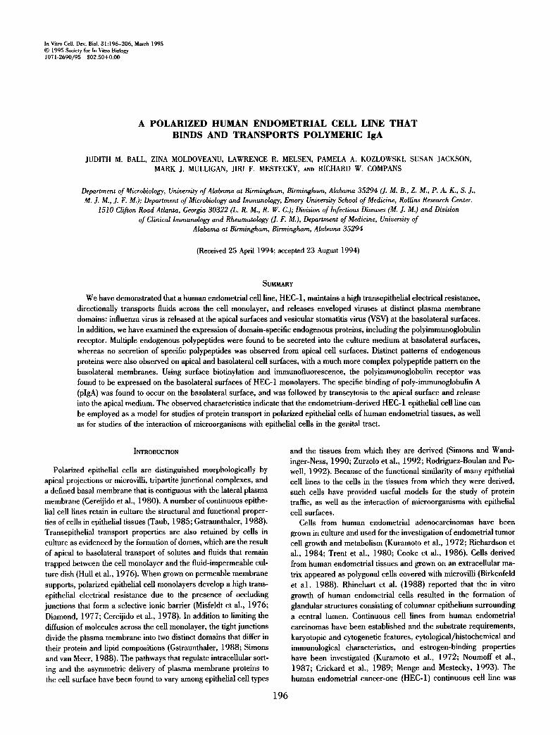
The HEC-1 cell line exhibits a polarized epithelial phenotype. Polarized epithelial cell layers exhibit structural and functional prop- erties that enable these cells to transport ions and fluids across the cell monolayer. This results in formation of fluid-filled domes that can be visualized by phase-contrast microscopy (Leighton et al., 1969; Hull et al., 1976). HEC-I cell monolayers exhibited a char- acteristic cobblestone appearance. In addition, large domes or fluid-filled blisters were observed throughout the monolayer (Fig. 1). Initial dome formation was observed about 24 h after the cells reached confluency (Fig. 1 a), and the appearance of the domes was more pronounced several days after confluency (Fig. 1 b). The formation of domes indicates that the HEC-1 cells form monolayers that exhibit the ability to transport fluids across the cell layers.
To further compare the HEC-1 cell line to other polarized epithe- lial cell lines, HEC-1 cells were grown on permeable membrane supports and transepithelial electrical resistance was measured across the confluent monolayer. Similar to the MDCK cell line, the establishment of cell surface polarity was gradual, requiring a mini- mum of 60 h of culture before a high transepithelial electrical resis- tance was established (Balcarova-Stander et al., 1984). Following 24 h of culture, the transepithelial electrical resistance was consis- tently below 100 ohms × cm 2, and increased to 125-150 ohms × cm 2 by 48 h after seeding at a density of about 4 × 10⁵ cells/weB. Subsequently, there was a dramatic rise in transepithelial electrical resistance concomitant with the establishment of extensive cell-cell contacts and junctional complexes. The resistance measurements reached levels greater than 400 ohms × cm 2 at 4 - 6 days postseed-
ing. Thus, HEC-1 cells establish a high transepithelial resistance, as is characteristic of polarized epithelial cell monolayers.
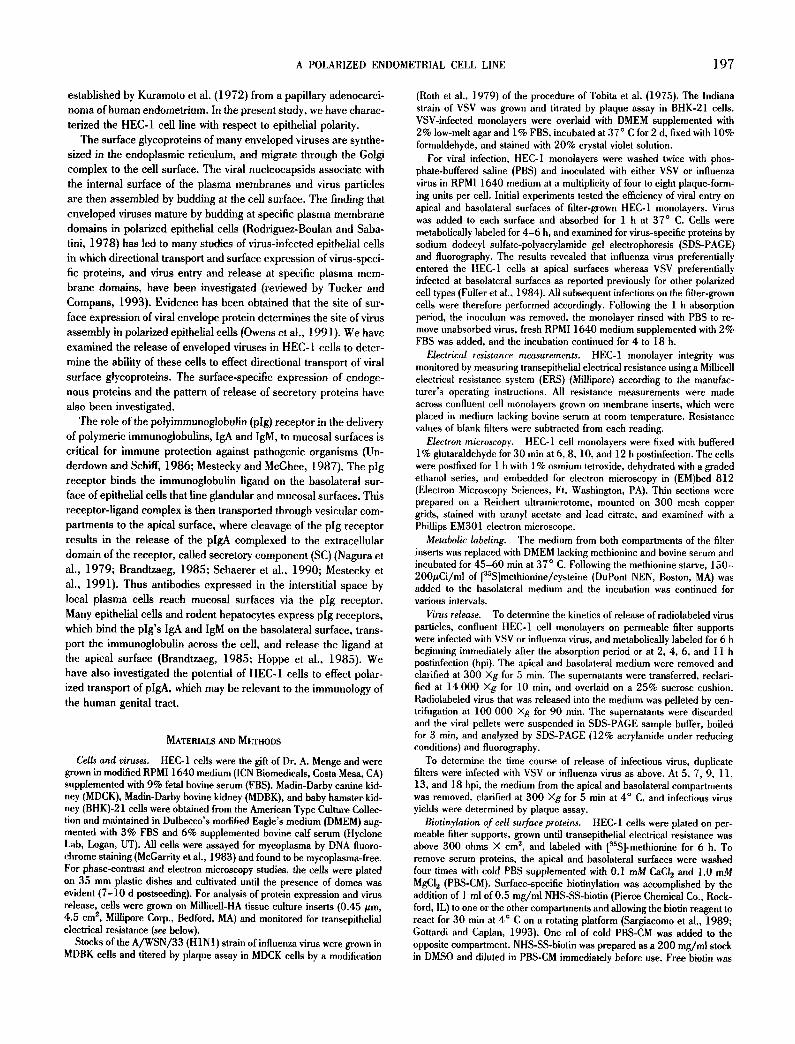
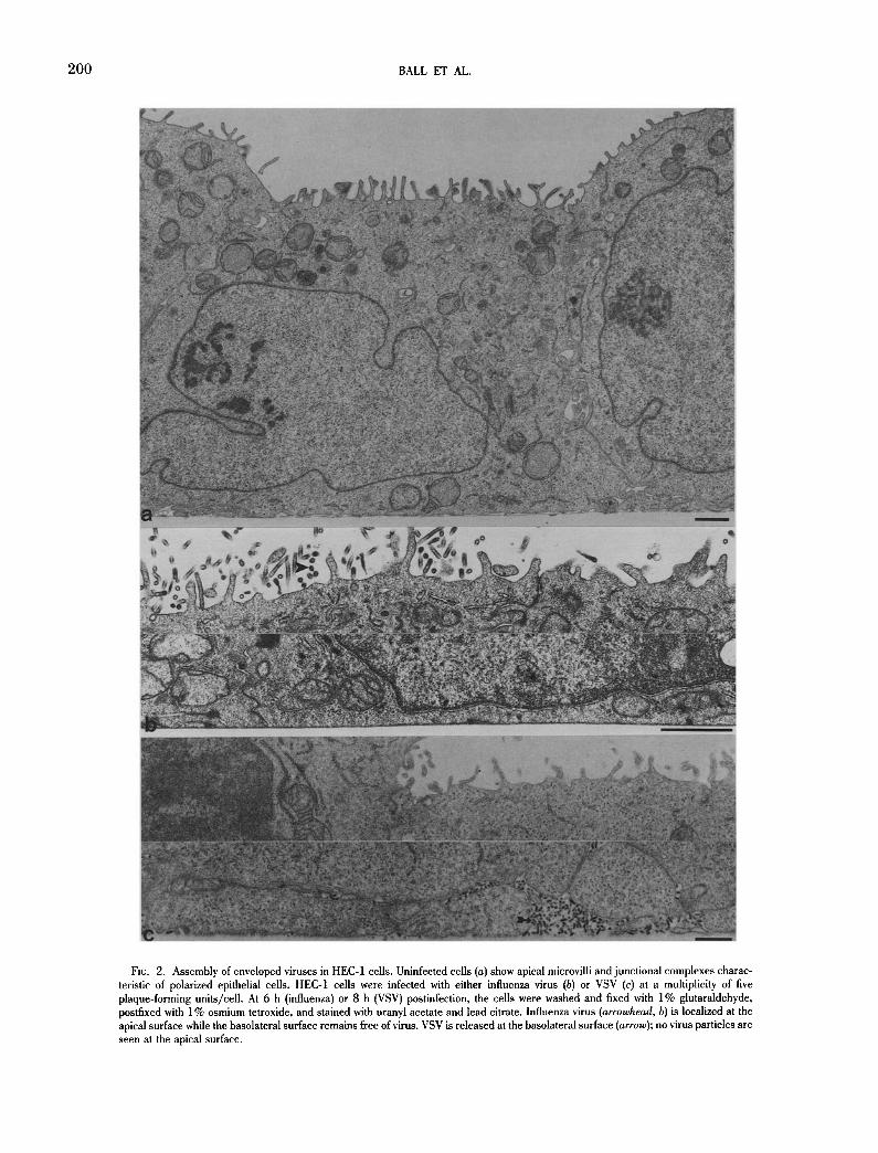
Asymmetric budding of enveloped viruses. The assembly of en- veloped viruses has been used frequently to investigate the pattern of transport of membrane proteins and to demonstrate the polarity of epithelial cells. To determine if enveloped viruses mature at dis- tinct plasma membrane domains in the HEC-1 cells, monolayers were infected with VSV or influenza virus and the site of maturation was examined. The HEC-1 cells exhibited morphological properties characteristic of a polarized phenotype including apical microvilli, junctional complexes, a distinguishable basal membrane, and lat- eral contacts with neighboring cells (Fig. 2 a). Influenza virus was found to mature exclusively at the apical surfaces; no virus budding was observed along the basolateral membranes (Fig. 2 b). In con- trast, VSV virions were only detected at the basolateral surfaces; the apical membranes remained virus-free (Fig. 2 c). These results demonstrate that the two enveloped viruses, influenza virus and VSV, are assembled and released at specific plasma membrane domains in infected HEC-1 cells. Hence, the HEC-1 cells exhibit the same pattern of polarized maturation of these enveloped viruses that was reported previously for cell lines originating from kidney or intestinal epithelium (Rodriguez-Boulan and Sabatini, 1978; Rindler and Traher, 1988).
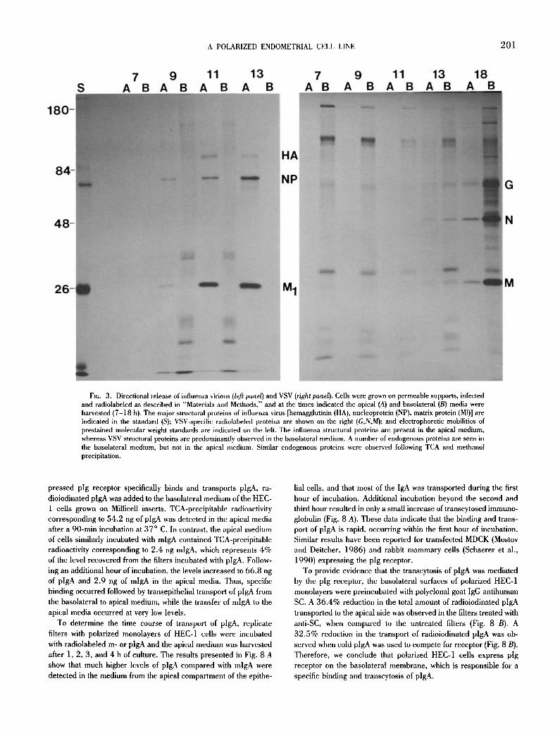
The asymmetric release of VSV and influenza virus in HEC-1 cells was quantitated by analyzing the apical and basolateral me- dium for the presence of metabolically labeled virus proteins during the time course of infection. As seen from the pattern of radiola- beled, virus-specific proteins in Fig. 3, influenza virus was released exclusively into the apical medium a 9, 11, and 13 hpi. No virus or viral proteins were recovered at 5 and 7 hpi. By 18 hpi, low levels of influenza virus proteins were also observed in the basolateral me- dium (not shown). It is likely that the presence of influenza virus particles in the basolateral medium at this time is due to virus-in- duced disruption of the tight junctions, which would interrupt cell polarity and allow the virus to diffuse into the basolateral medium. This conclusion was supported by the observation that a concurrent loss of transepithelial electrical resistance was observed at 18 hpi when HEC-1 cells were grown on permeable membranes and in- fected with influenza virus, and by the appearance of cytopathic effects (cpe) in the cell monolayer at 18 hpi. In contrast to the apical release of influenza virus, VSV is predominantly released into the basolateral medium at 13 and 18 hpi as shown by the progressively increasing amounts of radiolabeled virus-specific proteins found in the basolateral medium. HEC monolayers infected with VSV re- mained intact for up to 22 hpi with no evidence of cpe and no reduction in transepithelial resistance. Between 22 and 24 hpi, there was a dramatic drop in the transepithelial resistance and ob- servable cpe in the cell monolayer. In addition, at 24 hpi the major- ity of the VSV virions were recovered from the apical medium (data not shown).
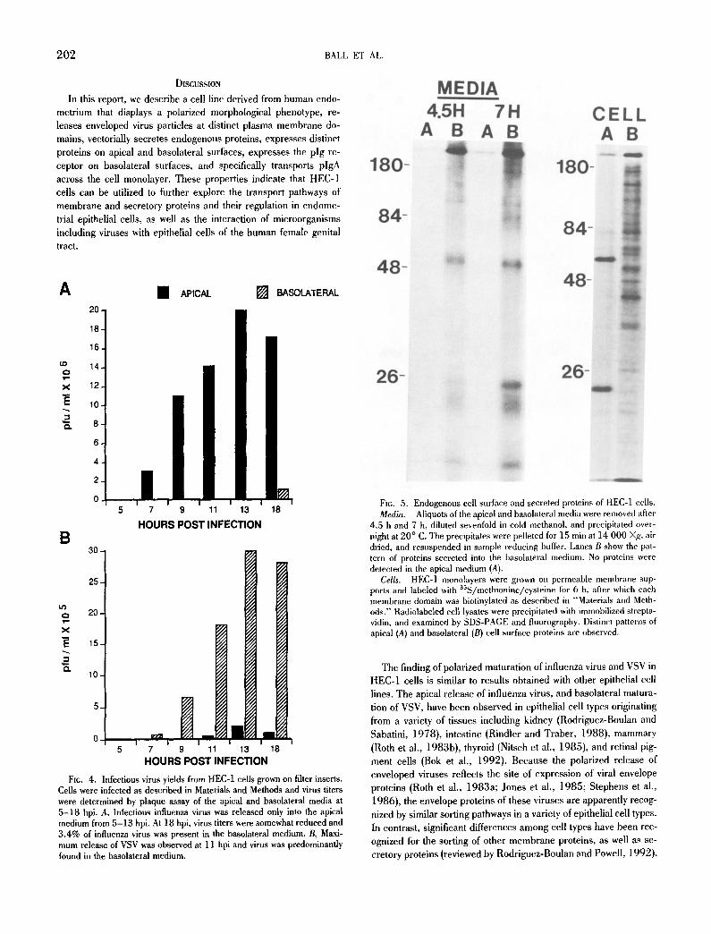
To demonstrate the release of infectious virus, the medium from the apical and basolateral compartments of infected, filter-grown HEC-1 cells was analyzed by plaque assay. Fig. 4 A shows that infectious influenza virus was detectable in the apical medium by 5 hpi, and the titer continued to rise progressively with time, reach- ing a titer of 2 × l 0 T plaque forming units/ml at 13 hpi. No influenza virus was detected in the basolateral medium at 5, 7, 9, or 11 hpi, and at 13 hpi the basolateral medium showed titers over 1000-fold lower than the corresponding apical compartment. In
A POLARIZED ENDOMETRIAL CELL LINE 199
Fir. 1. Phase-contrast micrographs of HEC-1 monolayers at a, 24h and b, 4 d after the cells reached confluency. The characteristic cobblestone appearance of an epithelial cell layer is evident. Initial dome formation was observed at 24 h (arrow); the domes then continued to grow in size with additional time of incubation.
contrast, infectious VSV particles were preferentially released into the basolateral medium (Fig. 4 B). Infectious virus was detected in the basolateral medium by 7 hpi. At 9 or 11 hpi, 99% of the infectious VSV was found in the basolateral medium; at 13 and 24 hpi, 9 1 - 9 2 % of the infectious virus was recovered in the basolat- eral medium. These quantitative results demonstrate that the re- lease of influenza virus and VSV occurs at specific membrane do- mains, as previously reported in other polarized cell types.
Polarized expression of endogenous proteins. During the devel- opment of a polarized phenotype, the distribution of membrane proteins becomes restricted to specific membrane domains (Almers and Stirling, 1984; Wollner et al., 1992). In addition, an asymmet- ric secretion of endogenous proteins has been reported in several epithelial cell lines (Urban et al., 1987; Gottlieb et al., 1986; Rindler and Trabe, 1988). Therefore, we compared the proteins expressed on the apical and basolateral surfaces of polarized HEC- 1 cells grown on permeable membrane supports, as well as the proteins secreted into the culture media. In addition to illustrating the polarized release of enveloped viruses, Fig. 3 reveals that sev- eral radiolabeled proteins were secreted into the basolateral me- dium. Two distinct, small polypeptides (apparent molecular mass below 26 kD), a doublet at approximately 37 kD, and a large poly- peptide at about 120 kD were evident in the basolateral medium of influenza virus-infected cells (Fig. 3). Similarly, the basolateral me- dium of the VSV infected HEC-1 cells shows the same two small protein bands and the band at approximately 120 kD. Similar pro- teins were observed by direct analysis of the basolateral medium of uninfected HEC-1 cells when precipitated with either TCA (not shown) or methanol. Methanol precipitation of the basolateral me- dium (Fig. 5) shows several bands below 26 kD, a sharp band at about 53 kD, and multiple proteins in the range of 80-180kD. These proteins were observed in the media beginning about 3 h after radiolabeling. In contrast, no endogenous protein bands were detected in the apical medium. Thus, unique endogenous proteins were found to be secreted exclusively into the basolateral medium of HEC-1 cells, a result that is similar to that reported for the polarized Caco-2 intestinal cell line (Rindler and Traber, 1988).
To compare the pattern of endogenous proteins expressed on the two surfaces of the HEC-1 cell line, cells were metabolically labeled
followed by domain-specific surface biotinylation. A larger number of basolateral surface proteins was detected by this methodology, although several distinct apical surface proteins were also observed (Fig. 5). Two prominent apical surface proteins were evident at approximately 62 and 20 kD (lane A). Multiple bands ranging from about 43 kD to 180 kD, a doublet at about 36 kD, and a band at approximately 29 kD were observed on the basolateral surfaces (lane B). The primary amino groups of the basolateral proteins could be more accessible to the hiotinylation reaction, which could play a role in the detection of a larger number of proteins on basolat- eral surfaces. These results indicate that the major proteins ex- pressed on apical and basolateral surfaces are distinct, consistent with the polarized epithelial phenotype of the cells.
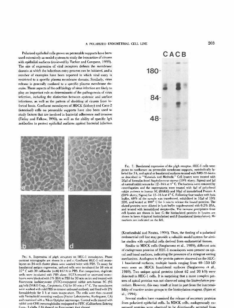
Basolateral expression of the pig receptor. The pig receptor plays a crucial role in mucosal immunity in that the receptor medi- ates the transport of polymeric immunoglobulins to mucosal sur- faces where they can cross-link or neutralize microbial pathogens (Dimmock, 1987; Mazanec et al., 1992; Mestecky et al., 1991). The expression of the pig receptor on HEC-1 cell surfaces was analyzed by immunofluorescence microscopy, and by surface-spe- cific biotinylation followed by immuno- and streptavidin precipita- tion. As shown in Fig. 6 b, the immunofluorescent staining pattern after treatment of cells with ethyleneglycol-b/s-(fl-aminoethyl ether)-N,N'-tetraacetic acid (EGTA) to open the junctional com- plexes showed a relatively bright cobblestone appearance, while little apical staining was observed when intact monolayers were examined (Fig. 6 d), indicating that the pig receptor is expressed in HEC-1 cells and is predominantly localized on basolateral mem- branes. Corresponding data was obtained with HEC-1 cells grown on filter inserts that exhibited a high transepithelial electrical resis- tance. Fig. 7 illustrates that the majority of the pig receptor was localized to basolateral surfaces when cells were analyzed by do- main-specific biotinylation and immune precipitation with antibody to secretory component. Based on phospho-imaging analyses, it was calculated that 82% of the total pig receptor found on the surface of polarized HEC-1 cells resides on the basolateral surface domain. These results raised the possibility that HEC-1 cell monolayers were capable of transcytosis of plgA.
Transcytosis of plgA. To determine if the basolaterally ex-
2 0 0 BALL ET AL.
FIG. 2. Assembly of enveloped viruses in HEC-1 ceils. Uninfected cells (a) show apical microvilli and junctional complexes charac- teristic of polarized epithelial cells. HEC-1 cells were infected with either influenza virus (b) or VSV (c) at a multiplicity of five plaque-forming units/cell. At 6 h (influenza) or 8 h (VSV) postinfection, the cells were washed and fixed with 1% glutaraldehyde, postflxed with 1% osmium tetroxide, and stained with uranyl acetate and lead citrate. Influenza virus (arrowhead, b) is localized at the apical surface while the basolateral surface remains free of virus. VSV is released at the basolateral surface (arrow); no virus particles are seen at the apical surface.
180-
84-
48-
!ii~iiiiiiiii~!iii
26-
S 7
A 9
B A B
A POLARIZED ENDOMETRIAL CELL LINE
11 13 7 9 A B A B A B A
11 13 18 B A B A B A B
HA
NP
M 1
FxG. 3. Directional release of influenza virions (left panel) and VSV (right panel). Cells were grown on permeable supports, infected and radiolabeled as described in "Materials and Methods," and at the times indicated the apical (A) and basolateral (B) media were harvested (7-18 h). The major structural proteins of influenza virus [hemagglutinin (HA), nucleoprotein (NP), matrix protein (MI)] are indicated in the standard (S); VSV-speciflc radiolabeled proteins are shown on the right (G,N,M); and electrophoretic mobilities of prestained molecular weight standards are indicated on the left. The influenza structural proteins are present in the apical medium, whereas VSV structural proteins are predominantly observed in the basolateral medium. A number of endogenous proteins are seen in the basolateral medium, but not in the apical medium. Similar endogenous proteins were observed following TCA and methanol precipitation.
201
G
N
M
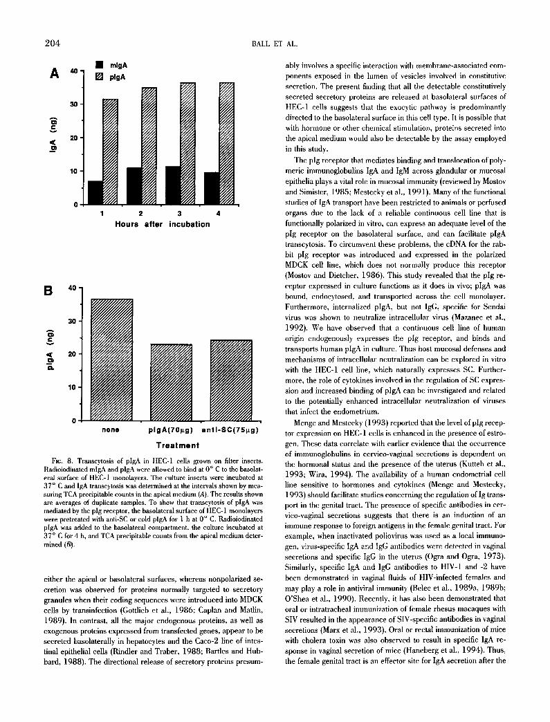
pressed plg receptor specifically binds and transports plgA, ra- dioiodinated plgA was added to the basolateral medium of the HEC- 1 cells grown on Millicell inserts. TCA-precipitable radioactivity corresponding to 54.2 ng of plgA was detected in the apical media after a 90-min incubation at 37 ° C. In contrast, the apical medium of cells similarly incubated with mlgA contained TCA-precipitable radioactivity corresponding to 2.4 ng mlgA, which represents 4% of the level recovered from the filters incubated with plgA. Follow- ing an additional hour of incubation, the levels increased to 66.8 ng of plgA and 2.9 ng of mlgA in the apical media. Thus, specific binding occurred followed by transepithelial transport of plgA from the basolateral to apical medium, while the transfer of mlgA to the apical media occurred at very low levels.
To determine the time course of transport of plgA, replicate filters with polarized monolayers of HEC-1 cells were incubated with radiolabeled m- or plgA and the apical medium was harvested after 1, 2, 3, and 4 h of culture. The results presented in Fig. 8 A show that much higher levels of plgA compared with mlgA were detected in the medium from the apical compartment of the epithe-
lial cells, and that most of the IgA was transported during the first hour of incubation. Additional incubation beyond the second and third hour resulted in only a small increase of transcytosed immuno- globulin (Fig. 8 A). These data indicate that the binding and trans- port of plgA is rapid, occurring within the first hour of incubation. Similar results have been reported for transfected MDCK (Mostov and Deitcher, 1986) and rabbit mammary cells (Schaerer et al., 1990) expressing the pig receptor.
To provide evidence that the transcytosis of plgA was mediated by the pig receptor, the basolateral surfaces of polarized HEC-1 monolayers were preincubated with polyclonal goat IgG antihuman SC. A 36.4% reduction in the total amount of radioiodinated plgA transported to the apical side was observed in the filters treated with anti-SC, when compared to the untreated filters (Fig. 8 B). A 32.5% reduction in the transport of radioiodinated plgA was ob- served when cold plgA was used to compete for receptor (Fig. 8 B). Therefore, we conclude that polarized HEC-1 cells express pig receptor on the basolateral membrane, which is responsible for a specific binding and transcytosis of plgA.
202 BALL ET AL.
DISCUSSION
In this report, we describe a cell line derived from human endo- metrium that displays a polarized morphological phenotype, re- leases enveloped virus particles at distinct plasma membrane do- mains, vectorially secretes endogenous proteins, expresses distinct proteins on apical and basolateral surfaces, expresses the pig re- ceptor on basolateral surfaces, and specifically transports plgA across the cell monolayer. These properties indicate that HEC-1 cells can be utilized to further explore the transport pathways of membrane and secretory proteins and their regulation in endome- trial epithelial cells, as well as the interaction of microorganisms including viruses with epithelial cells of the human female genital tract.
A 20.
18.
16.
14.
X 12.
10
£ 8 6
4
2
0
B 30-
• APICAL [ ] BASOLATERAL
Lt~ o 20, 1 = =
x
0. 10,
5 7 9 11 13 18
HOURS POST INFECTION
25
0 i ~ m m I I I I I
5 7 9 11 13 18 HOURS POST INFECTION
Fie. 4. Infectious virus yields from HEC-1 cells grown on filter inserts. Cells were infected as described in Materials and Methods and virus titers were determined by plaque assay of the apical and basolateral media at 5-18 hpi. A, Infectious influenza virus was released only into the apical medium from 5-13 hpi. At 18 hpi, virus titers were somewhat reduced and 3.4% of influenza virus was present in the basolateral medium. B, Maxi- mum release of VSV was observed at 11 hpi and virus was predominantly found in the basolateral medium.
180-
84-
48-
26-
MEDIA 4.5H 7 H
A B A B
<i i i ! ! :Y~ ¸
, i
ili!
1i ~!iiiijii I i ~
CELL A B
180-
84-
48-
26-
i!ii ̧
FIG. 5. Endogenous cell surface and secreted proteins of HEC-1 cells. Media. Aliquots of the apical and basolateral media were removed after
4.5 h and 7 h, diluted sevenfold in cold methanol, and precipitated over- night at 20" C, The precipitates were pelleted for 15 min at 14 000 Xg, air dried, and resuspended in sample reducing buffer. Lanes B show the pat- tern of proteins secreted into the basolateral medium. No proteins were detected in the apical medium (A).
Cells. HEC-1 monolayers were grown on permeable membrane sup- ports and labeled with 3SS/methionine/cysteine for 6 h, after which each membrane domain was biotinylated as described in "Materials and Meth- ods." Radiolabeled celt lysates were precipitated with immobilized strepta- vidin, and examined by SDS-PAGE and fluorography. Distinct patterns of apical (A) and basolateral (B) cell surface proteins are observed.
The finding of polarized maturation of influenza virus and VSV in HEC-1 cells is similar to results obtained with other epithelial cell lines. The apical release of influenza virus, and basolateral matura- tion of VSV, have been observed in epithelial cell types originating from a variety of tissues including kidney (Rodriguez-Boulan and Sabatini, 1978), intestine (Rindler and Traber, 1988), mammary (Roth et al., 1983b), thyroid (Nitsch et al., 1985), and retinal pig- ment cells (Bok et al., 1992). Because the polarized release of enveloped viruses reflects the site of expression of viral envelope proteins (Roth et al., 1983a; Jones et at., 1985; Stephens et at., 1986), the envelope proteins of these viruses are apparently recog- nized by similar sorting pathways in a variety of epithelial cell types. In contrast, significant differences among cell types have been rec- ognized for the sorting of other membrane proteins, as well as se- cretory proteins (reviewed by Rodriguez-Boulan and Powell, 1992).
A POLARIZED ENDOMETRIAL CELL LINE 203
Polarized epithelial cells grown on permeable supports have been used extensively as model systems to study the interaction of viruses with epithelial surfaces (reviewed by Tucker and Compans, 1993). The site of expression of viral receptors defines the membrane domain at which the infectious entry process can be initiated, and a number of examples have been reported in which viral entry is restricted to a specific plasma membrane domain. Similarly, virus release is generally confined to a specific plasma membrane do- main. These aspects of the cell biology of virus infection are likely to play an important role as determinants of the pathogenesis of virus infection, including the distinction between systemic and surface infections, as well as the pattern of shedding of viruses from in- fected hosts. Confluent monolayers of MDCK (kidney) and Caco-2 (intestinal) cells on permeable supports have also been used to study factors that are involved in bacterial adherence and invasion
(Finlay and Falkow, 1990), as well as the ability of specific IgA antibodies to protect epithelial surfaces against bacterial infection
180-
84-
CACB
48-
d
• : i l l
FIG. 7. Basolateral expression of the plgA receptor. HEC-1 cells were grown to confluence on permeable membrane supports, metabolically la- beled for 3 h, and apical or basolateral surfaces treated with NHS-SS-biotin as described in "Materials and Methods." Cell lysates were treated with 50#1 of formalin-fixed Staphylococcus aureus (10% slurry, Sigma) and 1#1 of normal rabbit serum for 12-16 h at 4 ° C. The bacteria were removed by eentrifugation and the supernatants were treated with 4gl of polyclonal rabbit antisera to human SC (DAKO) and 10#1 of immobilized Protein A (50% slurry, Sigma) for 12-16 h at 4 ° C. Following four washes with lysis buffer, 60% of the sample was transferred, solubilized in 10/.tl of 10% SDS, and heated at 100 ° C for 5 min to release the bound proteins. The eluted proteins were diluted in lysis buffer supplemented with 0.2% BSA, and treated with immobilized streptavidin. The immune precipitates from cell lysates are shown in lane C; the biotinylated proteins in lysates are shown in lanes A (apical biotinylation) and B (basolateral biotinylation). Mr markers are indicated on the left.
FIG. 6. Expression of plgA receptors on HEC-1 monolayers. Phase contrast micrographs are shown in a and c. Confluent HEC-1 cell mono° layers on 24-well cluster plates were washed twice with PBS. To assay for basolateral antigen expression, unfixed cells were incubated for 10 rain at 37 ° C with 30 millimolar (raM) EGTA in PBS. For comparison, duplicate wells were incubated with PBS alone. EGTA-treated or untreated mono- layers were blocked with 1% BSA in PBS for 30 rain on ice and treated with fluorescein isothiocyanate (FITC)-conjugated rabbit anti-human SC (95 ttg/ml) (DAKO Corp., Carpinteria, CA) for 30 rain a 4 ° C. The monolayers were washed with cold PBS to remove unbound antibody and fixed with 2% formaldehyde for 1 h at room temperature. The cells were then covered with Vectashield mounting medium (Vector Laboratories, Burlingame, CA) and examined with a Nikon Optiphot microscope. Control wells treated with rabbit anti-CD8 immunoglobulin conjugated to FITC (Calbiochem-Behring Corp., LaJolla, CA) showed no fluorescence (not shown).
(Kraehenbuhl and Neutra, 1994). Thus, the finding of a polarized endometrial cell line may provide a valuable model system for simi- lar studies with epithelial ceUs derived from endometrial tissues.
Similar to MDCK cells (Sargiacomo et al., 1989), different sets of endogenous proteins of HEC-1 monolayers were present on api- cal and basal surfaces, indicating the presence of a stringent sorting mechanism. Analogous to the protein pattern observed on the HEC- 1 basolateral surfaces, multiple bands ranging from 4 0 - 1 5 0 kD were seen on MDCK basolateral surfaces (Sargiacomo et al., 1989). Two unique apical proteins (about 62 and 20 kD) were detected in HEC-1 cells. It is surprising that a more complex pat- tern of apical proteins was not observed using the biotinylation pro-
cedure. However, this may result at least in part from the inaccessi- bility of reactive amino groups to the biotinylation reagent. (Spies et
al., 1994). Several studies have examined the release of secretory proteins
from polarized epithelial cells. In MDCK cells, endogenously ex- pressed proteins were observed to be directionally secreted from
40
30
10
2 Hours
20
1 3 4 after incubation
B
• mlgA
[ ] plgA
<
A
C
204 BALL ET AL.
40
v
m D .
30
20
10
none
A
plgA(70~g)
T r e a t m e n t
ant I -SC(75~.g)
FIG. 8. Transcytosis of pIgA in HEC-1 cells grown on filter inserts. Radioiodinated mlgA and plgA were allowed to bind at 0 ° C to the basolat- eral surface of HEC-1 monolayers. The culture inserts were incubated at 37 ° C and IgA transcytosis was determined at the intervals shown by mea- suring TCA precipitable counts in the apical medium (A). The results shown are averages of duplicate samples. To show that transcytosis of plgA was mediated by the pig receptor, the basolateral surface of HEC-1 monolayers were pretreated with anti-SC or cold plgA for 1 h at 0 ° C. Radioiodinated plgA was added to the basolateral compartment, the culture incubated at 37 ° C for 4 h, and TCA precipitable counts from the apical medium deter- mined (B).
either the apical or basolateral surfaces, whereas nonpolarized se- cretion was observed for proteins normally targeted to secretory granules when their coding sequences were introduced into MDCK cells by transinfection (Gottlieb et al., 1986; Caplan and Matlin, 1989). In contrast, all the major endogenous proteins, as well as exogenous proteins expressed from transfected genes, appear to be secreted basolaterally in hepatocytes and the Caco-2 line of intes- tinal epithelial cells (Rindler and Traber, 1988; Bartles and Hub- bard, 1988). The directional release of secretory proteins presum-
ably involves a specific interaction with membrane-associated com- ponents exposed in the lumen of vesicles involved in constitutive secretion. The present finding that all the detectable constitutively secreted secretory proteins are released at basolateral surfaces of HEC-1 cells suggests that the exocytic pathway is predominantly directed to the basolateral surface in this cell type. It is possible that with hormone or other chemical stimulation, proteins secreted into the apical medium would also be detectable by the assay employed in this study.
The pig receptor that mediates binding and translocation of poly- meric immunoglobulins IgA and IgM across glandular or mucosal epithelia plays a vital role in mucosal immunity (reviewed by Mostov and Simister, 1985; Mestecky et al., 1991). Many of the functional studies of IgA transport have been restricted to animals or perfused organs due to the lack of a reliable continuous cell line that is functionally polarized in vitro, can express an adequate level of the pig receptor on the basolateral surface, and can facilitate plgA transcytosis. To circumvent these problems, the cDNA for the rab- bit pig receptor was introduced and expressed in the polarized MDCK cell line, which does not normally produce this receptor (Mostov and Dietcher, 1986). This study revealed that the pig re- ceptor expressed in culture functions as it does in vivo; plgA was bound, endocytosed, and transported across the cell monolayer. Furthermore, internalized pIgA, but not IgG, specific for Sendal virus was shown to neutralize intracellular virus (Mazanec et al., 1992). We have observed that a continuous celt line of human origin endogenously expresses the plg receptor, and binds and transports human plgA in culture. Thus host mucosal defenses and mechanisms of intracellular neutralization can be explored in vitro with the HEC-1 cell line, which naturally expresses SC. Further- more, the role of cytokines involved in the regulation of SC expres- sion and increased binding of p|gA can be investigated and related to the potentially enhanced intracellular neutralization of viruses that infect the endometrium.
Menge and Mestecky (1993) reported that the level of plg recep- tor expression on HEC-1 cells is enhanced in the presence of estro- gen. These data correlate with earlier evidence that the occurrence of immunoglobulins in cervico-vaginal secretions is dependent on the hormonal status and the presence of the uterus (Kutteh et al., 1993; Wira, 1994). The availability of a human endometrial cell line sensitive to hormones and cytokines (Menge and Mestecky, 1993) should facilitate studies concerning the regulation of Ig trans- port in the genital tract. The presence of specific antibodies in cer- vico-vaginal secretions suggests that there is an induction of an immune response to foreign antigens in the female genital tract. For example, when inactivated poliovirus was used as a local immuno- gen, virus-specific IgA and IgG antibodies were detected in vaginal secretions and specific lgG in the uterus (Ogra and Ogra, 1973). Similarly, specific IgA and IgG antibodies to HIV-1 and -2 have been demonstrated in vaginal fluids of HIV-infected females and may play a role in antiviral immunity (Belec et al., 1989a, 1989b; O'Shea et al., 1990). Recently, it has also been demonstrated that oral or intratracheal immunization of female rhesus macaques with SIV resulted in the appearance of SIV-specific antibodies in vaginal secretions (Marx et al., 1993). Oral or rectal immunization of mice with cholera toxin was also observed to result in specific IgA re- sponse in vaginal secretion of mice (Haneberg et al., 1994). Thus, the female genital tract is an effector site for IgA secretion after the
A POLARIZED ENDOMETRIAL CELL LINE 205
induction of common mucosal immune responses in remote muco-
sal tissues. Female genital tract secretions contain antibodies that originate
from two sources: a) plasma and b) local plasma cells (Kutteh ct al., 1993). Transport of specific antibodies to/from the circulation to / from the female genital tract appears to play a vital role in local immunity, although additional studies are needed. The polarized HEC-1 cell line, therefore, provides a useful system to help eluci- date the prevalence, role, and mechanisms of immunity in the geni- tal tract.
ACKNOWLEDGMENTS
This work was supported by research grants A112680, CA18611, AI23952, AI 10854, A128147, AIO0912 and DK28537 from the National Institutes of Health. Judith Ball was supported by Institutional Research Service Award AI07150 from the National Institutes of Health.. The au- thors thank Tanya Cassingham for assistance in preparation of the manu- script.
REFERENCES
Almers, W.; Stirling, C. Distribution of transport proteins over animal cell membranes. J. Membr. Biol. 77:169-186; 1984.
Balcarova-Stander, J.; Pfeiffer, S. E.; Fuller, S. D., et al. Development of cell surface polarity in the epithelial Madin-Darby canine kidney (MDCK) cell line. EMBO J. 11:2687-2694; 1984.
Bartles, J. R.; Hubbard, A. L. Plasma membrane protein sorting in epithe- lial cells: Do secretory proteins hold the key? Trends Biochem. Sci. 13:181-184; 1988.
Belec, L.; Georges, A. J.; Steenman, G., et at. Antibodies to human immuno- deficiency virus in vaginal secretions of heterosexual women. J. Infect. Dis. 160:385-391; 1989a.
Belec, L.; Peghini, M.; Georges, A. J., et al. Antibodies to HIV-2 in genital secretions. Res. Virol. 140:15-21;1989b.
Birkenfeld, A.; Ezra, Y.; Ron, N,, et al. Indication of selective growth of human endometrial epithelial cells on extracellular matrix. In Vitro Cell, Dev. Biol. 12:1188-1192; 1988.
Bok, D.; O'Day, W.; Rodriguez-Boulan, E, Polarized budding of vesicular stomatitis and influenza virus from cultured human and bovine reti- nal pigment epithelium. Exp. Eye Res. 55:853-860; 1992.
Brandtzaeg, P. Role of J chain and secretory component in receptor-me- diated glandular and hepatic transport of immunoglobulins in man. Scand. J. Immunol. 22:111-146; 1985.
Caplan, M.; Matlin, K. S. Sorting of membrane and secretory proteins in polarized epithelial cells. In: Matlin, K. S.; Valentich, J. D. eds. Functional epithelial cells in culture. New York: Liss; 1989:71- 130.
Cereijido, M.; Robbins, E. S.; Dolan, W. J., et al. Polarized monolayers formed by epithelial cells on a permeable and translucent support. J. Cell Biol. 77:853-880; 1978.
Cereijido, M.; Stefani, E.; Martinez-Palomo, A. M. Occluding junctions in a cultured transporting epithelium: structural and functional heteroge- neity. J. Membr. Biol. 53:19-32; 1980.
Cooke, P. S.; Uchima, F.-D. A.; Fujii, D. K., et al. Restoration of normal morphology and estrogen responsiveness in cultured vaginal and uterine epithelial transplanted with stroma. Proc. Natl. Acad. Sci. USA 83:2109-2113; 1986.
Crickard, K.; Niedbala, M. J.; Crickard, U., et al. Characterization of human ovarian and endometrial carcinoma cell lines established on extra- cellular matrix. Gynecol. Oncol. 32:163-173; 1989.
Diamond, J. M. The epithelial junction: bridge, gate, and fence. Physiologist 20:10-18; 1977.
Dimmock, N. J. Multiple mechanisms of neutralization of animal viruses. Trends Biochem. Sci. 12:70-75; 1987.
Finlay, B. B.; Falkow, S. Salmonella interactions with polarized human Caco-2 epithelial cells. J. Infect. Dis. 162:1096-1106; 1990.
Fuller, S. D.; Bonsdorff, C. H.; Simons, K. Vesicular stomatitus virus in- fects and matures only through the basolateral surface of the polar- ized epithelial cell line, MDCK. Cell 38:65-77; 1984.
Gottardi, C. J.; Caplan, M. J. Delivery of Na+, K+-ATPase in polarized epithelial cells. Science 260:552-554; 1993.
Gottlieb, T. A.; Beaudry, G.; Rizzolo, L., et al. Secretion of endogenous and exogenous proteins from polarized MDCK cell monolayers. Proc. Natl. Acad. Sci. USA 83:2100-2104; 1986.
Greenwood, F. C.; Hunter, W. M.; Glove, J. S. The preparation of 13q-la- beled growth hormone of high specific activity. Biochem. J. 89:114-123; 1963.
Gstraunthaler, G. J. A. Epithelial cells in tissue culture. Renal Physiol. Biochem. 11:1-42; 1988.
Haneberg, B.; Kendall, D.; Amerongen, H. M., et al. Induction of specific immunoglobulin A in the small intestine, colon-rectum, and vagina measured by a new method for collection of secretions from local mucosal surfaces. Infect. Immun. 62:15-23; 1994.
Hoppe, C. A.; Connolly, T. P.; Hubbard, A. L. Transcellular transport of polymeric IgA in the rat hepatocyte: biochemical and morphological characterization of the transport pathway. J. Cell Biol. 101:2113 - 2123; 1985.
Hull, R. N.; Cherry, W. R.; Weaver, G. W. The origin and characteristics of a pig kidney cell strain LLC-PK t. In Vitro Cell. Dev. Biol. 12:670- 677; 1976.
Jones, L. V.; Compans, R. W.; Davis, A. R., et al. Surface expression of the influenza neuraminidase, an amino-terminally anchored viral mem- brane glycoprotein, in polarized epithelial cells. Mol. Cell. Biol. 5:2181-2189; 1985.
Kraehenbuhl, Jl P.; Neutra, M. R. Monoclonal secretory IgA for protection of the intestinal mucosa against viral and bacterial pathogens. In: Ogra, P. L.; Mestecky, J.; Lamm, M. E., et al., eds. Handbook of mucosal immunology. San Diego: Academic Press; 1994:403- 410.
Kuramoto, H.; Tamura, S.; Yukio, N. Establishment of a cell line of human endometrial adenocarcinoma in vitro. Am, J. Obstet. Gynecol. 114:1012-1019; 1972.
Kutteh, W. H.; Edwards, R. P.; Menge, A. C., et al. IgA immunity in female reproductive tract secretions. In: Gritfin, P. D.; Johnson, P. M., eds. Local immunity in reproductive tract tissues. Oxford: Oxford Univer- sity Press; 1993:17-51.
Leighton, J.; Brada, Z.; Estes, L. W., et al. Secretory activity and oncogeni- city of a cell line (MDCK) derived from canine kidney. Science 163:472-473; 1969.
Marx, P. A.; Compans, R. W.; Gettie, A., et at. Protection against S1V vaginal transmission with microencapsulated vaccine. Science: 260:1323-1327; 1993.
Mazanec, M. B.; Kaetzel, C. S.; Lamm, M. E., et ah Intracellular neutraliza- tion of virus by immunoglobulin A antibodies. Proc. Natl. Acad. Sci. USA 89:6901-6905; 1992.
McGarrity, G. J.; Steiner, T.; Vanaman, V. Detection of mycoplasmal infec- tion of cell cultures by DNA fluorochrome staining. In: Tully, J. G.; Razin, S., eds. Methods in mycoplasmology, vol. II. 1983:183- 190.
Menge, A. C.; Mestecky, J. Surface expression of secretory component and HLA class II DR antigen on glandular epithelial cells from human endometrium and two endometrial adenocarcinoma cell lines. J. Clin. Immunol. 13:1-5; 1993.
Mestecky, J.; Kilian, M. Immunoglobulin A (IgA). Methods Enzymol. 116:37-75; 1985.
Mestecky, J.; Lue, C.; Russell, M. W. Selective transport of IgA, cellular and molecular aspects. Gastroenterol. Clin. North Am. 20:441- 471; 1991.
Mestecky, J.; McGhee, J. R. Immunoglobulin A (IgA): Molecular and cellu- lar interactions involved in IgA biosynthesis and immune response. Adv. Immunol. 40:153-245; 1987.
Misfeldt, D. S.; Hamamoto, S. T.; Pitelka, D. R. Transepithelial transport in cell culture. Proc. Natl. Acad. Sei. USA 73:1212-1216; 1976.
Mostov, K. E.; Deitcher, D. L. Polymeric immunoglobulin receptor ex- pressed in MDCK cells transcytoses IgA. Cell 46:613-621; 1986.
Mostov, K. E.; Simister, N. E. Transcytosis. Cell 43:388-390; 1985. Nagura, H.; Nakane, P. K.; Brown, W. R. Translocation of dimeric IgA
through neoplastic colon cells in vitro. J. lmmunol. 123:2359- 2368; 1979.
2 0 6 BALL ET AL.
Nitsch, L.; Tramontano, D,; Ambesi-Impiombato, F. S., et al. Morphologi- cal and functional polarity in an epithelial thyroid cell line. Eur. J. Cell. Biol. 38:57-66; 1985.
Noumoff, J.; Haydock, S. W.; Sachdeva, R., et al. Characteristics of cell lines derived from normal and malignant endometrial tissue. Gyne- col. Oncol. 27:141-149; 1987.
Ogra, P. L.; Ogra, S. S. Local antibody response to polio vaccine in the human female genital tract. J. Immunol. 110:1307-1312; 1973.
O'Shea, S.; Cordey, M.; Barrett, W. Y., et al. HIV excretion patterns and specific antibody responses in body fluids. J. Med. Virol. 31 :291- 296; 1990.
Owens, R. J.; Dubay, J.; Hunter, E., et al. The human immunodeficiency virus envelope protein determines the site of virus release in polar- ized epithelial cells. Proc. Natl. Acad. Sci. USA 88:3987-3991; 1991.
Rhinehart, C.A., Jr.; Lyn-Cook, B. D.; Kaufman, D. G. Gland formation from human endometrial epithelial cells in vitro. In Vitro Cell. Dev. Biol. 24:1037-1041; 1988.
Richardson, G. S.; Dickersin, G. R.; Atkins, L., et al. KLE: A cell line with defective estrogen receptor derived from undifferentiated endome- trial cancer. Gynecol. Oncol. 17:213-230; 1984.
Rindler, M. J.; Traber, M. G. A specific sorting signal is not required for the polarized secretion of newly synthesized proteins from cultured in- testinal epithelial cells. J. Cell Biol. 107:471-479; 1988.
Rodriguez-Boulan, E.; Powell, S. K. Polarity of epithelial and neuronal cells. Annu. Rev. Cell Biol. 8 :395-427; 1992.
Rodriguez-Boulan, E.; Sabatini, D. D. Morphogenesis of the polarized epi- thelial phenotype. Proc. Natl. Acad. Sci. USA 75:5071-5075; 1978.
Roth, M. G.; Compans, R. W.; Giusti, L., et al. Influenza virus hemaggluti- nin expression is polarized in cells infected with recombinant SV40 viruses carrying cloned hemagglutinin DNA. Cell 33:435-442; 1983a.
Roth, M. G.; Srinivas, R. V.; Compans, R. W. Basolateral maturation of retroviruses in polarized epithelial cells. J. Virol. 45 :1065-1073; 1983b.
Roth, M. G.; Fitzpatrick, J. P.; Compans, R. W. Polarity of influenza and vesicular stomatitis virus maturation in MDCK cells: lack of a re- quirement for glycosylation of viral glycoproteins. Proc. Natl. Acad. Sci. USA 76:6430-6434; 1979.
Sargiacomo, M,; Lisanti, M.; Graeve, L., et al. Integral and peripheral protein composition of the apical and basolateral membrane do- mains in MDCK cells. Membr. Biol. 107:277-286; 1989.
Schaerer, E.; Verrey, F.; Racine, L., et al. Polarized transport of the poly- meric immunoglobulin receptor in transfected rabbit mammary epi- thelial ceils. J. Cell Biol. 110:987-998; 1990.
Simons, K.; van Meer, G. Lipid sorting in epithelial cells. Biochemistry 27:6197-6202; 1988.
Simons, K.; Wandinger-Ness, A. Polarized sorting in epithelia. Cell 62:207-210; 1990.
Spies, C. P.; Ritter, G. D., Jr.; Mulligan, M. J., et al. Truncation of the cytoplasmic domain of the simian immunodeficiency virus envelope glycoprotein alters conformation of the external domain. J. Virol. 68 :585-591; 1994.
Stephens, E. B.; Compans, R. W.; Earl, P., et at. Surface expression of viral glycoproteins in polarized epithelial cells using vaccinia virus vec- tors. EMBO J. 5 :237-245; 1986.
Taub, M. Tissue culture of epithelial cells. New York: Plenum Press; 1985. Tobita, K.; Sugiura, A.; Enomoto, C., et al. Plaque assay and primary
isolation of influenza A viruses in an established line of canine kid- ney cells (MDCK) in the presence of trypsin. Med. Microbiol. Im- munol. 162:9-14; 1975.
Trent, J. M.; Davis, J. R.; Payne, C. M. The establishment and morphologic characterization of finite cell lines from normal human endome- trium. Am. J. Obstet. Gynecol. 136:352-362; 1980.
Tucker, S. P.; Compans, R. W. Virus infection of polarized epithelial cells. In: Maramorosch, K.; Murphy, F. A.; Shatkin, A. J., eds. Advances in virus research. Vol. 42. Academic Press: 1993:187-247.
Underdown, B. J.; Schiff, J. M. lmmunoglobulin A: strategic defense initia- tive at the mucosal surface. Annu. Rev. Immunol. 4 :389-417; 1986.
Urban, J.; Parezyk, K.; Leutz, A., et at. Constitutive apical secretion of an 80-kD sulfated glycoprotein complex in the polarized epithelial Ma- din-Darby canine kidney cell line. J. Cell Biol. 105:2735-2743; 1987.
Wira, Charles R. Endocrine regulation of mucosal immunity: effect of sex hormones and cytokines on the afferent and efferent arms of the immune system in the female reproductive tract. In: Ogra, P. L.; Mestecky, J.; Lamm, M. E., eels. Handbook of mucosat immunol- ogy. San Diego: Academic Press; 1994:705-718.
Wollner, K.; Krzeminski, K.; Nelson, J. Remodeling the cell surface distri- bution of membrane proteins during the development of epithelial cell polarity. J. Cell Biol. 116:889-899; 1992.
Zurzolo, C.; Le Bivic, A.; Quaroni, A., et al. Modulation of transcytotic and direct targeting pathways in a polarized thyroid cell line. EMBO J. 11:2337-2344; 1992.
Copyright © 2022 FDOKUMEN