Paclitaxel-PHBV nanoparticles and their toxicity to endometrial and primary ovarian cancer cells
11
Paclitaxel-PHBV nanoparticles and their toxicity to endometrial and primary ovarian cancer cells Cristian Vilos a, c , Francisco A. Morales a , Paula A. Solar a , Natalia S. Herrera a , Fernando D. Gonzalez-Nilo b , Daniel A. Aguayo b , Hegaly L. Mendoza b , Jeffrey Comer b, h , Maria L. Bravo d , Pamela A. Gonzalez d , Sumie Kato e , Mauricio A. Cuello e , Catalina Alonso f , Erasmo J. Bravo f , Eva I. Bustamante g , Gareth I. Owen d , Luis A. Velasquez a, c, * a Universidad Andres Bello, Facultad de Medicina, Center for Integrative Medicine and Innovative Science (CIMIS), Echaurren 183, Santiago, Chile b Universidad Andres Bello, Facultad de Biología, Center for Bioinformatics and Integrative Biology (CBIB), Republica 239, Santiago, Chile c Centro para el Desarrollo de la Nanociencia y Nanotecnología, Universidad de Santiago de Chile, Ecuador 3493, Santiago, Chile d Facultad de Ciencias Biológicas, Pontificia Universidad Católica de Chile, Alameda 340, Santiago, Chile e Facultad de Medicina, Pontificia Universidad Católica de Chile, Alameda 340, Santiago, Chile f Hospital Gustavo Fricke, Alvarez 1532, Viña del Mar, Chile g Fundación Arturo López Pérez, Rancagua 878, Santiago, Chile h Fundación Fraunhofer Chile Research, M. Sánchez Fontecilla 310 piso 14, Las Condes, Chile article info Article history: Received 28 November 2012 Accepted 12 February 2013 Available online 5 March 2013 Keywords: Paclitaxel PHBV Nanoparticle Ovarian cancer Drug delivery Molecular dynamics simulations abstract This report is an integrated study to include the molecular simulation, physicochemical characterization and biological analysis of a paclitaxel-loaded PHBV nanoparticle that demonstrates uptake, release and cytotoxicity in cancer cell lines. Taking this nanoparticle one step closer to its use in a clinical setting, we demonstrate that it causes significant cell death in primary cultures of stage IIIc serous ovarian cancer cells isolated from six patients. Molecular simulations revealed a high affinity of paclitaxel for the water epolymer interface, thus the drug is delivered only when the polymer near it is degraded. The Fourier transform infrared spectroscopy suggests the formation of a short-lived crystalline phase, also observed in the CG simulations, and transmission electron microscopy revealed branched structures on the surface of particles, which disappeared after 4 days. Biological analyses indicated that these particles have a 48-h window of toxicity protection, allowing for the endocytosis of the particle by the cells; this finding was corroborated by confocal microscopy and flow cytometry. The low cost to synthesize PHBV using mi- croorganisms and the potential chemical modifications of the polymer make it attractive for inexpensive, large-scale pharmaceutical production. Ó 2013 Elsevier Ltd. All rights reserved. 1. Introduction The worldwide cancer burden continues to grow and is becoming a major economic expenditure for all developed coun- tries. Although the 5-year survival for ovarian cancer patients has improved to nearly 30%, this disease remains the fifth deadliest cancer among American women and is the leading cause of death from gynecologic malignancy. In 2010, 21,880 new cases and 13,850 new deaths were recorded in the United States [1,2]. The major obstacles in the field of oncology include the tremendous effort that is required for early detection, the devel- opment of many new drugs and drug delivery systems and the existence of effective chemotherapeutic agents [3,4]. One addi- tional challenge is the development of nanoparticles that meet the clinical demands of therapeutic agents, such as biocompatibility, biodegradability, a clinically relevant circulating half-life, a low rate of intravascular aggregation, and a long-term storage capacity [5]. During the last decade, drug delivery systems based on polymeric nanoparticles have been central to many significant advances in nanomedicine [6]. Accordingly, these particles have generated in- terest related to their potential use in preclinical and clinical cancer drug development [5]. Polymeric nanoparticles have numerous uses in drug delivery, including the specific and targeted delivery of therapeutic agents, * Corresponding author. Center for Integrative Medicine and Innovative Science (CIMIS), Universidad Andres Bello, Facultad de Medicina, Echaurren 183, 8370071, Santiago, Chile. E-mail address: [email protected] (L.A. Velasquez). Contents lists available at SciVerse ScienceDirect Biomaterials journal homepage: www.elsevier.com/locate/biomaterials 0142-9612/$ e see front matter Ó 2013 Elsevier Ltd. All rights reserved. http://dx.doi.org/10.1016/j.biomaterials.2013.02.034 Biomaterials 34 (2013) 4098e4108
-
Upload
independent -
Category
Documents
-
view
2 -
download
0
Transcript of Paclitaxel-PHBV nanoparticles and their toxicity to endometrial and primary ovarian cancer cells

at SciVerse ScienceDirect
Biomaterials 34 (2013) 4098e4108
Contents lists available
Biomaterials
journal homepage: www.elsevier .com/locate/biomateria ls
Paclitaxel-PHBV nanoparticles and their toxicity to endometrial and primaryovarian cancer cells
Cristian Vilos a,c, Francisco A. Morales a, Paula A. Solar a, Natalia S. Herrera a, Fernando D. Gonzalez-Nilo b,Daniel A. Aguayo b, Hegaly L. Mendoza b, Jeffrey Comer b,h, Maria L. Bravo d, Pamela A. Gonzalez d,Sumie Kato e, Mauricio A. Cuello e, Catalina Alonso f, Erasmo J. Bravo f, Eva I. Bustamante g,Gareth I. Owen d, Luis A. Velasquez a,c,*
aUniversidad Andres Bello, Facultad de Medicina, Center for Integrative Medicine and Innovative Science (CIMIS), Echaurren 183, Santiago, ChilebUniversidad Andres Bello, Facultad de Biología, Center for Bioinformatics and Integrative Biology (CBIB), Republica 239, Santiago, ChilecCentro para el Desarrollo de la Nanociencia y Nanotecnología, Universidad de Santiago de Chile, Ecuador 3493, Santiago, Chiled Facultad de Ciencias Biológicas, Pontificia Universidad Católica de Chile, Alameda 340, Santiago, Chilee Facultad de Medicina, Pontificia Universidad Católica de Chile, Alameda 340, Santiago, ChilefHospital Gustavo Fricke, Alvarez 1532, Viña del Mar, Chileg Fundación Arturo López Pérez, Rancagua 878, Santiago, Chileh Fundación Fraunhofer Chile Research, M. Sánchez Fontecilla 310 piso 14, Las Condes, Chile
a r t i c l e i n f o
Article history:Received 28 November 2012Accepted 12 February 2013Available online 5 March 2013
Keywords:PaclitaxelPHBVNanoparticleOvarian cancerDrug deliveryMolecular dynamics simulations
* Corresponding author. Center for Integrative Med(CIMIS), Universidad Andres Bello, Facultad de MedicSantiago, Chile.
E-mail address: [email protected] (L.A. Velas
0142-9612/$ e see front matter � 2013 Elsevier Ltd.http://dx.doi.org/10.1016/j.biomaterials.2013.02.034
a b s t r a c t
This report is an integrated study to include the molecular simulation, physicochemical characterizationand biological analysis of a paclitaxel-loaded PHBV nanoparticle that demonstrates uptake, release andcytotoxicity in cancer cell lines. Taking this nanoparticle one step closer to its use in a clinical setting, wedemonstrate that it causes significant cell death in primary cultures of stage IIIc serous ovarian cancercells isolated from six patients. Molecular simulations revealed a high affinity of paclitaxel for the waterepolymer interface, thus the drug is delivered only when the polymer near it is degraded. The Fouriertransform infrared spectroscopy suggests the formation of a short-lived crystalline phase, also observedin the CG simulations, and transmission electron microscopy revealed branched structures on the surfaceof particles, which disappeared after 4 days. Biological analyses indicated that these particles have a 48-hwindow of toxicity protection, allowing for the endocytosis of the particle by the cells; this finding wascorroborated by confocal microscopy and flow cytometry. The low cost to synthesize PHBV using mi-croorganisms and the potential chemical modifications of the polymer make it attractive for inexpensive,large-scale pharmaceutical production.
� 2013 Elsevier Ltd. All rights reserved.
1. Introduction
The worldwide cancer burden continues to grow and isbecoming a major economic expenditure for all developed coun-tries. Although the 5-year survival for ovarian cancer patients hasimproved to nearly 30%, this disease remains the fifth deadliestcancer among American women and is the leading cause of deathfrom gynecologic malignancy. In 2010, 21,880 new cases and 13,850new deaths were recorded in the United States [1,2].
icine and Innovative Scienceina, Echaurren 183, 8370071,
quez).
All rights reserved.
The major obstacles in the field of oncology include thetremendous effort that is required for early detection, the devel-opment of many new drugs and drug delivery systems and theexistence of effective chemotherapeutic agents [3,4]. One addi-tional challenge is the development of nanoparticles that meet theclinical demands of therapeutic agents, such as biocompatibility,biodegradability, a clinically relevant circulating half-life, a low rateof intravascular aggregation, and a long-term storage capacity [5].During the last decade, drug delivery systems based on polymericnanoparticles have been central to many significant advances innanomedicine [6]. Accordingly, these particles have generated in-terest related to their potential use in preclinical and clinical cancerdrug development [5].
Polymeric nanoparticles have numerous uses in drug delivery,including the specific and targeted delivery of therapeutic agents,

C. Vilos et al. / Biomaterials 34 (2013) 4098e4108 4099
such as cisplatin [7] and siRNA [8]. In addition, the use of nano-carriers that can cross the epithelial, endothelial or hema-toencephalic barriers [9], the co-liberation of drugs and/or contrastagents that improve the effectiveness of treatments and diagnosticimaging [10], and the encapsulation of highly toxic and poorlywater-soluble chemotherapeutics, such as cisplatin, carboplatin anddoxorubicin [11], are important. Paclitaxel is a new commercialTaxol preparation; it was originally isolated from the bark of thepacific yewtree (TaxusBrevifolia) in 1967. It is the chemotherapeuticagent of choice for the first-line treatment of all stages of ovariancancer, and it is commonly used to treat endometrial cancer [12].
An advantage of nanoparticles is that they exploit the enhancedpermeation and retention (EPR) of the vascular and lymphaticdrainage that occurs during the neoplastic process. Thus, by extrav-asation, these particles deliver the total amount of the encapsulateddrug to the tumor cell [13]. Tumors are known to create leaky bloodvasculature, permitting the passage of molecules in the nanometerrange. Therefore, the design of a biodegradable polymer-basednanoparticle represents an attractive drug delivery option for che-motherapeutics, especially if the drug remains encapsulated and,thus, inactive within the nanoparticle while in the circulation and ispreferentially released and activated once inside the tumor cells [13].
Poly(hydroxybutyric-co-hydroxyvaleric acid) (PHBV) is abiodegradable, non-toxic polyester with a low production cost [14];PHBV has been intensively investigated as a biomaterial for tissueengineering [15] and a microparticle-based drug delivery system[16e18].
Many nanoparticles have been designed for chemotherapeuticdrug encapsulation; however, reports on these preparations usuallyonly include their chemical and structural characterization andtheir activity in a cancer cell line. In this work, based on the suitableproperties of PHBV and the broad use of paclitaxel to treat manymalignancies, we formulated paclitaxel-loaded PHBV nanoparticlesthat protect the anticancer agent against premature degradation,provide 48 h of toxicity protection and allow the EPR effect. Inaddition, we used molecular dynamics simulations to understandthe interactions that occur between the polymer and the drug andto elucidate the phenomena that occur during the release process,which is essential to further refine the nanoparticle structure andfunction to increase the site-specific delivery of the chemothera-peutic cargo. Furthermore, we demonstrate cytotoxicity towardcancer cells lines, and we performed activity assays directly oncancer cells extracted from patients.
2. Materials and methods
Poly(3-hydroxybutyric acid-co-hydroxyvaleric acid) (PHBV) with 12 wt. % PHVand polyvinyl alcohol (PVA) (average mol wt. 30,000e70,000) were obtained fromSigmaeAldrich (St. Louis, MO, USA). Nile Red 552/636 was purchased from Invi-trogen (Carlsbad, CA, USA). Paclitaxel �97% was obtained from SigmaeAldrich (St.Louis, MO, USA). Dichloromethane (DCM), dimethyl sulfoxide (DMSO) andmethanolwere purchased from Merck (Darmstadt, Germany).
2.1. Preparation and characterization of paclitaxel-encapsulated PHBV nanoparticles(NP-Taxel)
The paclitaxel-encapsulated PHBV nanoparticles (NP-Taxel) were formulated viaa modification of the double emulsion (w1/o1/w2) solvent-evaporation method [19].Briefly, 400 mL of an aqueous solution containing either 6.84 or 34.2 mg/mL paclitaxel(equivalent to treatments used in cell culture experiments; 1 and 5 mM, respectively)were added to 1 mL of 3 mg/mL PHBV dissolved in dichloromethane. The firstemulsion (w1/o1) was prepared by sonication in an ultrasonic processor equippedwith a microtip probe for 60 s at 125W in an ice bath. Thewater-in-oil emulsionwasfurther emulsified by sonication under the same conditions in 4 mL of an aqueoussolution of 5mg/mL PVA (w2). This w1/o1/w2 emulsionwas immediately poured intoa beaker containing 20 mL of a 0.5 mg/mL PVA solution. The mixture was stirredwith an overhead propeller for 12 h under a flow hood, and the solvent was allowedto evaporate. The remaining organic solvent and free molecules were removed bypassing the particle solution 3 times through an Amicon Ultra-4 centrifugal filter
(Millipore, Billerica, MA) with a molecular weight cutoff of 100 kDa. NP-Taxel wasconcentrated by centrifugation and resuspended for use in 500 mL phosphate buff-ered saline (PBS).
The size (diameter, nm) and surface charge (zeta potential, mV) of NP-Taxel wasdetermined by quasi-elastic laser light scattering with a Zetasizer 3000 (MalvernInstruments, UK). Each preparation was suspended in 1 mL phosphate bufferedsaline (PBS; pH 7.4), and the size and zeta potential were calculated from 11 to 9independent batches, respectively.
2.2. Low-voltage electron microscopy (LVEM)
The NP-Taxel structure was also characterized by low-voltage electron micro-scopy (LVEM). One drop of the NP-Taxel sample was placed onto an ultra-thin Laceycarbon-coated 400-mesh copper grid and allowed to dry in ambient conditions for10 min prior to image acquisition, ensuring no more than 1 min of electron beamexposure to the sample. The TEM images were acquired using an LVEM5 electronmicroscope (Delong Instrument, Montreal, Quebec, Canada) at a nominal operatingvoltage of 5 kV. The small volume of the vacuum chamber in the LVEM5 microscopefacilitates rapid sample visualization within 3 min prior to observation. The lowvoltage used delivers high contrast in soft materials (up to 20-fold) compared withhigh-voltage electron microscopes, which use accelerating voltages of approxi-mately 100 kV; this procedure facilitates the omission of staining procedures andallows the direct visualization of biological samples. Digital images were capturedusing a Retiga 4000R camera (QImaging, Inc., USA) at its maximal resolution.
2.3. Paclitaxel encapsulation efficiency and release kinetics
The paclitaxel entrapment efficiency was analyzed using an extraction methoddescribed previously by Mao et al. [20]. NP-Taxel (10 mg) was dissolved in 1 mLDCM, followed by the addition of 5 mL methanol and agitation on an orbital shakerat 100 rpm for 24 h at 37 �C. The paclitaxel concentration was determined by ultra-performance liquid chromatography (UPLC) using a standard curve. To measurepaclitaxel release, 7 mg NP-Taxel underwent rapid equilibrium dialysis (ThermoScientific, see manufacturer’s instructions) through sequential bag dialysis at 37 �Cwith gentle shaking in 7 mL of either water or PBS (pH 7.4). At each sampling time,1 mL of the supernatant was removed and replaced with an equivalent volume ofwater or PBS. The supernatants were analyzed by UPLC to determine paclitaxelrelease.
2.4. Ultra-performance liquid chromatography (UPLC)
UPLC was performed using an Acquity system (Waters, Milford, MA, USA)equipped with a binary solvent delivery pump, an autosampler and a tunable UVdetector. Chromatographic separation was performed using a Waters Acquity BEHC18 column (50 � 2.1 mm, 1.7 mm). The mobile phase was a 70:30 (v:v) mixture ofmethanol and water at a flow rate of 0.4 mL/min. Detection was performed at awavelength of 290 nm using a 10 mL injection volume; themobile phase of water andmethanol was maintained at 27�C. The internal chromatographic standard solutions(1, 5, 10, 50 and 100 ng/mL) were freshly prepared in a volumetric flask along withthe mobile phase [21].
2.5. FTIR spectroscopy
We analyzed the spectroscopic profile of NP-Taxel over 5 days during which thesamples were suspended in PBS at 25 �C with light agitation. The samples werecollected at 0, 1, 2, 3, 4 and 5 days and subsequently lyophilized. In addition, empty-PHBV nanoparticles and the commercial crystal of PHBV were used as controls. Thelyophilized samples were mixed with KBr in a 1:2 ratio and were prepared as pelletsof 1 cm in diameter. The pills were analyzed using Bruker Vector 22 FTIR Spec-trometer (Hardtstrabe, Karlsruhe, Germany), and the data set was analyzed usingOPUS software (OPtical User Software, Hardtstrabe, Karlsruhe, Germany) and Originsoftware (6.0, Massachusetts, USA).
2.6. All-atom molecular dynamics methods
All-atom models of PHBV and paclitaxel were parameterized in the frameworkof the CHARMM General Force Field [22] using the ParamChem interface [https://www.paramchem.org]. The hydroxyvaleric component of the PHBV was neglectedin the all-atommodels because it comprised only about 10% of the PHBV by numberof monomers and because neglecting it accelerated convergence of the free energycalculation. Thus, we refer to the polymer molecules as PHB. The primary goal ofthese simulations was to determine the spatial distribution of the paclitaxel, espe-cially whether the paclitaxel would be found predominantly within the polymerlayer, within the aqueous phase, or at the interface of the two layers. This distri-bution should not be overly sensitive to the atomic details of the paclitaxel. Due tothe limitations of ParamChem, an additional carbon atom (along with two hydrogenatoms) was added to change the four-member ring of paclitaxel to a five-memberring. We did not expect that this change or the approximate nature of theCHARMM General Force Field would prevent us from roughly determining the
C. Vilos et al. / Biomaterials 34 (2013) 4098e41084100
spatial distribution of paclitaxel in the polymer layer. Moreover, the ring in questiondid not appear to be particularly important for the polymerepaclitaxel interaction.For example, much stronger associations were observed between the phenyl groupsof the paclitaxel and the methyl group of 3-hydroxybutyric acid and between thehydroxyl groups of the paclitaxel and the ester of the 3-hydroxybutyric acid.
The all-atom MD simulations described in this paper were performed with theprogram NAMD 2.9 [23] following established protocols [24].
Briefly, the simulations were performed using a 2-fs timestep, rigid TIP3P watermolecules, rigid bonds to hydrogen, a smooth 0.7e0.8-nm cutoff for the van derWaals interactions, and the particle-mesh Ewald method for electrostatics (gridspacingw0.13 nm). A Langevin thermostat with a damping constant of 0.2 ps�1 wasused to maintain the temperature at 310 K, while a Langevin piston maintained thepressure at 1 atm.
The all-atom simulations were more computationally expensive for a givensimulated system size when comparedwith the coarse-grained simulations describedin the next section. Therefore, the all-atom system consisted of only a small patch ofthe polymer layer, for which the interaction with paclitaxel could be studied. Largerscale features of the polymer nanoparticlewere studied in coarse-grained simulations.The all-atom systemwas constructed in a 6.3 nm� 6.3 nm� 12.1 nmperiodic box. Themolecular weight distribution of the polymer that was used in the experiments wasnot well characterized; however, the mean length of the polymers at the beginning ofthe experiments was likely larger than could be feasibly simulated due to the size andtime scale constraints on the simulated systems. Therefore, we chose molecules thatwere 40 monomers in length; these molecules had a radius of gyration of approxi-mately 2 nm in free solution and could be arrangedwithin the systems described here.Although we found that 10-mer PHB molecules were soluble and would not form astable polymer layer, the polymer layer constructed from the 40-mer PHB moleculesremained stable for approximately 180 ns of the all-atom simulation. Thirty pre-equilibrated PHB molecules, consisting of 40 monomers each, were randomly ar-ranged in the region (�2 nm< z< 2 nm). The remainder of the systemwas filled withwater molecules. Ions were added to produce a 150 mM NaCl solution. A singlemolecule of paclitaxel was placed in the aqueous phase. After energy minimization,the system underwent 180 ns of equilibrium simulation to allow for the relaxation ofthe PHB layer and to ensure that this layer appeared stable. Subsequently, the potentialof mean force (PMF), as a function of the distance along the z axis between the centerof mass of the paclitaxel and the center of mass of the PHB layer, was determined bythe adaptive biasing forcemethod (ABF) [25,26]. Ten ABF simulations were performedto sample different ranges of this distance for a total simulation time of 477 ns.
2.7. Coarse-grain molecular dynamics methods
Coarse-grain molecular dynamics simulations were performed using a model ofPHBV developedwithin theMARTINI framework [27]. TheMARTINI framework usedfor the coarse-grained simulations is sufficiently coarse that similar, but chemicallydistinct, species may have the same parameterization. For example, dodecaneconventionally is parameterized identically to decane. Similarly, our coarse-grainedmodel did not distinguish between hydroxyvaleric and hydroxybutyric monomers.Each PHBV monomer consisted of two beads that were assigned the standardMARTINI types, SNa and SC5, as these parameters gave the best agreement with thewater content of the polymer layer seen in the all-atom simulations. Bond and angleparameters were calibrated to correspond to the all-atom simulations.
The coarse-grained simulations were performed with Gromacs 4.5.5 [28] using a30 fs timestep. The temperature was maintained at 310 K by the velocity-rescalingthermostat [29] and the pressure was maintained at 1 atm using a Berendsen baro-stat. The systemwas constructed in a 38 nm� 38 nm� 38 nm periodic box.1222 PHBmolecules consisting of 40 monomers each were randomly placed in a spherical shellaround the center of the system at a radius 60 < R < 150. Following energy mini-mization, dynamics were performed under equilibrium conditions for 60 ns.
2.8. Cell lines
The endometrial cancer cell line Ishikawa [30] was altered in the Owen labo-ratory to stably express the ZsGreen-N1 green fluorescent protein (Clontech,Franklin Lakes, NJ). Briefly, this cell line was stably transfected using Lipofectamine�
(Invitrogen, Grand Island, NY) according to the manufacturer’s instructions, andclonal cell populations that were resistant to G418 were expanded in culture. Thecells were maintained in DMEM/F12 medium (Invitrogen) supplemented with 10%(v/v) fetal bovine serum (FBS, Gibco) and 1% (v/v) penicillin/streptomycin (Gibco)and periodically supplemented with 300 mg/mL G418 (Calbiochem). The ovariancancer cell line SKOV3 was also maintained in DMEM/F12 medium (Invitrogen)supplementedwith 10% (v/v) fetal bovine serum (FBS, Gibco) and 1% (v/v) penicillin/streptomycin (Gibco) [31].
2.9. Ovarian cancer primary culture
Ovarian cancer tissue was obtained with patient consent and ethical committeeapproval from the Oncology-Gynecology Department of the Hospital of the Pontif-icia Universidad Catolica de Chile, Santiago, Chile and the Hospital Gustavo Fricke,Viña del Mar, Chile. Samples were obtained from either cytoreduction operations to
remove the primary tumors or catheter drainage in patients presenting with ascites(liquid in the peritoneal cavity). Each cancer culture represented was derived fromstage III papillary serous ovarian carcinomas. The establishment of primary culturesof ovarian cancer cells has been previously described. Briefly, immediately aftersurgery, approximately 1e2 cm3 of cancerous tissue was separated under sterileconditions and collected into warm saline serum. The tumor was dissected me-chanically in a petri dish in the presence of trypsin (Gibco) for 1 min. The cells werewashed in serum-containing medium, centrifuged, resuspended in DMEM/F12 with10% FBS and incubated at 37�C for 48 h in 5% CO2. Ovarian cells obtained from asciticfluid were centrifuged, resuspended in the aforementioned medium and plateddirectly. Primary cultured cells from both preparations were expanded to obtainsufficient cell numbers to allow for analysis by the MTS assay in the presence ofpaclitaxel or NP-Taxel.
2.10. Confocal laser scanning microscopy
To examine the uptake of nanoparticles in Ishikawa cells, Nile Red 552/636 dyewas encapsulated into PHBV nanoparticles using the same protocol utilized forpaclitaxel. The Ishikawa cells were grown in BD Falcon 8-well CultureSlides andtreated with 3 mg/mL NP-Nile Red for 0e48 h. At the indicated time points, the cellswere fixed withmethanol and analyzed by confocal microscopy (LSM 510, Carl Zeiss,Germany). Stacking analysis was recorded at each time point using eight cross-sections. Image-J (NIH) was used to quantify the fluorescence intensity of eachcross-section, and each experiment was performed in triplicate.
2.11. MTS assay and flow cytometry analysis
Cell viability was measured by the MTS assay (Promega) and flow cytometry aspreviously described [32,33].
3. Results and discussion
3.1. Preparation of NP-Taxel, loading and encapsulation efficiency
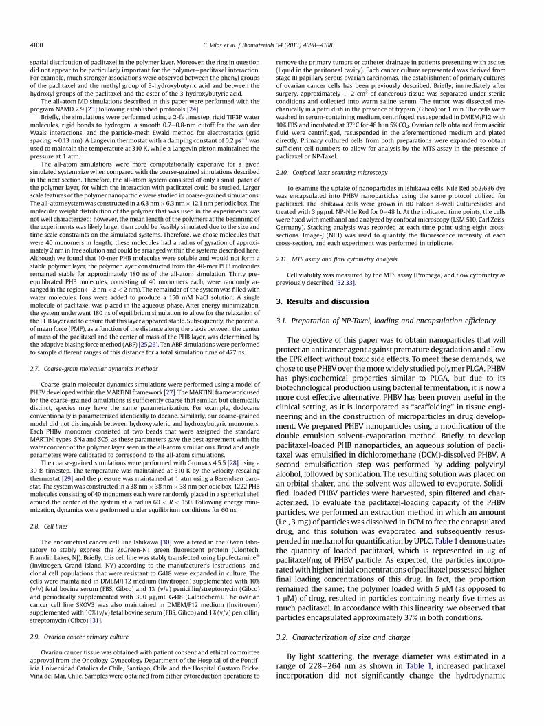
The objective of this paper was to obtain nanoparticles that willprotect an anticancer agent against premature degradation and allowthe EPR effect without toxic side effects. To meet these demands, wechose touse PHBVover themorewidely studiedpolymerPLGA. PHBVhas physicochemical properties similar to PLGA, but due to itsbiotechnological production using bacterial fermentation, it is now amore cost effective alternative. PHBV has been proven useful in theclinical setting, as it is incorporated as “scaffolding” in tissue engi-neering and in the construction of microparticles in drug develop-ment. We prepared PHBV nanoparticles using a modification of thedouble emulsion solvent-evaporation method. Briefly, to developpaclitaxel-loaded PHB nanoparticles, an aqueous solution of pacli-taxel was emulsified in dichloromethane (DCM)-dissolved PHBV. Asecond emulsification step was performed by adding polyvinylalcohol, followed by sonication. The resulting solutionwas placed onan orbital shaker, and the solvent was allowed to evaporate. Solidi-fied, loaded PHBV particles were harvested, spin filtered and char-acterized. To evaluate the paclitaxel-loading capacity of the PHBVparticles, we performed an extraction method in which an amount(i.e., 3 mg) of particles was dissolved in DCM to free the encapsulateddrug, and this solution was evaporated and subsequently resus-pended inmethanol for quantification byUPLC. Table 1 demonstratesthe quantity of loaded paclitaxel, which is represented in mg ofpaclitaxel/mg of PHBV particle. As expected, the particles incorpo-ratedwithhigher initial concentrationsofpaclitaxelpossessedhigherfinal loading concentrations of this drug. In fact, the proportionremained the same; the polymer loaded with 5 mM (as opposed to1 mM) of drug, resulted in particles containing nearly five times asmuch paclitaxel. In accordance with this linearity, we observed thatparticles encapsulated approximately 37% in both conditions.
3.2. Characterization of size and charge
By light scattering, the average diameter was estimated in arange of 228e264 nm as shown in Table 1, increased paclitaxelincorporation did not significantly change the hydrodynamic

Table 1Determination of the loading yield, encapsulation efficiency, size and zeta potentialfrom nanoparticles loaded in the presence of 1 mM or 5 mMpaclitaxel (NP-Taxel) andnon-loaded nanoparticles (NP-empty).
Preparation Loadinga Encapsulationefficiency (%)
Size (nm) Zeta potential(mV)
(n ¼ 4) (n ¼ 4) (n ¼ 11) (n ¼ 9)
NPs e empty ND ND 264.4 � 70.7 �8.9 � 2.7NP-Taxel 1 mM 0.40 � 0.09 34.9 � 2.0 228.1 � 25.1 �7.4 � 1.8NP-Taxel 5 mM 2.42 � 0.20 39.6 � 4.2 261.9 � 35.1 �6.0 � 1.4
ND, not determined.a Loading yield mg drug/mg NP-Taxel.
C. Vilos et al. / Biomaterials 34 (2013) 4098e4108 4101
diameter of the nanoparticle. This is an optimal size for passiveliberation in the future clinical use of this nanoparticle [34]. It hasbeen suggested that a beneficial characteristic of nanoparticles forbiomedical use is the presence of a negative charge on the outercoat. This negative chargewill theoretically reduce aberrant proteinbinding and, thus, is less likely to activate the immune system,resulting in a longer circulatory half-life. In addition, a negativecharge minimizes non-specific binding with the cell surface [35].The particle was also evaluated for its zeta potential by light scat-tering. As also shown in Table 1, the zeta potential ranged from �6to �8.9 mV, with no significant differences between eachpreparation.
3.3. Segregation of paclitaxel to the polymerewater interface
To better understand the microscopic details of the paclitaxelrelease process, we performed all-atom molecular dynamics sim-ulations of a system containing a PHB layer, an aqueous phase, andpaclitaxel. The behavior of the system was first studied in a 180-nsequilibrium simulation, wherein we observed that the PHB layerwas stable on this time scale. We also observed that the polymerlayer was water permeable. As shown in the mass density plot inFig. 1A, the PHB layer contained about 10% water by mass. Thesingle paclitaxel molecule spent a majority of the simulation at thepolymerewater interface.
To better characterize the interaction between the paclitaxel andPHB layer, we performed a hydrogen bond (H-bond) analysis on the
Fig. 1. Computer simulations of PHBepaclitaxel interaction and nanoparticle structure. (Apolymer and water components are shown as black and blue lines, respectively. The probabisnapshot of the final frame of the simulation is overlaid on the plot. PHB is shown in red andnot shown. (C) The potential of mean force (PMF) for the interaction between paclitaxel andthe PMF corresponds to the maximum of the paclitaxel probability in the equilibrium simulSnapshot from the coarse-grained MD simulation of a 24-nm diameter PHBV nanoparticle. TWater and ion particles are not shown. (For interpretation of the references to colour in th
equilibrium trajectory. The first 14 ns of the trajectory were dis-carded to reduce the extent to which the results depended on thestarting configuration. Snapshots from the trajectorywere extractedat 10 ps intervals; for each snapshot, the H-bonds between thepaclitaxel and the polymer were identified when the donoreacceptor distance was <0.35 nm and the donor-H-acceptor anglewas greater than 120�. The average number of H-bonds during the166 ns trajectorywas 0.26� 0.01. Interactions between the hydroxylgroups of the paclitaxel and the carbonyl of the PHB accounted for75.0% of the H-bonds, while interactions between the peptide ni-trogen of the paclitaxel and the carbonyl of the PHB comprised 22.6%of the H-bonds. The remaining H-bonds involved the PHB terminalhydroxyl groups. The PHB molecules used in the simulations (40monomers) were likely shorter than those present in the experi-ments, leading to a higher density of chain-terminating hydroxyls inthe simulations than in the experiments. However, only 3% of the H-bonds involved the terminal monomers, and only 2.4% of the H-bonds involved the terminal hydroxyl groups of the polymer.Therefore, similar results would likely have been obtained withlonger PHB molecules.
To unequivocally determine the energetics of the interactionbetween paclitaxel and the PHB layer, we calculated the PMF as afunction of the distance between the center of mass of the pacli-taxel molecule and the center of mass of the PHB layer along theaxis orthogonal to the polymer layer (z axis).
3.4. Microscopic structure of PHBV nanoparticles
A complete all-atom MD simulation of a nanoparticle in waterwould require the simulation of tens of millions of atoms, andtherefore, extensive all-atom simulations of nanoparticles areprohibitively expensive. Coarse-grained MD, in which a singleparticle represents several atoms, makes the simulation of largenanoparticle assemblies [36]. Thus, we constructed a coarse-grained model of our PHBV nanoparticles in order to determinetheir nanoscale structure. Fig. 1C reveals the structure of a stable,quasi-spherical nanoparticle with a diameter of 24 nm that iscomposed of 40-monomer PHBV molecules.
The coarse grain simulations show pores on the surface of thesphere, in agreement with previous experimental results [37].Therefore, if the drug is diluted in the solvent inside of the sphere,
) Stable PHB layer in an equilibrium all-atom simulation. The mass densities of thelity density along the z axis of the single paclitaxel molecule is shown as a green line. Agray, and paclitaxel is shown in green. Water molecules, ions, and hydrogen atoms arePHB layer was calculated from an adaptive biasing force simulation. The minimum ofation (panel B). The thickness of the line represents the standard error of the PMF. (A)he thickness of the shell is about 3 nm, and it contains a number of holes and defects.is figure legend, the reader is referred to the web version of this article.)

C. Vilos et al. / Biomaterials 34 (2013) 4098e41084102
the drug might conceivably diffuse rapidly to the outside of the NPthrough the pores; however, this does not occur, likely due tostrong attraction between the paclitaxel and the polymer surface.Thus, while porous particles are often assumed to accelerate releaseof the drug, our simulations suggest that porosity could, on thecontrary, delay release by providing a larger surface area forpaclitaxel binding. The drug might only then be released whennearby polymer is chemically degraded.
3.5. TEM characterization using low-voltage electron microscopyand analysis by FTIR spectroscopy
Transmission electron microscopy (TEM) is an excellent methodto characterize NPs; however, the high-voltage used in traditionalelectron microscopy prevents the direct observation of soft-matterstructures. In this work, we performed low voltage electron micro-scopy (LVEM) at 5 kV without metal staining to obtain high contrastimages of the nanoparticle structure at different degradation states.An LVEM image of an unstained NP-Taxel droplet is shown in Fig. 2.Notably, the size of the NP-Taxel at day 0 (Fig. 2A) is smaller than atday5 (Fig. 2F). During the intermediate days (Fig. 2BeD), theNP-Taxelwas surrounded by branched structures, presumably consisting ofpolymer. Interestingly, these structureswereno longer present at day4 (Fig. 2D), leavinganapparent spherical shapewith a smooth surfaceof nanoparticles (Fig. 2D, E). The branched structures likely have alower density than the polymer layer forming the nanoparticle,which may explain their relatively rapid disappearance.
NP-Taxel exhibited a different fingerprint region than the PHBVcrystals because thenanoparticles have amore complexcomposition,including PHBV-Taxel-PVA. PHBV unmasked the Taxel signal becausethe interactionspredominantly includedPHBV.As illustrated inFig. 3,the spectrum showed carboxyl groups (bands at 1680e1725 cm�1) inthe empty PHBV nanoparticles and at day 0 of degradation to NP-Taxel. The aldehyde groups increased between days 1 and 3 ofdegradation (bands at 2820e2900 and 2775e2700 cm�1, and 1660e
Fig. 2. Time-dependence of the NP-Taxel size and surface-polymer structures during Taxel
1740cm�1), andon the samedays,wealsoobservedan increase in thealcohols groups (bands at 3550e3200 cm�1), the signal to crystalli-zation in the polymer and in broken-in ester groups. This fact is veryimportant because if a signal to the ester groups is broken andaldehyde and alcohol groups are formed. This reaction is possiblebecause thedegradationoccurredat apHof 7.2; if this reactionoccursat an acidic pH, then cleavage next to the ester bondwould occur, andthis reactionwould form carboxyl groups instead of aldehyde groups.Finally, these results suggest that the polymer experienced localizedcrystallization for canaliculated formation, through which watercould enter and complete the degradation by polymer hydrolysis.
3.6. Uptake of Nile Red-loaded PHBV nanoparticles
Before testing the anticancer capacity of the NPs, we sought toconfirm the ability of cancer cells to uptake PHBV nanoparticles. Tothis end, we synthesized PHBV nanoparticles containing the hy-drophobic dye Nile Red (see methods). These nanoparticles werewashed with water, resuspended in PBS and added to culture me-dium in 6-well culture dishes containing a growingmonolayer of thehuman endometrial carcinoma cell line Ishikawa, which was stablytransfected with green fluorescent protein (ZsGreen, see methods).As shown in Fig. 4A, nanoparticles were observed at 30 min anduptake continued to increase over 48 h. The intracellular incorpo-ration of Nile Red was confirmed by stacking analysis using confocalmicroscopy, as shown by the representative images in Fig. 4A (thinimage), which are quantified in Fig. 4B. More complete confocalmicroscopy z-stack images are shown in the Supplementary figures.
3.7. In vitro release of paclitaxel
As the retention of the chemotherapeutic drug within thenanoparticle is fundamental for its future clinical application, wenext measured the liberation of paclitaxel from the PHBV nano-particle in non-biological conditions. As shown in Fig. 5, paclitaxel-
liberation processes observed using LVEM. 0 (A), 1 (B), 2 (C), 3 (D), 4 (E) and 5 (F) days.

Fig. 3. FTIR spectroscopy of paclitaxel-loaded PHBV nanoparticles from a time course in vitro release system (0e5 days).
C. Vilos et al. / Biomaterials 34 (2013) 4098e4108 4103
containing nanoparticles that were resuspended in distilled waterdemonstrated less than 4% release over a 5-day period at 25 �C.However, in a more physiologically relevant solution, PBS pH 7.4,less than 1% was liberated over a 5-day period. These results sug-gest high specificity for any potential future use of this nano-particle, as less than 1% of paclitaxel would be liberated into thecirculation, and given the leaky blood vasculature that irrigatestumors, this nanoparticle should be taken up preferentially by thecancer cells.
3.8. Cytotoxicity of NP-Taxel in Ishikawa cells
To evaluate the biological activity of paclitaxel-containingnanoparticles (referred to hereafter as NP-Taxel) we assessed thecytotoxicity using theMTS assay previously shown in our laboratory
to reflect apoptotic cell death in the presence of paclitaxel [32]. Fig. 6shows that paclitaxel alone is capable of bringing about cell death at0.1 mM. The three concentrations of paclitaxel used demonstratedthe same effect at each time point measured, with the exception of0.1 mM at 2 days. Interestingly, NP-Taxel in a quantity that delivers0.1 mMof paclitaxel did not induce cytotoxicity at any time point andhad the unexpected effect of increasing proliferation (or at least theredox state of the cells, which is measured in this assay). However,1 mM NP-Taxel initiated cell death after four days of treatment and5 mM NP-Taxel induced cell death at times and concentrationsundistinguishable from1 to5mMpaclitaxel. This result suggests that5 mMNP-Taxel liberates at least an equivalent of 1 mMpaclitaxel. Thisliberation is more than acceptable for any given future commercialapplication, as the estimated circulatory concentration of paclitaxelin patients is between 4 and 10 mM and theoretically the

Fig. 4. (A) Fluorescence confocal microscopy demonstrating the uptake of Nile Red-loaded PHBV nanoparticles in the Ishikawa cancer cell line, which had been previously stablytransfected with green fluorescent protein. The thin image on the side of each principal image demonstrates a stacking analysis by confocal microscopy, confirming the intracellularincorporation of the nanoparticles. (B) Determination of the fluorescence intensity at time points after exposure (n ¼ 3), *p < 0.05 KruskaleWallis and ManneWhitney post-test.
Fig. 5. Time course in days demonstrating the cumulative release of paclitaxel from5 mM-loaded nanoparticles in the presence of water (blue line) or PBS, pH 7.4 (greenline). (For interpretation of the references to colour in this figure legend, the reader isreferred to the web version of this article.)
C. Vilos et al. / Biomaterials 34 (2013) 4098e41084104
nanoparticles will be within the cancer cell at the moment ofpaclitaxel release [38].
3.9. Mechanism of cell death
To further understand the effect of NP-Taxel at earlier timepoints and to confirm whether the NP-Taxel induced cell deathoccurs through the samemechanisms as reported for paclitaxel, weused flow cytometry to assess changes in the cell cycle at 24 and48 h and measured cell death by the occurrence of a sub G0/G1peak in the cell cycle profile. As shown in Fig. 7, empty PHBVnanoparticles (i.e. without paclitaxel) produced the same cell-cycleprofile at both 24 and 48 h. In accordance with the literature,paclitaxel alone induced a cell cycle arrest in G2/M before insti-gating cell death (Fig. 7A, paclitaxel 5 mM). Interestingly, the incu-bation of paclitaxel together with empty PHBV nanoparticles had aprotective effect against cell death, with the majority of the cancercells being in G2/M arrest at 24 h (this protective effect will be

Fig. 6. The treatment of Ishikawa cancer cells with 5 mM paclitaxel-loaded nano-particles generated similar cytotoxicity to that of 1 mM paclitaxel as measured by theMTS assay. The treatments were performed for 48e120 h with different concentrationsof NP-Taxel or paclitaxel alone (0.1, 1 and 5 mM). ANOVA, post-test Bonferronidemonstrated no significant differences between 5 mM NP-Taxel and either 1 or 5 mMpaclitaxel alone.
C. Vilos et al. / Biomaterials 34 (2013) 4098e4108 4105
discussed later). The presence of 5 mM NP-Taxel demonstrated G2/M arrest at 24 h but not cell death, mirroring the result of paclitaxelwith empty PHBV nanoparticles. At 48 h, NP-Taxel had induced celldeath in the majority of the cancer cells analyzed (Fig. 7B). Theseresults demonstrate that paclitaxel-loaded PHBV nanoparticlesconfers cell death by the same mechanism (first an arrest in G2/Mand then apoptosis) as paclitaxel as a single agent.
Fig. 7. The observation of a preliminary G2/M cell cycle arrest suggests that paclitaxel-loaded nanoparticles induce cell death by the same mechanism as that of paclitaxelalone. The cell cycle analysis was performed by flow cytometry in the Ishikawa cancercell line after 24 h (A) or 48 h (B) of treatment with NP-Taxel or paclitaxel alone at thestated concentrations. For the graphical representation, the cell cycle was divided intonon-proliferative (G0/G1), proliferative (S/G2/M) and cell death (peak sub G0/G1)region.
3.10. Cytotoxicity of NP-Taxel in SKOV3 cells
To demonstrate that the effects observed in the endometrial cellline Ishikawa are not exclusive to this cell line or this type of cancer,we assessed the cytoxicity of NP-Taxel in the human ovarian cancercell line SKOV3. As demonstrated previously [32], this cell line issensitive to paclitaxel-induced cell death from 0.1 mM onwards(Fig. 8). However, unlike the endometrial cancer cells, the SKOV3cell line did not demonstrate a proliferative or a protective effectwhen empty NPs were administrated either alone or in the pres-ence of free paclitaxel. During the window of protection, this resultdemonstrates that the protective effect of empty nanoparticles iscell specific (this will be discussed in detail below). However, asobserved in Ishikawa cells, the addition of NP-Taxel induced celldeath equivalent to that of 1 mM paclitaxel.
3.11. Cytotoxicity of NP-Taxel in primary cultures of ovarian cancer
To determine the clinical relevance of this assessment of bio-logical activity, we examined the effect of NP-Taxel in primary cul-tures of ovarian cancer. These cancer cells were obtained from theprimary culture of either tumor or ascitis (fluid extracted from theperitoneal cavity) obtained with informed consent from patientsundergoing treatment for stage IIIc papillary serous ovarian cancer.As expected for six individual patients, the response to paclitaxelwas distinct in each case. The level of cytotoxicity observedwithNP-Taxel was lower than that of paclitaxel in each primary culture, butstill produced significant cell death in each case analyzed. This resultdemonstrates that cell death occurred within 48 h of the primaryculture of ovarian cancer cells. As previously mentioned, in futureclinical applications, this NP-Taxel mediated cell death may begreater than paclitaxel alone due to the incorporation of the nano-particles and drug release directly in the cancer cells and possibly aprolonged half-life compared to paclitaxel alone, due to the neces-sity of polymer breakdown in the lysosome to facilitate drug release.
Interestingly, as observed in the human endometrial cancer cellline and in two of six ovarian cancers derived from patients, theempty nanoparticles demonstrated cellular cytotoxicity (Fig. 9 pa-tients 5 & 6), as was observed in the ovarian cancer cell line SKOV3.Furthermore, four primary cultures of ovarian cancer demonstratedproliferation in the presence of empty NP. This result further dem-onstrates the well-known clinical observation that no two cancersare alike and no two patients respond in exactly the same way to agiven therapy. However, the question remains as to why a polymercomplex such as PHBV/PVA could bring about cell death or survival.The nature of PHBV (3-hydroxybutyric acid-co-hydroxyvaleric acid)suggests the potential liberation of hydroxybutyric acid in the livingcell. Hydroxybutyric acid, which is a ketone body that is producedin vivo in humans, is a group of four-carbon organic compounds thatpossess both hydroxyl and carboxylic acid functional groups. Incancer cells, this compound may be broken down into derivativessuch as D-3-hydroxybutyrate, DL-3-hydroxybutyrate andmethyl (D)-3-hydroxybutyrate [39]. In mouse glial cells, DL-3-hydroxybutyratehas been previously shown to possess a protective effect againstapoptosis, while D-beta-hydroxybutyrate prevents against neuro-toxicity in rat glial cells (PC12) [40]. Furthermore, ketone bodieshave been reported to be favorable alternative metabolic substratesand are protective in the face of several pathologies. A rich ketogenicdiet has also been shown to reduce cytochrome c release and thuscellular apoptosis in a neurological model [41]. Conversely, theliberation of alcohol groups from PVA (polyvinyl alcohol) could leadto oxidative stress within cancer cells and thus cell death. Themetabolic state of the cell and the accompanying stimulus present atany given timemay represent the factors that tip the balance of howthe cell responds to these nanoparticles. Irrespective of the

Fig. 8. Paclitaxel-loaded nanoparticles induced cell death in the ovarian cancer cell line SKOV3. Non-loaded nanoparticles (NP-empty), paclitaxel-loaded nanoparticles (NP-Taxel)and paclitaxel alone at the stated concentrations were added to the cancer cell cultures for 48 h, and cytotoxicity was measured by MTS. The treatments were 48 h with NP-Taxel orpaclitaxel alone. *p < 0.05 compared to the control; #p < 0.05 compared to the NP empty vector; ANOVA, post-test Bonferroni.
Fig. 9. NP-Taxel induces cell death in primary cultured human ovarian cancer cells. The cancer cells were obtained with informed consent from either the primary tumor orperitoneal fluid (ascitis) of patients with stage IIIc papillary serous ovarian cancer. The percentage of cytotoxicity was measured by MTS after incubation for 48 h with 5 mMpaclitaxel-loaded nanoparticles (NP-Taxel), non-loaded nanoparticles (NP-empty) or paclitaxel alone.
C. Vilos et al. / Biomaterials 34 (2013) 4098e41084106

C. Vilos et al. / Biomaterials 34 (2013) 4098e4108 4107
potentially protective effects of PHBV, any future PHBV nanoparticlewill come loaded with a chemotherapeutic drug and will thereforemake cell death more likely to occur.
While these PHBV nanoparticles displayedminimal drug releasein solution and minor cytoxicity toward primary cultured ovariancancer cells, there are other ways in which these nanoparticlescould be improved for future clinical application. For instance, anincrease in the loading of the nanoparticles that would give anaverage release of a minimum of 1e10 mM paclitaxel should resultin cancer cell toxicity that is similar to that of paclitaxel alone andwill hopefully confer fewer side effects. To this end, further inves-tigation is required to optimize the ratios of PHBV and paclitaxel inthe starting emulsion and to determine the saturation point ofpaclitaxel loading. A better understanding of the optimal timing ofdrug release in terms of cytoxicity may allow for the precise se-lection of the liberation rate, which could be altered by using aPHBV with a lower molecular weight or a PHBV with a highercomposition of valerate. Further improvements are necessary toexpand upon the passive targeting of nanoparticles by adding anactive uptake component to this preparation method. The surfaceincorporation of folic acid, which is preferentially taken up bycancer cells, or the addition of aptamer bioconjugates may addgreater specificity and intracellular drug accumulation.
4. Conclusion
Here, we describe an integrated study that includes the mo-lecular simulation, physicochemical characterization and biologicalevaluation of a paclitaxel-loaded PHBV nanoparticles. Molecularsimulation displayed a high affinity of paclitaxel for the waterepolymer interface and porous nanoparticle structure. The parti-cles size range 228e264 nm makes them appropriate for passivetargeting by EPR effect, and their negative charge (�6 to �8.9 mV),suitable for the biological environment. The analysis of NP-Taxel bylow voltage electron microscopy and FTIR during in vitro degra-dation process revealed the presence of branched structures on thesurface of particles, and the generation of new chemical bondssuggesting a temporal crystalline phase in the first 3 days. Finally,the NP-Taxel exhibited a low in vitro rate of release, intracellularuptake, and a high cytotoxic capacity in cell lines, and in primarycultures of stage IIIc serous ovarian cancer cells isolated from sixpatients. The promising results and the low cost of production,supports PHBVs as strong candidates in the development of noveldrug-loaded nanoparticles for large-scale production by the phar-maceutical industry.
Author contribution
C.V. and L.A.V. planned the experiments. C.V. and F.M. formu-lated the nanoparticles. P.S. and N.H. performed the physico-chemical characterization. F.G.N. and J.C. developed the dynamicmolecular modeling. D.A. and H.M. performed the electron micro-scopy analysis. P.G., S.K., M.A.C., C.A. and E.B. obtained the patientsamples for the primary cultures. M.L.B. and G.I.O. studied theantitumor activity cancer cells. C.V., G.I.O., F.G.N. and L.A.V.analyzed the data and wrote the paper.
Funding
The authors declare no competing financial interests.
Acknowledgments
We thank the medical institutions, the medical staff and thepatients for their generous participation and donation of primary
tissue. This research was supported by the: Centro para el Desar-rollo de la Nanociencia y Nanotecnologia (CEDENNA); FB0807 (C.V.,F.M., L.A.V.); and the Biomedical Research Consortium of Chile;CTU06 (G.I.O.). Finally, we acknowledge Fondo Nacional de Desar-rollo Científico y Tecnológico Gobierno de Chile (FONDECYT)1120712 (L.A.V.), 1080163 (M.A.C.), 1100870 (G.I.O.), and 3120003(M.L.B.).
Appendix A. Supplementary data
Supplementary data related to this article can be found at http://dx.doi.org/10.1016/j.biomaterials.2013.02.034.
References
[1] Jemal A, Siegel R, Xu J, Ward E. Cancer statistics. CA Cancer J Clin 2010;2010(60):277e300.
[2] Bast Jr RC, Hennessy B, Mills GB. The biology of ovarian cancer: new oppor-tunities for translation. Nat Rev Cancer 2009;9:415e28.
[3] Vaughan S, Coward JI, Bast Jr RC, Berchuck A, Berek JS, Brenton JD, et al.Rethinking ovarian cancer: recommendations for improving outcomes. NatRev Cancer 2011;11:719e25.
[4] Arias JL. Drug targeting strategies in cancer treatment: an overview. Mini RevMed Chem 2011;11:1e17.
[5] Wang AZ, Langer R, Farokhzad OC. Nanoparticle delivery of cancer drugs.Annu Rev Med 2012;63:185e98.
[6] Wagner V, Dullaart A, Bock AK, Zweck A. The emerging nanomedicine land-scape. Nat Biotechnol 2006;24:1211e7.
[7] Dhar S, Kolishetti N, Lippard SJ, Farokhzad OC. Targeted delivery of a cisplatinprodrug for safer and more effective prostate cancer therapy in vivo. Proc NatlAcad Sci USA 2011;108:1850e5.
[8] Shi J, Xiao Z, Votruba AR, Vilos C, Farokhzad OC. Differentially charged hollowcore/shell lipid-polymer-lipid hybrid nanoparticles for small interfering RNAdelivery. Angew Chem Int Ed Engl 2011;50:7027e31.
[9] Wohlfart S, Gelperina S, Kreuter J. Transport of drugs across the bloodebrainbarrier by nanoparticles. J Control Release 2012;161:264e73.
[10] Xie J, Liu G, Eden HS, Ai H, Chen X. Surface-engineered magnetic nanoparticleplatforms for cancer imaging and therapy. Acc Chem Res 2011;44:883e92.
[11] Kumari A, Yadav SK, Yadav SC. Biodegradable polymeric nanoparticles baseddrug delivery systems. Colloids Surf B Biointerfaces 2010;75:1e18.
[12] Cragg GM, Newman DJ. A tale of two tumor targets: topoisomerase I andtubulin. The Wall and Wani contribution to cancer chemotherapy. J Nat Prod2004;67:232e44.
[13] Acharya S, Sahoo SK. PLGA nanoparticles containing various anticancer agentsand tumour delivery by EPR effect. Adv Drug Deliv Rev 2011;63:170e83.
[14] Slater S, Mitsky TA, Houmiel KL, Hao M, Reiser SE, Taylor NB, et al. Metabolicengineering of Arabidopsis and Brassica for poly(3-hydroxybutyrate-co-3-hydroxyvalerate) copolymer production. Nat Biotechnol 1999;17:1011e6.
[15] Kuppan P, Vasanthan KS, Sundaramurthi D, Krishnan UM, Sethuraman S.Development of poly(3-hydroxybutyrate-co-3-hydroxyvalerate) fibers forskin tissue engineering: effects of topography, mechanical, and chemicalstimuli. Biomacromolecules 2011;12:3156e65.
[16] Sendil D, Gursel I, Wise DL, Hasirci V. Antibiotic release from biodegradablePHBV microparticles. J Control Release 1999;59:207e17.
[17] Vilos C, Velasquez LA. Therapeutic strategies based on polymeric micropar-ticles. J Biomed Biotechnol 2012;2012:672760.
[18] Vilos C, Constandil L, Herrera N, Solar P, Escobar-Fica J, Velasquez LA. Cef-tiofur-loaded PHBV microparticles: a potential formulation for a long-actingantibiotic to treat animal infections. Electron J Biotechnol 2012;15:1e13.
[19] Sahana B, Santra K, Basu S, Mukherjee B. Development of biodegradablepolymer based tamoxifen citrate loaded nanoparticles and effect of somemanufacturing process parameters on them: a physicochemical and in-vitroevaluation. Int J Nanomedicine 2010;5:621e30.
[20] Mao S, Xu J, Cai C, Germershaus O, Schaper A, Kissel T. Effect of WOW processparameters on morphology and burst release of FITC-dextran loaded PLGAmicrospheres. Int J Pharm 2007;334:137e48.
[21] Zhang SQ, Chen GH. Determination of paclitaxel in human plasma by UPLC-MS-MS. J Chromatogr Sci 2008;46:220e4.
[22] Vanommeslaeghe K, Hatcher E, Acharya C, Kundu S, Zhong S, Shim J, et al.CHARMM general force field: a force field for drug-like molecules compatiblewith the CHARMM all-atom additive biological force fields. J Comput Chem2010;31:671e90.
[23] Phillips JC, Braun R, Wang W, Gumbart J, Tajkhorshid E, Villa E, et al. Scalablemolecular dynamics with NAMD. J Comput Chem 2005;26:1781e802.
[24] Wells DB, Bhattacharya S, Carr R, Maffeo C, Ho A, Comer J, et al. Optimizationof the molecular dynamics method for simulations of DNA and ion transportthrough biological nanopores. Methods Mol Biol 2012;870:165e86.
[25] Darve E, Rodriguez-Gomez D, Pohorille A. Adaptive biasing force method forscalar and vector free energy calculations. J Chem Phys 2008;128:144120.

C. Vilos et al. / Biomaterials 34 (2013) 4098e41084108
[26] Henin J, Fiorin G, Chipot C, Klein M. Exploring multidimensional free energylandscapes using time-dependent biases on collective variables. J ChemTheory Comput 2009;6:35e47.
[27] Marrink SJ, Risselada HJ, Yefimov S, Tieleman DP, de Vries AH. The MARTINIforce field: coarse grained model for biomolecular simulations. J Phys Chem B2007;111:7812e24.
[28] Hess B, Kutzner C, Van Der Spoel D, Lindahl E. GROMACS 4: algorithms forhighly efficient, load-balanced, and scalable molecular simulation. J ChemTheory Comput 2008;4:435e47.
[29] Bussi G, Donadio D, Parrinello M. Canonical sampling through velocityrescaling. J Chem Phys 2007;126:014101.
[30] NishidaM.The Ishikawacells frombirth to thepresent.HumCell 2002;15:104e17.[31] Hill BT, Whelan RD, Gibby EM, Sheer D, Hosking LK, Shellard SA, et al.
Establishment and characterisation of three new human ovarian carcinomacell lines and initial evaluation of their potential in experimental chemo-therapy studies. Int J Cancer 1987;39:219e25.
[32] Kato S, Sadarangani A, Lange S, Delpiano A, Vargas M, Brañes J, et al. 2-methoxyestradiol mediates apoptosis through caspase-dependent and inde-pendent mechanisms in ovarian cancer cells but not in normal counterparts.Reprod Sci 2008;15:878e94.
[33] Sadarangani A, Kato S, Espinoza N, Lange S, Llados C, Espinosa M, et al. TRAILmediates apoptosis in cancerous but not normal primary cultured cells of thehuman reproductive tract. Apoptosis 2007;12:73e85.
[34] Peer D, Karp JM, Hong S, Farokhzad OC, Margalit R, Langer R. Nanocarriers asan emerging platform for cancer therapy. Nat Nanotechnol 2007;2:751e60.
[35] Nel AE, Madler L, Velegol D, Xia T, Hoek EM, Somasundaran P, et al. Under-standing biophysicochemical interactions at the nano-bio interface. Nat Mater2009;8:543e57.
[36] Loverde SM, Klein ML, Discher DE. Nanoparticle shape improves delivery:rational coarse grain molecular dynamics (rCG-MD) of taxol in worm-likePEG-PCL Micelles. Adv Mater 2012;24:3823e30.
[37] Li H, Chang J. Preparation, characterization and in vitro release of gentamicinfrom PHBV/wollastonite composite microspheres. J Control Release 2005;107:463e73.
[38] Hunz M, Jetter A, Warm M, Pantke E, Tuscher M, Hempel G, et al. Plasma andtissue pharmacokinetics of epirubicin and Paclitaxel in patients receivingneoadjuvant chemotherapy for locally advanced primary breast cancer. ClinPharmacol Ther 2007;81:659e68.
[39] Xiao XQ, Zhao Y, Chen GQ. The effect of 3-hydroxybutyrate and its derivativeson the growth of glial cells. Biomaterials 2007;28:3608e16.
[40] Cheng B, Yang X, Chen C, Cheng D, Xu X, Zhang X. D-beta-hydroxybutyrateprevents MPPþinduced neurotoxicity in PC12 cells. Neurochem Res 2010;35:444e51.
[41] Hu ZG, Wang HD, Jin W, Yin HX. Ketogenic diet reduces cytochrome c releaseand cellular apoptosis following traumatic brain injury in juvenile rats. AnnClin Lab Sci 2009;39:76e83.