Enhanced cytotoxicity of a polymer–drug conjugate with triple payload of paclitaxel
Upload
khangminh22Category
view
1download
0
RESEARCH ARTICLE Open Access
Combination of Paclitaxel and MG1oncolytic virus as a successful strategy forbreast cancer treatmentMarie-Claude Bourgeois-Daigneault1,2* , Lauren Elizabeth St-Germain1, Dominic Guy Roy1,2, Adrian Pelin1,2,Amelia Sadie Aitken1,2, Rozanne Arulanandam1, Theresa Falls1, Vanessa Garcia1,2, Jean-Simon Diallo1
and John Cameron Bell1,2
Abstract
Background: Breast cancer is the most common malignant disease amongst Western women. The lack oftreatment options for patients with chemotherapy-resistant or recurrent cancers is pushing the field toward therapid development of novel therapies. The use of oncolytic viruses is a promising approach for the treatment ofdisseminated diseases like breast cancer, with the first candidate recently approved by the Food and DrugAdministration for use in patients. In this report, we demonstrate the compatibility of oncolytic virotherapy andchemotherapy using various murine breast cancer models. This one-two punch has been explored in the past byseveral groups with different viruses and drugs and was shown to be a successful approach. Our strategy is tocombine Paclitaxel, one of the most common drugs used to treat patients with breast cancer, and the oncolyticRhabdovirus Maraba-MG1, a clinical trial candidate in a study currently recruiting patients with late-stagemetastatic cancer.
Methods: We used the EMT6, 4 T1 and E0771 murine breast cancer models to evaluate in vitro and in vivo the effectsof co-treatment with MG1 and Paclitaxel. Treatment-induced cytotoxicity was assessed and plaque assays, flowcytometry, microscopy and immunocytochemistry analysis were performed to quantify virus production and transgeneexpression. Orthotopically implanted tumors were measured during and after treatment to evaluate efficacy andKaplan-Meier survival curves were generated.
Results: Our data demonstrate not only the compatibility of the treatments, but also their synergistic cytopathicactivity. With Paclitaxel, EMT6 and 4 T1 tumors demonstrated increased virus production both in vitro and in vivo. Ourresults also show that Paclitaxel does not impair the safety profile of the virus treatment. Importantly, when combined,MG1 and the drug controlled tumor growth and prolonged survival.
Conclusions: The combination of MG1 and Paclitaxel improved efficacy in all of the breast cancer models we testedand thus is a promising alternative approach for the treatment of patients with refractory breast cancer. Our strategyhas potential for rapid translation to the clinic, given the current clinical status of both agents.
Keywords: Breast Cancer, Triple-negative breast cancer, TNBC, Oncolytic virus, Rhabdovirus Maraba-MG1, Paclitaxel,Viral Sensitizer, Combination therapy
* Correspondence: [email protected] Hospital Research Institute, Centre for Innovative Cancer Research,Ottawa K1H 8L6, Canada2Department of Biochemistry, Microbiology and Immunology, University ofOttawa, Ottawa K1H 8M5, Canada
© 2016 The Author(s). Open Access This article is distributed under the terms of the Creative Commons Attribution 4.0International License (http://creativecommons.org/licenses/by/4.0/), which permits unrestricted use, distribution, andreproduction in any medium, provided you give appropriate credit to the original author(s) and the source, provide a link tothe Creative Commons license, and indicate if changes were made. The Creative Commons Public Domain Dedication waiver(http://creativecommons.org/publicdomain/zero/1.0/) applies to the data made available in this article, unless otherwise stated.
Bourgeois-Daigneault et al. Breast Cancer Research (2016) 18:83 DOI 10.1186/s13058-016-0744-y

BackgroundBreast cancer is a highly aggressive disease with most ofthe deaths resulting from metastases within the firstthree years upon diagnosis [1]. Metastatic human triple-negative breast cancer (TNBC) has the worst prognosisamong all types of breast cancer, with high risk of rapidrecurrence and shortened survival [2]. TNBC is deficientin the expression of estrogen receptor, progesterone recep-tor and human epidermal growth factor receptor 2, andthus, is refractory to conventional breast cancer hormonaltherapy such as Tamoxifen. The main therapeutic optionwith surgery is chemotherapy, but some subsets of tumorare resistant and the prognosis for these patients is poor[3]. The standard of care for TNBC is the administrationof anthracyclines and/or taxanes [2]. Paclitaxel (PAC),also called Taxol, is a cancer chemotherapeutic agent ofthe taxane family that acts by stabilizing microtubulesand thus preventing cell division [4]. PAC is commonlyused as monotherapy or in combination with differentagents. Significant effort is currently being directed to-ward improving its efficacy and developing alternatestrategies for the treatment of chemotherapy-resistantand recurrent disease.A novel strategy being explored for the treatment of
metastatic diseases such as TNBC is the use of oncolyticviruses (OV). Several candidates are currently undergo-ing clinical trials and are considered promising ap-proaches for the treatment of various cancers includingTNBC [5]. At the forefront of this field is T-Vec, a her-pes virus that was successfully tested in a phase III studyin melanoma and was approved in 2015 by the Food andDrug Administration for clinical use. OVs specificallyreplicate in and destroy tumor cells by several mecha-nisms including direct oncolysis [6]. The rhabdovirusfamily members, vesicular stomatitis virus (VSV) andmaraba, were first identified as oncolytic agents byour group [7, 8]. The tumor specificity of these vi-ruses is conferred by the capacity of normal cells, butnot tumor cells, to respond to antiviral interferons(IFN) [7, 8]. Variants with a greater therapeutic index,VSVΔ51 and Maraba MG1, were subsequently developedfor clinical use [8, 9]. Importantly, enrolling recentlybegan for a clinical trial using MG1 both as a stand-alone therapy and in a vaccination strategy in patientswith late-stage disseminated disease (NCT02285816).A means to further improve the efficacy of the virus is
to augment its replication in the tumor. In a previousstudy, we identified drugs, so-called virus sensitizers(VSe), that enhanced VSV replication in a tumor-specificmanner [10]. The compound identified as VSe12 in thatstudy is PAC and it demonstrated the ability to substan-tially increase viral replication in vitro. Another VSe,colchicine, affects microtubule dynamics and was also thesubject of a recent detailed study [11]. As opposed to
PAC, which stabilizes microtubules, colchicine has a de-stabilizing effect, which also results in the blockade of celldivision [12]. Colchicine-mediated enhancement of VSVwas attributed in part to a defect in IFN secretion byinfected cells, thus preventing the cytokine-conferredantiviral protection [11].The combination of PAC with OV treatment has been
tested for vaccinia virus and herpes virus for other indi-cations [13, 14]. This study focuses on the efficacy ofMG1 for breast cancer treatment and investigates theco-treatment with PAC. Here, using three different mur-ine breast cancer models, we demonstrate that MG1 canbe enhanced by PAC both in vitro and in vivo and thatthe co-treatment improves efficacy better than eithertreatment on its own without impairing the safety profileof the virus.
MethodsCell lines and cultureVero kidney epithelial, 4 T1, EMT6 and EO771 murinemammary carcinoma and Hs578T, BT-549 and MDA-MB-231 human mammary carcinoma cell lines (AmericanType Culture Collection (Manassas, VA, USA)) werecultured in Dulbecco’s Modified Eagle’s Medium (DMEM)(Corning cellgro, Manassas, VA, USA) supplementedwith 10 % fetal bovine serum (FBS) (Sigma life science,St-Louis, MO, USA) and maintained at 37 °C with 5 % CO2.
Virus amplification and purificationMG1-green fluorescent protein (GFP) was purified aspreviously described [8]. Briefly, Vero cells were infectedfor 24 h at a multiplicity of infection (MOI) of 0.01. Su-pernatants were then filtered using a 0.2-um bottle topfilter (Millipore, MA, USA) prior to 1.5-h centrifugationat 30100 g. The pellet was resuspended in Dulbecco’sphosphate-buffered saline (DPBS) (Corning cellgro, Ma-nassas, VA, USA) and aliquots were stored at −80 °C.
PAC treatmentPAC was purchased from Accord healthcare Inc. (Durham,NC). The cells were pre-treated at a concentration of 2 uMin culture media for 4 h prior to infection, unless specifiedotherwise. For in vivo experiments, animals were treatedintraperitoneally (IP) with 2 mg/kg or 10 mg/kg of PAC asspecified (see figure legends).
Virus titrationTiters were obtained by plaque assay. Briefly, serialdilutions of the samples were transferred to monolayers ofVero cells. Following an incubation of 1 h, cells wereoverlaid with 0.5 % agarose/DMEM supplementedwith 10 % FBS. Plaques were counted 24 h later. Forin vivo experiments, tumors and organs were collected48 h post treatment, homogenized in PBS using a tissue
Bourgeois-Daigneault et al. Breast Cancer Research (2016) 18:83 Page 2 of 10

homogenizer, then serially diluted and virus quantified asdescribed above.
In vitro IFNβ treatment and quantificationMonolayers of tumor cells were treated with 250 U/mLof murine IFNβ (PBL interferon source, Piscataway, NJ,USA) 4 h prior to virus infection. The production ofIFNβ by tumor cells was quantified using the ELISAmouse IFNβ kit (R&D systems, Minneapolis, MN, USA)following the manufacturer’s protocol. The samples weregenerated by pre-treating the cells with PAC as describedabove and infecting them for 24 h at an MOI of 0.1.
Coomassie Blue staining/viability assayAt 72 h post infection, cells were fixed for 30 minutesusing fixative solution (3:1 methanol-acetic acid). Thefixative was then replaced by the Coomassie Blue stain-ing solution (3:1 methanol-acetic acid, 0.1 % CommassieBlue dye) for 30 minutes. The plates were washed anddried overnight prior to scanning. For quantification,the Coomassie Blue staining was solubilized using 10 %SDS, and serial dilutions were performed and transferredto a 96-well plate for reading using a Fluoroscan platereader at 450 nm.
MicroscopyFor nuclear staining, cells were cultured and treated oncoverslips for 72 h. Cells were then washed with coldPBS and fixed using ice-cold methanol-acetone (1:1).Nuclei were stained using 4',6-diamidino-2-phenylindole(DAPI) included in the Prolong gold anti-fade (MolecularProbes) used to mount the coverslips onto slides. Live im-ages of MG1-GFP infected cells were acquired using anEVOS Fl cell imaging system (ThermoFisher Scientific)microscope 24 h post infection.
Flow cytometryFor quantification of virus infection, cells were processedas previously described [15]. Briefly, cells were harvestedand fixed using IC fixation buffer (eBioscience) 24 hafter PAC treatment and infection with MG1-GFP at anMOI of 0.01. Cells were then washed twice and resus-pended in FACS buffer (3 % FBS, PBS) for analysisusing a Cyan ADP 9 flow cytometer (Beckman Coulter,Mississauga, ON, Canada).
In vivo experiments and tumor modelsBalb/c mice were used (Charles River Laboratories) forthe 4 T1 and EMT6 murine tumor models. For orthotopicimplantation of the tumors, 2 × 10⁵ cells were injectedinto the second left mammary fat pad. For the EO771tumor model, 1 × 10⁶ cells were implanted into the secondleft mammary fat pad of C57/Bl6 mice. For treatments,the virus and drug preparations were diluted to the
appropriate concentration in a total volume of 100 uL ofPBS and injected IP or intratumorally (IT) using insulinsyringes (The Stevens Co, Montreal, QC, Canada). Allexperiments were performed in accordance with theUniversity of Ottawa animal care and veterinary servicesguidelines.
Histological analysisTumors were collected 48 h after treatment and fixed in10 % buffered formalin phosphate (Fisher Scientific,Waltham, MA, USA) for 48 h. Paraffin-embeddedsections were stained using hematoxylin and eosin orthe specified antibodies. For antibody staining, thesections were rehydrated through graded alcohol andheat-mediated antigen retrieval was performed in citratebuffer (sodium citrate 10 mM, pH 6). Tissue sections werestained as described previously [16] using a rabbit anti-VSV (made in house) and rabbit anti-caspase-3 (Cellsignalling technology) antibodies.
TNBC ex-vivo samplesPatient-derived TNBC xenografts were grown into NOD/SCID mice as described previously [17, 18]. When thetumors reached 1500 mm3 in size they were collected andcores were generated as described previously [19]. Thecores were treated ex-vivo with MG1 (103 plaque-formingunits (pfu)) and PAC and culture supernatant wascollected 48 h later to titer the virus output.
Tumor measurements and survival experimentsThe length and width of the tumors were measuredusing digital calipers (Fowler). The formula (length ×width2)/2 was used to calculate tumor volumes. Themice were sacrificed when they displayed respiratory dis-tress, significant weight loss, ulceration, or discomfort,or when the tumor volume reached 1500 mm3 in size.
Statistical analysisStatistical analyses were performed using GraphPadPrism 6.0 software (see figure legends). Error bars repre-sent standard error of the mean.
ResultsPAC treatment enhances MG1 replication and killing indifferent breast cancer cell linesIn order to determine which concentration of drug andMOI of virus to use in our experiment, we first assessedthe sensitivity of our different murine breast cancer celllines to either treatment. Using a GFP-expressing versionof MG1, we infected monolayers of cells at various MOIsand assessed transgene expression as a readout of infectiv-ity. We also evaluated virus-mediated killing of the cellsby looking at visible cytotoxicity. Our results demonstratevarious sensitivities to the virus with the E0771 cells being
Bourgeois-Daigneault et al. Breast Cancer Research (2016) 18:83 Page 3 of 10

completely infected at an MOI of 0.01 and all dead at anMOI of 10, and the EMT6 cells being the most resistantwith only a few cells expressing GFP at an MOI of 10(Fig. 1). The 4 T1 cells displayed an intermediate pheno-type. We then investigated the sensitivity of the cell linesto PAC-induced killing. To do so, we incubated
monolayers of cells with increasing concentrations of thedrug for 48 h and performed a Coomassie Blue viabilityassay where decreased staining intensity reveals cyto-toxicity. Similar to what we observed with the virus, theEO771 cells were the most sensitive to PAC and theEMT6 the most resistant cell line (Additional file 1:
Fig. 1 Murine breast cancer cell lines display different sensitivities to MG1. Microscopy images of EMT6, 4 T1 and EO771 tumor cells infected withvarious multiplicities of infection of MG1-green fluorescent protein for 24 h
Bourgeois-Daigneault et al. Breast Cancer Research (2016) 18:83 Page 4 of 10

Figure S1a). The concentrations required to kill the vari-ous tumor cells ranged from 4 uM to 12 uM. To investi-gate the responsiveness of the cell lines to PAC-mediatedinhibition of cellular division, we stained the nucleus oftreated cells in order to detect enlarged, polynuclearcells. Interestingly, all three tumor cell lines were re-sponsive to low, sub-lethal concentrations of PAC(0.5 uM) (Additional file 1: Figure S1b).To evaluate the effect of PAC on viral replication, we
assessed the presence of GFP-positive cells following co-treatment with the drug in our murine breast cancer celllines. We pre-treated the cells for 4 h with 2 uM ofPAC, a concentration at which all cell lines displayedpolynucleation, but none of them exhibited drug-mediated killing (Additional file 1: Figure S1). Our re-sults indicate the presence of more MG1-infected EMT6
and 4 T1 cells for the co-treatment conditions (Fig. 2a,left panels). This enhancement was confirmed by quanti-fication of the virus in the supernatants where 10-fold to100-fold more virus was detected in the presence of thedrug (Fig. 2a, right panels). For the E0771 cell line, nodifference was observed in virus recovery and GFP ex-pression, demonstrating that the drug did not enhance,but did not impair virus production either. We obtainedsimilar results using three different human TNBC celllines, whereby virus enhancement was observed in twocell lines (MDA-MB-231 and BT-549) but not in thethird (Hs578T) (Fig. 2b). We used flow cytometry as ameans to quantify the percentage of GFP-expressingcells and the mean fluorescence value (MFV) of infectedcells in the presence or absence of the drug. Our resultsdemonstrate that there were more GFP-positive cells
Fig. 2 Paclitaxel (PAC) enhances MG1 in various human and mouse breast tumor cell lines. a Microscopy images of EMT6, 4 T1 and EO771 tumorcells infected with MG1-green fluorescent protein (GFP) after a 4-h pre-treatment with 2 uM PAC. Graphs right represent virus titers obtained 24 hpost infection. ND no drug, pfu plaque-forming units. b Microscopy images of MDA-MB-231, BT-549 and Hs578T human tumor cells infected withMG1-GFP after a 4-h pre-treatment with 2 uM PAC. Flow cytometry histograms show the GFP expression of infected EMT6 (c) or (d) 4 T1 cells24 h post infection with or without PAC pre-treatment. The right graphs show percentage of GFP+ cells and the mean fluorescence values (MFV).Samples were analyzed in triplicates. Statistical significance was tested using the unpaired two-tailed t test with Welch’s correction; *p < 0.05,**p < 0.01 ***p < 0.001. Ctrl control
Bourgeois-Daigneault et al. Breast Cancer Research (2016) 18:83 Page 5 of 10

when we pre-treated the 4 T1 and EMT6 cells with PAC,with percentages and average fluorescence twofold tothreefold higher in the presence of the drug (Fig. 2c and d).In a recent study we demonstrated that the viral
sensitization mediated by colchicine, another drug affect-ing microtubules and preventing cell division was medi-ated by a blockade in the secretion of antiviral IFNs [11].As many tumor cell lines are refractory to antiviral IFNsand would thus be refractory to enhancement involvingthis mechanism of action [7], we first assessed the sensitiv-ity of our cell lines to the cytokine. Additional file 2:Figure S2a shows that pre-treating the cells with IFNβ effi-ciently protected all three cell lines against the virus. Con-sistent with this, less killing of the cells was observed withIFNβ pre-treatment (Additional file 3: Figure S3). Tomeasure the IFNβ production in response to virus infec-tion, we performed an ELISA on culture supernatants ofinfected cells. For all three cell lines, the cytokine was de-tected following infection. Interestingly, and consistentwith our virus enhancement data (Fig. 2), our results showthat in both EMT6 and 4 T1 cells, the production of IFNβwas impaired in the presence of PAC, while the levels pro-duced by the E0771 cells were unaffected by the drug(Additional file 2: Figure S2b).As the aim of both MG1 and PAC treatments is ultim-
ately to kill tumor cells, we assessed cell death followingco-treatment. We used a concentration of the drugwhere no cytotoxicity was observed following a 48-h in-cubation. For all three murine cell lines, we observedmore cytotoxicity in the presence of both treatmentswith almost all the cells being dead, suggesting synergis-tic rather than cumulative killing (Fig. 3a). This decreasein viability was confirmed by quantification of the stain-ing (Additional file 4: Figure S4). This synergistic killingwas also confirmed using the MDA-MB-231, BT-549and Hs578T human cell lines (Fig. 3b).
The virus is enhanced by PAC treatment in vivoWe then sought to confirm our in vitro findings intumor-bearing animals. We implanted tumors orthotopi-cally in order to recapitulate the natural microenviron-ment as much as possible. The virus was administered
IT and quantified 48 h later by plaque assay. Our resultsdemonstrate that threefold to fourfold more virus wasdetected in the EMT6 and 4 T1 tumors of the animalsthat also received PAC treatment compared to those thatwere treated with MG1 alone (Fig. 4a and b). For theE0771 tumor-bearing animals, we did not observe anydifference in the amount of virus we recovered, consist-ent with the in vitro findings using this cell line (Figs. 2aand 4c). In order to assess if the increased viral replica-tion would also result in increased replication in normalorgans and impair the safety of the treatment, we per-formed a biodistribution experiment of MG1 48 h postinjection in EMT6-tumor-bearing animals using the twodrug concentrations used in this study. Our data demon-strate that, while both drug concentrations were able toincrease viral replication in the tumors, no differenceswere observed in normal organs following co-treatment(Fig 4d).To confirm the positive effect of PAC-treatment on
MG1 infection of human tumors, we used a breast can-cer patient xenograft model that was previously de-scribed to recapitulate the human disease [17, 18]. Weinfected tumor cores ex vivo in the presence or absenceof PAC. Our results demonstrate that PAC efficientlyenhanced viral replication in a TNBC patient-derivedxenograft (Fig. 4e).
PAC-MG1 combination therapy demonstrates greatertumor killing in vivoPrevious work by Lin et al. demonstrates that while bothPAC and an oncolytic herpes simplex virus induce apop-tosis, the combination of both was more effective in hu-man anaplastic thyroid cancer cell lines [14]. To investigateif this was also the case using MG1 in our tumor models,we performed immunohistochemical analysis against thecleaved pro-apoptotic molecule caspase-3 on EMT6 tu-mors from mice that received the various treatments. First,the hematoxylin and eosin staining clearly demonstratedthe presence of widespread necrotic regions for the MG1as well as the MG1 and PAC co-treated tumors (Fig. 5,left panels). This was confirmed by caspase-3 staining,which was extensive in these tumors. (Fig. 5, right panels).
Fig. 3 Paclitaxel (PAC) and MG1 synergistically kill breast cancer cell lines. Coomassie Blue staining of EMT6, 4 T1 and EO771 (a) and MDA-MB-231,BT-549 and Hs578T cells (b) infected or not with MG1-green fluorescent protein and co-treated with 2 uM PAC for 48 h. ND no drug
Bourgeois-Daigneault et al. Breast Cancer Research (2016) 18:83 Page 6 of 10

These regions were larger and more abundant in the co-treated animals compared to those that received the MG1treatment only. Also, consistent with the virus quantifica-tion shown in Fig. 4a, staining of the tumor sections witha virus-specific antibody demonstrated increased virusspread in the presence of PAC (Fig. 5, middle panels).
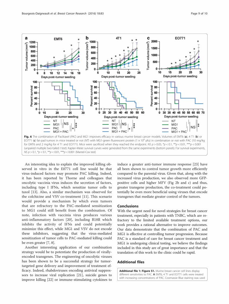
The treatment with MG1 and PAC demonstrates greaterefficiency in murine tumor modelsGiven the improved killing observed with PAC in allthree cell lines in vitro, the increased virus recoveryfrom tumors and the greater caspase-3-positive tumorregions observed in the presence of the co-treatment, wesought to determine if these phenomena would translateinto slower tumor growth and an improvement inthe survival of tumor-bearing animals. The mice weretreated with MG1, PAC or both and tumors weremeasured over time. As expected, we observed that thegrowth was slower following treatment with the combin-ation of the virus and the drug in all three tumor models(Fig. 6a, b and c, upper panels). Also, as observed in vitro,the E0771 model was the most sensitive to both treat-ments. Importantly, the enhanced control of tumorgrowth translated into a significant prolongation of sur-vival in all three models and some animals were evencured for the 4 T1 and E0771 tumor models (Fig. 6a, band c, bottom panels).
DiscussionIn this study, we demonstrated the compatibility of PAC,a standard-of-care chemotherapeutic agent for breastcancer, and MG1, an OV that is considered a promisingand novel strategy for treating disseminated diseases likebreast cancer. Our results not only show that treatmentsdo not interfere with one another, but they can also per-form even better when co-administered. Using three dif-ferent syngeneic murine breast cancer models, we showa prolongation of survival for animals that received bothtreatments compared to either treatment alone (Fig. 6).These findings have potential implications for the futuretreatment of patients. Our data support clinical testingof the combination. Even if the patient’s cancer has be-come resistant to the drug, it might still effectively en-hance MG1 and at the very least, should not impair theviral treatment. Second, because the beneficial effects weobserved were achieved using sub-lethal concentrationsof PAC and knowing the various side effects of the drugin patients with cancer, it is tempting to suggest thatusing a lower concentration of PAC in combination withthe virus would be a suitable strategy.Interestingly, we found that two out of the three mur-
ine tumor cell lines and the human tumor cell lines thatwe tested were sensitized to viral infection by PAC(Fig. 2). Indeed, while EMT6 and 4 T1 cells producedmore virus when pre-treated with the drug, the E0771
Fig. 4 Paclitaxel (PAC) enhances MG1 replication in tumors. EMT6 (a), 4 T1 (b) or EO771 (c) tumor-bearing mice were treated intratumorally with1 × 108 plaque-forming units (pfu) of MG1-green fluorescent protein (GFP) and intraperitoneally with PAC (10 mg/kg for the EMT6 model and2 mg/kg for the 4 T1 and EO771 models). Tumors were harvested 48 h later and the viral quantification was obtained by plaque assay. Three ormore tumors per condition were analysed. d EMT6 tumor-bearing mice were treated as in a and various organs were collected 48 h post treatment.The virus was quantified by plaque assay. e Tumor cores from human triple-negative breast cancer xenografts were infected ex-vivo with MG1-GFPwith or without PAC co-treatment. The viral outputs were quantified by plaque assay. Statistical significance was calculated using the unpairedtwo-tailed t test with Welch’s correction; *p < 0.05, **p < 0.01, ***p < 0.001. ND no drug
Bourgeois-Daigneault et al. Breast Cancer Research (2016) 18:83 Page 7 of 10

cell line was not affected. Our model is that PAC pre-treatment would block the secretion of antiviral factorslike IFNβ by infected cells (Additional file 2: Figure S2B),thereby increasing virus infection. In line with this idea,both EMT6 and 4 T1 cells in which we see sensitizationto the virus, but not the E0771 cells, which are refractoryto this effect, demonstrated impaired production of theantiviral cytokine (Additional file 2: Figure S2B). Import-antly, our results also show that while ex-vivo infectionand treatment of patient breast cancer xenografts caninform us on the potential enhancement of the virus bythe drug in specific patient samples (Fig. 4e), the lackof enhancement would not necessarily imply that bothtreatments would not be compatible. Indeed, increasedproduction of virus is desirable, but the killing of thetarget tumor cells is what ultimately matters. Our data
show that although the E0771 cell line does not demon-strate sensitization to the virus in the presence of PAC,complete killing of the cells was still observed at con-centrations of PAC and MG1 that did not affect cellviability alone. Notably, the E0771 tumor model wasthe one in which we observed the highest percentage ofcures with the treatment combination (Fig. 6c). This ismost likely due to the greater sensitivity of the E0771tumors to both single treatments alone compared tothe two other tumor models (Fig. 6). Remarkably, whilethe EMT6 and 4 T1 tumors are slightly smaller com-pared to the control animals when treated with eitherthe virus or the drug, the combination of both treat-ments was the only condition that was significantly dif-ferent in terms of the ability to control tumor growthand prolong survival.
Fig. 5 The co-treatment with paclitaxel (PAC) and MG1 increases tumor apoptosis. EMT6 tumor-bearing animals were treated intratumorally withMG1-green fluorescent protein (108 plaque-forming units) and intraperitoneally with 10 mg/kg with PAC, and tumor samples were harvested 48 hpost treatment. Sections were stained for the presence of virus and apoptotic cells (caspase-3)
Bourgeois-Daigneault et al. Breast Cancer Research (2016) 18:83 Page 8 of 10
An interesting idea to explain the improved killing ob-served in vitro in the E0771 cell line would be thatvirus-induced factors may promote PAC killing. Indeed,it has been reported by Thorne and colleagues thatoncolytic vaccinia virus induces the secretion of factors,including type I IFNs, which sensitize tumor cells totaxol [13]. Also, a similar mechanism was observed forthe colchicine and VSV co-treatment [11]. This scenariowould provide a mechanism by which even tumorsthat are refractory to the PAC-mediated sensitizationto MG1 could still benefit from the combination. Ofnote, infection with vaccinia virus produces variousanti-inflammatory factors [20], including B18R whichinhibits the activity of IFNs and could potentiallyminimize this effect, while MG1 and VSV do not encodethese inhibitors, suggesting that the virus-mediatedsensitization of tumor cells to PAC-mediated killing couldbe even greater [7, 8].Another interesting application of our combination
strategy would be to potentiate the production of virally-encoded transgenes. The engineering of oncolytic viruseshas been shown to be a successful strategy for tumor-targeted gene delivery and improvement of treatment ef-ficacy. Indeed, rhabdoviruses encoding antiviral suppres-sors to increase viral replication [21], suicide genes toimprove killing [22] or immune-stimulating cytokines to
induce a greater anti-tumor immune response [23] haveall been shown to control tumor growth more efficientlycompared to the parental virus. Given that, along with theincreased virus production, we also observed more GFP-positive cells and higher MFV (Fig 2b and c) and thus,greater transgene production, the co-treatment could po-tentially be even more beneficial using viruses that encodetransgenes that mediate greater control of the tumors.
ConclusionsWith the urgent need for novel strategies for breast cancertreatment, especially in patients with TNBC, which are re-fractory to the limited available treatment options, ourwork provides a rational alternative to improve outcomes.Our data demonstrate that the combination of PAC andMG1 is effective at controlling tumor progression. BecausePAC is a standard of care for breast cancer treatment andMG1 is undergoing clinical testing, we believe the findingsincluded in this study are of great importance and that thetranslation of this work to the clinic could be rapid.
Additional files
Additional file 1: Figure S1. Murine breast cancer cell lines displaydifferent sensitivities to PAC. A EMT6, 4 T1 and EO771 cells were treatedwith increasing concentrations of PAC. Coomassie Blue staining was used
Fig. 6 The combination of Paclitaxel (PAC) and MG1 improves efficacy in various murine breast cancer models. Volumes of EMT6 (a), 4 T1 (b) orEO771 (c) fat-pad tumors in mice treated or not (NT) with MG1-green fluorescent protein (1 × 10⁸ pfu) in combination or not with PAC (10 mg/kgfor EMT6 and 2 mg/kg for 4 T1 and EO771). Mice were sacrificed when they reached the endpoint. NS p > 0.05, *p < 0.1, **p < 0.01, ***p < 0.001(unpaired multiple two-tailed t test). Kaplan-Meier survival curves were generated from the same experiments (bottom panels). For survival experiments,NS p > 0.1, *p < 0.1, **p < 0.01, ***p < 0.001 (Mantel-Cox test)
Bourgeois-Daigneault et al. Breast Cancer Research (2016) 18:83 Page 9 of 10

to assess the toxic dose of the drug in the different cell lines after 72 h.B Fluorescent microscopy pictures of DAPI-stained cells with or withouttreatment with 0.5uM PAC for 72 h. (TIF 14849 kb)
Additional file 2: Figure S2. PAC blocks IFNβ production by infectedtumor cells. A Microscopy pictures of EMT6, 4 T1 and EO771 tumor cellspre-treated or not for 4 h with recombinant IFNβ. B The IFNβ releasedfrom MG1-infected cells in the presence or absence of PAC was quantifiedby ELISA. Samples were analysed in triplicates and statistical significancewas calculated using the unpaired two-tailed t test with Welch’s correction;*p < 0.05, **p < 0.01, ***p < 0.001. (TIF 6796 kb)
Additional file 3: Figure S3. IFNβ pre-treatment protects EMT6, 4 T1and EO771 cells from virus-mediated killing. Coomassie Blue staining ofthe various breast cancer cell lines 48 h post infection with MG1-GFP withor without pre-treatment with recombinant murine IFNβ. (TIF 2149 kb)
Additional file 4: Figure S4. PAC and MG1 synergistically kill breastcancer cell lines. Quantification of the signal obtained for triplicates orquadruplicates Coomassie Blue staining of EMT6, 4 T1 or EO771 cellsinfected or not with MG1-GFP and co-treated with 2 uM PAC for 48 hfrom Fig. 3. Statistical significance was calculated using the unpairedtwo-tailed t test with Welch’s correction; *p < 0.05, **p < 0.01, ***p < 0.001.(TIF 2942 kb)
AbbreviationsDAPI, 4',6-diamidino-2-phenylindole; DMEM, Dulbecco’s modified Eagle’smedium; ELISA, enzyme-linked immunosorbent assay; FBS, fetal bovineserum; GFP, green fluorescent protein; IFN, interferon; IP, intrperitoneally; IT,intratumorally; MFV, mean fluorescence value; MOI, multiplicity of infection;OV, oncolytic virus; PAC, paclitaxel; PBS, phosphate-buffered saline; pfu,plaque-forming units;TNBC, triple-negative breast cancer; VSe, virus sensitizers;VSV, vesicular stomatitis virus
AcknowledgementsWe would like to thank Dr. Alana Welm for kindly providing the patient-derivedTNBC xenograft samples.
FundingJCB is supported by the Terry Fox Foundation (TFF), the Canadian CancerSociety Research Institute (CCSRI), the Ontario Institute for Cancer Research(OICR), the Canadian Institute for Health Research (CIHR), the OttawaRegional Cancer Foundation and the Ottawa Hospital Foundation. MCBD,DGR and VG were supported by fellowships and scholarships from the CIHR.JSD was supported by the TFF, the CIHR and the CCSRI.
Authors’ contributionsMCBD designed this study, performed all experiments and wrote themanuscript. DGR participated in the in vivo efficacy experiments and helpeddraft and revise the manuscript. LESG helped with virus quantification andviability staining. AP performed statistical analysis, helped with virusquantification and revised the manuscript. RA helped with the design of thestudy and the revision of the manuscript. TF performed the tumorimplantations. VG helped with the acquisition of microscopy images. ASAhelped with the revisions to the manuscript. JSD was involved in the designof the study and JCB helped with the design of the study and the revisionof the manuscript. All authors read and approved the final manuscript.
Competing interestsThe authors declare that they have no competing interests.
Ethics approval and consent to participateAll experiments were performed in accordance with the University of Ottawaanimal care and veterinary services guidelines (reference number ME-2258).
Received: 12 April 2016 Accepted: 25 July 2016
References1. Weigelt B, Peterse JL, van ’t Veer LJ. Breast cancer metastasis: markers and
models. Nat Rev Cancer. 2005;5:591–602.
2. Anders C, Carey LA. Understanding and treating triple-negative breast cancer.Oncology (Williston Park). 2008;22:1233–9.
3. Kaur P, Nagaraja GM, Zheng H, Gizachew D, Galukande M, Krishnan S, et al.A mouse model for triple-negative breast cancer tumor-initiating cells(TNBC-TICs) exhibits similar aggressive phenotype to the human disease.BMC Cancer. 2012;12:120.
4. Arnal I, Wade RH. How does taxol stabilize microtubules? Curr Biol. 1995;5:900–8.
5. Mahamodhossen YA, Liu W, Rong-Rong Z. Triple-negative breast cancer: newperspectives for novel therapies. Med Oncol. 2013;30:653.
6. Zeyaullah M, Patro M, Ahmad I, Ibraheem K, Sultan P, Nehal M, et al.Oncolytic viruses in the treatment of cancer: a review of current strategies.Pathol Oncol Res. 2012;18:771–81.
7. Stojdl DF, Lichty B, Knowles S, Marius R, Atkins H, Sonenberg N, et al.Exploiting tumor-specific defects in the interferon pathway with apreviously unknown oncolytic virus. Nat Med. 2000;6:821–5.
8. Brun J, McManus D, Lefebvre C, Hu K, Falls T, Atkins H, et al.Identification of genetically modified Maraba virus as an oncolyticrhabdovirus. Mol Ther. 2010;18:1440–9.
9. Stojdl DF, Lichty BD, tenOever BR, Paterson JM, Power AT, Knowles S, et al.VSV strains with defects in their ability to shutdown innate immunity arepotent systemic anti-cancer agents. Cancer Cell. 2003;4:263–75.
10. Diallo J-S, Le Boeuf F, Lai F, Cox J, Vaha-Koskela M, Abdelbary H, et al. Ahigh-throughput pharmacoviral approach identifies novel oncolytic virussensitizers. Mol Ther. 2010;18:1123–9.
11. Arulanandam R, Batenchuk C, Varette O, Zakaria C, Garcia V, Forbes NE, et al.Microtubule disruption synergizes with oncolytic virotherapy by inhibitinginterferon translation and potentiating bystander killing. Nat Commun. 015;6:6410.
12. Margolis RL, Wilson L. Addition of colchicine–tubulin complex tomicrotubule ends: the mechanism of substoichiometric colchicinepoisoning. Proc Natl Acad Sci USA. 1977;74:3466–70.
13. Huang B, Sikorski R, Kirn DH, Thorne SH. Synergistic anti-tumor effectsbetween oncolytic vaccinia virus and paclitaxel are mediated by the IFNresponse and HMGB1. Gene Ther. 2011;18:164–72.
14. Lin S-F, Gao SP, Price DL, Li S, Chou T-C, Singh P, et al. Synergy of a herpesoncolytic virus and paclitaxel for anaplastic thyroid cancer. Clin CancerRes. 2008;14:1519–28.
15. Roy DG, Power AT, Bourgeois-Daigneault MC, Falls T, Ferreira L, Stern A, etal. Programmable insect cell carriers for systemic delivery of integratedcancer biotherapy. J Control Release. 2015;220:210–21.
16. Breitbach CJ, De Silva NS, Falls TJ, Aladl U, Evgin L, Paterson J, et al. Targetingtumor vasculature with an oncolytic virus. Mol Ther. 2011;19:886–94.
17. DeRose YS, Wang G, Lin Y-C, Bernard PS, Buys SS, Ebbert MTW, et al. Tumorgrafts derived from women with breast cancer authentically reflect tumorpathology, growth, metastasis and disease outcomes. Nat Med. 2011;17:1514–20.
18. Falls T, Roy DG, Bell JC, Bourgeois-Daigneault MC. Murine tumor models foroncolytic rhabdo-virotherapy. ILAR J. 2016;57(1):73–85.
19. Diallo J-S, Roy D, Abdelbary H, De Silva N, Bell JC. Ex vivo infection of livetissue with oncolytic viruses. J Vis Exp. 2011;e2854.
20. Seet BT, Johnston JB, Brunetti CR, Barrett JW, Everett H, Cameron C, et al.Poxviruses and immune evasion. Annu Rev Immunol. 2003;21:377–423.
21. Le Boeuf F, Batenchuk C, Vähä-Koskela M, Breton S, Roy D, Lemay C, et al.Model-based rational design of an oncolytic virus with improvedtherapeutic potential. Nat Commun. 2013;4:1974.
22. Fernandez M, Porosnicu M, Markovic D, Barber GN. Genetically engineeredvesicular stomatitis virus in gene therapy: application for treatment ofmalignant disease. J Virol. 2002;76:895–904.
23. Bourgeois-Daigneault M-C, Roy DG, Falls T, Twumasi-Boateng K,St-Germain LE, Marguerie M, et al. Oncolytic vesicular stomatitis virusexpressing interferon-γ has enhanced therapeutic activity. MolTher–Oncolytics. 2016;3:16001.
Bourgeois-Daigneault et al. Breast Cancer Research (2016) 18:83 Page 10 of 10
Copyright © 2022 FDOKUMEN