Regulators of Mitotic Arrest and Ceramide Metabolism Are Determinants of Sensitivity to Paclitaxel...
15
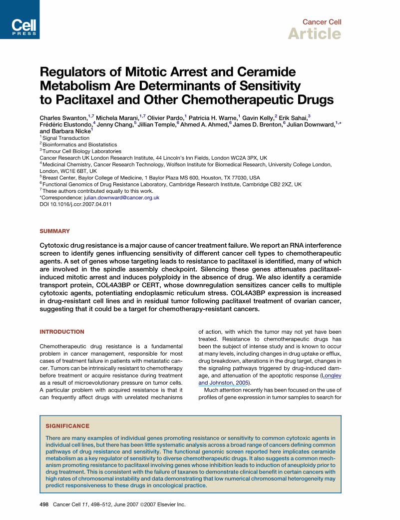
Cancer Cell Article Regulators of Mitotic Arrest and Ceramide Metabolism Are Determinants of Sensitivity to Paclitaxel and Other Chemotherapeutic Drugs Charles Swanton, 1,7 Michela Marani, 1,7 Olivier Pardo, 1 Patricia H. Warne, 1 Gavin Kelly, 2 Erik Sahai, 3 Fre ´ de ´ ric Elustondo, 4 Jenny Chang, 5 Jillian Temple, 6 Ahmed A. Ahmed, 6 James D. Brenton, 6 Julian Downward, 1, * and Barbara Nicke 1 1 Signal Transduction 2 Bioinformatics and Biostatistics 3 Tumour Cell Biology Laboratories Cancer Research UK London Research Institute, 44 Lincoln’s Inn Fields, London WC2A 3PX, UK 4 Medicinal Chemistry, Cancer Research Technology, Wolfson Institute for Biomedical Research, University College London, London, WC1E 6BT, UK 5 Breast Center, Baylor College of Medicine, 1 Baylor Plaza MS 600, Houston, TX 77030, USA 6 Functional Genomics of Drug Resistance Laboratory, Cambridge Research Institute, Cambridge CB2 2XZ, UK 7 These authors contributed equally to this work. *Correspondence: [email protected] DOI 10.1016/j.ccr.2007.04.011 SUMMARY Cytotoxic drug resistance is a major cause of cancer treatment failure. We report an RNA interference screen to identify genes influencing sensitivity of different cancer cell types to chemotherapeutic agents. A set of genes whose targeting leads to resistance to paclitaxel is identified, many of which are involved in the spindle assembly checkpoint. Silencing these genes attenuates paclitaxel- induced mitotic arrest and induces polyploidy in the absence of drug. We also identify a ceramide transport protein, COL4A3BP or CERT, whose downregulation sensitizes cancer cells to multiple cytotoxic agents, potentiating endoplasmic reticulum stress. COL4A3BP expression is increased in drug-resistant cell lines and in residual tumor following paclitaxel treatment of ovarian cancer, suggesting that it could be a target for chemotherapy-resistant cancers. INTRODUCTION Chemotherapeutic drug resistance is a fundamental problem in cancer management, responsible for most cases of treatment failure in patients with metastatic can- cer. Tumors can be intrinsically resistant to chemotherapy before treatment or acquire resistance during treatment as a result of microevolutionary pressure on tumor cells. A particular problem with acquired resistance is that it can frequently affect drugs with unrelated mechanisms of action, with which the tumor may not yet have been treated. Resistance to chemotherapeutic drugs has been the subject of intense study and is known to occur at many levels, including changes in drug uptake or efflux, drug breakdown, alterations in the drug target, changes in the signaling pathways triggered by drug-induced dam- age, and attenuation of the apoptotic response (Longley and Johnston, 2005). Much attention recently has been focused on the use of profiles of gene expression in tumor samples to search for SIGNIFICANCE There are many examples of individual genes promoting resistance or sensitivity to common cytotoxic agents in individual cell lines, but there has been little systematic analysis across a broad range of cancers defining common pathways of drug resistance and sensitivity. The functional genomic screen reported here implicates ceramide metabolism as a key regulator of sensitivity to diverse chemotherapeutic drugs. It also suggests a common mech- anism promoting resistance to paclitaxel involving genes whose inhibition leads to induction of aneuploidy prior to drug treatment. This is consistent with the failure of taxanes to demonstrate clinical benefit in certain cancers with high rates of chromosomal instability and data demonstrating that low numerical chromosomal heterogeneity may predict responsiveness to these drugs in oncological practice. 498 Cancer Cell 11, 498–512, June 2007 ª2007 Elsevier Inc.
-
Upload
independent -
Category
Documents
-
view
0 -
download
0
Transcript of Regulators of Mitotic Arrest and Ceramide Metabolism Are Determinants of Sensitivity to Paclitaxel...
Cancer Cell
Article
Regulators of Mitotic Arrest and CeramideMetabolism Are Determinants of Sensitivityto Paclitaxel and Other Chemotherapeutic DrugsCharles Swanton,1,7 Michela Marani,1,7 Olivier Pardo,1 Patricia H. Warne,1 Gavin Kelly,2 Erik Sahai,3
Frederic Elustondo,4 Jenny Chang,5 Jillian Temple,6 Ahmed A. Ahmed,6 James D. Brenton,6 Julian Downward,1,*and Barbara Nicke1
1 Signal Transduction2 Bioinformatics and Biostatistics3 Tumour Cell Biology Laboratories
Cancer Research UK London Research Institute, 44 Lincoln’s Inn Fields, London WC2A 3PX, UK4 Medicinal Chemistry, Cancer Research Technology, Wolfson Institute for Biomedical Research, University College London,London, WC1E 6BT, UK5 Breast Center, Baylor College of Medicine, 1 Baylor Plaza MS 600, Houston, TX 77030, USA6 Functional Genomics of Drug Resistance Laboratory, Cambridge Research Institute, Cambridge CB2 2XZ, UK7 These authors contributed equally to this work.*Correspondence: [email protected]
DOI 10.1016/j.ccr.2007.04.011
SUMMARY
Cytotoxic drug resistance is a major cause of cancer treatment failure. We report an RNA interferencescreen to identify genes influencing sensitivity of different cancer cell types to chemotherapeuticagents. A set of genes whose targeting leads to resistance to paclitaxel is identified, many of whichare involved in the spindle assembly checkpoint. Silencing these genes attenuates paclitaxel-induced mitotic arrest and induces polyploidy in the absence of drug. We also identify a ceramidetransport protein, COL4A3BP or CERT, whose downregulation sensitizes cancer cells to multiplecytotoxic agents, potentiating endoplasmic reticulum stress. COL4A3BP expression is increasedin drug-resistant cell lines and in residual tumor following paclitaxel treatment of ovarian cancer,suggesting that it could be a target for chemotherapy-resistant cancers.
INTRODUCTION
Chemotherapeutic drug resistance is a fundamental
problem in cancer management, responsible for most
cases of treatment failure in patients with metastatic can-
cer. Tumors can be intrinsically resistant to chemotherapy
before treatment or acquire resistance during treatment
as a result of microevolutionary pressure on tumor cells.
A particular problem with acquired resistance is that it
can frequently affect drugs with unrelated mechanisms
498 Cancer Cell 11, 498–512, June 2007 ª2007 Elsevier Inc.
of action, with which the tumor may not yet have been
treated. Resistance to chemotherapeutic drugs has
been the subject of intense study and is known to occur
at many levels, including changes in drug uptake or efflux,
drug breakdown, alterations in the drug target, changes in
the signaling pathways triggered by drug-induced dam-
age, and attenuation of the apoptotic response (Longley
and Johnston, 2005).
Much attention recently has been focused on the use of
profiles of gene expression in tumor samples to search for
SIGNIFICANCE
There are many examples of individual genes promoting resistance or sensitivity to common cytotoxic agents inindividual cell lines, but there has been little systematic analysis across a broad range of cancers defining commonpathways of drug resistance and sensitivity. The functional genomic screen reported here implicates ceramidemetabolism as a key regulator of sensitivity to diverse chemotherapeutic drugs. It also suggests a common mech-anism promoting resistance to paclitaxel involving genes whose inhibition leads to induction of aneuploidy prior todrug treatment. This is consistent with the failure of taxanes to demonstrate clinical benefit in certain cancers withhigh rates of chromosomal instability and data demonstrating that low numerical chromosomal heterogeneity maypredict responsiveness to these drugs in oncological practice.

Cancer Cell
Determinants of Chemotherapeutic Drug Sensitivity
correlations between expression of specific genes and
acquisition of drug resistance in the clinic. This approach
has been used to predict the outcome of therapy but may
also provide indirect mechanistic insights into how drug
resistance arises (Alaoui-Jamali et al., 2004; Lonning
et al., 2005). However, obtaining sufficient numbers of
high-quality tumor samples, whose drug sensitivity is
known, in order to make strong correlations with expres-
sion of particular genes has proved challenging. It may
be that far greater numbers of tumor samples need to
be analyzed than previously assumed (Ein-Dor et al.,
2006). One way around this has been to analyze gene
expression in large numbers of cancer cell lines with
known drug sensitivity (Potti et al., 2006). An alternative
approach is taken here to assess more directly the con-
tribution of over 800 candidate proteins to the sensitivity
or resistance of cancer cells to a number of common
chemotherapeutic drugs.
The ability to use RNA interference as a tool for gene
silencing in mammalian cells has opened up the possibility
of carrying out high-throughput loss-of-function screens
in tissue culture systems (MacKeigan et al., 2005). Here
we utilize a collection of synthetic siRNA oligonucleotide
pools targeting the expression of all protein kinases,
plus associated proteins and proteins involved in ceram-
ide lipid metabolism, to address whether loss of these
proteins impacts on drug sensitivity in vitro. The screen
has been carried out in three commonly studied tumor
cell lines with mutations in the KRAS oncogene and has
employed four different chemotherapeutic drugs. We
report that targeting COL4A3BP caused the most striking
multidrug sensitization. This gene encodes a ceramide-
binding protein, CERT, that is involved in transport of the
proapoptotic lipid ceramide from its site of de novo syn-
thesis in the endoplasmic reticulum to the Golgi, where it
is metabolized to sphingomyelin (Hanada et al., 2003).
We define a set of drug resistance genes that, when
silenced, cause resistance to paclitaxel in multiple cell
lines, many of which are involved in the spindle assembly
checkpoint. These share the ability not only to impair
paclitaxel-induced mitotic arrest, but also to induce poly-
ploidy in the absence of drug exposure. These data
suggest that taxanes may lack efficacy in tumors with
high levels of chromosomal instability (CIN) characterized
by chromosomal numerical heterogeneity and provide
a rational basis for identification of patients likely to benefit
from these drugs.
RESULTS
Kinases Affecting Cell Viability in the Absence
of Drug Treatment
HCT-116 (HCT) colon carcinoma, MDA-MB-231 (MDA)
breast adenocarcinoma, and A549 non-small-cell lung
carcinoma cells were transfected with 779 synthetic
siRNA pools targeting all human protein kinases, plus
a number of kinase-associated and kinase-related
proteins. Transfections were performed in duplicate for
each of three conditions per cell line: vehicle-treated
control (DMSO), paclitaxel-treated, and a custom drug
treatment that was cell type specific (5-FU for HCT-116,
doxorubicin for MDA-MB-231, and cisplatin for A549).
Cells were transfected, followed 48 hr later with drug treat-
ment for a further 48 hr, before cell viability was measured
using the CellTiter-Glo assay. A concentration of drug was
used that allowed identification of both siRNAs protecting
cells and those giving enhanced killing in the same screen.
To assess viability effects induced by the siRNAs in the
absence of drug, we analyzed data obtained from DMSO-
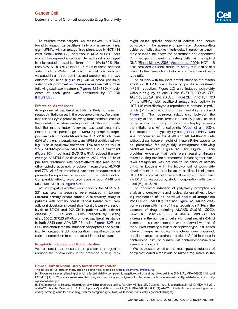
treated transfections. 14 genes were identified whose
knockdown significantly decreased cell number com-
pared to scrambled control in at least two different cell
lines (Figure 1A). Five siRNAs promoted an increase in
viability in at least two different cell lines. A heatmap
(Figure S1A in the Supplemental Data available with this
article online) shows all siRNAs with a significant effect
on cell viability detected in this screen. Those siRNAs
with a statistically significant reduction in cell viability
>25% averaged across all three cell lines are listed in
graphical format in Figure S1B.
Four of the five top viability targets, CNKSR1 (the adap-
tor protein CNK required for Ras signaling), FN3KRP,
EPHA6, and COPB2 (Figure S1B), were readily validated,
with at least two of the four siRNAs in each pool promoting
a significant reduction in viability in at least two cell lines
(Figure S1C). The Vi-CELL trypan blue exclusion assay
(Beckman Coulter) confirmed that gene silencing reduced
cell viability by more than 20% (Figure S1D). Data from the
gene expression database Oncomine (http://www.
oncomine.org/) indicated that, of these genes, COPB2
appeared to be significantly upregulated in various tumors
compared to normal tissues (Figures S1E and S1F), raising
the possibility that it could be a tumor-selective target.
Kinases Involved in Resistance
to Paclitaxel Treatment
To identify proteins whose knockdown promoted a drug-
sensitizing or -antagonistic phenotype, we analyzed data
obtained after drug treatment, utilizing the sensitivity index
(SI) equation. Figure 1B shows all siRNAs that alter drug
action in at least one of the three cell lines. siRNAs against
BUB1B and CDADC1 can be readily identified as antago-
nizing the paclitaxel response in all cell lines tested.
Forty-three siRNAs with statistically significant pacli-
taxel antagonistic activity in two or more cell lines were
identified (Table S1). These genes were categorized
(Figure 2A) using gene ontology databases, available liter-
ature, and published RNAi screens in Drosophila (FLIGHT
database, http://www.flight.licr.org/) and C. elegans (Bet-
tencourt-Dias et al., 2004; Moffat et al., 2006; Whitfield
et al., 2002). Genes with mitotic related function formed
the most frequent group. Homologs of ten of these genes
displayed mitotic phenotypes when knocked down in
Drosophila, while overall 18/43 (42%) of these targeted
genes have been associated with functions at the G2/M
transition (Bettencourt-Dias et al., 2004; Eggert et al.,
2004; Moffat et al., 2006).
Cancer Cell 11, 498–512, June 2007 ª2007 Elsevier Inc. 499
500 C
Cancer Cell
Determinants of Chemotherapeutic Drug Sensitivity
ancer Cell 11, 498–512, June 2007 ª2007 Elsevier Inc.
Cancer Cell
Determinants of Chemotherapeutic Drug Sensitivity
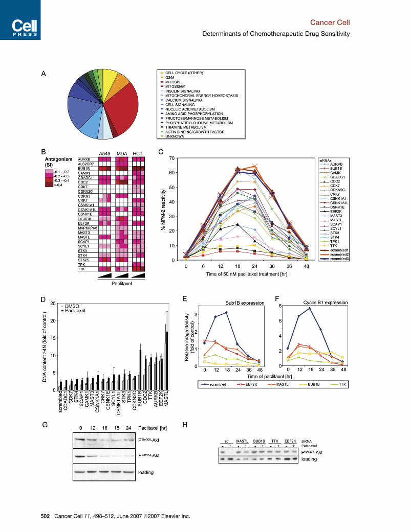
To validate these targets, we reassayed 16 siRNAs
found to antagonize paclitaxel in two or more cell lines,
eight siRNAs with an antagonistic phenotype in HCT-116
cells alone (Table S2), and two in MDA-MB-231 cells
alone. The degree of antagonism to paclitaxel is portrayed
in color-coded or graphical format from 10% to 50% (Fig-
ures S2A–S2D). We validated 25 of 26 of these selected
antagonistic siRNAs in at least one cell line, with ten
validated in all three cell lines and another eight in two
different cell lines (Figure 2B). All validated paclitaxel
antagonists promoted an increase in relative cell number
following paclitaxel treatment (Figures S2B–S2D). Knock-
down of each gene was confirmed by RT-PCR
(Figure S2E).
Effects on Mitotic Index
Antagonism of paclitaxel activity is likely to result in
reduced mitotic arrest in the presence of drug. We exam-
ined the cell-cycle profile following transfection of each of
the validated paclitaxel-antagonistic siRNAs and quanti-
fied the mitotic index following paclitaxel treatment,
defined as the percentage of MPM-2-phosphoepitope-
positive cells. In control-transfected HCT-116 cells, over
60% of the entire population were MPM-2 positive follow-
ing 18 hr of paclitaxel treatment. This compared to just
3.5% MPM-2-positive cells following DMSO treatment
(Figure 2C). In contrast, BUB1B siRNA reduced the per-
centage of MPM-2-positive cells to <5% after 18 hr of
paclitaxel treatment, with potent effects also seen for the
other spindle assembly checkpoint regulators, AURKB
and TTK. All of the remaining paclitaxel antagonists also
promoted a reproducible reduction in the mitotic index.
Comparable effects were also seen in both A549 and
MDA-MB-231 cells (Figure S2F).
We investigated whether expression of the MDA-MB-
231 paclitaxel antagonists were reduced in taxane-
resistant primary breast cancer. A microarray study of
patients with primary breast cancer treated with neo-
adjuvant docetaxel showed significantly lower expression
levels of STK25 and DGUOK in patients with resistant
disease (p = 0.02 and 0.00027, respectively) (Chang
et al., 2003). STK25 siRNA promoted paclitaxel resistance
in both A549 and MDA-MB-231 cells (Figures S2B and
S2C) and attenuated the induction of apoptosis and signif-
icantly increased BrdU incorporation in paclitaxel-treated
cells in comparison to control cells (data not shown).
Polyploidy Induction and Multinucleation
We reasoned that, since all the paclitaxel antagonists
reduced the mitotic index in the presence of drug, they
might cause spindle checkpoint defects and induce
polyploidy in the absence of paclitaxel. Accumulating
evidence implies that the mitotic delay in response to spin-
dle disruption influences the postmitotic p53-dependent
G1 checkpoint, thereby arresting cells with tetraploid
DNA (Blagosklonny, 2006; Vogel et al., 2004). HCT-116
cells provided an ideal model to study this relationship
owing to their near-diploid status and retention of wild-
type p53.
The siRNAs with the most potent effect on the mitotic
arrest in HCT-116 cells following paclitaxel treatment
(>75% reduction; Figure 2C) also induced polyploidy
without drug by at least 4-fold (BUB1B, CDC2, TTK,
AURKB, EEF2K, and MASTL; Figure 2D). In total, 17/20
of the siRNAs with paclitaxel antagonistic activity in
HCT-116 cells displayed a reproducible increase in poly-
ploidy (>1.5-fold) without drug treatment (Figure 2D and
Figure 3). The reciprocal relationship between the
potency of the mitotic arrest induced by paclitaxel and
polyploidy without drug supports the interplay between
the mitotic and G1 checkpoints (Vogel et al., 2004).
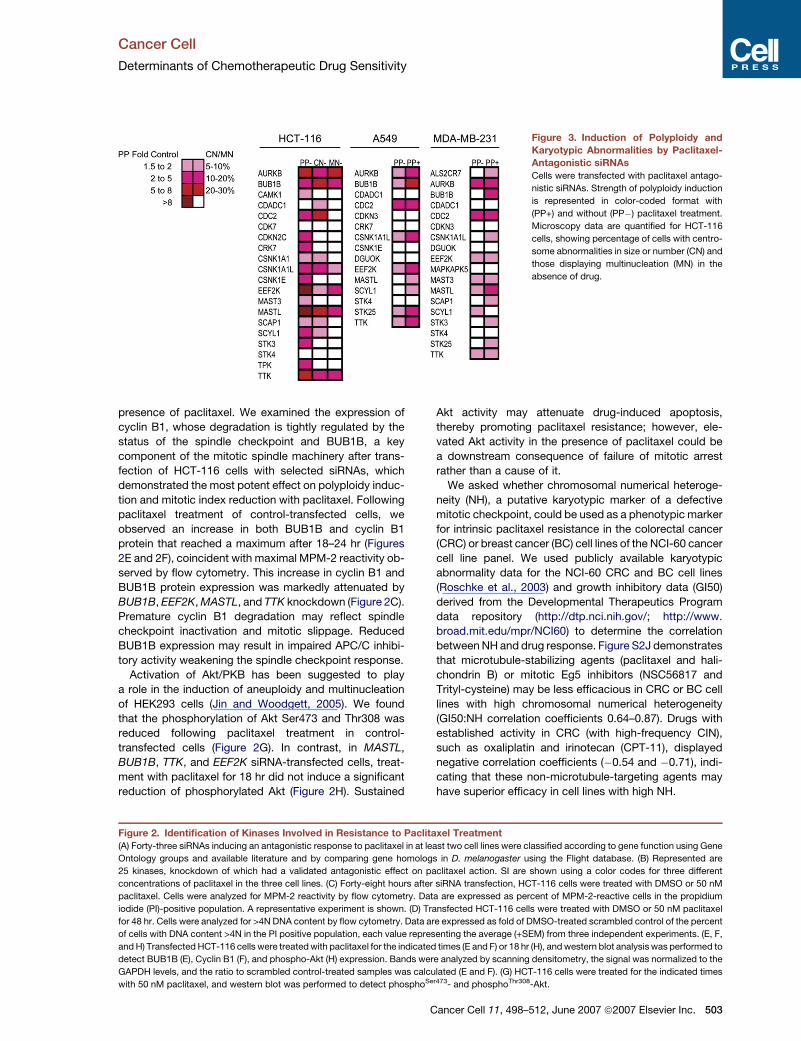
The induction of polyploidy by antagonistic siRNAs was
less pronounced in the A549 and MDA-MB-231 cells
without drug; however, eight of these siRNAs appear to
be permissive for polyploidy development following
paclitaxel treatment (Figure S2G and Figure 3). This
provides evidence that cells were passing through
mitosis during paclitaxel treatment, indicating that pacli-
taxel antagonism was not due to inhibition of mitosis
entry. In keeping with the involvement of polyploidy
development in the acquisition of paclitaxel resistance,
HCT-116 polyploid cells were still capable of synthesiz-
ing DNA as assessed by BrdU incorporation with pacli-
taxel (Figure S2H).
The observed induction of polyploidy prompted an
analysis of centrosome and nuclear abnormalities follow-
ing transfection of the individual paclitaxel antagonists
into HCT-116 cells (Figure 3 and Figure S2I). Multinuclea-
tion was seen with many of the antagonistic siRNAs in the
absence of drug, including AURKB, BUB1B, CDC2,
CSNK1A1, CSNK1A1L, EEF2K, MASTL, and TTK. An
increase in the number of cells with giant nuclei (>2-fold
increase in nuclear diameter) was observed with all of
the siRNAs inducing a multinuclear phenotype. In all cases
where changes in nuclear phenotype were observed,
parallel changes in centrosome size (>2-fold increase in
centrosomal size) or number (>3 centrosomes/nucleus)
were also apparent.
We addressed whether the most potent inducers of
polyploidy could alter levels of mitotic regulators in the
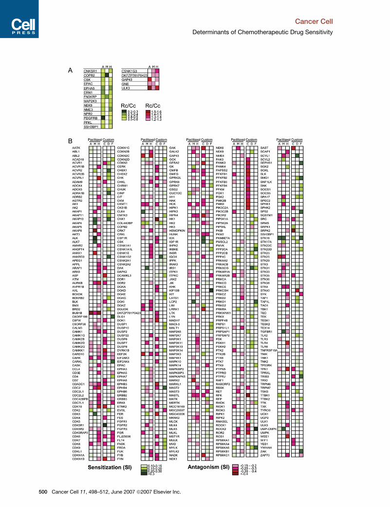
Figure 1. Human Kinome Library Screen Primary Analysis
The screen set-up, data analysis, and hit selection are described in the Experimental Procedures.
(A) Shown are kinases, silencing of which affected viability compared to negative control in at least two cell lines (A549 [A], MDA-MB-231 [M], and
HCT-116 [H]). Rc/Cc values are represented using a color-coding format (greens for decreased, reds for increased viability; white for no statistically
significant changes).
(B) Figure represents kinases, knockdown of which altered drug activity (sensitivity index [SI]). Columns 1 to 3: SI to paclitaxel in A549, MDA-MB-231,
and HCT-116 cells. Columns 4 to 6: SI to cisplatin (C) in A549, doxorubicin (D) in MDA-MB-231, 5-FU (F) in HCT-116 cells. SI are shown using a color-
coding format (greens for sensitizing, reds for antagonizing siRNAs; white for no statistically significant changes).
Cancer Cell 11, 498–512, June 2007 ª2007 Elsevier Inc. 501
Cancer Cell
Determinants of Chemotherapeutic Drug Sensitivity
502 Cancer Cell 11, 498–512, June 2007 ª2007 Elsevier Inc.
Cancer Cell
Determinants of Chemotherapeutic Drug Sensitivity
Figure 3. Induction of Polyploidy and
Karyotypic Abnormalities by Paclitaxel-
Antagonistic siRNAs
Cells were transfected with paclitaxel antago-
nistic siRNAs. Strength of polyploidy induction
is represented in color-coded format with
(PP+) and without (PP�) paclitaxel treatment.
Microscopy data are quantified for HCT-116
cells, showing percentage of cells with centro-
some abnormalities in size or number (CN) and
those displaying multinucleation (MN) in the
absence of drug.
presence of paclitaxel. We examined the expression of
cyclin B1, whose degradation is tightly regulated by the
status of the spindle checkpoint and BUB1B, a key
component of the mitotic spindle machinery after trans-
fection of HCT-116 cells with selected siRNAs, which
demonstrated the most potent effect on polyploidy induc-
tion and mitotic index reduction with paclitaxel. Following
paclitaxel treatment of control-transfected cells, we
observed an increase in both BUB1B and cyclin B1
protein that reached a maximum after 18–24 hr (Figures
2E and 2F), coincident with maximal MPM-2 reactivity ob-
served by flow cytometry. This increase in cyclin B1 and
BUB1B protein expression was markedly attenuated by
BUB1B, EEF2K, MASTL, and TTK knockdown (Figure 2C).
Premature cyclin B1 degradation may reflect spindle
checkpoint inactivation and mitotic slippage. Reduced
BUB1B expression may result in impaired APC/C inhibi-
tory activity weakening the spindle checkpoint response.
Activation of Akt/PKB has been suggested to play
a role in the induction of aneuploidy and multinucleation
of HEK293 cells (Jin and Woodgett, 2005). We found
that the phosphorylation of Akt Ser473 and Thr308 was
reduced following paclitaxel treatment in control-
transfected cells (Figure 2G). In contrast, in MASTL,
BUB1B, TTK, and EEF2K siRNA-transfected cells, treat-
ment with paclitaxel for 18 hr did not induce a significant
reduction of phosphorylated Akt (Figure 2H). Sustained
Akt activity may attenuate drug-induced apoptosis,
thereby promoting paclitaxel resistance; however, ele-
vated Akt activity in the presence of paclitaxel could be
a downstream consequence of failure of mitotic arrest
rather than a cause of it.
We asked whether chromosomal numerical heteroge-
neity (NH), a putative karyotypic marker of a defective
mitotic checkpoint, could be used as a phenotypic marker
for intrinsic paclitaxel resistance in the colorectal cancer
(CRC) or breast cancer (BC) cell lines of the NCI-60 cancer
cell line panel. We used publicly available karyotypic
abnormality data for the NCI-60 CRC and BC cell lines
(Roschke et al., 2003) and growth inhibitory data (GI50)
derived from the Developmental Therapeutics Program
data repository (http://dtp.nci.nih.gov/; http://www.
broad.mit.edu/mpr/NCI60) to determine the correlation
between NH and drug response. Figure S2J demonstrates
that microtubule-stabilizing agents (paclitaxel and hali-
chondrin B) or mitotic Eg5 inhibitors (NSC56817 and
Trityl-cysteine) may be less efficacious in CRC or BC cell
lines with high chromosomal numerical heterogeneity
(GI50:NH correlation coefficients 0.64–0.87). Drugs with
established activity in CRC (with high-frequency CIN),
such as oxaliplatin and irinotecan (CPT-11), displayed
negative correlation coefficients (�0.54 and �0.71), indi-
cating that these non-microtubule-targeting agents may
have superior efficacy in cell lines with high NH.
Figure 2. Identification of Kinases Involved in Resistance to Paclitaxel Treatment
(A) Forty-three siRNAs inducing an antagonistic response to paclitaxel in at least two cell lines were classified according to gene function using Gene
Ontology groups and available literature and by comparing gene homologs in D. melanogaster using the Flight database. (B) Represented are
25 kinases, knockdown of which had a validated antagonistic effect on paclitaxel action. SI are shown using a color codes for three different
concentrations of paclitaxel in the three cell lines. (C) Forty-eight hours after siRNA transfection, HCT-116 cells were treated with DMSO or 50 nM
paclitaxel. Cells were analyzed for MPM-2 reactivity by flow cytometry. Data are expressed as percent of MPM-2-reactive cells in the propidium
iodide (PI)-positive population. A representative experiment is shown. (D) Transfected HCT-116 cells were treated with DMSO or 50 nM paclitaxel
for 48 hr. Cells were analyzed for >4N DNA content by flow cytometry. Data are expressed as fold of DMSO-treated scrambled control of the percent
of cells with DNA content >4N in the PI positive population, each value representing the average (+SEM) from three independent experiments. (E, F,
and H) Transfected HCT-116 cells were treated with paclitaxel for the indicated times (E and F) or 18 hr (H), and western blot analysis was performed to
detect BUB1B (E), Cyclin B1 (F), and phospho-Akt (H) expression. Bands were analyzed by scanning densitometry, the signal was normalized to the
GAPDH levels, and the ratio to scrambled control-treated samples was calculated (E and F). (G) HCT-116 cells were treated for the indicated times
with 50 nM paclitaxel, and western blot was performed to detect phosphoSer473- and phosphoThr308-Akt.
Cancer Cell 11, 498–512, June 2007 ª2007 Elsevier Inc. 503
Cancer Cell
Determinants of Chemotherapeutic Drug Sensitivity
504 Cancer Cell 11, 498–512, June 2007 ª2007 Elsevier Inc.
Cancer Cell
Determinants of Chemotherapeutic Drug Sensitivity
In the management of metastatic breast cancer, the
combination of trastuzumab (Herceptin) and paclitaxel
increases the clinical benefit compared to paclitaxel alone
(Slamon et al., 2001). We asked whether this clinical
benefit might be attributable to downregulation of genes
implicated in CIN following HER2 blockade. We compared
the 70 gene microarray signature data set associated with
CIN (Carter et al., 2006) with an in vitro data set of 16 genes
repressed by anti-HER2 antibody (Le et al., 2005) and
found 8 of these 16 genes to be represented in the CIN
signature set (p = 2.3 3 10�16: probability of observing 8
or more genes in the top 70 [of 10,151 genes] when tested
using the hypergeometric test based on the 16 [out of
12,558] differential genes on the U95A version 2 chip).
In summary, while all of the validated paclitaxel-
antagonistic siRNAs reduced the proportion of cells
arrested in mitosis in response to paclitaxel treatment,
most of them also induced polyploidy, multinucleation,
and abnormalities of centrosome size or number in the
absence of drug. This suggests a possible mitotic regula-
tory role for these genes and an association between CIN
and paclitaxel resistance. Supporting a role for polyploidy
in the acquisition of paclitaxel resistance, polyploid cells
continued to synthesize DNA in the presence of paclitaxel,
and increasing chromosomal numerical heterogeneity
correlated with increasing resistance to paclitaxel in
breast and colon cancer cell lines.
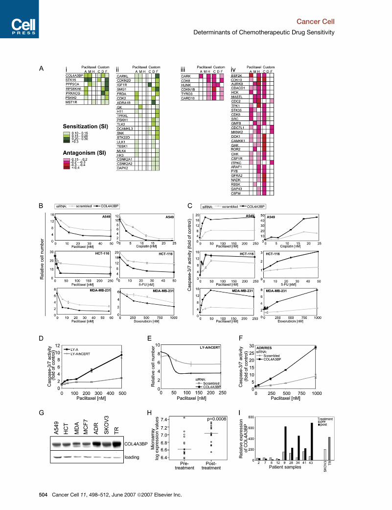
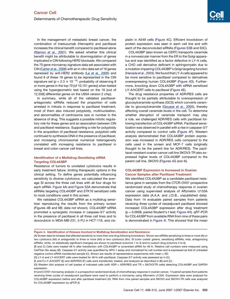
Identification of a Multidrug-Sensitizing siRNA
Targeting COL4A3BP
Resistance of tumors to unrelated cytotoxics results in
early treatment failure, limiting therapeutic options in the
clinical setting. To define genes potentially influencing
sensitivity to diverse cytotoxics, we calculated the aver-
age SI across the three cell lines with all four drugs for
each siRNA. Figure 4Ai and Figure S3A demonstrate that
siRNAs targeting COL4A3BP and STK16 sensitized cells
to most conditions used in this screen.
We validated COL4A3BP siRNA as a multidrug sensi-
tizer reproducing the results from the primary screen
(Figures 4B and 6B; data not shown). COL4A3BP siRNA
promoted a synergistic increase in caspase-3/7 activity
in the presence of paclitaxel in all three cell lines and to
doxorubicin in MDA-MB-231, 5-FU in HCT-116, and cis-
platin in A549 cells (Figure 4C). Efficient knockdown of
protein expression was seen in each cell line and with
each of the deconvoluted siRNAs (Figures S3B and S3C).
COL4A3BP (also known as CERT) transports ceramide
in a nonvesicular manner from the ER to the Golgi appara-
tus and was identified as a factor defective in LY-A cells,
a CHO cell derivative deficient in sphingomyelin due to
a mutation impairing COL4A3BP’s Golgi targeting function
(Hanada et al., 2003). We found that LY-A cells appeared to
be more sensitive to paclitaxel compared to derivatives
overexpressing human COL4A3BP (Figure 4D). Further-
more, knocking down COL4A3BP with siRNA sensitized
LY-A/hCERT cells to paclitaxel (Figure 4E).
The drug resistance properties of ADR/RES cells are
thought to be partially attributable to overexpression of
glucosylceramide synthase (GCS) which converts ceram-
ide to glucosylceramide (Gouaze et al., 2005), thereby
affecting overall ceramide levels in the cell. To determine
whether disruption of ceramide transport may play
a role, we challenged ADR/RES cells with paclitaxel fol-
lowing transfection of COL4A3BP siRNA. Paclitaxel sensi-
tization was observed in parallel with a rise in caspase-3/7
activity compared to control cells (Figure 4F). Western
analysis demonstrated that COL4A3BP protein expres-
sion was increased in ADR/RES cells compared to the
cells used in the screen and MCF-7 cells (originally
thought to be the parent line for ADR/RES). The pacli-
taxel-resistant ovarian cancer cell line SKOV3-TR also ex-
pressed higher levels of COL4A3BP compared to the
parent cell line, SKOV3 (Figures 4G and 4I).
COL4A3BP Expression Is Increased in Ovarian
Cancer Samples after Paclitaxel Treatment
We identified COL4A3BP as a candidate paclitaxel resis-
tance gene in samples from the CTCR-OV01 prospective
randomized study of chemotherapy response in ovarian
cancer using supervised analysis of Affymetrix U133A
expression data (A.A.A. and J.D.B., unpublished data).
Data from 14 evaluable paired samples from patients
receiving three cycles of neoadjuvant paclitaxel showed
increased COL4A3BP expression after drug treatment
(p = 0.0008, paired Student’s t test; Figure 4H). qRT-PCR
for COL4A3BP from available RNA from nine of these pairs
is demonstrated in Figure 4I. This showed that the mean
Figure 4. Identification of Kinases Involved in Multidrug Sensitization and Resistance
(A) Screen data for kinases that affected sensitivity to more than one drug following knockdown. Shown are siRNAs sensitizing to three or more (Ai) or
two cytotoxics (Aii) or antagonistic to three or more (Aiii) or two cytotoxics (Aiv). SI (color coded: greens, sensitizing siRNAs; reds, antagonizing
siRNAs; white, no statistically significant changes) are shown to paclitaxel (columns 1 to 3) and to custom drug (columns 4 to 6).
(B and C) Cells were treated 48 hr after transfection with COL4A3BP or scrambled siRNA for 48 hr. Relative cell numbers were measured using
CellTiter-Glo assay (B). Caspase-3/7 activity was quantified by APO-1 assay and normalized for cell number and is expressed as fold of untreated
negative control siRNA-transfected sample (C). Shown are results of representative experiments with mean ± SD.
(D) LY-A and LY-A/hCERT cells were treated for 48 hr with paclitaxel. Caspase-3/7 activity was assessed as in (C).
(E and F) LY-A/hCERT (E) and ADR/RES (F) cells were transfected, treated, and analyzed as described in (B) and (C).
(G) Western blot analysis of cell lysates of indicated cells (with ADR = ADR/RES and TR = SKOV3/TR cells) detecting COL4A3BP and GAPDH
expression.
(H and I) OV01 microarray analysis: in a prospective randomized study of chemotherapy response in ovarian cancer, 14 paired samples from patients
receiving three cycles of neoadjuvant paclitaxel were used to perform a microarray using Affymetrix U133A. Expression data were analyzed for
COL4A3BP expression before and after paclitaxel treatment (H). RNA from nine paired samples and SKOV3 and SKOV3-TR cells was analyzed
for COL4A3BP expression by qPCR (I).
Cancer Cell 11, 498–512, June 2007 ª2007 Elsevier Inc. 505
Cancer Cell
Determinants of Chemotherapeutic Drug Sensitivity
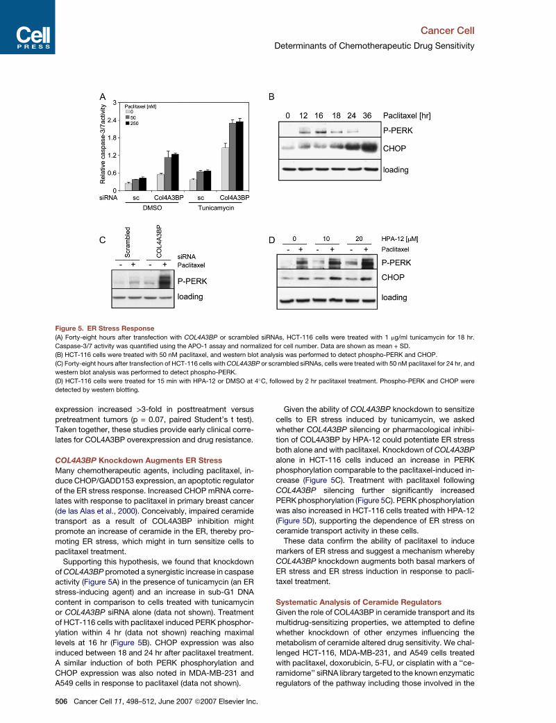
Figure 5. ER Stress Response
(A) Forty-eight hours after transfection with COL4A3BP or scrambled siRNAs, HCT-116 cells were treated with 1 mg/ml tunicamycin for 18 hr.
Caspase-3/7 activity was quantified using the APO-1 assay and normalized for cell number. Data are shown as mean + SD.
(B) HCT-116 cells were treated with 50 nM paclitaxel, and western blot analysis was performed to detect phospho-PERK and CHOP.
(C) Forty-eight hours after transfection of HCT-116 cells with COL4A3BP or scrambled siRNAs, cells were treated with 50 nM paclitaxel for 24 hr, and
western blot analysis was performed to detect phospho-PERK.
(D) HCT-116 cells were treated for 15 min with HPA-12 or DMSO at 4�C, followed by 2 hr paclitaxel treatment. Phospho-PERK and CHOP were
detected by western blotting.
expression increased >3-fold in posttreatment versus
pretreatment tumors (p = 0.07, paired Student’s t test).
Taken together, these studies provide early clinical corre-
lates for COL4A3BP overexpression and drug resistance.
COL4A3BP Knockdown Augments ER Stress
Many chemotherapeutic agents, including paclitaxel, in-
duce CHOP/GADD153 expression, an apoptotic regulator
of the ER stress response. Increased CHOP mRNA corre-
lates with response to paclitaxel in primary breast cancer
(de las Alas et al., 2000). Conceivably, impaired ceramide
transport as a result of COL4A3BP inhibition might
promote an increase of ceramide in the ER, thereby pro-
moting ER stress, which might in turn sensitize cells to
paclitaxel treatment.
Supporting this hypothesis, we found that knockdown
of COL4A3BP promoted a synergistic increase in caspase
activity (Figure 5A) in the presence of tunicamycin (an ER
stress-inducing agent) and an increase in sub-G1 DNA
content in comparison to cells treated with tunicamycin
or COL4A3BP siRNA alone (data not shown). Treatment
of HCT-116 cells with paclitaxel induced PERK phosphor-
ylation within 4 hr (data not shown) reaching maximal
levels at 16 hr (Figure 5B). CHOP expression was also
induced between 18 and 24 hr after paclitaxel treatment.
A similar induction of both PERK phosphorylation and
CHOP expression was also noted in MDA-MB-231 and
A549 cells in response to paclitaxel (data not shown).
506 Cancer Cell 11, 498–512, June 2007 ª2007 Elsevier Inc.
Given the ability of COL4A3BP knockdown to sensitize
cells to ER stress induced by tunicamycin, we asked
whether COL4A3BP silencing or pharmacological inhibi-
tion of COL4A3BP by HPA-12 could potentiate ER stress
both alone and with paclitaxel. Knockdown of COL4A3BP
alone in HCT-116 cells induced an increase in PERK
phosphorylation comparable to the paclitaxel-induced in-
crease (Figure 5C). Treatment with paclitaxel following
COL4A3BP silencing further significantly increased
PERK phosphorylation (Figure 5C). PERK phosphorylation
was also increased in HCT-116 cells treated with HPA-12
(Figure 5D), supporting the dependence of ER stress on
ceramide transport activity in these cells.
These data confirm the ability of paclitaxel to induce
markers of ER stress and suggest a mechanism whereby
COL4A3BP knockdown augments both basal markers of
ER stress and ER stress induction in response to pacli-
taxel treatment.
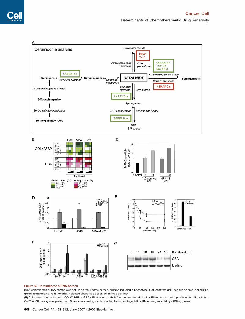
Systematic Analysis of Ceramide Regulators
Given the role of COL4A3BP in ceramide transport and its
multidrug-sensitizing properties, we attempted to define
whether knockdown of other enzymes influencing the
metabolism of ceramide altered drug sensitivity. We chal-
lenged HCT-116, MDA-MB-231, and A549 cells treated
with paclitaxel, doxorubicin, 5-FU, or cisplatin with a ‘‘ce-
ramidome’’ siRNA library targeted to the known enzymatic
regulators of the pathway including those involved in the
Cancer Cell
Determinants of Chemotherapeutic Drug Sensitivity
de novo synthesis of ceramide and sphingomyelin hydro-
lysis. siRNAs inducing a statistically significant phenotype
are shown in Figure S4A. Figure 6A depicts each of the en-
zymes involved in ceramide and sphingomyelin metabo-
lism with the gene name(s) adjacent in color-coded for-
mat, indicating sensitization or antagonism by their
targeting siRNA in two or more cell lines. COL4A3BP
siRNA sensitized the three cell lines to 9 out of 12 condi-
tions. No other siRNA-targeting molecules involved in ce-
ramide metabolism had such a profound effect on drug
sensitivity.
We chose to further investigate GBA (b-glucosidase), as
this was the only siRNA that promoted resistance to
paclitaxel in all three cell lines. GBA converts glucosylcer-
amide into ceramide, opposing the action of glucosylcer-
amide synthase. The GBA siRNA promoted paclitaxel
antagonism in all three cell lines (Figure 6B). Efficient
knockdown of GBA protein was observed (Figure S4B).
Next we addressed whether pharmacological inhibition
or manipulation of the ceramide pathway using selected
siRNAs could influence a mitotic arrest induced by pacli-
taxel. Pretreatment of HCT-116 cells with C6-ceramide
increased paclitaxel-induced mitotic arrest (Figure 6C). In-
hibition of COL4A3BP by HPA-12 (Kumagai et al., 2005) or
silencing by siRNA accentuated a mitotic arrest as early as
2 hr following paclitaxel treatment (Figures 6C and 6D).
In common with the other paclitaxel antagonists found
in this screen, GBA siRNA reduced the induction of
MPM-2 reactivity after paclitaxel treatment in the three
cell lines (Figure 6D). Furthermore, knockdown of another
b-glucosidase, GBA3, also attenuated a mitotic arrest and
promoted resistance to paclitaxel in HCT-116 cells
(Figure 6E). These data suggest that alterations in nonve-
sicular ceramide transport or ceramide synthesis from
glucosylceramide may affect the mitotic arrest induced
by paclitaxel.
In keeping with many of the paclitaxel antagonists
identified in this screen, GBA siRNA promoted an in-
crease in polyploidy in all three cell lines without pacli-
taxel (Figure 6F). Furthermore, we noted significant
induction of GBA protein at 12 hr following paclitaxel
treatment (Figure 6G), suggesting that GBA induction
may play a role in ceramide elevation involved in pacli-
taxel toxicity.
PI3K/Akt Inhibition Attenuates Development
of Aneuploidy
As observed previously, following 24 hr treatment of HCT-
116 cells with paclitaxel, levels of phosphorylated Akt
were reduced in comparison to untreated control cells
(Figures 2G and 7A). Knockdown of COL4A3BP further
enhanced the loss of phosphorylated Akt, while knock-
down of GBA prevented the paclitaxel-induced decrease
of phosphorylated Akt levels.
The associations between PI3K pathway activation, CIN
and taxane resistance, and the sustained activation of Akt
in the presence of paclitaxel following transfection of poly-
ploidy inducing paclitaxel antagonists prompted us to in-
vestigate whether inhibition of PI3K/Akt signaling could
prevent the induction of aneuploid cells. Following trans-
fection of BUB1B and GBA siRNAs, pretreatment with in-
hibitors of PI3K (LY294002) or Akt (inhibitors V and VIII)
inhibited paclitaxel-induced aneuploidy development
(Figure 7B). Analysis by flow cytometry showed that aneu-
ploidy reduction is associated with an increased propor-
tion of cells in the G1 and G2/M fraction of the cell cycle,
which might indicate a 2N and 4N G1 arrest (data not
shown). Therefore, it is possible that inactivation of Akt
may induce a G1 cell-cycle arrest following mitotic slip-
page, thereby preventing endoreduplication of DNA and
propagation of aneuploid progeny.
Topology of Akt Inactivation
after COL4A3BP Inhibition
The mechanism whereby COL4A3BP inhibition, which in-
duces ceramide accumulation in the ER (Hanada et al.,
2003), might promote Akt inactivation by paclitaxel
(Figure 7A) was explored. Activation of Akt is initiated by
the binding of 30 phosphorylated phosphoinositide lipids
to its pleckstrin homology (PH) domain, promoting plasma
membrane localization. We used HCT-116 cells express-
ing a construct in which GFP is fused to the Akt amino-ter-
minal regulatory region that spans the PH domain (termed
GFP-AH). As shown before, in untreated cells growing in
serum this protein is localized to the plasma membrane
(Watton and Downward, 1999). Figure 7C demonstrates
that pretreatment of cells growing in serum with the
COL4A3BP inhibitor HPA-12 significantly reduced the
percentage of cells with membrane GFP-AH staining at
18 hr of paclitaxel treatment. The translocation of the Akt
PH domain to the plasma membrane is thus influenced
by ceramide accumulation in the ER.
DISCUSSION
Development of Aneuploidy
and Resistance to Paclitaxel
A characteristic of all siRNAs promoting paclitaxel resis-
tance in this study is a reduction in the accumulation of
drug-treated cells at the mitotic checkpoint. This is
expected, given the assumption that mitotic arrest induc-
tion is required for paclitaxel cytotoxicity: any mechanism
reducing the efficacy of the drug is likely to be reflected in
a reduction of the mitotic index in the presence of pacli-
taxel. Unexpectedly, however, most of these siRNAs
also induced the appearance of polyploid cells, multinu-
cleation, and centrosomal abnormalities without drug
treatment. A significant number of mitotic regulators ap-
peared as paclitaxel antagonists following knockdown,
some of which have been implicated in sensitivity to
microtubule-targeted drugs. The results of this screen
support the concept that spindle checkpoint disruption
promotes paclitaxel antagonism. The early mitotic spindle
assembly checkpoint influences the postmitotic check-
point following mitotic slippage, thereby preventing
endoreduplication of DNA and the propagation of poly-
ploid cells, which may contribute to aneuploidy (Blagos-
klonny, 2006; Shi and King, 2005; Vogel et al., 2004).
Cancer Cell 11, 498–512, June 2007 ª2007 Elsevier Inc. 507
Cancer Cell
Determinants of Chemotherapeutic Drug Sensitivity
Figure 6. Ceramidome siRNA Screen
(A) A ceramidome siRNA screen was set up as the kinome screen. siRNAs inducing a phenotype in at least two cell lines are colored (sensitizing,
green; antagonizing, red). Asterisk indicates phenotype observed in three cell lines.
(B) Cells were transfected with COL4A3BP or GBA siRNA pools or their four deconvoluted single siRNAs, treated with paclitaxel for 48 hr before
CellTiter-Glo assay was performed. SI are shown using a color-coding format (antagonistic siRNAs, red; sensitizing siRNAs, green).
508 Cancer Cell 11, 498–512, June 2007 ª2007 Elsevier Inc.
Cancer Cell
Determinants of Chemotherapeutic Drug Sensitivity
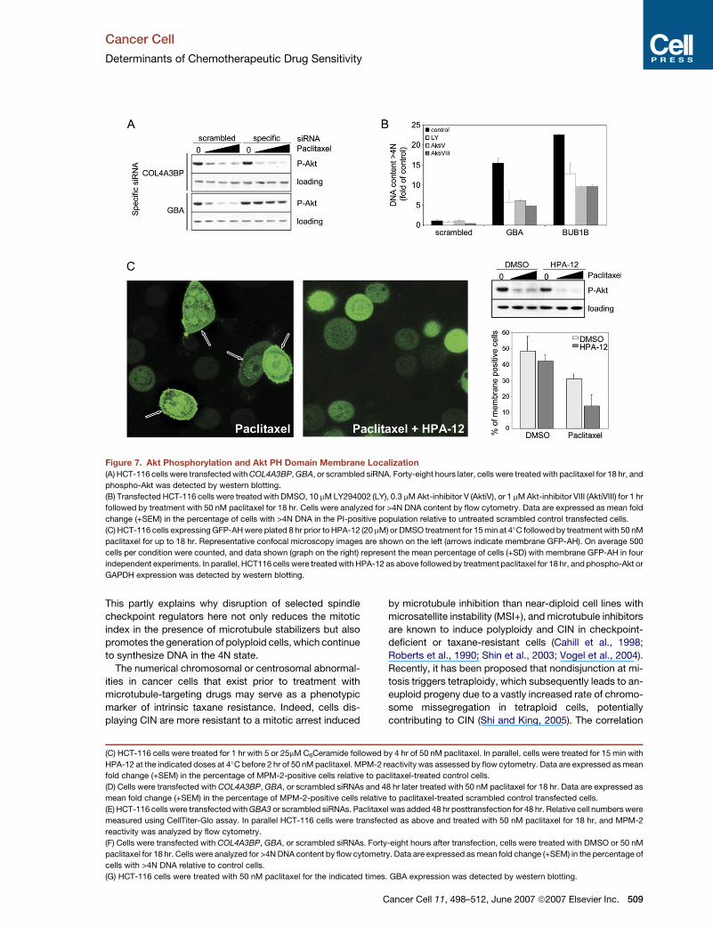
Figure 7. Akt Phosphorylation and Akt PH Domain Membrane Localization
(A) HCT-116 cells were transfected with COL4A3BP, GBA, or scrambled siRNA. Forty-eight hours later, cells were treated with paclitaxel for 18 hr, and
phospho-Akt was detected by western blotting.
(B) Transfected HCT-116 cells were treated with DMSO, 10 mM LY294002 (LY), 0.3 mM Akt-inhibitor V (AktiV), or 1 mM Akt-inhibitor VIII (AktiVIII) for 1 hr
followed by treatment with 50 nM paclitaxel for 18 hr. Cells were analyzed for >4N DNA content by flow cytometry. Data are expressed as mean fold
change (+SEM) in the percentage of cells with >4N DNA in the PI-positive population relative to untreated scrambled control transfected cells.
(C) HCT-116 cells expressing GFP-AH were plated 8 hr prior to HPA-12 (20 mM) or DMSO treatment for 15 min at 4�C followed by treatment with 50 nM
paclitaxel for up to 18 hr. Representative confocal microscopy images are shown on the left (arrows indicate membrane GFP-AH). On average 500
cells per condition were counted, and data shown (graph on the right) represent the mean percentage of cells (+SD) with membrane GFP-AH in four
independent experiments. In parallel, HCT116 cells were treated with HPA-12 as above followed by treatment paclitaxel for 18 hr, and phospho-Akt or
GAPDH expression was detected by western blotting.
This partly explains why disruption of selected spindle
checkpoint regulators here not only reduces the mitotic
index in the presence of microtubule stabilizers but also
promotes the generation of polyploid cells, which continue
to synthesize DNA in the 4N state.
The numerical chromosomal or centrosomal abnormal-
ities in cancer cells that exist prior to treatment with
microtubule-targeting drugs may serve as a phenotypic
marker of intrinsic taxane resistance. Indeed, cells dis-
playing CIN are more resistant to a mitotic arrest induced
by microtubule inhibition than near-diploid cell lines with
microsatellite instability (MSI+), and microtubule inhibitors
are known to induce polyploidy and CIN in checkpoint-
deficient or taxane-resistant cells (Cahill et al., 1998;
Roberts et al., 1990; Shin et al., 2003; Vogel et al., 2004).
Recently, it has been proposed that nondisjunction at mi-
tosis triggers tetraploidy, which subsequently leads to an-
euploid progeny due to a vastly increased rate of chromo-
some missegregation in tetraploid cells, potentially
contributing to CIN (Shi and King, 2005). The correlation
(C) HCT-116 cells were treated for 1 hr with 5 or 25mM C6Ceramide followed by 4 hr of 50 nM paclitaxel. In parallel, cells were treated for 15 min with
HPA-12 at the indicated doses at 4�C before 2 hr of 50 nM paclitaxel. MPM-2 reactivity was assessed by flow cytometry. Data are expressed as mean
fold change (+SEM) in the percentage of MPM-2-positive cells relative to paclitaxel-treated control cells.
(D) Cells were transfected with COL4A3BP, GBA, or scrambled siRNAs and 48 hr later treated with 50 nM paclitaxel for 18 hr. Data are expressed as
mean fold change (+SEM) in the percentage of MPM-2-positive cells relative to paclitaxel-treated scrambled control transfected cells.
(E) HCT-116 cells were transfected with GBA3 or scrambled siRNAs. Paclitaxel was added 48 hr posttransfection for 48 hr. Relative cell numbers were
measured using CellTiter-Glo assay. In parallel HCT-116 cells were transfected as above and treated with 50 nM paclitaxel for 18 hr, and MPM-2
reactivity was analyzed by flow cytometry.
(F) Cells were transfected with COL4A3BP, GBA, or scrambled siRNAs. Forty-eight hours after transfection, cells were treated with DMSO or 50 nM
paclitaxel for 18 hr. Cells were analyzed for >4N DNA content by flow cytometry. Data are expressed as mean fold change (+SEM) in the percentage of
cells with >4N DNA relative to control cells.
(G) HCT-116 cells were treated with 50 nM paclitaxel for the indicated times. GBA expression was detected by western blotting.
Cancer Cell 11, 498–512, June 2007 ª2007 Elsevier Inc. 509

Cancer Cell
Determinants of Chemotherapeutic Drug Sensitivity
between paclitaxel-induced polyploidy in K562 leukemia
cells and paclitaxel resistance has prompted the sugges-
tion that polyploidy in response to paclitaxel may be a use-
ful indicator of drug resistance (Roberts et al., 1990). We
extend this observation and suggest that genes that are
permissive for polyploidy, when targeted by RNA interfer-
ence, influence pathways that taxanes depend upon for
cellular cytotoxicity, and that polyploidy or CIN prior to
drug treatment may serve as a marker for de novo taxane
resistance. Conversely, taxane treatment may select for
a higher frequency of CIN in residual tumor.
These data suggest a compelling reason for the failure
of taxanes to demonstrate activity in CRC, a disease
with a high frequency of CIN. It also implies that 15% of
CRCs, which are MSI+/near diploid, might be more sensi-
tive to microtubule-stabilizing agents. Treatment of MSI+
patients with microtubule inhibitors is the focus of a clinical
trial, CINATRA (chromosomal instability and anti-tubulin
response assessment, Eudract number 2006-006073-24
and Supplemental Data).
Consistently, cells transfected with the siRNAs inducing
the most potent induction of polyploidy without drug dis-
played elevated phosphorylated Akt following paclitaxel
treatment. PI3K pathway deregulation has been shown
to correlate with resistance to docetaxel (Potti et al.,
2006). The highly significant (p = 1.3 3 10�21) representa-
tion of 16 genes in the PI3K predictor set in the CIN70 sig-
nature set suggests a role for PI3K activation and CIN
(Potti et al., 2006; Carter et al., 2006). It is intriguing that
8 of the 16 genes significantly repressed by anti-HER2 an-
tibody treatment are overexpressed in the CIN70 gene
signature set (p = 2.3 3 10�16). The clinical studies dem-
onstrating the impressive clinical benefit with taxanes
and trastuzumab (Slamon et al., 2001) may be attributable
to preferential repression by trastuzumab of genes pro-
moting CIN.
Can spindle checkpoint disruption give long-term ad-
vantage to tumor cells in vivo in terms of taxane resistance?
Prolonged major disruption of the spindle checkpoint is
not tolerated by cells due to massive chromosome loss
resulting in apoptosis (Kops et al., 2004). In a clinical set-
ting, the data presented here suggest that partial
inhibition of this checkpoint in tumor cells could result in
acquisition of taxane resistance, while cells with more
complete inhibition would be lost by apoptosis. Taxane
treatment may result in selection of a population of cells
with checkpoint function that has been partially attenuated
but is still sufficient to support long-term growth and
survival.
Ceramide Metabolism and Multidrug Resistance
Multidrug resistance mechanisms have proved difficult to
define and to target effectively. Agents targeting the most
well-known pathway, the multidrug-resistant transporters
that promote drug resistance to structurally unrelated
cytotoxic agents, have proved disappointing in clinical
trials (Nobili et al., 2006). This screen revealed a small
number of unexpected proteins that, when knocked
down, led to sensitization to multiple drugs, suggesting
510 Cancer Cell 11, 498–512, June 2007 ª2007 Elsevier Inc.
that they may be important mediators of multidrug resis-
tance. COL4A3BP siRNA was the strongest hit in this
category, sensitizing diverse cell types to paclitaxel, doxo-
rubicin, cisplatin, and 5-FU. A closer systematic analysis
of the regulators of ceramide metabolism revealed other
enzymes that impact strongly on drug sensitivity, includ-
ing b-glucosidase (GBA), the Gaucher’s disease gene
product, which on knockdown promotes resistance to
paclitaxel in all cell lines tested. COL4A3BP binds to ce-
ramide and transports it from its site of de novo synthesis
in the endoplasmic reticulum to the Golgi apparatus,
where it is metabolized to sphingomyelin (Hanada et al.,
2003). Reduction in COL4A3BP expression would be ex-
pected to result in accumulation of ceramide in the endo-
plasmic reticulum and increased total cellular ceramide.
By contrast, b-glucosidase, also known as cerebrosidase,
catalyzes the conversion of glucosylceramide to ceram-
ide, so reducing its level would tend to decrease cellular
ceramide levels (Ogretmen and Hannun, 2004). Ceramide
has long been associated with promotion of apoptosis
(Obeid et al., 1993), although the mechanisms involved
have proved complex (Pettus et al., 2002). Many chemo-
therapeutic agents promote ceramide accumulation in
cells, either due to increased de novo synthesis or sphin-
gomyelinase activity opening up the therapeutic potential
of this pathway (Kolesnick, 2002; Reynolds et al., 2004). It
is thus logical that targeting COL4A3BP or GBA should
lead to increased or decreased sensitivity to these drugs,
respectively.
Previously, the only well-described connection between
a ceramide pathway enzyme and multidrug resistance has
been for glucosylceramide synthase (GCS, gene name
UGCG), which catalyzes the reverse reaction to GBA, syn-
thesizing glucosylceramide from ceramide in the Golgi.
Targeting GCS has been shown to increase the sensitivity
of breast cancer cells, including ADR/RES drug-resistant
cells, to anthracyclines and taxanes (Liu et al., 2001),
although we also show here that ADR/RES cells overex-
press COL4A3BP and are sensitized to paclitaxel when
it is knocked down. GCS siRNA did score as a drug sensi-
tizer in our screen, for cisplatin in HCT-116 cells and doxo-
rubicin in A549 cells, but was markedly weaker overall
than COL4A3BP siRNA. Our data suggest that COL4A3BP
would be likely to make a better therapeutic target for the
reversal of multidrug resistance than GCS.
Akt activation is known to be suppressed by elevated
ceramide levels, possibly in part due to the ability of
ceramide to activate the phosphatase PP2A and in part
due to inhibition of the PIP3-binding ability of the PH
domain of Akt, perhaps due to direct phosphorylation by
PKCz (Powell et al., 2003; Stratford et al., 2001, 2004).
Targeting COL4A3BP leads to decreased Akt activation
state after paclitaxel treatment, while targeting GBA has
the opposite effect. Accumulation of ceramide in the ER,
while physically distant from the site of Akt activation at
the plasma membrane, is able to inhibit Akt PH domain
translocation.
It is possible that prolonged mitotic arrest is responsible
for ceramide elevation by paclitaxel and that any inhibition
Cancer Cell
Determinants of Chemotherapeutic Drug Sensitivity
of this arrest would suppress ceramide generation and Akt
suppression, thereby also promoting cell survival. While
this could account for the effect of targeting mitotic check-
point proteins on ceramide and phospho-Akt levels, it is
less clear why targeting a ceramide-generating enzyme
should affect the mitotic checkpoint, as is the case for
GBA. It is possible that this could be due to decreased
ceramide levels leading to elevated Akt activity at the
mitotic checkpoint; activated Akt has been reported to
overcome other cell-cycle checkpoints, such as a DNA-
damage-induced G2/M checkpoint (Kandel et al., 2002)
and also G1/S arrest, via phosphorylation of p21Cip1/WAF1
and p27KIP1 (Shin et al., 2002; Zhou et al., 2001).
The RNA interference approach taken here directly
screens for single targets whose inhibition might reverse
chemotherapeutic drug resistance in tumors: COL4A3BP
represents one such candidate that clearly merits further
investigation. It also identifies individual genes that when
ablated promote drug resistance, such as various mitotic
checkpoint regulators in the case of paclitaxel. Whether or
not these have prognostic value, either alone or in combi-
nation, in predicting the response of tumors to chemother-
apy, and how they compare with response signatures
determined by gene expression profiling, will be the sub-
ject of further investigation.
EXPERIMENTAL PROCEDURES
Screen Set-Up
The Kinome (779 siRNAs) and Ceramidome libraries (50 siRNAs) were
supplied by Dharmacon as SMARTpool collections. As negative con-
trol we used siCONTROL Non-Targeting siRNA pool (scrambled).
HCT-116, A549, and MDA-MB-231 cells were transfected 24 hr after
being plated on 96-well plates (Packard) with 70 nM siRNA using
Oligofectamine transfection reagent (Invitrogen). After 48 hr, the
appropriate cytotoxic drug was added: 58 nM paclitaxel (Sigma;
HCT-116 and A549 cells) and 250 nM for MDA-MB-231, 50 mM
5-fluorouracil (5-FU, Calbiochem), 1 mM doxorubicin (Calbiochem),
and 12.5 mM cisplatin (Sigma). Forty-eight hours later, the CellTiter-
Glo Luminescent Cell Viability Assay (Promega) was performed.
Data Analysis: Derivation of Drug Sensitivity and Antagonism
In order to establish the influence of siRNA-induced knockdown of
gene expression on drug sensitivity, the individual effects of drug
and siRNAs on viability were taken into account. The viability effect
of siRNA without drug compared to scrambled siRNA control was
designated Rc/Cc. The effect of the drugs on viability of control-
transfected cells was designated Cd/Cc. This enabled a calculation
of the Expected combined effect of siRNA and drug on cell viability:
Rc/Cc*Cd/Cc. The Observed combined effects of drug and siRNA
on cell viability compared to untreated scrambled control transfected
cells was designated Rd/Cc. Therefore, an index of antagonism or sen-
sitivity (SI) for each siRNA was calculated by subtracting the Observed
combined effect of drug and siRNA (Rd/Cc) from the Expected total
viability effect: SI = (Rc/Cc*Cd/Cc) � (Rd/Cc). In order for the SI to
usefully predict antagonism or sensitivity, an additional criteria that
Rd/Cd > 1.05 (for antagonistic siRNAs) and Rd/Cd < 0.95 (for sensitiz-
ing siRNAs) was employed for hit selection. Further information on data
analysis and statistical considerations can be found in the Supplemen-
tal Data.
Determination of Mitotic Index and Polyploidy
Cells were fixed with 70% ethanol and incubated with 6 mg/ml anti-
MPM-2 antibody (Upstate) diluted in PBS/0.2% BSA for 1 hr. Cells
were then washed and incubated with Alexa Fluor 488 conjugated
antibody (Invitrogen) diluted in PBS containing 50 mg/ml RNaseA and
50 mg/ml propidium iodide and analyzed on a LSRII (Becton Dickin-
son). Cell doublets and debris were excluded from analysis on the
basis of their pulse height and area, and at least 30,000 events were
recorded.
CTCR-OV01 Clinical Trial and Consent Procedures
Ovarian cancer samples were obtained from the CTCR-OV01 study
(http://pfsearch.ukcrn.org.uk/StudyDetail.aspx?TopicID=1&StudyID=1206).
Patients with histologically confirmed advanced (stages III and IV)
epithelial ovarian cancer with a WHO performance status of 0 or 1
were eligible to participate. Exclusion criteria were (1) nonepithelial
ovarian tumors, (2) patients who had received prior chemotherapy or
radiotherapy, and (3) patients who were not fit to receive paclitaxel
treatment. Samples were collected immediately prior to chemotherapy
and then 3 weeks postchemotherapy at debulking surgery from meta-
static deposits in the abdomen. The study was approved by the
Cambridge Local Research Ethics Committee (LREC). All patients
gave written informed consent prior to participation.
Further description of experimental procedures can be found in the
Supplemental Data.
Supplemental Data
The Supplemental Data include Supplemental Experimental Proce-
dures, two supplemental tables, and four supplemental figures and
can be found with this article online at http://www.cancercell.org/
cgi/content/full/11/6/498/DC1/.
ACKNOWLEDGMENTS
We would like to thank C. Sachse and CENIX BioScience, GmbH, for
their technical support; K. Hanada for LY-A cells; and Q. Bose, R. Gadel-
rab, and Dharmacon Inc. for technical assistance. We are grateful to K.
Allen and D. Davies for flow cytometry expertise and to Thomas J. Hard-
castle for bioinformatic analysis of the CTCR-OV01 data set. We are also
grateful to A. Walther, A. Schulze, A. Venkitaraman, and D. Hancock for
helpful discussions. This work was funded by Cancer Research UK.
Received: November 3, 2006
Revised: January 31, 2007
Accepted: April 2, 2007
Published: June 11, 2007
REFERENCES
Alaoui-Jamali, M.A., Dupre, I., and Qiang, H. (2004). Prediction of drug
sensitivity and drug resistance in cancer by transcriptional and proteo-
mic profiling. Drug Resist. Updat. 7, 245–255.
Bettencourt-Dias, M., Giet, R., Sinka, R., Mazumdar, A., Lock, W.G.,
Balloux, F., Zafiropoulos, P.J., Yamaguchi, S., Winter, S., Carthew,
R.W., et al. (2004). Genome-wide survey of protein kinases required
for cell cycle progression. Nature 432, 980–987.
Blagosklonny, M.V. (2006). Prolonged mitosis versus tetraploid check-
point: how p53 measures the duration of mitosis. Cell Cycle 5, 971–975.
Cahill, D.P., Lengauer, C., Yu, J., Riggins, G.J., Willson, J.K., Marko-
witz, S.D., Kinzler, K.W., and Vogelstein, B. (1998). Mutations of mitotic
checkpoint genes in human cancers. Nature 392, 300–303.
Carter, S.L., Eklund, A.C., Kohane, I.S., Harris, L.N., and Szallasi, Z.
(2006). A signature of chromosomal instability inferred from gene
expression profiles predicts clinical outcome in multiple human
cancers. Nat. Genet. 38, 1043–1048.
Chang, J.C., Wooten, E.C., Tsimelzon, A., Hilsenbeck, S.G., Gutierrez,
M.C., Elledge, R., Mohsin, S., Osborne, C.K., Chamness, G.C., Allred,
D.C., and O’Connell, P. (2003). Gene expression profiling for the
prediction of therapeutic response to docetaxel in patients with breast
cancer. Lancet 362, 362–369.
Cancer Cell 11, 498–512, June 2007 ª2007 Elsevier Inc. 511
Cancer Cell
Determinants of Chemotherapeutic Drug Sensitivity
de las Alas, M.M., Christen, R.D., Gately, D.P., Weiner, D.E., Benba-
toul, K., Kirmani, S., D’Agostino, H.R., Plaxe, S.C., Darrah, D., McClay,
E.F., et al. (2000). Increase in tumor GADD153 mRNA level following
treatment correlates with response to paclitaxel. Cancer Chemother.
Pharmacol. 45, 381–388.
Eggert, U.S., Kiger, A.A., Richter, C., Perlman, Z.E., Perrimon, N.,
Mitchison, T.J., and Field, C.M. (2004). Parallel chemical genetic and
genome-wide RNAi screens identify cytokinesis inhibitors and targets.
PLoS Biol. 2, e379. 10.1371/journal.pbio.0020379.
Ein-Dor, L., Zuk, O., and Domany, E. (2006). Thousands of samples are
needed to generate a robust gene list for predicting outcome in cancer.
Proc. Natl. Acad. Sci. USA 103, 5923–5928.
Gouaze, V., Liu, Y.Y., Prickett, C.S., Yu, J.Y., Giuliano, A.E., and Cabot,
M.C. (2005). Glucosylceramide synthase blockade down-regulates
P-glycoprotein and resensitizes multidrug-resistant breast cancer
cells to anticancer drugs. Cancer Res. 65, 3861–3867.
Hanada, K., Kumagai, K., Yasuda, S., Miura, Y., Kawano, M., Fuka-
sawa, M., and Nishijima, M. (2003). Molecular machinery for non-
vesicular trafficking of ceramide. Nature 426, 803–809.
Jin, J., and Woodgett, J.R. (2005). Chronic activation of protein kinase
Bbeta/Akt2 leads to multinucleation and cell fusion in human epithelial
kidney cells: events associated with tumorigenesis. Oncogene 24,
5459–5470.
Kandel, E.S., Skeen, J., Majewski, N., Di Cristofano, A., Pandolfi, P.P.,
Feliciano, C.S., Gartel, A., and Hay, N. (2002). Activation of Akt/protein
kinase B overcomes a G(2)/m cell cycle checkpoint induced by DNA
damage. Mol. Cell. Biol. 22, 7831–7841.
Kolesnick, R. (2002). The therapeutic potential of modulating the
ceramide/sphingomyelin pathway. J. Clin. Invest. 110, 3–8.
Kops, G.J., Foltz, D.R., and Cleveland, D.W. (2004). Lethality to human
cancer cells through massive chromosome loss by inhibition of the
mitotic checkpoint. Proc. Natl. Acad. Sci. USA 101, 8699–8704.
Kumagai, K., Yasuda, S., Okemoto, K., Nishijima, M., Kobayashi, S.,
and Hanada, K. (2005). CERT mediates intermembrane transfer of
various molecular species of ceramides. J. Biol. Chem. 280, 6488–
6495.
Le, X.F., Pruefer, F., and Bast, R.C., Jr. (2005). HER2-targeting anti-
bodies modulate the cyclin-dependent kinase inhibitor p27(Kip1) via
multiple signaling pathways. Cell Cycle 4, 87–95.
Liu, Y.Y., Han, T.Y., Giuliano, A.E., and Cabot, M.C. (2001). Ceramide
glycosylation potentiates cellular multidrug resistance. FASEB J. 15,
719–730.
Longley, D.B., and Johnston, P.G. (2005). Molecular mechanisms of
drug resistance. J. Pathol. 205, 275–292.
Lonning, P.E., Sorlie, T., and Borresen-Dale, A.L. (2005). Genomics
in breast cancer-therapeutic implications. Nat. Clin. Pract. Oncol. 2,
26–33.
MacKeigan, J.P., Murphy, L.O., and Blenis, J. (2005). Sensitized RNAi
screen of human kinases and phosphatases identifies new regulators
of apoptosis and chemoresistance. Nat. Cell Biol. 7, 591–600.
Moffat, J., Grueneberg, D.A., Yang, X., Kim, S.Y., Kloepfer, A.M.,
Hinkle, G., Piqani, B., Eisenhaure, T.M., Luo, B., Grenier, J.K., et al.
(2006). A lentiviral RNAi library for human and mouse genes applied
to an arrayed viral high-content screen. Cell 124, 1283–1298.
Nobili, S., Landini, I., Giglioni, B., and Mini, E. (2006). Pharmacological
strategies for overcoming multidrug resistance. Curr. Drug Targets 7,
861–879.
Obeid, L.M., Linardic, C.M., Karolak, L.A., and Hannun, Y.A. (1993). Pro-
grammed cell death induced by ceramide. Science 259, 1769–1771.
Ogretmen, B., and Hannun, Y.A. (2004). Biologically active sphingoli-
pids in cancer pathogenesis and treatment. Nat. Rev. Cancer 4,
604–616.
512 Cancer Cell 11, 498–512, June 2007 ª2007 Elsevier Inc.
Pettus, B.J., Chalfant, C.E., and Hannun, Y.A. (2002). Ceramide in
apoptosis: an overview and current perspectives. Biochim. Biophys.
Acta 1585, 114–125.
Potti, A., Dressman, H.K., Bild, A., Riedel, R.F., Chan, G., Sayer, R.,
Cragun, J., Cottrill, H., Kelley, M.J., Petersen, R., et al. (2006). Genomic
signatures to guide the use of chemotherapeutics. Nat. Med. 12, 1294–
1300.
Powell, D.J., Hajduch, E., Kular, G., and Hundal, H.S. (2003). Ceramide
disables 3-phosphoinositide binding to the pleckstrin homology do-
main of protein kinase B (PKB)/Akt by a PKCzeta-dependent mecha-
nism. Mol. Cell. Biol. 23, 7794–7808.
Reynolds, C.P., Maurer, B.J., and Kolesnick, R.N. (2004). Ceramide
synthesis and metabolism as a target for cancer therapy. Cancer
Lett. 206, 169–180.
Roberts, J.R., Allison, D.C., Donehower, R.C., and Rowinsky, E.K.
(1990). Development of polyploidization in taxol-resistant human
leukemia cells in vitro. Cancer Res. 50, 710–716.
Roschke, A.V., Tonon, G., Gehlhaus, K.S., McTyre, N., Bussey, K.J.,
Lababidi, S., Scudiero, D.A., Weinstein, J.N., and Kirsch, I.R. (2003).
Karyotypic complexity of the NCI-60 drug-screening panel. Cancer
Res. 63, 8634–8647.
Shi, Q., and King, R.W. (2005). Chromosome nondisjunction yields
tetraploid rather than aneuploid cells in human cell lines. Nature 437,
1038–1042.
Shin, H.J., Baek, K.H., Jeon, A.H., Park, M.T., Lee, S.J., Kang, C.M.,
Lee, H.S., Yoo, S.H., Chung, D.H., Sung, Y.C., et al. (2003). Dual roles
of human BubR1, a mitotic checkpoint kinase, in the monitoring of
chromosomal instability. Cancer Cell 4, 483–497.
Shin, I., Yakes, F.M., Rojo, F., Shin, N.Y., Bakin, A.V., Baselga, J., and
Arteaga, C.L. (2002). PKB/Akt mediates cell-cycle progression by
phosphorylation of p27(Kip1) at threonine 157 and modulation of its
cellular localisation. Nat. Med. 8, 1145–1152.
Slamon, D.J., Leyland-Jones, B., Shak, S., Fuchs, H., Paton, V., Baja-
monde, A., Fleming, T., Eiermann, W., Wolter, J., Pegram, M., et al.
(2001). Use of chemotherapy plus a monoclonal antibody against
HER2 for metastatic breast cancer that overexpresses HER2. N.
Engl. J. Med. 344, 783–792.
Stratford, S., DeWald, D.B., and Summers, S.A. (2001). Ceramide dis-
sociates 30-phosphoinositide production from pleckstrin homology
domain translocation. Biochem. J. 354, 359–368.
Stratford, S., Hoehn, K.L., Liu, F., and Summers, S.A. (2004). Regula-
tion of insulin action by ceramide: dual mechanisms linking ceramide
accumulation to the inhibition of Akt/protein kinase B. J. Biol. Chem.
279, 36608–36615.
Vogel, C., Kienitz, A., Hofmann, I., and Bastians, H. (2004). Crosstalk of
the mitotic spindle assembly checkpoint with p53 to prevent poly-
ploidy. Oncogene 23, 6845–6853.
Watton, S.J., and Downward, J. (1999). Akt/PKB localisation and
30 phosphoinositide generation at sites of epithelial cell-matrix and
cell-cell interaction. Curr. Biol. 9, 433–436.
Whitfield, M.L., Sherlock, G., Saldanha, A.J., Murray, J.I., Ball, C.A.,
Alexander, K.E., Matese, J.C., Perou, C.M., Hurt, M.M., Brown, P.O.,
and Botstein, D. (2002). Identification of genes periodically expressed
in the human cell cycle and their expression in tumors. Mol. Biol. Cell
13, 1977–2000.
Zhou, B.P., Liao, Y., Xia, W., Spohn, B., Lee, M.H., and Hung, M.C.
(2001). Cytoplasmic localization of p21Cip1/WAF1 by Akt-induced
phosphorylation in HER2/neu-overexpressing cells. Nat. Cell Biol. 3,
245–252.