Sensory reinnervation of cat peroneus brevis muscle spindles after nerve crush

Neuron 49, 25–39, January 5, 2006 ª2006 Elsevier Inc. DOI 10.1016/j.neuron.2005.10.039
Doublecortin-like Kinase ControlsNeurogenesis by Regulating MitoticSpindles and M Phase Progression
Tianzhi Shu,1 Huang-Chun Tseng,1,2,5 Tamar Sapir,4,5
Patrick Stern,3 Ying Zhou,1,2 Kamon Sanada,1
Andre Fischer,1 Frederic M. Coquelle,4 Orly Reiner,4
and Li-Huei Tsai1,2,*1Department of PathologyHarvard Medical School and2Howard Hughes Medical Institute77 Avenue Louis PasteurBoston, Massachusetts 021153Center for Cancer ResearchMassachusetts Institution of TechnologyCambridge, Massachusetts 021394Department of Molecular GeneticsThe Weizmann Institute of Science76100 RehovotIsrael
Summary
Themechanismscontrollingneurogenesis duringbrain
development remain relatively unknown. Through adifferential protein screen with developmental versus
mature neural tissues, we identified a group of devel-opmentally enriched microtubule-associated proteins
(MAPs) including doublecortin-like kinase (DCLK),a protein that shares high homology with doublecortin
(DCX). DCLK, but not DCX, is highly expressed in re-gions of active neurogenesis in the neocortex and
cerebellum. Through a dynein-dependent mechanism,DCLK regulates the formation of bipolar mitotic spin-
dles and the proper transition from prometaphase
to metaphase during mitosis. In cultured cortical neu-ral progenitors, DCLK RNAi Lentivirus disrupts the
structure of mitotic spindles and the progression ofM phase, causing an increase of cell-cycle exit index
and an ectopic commitment to a neuronal fate. Further-more, both DCLK gain and loss of function in vivo spe-
cifically promote a neuronal identity in neural progen-itors. These data provide evidence that DCLK controls
mitotic division by regulating spindle formation andalso determines the fate of neural progenitors during
cortical neurogenesis.
Introduction
The mammalian brain undergoes pronounced cytoarchi-tectural changes during development. Through mitoticdivision, neural progenitor cells give rise to lineages ofneurons. These neurons then migrate, often over longdistances, to their final positions (Caviness et al., 2003;Gupta et al., 2002). Neuronal proliferation and migrationare temporally and spatially coordinated to constructthe distinctive structure of the central nervous system.For instance, the neocortex, which exhibits six layers ofneurons, is built through precisely orchestrated waves
*Correspondence: [email protected] These authors contributed equally to this work.
of newly born neurons that migrate past their predeces-sors (Gupta et al., 2002). After migration, neuronal polar-ity is established upon the specification of the axon anddendrites (Horton and Ehlers, 2003), which is followed byelaborate axonal targeting, dendritic arborization, andsynaptogenesis (Huber et al., 2003; Tessier-Lavigne andGoodman, 1996).
Diverse as they are, these morphological changesare executed through dynamic rearrangement of the cy-toskeletal network components, such as microtubulesand actin filaments (Baas, 1999; Huber et al., 2003;Tucker, 1990). The microtubule network undergoes dras-tic changes in neural cells at different developmentalstages. For instance, during neural proliferation, micro-tubules assemble into the highly organized mitotic spin-dle upon the entry of mitosis (Ohnuma and Harris, 2002).Recent studies suggest that the orientation of the mitoticspindle may determine the mode of neural division(Haydar et al., 2003; Kaltschmidt et al., 2000). After thepostmitotic neurons are generated, they extend a direc-tional leading process and migrate toward their destina-tion, the cortical plate. During migration, another specificmicrotubule network couples the centrosome and nu-cleus, resulting in the salient translocation of the nucleus(Shu et al., 2004; Solecki et al., 2004; Tanaka et al., 2004a;Xie et al., 2003).
Microtubule-associated proteins (MAPs) have beenshown to be the direct regulators of microtubule dynam-ics during many of these developmental processes(Paglini et al., 2000; Sanchez et al., 2000; Tucker, 1990).Through genetic approaches, two genes responsible forhuman type I lissencephaly, LIS1 and DCX, were identi-fied (Gleeson et al., 1998; Hattori et al., 1994; Reiner et al.,1993). LIS1 and DCX are both MAPs that regulate micro-tubule dynamics in vitro and in vivo (Gleeson et al., 1999;Sapir et al., 1997). Depletion of LIS1 or DCX by either agenetic or RNAi approach causes severe neuronal mi-gration and positioning defects in the neocortex (Baiet al., 2003; Cahana et al., 2001; Hirotsune et al., 1998).Other studies further showed that LIS1 forms a complexwith NudE-like protein 1 (Ndel1) and dynein, and thiscomplex is implicated in orchestrating nucleokinesisduring cortical neuronal migration by sustaining the mi-crotubule network that couples the centrosome and nu-cleus (Shu et al., 2004; Smith et al., 2000). In migratingcerebellar granule neurons, DCX also consolidates thismicrotubule network through the interaction with LIS1and dynein (Tanaka et al., 2004a).
To identify MAPs with novel functions during brain de-velopment, we performed a differential protein screenon developmental and mature neural tissues. One of themost intriguing MAPs that we identified in our screen wasdoublecortin-like kinase (DCLK), a protein that shares70% homology in the N-terminal domain with DCX. TheC-terminal domain of DCLK, however, contains a kinasedomain homologous to CaM kinase II (Burgess et al.,1999). The DCLK gene is extremely complex, and it hasbeen reported that at least nine alternative products ex-ist (Burgess and Reiner, 2000, 2002). In this study, we in-vestigated the role of DCLK and provided evidence that

Neuron26
Figure 1. Identification of MAPs Enriched
during Neural Development
(A) MAPs were purified from P8 and adult cer-
ebellum, respectively, and resolved on a pro-
tein gel. After Coomassie blue staining, the
protein bands that show differential pattern
between the two ages (indicated by arrows
and named p1–p20) were dissected out for
MALD-TOF MS.
(B) Results of the protein identification. Note
that protein band p10 was identified as mouse
DCLK.
(C) Western blot confirmed that identified
MAPs such as DCLK and DCX are enriched
during development, and a large portion of
these proteins is found associated with mi-
crotubules (compared to the amount in the
supernatant). It should be noted that other
MAPs such as the Ndel1/LIS1/dynein com-
plex does not show similar enrichment in de-
veloping neural tissues and a smaller propor-
tion of these proteins is found associated
with microtubules.
DCLK regulates the structure and dynamics of the mi-totic spindles and further controls neural division andfate determination during cortical development.
Results
Doublecortin-like Kinase Is a Microtubule-Associated Protein Enriched during Development
To identify microtubule-associated proteins (MAPs) withpotentially novel functions during neural development,we performed a protein-based differential screen. MAPswere purified from postnatal day (P) 8 and adult (>8weeks) cerebellum by a standard biochemical method(see Experimental Procedures). Cerebellum was chosenas the tissue source for MAPs purification because itundergoes dramatic morphological changes during earlypostnatal stages including active neurogenesis, neuro-nal migration, and differentiation (Goldowitz and Hamre,1998). In addition, the cerebellum provides sufficienttissue mass for the large-scale protein purification.MAPs were purified from P8 and adult cerebellar lysates.Purified MAPs were resolved on a SDS-PAGE gel andcounter-stained with Coomassie blue. Many proteinbands were found to be preferentially present in MAPsfrom P8 cerebellum (indicated by arrows in Figure1A).These protein bands were dissected out and digestedwith trypsin for peptide fingerprint identification byMALDI-TOF mass spectrometry. Among the identifiedproteins, two homologous proteins, doublecortin (DCX)and doublecortin-like kinase (DCLK) were found amongthe MAPs enriched on P8 (Figures 1A and 1B). DCLK
was originally identified as KIAA0369 through a homolo-gous comparison with DCX on its N terminus (Burgesset al., 1999). Like DCX, DCLK is able to polymerize micro-tubules in vitro, suggesting a role in regulating microtu-bule dynamics (Burgess and Reiner, 2000; Lin et al.,2000). The function of DCLK during neural development,however, remains largely unknown.
Western blots were performed with specific antibod-ies against DCLK and DCX to confirm the results of thedifferential screen. Both DCLK and DCX were found pre-dominantly expressed in P8 cerebellum compared toadult. A significant portion of DCLK (about 50%) andDCX (about 25%) was associated with microtubules,suggesting an active shuffling of these proteins betweena microtubule associated form and a cytosolic form (Fig-ure 1C). This differential expression profile was specificto DCLK and DCX because none of Ndel1, LIS1, and dy-nein displayed a similar pattern (Figure 1C). The obser-vation that DCX and DCLK are more highly associatedwith microtubules compared to motor-based MAPs sug-gests that DCX and DCLK may exert different but morerobust functions in regulating microtubules during de-velopment.
DCLK Is Expressed in the Neural Progenitor Cells
Immunostaining with an antibody generated against theC-terminal of DCLK showed that similar to DCX, DCLKwas highly expressed in P8 cerebellum, including the ex-ternal granule layer (EGL) (arrowheads in Figure 2A) andthe internal granule layer (IGL) (arrows in Figure 2A). Bycontrast, the expression of DCLK and DCX were nearly

Doublecortin-like Kinase Controls Neural Division27
Figure 2. Expression of DCLK in Brain Re-
gions of Neurogenesis
(A) In cerebellum, both DCX and DCLK are
highly expressed in the external granule layer
(EGL, arrowheads) and internal granule layer
(IGL, arrows) on P8, and their expression is
drastically diminished in adult.
(B) Coimmunostaining with phophoH3 shows
that DCLK is highly expressed in the outer
zone of EGL and DCX is expressed in the in-
ner zone of EGL (the high-power panels indi-
cate the boxed region).
(C) DCLK is expressed in the mitotic spindle
in a cerebellar neural progenitor at prometa-
phase.
(D) The expression of DCX and DCLK during
cortical development. The staining of pho-
phoH3 indicates dividing neural progenitors.
The inset shows the migrating neurons in
the cortex indicated by the arrow.
(E) On E13, DCLK is expressed in radial glia
lining the ventricle wall (arrowheads). The
high-power panel shows the boxed region.
(F and G) The expression of DCLK associated
with mitotic spindle in dividing progenitors
lining the ventricle wall (F) or located within
the CP (G). Scale bar in (A) = 80 mm in P8 cer-
ebellum and 50 mm in adult cerebellum; bar
in (B) = 100 mm in lower power and 10 mm in
higher power. Bar in (C) = 2.5 mm, also in (F)
and (G); bar in (D) = 60 mm in E13 panel,
80 mm in E15 panel, and 100 mm in E17 panel;
bar in (E) = 60 mm in the low-power panel and
15 mm in the high-power panel.
absent in the adult cerebellum (Figure 2A). Although DCXwas expressed in the inner zone of the EGL, DCLK waslargely restricted to the outer zone, where dividing neu-ral progenitors labeled with mitotic marker phosph-H3were located (Figure 2B, the boxed areas were shownin high power). Immunolabeling of the postmitotic neu-rons with NeuN further showed an overlap with DCX inthe inner zone of EGL (data not shown). Closer inspec-tion of EGL progenitors with confocal z series further re-vealed that DCLK was expressed in the mitotic spindle(arrows in Figure 2C indicate a cell at prometaphase).
In the developing neocortex, DCLK was also ex-pressed in the active region of neurogenesis, the sub-ventricular and ventricular zones (SVZ/VZ) (Figure 2D).Consistent with previous studies (Burgess and Reiner,2002; Gleeson et al., 1999; Lin et al., 2000), we foundthat throughout development (from E13 to E17), DCX ex-pression was largely excluded from the SVZ/VZ in whichphosphoH3-positive neural progenitors were located(Figure 2D). On E13, DCLK was found expressed in themarginal zone, cortical plate (CP), and the SVZ/VZ (Fig-ure 2D; inset shows the migrating neurons in the CP).DCLK was expressed in radial glia labeled with an anti-body against nestin (arrowheads in Figure 2E point totwo cell bodies in the VZ). In dividing neural progenitorslining the surface of the ventricular wall, the expressionof DCLK was associated with the mitotic spindle (arrowsin Figure 2F point to a cell at metaphase). Similar obser-vation is also made in dividing neural progenitors lo-cated in other regions such as the CP (Figure 2G showsa metaphase cell in the CP). On E15 and E17, expressionof DCLK was high in the CP and the SVZ/VZ and rela-tively lower in the intermediate zone (IZ) (Figure 2D).
We also examined the expression pattern of DCX andDCLK in cultured neural progenitors, which were iso-lated from E14 mouse cortices and maintained in a de-fined medium containing bFGF-2. Double immunostain-ing with Nestin showed that DCLK, but not DCX, wasexpressed throughout the cytoplasm in the neural pro-genitors (Figure S1). In cultured dividing neural progen-itors, the expression of DCLK was also found associatedclosely with mitotic spindles (Figure S1).
DCLK Induces Monopolar Mitotic Spindlesand Arrests Mitosis at Prometaphase
To directly test whether DCLK regulates the formation ofmitotic spindles, we overexpressed EGFP-tagged wild-type DCLK in 293 HEK cells as a mean of gain of func-tion. At interphase, DCLK gain of function dramaticallyinduced microtubule polymerization, which formed pro-nounced ring-like structures (arrowhead in Figure 3A).Interestingly, at M phase, DCLK induced large monopo-lar mitotic spindles. These abnormal spindles usuallyextended several thick and rigid branches (arrow in Fig-ure 3A), displaying striking differences from the stereo-typical bipolar spindles in cells transfected with EGFPalone (Figure S2). Analysis of the mitotic index (the per-centage of mitotic cells) showed no significant differ-ence between control and DCLK gain of function, sug-gesting that the balance of M phase entry and exit wasnot disrupted (Figure 3B). However, an obvious abnor-mality was observed regarding the distribution of the mi-totic cells overexpressing DCLK. Although control cellswere distributed across all mitotic phases, including pro-metaphase, metaphase, anaphase, and telophase, cellswith DCLK gain of function were mostly arrested at

Neuron28
Figure 3. DCLK Gain of Function Induces Monopolar Mitotic Spindles and Arrests Mitotic Cells at Prometaphase
(A) Overexpression of DCLK-EGFP construct in HEK293 cells induces microtubule bundling. At interphase, the hyperpolymerized microtubules
are evident by both EGFP (green) and a-tubulin staining (red). At M phase, overexpression of DCLK-EGFP induces a large monopolar spindle that
extends thick and stiff branches. Overexpression of CPG-16 fails to induce such monopolar spindle at M phase.
(B) Schematic of splice variants of DCLK and quantification of the effects on mitotic index, M phase arrest, and nuclear morphology after the
overexpression of DCLK, DCL, CPG16, and DCLK kinase-dead form (total number of the analyzed cells are 721, 502, 926, 1370, and 684 for con-
trol, DCLK, DCL, CPG16, and DCLK-KD, respectively). Number of the mitotic cells are 26, 22, 48, 75, and 35, respectively.
(C) Pie graph of effect of DCLK gain of function on the distribution of mitotic cells in different phases.
(D) Centrosome is labeled with RFP-CentrinII in cells arrested at prometaphase after DCLK gain of function. The centrosome displays normal
morphology (arrows in [D] point to centrosome). Scale bar in (D) = 10 mm in (A) and 25 mm in (D). Repeated student t tests between control
and different experimental groups were performed, and triple asterisk indicates p < 0.001.
prometaphase (95% compared to 15% in control) (Fig-ures 3B and 3C). In addition, we observed frequent oc-currences of nuclear abnormalities such as meganuclei,double nuclei, and fragmented nuclei at interphase ofcells overexpressing DCLK, which was likely the conse-quence of the M phase arrest (Figure 3C and data notshown).
Next, we investigated which domain of DCLK was re-sponsible for inducing the abnormal mitotic spindles andM phase arrest. Two natural splice variants of DCLK, theN terminus splicing form, doublecortin like (DCL; Gen-Bank accession AF155821) and the C-terminal form,Candidate Plasticity Gene 16 (CPG16; GenBank acces-sion AF155820) were used in these experiments (Bur-gess and Reiner, 2002). DCL contains the tandem micro-
tubule binding domain (MBD) but not the kinase domainwhile CPG16 contains almost exclusively the kinasedomain (Figure 3B). In addition, a previously reportedDCLK kinase-dead form (K149R) was also included totest whether the kinase activity was required (Lin et al.,2000). Similar to DCLK, overexpression of either DCLor DCLK kinase dead induced large monopolar mitoticspindle and caused a comparable arrest of M phase pro-gression (72% and 77% of M phase cells were arrestedat prometaphase, respectively) (Figures 3B and 3C).CPG16, however, exerted no effect on either the mitoticspindles or M phase progression (Figure 3A–3C). Theseexperiments showed that the N terminus of DCLK withthe tandem MTB domains is necessary and sufficient todisrupt normal spindle formation and to cause M phase
Doublecortin-like Kinase Controls Neural Division29
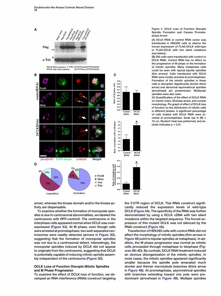
Figure 4. DCLK Loss of Function Disrupts
Spindle Formation and Causes Prometa-
phase Arrest
(A) DCLK RNAi or control RNAi vector was
transfected in HEK293 cells to silence the
forced expression of FLAG-DCLK wild-type
or FLAG-DCLK with two silent mutations
(red letters).
(B) 293 cells were transfected with control or
DCLK RNAi. Control RNAi has no effect on
the progression of M phase or the formation
of mitotic spindles. Many metaphase cells
could be seen with typical bipolar spindles
(thin arrows). Cells transfected with DCLK
RNAi were mostly arrested at prometaphase.
Formation of the mitotic spindles in these
cells is disrupted. Significantly shorter (thick
arrow) and abnormal asymmetrical spindles
(arrowhead) are predominant. Multipolar
spindles were also seen.
(C) Quantification of the effect of DCLK RNAi
on mitotic index, M phase arrest, and nuclear
morphology. Pie graph of effect of DCLK loss
of function on the distribution of mitotic cells
in different phases. A significant percentage
of cells treated with DCLK RNAi were ar-
rested at prometaphase. Scale bar in (B) =
10 mm. Student t test was performed, and as-
terisk indicates p < 0.01.
arrest, whereas the kinase domain and/or the kinase ac-tivity are dispensable.
To examine whether the formation of monopolar spin-dles is due to centrosomal abnormalities, we labeled thecentrosome with RFP-centrinII. The centrosome in theinterphase cells appeared normal when DCLK was over-expressed (Figure S3). At M phase, even though cellswere arrested at prometaphase, two well-separated cen-trosomes were readily detected (arrows in Figure 3D),suggesting that the formation of monopolar spindleswas not due to a centrosomal defect. Interestingly, themonopolar spindles induced by DCLK did not appearto originate from the centrosome, suggesting that DCLKis potentially capable of inducing mitotic spindle assem-bly independent of the centrosome (Figure 3D).
DCLK Loss of Function Disrupts Mitotic Spindlesand M Phase Progression
To examine the effect of DCLK loss of function, we de-veloped an RNA interference (RNAi) construct targeting
the 30UTR region of DCLK. This RNAi construct signifi-cantly reduced the expression levels of wild-typeDCLK (Figure 4A). The specificity of the RNAi was furtherdemonstrated by using a DCLK cDNA with two silentmutations within the targeted sequence. The forced ex-pression of this mutant DCLK was not silenced by theRNAi construct (Figure 4A).
Transfection of HEK293 cells with control RNAi did notaffect the morphology of mitotic spindles (thin arrows inFigure 4B point to bipolar spindles at metaphase). In ad-dition, the M phase progression was normal as mitoticcells proceeded through metaphase to telophase (Fig-ures 4B–4D). By contrast, DCLK RNAi treatment inducedan obvious disorganization of the mitotic spindles. Inmost cases, the mitotic spindles appeared significantlysmaller because the spindle pole emanated muchshorter and thinner microtubule branches (thick arrowin Figure 4B). At prometaphase, asymmetrical spindleswith branches extending toward one pole were pre-dominant (arrowhead in Figure 4B). Multiple spindles

Neuron30
Figure 5. The Function of DCLK to Assemble
Microtubules Is Dynein Dependent
(A) HEK293 cells were treated with control
RNAi vector or RNAi vectors against LIS1
and dynein heavy chain (DHC) with DsRed
construct for 48 hr followed by the transfec-
tion of DCLK-GFAP vector for 24 hr. DCLK in-
duces microtubule polymerization in cells
treated with control RNAi but not with LIS1
or DHC RNAi. Taxol treatment (50 nM for
2 hr) induces microtubule polymerization in
all conditions.
(B) Quantification of the percentage of cells
with microtubule bundles induced by DCLK-
EGFP.
(C) Coimmunoprecipitation with a FLAG anti-
body pulls down dynein intermediate chain
(DIC) from cells lysate with overexpression
of FLAG-DCLK. Scale bar in (A) = 10 mm. Stu-
dent t test was performed, and triple askter-
isk indicates p < 0.001.
were also frequently observed (bottom in Figure 4B). AsM phase progressed, these abnormal spindles did notseparate or migrate to the two poles, and the bipolarspindles failed to form. Consequently, 79% of the mi-totic cells were arrested at prometaphase (Figures 4Cand 4D). In addition, a significant percentage of cellsalso displayed a wide range of nuclear abnormalitiessimilar to those observed in DCLK gain-of-function ex-periments (Figure 4C and data not shown).
The Function of DCLK to Assemble Microtubule IsDynein Dependent
As a microtubule-based motor, dynein complex has beenshown to play important roles in microtubule network as-sembly and dynamics in both mitotic and postmitoticcells (Rusan et al., 2002; Shu et al., 2004; Tanaka et al.,2004a). To test whether the function of DCLK to assemblemicrotubules is dependent on dynein activity, we in-
hibited dynein activity by using RNAi against dyneinheavy chain (DHC) or Lis1, a positive regulator of dyneinmotor (Smith et al., 2000; Shu et al., 2004). HEK293 cellswere transfected with control, DHC, or LIS1 RNAi in con-junction with DsRed vector at a ration of 10:1. After 48 to72 hr, cells were transfected with DCLK-EGFP vector.Cells were fixed 24 hr later for a-tubulin staining. In cellstransfected with control RNAi, DCLK induced pro-nounced microtubule polymerization (Figure 5A). How-ever, in a significant percentage of cells treated withDHC or LIS1 RNAi, DCLK failed to assemble microtubules(Figure 5A). Taxol, a compound that induces microtubulepolymerization, is still capable of inducing microtubuleassembly in cells treated with DHC or LIS1 RNAi, sug-gesting that the tubulin monomers in cells treated withLIS1 or DHC RNAi have the potential to polymerize; how-ever, the function of DCLK to induce such polymerizationis dependent upon Lis1 and dynein activity.

Doublecortin-like Kinase Controls Neural Division31
To seek evidence of a physical interaction betweenDCLK and dynein, we performed coimmunoprecipita-tion experiments. HEK293 cells were transfected witheither FLAG-DCLK or control FLAG vector. Immunopre-cipitation with FLAG antibody pulled down dynein inter-mediate chain (DIC) from cell lysates transfected withFLAG-DCLK but not control FLAG vector (Figure 5C).However, no significant amount of LIS1 was coimmuno-preciptated with DCLK (Figure 5C). This result suggeststhat the function of DCLK to assemble microtubules maydepend on its direct interaction with the dynein motorcomplex, which may play a role in shuttling DCLK fromthe cytosol to the microtubules.
DCLK Controls the Mitotic Division and Fate
Determination of Cultured Neural ProgenitorsWe next examined whether DCLK controls spindle for-mation and mitotic progression during proliferation ofneural progenitors. To sufficiently silence DCLK geneexpression in neural progenitors, we generated a DCLKRNAi lentiviral vector. DCLK RNAi hairpin sequence wassubcloned into a lentiviral vector under the control of U6promoter. This lentiviral vector also contains an EGFPtransgene under a separate CMV promoter (see theschematic in Figure 6A). A lentiviral vector containingan efficient hairpin sequence against CD8, an irrelevantgene not expressed in neural progenitors, was used asa control. Both control and DCLK RNAi Lentivirus pro-duced an average of 90%–95% transduction rate in cul-tured neural progenitor cells (Figure 6).
We first examined the effect of the RNAi Lentivirus onmitotic progression 4 days after transduction. Neuralprogenitors infected with control Lentivirus were ableto progress through M phase, with intact mitotic figures(Figures 6B and 6C). By contrast, 64% of neural progen-itors infected with DCLK RNAi Lentivirus were arrested atprometaphase. The integrity of mitotic spindles in the in-fected neural progenitors was further examined with ana-tubulin antibody. In control cells, stereotypical bipolarspindles formed at metaphase (arrows in Figure 6D).However, in cells infected with DCLK RNAi Lentivirus,the structure of the mitotic spindles was markedlydisrupted. The spindles often displayed an asymmetricalmorphology with significantly shorter and weakerbranches that failed to develop into bipolar spindles (ar-rowheads in Figure 6D). Consequently, cells were haltedat prometaphase and failed to enter metaphase (Fig-ure 6C). Interestingly, we noticed that DCLK RNAi Lenti-virus generally seemed to have a less pronounced effecton the microtubule network of interphase cells (asterisksindicates such cells). This may be explained by the pos-sibility that additional MAPs other than DCLK are re-cruited to stabilize the interphase microtubules, whichare much more stable than the spindle microtubules dur-ing mitosis (Saxton et al., 1984).
To investigate the impact of the mitotic arrest on cellfate, we first examined whether there was an increasein cell-cycle exit. To this end, BrdU was added to the cul-ture to label S-phase cells after lentiviral infection for 72hr. 24 hr later, cells were fixed and stained with antibod-ies against BrdU and Ki67, a proliferation marker for di-viding cells in all phases except late G1. Compared tocontrol, cell-cycle-exit index (the percentage of cellsthat are BrdU positive but Ki67 negative) was signifi-
cantly increased in cells infected with DCLK RNAi Lenti-virus (Figures 6E and 6F; arrows in Figure 6E indicatecells that exited cell cycle). The BrdU labeling indexwas decreased in neural progenitors infected withDCLK RNAi Lentivirus and the percentage of Ki67 posi-tive cells also decreased, although the difference wasnot significant (data not shown). This suggests that theremight be a reduction in cell proliferation. To examinewhether these cells died of apoptosis after they exitedthe cell cycle, we used a caspase-3 antibody to label ap-optotic cells. No significant number of apoptotic cellswas detected in either control or DCLK RNAi treatment(data not shown). Abnormal nuclei were not detectedin neural progenitors infected with DCLK RNAi Lentivi-rus; therefore, the neural progenitors appeared to sur-vive the M phase arrest, at least during the experimentaltime frame. Intriguingly, we detected that a significantpercentage of neural progenitors infected with DCLKRNAi Lentivirus became Tuj1 positive (Figures 6G and6H). These Tuj1-positive cells often developed longand thin processes that resembled axons (arrows inFigure 6G). Note that the percentage of cells that exitedthe cell cycle (18%) was slightly higher than that of theTuj1-positive cells (11%), suggesting that a large portionof the cells that prematurely exited the cell cycle termi-nally differentiated and adopted a neuronal fate.
In Utero DCLK Gain of Function Inducesthe Differentiation of Neural Progenitors into
Cortical NeuronsNext, we directly examined the impact of DCLK gainof function on the fate of cortical neural progenitorsin vivo. In utero brain electroporation of DCLK-EGFP orcontrol EGFP was performed at E14 when massive neu-rogenesis took place in the cerebral cortex. 24 hr later,neural progenitors electroporated with control EGFPwere found mostly remained in the SVZ/VZ, whereasabout 10% of the electroporated cells migrated to theIZ (arrowheads in Figure 7A). An average of 22.7% ofthe cells electroporated with control EGFP were labeledwith phophoH3, implicating their nature as dividing pro-genitors (arrows in Figure 7A). By contrast, the majorityof neural progenitors electroporated with DCLK-EGFPwere detected between IZ and CP, forming a distinguish-able ectopic layer (Figures 7A and 7Aa1) (Figure 7Aa1 isthe higher power of the boxed region). An average of4.6% of the neural progenitors overexpressing DCLKwere labeled with phosphoH3 (Figures 7A and 7D). ByE16, rarely any neural progenitors electroporated withDCLK-EGFP remaining in the SVZ were phophoH3-pos-itive (2 out of 150 cells examined, n = 3) (Figure 7B; arrowin Figure 7Ba2). In addition, many cells further migratedinto the CP, displaying a typical morphology of postmi-totic migrating neurons (arrowhead in Figure 7Ba3). Wefurther reasoned that if the neural progenitors with DCLKgain of function were committed to a neuronal fate, theymight prematurely express layer-specific neuronalmarkers. To this end, we analyzed the expression ofTbr1, a T-domain transcription factor that is specificallyexpressed in the glutamatergic cortical neurons pro-duced during early stages of corticogenesis (Englundet al., 2005; Hevner et al., 2001). We found that manycells electroporated with DCLK-EGFP that translocatedto IZ became Tbr1 positive. (Arrows in Figure 7Cc2;

Neuron32
Figure 6. Silencing DCLK by RNAi Lentivirus Disrupts Proper Division of Neural Progenitors and Induces Premature Neuronal Differentiation
(A) Schematic of the lentiviral RNAi construct.
(B and C) Neural progenitors were infected with control or DCLK RNAi Lentivirus. Control RNAi has no effect on mitotic progression. Neural pro-
genitors infected with DCLK RNAi are arrested at prometaphase. Student t test was performed, and triple asterisk indicates p < 0.001.

Doublecortin-like Kinase Controls Neural Division33
Figure 7. In Utero Electroporation of DCLK In-
duces Neuronal Identity
In utero electroporation of DCLK-EGFP or
control EGFP (green) was performed on E14.
(A) By E15, neural progenitors overexpressing
EGFP remain mostly in the SVZ/VZ, and many
of them express phosphoH3 (indicated by
arrows). A few cells exit SVZ/VZ to enter IZ
(arrowheads). Cells overexpressing DCLK-
EGFP are mostly translocated to the upper
IZ and lower CP, and none of them express
phosphoH3 (a1). (B) By E16, virtually none of
the cells overexpressing DCLK-EGFP are
found in M phase (arrow in a2). The majority
of them are translocated to the IZ, and many
of them further migrate into the CP (arrow in
a3). (C) Neural progenitors electroporated
with control EGFP remain in the SVZ/VZ and
are Tbr1 negative (thin arrows in c1), and
only a few cells that reach the border be-
tween SVZ and IZ express Tbr1 (arrowheads
in c1). Overexpression of DCLK-EGFP indu-
ces the translocation of cells from the SVZ
to the IZ and the CP. These translocated cells
express Tbr1 (arrows in c2, the high power of
these cells is also shown in the inset). (D)
Quantification of the electroporated neural
progenitors that are phosphoH3 positive
and those that become postmitotic neurons.
Student t tests were performed, and triple as-
terisk indicates p < 0.001. Scale bar in (C) =
100 mm in all panels with lower power and
30 mm in all panels with higher power.
high-power image of these cells is shown in the inset.) Bycontrast, control neural progenitors that remainedlargely in the SVZ/VZ were Tbr-1 negative (thin arrowsin Figure 7Cc1), with the exception of only a few cellsreaching the IZ (arrowheads in Figure 7C; quantificationin Figure 7D). These experiments show that overexpres-sion of DCLK in vivo promotes the differentiation of cor-tical neural progenitors into postmitotic neurons.
In Utero DCLK Loss of Function Alters the Fate
of Neural ProgenitorsThe impact of DCLK loss of function on neural fatein vivo was tested by RNAi lentiviral delivery. On E13,control or DCLK RNAi Lentivirus was introduced into
the lateral ventricles to infect neural progenitors liningthe ventricular wall. By E16, neural progenitors in theSVZ/VZ infected with control Lentivirus were negativefor Tuj1 staining, suggesting that these cells were notpostmitotic neurons (thin arrow in Figure 8A). As a result,a distinct Tuj1 boundary can be seen between the prolif-erating SVZ/VZ and the postmitotic IZ and CP (dottedlines in Figure 8A). In some regions of the SVZ/VZ, cellsinfected with control Lentivirus migrated toward the IZ,yet still nearly all of them were Tuj1 negative, suggestinga progenitor feature (arrows in Figure 8A). By contrast,many neural progenitors in the SVZ/VZ infected withDCLK RNAi Lentivirus became Tuj1 positive (thin arrowsin Figure 8B); thereby, the Tuj1 boundary observed in
(D) In cells infected with control RNAi Lentivirus, mitotic spindle shows a normal bipolar morphology (arrows). In cells infected with DCLK RNAi
Lentivirus, the structure of mitotic spindles is disrupted, displays shorter and asymmetrical microtubule branches, and fails to develop into the
bipolar structure (arrowheads in [D] indicate such abnormal spindles).
(E and F) Neural progenitors were infected with control or DCLK RNAi Lentivirus and then BrdU and Ki67 were used to analyze the cell-cycle exit
index (arrows in [A] point to such cells). There is a decrease of BrdU labeling index and an increase of cell-cycle-exit index after DCLK RNAi treat-
ment. Student t tests were performed, and asterisk indicates p < 0.01.
(G and H) Treatment of DCLK RNAi Lentivirus induces a significant neuronal differentiation indicated by Tuj1 staining (arrows in [C] point to dif-
ferentiated neuronal cells that express Tuj1). Student t tests were performed, and triple asterisk indicates p < 0.001. Scale bar in (B) = 10 mm; scale
bar in (D) = 10 mm. Scale bar in (E) and (G) = 30 mm.

Neuron34
Figure 8. DCLK Loss of Function Promotes
Neuronal Differentiation In Vivo
(A and B) Control or DCLK RNAi Lentivirus
was delivered into E13 embryonic brain. Neu-
ral progenitors infected with control RNAi
Lentivirus in the SVZ/VZ are mostly Tuj1 neg-
ative (thin arrows in [A] point to infected cells
in the SVZ/VZ, and arrows in [A] point to cells
in the SVZ migrating toward the intermediate
zone). DCLK RNAi induced ectopic neuronal
differentiation in the SVZ/VZ (thin arrows in
[B] point to such infected cells expressing
Tuj1). Some cells in the SVZ migrating toward
the intermediate zone also express Tuj1 (ar-
rows in [B]).
(C and D) Control or DCLK RNAi Lentivirus
was delivered into the brain on E17, and
brains were collected on P1. Cells in the
SVZ/VZ infected by control Lentivirus differ-
entiate into glial cells, indicated by the ex-
pression of GFAP (arrow in [C]). Cells infected
by DCLK RNAi Lentivirus appear to differenti-
ate into Tuj1-positive cells with extremely low
expression of GFAP (arrows in [D]) or no ex-
pression of GFAP (long arrow in [D]). Scale
bar in (B) = 80 mm in panels with lower power
in (A) and (B) and 20 mm in panels with higher
power in (A) and (B); scale bar in (C) and (D) =
15 mm.
control RNAi infected brains was lost (dotted lines).Many cells en route to the intermediate zone were alsofrequently found Tuj1 positive (arrows in Figure 8B).
Although in vivo evidence from DCLK gain- and loss-of-function studies appears to advocate the notionthat perturbation of DCLK levels favors a neuronal fate,it remains speculative as to whether the primary actionof these treatments was to drive progenitors out of thecell cycle. It is possible that once becoming postmitotic,the preference of these cells to adopt a neuronal fate ismerely a default response to environmental cues. To de-
termine whether DCLK exerts an instructive role in neu-ronal fate determination, we delivered DCLK RNAi Len-tivirus to E17 brains, and the brains were collected andanalyzed on postnatal day (P) 1, a period during whichgliogenesis occurs. Compared to E13, less infected cellscould be detected at this later time frame. Nonetheless,we found that out of 36 SVZ cells infected by controlLentivirus from three brains randomly selected for con-focal microscopy analysis, 32 cells (88.9%) expressedglial fibrillary acidic protein (GFAP), but not discernibleTuj1 (arrow in Figure 8C). In contrast, 29 out of 30 SVZ

Doublecortin-like Kinase Controls Neural Division35
cells infected by DCLK Lentivirus displayed robust Tuj1signal (96.7%) (arrows in Figure 8D). 21 of the 29 Tuj1positive cells showed very low expression of GFAP, sug-gesting a probable transition stage from the glial to neu-ronal fate (arrows in Figure 8D). GFAP signal was not de-tectable in the remaining 8 Tuj1 positive cells (thin arrowin Figure 8D; Table S1).
To rule out the possibility that defects in neuronal mi-gration contribute to the abnormalities in neurogenesis,we introduced control or DCLK RNAi Lentivirus into E13embryonic brains and harvested the brains at E19. Sur-prisingly, no obvious difference in neuronal migrationcould be detected between control and DCLK RNAi vi-rus infected brains (Figure S4). Likewise, neuronal mi-gration abnormality was not detected in embryonicbrains electroporated with DCLK RNAi (Figure S4).
Discussion
Microtubule-associated proteins (MAPs) have long beenimplicated in various aspects of neural development aswell as pathological processes of neurodegeneration(Garcia and Cleveland, 2001; Geschwind, 2003; Tucker,1990). During development of the CNS, microtubules un-dergo drastic morphological rearrangements at variousstages to achieve tasks such as neuronal division andmigration (Tucker, 1990). We found that DCLK regulatesthe formation of mitotic spindles and the progression ofM phase. This, in turn, impacts on the determination ofneural fate during neurogenesis.
DCX-Related MAPs with Tandem MicrotubuleBinding Domains
Classic MAPs, such as MAP1/2 and Tau, are the firstidentified regulators of microtubules (Al-Bassam et al.,2002; Cassimeris and Spittle, 2001). An expanding poolof ‘‘nonclassic’’ MAPs with microtubule binding domains(MBDs) has recently been shown to stabilize microtu-bules, possibly through distinct mechanisms (Edelmanet al., 2005; Gonczy et al., 2001; Kim et al., 2003; Liuet al., 2004; Sapir et al., 2000; Taylor et al., 2000). Thisfamily now includes DCX, DCLK, DCLK2 (with high ex-pression in adult) and RP1 (retinitis pigmentosa 1) inmammals, and ZYG-8 in C. elegans (Burgess et al.,1999; Burgess and Reiner, 2000; Edelman et al., 2005;Gleeson et al., 1998, 1999; Gonczy et al., 2001; Liuet al., 2004; Lin et al., 2000). Although both DCX andDCLK are enriched during neural development, DCLKprotein is enriched in the active zones of neurogenesiswhere DCX expression is excluded. This pattern likelywarrants a specific role for DCLK during the division ofneural progenitors.
The function of DCLK in regulation of the spindle for-mation is supported by a study in C. elegans (Gonczyet al., 2001). ZYG-8, the ortholog of DCLK is requiredfor the positioning and assembly of mitotic spindlesduring asymmetrical division at one-cell stage. ZYG-8mutants show disorganized small spindles that highlyresemble the phenotype induced by DCLK RNAi in ourstudy. Noticeably, mutations in the kinase domain ofZYG-8 cause a similar defect in spindle formation asthose in the MBDs, suggesting that different from DCLK,the kinase domain of Zyg-8 also contributes to the as-sembly of mitotic spindles (Gonczy et al., 2001). Thus,
the function of the kinase domain may be modifiedthrough evolution.
It is important to consider that there are many differentsplice forms of DCLK. There are nine alternative prod-ucts of the DCLK gene that initiate from two differentpromoters and display different expression patterns andkinase activities (Burgess and Reiner, 2000, 2002). Byusing the UCSC Genome Browser, it is possible to iden-tify more than ten additional forms. Interestingly, ourRNAi targets the full-length DCLK and potentially smallervariants containing the C-terminal CaMKII homologydomain but not the smaller forms containing the N-ter-minal DCL domain (Figure S5). Therefore, we concludethat DCLK and splice variants containing the C-terminaldomain are likely necessary for proper neurogenesisto occur. In this issue of Neuron, Koizumi et al. (2005)report that RNAi against exon 2 of DCLK and variantscontaining the DCL domain causes a marked neuronalpositioning abnormality. Taken together, these studiessuggest that the distinct domains of DCLK may have dif-ferent developmental functions.
Potential Mechanism by which DCLK Stabilizes
Mitotic SpindlesIt was reported recently that DCX preferentially binds to13 protofilament microtubules and induces their assem-bly and nucleation (Moores et al., 2004). By using cryo-electron microscopy, DCX was shown wedged in thevalley between protofilaments and making contact withfour tubulin monomers. The binding of DCX to microtu-bules is distinct from the classic MAPs such as MAP2and Tau, which bind the microtubules along the crest ofthe protofilaments and form an ordered alignment (Al-Bassam et al., 2002; Cassimeris and Spittle, 2001).
Because DCLK and DCX share a high homology attheir N terminus, it is likely that DCLK stabilizes microtu-bules through a similar mechanism. This is further sup-ported by our observation that DCLK and DCL both dra-matically stabilize the microtubule spindle structure.
It is well established that dynein plays an essential roleduring mitosis by functioning as a microtubule-basedmotor (Rusan et al., 2002; Dujardin and Vallee, 2002).Our data provides evidence that dynein is also requiredfor DCLK to polymerize microtubules. Therefore, it isplausible that dynein facilitates the formation of mitoticspindles in part through distributing DCLK to microtu-bules. Interestingly, Lis1 was not found to coimmuno-precipitate with DCLK in our study, suggesting thatLis1 indirectly regulates DCLK through dynein.
Regulation of the Formation of Mitotic SpindlesCompared to interphase, the tubulin polymers display asignificantly higher turnover rate during M phase, andas a result, the spindle microtubules are 20-fold moredynamic (Saxton et al., 1984). A study with Xenopusegg extract showed that microtubule dynamics duringmitosis was controlled through the antagonistic activityof the stabilizer XMAP125 and the destabilizer XKCM1(Tournebize et al., 2000). Interestingly, the in vitro forma-tion of the astral microtubules is largely shortened in eggextract with the depletion of XMAP125, highly resem-bling the phenotype induced by DCLK RNAi in our study.Conceivably, the regulation of microtubule dynamics inneurons is more intricate. During cortical development,

Neuron36
several microtubule stabilizers such as DCX, LIS1,MAP2, and Tau are abundantly expressed in postmitoticneurons, whereas DCLK seems thus far to be one of thekey MAPs expressed in neural progenitors. This expres-sion profile may be a strategy that provides a less robustbut sufficient polymerization force during spindle forma-tion, which grants the structure a more dynamic feature.
It is well established that the binding between MAPsand microtubules is regulated by phosphorylation dur-ing the cell cycle (Drechsal et al., 1992; Hoehi et al.,1992). For instance, MAP4 and Tau are hyperphosphory-lated during mitosis and therefore bind to microtubuleswith lower affinity (Drechsal et al., 1992; Hoehi et al.,1992). Studies in Xenopus showed that cyclin B-cdc2phosphorylates MAP4, a ubiquitously expressed MAP,at the onset of mitosis upon nuclear envelop breakdown(NEB). This leads to an increment of microtubule turn-over that is characteristic of the mitotic spindles (Ookataet al., 1995). More recent studies showed that other im-portant kinases and phosphatases such as CDK5,GSK3b, PKA, MARKs, SADs, and protein phosphatase2A (PP2A) also play important roles in regulating thebinding between MAPs and microtubules (Cruz andTsai, 2004; Kishi et al., 2005; Schaar et al., 2004; Tanakaet al., 2004b). It is important to test whether some ofthese kinases and phosphatases present in the neuralgerminal zone regulate DCLK during mitosis. One poten-tial candidate is MARK4L, an isoform of MARK4, be-cause its expression is restricted to neural progenitors(Beghini et al., 2003).
How Cell Cycle Links to the Cell FateRegulation of neural proliferation ultimately controls thesize of the brain. Mutations in several genes cause mi-crocephaly (small brain) in humans (Woods, 2004). Theaffected genes in human microcephaly include Dyrk1(a mammalian homolog of Drosophila minibrain), ab-normal spindle in microcephaly (ASPM), microcephalin,and more recently identified CDK5RAP2 and CenPJ(Blagden and Glover, 2003; Bond et al., 2002, 2003,2005; Fotaki et al., 2002; Hammerle et al., 2002; Jacksonet al., 2002). Among these genes, ASPM, microcephalin,CDK5RAP2, and CentPJ have all been found associatedwith key elements involved in mitosis such as the cen-trosome and mitotic spindles (Blagden and Glover,2003; Bond et al., 2002, 2003, 2005; Jackson et al.,2002). By using two-photon time-lapse microscopy todirectly visualize mouse cortical neurogenesis, a recentstudy showed that neural progenitors exhibit two differ-ent modes of division, which were proposed to correlatewith the neural fate (Haydar et al., 2003). Although thismodel was challenged by another study showing thatasymmetrical distribution of the apical plasma mem-brane, instead of the division mode, appears to deter-mine the neuronal fate (Kosodo et al., 2004), it is well be-lieved that division mode impacts on the partition of thecell-fate determinants, such as Numb and Catenin(Chenn and Walsh, 2002; Roegiers and Jan, 2004). Curi-ously, the mitotic spindle rotates rapidly and oscillatesconsistently within the cells prior to the decision of divi-sion mode, suggesting an extremely active selectionprocess for the position on the cell cortex where theplus ends of the astral spindles are to anchor (Haydaret al., 2003; Kaltschmidt et al., 2000). The identity of pro-
teins involved in this process remains unknown al-though ASPM may be involved in the spindle orientation.
Our results demonstrate a novel element for cell-fatedetermination, namely, the regulation of cell cycle. Akey apparatus that mediates progression of the cell cy-cle is the mitotic spindle, which is an extremely dynamicstructure that demands a fine balance between activemicrotubule polymerization and depolymerization. Ourstudy suggests that a precise level of DCLK is requiredto achieve such a balance. Although DCLK gain andloss of function induce opposite morphological altera-tions of the mitotic spindles, they lead to similar mitoticarrest at prometaphase, suggesting that tilting the bal-ance of spindle morphology in either direction resultsin aberrant functions that lead to a comparable disrup-tion in the mitotic progression. Interestingly, after ar-rested cells exit the cell cycle, they appear to adopt,both in vivo and in vitro, a neuronal fate. The mechanismunderlying this fate determination is currently unknown;however, it is evident that the observed fate preferenceis tightly linked to the cell cycle. Without the comple-tion of M phase and the segregation of the two setsof chromosomes, many cellular events essential forfate determination may not occur properly. For example,after neural progenitors are arrested and forced out ofthe cell cycle at prometaphase, the presence of two ad-ditional copies of chromosomes may disrupt chromo-somal integrity and lead to genomic instability. As aresult, gene transcription that is responsible for eithermaintaining cells as undifferentiated progenitors or re-sponding to instructive cues to differentiate into gliamay be altered. Whether in the long term these cells sur-vive and function as healthy neurons and, furthermore,what causes the adoption of the preferred neuronalfate on the gene regulation level await further investiga-tion.
Experimental Procedures
Purification of MAPs from Neural Tissue
The purification procedure of MAPs from P8 and adult cerebellum
follows the standard protocol in Sloboda (1998). All experimental
procedures began with the same amount of protein. On average,
30–40 P8 cerebellums and 10–20 adult cerebellums are required
and pooled together for each sample. Briefly, tissues were homog-
enized at a ratio of 1 ml PME buffer per gram brain. After spinning
at 150,000 3 g for 1 hr, supernatant was recovered and Taxol was
added at a final concentration of 20 mM. After incubation at 37ºC
for 20 min, the solution was underlain with PME buffer containing
10% sucrose and 10 mM Taxol. The assembled microtubules were
collected after spinning at 45,000 3 g at 25ºC for 30 min after repeat-
ing the procedure once. The MAPs were further isolated from the as-
sembled microtubules with high-concentration salt solution (NaCl,
0.35 M).
Peptide Sample Preparation for Mass Spectrometry
MAPs isolated from P8 and adult cerebellum were resolved by SDS-
PAGE. Proteins in the gel were visualized by Coomassie blue stain-
ing. Differential protein bands were excised and sliced into 1 mm3
pieces. A standard in-gel trypsin digestion protocol including carba-
midomethylation modification on cystein residues was applied to
the sliced gel to yield tryptic peptides. Tryptic digests were ex-
tracted by using 10% dimethylformamide in 20 mM ammonium bi-
carbonate (pH 7.6) and 100% acetonitrile. Extracted peptides were
dried and reconstituted in a solution containing 0.1% formic acid
and 5% acetonitrile. All peptide samples for mass spectrometry
analysis (MALDI-TOF MS, ion trap tandem MS) were desalted by us-
ing ZipTipC18 technique. For MALDI-TOF MS, peptides were eluted

Doublecortin-like Kinase Controls Neural Division37
from the ZipTips with 10 mg/ml a-cyano-4-hydroxycinnamic acid in
50% acetonitrile, 10% methanol, and spotted on a polished steel tar-
get plate.
Mass Spectrometry
MALDI-TOF MS was performed in a Bruker Daltonics’s Ultraflex in-
strument in positive reflecton mode. Laser power was adjusted to
40%–50%. Mass spectra were constructed from 150–300 laser
shots, depending on the signal-to-noise ratio. Three-point mass cal-
ibration spot was placed next to peptide samples. ProteinProspec-
tor (http://prospector.ucsf.edu/) was used for peptide mass finger-
print analysis.
Immunocytochemistry and Western Blot
Immunocytochemistry (ICC) and Western blot (WB) were performed
as previously described (Shu et al., 2004). Antibodies used are Nudel
(polyclonal; 1:1000 for WB), LIS1 (polyclonal; 1:1000 for WB, 1:50 for
ICC), DIC (monoclonal, Santa Cruz, 1:1000 for WB), DHC (polyclonal,
Santa Cruz, 1:1000 for WB, 1:100 for ICC), DCX (polyclonal, a gift
from Dr. Christopher Walsh, Harvard Medical School, 1:1000 for
WB and 1:250 for ICC), DCLK (1:1000 for WB and 1:250 for ICC),
bIII-tubulin (monoclonal, Babco, 1:5000 for WB, 1:1000 for ICC),
a-tubulin (monoclonal, Sigma, 1:2000 for ICC), phospho H3 (poly-
clonal, Upstate, 1:200 for ICC), NeuN (monoclonal, Chemicon,
1:1000 for ICC), Ki67 (polyclonal, Novocastra, 1:1000 for ICC),
BrdU (monoclonal, Sigma, 1:1000 for ICC), Nestin (monoclonal, BD
Bioscience, 1:1000 for ICC), and Tbr-1 (polyclonal, a gift from
Dr. Robert F. Hevner, 1:2500 for ICC).
Cell-Cycle Analysis
HEK293 cells were transfected with various plasmids for gain of func-
tion or loss of function with lipofectamine (Invitrogen). After 2–3 days,
cells were fixed in 4% PFA at room temperature for 10 min followed
by 5 min fixation with methanol at 220ºC. The cell cycle was moni-
tored by the morphology of chromosomes stained with both phos-
pho H3, which labels DNA at M phase and DAPI. The mitotic spindles
were labeled by a-tubulin antibody. Cells were grouped into different
phases of mitosis according to the stereotypical morphology of the
DNA and the mitotic spindles.
Coimmunoprecipitation
HEK293 cells were transfected with FLAG-DCLK construct for up to
48 hr. The transfection efficiency was monitored by EGFP expres-
sion level. Cells were then lysed in ELB buffer (250 mM NaCl, 0.1%
NP-40, 5 mM EDTA, 50 mM Tris [pH 7.5] and proteinase and phos-
photase inhibitor cocktail). The cell lysates (0.5–1mg) were incu-
bated with 1 mg anti-FLAG antibody (Sigma) for 1 hr at 4ºC followed
by 1 hr of incubation with 40 ml of 50% slurry of protein G Sepharose
beads (Amersham Phamacia Biotech). The IPs were run on a gel and
processed for Western blotting with antibodies against DIC.
Generation of RNAi Plasmid and Lentivirus
Targeting sequence for DCLK is CCCTTTAAGACTCTGAGAT, which
is located at the 30UTR region of DCLK gene. Oligos for knockdown
constructs were purchased from IDT and cloned into the BglII-
HindIII sites of pSuper plasmid under H1 promoter.
DHC RNAi and LIS1 RNAi constructs were used as previously re-
ported (Shu et al., 2004). For lentiviral construct, the hairpin se-
quence was cloned into the HpaI-XhoI sites of pLL3.7 under U6 pro-
moter. An additional nucleotide G was added at the 50 to facilitate the
U6 promoter. Production of VSV-G pseudotped Lentivirus was per-
formed. Briefly, 293FT cells (Invitrogen) were plated into T150 flasks
and on the following day, were cotransfected with pLL3.7 transfer
and packaging plasmids. The culture media was changed every
24 hr, and the 48 and 72 hr supernatants were collected and pooled.
The pooled supernatants were ultracentrifuged in a Beckman SW28
rotor at 25,000 rpm for 1.5 hr and resuspended in 120 ml PBS. Esti-
mates of viral titer were determined by infecting 293FT cells with se-
rial dilutions and assaying GFP expression 48 hr postinfection. Titers
ranged from 1–5 3 108 infectious units (IFU) per ml.
Neural Progenitor Culture
Mouse neural progenitors were isolated from E14 neocortex and cul-
tured in an 8-well Lab-Trek culture chamber (Nalge Nunc Interna-
tional). The culture medium is made up of Neurobasal medium sup-
plemented with N2 (Gibco) and bFGF-2 at a final concentration of 10
ng/ml (Invitrogen).
In Utero Electroporation and Delivery of Lentivirus
In utero DNA electroporation was performed as previously de-
scribed (Shu et al., 2004). For the delivery of Lentivirus, E13 embryos
were exposed in the uterus, and 1 ml viral particles with a titre of 2 3
108 was injected into the lateral ventricle through the uterus wall. On
E16, brains were perfused and sectioned on a vibrotome. Brain sec-
tions were immunostained with Alexa-488 conjugated anti-EGFP
(polyclonal, Molecular Probe, 1:1000) and anti-Tuj1 (monoclonal,
Babco, 1:1000).
Supplemental Data
The Supplemental Data for this article can be found online at http://
www.neuron.org/cgi/content/full/49/1/25/DC1/.
Acknowledgments
We thank Ms. Xuecai Ge, Mr. Benjamin Samuels, and Dr. Sang Ki
Park of Harvard Medical School for technical support and advice
on the manuscript. Ms. Michelle Ocana and Mr. Mark Chafel at the
Harvard Center for Neurodegeneration and Repair (HCNR) provided
us with excellent technical support on the confocal microscopy. We
thank Dr. Harold A. Burgess and Dr. Amos Gdalyahu of The Weiz-
mann Institute of Science for DCLK-EGFP and DCLK-FLAG con-
structs, Dr. Robert F. Hevner of University of Washington for the
Tbr1 antibody, and Dr. Christopher A. Walsh of Harvard Medical
School and HHMI for the DCX antibody. We are grateful to Joe Glee-
son for sharing manuscript prior to publication. T.S. was supported
by the Taplin Fellowship and is now funded by Charles A. King Trust
Postdoctoral fellowship (Charles A. King Trust, Bank of America
Co-Trustee). F.M.C. was supported by the Post-Doctoral Fellow-
ship from the Association pour la Recherche sur le Cancer (Villejuif,
France) and currently by the Sir Charles Clore Post-Doctoral Fellow-
ship, (Rehovot, Israel). O.R. is an Incumbent of the Berstein-Mason
professorial chair of Neurochemistry. L.-H.T. is a Howard Hughes
Medical Institute (HHMI) investigator. This work is partially supported
by the National Institutes of Health grant (No. RO3TW007048) and Is-
raeli Science Foundation grant (No.270/04) and support from Nella
and Leon Benozyio Center for Neurological Diseases to O.R. and
the National Institutes of Health grant (NS37007) to L.-H.T.
Received: May 20, 2005
Revised: August 22, 2005
Accepted: October 19, 2005
Published: January 4, 2006
References
Al-Bassam, J., Ozer, R.S., Safer, D., Halpain, S., and Milligan, R.A.
(2002). MAP2 and tau bind longitudinally along the outer ridges of
microtubule protofilaments. J. Cell Biol. 157, 1187–1196.
Baas, P.W. (1999). Microtubules and neuronal polarity: lessons from
mitosis. Neuron 22, 23–31.
Bai, J., Ramos, R.L., Ackman, J.B., Thomas, A.M., Lee, R.V., and Lo-
Turco, J.J. (2003). RNAi reveals doublecortin is required for radial
migration in rat neocortex. Nat. Neurosci. 12, 1277–1283.
Beghini, A., Magnani, I., Roversi, G., Piepoli, T., Di Terlizzi, S., Mo-
roni, R.F., Pollo, B., Fuhrman Conti, A.M., Cowell, J.K., Finocchiaro,
G., and Larizza, L. (2003). The neural progenitor-restricted isoform of
the MARK4 gene in 19q13.2 is upregulated in human gliomas and
overexpressed in a subset of glioblastoma cell lines. Oncogene
22, 2581–2591.
Blagden, S.P., and Glover, D.M. (2003). Polar expeditions—provi-
sioning the centrosome for mitosis. Nat. Cell Biol. 5, 505–511.
Bond, J., Roberts, E., Mochida, G.H., Hampshire, D.J., Scott, S.,
Askham, J.M., Springell, K., Mahadevan, M., Crow, Y.J., Markham,
A.F., et al. (2002). ASPM is a major determinant of cerebral cortical
size. Nat. Genet. 32, 316–320.

Neuron38
Bond, J., Scott, S., Hampshire, D.J., Springell, K., Corry, P., Abramo-
wicz, M.J., Mochida, G.H., Hennekam, R.C., Maher, E.R., Fryns, J.P.,
et al. (2003). Protein truncating mutations in ASPM cause variable re-
duction in brain size. Am. J. Hum. Genet. 73, 1170–1177.
Bond, J., Roberts, E., Springell, K., Lizarraga, S., Scott, S., Higgins,
J., Hampshire, D.J., Morrison, E.E., Leal, G.F., Silva, E.O., et al.
(2005). A centrosomal mechanism involving CDK5RAP2 and CENPJ
controls brain size. Nat. Genet. 37, 353–355.
Burgess, H.A., and Reiner, O. (2000). Doublecortin-like kinase is as-
sociated with microtubules in neuronal growth cones. Mol. Cell.
Neurosci. 16, 529–541.
Burgess, H.A., and Reiner, O. (2002). Alternative splice variants of
doublecortin-like kinase are differentially expressed and have differ-
ent kinase activities. J. Biol. Chem. 277, 17696–17705.
Burgess, H.A., Martinez, S., and Reiner, O. (1999). KIAA0369,
doublecortin-like kinase, is expressed during brain development.
J. Neurosci. Res. 58, 567–575.
Cahana, A., Escamez, T., Nowakowski, R.S., Hayes, N.L., Giacobini,
M., von Holst, A., Shmueli, O., Sapir, T., McConnell, S.K., Wurst, W.,
et al. (2001). Targeted mutagenesis of LIS1 disrupts cortical devel-
opment and LIS1 homodimerization. Proc. Natl. Acad. Sci. USA 98,
6429–6434.
Cassimeris, L., and Spittle, C. (2001). Regulation of microtubule as-
sociated proteins. Int. Rev. Cytol. 210, 163–226.
Caviness, V.S., Jr., Goto, T., Tarui, T., Takahashi, T., Bhide, P.G., and
Nowakowski, R.S. (2003). Cell output, cell cycle duration and neuro-
nal specification: a model of integrated mechanisms of the neocor-
tical proliferative process. Cereb. Cortex 13, 592–598.
Chenn, A., and Walsh, C.A. (2002). Regulation of cerebral cortical
size by control of cell cycle exit in neural precursors. Science 297,
365–369.
Cruz, J.C., and Tsai, L.H. (2004). A Jekyll and Hyde kinase: roles for
Cdk5 in brain development and disease. Curr. Opin. Neurobiol. 14,
390–394.
Drechsal, D.N., Hyman, A.A., Cobb, M.H., and Kirschner, M.W. (1992).
Modulation of dynamic instability of tubulin assembly by the micro-
tubule associated protein tau. Mol Biol Cell. 3, 1141–1154.
Dujardin, D.L., and Vallee, R.B. (2002). Dynein at the cortex. Curr.
Opin. Cell Biol. 14, 44–49.
Edelman, A.M., Kim, W.Y., Higgins, D., Goldstein, E.G., Oberdoer-
ster, M., and Sigurdson, W. (2005). Doublecortin kinase-2, a novel
doublecortin-related protein kinase associated with terminal seg-
ments of axons and dendrites. J. Biol. Chem. 280, 8531–8543.
Englund, C., Fink, A., Lau, C., Pham, D., Daza, R.A., Bulfone, A., Ko-
walczyk, T., and Hevner, R.F. (2005). Pax6, Tbr2, and Tbr1 are ex-
pressed sequentially by radial glia, intermediate progenitor cells,
and postmitotic neurons in developing neocortex. J. Neurosci. 25,
247–251.
Fotaki, V., Dierssen, M., Alcantara, S., Martinez, S., Marti, E., Casas,
C., Visa, J., Soriano, E., Estivill, X., and Arbones, M.L. (2002). Dyrk1A
haploinsufficiency affects viability and causes developmental delay
and abnormal brain morphology in mice. Mol. Cell Biol. 22, 6636–
6647.
Garcia, M.L., and Cleveland, D.W. (2001). Going new places using an
old MAP: tau, microtubules and human neurodegenerative disease.
Curr. Opin. Cell Biol. 13, 41–48.
Geschwind, D.H. (2003). Tau phosphorylation, tangles, and neuro-
degeneration: the chicken or the egg? Neuron 40, 457–460.
Gleeson, J.G., Allen, K.M., Fox, J.W., Lamperti, E.D., Berkovic, S.,
Scheffer, I., Cooper, E.C., Dobyns, W.B., Minnerath, S.R., Ross,
M.E., and Walsh, C.A. (1998). Doublecortin, a brain-specific gene
mutated in human X-linked lissencephaly and double cortex syn-
drome, encodes a putative signaling protein. Cell 92, 63–72.
Gleeson, J.G., Lin, P.T., Flanagan, L.A., and Walsh, C.A. (1999). Dou-
blecortin is a microtubule-associated protein and is expressed
widely by migrating neurons. Neuron 23, 257–271.
Goldowitz, D., and Hamre, K. (1998). The cells and molecules that
make a cerebellum. Trends Neurosci. 21, 375–382.
Gonczy, P., Bellanger, J.M., Kirkham, M., Pozniakowski, A., Baumer,
K., Phillips, J.B., and Hyman, A.A. (2001). zyg-8, a gene required for
spindle positioning in C. elegans, encodes a doublecortin-related
kinase that promotes microtubule assembly. Dev. Cell 1, 363–375.
Gupta, A., Tsai, L.H., and Wynshaw-Boris, A. (2002). Life is a journey:
a genetic look at neocortical development. Nat. Rev. Genet. 3, 342–
355.
Hammerle, B., Vera-Samper, E., Speicher, S., Arencibia, R., Martinez,
S., and Tejedor, F.J. (2002). Mnb/Dyrk1A is transiently expressed
and asymmetrically segregated in neural progenitor cells at the tran-
sition to neurogenic divisions. Dev. Biol. 246, 259–273.
Hattori, K., Hattori, M., Adachi, H., Tsujimoto, M., Arai, H., and Inoue,
K. (1994). Miller-Dieker lissencephaly gene encodes a subunit of
brain platelet-activating factor acetylhydrolase. Nature 370, 216–
218.
Haydar, T.F., Ang, E., Jr., and Rakic, P. (2003). Mitotic spindle rota-
tion and mode of cell division in the developing telencephalon. Proc.
Natl. Acad. Sci. USA 100, 2890–2895.
Hevner, R.F., Shi, L., Justice, N., Hsueh, Y., Sheng, M., Smiga, S.,
Bulfone, A., Goffinet, A.M., Campagnoni, A.T., and Rubenstein,
J.L. (2001). Tbr1 regulates differentiation of the preplate and layer
6. Neuron 29, 353–366.
Hirotsune, S., Fleck, M.W., Gambello, M.J., Bix, G.J., Chen, A., Clark,
G.D., Ledbetter, D.H., McBain, C.J., and Wynshaw-Boris, A. (1998).
Graded reduction of Pafah1b1 (LIS1) activity results in neuronal mi-
gration defects and early embryonic lethality. Nat. Genet. 19, 333–
339.
Hoehi, M., Ohta, K., Gotoh, Y., Mori, A., Murofushi, H., Sakai, H., and
Nishida,E. (1992).Mitogen-activated-protein-kinase-catalyzedphos-
phorylation of microtubule-associated proteins, microtubule-asso-
ciated protein 2 and mlcrotubule-associated protein 4, induces an
alteration in their function. Eur. J. Biochem. 203, 43–52.
Horton, A.C., and Ehlers, M.D. (2003). Neuronal polarity and traffick-
ing. Neuron 40, 277–295.
Huber, A.B., Kolodkin, A.L., Ginty, D.D., and Cloutier, J.F. (2003). Sig-
naling at the growth cone: ligand-receptor complexes and the con-
trol of axon growth and guidance. Annu. Rev. Neurosci. 26, 509–563.
Jackson, A.P., Eastwood, H., Bell, S.M., Adu, J., Toomes, C., Carr,
I.M., Roberts, E., Hampshire, D.J., Crow, Y.J., Mighell, A.J., et al.
(2002). Identification of microcephalin, a protein implicated in deter-
mining the size of the human brain. Am. J. Hum. Genet. 71, 136–142.
Kaltschmidt, J.A., Davidson, C.M., Brown, N.H., and Brand, A.H.
(2000). Rotation and asymmetry of the mitotic spindle direct asym-
metric cell division in the developing central nervous system. Nat.
Cell Biol. 2, 7–12.
Kim, M.H., Cierpicki, T., Derewenda, U., Krowarsch, D., Feng, Y., De-
vedjiev, Y., Dauter, Z., Walsh, C.A., Otlewski, J., Bushweller, J.H.,
and Derewenda, Z.S. (2003). The DCX-domain tandems of double-
cortin and doublecortin-like kinase. Nat. Struct. Biol. 10, 324–333.
Kishi, M., Pan, Y.A., Crump, J.G., and Sanes, J.R. (2005). Mammalian
SAD kinases are required for neuronal polarization. Science 307,
929–932.
Koizumi, H., Tanaka, T., and Gleeson, J.G. (2005). doublecortin-like
kinase functions with doublecortin to mediate fiber tract decussa-
tion and neuronal migration. Neuron 49, this issue, 55–66.
Kosodo, Y., Roper, K., Haubensak, W., Marzesco, A.M., Corbeil, D.,
and Huttner, W.B. (2004). Asymmetric distribution of the apical
plasma membrane during neurogenic divisions of mammalian neu-
roepithelial cells. EMBO J. 23, 2314–2324.
Lin, P.T., Gleeson, J.G., Corbo, J.C., Flanagan, L., and Walsh, C.A.
(2000). DCAMKL1 encodes a protein kinase with homology to dou-
blecortin that regulates microtubule polymerization. J. Neurosci.
20, 9152–9161.
Liu, Q., Zuo, J., and Pierce, E.A. (2004). The retinitis pigmentosa 1
protein is a photoreceptor microtubule-associated protein. J. Neu-
rosci. 24, 6427–6436.
Moores, C.A., Perderiset, M., Francis, F., Chelly, J., Houdusse, A.,
and Milligan, R.A. (2004). Mechanism of microtubule stabilization
by doublecortin. Mol. Cell 14, 833–839.
Ohnuma, S., and Harris, W.A. (2002). Neurogenesis and the cell cy-
cle. Neuron 40, 199–208.
Doublecortin-like Kinase Controls Neural Division39
Ookata, K., Hisanaga, S., Bulinski, J.C., Murofuahi, H., Aizawa, H.,
Itoh, T.J., Hotani, H., Okumura, E., Tachibana, K., and Kishimoto,
T. (1995). Cydln B interaction with microtubule-associated protein
4 (MAP4) targets p34cdc2 kinase to microtubules and is a potential
regulator of M-phase microtubule dynamics. J. Cell. Biol. 128, 849–
862.
Paglini, G., Peris, L., Mascotti, F., Quiroga, S., and Caceres, A.
(2000). Tau protein function in axonal formation. Neurochem. Res.
25, 37–42.
Reiner, O., Carrozzo, R., Shen, Y., Wehnert, M., Faustinella, F.,
Dobyns, W.B., Caskey, C.T., and Ledbetter, D.H. (1993). Isolation
of a Miller-Dieker lissencephaly gene containing G protein beta-sub-
unit-like repeats. Nature 364, 717–721.
Roegiers, F., and Jan, Y.N. (2004). Asymmetric cell division. Curr.
Opin. Cell Biol. 16, 195–205.
Rusan, N.M., Tulu, U.S., Fagerstrom, C., and Wadsworth, P. (2002).
Reorganization of the microtubule array in prophase/prometaphase
requires cytoplasmic dynein-dependent microtubule transport.
J. Cell Biol. 158, 997–1003.
Sanchez, C., Diaz-Nido, J., and Avila, J. (2000). Phosphorylation of
microtubule-associated protein 2 (MAP2) and its relevance for the
regulation of the neuronal cytoskeleton function. Prog. Neurobiol.
61, 133–168.
Sapir, T., Elbaum, M., and Reiner, O. (1997). Reduction of microtu-
bule catastrophe events by LIS1, platelet-activating factor acetylhy-
drolase subunit. EMBO J. 16, 6977–6984.
Sapir, T., Horesh, D., Caspi, M., Atlas, R., Burgess, H.A., Wolf, S.G.,
Francis, F., Chelly, J., Elbaum, M., Pietrokovski, S., and Reiner, O.
(2000). Doublecortin mutations cluster in evolutionarily conserved
functional domains. Hum. Mol. Genet. 9, 703–712.
Saxton, W.M., Stemple, D.L., Leslie, R.J., Salmon, E.D., Zavortink,
M., and McIntosh, R.J. (1984). Tubulin dynamics in cultured mam-
malian ceils. J. Cell Biol. 99, 2175–2186.
Schaar, B.T., Kinoshita, K., and McConnell, S.K. (2004). Doublecortin
microtubule affinity is regulated by a balance of kinase and phos-
phatase activity at the leading edge of migrating neurons. Neuron
41, 203–213.
Shu, T., Ayala, R., Nguyen, M.D., Xie, Z., Gleeson, J.G., and Tsai, L.H.
(2004). Ndel1 operates in a common pathway with LIS1 and cyto-
plasmic dynein to regulate cortical neuronal positioning. Neuron
44, 263–277.
Sloboda, R.D. (1998). Isolation of microtubules and MAPs. In Cell: A
Laboratory Manuel, D.L. Spector, R.D. Goldman, and L.A. Leinwand,
eds. (Cold Spring Harbor, NY: CSHL Press), pp. 54.3–54.22.
Smith, D.S., Niethammer, M., Ayala, R., Zhou, Y., Gambello, M.J.,
Wynshaw-Boris, A., and Tsai, L.H. (2000). Regulation of cytoplasmic
dynein behavior and microtubule organization by mammalian LIS1.
Nat. Cell Biol. 2, 767–775.
Solecki, D.J., Model, L., Gaetz, J., Kapoor, T.M., and Hatten, M.E.
(2004). Par6alpha signaling controls glial-guided neuronal migration.
Nat. Neurosci. 7, 1195–1203.
Tanaka, T., Serneo, F.F., Higgins, C., Gambello, M.J., Wynshaw-
Boris, A., and Gleeson, J.G. (2004a). LIS1 and doublecortin function
with dynein to mediate coupling of the nucleus to the centrosome
in neuronal migration. J. Cell Biol. 165, 709–721.
Tanaka, T., Serneo, F.F., Tseng, H.C., Kulkarni, A.B., Tsai, L.H., and
Gleeson, J.G. (2004b). Cdk5 phosphorylation of doublecortin ser297
regulates its effect on neuronal migration. Neuron 41, 215–227.
Taylor, K.R., Holzer, A.K., Bazan, J.F., Walsh, C.A., and Gleeson, J.G.
(2000). Patient mutations in doublecortin define a repeated tubulin-
binding domain. J. Biol. Chem. 275, 34442–34450.
Tessier-Lavigne, M., and Goodman, C.S. (1996). The molecular biol-
ogy of axon guidance. Science 274, 1123–1133.
Tournebize, R., Popov, A., Kinoshita, K., Ashford, A.J., Rybina, S.,
Pozniakovsky, A., Mayer, T.U., Walczak, C.E., Karsenti, E., and Hy-
man, A.A. (2000). Control of microtubule dynamics by the antagonis-
tic activities of XMAP215 and XKCM1 in Xenopus egg extracts. Nat.
Cell Biol. 2, 13–19.
Tucker, R.P. (1990). The roles of microtubule-associated proteins in
brain morphogenesis: a review. Brain Res. Brain Res. Rev. 15, 101–
120.
Woods, C.G. (2004). Human microcephaly. Curr. Opin. Neurobiol. 14,
112–117.
Xie, Z., Sanada, K., Samuels, B.A., Shih, H., and Tsai, L.H. (2003).
Serine 732 phosphorylation of FAK by Cdk5 is important for micro-
tubule organization, nuclear movement, and neuronal migration. Cell
114, 469–482.
Copyright © 2022 FDOKUMEN