New insights into genotype-phenotype correlations for the doublecortin-related lissencephaly...
11
ARTICLE New insights into genotype–phenotype correlation for GLI3 mutations Florence De ´murger 1 , Amale Ichkou 2 , Soumaya Mougou-Zerelli 3,4 , Martine Le Merrer 3 , Ge ´raldine Goudefroye 2 , Anne-Lise Delezoide 5 , Chloe ´ Que ´lin 1 , Sylvie Manouvrier 6 , Genevie `ve Baujat 2,3,7 , Me ´lanie Fradin 1 , Laurent Pasquier 1 , Andre ´ Megarbane ´ 8 , Laurence Faivre 9 , Clarisse Baumann 10 , Sheela Nampoothiri 11 , Joe ¨lle Roume 12 , Bertrand Isidor 13 , Didier Lacombe 14 , Marie-Ange Delrue 14 , Sandra Mercier 13 , Nicole Philip 15 , Elise Schaefer 16 , Muriel Holder 6 , Amanda Krause 17 , Fanny Laffargue 18 , Martine Sinico 19 , Daniel Amram 20 , Gwenaelle Andre ´ 21 , Alain Liquier 22 , Massimiliano Rossi 23 , Jeanne Amiel 2,3,7 , Fabienne Giuliano 24 , Odile Boute 6 , Anne Dieux-Coeslier 6 , Marie-Line Jacquemont 25 , Alexandra Afenjar 26,27 , Lionel Van Maldergem 28 , Marylin Lackmy-Port-Lis 29 , Catherine Vincent- Delorme 30 , Marie-Liesse Chauvet 2,3 , Vale ´rie Cormier-Daire 2,3,7 , Louise Devisme 31 , David Genevie `ve 32 , Arnold Munnich 2,3,7 , Ge ´raldine Viot 33 , Odile Raoul 2 , Serge Romana 2,3,7 , Marie Gonzales 34 , Ferechte Encha-Razavi 2,7 , Sylvie Odent 1 , Michel Vekemans 2,3,7 and Tania Attie-Bitach* ,2,3,7 The phenotypic spectrum of GLI3 mutations includes autosomal dominant Greig cephalopolysyndactyly syndrome (GCPS) and Pallister–Hall syndrome (PHS). PHS was first described as a lethal condition associating hypothalamic hamartoma, postaxial or central polydactyly, anal atresia and bifid epiglottis. Typical GCPS combines polysyndactyly of hands and feet and craniofacial features. Genotype–phenotype correlations have been found both for the location and the nature of GLI3 mutations, highlighting the bifunctional nature of GLI3 during development. Here we report on the molecular and clinical study of 76 cases from 55 families with either a GLI3 mutation (49 GCPS and 21 PHS), or a large deletion encompassing the GLI3 gene (6 GCPS cases). Most of mutations are novel and consistent with the previously reported genotype–phenotype correlation. Our results also show a correlation between the location of the mutation and abnormal corpus callosum observed in some patients with GCPS. Fetal PHS observations emphasize on the possible lethality of GLI3 mutations and extend the phenotypic spectrum of malformations such as agnathia and reductional limbs defects. GLI3 expression studied by in situ hybridization during human development confirms its early expression in target tissues. European Journal of Human Genetics advance online publication, 16 April 2014; doi:10.1038/ejhg.2014.62 INTRODUCTION Mutations in the GLI3 gene lead to several clinical phenotypes including Greig cephalopolysyndactyly syndrome (GCPS; MIM# 175700) 1 and Pallister–Hall syndrome (PHS; MIM# 146510). 2 PHS first described in 1980 by Hall as a lethal condition in neonatal period 3,4 associates mainly hypothalamic hamartoma (HH), postaxial polydactyly (PD), bifid epiglottis and imperforate anus (IA). PHS forms a spectrum from very mild cases with subtle insertional PD to severe cases. 5 Typical GCPS is characterized by polysyndactyly in hands and/or feet, craniofacial abnormalities, such as macrocephaly and hypertelorism, 6 and developmental delay in individuals with large deletions encompassing GLI3. 7 GCPS and PHS are distinct entities both caused by mutations in the transcription factor GLI3, a modulator of Sonic hedgehog (SHH) pathway with a bifunctional nature, either activator or repressor. In the presence of SHH, full-length GLI3 functions as a transcriptional activator (GLI3A), whereas in the absence of SHH, GLI3 is cleaved to produce a repressor (GLI3R). 8 1 Service de Ge ´ne ´tique Clinique, CLAD-Ouest, Ho ˆpital Sud, Rennes, France; 2 De ´partement de Ge ´ne ´ tique, Ho ˆpital Necker-Enfants Malades, Assistance Publique –Ho ˆpitaux de Paris (AP-HP), Paris, France; 3 Inserm U1163, Ho ˆpital Necker-Enfants Malades, Paris, France; 4 Service de Cytoge ´ne ´tique et Biologie de la Reproduction, CHU Farhat Hached, Sousse, Tunisia; 5 Service de Fœtopathologie, Ho ˆpital Robert Debre ´, AP-HP, Paris, France; 6 Service de Ge ´ne ´tique Clinique, CLAD-NdF, CHRU de Lille, Lille, France; 7 Universite ´ Paris Descartes - Sorbonne Paris Cite ´, Institut Imagine, Paris, France; 8 Unite ´ de Ge ´ne ´ tique Me ´dicale, Faculte ´ de Me ´decine, Universite ´ St Joseph, Beirut, Lebanon; 9 Centre de Ge ´ne ´tique, Ho ˆpital d’enfants, CHU de Dijon, Dijon, France; 10 De ´partement de Ge ´ne ´tique, Ho ˆpital Robert Debre ´, AP-HP, Paris, France; 11 Department of Pediatric Genetics, Amrita Institute of Medical Sciences, Kerala, India; 12 Unite ´ de Ge ´ne ´ tique Me ´dicale, CH Poissy St-Germain-en-Laye, Poissy, France; 13 Service de Ge ´ne ´ tique Me ´dicale, Unite ´ de Ge ´ne ´tique Clinique, CLAD-Ouest, CHU de Nantes, Nantes, France; 14 Service de Ge ´ne ´tique Me ´dicale, CHU de Bordeaux, Bordeaux, France; 15 De ´partement de Ge ´ne ´ tique Me ´dicale, Ho ˆpital d’Enfants de La Timone, Marseille, France; 16 Service de Ge ´ne ´ tique Me ´dicale, CHU de Strasbourg, Strasbourg, France; 17 Division de Ge ´ne ´tique Humaine, Hospital St Hillbrow, Johannesburg, South Africa; 18 Service de Ge ´ne ´tique Me ´dicale, CHU Estaing, Clermont-Ferrand, France; 19 Service d’Anatomie Pathologique, CH Intercommunal de Cre ´ teil, Cre ´teil, France; 20 Unite ´ de Ge ´ne ´tique Clinique, CH Intercommunal de Cre ´teil, Cre ´teil, France; 21 Service d’Anatomie Pathologique, CHU Pellegrin, Bordeaux, France; 22 Laboratoire de Cytoge ´ne ´tique Bioffice, Bordeaux, France; 23 Service de Ge ´ne ´tique, Hospices Civils de Lyon, CHU de Lyon, France; 24 Service de Ge ´ne ´ tique Me ´ dicale, Ho ˆ pital de l’Archet II, CHU de Nice, France; 25 Unite ´ de Ge ´ne ´ tique Me ´ dicale, CHU la Re ´ union, France; 26 Service de Ge ´ne ´ tique, Ho ˆ pital Pitie ´ Salpe ˆ trie ` re, Paris, France; 27 Centre de Re ´fe ´rence des Malformations et Maladies Conge ´nitales du Cervelet, Ho ˆpital Trousseau, AP-HP, Paris, France; 28 Centre de Ge ´ne ´tique Humaine, Universite ´ de Franche-Comte ´ , Besanc ¸on, France; 29 Unite ´ de Ge ´ne ´tique Clinique, CHU de Pointe a ` Pitre, France; 30 Service de Ge ´ne ´tique, CH d’Arras, France; 31 Institut de Pathologie, Centre de Biologie-Pathologie, CHRU de Lille, France; 32 De ´ partement de Ge ´ne ´ tique Me ´ dicale, CHU de Montpellier, France; 33 Unite ´ de Ge ´ne ´tique, Maternite ´ Port-Royal, Ho ˆ pital Cochin, AP-HP, Paris, France; 34 Service de Ge ´ne ´tique et d’Embryologie Me ´dicales, Ho ˆpital Armand Trousseau, AP-HP, Paris, France *Correspondence: Professor T Attie-Bitach, De ´ partement de Ge ´ne ´tique et INSERM U781 Ho ˆpital, Necker-Enfants Malades 149 rue de Se `vres, Paris 75743, Cedex 15, France. Tel: +33 (0) 144495144; Fax: +33 (0) 171196420; E-mail: [email protected] Received 27 October 2013; revised 20 January 2014; accepted 13 March 2014 European Journal of Human Genetics (2014), 1–11 & 2014 Macmillan Publishers Limited All rights reserved 1018-4813/14 www.nature.com/ejhg
-
Upload
univ-paris5 -
Category
Documents
-
view
5 -
download
0
Transcript of New insights into genotype-phenotype correlations for the doublecortin-related lissencephaly...

ARTICLE
New insights into genotype–phenotype correlation forGLI3 mutations
Florence Demurger1, Amale Ichkou2, Soumaya Mougou-Zerelli3,4, Martine Le Merrer3, Geraldine Goudefroye2,Anne-Lise Delezoide5, Chloe Quelin1, Sylvie Manouvrier6, Genevieve Baujat2,3,7, Melanie Fradin1,Laurent Pasquier1, Andre Megarbane8, Laurence Faivre9, Clarisse Baumann10, Sheela Nampoothiri11,Joelle Roume12, Bertrand Isidor13, Didier Lacombe14, Marie-Ange Delrue14, Sandra Mercier13, Nicole Philip15,Elise Schaefer16, Muriel Holder6, Amanda Krause17, Fanny Laffargue18, Martine Sinico19, Daniel Amram20,Gwenaelle Andre21, Alain Liquier22, Massimiliano Rossi23, Jeanne Amiel2,3,7, Fabienne Giuliano24,Odile Boute6, Anne Dieux-Coeslier6, Marie-Line Jacquemont25, Alexandra Afenjar26,27, Lionel Van Maldergem28,Marylin Lackmy-Port-Lis29, Catherine Vincent- Delorme30, Marie-Liesse Chauvet2,3, Valerie Cormier-Daire2,3,7,Louise Devisme31, David Genevieve32, Arnold Munnich2,3,7, Geraldine Viot33, Odile Raoul2, Serge Romana2,3,7,Marie Gonzales34, Ferechte Encha-Razavi2,7, Sylvie Odent1, Michel Vekemans2,3,7 and Tania Attie-Bitach*,2,3,7
The phenotypic spectrum of GLI3 mutations includes autosomal dominant Greig cephalopolysyndactyly syndrome (GCPS) and
Pallister–Hall syndrome (PHS). PHS was first described as a lethal condition associating hypothalamic hamartoma, postaxial or
central polydactyly, anal atresia and bifid epiglottis. Typical GCPS combines polysyndactyly of hands and feet and craniofacial
features. Genotype–phenotype correlations have been found both for the location and the nature of GLI3 mutations, highlighting
the bifunctional nature of GLI3 during development. Here we report on the molecular and clinical study of 76 cases from 55
families with either a GLI3 mutation (49 GCPS and 21 PHS), or a large deletion encompassing the GLI3 gene (6 GCPS cases).
Most of mutations are novel and consistent with the previously reported genotype–phenotype correlation. Our results also show
a correlation between the location of the mutation and abnormal corpus callosum observed in some patients with GCPS. Fetal
PHS observations emphasize on the possible lethality of GLI3 mutations and extend the phenotypic spectrum of malformations
such as agnathia and reductional limbs defects. GLI3 expression studied by in situ hybridization during human development
confirms its early expression in target tissues.
European Journal of Human Genetics advance online publication, 16 April 2014; doi:10.1038/ejhg.2014.62
INTRODUCTION
Mutations in the GLI3 gene lead to several clinical phenotypesincluding Greig cephalopolysyndactyly syndrome (GCPS; MIM#175700)1 and Pallister–Hall syndrome (PHS; MIM# 146510).2 PHSfirst described in 1980 by Hall as a lethal condition in neonatalperiod3,4 associates mainly hypothalamic hamartoma (HH), postaxialpolydactyly (PD), bifid epiglottis and imperforate anus (IA). PHSforms a spectrum from very mild cases with subtle insertional PD tosevere cases.5 Typical GCPS is characterized by polysyndactyly in
hands and/or feet, craniofacial abnormalities, such as macrocephalyand hypertelorism,6 and developmental delay in individuals with largedeletions encompassing GLI3.7
GCPS and PHS are distinct entities both caused by mutations inthe transcription factor GLI3, a modulator of Sonic hedgehog (SHH)pathway with a bifunctional nature, either activator or repressor. Inthe presence of SHH, full-length GLI3 functions as a transcriptionalactivator (GLI3A), whereas in the absence of SHH, GLI3 is cleaved toproduce a repressor (GLI3R).8
1Service de Genetique Clinique, CLAD-Ouest, Hopital Sud, Rennes, France; 2Departement de Genetique, Hopital Necker-Enfants Malades, Assistance Publique –Hopitaux deParis (AP-HP), Paris, France; 3Inserm U1163, Hopital Necker-Enfants Malades, Paris, France; 4Service de Cytogenetique et Biologie de la Reproduction, CHU Farhat Hached,Sousse, Tunisia; 5Service de Fœtopathologie, Hopital Robert Debre, AP-HP, Paris, France; 6Service de Genetique Clinique, CLAD-NdF, CHRU de Lille, Lille, France; 7UniversiteParis Descartes - Sorbonne Paris Cite, Institut Imagine, Paris, France; 8Unite de Genetique Medicale, Faculte de Medecine, Universite St Joseph, Beirut, Lebanon; 9Centre deGenetique, Hopital d’enfants, CHU de Dijon, Dijon, France; 10Departement de Genetique, Hopital Robert Debre, AP-HP, Paris, France; 11Department of Pediatric Genetics,Amrita Institute of Medical Sciences, Kerala, India; 12Unite de Genetique Medicale, CH Poissy St-Germain-en-Laye, Poissy, France; 13Service de Genetique Medicale, Unite deGenetique Clinique, CLAD-Ouest, CHU de Nantes, Nantes, France; 14Service de Genetique Medicale, CHU de Bordeaux, Bordeaux, France; 15Departement de GenetiqueMedicale, Hopital d’Enfants de La Timone, Marseille, France; 16Service de Genetique Medicale, CHU de Strasbourg, Strasbourg, France; 17Division de Genetique Humaine,Hospital St Hillbrow, Johannesburg, South Africa; 18Service de Genetique Medicale, CHU Estaing, Clermont-Ferrand, France; 19Service d’Anatomie Pathologique, CHIntercommunal de Creteil, Creteil, France; 20Unite de Genetique Clinique, CH Intercommunal de Creteil, Creteil, France; 21Service d’Anatomie Pathologique, CHU Pellegrin,Bordeaux, France; 22Laboratoire de Cytogenetique Bioffice, Bordeaux, France; 23Service de Genetique, Hospices Civils de Lyon, CHU de Lyon, France; 24Service de GenetiqueMedicale, Hopital de l’Archet II, CHU de Nice, France; 25Unite de Genetique Medicale, CHU la Reunion, France; 26Service de Genetique, Hopital Pitie Salpetriere, Paris, France;27Centre de Reference des Malformations et Maladies Congenitales du Cervelet, Hopital Trousseau, AP-HP, Paris, France; 28Centre de Genetique Humaine, Universite deFranche-Comte, Besancon, France; 29Unite de Genetique Clinique, CHU de Pointe a Pitre, France; 30Service de Genetique, CH d’Arras, France; 31Institut de Pathologie, Centrede Biologie-Pathologie, CHRU de Lille, France; 32Departement de Genetique Medicale, CHU de Montpellier, France; 33Unite de Genetique, Maternite Port-Royal, Hopital Cochin,AP-HP, Paris, France; 34Service de Genetique et d’Embryologie Medicales, Hopital Armand Trousseau, AP-HP, Paris, France*Correspondence: Professor T Attie-Bitach, Departement de Genetique et INSERM U781 Hopital, Necker-Enfants Malades 149 rue de Sevres, Paris 75743, Cedex 15, France.Tel: +33 (0) 144495144; Fax: +33 (0) 171196420; E-mail: [email protected]
Received 27 October 2013; revised 20 January 2014; accepted 13 March 2014
European Journal of Human Genetics (2014), 1–11& 2014 Macmillan Publishers Limited All rights reserved 1018-4813/14
www.nature.com/ejhg
Previous reports demonstrate a robust genotype–phenotype correla-tion of GLI3 mutations.9,10 Truncating mutations in the middle third ofthe gene generally cause PHS, resulting in a constitutive repressorprotein. By contrast, haploinsufficiency resulting from chromosomalrearrangements, but also missense, splicing or truncating mutationselsewhere in the gene cause GCPS by loss of the DNA-bindingcapacity11 or activation of nonsense-mediated mRNA decay,12 or bythe formation of an unstable or mislocalized protein.13,14
In this study, we report on the clinical and molecular data of a Frenchcohort of 76 individuals from 55 families carrying a GLI3 moleculardefect. Most of mutations are novel and consistent with the previouslyreported genotype–phenotype correlation. In addition, our results alsoshow a correlation between the location of the mutation and corpuscallosum dygenesis observed in some GCPS individuals. Fetal observa-tions emphasize on the possible lethality of GLI3 mutations, extend thephenotypic spectrum of malformations to severe craniofacial andreductional limb defects. GLI3 expression studied by in situ hybridiza-tion during human development confirms its early expression in targettissues including in pharyngeal arches, and later in mandible.
PATIENTS AND METHODS
PatientsIndex cases were tested for mutations in the GLI3 gene because of the presence of
clinical findings compatible with the diagnosis of GCPS or PHS and 76 cases from
55 families are included in this upon identification of a GLI3 molecular defect.
A total of 55 patients from 38 families with features compatible with GCPS
(polysyndactyly in hands and/or in feet and/or dysmorphic features associating
high forehead, macrocephaly or widely spaced eyes and/or corpus callosum
anomalies) were identified with a GLI3 mutation or rearrangement, 39 cases
were familial and 16 sporadic.
Also, 21 PHS individuals from 17 families with postaxial or insertional PD
and/or HH with pathogenic GLI3 mutations have been included. Among
them, 13 were sporadic, 7 familial and 1 of unknown inheritance. Antenatal
cases were selected for either HH or IA plus at least 2/5 features belonging to
the PHS spectrum namely intrauterine growth retardation (IUGR), limb
malformation, heart disease, micropenis and renal anomaly. In all seven fetuses
with GLI3 mutation, pregnancy was terminated because of severe clinical
findings, in accordance with French legislation. A written informed consent for
genetic analysis was obtained from each family before testing, and for autopsy
in all fetal cases.
Molecular genetic studies
GLI3 mutation screening. Genomic DNA was extracted from frozen fetal
tissue or amniocytes for the fetal cases and from peripheral blood samples in
the postnatal cases. The 14 coding exons and the adjacent intronic regions of
the GLI3 gene were amplified using GLI3-specific primers pairs (available on
request). Direct sequencing of PCR products was performed using the Big Dye
Terminator Cycle Sequencing Kit v3 (Applied Biosystems, Courtaboeuf, France)
and analyzed on an ABI3130 automated sequencer (Applied Biosystems).
Sequences were analyzed with Seqscape software v2.5 (Applied Biosystems).
Sequence data were compared with the GLI3 reference sequence NM_000168.5,
and mutation named according to the HGVS nomenclature and checked by
the Mutalyzer programme.15 Mutations have been submitted to the public
database LOVD.
All missense variants identified were investigated by in silico analysis using
SIFT and PolyPhen2. Parental studies were done in sporadic cases to confirm a
de novo occurrence of the alterations when parental DNA was available. We
classified novel variants as pathogenic mutations the nonsense, frameshift and
splice variants, and the missense variant, which affected conserved nucleotide
or amino acid, segregating with the disease in familial cases, and/or apparently
de novo in sporadic cases. Confirmation of biological parentage was performed
for the de novo missense mutation only.
GLI3 rearrangement analysis. Individuals without GLI3 coding sequence
mutations underwent fluorescent in situ hybridization (FISH), Multiplex
Ligation-dependent Probe Amplification (SALSA MLPA KIT P179 Limb
Malformations-1—MRC-Holland, Amsterdam, The Netherlands) or Array
comparative genomic hybridization (Array CGH Agilent 244 or 180K
oligonucleotide microarrays, Agilent Technologies, Santa Clara, CA, USA).
Gene expression analyses using in situ hybridizationHuman embryos and fetal tissues were obtained from legally terminated
pregnancies in agreement with French law (94–654 of 29 July 1994), following
National Ethics Committee recommendations and with approval from the
Necker Hospital ethics committee. Five developmental stages using Carnegie
staging (CS)16 were studied: C14, C16, C18, C19 and 8.5 weeks of
development. Tissues were fixed in 4% phosphatase-buffered
paraformaldheyde, dehydrated and embedded in paraffin blocks, and
5mm-thick serial sections were cut. Exon 3 primers were selected for PCR
amplification (3F-TACTTCTTTTCCGGGAGAGG and 3R-CCATAGCTC
CTGAACAAGTG). Sense and antisense riboprobes were generated using
either T7 or T3 RNA polymerase. Riboprobe labeling, tissue fixation,
hybridization and developing were carried out according to standard
protocols as described previously.17 No hybridization signal was detected
with the labeled sense probe, confirming that the expression pattern obtained
with the antisense probe was specific. Adjacent slides were hematoxylin/eosin
stained for morphological studies.
RESULTS
Detailed clinical phenotypes and molecular results are described inTable 1 (GCPS) and Table 2 (PHS). Frequencies reported in Tables 3and 4 are represented as the ratio between the number of patientswith a particular finding and the total number of patients for whichthe information was available.
GLI3 mutations and deletionsGreig cephalopolysyndactyly syndrome. We identified 32 causativemutations and 6 large deletions in 38 GCPS index cases. Among the16 sporadic cases, the de novo occurrence in the proband wasconfirmed for 6 patients after analysis of both parents. Threemutations were recurrent (c.1874G4A, c.4463del and c.444C4Aidentified each in two families). Overall, 8/38 (21%) were nonsensemutations, 17/38 (45%) were frameshift mutations predicting apremature stop codon, 6/38 (16%) were missense mutations, 1/38(3%) was a splice mutation and 6/38 (16%) were complete deletionsof the gene. Nine of them were located in the N-terminal part of GLI3before the zinc-finger domain predicting a prematurely terminatedprotein lacking the DNA-binding domain. Interestingly, all 6 missensemutations were within DNA-binding domain extending from amino-acid 462 to 645 encoded by exons 10 to 13 of the GLI3 gene(Figure 1). All six missense mutations are predicted to be probablydamaging to the protein function in silico by both PolyPhen-2 andSIFT softwares, involving conserved amino acids. Ten truncatingmutations are located in the 1/3 end of the protein, within thetransactivation domains TA2 and TA1.11 Interestingly, two truncatingmutations (c.2082_2083delinsAGAGAAGCC and c.3427_3443del)were in the previously defined PHS region (between cDNApositions 1998 and 3481).9 Among the 29 different mutationsfound in this series, two (c.868C4T and c.1874G4A) werepreviously described in other patients9 and 27 are novel mutations.Of note, a frameshift mutation (c.1543_1544dup) found in twoaffected sibs, was present at low level in DNA extracted from blood oftheir father (Family G068), suggesting a somatic mosaicism. Alongthe same line, a FISH analysis revealed a GLI3 deletion in only 56% ofblood cells of a patient (G059) with bilateral preaxial PD of the feetand developmental delay. At least two patients (G005 and G019) hadGreig cephalopolysyndactyly contiguous gene syndrome (GCPS-CGS)
GLI3 mutations in a large seriesF Demurger et al
2
European Journal of Human Genetics

Table
1Clinic
al
featu
res
and
GLI3
mole
cula
rre
sults
inG
CPS
case
s
N1
(#)
cDN
Aal
tera
tion
(c.)
Pre
dic
ted
pro
tein
alte
ration
(p.)
Inher
itan
ce
Pos
taxi
al
PD
Pre
axia
l
PD
Bro
ad
thum
bs
or
hal
luce
s
Syn
d-
acty
ly
Mac
ro-
cephal
y
Wid
ely
spac
ed
eyes
MR
I
Fin
din
gs
Dev
elop
men
tal
del
ayA
dditio
nal
findin
gs
G0
29
327
del
Phe1
09
Leu
fs*5
0F
–FB
þ–
––
–P
reco
ciou
spuber
ty,
scap
hoc
ephal
y
G0
70
427
G4
TG
lu14
3*
FH
BFL
–þ
–
Mot
her
427
G4
TG
lu14
3*
F–
––
––
–
G1
18
444
C4
ATy
r14
8*
F–
FB
BT
þþ
–
G13
68
44
44
C4
ATy
r14
8*
F–
FB
þþ
þ–
Bro
ther
444
C4
ATy
r14
8*
F–
FL
þ–
þ–
Mot
her
444
C4
ATy
r14
8*
F–
FB
þþ
–
G0
99
518
dup
Ile1
74
His
fs*2
F–
FB
þþ
––
G0
48
679þ
1G4
TS
plice
De
nov
oH
BFB
,H
BVD
Del
taphal
anx
G15
19
88
33
_84
3del
Arg
278
Thrf
s*2
2F
HB
,FB
FB
,H
BB
Tþ
þþ
––
G0
79
868
C4
TA
rg2
90
*D
enov
oH
BFB
þþ
þ–
Atr
ial
septa
ldef
ect
G0
88
997
_99
8dup
Tyr3
34P
rofs
*14
De
nov
o–
FB
BT
þþ
þ–
Um
bilic
alher
nia
G14
08
31
06
3_1
06
7dup
Leu
357
Ser
fs*1
0D
enov
o–
FB
,H
BB
Tþ
––
Bifi
ddis
tal
phal
anx,
BW¼
415
0
G0
33
137
8del
Val
461
Ser
fs*4
1F
–FB
BT
þþ
þ–
G0
76
149
8C4
TH
is5
00
Tyr
F–
FB
þþ
–þ
þC
ereb
ral
pre
mat
urity
sequel
ae
Mot
her
149
8C4
TH
is5
00
Tyr
F–
FB
þ–
––
Bro
ther
149
8C4
TH
is5
00
Tyr
FH
BFB
þ–
––
G0
68
154
3_1
54
4dup
Arg
516
Ala
fs*2
0F
–FB
þ–
––
Del
tam
etac
arpal
,B
W¼
48
80
Bro
ther
154
3_1
54
4dup
Arg
516
Ala
fs*2
0F
–FB
þ–
––
BW¼
474
0
Fat
her
[¼/1
54
3_1
544
dup]
[¼/A
rg5
16
Ala
fs*2
0]
F–
––
––
–
A0
18
173
3G4
CC
ys57
8S
erS
HB
FB
BT
þþ
þCC
H,
VD
Hyp
opla
stic
cere
bel
lum
,
mic
rore
trog
nat
his
m
G0
94
174
5del
Gly
58
2Val
fs*4
7F
HB
,FB
–þ
––
G16
01
21
76
7del
Asn
58
9Ly
sfs*
40
F–
FB
þ–
––
Bilat
eral
kera
toco
nus,
um
bili-
cal
and
bilat
eral
ingu
inal
her
nia
Dau
ghte
r1
76
7del
Asn
58
9Ly
sfs*
40
F–
–B
Tþ
––
–U
mbilic
alher
nia
G1
09
178
6C4
TH
is5
96
Tyr
FH
BFB
––
þM
acro
som
ia
Fat
her
178
6C4
TH
is5
96
Tyr
F–
–B
H–
––
–
Sis
ter
178
6C4
TH
is5
96
Tyr
F–
FB
þ–
þG
05
61
78
7A4
CH
is5
96
Pro
F–
FB
þþ
þþ
Neu
rofibro
mat
osis
type
1
Fat
her
178
7A4
CH
is5
96
Pro
F–
FB
þþ
–
G0
26
187
4G4
AA
rg6
25
Gln
S–
FB
,H
Bþ
––
–B
rach
ydac
tyly
,del
taphal
anx
G14
33
11
87
4G4
AA
rg6
25
Gln
De
nov
o–
FB
BT,
BH
þþ
þB
rach
ydac
tyly
,sp
eech
del
ay
G1
11
208
2_2
08
3del
insA
GA
GAA
GCC
Val
695
Glu
fs*4
5F
HB
FB
––
––
G0
63
342
7_3
44
3del
Phe1
14
3Ala
fs*9
8F
–FB
––
þVD
þS
pee
chdel
ay,
exom
phal
os
G0
03
355
9C4
TG
ln11
87*
FH
B–
–þ
þM
D
Bro
ther
355
9C4
TG
ln11
87*
FH
B,
FB
–B
T–
þþ
–
Mot
her
355
9C4
TG
ln11
87*
FH
B–
––
–
GLI3 mutations in a large seriesF Demurger et al
3
European Journal of Human Genetics

Table
1(C
ontinued
)
N1
(#)
cDN
Aal
tera
tion
(c.)
Pre
dic
ted
pro
tein
alte
ration
(p.)
Inher
itan
ce
Pos
taxi
al
PD
Pre
axia
l
PD
Bro
ad
thum
bs
or
hal
luce
s
Syn
d-
acty
ly
Mac
ro-
cephal
y
Wid
ely
spac
ed
eyes
MR
I
Fin
din
gs
Dev
elop
men
tal
del
ayA
dditio
nal
findin
gs
G0
51
395
0del
Pro
131
7G
lnfs
*102
FH
B–
–þ
–pC
CA
Mild
Mot
her
395
0del
Pro
131
7G
lnfs
*102
FH
BFB
–þ
–
A0
19
F*
409
9dup
Ala
13
67
Gly
fs*4
5S
HB
,FB
–þ
–þ
pC
CA
G0
04
432
4C4
TG
ln14
42*
SH
B,
FB
–B
T–
þþ
pC
CA
þU
mbilic
alher
nia
,an
terior
anus
G0
02
440
8C4
TG
ln14
70*
FH
B,
FB
–þ
þþ
CC
H,
VD
–Lar
yngo
mal
acia
G0
06
443
1dup
Glu
14
78*
FH
B,
FB
––
þþ
CCH
BW¼
444
0
A0
17
445
6C4
TG
ln14
86*
SH
B,
FB
––
þ–
CCH
–
G0
16
446
3del
Thr1
488
Lysf
s*2
3D
enov
o–
FB
þþ
–CCA
,VD
Fin
e
G0
43
446
3del
Thr1
488
Lysf
s*2
3F
HB
,FB
FB
þþ
–M
ild
Super
num
erar
ynip
ple
s
Fat
her
446
3del
Thr1
488
Lysf
s*2
3F
HB
,FB
FB
–þ
–
Bro
ther
446
3del
Thr1
488
Lysf
s*2
3F
HB
,FB
FR
–M
ild
G1
14
461
5_4
62
4del
Thr1
539
Gly
fs*1
1F
HB
–B
H–
þ–
–
Sis
ter
461
5_4
62
4del
Thr1
539
Gly
fs*1
1F
HB
FB
þþ
––
G0
19
chr7
.hg1
9:g
.(35
674
000
_37
28
000
0)_
(46
116
000
_46
59
800
0)d
el
De
nov
o–
FB
þþ
þThin
CC
þB
ilat
eral
ingu
inal
her
nia
,st
rabis
mus
G0
05
chr7
.hg1
9:g
(38
52
17
04
_458
10
26
7)d
elD
enov
o–
FB
BT
þ–
þþ
DD
Cat
arac
t,st
rabis
mus,
ver-
mis
dys
genes
is
G0
28
rsa7
p14.1
(kit
P17
9)x
1S
–FB
BT
þþ
VD
DD
Sei
zure
s,st
rabis
mus,
hor
sesh
oeki
dney
,
BW¼
428
0
G1
15
46,X
Y.is
hdel
(7)(
p14.1
)(R
P11
-816
F16
-)F
HB
FB
þþ
––
–
G0
38
46,X
X.ish
del
(7)(
p14.1
p1
4.1
)(G
LI3
-)S
–FB
þþ
–VD
MD
G0
59
46,X
Y.is
hdel
(7)(
p1
4.1
)(G
LI3
-)[5
6]/
7p1
4.1
(GLI3
x2)[
44
]
S–
FB
––
––
DD
Trig
onoc
ephal
y,
mac
roso
mia
Abbre
viat
ions:
BH
,bro
adhal
luce
s;B
T,bro
adth
um
b;
BW
,birth
wei
ght;
CCA,
corp
us
callos
um
agen
esis
;C
CH
,co
rpus
callos
um
hyp
opla
sia;
DD
,dev
elop
men
tal
del
ay;
F,fa
milia
l;FB
,fe
etbilat
eral
;FR
,fo
otrigh
t;FL,
foot
left
;F*,
feta
lca
se;
HB
,han
dbilat
eral
;M
D,
mot
ordel
ay;
pCC
A,
par
tial
CCA;
PD
,pol
ydac
tyly
;S
,sp
orad
ic;
VD
,ve
ntr
icula
rdilat
atio
n.
#:
Index
case
sar
ein
dic
ated
inbol
dan
dre
late
dar
ebel
ow.
GLI3 mutations in a large seriesF Demurger et al
4
European Journal of Human Genetics

Table
2Clinic
al
featu
res
and
GLI3
mole
cula
rre
sults
inPH
Scase
s
N1
cDN
A
alte
ration
(c.)
Pre
dic
ted
pro
tein
alte
ration
(p.)
Inher
itan
ce
Gro
wth
del
ay/G
H
defi
cien
cy
Inse
rtio
nal
/
pos
taxi
al
PD
Bra
chyt
elep
hal
angi
sm/
dac
tyly
Y-sh
aped
met
acar
pal
/
met
atar
sal
Hyp
othal
amic
ham
arto
ma
Cra
nio
faci
al
anom
alie
s
Anal
atre
sia
Bifi
d
epig
lott
is
Car
dia
c
anom
alie
s
Ren
al
anom
alie
s
Gen
ital
anom
alie
s
Lung
dys
pla
sia
Inte
llec
tual
defi
cien
cy
Nai
l
dys
pla
sia
Oth
erfindin
gs
P1
51
12
19
95
del
Gly
666
Ala
fs*2
7D
enov
o–
–þ
þþ
þþ
þ–
––
––
þO
verlap
pin
gto
es,
pre
auricu
lar
tag
G09
720
72
del
Gln
691A
rgfs
*2D
enov
oþ
þþ
þ–
––
–þ
––
Mic
ropen
is,
thin
CC
G08
521
23
_21
26del
Gly
70
8Val
fs*2
4Fam
ilia
l–
þþ
––
––
–þ
Fat
her
Gly
708Val
fs*2
4G
ly70
8Val
fs*2
4þ
þþ
––
–þ
–M
icro
pen
is
Aunt
Gly
708Val
fs*2
4G
ly70
8Val
fs*2
4þ
þ–
––
––
þG
rand-m
other
Gly
708Val
fs*2
4G
ly70
8Val
fs*2
4þ
––
––
–þ
G12
121
49
_21
50in
sTG
ln71
7Leu
fs*2
1?
–þ
þ–
––
––
––
G00
12
14
9C4
TG
ln7
17
*D
enov
oþ
þþ
þþ
–þ
–þ
þ–
Sac
roco
ccyg
eal
tera
tom
a,
conic
alte
eth,
cryp
torc
hid
ism
,
mic
ropen
is,
syndac
tyly
,
unilat
eral
renal
agen
esis
,fine
mot
ordel
ay
G08
323
85
del
Ile7
96
fs*1
3Fam
ilia
l–
þþ
––
––
––
–
Fat
her
23
85
del
Ile7
96
fs*1
3–
þþ
––
––
––
–
G08
22
64
1_2
642
dup
Gln
88
1H
isfs
*10
De
nov
oþ
þþ
þþ
þþ
–þ
þ–
Mic
ropen
is,
choa
nal
atre
sia,
IVC
,fine
DD
G04
426
47
G4
TG
lu8
83
*Fam
ilia
lþ
þþ
þþ
––
––
––
–þ
Sco
lios
is,
den
tal
mal
pos
itio
n
G07
2F*
2685
C4
GTy
r89
5*
De
nov
o–
þþ
þ–
–þ
––
––
Oligo
hyd
ram
nio
s,sy
ndac
tyly
GB
UL
27
99
del
Tyr9
33*
De
nov
oþ
þþ
þþ
–þ
–þ
þþ
þS
eizu
res,
pan
hyp
opituitar
-
ism
,D
SD
(kar
yoty
pe
46XY,
test
isw
ith
anundev
elop
ed
genital
tuber
cle)
,re
nal
hyp
opla
sia
G04
028
75
_29
02del
Leu
95
9Tr
pfs
*35
De
nov
o–
þþ
þ–
––
––
–þ
Syn
dac
tyly
G01
2F*
294
1dup
Asp
981
Gly
fs*1
03
De
nov
oþ
–þ
–þ
þþ
–þ
þþ
–N
AA
gnat
hia
,hyp
opla
stic
max
il-
lary
,ab
sence
ofor
alor
ifice
,
bilat
eral
choa
nal
atre
sia,
oli-
gosy
ndac
tyly
,ar
thro
gryp
osis
,
mes
omel
iabilat
eral
radio
-
uln
arbow
ing,
abse
nce
of
tibia
and
fibula
,bilat
eral
renal
agen
esis
,pituitar
y
glan
dag
enes
is,
adre
nal
agen
esis
,ute
rova
ginal
apla
-
sia,
AVC
,C
CA,
mic
roce
phal
y
G02
4F*
30
06
_30
07in
sTA
rg10
03
*D
enov
oþ
þþ
–þ
þþ
/�–
–þ
–þ
NA
Pos
terior
clef
tpal
ate,
mic
ro-
gnat
hia
,m
icro
mel
ia,
olig
o-
syndac
tyly
,cl
ub
feet
,ad
renal
glan
dhyp
opla
sia,
bilat
eral
renal
agen
esis
,an
tepos
ed
anus
G01
3F*
30
40
G4
TG
lu1
014
*D
enov
oþ
þþ
þþ
þþ
–þ
þþ
þN
Aþ
Bilat
eral
choa
nal
atre
sia,
retr
ognat
hia
,pos
terior
clef
t
pal
ate,
ear
dys
pla
sia,
cerv
i-
cal
chon
dro
ma,
syndac
tyly
,
adre
nal
and
pituitar
ygl
and
agen
esis
,m
icro
pen
is,
unilat
-
eral
renal
agen
esis
,ab
nor
mal
aort
icar
ch
GLI3 mutations in a large seriesF Demurger et al
5
European Journal of Human Genetics

caused by haploinsufficiency of GLI3 and adjacent genes confirmedby array-CGH with a deletion of 7 and 9 Mb, respectively. Bothpresented preaxial PD of the feet, developmental delay andophthalmologic findings (strabismus, cataract). The mutationssegregated with the disease in all familial cases. In one apparentlysporadic case (G070), the mutation was inherited from a healthymother with no sign of GCPS.
Pallister–Hall syndrome. We identified heterozygous GLI3mutations in 21 patients from 17 families with features ofPHS (HH and/or insertional or postaxial PD and/or Y-shapedmetacarpal) and all were truncating (12 frameshift and 5nonsense). Among the 13 sporadic cases, the de novo occurrencewas confirmed in 13. Three mutations were previously reported inother patients (c.2149C4T, c.3040G4T and c.3386_3387del)9,18 and14 mutations were novel. All mutations identified in probands withPHS were 30 of the DNA-binding domain and predicted theformation of a truncated protein. All were located in the previouslydescribed PHS region stretching from aa 667 to 1161, one starting justone amino-acid upstream, which predicts a premature terminationcodon 27 triplets downstream (P15112). All mutations found infetuses with severe phenotypes affect a delineated region of the middlethird of GLI3 in the transactivation/CBP-binding region (Figure 1).
Clinical and radiological findingsGCPSLimb anomalies. Preaxial PD of the feet was the most frequentfinding (40/55) (Figures 2a, b and d), but broad halluces were alsoobserved (Figure 2c). Complete preaxial PD of the hands was seenonly in 1 case (Figure 2f) and broad thumbs in 12 cases (Figure 2g)with the presence in 2 cases of a delta phalanx or a bifid terminalphalanx on X-rays (Figure 2e). Postaxial PD was observed in 25/55cases (45%) in feet (20%) or hands (45%). The severity of the PDT
able
2(C
ontinued
)
N1
cDN
A
alte
ration
(c.)
Pre
dic
ted
pro
tein
alte
ration
(p.)
Inher
itan
ce
Gro
wth
del
ay/G
H
defi
cien
cy
Inse
rtio
nal
/
pos
taxi
al
PD
Bra
chyt
elep
hal
angi
sm/
dac
tyly
Y-sh
aped
met
acar
pal
/
met
atar
sal
Hyp
othal
amic
ham
arto
ma
Cra
nio
faci
al
anom
alie
s
Anal
atre
sia
Bifi
d
epig
lott
is
Car
dia
c
anom
alie
s
Ren
al
anom
alie
s
Gen
ital
anom
alie
s
Lung
dys
pla
sia
Inte
llec
tual
defi
cien
cy
Nai
l
dys
pla
sia
Oth
erfindin
gs
G08
0F*
309
8dup
Ala
103
4G
lyfs
*50
Spor
adic
þ–
–þ
þþ
þþ
þþ
þþ
NA
Pre
max
illa
ryag
enes
is,
mic
rore
trog
nat
his
m,
arhin
en-
cephal
y,hyg
rom
aco
lli,
inte
stin
alm
alro
tation
,
mic
ropen
is,
bilat
eral
renal
agen
esis
,IA
C,
IVC
,ad
renal
glan
dag
enes
is
G10
4F*
3324
C4
GTy
r11
08
*D
enov
o–
þþ
þþ
þ–
þþ
þþ
NA
Hyp
erte
lorism
,re
trog
nat
ism
,
clef
tpal
ate,
olig
osyn
dac
tyly
,
abnor
mal
met
acar
pal
s,uni-
late
ral
renal
agen
esis
,
mic
ropen
is,
IAC
G08
133
86
_33
87del
Phe1
129*
De
nov
o–
–þ
–þ
–þ
þ–
–þ
þþ
Lim
ited
ankl
em
obility,
syn-
dac
tyly
,hyp
opituitar
ism
,
mic
ropen
is,
hyp
ospad
ias,
spee
chdel
ay,
gela
stic
seiz
ure
s
Abbre
viat
ions:
AVC,
atriov
entr
icula
rco
mm
unic
atio
n;
CC,
corp
us
callos
um
;C
CA,
corp
us
callos
um
agen
esis
;D
SD
,dev
elop
men
tal
sexu
aldis
order
;F*,
Fet
alca
se;
IAC,
inte
rauricu
lar
com
munic
atio
n;
IVC
,in
terv
entr
icula
rco
mm
unic
atio
n;
NA,
non
applica
ble
;PD
,pol
ydac
tyly
.#
,In
dex
case
sar
ein
dic
ated
inbol
dan
dre
late
dar
ebel
ow.
Table 3 Frequencies of clinical features in GCPS individuals
Features Frequency
Facial anomalies
Widely spaced eyes 43% (20/47)
Macrocephaly 60% (32/53)
Craniosynostosis 4% (2/55)
Hand anomalies
Preaxial polydactyly 7% (4/55)
Postaxial polydactyly 45% (25/55)
Broad thumbs 22% (12/55)
Foot anomalies
Preaxial polydactyly 73% (40/55)
Postaxial polydactyly 20% (11/55)
Syndactyly 64% (34/53)
Cerebral anomalies
Corpus callosum anomalies 50% (9/18)
Ventricular dilatation 39% (7/18)
Developmental delay
Severe 11% (5/45)
Mild 20% (9/45)
Birth weight 44000 g 12% (7/55)
Inguinal or umbilical hernia 11% (6/54)
GLI3 mutations in a large seriesF Demurger et al
6
European Journal of Human Genetics

extended from a pedunculated postminimus (Figure 2h) to a fullyformed supernumerary digit (Figure 2f). Syndactyly present in 64%(34/53) may occur in any limb and varied from partial to completecutaneous syndactyly of the digits. Metacarpals were not affectedexcept the first metacarpal, sometimes shorter and squatter(Figure 2b).
Craniofacial dysmorphism. In our series, 43% of patients hadwidely spaced eyes and 60% had macrocephaly. Scaphocephaly andtrigonocephaly were noted both in one case.
Cerebral anomalies. A brain MRI was performed in 18 patients.A ventricular dilatation was found in seven cases. Surprisingly, corpuscallosum dysgenesis (hypoplasia or agenesis) were not found inpatients with large deletion except one but mainly in those bearing atruncating mutation in the C-terminal region of GLI3 (7/9). In onefamily (G006), corpus callosum abnormalities were present in allaffected individuals. Hypoplastic cerebellum was found in twopatients (G005, A018) without molar tooth sign. Among patientswith a large deletion encompassing GLI3, 5/6 manifested develop-mental delay and 4 had abnormal brain MRI findings. Apart fromthese deleted cases, a mild developmental delay (fine motor delay) was
observed in nine cases. Among them, seven had a C-terminalmutation, the two others had neurofibromatosis type 1 and pre-maturity complications.
Other occasional signs. Other less common anomalies in GCPSincluded umbilical and inguinal hernias (six patients). Birth weightwas indicated for 15 patients and macrosomia was noted for 7.
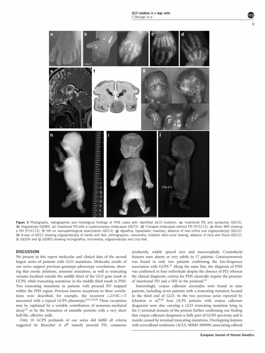
Pallister–Hall syndromeLimb anomalies. Limb anomalies were present in all 21 PHSindividuals of our cohort. The most common feature waspostaxial (48%) or insertional PD (48%) (Figures 3a and c).No patient with preaxial PD was recorded. Interestingly, Y-shapedmetacarpal/metatarsal was visualized on X-rays in 83% of casesand in all other cases, numeric or morphologic anomalies ofmetacarpal/metatarsal were noted (Figures 3c and d). Only onepatient (P15112) had bilateral and symmetrical Y-shapedmetacarpals without PD (Figure 3d). Brachydactyly with brachy-telephalangism was observed in at least 52% of PHS cases and nailhypoplasia in 69%.
Syndactyly (38%) and overlapping toes (9%) were frequentlyreported. Four fetuses with severe phenotypes exhibited mesomeliaor micromelia and three of them presented oligodactyly, club feet andarthrogryposis (Figures 3b, g–j).
Neurological findings. A HH was present in 12 patients, all withmutations falling in the ‘PHS’ domain (Figures 3e and f). In onefetus, neuropathological examination of the hypothalamic regionfound histological lesions of hamartoma, although a macroscopicmass was not visualized in the infandibular region (G024). Two casesdisplayed corpus callosum dysgenesis. Most patients had a normalintellectual efficiency, only three were slightly delayed. Seizures werereported in two cases as gelastic epilepsy.
Other findings. IUGR was found in 4/5 fetuses and growth wasdelayed in 6 patients. Besides, the endocrine manifestations of a HHranged from isolated growth hormone deficiency (4/13) to panhypo-pituitarism (1 case); 4/5 fetuses displayed adrenal hypoplasia. Oralanomalies were reported in all prenatal cases: cleft palate in threefetuses, micro/retrognatia in four and unexpectedly, a completeagnathia with absence of oral orifice in one (Figure 3c). Laryngealexamination revealed bifid epiglottis in half cases, always asympto-matic in postnatal cases. Choanal atresia was present in three patientsand two displayed cervical or preauricular chondroma. Moreover,imperforate or anteposed anus was present in half PHS casesincluding all fetuses. Congenital heart defects were diagnosed in sixpatients: interauricular septal defect in two, interventricular commu-nication in two, atrioventricular septal defect in one and an aorticarch anomaly in one. Renal anomalies were present in 41% rangingfrom kidney hypoplasia to agenesis and resulting in oligoamnios inprenatal cases with bilateral agenesis (three cases). Genitourinaryanomalies including micropenis (nine cases), hypospadias (one case)and uterovaginal aplasia (one case) were present in half of theindividuals. A severe developmental sexual disorder was present in agirl with a male caryotype exhibiting an undeveloped genital tubercle.Lung anomalies including abnormal lobulation or hypoplatic lungswere present in four individuals.
In situ hybridization of GLI3GLI3 expression pattern was studied during early human develop-ment using in situ hybridization on human embryo sections at CSs 14(day 32), 16 (day 40), 18 (day 44), and 19 (day 47) and at 8.5 weeks of
Table 4 Frequencies of clinical features in PHS individuals
Clinical features Frequency
Growth delay/GH deficiency 53% (10/19)
Limb anomalies
Y-shaped metacarpal 83% (15/18
Postaxial polydactyly 48% (10/21)
Insertional polydactyly 48% (10/21)
Preaxial polydactyly 0% (0/21)
Oligodactyly 14% (3/21)
Syndactyly 38% (8/21)
Brachydactyly/brachytelephalangism 52% (13/21)
Mesomelia 19% (4/21)
Nail hypoplasia 69% (9/13)
Overlapping toes 9% (2/21)
Cerebral anomalies
Hypothalamic hamartoma 100% (12/12)
Corpus callosum anomalies 17% (2/12)
Craniofacial anomalies
Micro/retro/agnathia 24% (5/21)
Choanal atresia 14% (3/21)
Cleft palate 14% (3/21)
Chondroma 9% (2/21)
Bifid epiglottis 44% (4/9)
Anal anomalies
Anal imperforation 43% (9/21)
Anteposed anus 5% (1/21)
Cardiac anomalies
Interauricular communication 9% (2/21)
Interventricular communication 9% (2/21)
Atrioventricular communication 5% (1/21)
Aortic arch anomaly 5% (1/21)
Renal anomalies 41% (7/17)
Genital anomalies 48% (10/21)
Lung dysplasia 50% (4/8)
Developmental delay 21% (3/14)
Seizures 13% (2/15)
GLI3 mutations in a large seriesF Demurger et al
7
European Journal of Human Genetics

development. At day 32, GLI3 was strongly expressed in ventral partof the prosencephalon, the mesencephalon and neural tube. GLI3expression was also expressed in otic vesicle. At day 40, the expressionpattern was observed in pharyngeal archs and was restricted later at
day 49 in maxillary and mandible. At the same time, GLI3 was alsoexpressed in distal limb buds, floor plate of the telencephalon,diencephalon and mesencephalon, neural tube and strongly in kidney(Figure 4).
Figure 1 Schematic representation of GLI3 domains and localization of the GLI3 mutations reported in this study. Red bars at the nucleotides 1998 and
3481 divide the gene into three segments, limiting the PHS region as described elsewhere.9 The colored boxes within GLI3 represent the seven regions of
similarity between human GLI proteins originally defined by Ruppert et al.34 ZNF: zinc-finger domain (aa 462–645), PC: proteolytic cleavage site, TA1
(aa 1376–1580) and TA2 (aa 1044–1322): two independent transactivation domains as described by Kalff-Suske et al.11 Mutations written in red: PHS
patients with severe phenotypes; in green: GCPS cases with abnormal corpus callosum; black bars: truncating nonsense and frameshift mutations; purple
bar: splice mutation; blue bars: missense mutations. A full color version of this figure is available at the European Journal of Human Genetics journal
online.
Figure 2 Photographs and radiographies of GCPS cases with identified GLI3 mutation. (a, b) Preaxial polysyndactyly in the feet with a broad first
metacarpal on X-rays (G076, G14083). (c) Broad hallux and syndactyly (G16012, daughter). (d) Preaxial polysyndactyly (G16012). (e) Bifid terminal
phalanx of the thumb (G14083). (f) Heptadactyly (preaxial and postaxial PD, G15198). (g) Broad thumbs (G16012, daughter). (h) Broad thumb and
postaxial PD type (b) (G16012).
GLI3 mutations in a large seriesF Demurger et al
8
European Journal of Human Genetics
DISCUSSION
We present in this report molecular and clinical data of the secondlargest series of patients with GLI3 mutations. Molecular results ofour series support previous genotype–phenotype correlations, show-ing that exonic deletions, missense mutations, as well as truncatingvariants localized outside the middle third of the GLI3 gene result inGCPS, while truncating mutations in the middle third result in PHS.Two truncating mutations in patients with preaxial PD mappedwithin the PHS region. Previous known exceptions to these correla-tions were described, for example, the recurrent c.2374C4Tassociated with a typical GCPS phenotype.9,11,19,20 These exceptionsmay be explained by a variable contribution of nonsense-mediateddecay12 or by the formation of unstable proteins with a very shorthalf-life, effective nulls.
Only 10 GCPS probands of our series did fulfill all criteriasuggested by Biesecker et al6 namely preaxial PD, cutaneous
syndactyly, widely spaced eyes and macrocephaly. Craniofacialfeatures were absent or very subtle in 17 patients. Craniosynostosiswas found in only two patients confirming the low-frequencyassociation with GCPS.21 Along the same line, the diagnosis of PHSwas confirmed in four individuals despite the absence of PD, whereasthe clinical diagnostic criteria for PHS classically require the presenceof insertional PD and a HH in the proband.22
Interestingly, corpus callosum anomalies were found in ninepatients, including seven patients with a truncating mutation locatedin the third end of GLI3. In the two previous series reported byJohnston et al,9,10 four GCPS patients with corpus callosumdysgenesis were also carrying a GLI3 truncating mutation lying inthe C-terminal domain of the protein further confirming our findingthat corpus callosum dysgenesis is fully part of GCPS spectrum and ismainly caused by terminal truncating mutations. Overlapping featureswith acrocallosal syndrome (ACLS, MIM# 200990) associating callosal
Figure 3 Photographs, radiographies and histological findings of PHS cases with identified GLI3 mutation. (a) Insertional PD and syndactyly (G072).
(b) Oligodactyly (G080). (c) Insertional PD with a supernumerary metacarpal (G072). (d) Y-shaped metacarpal without PD (P15112). (e) Brain MRI showing
a HH (P15112). (f) HH on neuropathological examination (G013). (g) Agnathia, hypoplastic maxillary, absence of oral orifice and oligosyndactyly (G012).
(h) X-rays of G012 showing oligosyndactyly of hands and feet, arthrogryposis, mesomelia, bilateral radio-ulnar bowing, absence of tibia and fibula (G012).
(i) (G024) and (j) (G080) showing micrognathia, micromelia, oligosyndactyly and club feet.
GLI3 mutations in a large seriesF Demurger et al
9
European Journal of Human Genetics

dysgenesis, hypertelorism, intellectual disability and PD23 areexplained by an impaired GLI3 processing in patients with KIF7mutations.24 Facial dysmorphism, as well as vermis dysgenesis withbrainstem anomalies (molar tooth sign), strongly indicated thediagnosis of ACLS. Conversely, two GLI3 mutated cases with corpuscallosum dysgenesis have been reported as ACLS25,26 and a thirdsimilar patient has been reported by Johnston et al.10 All threemutations were missense and clustered in the same region between aa903 and 934 suggesting a potential severe phenotype associated withalterations of this region. Whatever, mutation analysis in bothgenes is therefore essential as the distinction between these twosyndromes is of obvious significance for genetic counselingconsidering the difference in heredity and neurodevelopmentaloutcome and patients with a GLI3 mutation may be diagnosed asGCPS.
Interestingly, macrosomia was observed in at least 13% of GCPScases in our series. Macrosomia and PD are also observed inSimpson–Golabi–Behmel syndrome type 1 (MIM# 312870), aX-linked mental retardation syndrome ascribed to Glypican3(GPC3) mutation, which was suspected in family G068, with twobrothers displaying macrosomia and PD at birth. The frameshift GLI3mutation was inherited from their asymptomatic father carrying asomatic mosaic mutation. Along the same way, we identified a mosaiclarge deletion in a GCPS patient with developmental delay (G059). Toour knowledge, only one instance of GLI3 germline mosaicism hasbeen already described in two PHS sibs,18 which is therefore a rareevent.
Cerebral MRI may be useful to detect HH that was found in allPHS individuals of our series. Abnormal metacarpals in particularY-shaped metacapals appear to be a more significant criterion thaninsertional PD. At least three PHS patients without PD were alreadyreported.10,18,27 All of them presented fused or hypoplasticmetacarpal. The mouse model for PHS Gli3D699/D699 displaysabnormal metacarpal morphology with PD or oligodactyly at alower frequency.28 Central poly/syndactyly and Y-shapedmetacarpals are extremely uncommon in other syndromes.Although associated to a good neurodevelopmental outcome, PHS
displays a wide range of severity varying from mild to lethalphenotypes depending on the severity of malformations present inthe individuals, in particular bilateral kidney agenesis, craniofacialfeatures (agnathia, absence of oral orifice, cleft palate or premaxillaryagenesis in one case), heart defects and/or reductional limb defects.Interestingly, skeletal dysplasia with ulna bowing, tibia and fibulahypoplasia was already reported in other cases described asPHS,10,29,30 further suggesting that acromesomelic limb shorteningwith radio-ulnar bowing, tibial and fibular hypolasia/agenesis are partof the phenotypic spectrum of PHS. Interestingly, all mutations foundin severe fetal phenotypes of our series were clustered in the middlethird of the gene, between c.2941 and c.3324. To assess whether thesevere craniofacial features observed (agnathia, absence of oral orifice)were a direct effect of the GLI3 mutation, we undertook theexpression analysis of GLI3 during human development. Indeed, inaddition to the early expression of GLI3 in pharyngeal and then laterin mandible and maxillary, GLI3 was highly expressed in all targettissues of the disease.
Besides PHS cases, Y-shaped metacarpal is also observed in oro-facio-digital syndrome type VI (OFD VI; MIM# 277170).31 Overlap ofPHS with OFD has been previously discussed as oral anomalies (oralfrenula, hamartoma, cleft palate) and/or skeletal dysplasia are oftenassociated to GLI3 mutations.10 Avila et al32 screened eight patientswith OFD associated with midline abnormalities but no mutation wasfound. They suggested that GLI3 should be screened in patients withOFD only when associated to one of the pathognomonic sign of PHS(HH, mesoaxial PD, bifid epiglottis and IA).
In one case (G013), the association of IUGR, PD, bilateral renalagenesis and anal anteposition without macroscopic HH first led tothe suspicion of Smith–Lemli–Opitz (SLO; MIM# 270400). Afterexclusion of a cholesterol biosynthesis defect, a GLI3 screeningidentified a de novo frameshift mutation in exon 15. Retrospectiveanalysis of the brain identified microscopic changes suggestive ofhamartoma. This observation underlines the phenotypic overlap ofPHS and SLO that was suggested previously,22 both disordersassociating IUGR, PD and possible renal agenesis but IA,insertional PD and HH are exceptional in SLO.33
CONCLUSION
Here we report on clinical and molecular data of a large series of 76individuals from 55 families carrying a heterozygous GLI3 mutationor rearrangement, 55 GCPS and 21 PHS, 41 being novel mutations.Our results render more precisely the genotype–phenotype correla-tion of GLI3 mutations proposed by Johnston et al, and furtherhighlight the clinical overlap between GCPS and ACS and betweenPHS, SLO and OFD. Interestingly, our series including fetal casesenlarge the phenotypic spectrum of PHS to severe craniofacial andreductional limb defects, emphasize on the possible lethality of PHSwith a clustering of truncating mutation in a subdomain of the ‘PHS’GLI3 domain. In addition, we add CCA among frequent signs ofGCPS with a strong genotype–phenotype correlation of corpuscallosum dysgenesis with truncating C terminal mutations, andmacrosomia as a new clinical feature of GCPS.
CONFLICT OF INTEREST
The authors declare no conflict of interest.
ACKNOWLEDGEMENTSWe are grateful to the patients and their parents participating in this study.
Figure 4 In situ hybridization of GLI3 during human development. (a, b) CS
15; (c, d) CS 19. (b, d) Slides hybridized with an antisense GLI3 probe.
(a, c) Adjacent slides respectively to (b) and (d) stained with HES. In
addition to the expression in central nervous system (prosencephalon (pr),
rhombencephalon (rh) neural tube (nt)), limb bud (lb), pituitary gland(p, arrowhead) and kidney (K), a signal was observed in human pharyngeal
arches (pa1) then in mandible (mn) and maxillary (mx) (red arrows). A full
color version of this figure is available at the European Journal of Human
Genetics journal online.
GLI3 mutations in a large seriesF Demurger et al
10
European Journal of Human Genetics

WEB SOURCES
OMIM: http://www.ncbi.nlm.nih.gov/omimPolyPhen2: http://genetics.bwh.harvard.edu/pph2SIFT: http://blocks.fhcrc.org/sift/SIFT.htmlMutalyzer: http://www.humgen.nl/mutalyzer/1.0.1LOVD: http://www.lovd.nl/3.0
1 Vortkamp A, Franz T, Gessler M, Grzeschik KH: Deletion of GLI3 supports the homologyof the human Greig cephalopolysyndactyly syndrome (GCPS) and the mouse mutantextra toes (Xt). Mamm Genome Off J Int Mamm Genome Soc 1992; 3: 461–463.
2 Kang S, Rosenberg M, Ko VD, Biesecker LG: Gene structure and allelic expressionassay of the human GLI3 gene. Hum Genet 1997; 101: 154–157.
3 Hall JG, Pallister PD, Clarren SK et al: Congenital hypothalamic hamartoblastoma,hypopituitarism, imperforate anus and postaxial polydactyly–a new syndrome? Part I:clinical, causal, and pathogenetic considerations. Am J Med Genet 1980; 7: 47–74.
4 Clarren SK, Alvord Jr EC, Hall JG: Congenital hypothalamic hamartoblastoma,68(3):hypopituitarism, imperforate anus, and postaxial polydactyly–a new syndrome?Part II: neuropathological considerations. Am J Med Genet 1980; 7: 75–83.
5 Biesecker L, Johnston J: Syndromic and non-syndromic GLI3 phenotypes. Clin Genet2005; 68: 284–284.
6 Biesecker LG: The Greig cephalopolysyndactyly syndrome. Orphanet J Rare Dis 2008;3: 10.
7 Johnston JJ, Walker RL, Davis S et al: Zoom-in comparative genomic hybridisationarrays for the characterisation of variable breakpoint contiguous gene syndromes.J Med Genet 2007; 44: e59.
8 Aza-Blanc P, Lin HY, Ruiz i Altaba A, Kornberg TB: Expression of the vertebrate Gliproteins in Drosophila reveals a distribution of activator and repressor activities.Development (Cambridge, England) 2000; 127: 4293–4301.
9 Johnston JJ, Olivos-Glander I, Killoran C et al: Molecular and clinical analyses of Greigcephalopolysyndactyly and Pallister-Hall syndromes: robust phenotype prediction fromthe type and position of GLI3 mutations. Am J Hum Genet 2005; 76: 609–622.
10 Johnston JJ, Sapp JC, Turner JT et al: Molecular analysis expands the spectrum ofphenotypes associated with GLI3 mutations. Hum Mutat 2010; 31: 1142–1154.
11 Kalff-Suske M, Wild A, Topp J et al: Point mutations throughout the GLI3 gene causeGreig cephalopolysyndactyly syndrome. Hum Mol Genet 1999; 8: 1769–1777.
12 Furniss D, Critchley P, Giele H, Wilkie AOM: Nonsense-mediated decay and themolecular pathogenesis of mutations in SALL1 and GLI3. Am J Med Genet A 2007;143A: 3150–3160.
13 Shin SH, Kogerman P, Lindstrom E, Toftgard R, Biesecker LG: GLI3 mutations inhuman disorders mimic Drosophila cubitus interruptus protein functions and localiza-tion. Proc Natl Acad Sci USA 1999; 96: 2880–2884.
14 Krauss S, So J, Hambrock M et al: Point mutations in GLI3 lead to misregulation of itssubcellular localization. PLoS One 2009; 4: e7471.
15 Wildeman M, van Ophuizen E, den Dunnen JT, Taschner PEM: Improving sequencevariant descriptions in mutation databases and literature using the Mutalyzer sequencevariation nomenclature checker. Hum Mutat 2008; 29: 6–13.
16 O’Rahilly R: Human embryo. Nature 1987; 329: 385.17 Crosnier C, Attie-Bitach T, Encha-Razavi F et al: JAGGED1 gene expression during
human embryogenesis elucidates the wide phenotypic spectrum of Alagille syndrome.Hepatology (Baltimore, MD) 2000; 32: 574–581.
18 Ng D, Johnston JJ, Turner JT et al: Gonadal mosaicism in severe Pallister-Hallsyndrome. Am J Med Genet A 2004; 124A: 296–302.
19 Debeer P, Peeters H, Driess S et al: Variable phenotype in Greig cephalopolysyndactylysyndrome: clinical and radiological findings in 4 independent families and 3sporadic cases with identified GLI3 mutations. Am J Med Genet A 2003; 120A:49–58.
20 Furniss D, Kan S-H, Taylor IB et al: Genetic screening of 202 individuals withcongenital limb malformations and requiring reconstructive surgery. J Med Genet2009; 46: 730–735.
21 Hurst JA, Jenkins D, Vasudevan PC et al: Metopic and sagittal synostosis in Greigcephalopolysyndactyly syndrome: five cases with intragenic mutations or completedeletions of GLI3. Eur J Hum Genet 2011; 19: 757–762.
22 Biesecker LG, Graham Jr JM: Pallister-Hall syndrome. J Med Genet 1996; 33:585–589.
23 Putoux A, Nampoothiri S, Laurent N et al: Novel KIF7 mutations extend thephenotypic spectrum of acrocallosal syndrome. J Med Genet 2012; 49: 713–720.
24 Putoux A, Thomas S, Coene KLM et al: KIF7 mutations cause fetal hydrolethalus andacrocallosal syndromes. Nat Genet 2011; 43: 601–606.
25 Elson E, Perveen R, Donnai D, Wall S, Black GCM: De novo GLI3 mutation inacrocallosal syndrome: broadening the phenotypic spectrum of GLI3 defects andoverlap with murine models. J Med Genet 2002; 39: 804–806.
26 Speksnijder L, Cohen-Overbeek TE, MFCM Knapen et al: A de novo GLI3 mutation in apatient with acrocallosal syndrome. Am J Med Genet A 2013; 161: 1394–1400.
27 Verloes A, David A, Ngo L, Bottani A: Stringent delineation of Pallister-Hall syndromein two long surviving patients: importance of radiological anomalies of the hands.J Med Genet 1995; 32: 605–611.
28 Hill P, Wang B, Ruther U: The molecular basis of Pallister–Hall associated polydactyly.Hum Mol Genet 2007; 16: 2089–2096.
29 Encha-Razavi F, Larroche JC, Roume J et al: Congenital hypothalamic hamartomasyndrome: nosological discussion and minimum diagnostic criteria of a possiblyfamilial form. Am J Med Genet 1992; 42: 44–50.
30 Roscioli T, Kennedy D, Cui J et al: Pallister-Hall syndrome: unreported skeletalfeatures of a GLI3 mutation. Am J Med Genet A 2005; 136A: 390–394.
31 Poretti A, Vitiello G, Hennekam RCM et al: Delineation and diagnostic criteria ofOral-Facial-Digital Syndrome type VI. Orphanet J Rare Dis 2012; 7: 4.
32 Avila M, Gigot N, Aral B et al: GLI3 is rarely implicated in OFD syndromes with midlineabnormalities. Hum Mutat 2011; 32: 1332–1333.
33 Quelin C, Loget P, Verloes A et al: Phenotypic spectrum of fetal Smith-Lemli-Opitzsyndrome. Eur J Med Genet 2012; 55: 81–90.
34 Ruppert JM, Vogelstein B, Arheden K, Kinzler KW: GLI3 encodes a 190-kilodaltonprotein with multiple regions of GLI similarity. Mol Cell Biol 1990; 10: 5408–5415.
GLI3 mutations in a large seriesF Demurger et al
11
European Journal of Human Genetics