Correlation of genotype and phenotype in Beta- thalassemia
171
Correlation of genotype and phenotype in Beta- thalassemia Citation for published version (APA): Efremov, D. G. (1994). Correlation of genotype and phenotype in Beta-thalassemia. [Doctoral Thesis, Maastricht University]. Rijksuniversiteit Limburg. https://doi.org/10.26481/dis.19940422de Document status and date: Published: 01/01/1994 DOI: 10.26481/dis.19940422de Document Version: Publisher's PDF, also known as Version of record Please check the document version of this publication: • A submitted manuscript is the version of the article upon submission and before peer-review. There can be important differences between the submitted version and the official published version of record. People interested in the research are advised to contact the author for the final version of the publication, or visit the DOI to the publisher's website. • The final author version and the galley proof are versions of the publication after peer review. • The final published version features the final layout of the paper including the volume, issue and page numbers. Link to publication General rights Copyright and moral rights for the publications made accessible in the public portal are retained by the authors and/or other copyright owners and it is a condition of accessing publications that users recognise and abide by the legal requirements associated with these rights. • Users may download and print one copy of any publication from the public portal for the purpose of private study or research. • You may not further distribute the material or use it for any profit-making activity or commercial gain • You may freely distribute the URL identifying the publication in the public portal. If the publication is distributed under the terms of Article 25fa of the Dutch Copyright Act, indicated by the “Taverne” license above, please follow below link for the End User Agreement: www.umlib.nl/taverne-license Take down policy If you believe that this document breaches copyright please contact us at: [email protected] providing details and we will investigate your claim. Download date: 23 Jul. 2022
-
Upload
khangminh22 -
Category
Documents
-
view
0 -
download
0
Transcript of Correlation of genotype and phenotype in Beta- thalassemia
Correlation of genotype and phenotype in Beta-thalassemiaCitation for published version (APA):
Efremov, D. G. (1994). Correlation of genotype and phenotype in Beta-thalassemia. [Doctoral Thesis,Maastricht University]. Rijksuniversiteit Limburg. https://doi.org/10.26481/dis.19940422de
Document status and date:Published: 01/01/1994
DOI:10.26481/dis.19940422de
Document Version:Publisher's PDF, also known as Version of record
Please check the document version of this publication:
• A submitted manuscript is the version of the article upon submission and before peer-review. There canbe important differences between the submitted version and the official published version of record.People interested in the research are advised to contact the author for the final version of the publication,or visit the DOI to the publisher's website.• The final author version and the galley proof are versions of the publication after peer review.• The final published version features the final layout of the paper including the volume, issue and pagenumbers.Link to publication
General rightsCopyright and moral rights for the publications made accessible in the public portal are retained by the authors and/or other copyrightowners and it is a condition of accessing publications that users recognise and abide by the legal requirements associated with theserights.
• Users may download and print one copy of any publication from the public portal for the purpose of private study or research.• You may not further distribute the material or use it for any profit-making activity or commercial gain• You may freely distribute the URL identifying the publication in the public portal.
If the publication is distributed under the terms of Article 25fa of the Dutch Copyright Act, indicated by the “Taverne” license above,please follow below link for the End User Agreement:
www.umlib.nl/taverne-license
Take down policyIf you believe that this document breaches copyright please contact us at:
providing details and we will investigate your claim.
Download date: 23 Jul. 2022

CORRÉLATION OF GENOTYPE ANDPHENOTYPE IN B-THALASSEMIA
PROEFSCHRIFT
ter verkryging van de graad van doctoraan de Rijksuniversiteit Limburg te Maastricht,
op gezag van de Rector Magnificus, Prof. dr. H. Philipsen,volgens het besluit van het College van Dekanen,
in het opcnbaar te verdedigenop vrydag 22 april 1994 om 14.00 uur
door
DEVIITAR GEORGI EFREMOV

PROMOTORES:Prof. dr. J.P.M. GeraedtsProf. dr. T.H.J. Huisman, Augusta Georgia, USA
CO-PROMOTOR:Dr. J. ten Kate
BEOORDELINGSCOMMISSIE:Prof. dr. F.C.S. Ramaekers (voorzitter)Prof. dr. P.J. BrombacherProf. dr. A. Burrone, Buenos Aires, ArgentiniëProf. dr. H.F.P. HillenProf. dr. G.H. Petkov, Stara Zagora, Bulgarije

ACKNOWLEDGEMENTS
I am extremely grateful to Professor Dr. Titus H.J. Huisman for hisguidance during the course of this work, and for his continuous care andsupport which greatly influenced my career and research determination.I am indebted to Professor Dr. Joep P.M. Geraedts for giving me the oppor-tunity of obtaining a doctoral degree in The Netherlands and for his helpduring the preparation of this dissertation. The generous help of Pro-fessor Dr. Paul J. Brombacher in the realization of this project is alsogreatly appreciated. I thank my father, Professor Dr. Georgi D. Efremov,for his encouragement and continuous scientific and moral support. Theexcellent editorial and technical assistance of Mrs. Marianne F.H. Carver,and the contribution of colleagues and friends who participated 1n thiswork, are gratefully acknowledged.


Dedicated to Maria and Kristian

CONTENTS
Abbreviations
Introduction and Review of the Literature 1
la Appendix I 35
Materials and Methods 53
Molecular Characterization of B-Thalassemia Mutations 69
3a B-Thalassemia in Yugoslavia 71
3b B-Thalassean'a In Bulgaria 79
Correlation of Clinical Severity With Different e-Glo- 85bin Gene Defects
4a Variation in Clinical Severity Among Patients With 87Hb Lepore-Boston-B-Thalassenria is Related to the Typeof B-Thalassenia
4b B-Thalassenia Due to a T->A Mutation Within the ATA 95Box
4c Mild and Severe B-Thalassenia Among Homozygotes From 101Turkey: Identification of the Types by Hybridizationof Amplified DNA With Synthetic Probes
Possible Factors Influencing the Hemoglobin and Fetal 109Hemoglobin Levels in Patients With B-Thalassemia Dueto a Homozygosity for the IVS-I-6 (T->C) Mutation
The -158 (C->T) Promoter Mutation is Responsible For 123the Increased Transcription of the 3' Y Gene in theAtlanta Type of HPFH
Discussion 137
Summary 151
Samenvatting 157
Curriculum Vitae 161
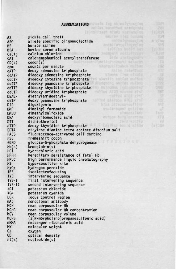
ABBREVIATIONS
AS sickle cell traitASO all ele specific oligonucleotideBS borate salineBSA bovine serum albuminCaCl2 calcium chlorideCAT chloramephenicol acetyltransferaseCD(s) codon(s)cpm counts per minutedATP deoxy adenosine tri phosphateddATP dideoxy adenosine triphosphateddCTP dideoxy cytosine triphosphateddGTP dideoxy guanosine triphospateddTTP dideoxy thymidine triphosphateddUTP dideoxy uridine triphosphateDEAE- diethylaminoethyl-dGTP deoxy guanosine triphosphateDIG digoxigeninDMF dimethyl formamideDMSO d i me thyls u1 fox i deDNA deoxyribonucleic acidDTT dithiothreitoldTTP deoxy thymidine triphosphateEDTA ethylene diamine tetra acetate disodium saltFACS fluorescence-activated cell sortingFSC frameshift codonG6PD glucose-6-phosphate dehydrogenaseHb(s) hemoglobin(s)HC1 hydrochloric acidHPFH hereditary persistence of fetal HbHPLC high performance liquid chromatographyHS hypersensitive siteH2O2 hydrogen peroxideIEF isoelectrofocusingIVS intervening sequenceIVS-I first intervening sequenceIVS-11 second intervening sequenceKC1 potassium chlorideKCN potassium cyanideLCR locus control regionmAb monoclonal antibodyMCH mean corpuscular HbMCHC mean corpuscular Hb concentrationMCV mean corpuscular volumeMOPS (3[N-morpholino]propanesulfonic acid)mRNA messenger ribonucleic acidMW molecular weightO2 oxygenOD optical densitynt(s) nucleotide(s)

PAGEPBSPCR(s)PCVpoly APVPRBC(s)RFLPRNARTSOSSEASSsscSSPETMACTBETETEATFATmthaiUTRUVWBC(s)
poiyacrylamide gel electrophoresisphosphate buffered salinepolymerase chain reaction(s)packed cell volumepoiyadenylationpolyvyni1pyrolidonered blood cell(s)restriction fragment length polymorphismribonucleic acidreverse transcription/transcribedsodium dodecilsuphateSoutheast Asiansickle cell anemiasodium saline citrate buffersodium saline phosphate EDTA buffertetramethyl ammonium chlorideTris borate EDTA bufferTris EDTA bufferTris EDTA acetate buffertrifluoroacetic acidmelting temperaturethalassemiauntranslated regionultravioletwhite blood cell(s)

CHAPTER 1
I N T R O D U C T I O N
A N D
R E V I E W O F T H E L I T E R A T U R E

CHAPTER 1
INTRODUCTION
B-Thal is one of the most common single gene disorders worldwide,causing a major health problem especially in underdeveloped countries.It has the highest frequency in areas where malaria was/is endemic, suchas the Mediterranean, Southeast Asia, and India, suggesting positive selec-tion by the malaria parasite in sustaining the affected g-globin gene.
B-Thal is a heterogeneous group of disorders, both in terms of molec-ular defects and in the phenotypic expression. In all cases, however, themolecular defect causes a reduction or absence of B-globin chain synthesisleading to B+- or B°-thal, respectively. The B-globin chain deficiencyresults in a decrease or absence of the adult Hb (Hb A) which is composedof two identical B- and two identical o-globin chains. The levels of theother adult Hb (Hb A2) and the fetal Hb (Hb F) are variably increased, butthis is usually not sufficient to counterbalance the Hb A deficiency.
The clinical course of the B-thal syndromes is variable and rangesfrom mild (B-thal minor) through intermediate (B-thal intermedia) toextremely severe (B-thal major). In the latter, patients are transfusion-dependent and death rates are high in children and young adults, even withproper transfusion therapy. The severity of the disease is largely deter-mined by the level of a/non-a-globin chain imbalance. The excess a-globinchains precipitate and damage the erythroid precursors in the bone marrow,leading to their premature destruction and an ineffective erythropoiesis.Apart from the degree of B-globin chain deficiency, the globin chainimbalance can also be influenced by the level of a- and r-globin chainproduction. Co-inheritance of o-thal (which reduces the a-globin chainexcess) and/or genetic factors which increase the Y-globin gene outputcan ameliorate the clinical course of the disease.
Management strategies in B-thal have focused mainly on two areas:Prevention by means of prenatal diagnosis and development of new thera-peutic approaches such as gene therapy and pharmacologie induction of Hb Fsynthesis. Assessment of different factors that influence the clinicalexpression of the disease can also provide a more accurate prognosis andindicate the most appropriate treatment for each individual patient.
The main objectives of the work presented in this dissertation are:
To characterize the molecular defects leading to B-thal inthe countries of the former Yugoslavia and Bulgaria in order toestablish a DNA-based prenatal diagnosis program in this part ofthe Balkan Peninsula;
to investigate the relative contribution of the differentfactors that can affect the clinical presentation of the disease;
to evaluate the influence of genetic factors, both linkedand unlinked to the B-globin gene complex on Hb F production inB-thal.

4 • ]
HISTORY
The first description of thalassemia, which appeared in 1925, wasby the American pediatrician Thomas B. Cooley (1). Although some ancientGreek and early Italian writers also seem to have referred to this condi-tion (2), it is interesting that the disease had been overlooked as a sepa-rate clinical entity by Mediterranean physicians who must have frequentlyencountered it among their patients. Within the following 10 years, manyreports of Cooley's anemia appeared in the literature and it became evidentthat the disease occurred predominantly in the Mediterranean population.To associate the disease with the Mediterranean area, Whipple and Bradford1n 1932 gave it the name 'thalassemia', referring to the Greek word'thalassa' meaning 'the sea' (3). During the 1930s the clinical syndromeof thalassemia had been well described, and in 1938 a genetic determinantunderlying the disease was suggested (4). By the end of the 1940s it wasalready apparent that thalassemia was not a single disorder but a complexsyndrome resulting from the interaction of many genetic factors. In the 1late 1950s detailed Hb analyses were performed and Hb F (02Y2)» Hb A2(0262)» Hb H (04), and Hb Bart's (Y4) were observed in different patientswith thalassemia (5-8). In 1959 these findings allowed Ingram and Stretton Jto define two major classes of thalassemia, namely oand e, and to suggest ian inherited defect in the a- or B-globin chain synthesis in these twoforms, respectively (9); experimental evidence came with the advancement1n biosynthesis studies showing an imbalanced synthesis in both disorders I(10). Further heterogeneity among the thalassemias was noted, and forms "with decreased or absent o- and B-globin chain synthesis were described.Advancements in recombinant DNA technology during the 1970s made it pos-sible to show a decreased production in globin mRNA (11), and to identifydeletions of individual globin genes in certain thalassemic patients (12).Studies at the DNA level allowed the characterization of the genomic organ-ization and structure of individual globin genes (13-19). Further develop-ment of molecular biology techniques, and especially the introduction ofthe PCR procedure in the second half of the 1980s, allowed extensive andalmost complete characterization of the molecular defects underlying thethalassemias (20,21). These advancements also allowed the development ofsuccessful screening and prenatal diagnostic programs, and formed the basisfor future therapeutic modalities such as gene therapy and reactivation ofthe Y-globin genes.
ETHNIC DISTRIBUTION
The thalassemias probably represent the commonest gene disorder tocause a major public health problem in the world population (22). Peopleof Mediterranean, Asian and African ancestry are primarily affected, butsporadic cases have been reported in many other ethnic groups. It isbelieved that the geographic distribution of the thalassemias is due toa decreased morbidity of individuals carrying the trait when infected withmalarial parasites. The reasons for this protection of the thalassemiahétérozygotes are still unknown, but it has been proposed that the redcell membrane in thalassemia hétérozygotes is particularly susceptibleto damage by oxidation and that infection with the malaria parasite pro-vides sufficient oxidative stress to perturb intracellular metabolism in

a manner that leads to premature death of the parasite (23). This hypo-thesis also provides a uniform explanation for the decreased malaria sus-ceptibility observed among hétérozygotes for a- and B-thal, and for Hb Sand G6PD deficiency. More recently, a role for enhanced immune recognitionand subsequent clearance of thalassemic red cells infected with the malariaparasite has been suggested (24).
Starting in the mid 1940s, studies have been performed worldwide toestimate the prevalence of thalassemia in various populations (reviewedin Ref. 22). While a-thal appears to be more common in Southeast Asia,India, and the South Pacific (23), B-thal is the most common genetic dis-order in Mediterranean countries, with frequencies ranging from 5 to 20%in some of the Mediterranean islands (25). As this dissertation dealsmainly with B-thal in the Mediterranean, its incidence in this area willbe described in greater detail later. The incidence of heterozygous B-thalin Southeast Asian populations is approximately 5% (23), while in AmericanBlacks it is approximately 0.5% (26). The frequency of heterozygous o-thaiin Southeast Asia is also around 5%, but increases to 20% in Thailand,and even to 80% in certain regions of New Guinea and India (23,27). Theheterozygosity for a-thal in Southeast Asia includes both a-thal-1, whereboth a-globin genes are deleted from a single chromosome, and a-thal-2where only a single a-globin gene is missing. Heterozygosity for an a-thal-2 deletion is found in 25 to 30% of American Blacks; approximately3% have a homozygosity for this deletion.
A recent compilation of data for 535,000 Mediterranean individualstested for the incidence of heterozygous B-thal confirmed that B-thaloccurs mainly in Cypriots (14.7%), Greeks (8%), Albanians (7.1%), and Ital-ians (3.7%), especially Sardinians (12.6%) and Sicilians (5.9%) (Fig. 1/1).A high incidence of B-thal was also found among Lybians (4.6%) and Tuni-sians (4.4%). A frequency of 2 to 3% was reported for the populations ofTurkey, Lebanon, Israel, Malta, Algeria, Morocco, and Corsica. Lower fre-quencies were reported in countries of the former Yugoslavia (1.4%) andSpain (0.5%), with the lowest incidence in France (0.1%) (see Ref. 25 forreferences). The frequency of a B-thal heterozygosity in 1,745 Albanianschool children living in Macedonia was only 2% (25). However, a differ-ence in the frequency of B-thal in different regions of Albania has alsobeen observed, with the lowest values in the eastern provinces that borderMacedonia (28). The incidence of a-thal in the Mediterranean Basin hasnot been completely evaluated; some data are available for Spain, Mace-donia, Croatia, Greece, Sardinia, Tunisia, and Algeria. The incidence ofa-thal in Spain, Macedonia, Croatia, and Greece is low, as judged by therarity of patients with Hb H disease and by cord blood surveys. Screeningof 3,326 Macedonian newborns showed an a-thal incidence of 2.3%, whilescreening of 1,546 Croatian newborns showed an incidence of 1.4% (25). Anincidence of 0.2% was observed among 1,043 newborns of Albanian national-ity living in Macedonia. The incidence of a-thal in Spain and Greece islow, namely 0.2 and 0.5%, respectively. However, this condition occursat a much higher frequency in Sardinia (6.9%), Tunisia (4.8%), and Algeria(9%) (see Ref. 25 for references).

0-0,1(1,5-10)*[>20.Û00]
3,7(0,4-30)[153.159]
2,1(0,2-6,7)
2,1[3.819]
FI6. 1/1. The incidence of B-thal trait in Mediterranean countries deter-mined during population studies. Total number of screened subjectswas 535,528; the numbers in parentheses give the ranges and thosebetween brackets the numbers of- persons involved in these surveys.The asterisk (*) indicates immigrants.
CLINICAL AND HEMATOLOGICAL FEATURES
The three clinical syndromes associated with differences in the degreeof severity have been defined historically as B-thal major, B-thal minor,and B-thal intermedia. While B-thal major and B-thal minor represent theclinical features observed in B-thal homozygotes and hétérozygotes, respec-tively, individuals with B-thal intermedia may belong to both groups. Inall cases, however, the basic pathophysiological phenomenon is an imbalancein globin chain synthesis due to a deficient or absent B-globin chain pro-duction. The consequence is an excess of a chains which cannot formtetramers and therefore precipitate within the developing RBC precursorsin the bone marrow. The precipitated a chains damage the cell membraneand the various organelles, leading to the premature destruction of theerythroblasts in the marrow and an ineffective erythropoiesis. The clin-ical severity of B-thal is therefore directly proportional to the degreeof chain imbalance, and depends mainly on the level of B-globin chain pro-duction. However, two additional factors can have a positive influenceon the globin chain imbalance, namely the substitution of 6 chain synthesisby an increased Y chain production, and a decrease in the a chain excessdue to a concomitant o-thal condition.
Heterozygous B-Thai (B-thal minor). B-Thal hétérozygotes are usuallyasymptomatic, presenting with only a mild anemia or even normal .Mb levels.

Anemia of moderate severity, as seen in B-thal intermedia, has only occa-sionally been described. The MCV and MCH are usually decreased well belownormal, with typical values of 60 to 70 fl (normal range 85-92 f 1 ) and 20to 25 pg (normal range 27-32 pg), respectively. Further hématologie char-acteristics include microcytosis, hypochromia, anisocytosis, and poikilo-cytosis with targeting and basophilic stippling of the red cells in theperipheral blood, and mild erythroid hyperplasia in the bone marrow. Mildto moderate splenomegaly is present in only a few cases. The diagnosisis confirmed by Hb electrophoresis, and by quantitating the levels of Hb A2which are about twice the normal values of 2-32. Hb F levels are slightlyelevated in approximately half the cases (1-5%). Jji vitro globin chainbiosynthesis analysis shows an o/B chain synthesis ratio of 1.5 to 2.0(normal range 0.9-1.1). Less frequently, B-thal hétérozygotes can havenormal Hb A2 and Hb F levels, and decreased or even normal MCV and MCHvalues. These silent carriers are usually distinguished from o-thal hét-érozygotes by the finding of a clinically significant B-thal syndrome intheir offspring and by an impaired a/B-globin chain synthesis ratio. Hét-érozygotes with normal or slightly decreased Hb A2 levels and increasedHb F levels (5-20%) carry a «B-thal trait. Finally, some B-thal hétérozy-gotes are characterized by the presence of a Hb Lepore, which containsnormal a chains and hybrid 6B chains; the latter have an amino acidsequence of the S chain at the N-terminus and that of the B chain at theC-terminus. Apart from 8-12% Hb Lepore, these B-thal hétérozygotes havenormal Hb A2 and slightly elevated Hb F levels.
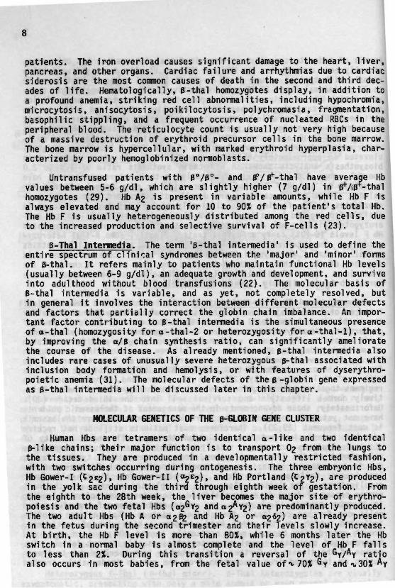
Homozygous B-Thai (B-thal major). The clinical and hematologicalfeatures of B-thal major become manifest several months after birth, afterthe completion of the fetal to adult Hb switch and the decrease in Hb Fproduction. Usually within a year, a severe hypochromic, microcytic, hemo-lytic anemia develops, and a regular transfusion program must be undertakento maintain adequate Hb levels. In children not receiving transfusiontherapy the severe anemia leads to an early death at 3 to 4 years of agefor those homozygous for B°-thal, and at 8 to 12 years of age for thosewith a B+-thal homozygosity (29). On the other hand, proper transfusionregimens together with adequate iron chelation therapy have allowed a rela-tively normal life style and survival well into the third decade (30).Unfortunately, these advancements in the management of thalassemic patientshave not yet been reached in the Third World countries where thalassemiais most frequent (29,30). Therefore, most patients still do not receiveadequate transfusion and iron chelation therapy, allowing a variety ofclinical features to emerge. Early manifestations include progressiveenlargement of the liver and spleen, bone changes with a typical facièsdue to bone marrow expansion, and repeated pathological fractures of thelong bones. Gallstones, leg ulcers, and recurrent infections are frequentcomplications. The latter, along with a neglected anemia, are the mostcommon causes of death in early childhood. Secondary hypersplenism leadingto thrombocytopenia, leukopenia, and rapid destruction of transfused redcells can further complicate the clinical picture. Splenectomy is oftenindicated, which, on the other hand, imposes a high risk of septicemia.Growth retardation becomes most noticeable around 10 years of age, andmenarche and secondary sexual characteristics usually do not follow. Thecourse of the disease is further complicated by iron overload due to trans-fusion or increased iron absorption from the gut in inadequately treated
8
patients. The iron overload causes significant damage to the heart, liverfpancreas, and other organs. Cardiac failure and arrhythmias due to cardiacsiderosis are the most common causes of death in the second and third dec-ades of life. Hematologically, B-thal homozygotes display, in addition toa profound anemia, striking red cell abnormalities, including hypochromia,microcytosis, anisocytosis, poikilocytosis, polychromasia, fragmentation,basophilic stippling, and a frequent occurrence of nucleated RBCs in theperipheral blood. The reticulocyte count is usually not very high becauseof a massive destruction of erythroid precursor cells in the bone marrow.•The bone marrow is hypercellular, with marked erythroid hyperplasia, char-iacterized by poorly hemoglobinized normoblasts. 1
Untransfused patients with B°/B°- and BVB*-thal have average Hbvalues between 5-6 g/dl, which are slightly higher (7 g/dl) in B+/iB*-thalhomozygotes (29). Hb A2 is present in variable amounts, while Hb F isalways elevated and may account for 10 to 90% of the patient's total Hb.The Hb F is usually heterogeneously distributed among the red cells, dueto the increased production and selective survival of F-cells (23).
B-Thal Intermedia. The term 'B-thal intermedia' is used to define the Ientire spectrum of clinical syndromes between the 'major' and 'minor' formsof B-thal. It refers mainly to patients who maintain functional Hb levels(usually between 6-9 g/dl), an adequate growth and development, and surviveinto adulthood without blood transfusions (22). The molecular basis ofB-thal intermedia is variable, and as yet, not completely resolved, butin general it involves the interaction between different molecular defectsand factors that partially correct the globin chain imbalance. An impor-tant factor contributing to B-thal intermedia is the simultaneous presenceof o-thal (homozygosity foro-thal-2 or heterozygosity for o-thal-1), that,by improving the O/B chain synthesis ratio, can significantly amelioratethe course of the disease. As already mentioned, B-thal intermedia alsoincludes rare cases of unusually severe heterozygous B-thal associated withinclusion body formation and hemolysis, or with features of dyserythro-poietic anemia (31). The molecular defects of the B-globin gene expressedas B-thal intermedia will be discussed later in this chapter.
MOLECULAR GENETICS OF THE B-6L0BIN GENE CLUSTER
Human Hbs are tetramers of two identical a-like and two identicalB-like chains; their major function is to transport O2 from the lungs tothe tissues. They are produced in a developmentally restricted fashion,with two switches occurring during ontogenesis. The three embryonic Hbs,Hb Gower-I (£2^2)» Hb Gower-II («^e^), *" ^b Portland (^2*2)» ^re producedin the yolk sac during the third through eighth week of gestation. Fromthe eighth to the 28th week, the liver becomes the major site of erythro-poiesis and the two fetal Hbs (02^2 and 0 2 ^ 2 ) are predominantly produced.The two adult Hbs (Hb A or 02B2 and Hb A2 or 026?) ^re already presentin the fetus during the second trimester and their levels slowly increase.At birth, the Hb F level is more than 80X, while 6 months later the Hbswitch in a normal baby is almost complete and the level of Hb F fallsto less than 2%. During this transition a reversal of the Gy/Ay ratioalso occurs in most babies, from the fetal value o f W O X &y and * 30% Ay

to the adult value of M O X G y and ^60% * Y. The adult values for Hb A(2.5 ± 0.3X) and for Hb F (<1X) are established within the first year olife (32).
The genes that encode the globin chains are located on two differentchromosomes. The a-like genes are clustered on the short arm of chromosome#16, while the B-like genes are located on the short arm of chromosome #11,The arrangement of the genes within each cluster is in the same order asthey are expressed during development (Fig. 1/2). The o-globin gene clus-ter spans a region of about 50 kb and contains three functional genes (c,<*2, " 1 ) , three pseudo genes (<K, * a 2 . * a l ) , and one gene of undeterminedfunction (el). The two a genes have the same coding sequence, but <*2 isexpressed at a higher level (70%). The B-globin gene cluster spans aregion of about 90 kb. The genes that are expressed at the same develop-mental period, i.e. the Gy/Ay pair and the 6/6 pair, are spaced relativelyclose to one another (5-6 kb), while a considerably longer segment of DNA(15-18 kb) separates the 6/B pair from the Gy/Ay pair, and the latter fromthe e gene. Only one pseudo gene, the *e gene, is present in the B-globingene cluster.
Owns Synthesized: ^
Hb Types ^ E ^
(GoweH)
Cftro/nonuna #7 ff
yÇ wa2 ural a2 at 61 3
a
^ • ' 2 ° 2 ^ 2(Portland) (Gower-ll)
5 E
E
a
CAramoso/ns #TT
(Hb-f)
iva s
5
°2*2-(Hb^2)
p Ï
°2^2(Hb-A)
6ndr>o ftfus
FIG. 1/2. The human <*-like and 6-1 ike globin genes and their developmentalexpression.
The coding region of each globin gene consists of three exons sepa-rated by two introns or IVS. In the B-like globin genes the introns inter-rupt the sequences between CDs 30 and 31 and between CDs 104 and 105.The intron lengths between different e-like globin genes are rather similarand vary from 122 to 130 nts for IVS-1 and 850-904 nts for IVS-II. How-ever, despite a significant homology in the exons, the intron sequencesof the B-like genes, especially the IVS-II. have diverged considerably,except in the case of the duplicated Gy- and Ay-giobin genes (18,19).
The intron sequences are removed from the precursor RNA by the processof RNA splicing. Critical sequences for proper splicing lie at the exon-intron boundaries, and are represented by the two invariant dinucleotidesGT and AG at the 5' and 3" ends of the introns, respectively, togetherwith their surrounding consensus sequences. The consensus sequence ofthe 5' or donor splice site includes the last three nts of the exon andthe first six nts of the intron, while the 3' or acceptor site encompassesthe last 10 nts of the intron and the first nt of the exon.
10
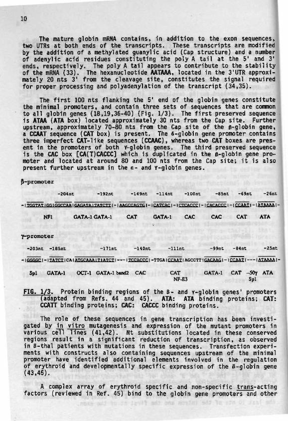
The mature globin mRNA contains, in addition to the exon sequences,two UTRs at both ends of the transcripts. These transcripts are modifiedby the addition of a methylated guanylic acid (Cap structure) and a numberof adenylic acid residues constituting the poly A tail at the 5' and 3'ends, respectively. The poly A tail appears to contribute to the stabilityof the mRNA (33). The hexanucleotide AATAAA, located in the 3'UTR approxi-mately 20 nts 3' from the cleavage site, constitutes the signal requiredfor proper processing and polyadenylation of the transcript (34,35).
The first 100 nts flanking the 5' end of the globin genes constitutethe minimal promoters, and contain three sets of sequences that are commonto all globin genes (18,19,36-40) (Fig. 1/3). The first preserved sequenceis ATAA (ATA box) located approximately 30 nts from the Cap site. Furtherupstream, approximately 70-80 nts from the Cap site of the B-globin gene,a CCAAT sequence (CAT box) is present. The 6-globin gene promoter containsthree imperfect CAT-like sequences (CCAAC), whereas two CAT boxes are pres-ent in the promoters of both Y-globin genes. The third preserved sequenceis the CAC box [CA(T)CACCC] which is duplicated in the B-globin gene pro-moter and located at around 80 and 100 nts from the Cap site; it is alsopresent further upstream in the e- and Y-globin genes.
P-promoter
-2O4nt -192nt -H9nt -114nt -lOOnt -85nt -69nt -26nt
- I TftKTAT I fin I CnCTAA IGAGATA I TATCTT I - I AACCCAGTG I - I CATC!AC I - I CTCACCr I - I C ACACCC I - I CCAATI - I ATAAAA I •
NF1 GATA-1 GATA-1 CAT GATA-1 CAC CAC CAT ATA
f-promoter
-203nt -185nt -171nt -HOnt -Hint -99nt -84nt -25nt
- I TATCT 1 CA I ATftCAAA IT I ATCT I I TCCACCC I -TTGA I CCAAT I AGCCTT I GACAAG I - I CCAAT I I ATAAAA I
Spl GATA-1 OCT-1 GATA-1 buid2 CAC CAT GATA-1 CAT -5Oy ATANF-E3 Spl
FI6. 1/3. Protein binding regions of the B- and y-globin genes' promoters[adapted from Refs. 44 and 45). ATA: ATA binding proteins; CAT:CCATT binding proteins; CAC: CACCC binding proteins.
The role of these sequences in gene transcription has been investi-gated by in vitro mutagenesis and expression of the mutant promoters invarious cèTl lines (41,42). Nt substitutions located in these conservedregions result in a significant reduction of transcription, as observedin B-thal patients with mutations in these sequences. Transfection experi-ments with constructs also containing sequences upstream of the minimalpromoter have identified additional elements involved in the regulationof erythroid and developmentally specific expression of the B-globin gene(43,45).
A complex array of erythroid specific and non-specific trans-actingfactors (reviewed in Ref. 45) bind to the globin gene promoters and other

11
B-globin gene cluster regulatory elements. Ubiquitous transcription fac-tors such as the TATA-binding protein TBP, the CCAAT binding proteins CP1,CTG/NF1, and CDP, and various other non-erythroid specific factors suchas SP1, TEF-2, jun, fos, USF, J-BP, and YY1, have been shown to bind jjivitro to critical ^s-acting regulatory sequences; however, their rolein regulating the globin genes in vivo is at present still unknown.
Several erythroid specific factors have been described which appearto be involved in the regulation of globin gene expression. The best char-acterized is the GATA-1 protein, which belongs to the family of GATA bind-ing proteins (GATA-1, GATA-2, GATA-3, and GATA-4) that are expressed indifferent hematopoietic and other cell types (reviewed in Ref. 46). TheGATA-1 is expressed only in erythroid, megakaryocytic, and mast cells,and binds to promoter, enhancer, and LCR elements of all known erythroidspecific genes, including its own promoter. Other erythroid specific fac-tors include the NF-E1, NF-E2, NF-E3, NF-E4, NF-E5, NF-E6, BGP1, Pall, and-50 , of which only several (NF-E3, NF-E4, Pall, BGP1, and -50 ) seem tobe expressed at different levels in cells representing different stagesof development, and could therefore be implicated in the control of globingene switching (47).
The presence of additional sequences within the B-globin gene clusterthat are required for the expression of the B-globin gene was first sug-gested in 1980 from the study of a Dutch form of r«B-thal (48). This typeof thalassemia is caused by a large deletion that leaves the B-globin geneintact but removes 100 kb of upstream sequences. Several years later,Tuan et al (49) characterized a set of developmentally-stable DNase I HSlocated in one region 15 kb upstream of the c-globin gene, and in another,18 kb downstream of the B-globin gene. The 5' sites, which comprise theLCR were termed (5')HS-1, HS-2, HS-3, and HS-4 with respect to their orderfrom the e-globin gene, and the single 3' site was termed 3'HS-l (50).The functional significance of the LCR became apparent when Grosveld et al(51) showed that the human B-globin gene linked to the LCR was expressedin transgenic mice at levels comparable to those of the endogenous mouseglobin genes. In addition, the level of expression was independent of theintegration within the mouse genome, but was dependent on the number ofcopies integrated. The LCR can thus override the influence of the neigh-boring chromosomal sequences to create an open chromatin domain capableof supporting high levels of globin gene transcription. Each of the fourB-LCR elements has subsequently been mapped to 200-300 bp core regions ofDNase I hypersensitivity and has been found to consist of a high densityof binding sites for a number of the erythroid and ubiquitous factorsdescribed above (52-54). Studies using different subfragments of the LCR,alone or in combination, have shown that sites HS-2 and HS-3 are eachresponsible for approximately 40-50% of the full enhancing activity ofthe LCR (55,56). However, all four sites appear to be necessary to obtainthe full stimulatory effect, and it has been suggested that the individualB-LCR elements assemble to form a single larger complex capable of inter-acting with the B-like globin genes (57,58).
Further experiments in transgenic mice have shown competition of theindividual B-like globin genes for the LCR, in such a way that the proximalglobin genes can suppress the expression of the more distal genes (59,60).
12
Therefore, to obtain the developmental 1 y regulated switching of the indi- ;vidual globin genes, the interaction of the more proximal e- and Y-globingenes with the LCR needs to be blocked. Experiments in transgenic micehave shown that this occurs since the Y-globin gene was expressed at muchlower levels in adult animals, even when introduced independently from theother globin genes (61). It therefore appears that the developmental regu-lation depends on the state of the globin gene promoters and their avail-ability to interact with the LCR. In addition, a gene developmental stagespecificity has been observed for the HS-3 and HS-4 LCR elements (62).
MOLECULAR BASIS OF B-THALASSEMIA
More than 160 mutations or deletions of the B-globin gene have beendescribed (listed as an addendum to this chapter), approaching an almostcomplete characterization of the molecular defects causing B-thal in vari-ous population groups (63). The identification of the B-globin gene muta-tions was mainly performed in the last decade, and two major advancementsthat greatly facilitated this effort were the application of B-globin genehaplotyping and the PCR procedure.
Haplotyping of the B-globin gene cluster refers to the analysis of DNApolymorphisms, namely various RFLP, which have been shown to be associatedwith different chromosomal backgrounds in various ethnic groups. A patternof different restriction enzyme polymorphisms spread throughout the B-glo-bin gene cluster determines a particular haplotype (64). Table 1/1 showsthe most commonly investigated polymorphisms that define the major haplo-types among Mediterraneans.
Screening for new mutations 1n the first half of the 1980s took advan-tage of the observation that, in general, different mutations were asso-ciated with different haplotypes. Haplotype analysis, therefore, loweredthe chance of repeatedly identifying the same mutation. With the introduc-tion of the PCR methodology in the second half of the 1980s, haplotypeanalyses became less significant, especially since a large number of iden-tical mutations were already identified as being present on differentchromosomal backgrounds. However, some of these mutations differed intheir phenotypic expression, especially in Hb F production, which poten-tiated a new role for haplotype analysis in the identification of sequencevariations that can modulate the expression of the y-globin genes jji cis.
The B-globin gene mutations described in the following pages arerepresentative examples of the various defects that can affect the functionof the B-globin gene. Appendix I contains a complete, updated list ofB-thal mutations, including references and other relevant information.
Transcription Mutants. As mentioned earlier, the B-globin gene pro-moter contains sequences that regulate the efficiency and accuracy of thetranscription process. Until recently, 17 different promoter mutations,all causing a B*-thal, have been reported. They are clustered in the ATAbox and in the proximal and distal CAC boxes of the B-globin gene promoter.

13
TABLE 1/1. The Common Mediterranean Haplotypes and the Most FrequentlyAssociated Mutations
• t + +
He X Hd Hd He He A B
Haplotypes © © © © © © © © Associated MutationsI + + + IVS-I-11O; CD 39; -101; FSC 6;
Hb Knossos; IVS-I-116II - - + + - + + + CD 39; IVS-I-110III _ + + _ + + + _ IVS-II-1IV - + + - + + - + FSC 8V + - - - - - + - FSC 5; IVS-I-5 (G->T); IVS-I-1VI - _ + + _ - - + IVS-I-6VII + + IVS-II-745; IVS-I-6; CD 39VIII - + + - + - + - - 8 7
IX _ + + _ + + + + IVS-I-110; FSC 6; CD 39
1 (Hc) = Hinc II 5' to c; 2 (X) = Xmn I 5' to G ^ 3 and 4 (Hd) = Hind IIIat Gy and \; 5 and 6 (Hc) = Hinc II in *B and 3' to it; 7 (A) = Ava II inB; 8 (B) = Bam HI 3' to B.
The seven mutations located at five different positions in the ATA box,between positions -28 and -32 from the Cap site, are found within differentethnic groups. The -29 (A->G) mutation is associated with a very mildclinical phenotype among Blacks, where it accounts for the majority ofB-thal mutations (65). This is most likely due to the chromosomal back-ground which carries the C->T substitution at -158 to the Gy.giobin gene.The latter substitution has been shown to be associated with high Hb Fproduction under conditions of erythropoietic stress (66). The Black homo-zygotes for this mutation have average Hb levels of 10.6 g/dl, with morethan 60X Hb F (67). On the other hand, a Chinese homozygote with the samemutation on a different chromosome [lacking the Gy -158 (C->T) mutation]had a transfusion-dependent thalassemia major (68). Similarly, Japanesehomozygotes for the -31 (A->G) mutation have low Hb (6.9 g/dl) and lowHb F (8.8X) levels (69).
Nine relatively mild mutations have been identified in the proximalCACCC box, at positions -86, -87, -88, -90, and -92 relative to the Capsite of the B-globin gene. The -88 (C->T) substitution is the second mostcommon B-thal mutation among Blacks and, as in the case of the -29 (A->G)mutation, is associated with the Gy -158 (C->T) substitution and high Hb Flevels (64.5X) (65,67). It is interesting that unlike other mild B-thalmutations, the proximal CACCC box mutations are also associated with highHb A2 levels in the hétérozygotes (70). The presence of the 6 chain vari-ant Hb B2, that can be chromatographically separated from Hb A2, made 1tpossible to evaluate whether the increased fi-globin gene expression is
14
lin cis or iji trans to the mutated B-glob1n gene (71). While the levelsof Hb~A2 and Hb B2 were identical in normal controls and were increasedto the same extent in the B° hétérozygotes with the CD 47 (+A) mutation,the output of the 6-globin gene from the chromosome with the -88 (C->T)substitution was twice as high as that from the fi gene on the normal chro-mosome. The same increase in 6 gene expression jji c[s was also observedin the 1393 bp deletion which completely deletes the B-globin gene pro-moter. These results strongly suggest a competition between sequenceswithin the B- and 6-globin gene promoters for transcription factors orinteractions with regulatory elements jhn £is» such as the LCR. .J
The mutation in the distal CACCC box [-101 (C->T)] was identified 'in three Turkish and one Bulgarian family (72), and was subsequently found 1among Italian B-thal subjects (73). The hétérozygotes carrying this muta- Ition have the silent B-thal phenotype with normal hematological parameters 1(MCV and MCH) and normal levels of Hb A2 and Hb F. Homozygotes for thismutation have not yet been identified, but compound hétérozygotes havea B-thal intermedia with relatively mild hematological abnormalities.
It is interesting that even though 17 different B-thal mutations havebeen identified in the B-globin gene promoter, so far none has been foundin the CCAAT box.
RNA Splicing Mutants. A large number of H*-thal mutations affect RNAsplicing by inactivating the normal splice sites, by activating crypticsplice sites, or by producing novel splice sites. The first B-thal muta-tion that was identified belongs to the last group and is the most commonmutation in the Mediterranean. This 6->A substitution at IVS-I-110 createsan AG dinucleotide within a consensus sequence which is almost identicalto the sequence of the acceptor splice site of IVS-I. As a consequence,an alternate acceptor splice site is created 19 nts upstream of the 3'end of the intron, and this site is used for the splicing of 90% of theB-globin mRNA. The improperly spliced B-globin transcripts contain a 19 ntintron remnant which changes the reading frame, and are highly unstable.Even though the IVS-I-110 mutation causes a B*-thal, the clinical expres-sion is of thalassemia major due to the extremely low B-globin chain outputof 10% (74). Three mutations at positions 654, 705, and 745 nts of thesecond intron create alternative donor splice sites. The latter two muta-tions greatly reduce the level of normally spliced transcripts, while theIVS-I1-654 mutation eliminates normal splicing and is a B° allele. Inall three cases, an alternate acceptor splice site at IVS-II-579 is acti-vated, and sequences between this site and the new donor site are retainedin the improperly spliced B-globin transcripts.
Twelve mutations have been described that disrupt the invariant GTand AG dinucleotides in the donor and acceptor splice sites, respectively.The mutations in the GT dinucleotide at the 3' end of IVS-I prevent anyproper splicing at this site, and lead to the activation of alternativesplice sites, of which two are located in the first exon (CDs 18 and 25)and the third at positions 13 and 14 of IVS-I. Similarly, the IVS-II-1(G->A) mutation leads to inactivation of the IVS-II donor splice site andactivation of an alternative donor site 47 nts downstream.
15
Mutations in the consensus sequences of the normal splice sites canlead to their underutilization and again, abnormal use of the alternativesplice sites described above. A common Mediterranean 6-thal mutation, theT->C substitution at position IVS-I-6, leads to a relatively mild B*-thalsyndrome, presumably due to correct splicing of a substantial number oftranscripts. At position IVS-I-5, all three possible nt changes have beenreported, associated with different clinical phenotypes. It has beensuggested that transversions (G->C or 6->T) at this position could havea more profound effect on the tertiary structure of the DNA than the 6->Atransition, that latter leading to a more moderate reduction of splicingefficiency and a milder clinical phenotype (75).
The two cryptic splice sites in exon 1 can also be activated by muta-tions that change their consensus sequence, making them more favorable tothe splicing mechanism. Three of these mutations activate the alternativesplice site around CD 25, while the fourth activates the more upstreamsite around CD 18. The latter mutation introduces an amino acid substitu-tion which leads to the synthesis of an abnormal Hb variant, Hb Malay.The mutations in CDs 26 and 27 also lead to amino acid replacements andresult in the Hb E and Hb Knossos variants, respectively, while the muta-tion in CD 24 is silent. This mutation is associated with a more severeclinical syndrome and with less normal splicing of precursor B-mRNA mole-cules than the Hb E and Hb Knossos mutations (23).
Translation Mutants. Most of the mutations that affect translationare nonsense CDs or frameshift mutations that alter the reading frame andultimately create a new stop CD. The frameshift mutations result fromsmall insertions or deletions of up to 7 nt in the coding region of theB-globin gene. The premature termination of translation is expected togive rise to shorter abnormal peptides in the erythroid cells of theaffected individuals. Such peptides have not been identified, probablydue to their rapid turnover in red cell precursors. It is interestingthat these mutations also affect the level of B-globin mRNA in the eryth-roid cells. Studies of the CD 39 (C->T) mutation have implicated ineffi-cient nuclear-cytoplasmic transport or nuclear instability as mechanismsthat decrease the levels of the B-39 transcripts (76). All frameshiftand nonsense mutations result in a B° type of thalassemia, and are usuallyassociated with a severe clinical phenotype.
Several mutations that affect initiation of translation have beenreported. Three of them destroy the initiaton CD, while a G->A substitu-tion at position +22 from the Cap site creates a new initiation CD upstreamof the normal one.
RNA Cleavage and Poly A Mutants. Four mutations and two small dele-tions have been identified in the cleavage and poly A signal sequenceAATAAA. Two of these mutations have been studied by transient expressionanalysis, and only a small percentage of the transcripts were found to bepolyadenylated at the normal site (77,78). Elongated transcripts, cleavedfollowing AATAAA signals located 1 to 3 kb 3" to the gene, were present atless than 10% of the normal level, suggesting that the deficient B-globinsynthesis in these mutants is due to the instability of the abnormally
16
elongated transcripts. All of these mutations are associated with a gfthai, as they do not completely abolish normal polyadenylation, and thejdo not affect translation of the abnormal transcripts.
A C->G mutation in nt 6 of the 3'UTR, that probably affects the sta-'bility or processing of B-globin RNA, has been associated with silent 0-thal. Similarly, hétérozygotes for the Cap site mutation [+1 (A->C)] havenormal MCV values and borderline normal Hb Ag levels. The latter mutationcould have an effect on transcription or on capping with a secondary effecton translation (79).
Dominant B-Thal Allel es. The dominant forms of B-thal are character-ized by a thalassemia intermedia clinical phenotype in hétérozygotes, andthe presence of inclusion bodies in the erythroid precursors and peripheralRBC. The term 'inclusion body' B-thal that was initially adopted for thissyndrome, is now avoided due to the identification of such inclusion bodiesin all severe forms of B-thal. The mutations that fall into this categoryproduce highly unstable B chain variants as a result of single base sub-stitutions or CD deletions, truncated B chain variants due to prematuretermination, or elongated B-globins with an altered carboxy-terminal end.A common determinant of these mutations is the production of B chains thatare unable to form viable tetramers with the a chains and are rapidlydegraded. The continuous degradation of these non-functional B chain vari-ants overloads the capacity of the proteolytic defense mechanism and com-promises the proteolysis of the free a chains, thus leading to their accu-mulation and precipitation at a greater extent than usually observed inB-thal hétérozygotes (reviewed in Refs. 31 and 79). These dominant B-thalmutations [except for the CD 121 (6AA->TAA) mutation) occur at very lowfrequencies, among widely dispersed ethnic groups, and many have beenreported as de novo events. These data suggest a lack of positive selec-tion by malaria, as the reduced morbidity to the latter disease would becounterbalanced by the severity of the B-thal trait.
Deietional B°-Tha1. Twelve deletions, ranging from 44 bp up to 27 kb,that remove part or the entire B-globin gene, have been described so far.In eight, various portions of the 5' B-globin region are removed. Hétéro-zygotes for each of these deletions, except for a recently discovered 27 kbdeletion in Southeast Asians, exhibit significantly higher Hb A2 levels(5.5-9%) than B-thal hétérozygotes with point mutations or deletions inother parts of the gene (80). It has been suggested that the loss of theB-globin gene promoter would allow the 6 and y promoters to react morereadily with regulatory elements within the LCR (81). In keeping withthis hypothesis is the level of Hb F which is elevated in most cases, butto a variable extent (2.5-14X). Again, the only exception is the SoutheastAsian B-thal deletion, which apart from B-globin promoter sequences, alsoremoves the 3'HS-l site, and is associated with slightly elevated Hb A2levels (2.7-5.IX) and markedly elevated Hb F levels, comparable to thoseobserved in HPFH (17.6-26.6X) (82).
17
HB LEPORE AND ANTI-LEPORE SYNDROMES
The Lepore and anti-Lepore Hbs are abnormal Hbs which contain hybridB-like chains and are associated with a thaiassemia-1ike syndrome. Thefirst Hb Lepore variant, Hb Lepore-Washington (also known as Hb Lepore-Boston), was described in an Italian family by Gerald and Diamond in 1958(83). Subsequently, other Hb Lepore variants were described; they arefound in various populations but mainly in Mediterranean countries suchas the former Yugoslavia, Italy, and Greece (reviewed in Ref. 84).
Hb Lepore hétérozygotes are usually asymptomatic, without anemia, andwith identical hématologie findings as those seen in B-thal trait. Theunique difference that distinguishes Hb Lepore is the presence of %10X ofthe abnormal Hb. The amount of Hb F may vary but does not usually exceed5%, and the level of Hb A2 is normal or slightly decreased (84,85). Theclinical features of homozygotes for Hb Lepore or subjects with Hb Lepore-B-thal are variable, and patients with both mild and severe clinicalcourses have been described (84). Hb analysis in Hb Lepore homozygotesidentifies some 25% Hb Lepore, the rest being Hb F. In compound hétéro-zygotes with B-thal, the Hb Lepore level does not usually exceed 15%; Hb F,Hb A2, and sometimes Hb A form the remainder of the Hb (84).
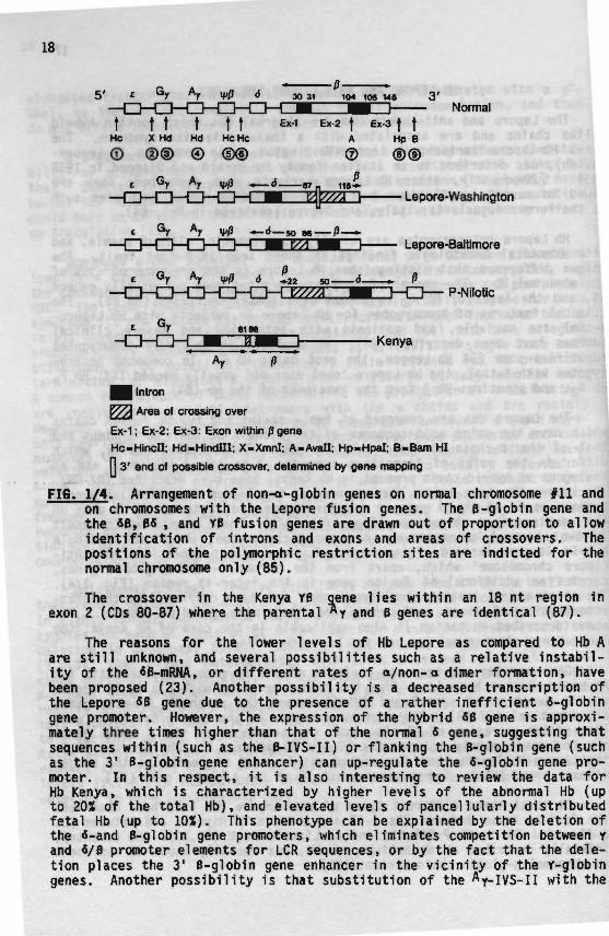
The Lepore Hbs are composed of two a chains and two 6B fusion chainswhich have the amino acid sequence of the 6 chain at their N-terminus andthat of the B chain at the C-terminus (86). Three Lepore chains, whichdiffer in the point of transition from the 6 to the B sequence, and ananalogous YB hybrid chain present in Hb Kenya, have been described (87-89).The Lepore Hbs have arisen through unequal homologous recombination eventsbetween parts of the 6 gene on one chromosome and parts of the B gene onthe complementary chromosome (Fig. 1/4) (87). These events would resultin two abnormal chromosomes: A 'Lepore chromosome' which has a singlehybrid «B gene instead of the normal 6- and B-globin genes, and an 'anti-Lepore chromosome' which, apart from the normal 6- and B-globin genes,carries an additional B« fusion gene in the inter-«B region (Fig. 1/4).The existence of 'anti-Lepore chromosomes' has been confirmed by the char-acterization of three anti-Lepore Hbs containing fused B« chains. Theabove described mechanism is also applicable in the case of Hb Kenya; the'anti-Kenya chromosome' has not yet been identified.
Gene mapping and sequence analysis of the «B gene characteristic forHb Lepore-Boston have identified the crossover point within a 58 bp regionat the junction of exon 2 and IVS-II, where the parental 6 and B genesare identical (87,88). The crossover region in the Lepore-Baltimoré genelies within a region of 47 nts where the sequences of the 6 and B genesare the same, namely between CDs 69 and 84 of exon 2. The breakpoint inthe Lepore-Hollandia gene is located within a 40 nt region at the borderof exon 1 and IVS-I, downstream from CD 25 of the 6-globin gene (87).
At least three different types of anti-Lepore Hbs have been observed.The fusions occur between CDs p-12 and 6-22 in Hb Miyada, and between CDsB-22 and 6-50 in Hb P-Nilotic and Hb Lincoln Park (84,89). Hb Coventry,initially described as an anti-Lepore Hb, was recently shown to representan abnormal B-globin chain variant (90).
18
Normal
Lepore-Washington
Lepore-Bal timoré
P-Nilotic
81MKenya
| Intron
| Area of crossing over
Ex-1; Ex-2; Ex-3: Exon within Pgene
Hc-HincU; Hd-HindUI; X-Xmnl; A-AvaU; Hp-Hpal; B-Bam HI
n 3' end of possible crossover, determined by gene mapping
FI6. 1/4. Arrangement of non-a-globin genes on normal chromosome #11 andon chromosomes with the Lepore fusion genes. The B-globin gene andthe 6B, 06 , and YB fusion genes are drawn out of proportion to allowidentification of introns and exons and areas of crossovers. Thepositions of the polymorphic restriction sites are indicted for thenormal chromosome only (85).
The crossover in the Kenya YB gene lies within an 18 nt region inexon 2 (CDs 80-87) where the parental ^ Y and B genes are identical (87).
The reasons for the lower levels of Hb Lepore as compared to Hb Aare still unknown, and several possibilities such as a relative instabil-ity of the 6B-mRNA, or different rates of a/non-odimer formation, havebeen proposed (23). Another possibility is a decreased transcription ofthe Lepore SB gene due to the presence of a rather inefficient 6-globingene promoter. However, the expression of the hybrid 6B gene is approxi-mately three times higher than that of the normal 6 gene, suggesting thatsequences within (such as the B-IVS-II) or flanking the B-globin gene (suchas the 3' B-globin gene enhancer) can up-regulate the 6-globin gene pro-moter. In this respect, it is also interesting to review the data forHb Kenya, which is characterized by higher levels of the abnormal Hb (upto 20X of the total Hb), and elevated levels of pancellularly distributedfetal Hb (up to 10X). This phenotype can be explained by the deletion ofthe 6-and B-globin gene promoters, which eliminates competition between yand 6/fl promoter elements for LCR sequences, or by the fact that the dele-tion places the 3' B-globin gene enhancer in the vicinity of the Y-globingenes. Another possibility is that substitution of the Ay-ivS-II with the

19
B-IVS-II can influence the adult silencing of the Ay-globin gene. Anenhancer element has been identified in the B-IVS-II (43), and its elimi-nation may at least in part be responsible for the relatively low levelof expression of the anti-Lepore genes, which, despite a functional B-glo-bin promoter, is similar to that of the Lepore «B genes. Replacement ofthe B-IVS-II sequence by the fi-IVS-II sequence results in a markedly re-duced expression of the B-globin gene in HeLa and MEL cells, while the sub-stitution of the 6-IVS-II with the B-IVS-II is not sufficient to overridethe low level of expression of the 6-globin gene (91,92). These data sug-gest that sequences in the B-IVS-II and the 3' B enhancer, together withthe B-globin gene promoter, are important for the high level expression ofthe B-globin gene in normal adult erythroid cells.
HEREDITARY PERSISTENCE OF FETAL HEMOGLOBIN (HPFH)
HPFH refers to a heterogeneous group of disorders that, in the hetero-zygous state, are associated with increased levels of Hb F and normal RBCindices. In homozygous HPFH the red cells are often somewhat hypochromicand microcytic, reflecting a mild imbalance in the synthesis of o/non-o-globin chains (93). Several classifications of the HPFH disorders areused, based on the molecular defect (deletional or nondeletional), thetype of Y-globin chain produced (Gy, *Y, or both), and the cellular patternof Hb F distribution (pancellular HPFH where all the red cells haveincreased levels of Hb F, and heterocellular HPFH where only a subpopula-tion of red cells contain Hb F). However, it appears that the last classi-fication is more a reflection of the methodology used, rather than ofunderlying mechanisms of the HPFH disorders, because certain conditionswhich give a heterocellular Hb F distribution with an acid elution tech-nique may show a pancellular distribution with a more sensitive immuno-fluorescent assay (94).
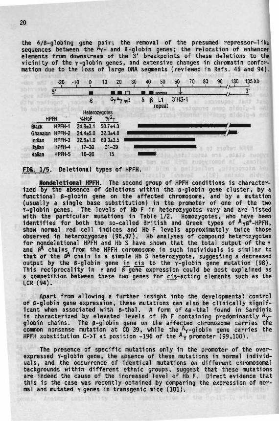
Deletional HPFH. Five different deletions of the B-globin gene clus-ter have been found in patients with this syndrome, characterized by Hb Flevels of 20-30% in the hétérozygotes, by a pancellular Hb F distribution,and by the absence of B and « chain production jm £i_s (45). Homozygotesare clinically normal but have an imbalanced o/y chain synthesis ratiobetween 1.4 to 2.0, and microcytic, hypochromic red cell indices due tothe inability of the y-globin genes to completely compensate the lack of4- and B-globin chain production. The increased 0? affinity of the redcells that contain 100% Hb F is compensated by a mild erythrocytosis withHb levels of 17 to 19 g/dl (95). Compound hétérozygotes of deletionalHPFH with Hb S have 30% Hb F and 70% Hb S, while compound hétérozygoteswith B-thal usually have the hematological features of B-thal trait butwith Hb F levels greater than 70%.
The 5' breakpoints of all of these deletions are within theintergenic DNA segment, while the 3' ends are more variable due to thelarge differences in size between the different HPFH deletions (Fig. 1/5).Several models, not mutually exclusive, have been proposed to explain theincreased Hb F levels and the phenotypic differences with the «B-thalas-semias, of which some are characterized by deletions of similar size.These models include lack of competition between the y-globin genes and
20
the ô/B-globing gene pa i r ; the removal of the presumed repressor-likesequences between the Ay- and 6-globin genes; the relocation of enhancerelements from downstream of the 3' breakpoints of these deletions to thev ic in i ty of the y-globin genes, and extensive changes in chromatin confor-mation due to the loss of large DNA segments (reviewed in Refs. 45 and 94).
-20 -10 0 10 20 30 40 50 50 70 80 90 130 135 kb• ' ' ' ' ' • I U I I i — / / _ J 1
P 5 p 11 3'HS-1repeat
HétérozygotesHPFH ' %HbF 6
Black HPFH-1 24.8±3.1 50.7±4.3Ghanaian HPFH-2 24.4±5.0 32.3*4.8Indian HPFH-3 22.6±1.0 69.3±3.5Italian HPFH-4 17-30 31-39Italian HPFH-5 16-20 15
FI6. 1/5. Deletional types of HPFH.
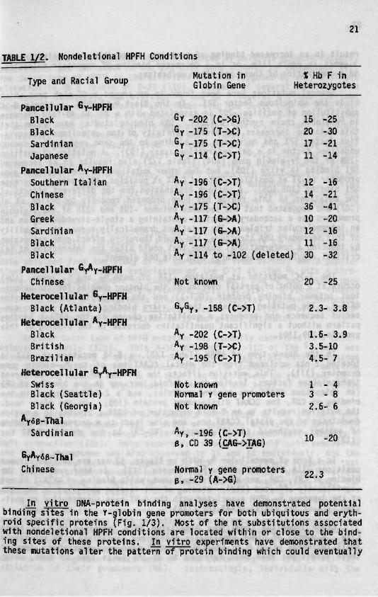
Nondeletional HPFH. The second group of HPFH conditions is character- jized by the absence of deletions within the B-globin gene cluster, by afunctional B-globin gene on the affected chromosome, and by a mutation(usually a single base substitution) in the promoter of one of the twoY-globin genes. The levels of Hb F in hétérozygotes vary and are listedwith the particular mutations in Table 1/2. Homozygotes, who have beenidentified for both the so-called British and Greek types of Ayp+-HPFH,show normal red cell indices and Hb F levels approximately twice thoseobserved in hétérozygotes (96,97). Hb analyses of compound hétérozygotesfor nondeletional HPFH and Hb S have shown that the total output of the Yand pA chains from the HPFH chromosome in such individuals is similar tothat of the B* chain in a simple Hb S hétérozygote, suggesting a decreasedoutput by the B-globin gene jm cis to the y-globin gene mutation (98).This reciprocality in y and B gene expression could be best explained asa competition between these two genes for cj_s-acting elements such as theLCR (94).
Apart from allowing a further insight into the developmental controlof B-globin gene expression, these mutations can also be clinically signif-icant when associated with B-thal. A form of «B -thai found in Sardinia1s characterized by elevated levels of Hb F containing predominantly Ay-globin chains. The B-globin gene on the affected chromosome carries thecommon nonsense mutation at CD 39, while the \-globin gene carries theHPFH substitution C->T at position -196 of the Ay promoter (99,100).
The presence of specific mutations only in the promoter of the over-expressed Y-globin gene, the absence of these mutations in normal individ-uals, and the occurrence of identical mutations on different chromosomalbackgrounds within different ethnic groups, suggest that these mutationsare indeed the cause of the increased level of Hb F. Direct evidence thatthis is the case was recently obtained by comparing the expression of nor-mal and mutated y genes in transgenic mice (101).
21
TABLE 1/2. Nondeletional HPFH Conditions
Type and Racial Group
Pancellular 6y-HPFHBlackBlackSardinianJapanese
Pancellular Ay-HPFHSouthern I ta l ianChineseBlackGreekSardinianBlackBlack
Pancellular G / Y - H P F H
Chinese
Heterocellular V H P F H
Black (Atlanta)
Heterocellular Ay-HPFHBlackBritishBrazilian
Heterocellular G/y-HPFHSwissBlack (Seattle)Black (Georgia)
AyôB-ThalSardinian
Chinese
Mutation inGlobin Gene
G Y -202 (C->G)Gy -175 (T->C)6y -175 (T->C)Gy -114 (C->T)
Ay -196 (C->T)Ay -196 (C->T)Ay -175 (T->C)Ay -117 (G->A)Ay -117 (G->A)Ay -117 (G->A)Ay -114 to -102 (deleted)
Not known
GyGy, -158 (C->T)
Ay -202 (C->T)Ay -198 (T->C)Ay -195 (C->T)
Not knownNormal y gene promotersNot known
Ay, -196 (C->T)B, CD 39 (CAG->TAG)
Normal y gene promotersB, -29 (A->G)
X Hb F inHétérozygotes
15 -2520 -3017 -2111 -14
12 -1614 -2136 -4110 -2012 -1611 -1630 -32
20 -25
2.3- 3.8
1.6- 3.93.5-104.5- 7
1 - 43 - 82.6- 6
in 9n
? ? •»
J_n vitro DNA-protein binding analyses have demonstrated potentialbinding sites in the Y-globin gene promoters for both ubiquitous and eryth-roid specific proteins (Fig. 1/3). Most of the nt substitutions associatedwith nondeletional HPFH conditions are located within or close to the bind-ing sites of these proteins, ri vitro experiments have demonstrated thatthese mutations alter the pattern of protein binding which could eventually

22
result in an increased binding of proteins responsible for activating they genes in adult life, or in a decreased binding of repressor molecules,or a combination of these and other mechanisms.
Several mutations have been found in or around a GC-rich area whichbinds the ubiquitous factor SP1. In the Black GyB^-HPFH, a single G->Csubstitution at position -202 is associated with a 30- to 40-fold increase1n Gy-globin chain synthesis (102). DNA binding studies have shown thatthis mutation increases the binding affinity of the ubiquitous transcrip-tion factor SP1 and decreases the binding to the same region of a secondundefined factor in a gel retardation assay (103). A mutation at position-198 of the Ay.giobin gene (104) (Table 1/2) also results in an increasedSP1 binding jji vitro (105), but two other mutations in the Ay_giobin genepromoter (C->T at position -202 and C->T at position -196) seem to decreaseSP1 binding (105,106). A uniform explanation for the different effects ofthe mutations in this region has been proposed recently by Urlich et al(107) who suggested that the sequence between -195 and -218 of the y-globinpromoters has a secondary structure containing a single-stranded region andan intramolecular triplex. The mutations dramatically reduce the stabilityof this secondary DNA structure, and could therefore affect gene expressionby inducing a «informational change that could alter the interaction of (a)critical regulatory molecule(s) with this DNA element.
The T->C mutation at position -175 of both Gy and *y genes (108)occurs at a point of partial overlap of the binding domains for the ubiqui-tous octamer binding protein (OCT-1) and the erythroid specific factorGATA-1. The mutation abolishes the jiji vitro binding of OCT-1 to its con-sensus sequence and produces a qualitative change in the GATA-1 bindingpattern without a significant quantitative decrease in the binding of thisprotein (109,110).
In the Greek type of HPFH (Table 1/2), a G->A mutation at -117 islocated 2 nts upstream from the distal CCAAT box of the \-globin genepromoter (111). This mutation affects the binding of several differenttranscription factors, leading to a 4-fold increase in the binding of CP1(an ubiquitous CCAAT box binding protein), and a 2-fold increase in thebinding of CDP, the CCAAT displacement protein (109). On the other hand,the mutation results in an 8-fold reduction of GATA-1 binding to a regiondownstream of the distal CCAAT box, and also leads to a decrease in bindingof another erythroid specific protein (NF-E3) to the distal CCAAT box(112).
In a form of pancellular Ay-HPFH in Blacks, 13 nts are deleted betweenpositions -114 and -102 of the Ay promoter (113). In this condition, thebinding of CP1, CDP, and NF-E3 is abolished, while binding of GATA-1 is notsignificantly affected (112). Binding of CP1 is also abolished in the caseof the Japanese Gy-HPFH associated with the C->T base substitution at posi-tion -114 of the distal CCAAT box (114).
Other Inherited Conditions Associated With Increased Hb F in AdultLife. A number of other genetically determined conditions with relativelysmall increases of Hb F in adult life (<5X) have been described. Some ofthese conditions have been referred to as the Swiss type of HPFH, which
23
appears to be a heterogeneous state influenced by multiple genetic factors,both linked and unlinked to the B-globin gene complex on chromosome #11.The Swiss type of HPFH is characterized in hétérozygotes by only a slightelevation in Hb F levels of 2-3% (115). Members of several families fromthe former Yugoslavia had elevated Gy and Hb F levels; all had the samehaplotype which was apparently associated with the Swiss type of HPFH con-dition (116). However, this haplotype, which carries the C->T substitutionat position -158 of the Gy-qlobin gene promoter, was also present in normalindividuals with elevated Gy and low Hb F levels, suggesting that thisSwiss HPFH phenotype results from the interaction of a particular chromo-somal background, which also causes high Gy values, with other factors notlinked to the B-globin gene cluster (116). Using a FACS technique with amonoclonal anti-ychain antibody, the Hb F production was recently reexam-ined in normal subjects with and without this C->T mutation (117). A sig-nificant correlation of T at -158 (Gy) with higher levels of F cells wasobserved, presumably due to slightly higher Hb F/cell in individuals withthis mutation.
The -158 (C->T) substitution has also been found to be associatedwith significantly increased Hb F levels, predominantly of the Gy type, inpatients with SS, homozygous B-thal, and with Hb S/B-thal (118). Geneticdata obtained from the study of flS chromosomes from various world popula-tions have shown a strong association of the C->T at -158 Gy with a chro-mosomal background capable of conferring high Hb F and Gy expression uponhematopoietic stress in adult erythroid cells (32,119). A recent findingthat this mutation also affects binding of a potential repressor molecule,possibly BP1 (120), suggests a direct effect of the -158 (C->T) substitu-tion in the Gy and Hb F expression in SS and B-thal patients.
Another Swiss type of HPFH mutation was recently described in a Czech-oslovakian family (121). This A->C mutation at -110 of the Gy gene wasassociated with only a slight increase in Hb F (F/\D 2.3%; cation exchangeHPLC 0.8%), and with high Gy values (95%). The presence of this mutationin two family members who had co-inherited a B° allele on the other chro-mosome led to a more significant increase in the level of Hb F (cationexchange HPLC 3.1%).
The nature of the HPFH determinants not linked to the B-globin genecluster is still unknown, but a study of Japanese adults with Swiss HPFHhas suggested the existence of an X-linked determinant which affects Fcell production (122). More recently, linkage between F cell productionand the X chromosome was also observed in SS and AA individuals, and theF cell producing locus was mapped to Xp 22.2 (123). It was suggested thatthe putative protein product of this locus could either directly affectF cell production or exert its influence through interactions with theB-globin gene cluster (123).
Changes in the arrangement of the Y-globin genes are also frequentlyassociated with a Swiss HPFH phenotype. The Atlanta type of heterocellularHPFH is associated with Hb F levels of 2.5-5% in hétérozygotes (in adultAA individuals) and 10-13% (in AS individuals), and Gy values of more than90% (124,125). Instead of the normal G^/Ay gene pair, this HPFH chromosomecarries two Gy-globin 3^nes, both of them containing the -158 (C->T) sub-stitution in their promoters (66). Interestingly, individuals with the
24
same y-globin gene arrangement but without the Gy _i58 (C->T) substitutionshow normal Hb F levels (below 1%) with only slightly elevated Gy values(126).
Slightly higher Hb F levels (around 5%, Gy around 94%) than thoseobserved in the Atlanta type of HPFH were found in healthy individuals fromthe former Yugoslavia who had a y-globin gene triplication. This HPFHchromosome contained two Gy_giobin genes, each with a T at position -158,followed by an A y gene (126). Three other categories of GyGyAy gene trip-lications have been described and classified according to the Hb F and Gylevels in heterozygous adults, and on the presence or absence of the C->fsubstitution at -158 of the Gy promoters (127). The G , genes from both thefirst and second category lack the -158 (C->T) substitution, but differ inthe Hb F (%0.8 versus T.3.5%) and the Gy ( 35 versus -v.70%) levels. It hasbeen suggested that this difference in y gene expression is due to a 4 bpdeletion at -225 to -222 of the middle Y-globin gene in the chromosome withlower Gy and Hb F values. The third category exhibits similar Gy and Hb Flevels as the second category and contains the -158 (C->T) substitution inthe promoter of the 5' Gy gene.
A y-globin gene quadruplicate jji cis to a B°-thal mutation [CD 8-AA)] has been described in a Turkish famTTy (128). This chromosome had alyGyAy arrangement with a C at -158 in the promoters of each of the y-
globin genes. The eight hétérozygotes for this chromosome had Hb F levelsbetween 0.5 and 4.2%, with Gy values of 87 to 95%. The single homozygotehad a thalassemia intermedia phenotype with 99% Hb F, almost completely ofthe Gy type, suggesting a greatly increased output of the Gy.giobin genesfrom this chromosome under conditions of erythropoietic stress.
A delayed fetal to adult switch has been observed in a Black newbornwith five Y-globin genes (GyGyGyGyAy) which all lacked the Gy -158 (C->T)substitution (129). This baby had Hb F levels of 7.1% at 280 days afterbirth which declined to 3.3% 60 days later. The Gy value during the entirefirst year was consistently above 80%.
An additional number of y gene abnormalities and rearrangements havealso been identified, such as different y chain variants, y-thal, or Ayduplications (reviewed in Ref. 130). Further studies of these abnormal-ities could eventually lead to a better understanding of the mechanismsresponsible for the switch in fetal to adult Hb production, and the changein Gy/Ayratio that occurs after birth in most infants.
REFERENCES
1. Cooley, T.B. and Lee, P.: A series of cases of splenomegaly in chil-dren with anemia and peculiar bone changes. Trans. Am. Pediatr.Soc, 37:29, 1925.
2. Bannerman, R.M.: Thalassemia. A Survey of Some Aspects, Grune andStratton, New York, 1961.
3. Whipple, J.H. and Bradford, W.L.: Mediterranean disease-thalassemia(erythroblastic anemia of Cooley); associated pigment abnormalitiessimulating hemochromatosis. J. Pediatr., 9:279, 1932.

25
4. Caminopetros, J.: Recherches sur Tanemia erythroblastique infantiledes peuples de la Méditerranée orientale, edute anthropologique,etiologique et pathogenique; la transmission héréditaire de lamaladie. Ann. Med., 43:104, 1938.
5. Huisman, T.H.J., Prins, H.K., and van der Schaaf, P.C.: Is alkali-resistant haemoglobin in Cooley's anaemia different from foetal hae-moglobin? Experientia, 12:107, 1956.
6. Kunkel, G.H., Ceppellini, R., Muller-Eberhard, U., and Wolf, J.:Observations on the minor basic hemoglobin component in the blood ofnormal individuals and patients with thalassemia. J. Clin. Invest.,36:1615, 1957.
7. Rigas, D.S., Koler, R.D., and Osgood, E.E.: New hemoglobin possess-ing a higher electrophoretic mobility than normal adult hemoglobin.Science, 121:372, 1955.
8. Hunt, J.A. and Lehmann, H.: Abnormal human haemoglobins. Haemo-globin "Bart's": A foetal haemoglobin without chains. Nature,184:372, 1959.
9. Ingram, V.M. and Stretton, A.O.W.: Genetic basis of the thalassaemiadisease. Nature, 184:1903, 1959.
10. Weatehrall, D.J., Clegg, J.B., and Naughton, M.A.: Globin synthesisin thalassaemia: An in vitro study. Nature, 208:1061, 1965.
11. Kacian, D.L., Gambino, R., Dow, L.W., Grossbard, E., natta, C ,Ramirez, F., Spiegelman, S., Marks, P.A., and Bank, A.: Decreasedglobin messenger RNA in thalassemia detected by molecular hybridiza-tion. Proc. Natl. Acad. Sci. USA, 70:1886, 1973.
12. Ottolenghi, S., Lanyon, W.G., Paul, J., Williamson, R., Weatherall,D.J., Clegg, J.B., Pritchard, J., Pootrakul, S., and Wong, H.B.:The severe form of o thalassaemia is caused by a haemoglobin genedeletion. Nature, 251:389, 1974.
13. Maniatis, T., Kee, S.G., Efstratiadis, A., and Kafatos, F.C.: Ampli-fication and characterization of a B-globin gene synthesized ^nvitro. Cell, 8:163, 1976.
14. Little, P., Curtis, P., Coutelle, Ch., van den Berg, J., Dalgleish,R., Malcolm, S., Courtney, M., Westaway, W., and Williamson, R. :Isolation and partial sequence of recombinant plasmids containinghuman a-, B-, and Y-globin cDNA fragments. Nature, 273:640, 1978.
15. Fritsch, E.F., Lawn, R.M., and Maniatis, T.: Molecular cloning andcharacterization of the human B-globin gene cluster. Cell, 19:959,1980.
16. Lauer, J., Shen, C-K.J., and Maniatis, T.: The chromosomal arrange-ment of human a-like globin genes: Sequence homology and a-globingene deletions. Cell, 20:119, 1980.
17. Baralle, F.E., Shoulders, C.C., and Proudfoot, N.J.: The primarystructure of the human e-globin gene. Cell, 21:621, 1980.
18. Slightom, J.L., Blechl, A.E., and Smithies, 0.: Human fetal <\- andAy-globin genes: Complete nucleotide sequences suggest that DNA canbe exchanged between these duplicated genes. Cell, 21:627, 1980.
19. Efstratiadis, A., Posakony, J.W., Maniatis, T., Lawn, R.M., O'Con-nell, C , Spritz, R.A., deRiel, J.K., Forget, B.G., Weissman, S.M.,Slightom, J.L., Blechl, A.E., Smithies, 0., Baralle, F.E., Shoulders,C.C., and Proudfoot, N.J.: The structure and evolution of the human
« B-globin gene family. Cell, 21:653, 1980.

26
20. Saiki, R.K., Scharf, S., Faloona, F., Mullis, K.B., Horn, G.T.,Erlich, H.A., and Arnheim, N.: Enzymatic amplification of B-globingenomic sequences and restriction site analysis for diagnosis ofsickle cell anemia. Science, 230:1350, 1985.
21. Saiki, R.K., Gelfand, D.H., Stoffel, S., Scharf, S.J., Higuchi, R.,Horn, G.T., Mullis, K.B., and Erlich, H.A.: Primer-directed enzy-matic amplification of DNA with a thermostable DNA polymerase.Science, 239:487, 1988.
22. Weatherall, D.J. and Clegg, J.B.: The Thalassaemia Syndromes, 3rdedition, Blackwell Scientific Publications, Oxford, 1981.
23. Bunn, H.F. and Forget, B.G.: Hemoglobin: Molecular, Genetic andClinical Aspects, W.B. Saunders Company, Philadelphia, PA, 1986.
24. Luzzi, G.A., Merry, H.A., Newbold, C.I., Marsh, K., and Pasvol, G.:Protection by a-thalassemia against Plasmodium falciparum malaria:Modified surface antigen expression rathern than impaired growthor cytoadherence. Immunol. Lett., 30:233, 1991.
25. Efremov, G.D.: Overview of hemoglobin variants in the MediterraneanBasin. In Current Views on Thalassaemia With Special Reference toIts Mediterranean Presence, edited by S. Roath and T.H.J. Huisman,page 43, Harwood Academic Publishers, Chur, Switzerland, 1992.
26. Huisman, T.H.J., Abraham, B.L., Harris, H.F., Gravely, M.E., Henson,J., Williams, D., Wilson, J.B., Miller, A., Mayson, S., Wrightstone,R.N., Moss, E., Joseph, B., Walker, L., Brisco, J., and Brisco, L.:Hemoglobinopathies observed in the population of the SoutheasternUnited States (SE-USA). Hemoglobin, 4:373, 1980.
27. Gupta, R.B., Tiwary, R.S., Pande, P.L., Kutlar, F., Oner, C , Oner,R., and Huisman, T.H.J.: Hemoglobinopathies among the Gond tribalgroups of Central India; interaction of a- and e-thalassemia with 6chain variants. Hemoglobin, 15:441, 1991.
28. Boletini, E., Svobodova, M., Divoky, V., Baysal, E., Çuruk, M.A.,Dimovski, A.J., Liang, R., Adekile, A.D., and Huisman, T.H.J.:Sickle cell anemia, sickle cell B-thalassemia, and thalassemia majorin Albania: Characterization of Mutations. Hum. Genet., in press.
29. Liang, R., Liang, S., Jiang, N.H., Wen, X-J., Zhao, J-B., Nechtman,J.F., Stoming, T.A., and Huisman, T.H.J.: o- And B-thalassaemiaamong Chinese children in Guangxi Province, P.R. China: Molecularand haematological characterisation. Br. J. Haematol., submitted.
30. Piomelli, S.: Management of Cooley's anaemia. In The Hemoglobin-opathies, edited by D.J. Weatherall and D.R. Higgs, Bailliere's Clin-ical Haematology, Vol. 6, page 287, W.B. Saunders Company, London,1993.
31. Thein, S.L.: B-Thalassaemia. In The Hemoglobinopathies, editedby D.J. Weatherall and D.R. Higgs, Bailliere's Clinical Haematology,Vol. 6, page 151, W.B. Saunders Company, London, 1993.
32. Huisman, T.H.J.: The structure and function of normal and abnormalhaemoglobins. In The Hemoglobinopathies, edited by D.J. Weatheralland D.R. Higgs, Bailliere's Clinical Haematology, Vol. 6, page 1,W.B. Saunders Company, London, 1993.
33. Marbaix, G., Huez, G., Burny, A., Cleuter, Y., Hubert, E., Leclercq,M., Chantrenne, H., Soreq, H., Nudel, U., and Littauer, U.Z.: Ab-sence of polyadenilate segment in globin messenger RNA acceleratorits degradation in Xenopus oocytes. Proc. Natl. Acad. Sci. USA,72:3065, 1975.
27
34. Proudfoot, N.J.: The end of the message and beyond. Nature, 307:412, 1984.
35. Wickens, M. and Stephenson, P.: Role of the conserved AAUAAA se-quence: Four AAUAAA point mutations prevent messenger RNA 3' endformation. Science, 226:1045, 1984.
36. Proudfoot, N.J., Gill, A., and Maniatis, T.: The structure of humanzeta-globin gene and a closely linked, nearly identical pseudogene.Cell, 31:553, 1982.
37. Liebhaber, S.A., Goossens, M., and Kan, Y.W.: Cloning and completenucleotide sequence of the human 5'-aglobin gene. Proc. Natl. Acad.Sci. USA, 77:7054, 1980.
38. Spritz, R.A., deRiel, J.K., Forget, B.G., and Weissman, S.M.: Com-plete nucleotide sequence of the human 6-globin gene. Cell, 21:638,1980.
39. Shen, S.H., Slightom, J.L., and Smithies, 0.: A history of the humanfetal globin gene duplication. Cell, 26:191, 1981.
40. Collins, F.S. and Weissman, S.M.: The molecular genetics of humanhemoglobins. In Progress in Nucleic Acids Research and MolecularBiology, edited by W.E. Cohn and K. Moldave, Vol. 31, page 315,Academic Press, Inc., New York, 1984.
41. Grosveld, G.C., de Boer, E., Shewmaker, C.K., and Flavell, R.A.: DNAsequences necessary for transcription of the rabbit B-globin genein vivo. Nature, 295:120, 1982.
42. Myers, R.M., Tilly, K., and Maniatis, T.: Fine structure geneticanalysis of a B-giobin promoter. Science, 232:613, 1986.
43. Antoniou, M., de Boer, E., Habets, G., and Grosveld, F.: The humanB-globin gene contains multiple regulatory regions: Identificationof one promoter and two downstream enhancers. EMBO J., 7:377, 1988.
44. de Boer, E., Antoniou, M., Mignotte, V., Wall, L., and Grosveld, F.:The human B-globin promoter; nuclear protein factors, and erythroidspecific induction of transcription. EMBO J., 7:4203, 1988.
45. Bollekens, J.A. and Forget, B.G.: 56 Thalassemia and hereditarypersistence of fetal hemgolobin. Hematol. Oncol. Clin. N. Am., 5:399, 1991.
46. Orkin, S.H.: GATA-binding transcription factors in hematopoieticstress. Blood, 80:575, 1992.
47. Grosveld, F., Dillon, N., and Higgs, D.R.: The regulation of humanglobin gene expression. In The Hemoglobinopathies, edited by D.J.Weatherall and D.R. Higgs, Bailliere's Clinical Haematology, Vol. 6,page 31, W.B. Saunders Company, London, 1993.
48. van der Ploeg, L.H.T., Koenings, A., Oort, M., Roos, D., Bernini, L.,and Flavell, R.A.: YB-Thalassaemia studies showing that deletionthat deletion of the y and * genes influences B globin gene expres-sion in man. Nature, 283:637, 1980.
49. Tuan, D., Solomoon, W., Li, Q., and london, I.M.: The "B-like globingene domain in human erythroid cells. Proc. Natl. Acad. Sci. USA,82:6384, 1985.
50. Orkin, S.H.: Globin gene regulation and switching: Circa 1990.Cell, 63:665, 1990.
51. Grosveld, F., van Assendelft, G.B., Greaves, D.R., and Kollias, G.:Position-independent high-level expression of human B-globin genein transgenic mice. Cell, 51:975, 1987.

28
52. Philipsen, S., Talbot, D., Fraser, P., and Grosveld, F.: The B-glo-bin dominant control region: Hypersensitive site 2. EMBO J., 9:2159, 1990.
53. Talbot, D., Philipsen, S., Fraser, P., and Grosveld, F.: Detailedanalysis of the site 3 region of the human B-globin dominant controlregion. EMBO J., 9:2169, 1990.
54. Pruzina, S., Hanscombe, 0., Whyatt, D., Grosveld, F., and Philipsen,S.: Hypersensitive site 4 of the human B-globin locus controlregion. Nucleic Acids Res., 19:1413, 1990.
55. Col lis, P., Antoniou, M., and Grosveld, F.: Definition of the mini-mal requirements within the human B-globin gene and the dominantcontrol region for high level expression. EMBO J., 9:233, 1990.
56. Ryan, T.M., Boehringer, R.R., Martin, N.C., Townes, T.M., Palmiter,R.D., and Brinster, R.L.: A single erythroid specific DNase I superhypersensitive site activates high levels of human B-globin geneexpression in transgenic mice. Genes Develop., 3:314, 1989.
57. Ellis, J., Talbot, D., Dillon, N., and Grosveld, F.: Synthetic humanB-globin 5' HS2 constructs function as locus control regions onlyin multicopy transgene concatamers. EMBO J., 12:127, 1993.
58. Philipsen, S., Pruzina, S., and Grosveld, F.: The minimal require-ments for activity in transgenic mice at hypersensitive site 3 ofthe B globin locus control region. EMBO J., 12:1077, 1993.
59. Enver, T., Raich, N., Ebens, A.J., Papayannopoulou, Th., Constantini,F., and Stamatoyannopoulos, G.: Developmental regulation of humanfetal-to-adult globin gene switching in transgenic mice. Nature,344:309, 1990.
60. Stronboulis, J., Dillon, N., and Grosveld, F.: Developmental regula-tion of a complete 70-kb human B-globin locus in transgenic mice.Genes Develop., 6:1857, 1992.
61. Dillon, N. and Grosveld, F.: Human y-globin genes silenced independ-ently of other genes in the B-globin locus. Nature, 350:252, 1991.
62. Fraser, P., Pruzina, S., Antoniou, M., and Grosveld, F.: Each hyper-sensitive site of the human B globin locus control region confersa different developmental pattern of expression to the globin genes.Genes Develop., 7:106, 1993.
63. The B- and 6-thalassemia repository (seventh edition). Hemoglobin,17:479, 1993.
64. Orkin, S.H., Kazazian, H.H., Jr., Antonarakis, S.E., Goff, S.C.,Boehm, C D . , Sexton, J.P., Waber, P.G., and Giardina, P.J.V.: Link-age of B-thalassaemia mutations and B-globin gene polymorphisms withDNA polymophisms in human B-globin gene cluster. Nature, 296:627,1982.
65. Huisman, T.H.J.: Frequencies of common B-thalassaemia alleles amongdifferent populations: Variability in clinical severity. Br. J.Haematol., 75:454, 1990.
66. Gilman, J.G. and Huisman, T.H.J.: DNA sequence variation associatedwith elevated fetal ^ production. Blood, 66:783, 1985.
67. Gonzalez-Redondo, J.M., Stoming, T.A., Lanclos, K.D., Gu, Y.C.,Kutlar, A., Kutlar, F., Nakatsuji, T., Deng, B., Han, I.S., McKie,V.C., and Huisman, T.H.J.: Clinical and genetic heterogeneity inBlack patients with homozygous B-thalassemia from the SoutheasternUnited States. Blood, 72:1007, 1988.
29
68. Huang, S.Z., Wong, C , Antonarakis, S.E., Ro-lien, T., Lo, W.H.Y.,and Kazazian, H.H., Jr.: The same TATA box B-thalassemia mutationin Chinese and US Blacks: Another example of independent originof mutation. Hum. Genet., 74:162, 1986.
69. Huisman, T.H.J.: Silent B-thalassemia and thalassemia intermedia.Haematologica, 75:1, 1990.
70. Kutlar, A.: The B-thalassemias: An overview. In Current Viewson Thalassemia With Special Reference to Its Mediterranean Presence,edited by S. Roath and T.H.J. Huisman, page 17, Harwood AcademicPublishers, Chur, Switzerland, 1992.
71. Codrington, J.F., Li, H-W., Kutlar, F., Gu, L-H., Ramachandran, M.,and Huisman, T.H.J.: Observations on the levels of Hb A;? in patientswith different B-thalassemia mutations and a chain variant. Blood,76:1246, 1990.
72. Gonzalez-Redondo, J.M., Stoming, T.A., Kutlar, A., Kutlar, F., Lan-clos, K.D., Howard, E.F., Fei, Y.J., Aksoy, M., Altay, Ç., Gurgey,A, Basak, A.N., Efremov, G.D., Petkov, G., and Huisman, T.H.J.: AC->T substitution at nt -101 in a conserved DNA sequence of the pro-moter region of the B-globin gene is associated with "silent" B-thal-assemia. Blood, 73:1705, 1989.
73. Ristaldi, M.S., Murru, S., Loudianos, G., Casula, L., Porcu, S.,Pigheddu, D., Fanni, B., Sciarratta, G.V., Agosti, S., Parodi, M.A.,Leon, D., Camaschella, C , Serra, A., Pirastu, M., and Cao, A.: TheC->T substitution in the distal CACCC box of the B-globin gene pro-moter is a common cause of silent B-thalassaemia in the Italian popu-lation. Br. J. Haematol., 74:480, 1990.
74. Gonzalez-Redondo, J.M., Kutlar, F., Kutlar, A., Stoming, T.A., dePablos, J.Ma, Kilinç, Y., and Huisman, T.H.J.: Hb S(C)-B+-thalas-saemia: Different mutations are associated with different levelsof normal Hb A. Br. J. Haematol., 70:85, 1988.
75. Kulozik, A.E., Bellan-Koch, A., Bail, S., Kohne, E., and Kleihauer,E.: Thalassemia intermedia: Moderate reduction of B globin genetranscriptional activity by a novel mutation of the proximal CACCCpromoter element. Blood, 77:2054, 1991.
76. Baserga, S.J. and Benz, E.J., Jr.: B-Globin nonsense mutation:Deficient accumulation of mRNA occurs despite normal cytoplasmicstability. Proc. Natl. Acad. Sci. USA, 89:2935, 1992.
77. Orkin, S.H., Cheng, T.C., Antonarakis, S.E., and Kazazian, H.H.,Jr.: Thalassemia due to a mutation in the cieavage-polyadenylationsignal of the human B-globin gene. EMBO J., 4:453, 1985.
78. Rund, D., Dowling, C , Najjar, K., Rachmilewitz, E.A., Kazazian,H.H., Jr., and Oppenheim, A.: Two mutations in the B-globin polyade-nylation signal reveal extended transcripts and new RNA polyadenyl-ation sites. Proc. Natl. Acad. Sci. USA, 89:4324, 1992.
79. Kazazian, H.H., Jr.: The. thalassemia syndromes: Molecular basisand prenatal diagnosis in 1990. Semin. Hematol., 27:209, 1990.
80. Dimovski, A.J., Efremov, D.G., Jankovic, L., Plaseska, D., Juricic,D., and Efremov, G.D.: A B°-thalassaemia due to a 1605 bp deletionof the 5' B-globin gene region. Br. J. Haematol., 85:143, 1993.
81. Boehringer, R.R., Ryan, T.M., Palmiter, R.D., Brinster, R.L., andTownes, T.M.: Human y to B globin gene switching in transgenic mice.Genes Develop., 4:380, 1990.

30
82. Dimovski, A.J., Divoky, V., Adekile, A.O., Baysal, E., Wilson, J.B.,Prior, J.F., Raven, J.L., and Huisman, T.H.J.: A novel deletionof -«27 kb including the p-globin gene and the LCR 3'HS-l regulatorysequence: B°-Thalassemia or HPFH? Blood, in press.
83. Gerald, P.S. and Diamond, L.K.: A new hereditary hemoglobinopathy(the Lepore trait) and its interaction with thalassemia trait.Blood, 13:835, 1958.
84. Efremov, 6.D.: Hemoglobin Lepore and anti-Lepore. Hemoglobin,2:197, 1978.
85. Lanclos, K.D., Patterson, J., Efremov, G.D., Wong, S.C., Villegas,A., Ojwang, P.J., Wilson, J.B., Kutlar, F., and Huisman, T.H.J.:Characterization of chromosomes with hybrid genes for Hb Lepore-Washington, Hb Lepore-Baltimoré, Hb P-Nilotie, and Hb Kenya. Hum.Genet., 77:40, 1987.
86. Baglioni, C : The fusion of two peptide chains in Hb Lepore and itsinterpretation as a genetic deletion. Proc. Natl. Acad. Sci. USA,48:1880, 1962.
87. Metzenberg, A.B., Wurzer, G., Huisman, T.H.J., and Smithies, 0.:Homology requirements for unequal crossing-over in man. Genetics,128:143, 1991.
88. Baird, M., Schreiner, H., Driscoll, C , and Bank, A.: Localizationof the site of recombination in the formation of the Lepore Bostonglobin gene. J. Clin. Invest., 58:560, 1981.
89. Driscoll, M.C., Ohta, Y., Nakamura, F., Bloom, A., and Bank, A.:Hemoglobin Miyada: DNA analysis of the anti-Lepore B<5 fusion gene.Am. J. Hematol., 17:355, 1984.
90. Brennan, S.O., Shaw, J.G., George, P.M., and Huisman, T.H.J.: Post-translational modification of 141 Leu associated with the£75(E19)Leu->Pro mutation in Hb Atlanta. Hemoglobin, 17:1, 1993.
91. Kosche, K.A., Dobkin, C , and Bank, A.: DNA sequences regulatinghuman B globin gene expression. Nucleic Acids Res., 13:7781, 1985.
92. LaFlamme, S., Acuto, S., Markowitz, D., Vick, L., Landschultz, W.,and Bank, A.: Expression of chimeric human 0- and 6-globin genesduirng erythroid differentiation. J. Biol. Chem., 262:4819, 1987.
93. Charache, S., Clegg, J.B., and Weatherall, D.J.: The Negro varietyof hereditary persistence of foetal haemoglobin is a mild form ofthalassaemia. Br. J. Haematol., 34:527, 1976.
94. Wood, W.G.: Increased Hb F in adult life. In The Hemoglobinopa-thies, edited by D.J. Weatherall and D.R. Higgs, Bailliere's ClinicalHaematology, Vol. 6, page 117, W.B. Saunders Company, London, 1993.
95. Huisman, T.H.J.: The first homozygote for the hereditary persistenceof fetal hemoglobin observed in the Southeastern United States.Hemoglobin, 5:411, 1981.
96. Weatherall, D.J., Cartner, R., Clegg, J.B., Wood, W.G., Macrae, E.A.,and MacKenzie, A.: A form of hereditary persistence of foetal haemo-globin characterised by uneven cellular distribution of haemoglobinF and the production of haemoglobins A and A2 in homozygotes. Br. J.Haematol., 29:205, 1975.
97. Camaschella, C , Oggiano, L., Sampietro, M., Gottardi, E., Alfarano,A., Pistidda, P., Dore, F., Taramelli, R., Ottolenghi, S., and Longi-notti, M.: The homozygous state of G for A -117 Ay hereditary per-sistence of fetal hemoglobin. Blood, 73:1999, 1989.
31
98. Huisman, T.H.J., Miller, A., and Schroeder, W.A.: A Gy type ofhereditary persistence of fetal hemoglobin with B chain productionjmcjs. Am. J. Hum. Genet., 27:765, 1975.
99. Guida, S., Giglioni, B., Comi, P., Ottolenghi, S., Camaschella, C ,and Saglio, G.: The B-globin gene in Sardinian 6B°-thalassemiacarries a C->T nonsense mutation at codon 39. EMBO J., 3:785, 1984.
100. Ottolenghi, S., Giglioni, B., Pulazzini, A., Comi, P., Camaschella,C , Serra, A., Guerrasio, A., and Saglio, G.: Sardinian 60°-thalas-semia: A further example of the C or T substitution at position-196 of the Ay-giobin gene promoter. Blood, 69:1058, 1987.
101. Berry, M., Grosveld, F., and Dillon, N.: A single point mutationis the cause of the Greek form of hereditary persistence of foetalhaemoglobin. Nature, 358:499, 1992.
102. Collins, F.S., Stoeckert, C.J., Jr., Serjeant, G.R., Forget, B.G.,and Weissman, S.: GyB* Hereditary persistence of fetal hemoglobin:Cosmid cloning and identification of a specific mutation 5' to the »ygene. Proc. Natl. Acad. Sci. USA, 81:4894, 1984.
103. Sykes, K. and Kaufman, R.: A naturally occurring gamma globin genemutation enhances SP1 binding activity. Mol. Cell Biol., 10:95,1989.
104. Tate, V.E., Wood, W.G., and Weatherall, D.J.: The British form ofhereditary persistence of fetal hemoglobin results from a singlebase pair mutation adjacent to an SI hypersensitive site 5' to the Ayglobin gene. Blood, 68:1389, 1986.
105. Ronchi, A., Nicolis, S., Santoro, C , and Ottolenghi, S.: IncreasedSp 1 binding mediates erythroid-specific overexpression of a mutated(HPFH) Y-globin promoter. Nucleic Acids Res., 17:10231, 1989.
106. Gelinas, R., Bender, M., Lotshaw, C , Waber, P., Kazazian, H.H.,Jr., and Stamatoyannopoulos, G.: Chinese Ay fetal hemoglobin: Cto T substitution at position -196 of the Ay gene promoter. Blood,67:1777, 1986.
107. Ulrich, M.J., Gray, W.J., and Ley, T.J.: An intramolecular DNAtriplex is disrupted by point mutations associated with hereditarypersistence of fetal hemoglobin. J. Biol. Chem., 267:18649, 1992.
108. Ottolenghi, S., Nicolis, S., Taramelli, R., Malgaretti, N., Manto-vani, R., Comi, P., Giglioni, B., Longinotti, M., Dore, F., Oggiano,L., Pistidda, P., Serra, A., Camaschella, C , and Saglio, G. : Sardi-nian G y HPFH: A T->C substitution in a conserved "octamer" sequencein the «y-globin promoter. Blood, 71:815, 1988.
109. Mantovani, R., Malgaretti, N., Nicolis, S., Ronchi, A., Giglioni,B., and Ottolenghi, S.: The effects of HPFH mutations in the humany-globin promoter on binding of ubiquitous and erythroid-specificnuclear factors. Nucleic Acids Res., 16:7783, 1988.
110. Martin, D., Tsai, S., and Orkin, S.H.: Increased y globin expressionin a non-deletion HPFH is mediated by an erythroid-specific DNA-bind-ing factor. Nature, 338:435, 1989.
111. Collins, F.S., Metherall, J.E., Yamakawa, M., Pau, J., Weissman,S.M., and Forget, B.G.: A point mutation in the Ay globin genepromoter in Greek hereditary persistence of foetal haemoglobin.Nature, 313:325, 1985.
32
112. Mantovani, R., Superti-Furga, G., Gilman, J., and Ottolenghi, S.:The deletion of the distal CCAAT box region of the Ay-globin genein Black HPFH abolishes the binding of the erythroid specific pro-tein NFE3 and of the CCAAT displacement protein. Nucleic Acids Res.,17:6681, 1989.
113. Gilman, J.G., Mishima, N., Wen, X.J., Stoming, T.A., Lobel, J., andHuisman, T.H.J.: Distal CCAAT box deletion in the Ay globin geneof two Blacks with elevated fetal Ay globin. Nucleic Acids Res.,16:10635, 1988.
114. Fucharoen, S., Shimizu, K., and Fukumaki, Y.: A novel C to T transi-tion within the distal CCAAT motif of the Gy-globin gene in theJapanese HPHF: Implication of factor binding in elevated fetal glo-bin expression. Nucleic Acids Res., 18:5245, 1990.
115. von Marti, H.R. and Butler, R.: Hamoglobin F- und Hemoglobin A?-Vermerhrung bei der Schweizer Bevolkerung. Acta Haematol., 26:65,1961.
116. Efremov, G.D., Gjorgovski, I., Stojanovski, N., Diaz-Chico, J.C.,Harano, T., Kutlar, F., and Huisman, T.H.J.: One haplotype is asso-ciated with the Swiss type of hereditary persistence of fetal hemo-globin in the Yugoslavian population. Hum. Genet., 77:132, 1987.
117. Sampietro, M., Thein, S.L., Contreras, M., and Pazmany, L.: Varia-tion of Hb F and F-cell number with the «y XmnI (C-T) polymorphismin normal individuals. Blood, 79:832, 1992.
118. Kutlar, A., Kutlar, F., Gu, L-G., Mayson, S.M., and Huisman, T.H.J.:Fetal hemoglobin in normal adults and B-thalassemia hétérozygotes.Hum. Genet., 86:106, 1990.
119. Lanclos, K.D., Oner, C , Dimovski, A.J., and Huisman, T.H.J.: Se-quence variations in the 5' flanking and IVS-II regions of the *r-and Ay.giobin genes of B$ chromosomes with five different haplotypes.Blood, 77:2488, 1991.
120. Gilman, J.G.: Putative repressor protein from human and mouse cellsbinds between GATA-1 and CACCC sites of human y globin promoter.Blood, 80:3a (Suppl. 1), 1992.
121. Indrak, K., Indrakova, J., Kutlar, F., Pospisilova, D., Sulovska, I.,Baysal, E., and Huisman, T.H.J.: Compound heterozygosity for a B ° -thalassemia (frameshift codons 38/39; -C) and a nondeletional Swisstype of HPFH (A->C at nt -110, Gy) in a Czechoslovakian family. Ann.Hematol., 63:111, 1991.
122. Miyoshi, K., Kaneto, Y., Kawai, H., Ohchi, H., Niki, S., Hasegawa,K., Shirakami, A., and Yamano, T.: X-Linked dominant control ofF cells in normal adult life: Characterization of the Swiss typehereditary persistence of fetal hemoglobin regulated dominantly bygene(s) on X chromosome. Blood, 72:1854, 1988.
123. Dover, G.J., Smith, K.D., Chang, Y.C., Purvis, S., Mays, A., Meyers,D.A., Sheils, C , and Serjeant, G.: Fetal hemoglobin levels insickle cell disease and normal individuals are partially controlledby an X-linked gene located at Xp22.2. Blood, 80:816, 1992.
124. Altay, Ç., Huisman, T.H.J., and Schroeder, W.A.: Another form ofhereditary persistence of fetal hemoglobin (the Atlanta type)? Hemo-globin, 1:125, 1977.
125. Huisman, T.H.J., Chen, S.S., Nakatsuji, T., and Kutlar, F.: A secondfamily with the Atlanta type of HPFH. Hemoglobin, 9:393, 1985.
33
126. Efremov, G.D., Filipce, V., Gjorgovski, I., Juricic, D., Stojanovski,N., Harano, T., Nakatsuji, T., Kutlar, A., Kutlar, F., Bakioglu,I., and Huisman, T.H.J.: ^Y^Y(4B)°-Thalassaemia and a new form ofy globin gene triplication identified in the Yugoslavian population.Br. J. Haematol., 63:17, 1986.
127. Liu, J.Z., Gilman, J.G., Cao, Q., Bakioglu, I., and Huisman, T.H.J.:Four categories of y-globin gene triplications: DNA sequence com-parison of low ^y and high Gy triplications. Blood, 72:480, 1988.
128. Yang, K.G., Liu, J.Z., Kutlar, F., Kutlar, A., Altay, Ç., Gurgey, A.,and Huisman, T.H.J.: e°-Thalassemia in association with a y globingene quadruplication. Blood, 68:1394, 1986.
129. Fei, Y.J., Lanclos, Ke.D., Kutlar, F., Walker, E.L., III, and Huis-man, T.H.J.: A chromosome with five yglobin genes. Blood, 72:827,1988.
130. Huisman, T.H.J., Kutlar, F., and Gu, L-H.: y Chain abnormalitiesand y-globin gene rearrangements in newborn babies of various popu-lations. Hemoglobin, 15:349, 1991.

35
APPENDIX I
TABLE A/1. Mutations Causing B-Thalassemia
Mutation Type Name/Ethnic Group(s) Reference(s)
A. Transcriptionai Mutants
-101
-92 (-90 (-88 (
-88 (-87 (-87 (-87 (
-86 (-86 (-32 (-31 (-30 (
-30 (-29 (
-28 (-28 <+22
+43 to
Turkish; Bulgarian;Italian
B+ MediterraneanPortuguese
B* American Black;Asian IndianKurdish
B* MediterraneanB+ German/ItalianB* Yugoslavian;
American BlackB+ Lebanese
ItalianTaiwaneseJapaneseTurkish;BulgarianChinese
B
B
B+p+
BB+B+B+
BB+ American Black;
B+|S Mil II I*. ^ V>
B* Turkish; Bulgarian
ChineseKurdishChinese
Italian+40 (-AAAC) B+ Chinese
Gonzalez-Redondo et al, 1989c
Kazazian, 1990Faustino et al, 1992Orkin et al, 1984a
Rund et al, 1990Orkin et al, 1982aKulozik et al, 1991Dimovski et al, 1991; Colemanet al, 1992Kazazian, 1990Meloni et al, 1992Lin et al, 1992Takihara et al, 1986Fei et al, 1988
Cai et al, 1989Antonarakis etHuang et al, 1986Poncz et al, 1983Orkin et al, 1983aOner et al, 1991;1992Huang et al, 1991
al, 1984;
Cai et al,
B. RUA Processing Mutants
1) Splice
IVS-I-1 (G->A)IVS-I-1 (G->T)
IVS-II-1 (G->A)
IVS-I-2 (T->G)IVS-I-2 (T->C)IVS-I-2 (T->A)IVS-I, 17 nt del(3' end)IVS-I, 25 nt del(3' end)
Junction
B°B°
B°
B°B°B°B°
B°
MediterraneanAsian Indian;ChineseMediterranean;Tunisian; AmericanBlackTunisianAmerican BlackAlgerianKuwaiti
Asian Indian
Orkin et al, 1982aKazazian et al, 1984
Treisman et al, 1982; Chibaniet al, 1988; Wong et al, 1986
Chibani et al, 1988Gonzalez-Redondo et al, 1989aBouhass et al, 1990Kazazian & Boehm, 1988
Orkin et al, 1983b

36
Mutation
IVS-I-130 (G->C)
IVS-I-130 (G->A)IVS-II-849 (A->G)
IVS-II-849 (A->C)IVS-II-850 (G->C)IVS-II-850 (-G)IVS-II-850 (G->A)
2) Consensus
IVS-I-5 (G->C)
IVS-I-5 (G->T)
IVS-I-5 (G->A)
IVS-I-6 (T->C)IVS-I, -1 (G->C)(CD 30)IVS-I, -1 (G->A)(CD 30)IVS-I, -3 (C->T)(CD 29)IVS-I-128 (T->G)IVS-II-837 (T->G)IVS-I1-843 (T->6)IVS-I1-844 (C->G)IVS-II-848 (C->A)
IVS-II-848 (C->G)
Type
B°
B°B°
U°B°B°B°
> Name/Ethnic Group(
Turkish;JapaneseEgyptianAmerican BlackAtweh et al, 1985American BlackYugoslavianItalianIrish
Sequence and Splicing
B+
B+
B+
B+B+
?
?
B+?B+B+B+
B+
3) IVS Changes
IVS-I-110 (G->A)
IVS-I-116 (T->G)IVS-II-4,5 (-AG)
IVS-I1-654 (C->T)IVS-II-705 (T->C)IVS-II-745 (C->G)
B+
B°B+
B+B+B+
4) Cryptic Splice i
CD 19 (A->G)(Asn->Ser)
B+
Asian Indian;Chinese;MelanesianMediterranean;American BlackAlgerian;MediterraneanMediterraneanTunisian;American Black;Bulgarian
Lebanese
Saudi ArabianAsian IndianAlgerianItalianAmerican Black;Egyptian; IranianJapanese
Mediterranean
MediterraneanPortuguese
ChineseMediterraneanMediterranean
Activation
Malaysian(Hb Malay)
s) Reference(s)
Oner et al, 1990; Yamamoto etal, 1992Deidda et al, 1990Antonarakis et al, 1984;
Padanilam & Huisman, 1986Jankovic et al, 1992Rosatelli et al, 1992bÇuruk et al, personal commu-nication (1993)
Treisman et al, 1983; Kaza-zian et al, 1984; Cheng etal, 1984Atweh et al, 1987; Gonzalez-Redondo et al, 1991Lapoumeroulie et al, 1986
Orkin et al, 1982aChibani et al, 1988; Gonzal-ez-Redondo et al, 1989bKalaydjieva et al, 1989
Chehab et al, 1987
Wong et al, 1989Varawalla et al, 1991Beldjord et al, 1988Murru et al, 1991Gonzalez-Redondo et al, 1988;Wong et al, 1989Hattori et al, 1992
Spritz et al, 1981; Westaway& Williamson, 1981Metherall et al, 1986Faustino, personal communica-tion (1992)Cheng et al, 1984Dobkin et al, 1983Orkin et al, 1982a
Yang et al, 1989

37
Mutation
CD 24 (T->A)
CD 26 (G->A)(Glu->Lys)CD 27 (G->T)(Ala->Ser)
Type
B+
B+
3+
Name/Ethnic Group(s)
American Black;JapaneseSoutheast Asian(Hb E)Mediterranean(Hb Knossos)
C. Nonsense and Frameshift
1) Nonsense
CD 15 (TGG->TAG)CD 15 (TGG->TGA)CD 17 (A->T)CD 22 (G->T)
CD 26 (G->T)CD 35 (C->A)CD 37 (G->A)CD 39 (C->T)CD 43 (G->T)CD 61 (A->T)CD 90 (G->T)
CD 112 (T->A)CD 121 (G->T)
CD 127 (C->T)
2) Frameshift
CD 1 (-G)CD 5 (-CT)CD 6 (-A)
CD 8 (-AA)CDs 8/9 (+€)CDs 9/10 (+C)CD 11 (-T)CDs 14/15 (+G)CD 15 (-T)
CD 16 (-C)CD 22 (-AAGTTGG)CD 24 (-G.+CAC)CDs 25/26 (+T)
B°B°B°B°
P*B°P"goB°B°B°
B°B°
B°
B°B°B°
B°B°B°B°B°B°
B°B°B°B°
Asian IndianPortugueseChineseReunion Islander
ThaiThaiSaudi ArabianMediterraneanChineseBlackJapanese
SlovakianPolish; French-Swiss; Japanese
English
MediterraneanMediterraneanMediterranean;American BlackMediterraneanAsian IndianTurkishMexicanChineseAsian Indian;ThaiAsian IndianTurkishEgyptianTunisian
Reference(s)
Goldsmith et al, 1983; Hat-tori et al, 1989Orkin et al, 1982b
Orkin et al, 1984b
Kazazian et al, 1984Ribeiro et al, 1992Chang & Kan, 1980Goossens, personal communi-cation (1990)Fucharoen et al, 1990cFucharoen et al, 1989Boehm et al, 1986Trecartin et al, 1981Atweh et al, 1988Gonzalez-Redondo et al, 1988Fucharoen et al, 1990a; Hat-tori et al, 1992Divoky et al, 1992Kazazian et al, 1986; Fei etal, 1989; Yamamoto et al,1992Hall et al, 1991
Rosatelli et al, 1992aKollia et al, 1989Kazazian et al, 1983; Gon-zalez-Redondo et al, 1988Orkin & Goff, 1981Kazazian et al, 1984Aulehla-Scholtz et al, 1990Economou et al, 1991Chan et al, 1988Baysal/Fucharoen, personalcommunications (1992)Kazazian et al, 1984Ozçelik et al, 1993Deidda et al, 1991Fattoum et al, 1991

38
Mutation
CDs 27/28 (+C)CD 35 (-C)CDs 36/37 (-T)CDs 37/38/39(-GACCCAG)CDs 38/39 (-C)CD 41 (-C)CDs 41/42 (-TTCT)CD 44 (-C)CD 47 (+A)CD 51 (-C)CD 54 (+G)CD 64 (-G)CD 71 (+T)CDs 71/72 (+A)CDs 74/75 (-C)CD 76 (-C)CDs 82/83 (-6)
CD 88 (+T)CD 95 (+A)
CDs 106/107 (+6)CDs 109/110 (-G)CD 124 (-A)
Type
B°B°B°B°
B°B°B°B°B°B°B°B°B°B°B°B°B°
B°B*
B°B°B°
! Name/Ethnic Group(
ChineseMalaysianIranian; KurdishTurkish
CzechThai-ChineseKurdishSurinameseHungarianJapaneseSwissChineseChineseTurkishItalianAzerbaijani;CzechAsian IndianThai
American BlackLithuanianRussian
D. RNA Cleavage and Polyadenylation
AATAAA->AACAAAAATAAA->AATAAGAATAAA->AATGAAAATAAA->AATAGAAATAAA->A (-AATAA)AATAAA->AAAA (-AT)
E. Cap Site
+1 (A->C)3'UTR (+1565 to
B+B+B+B+B+B+
B+?
+1577 bp, -GCATCTGGATTCT3'UTR (Term. CD +6,C->6)
B+
American BlackKurdishMediterraneanMalaysianArabianFrench
Asian IndianTurkish
)Greek
s) Reference(s)
Lin et al, 1991Yang et al, 1989Rund et al, 1990Schnee et al, 1989
Indrak et al, 1991Fucharoen et al, 1991Kimura et al, 1983Kinniburgh et al, 1982Codrington et al, 1991Ringelhann et al, 1993Fucharoen et al, 1990aChehab et al, 1989Chan et al, 1989Cheng et al, 1984Basak et al, 1992Maggio et al, 1988Schwartz et al, 1989; Indraket al, 1992Varawalla et al, 1991Fucharoen, personal communi-cation (1992)Wong et al, 1987Kazazian, 1990Çuruk et al, personal commu-nication (1993)
Orkin et al, 1985Kazazian & Boehm, 1988Jankovic et al, 1990Jankovic et al, 1990Rund et al, 1991Goossens, personal communi-cation (1991)
Wong et al, 1987Basak et al, 1993
Jankovic et al, 1991
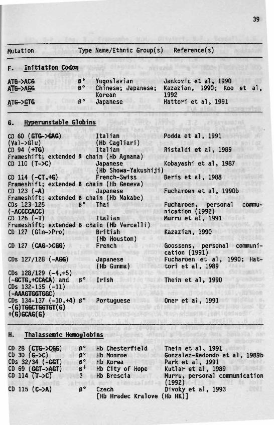
39
Mutation Type Name/Ethnic Group(s) Reference(s)
Initiation Codon
ATG->AÇGATG->AGG
ATG->GTG
B' Yugoslavian Jankovic et al, 1990B° Chinese; Japanese; Kazazian, 1990; Koo et al,
Korean 1992B° Japanese Hattori et al, 1991
G. Hyperunstable Giobins
CD 60 (GTG->6AG)(Val->Glu)CD 94 (+T6)Frameshift; extendedCD 110 (T->C)
CD 114 (-CT.+G)Frameshift; extendedCD 123 (-A)Frameshift; extendedCDs 123-125(-ACCCCACC)CD 126 (-T)Frameshift; extendedCD 127 (Gln->Pro)
CD 127 (CAG->CGG)
CDs 127/128 (-AGG)
Italian(Hb CagliaH)Italian
B chain (Hb Agnana)Japanese(Hb Showa-YakushijiFrench-Swiss
B chain (Hb Geneva)Japanese
B chain (Hb Makabe)B° Thai
ItalianB chain (Hb Vercelli)
British(Hb Houston)French
CDs 128/129 (-4,+5)(-GCTG.+CCACA) and B'CDs 132-135 (-11)(-AAAGTGGTGGC)CDs 134-137 (-10,+4) B'-(G)TGGCTGGTGT(G)+(G)GCAG(G)
Japanese(Hb Gunma)
Irish
Portuguese
Podda et al, 1991
Ristaldi et al, 1989
Kobayashi et al, 1987)Beris et al, 1988
Fucharoen et al, 1990b
Fucharoen, personal commu-nication (1992)Murru et al, 1991
Kazazian, 1990
Goossens, personal communi-cation (1991)Fucharoen et al, 1990; Hat-tori et al, 1989
Thein et al, 1990
Oner et al, 1991
H.
CDCDCDsCDCD
CD
Thalassenic
28 (CTG->CG6)30 (6->C)32/34 (-GGT)69 (GGT->AGT)114 TT->C7
115 (C->A)
Hemoglobins
B°B°B°B+?
B°
HbHbHbHbHb
ChesterfieldMonroeKoreaCity of HopeBrescia
Czech
Thein et al, 1991Gonzalez-Redondo et al, 1989bPark et al, 1991Kutlar et al, 1989Murru, personal communication(1992)Divoky et al, 1993
[Hb Hradec Kralove (Hb HK)]
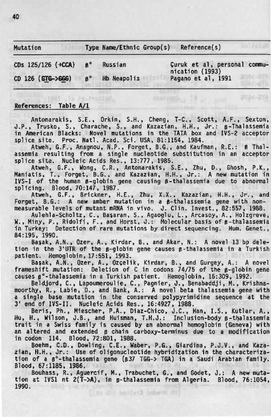
40
Mutation
CDs 125/126 (-KXA)
CD 126 (GTG->GGG)
References: Table
Type
B°
B°
A/1
Name/Ethnic
Russian
Hb Neapolis
Group(s) Reference(s)
Çuruk etnicationPagano et
al, personal commu-(1993)al, 1991
Antonarakis, S.E., Orkin, S.H., Cheng, T-C, Scott, A.F., Sexton,J.P., Trusko, S., Charache, S., and Kazazian, H.H., Jr.: e-Thalassemiain American Blacks: Novel mutations in the TATA box and IVS-2 acceptorsplice site. Proc. Natl. Acad. Sci. USA, 81:1154, 1984.
Atweh, G.F., Anagnou, N.P., Forget, B.G., and Kaufman, R.E.: B Thal-assemia resulting from a single nucleotide substitution in an acceptorsplice site. Nucleic Acids Res., 13:777, 1985.
Atweh, G.F., Wong, C.R., Antonarakis, S.E., Zhu, D., Ghosh, P.K.,Maniatis, T., Forget, B.G., and Kazazian, H.H., Jr.: A new mutation inIVS-I of the human B-globin gene causing B-thalassemia due to abnormalsplicing. Blood, 70:147, 1987.
Atweh, G.F., Brickner, H.E., Zhu, X.X., Kazazian, H.H., Jr., andForget, B.G.: A new amber mutation in a B-thalassemia gene with non-measurable levels of mutant mRNA in vivo. J. Clin. Invest., 82:557, 1988.
Aulehla-Scholtz, C , Basaran, S., Agaoglu, L., Arcasoy, A., Holzgreve,W., Miny, P., Ridolfi, F., and Horst, J.: Molecular basis of B-thalassemiain Turkey: Detection of rare mutations by direct sequencing. Hum. Genet.,84:195, 1990.
Ba$ak, A.N., Ozer, A., Kirdar, B., and Akar, N.: A novel 13 bp dele-tion in the 3'UTR of the B-globin gene causes B-thalassemia in a Turkishpatient. Hemoglobin, 17:551, 1993.
Basak, A.N., Ozer, A., Ozçelik, Kirdar, B., and Gurgey, A.: A novelframeshift mutation: Deletion of C in codons 74/75 of the B-globin genecauses p°-thalassemia in a Turkish patient. Hemoglobin, 16:309, 1992.
Beldjord, C , Lapoumeroulie, C , Pagnier, J., Benabaddji, M., Krishna-moorthy, R., Labié, D., and Bank, A.: A novel beta thalassemia gene witha single base mutation in the conserved polypyrimidine sequence at the3' end of IVS-II. Nucleic Acids Res., 16:4927, 1988.
Beris, Ph., Miescher, P.A., Diaz-Chico, J.C., Han, I.S., Kutlar, A.,Hu, H., Wilson, J.B., and Huisman, T.H.J.: Inclusion-body B-thalassemiatrait in a Swiss family is caused by an abnormal hemoglobin (Geneva) withan altered and extended B chain carboxy-terminus due to a modificationin codon 114. Blood, 72:801, 1988.
Boehm, C D . , Dowling, C.E., Waber, P.G., Giardina, P.J.V., and Kaza-zian, H.H., Jr.: Use of oligonucleotide hybridization in the characteriza-tion of a p°-thalassemia gene (B37 TGG-> TGA) in a Saudi Arabian family.Blood, 67:1185, 1986.
Bouhass, R., Aguercif, M., Trabuchet, G., and Godet, J.: A new muta-tion at IVS1 nt 2(T->A), in B-thalassemia from Algeria. Blood, 76:1054,1990.

41
Cai, S-P., Eng, B., Francombe, W.H., Olivieri, N.F., Kendall, A.G.,Waye, J.S., and Chui, D.H.K.: Two novel B-thalassemia mutations in the5' and 3' noncoding regions of the B-globin gene. Blood, 79:1342, 1992.
Cai, S.P., Zeng, J.Z., Doherty, M., and Kan, Y.W.: A new TATA boxmutation detected in prenatal diagnosis. Am. J. Hum. Genet., 45:112, 1989.
Chan, V., Chan, T.K., Kan, Y.W., and Todd, D.: A novel B-thalassemiaframeshift mutation (codon 14/15) detectable by direct visualization ofabnormal restriction fragment in amplified genomic DNA. Blood, 72:1420,1988.
Chan, V., Chan, T.K., and Todd, D.: A new codon 71 (+T) mutant re-sulting in B° thalassemia. Blood, 74:2304, 1989.
Chang, J.C. and Kan, Y.W.: B°-Thalassemia, a nonsense mutation inman. Proc. Natl. Acad. Sci. USA, 76:2886, 1980.
Chehab, F.F., Der Kaloustian, V., Khouri, F.P., Deeb, S.S., and Kan,Y.W.: The molecular basis of B-thalassemia in Lebanon: Application ofprenatal diagnosis. Blood, 69:1141, 1987.
Chehab, F.F., Winterhalter, K.H., and Kan, Y.W.: Characterization ofa spontaneous mutation in B-thalassemia associated with advanced paternalage. Blood, 74:852, 1989.
Cheng, T-C, Orkin, S.H., Antonarakis, S.E., Potter, M.J., Sexton,J.P., Giardina, P.J.V., Li, A., and Kazazian, H.H., Jr.: B-Thalassemiain Chinese: Use of in vivo RNA analysis and oligonucleotide hybridizationin systematic characterization of molecular defects. Proc. Natl. Acad.Sci. USA, 81:2821, 1984.
Chibani, J., Vidaud, M., Duquesnoy, P., Berge-Lefranc, J.L., Pirastu,M., Ellouze, F., Rosa, J., and Goossens, M.: The peculiar spectrum ofB-thalassemia genes in Tunisia. Hum. Genet., 78:190, 1988.
Codrington, J.F., Li, H-W., Kutlar, F., Gu, L-H., Ramachandran, M.,and Huisman, T.H.J.: Observations on the levels of Hb A£ in patients withdifferent B-thalassemia mutations and a 6 chain variant. Blood, 76:1246,1990.
Coleman, M.B., Steinberg, M.H., Harrell, A.H., Plonczynski, M.W.,Walker, A.M., and Adams, J.G., III: The -87 (C->A) B*-thalassemia muta-tion in a Black family. Hemoglobin, 16:399, 1992.
Deidda, G., Novelletto, A., Hafez, M., Al-Tonbary, Y., Felicetti,L., Terrenato, L., and Colombo, B.: A new B-thalassemia mutation producedby a single nucleotide substitution in the conserved dinucleotide sequenceof the IVS-I consensus acceptor site (AG->AA). Hemoglobin, 14:431, 1990.
Deidda, G., Novelletto, A., Hafez, M., El-Ziny, M., Terrenato, L.,and Felicetti, L.: A new B-thalassaemia frameshift mutation detected byPCR after selective hybridization to immobilized oligonucleotides. Br. J.Haematol., 79:90, 1991.
Dimovski, A.J., Jankovic, L., Efremov, D.G., Juricic, D., and Efremov,G.D.: Three new mutations (87 C->A; IVS 11-850 G->C; del. 1605 bp) inthe B-globin gene. 4th International Conference on Thalassemia and Hemo-globinopathies, Nice, Abstract, page 135, 1991.
Divoky, V., Gu, L-H., Indrak, K., Mocikova, K., Zarnovicanova, M.,and Huisman, T.H.J.: A new B°-thalassaemia nonsense mutation (codon 112,T->A) not associated with a dominant type of thalassaemia in the hétérozy-gote. Br. J. Haematol., 83:523, 1992.
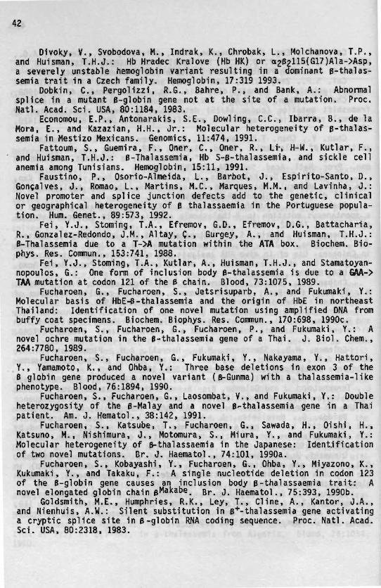
42
Divoky, V., Svobodova, M., Indrak, K., Chrobak, L., Molchanova, T.P.,and Huisman, T.H.J.: Hb Hradec Kralove (Hb HK) or a2B2ll5(G17)Ala->Asp,a severely unstable hemoglobin variant resulting in a dominant B-thalas-semia trait in a Czech family. Hemoglobin, 17:319 1993.
Dobkin, C , Pergolizzi, R.G., Bahre, P., and Bank, A.: Abnormalsplice in a mutant B-globin gene not at the site of a mutation. Proc.Natl. Acad. Sci. USA, 80:1184, 1983.
Economou, E.P., Antonarakis, S.E., Dowling, C.C., Ibarra, B., de laMora, E., and Kazazian, H.H., Jr.: Molecular heterogeneity of p-thalas-semia in Mestizo Mexicans. Genomics, 11:474, 1991.
Fattoum, S., Guemira, F., Oner, C , Oner, R., Li-, H-W., Kutlar, F.,and Huisman, T.H.J.: B-Thalassemia, Hb S-B-thalassemia, and sickle cellanemia among Tunisians. Hemoglobin, 15:11, 1991.
Faustino, P., Osorio-Almeida, L., Barbot, J., Espirito-Santo, D.,Gonçalves, J., Romao, L., Martins, M.C., Marques, M.M., and Lavinha, J.:Novel promoter and splice junction defects add to the genetic, clinicalor geographical heterogeneity of B thaiassaemia in the Portuguese popula-tion. Hum. Genet., 89:573, 1992.
Fei, Y.J., Stoming, T.A., Efremov, G.D., Efremov, D.G., Battacharia,R., Gonzalez-Redondo, J.M., Altay, Ç., Gurgey, A., and Huisman, T.H.J.:B-Thalassemia due to a T->A mutation within the ATA box. Biochem. Bio-phys. Res. Commun., 153:741, 1988.
Fei, Y.J., Stoming, T.A., Kutlar, A., Huisman, T.H.J., and Stamatoyan-nopoulos, G.: One form of inclusion body B-thalassemia is due to a GAA->TAA mutation at codon 121 of the B chain. Blood, 73:1075, 1989.
Fucharoen, G., Fucharoen, S., Jetsrisuparb, A., and Fukumaki, Y.:Molecular basis of HbE-B-thalassemia and the origin of HbE in northeastThailand: Identification of one novel mutation using amplified DNA frombuffy coat specimens. Biochem. Biophys. Res. Commun., 170:698, 1990c.
Fucharoen, S., Fucharoen, G., Fucharoen, P., and Fukumaki, Y.: Anovel ochre mutation in the B-thalassemia gene of a Thai. J. Biol. Chem.,264:7780, 1989.
Fucharoen, S., Fucharoen, G., Fukumaki, Y., Nakayama, Y., Hattori,Y., Yamamoto, K., and Ohba, Y.: Three base deletions in exon 3 of theB globin gene produced a novel variant (B-Gunma) with a thaiassemia-1ikephenotype. Blood, 76:1894, 1990.
Fucharoen, S., Fucharoen, G., Laosombat, V., and Fukumaki, Y.: Doubleheterozygosity of the B-Malay and a novel B--thalassemia gene in a Thaipatient. Am. J. Hematol., 38:142, 1991.
Fucharoen, S., Katsube, T., Fucharoen, G., Sawada, H., Oishi, H.,Katsuno, M., Nishimura, J., Motomura, S., Miura, Y., and Fukumaki, Y.:Molecular heterogeneity of B-thalassaemia in the Japanese: Identificationof two novel mutations. Br. J. Haematol., 74:101, 1990a.
Fucharoen, S., Kobayashi, Y., Fucharoen, G., Ohba, Y., Miyazono, K.,Kukumaki, Y., and Takaku, F.: A single nucleotide deletion in codon 123of the B-globin gene causes an inclusion body B-thalassaemia trait: Anovel elongated globin chain pMakabe. Br. J. Haematol., 75:393, 1990b.
Goldsmith, M.E., Humphries, R.K., Ley, T., Cline, A., Kantor, J.A.,and Nienhuis, A.W.: Silent substitution in B*-thalassemia gene activatinga cryptic splice site in B-globin RNA coding sequence. Proc. Natl. Acad.Sci. USA, 80:2318, 1983.
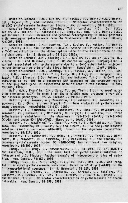
43
Gonzalez-Redondo, J.M., Kutlar, A., Kutlar, F., McKie, V.C., McKie,K.M., Baysal, E., and Huisman, T.H.J.: Molecular characterization ofHb S(C) B-thalassemia in American Blacks. Am. J. Hematol., 38:9, 1991.
Gonzalez-Redondo, J.M., Stoming, T.A., Lanclos, K.D., Gu, Y.C.,Kutlar, A., Kutlar, F., Nakatsuji, T., Deng, B., Han, I.S., McKie, V.C.,and Huisman, T.H.J.: Clinical and genetic heterogeneity in Black patientswith homozygous B-thalassemia from the Southeastern United States. Blood,72:1007, 1988.
Gonzalez-Redondo, J.M., Stoming, T.A., Kutlar, F., Kutlar, A., McKie,V.C., McKie, K.M., and Huisman, T.H.J.: Severe Hb SB°-thalassaemia witha T->C substitution in the donor splice site of the first intron of theB-globin gene. Br. J. Haematol., 71:113, 1989a.
Gonzalez-Redondo, J.M., Stoming, T.A., Kutlar, F., Kutlar, A., Hu, H.,Wilson, J.B., and Huisman, T.H.J.: Hb Monroe or a2B230 (B12)Arg->Thr, avariant associated with B-thalassemia due to a G->C substitution adjacentto the donor splice site of the first intron. Hemoglobin, 13:67, 1989b.
Gonzalez-Redondo, J.M., Stoming, T.A., Kutlar, A., Kutlar, F., Lan-clos, K.D., Howard, E.F., Fei, Y.J., Aksoy, M., Altay, Ç., Gurgey, A.,Basak, A.N., Efremov, G.D., Petkov, G., and Huisman, T.H.J.: A C->T sub-stitution at nt -101 in a conserved DNA sequence of the promoter regionof the B-globin gene is associated with "silent" B-thalassemia. Blood,73:1705, 1989c.
Hall, G.W., Franklin, I.M., Sura, T., and Thein, S.L.: A novel muta-tion (nonsense B127) in exon 3 of the B globin gene produces a variablethalassaemia phenotype. Br. J. Haematol., 79:342, 1991.
Hattori, Y., Yamane, A., Yamashiro, Y., Matsuno, Y., Yamamoto, Ki.,Yamamoto, Ku., Ohba, Y., and Miyaji, T.: Gene analysis of B-thalassemiaamong Japanese. Hemoglobin, 13:657, 1989.
Hattori, Y., Yamamoto, Ku., Yamashiro, Y., Ohba, Y., Miyamura, S.,Yamamoto, Ki., Matsuno, Y., Morishita, M., Miyaji, T., and Era, T.: ThreeB-thalassemia mutations in the Japanese: IVS-II-1 (G->A), IVS-II-848(C->G), and codon 90 (6AG->TAG). Hemoglobin, 16:93, 1992.
Hattori, Y., Yamashiro, Y., Ohba, Y., Miyaji, T., Morishita, M., Yama-moto, Ku., Yamamoto, Ki., Narai, S., and Kimura, A.: A new B-thalassemiamutation (initiation codon ATG->GTG) found in the Japanese population.Hemoglobin, 15:317, 1991.
Hattori, Y., Yamashiro, Y., Ohba, Y., Miyaji, T., Terai, S., Mori-shita, M., Yamamoto, Ku., Yamamoto, Ki., Matsumoto, N., and Kawano, F.:Japanese B-thalassemia [codon 90 (6AG->TAG) has at least two origins.Hemoglobin, 16:303, 1992.
Huang, S-Z., Wong, C , Antonarakis, S.E., Ro-Lein, T., Lo, W.H.Y.,and Kazazian, H.H., Jr.: The same TATA box B-thalassemia mutation inChinese and U.S. Blacks: Another example of independent origins of muta-tion. Hum. Genet., 74:152, 1986.
Huang, S-Z., Xu, Y-H., Zeng, F-Y., Wu, D-F., Ren, Z-R., and Zeng,Y-T.: A novel g-thalassaemia mutation: Deletion of 4 bp (-AAAC) in the5' transcriptional sequence. Br. J. Haematol., 78:125, 1991.
Indrak, K., Brabec, V., Indrakova, J., Chrobak, L., Sakalova, A.,Jarosova, M., Cermak, J., Fei, Y-J., Kutlar, F., Gu, Y-C, Baysal, E.,and Huisman, T.H.J.: Molecular characterization of B-thalassemia in Czech-oslovakia. Hum. Genet., 88:399, 1992.
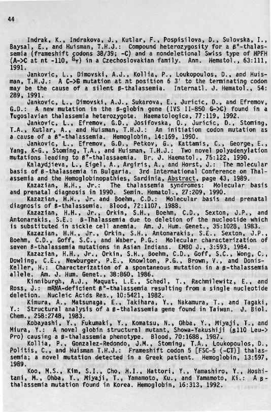
44
Indrak, K., Indrakova, J., Kutlar, F., Pospisilova, D., Sulovska, I.,Baysal, E., and Huisman, T.H.J.: Compound heterozygosity for a B°-thalas-semia (frameshift codons 38/39; -C) and a nondeletional Swiss type of HPFH(A->C at nt -110, &Y) in a Czechoslovakian family. Ann. Hematol., 63:111,1991.
Jankovic, L., Dimovski, A.J., Kollia, P., Loukopoulos, D., and Huis-man, T.H.J.: A C->G mutation at nt position 6 3' to the terminating codonmay be the cause of a silent B-thalassemia. Internatl. J. Hematol., 54:289, 1991.
Jankovic, L., Dimovski, A.J., Sukarova, E., Juricic, D., and Efremov,G.D.: A new mutation in the B-globin gene (IVS 11-850 G->C) found in aYugoslavian thalassemia hétérozygote. Haematologica, 77:119, 1992.
Jankovic, L., Efremov, G.D., Josifovska, 0., Juricic, D., Stoming,T.A., Kutlar, A., and Huisman, T.H.J.: An initiation codon mutation asa cause of a B°-thalassemia. Hemoglobin, 14:169, 1990.
Jankovic, L., Efremov, G.D., Petkov, G., Kattamis, C , George, E.,Yang, K-G., Stoming, T.A., and Huisman, T.H.J.: Two novel polyadenylationmutations leading to B+-thalassaemia. Br. J. Haematol., 75:122, 1990.
Kalaydjieva, L., Eigel, A., Argiris, A., and Horst, J.: The molecularbasis of B-thalassemia in Bulgaria. 3rd International Conference on Thal-assemia and the Hemoglobinopathies, Sardinia, Abstract, page 43, 1989.
Kazazian, H.H., Jr.: The thalassemia syndromes: Molecular basisand prenatal diagnosis in 1990. Semin. Hematol., 27:209, 1990.
Kazazian, H.H., Jr. and Boehm, C D . : Molecular basis and prenataldiagnosis of B-thalassemia. Blood, 72:1107, 1988.
Kazazian, H.H., Jr., Orkin, S.H., Boehm, C D . , Sexton, J.P., andAntonarakis, S.E.: B-Thalassemia due to deletion of the nucleotide whichis substituted in sickle cell anemia. Am. J. Hum. Genet., 35:1028, 1983.
Kazazian, H.H., Jr., Orkin, S.H., Antonarakis, S.E., Sexton, J.P.,Boehm, C D . , Goff, S.C, and Waber, P.G.: Molecular characterization ofseven B-thalassemia mutations in Asian Indians. EMBO J., 3:593, 1984.
Kazazian, H.H., Jr., Orkin, S.H., Boehm, C D . , Goff, S.C, Wong, C ,Dowling, C E . , Newburger, P.E., Knowlton, P.G., Brown, V., and Donis-Keller, H.: Characterization of a spontaneous mutation in a B-thalassemiaallele. Am. J. Hum. Genet., 38:860, 1986.
Kinniburgh, A.J., Maquat, L.E., Schedl, T., Rachmilewitz, E., andRoss, J.: mRNA-deficient B°-thalassemia resulting from a single nucleotidedeletion. Nucleic Acids Res., 10:5421, 1982.
Kimura, A., Matsunaga, E., Takihara, Y., Nakamura, T., and Tagaki,Y.: Structural analysis of a B-thalassemia gene found in Taiwan. J. Biol.Chem., 258:2748, 1983.
Kobayashi, Y., Fukumaki, Y., Komatsu, N., Ohba, Y., Miyaji, T., andMiura, Y.: A novel globin structural mutant, Showa-Yakushiji (B110 Leu->Pro) causing a B-thalassemia phenotype. Blood, 70:1688, 1987.
Kollia, P., Gonzalez-Redondo, J.M., Stoming, T.A., Loukopoulos, D.,Politis, C , and Huisman T.H.J.: Frameshift codon 5 [FSC-5 (-CT)] thalas-semia; a novel mutation detected in a Greek patient. Hemoglobin, 13:597,1989.
Koo, M.S., Kim, S.I., Cho, H.I., Hattori, Y., Yamashiro, Y., Hoshi-tani, M., Ohba, Y., Miyaji, T., Yamamoto, Ku., and Yamamoto, Ki. : A B -thalassemia mutation found in Korea. Hemoglobin, 16:313, 1992.
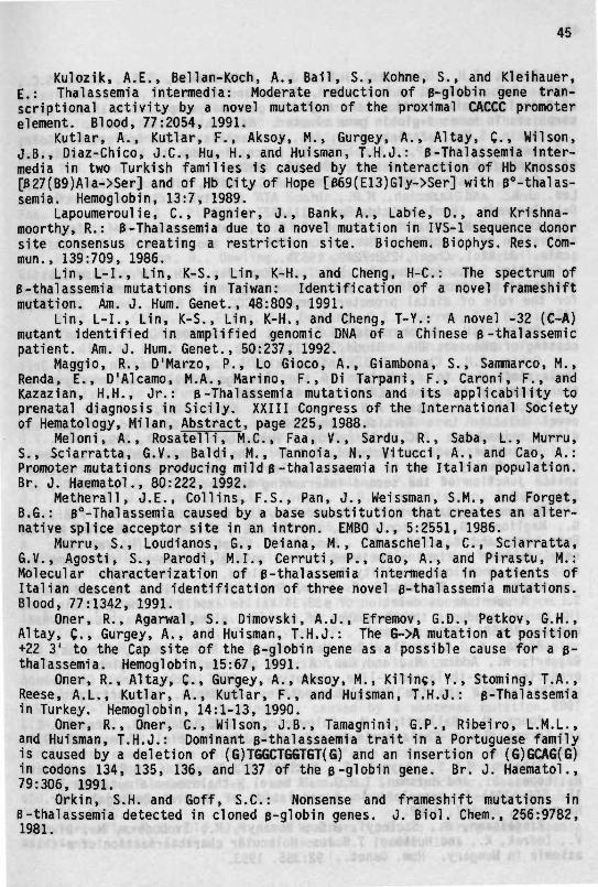
45
Kulozik, A.E., Beilan-Koch, A., Bail, S., Kohne, S., and Kleihauer,E.: Thalassemia intermedia: Moderate reduction of B-globin gene tran-scriptional activity by a novel mutation of the proximal CACCC promoterelement. Blood, 77:2054, 1991.
Kutlar, A., Kutlar, F., Aksoy, M., Gurgey, A., Altay, Ç., Wilson,J.B., Diaz-Chico, J.C., Hu, H., and Huisman, T.H.J.: B-Thalassemia inter-media in two Turkish families is caused by the interaction of Hb Knossos[B27(B9)Ala->Ser] and of Hb City of Hope [B69(E13)Gly->Ser] with B°-thalas-semia. Hemoglobin, 13:7, 1989.
Lapoumeroulie, C , Pagnier, J., Bank, A., Labie, D., and Krishna-moorthy, R.: B-Thalassemia due to a novel mutation in IVS-1 sequence donorsite consensus creating a restriction site. Biochem. Biophys. Res. Com-mun., 139:709, 1986.
Lin, L-I., Lin, K-S., Lin, K-H., and Cheng, H-C: The spectrum ofB-thalassemia mutations in Taiwan: Identification of a novel frameshiftmutation. Am. J. Hum. Genet., 48:809, 1991.
Lin, L-I., Lin, K-S., Lin, K-H., and Cheng, T-Y.: A novel -32 (C-A)mutant identified in amplified genomic DNA of a Chinese B-thalassemicpatient. Am. J. Hum. Genet., 50:237, 1992.
Maggio, R., D'Marzo, P., Lo Gioco, A., Giambona, S., Sammarco, M.,Renda, E., D'Alcamo, M.A., Marino, F., Di Tarpani, F., Caroni, F., andKazazian, H.H., Jr.: B-Thalassemia mutations and its applicability toprenatal diagnosis in Sicily. XXIII Congress of the International Societyof Hematology, Milan, Abstract, page 225, 1988.
Meloni, A., Rosatelli, M.C., Faa, V., Sardu, R., Saba, L., Murru,S., Sciarratta, G.V., Baldi, M., Tannoia, N., Vitucci, A., and Cao, A.:Promoter mutations producing mild 6-thalassaemia in the Italian population.Br. J. Haematol., 80:222, 1992.
Metherall, J.E., Collins, F.S., Pan, J., Weissman, S.M., and Forget,B.G.: B°-Thalassemia caused by a base substitution that creates an alter-native splice acceptor site in an intron. EMBO J., 5:2551, 1986.
Murru, S., Loudianos, G., Deiana, M., Camaschella, C , Sciarratta,G.V., Agosti, S., Parodi, M.I., Cerruti, P., Cao, A., and Pirastu, M.:Molecular characterization of B-thalassemia intermedia in patients ofItalian descent and identification of three novel B-thalassemia mutations.Blood, 77:1342, 1991.
Oner, R., Agarwal, S., Dimovski, A.J., Efremov, G.D., Petkov, G.H.,Altay, C , Gurgey, A., and Huisman, T.H.J.: The G->A mutation at position+22 3' to the Cap site of the B-globin gene as a possible cause for a B-thalassemia. Hemoglobin, 15:67, 1991.
Oner, R., Altay, Ç., Gurgey, A., Aksoy, M., Kilinc, Y., Stoming, T.A.,Reese, A.L., Kutlar, A., Kutlar, F., and Huisman, T.H.J.: B-Thalassemiain Turkey. Hemoglobin, 14:1-13, 1990.
Oner, R., Oner, C , Wilson, J.B., Tamagnini, G.P., Ribeiro, L.M.L.,and Huisman, T.H.J.: Dominant B-thalassaemia trait in a Portuguese familyis caused by a deletion of (G)TGGCTGGTGT(G) and an insertion of (G)GCAG(G)in codons 134, 135, 136, and 137 of the B-globin gene. Br. J. Haematol.,79:306, 1991.
Orkin, S.H. and Goff, S.C.: Nonsense and frameshift mutations inB-thalassemia detected in cloned B-globin genes. J. Biol. Chem., 256:9782,1981.

46
Orkin, S.H., Kazazian, H.H., Jr., Antonarakis, S.E., Goff, S.C.,Boehm, C D . , Sexton, J.P., Waber, P.G., and Giardina, P.J.V.: Linkageof B-thalassaemia mutations and B-globin gene polymorphisms with DNA poly-morphisms in human B-globin gene cluster. Nature, 296:627, 1982a.
Orkin, S.H., Kazazian, H.H., Jr., Antonarakis, S.E., Ostrer, H., Goff,S.C., and Sexton, J.P.: Abnormal RNA processing due to the exon mutationof the B^-globin gene. Nature, 300:768, 1982b.
Orkin, S.H., Sexton, J.P., Cheng, T-C, Goff, S.C., Giardina, P.J.V.,Lee, J.I., and Kazazian, H.H., Jr.: ATA box transcription mutation inB-thalassemia. Nucleic Acids Res., 11:4727, 1983a.
Orkin, S.H., Sexton, J.P., Goff, S.C., and Kazazian, H.H., Jr.: In-activation of an acceptor RNA splice site by a short deletion in e-thalas-semia. J. Biol. Chem., 258:7249, 1983b.
Orkin, S.H., Antonarakis, S.E., and Kazazian, H.H., Jr.: Base substi-tution at position -88 in a B-thalassemic globin gene: Further evidencefor the role of distal promoter element CACCC. J. Biol. Chem., 259:8679,1984a.
Orkin, S.H., Antonarakis, S.E., and Loukopoulos, D.: Abnormal pro-cessing of BKnossos RNA. Blood, 64:311, 1984b.
Orkin, S.H., Cheng, T-C, Antonarakis, S.E., and Kazazian, H.H., Jr.:Thalassemia due to a mutation in the cleavage-polyadenylation signal ofthe human B-globin gene. EMBO J., 4:453, 1985.
Ozçelik, H., Basak, A.N., Tuzmen, §., Kirdar, B., and Akar, N.: Anovel deletion in a Turkish B-thalassemia patient detected by DGGE anddirect sequencing: FSC 22-24 (-7 bp). Hemoglobin, 17:387, 1993.
Padanilam, B.J. and Huisman, T.H.J.: The B°-thalassemia in an Ameri-can Black family is due to a single nucleotide substitution in the acceptorsplice junction of the second intervening sequence. Am. J. Hematol., 22:259, 1986.
Pagano, L., Lacerra, G., Camardella, L., De Angioletti, M., Fioretti,G., Magi i one, G., de Bonis, C , Guarino, E., Viola, A., Cutolo, R., DeRosa, L., and Carestia, C : Hemoglobin Neapolis, Bl26(H4)Val->Gly: Anovel B-chain variant associated with a mild p-thalassemia phenotype anddisplaying anomalous stability features. Blood, 78:3070, 1991.
Park, S.S., Barnetson, R., Kim, S.W., Weatherall, D.J., and Thein,S.L.: A spontaneous deletion of B33/34 Val in exon 2 of the g globin gene(Hb Korea) produces the phenotype of dominant 3 thalassaemia. Br. J. Hae-matol., 78:581, 1991.
Podda, A., Galanello, R., Maccioni, L., Mel is, M.A., Perseus, L.,Ruggeri, R., Addis, M., and Cao, A.: A new unstable hemoglobin variantproducing a B-thalassemia-like phenotype. 3rd International Conferenceon Thalassemia and the Hemoglobinopathies, Sardinia, Abstract, page 51,1989.
Poncz, M., Ballantine, M., Solowiejczyk, D., Barak, I., Schwartz, E.,and Surrey, S.: B-Thalassemia in a Kurdish Jew. J. Biol. Chem., 257:5994,1983.
Ribeiro, M.L.S., Baysal, E., Kutlar, F., Tamagnini, G.P., Gonçalves,P., Lopes, D., and Huisman, T.H.J.: A novel 3-thalassaemia mutation (codon15, TGG->TGA) is prevalent in a population of Central Portugal. Br. J.Haematol., 80:567, 1992.
Ringelhann, B., Szelenyi, J.G., Horanyi, M., Svobodova, M., Divoky,V., Indrak, K., and Huisman, T.H.J.: Molecular characterization of B-thal-assemia in Hungary. Hum. Genet., 92:385. 1993.

47
Ristaldi, M.S., Loudianos, G., Murdu, S., Casula, L., Porcu, S.,Sciarratta, G.V., Agosti, S., Parodi, M.I., Leone, D., Camaschella, C ,Serra, A., Cao, A., and Pirastu, M.: Characterization of molecular basisof atypical B-thalassemia. 3rd International Conference on Thalassemiaand the Hemoglobinopathies, Sardinia, Abstract, page 45, 1989.
Rosatelli, M.C., Dozy, A., Faa, V., Meloni, A., Sardu, R., Saba, L.,Kan, Y.W., and Cao, A.: Molecular characterization of B-thalassemia inthe Sardinian population. Am. J. Hum. Genet., 50:422, 1992a.
Rosatelli, M.C., Tuveri, T., Scalas, M.T., Leoni, G.B., Sardu, R.,Faa, V., Meloni, A., Pischedda, M.A., Demurtas, M., Monni, G., and Cao,A.: Molecular screening and fetal diagnosis of B-thalassemia in theItalian population. Am. J. Hum. Genet., in press (1992b).
Rund, D., Filon, D., Dowling, C , Kazazian, H.H., Jr., Rachmilewitz,E.A., and Oppenheim, A.: Molecular studies of B-thalassemia in Israel -Mutational analysis and expression studies. Ann. N.Y. Acad. Sci., 619:98,1990.
Rund, D., Cohen, T., Filon, D., Dowling, C E . , Warren, T., Barak, I.,Rachmilewitz, E., Kazazian, H.H., Jr., and Oppenheim, A.: Evolution of agenetic disease in an ethnic isolate: B-Thalassemia in the Jews of Kurdi-stan. Proc. Natl. Acad. Sci. USA, 88:310, 1991.
Schnee, J., Griese, E.V., Eigel, A., and Horst, J.: B-Thalassemiagene analysis in a Turkish family reveals a 7 bp deletion in the codingregion. Blood, 73:2224, 1989.
Schwartz, E., Goltsov, A.A., Kaboaev, O.K., Alexeev, A.A., Solouyev,G.Y., Surin, V.L., Lukainento, A.V., Vinogradov, S.U., and Berlin, Y.A.:A novel frameshift mutation causing e-thalassemia in Azerbaijan. NucleicAcids Res., 17:3997, 1989.
Spritz, R.A., Jagadeeswaran, P., Choudary, P.V., Biro, P.A., Elder,J.T., deRiel, J.K., Manley, J.L., Forget, B.G., and Weissman, S.M.: Basesubstitution in an intervening sequence of a B*thalassemic human globingene. Proc. Natl. Acad. Sci. USA, 78:2455, 1981.
Takihara, Y., Nakamura, T., Yamada, H., Takagi, Y., and Kukumaki, Y.:A novel mutation in the TATA box in a Japanese patient with B"*"-thalassemia.Blood, 67:547, 1986.
Thein, S.L., Best, S., Sharpe, J., Paul, B., Clark, D.J., and Brown,M.J.: Hemoglobin Chesterfield (B28 Leu->Arg) produces the phenotype ofinclusion body ethalassemia. Blood, 77: 2971, 1991.
Thein, S.L., Hesketh, C , Taylor, P., Temperley, I.J., Hutchinson,R.M., Old, J.M., Wood, W.G., Clegg, J.B., and Weatherall, D.J.: Molecularbasis for dominantly inherited inclusion body B-thalassemia. Proc. Natl.Acad. Sci. USA, 87:3924, 1990.
Trecartin, R.F., Liebhaber, S.A., Chang, J.C., Lee, Y.W.,and Kan,Y.W.: B°-Thalassemia in Sardinia is caused by a nonsense mutation. J.Clin. Invest., 68:1012, 1981.
Treisman, R., Proudfoot, N.J., Shander, M., and Maniatis, T.: Asingle base change at a splice site in a e°-thalassemic gene causes abnor-mal RNA splicing. Cell, 29:903, 1982.
Treisman, R., Orkin, S.H., and Maniatis, T.: Specific transcriptionand RNA splicing defects in five cloned 0-thalassaemia genes. Nature,302:591, 1983.
Varawalla, N.Y., Old, J.M., and Weatherall, D.J.: Rare g-thalassaemiamutations in Asian Indians. Br. J. Haematol., 79:640, 1991.

48
Westaway, D. i/id Williamson, R.: An intron nucleotide sequence vari-ant in a cloned B*-thalassemia globin gene. Nucleic Acids Res., 9:1777,1981.
Hong, C , Antonarakis, S.E., Goff, S.C., Orkin, S.H., Boehm, C D . , andKazazian, H.H., Jr.: On the origin and spread of B-thalassemia: Recurrentobservation of four mutations in different ethnic groups. Proc. Natl.Acad. Sci. USA, 83:6529, 1986.
Wong, C , Dowling, C E . , Saiki, R.K., Higuchi, R.G., Erlich, H.A.,H.H., Jr.: Characterization of B-thalassaemia mutations
genomic sequencing of amplified single copy DNA. Nature,and Kazazian,using direct330:384, 1987.
Wong, C.Nathan, O.G.,
, Forget, B.G.,B-Thalassemia
acceptor splice
Antonarakis, S.E., Goff, S.C., Orkin, S.H.Giardina, P.J.V., and Kazazian, H.H., Jr.:
due to two novel nucleotide substitutions in consensussequences of the B-globin gene. Blood, 73:914, 1989.
Yamamoto, Ku., Yamamoto, Ki., Hattori, Y., Yamashiro, Y., Hoshitani,M., Morishita, M., Ohba, Y., Katahira, H., Karasawa, M., Omine, M., Naru-kiyo, T., Hirabayashi, K., and Miyawaki, S.: Two e-thalassemia mutations:Codon 121 (6AA->TAA) and IVS-I130 (G->C) in Japan. Hemoglobin, 16:295,1992.
Yang, K.G., Kutlar, F., George, E., Wilson, J.B., Kutlar, A., Stoming,T.A., Gonzalez-Redondo, J.M., and Huisman, T.H.J.: Molecular characteriza-tion of B-globin gene mutations in Malay patients with Hb E-p-thalassaemiaand thalassaemia major. Br. J. Haematol., 72:73, 1989.
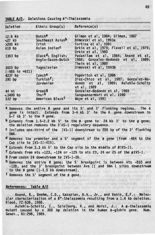
49
TABLE A/2. Deletions Causing B°-Thalassemia
Deletion Ethnie Group(s) Reference(s)
12.6 kb*27 kb>200 kb619 bp
1393 bp
1605 bp(-985 to +621)4237 bp290 bp
44 nta 3400 bp532 bp
Dutch»Southeast Asian"IrishAsian Indian^
Black**; English;Anglo-Saxon-Dutch
Yugoslavian
Czech*Turkish*;Jordanian
Greek9Thai"American Black*
Gil man et al, 1984; Gil man, 1987Dimovski et al, 1993aFortina et al, 1991Orkin et al, 1979; Flavell et al, 1979;Orkin et al, 1980Padanilam et al, 1984; Anand et al,1988; Gonzalez-Redondo et al, 1989;Thein et al, 1989Dimovski et al, 1993b
Popovitch et al, 1986Diaz-Chico et al, 1987; Gonzalez-Re-dondo et al, 1989; Aulehla-Scholtzet al, 1989Gonzalez-Redondo et al, 1989Sanguansermsri et al, 1990Waye et al, 1991
* Removes the entire B gene and its 5'gene is intact. Extends from 3-4 kb
and 3' flanking regions. The 63' to the 6 gene downstream to
6-7 kb 3' to the B gene,b Extends from 1.5-2.2 kb 5' to the B gene to 24 kb 3' to the e gene;the deletion removes the 3'HS-l regulatory sequence,
c includes one-third of the IVS-II downstream to 209 bp of the 3' flankingDNA.
d Removes the promoter and a 5' segment of the 6 gene (from -484 to theCap site to IVS-II-415).
e Extends from 3.3 kb 5' to the Cap site to the middle of BIVS-II.f Extends from nts -123, -124 or -125 to nts 23, 24 or 25 of the BlVS-I.9 From codon 24 downstream to IVS-I-26." Removes the entire B gene; the 5' breakpoint is between nts -810 andg-128, and the 3' breakpoint between Ava IIto the B gene (1-1.5 kb downstream).Removes the 5' segment of the B gene.
and Xmn I sites downstream
References: Table A/2
Anand, R., Boehm, C D . , Kazazian, H.H., Jr., and Vanin, E.F.: Molec-ular characterization of a B°-thalassemia resulting from a 1.4 kb deletion.Blood, 72:636, 1988.
Aulehla-Scholtz, C , Spielberg, R., and Horst, J.: A B-thalassemiamutant caused by a 300 bp deletion in the human B-globin gene. Hum.Genet., 81:298, 1989.

50
Diaz-Chico, J.C., Yang, K.G., Kutlar, A., Reese, A.L., Aksoy, M.,and Huisman, T.H.J.: An t300 bp deletion involving part of the 5' B-globingene region is observed in members of a Turkish family with B-thalassemia.Blood, 70:583, 1987.
Dimovski, A.J., Divoky, V., Adekile, A.D., Baysal, E., Wilson, J.B.,Prior, J.F., Raven, J.L., and Huisman, T.H.J.: A novel deletion of -x.27 kbincluding the B-globin gene and the LCR 3'HS-l regulatory sequence: B°-Thalassemia or HPFH? Blood, in press (1993a).
Dimovski, A.J., Efremov, D.G., Jankovic, L., Plaseska, D., Juricic,D., and Efremov, G.D.: A B°-Thalassaemia due to a 1605 bp deletion ofthe 5' B-globin gene region. Br. J. Haematol., 85:143, 1993b.
Flavell, R.A., Bernards, B., Kooter, J.M., de Boer, E., Little,P.F.R., Annison, G., and Williamson, D.: The structure of the human B-globin gene in B-thalassemia. Nucleic Acids Res., 8:2749, 1979.
Fortina, P., Delgrosso, K., Werner, E., Haines, K., Rappaport, E.,Schwartz, E., and Surrey, S.: A >200 kb deletion removing the entire B-like globin gene cluster in a family of Irish descent. Hemoglobin, 15:23,1991.
Gilman, J.G.: The 12.6 kilobase deletion in Dutch B°-thalssaemia.Br. J. Haematol., 67:369, 1987.
Gilman, J.G., Huisman, T.H.J., and Abels, J.: Dutch B°-thalassaemia:A 10 kilobase DNA deletion associated with significant Y-chain production.Br. J. Haematol., 56:339, 1984.
Gonzalez-Redondo, J.M., Kattamis, C , and Huisman, T.H.J.: Character-ization of three types of B°-thalassemia resulting from a partial deletionof the B-globin gene. Hemoglobin, 13:377, 1989.
Orkin, S.H., Old, J.M., Weatherall, D.J., and Nathan, D.G.: Partialdeletion of B-globin gene DNA in certain patients with B°-thalassemia.Proc. Natl. Acad. Sci. USA, 76:2400, 1979.
Orkin, S.H., Kolodner, R., Michelson, A., and Husson, R.: Cloningand direct examination of a structurally abnormal human B° thalassemiaglobin gene. Proc. Natl. Acad. Sci. USA, 77:3558, 1980.
Padanilam, B.J., Felice, A.E., and Huisman, T.H.J.: Partial deletionof the 5' B-globin gene region causes B°-thalassemia in members of an Amer-ican Black family. Blood, 64:941, 1984.
Popovitch, B.W., Rosenblatt, D.S., Kendall, A.G., and Nishioka, Y.:Molecular characterization of an atypical B-thalassemia caused by a largedeletion in the 5' B-globin gene region. Am. J. Hum. Genet., 39:797, 1986.
Sanguansermsri, T., Pape, M., Laig, M., Hundrieser, J., and Flatz, G.:B°-Thalassemia in a Thai family is caused by a 3.4 kb deletion includingthe entire B-globin gene. Hemoglobin, 14:157, 1990.
Thein, S.L., Hesketh, C , Brown, J.M., Anstey, A.V., and Weatherall,D.J.: Molecular characterization of a high A2 B-thalassemia by directsequencing of single strand enriched amplified genomic DNA. Blood, 73:924,1989.
Waye, J.S., Cai, S.P., Eng, B., Clark, C , Adams, J.G., III, Chui,D.H.K., and Steinberg, M.H.: High Hemoglobin A2 B°-thalassemia due toa 532-basepair deletion of the 5' B-globin gene region. Blood, 77:1100,1991.
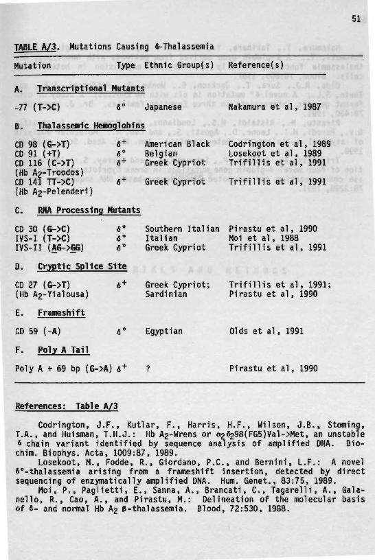
TABLE A/3. Mutations Causing &-Thalassemia
Mutation Type Ethnie 6roup(s) Reference(s)
A. Transcriptionai Mutants
-77 (T->C) «° Japanese
B. Thalassemic Hemoglobins
CD 98 (G->T) 6+CD 91 (+T) 6°CD 116 (C->T) «+(Hb A2-Troodos)CD 141 TT->C) 6+(Hb A2-Pe1enderi)
C. RMA Processing Mutants
American BlackBelgianGreek Cypriot
Greek Cypriot
CD 30 (G->C) fi°IVS-I (T->C) 6°IVS- I I (AG->GG) fi°
D. Cryptic Splice Site
CD 27 (G->T) « +
(Hb A2-Yialousa)
E. Frameshift
CD 59 (-A) 6°
F. Poly A Tail
Poly A + 69 bp (G->A) 6+
Southern ItalianItalianGreek Cypriot
Greek Cypriot;Sardinian
Egyptian
Nakamura et al, 1987
Codrington et al, 1989Losekoot et al, 1989Trifillis et al, 1991
Trifillis et al, 1991
Pirastu et al, 1990Moi et al, 1988Trifillis et al, 1991
Trifillis et al, 1991;Pirastu et al, 1990
Olds et al, 1991
Pirastu et al, 1990
References: Table A/3
Codrington, J.F., Kutlar, F., Harris, H.F., Wilson, J.B.» Stoming,T.A., and Huisman, T.H.J.: Hb A2-Wrens or a2*298(FG5)Val->Met, an unstable* chain variant identified by sequence analysis of amplified DNA. Bio-chim. Biophys. Acta, 1009:87, 1989.
Losekoot, M., Fodde, R., Giordano, P.C., and Bernini, L.F.: A novel6°-thalassemia arising from a frameshift insertion, detected by directsequencing of enzymatically amplified DNA. Hum. Genet., 83:75, 1989.
Moi, P., Paglietti, E., Sanna, A., Brancati, C , Tagarelli, A., Gala-nello, R., Cao, A., and Pirastu, M.: Delineation of the molecular basisof 6- and normal Hb A2 B-thalassemia. Blood, 72:530, 1988.
52
Nakamura, T., Takihara, Y., Ohta, Y., Fujita, S., Takagi, Y., andFukumaki, Y.: A 6-globin gene derived from patients with homozygous 6°-thalassemia functions normally on transient expression in heterologouscells. Blood, 70:809, 1987.
Olds, R.J., Sura, T., Jackson, B., Wonke, B., Hoffbrand, A.V., andThein, S.L.: A novel 6° mutation in cis with Hb Knossos: A study of dif-ferent genetic interactions in three Egyptian families. Br. J. Haematol.,78:430, 1991.
Pirastu, M., Ristaldi, M.S., Loudianos, G., Murru, S., Sciarratta,G.V., Parodi, M.I., Leone, D., Agosti, S., and Cao, A.: Molecular analysisof atypical B-thalassemia hétérozygotes. Ann. N.Y. Acad. Sci., 612:90,1990.
Trifillis, P., Ioannou, P., Schwartz, E., and Surrey, S.: Identifica-tion of four novel 6-globin gene mutations in Greek Cypriots using polymer-ase chain reaction and automated fluorescence-based DNA sequence. Blood,78:3298, 1991.
CHAPTER 2
M A T E R I A L S A N D M E T H O D S
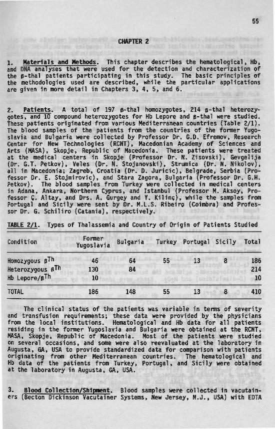
55
CHAPTER 2
1. Materials and Methods. This chapter describes the hematological, Hb,and DNA analyses that were used for the detection and characterization ofthe B-thal patients participating in this study. The basic principles ofthe methodologies used are described, while the particular applicationsare given in more detail in Chapters 3, 4, 5, and 6.
2. Patients. A total of 197 B-thal homozygotes, 214 g-thal hétérozy-gotes, and 10 compound hétérozygotes for Hb Lepore and p-thal were studied.These patients originated from various Mediterranean countries (Table 2/1).The blood samples of the patients from the countries of the former Yugo-slavia and Bulgaria were collected by Professor Dr. G.D. Efremov, ResearchCenter for New Technologies (RCNT), Macedonian Academy of Sciences andArts (MASA), Skopje, Republic of Macedonia. These patients were treatedat the medical centers in Skopje (Professor Dr. N. Zisovski), Gevgelija(Dr. G.T. Petkov), Veles (Dr. N. Stojanovski), Strumica (Dr. N. Nikolov),all in Macedonia; Zagreb, Croatia (Dr. D. Juricic), Belgrade, Serbia (Pro-fessor Dr. E. Stojmirovic), and Stara Zagora, Bulgaria (Professor Dr. G.H.Petkov). The blood samples from Turkey were collected in medical centersin Adana, Ankara, Northern Cyprus, and Istanbul (Professor M. Aksoy, Pro-fessor Ç. Altay, and Drs. A. Gurgey and Y. Kilincj, while the samples fromPortugal and Sicily were sent by Dr. M.L.S. Ribeiro (Coimbra) and Profes-sor Dr. G. Schiliro (Catania), respectively.
TABLE 2/1. Types of Thalassemia and Country of Origin of Patients Studied
Condition
Homozygous B^"Heterozygous B^"Hb Lepore/B™
TOTAL
FormerYugoslavia
4613010
186
Bulgaria
6484
148
Turkey
55
55
Portugal
13
13
Sicily
8
8
Total
18621410
410
The clinical status of the patients was variable in terms of severityand transfusion requirements; these data were provided by the physiciansfrom the local institutions. Hematological and Hb data for all patientsresiding in the former Yugoslavia and Bulgaria were obtained at the RCNT,MASA, Skopje, Republic of Macedonia. Most of the patients were studiedon several occasions, and some were also reevaluated at the laboratory inAugusta, GA, USA to provide standardized data for comparison with patientsoriginating from other Mediterranean countries. The hematological andHb data of the patients from Turkey, Portugal, and Sicily were obtainedat the laboratory in Augusta, GA, USA.
3. Blood Collection/Shipment. Blood samples were collected in vacutain-ers (Becton Dickinson Vacutainer Systems, New Jersey, N.J., USA) with EDTA
56
as anticoagulant. Samples from patients on transfusion regimens were col-lected prior to the blood transfusion. All samples were stored at 4°Cuntil the time of shipment and were brought by air by collaborators fromparticipating institutions, or were shipped on ice by fast air mail serviceto Augusta, 6A, USA, where they arrived within 5 days. Informed consentwas obtained under the regulations of the local institutions.
4. Hb and Hematological Analyses. The procedures for hematological andHb analysis are described below in the order routinely used in the labora-tories in Augusta, GA, USA, and RCNT, MASA, Skopje, Macedonia.
4/a Heaatological Data. Hematological values were obtained with afully automated cell counter (Sysmex CC-620, Toa Medical Electronics Co.,Kobe, Japan). The RBC indices, MCV (in f1) and MCH (in pg) were calculatedfrom the values of the total Hb concentration (g/dl), the PCV (in 1/1) andRBC count (x 10*12/1). The MCV and MCH values are especially useful param-eters in the diagnosis of heterozygous B-thal. MCV values below 80 fl andMCH values below 27 pg are indicative of the presence of a B-thal carrierstate. It is noteworthy that the MCV value can be increased by prolongedstorage of the RBCs.
4/b Hb Analyses. Plasma was removed by three washes with isotonicsaline and the RBCs were hemolyzed in 1.5 volumes of water for 15 minutesat room temperature with occasional stirring. The hemolysates were centri-fuged for 20 minutes at 4°C (4,000 rpm) and the supernatant were used forsubsequent electrophoretic and chromatographic analyses.
I. 1£F. Initial Hb identification was performed by IEF analysis(1) on commercially available agarose gels with buffers supplied by themanufacturer (Isolab, Inc., Akron, OH, USA). Approximately 10 ul of eachhemolysate were applied on a gel, followed by 2 hours of electrophoresisat constant power (30 W) using an LKB Multiphor II Horizontal Unit (Phar-macia-LKB, Uppsala, Sweden). The gels were fixed for about 10 minutes in10X trichloroacetic acid and the bands were visualized with a heme specificdye (0-dianisidine) and H2O2. Bands were identified by comparing the dis-tances of migration to control samples containing the known Hb types of A,F, S, C, and Bart's.
II. Fetal Hb Quantitation. Two procedures were used for quantitationof Hb F. The alkali denaturation method (2-4) was used in earlier studies,and was later replaced by a recently developed HPLC procedure (5,6).
III. Alkali Denaturation Method. The alkali denaturation method,described by Betke et al (2), is based on the higher resistance of Hb F todenaturation in alkali. The modified procedure used in this study involvedthe conversion of Hb into its cyanmet derivative before denaturation whichprevented false-positives, particularly in patients with increased concen-tration of the alkali resistant CO-Hb, such as heavy smokers. The methodoffered a reasonable accuracy for the Hb F concentrations between 5-20%.The disadvantage was that the Hb F levels below 5% were determined toohigh, and the Hb F levels above 20Ï were determined too low (6).

57
iv. Cation Exchange HPLC. The Hb F quantisations were performed ona polyCAT A cation exchange column (4.6 x 200 mm, with a particle size of5 urn; PolyLC, Columbia, MD, USA) using a Waters HPLC system (Waters Chroma-tography Division, Milford, MA, USA). The following developers were used:Developer A contains 35 mM Bis-Tris, 3 mM ammonium acetate, 1.5 mM KCN, pH6.47; Developer B contains 35 mM Bis-Tris, 1.5 mM KCN, 150 mM Na-acetate,16.85 mM ammonium acetate, pH 7.0. Ten to 15 ul of hemolysate (50-200 ugof Hb) were analyzed. The chromatograms were developed with a gradientof 25 to 85% Developer B in 85 minutes with a flow-rate of 0.8 ml/min.The absorbances of the effluents were continuously recorded at a wavelengthof 415 nm. The different Hb types were separated in the following order:F, A, and A2 (Fig. 2/1).
HomP+Th A (28.5)
Ecm
S
10 20
Time (Minutes)
FIG. 2/1. Separation of major and minor Hb components in red cell lysatesof one patient with homozygous B^-thal, and a second with homozygousB°-thal. The numbers between parentheses are pecentages.
v. Analysis of Hb F Composition. Two methods were used for thedetermination of the Y chain composition of Hb F (Gy> Ayl, and AyT). Thevisual inspection of the PAGE gel (7) allowed a semi-quantitative analysisof the Gy/\ ratio; it did not discriminate between the AyT and Ayl vari-ants. A more accurate analysis was obtained by reversed phase HPLC (8,9).Hemolysates that contained less than 20% of Hb F (as estimated by IEF)were enriched by preparative anion exchange DEAE-cellulose chromatographyprior to the Hb F composition analysis (10). This was performed on a 30 x1.5 cm column filled with DEAE-Sephadex equilibrated with 0.2 M glycine,15 mM KCN buffer, pH 7.5. Hemolysates containing approximately 20-60 mgHb were applied to the column and the chroma tograms were developed with
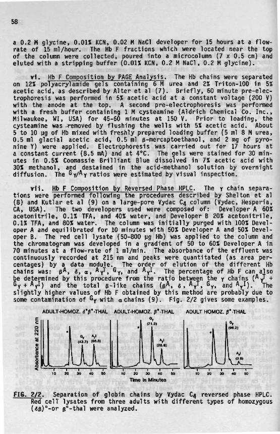
58
a 0.2 M glycine, 0.01% KCN, 0.02 M NaCl developer for 15 hours at a flow-rate of 15 ml/hour. The Hb F fractions which were located near the topof the column were collected, poured into a microcolumn (7 x 0.5 cm) andeluted with a stripping buffer (0.01% KCN, 0.2 M NaCl, 0.2 M glycine).
vi. Hb F Composition by PAGE Analysis. The Hb chains were separatedon 12% polyacrylamide gels containing 6 M urea and 2% Triton-100 in 5%acetic acid, as described by Alter et al (7). Briefly, 60 minute pre-elec-trophoresis was performed in 5% acetic acid at a constant voltage (200 V)with the anode at the top. A second pre-electrophoresis was performedwith a fresh buffer containing 1 M cysteamine (Aldrich Chemical Co. Inc.,Milwaukee, WI, USA) for 45-60 minutes at 150 V. Prior to loading, thecysteamine was -removed by flushing the wells with 5% acetic acid. About5 to 10 u.g of Hb mixed with freshly prepared loading buffer (5 ml 8 M urea,0.5ml glacial acetic acid, 0.5ml p-mercaptoethanol, and 2 mg of pyro-nine Y) were applied. Electrophoresis was carried out for 17 hours ata constant current (8.5 mA) and at 4°C. The gels were stained for 30 min-utes in 0.5% Coomassie Brilliant Blue dissolved in 7% acetic acid with30% methanol, and destained in the acid-methanol solution by overnightdiffusion. The Gy/Ay ratios were estimated by visual inspection.
vil. Hb F Composition by Reversed Phase HPLC. The y chain separa-tions were performed following the procedures described by Shelton et al(8) and Kutlar et al (9) on a large-pore Vydac C4 column (Vydac, Hesperia,CA, USA). The two developers used were composed of: Developer A 60%acetonitrile, 0.1% TFA, and 40% water, and Developer B 20% acetonitrile,0.1% TFA, and 80% water. The column was initially purged with 100% Devel-oper A and equilibrated for 10 minutes with 50% Developer A and 50% Devel-oper B. The red cell lysate (50-800 ug Hb) was applied to the column andthe chromatogram was developed in a gradient of 50 to 60% Developer A in70 minutes at a flow-rate of 1 ml/min. The absorbance of the effluent wascontinuously recorded at 215 nm and peaks were quantitated (as area per-centages) by a data module. The order of elution of the different Hbchains was: B*. 8, a, AyT, G ^ and Ay*. The percentage of Hb F can alsobe determined by this procedure from the ratio between the y chains (*/ +Gy + Ayl) and the total e-like chains (B*. 5, A^T, G^, a n d \ I ) . Theslightly higher values of Hb F obtained by this method are probably due tosome contamination of Gy with a chains (9). Fig. 2/2 gives some examples.
ADULT-HOMOZ. d'p'-jHAL ADULT-HOMOZ. P'-THAL ADULT HOMOZ.
10 20 30 40 SO 20 30 40 10 20 30 40 SO
Time in Minutes
FI6. 2/2. Separation of globin chains by Vydac C4 reversed phase HPLC.Red cell lysates from three adults with different types of homozygous(«B)°-or p°-thal were analyzed.
59
5. Hb A? Quantitation. The Hb A2 levels were usually determined withthe cation exchange HPLC procedure described above. However, in someinstances, Hb A2 was quantitated by anion exchange chromatography usingthe Quick-Sep Hb A2 Test System (Isolab). This procedure allowed the dif-ferential elution of Hb A2 and Hb A under specific concentrations of sodiumchloride at constant pH. Five to 10 ul of hemolysate were applied to acolumn and the Hb A2 was eluted with Developer A (0.2 M glycine, 0.01%KCN). The remaining Hbs were eluted with Developer B (0.2 M glycine, 0.01%KCN, 0.2 M NaCl). The Hb A2 concentrations were determined from the ratioof the absorbance values of both fractions at a wavelength of 415 nm.
6. In Vitro Giobin Chain Synthesis Analysis. The procedure consistedof incubating reticulocytes with an amino acid mixture containing [ H]leucine, followed by separation of the individual globin chains by columnchromatography, and determination of the incorporated radioactivity ineach particular chain by scintillation counting (4). Fresh blood was cen-trifuged immediately upon collection to remove the plasma and washed threetimes in cold reticulocyte saline. The top, reticulocyte rich, fractionfrom the packed red cells (approximately 1 ml) was collected and incubatedin 5 ml of incubation mixture (see Ref. 4 for composition), 0.1 ml trans-ferrin, and 10 mg glucose in a 25 ml Erlenmeyer flask, at 37°C for 10 min-utes in a metabolic shaker. Next, 0.1 ml of the radioactive [ H] leucinewas added, and the mixture further incubated at 37°C for 2 hours withgentle agitation. The sample was removed, washed three times with reticu-locyte saline, and hemolyzed with an equal volume of distilled water.Globin was prepared by adding the Hb solution under constant stirring to a20-fold volume of prechilied acid acetone (-20°C). The mixture was stirredfor 30 minutes, centrifuged at 4°C to collect the globin, washed threetimes with cold acetone, and dried over CaCl2 in a desiccator at atmos-pheric pressure.
A CM-cellulose (CM-52) column (18-20 cm) was equilibrated with Devel-oper A (8 M urea, 0.005 M ^HPO/j, 0.05 M B-mercaptoethanol, pH 6.5) for1 hour at a flow-rate of 20 ml/hour. Globin (50-60 mg) was dissolved in2 ml Developer A and dialyzed against the same developer for 2 hours atroom temperature. Next, the globin solution was applied to the column,allowed to drain under gravity, and elution with Developer A was performedfor 3 hours. The chromatography was continued with a linear Na+ gradientobtained by mixing 300 ml of Developer A with 300 ml Developer B (8 M urea,0.04 M Na2HPÛ4, 0.05 M B-mercaptoethanol, pH 6.5), both in open cylinders.The procedure was carried out at room temperature, at a constant flow-rateof 20 ml/hour. Five ml fractions were collected with an automated fractioncollector and analyzed at 280 nm. The radioactivity of each isolated frac-tion was determined in a liquid scintillation counter.
7. DNA Analyses
1. Isolation of Genomic DNA From WBCs. High MW genomic DNA was iso-lated from WBCs by the method of Poncz et al (11). About 10 ml of bloodwere washed three times with 30-40 ml of 1 x reticulocyte saline (140 mM
60
NaCl, 4 mM KC1, 6.8 mM MgCl2) by pelleting the cells with centrifugationat 2,500 g for 10 minutes at 4°C. The supernatant was removed after eachcentrifugation by aspiration. Next, the RBCs were hemolyzed with 30 mlfreshly prepared lysing solution [131 mM NH4CI, 0.9 mM (1^4)2003, pH 6.5]for 20 minutes at room temperature with gentle mixing. The WBCs were col-lected by centrifugation at 2,500 g for 10 minutes at 4°C and the superna-tant (hemolysate) was carefully removed and stored at -20°C for subsequentHb analysis. The lysis was repeated once more to remove the remainingRBCs, and the pellet of WBCs was resuspended in 10 ml of STE buffer (0.1 MNaCl, 0.05 M Tris-HCl, pH 7.4, 1 M EDTA). The WBCs were dispersed thor-oughly by vigorous shaking and 0.5 ml of a 10% SDS solution and 0.1 ml ofproteinase K (10 mg/ml in 10 mM Tris-HCl, pH 7.5, 1 mM EDTA) were added.The sample was incubated overnight at 37°C without shaking; the mixturebecame viscous upon incubation. Proteins were removed by several phenol/chloroform/isoamyl extractions. An equal volume of redistilled phenolsaturated with 200 mM Tris-HCl, pH 8.0, and containing 0.1% hydroxyquino-line as an anti-oxidant, was added; the mixture was shaken gently for 10minutes at room temperature and cooled on ice for 10 minutes. After 10minutes of centrifugation at 2,500 g and at 4°C, the aqueous layer wastransferred with a large bore pipet to a second tube and the phenol extrac-tion was repeated. An equal volume of chloroform/isoamyl (49:1) was added,and the mixture was again shaken gently at room temperature for 10 minutes,cooled on ice for 10 minutes, and centrifuged for 10 minutes at 4°C. Fol-lowing centrifugation, the aqueous phase containing the DNA was transferredto a clean tube. The DNA was precipitated with 3-4 volumes of ice-coldabsolute ethanol by gentle mixing. The precipitate was transferred toa 1.5 ml Eppendorf tube using a sterile Pasteur pipet, and washed threetimes with 70% ethanol to remove any residual phenol or salt. After dryingunder vacuum, the DNA pellet was dissolved overnight at 37°C in 1 ml ofTE buffer (10 mM Tris-HCl, pH 7.5, 1 mM EDTA). The DNA concentration wasdetermined by measuring the 0D at a wavelength of 260 nm. An OD25O of1.0 corresponds to approximately 50 ug/ml DNA. The DNA was stored at 4°Cuntil further use.
II. Gene Mapping. Gene mapping analyses were performed to determinethe B-globin gene haplotypes, the number of o-globin genes, and the typeof Hb Lepore, using procedures described in Refs. 12, 13, and 14, withslight modifications as described below.
III. Restriction Enzyme Digestion, Gel Electrophoresis, and SouthernTransfer. Five ug of genomic DNA were digested with the appropriate re-striction enzyme for 6 hours under conditions recommended by the manufac-turer (temperature and buffer). The resulting DNA fragments were separatedon a 0.8% agarose gel prepared with 1 x TEA buffer (0.04 M Tris-acetate,0.001 M EDTA, pH 8.3). The same buffer was used for gel electrophoresis.Electrophoresis was performed overnight, at room temperature, in a hori-zontal electrophoresis chamber (Bio-Rad DNA Sub Cell; Bio-Rad, Richmond,CA, USA) at a constant voltage of 25 to 30 volts. Hind III digested x-DNAwas always included as a size marker. Following electrophoresis, the DNAwas stained for 5-10 minutes with ethidium bromide (0.5 mg/ml) and thegel was photographed under long wavelength UV light. The DNA was thendepurinated, to facilitate subsequent transfer, by soaking the gel for

61
30 minutes in 0.1 N HC1. After one wash with distilled water, the gelwas soaked for 60 minutes in denaturation solution (0.5 M NaOH, 1.5 MNaCl), and next washed for at least 1 hour in a neutralizing solution (3 MNaCl, 1.5 M Tris-HCl, pH 7.0). The depurination, denaturation, and neu-tralization steps were performed at room temperature with gentle agitationon a rocking platform. The DNA was then transferred to either a nitrocel-lulose (Trans-Blot Transfer Membrane; Bio-Rad) or nylon membrane (Hybond CMembrane; Amersham International, Amersham, Bucks, England) using 20 x SSCas a transfer solution for the Southern blotting. The transfer was per-formed for 20-24 hours. The DNA was fixed by baking the membrane for 2-3hours in a vacuum oven at 70-80°C. Membranes were stored at 4°C until use.
1v. Prehybridization. The membrane was wetted with a few ml of 6 xSSC plus Denhardt's solution (0.5% SDS, 0.02% BSA 0.02% Ficoll 400, 0.02%PVP, 0.09 M Na-citrate, pH 7.0, and 0.9 M NaCl), and prehybridized over-night at 65°C in a sealed plastic bag with 15-20 ml of 1 x Flavel ModifiedSolution (3 X SSC, 0.1% SDS, 0.2% BSA, 0.2% PVP, 0.02 M phosphate buffer,pH 6.0) containing denatured, sonicated salmon sperm DNA at 50-100 ug/ml.
v. Radioactive Labeling of Probes. Probes were labeled to high spe-cific activity using the random-primer oligolabeling procedure (15) asrecommended by the manufacturer (Oligolabeling Kit, Pharmacia-LKB). Anaverage of 50-100 ng of probe was used per labeling reaction. The probewas denatured by boiling for 10 minutes in a water-bath and immediatelyplaced on ice to prevent reannealing. After a brief centrifugation tospin down the condensation inside the tube, a reaction mixture of a buf-fered aqueous solution of dATP, dCTP, dGTP, dTTP, and random hexanucleo-tides was added together with 50 uCi of a^P-dCTP (ICN Biochemicals, Inc.,Irvine, CA, USA) and 10 units of Klenow fragment, up to a total volumeof 50 ul. The reaction was incubated for 3 hours at 37°C, and the labeledprobe was purified from unincorporated nts by gel filtration through aG-50 Sephadex column with TE elution buffer. The total radioactivity wascalculated from the counts obtained for 5 ul of the effluent using a liquidscintillation counter. The labeled probes were stored at -20°C.
vi. Hybridization. An average of 10 x 10^ cpm of anprobe was used per 10 ml of hybridization solution. The probes were dena-tured for 10 minutes in a boiling water-bath and immediately diluted intothe prehybridization solution. Hybridizations were performed overnight(at least 16 hours), at 65°C in an incubator with a rocking platform.
vii. Washing and Autoradiography. The membranes were washed at 65°Cin 3 x SSC and 0.5% SDS with several (at least three) half-hourly changesof washing solution. The washing was performed until only a few countsabove background could be detected with a Geiger counter. The membraneswere air-dried, wrapped in plastic wrap and exposed for 5-6 days to a KodakX-Omat film (Eastman Kodak Co., Rochester, N.Y., USA) with DuPont QuantaIII intensifying screens (DuPont, Boston, MA, USA) at -70°C. Films weredeveloped either manually or using a Kodak X-ray film processor.
The sizes of particular DNA fragments were determined by plottingthe migration distances of the corresponding bands against the standardcurve derived from the migration of the x-DNA-Hind III marker.

62
v111. Restriction Endonucieases and DNA Probes. Nine polymorphicrestriction sites in the B-globin gene cluster were analyzed to determinethe haplotypes of thee-thal patients in this study (16). These polymor-phic sites were: a) Hinc II 5' to e, detected with a 1.2 kb Bam HI/Eco RIprobe spanning the entire e-globin gene, b) Xmn I 5' to <V > and Hind IIIat Gy and ^r» all three sites were analyzed with the r-IVS-II probe, a457 bp Bam HI/Pvu II fragment complementary to the «y- and Ay-IVS-IIregions, c) Hinc II at *B and 3' to it, both sites were detected with the•6 probe, a 1.6 kb Eco RI/Xba I fragment spanning the entire $B-globingene, and d) Ava II within B, and Hpa I and Bam HI 3' to the B-globin gene,all three sites analyzed with the B-IVS-II probe, a 1.2 kb Bam HI/Eco RIfragment of the B-IVS-II region.
The 7 kb deletion involving the 3' end of the 6-globin gene, theIntergenic region, and the 5' end of the B-globin gene, which leads tothe fusion of the 6- and B-globin genes into a hybrid Lepore-Boston-Wash-ington 66 gene, was detected by restriction enzyme digestion with Xba Iand Bgl II, and hybridization with the B-IVS-II probe (17).
The number of o-globin genes was determined through gene mapping anal-ysis with the restriction enzymes Eco RI, Bgl II, and Bam HI, and hybrid-ization with the a(a 1.5 kb Bam HI fragment containing the entire a-globingene), C (a 1.9 kb Hinf I fragment containing the entire c-globin gene),and e(a 0.8 kb Bam HI fragment of the o-globin gene) probes (18,19).
The DNA probes discussed above were cloned into plasmid vectors (pUC19 or pBR 322) and were isolated when necessary by mini-preparation ofplasmid DNA (14) and digestion with the appropriate restriction enzymes.The restriction fragments corresponding to the probes of interest wereseparated on IX agarose gels, sliced out from the gels, and the DNA waseluted either with a Gene Clean Kit (Bio 101; Bio-Rad) or by electroelutionwith a dialysis bag (14).
8. PCR Methodology. The discovery of the PCR procedure in 1985 (20)has had a significant impact on DNA research in general, and especiallyon studies of molecular defects and other sequence variations. Over theyears, there have been considerable improvements both in the PCR reactionitself, and the techniques with which it is associated. Such improvementsinclude substitution of the Klenow fragment with the thermostable Taq poly-merase (21), non-radioactive procedures for the detection of point muta-tions (22-24), and the identification of sequence variations by directsequencing of the PCR amplified DNA (25). As the work described in thisstudy extends over a period of several years, it has followed and includedthe methodological improvements, and has also contributed to the develop-ment of PCR based procedures for the detection of point mutations (26-28).Therefore, while this section will cover all the procedures used, the pres-ent ones will be described in greater detail.
1. 01igonucleotide Synthesis. All oligonucleotides used in thisstudy were synthesized by a solid-phase oligonucleotide synthesis method(29) either on an Applied BioSystems 380B DNA Synthesizer (Applied BioSys-tems, Inc., Foster City, CA, USA) or on a Beckman Plus 1 DNA Synthesizer
63
(Beckman, Palo Alto, CA, USA). Following synthesis, the oligonudeotideswere removed from the solid support by an ammonia treatment (55°C) over-night, dried under vacuum, and purified by G-50 Sephadex molecular sievechromatography using TE elution buffer. These oligonudeotides were usedas PCR primers, oligonucleotide probes for ASO hybridization, and as se-quencing primers. The sequences of these oligonudeotides are given inChapters 3, 4, 5, and 6, together with appropriate references.
II. PCR Using Kienow Fragment. Amplifications of 1 ug of genomicDNA were performed in 1.5 ml Eppendorf tubes containing the following ina total volume of 100 uL; 50 mM NaCl, 10 mM Tris-HCl (pH 7.6), 10 mM MgCl2,100 pM of each primer, 100-125 pM of each dNTP, and 10% DMSO. A hole wasmade in the cap of each tube with a small needle and the samples were dena-tured for 10 minutes at 95°C. After centrifugation for 10 seconds thesamples were kept at 37°C for 2.25 minutes to allow the primers to annealto the DNA, and 1 unit of Klenow fragment was added. The samples wereincubated at 37°C for 2-3.5 minutes. This cycle of denaturation, anneal-ing, and synthesis was repeated once more under the same conditions, andthen 25 times at a denaturation time of 1 minutes (1 unit of Klenow frag-ment was added at each cycle). One-fifth of each sample was then usedfor ASO hybridization as described below.
III. PCR Using Tag Polymerase. In general, amplifications were per-formed on 1 ug of genomic DNA using Taq polymerase from various manufac-turers. The reaction mixtures contained 200 uM of each dNTP, 50-100 pMof each oligonucleotide primer, 2.5 units of Taq polymerase, and reactionbuffer supplied by the manufacturer, in a total volume of 100 uL. Thesamples were overlayed with 60 uL of mineral oil to prevent evaporation,and were subjected to 30-35 repeated cycles of denaturation, annealing,and extension. The denaturation step in the first cycle was at 95°C for4 minutes, at the same temperature but lasting for only 1 minute in subse-quent cycles. The annealing was always for 1 minute, while the tempera-tures used varied from 58 to 65°C, and were usually several degrees abovethe Tm °f ^"® primers (see below). The extension reactions were performedat 72°C for 1-5 minutes, depending on the lengths of the expected PCR frag-ments. The extension step in the last cycle was always prolonged for atleast 7 minutes, to allow complete extension of all DNA strands synthesizedin the previous steps. The efficiency and specificity of the PCR was esti-mated by analyzing a 10 uL aliquot from each reaction on an ethidium bro-mide stained (10 ug/ml) 2% agarose gel under UV light. The samples werestored at 4°C until further use.
iv. ASO Hybridization With 32p_[_abeled piigonucleotides. Screeningfor previously characterized point mutations was by ASO hybridization (30).Two 19 mer oligonudeotides were used; one was complementary to the se-quence carrying the mutation of interest in such a way that the mutationwas positioned in the middle (mutant oligonucleotide), while the second(normal oligonucleotide) was complementary to the corresponding sequencefrom the normal allele.
v. 32p 5' End Labeling of Oligonudeotides. Both oligonudeotideswere labeled at the 5' end to a high specific activity with y32p_dATP(7,000 Ci/mM, 160 mCi/ml; ICN) by T4 polynucleotide kinase [United States

64
Biochemical (USB), Cleveland, OH, USA] (14). After 1-2 hours of incubationat 37°C, the unincorporated nt was removed from the probes using eitherG-50 Sephadex molecular sieve chromatography with the TE elution bufferor by commercially available Nensorb (NEN) columns (NEN Products, DuPont)under the conditions recommended by the manufacturer. The activity ofthe probes was determined as cpm in a scintillation counter (Beckman).
vi. Dot-Blot Hybridization. Two 20 ul aliquots of each PCR reactionwere denatured in the presence of 0.4 M NaOH, 25 mM EDTA. Both sampleswere applied to a zeta probe nylon membrane using a Bio-Dot spotting appa-ratus (both from Bio-Rad), so that one of the spots could later be usedfor hybridization to the normal and the other to the mutant probe. Themembrane was cut in the middle, and the DNA was fixed to the membranesby baking in a vacuum oven at 80°C for 2 hours. The duplicate membraneswere prehybridized in separate plastic bags in a solution of 5 x SSPE, 5 xDenhardt's, 0.5% SDS, for 30 minutes at the hybridization temperature (seebelow). Following prehybridization, one probe (50-100 x 10^ cpm) was addedto each membrane, and hybridization was performed in a circulating water-bath for 1-2 hours. Next, the membranes were washed twice for 10 minutesat room temperature with 2 x SSPE, 0.1% SDS, followed by a stringent 10-minute wash with 5 x SSPE, 0.5% SDS, usually at the same temperature asfor the hybridization (see below). After washing, the membranes were air-dried, wrapped in plastic film, and autoradiographed for 2-12 hours at-70°C.
The hybridization and subsequent washing temperatures were crucialfor the specific binding of the oligonucleotides to perfectly matched PCRamplified sequence. These temperatures were determined empirically (usingpositive and negative controls) for each pair of oligonucleotides, butwere generally near the Tm of the oligonucleotides (Tm -2 to Tm +4°C).The Tm depend mainly on the nt content of the oligonucleotides and werecalculated by the following formula: Tm = 4 x (G+C) + 2 x (A+T). Thehybridization and washing conditions for the detection of the most commonB-thal mutations in the Mediterranean Basin have been reported in a sepa-rate publication (25).
vil. ASO Hybridization With DIG-ddUTP Labeled Oligonucleotides.There has been a considerable effort in the past few years to developsimple, safe, and reliable nonradioactive procedures for the detectionof mutations that could also be easily implemented in the setting of aclinical laboratory. We therefore modified the standard ASO hybridizationprocedure by replacing the 5' 32p_end labeled oligonucleotides with oligo-nucleotides labeled at the 3' end with DIG (28). The 3' end labeling reac-tion was performed with 100 pM oligonucleotide, 1 nM DIG-ddUTP (Boehringer,Mannheim, Germany), 5 units Terminal transferase [Bethesda Research Labora-tories (BRL), Inc., Gaithersburg, MD, USA], and a reaction buffer suppliedby the manufacturer in a 20 uL final volume. After incubation at 37°C for15 minutes, the reaction was stopped with 2 ul 0.2 M EDTA, pH 8.O. Thelabeled oligonucleotide was precipitated with 2.5 ul 4 M LiCl and 75 uLprecMlled absolute ethanol, incubated at -70°C for 30 minutes and centri-fuged at 12,000 g, 4°C for 15 minutes. The pellet was washed with 70%cold ethanol, dried under vacuum and dissolved in sterile redistilled
65
water. The prehybridization, hybridization, and washing of the nylon mem-branes was performed as described above for the radioactive ASO procedure.The hybridized oligonucleotides were detected with an anti-DIG antibody(DIG Nucleic Acids Detection Kit; Boehringer). Rehybridization of themembranes was done after color removal with N' N'-DMF at 65°C in a glasstray (approximately 4 x 30 minutes). Next, the membranes were washed twiceat room temperature in 2 x SSC, 0.1% SDS, and incubated at 65°C for 2 hourswith 0.5 mg/ml proteinase K in 2 x SSC, 0.1% SDS, followed by three 5-min-ute washes in 2 x SSC, 0.1% SDS. The probes were finally removed with50% formamide, 10 mM sodium phosphate, pH 6.5 at 65°C for 1 hour.
viii. Direct Sequencing of PCR Amplified DNA. Two procedures werefollowed to obtain the PCR fragments that were used in the dideoxy chaintermination sequencing reactions (31). The first procedure included astandard PCR reaction, as described above, to generate a double-strandedPCR product that was denatured with NaOH and purified from the remainingPCR primers with a Miniprep Spun Column (Pharmacia-LKB). The second proce-dure involved an asymmetric PCR reaction to generate a single-strandedPCR product for sequencing. These two procedures also differed in theannealing step of the sequencing reaction, while the labeling and termina-tion steps were identical.
1x. Miniprep Spun Column for the Purification of the PCR Product.The PCR sample was transferred to a clean Eppendorf tube and an equal vol-ume of a phenol-chloroform solution was added to remove the remaining min-eral oil and protein. The sample was vortexed, centrifuged for 10 minutesat top speed, and the supernatant was transferred to a clean tube. Thesample was then precipitated in 0.3 M sodium acetate, pH 5.2, and 2.5 vol-umes of absolute ethanol for 30 minutes at -20°C. After a 15 minute cen-trifugation at 4°C and at top speed, the recovered pellet was dried undervacuum in a Speed-Vac (Savant, Farmingdale, N.Y., USA) and dissolved in20 uL water. The amplified DNA was denatured with 5 ui of 2 M NaOH for atleast 10 minutes at room temperature. Meanwhile, a Miniprep Spun columnwas prepared as described in the manufacturer's instructions. Briefly, theMiniprep Spun column was equilibrated with 5 ml of TE buffer, placed in a15 ml Corex tube, and centrifuged at 400 g for 2 minutes in a centrifugewith a swinging bucket rotor. Twenty-five uL of TE buffer were appliedto the center of the flat upper surface of the compacted bed of SephacrylS-400. The centrifugation was repeated and the 25 ul denatured DNA samplewas applied to the upper surface of the Sephacryl. A capless Eppendorftube was placed in the bottom of the Corex tube to recover the eluted DNA.The centrifugation was repeated and the eluted 25 uL sample was transferredto a clean tube. Two more elutions were performed with 25 ul of TE bufferin the same manner. Ten ul aliquots of each of the three effluents wereanalyzed on an ethidium bromide stained 2% agarose gel to identify thefraction with the highest DNA yield. Seven ul of this fraction were usedper sequencing reaction with the Sequenase Version 2.0 DNA Sequencing Kit(USB).
x. Annealing Reaction. One ui (50 pM) of sequencing primer and 2 ulof 5x reaction buffer were added to the denatured DNA sample up to a totalvolume of 10 ul. The reaction mixture was vortexed briefly, centrifuged,and incubated at 37°C for 15 minutes.
66
xi. Labeling Reaction. The labeling mix provided in the sequencingkit was diluted 10 times with sterile water. The Sequenase enzyme (13U/uL) was then diluted (1:8) in Enzvme Dilution Buffer (USB) up to a totalvolume of 2 uL. Next, 0.5 ui of a^S-dATP (800 Ci/mM, 10.0 mCi/ml; DuPont)were added to the diluted enzyme. All the dilutions were performed on ice.The labeling reaction was started with the addition of 1 uL 0.1 M DTT, i1 u.L diluted Labeling Mix, and 2.3 ul of diluted enzyme/a 35$_dATP mixture}to the tube containing the annealed template/primer. The sample was incu-bated at room temperature for 2 minutes.
xii. Termination Reaction. Two and a half uL of each Termination.Mixture (ddATP, ddCTP, ddGTP, and ddTTP) were placed in clean 1.5 ml Eppen-Idorf tubes labeled A, C, G, and T, respectively. The tubes were prewarmedfor at least 1 minute at 37°C. When the labeling reaction was complete,3.5 uL of the reaction mixture was added to each of the labeled tubes andincubated at 37°C for 5 minutes. The reactions were stopped with 4 uLof Stop Solution and the tubes were kept on ice until loaded onto the gel.
xiii. Denaturing PAGE. The sequencing reactions were separated ona 0.04 mm thick 7 M urea/8% polyacrylamide/TBE gel using a vertical elec-trophoresis apparatus for DNA sequencing (Model S2; BRL-GIBCO) and a highvoltage constant power supply (2297-Microdrives; LBK-Broma, Uppsala, Swe-den). The gel was allowed to polymerize for at least 1 hour, and nextwas pre-electrophoresed at 70 W constant power (1400-1600 V, 50-60 mA)for at least 30 minutes. The DNA samples were denatured for 3 minutesat 80°C, placed on ice for 3-5 minutes, and loaded onto the gel (2-3 uLper well). The gel was run at constant power (75 W) for 1.5 to 5 hours,depending upon the proximity of the desired sequence to the sequencingprimer. Following electrophoresis, the gel was fixed in a 10% aceticacid/12% methanol solution for 30 minutes, transferred to a Whatman 3MMpaper (Whatman, Maidstone, Kent, England), covered with plastic film, anddried under vacuum on a gel dryer (Slab Dryer Model 483; Bio-Rad) at 80°Cfor 1 hour. The gel was then unwrapped and autoradiographed by overnightexposure to a Kodak X-ray film.
x1v. Sequencing of Single-Stranded PCR Fragment. To generate single-stranded PCR products, the standard PCR reaction was adjusted by decreasingthe concentration of one of the primers to 1 pM/100 uL, resulting in animbalanced primer ratio of 1:100. The PCR primers that remained after thePCR reaction were eliminated by differential precipitation of the amplifiedPCR product with 1 volume of 2.5 M ammonium acetate and 2 volumes of abso-lute ethanol at room temperature for 15 minutes. The pellet was recoveredby centrifugation for 15 minutes at 4°C, washed three times with 70% etha-nol, dried under vacuum, and dissolved in 10 uL water. The recovery wasestimated by analyzing 2 ul of the PCR product on a 2% agarose gel. Theamount of amplified DNA corresponding to 0.3 ug M-13 single-strand DNA wasused for the sequencing reaction. The sequencing reaction was performedas described above except that the annealing was at 65°C for 3 minutesfollowed by a 30 minute" incubation on ice.
67
REFERENCES
1. Righetti, P.G., Gianazza, E., Bianchi-Bosisio, A., and Cossu, G.:Conventional isoelectric focusing and immobilized pH gradients forhemoglobin separation and identification. In The Hemoglobinopathies,edited by T.H.J. Huisman, Methods in Hematology, Vol. 15, page 47,Churchill Livingstone, Edinburgh, 1986.
2. Betke, K., Marti, H.R., and Schlicht, I.: Estimation of small per-centages of foetal haemoglobin. Nature, 184:1877, 1959.
3. Singer, K., Chernoff, A.I., and Singer, L.: Studies on abnormal hemo-globins. I. Their demonstration in sickle cell anemia and otherhematological disorders by means of alkali denaturation. Blood, 6:413, 1951.
4. Huisman, T.H.J. and Jonxis, J.H.P.: The Hemoglobinopathies Techniquesof Identification, Clinical and Biochemical Analysis, Vol. 6, MarcelDekker, Inc., New York, N.Y., 1977.
5. Bisse, E. and Wieland, H.: High performance liquid chromatographicseparation of human haemoglobins - Simultaneous quantisation of foetaland glycated haemoglobins. J. Chromatogr., 434:95, 1988.
6. Kutlar, A., Kutlar, F., Gu, L-G., Mayson, S.M., and Huisman, T.H.J.:Fetal hemoglobin in normal adults and g-thalassemia hétérozygotes.Hum. Genet., 85:106, 1990.
7. Alter, B.P., Goff, S.C., Efremov, G.D., Gravely, M.E., and Huisman,T.H.J.: Globin chain electrophoresis: A new approach to the deter-mination of the ^Y/^Y ratio of foetal haemoglobin and to studies ofglobin synthesis. Br. J. Haematol., 44:527, 1980.
8. Shelton, J.B., Shelton, J.R., and Schroeder, W.A.: High performanceliquid chromatographic separation of globin chains on a large-poreC4 column. J. Liq. Chromatogr., 7:1969, 1984.
9. Kutlar, F., Kutlar, A., and Huisman, T.H.J.: Separation of normaland abnormal hemoglobin chains by reversed-phase high-performanceliquid chromatography. J. Chromatogr., 357:147, 1986.
10. Schroeder, W.A. and Huisman, T.H.J.: The Chromatography of Hemoglo-bin, Clinical and Biochemical Analysis, Vol. 9, Marcel Dekker, Inc.,New York, N.Y., 1980.
11. Poncz, M., Solowiejczyk, D., Harpel, B., Mory, Y., Schwartz, E., andSurrey, S.: Construction of human gene libraries from small amountsof peripheral blood: Analysis of B-like globin genes. Hemoglobin,6:27, 1982.
12. Southern, E.M.: Detection of specific sequences among DNA fragmentsseparated by gel electrophoresis. J. Mol. Biol., 98:503, 1975.
13. Blattner, F.R., Blech, A.E., Dennistron-Thomson, K., Faber, H.E.,Richards, J.E., Slightom, J.L., Tucker. P.W., and Smithies, 0.: Clon-ing of human fetal Y globin and mouse B type globin DNA. Preparationand screening shutguns collections. Science, 202:1284, 1978.
14. Sambrook, J., Fritsch, E.F., and Maniatis, T.: Molecular Cloning.A Laboratory Manual, Cold Spring Harbor Laboratory Press, 1989.
15. Freiberg, A.P. and Vogelstein, B.: A technique for radiolabelingDNA restriction endonuclease fragments to high specific activity.Analyt. Biochem., 132:6, 1983.
16. Orkin, S.H., Kazazian, H.H., Jr., Antonarakis, S.E., Goff, S.C.,Boehm, C D . , Sexton, J.P., Waber, P.G., and Giardina, P.J.V.: Linkageof B-thalassaemia mutations and B-globin gene polymorphisms with DNApolymorphisms in human B-globin gene cluster. Nature, 296:627, 1982.

68
17. Lanclos, K.D., Patterson, J., Efremov, G.D., Wong, S.C., Villegas, A.,Ojwang, P.J., Wilson, J.B., Kutlar, F., and Huisman, T.H.J.: Charac-terization of chromosomes with hybrid genes for Hb Lepore-Washington,Hb Lepore-Baltimoré, Hb P-Nilotic, and Hb Kenya. Hum. Genet., 77:40,1987.
18. Felice, A.E., Cleek, M.P., McKie, K., McKie, V., and Huisman, T.H.J.:The rare o-thalassemia-1 of Blacks is a ça -thalassemia-1 associatedwith deletion of all o- and ç-globin genes. Blood, 63:1253, 1984.
19. Felice, A.E., Cleek, M.P., Marino, E.M., McKie, K., McKie, V.C.,Chang, B.K., and Huisman, T.H.J.: Different ç globin gene deletionsamong Black Americans. Hum. Genet., 73:221, 1986.
20. Erlich, H.A. and Arnheim, N.: Enzymatic amplification of 6 globingenomic sequences and restriction site analysis for diagnosis ofsickle cell anemia. Science, 230:1350, 1985.
21. Saiki, R.K., Gelfand, D.H., Stoffel, S., Scharf, S.J., Higuchi, R,Horn, G.T., Mullis, K.B., and Erlich, H.A.: Primer directed enzymaticamplification of DNA with a thermostable DNA polymerase. Science,239:487, 1988.
22. Saiki, R.K., Chang, C.A., Leveson, C.H., Warren, T.C., Boehm, CD.,Kazazian, H.H., Jr. and Erlich, H.A.: Diagnosis of sickle cell anemiaand 6-thalassemia with enzymatically amplified DNA and nonradioactiveallele specific oligonucleotide probes. N. Engl. J. Med., 319:537,1988.
23. Chehab, F.F. and Kan, Y.W.: Detection of specific DNA sequences byfluorescence amplification. A color complementation assay. Proc.Natl. Acad. Sci. USA, 86:9178, 1989.
24. Efremov, D.G., Dimovski, A.J., Jankovic, L., and Efremov, G.D.: Mu-tant oligonucleotide extension amplification. A non-labeling polymer-ase chain reaction assay for the detection of point mutations. ActaHaematol., 85:66, 1991.
25. Gyllenstein, U.B. and Erlich, H.A.: Generation of single strandedDNA by the polymerase chain reaction and its application to directsequencing of the HLA-DQA locus. Proc. Natl. Acad. Sci. USA, 85:7652,1988.
26. Diaz-Chico, J.C., Yang, K.G., Yang, K.Y., Efremov, D.G., Stoming,T.A., and Huisman, T.H.J.: The detection of B-globin gene mutationsin B-thalassemia using oligonucleotide probes and amplified DNA.Biochim. Biophys. Acta, 949:43, 1988.
27. Stoming, T.A., Diaz-Chico, J.C., Yang, K.G., Efremov, D.G., and Huis-man, T.H.J.: Newer developments in the identification of B-thalas-semia. Hemoglobin, 12:565, 1988.
28. Efremov, D.G., Dimovski, A.J., and Efremov, G.D.: Detection of B-thalassemia mutations by ASO hybridization of PCR amplified DNA withdigoxigenin ddUTP labeled oligonucleotides. Hemoglobin, 15:525, 1991.
29. Matteucci, M.D. and Caruthers, M.H.: Synthesis of deoxyoligonucleo-tides on a polymer support. Biotechnology, 24:92, 1992.
30. Saiki, R.K., Bugawan, T.L., Horn, G.T., Mullis, K.B., and Erlich,H.A.: Analysis of enzymatically amplified B globin and HLA-DQa DNAwith allele-specific oligonucleotide probes. Nature, 324:163, 1986.
31. Sanger, F., Nicklen, S., and Coulson, A.R.: DNA sequencing withchain-terminating inhibitors. Proc. Natl. Acad. Sci. USA, 74:5463,1977.
CHAPTER 3
MOLECULAR CHARACTERIZATION OF B-THALASSEMIA MUTATIONS
3a B-Thaiassenia in Yugoslavia 71
3b 6-Thalassemia in Bulgaria 79

71
CHAPTER 3a
B-THALASSEMIA IN YUGOSLAVIA
A. Dimovskil, D.G. Efremovl, L. Jankovicl, D. Juricic*, N. Z1sovski3,N. Stojanovski*. N. NikolovS, G.T. Petkov^, A.L. Reese?,
T.A. Stoning?, G.D. Efremovl, and T.H.J. Huisman?
1 Macedonian Academy of Sciences and ArtsResearch Center for New Technologies, 91000 Skopje, Yugoslavia
2 Department of Clinical ChemistryUniversity Hospital Dr. 0. Novosel, 41000 Zagreb, Yugoslavia
3 Department of Pediatrics, Faculty of Medicine91000 Skopje, Yugoslavia
* Medical Center Titov Veles, 93000 Titov Veles, Yugoslavia
5 Medical Center Strumica, 92000 Strumica, Yugoslavia
6 Medical Center Gevgelija, 93000 Gevgelija, Yugoslavia
^ Department of Cell and Molecular BiologyMedical College of Georgia, Augusta, GA 30912-2100, USA
(Adapted from: Hemoglobin, 14:15-24, 1990)
ABSTRACT
This study concerned the evaluation of B-thal alleles in nearly 50patients with B-thal major and in 130 B-thal hétérozygotes using geneamplification and dot-blot hybridization with synthetic probes. Fourteendifferent mutations were observed (Table 3a/l); of these, three (IVS-I-110;IVS-I-6; IVS-I-1) account for some 75% of all B-thal alleles. Newly dis-covered variants, I.e. T->C in the initiation CD and AATAAA->AATGAA inthe poly A site were observed in a few patients. The poly A mutation withclassical B-thal alleles result in thalassemia intermedia. Hb Lepore isa rather common abnormality and combinations of this variant with B-thaloften result in severe disease; a search for B-thal mutations amongpatients affected with this disease should include an analysis to detectthis Hb abnormality.

72
INTRODUCTION
Several reports have been published describing the occurrence of B-thal and the related «B-thal in the Yugoslavian population (1-6). Theseabnormalities were observed as simple hétérozygotes, and in patients withthalassemia major or with a compound heterozygosity for B-thal and an !abnormal Hb. We have recently completed a program to identify the various !mutations and frameshifts in the p-globin gene which result in the differ- !ent types of thai found in Yugoslavia. The results of this study involv-ing nearly 50 subjects with thalassemia major and 130 B-thal hétérozygotesare summarized here.
MATERIALS AND METHODS
Patients. Blood samples were collected from 46 subjects with thalas-semia major, from 68 of their heterozygous relatives, and from 62 randomlyselected, unrelated, B-thal hétérozygotes. Most patients (i.e. 80%) camefrom Macedonia (one of the six republics of Yugoslavia which is locatedin the southern part of the country), where they attended clinics in thecities of Skopje, Strumica, Gevgelija, Titov Veles, and others. Patientsfrom other areas were followed in clinics in Zagreb (Croatia) and Belgrade(Serbia). Blood samples were collected with EDTA as anticoagulant andshipped to the general laboratory in Skopje, Macedonia.
Hematological data were collected with an automated cell counter inthe laboratory in Zagreb. Hb analyses were done in Skopje by the sameprocedures as listed in the preceding paper (7) with the following excep-tions: Hb A2 was quantitated by microcolumn chromatography using Tris-HCldevelopers (8), while the Gyand ^Y levels in isolated Hb F were determinedby PAGE (9,10).
DNA methodology, including gene mapping and dot-blot analysis ofamplified DNA, was the same as described in the preceding paper (7). Theanalyses of the samples from the B-thal major patients were done in thelaboratory in Skopje. The B-thal mutations in the majority of the B-thalhétérozygotes were determined in the laboratories in Augusta, GA, USA.
RESULTS AND DISCUSSION
The Frequencies. Fourteen different mutations have been observed(Table 3a/l). Eight of these were present in the patients with thalassemiamajor and in B-thal hétérozygotes, while the remaining six were observedonly among the B-thal hétérozygotes who participated in the random survey(see Ref. 11 for references describing individual mutations and frame-shifts). About 10% of the B-thal genes present in the patients remainedunknown mainly because the survey of these DNA samples was limited to anevaluation of the presence of the 12 most common B-thal mutations. More-over, deletional types of B-thal are not included in the data listed inTable 3a/l. Nearly 40% of the chromosomes carried the IVS-I-110 mutation(45.4% in the survey of the hétérozygotes) and 29% the IVS-I-6 mutation.

73
This high percentage likely does not reflect the true incidence becausethe mild clinical course of the B-thal conditions due to a homozygosityof this mutation, or to a combination of the IVS-I-6 and other mutations,affects the frequency values calculated for this relatively small groupof patients. The frequency value of 19.2% found for the IVS-I-6 mutationamong B-thal hétérozygotes may be too low because the hematological andHb A2 data may be closer to normal which interferes with its detectionin a field survey. The third most frequently occurring variant is theG->A mutation at IVS-I-1 (9-11%). Thus, three mutations, IVS-I-110 (G->A),IVS-I-6 (T->C), and IVS-I-1 (G->A), account for more than 75% of the B-thalgenes observed in Yugoslavia.
TABLE 3a/l. The Frequenciesvian Population
of 14 Different B-Thal Alleles in the Yugosla-
Thalassemia Major PatientsMutation
-87 (C->G)Initiation CD (T->C)CD 5 (-CT)CD 6 (-A)CD 8 (-AA)CD 29 (C->T)CD 39 (C->T)IVS-I-1 (G->A)IVS-I-6 (T->C)IVS-I-110 (G->A)IVS-II-1 (G->A)IVS-II-745 (C->G)IVS-II-848 (C->A)Poly A (AATAAA->AATGAA)Unknown
Total
# Chroms.
1
1382534
1
49
86
%
1.16
1.163.499.31
29.0739.53
1.16
4.6510.47
100.00
Hétérozygotes# Chroms.
212211515255922256
130
%
1.540.771.541.540.770.773.8511.5419.2345.371.541.541.543.954.61
100.00
The CD 39 (C->T) mutation appears to be relatively rare in Yugoslavia(3.5-4.0%) but the newly discovered poly A mutation (AATAAA->AATGAA; Ref.12) was present in four unrelated families, suggesting that this mild B*-type of thalassemia may be present with a frequency of some 4%. Anothernewly discovered mutation, i.e. the T->C mutation in the initiation CD(13), was found in only one B-thal hétérozygote.
No significant differences were observed between the types of B-thalmutations among Macedonians and Yugoslavians living in the northern partof Yugoslavia; however, the numbers are too small for a definitive compar-i son.

74
B-Thal Homozygotes. Ten of the 0-thal major patients (i.e. 23% ofthe total ) were homozygous for the IVS-I-11O (G->A) mutation and one washomozygous for the IVS-I-1 (6->A) mutation. All 11 subjects were severelyaffected and were transfusion-dependent. Of the 16 chromosomes with theIVS-I-11O mutation tested, 15 had haplotype I, and one had haplotype IX(see Ref. 14 for details). Five subjects were homozygous for the IVS-I-6(T->C) mutation, and some of the data observed for these patients arelisted in Table 3a/2. All were mildly affected by their disease but werenot transfusion-dependent. Patients #1, #2, and #3 were homozygous forhaplotype VI and their Hb F levels averaged about 8%. In contrast,patients #4 and #5 (who are brother and sister) were homozygous for haplo-type VII and had much higher Hb F levels of 15% and 35%; such differenceshave also been observed for patients in other countries (7).
TABLE 3a/2. Hematological and Hb Composition Data for Five YugoslavianSubjects With a Homozygosity for the IVS-I-6 (T->C) Mutation
Sub- Sex- Hb MCV MCH Hb A2 Hb F G- A^T Haplo-ject Age g/dl fl pg %« %«> %<= %<> type*
12345
F-34M-23M-32F-14M-12
11.310.210.99.19.6
7561657670
2221242220
6.33.05.96.24.7
3.912.07.715.035.0
5752614245
43483900
VI/VIVI/VIVI/VIVII/VIIVII/VII
* By microcolumn chromatography using Tris-HCl developers (8).*> By alkali denaturation (15).c By PAGE (9,10).d By reversed phase HPLC (16) showing the absence of Ay (haplotype VI/VI)
or of AyT (haplotype VII/VII).« Haplotype VI or [-- + + -- + + +] and haplotype VII or [+
+ +]. Restriction sites: Hinc II 5' to e ; Xmn I 5' to Gy; Hind III atGy and Ay; Hinc II at *B and 3' to it; Ava II at B, and Hpa I and Bam HI3' to B.
Compound Hétérozygotes. Twenty-eight of the 44 patients were compoundhétérozygotes; most were severely affected by their disease and were trans-fusion-dependent. Exceptions were two patients with the IVS-I-110/IVS-I-6combination, both having haplotype I for the IVS-I-110 chromosome and hap-lotype VII for the IVS-I-6 chromosome, one patient with the IVS-I-l/CD 6(-A) combination and four patients with a combination which involved theAATAAA->AATGAA mutation at the poly A site (Table 3a/3). Patient #3 (list-ed in this table) was mildly affected and had not been transfused; herclinical condition was diagnosed as thalassemia intermedia. The same wasthe case for patient #4, although he had a more severe disease.
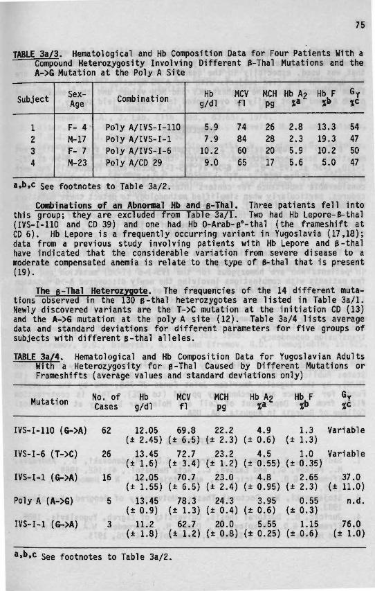
75
JABLE 3a/3. Hematological and Hb Composition Data for Four Patients With aCompound Heterozygosity Involving Different B-Thal Mutations and theA->G Mutation at the Poly A Site
Subject
1234
Sex-Age
F- 4M-17F- 7M-23
Combination
Poly A/IVS-I-110Poly A/IVS-I-1Poly A/IVS-I-6Poly A/CD 29
Hbg/dl
5.97.910.29.0
MCVfl
74846065
MCHP9
26282017
Hb A?%«
2.82.35.95.6
Hb F%b
13.319.310.25.0
*c
54475047
a.b.c see footnotes to Table 3a/2.
Combinations of an Abnormal Hb and p-Thal. Three patients fell intothis group; they are excluded from Table 3a/l. Two had Hb Lepore-B-thal(IVS-I-110 and CD 39) and one had Hb O-Arab-B°-thal (the frameshift atCD 6). Hb Lepore is a frequently occurring variant in Yugoslavia (17,18);data from a previous study involving patients with Hb Lepore and B-thalhave indicated that the considerable variation from severe disease to amoderate compensated anemia is relate to the type of B-thal that is present(19).
The B-Thal Hétérozygote. The frequencies of the 14 different muta-tions observed in the 130 B-thal hétérozygotes are listed in Table 3a/l.Newly discovered variants are the T->C mutation at the initiation CD (13)and the A->G mutation at the poly A site (12). Table 3a/4 lists averagedata and standard deviations for different parameters for five groups ofsubjects with different B-thal alleles.
TABLE 3a/4. Hematological and Hb Composition Data for Yugoslavian AdultsWith a Heterozygosity for B-Thal Caused by Different Mutations orFrameshifts (average values and standard deviations only)
Mutation No. ofCases
Hbg/dl
MCVfl
MCHpg
Hb A2 Hb F
IVS-I-110 (G->A) 62
IVS-I-6 (T->C) 26
IVS-I-1 (G->A) 16
Poly A (A->G) 5
IVS-I-1 (G->A) 3
12.05 69.8 22.2 4.9 1.3 Variable(± 2.45) (± 6.5) (± 2.3) (± 0.6) (± 1.3)13.45 72.7 23.2 4.5 1.0 Variable
(± 1.6) (± 3.4) (± 1.2) (± 0.55) (± 0.35)12.05 70.7 23.0 4.8 2.65 37.0
(± 1.55) (± 6.5) (± 2.4) (± 0.95) (± 2.3) (± 11.0)13.45 78.3 24.3 3.95 0.55 n.d.
(± 0.9) (± 1.3) (± 0.4) (± 0.6) (± 0.3)11.2 62.7 20.0 5.55 1.15
(± 1.8) (± 1.2) (± 0.8) (± 0.25) (± 0.6)76.0± 1.0)
a.b.c see footnotes to Table 3a/2.
76
The data are somewhat different from those listed for the TurkishB-thal hétérozygotes (7), presumably because of the different methodologythat was used, especially in the quantisation of Hb A2. Of interest arethe data for the five subjects with the A->G mutation in the poly A site.These subjects have the highest MCV and MCH values and the lowest levelof Hb A2 and might easily be overlooked in a simple field survey.
CONCLUSIONS
B-Thal in Yugoslavia is a heterogeneous condition since 14 differentmutations have been detected. However, three mutations, i.e. G->A at IVS-1-110, T->C at IVS-I-6, and G->A at IVS-I-1 occur most frequently and werepresent in 75 to 80% of all chromosomes tested. This information is ofconsiderable importance for centers interested in prenatal diagnostic pro-cedures, particularly when simplified methodology to detect these mutationsbecomes available (20,21). However, such centers should also be equippedto detect certain Hb variants because combinations of some of these (forinstance, Hb Lepore) with certain types of B-thal (mainly the IVS-I-110,IVS-I-1, and CD 39 mutations) cause a severe thalassemic condition.
Thalassemia intermedia, i.e. a relatively mild clinical conditionwithout the requirement for regular blood transfusions, has been observedin patients who are homozygous for the IVS-I-6 (T->C) mutation and in afew patients with combinations involving the newly discovered A->G mutationin the poly A site. The high frequencies of the IVS-I-110 (G->A) and theIVS-I-1 (G->A) mutations, and the relatively high incidence of Hb Leporeleads to the conclusion that at least 80% of all Yugoslavian patients witha homozygosity or a compound heterozygosity for one (or two) of the discov-ered fl-thal all el es, is affected by severe disease and a complete bloodtransfusion dependency.
Acknowledganents. This study was supported in part by the UnitedStates Public Health Services research grants HLB-41544 and HLB-05168 (toTHJH), by the Yugoslav-American Joint Funds JF 788, and by the YugoslavFunds for Science and Technology pp 244 (to GDE). This is contribution#1221 from the Department of Cell and Molecular Biology at the MedicalCollege of Georgia in Augusta, GA, USA.
REFERENCES
1. Sadikario, A., Duma, H., Efremov, G.D., Mladenovski, B., Andreeva,M., Petkov, G., and Lazova, C , Acta Haematol., 41:162, 1969.
2. Efremov, G.D., Juricic, D., and Stojanovski, N., Hemoglobin, 6:643,1982.
3. Stojanovski, N., Doctoral Thesis, University of Beograd, Yugoslavia,1983.
4. Juricic, D., Doctoral Thesis, University of Zagreb, Yugoslavia, 1989.5. Efremov, G.D., Nikolov, N., Duma, H., Schroeder, W.A., Miller, A.,
and Huisman, T.H.J., Scand. J. Haematol., 14:226, 1975.

77
6. Efremov, G.D., Filipce, V., Gjorgovski, I., Juricic, D., Stojanovski,N., Harano, T., Nakatsuji, T., Kutiar, A., Kutlar, F., Bakioglu, I.,and Huisman, T.H.J., Br. J. Haematol., 63:17, 1986.
7. Oner, R., Altay, Ç., Gurgey, A., Aksoy, M., Kilinç, Y., Stoming, T.A.,Reese, A.L., Kutlar, A., Kutlar, F., and Huisman, T.H.J., Hemoglobin,14:1, 1990.
8. Efremov, G.D., Huisman, T.H.J., Bowman, K., Wrightstone, R.N., andSchroeder, W.A., J. Lab. Clin. Med., 83:657, 1974.
9. Alter, B.P., Goff, S.C., Efremov, G.D., Gravely, M.E., and Huisman,T.H.J., Br. J. Haematol., 44:527, 1980.
10. Efremov, G.D., Markovska, B., Stojanovski, N., Petkov, G., Nikolov,N., and Huisman, T.H.J., Hemoglobin, 5:637, 1981.
11. The B-thalassemia repository, Hemoglobin, 14:661, 1990.12. Jankovic, L., Efremov, G.D., Petkov, G.H., Ptekov, G.T., Kattamis,
C , George, E., Yang, K-G., Stoming, T.A., and Huisman, T.H.J., Br. J.Haematol., 75:122, 1990.
13. Jankovic, L., Efremov, G.D., Josifovska, 0., Juricic, D., Stoming,T.A., Kutlar, A., and Huisman, T.H.J., Hemoglobin, 14:169, 1990.
14. Orkin, S.H., Kazazian, H.H., Jr., Antonarakis, S.E., Goff, S.C.,Boehm, C D . , Sexton, J.P., Waber, P.G., and Giardina, P.J.V., Nature,296:627, 1982.
15. Pembrey, M.E., Weatherall, D.J., and Clegg, J.B., The Lancet, i: 1350,1973.
16. Kutlar, F., Kutlar, A., and Huisman, T.H.J., J. Chromatogr., 357:147,1986.
17. Duma, H., Efremov, G.D., Sadikario, A., Teodisijev, D., Mladenovski,B., Slaski, R., and Andreeva, M., Br. J. Haematol., 15:161, 1968.
18. Efremov, G.D., Hemoglobin, 2:197, 1978.19. Efremov, D.G., Efremov, G.D., Zisovski, N., Stojanovski, N., Kutlar,
F., Diaz-Chico, J.C., Kutlar, A., Yang, K.G., Stoming, T.A., and Huis-man, T.H.J., Br. J. Haematol., 68:351, 1988.
20. Efremov, D.G., Dimovski, A.J., and Efremov, G.D., Hemoglobin, 15:525,1991.
21. Chehab, F.F. and Kan, Y.W., Proc. Natl. Acad. Sci. USA, 86:9178, 1989.

79
CHAPTER 3b
6-THALASSEHIA IN BULGARIA
G.H. Petkovl, G.D. Efremov2, D.G. Efremov2, A. Dimovskil«2,
P. Tchaicarova*, R. Tchaicarov*, B. Rogina^, S. Agarwal*.A. Kutlar*. F. Kutlar*. A.L. Reese*, T.A. Stoning*, and T.H.J. Huisman*
1 Department of PediatricsHigher Medical Institute, Stara Zagora, Bulgaria
2 Macedonian Academy of Sciences and ArtsResearch Center for New Technologies, 9100 Skopje, Yugoslavia
3 Department of Clinical ChemistryUniversity Hospital Dr. 0. Novosel, 41000 Zagreb, Yugoslavia
* Department of Cell and Molecular BiologyMedical College of Georgia, Augusta, GA 30912-2100, USA
(Adapted from: Hemoglobin, 14:25-33, 1990)
ABSTRACT
Analyses of DNA from 64 patients with thalassemia major using thehybridization technique of amplified DNA with radio-labeled synthetic oli-gonucleotide probes identified 13 different B-thal mutations. The CD 39(C->T) and IVS-I-110 (G->A) mutations occurred most frequently but sevenadditional mutations were observed which were present at frequencies of3.9 to 10.2%. This broad spectrum of B-thal alleles complicates the anal-yses for institutions involved in prenatal diagnosis. Promoter mutationswere rare and the frequencies of two other mild mutations [IVS-I-6 (T->C)and the poly A mutation] were relatively low, indicating that B-thal isa severe disease among Bulgarians. The high frequencies of 4.7-5.5% forthe four frameshifts at CDs 5, 6, 8, and 8/9 may be specific for this popu-lation.

80
INTRODUCTION
Since 1937, when the first patients with Cooley's anemia in Bulgariawere described (1), numerous additional cases have been reported from allparts of the country (2-9). Over the past 20 years population studies havebeen carried in most parts of the country (10-18); different approaches forthe detection of B-thal carriers were used and a frequency of 0.05 to 19.9Xwas found (11,13). It is estimated that the average frequency of B-thal inBulgaria is about 2% (9). At present, more than 250 patients with Cooley'sanemia are registered in different hospitals. Here we describe the identi-fication of numerous B-thal alleles among more than 60 patients with B-thalmajor and over 80 of their relatives. The data indicate a considerableheterogeneity which may complicate the general activities of the prenataldiagnostic programs.
MATERIALS AND METHODS
Patients. The total number of patients was 64, and 82 heterozygousrelatives were also evaluated. Most patients attended a pédiatrie clinicat the university at Stara Zagora but others were seen in local clinicsin various provinces (Fig. 3b/l). Nearly all patients were children oryoung adults. Blood samples were collected in vacutainers with EDTA asanticoagulant and studied at the local institution, at the laboratory inSkopje, Yugoslavia, and were shipped by fast air mail service to Augusta,GA, USA for detailed analysis. Informed consent was obtained.
Yugo^vi. ' - A L-,_A * V \ 7 "***»
Turkey
Greece
FIG. 3b/l. The provinces of Bulgaria where the patients and their parentswere located. A major B-thal center is located in Stara Zagora.
Methods. Hematological and Hb composition data were collected asdescribed in a previous study (19). The same is the case for some genemapping analysis and for the identification of the various mutations andframeshifts.
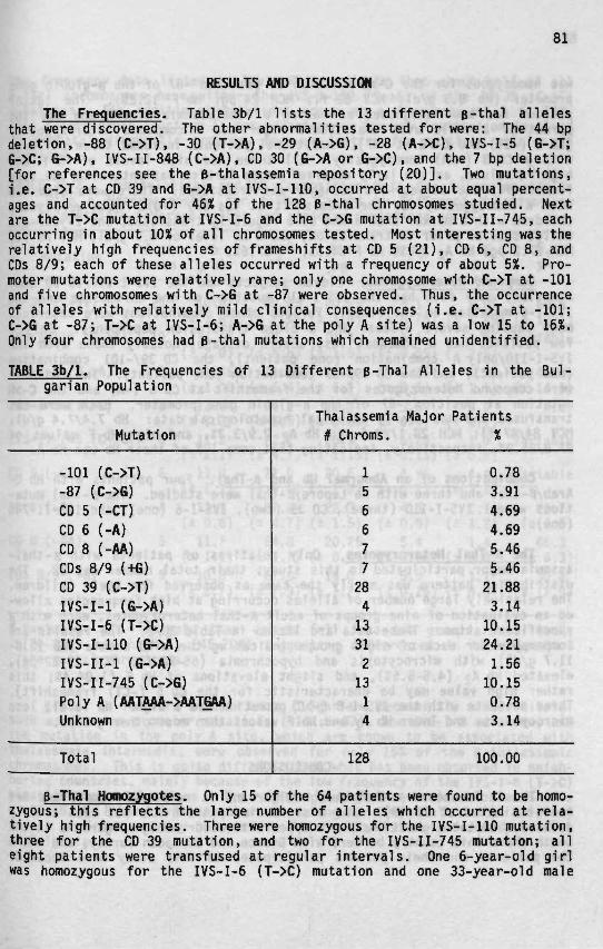
81
RESULTS AND DISCUSSION
The Frequencies. Table 3b/l lists the 13 different B-thal allelesthat were discovered. The other abnormalities tested for were: The 44 bpdeletion, -88 (C->T), -30 (T->A), -29 (A->G), -28 (A->C), IVS-I-5 (G->T;G->C; G->A), IVS-II-848 (C->A), CD 30 (G->A or G->C), and the 7 bp deletion[for references see the B-thalassemia repository (20)]. Two mutations,i.e. C->T at CD 39 and G->A at IVS-I-110, occurred at about equal percent-ages and accounted for 46% of the 128 B-thal chromosomes studied. Nextare the T->C mutation at IVS-I-6 and the C->G mutation at IVS-II-745, eachoccurring in about 10% of all chromosomes tested. Most interesting was therelatively high frequencies of frameshifts at CD 5 (21), CD 6, CD 8, andCDs 8/9; each of these alleles occurred with a frequency of about 5%. Pro-moter mutations were relatively rare; only one chromosome with C->T at -101and five chromosomes with C->G at -87 were observed. Thus, the occurrenceof alleles with relatively mild clinical consequences (i.e. C->T at -101;C->G at -87; T->C at IVS-I-6; A->G at the poly A site) was a low 15 to 16%.Only four chromosomes had B-thal mutations which remained unidentified.
TABLE 3b/l. The Frequencies of 13 Different B-Thal Alleles in the Bul-garian Population
Mutation
-101 (C->T)-87 (C->G)CD 5 (-CT)CD 6 (-A)CD 8 (-AA)CDs 8/9 (+G)CD 39 (C->T)IVS-I-1 (G->A)IVS-I-6 (T->C)IVS-I-110 (G->A)IVS-II-1 (G->A)IVS-II-745 (C->G)Poly A (AATAAA->AATGAA)Unknown
Total
Thalassemia# Chroms.
156677284133121314
128
Major Patients%
0.783.914.694.695.465.46
21.883.1410.1524.211.56
10.150.783.14
100.00
B-Thal Homozygotes. Only 15 of the 64 patients were found to be homo-zygous; this reflects the large number of alleles which occurred at rela-tively high frequencies. Three were homozygous for the IVS-I-110 mutation,three for the CD 39 mutation, and two for the IVS-II-745 mutation; alleight patients were transfused at regular intervals. One 6-year-old girlwas homozygous for the IVS-I-6 (T->C) mutation and one 33-year-old male
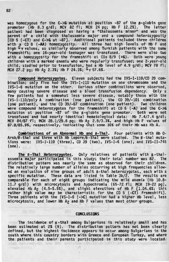
82
was homozygous for the C->G mutation at position -87 of the p-globin genepromoter (Hb 8.3 g/dl; MCV 82 f 1 ; MCH 24 pg; Hb F 12.2%). The latterpatient had been diagnosed as having a "thalassemia minor" and was theparent of a child with thalassemia major and a compound heterozygosity[CD 5 (-CT) and C->G at -87]. Additional patients included three childrenwith a CD 8 (-AA) homozygosity. All three had high levels of Hb F andhigh Gy values, as similarly observed among Turkish patients with the sameframeshift; one 16-year-old teenager was transfused. There were also twowith a homozygosity for the frameshift at CDs 8/9 (+G). Both were youngchildren with a marked anemia who were regularly transfused; one 2-year-oldchild, studied prior to transfusion, had a Hb level of 4.9 g/dl; MCV 78 fl;MCH 27.2 pg; Hb A2 2.3%; Hb F 24.3%; Gy 57.6%.
Compound Hétérozygotes. Eleven subjects had the IVS-I-11O/CD 39 com-bina tT^nT~ô7Tîy^Tvihâdtïïe IVS-I-110 mutation on one chromosome and theIVS-I-6 mutation on the other. Various other combinations were observed,!many causing severe disease and a blood transfusion dependency. Only a]few patients had a somewhat less severe disease, notably those with the"IVS-I-llO/poly A combination (one patient), the CD 39/-101 combination(one patient), and the CD 39/-87 combination (one patient). Two childrenwere compound hétérozygotes for the frameshift at CD 6 (-A) and the C->Gmutation at position -87 of the e-globin gene promoter. Both were nottransfused and had nearly identical hematological data: Hb 7.4/7.4 g/dl;MCV 84/87 fl; MCH 28.1/29.6 pg; Hb A2 2.9/3.3%, and high Hb F values of87.8/85.9%, respectively, indicating that some 10% of their Hb was Hb A.
Combinations of an Abnormal Hb and B-Thal. Four patients with Hb 0-Arab/B-thal and three with Hb Lepore/B-thal were studied. The B-thal muta-tions were: IVS-I-110 (three), CD 39 (two), IVS-I-6 (one), and IVS-II-745(one).
The B-Thal Hétérozygotes. Only relatives of patients with B-thal-assemia major participated in this study; their total number was 82. Thedistribution pattern was nearly the same as observed for their children.The relatively large number of alleles occurring at high frequencies allow-ed an evaluation of nine groups of adult B-thal hétérozygotes, each with aspecific mutation. These data are listed in Table 3b/2. The results arecomparable for each of eight groups indicating the mild anemia (Hb 10.8-11.7 g/dl) with microcytosis and hypochromia (65-72 f1 ; MCH 19-22 pg),elevated Hb A2 (4.5-5.5%), and slight elevations of Hb F [1.04.4%; thisrather high value may be characteristic for the CD 5 (-CT) frameshift].Three patients with the IVS-I-6 (->C) mutation had a higher Hb level, lessmicrocytosis, and lower Hb A2 and Hb F values than most other groups.
CONCLUSIONS
The incidence of B-thal among Bulgarians is relatively small and hasbeen estimated at 2% (9). The distribution pattern has not been clearlydefined, but the highest incidence appears to occur among Bulgarians in thesouth where this country borders with Greece and European Turkey, and wherethe patients and their parents participated in this study were located.
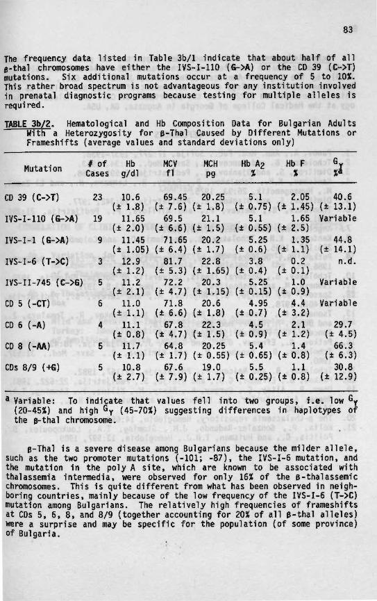
83
The frequency data listed in Table 3b/l indicate that about half of allp-thal chromosomes have either the IVS-I-11O (G->A) or the CD 39 (C->T)mutations. Six additional mutations occur at a frequency of 5 to 10%.This rather broad spectrum is not advantageous for any institution involvedin prenatal diagnostic programs because testing for multiple alleles isrequired.
TABLE 3b/2. Hematological and Hb Composition Data for Bulgarian AdultsWith a Heterozygosity for B-Thal Caused by Different Mutations orFrameshifts (average values and standard deviations only)
Mutation # ofCases
Hbg/dl
MCVfl
MCHP9
Hb A2 Hb FX
CD 39 (C->T)
IVS-I-110 (G->A)
IVS-I-1 (G->A)
IVS-I-6 (T->C)
IVS-II-745 (C->G)
CD 5 (-CT)
CD 6 (-A)
CD 8 (-AA)
CDs 8/9 (+G)
23
19
9
3
5
6
4
5
5
10.6(± 1.8)
11.65(± 2.0)
11.45(± 1.05)
12.9(± 1-2)
11.2(± 2.1)
11.0(± l . D
11.1(± 0.8)
11.7(± l . D
10.8(± 2.7)
69.45(± 7.6)
69.5(± 6.6)
71.65(± 6.4)
81.7(± 5.3)
72.2(± 4.7)
71.8(± 6.6)
67.8(± 4.7)
64.8(± 1-7)
67.6(± 7.9)
20.25(± 1-8)
21.1(± 1.5)
20.2(± 1-7)
22.8(± 1.65)
20.3(± 1.15)
20.6(± 1.8)
22.3(± 1.5)
20.25(± 0.55)
19.0(± 1-7)
(±
(±
(±
(±
(±
(±
(±
(±
(±
5.10.75)5.10.55)5.250.6)3.80.4)5.250.15)4.950.7)4.50.9)5.40.65)5.50.25)
(±
(±
(±
(±
(±
(±
(±
(±
(±
2.051.45)1.652.5)1.351.1)0.20.1)1.00.9)4.43.2)2.11.2)1.40.8)1.10.8)
40.(± 13.
61)
Variable
44.(± 14.
n.
,81)
,d.
Variable
Variable
29,(± 4,
66(± 6
30(± 12
.7• 5).3• 3).8• 9)
* Variable: To indicate that values fell into two groups, i.e. low Gy(20-45%) and high Gy (45-70%) suggesting differences in haplotypes ofthe e-thal chromosome.
B-Thal 1s a severe disease among Bulgarians because the milder all ele,such as the two promoter mutations (-101; -87), the IVS-I-6 mutation, andthe mutation in the poly A site, which are known to be associated withthalassemia intermedia, were observed for only 16% of the p-thalassemicchromosomes. This is quite different from what has been observed in neigh-boring countries, mainly because of the low frequency of the IVS-I-6 (T->C)mutation among Bulgarians. The relatively high frequencies of frameshiftsat CDs 5, 6, 8, and 8/9 (together accounting for 20% of all B-thal alleles)were a surprise and may be specific for the population (of some province)of Bulgaria.

84
Acknowledgements. This study was supported in part by the UnitedStates Public Health Services research grants HLB-41544 and HLB-05168 (toTHJH), by the Yugoslav-American Joint Funds, JF 788 (to GDE), and by theDepartment of Biotechnology of the Government of India, New Delhi (to SA).This is contribution #1222 from the Department of Cell and Molecular Biol-ogy at the Medical College of Georgia in Augusta, GA, USA.
REFERENCES
1. Panov, A., Ann. Med. Fac, Sofija, 17:277, 1937-1938.2. Tchoiakov, B. and Kancev, K., Savr. Med., 11:34, 1963.3. Dimitrov, S. and Djankov, E., Pedijatrija, 6:591, 1968.4. Kantchev, K.N., Tcholakov, B., Casey, R., Lehmann, H., and El-Zazmi,
M., Humangenetik, 26:93, 1975.5. Dimitrov, S. and Diankov, E., Pedijatrija, 16:342, 1977.6. Tcholakov, B., Neicev, P., and Kirilova, P., Pedijatrija, 21:286,
1981.7. Angelov, A., Savr. Med., 32:56, 1981.8. Kantchev, K.N., Tcholakov, B., Baglioni, C , and Colombo, B., Nature,
205:187, 1965.9. Petkov, G., Doctoral Thesis, University of Stara Zagora, Bulgaria,
1986.10. Tcholakov, B., Doctoral Thesis, University of Sofija, Bulgaria, 1970.11. Tsoneva, M. and Boshnakova, E., Savr. Med., 23:8, 1972.12. Spasov, V., Ninijo, S., Angelov, A., and Zareva, B., Pedijatrija,
17:402, 1978.13. Tsoneva, M., Tocheva, D., Mavrudieva, M., Rashkov, R., Tomaleva, Z.,
and Donov, M., Savr. Med., 29:13, 1978.14. Tonceva, D., Pedijatrija, 18:57, 1979.15. Petkov, G., Efremov, G.D., and Tonev, J., Pedijatrija, 18:43, 1979.16. Tcholakov, B., Nenchev, P., and Kirilova, R., Savr. Med., 31:537,
1980.17. Tcholakov, B. and Neicev, P., Pdijatrija, 21:167, 1982.18. Petkov, G., Efremov, G.D., and Ninijo, S., Pedijatrija, 22:522, 1983.19. Oner, R., Altay, Ç., Gurgey, A., Aksoy, M., Kilinç, Y., Stoming, T.A.,
Reese, A.L., Kutlar, A., Kutlar, F., and Huisman, T.H.J., Hemoglobin,14:1, 1990.
20. The e-thalassemia repository, Hemoglobin, 14:661, 1990.21. Kolia, P., Gonzalez-Redondo, J.M., Stoming, T.A., Loukopoulos, D.,
Politis, C , and Huisman, T.H.J., Hemoglobin, 13:597, 1989.

CHAPTER 4
CORRELATION OF CLINICAL SEVERITY WITH DIFFERENT 6-GLOBIN GENE DEFECTS
4a Variation in Clinical Severity Among Patients With 87Hb Lepore-Boston-p-Thalassemia is Related to the Typeof 8-Thalassemia
4b B-Thalassenia Due to a T->A Mutation Within the ATA 95Box
4c Mild and Severe B-Thalassemia Among Homozygotes From 101Turkey: Identification of the Types by Hybridizationof Amplified DNA With Synthetic Probes
87
CHAPTER 4a
VARIATION IN CLINICAL SEVERITY AMONG PATIENTS WITHHB LEPORE-BOSTON-B-THALASSEMIA IS RELATED TO THE TYPE OF B-THALASSEMIA
D.G. Efranvl, G.D. Efreaov?, N. Z1zovski'3,N. Stojanovski*. F. Kutlarl, J.C. Diaz-Chico*.
A. Kutlar*, K.G. Yangl, T.A. Stoning!, and T.H.J. Huismanl
1 Department of Cell and Molecular BiologyMedical College of Georgia, Augusta, GA 30912-2100, USA
2 National Reference Laboratory of Hemoglobinopathies ZFUniversity of Skopje, P.F. 355, Skopje, Yugoslavia
3 Department of Pediatrics, Faculty of MedicineUniversity of Skopje, Skopje, Yugoslavia
* Medical Center, Titov Veles, Yugoslavia
(Adapted fro» : Br. J. Haenatol., 68:351-355, 1988)
SUMMARY
Clinical and hematological observations, made for 10 Yugoslavianpatients with the Hb Lepore-6-thal condition, suggested a considerablevariation from severe disease and complete blood transfusion dependencyto a moderate, compensated, anemia without major complications and withouta need for regular blood transfusions. As the type of Hb Lepore was thesame in all patients (Lepore-Washington-Boston) and an a-globin gene defi-ciency was absent, it was concluded that the type of B-thal determinedthe severity of the disease. Six patients with severe disease had one ofthe following three B-thal determinants: IVS-I-110 (G->A), exon 2 CD 39(C->T), and IVS-I-1 (G->A), while the three patients with mild diseasehad the Portuguese type of thalassemia which is caused by the T->C substi-tution at IVS-I-6. In one patient with severe disease the B-thal deter-minant remained unknown. Our observations are consistent with those madefor thalassemia patients with a homozygosity for these determinants.

88
INTRODUCTION
Hb Lepore-Washington-Boston (Lepore-W-B) is a variant with a «B hybridchain which results from a crossover between CDs 87 and 116 of the 6- andB-globin genes (Gerald and Diamond, 1958; Baglioni, 1965). Hétérozygoteshave a mild anemia resembling a thalassemia trait, while homozygotes areseverely affected with a severe B-thalassemic type of disease. Hb Lepore-W-B is found in various Mediterranean populations; its highest incidenceis in Italy, Yugoslavia, and Greece (reviewed in Efremov, 1978).
Hb Lepore-W-B has been observed in association with B chain variants(I.e. S and C) and with B- and 66-thai (references in Efremov, 1978). Theclinical features of most Lepore-B-thal patients resemble those of patientswith homozygous B-thal (Duma et al, 1968; Weatherall and Clegg, 1981) butthere are reports of cases with a milder clinical course (Quattrin et al,1967; Efremov, 1978). In the study to be described here, we have comparedclinical and hematological data for 10 patients with the Lepore-8-thalcondition, and have determined the haplotypes and mutations that are asso-ciated with the B-thal. The data offer a satisfactory explanation forthe milder form of the disease seen in some of the patients.
MATERIALS AND METHODS
Blood Samples. These were collected in vacutainers with EDTA as anti-coagulant at various locations in Yugoslavia and transported to the CentralLaboratory in Skopje, Yugoslavia. Most of this material, together withblood samples from parents and/or siblings, were mailed by air at 40°Cto Augusta, GA, USA, where it arrived within 5 days. Informed consentwas obtained.
Hewatoiogical Analyses. Hematological parameters were determinedon freshly collected samples at local institutions using automated cellcounters. Quantisation of the various Hb components was by DEAE-cellulosechromatography (Schroeder and Huisman, 1980), while the percentages of the^ Y T , <*Y, and *W chains were determined by reversed phase HPLC (Shelton etal, 1984; Kutlar et al, 1986). In vitro chain synthesis analyses followedprocedures detailed before (Huisman and Jonxis, 1977); these analyses weremade on different blood specimens, collected a few years prior to the pres-ent study.
DNA Studies. DNA was isolated from white blood cells following themethod of Poncz et al (1982). The determination of the presence or absenceof several restriction sites (listed in the footnote of Table 4a/3) fol-lowed procedures routinely in use in our laboratories (Harano et al, 1985;Land os et al, 1987; Efremov et al, 1987); the numbering system was thatwhich was introduced by Orkin et al (1982). The number ofa-globin geneswas determined through gene mapping with the restriction enzymes Eco RI,Bgl II, and Bam HI as described before (Felice et al, 1984; 1986).
The types of B-thal were determined by a modification of the procedureof Saiki et al (1986) using gene amplification with specific primers and

89
with synthetic oligonucleotide probes for the identification of the muta-tion. Details about this method have been described elsewhere (Diaz-Chicoet al, 1988).
RESULTS
The Patients. These persons were located in various areas of Yugo-slavia, but five regularly attend clinics in Skopje and Titov Vêles, SRMacedonia. Some clinical data are listed in Table 4a/l. Their ages in1986 ranged from 2 to 41 years; six patients were diagnosed before theywere 1 year of age, while the other four were 3-19 years old before thediagnosis of Lepore-B-thal was made. Patient P-361B is the brother ofP-361. The clinical severity varied considerably as indicated. PatientP-303 is married and has two sons, both with a B-thal heterozygosity.This patient and patients P-4 and R-27 are doing clinically well havinga moderate, compensated, anemia with mild hemolysis and most modest (ifany) blood transfusion requirements.
TABLE 4a/l. Some Clinical and X-ray Data for 10 Patients With Lepore-W-B-B-Thal
Patient Sex-Age
Enlarged (cm)Afls cnioon Muor Splenectomy Faciès and Trans-of DY *P'**" ^ ^ Age (yr) Bone Changes» fusion«>
PIP-20I-CR-13P-361P-361BR-927P-4P-303R-27
F-24F-22F- 8F-10F-10M- 2M- 7M-25M-41F-17
6 mos1 yr8 mos6 mos6 mos6 mos3 yrs3 yrs
19 yrs8 yrs
78
7
10467104
45
56424120
787
NoNoNoNo9
20No
OftenVery oftenVery oftenVery oftenVery oftenVery oftenOftenRarelyRarelyNone
* Moderate (+); severe (++); very severe (+++).b Rarely = once in 2 or 3 years; often = once in 2-3 months; very often =once a month.
Hewatological and Hb Analyses (Table 4a/2). Hematological data werecomparable for all patients but were influenced by the (often frequent)blood transfusions. Blood transfusions also affected the quantitative Hbdata; only a few patients (P-l, R-927, R-27) had Hb F levels of 60-80% asexpected with Hb Lepore levels of 8-11%. As patient R-27 had not receivedany blood, it can be assumed that his e+-thal chromosome was responsiblefor the production of some 25% Hb A. No such calculations were possiblefor the other patients.

90
TABLE 4a/2.B-Thal*
Hematological
<- Hbe g/dl
and
PCV1/1
Hb Data
RBC1012/1
for
A
10 Patients
Lepore^
X
With the
F«>x •
Lepore-W-B-
/non-a B/b
P-lP-20I-CR-13P-361P-361BR-927P-4P-303R-27
F-24F-22F- 8F-10F-10M- 2M- 7M-25M-41F-17
7.77.99.47.48.07.87.59.28.68.6
0.3000.2900.3000.2400.2640.2600.2300.3250.2900.255
3.302.993.602.802.822.903.253.713.403.35
2.33.11.92.72.62.11.92.92.52.0
6.23.7<1.00.56.76.47.58.710.16.7
82.729.03.54.622.014.080.149.563.062.5
3.53.3n.d.2.83.33.12.01.72.21.6
0.160.19n.d.00000.210.180.24
* Hematology data were collected prior to the next blood transfusion.b Hbs A£, Lepore, and F were determined by DEAE-cellulose chromatography
in blood specimens collected prior to (the next) blood transfusion,n.d. = not determined.
The Gy values in the Hb F varied from 41.8 to 60.6%, while threepatients were positive for the AyT chain (Huisman et al, 1985, and refer-ences quoted). Jji vitro chain synthesis analyses showed a severe defi-ciency for four of the first seven patients (P-l, P-361, P-361B, and R-927)with an average a/non-o ratio of 3.3 (range 3.0-3.5); these values werenearly 2 for the remaining three patients, Jji vitro chain synthesis wasdemonstrable for four patients (P-l, P-4, P-303, and R-27). Structuralanalyses of the isolated non-a chain of Hb Lepore were made during thepast several years for six of the 10 patients (P-l, P-20, P-361, P-4,P-303, and R-27). In all instances the 6B hybrid chain was type Lepore-W-Bwith a crossover between residues 87 and 116 (data not shown; for struc-tural information see Efremov, 1978).
DNA Analyses. Haplotype analyses involved the nine restriction siteslisted in Table 4a/3; family studies facilitated these analyses. Asexpected from previous studies (Lanclos et al, 1987) the haplotype of theLepore-W-B chromosomes was the same in all nine families. Six differenthaplotypes were observed for the B-thal chromosomes (Table 4a/3); haplo-types I and II are the most common among Mediterranean populations (Orkinet al, 1982).
Identification of the molecular abnormality through hybridizationof amplified DNA with synthetic probes was possible for nine patients.Table 4a/3 lists the final data for the different types of B-thal; patientsP-l, P-20, and I-C had the B-thal with the G->A substitution at IVS-I-110(haplotype I); patient R-13 that of a C->T replacement in CD 39 of thesecond exon (haplotype II). The two patients of Family 361 had the G->A
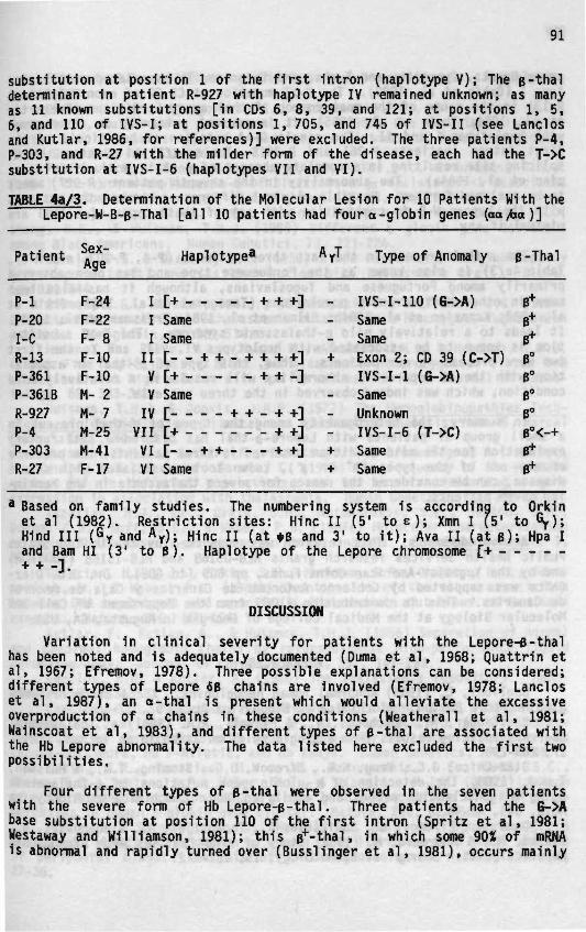
91
substitution at position 1 of the first intron (haplotype V); The B-thaldeterminant in patient R-927 with haplotype IV remained unknown; as manyas 11 known substitutions [in CDs 6, 8, 39, and 121; at positions 1, 5,6, and 110 of IVS-I; at positions 1, 705, and 745 of IVS-II (see Lanclosand Kutlar, 1986, for references)] were excluded. The three patients P-4,P-303, and R-27 with the milder form of the disease, each had the T->Csubstitution at IVS-I-6 (haplotypes VII and VI).
TABLE 4a/3. Determination of the Molecular Lesion for 10 Patients With theLepore-W-B-B-Thal [all 10 patients had fouro-globin genes (aa/ho)]
Patient Sex-Age Haplotype* Type of Anomaly B-Thal
P-lP-20I-CR-13P-361P-361BR-927P-4P-303R-27
F-24F-22F- 8F-10F-10M- 2M- 7M-25M-41F-17
I [+I SameI Same[--
+ + +]
+ + - + + + +]+ + -]
IIV [ +V SameIV [ + + - + +]
VII [+ + +]VI [- - + + + +]VI Same
- IVS- I -110 (G->A)SameSame
+ Exon 2 ;- IVS-I-1
SameUnknown
- IVS-I-6+ Same+ Same
CD 39 (C->T)(G->A)
(T->C)
B+B*B+
B°B°B°B"B°<-+B+B+
Based on family studies. The numbering system is according to Orkinet al (1982). Restriction sites: Hinc II (5' t o e ) ; Xmn I (5' to Gy);Hind III (Gy and Ay); Hinc II (at *B and 3' to it); Ava II (at B ) ; Hpa Iand Bam HI (3' to B ) . Haplotype of the Lepore chromosome [+
]
DISCUSSION
Variation in clinical severity for patients with the Lepore-B-thalhas been noted and is adequately documented (Duma et al, 1968; Quattrin etal, 1967; Efremov, 1978). Three possible explanations can be considered;different types of Lepore SB chains are involved (Efremov, 1978; Lancloset al, 1987), an a-thal 1s present which would alleviate the excessiveoverproduction of a chains in these conditions (Weatherall et al, 1981;Wainscoat et al, 1983), and different types of B-thal are associated withthe Hb Lepore abnormality. The data listed here excluded the first twopossibilities.
Four different types of B-thal were observed 1n the seven patientswith the severe form of Hb Lepore-B-thal. Three patients had the G->Abase substitution at position 110 of the first intron (Spritz et al, 1981;Westaway and Williamson, 1981); this B*-thal, in which some 90Ï of mRNAis abnormal and rapidly turned over (Busslinger et al, 1981), occurs mainly
92
among Greeks, Cypriots, and Turks, and is primarily associated with haplo-type I (Orkin et al, 1982). The C->T mutation at CD 39 of exon 2 (patientR-13) is the cause of the most common B°-thal among Sardinians (Trecartinet al, 1981; Thein et al, 1985); it is present primarily on chromosomeswith haplotypes I and II. The single G->A mutation at position 1 of thefirst intron (patients P-361 and P-361B, Table 4a/3) eliminates the 5'splicing site resulting in a complete absence of e chain production (Kaza-zian et al, 1984a). The abnormality in the seventh patient (R-927) needsto be analyzed further. Possible clinical differences between the patientswith the Lepore-W-B in combination with any of these types of B-thal areminimal if any.
The B-thal present in the last three patients (P-4, P-303, and R-27,Table 4a/3) is also known as the Portuguese type and has been observedprimarily among Portuguese and Yugoslavians, although it has also beenseen in other Mediterranean populations (Tamagnini et al, 1983; Orkin etal, 1982; Kazazian et al, 1984b; Gil man et al, 1984; Treisman et al, 1983).It leads to a relatively mild B-thalassemic syndrome. The T->C substitu-tion is known to be associated with haplotypes VI, VII, and X; the firsttwo were present in our patients. Thus, this type of e+-thal in associa-tion with the Hb Lepore-W-B abnormality will result in a relatively mildcondition, which was indeed observed in the three patients.
In summary, the heterogeneity among the types of B-thal present ina small group of patients with Lepore-B-thal has offered a satisfactoryexplanation for the mild condition observed in three patients; the presenceof any one of the types ( B* or B°)> known to be associated with severedisease, can be considered the reason for severe thalassemia in the remain-ing seven patients.
Acknowledgements. This research was supported by the United StatesPublic Health Services research grants HLB-05168 and HLB-15158 (to THJH)and by the Yugoslav-American Joint Funds, pp 509 (to GDE). Dr. J.C. Diaz-Chico was supported by Gobierno Autonomo de Canarias y Caja de Ahorrosde Canarias. This is contribution #1063 from the Department of Cell andMolecular Biology at the Medical College of Georgia in Augusta, GA, USA.
REFERENCES
Baglioni, C. (1965) Abnormal human hemoglobins. X. A study of Hemo-globin Lepore Boston. Biochimica et Biophysica Acta, 97, 37-45.
Busslinger, M., Moschonas, N., & Flavell, R.A. (1981) B* Thalassemia:Aberrant splicing results from a single point mutation in an intron. Cell,27, 289-298.
Diaz-Chico, J.C, Yang, K.G., Efremov, D.G., Stoming, T.A., & Huisman,T.H.J. (1988) The detection of B-globin gene mutations in B-thalassemiausing oligonucieotide probes and amplified DNA. Biochimica et BiophysicaActa, 949, 43-48.
Duma, H., Efremov, G., Sadikario, A., Teodosijev, D., Mladenovski, B.,Vlaski, R., & Andreeva, M. (1968) Study of nine families with HaemoglobinLepore. British Journal of Haematology, 15, 161-172.

93
Efremov, G.D. (1978) Hemoglobin Lepore and anti-Lepore. Hemoglobin,2, 197-233.
Efremov, G.D., Gjorgovski, I., Stojanvoski, N., Diaz-Chico, J.C.,Harano, T., Kutlar, F., & Huisman, T.H.J. (1987) One haplotype is asso-ciated with the Swiss type of hereditary persistence of fetal hemoglobin.Human Genetics, 77, 132-136.
Felice, A.E., Cleek, M.P., McKie, K., McKie, V., & Huisman, T.H.J.(1984) The rare o-thalassemia-1 of Blacks is a ça-thalassemia-1 associatedwith deletion of all a- and ç-globin genes. Blood, 63, 1253-1257.
Felice, A.E., Cleek, M.P., Marino, E.M., McKie, K., McKie, V.C.,Chang, B.K., & Huisman, T.H.J. (1986) Different ç giobin gene deletionsamong Black Americans. Human Genetics, 73, 221-224.
Gerald, P.S. & Diamond, L.K. (1958) A new hereditary hemoglobinopathy(the Lepore trait) and its interaction with thalassemia trait. Blood,13, 835-844.
Gilman, J.G., Huisman, T.H.J., Stojanovski, N., & Efremov, D.G. (1984)Characterization of the B•''-thalassemia mutation in a homozygous Yugoslavianpatient. Hemoglobin, 8, 529-534.
Harano, T., Reese, A.L., Ryan, R., Abraham, B.L., & Huisman, T.H.J.(1985) Five haplotypes in Black B-thalassaemia hétérozygotes: Three areassociated with high and two with low Gy values. British Journal of Haema-tology, 59, 333-342.
Huisman, T.H.J. & Jonxis, J.H.P. (1977) The Hemoglobinopathies Tech-niques of Identification, Clinical and Biochemical Analysis, Vol. 6, MarcelDekker, Inc., New York.
Huisman, T.H.J., Kutlar, F., Nakatsuji, T., Bruce-Tagoe, A., Kilinç,Y., Cauchi, M.N., & Romero Garcia, C. (1985) The frequency of they chainvariant AyT -jn different populations, and its use in evaluating y geneexpression in association with thalassemia. Human Genetics, 71, 127-133.
Kazazian, H.J., Jr., Orkin, S.H., Antonarakis, S.E., Sexton, J.P.,Boehm, C D . , Goff, S.C., & Waber, P.G. (1984a) Molecular characterizationof seven B-thalassemia mutations in Asian Indians. The EMBO Journal, 3,593-600.
Kazazian, H.H., Jr., Orkin, S.H., Markham, A.F., Chapman, C.R., Yous-soufian, H., & Waber, P.G. (1984b) Quantitation of the close associationbetween DNA haplotypes and specific B-thalassaemia mutations on Mediter-raneans. Nature, 310, 152-154.
Kutlar, F., Kutlar, A., & Huisman, T.H.J. (1986) Separation of normaland abnormal hemoglobin chains by reversed-phase high-performance liquidchromatography. Journal of Chromatography, 357, 147-153.
Lanclos, K.D. & Kutlar, A. (1986) The thalassemia repository. Hemo-globin, 10, 533-558.
Lanclos, K.D., Patterson, J., Efremov, G.D., Wong, S.C., Villegas, A.,Ojwang, P.J., Wilson, J.B., Kutlar, F., & Huisman, T.H.J. (1987) Charac-terization of chromosomes with hybrid genes for Hb Lepore-Washington, HbLepore-Baltimore, Hb P-Nilotic, and Hb Kenya. Human Genetics, 77, 40-45.
Orkin, S.H., Kazazian, H.H., Jr., Antonarakis, S.E., Goff, S.C.,Boehm, C D . , Sexton, J.P., Waber, P.G., & Giardina, P.J.V. (1982) Linkageof B thalassaemia mutations and B globin gene polymorphisms with DNA poly-morphisms in human B globin gene cluster. Nature, 296, 627-631.
Poncz, M., Solowiejczyk, D., Harpel, B., Mory, Y., Schwartz, E., &Surrey, S. (1982) Construction of human gene libraries from small amountsof peripheral blood: Analysis of p-like globin genes. Hemoglobin, 6,27-36.
94
Quattrin, N., Blanchi, P., Cimino, R., De Rosa, L., Dim", E., & Ven-truto, V. (1967) Study of nine families with Haemoglobinn Lepore in Cam-pania: Hb Lepore trait heterozygosity for Hb Lepore and B-thalassaemia,homozygosity for Hb Lepore. Acta Haematologica, 37, 266-276.
Saiki, R.K., Bugawan, T.L., Horn, G.T., Mullis, K.B., & Erlich, H.A.(1986) Analysis of enzymaticaily amplified e-globin and HLA-DQa DNA withallele-specific oligonucleotide probes. Nature, 324, 163-166.
Schroeder, W.A. & Huisman, T.H.J. (1980) The Chromatography of Hemo-globin, Clinical and Biochemical Analysis, Vol. 9, Marcel Dekker, Inc.,New York.
Sheiton, J.B., Shelton, J.R., and Schroeder, W.A. (1984) High perfor-mance liquid chromatographic separation of globin chains on a large-poreC4 column. Journal of Liquid Chromatography, 7, 1969-1977.
Spritz, R.A., Jagadeeswaran, P., Choudary, P.V., Biro, P.A., Elder,J.T., de Riel, J.K., Manley, J.L., Gefter, M.L., Forget, B.G., & Weissman,S.M. (1981) Base substitution in an intervening sequence of a B-thalassemichuman globin gene. Proceedings of the National Academy of Sciences ofthe United States of America, 78, 2488-2459.
Tamagnini, G.P., Lopes, M.C., Castanheira, M.E., Wainscoat, J.S.,& Wood, W.G. (1983) B+ Thalassaemia-Portuguese type: Clinical, haemato-logical and molecular studies of a newly defined form of B-thalassaemia.British Journal of Haematology, 54, 189-200.
Thein, S.L., Wainscoat, J.S., Lynch, J.R., Weatherall, D.J., Sam-pietro, M., & Fiorelli, G. (1985) Direct detection of the B ° 39 thalassae-mic mutation with Mae I. Lancet, 1, 1095-1096.
Trecartin, R.F., Liebhaber, S.A., Chang, J.C., Lee, K.Y., Kan, Y.W.,Furbetta, M., Angiu, A., & Cao, A. (1981) B° Thalassemia in Sardinia iscaused by a nonsense mutation. Journal of Clinical Investigation, 68,1012-1019.
Treisman, R., Orkin, S.H., & Maniatis, T. (1983) Specific transcrip-tion and RNA splicing defects in five cloned B-thalassaemia genes. Nature,302, 591-596.
Wainscoat, J.S., Kanavakis, E., Wood, W.G., Letsky, E.A. Huehns, E.R.,Marsh, G.W., Higgs, D.R., Clegg, J.B., & Weatherall, D.J. (1983) Thalas-saemia intermedia in Cypriots: The interaction of a- and B-thalassaemia.British Journal of Haematology, 53, 411-419.
Weatherall, D.J. & Clegg, J.B. (1981) The Thalassaemia Syndromes,3rd edition, Blackwell Scientific Publications, Oxford.
Weatherall, D.J., Pressley, L., Wood, W.G., Higgs, D.R., & Cegg,J.B. (1981) Molecular basis for mild forms of homozygous beta-thaiassaemia.Lancet, i, 527-529.
Westaway, D. & Williamson, R. (1981) An intron nucleotide sequencevariant in a cloned B-thalassemia globin gene. Nucleic Acids Research,9, 1777-1788.
95
CHAPTER 4b
B-THALASSEMIA DUE TO A T->A MUTATION WITHIN THE ATA BOX
Y.J. Feil, T.A. Stooingl, G.O. Efrenov*, D.6. Efrenov*, R. Battacharial,J.M. Gonzalez-Redondol, Ç. Altay3, A. Gurgey3, and T.H.J. Huismanl
1 Department of Cell and Molecular BiologyMedical College of Georgia, Augusta, GA 30912-2100, USA
2 Macedonian Academy of Sciences and ArtsResearch Center for New Technologies, 91000 Skopje, Yugoslavia
3 Institute of Child HealthHacettepe University, Ankara, Turkey
(Adapted from: Biochen. Biophys. Res. Coamun., 153:741-747, 1988)
ABSTRACT
Sequence analyses of amplified DNA from a Yugoslavian patient withHb Lepore-e-thal and from his father with a simple e-thal trait haverevealed a T->A mutation within the ATA box at a position 30 bp upstreamfrom the Cap site. The nt substitution was confirmed through dot-blotanalysis of amplified DNA with specific 32p_iabeled synthetic oligonucleo-tide probes. The patient had a clinically severe condition; his Hb Lepore-B-thal was of the B + type, as about 8-10% of the non-a chain was normalBA. The same T->A mutation at nt -30 was present on both chromosomes ofa young Turkish patient who suffered from a thalassemia intermedia witha low level of Hb F (13.1X) and a relatively high pA chain synthesis.These data are similar to those obtained for other types of B*-thal causedby comparable substitutions at positions 31, 29, and 28 bp upstream fromthe Cap site of the B-globin gene.
96
INTRODUCTION
Mutations in the human p-globin gene leading to a deficiency in the jsynthesis of the B chain of human Hb A, characteristic of B-thal, arenumerous and over 40 different types have been discovered [summarized in(1)]. Four mutations were found in the proximal segment at -31 to -28 bp(the ATA box) relative to the Cap site of the B-globin gene. These muta-tions [A->G at nt -31 (2), A->G at nt -29 (3), and A->8 and A->C at nt-28 (4,5)] decrease the efficiency of transcription leading to mild tosevere 0+ types of B-thal. In this communication we describe an additionaltranscription mutation found in a Yugoslavian and two Turkish families,which involves a T->A substitution at nt -30 and results in a relativelymild type of B*-thal in the homozygote.
METHODS
The first patient (R-927) was an 8-year-old Yugoslavian boy with aclinically severe Hb Lepore-B-thal who was studied earlier when the B-thalchromosomes of 10 patients with this condition were analyzed (6). Hemato-logical and Hb composition data for the propositus and his parents aregiven in Table 4b/l. The patient and his father were chosen for furtherinvestigation of their DNA because an extensive search for the mutationcausing their B-thal was negative. Haplotype analysis showed that theirB-thal chromosome was type IV (6). The second patient was a Turkish teen-ager who suffered from an intermedia type of B"*"-thal with a low level (13%)of Hb F and Hb levels between 9 and 10 g/dl. His hematological data andthat for his mother are also included in Table 4b/l. A third, Turkish,patient had the same rare mutation on one chromosome and the common IVS-I-110 (6->A) mutation on the other.
DNA was isolated from white cells of peripheral blood using the methodof Poncz et al (7). An-v700 bp segment of DNA, which includes exon 1,exon 2, intron 1, a large segment 5' to exon 1, and a smaller segment 3'to exon 2 was amplified with Taq polymerase using methodology developedby Saiki and collaborators (8,9) and described in detail before (10).Sequence analyses of amplified DNA was with the dideoxy procedure of Sangeret al (11) using three 19-20 nt long primers, as indicated elsewhere (10).Details about this procedure can be found in Refs. 9,12-14). The presenceof the newly discovered mutation was confirmed by dot-blot hybridizationof amplified DNA using appropriate probes (10,15,16).
Jji vitro chain synthesis analyses involved the incorporation of 1*C-leucine into the various globin chains synthesized by reticulocytes fromblood of the Yugoslavian patient; the methodology has been detailed before(17,18).
RESULTS AND DISCUSSION
It is often difficult to determine if normal B ^ chain synthesis existsj\n £21 to the B-thal determinant in patients with a combination of a B-thalmutation and an abnormal Hb, mainly because the severity of the condition

97
may require frequent blood transfusions. The propositus in this study hasbeen reported to have B°-thal (6). However, more recent jn. vitro chainsynthesis analyses using a blood sample which was collected several monthsafter the last transfusion showed that about 10% of the non-a (i.e B+ «<•6B-Lepore + Y ) chain production is contributed by normal &A chains. Thisnumber agrees reasonably well with the 7.8% Hb A that was present in thatblood sample (Table 4b/l). Thus, the propositus has a Hb Lepore-p^-thalcondition of a clinical severity comparable to that seen in other patientswith this condition but with other B-thal determinants (6).
TABLE 4b/1. Hematological and Hb Composition Data*
OriginSex-AgeDiagnosis
Hb (g/dl)RBC (1012/1)MCV (fl)MCH (pg)Hb F (X)Hb A (X)Hb A2 (X)Hb Lepore (X)S, in Hb F (X)
R-927&
YugoslaviaM- 8Lepore-pT"
9.23.9
75.024.080.07.82.49.861.4
Father
YugoslaviaM-37A-B™14.26.0
71.023.72.0
92.55.5074.2
Mother
YugoslaviaF-30A-Lepore
12.46.164.020.21.5
86.92.59.119.2
O.K.c
TurkeyM-Homoz. B ™10.04.0
82.024.413.182.74.2033.0
Mother
TurkeyF- ^A-B™12.95.9
75.021.60.893.95.3013.5
" Hbs F, A, A2 and Lepore quantitated by cation exchange HPLC (19); Gyquantitated in isolated Hb F by reversed phase HPLC (20,21).
b Sample was collected 8 months after the last blood transfusion. TheB-thal mutation occurred on a chromosome with haplotype IV (6,22) or[_ + + - + + - + + ] ; restriction sites: Hinc II at e ; Xmn I 5' to Gy;Hind III at Gy and Ay; Hinc II at *B and 3' to it; Ava II at B; Hpa Iand Bam HI 3' to B.
c Patient was not transfused. The mutation occurred on both chromosomes,each with haplotype VII or [+ + +].
Amplified DNA samples from both the patient and his father weresequenced. One can assume that only DNA from the B-thal chromosome ofthe propositus was amplified because the second chromosome is characterizedby a large deletion of 7 kb involving the 3' end of the 5-globin gene,the intergenic region, and the 5' end of the B-globin gene (23). Sequenceanalyses identified one single base substitution at nt position -30 tothe Cap site; in normal DNA this nt is T but in the DNA of the propositusit was replaced by A. Similar analyses of DNA from the father showed thatthe same position was occupied by a T and an A, indicating the presenceof two species of DNA. Dot-blot analyses with synthetic probes specificfor the T->A mutation at nt -30 to the Cap site confirmed the substitution(data not shown). DNA samples from several Mediterranean patients withB-thal were tested with the same procedure. This survey resulted in thedetection of one heterozygous and one homozygous patient.

98
The ATAAA (nts -31 through -26) element of the promoter region 5'to the B-globin gene is a determinant in the efficiency of transcription,and mutations within this ATA box will have a quantitative effect on theexpression of the p-giobin gene. Four substitutions have been describedthus far, all leading to a mild to severe type of B-thal. Nt A at -31was replaced by G in a Japanese B-thal patient (2); an A->G substitution jat nt -29 is common in Black B-thal patients (3,10); A->G and A->C substi-tutions at nt -28 have been found in Chinese B-thal patients (4), and ina Kurdish Jew (5), respectively. All subjects have a B+-thal with a rathervariable a chain synthesis jji £li to the B-thal determinant and a variableclinical condition. The newly discovered T->A substitution at nt -30 isno exception; the B-thal condition in combination with Hb Lepore appearsto be rather severe with a modest B* chain synthesis jji £i_s. The T->Aat nt -30 occurred on a chromosome with haplotype IV, which also has aC->T substitution at position -158 to the Cap site of the Gy-globin gene.The presence of such a chromosome may be advantageous because an increasein Y chain production (mainly Gy) has been noted in patients with SS(24,25) and B-thal (10,16) who have one or two chromosomes with this poly-morphism. The Hb F production in our propositus was also considerableand nearly two-thirds of the Y chains were of the Gy type (Table 4b/l).
The low Hb F level in the untransfused patient O.K. with a homozy-gosity for this mutation is surprising, while the B chain production isconsiderable and much higher than expected from the data obtained for R-927with Hb Lepore-B+-thal. The difference between these two patients is inthe haplotypes characterizing their B*-thal chromosome; perhaps the B-thalchromosome with haplotype VII (patient O.K.) is capable of directing ahigher jji vitro B* chain synthesis than is the B-thal chromosome with hap-lotype IV [patient R-927).
Acknowledgements. This research was supported by USPHS ResearchGrants HLB-05168 and HLB-15158, by the Yugoslav-American Joint Funds, pp509, and by funds from the Prenatal Diagnosis Unit of the Turkish Scien-tific Research Foundation in Ankara, Turkey. This is contribution #1124from the Department of Cell and Molecular Biology at the Medical Collegeof Georgia in Augusta, Georgia.
REFERENCES
1. Lanclos, K.D. and Kutlar, A. (1986) Hemoglobin 10, 533-558.2. Takihara, Y., Nakamura, T., Yamada, H., Takagi, Y., and Fukumaki, Y.
(1986) Blood 67, 547-550.3. Antonarakis, S.E., Orkin, S.H., Cheng, T-C, Scott, A.F., Sexton,
J.P., Trusko, S.P., Charache, S., and Kazazian, H.H., Jr. (1984) Proc.Natl. Acad. Sci. USA 81, 2821-2825.
4. Orkin, S.H., Sexton, J.P., Cheng, T-C, Goff, S.C., Giardina, P.J.V.,Lee, J.I., and Kazazian, H.H., Jr. (1983) Nucleic Acids Res. 11, 4727-4734.
5. Poncz, M., Ballantine, M., Solowiejczyk, D., Barak, L., Schwartz, E.,and Surrey, S. (1982) J. Biol. Chem. 257, 5994-5996.

99
6. Efremov, D.G., Efremov, G.D., Zisovski, N., Stojanovski, N., Kutlar,F., Diaz-Chico, J.C., Kutlar, A., Yang, K.G., Stoming, T.A., and Huis-man, T.H.J. (1988) Br. J. Haematol. 68, 351-355.
7. Poncz, M., Solowiejczyk, D., Harpel, B., Mory, Y., Schwartz, E., andSurrey, S. (1982) Hemoglobin 6, 27-36.
8. Saiki, R.K., Bugawan, T.L., Horn, G.T., Mull is, K.B., and Erlich,H.A. (1986) Nature 324, 163-166.
9. Saiki, R.K., Gelfand, D.H., Stoffel, S., Scharf, S.J., Higuchi, R.,Horn, G.T., Mullis, K.B., and Erlich, H.A. (1988) Science 239, 487-491.
10. Gonzalez-Redondo, J.M., Stoming, T.A., Lanclos, K.D., Gu, Y.C.,Kutlar, A., Kutlar, F., Nakatsuji, T., Deng, B., Han, I.S., McKie,V.C., and Huisman, T.H.J. (1988) Blood 72, 1007-1014.
11. Sanger, F., Nicklen, S., and Coulson, A.R. (1977) Proc. Natl. Acad.Sci. USA 74, 5463-5467.
12. Wong, C , Dowling, C E . , Saiki, R.K., Higuchi, R.G., Erlich, H.A.,and Kazazian, H.H., Jr. (1987) Nature 330, 384-386.
13. Wrischnik, L.A., Higuch, R.G., Stoneking, M., Erlich, H.A., Arnheim,N., and Wilson, A.C. (1987) Nucleic Acids Res. 15, 529-542.
14. Scharf, S.J., Horn, G.T., and Erlich, H.A. (1986) Science 233, 1076-1078.
15. Diaz-Chico, J.C., Yang, K.G., Yang, K.Y., Efremov, D.G., Stoming,T.A., and Huisman, T.H.J. (1988) Biochim. Biophys. Acta 949, 43-48.
16. Diaz-Chico, J.C., Yang, K.G., Stoming, T.A., Efremov, D.G., Kutlar,A., Kutlar, F., Aksoy, M., Altay, Ç., Gurgey, A., Kilinç, Y., andHuisman, T.H.J. (1988) Blood 71, 248-251.
17. Huisman, T.H.J. and Jonxis, J.H.P. (1977) The Hemoglobinopathies Tech-niques of Identification. Marcel Dekker, Inc., New York.
18. Clegg, J.B., Naughton, M.A., and Weatherall, D.J. (1966) J. Mol. Biol.19, 91-108.
19. Wilson, J.B., Headlee, M.E., and Huisman, T.H.J. (1983) J. Lab. Clin.Med. 102, 174-186.
20. Shelton, J.B., Shelton, J.R., and Schroeder, W.A. (1984) J. Liq. Chro-matogr. 7, 1969-1977.
21. Kutlar, F., Kutlar, A., and Huisman, T.H.J. (1986) J. Chromatogr. 357,147-153.
22. Orkin, S.H., Kazazian, H.H., Jr., Antonarakis, S.E., Goff, S.C.,Boehm, C D . , Sexton, J.P., Waber, P.G., and Giardina, P.J.V. (1982)Nature 296, 627-631.
23. Flavell, R.A., Kooter, J.M., and de Boer, E. (1978) Cell 15, 25-41.24. Gilman, J.G. and Huisman, T.H.J. (1985) Blood 66, 783-787.25. Hattori, Y., Kutlar, F., Kutlar, A., McKie, V . C , and Huisman, T.H.J.
(1986) Hemoglobin 10, 623-642.
101
CHAPTER 4c
HILD AND SEVERE B-THALASSEMIA AMONG HOMOZYGOTES FROM TURKEY:AMPLIFICATION OF THE TYPES BY HYBRIDIZATION
OF AMPLIFIED DNA WITH SYNTHETIC PROBES
J.C. Diaz-Chico*. K.G. Yangl, T.A. Stoningl, D.G. Efrenovl,A. Kutlarl, F. Kutlarl, M. AksoyZ, Ç. Altay3,A. Gurgey3, y. Kilinç*. and T.H.J. Huisnanl
1 Department of Cell and Molecular BiologyMedical College of Georgia, Augusta, GA 30912-2100, USA
2 Department of Medicine, Hematology SectionIstanbul University Medical School, Çapa, Istanbul, Turkey
3 Children's Medical Center, Hacettepe University, Ankara, Turkey
^ Department of Pediatrics, Çukurova Unitersity, Adana, Turkey
(Adapted from: Blood, 71:248-251, 1988)
ABSTRACT
Through the procedure of gene amplification combined with hybridiza-tion to synthetic 19 bp oligonucleotide probes, it has been possible toIdentify nine different mutations in the DNA of 47 subjects from Turkeyand Northern Cyprus with a B-thal homozygosity. The IVS-I-110 (G->A) andthe IVS-I-6 (T->C) substitutions and the frameshift at CD 8 were most fre-quently observed. Direct correlations were made between these data andclinical observations; mild disease was associated with homozygosity forIVS-I-6 (T->C), for frameshift at CD 8, for the C->G substitution at nt-87, and for IVS-I-5 (G->T), and for a double heterozygosity for some ofthese conditions. Moderate disease, observed in some of the patients,could be explained by combinations of specific mutations. All mutationswere associated with specific haplotypes, while in some, the observed ff*-thal was of the mild type due to a considerable production of Hb F.

102
INTRODUCTION
The clinical condition of the B-thal homozygotes, attending majorhematology clinics in Istanbul, Ankara, Adana, and in Northern (Turkish)Cyprus, varies from mild to severe, which has been documented in differentreports (1-5). The present study was designed to identify the differenttypes of B-thal in a large number of patients through hybridization ofamplified DNA with various synthetic probes. These results will be com-pared with clinical and hematological data, and with the haplotypes thatwere observed.
MATERIALS AND METHODS
Blood samples were collected in vacutainers with EDTA as anticoagulantand studied at the home institutions, and at the laboratories in Augusta,GA, USA. Most samples were brought by air by some of the authors, whichgreatly shortened the transportation time. The patient distribution was:Adana, 14; Ankara, 14; Cyprus, 14; Istanbul, five. The selection of thepatients attending the various clinics was at random; they originated fromNorthern Cyprus; Thrace or Northwestern Turkey (attending the Istanbulclinic); the Mediterranean coast, Central and Western Turkey (attendingthe Ankara clinic); Southeastern Turkey (Adana and surrounding villages);the Black Sea coast in Northern Turkey (one patient), and Eastern Turkeynear the Syrian border (one patient). None of the patients were related.Informed consent was obtained.
Hematological data were collected with automated cell counters. Thelevel of fetal Hb was determined by alkali denaturation (6) or by cationexchange HPLC (7) when Hb A was absent. The y chain composition of (iso-lated) Hb F was determined by reversed phase HPLC (8,9).
DNA was isolated from white cells using the method described by Ponczet al (10). Haplotyping followed procedures routinely in use in our labo-ratories (11,12). Restriction sites included Hinc II 5' to e, Xmn I 5'to Gy, Hind III at Gy and Ay, Hinc II at *B and 3' to it, Ava II withinB, and Hpa I and Bam HI 3' to 6. Nomenclature used was the one introducedby Orkin et al (13). Family studies facilitated the determination of thehaplotypes of the various B-thal chromosomes.
The method of DNA amplification was a modification of the procedureof Saiki et al (14) and has been described in detail elsewhere (15). Eachamplification required two deoxynucleotide primers, 19 or 20 bases long,chosen so they flanked the suspected mutation point, and had sufficientsequence differences with the 6-globin gene to prevent amplification ofthe comparable segment of this gene. Hybridization was with 9 bp oligo-nucleotide probes, synthesized so that the mutation points were in thecenter. The amplified DNA samples were applied to a nylon membrane usinga Bio-Dot spotting apparatus (both from Bio-Rad, Rockville Center, NY,USA). Application was in duplicate; one of the spots was used for hybrid-ization to the normal probe, and the other to the mutant probe. All probeswere labeled at the 5' end with 32p_y_ATP as previously reported (16). DNAprimers and probes were synthesized using an Applied BioSystems 380 B DNAsynthesizer (Applied BioSystems Inc., Foster City, CA, USA).
103
RESULTS
Identification of B-Thai Mutations. Hybridization of amplified DNAwith synthetic probes identified the following mutations among the 94 chro-mosomes of the 47 patients who were studied: The G->A substitution atIVS-I-110 (19,20) 40 chromosomes; the T->C substitution at IVS-I-6 (21,22),22 chromosomes; the frameshift at CD 8 (17,18), 13 chromosomes; the G->Asubstitution at IVS-II-1 (23), eight chromosomes; the 6->A substitutionat IVS-I-1 (24), four chromosomes; the C->T substitution at CD 49 (29),two chromosomes; the G->T substitution at IVS-I-6 (26), two chromosomes;the C->G substitution at nt -87, 5' to B (27), two chromosomes, and theC->G substitution at IVS-II-745 (27), one chromosome.
Correlation of Specific Mutations With Haplotypes and Clinical Obser-vation?! Thi data ari summarized TTÏ Table 4c/l. Presentation of theresults is facilitated by listing average values only; some of the rangeswill be listed in the text.
The mutation, most frequently observed, was the G->A substitutionat IVS-I-110; 15 patients were homozygous for this condition (10 were fromCyprus, four from Adana, and one from Istanbul) and all were severelyaffected. Hb F values varied from 1.3 to 36.6% and were greatly influenceby the blood transfusion schedule. As many as 27 chromosomes had haplo-type I, one patient was homozygous for haplotype II [which also carriesthe \ T substitution resulting in the AyT variant (28)], and one patientwas heterozygous for haplotype IX [which has an Xmn I site 5' to Gy beingassociated with high Gy values (29)].
Seven of the eight patients with the Portuguese type of p-thal (theT->C substitution at IVS-I-6) were homozygous for homozygotes for haplo-type VI (and for the Ay' chain), and one for haplotype VII. All eight(three from Adana and five from Ankara) were mildly affected with markedsplenomegaly (two were splenectomized) and low Hb F levels (range 3 to12.2%).
Among the various patients with relatively mild disease were fourwith a homozygosity for the frameshift at CD 8 (one from Adana, three fromAnkara), one patient from Istanbul with a homozygosity for the C->G substi-tution at nt -87, one Ankara patient with a homozygosity for the 6->T forthe S->T substitution at IVS-I-5, and two patients (from Ankara) with ahomozygosity for the G->A substitution at IVS-II-1. Most of these subjectsproduced high levels of Hb F (96% or higher; one patient with the frame-shift at CD 8 was transfused prior to collection) or had a modest decreasein B chain production (the 52-year-old patient with the C->G substitutionat nt -87), while characteristic haplotypes were associated with each ofthe conditions.
The lower part of Table 4c/l lists comparable data for 16 subjectswho were double hétérozygotes for the stated mutations. Moderate to severedisease was present in all patients with the G->A substitution at IVS-I-110on one chromosome. When the second chromosome carried the G->T mutationat CD 39 or the G->A substitution at IVS-I-1, the transfusion requirements
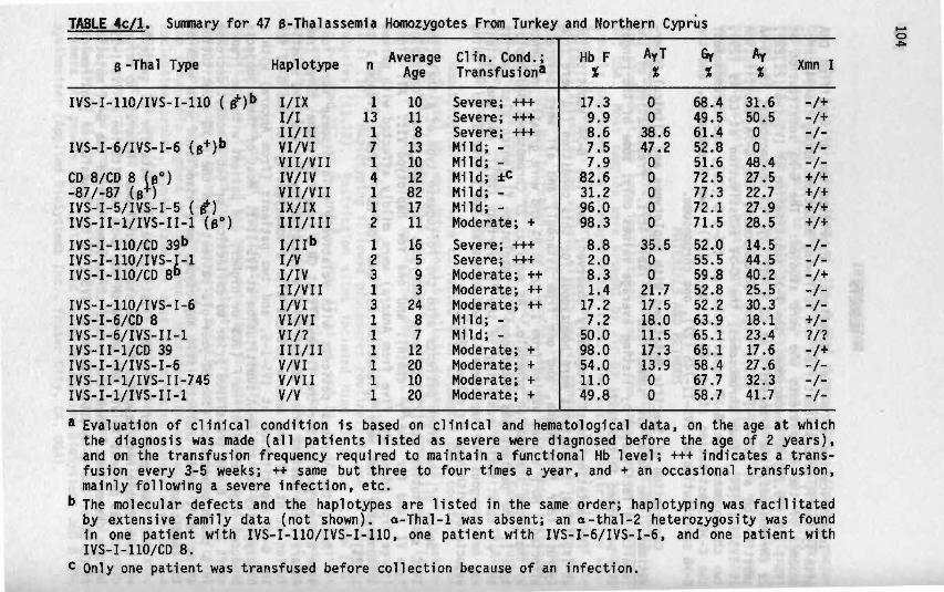
TABLE 4c/l. Summary for
0-Thai Type
IVS-I-11O/IVS-I-11O ( g*)'
IVS-I-6/IVS-I-6 (p+)b
CD 8/CD 8 (0°)-87/-87 (B+)IVS-I-5/IVS-I-5 ( B+)IVS-II-l/IVS-II-l ( B ° )
IVS-I-llO/CD 39bIVS-I-llO/IVS-I-lIVS-I-llO/CD 8°
IVS-I-110/IVS-I-6IVS-I-6/CD 8IVS-I-6/IVS-II-1IVS-II-l/CD 39IVS-I-1/IVS-I-6IVS-II-1/IVS-II-745IVS-I-l/IVS-II-l
47 B-Thalassemia
Haplotype
> I/IXI/III/IIVI/VIVII/VIIIV/IVVII/VIIIX/IXIII/IIII/libI/VI/IVII/VIII/VIVI/VIVI/?III/IIV/VIV/VII
v/v
n
1131714112
12313111111
Homozygotes From Turkey
AverageAge
10118131012821711
16593
248712201020
Clin. Cond.;Transfusion*
Severe; +++Severe; +++Severe; +++Mild; -Mild; -Mild; ±cMild; -Mild; -Moderate; +Severe; +++Severe; +++Moderate; ++Moderate; ++Moderate; ++Mild; -Mild; -Moderate; +Moderate; +Moderate; +Moderate; +
and Northern Cyprus
Hb FX
17.39.98.67.57.9
82.631.296.098.3
8.82.08.31.4
17.27.250.098.054.011.049.8
AyTX
0038.647.200000
35.50021.717.518.011.517.313.900
X
68.449.561.452.851.672.577.372.171.5
52.055.559.852.852.263.965.165.158.467.758.7
Ayj Xmn I
31.6 -/+50.5 -/+0 -/-0 -/-48.4 -/-27.5 +/+22.7 +/+27.9 +/+28.5 +/+
14.5 -/-44.5 -/-40.2 -/+25.5 -/-30.3 -/-18.1 +/-23.4 ?/?17.6 -/+27.6 -/-32.3 -/-41.7 -/-
• Evaluation of clinical condition is based on clinical and hematological data, on the age at whichthe diagnosis was made (all patients listed as severe were diagnosed before the age of 2 years),and on the transfusion frequency required to maintain a functional Hb level; +++ indicates a trans-fusion every 3-5 weeks; ++ same but three to four times a year, and + an occasional transfusion,mainly following a severe infection, etc.
b The molecular defects and the haplotypes are listed in the same order; haplotyping was facilitatedby extensive family data (not shown). a-Thal-1 was absent; an o-thal-2 heterozygosity was foundin one patient with IVS-I-110/IVS-I-110, one patient with IVS-I-6/IVS-I-6, and one patient withIVS-I-llO/CD 8.
c Only one patient was transfused before collection because of an infection.

105
were considerably higher than seen for patients with the second chromosomehaving the frameshift at CD 8 or the T->C substitution at IVS-I-6. Compar-able observations were made for the six patients with the rarer combina-tions; milder disease was present when one chromosome carried the Portu-guese B-thal mutation.
DISCUSSION
The relative simplicity of the DNA amplification method, combinedwith hybridization to radio-labeled synthetic oligonucleotide probes, hasmade it possible to identify specific mutations in the DNA of a large num-ber of B-thal homozygotes. Nine different substitutions were identified,with the IVS-I-110 (G->A), the IVS-I-6 (T->C), and the frameshift at CD 8mutations being the most frequent ones. Several of the patients were homo-zygous for one specific abnormality, which finds its basis in the factthat parents of some of these subjects were related.
Some of the observations reported here are not surprising, such asthe severity and high incidence of the IVS-I-110 (6->A) substitution amongthe patients from Cyprus and Southern Turkey, and the mild condition withlow Hb F levels in subjects with the IVS-I-6 (T->C) mutation. Thesepatients formed the largest group among the 16 subjects clinically charac-terized as being mildly affected; eight were homozygous, while two hadthis mutation in combination with another, relatively mild, mutation (Table4c/l). Rather surprisingly, three mutations were found among the mildhomozygous patients who also produced large quantities of Hb F; four suchpatients had the frameshift at CD 8, one the G->T substitution at IVS-I-5,and two the IVS-II-1 (G->A) substitution. All except the last two patientscould be managed without blood transfusions, except for surgical conditionsor during periods of severe infections. Indeed, identification of specificmutations is of considerable importance for the management of the patients.
The reasons for the high Hb F production in some of the patientsremain unclear, but it may be directly related to the chromosomal haplotype(Table 4c/l). Moreover, the presence of an Xmn I site, due to the C->Tsubstitution at position -158 5' to Gy (29), invariably results in ^values in excess of 70%, confirming earlier observations (3). Carefulexamination of mutations within the promoter regions 5' to "y and to \,and within the enhancer regions 3' to \ of the DNA of some of thesepatients might provide further insight into the observed variations inHb F levels.
Acknowledgements. This study was supported by the United StatesPublic Health Service research grants HLB-05168 and HLB-15158, and by fundsfrom the Prenatal Diagnosis Unit of the Turkish Scientific Research Founda-tion in Ankara, Turkey. J.C.D-C. was supported by Gobierno Autonomo deCanarias y Caja de Ahorros de Canarias. This is contribution #1069 fromthe Department of Cell and Molecular Biology at the Medical College ofGeorgia in Augusta, GA, USA.

106
REFERENCES
1. Aksoy M: Thalassemia intermedia: A genetic study in 11 patients.J Med Genet 7:47, 1970
2. Aksoy M, Dincol G, Erdem S: Different types of beta thalassemiaIntermedia: A genetic study in 20 patients. Acta Haematol 59:178, 1978
3. Altay Ç, Gurgy A: Clinical and haematological evaluation ofB-thalassaemia intermedia with increased Hb F and Hb A2 in hétérozygotes:B-Thalassemia intermedia-I. J Med Genet 22:205, 1985
4. Gurgey A, Kayin S, Kansu E, Altay Ç: Clinical and haematologicalevaluation of B-thalassemia intermedia characterized by unusually low Hb Fand increased Hb A2: B-Thalassaemia intermedia-II. J Med Genet 22:213,1985
5. Wainscoat JS, Old JM, Weatherall DJ, Orkin SH: The molecularbasis for the clinical diversity of B-thalassaemia in Cypriots. Lancet1:1235, 1983
6. Betke K, Marti HR, Schlicht I: Estimation of small percentagesof foetal haemoglobin. Nature 184:1877, 1959
7. Wilson JB, Headlee ME, Huisman THJ: A new high performanceliquid chromatographic procedure for the separation and quantisation ofvarious hemoglobin variants in adults and newborn babies. J Lab Clin Med102:174, 1983
8. Shelton JB, Shelton JR, Schroeder WA: High performance liquidchromatographic separation of globin chains on a large-pore C4 column.J Liq Chromatogr 7:1969, 1984
9. Kutlar F, Kutlar A, Huisman THJ: Separation of normal and abnor-mal hemoglobin chains by reversed-phase high-performance liquid chromatog-raphy. J Chromatogr 357:147, 1986
10. Poncz M, Solowiejczyk D, Harpel B, Mory Y, Schwartz E, SurreyS: Construction of human gene libraries from small amounts of peripheralblood: Analysis of B-like globin genes. Hemoglobin 6:27, 1982
11. Harano T, Reese AL, Ryan R, Abraham BL, Huisman THJ: Five haplo-types 1n Black B-thalassaemia hétérozygotes: Three are associated withhigh and two with low G Y values in fetal haemoglobin. Br J Haematol 59:333, 1985
12. Hattori Y, Kutlar F, Kutlar A, McKie VC, Huisman THJ: Haplotypesof B^ chromosomes among patients with sickle cell anemia from Georgia.Hemoglobin 10:623, 1986
13. Orkin SH, Kazazian HH Jr, Antonarakis SE, Goff SC, Boehm CD,Sexton JP, Waber PG, Giardina PJV: Linkage of B-thalassaemia mutationsand B-globin gene polymorphisms with DNA polymorphisms in human B-globingene cluster. Nature 296:627, 1982
14. Saiki RK, Bugawan TL, Horn GT, Mullis KB, Erlich HA: Analysisof enzymatically amplified B-globin and HLA-DQa DNA with allele-specificoligonucleotide probes. Nature 324:163, 1986
15. Diaz-Chico JC, Yang KG, Yang KY, Efremov DG, Stoming TA, HuismanTHJ: The detection of B-globin gene mutations using oligonucleotide probesand amplified DNA. Biochim Biophys Acta 949:43, 1988
16. Huang HJ, Stoming TA, Harris HF, Kutlar F, Huisman THJ: TheGreek AyB+-HPFH observed in a large Black family. Am J Hematol 25:401,1987
17. Orkin SH, Goff SC: Nonsense and frameshift mutations in Bassemia detected in cloned B-globin genes. J Biol Chem 256:9789, 1981

107
18. Yang KG, Liu JZ, Kutlar F, Kutlar A, Aitay Ç, Gurgey A, HuismanTHJ: e°-Thalassemia in association with a y globin gene quadruplicate.Blood 68:1394, 1986
19. Spritz RA, Jagadeeswaran P, Choudary PV, Biro PA, Elder JT, deRiel JK, Manley JL, Gefter ML, Forget BG, Weissman SM: Base substitutionin an intervening sequence of a B-thalassemic human globin gene. ProcNatl Acad Sci USA 78:2454, 1981
20. Westaway D, Williamson R: An intron nucleotide sequence variantin a cloned B-thalassemia globin gene. Nucleic Acids Res 9:1777, 1981
21. Wainscoat JS, Kanavakis E, Wood WG, Letsky EA, Huehns ER, MarshGW, Higgs DR, Clegg JB, Weatherail DJ: Thalassaemia intermedia in Cyp-riots: The interation of o- and B-thalassaemia. Br J Haematol 53:411,1983
22. Tamagnini GP, Lopes MC, Castanheira ME, Wainscoat JS, Wood WG:8+ Thalassaemia-Portuguese type: Clinical, haematological and molecularstudies of a newly defined form of B-thalassaemia. Br J Haematol 54:189,1984
23. Treisman R, Proudfoot NJ, Shander M, Maniatis T: A single basechange at a splice site in a p°-thalassemia gene causes abnormal RNA splic-ing. Cell 29:903, 1982
24. Kazazian HH Jr, Orkin SH, Antonarakis SE, Sexton JP, Boehm CD,Goff SC, Waber PG: Molecular characterization of seven B-thalassemia muta-tions in Asian Indians. EMBO J 3:593, 1984
25. Trecartin RF, Liebhaber SA, Chang JC, Lee KY, Kan YW, FurbettaM, Angius A, Cao A: 0° Thalassemia in Sardinia is caused by a nonsensemutation. J Clin Invest 68:1012, 1981
26. Atweh GH, Hsu H, Forget BG: A new IVS-I position 5 mutation re-sulting in p°-thalassemia in Mediterraneans. Blood 64:550, 1984 (suppl 1)
27. Treisman R, Orkin SH, Maniatis T: Specific transcription andRNA splicing defects in five cloned B-thalassaemia genes. Nature 302:591,1983
28. Huisman THJ, Kutlar F, Nakatsuji T, Bruce-Tagoe A, Kilinc Y,Cauchi MN, Romero Garcia C: The frequency of the y chain variant AyT indifferent populations, and its use in evaluation y gene expression in asso-ciation with thalassemia. Hum Genet 71:127, 1985
29. Gil man JG, Huisman THJ: DNA sequence variation associated withelevated fetal Gy globin production. Blood 66:783, 1985
CHAPTER 5
POSSIBLE FACTORS INFLUENCING THE HEMOGLOBIN AND
FETAL HEMOGLOBIN LEVELS IN PATIENTS WITH B-THALASSEMIA
DUE TO A HOMOZYGOSITY FOR THE IVS-I-6 (T->C) MUTATION

Ill
CHAPTER 5
POSSIBLE FACTORS INFLUENCING THE HEMOGLOBIN ANDFETAL HEMOGLOBIN LEVELS IN PATIENTS WITH B-THALASSEMIADUE TO A HOMOZYGOSITY FOR THE IVS-I-6 (T->C) MUTATION
D.G. Efreoovl, A.J. Dinovskil, E. Baysal*. Z. Ye*, A.D. Adekile^,M.L.S. Ribeiro*, G. Sch1liro3, c. Altay*. A. Gurgey*.
G.D. Efremov^, and T.H.J. Huismanl
1 Department of Biochemistry and Molecular BiologyMedical College of Georgia, Augusta, GA 30912-2100, USA
2 Department of HematologyCentro Hospitalar de Coimbra, 3000 Coimbra, Portugal
3 Cattedra di Ematologia Pediatrica dell'Universita di CataniaIstituto di Clinica Pediatrica 1°, 95125 Catania, Sicilia, Italia
* Department of Pediatrics, Children's Medical CenterHacettepe University, Ankara, Turkey
5 Macedonian Academy of Sciences and ArtsResearch Center for New Technologies9100 Skopje, Republic of Macedonia
[Adapted from: Br. J. Haenatol. (In press)]
ABSTRACT
We have collected hematological, Hb, and DNA sequence data for 29patients with a homozygosity for the IVS-I-6 (T->C) mutation with theintention of identifying factors contributing to the observed variabilityin the severity of the disease. None of the patients had received bloodtransfusion therapy for at least six months prior to the study. Hb levelsvaried from 5.0 to 9.9 g/dl. Patients with high Hb F (more than 1.5 g/dlor >20%) had high total Hb levels (7.5 to 9.7 g/dl) but some with low Hb Falso had high total Hb levels; two had a concomitant a-thai-2 heterozy-gosity. An inverse correlation between the Hb F and Hb A2 levels wasobserved. The majority of the patients was homozygous for haplotype VI(49/58 chromosomes) but haplotypes IV (2/58) and VII (7/58) were also pres-ent. The only haplotype IV homozygote had high Hb F levels with high fy
112
values and the C->T mutation at position -158 in the Gy promoter, while]both high and low Hb F levels were observed among patients with haplotypes]VI and VII. Analysis of sequence variations in regulatory regions included!the 5' HS 4, 3, and 2 of the LCR, the Gy and Ay 5' flanking regions, the!IVS-II, and the 5' B-globin gene region in two patients with high Hb F|(one homozygote each for haplotypes VI and IV), and in two patients with]low Hb F levels (one homozygote each for haplotypes VI and VII). HaplotypeJspecific differences were observed in the LCR 5' HS-2 and in the *V and \ jflanking and IVS-II regions; however, no differences were present between!the low and high Hb F-producing haplotype VI chromosomes, suggesting ajmajor role for factors which are not linked to the s-glcbin gene clusterin mediating Y-globin gene expression in patients with this type of B-thal .|
INTRODUCTION
The clinical severity of B-thal mainly depends on the degree of 0]chain deficiency, while a co-inheritance of an o-thal determinant or ofjfactors that increase Y-globin gene expression can moderate the a/non-a-jglobin chain imbalance and ameliorate the course of the disease (Weatheralljand Clegg, 1981). Increased Hb F levels have been associated with certaiiB-globin gene promoter mutations such as those in the proximal CACCC box(Gonzalez-Redondo et al, 1988; Camaschella et al, 1990), with the co-inher-1itance of an HPFH determinant in c[s to the mutated B gene as in the Sardi-nian 6B-thal (Ottolenghi et aTT 1987), and most frequently with a chromo-some that carries the -158 (C->T) substitution in the ^y promoter (Gilmanand Huisman, 1985; Thein et al, 1987; Diaz-Chico et al, 1988). Sequencevariations located in the LCR 5' HS-2 (Oner et al, 1992), the Ay IVS-II(Ragusa et al, 1992a), and the 5' flanking region of the B-gl°bin gene(Ragusa et al, 1992b) have also been implicated in the regulation of Hb Fexpression, mainly in patients with SS and B-thal. An X-linked determinanthas also been associated with elevated Hb F levels and F-cell productionin normal adults and SS patients (Miyoshi et al, 1988; Dover et al, 1992).
The influence of these factors on Hb F production and on the total Hblevels in B-thal patients is difficult to assess due to the large number ofmutations that affect B chain synthesis, while blood transfusions receivedby most of these patients also complicate the studies. We have analyzeda group of 29 patients homozygous for the IVS-I-6 (T->C) mutation who hadnot received blood transfusions for at least six months prior to the study.This mutation is one of the more common B-thal defects in the Mediterraneanpopulation, with high frequencies in Sicily, the former Yugoslavia, Turkey,and Portugal (Huisman, 1990a). Homozygotes are often mildly affected dueto the considerable B-globin chain synthesis by the mutated chromosomeand usually have low Hb F and high Hb A2 levels (Huisman, 1990b). Themutations present in the subjects of this study were previously character-ized by PCR and by dot-blot hybridization during the molecular characteri-zation of B-thal alleles in Portugal, Sicily, Turkey, and the former Yugo-slavia (Tamagnini et al, 1993; Schiliro et al, in preparation; Oner et al,1990; Dimovski et al, 1990). Fresh blood samples were collected from allpatients and re-analyzed to investigate the factors that could account forvariations in total Hb, Hb F, and Hb A2 levels.
113
PATIENTS AND METHODS
Patients. Blood samples from 29 homozygotes (13 from Portugal, sevenfrom Sicily, seven from Turkey, and two from Macedonia) and 22 family mem-bers were collected in EDTA and transported to Augusta, GA, on ice by fastair service. The ages of the patients ranged from 2 to 58 years; abouthalf had been splenectomized. All patients were clinically classified asthalassemia intermedia with Hb levels at steady state ranging from 5.0 to9.7 g/dl. More than two-thirds of the patients had never been transfused,and none had received blood transfusions during the last six months priorto blood sampling. Variability in the severity of the disease has beenreported by several of the participating physicians; we have relied mainlyon the hematological observations, especially the total Hb levels, in com-paring the severity of the disease in the 29 homozygotes, since these datawere obtained in the same laboratory and were confirmed in repeated anal-yses. Informed consent was obtained in all cases.
Hematological and Hb Analysis. Complete blood counts and red cellindices were obtained with an automated cell counter. The Hb F and Hb A2levels were quantified by cation exchange HPLC (Bisse and Wieland, 1988;Kutlar et al, 1990). The relative quantities of the three y chains inisolated Hb F were determined by reversed phase HPLC (Shelton et al, 1984;Kutlar et al, 1986).
DNA Analysis. DNA was isolated from white blood cells with the methodof Poncz et al (1982). Haplotypes of the B-globin gene cluster were deter-mined with methodology described previously (Aksoy et al, 1985); therestriction sites were Hinc II 5' to e, Xmn I 5' to Gy, Hind III at "yand Ay, Hinc II at 4,3 and 3' to it; Ava II at 0, and Bam HI 3' to p.
The number of a-globin genes was determined by digestion with Bam HI,Hind III, Hpa I, and Bgl II, and hybridization with o, ç, and 01 probes(Felice et al, 1984; Gu et al, 1987). The possible presence of a2-globingene mutations commonly found in the Mediterranean populations (initiationcodon mutation ATG->A£G; 5 bp deletion in the IVS-I donor splice site;AATAAA->AATAAG and AATAAA->AATGAA in the poly A signal, and the terminationcodon TAA->AAA mutation) (Higgs et al, 1989) was evaluated by hybridizationof PCR amplified DNA with mutation specific oligonucleotide probes as pre-viously described (Yuregir et al, 1992).
Homozygosity for the IVS-I-6 (T->C) mutation was confirmed for allpatients by direct sequencing of PCR amplified DNA. Sequence variationsin regulatory regions of the {$-globin gene cluster were analyzed by ampli-fication and direct sequencing in two patients with high Hb F levels andin two patients with low Hb F levels. The sequenced regions included the739 bp fragment between the Hind III and Bgl II sites of LCR 5' HS-2; the300 bp fragment between the Hph I and Fnu 4HI sites of LCR 5' HS-3; the280 bp fragment between the Sst I and Ava I sites of LCR 5' HS-4; the%1.3 kb 5' flanking regions of the Gy- and \-globin genes; the t400 ntsin the 3' halves of the IVS-II (Gy and \ ) , and the 5' flanking regionof the e-globin gene between positions -550 to -200 relative to the Capsite. Procedural details have been described previously (Lanclos et al,1991; Oner et al, 1992; Adekile et al, 1993; Dimovski et al, 1993).

114
RESULTS
Hematological data for the 29 e-thal homozygotes are listed in Table5/1. Considerable variation in total Hb levels (5.0-9.9 g/dl) was observedwhich did not correlate with gender, age, splenectomy, or Hb F levels. Allpatients with high Hb F (>1.5 g/dl) also had high total Hb levels (>8.0g/dl). Furthermore, an inverse correlation was observed between the Hb Fand Hb A2 levels (r = -0.87, p <0.00001) (Fig. 5/1). Repeated analyseswere made over the past three years for many Portuguese, Turkish, and Mace-donian patients; the data showed that the Hb A2 and Hb F levels did notchange significantly for each individual subject. All 22 heterozygousfamily members had Hb F levels lower than 1%.
9.0-
8.0-
HbAj7.0-
6.0-
5.0-
4.0-
10 15 20 25
HbF (%)30 35
FI6. 5/1. The relationship between the levels of Hb F and Hb A2.
Twenty-four patients were homozygous for the \ T variant [\75(E19)Ile->Thr] with an average level of 47.0 ± 11.3%. Four homozygotes for theAyl allele were observed; the Gy to Ay ratio in three averaged 47 to 53,while the fourth patient had a Gy to Ay ratio of 74:26. The one patientwith all three y chains (#380) had almost equal amounts of the two typesof Ay chain with a G y level of 49.7%. These data suggested that theIVS-I-6 (T->C) mutation occurs on at least three different chromosomes:One characterized by the presence of the \^ variant, the second with Ay*and low G y levels, and the third with \* and high Gy levels. This wasconfirmed by DNA analysis identifying the mutation on Mediterranean haplo-types VI (I- - + + +], AyT positive, 49 chromosomes), VII ([+- - - + ] , ' V negative; low Gy, 7 chromosomes), and IV ([-+ + -+ + -+]»AyT negative; high Gy, 2 chromosomes).
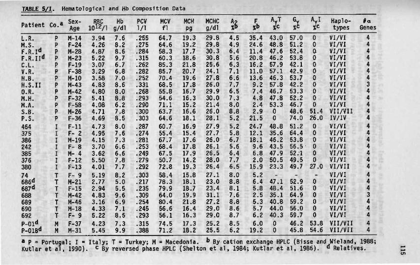
TABLE 5/1. Hematological and Hb Composition Data
Patient
L.R.M.S.F.R.I**F.R.II**CL.V.R.H.B.H.S.IIO.R.M.H.M.A.S.B.P.S.46437538624238537638074686**687 <*688689690692P-01**P-01B**
Co."
PPPPPPPPPPPPPIIIIIIITTTTTTTMM
Sex-Age
M-14F-24M-28M-23F-19F-38M-10M-43M-42F-32F-58M-26F-36F-llF- 2M-19F- 8M- 4F-12F-13F- 9M-21F-15M-42M-46M-18F- 9F-37M-31
RBC1012/1
3.944.264.875.223.073.293.584.834.805.414.084.714.694.734.954.153.703.625.504.015.192.772.944.833.164.335.224.235.45
Hbg/dl
7.68.28.69.76.76.87.08.68.08.86.27.88.58.07.67.36.66.67.87.78.25.05.59.66.97.18.57.39.9
PCV1/1.255.275.284.315.262.282.252.331.268.293.290.300.303.287.274.281.253.249.279.292.303.217.235.309.254.245.293.315.388
MCVfl
64.764.658.360.385.385.770.468.555.854.671.163.764.660.755.467.768.467.550.772.858.478.379.964.080.456.656.174.571.2
MCHP919.319.217.718.621.820.719.617.816.716.315.216.618.116.915.417.617.817.914.219.315.818.118.719.921.816.416.317.318.2
MCHCg/dl
29.829.830.330.825.624.127.826.029.930.021.426.028.127.927.726.026.126.528.026.427.123.023.431.127.229.029.025.225.5
4.54.96.45.66.37.16.67.76.57.38.08.85.25.25.86.75.66.47.76.58.08.88.17.68.88.68.78.56.2
P%•>
35.424.611.420.816.211.013.69.27.44.82.42.921.524.712.118.19.66.82.015.95.26.45.82.55.35.76.26.019.2
AyT
43.048.847.646.257.957.146.357.846.747.853.300
48.835.646.243.547.950.523.3_
47.148.435.140.844.040.300
6
57.051.252.453.842.142.953.742.253.352.246.748.674.051.264.453.856.552.149.549.7_
52.951.664.959.256.059.746.245.8
Ayl%c0000000000051.426.000000027.0-00000053.854.6
Haplo-types
VI/VIVI/VIVI/VIVI/VIVI/VIVI/VIVI/VIVI/VIVI/VIVI/VIVI/VIVII/VIIIV/IVVI/VIVI/VIVI/VIVI/VIVI/VIVI/VIVI/VIIVI/VIVI/VIVI/VIVI/VIVI/VIVI/VIVI/VIVII/VIIVII/VII
#aGenes
44444443444444444444444344444
* P = Portugal; I = Italy; T = Turkey; M = Macedonia. •> By cation exchange HPLC (Bisse and Wieland, 1988;Kutlar et al, 1990). c By reversed phase HPLC (Shelton et al, 1984; Kutlar et al, 1986). *• Relatives.
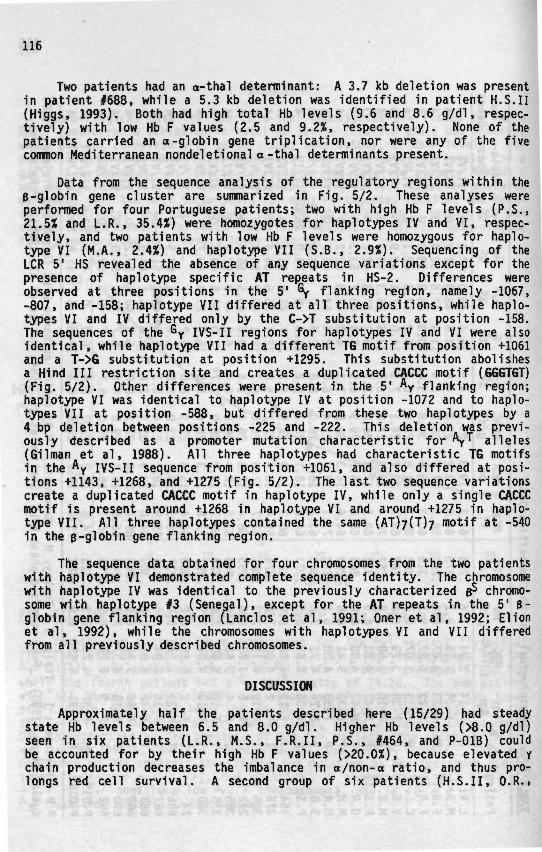
116
Two patients had an a-thal determinant: A 3.7 kb deletion was present !1n patient #688, while a 5.3 kb deletion was identified in patient H.S.II(Higgs, 1993). Both had high total Hb levels (9.6 and 8.6 g/dl, respec-tively) with low Hb F values (2.5 and 9.2%, respectively). None of thepatients carried an o-globin gene triplication, nor were any of the fivecommon Mediterranean nondeletional o-thal determinants present.
Data from the sequence analysis of the regulatory regions within theB-globin gene cluster are summarized in Fig. 5/2. These analyses wereperformed for four Portuguese patients; two with high Hb F levels (P.S.,21.5% and L.R., 35.4%) were homozygotes for haplotypes IV and VI, respec-tively, and two patients with low Hb F levels were homozygous for haplo-type VI (M.A., 2.4%) and haplotype VII (S.B., 2.9%). Sequencing of theLCR 5' HS revealed the absence of any sequence variations except for thepresence of haplotype specific AT repeats in HS-2. Differences wereobserved at three positions in the 5' Gy flanking region, namely -1067,-807, and -158; haplotype VII differed at all three positions, while haplo-types VI and IV differed only by the C->T substitution at position -158.The sequences of the Gy IVS-II regions for haplotypes IV and VI were alsoIdentical, while haplotype VII had a different TG motif from position +1061and a T->G substitution at position +1295. This substitution abolishesa Hind III restriction site and creates a duplicated CACCC motif (GGGTGT)(Fig. 5/2). Other differences were present in the 5' \ flanking region;haplotype VI was identical to haplotype IV at position -1072 and to haplo-types VII at position -588, but differed from these two haplotypes by a4 bp deletion between positions -225 and -222. This deletion was previ-ously described as a promoter mutation characteristic for \ T alleles(Gilman et al, 1988). All three haplotypes had characteristic TG motifsIn the Ay IVS-II sequence from position +1061, and also differed at posi-tions +1143, +1268, and +1275 (Fig. 5/2). The last two sequence variationscreate a duplicated CACCC motif in haplotype IV, while only a single CACCCmotif is present around +1268 in haplotype VI and around +1275 in haplo-type VII. All three haplotypes contained the same (AT)y(T)7 motif at -540in the p-globin gene flanking region.
The sequence data obtained for four chromosomes from the two patientswith haplotype VI demonstrated complete sequence identity. The chromosomewith haplotype IV was identical to the previously characterized e^ chromo-some with haplotype #3 (Senegal), except for the AT repeats in the 5' B-globin gene flanking region (Lanclos et al, 1991; Oner et al, 1992; El ionet al, 1992), while the chromosomes with haplotypes VI and VII differedfrom all previously described chromosomes.
DISCUSSION
Approximately half the patients described here (15/29) had steadystate Hb levels between 6.5 and 8.0 g/dl. Higher Hb levels (>8.0 g/dl)seen in six patients (L.R., M.S., F.R.II, P.S., #464, and P-01B) couldbe accounted for by their high Hb F values (>20.0%), because elevated ychain production decreases the imbalance in a/non-a ratio, and thus pro-longs red cell survival. A second group of six patients (H.S.II, O.R.,
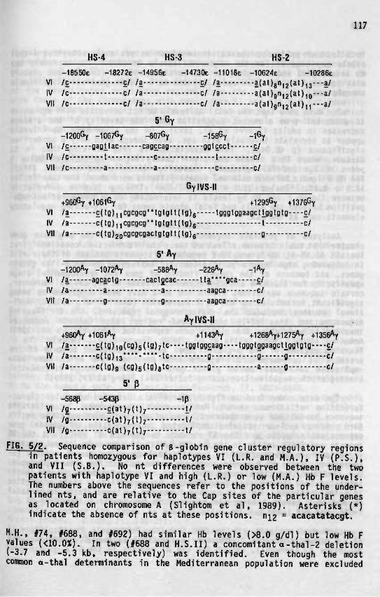
117
HS-4 HS-3 HS-2-18 5 5 0 E -18272e -14956e -14730e -11018e -10624e -10286e
VI /ç ç/ /a c/ /a a(at)8"i2(aUi3---a/IV /c c/ /a c/ /a a(at)gn,2(at),,,---a/VII /c c/ /a c/ /a a(at)gn,2(at),,---a/
-1200% -1067% -807% -158% - 1 %VI /ç gagHac cagçcag ggtçcct ç/IV /c t c t c/VII /c a a c c/
GylVSII
+960% +1061% +1295% +1376%VI /a £(tg),,cgcgcg"lgtgtt(tg)g tgggtggaagcttflgtgtg ç/IV /a c(tg),,cgcgcg*'tgtgtt(tg)g t c/VII /a c(tg)2ocgcgcgactgtgtt(tg)g g c/
VI /a agcactg cactjcac tta*'**gca ç/IV /a a a aagca c/VII /a g g aagca c/
AylVS-ll
+960*y +1061*? +1143*? +1268*7+1275*? +1356*YVI /a Ç(tg),o(cg)5(tg)7tc tggtggcaag tgggtggaagcUggtgtg ç/IV /a c(to)i3*"*-""-tc g g g c/VII /a c(tg)g (cg)s(tg)gtc g a g c/
5M>-5680 -5430 -10
VI /g c(at )7( t )7 t/IV /g c(at)7(t)7 t/VII /g c(at)7(t)7 t/
FIG. 5/2. Sequence comparison of B-globin gene cluster regulatory regionsin patients homozygous for haplotypes VI (L.R. and M.A.), IV (P.S.),and VII (S.B.). No nt differences were observed between the twopatients with haplotype VI and high (L.R.) or low (M.A.) Hb F levels.The numbers above the sequences refer to the positions of the under-lined nts, and are relative to the Cap sites of the particular genesas located on chromosome A (Slightom et al, 1989). Asterisks (*)indicate the absence of nts at these positions. ni2 = acacatatacgt.
H.H., #74, #688, and #692) had similar Hb levels (>8.0 g/dl) but low Hb Fvalues (<10.0X). In two (#688 and H.S.II) a concomitant a- tha i -2 deletion(-3.7 and -5.3 kb, respectively) was ident i f ied . Even though the mostcommon a-thal determinants in the Mediterranean population were excluded
118
1n all other patients, the existence of an as yet uncharacterized a-thalall el e may account for the higher Hb levels in some. The extremely lowHb levels (5.0 and 5.5 g/dl) in two Turkish patients (brother and sister)with low Hb F values (6.4 and 5.8%) and no a-globin gene variation remainunexplained.
Our data show that the total Hb levels do not correlate with age whichis different from a recent suggestion that the severity of the diseasedecreases in adults (Scerri et al, 1993). On the other hand, Hb F levelsappear to correlate inversely with age, even though with only a borderlinesignificance (p <0.045). This association is less striking than in SSdisease, where Hb F levels are significantly higher in children below 5years of age and fall gradually thereafter (Adekile and Huisman, 1993).The Hb F levels between male and female patients were about the same (males11.8 ± 8.7%; females 11.3 ± 7.4%). The striking inverse correlationbetween the Hb F and Hb A2 levels is interesting as it could reflect com-petition between the adult and fetal globin genes for cis-acting regulatoryelements such as the LCR.
Analyses of possible sequence variations within the 3-globin genecluster that could have an effect on the Y chain production concerned firstthe LCR 5' HS-4, HS-3, and HS-2 regulatory segments because of their rolein conferring high level erythroid-specific expression on the p-like globingenes, and because of the recent finding of gene and developmental stagespecificity for the HS-4 and HS-3 elements (Fraser et al, 1993). Moreover,certain sequence variations within the HS-2 have been suggested as beingsignificant in modulating the Hb F expression (Oner et al, 1992). Addi-tional studies included the y-globin gene promoters, the 5' flankingsequences and the IVS-II since these regions also contain sequencesinvolved in modulating the globin gene expression (Grosveld et al, 1993;Antoniou et al, 1988; Collis et al, 1990; Ragusa et al, 1992a). The 5'flanking regions of the B-globin genes were investigated because of thepresence of polymorphic AT repeats from position -540 which have beenimplicated in regulation of globin gene expression through differentialbinding of the negative transacting factor BP-1 (Berg et al, 1989).
The data summarized in Fig. 5/2 provide a detailed molecular charac-terization of three common Mediterranean chromosomes. Considerablesequence variations were observed, some of which involve possible motifsfor binding nuclear proteins implicated in regulation of globin geneexpression; examples are the AT repeats in the LCR 5' HS-2, and the TGrepeats and CACCC motifs in the Gy and Ay IVS-II. However, these sequencevariations are all confined to specific haplotypes and cannot account forvariations in the Hb F levels among the patients with the same haplotypes.Our findings, therefore, suggest a major role for unknown genetic loci,separated from the B-globin gene cluster, in controlling y chain productionin B-thal patients. This is further supported by the differences in totalHb and Hb F levels between the siblings in two families, namely P-01 andP-01B, and F.R.I and F.R.II.
Sequence analysis of the DNA from the homozygote for haplotype IV(P.S.) showed that this chromosome is identical to the B$ chromosome withhaplotype #3 (Senegal), except for the AT repeats in the 5' B-globin gene

119
flanking region. These two chromosomes are characterized by elevated Hb Fexpression under conditions of erythropoietic stress and by the presenceof the -158 (C->T) promoter mutation (Gilman and Huisman, 1985; Labié etal, 1985; Thein et al, 1987; Diaz-Chico et al, 1988). Sequence comparisonof the investigated regulatory regions from the haplotype IV chromosomewith those of the chromosomes with haplotypes VI and VII did not identifyany significant sequence variation other than the Sy -158 (C->T), whichcould account for the consistently elevated Hb F levels in e-thal patientswith this chromosome.
Acknowledgewents. This study was supported in part by the UnitedStates Public Health Services research grant HLB-41544 (to THJH), theTurkish Scientific and Technical Research Council (TUBITAK) Tag #0757 (toÇA and AG), and by a grant (#336/92) of the Ministry of Science of theRepublic of Macedonia (to GDE).
REFERENCES
Adekile, A.D., Dimovski, A.J., Oner, C , Lanclos, K.D. & Huisman,T.H.J. (1993) Haplotype specific sequence variations in the locus controlregion (5' hypersensitive sites 2, 3, 4) of ^ chromosomes. Hemoglobin,17, 475-478.
Adekile, A.D. & Huisman, T.H.J. (1993) Hb F in sickle cell anemia.Experientia, 49, 1-12.
Aksoy, M., Kutlar, A., Efremov, G.D., Nikolov, N., Petkov, G., Reese,A.L., Harano, T., Chen, S.S. & Huisman, T.H.J. (1985) Haplotypes and levelsof fetal hemoglobin and Gy to Ay ratios in Mediterranean patients withthalassemia minor and major. American Journal of Hematology, 20, 7-16.
Antoniou, M., de Boer, E., Habets, G. & Grosveld, F. (1988) The humanB globin gene contains multiple regulatory regions: Identification ofone promoter and two downstream enhancers. The EMBO Journal, 7, 377-384.
Berg, P.E., Williams, D.M., Qian, R-L., Cohen, R.B., Cao, S-X., Mit-telman, M. & Schechter, A.N. (1989) A common protein binds to two silencers5' to the human p gene. Nucleic Acids Research, 17, 8833-8852.
Bisse, E. & Wieland, H. (1988) High-performance liquid chromatographicseparation of human haemoglobins - Simultaneous quantitation of foetaland glycated haemoglobins. Journal of Chromatography, 434, 95-110.
Camaschella, C , Alfarano, A., Gottardi, E., Serra, A., Revello, D. &Saglio, G. (1990) The homozygous state for the -87 (C->G) B+ thalassaemia.British Journal of Haematology, 75, 132-133.
Col lis, P., Antoniou, M. & Grosveld, F. (1990) Definition of the mini-mal requirements within the human B-globin gene and the dominant controlregion for high level expression. The EMBO Journal, 9, 233-240.
Diaz-Chico, J.C., Yang, K.G., Stoming, T.A., Efremov, D.G., Kutlar,A., Kutlar, F., Aksoy, M., Altay, Ç., Gurgey, A., Kilinç, Y. & Huisman,T.H.J. (1988) Mild and severe B-thalassemia among homozygotes from Turkey:Identification of the types by hybridization of amplified DNA with syn-thetic probes. Blood, 71, 248-251.
Dimovski, A.J., Adekile, A.D., Divoky, V., Baysal, E. & Huisman,T.H.J. (1993) Polymorphic pattern of the (AT)X(T)Y motif at -530 5' to
120
the B-globin gene in over 40 patients homozygous for various B-thalassemiamutations. American Journal of Hematology, 45, 51-57.
Dimovski, A.J., Efremov, D.G., Jankovic, L., Juricic, D., Zisovski,N., Stojanovski, N., Nikolov, N., Petkov, G.T., Reese, A.L., Stoming, T.A.,Efremov, G.D. & Huisman, T.H.J. (1990) B-Thalassemia in Yugoslavia. Hemo-globin, 14, 15-24.
Dover, G.J., Smith, K.D., Chang, Y.C., Purvis, S., Mays, A., Meyers,D.A., Shells, C. & Serjeant, G. (1992) Fetal hemoglobin levels in sicklecell disease and normal individuals are partially controlled by an X-linkedgene located at Xp22.2. Blood, 80, 816-824.
Elion, J., Berg, P., Lapoumeroulie, C., Trabuchet, G., Mittelman,M., Krishnamoorthy, R., Schechter, A.N. & Labié, D. (1992) DNA sequencevariation in a negative control region 5' to the B-globin gene correlateswith the phenotypic expression of the 0$ mutation. Blood, 79, 787-792.
Felice, A.E., Cleek, M.P., McKie, K., McKie, V. & Huisman, T.H.J.(1984) The rare a-thalassemia-1 of Blacks is a ço-thalassemia-1 associatedwith deletion of all c- and a-globin genes. Blood, 63, 1253-1257.
Fraser, P., Pruzina, S., Antoniou, M. & Grosveld, F. (1993) Eachhypersensitive site of the human B-globin gene locus control region confersa different developmental pattern of expression to the globin genes. Genesand Development, 7, 106-113.
Gil man, J.G. & Huisman, T.H.J. (1985) DNA sequence variation asso-ciated with elevated fetal Gy. production. Blood, 66, 783-787.
Gilman, J.G., Johnson, M.E. & Mishima, N. (1988) Four base-pair DNAdeletion 1n human \-globin gene promoter associated with low \ expressionin adults. British Journal of Haematology, 68, 455-458.
Grosveld, F., Dillon, N. & Higgs, D. (1993) The regulation of humanglobin gene expression. In: The Haemoglobinopathies, D.J. Weatherall &D.R. Higgs (eds), Bailliere's Clinical Haematology, Vol. 6, pp 31-55, W.B.Saunders Company, London.
Gonzalez-Redondo, J.M., Stoming, T.A., Lanclos, K.D., Gu, Y.C.,Kutlar, A., Kutlar, F., Nakatsuji, T., Deng, B., Han, I.S., McKie, V.C. SHuisman, T.H.J. (1988) Clinical and genetic heterogeneity in Black patientswith homozygous B-thalassemia from the Southeastern United States. Blood,72, 1007-1014.
Gu, Y.C., Landman, H. & Huisman, T.H.J. (1987) Two different quadru-plicated a globin gene arrangements. British Journal of Haematology, 66,245-250.
Higgs, D.R. (1993) a-Thalassaemia. In: The Haemoglobinopathies, D.J.Weatherall & D.R. Higgs (eds), Bailliere's Clinical Haematology, Vol. 6,pp 117-150, W.B. Saunders Company, London.
Higgs, D.R., Vickers, M.A., Wilkie, A.O.M., Pretorius, I-M., Jarman,A.P. & Weatherall, D.J. (1989) A review of the molecular genetics of thehuman a-globin gene cluster. Blood, 73, 1081-1104.
Huisman, T.H.J. (1990a) Frequencies of common p-thalassaemia allelesamong different populations: Variability in clinical severity. BritishJournal of Haematology, 75, 454-457.
Huisman, T.H.J. (1990b) Silent B-thalassemia and thalassemia inter-media. Haematologica, 75, 1-8.
Kutlar, A., Kutlar, F., Gu, L-G., Mayson, S.M. & Huisman, T.H.J.(1990) Fetal hemoglobin in normal adults and B-thalassemia hétérozygotes.Human Genetics, 85, 106-110.
Kutlar, F., Kutlar, A. & Huisman, T.H.J. (1986) Separation of normaland abnormal hemoglobin chains by reversed-phase high-performance liquidchromatography. Journal of Chromatography, 357, 147-153.
Labie, D., Pagnier, J., Lapoumeroulie, C , Rouabhi, F., Dunda-Belk-hodja, 0., Chardin, P., Beldjord, C , Wajcman, H., Fabry, M.E. & Nagel,R.L. (1985) Common haplotype dependency of high Gy-globin gene expressionand high Hb F levels in B-thalassemia and sickle cell anemia patients.Proceedings of the National Academy of Sciences of the United States ofAmerica, 82, 2111-2114.
Lanclos, K.D., Oner, C , Dimovski, A.J., Gu, Y-C. & Huisman, T.H.J.(1991) Sequence variations in the 5' flanking and the IVS-II regions ofthe »y- and \-globin genes of ^ chromosomes with five different haplo-types. Blood, 77, 2488-2496.
Miyoshi, K., Kaneto, Y., Hisaomi, K., Ihchi, H., Niki, S., Hasegawa,K., Shirakami, A. & Yamano, T. (1988) X-Linked dominant control of F-cellsin normal adult life: Characterization of the Swiss type of hereditarypersistence of fetal hemoglobin regulated dominantly by gene(s) on X chro-mosome. Blood, 72, 1854-1860.
Oner, C , Dimovski, A.J., Altay, Ç., Gurgey, A., Gu, Y.C. & Huisman,T.H.J. (1992) Sequence variations in the 5' hypersensitive site-2 of thelocus control region of B^ chromosomes are associated with different levelsof fetal globin in Hemoglobin S homozygotes. Blood, 79, 813-819.
Oner, R., Altay, Ç., Gurgey, A., Aksoy, M., Kilinç, Y., Stoming, T.A.,Reese, A.L., Kutlar, A., Kutlar, F. & Huisman, T.H.J. (1990) B-Thalassemiain Turkey. Hemoglobin, 14, 1-14.
Ottolenghi, S., Giglioni, B., Pulzini, A., Comi, P., Camaschella,C , Serra, A., Guerrasio, A. & Saglio, G. (1987) Sardinian 6B-thalassemia:A further example of a C->T substitution at position -196 of the \ globingene promoter. Blood, 69, 1058-1061.
Poncz, M., Solowiejczyk, D., Harpel, B., Mory, Y., Schwartz, E. &Surrey, S. (1982) Construction of human gene libraries from small amountsof peripheral blood: Analysis of B-like globin genes. Hemoglobin, 6,27-36.
Ragusa, A., Lapoumeroulie, C , Lombardo, M., Elion, J., Labie, D. &Krishnamoorthy, R. Û992a) Differential binding of nuclear protein factorsto the polymorphic *y IVS-2 (TG) repeat region and its relevance to Hb Fresponse in stress anemia. Blood, 80, 310a (Suppl. 1).
Ragusa, A., Lombardo, M., Beldjord, C , Ruberto, C , Lombardo, T.,Elion, J., Nagel, R. & Krishnamoorthy, R. (1992b) Genetic epidemiologyof B-thalassemia in Sicily: Do sequences 5' to the Gy gene and 5' to theB gene interact to enhance Hb F expression in B-thalassemia? AmericanJournal of Hematology, 40, 199-206.
Scerri, C.A., Abela, W., Gaidies, R., Pizzuto, M., Grech, J.L. &Felice, A.E. (1993) The B+ IVS-I-NT no. 6 (T->C) thalassaemia in hétéro-zygotes with an associated Hb Valletta or Hb S heterozygoslty in homozy-gotes from Malta. British Journal of Haematology, 83, 669-671.
Schiliro, G., et al. (1993) In preparation.Shelton, J.B., Shelton, J.R. & Schroeder, W.A. (1984) High performance
liquid chromatographic separation of globin chains on a large-pore C4column. Journal of Liquid Chromatography, 7, 1969-1977.
Slightom, J.L., Blechl, A.E. & Smithies, 0. (1980) Human fetal Gyand Ay globin genes: Complete nucleotide sequences suggest that DNA canbe exchanged between these duplicated genes. Cell, 21, 627-638.
122
Tamagm'ni, G.P., Gonçalves, P., Ribeiro, M.L.S., Kaeda, J., Kutlar,F., Baysal, E. & Huisman, T.H.J. (1993) B-Thalassemia mutations in thePortuguese; high frequencies of two all el es in restricted populations.Hemoglobin, 17, 31-40.
Thein, S.L., Wainscoat, J.S., Sampietro, M., Old, J.M., Cappeilini,D., Fiorelli, G., Mode11, B. & Weatherall, D.J. (1987) Association of thal-assaemia intermedia with a e-globin gene haplotype. British Journal ofHaematology, 65, 367-373.
Weatherall, D.J. & Clegg, J.B. (1981) The Thalassaemia Syndromes,3rd edition, Blackwell Scientific Publications, Oxford.
Yuregir, G.T., Aksoy, K., Çuruk, M.A., Dikmen, N., Fei, Y-J., Baysal,E. & Huisman, T.H.J. (1992) Hb H disease in a Turkish family resultingfrom the interaction of a deletional a-thalassaemia-1 and a newly discov-ered poly A mutation. British Journal of Haematology, 80, 527-532.

CHAPTER 6
THE -158 (C->T) PROHOTER MUTATION IS RESPONSIBLE FOR THE
INCREASED TRANSCRIPTION OF THE 3' Y GENE IN THE ATLANTA TYPE OF HPFH

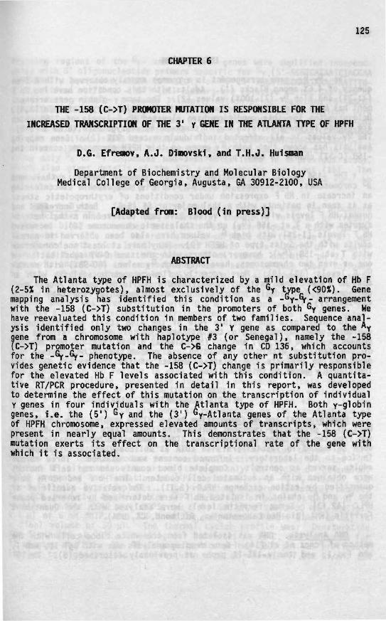
125
CHAPTER 6
THE -158 (C->T) PRONOTER MUTATION IS RESPONSIBLE FOR THEINCREASED TRANSCRIPTION OF THE 3' r GENE IN THE ATLANTA TYPE OF HPFH
D.G. Efremov, A.J. Dinovski, and T.H.J. Huismart
Department of Biochemistry and Molecular BiologyMedical College of Georgia, Augusta, GA 30912-2100, USA
[Adapted from: Blood (in press)]
ABSTRACT
The Atlanta type of HPFH is characterized by a mild elevation of Hb F(2-5% in hétérozygotes), almost exclusively of the Gy type (<90X). Genemapping analysis has identified this condition as a -"y-^r- arrangementwith the -158 (C->T) substitution in the promoters of both Gy genes. Wehave reevaluated this condition in members of two families. Sequence anal-ysis identified only two changes in the 3' Y gene as compared to the Aygene from a chromosome with haplotype #3 (or Senegal), namely the -158(C->T) promoter mutation and the C->G change in CD 136, which accountsfor the -Gy-Gy- phenotype. The absence of any other nt substitution pro-vides genetic evidence that the -158 (C->T) change is primarily responsiblefor the elevated Hb F levels associated with this condition. A quantita-tive RT/PCR procedure, presented in detail in this report, was developedto determine the effect of this mutation on the transcription of individualy genes in four individuals with the Atlanta type of HPFH. Both y-globingenes, i.e. the (5') Gy and the (3') Gy_Atlanta genes of the Atlanta typeof HPFH chromosome, expressed elevated amounts of transcripts, which werepresent in nearly equal amounts. This demonstrates that the -158 (C->T)mutation exerts its effect on the transcriptional rate of the gene withwhich it is associated.
126
INTRODUCTION
The -Gy_Gy_ gene arrangement is a common anomaly observed with a fre-quency of 4.6% among Blacks (1). Adults with this condition have low Hb F(<1.3%) and high Gy (71-100%) values (2). The same y-globin gene arrange-ment has been observed in two families with the Atlanta type of HPFH (3,4).Hétérozygotes for this condition have elevated Hb F levels of 2-5% whichare higher in young children and in compound hétérozygotes with Hb S (2-5).The Gy values in all affected individuals exceed 90% (2). Gene mappinganalysis has shown that both G y genes on this HPFH chromosome contain the-158. (C->T) change in their promoters (5). ;
The -158 (C->T, Gy) mutation has been found on normal, p-thal, andB$ chromosomes. Chromosomes with T at -158 (Gy) are capable of conferringan increase in Hb F expression under conditions of erythropoietic stressas is seen in patients with B-thal major or SS (5-8). Both increased andnormal Hb F levels with high Gy values have been observed in B-thal hétéro-zygotes with a T at -158 (Gy) on the thalassemic chromosome (2). IncreasedHb F levels (1-3%) with high G values have also been observed in normaladults with the Swiss type of HPFH (9). Many have at least one chromosomewith T at -158 (Gy); however, others with the same C->T substitution onone or even two chromosomes, have only high Gy values (60-98%) with normalHb F levels (1%) (9). More recently, flow-cytometric analysis with ananti Hb F monoclonal antibody demonstrated a small increase in F-cell per-centages in individuals homozygous or heterozygous for the C->T changeat -158 (Gy) (10).
To investigate the possible role of the -158 (C->T) substitution ony-globin gene expression in the Atlanta type of HPFH, we studied thesequences of the 5' y (Gy) and 3' y (Gy-Atlanta) globin genes in four membersof two families with this condition. A competitive RT/PCR method wasdeveloped to evaluate the relative amounts of the transcripts from theindividual y genes in this condition.
MATERIALS AND METHODS
Hematoiogical and Hb Conposition Data. Blood samples from three mem-bers of a previously reported family with the Atlanta type of HPFH (4)and from two new cases (a 19-year-old Black female and her 2-year-old son)were collected in vacutainers with EDTA as anticoagulant and transportedin ice to the laboratory. Informed consent was obtained. Three normaladults served as controls. Complete blood counts and red cell indiceswere obtained with an automated cell counter. The Hb F and Hb A2 werequantified by cation exchange HPLC (2,11). The relative quantities ofthe Gy and Ay chains in isolated Hb F were determined by reversed phaseHPLC (12,13). Serum ferritin levels were analyzed with the QuantimuneFerritin IRMA (Bio-Rad Laboratories, Richmond, CA, USA).
DMA Analysis. DNA was isolated from the white blood cells with themethod of Poncz et al (14). Gene mapping analysis was with Bgl II, Xmn I,and Pst I, and the y-IVS-II probe as previously described (5). The 5'
127
flanking regions of the &r- and \-globin genes were amplified independ-ently with 5' oligonucleotide primers specific for Gy (5'-ACGTCATAATCTACCAAGGTCATG-3', positions -1227 to -1204) or Ay (5'-AGCTTAGGGGATAAACTAATTTG-3', positions -1279 to -1257) (all numbers are relative to the Cap site ofthe nearest y gene). A common 3' amplification primer was used (5'-GGCGTCTGGACTAGGAGCTTATTG-3', positions +53 to +30). The IVS-II regions wereamplified with a common 5' primer (5'-TGGACCCAGAGGTTCTTTGA-3', positions+287 to +306), and 3' primers specific for Gy (5'-TGTGCTGCAATCCAGGGAAGGGGGT-3', positions +1948 to +1924) or Ay (5'-CTGCAATCAATCCAGCCCCCAGGTC-3',positions +1908 to +1885). The specificity of the PCR procedures in ampli-fying only Gy or Ay sequences has been established in a previous study,which also gives details of the PCR and sequencing reactions (15). Thepresence of the most common a-thal-2 determinant among Blacks (the - a ^ kbdeletion) was investigated with a PCR-based procedure (16).
RNA Analysis. Total cellular RNA was isolated with the procedureof Chomczynski and Sacchi (17). Briefly, the cells in 10 ml of freshlycollected blood were washed twice with PBS, centrifuged at 2,500 rpm for10 minutes at 4°C, and the reticulocyte-enriched top fraction (1 ml) wastransferred into four 1.5 ml Eppendorf tubes. Five hundred ul of solu-tion D (4 M guanidinium thiocyanate, 25 mM sodium citrate, pH 7, 0.5Xsarcosyl, and 0.1 M -mercaptoethanol) were added to each tube, followedby 50 ni of 2 M sodium acetate, pH 4, 0.5 ml of acid phenol (water satu-rated), and 100 u.1 of a chloroform-isoamylalcohol mixture (49:1). Aftercentrifugation at 10,000 g for 20 minutes at 4°C, the RNA present in theaqueous phase was precipitated with 0.5 ml of isopropanol at -20°C forat least 1 hour, collected by centrifugation, and dissolved in 15 ul water.
Three ul (1-2 ug of RNA) were RT with an oligo-dT primer, using theGene Amp RNA/PCR kit (Perkin Elmer Cetus, Norwalk, CT, USA) and the proce-dure recommended by the manufacturer. The entire RT sample was next sub-jected to PCR amplification with two oligonucleotide primers (50 pM each)complementary to two regions in exon 2 and in the 3' UTR where the Gy, Ay,and Gy-Atlanta globin genes have identical sequences. Their sequencesand positions were: GF 5'-CTGACTTCCTTGGGAGAT-3', positions +381 to +397,and GR 5'-GCTTGCAGAATAAAGCCTA-3' located from nt +55 to nt +33, 3' to thetermination CD. The PCR was done as recommended by the manufacturer ofthe RNA/PCR kit, except that the Taq polymerase was added during the firstcycle of denaturation (95°C, 5 minutes). The PCR required 30 cycles ofone minute of denaturation at 95°C, one minute of annealing at 54°C, andone minute of extension at 72°C, in a Perkin Elmer Cetus thermal cycler.Fifteen ul of the PCR product were next radioactively labeled with the 32p_labeled GF oligonucleotide primer through only one cycle of extension,which prevented the formation of 32p_iabeled -6y-Ay_, -Gy-Gy.Atlanta, and-\_Gy-Atlanta heteroduplexes. The reaction mixture contained, besidesthe PCR product, 40 pM of the 32p_-|abeled GF oligonucleotide, 5 ul ofreaction buffer (200 mM/L MOPS, pH 7.8, 500 mM/L NaCl, 30 mM/L MgCl2),2.5 ul of 6 mM dNTP, and 2.5 U of Taq polymerase (Perkin Elmer Cetus) ina final volume of 50 ul • The thermal cycler profile was: Denaturationfor 5 minutes at 95°C, annealing for 2 minutes at 54°C, and extension for12 minutes at 72°C. The GF oligonucleotide primer (800 pM) was previouslylabeled to high specific activity with 2 ul of 32p_ATP (7,000 Ci/mM; ICN
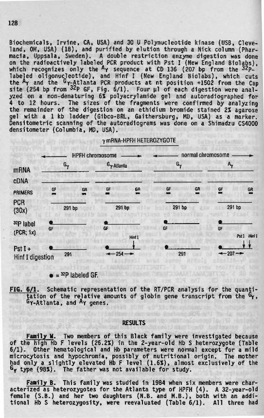
128
Biochemical s, Irvine, CA, USA) and 30 U Polynucleotide kinase (USB, Cleve-land, OH, USA) (18), and purified by elution through a Nick column (Phar-macia, Uppsala, Sweden). A double restriction enzyme digestion was doneon the radioactively labeled PCR product with Pst I (New England Biolabs),which recognizes only the Ay sequence at CD 136 (207 bp from the 32p_labeled oligonucleotide), and Hinf I (New England Biolabs), which cutsthe *y and the Gy-Atlanta PCR products at nt position +1502 from the Capsite (254 bp from 32p GF, Fig. 6/1). Four u.1 of each digestion were anal-yzed on a non-denaturing 6% polyacrylamide gel and autoradiographed for4 to 12 hours. The sizes of the fragments were confirmed by analyzingthe remainder of the digestion on an ethidium bromide stained 2% agarosegel with a 1 kb ladder (Gibco-BRL, Gaithersburg, MD, USA) as a marker.Densitometric scanning of the autoradiograms was done on a Shimadzu CS4000densitometer (Columbia, MD, USA).
Y mRNA-HPFH HETEROZYGOTE
HPFH chromosome •- •* normal chromosome
mRNA 1
cDNAPRIMERS 2 OR • OR
32p label •GF GF GF
GF
GF
291
GR
bp
HM1
PCR/3Q^\ 291 bp 291 bp 291 bp
GF
« _
GF
291 bp
Pstl
GR
Hinll
Pst I + !Hint I digestion » * " » " * » ^ " ~
• = *P labeled GF.
FIG. 6/1. Schematic representation of the RT/PCR analysis for the quanti-tation of the relative amounts of globin gene transcript from the Gy,Gy-Atlanta, and Ay genes.
RESULTS
Family H. Two members of this Black family were investigated becauseof the high Hb F levels (26.2%) in the 2-year-old Hb S hétérozygote (Table6/1). Other hematological and Hb parameters were normal except for a mildmicrocytosis and hypochromia, possibly of nutritional origin. The motherhad only a slightly elevated Hb F level (1.6%), almost exclusively of theGy type (98%). The father was not available for study.
FawHy B. This family was studied in 1984 when six members were char-acterized as hétérozygotes for the Atlanta type of HPFH (4). A 32-year-oldfemale (S.B.) and her two daughters (N.B. and M.B.), both with an addi-tional Hb S heterozygoslty, were reevaluated (Table 6/1). All three had
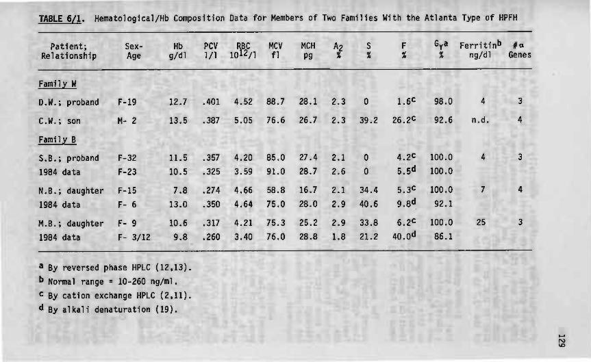
TABLE 6/1. Hemato1og1ca1/Hb Composition Data for Members of Two Families With the Atlanta Type of HPFH
Patient;Relationship
Family W
D.W.; proband
C.W.; son
Family B
S.B.; proband
1984 data
N.B.; daughter
1984 data
M.B.; daughter
1984 data
Sex-Age
F-19
M- 2
F-32
F-23
F-15
F- 6
F- 9
F- 3/12
Hbg/dl
12.7
13.5
11.5
10.5
7.8
13.0
10.6
9.8
PCV1/1
.401
.387
.357
.325
.274
.350
.317
.260
RBC1012/1
4.52
5.05
4.20
3.59
4.66
4.64
4.21
3.40
MCVfl
88.7
76.6
85.0
91.0
58.8
75.0
75.3
76.0
MCHP9
28.1
26.7
27.4
28.7
16.7
28.0
25.2
28.8
"I
2.3
2.3
2.1
2.6
2.1
2.9
2.9
1.8
S%
0
39.2
0
0
34.4
40.6
33.8
21.2
F%
1.6C
26.2C
4.2C
5.5<«
5.3C
9.8<*
6.2C
40. Qd
G^a%
98.0
92.6
100.0
100.0
100.0
92.1
100.0
86.1
Ferrit1n*>ng/dl
4
n.d.
4
7
25
Genes
3
4
3
4
3
« By reversed phase HPLC (12,13).
b Normal range = 10-260 ng/ml.
c By cation exchange HPLC (2,11).
<* By alkali denaturation (19).

132
in Table 6/2. The Gy-Atlanta transcripts were present only in the fourindividuals with the Atlanta type of HPFH (Fig. 6/3). The levels of thesetranscripts were equal or slightly higher than those of the Gy transcripts,while the Ay transcripts were absent (in S.B.) or below 4% (Table 6/2).Higher levels of Gy than Ay transcripts were present in the three controls[Table 6/2). The values were comparable to those obtained for the G y andAy chains in isolated Hb F by reversed phase HPLC.
D.W. S.B. N.B. M.B. A.D. D.P. E.B.
'7(291)
y Atlanta (254)
W ^ *
Ay (207)
Hint Ï + Pst T
FI6 . 6/3. Autoradiography of digested 32p_iabeled RT/PCR products forthe analysis of Gy, Gy-Atlanta, and Ay mRNA in hétérozygotes for theAtlanta type of HPFH and in three controls. The values obtained afterdensitometric scanning of the autoradiograms are listed in Table 6/2.
DISCUSSION
Gene mapping studies of the Atlanta type of HPFH have identified twoGy genes on the HPFH chromosome, both having a T at position -158 (4,5).The sequence data presented here show that the Gy-Atlanta gene of the HPFHchromosome, located in 3' position, is similar to an Ay gene with onlytwo nt substitutions: The C->T at -158 and the GCA->GGA at CD 136. Anal-ysis of the LCR 5'HS-2, the 5' flanking and the IVS-II regions of the 5'(Gy) and 3' (Ay) y genes, and the 5' flanking region of the e-globin gene,identified sequence variations which are characteristic for a chromosomewith haplotype #3 (15) (some of these data are not shown here). Therefore,the Atlanta type of HPFH chromosome may have arisen either by two independ-ent mutations in the \ gene of a chromosome with haplotype #3, or morelikely by a "patchy" conversion between an Ay gene and a Gy gene containinga T at -158 (20-22). Chromosomes with haplotype #3 have been found in
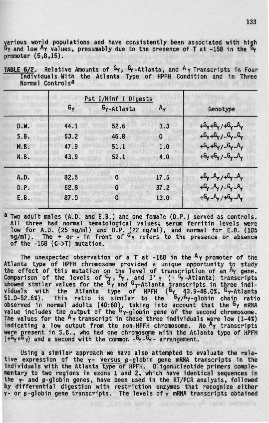
133
various world populations and have consistently been associated with highGy and low Ay values, presumably due to the presence of T at -158 in the °rpromoter (5,8,15).
TABLE 6/2. Relative Amounts of Gy, Gy-Atlanta, and Ay Transcripts in Fouri lIndividuals With
Normal
D.W.
S.B.
M.B.
N.B.
A.D.
D.P.
E.B.
Controls*
Gy
44.1
53.2
47.9
43.9
82.5
62.8
87.0
the Atlanta Type of
Pst I/Hinf I Digests
Gy-Atlanta
52.6
46.8
5 1 . 1
52.1
0
0
0
HPFH Condition and in Three
Ay
3.3
0
1.0
4.0
17.537.213.0
Genotype
+Gy +Gy /+Gy -Ay
+Gy+Gy/_GL_Gy
+Gy +Gy /-Gy _Ay
+Gy+GL/_Gy_Ay
+Gy-Ay/+GyA+6y_Ay/_Gy_Ay
* Two adult males (A.D. and E.B.) and one female (D.P.) served as controls.All three had normal hematological values; serum ferritin levels werelow for A.D. (25 ng/ml ) and D.P. (22 ng/ml ), and normal for E.B. (105ng/ml). The + or - in front of Gy refers toof the -158 (C->T) mutation.
the presence or absence
The unexpected observation of a T at -158 in the Ay promoter of theAtlanta type of HPFH chromosome provided a unique opportunity to studythe effect of this mutation on the level of transcription of an Ay gene.Comparison of the levels of G*, Ay, and 3' y (= fy-Atlanta) transcriptsshowed similar values for the "y and Gy-Atlanta transcripts in three indi-viduals with the Atlanta type of HPFH (Gy 43.9-48.OX, Gy-Atlanta51.0-52.6%). This ratio is similar to the Gy/Ay_giobin chain ratioobserved in normal adults (40:60), taking into account that the Gy mRNAvalue includes the output of the Gy_giobin gene of the second chromosome.The values for the Ay transcript in these three individuals were low (1-4%)indicating a low output from the non-HPFH chromosome. No Ay transcriptswere present in S.B., who had one chromosome with the Atlanta type of HPFH(+Gy+Gy) and a second with the common -Gy-Gy_ arrangement.
Using a similar approach we have also attempted to evaluate the rela-tive expression of the y- versus e-globin gene mRNA transcripts in theindividuals with the Atlanta type of HPFH. Oligonucleotide primers comple-mentary to two regions in exons 1 and 2, which have identical sequences inthe y- and B-globin genes, have been used in the RT/PCR analysis, followedby differential digestion with restriction enzymes that recognize eithery- or B-globin gene transcripts. The levels of y mRNA transcripts obtained
134
by this approach were significantly higher than expected from the levelsof Hb F observed in the individuals with the Atlanta type of HPFH, as wellas in several normal controls, suggesting the preferential amplificationof the y at the expense of the B mRNA transcripts. The analysis of serialdilution of cloned y and B RT/PCR fragments confirmed this observation, andwe are currently investigating this phenomenon; details of these analyseswill be published separately.
The quantitative RT/PCR procedure described here has been shown to beuseful for the determination of the relative amounts of individual y mRNAtranscripts, even in individuals with low Hb F levels and reticulocytecounts of less than IX. The &y, Ay, and Gy-Atlanta transcripts are co-amplified in a single competitive PCR, followed by endonuclease digestionwith restriction enzymes that recognize sequences specific for the indi-vidual transcripts. This approach has been used for the quantificationof GM-CSF mRNA by comparison with serial dilutions of external competitorDNA containing a unique restriction enzyme site (23). We have modifiedthis approach by competing between different mRNA molecules present withinthe same cell. No preferential amplification can occur because the tem-plate sizes and the target sequences for the oligonucleotide primers areIdentical in the different transcripts. Moreover, high sequence homologybetween the sequences of individual transcripts exclude the possibilityof preferential amplification of particular templates. A further develop-ment is the absence of -Gy-\-, -Gy-Gy-Atlanta, and -\-Gy-Atlanta hetero-duplexes which will remain uncleaved by the restriction enzymes and willnot affect the ratios of the different transcripts (23, and unpublishedobservations). The high sequence homology between the coding regions ofthe different globin genes allows further use of this procedure in studiespertaining to their expression. However, a possible preferential amplifi-cation of transcripts that have a high degree of sequence differences as,for instance, observed for the y and B mRNA transcripts, should be consid-ered in evaluating the results of these experiments.
Acknowledgements. The authors are indebted to Mr. E.L.D. Walker, III,for his help in obtaining the blood samples. This study was supported bythe United States Public Health Services research grants HLB-05168 and HLB-41544.
REFERENCES
1. Huisman THJ, Fei YJ, Kutlar F: Variations in the Gy and Ayratios in fetal hemoglobin in newborn babies. Hemoglobin 12:699, 1988
2. Kutlar A, Kutlar F, Gu L-G, Mayson SM, Huisman THJ: Fetal hemo-globin in normal adults and B-thalassemia hétérozygotes. Hum Genet 85:106,1990
3. Altay C, Huisman TJH, Schroeder WA: Another form of the heredi-tary persistence of fetal hemoglobin (the Atlanta type)? Hemoglobin 1:125,1977
4. Huisman THJ, Chen SS, Nakatsuji T, Kutlar F: A second familywith the Atlanta type of HPFH. Hemoglobin 9:393, 1985
5. Gilman JG, Huisman THJ: DNA sequence variation associated withelevated fetal Gy production. Blood 66:783, 1985

135
6. Thein SL, Wainscoat JS, Sampietro M, Old JM, Capellini D,Fiorelli G, Model 1 B, Weatherall DJ: Association of thalassaemia inter-media with a beta-globin gene haplotype. Br J Haematol 65:367, 1987
7. Diaz-Chico JC, Yang KG, Stoming TA, Efremov GD, Kutlar A, KutlarF, Aksoy M, Altay Ç, Gurgey A, Kilinç Y, Huisman THJ: Mild and severeB-thalassemia among homozygotes from Turkey: Identification of the typesby hybridization of amplified DNA with synthetic probes. Blood 71:248,1988.
8. Dner C, Dimovski AJ, Olivieri NF, Schiliro G, Codrington JF,Fattoum S, Adekile AD, Oner R, Yuregir GT, Altay Ç, Gurgey A, Gupta RB,Jogessar VB, Kitundu MN, Loukopoulos D. Tamagnini GP, Ribeiro MLS, KutlarF, Gu L-H, Lanclos KD, Huisman THJ: B^ Haplotypes in various world popu-lations. Hum Genet 89:99, 1992
9. Efremov GD, Gjorgovski I, Stojanovski N, Diaz-Chico JC, Harano T,Kutlar F, Huisman THJ: One haplotype is associated with the Swiss type ofhereditary persistence of fetal hemoglobin in the Yugoslavian population.Hum Genet 77:132, 1987
10. Sampietro M, Thein SL, Contreras M., Pazmany L: Variation ofHb F and F-cell number with the &y Xmn I (C->T) polymorphism in normalindividuals. Blood 79:832, 1992
11. Bisse E, Wieland H: High-performance liquid chromatographicseparation of human haemoglobins - Simultaneous quantitation of foetaland glycated haemoglobins. J Chromatogr 434:95, 1988
12. Shelton JB, Shelton JR, Schroeder WA: High performance liquidchromatographic separation of globin genes on a large-pore C4 column.J Liq Chromatogr 7:1969, 1984
13. Kutlar F, Kutlar A, Huisman THJ: Separation of normal and abnor-mal hemoglobin chains by reversed-phase high-performance liquid chromatog-raphy. J Chromatogr 357:147, 1986
14. Poncz M, Solowiejczyk D, Harpel B, Mory Y, Schwartz E, SurreyS: Construction of human gene libraries from small amounts of peripheralblood: Analysis of e-1ike globin genes. Hemoglobin 6:27, 1982
15. Lanclos KD, Oner C, Dimovski AJ, Gu Y-C, Huisman THJ: Sequencevariations in the 5' flanking and the IVS-II regions of the G y and A^_globin genes of 0$ chromosomes with five different haplotypes. Blood 77:2488, 1991
16. Baysal E, Huisman THJ: PCR-Based methodology for the detectionof common o-thal-2 determinants. Abstract, 5th International Conferenceon Thalassaemias and Haemoglobinopathies, Nicosia, Cyprus, April 1993
17. Chomczynski P, Sacchi N: Single-step method of RNA isolation byacid guanidiniun thiocyanate-phenol-chloroform extraction. Analyt Biochem162:156, 1987
18. Sambrook J, Fritsch EF, Maniatis T: Molecular Cloning: A Labo-ratory Manual, 2nd edition. Cold Spring Harbor, NY: Cold Spring HarborLaboratory, p 568, 1989
19. Betke K, Marti HR, Schlicht I: Estimation of small percentagesof foetal haemoglobin. Nature 184:1877, 1959
20. Powers PA, Altay C, Huisman THJ, Smithies 0: Two novel arrange-ments of the human fetal globin genes: Gy-Gy and *y-Ay. Nucleic AcidsRes 12:7023, 1984
21. Powers PA, Smithies 0: Short gene conversions in the human fetalglobin gene region: A by-product of chromosome pairing during meiosis?Genetics 112:343, 1986

136
22. Stoeckert CJ Jr, Collins FS, Weissman SM: Human fetal globin DNAsequences suggest novel conversion event. Nucleic Acids Res 12:4469, 1984
23. Gilliland G, Perrin S, Blanchard K, Bunn HF: Analysis of cyto-k1ne mRNA and DNA: Detection and quantitation by competitive polymerasechain reaction. Proc Natl Acad Sci USA 87:2725, 1990
CHAPTER 7
D I S C U S S I O N

139
CHAPTER 7
DISCUSSION
Characterization of the molecular defects leading to g-thal in a givenpopulation is an important prerequisite for the implementation of a suc-cessful prenatal diagnosis program (1). Identification of the e-thal muta-tions and the factors that can influence the clinical phenotype can alsoprovide useful information needed for a more accurate prognosis, a moreadequate patient management, and appropriate usage of new therapeuticmodalities such as bone marrow transplantation, pharmacological manipula-tion of Hb F expression or gene therapy.
Molecular characterization of g-thal alleles was therefore undertakento identify the mutations leading to 8-thal in the countries of the formerYugoslavia and Bulgaria. The frequencies of the various mutations wereestablished which facilitated the identification of the B-thal defect 1nother g-thal patients. The development of a new nonradioactive PCR-basedprocedure allowed rapid and safe detection of the common B-thal mutations(2).
The study was extended to B-thal homozygotes from Turkey and compoundhétérozygotes for B-thal and Hb Lepore from the former Yugoslavia, whichallowed correlation of the different B-thal mutations with the clinicalseverity of the disease. Additional factors that can ameliorate the clin-ical phenotype were investigated by studying 29 Mediterranean B-thal inter-media patients, all homozygous for the same mutation. The factors investi-gated included sex, age, the presence of a-thal, and the Hb F levels.
The study of the factors that influence the levels of Hb F was under-taken through sequence analysis of regulatory regions within the e-globingene cluster in four B-thal patients. A number of sequence variationswere observed which appeared to be unrelated to Hb F expression but werecharacteristic for certain chromosome backgrounds, previously defined byhaplotype analysis. The only exception was the -158 (C->T) substitutionin the promoter of the Gy gene which was consistently associated withincreased Hb F levels, mainly of the Gy type.
The role of the -158 (C->T) mutation was further studied through themolecular characterization of the Atlanta type of HPFH. This conditionwas reevaluated because of its association with a chromosome that containedthe -158 (C->T) mutation in the promoters of both y genes, previously char-acterized as Gy genes. A new quantitative RT/PCR procedure was developedwhich allowed comparison of the levels of different Y transcripts in thiscondition, and demonstrated a direct effect of the -158 (C->T) mutationon the transcriptional activity of the gene with which it is associated.
Molecular Characterization of B-Thai Mutations in the Former Yugo-slavia and Bulgaria. The incidence of B-thal trait in the former Yugo-slavia has been estimated at about 1% (3) and is slightly higher in neigh-boring Bulgaria (around 2%) (4). A broad spectrum of B-thal mutations
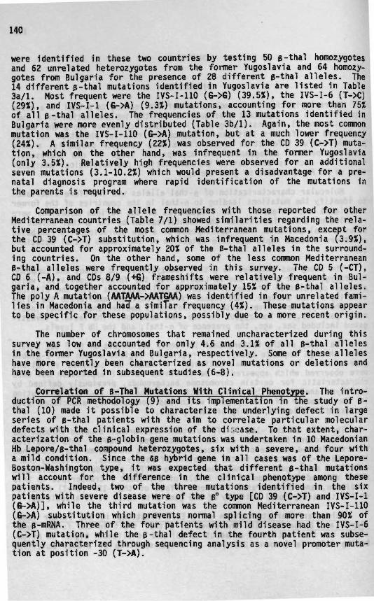
140
were identified in these two countries by testing 50 B-thal homozygotesand 62 unrelated hétérozygotes from the former Yugoslavia and 64 homozy-gotes from Bulgaria for the presence of 28 different B-thal alleles. The14 different B-thal mutations identified in Yugoslavia are listed in Table3a/l. Most frequent were the IVS-I-110 (G->G) (39.5%), the IVS-I-6 (T->C)(29%), and IVS-I-1 (G->A) (9.3%) mutations, accounting for more than 75%of all B-thal alleles. The frequencies of the 13 mutations identified inBulgaria were more evenly distributed (Table 3b/l). Again, the most commonmutation was the IVS-I-110 (G->A) mutation, but at a much lower frequency(24%). A similar frequency (22%) was observed for the CD 39 (C->T) muta-tion, which on the other hand, was infrequent in the former Yugoslavia(only 3.5%). Relatively high frequencies were observed for an additionalseven mutations (3.1-10.2%) which would present a disadvantage for a pre-natal diagnosis program where rapid identification of the mutations inthe parents is required.
Comparison of the allele frequencies with those reported for otherMediterranean countries (Table 7/1) showed similarities regarding the rela-tive percentages of the most common Mediterranean mutations, except forthe CD 39 (C->T) substitution, which was infrequent in Macedonia (3.9%),but accounted for approximately 20% of the B-thal alleles in the surround-ing countries. On the other hand, some of the less common MediterraneanB-thal alleles were frequently observed in this survey. The CD 5 (-CT),CD 6 (-A), and CDs 8/9 (+G) frameshifts were relatively frequent in Bul-garia, and together accounted for approximately 15% of the B-thal alleles.The poly A mutation (AATAAA->AATGAA) was identified in four unrelated fami-lies in Macedonia and had a similar frequency (4%). These mutations appearto be specific for these populations, possibly due to a more recent origin.
The number of chromosomes that remained uncharacterized during thissurvey was low and accounted for only 4.6 and 3.1% of all B-thal alleles1n the former Yugoslavia and Bulgaria, respectively. Some of these alleleshave more recently been characterized as novel mutations or deletions andhave been reported in subsequent studies (6-8).
Correlation of B-Thal Mutations With Clinical Phenotype. The intro-duction of PCR methodology (9) and its implementation in the study of B-thal (10) made it possible to characterize the underlying defect in largeseries of B-thal patients with the aim to correlate particular moleculardefects with the clinical expression of the di.jase. To that extent, char-acterization of the B-globin gene mutations was undertaken in 10 MacedonianHb Lepore/B-thal compound hétérozygotes, six with a severe, and four witha mild condition. Since the SB hybrid gene in all cases was of the Lepore-Boston-Washington type, 1t was expected that different B-thal mutationswill account for the difference in the clinical phenotype among thesepatients. Indeed, two of the three mutations identified in the sixpatients with severe disease were of the B° type [CD 39 (C->T) and IVS-I-1(G->A)], while the third mutation was the common Mediterranean IVS-I-110(G->A) substitution which prevents normal splicing of more than 90% ofthe B-mRNA. Three of the four patients with mild disease had the IVS-I-6(C->T) mutation, while the B-thal defect in the fourth patient was subse-quently characterized through sequencing analysis as a novel promoter muta-tion at position -30 (T->A).

TABLE 7/1. Frequency Distribution of 11 Common 0-Thal All el es in 17 Mediterranean Countries* (dataïnT)
Country No. of -28 CD 6 CD 8 CD 15 CD 39 CD 44 1-1 1-6 1-110 II-l 11-745Chroms. A->C -A -AA G->A C->T -C G->A T->C S->A 6~>A C->6 Others
Albania
Algeria
Azerbaijan
Bulgaria
Cyprus
Czech/Slovakia
Egypt
Greece
Hungary
Israel
Italy
(Sardinia)
(Sicily)
Lebanon
Portugal
Spain
Tunisia
Turkey
Former Yugoslavia
58
129
99
483
937
93
50
348
17
46
914
494
399
50
334
58
48
859
469
Ob
0
0
0
0
0.4
0
0
0
15.0
0
0
0
0
0
0
0
0.40
0
17.9
0
6.1
0.1
0
4.2
2.9
0
0
1.2
2.2
2.2
0
0
5.0
2.3
0.4
1.6
0
0
21.2
5.5
0.2
0
21.2
0.6
0
0
0
0
0
6.0
0
1.7
2.3
5.8
0.8
0
0
0
0
0
0
0
0
0
0
0
0
0
0
15.2
0
0
0
0
22.4
26.0
2.0
21.9
1.9
2.1
2.1
17.0
29.4
12.0
40.1
95.4
37.6
4.0
40.1
64.0
40.8
3.2
3.9
3.40
3.1
0
0
0
3.10
0
19.5
0.7
0
0
0
0
0
9.1
0
0
5.214.5
2.0
5.3
6.3
45.2
10.4
13.2
29.4
4.0
10.2
0
5.5
0
18.7
3.5
0
3.7
11.5
17.2
3.57.19.2
6.7
0
18.8
7.2
5.9
2.5
9.9
0
23.9
6.0
20.3
15.5
0
17.2
19.2
43.2
25.4
20.2
27.4
77.0
5.9
27.1
42.6
0
18.0
23.0
0.4
24.1
62.0
15.2
8.5
20.5
41.2
45.4
1.7021.2
1.7
0
14.1
0
2.011.8
13.5
3.90
0
4.00
0
0
8.0
3.9
003.16.6
5.8
4.3
8.3
6.9
5.9
0.85.0
0.43.5
4.0
0
0
0
1.9
1.6
6.912.7
20.1
16.3
1.4
28.4
4.87.617.6
14.7
6.0
1.6
3.2
14.0
5.7
1.8
25.0
18.2
12.1
Adapted from Ref. 5.Zero = not observed or not reported.
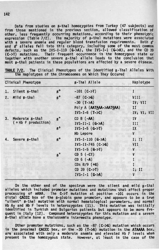
142
Data from studies on B-thal homozygotes from Turkey (47 subjects) andfrom those mentioned in the previous section, allowed classification ofother, less frequently occurring mutations, according to their phenotypicexpression (Table 7/2). The majority of p-thal mutations were associatedwith a severe anemia and regular blood transfusion requirements. Both p°and B* alleles fell into this category, including some of the most commondefects, such as the IVS-I-11O (G->A), the IVS-I-1 (G->A), and the CD 39(C->T) mutations. Their frequent occurrence in the homozygous state ortogether with another severe B-thal allele leads to the conclusion thatmost B-thal patients in these populations are affected by a severe disease.
TABLE 7/2. The Clinical Phenotypes of the Identified B-Thal Alleles Withthe Haplotypes of the Chromosomes on Which They Occured
Clinical Phenotype B-Thal Allele Haplotype
1.
2.
3.
4.
Silent B-thal
Mild B-thal
Moderate B-thal( tHb F production)
Severe B-thal
B+
B+
B°
8+
0+
-101 (C->T)
-87 (C->G)
-30 (T->A)Poly A (AATAAA->AATGAA)IVS-I-6 (T->C)
CD 8 (-AA)IVS-II-1 (G->A)IVS-I-5 (G->T)Hb Lepore
IVS-I-110 (G->A)IVS-II-745 (C->G)IVS-I-5 (G->T)CD 5 (-CT)CD 6 (-A)CDs 8/9 (+G)CD 39 (C->T)IVS-I-1 (G->A)
IVIIIIV; VIIIIIV; VI; VII
IVIIIIXV
i; uVIIVVIII; IIV
On the other end of the spectrum were the silent and mild Balleles which included promoter mutations and mutations that affect properprocessing of mRNA. The C->T mutation at position -101 occurs in thedistal CACCC box of the B-globin gene promoter, and appears to be a true"silent" B-thal mutation with normal hematological parameters, and normalHb A2 and Hb F levels in hétérozygotes (11). This mutation was initiallyidentified in Turkish and Bulgarian patients but appears to be more fre-quent in Italy (12). Compound hétérozygotes for this mutation and a severeB-thal allele have a thalassemia intermedia phenotype.
Other promoter mutations, such as the -87 (C->G) mutation which occursin the proximal CACCC box, or the -30 (T->A) mutation in the ATAAAA box,are associated with only a moderate anemia and elevated Hb F levels whenpresent in the homozygous state. However, at least in the case of the

143
ATAAAA box mutation, the increased Hb F level might be a consequence of aparticular chromosomal background that contains the -158 (C->T) mutationin the promoter of the Gy gene. In combination with severe B-thal allelesthese promoter mutations will produce either B-thal intermedia or B-thalmajor. The same is true for the IVS-I-6 (T->C) mutation which is the mostfrequently occurring mild B-thal allele in the Mediterranean populations.Homozygotes for this mutation are mildly affected and do not usuallyrequire blood transfusions.
Two B-thal mutations [CD 8 (-AA) and IVS-II-1 (G->A)] were also asso-ciated with only a moderate anemia, despite the absence of any 6 chain pro-duction. These mutations occurred on particular chromosomal backgrounds,defined by haplotypes III and IV, which appeared to be responsible forthe markedly increased Hb F production seen in these patients. Anotherinteresting example of the effect of chromosomal background on the clinicalphenotype was the IVS-I-5 (G->T) mutation. This mutation was present inthe homozygous form, both chromosomes having haplotype IX, in a Turkishpatient with a mild disease, no transfusion requirements, and 95% Hb F.Mutations at the IVS-I-5 position have been found in various world popula-tions and associated with different haplotypes produce diverse clinicalphenotypes (1). The three mutations discussed above occurred on threeMediterranean haplotypes (III, IV, and IX) which share the same patternof the polymorphic restriction enzyme sites in the 5' part of the B-globingene cluster. A significant characteristic of these haplotypes is thepresence of an Xmn I site created by the -158 (C->T) substitution in the & Ypromoter.
In summary, these data provide a satisfactory explanation for thevariability in the severity of the disease in most of the studied B-thalpatients. Many of the B° mutations, and some of the B * mutations, areassociated with a severe disease, while the promoter mutations, and someof the mutations affecting mRNA processing, are found in patients witha milder condition. Interactions between these two types of mutationsresult in various clinical phenotypes, but in most instances a B-thalintermedia is observed. However, a role of additional factors in amelio-rating the course of the disease is obvious, and is most striking amongthe patients homozygous for B°-thal mutations with high Hb F levels.
Factors Affecting the Total Hb and Hb F Levels in B-Thal Patients.The influence of additional factors on the clinical phenotype of B-thalis difficult to assess because most B-thal patients require regular bloodtransfusions, and are therefore uninformative regarding their steady stateHb levels. The heterogeneity of the molecular defects leading to B-thalfurther complicates such studies since particular mutations affect theB-globin gene output to a different extent.
The study of the 29 homozygotes for the IVS-I-6 (T->C) mutation over-came these problems. All patients had the same molecular defect and noneof them had received a blood transfusion for at least six months prior tothe study. In most patients, the Hb levels were between 6.5 and 8.0 g/dl,but higher and lower values were also observed. The Hb levels were notaffected by the age or sex of the patients, but correlated well with theHb F levels, except in six patients with low Hb F and high total Hb. Aconcomitant o-thal-2 determinant was identified in two of these patients.
144
The co-inheritance of o-thal or increased y chain production canreduce the overall a/non-a chain imbalance and decrease the peripheralhemolysis of the RBCs (13). Both a-thal and increased Y chain productionmight have the same effect on the ineffective erythropoiesis in the bonemarrow of B-thal patients, recently shown to be due to accelerated pro-grammed cell death (apoptosis) of erythroid progenitors containing a-globinchain deposits (14). The absence of increased Hb F levels and of a commono-thal determinant in some of the patients with high total Hb could implythe presence of as yet uncharacterized nondeletional a-thal alleles, orthe existence of other factors that can increase the Hb A levels in homozy-gotes for the IVS-I-6 (T->C) mutation.
Further investigation focused on the factors that could affect theHb F levels. Recent studies have suggested the existence of an X-linkeddeterminant which regulates Hb F and F-cell production (15,16). However,no association between sex and Hb F levels was observed in the IVS-I-6homozygotes. On the other hand, the Hb F levels correlated inversely withage, even though with only a borderline significance. A more significanteffect of age on Hb F levels has been observed in SS patients (17).
Sequence Variations in Regulatory Regions of the B-61obin GeneCluster. To investigate the influence of different chromosomal backgroundson Hb F expression, haplotype analysis was performed on all IVS-I-6 homo-zygotes. Three haplotypes were identified (IV, VI, and VII). HaplotypeIV was found on both chromosomes in one patient with high Hb F levels,while the other two haplotypes were associated with both high and low Hb Fproduction. Since the haplotypes are defined only by the presence orabsence of certain polymorphic restriction enzyme sites on a particularchromosome, they do not rule out the presence of variations in regulatorycis-acting sequences between chromosomes with identical haplotypes but{fTfferent Hb F expression. To further explore such a possibility, sequenceanalysis of regulatory regions within the B-globin gene cluster was under-taken in four patients, two with high Hb F levels (homozygotes for haplo-types IV or VI), and two with low Hb F levels (homozygotes for haplotypesVI or VII). Sequence analysis of the LCR showed identical 5' HS-4, HS-3,and HS-2 elements in all four patients, except for the presence of haplo-type-specific AT repeats in HS-2 (Chapter 5, Fig. 5/2). Sequence varia-tions between the three different haplotypes were common in the 5' flankingand IVS-II regions of both y-globin genes, while the AT repeat motif inthe 5' flanking region of the B-globin gene was identical in all three.
Some of these sequence variations occurred in possible motifs forbinding of regulatory proteins. The AT repeats in the LCR 5' HS-2 and theTG repeats in the Gy- and A^-ivs-II regions have been Implicated throughgenetic studies in modulating y-globin gene expression (18,19). Moreover,binding of a nuclear protein factor has been reported for the TG motif inthe Ay-ivS-II (19). The two polymorphic motifs that have been associatedwith higher Hb F levels [(AT)gni2(AT)io in LCR 5' HS-2 and (TG)i3 in VIVS-II)] were found only on the chromosome with haplotype IV. A possibleeffect of the negative regulatory factor BP1 (20) on y-globin gene expres-sion appeared unlikely, since no variations were present in the bindingsite for this protein in the 5' flanking region of the B-globin gene [(AT)7(T)7 was found in all patients]. An interesting observation was the près-
145
ence of duplicated CACCC sequences in the Gy-IVS-II on haplotype VII andin the Ay-IVS-II on haplotype IV. Whether these sequence variations haveany effect on y-globin gene expression, as shown for the same sequences inthe y-globin gene promoters (21) remains to be determined through nuclearprotein binding and transfection experiments.
The sequence variations described above were all confined to the spe-cific haplotypes and no nt differences were observed between the two homo-zygotes for haplotype VI with various Hb F levels. The existence of otherunidentified cni-acting sequence variations is unlikely because differencesin Hb F levels were also observed between the siblings in two families.These findings, therefore, suggest a major role for unknown genetic loci,separated from the B-globin gene cluster, in controlling Hb F productionin B-thal.
The only exception appears to be the chromosome with haplotype IV»which was consistently found to be associated with increased Hb F levels.The chromosome with this haplotypes shares identical RFLPs in the 5' partof the B-globin gene cluster with two other Mediterranean B-thal chromo-somes associated with elevated Hb F production (haplotypes III and IX).Furthermore, sequence analysis of this chromosome found it to be identicalto the B$ chromosome with haplotype #3 (Senegal), which has also been asso-ciated with increased Hb F production under conditions of erythropoieticstress (22-24). These two chromosomes differ only in the 3' subhaplotype,suggesting that £is-acting factors are likely to exist in the 5' part ofthe B-globin gene cluster.
The -158 (C->T Promoter Mutation. The most significant feature of theMediterranean B-thal chromosomes with haplotypes III, IV, and IX, and theI$S chromosome with haplotype #3 appears to be the -158 (C->T) substitutionin the promoter of the Gy gene (25). Normal individuals with this mutation(either in the homozygous or heterozygous state) have normal Hb F levelswith elevated Gy values (60-98%); however, in a few families slightlyincreased Hb F levels (1-3%) have been observed. It has been suggestedthat this substitution is a promoter mutation which increases the expres-sion of the Gy gene, but additional factors can also accentuate the effectof this mutation, and result in increased Hb F levels (26). One factorappears to be the chronic erythropoietic stress present in B-thal or SSdisease.
To further investigate the effect of this mutation on y-globin geneexpression, a rare HPFH condition known as the Atlanta type of HPFH wasreevaluated. Individuals with this condition have variable but usuallymoderately increased Hb F levels (2-12X), mainly of the Gy type (>90X).Previous gene mapping studies had identified a "y-Gy arrangement on thisHPFH chromosome with the -158 (C->T) substitution in the promoters of bothY gene (25,27).
Sequence analysis of the y-globin genes from the HPFH chromosome wasundertaken to investigate the possible presence of additional sequencevariations that could account for the elevated Hb F levels in this condi-tion. The 5' Gy gene was found to be completely identical to the Gy gene

146
found on the previously discussed chromosome with haplotype #3 (Senegal),while the 3' Y gene contained only two nt substitutions with respect tothe Ay gene from the same chromosome. These substitutions were the onesdetected by the restriction enzymes in the gene mapping analysis, i.e. the-158 (C->T) and the CD 136 (GÇA->GGA) mutations. The CD 136 mutationintroduced the Ala->Gly amino acid substitution which converted this geneinto a S y gene. The absence of sequence variations, other than the -158(C->T) mutation, in the investigated regulatory regions of the HPFH chromo-some (the LCR 5' HS-2, the 5" flanking and IVS-II regions of both Y genes,and the 5' flanking region of the p-globin gene) provided genetic evidencethat this promoter mutation is responsible for the increased Hb F levelsin the Atlanta type of HPFH.
The individual contribution of the two Y-globin genes from the HPFHchromosome on the Hb F levels was assessed by a new, competitive RT/PCRprocedure. This procedure allowed quantisation of the relative amounts oftranscripts expressed by the Gy, \, and HPFH 3'Y genes. Similar valuesfor the «Y and 3'Y transcripts were obtained in the four investigated hét-érozygotes for the Atlanta type of HPFH. This was expected since both Ygenes on the HPFH chromosome contained the -158 (C->T) mutation. The pos-sibility of discriminating between the Ay and 3'Y transcripts also allowedcomparison of the outputs of the HPFH 3' Y gene with different Ay genes.In this respect especially interesting was the comparison with the Ay genefrom the chromosome with haplotype #3 (Senegal) which differed from theHPFH 3' Y gene only 1n the -158 and CD 136 positions. The data showed a15-fold higher level of transcripts from the HPFH 3'y gene, demonstratingthat the -158 (C->T) mutation can also increase the expression of an \gene.
To investigate whether the observed differences in Hb F levels amongthe Atlanta type of HPFH hétérozygotes were due to differences in y geneexpression, the relative amounts of Y- and B-mRNA were determined withthe same RT/PCR method. The levels of Y transcripts varied from 9-38%,and were highest in the individual with a moderate iron deficiency anemia(Chapter 6, Table 6/2). These data further support the existence of addi-tional genetic factors, separated from the B-globin gene cluster, whichcan affect the expression of the Y-globin genes. It is tempting to specu-late that such factors could affect the silencing of the y-globin genes;when they are present the effect of the -158 (C->T) mutation would be morepronounced, resulting in higher Hb F levels. Incomplete silencing of theY-globin genes could also occur in conditions with erythropoietic stress,such as e-thal, SS disease, or iron deficiency anemia which would accen-tuate the effect of the -158 (C->T) mutation on the expression of the asso-ciated Y-globin gene.
Quantitative RT/PCR Analysis of 61obin Gene Transcripts. The newlydeveloped competitive RT/PCR assay allowed the accurate quantitation ofthe relative amounts of different globin gene transcripts even in individ-uals with Hb F levels and reticulocyte counts of less than 1%. The Y and0, or the Gy, A ^ and 3' y transcripts were co-amplified in a single compet-itive PCR, followed by endonuclease digestion with restriction enzymesthat recognize sequences specific for the individual transcripts. Since
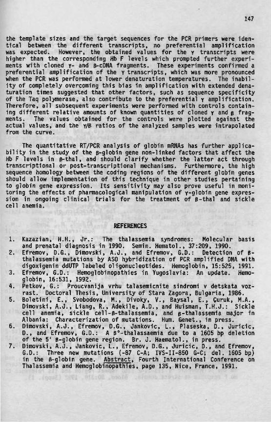
147
the template sizes and the target sequences for the PCR primers were iden-tical between the different transcripts, no preferential amplificationwas expected. However, the obtained values for the y transcripts werehigher than the corresponding Hb F levels which prompted further experi-ments with cloned y- and B-cDNA fragments. These experiments confirmed apreferential amplification of the y transcripts, which was more pronouncedwhen the PCR was performed at lower denaturation temperatures. The inabil-ity of completely overcoming this bias in amplification with extended dena-turation times suggested that other factors, such as sequence specificityof the Taq polymerase, also contribute to the preferential y amplification.Therefore, all subsequent experiments were performed with controls contain-ing different relative amounts of known quantities of cloned y and B frag-ments. The values obtained for the controls were plotted against theactual values, and the y/B ratios of the analyzed samples were intrapolatedfrom the curve.
The quantitative RT/PCR analysis of globin mRNAs has further applica-bility in the study of the B-globin gene non-linked factors that affect theHb F levels in B-thal, and should clarify whether the latter act throughtranscriptional or post-transcriptional mechanisms. Furthermore, the highsequence homology between the coding regions of the different globin genesshould allow implementation of this technique in other studies pertainingto globin gene expression. Its sensitivity may also prove useful in moni-toring the effects of pharmacological manipulation of y-globin gene expres-sion in ongoing clinical trials for the treatment of B-thal and sicklecell anemia.
REFERENCES
1. Kazazian, H.H., Jr.: The thalassemia syndromes: Molecular basisand prenatal diagnosis in 1990. Semin. Hematol., 37:209, 1990.
2. Efremov, D.G., Dimovski, A.J., and Efremov, G.D.: Detection of B-thalassemia mutations by ASO hybridization of PCR amplified DNA withdigoxigenin ddUTP labeled oligonucleotides. Hemoglobin, 15:525, 1991.
3. Efremov, G.D.: Hemoglobinopathies in Yugoslavia: An update. Hemo-globin, 16:531, 1992.
4. Petkov, G.: Proucvanija vrhu talasemicnite sindromi v detskata voz-rast. Doctoral Thesis, University of Stara Zagora, Bulgaria, 1986.
5. Boletini, E., Svobodova, M., Divoky, V., Baysal, E., Çuruk, M.A.,Dimovski, A.J., Liang, R., Adekile, A.D., and Huisman, T.H.J.: Sicklecell anemia, sickle cell-B-thalassemia, and 0-thalassemia major inAlbania: Characterization of mutations. Hum. Genet., in press.
6. Dimovski, A.J., Efremov, D.G., Jankovic, L., Plaseska, D., Juricic,D., and Efremov, G.D.: A B°-thalassaemia due to a 1605 bp deletionof the 5' B-globin gene region. Br. J. Haematol., in press.
7. Dimovski, A.J., Jankovic, L., Efremov, D.G., Juricic, D., and Efremov,G.D.: Three new mutations (-87 C-A; IVS-II-850 G-C; del. 1605 bp)in the B-globin gene. Abstract, Fourth International Conference onThalassemia and Hemoglobinopathies, page 135, Nice, France, 1991.

Oner, R., Agarwal, S., Dimovski, A.J., Efremov, G.D., Petkov, G.H.,Altay, Ç., Gurgey, A., and Huisman, T.H.J.: The G->A mutation atposition +22 3' to the Cap site of the B-globin gene as a possiblecause for a B-thalassemia. Hemoglobin, 15:67, 1991.
9. Saiki, R.K., Scharf, S., Faloona, F., Mullis, K.B., Horn, G.T.,Erlich, H.A., and Arnheim, N.: Enzymatic amplification of B-globingenomic sequences and restriction site analysis for diagnosis ofsickle cell anemia. Science, 230:1350, 1985.
10. Diaz-Chico, J.C., Yang, K.G., Yang, K.Y., Efremov, D.G., Stoming,T.A., and Huisman, T.H.J.: The detection of B-globin gene mutationsin B-thalassemia using oligonucleotide probes and amplified DNA.Biochim. Biophys. Acta, 949:43, 1988.
11. Gonzalez-Redondo, J.M., Stoming, T.A., Kutlar, A., Kutlar, F., Lan-clos, K.D., Howard, E.F., Fei, Y.J., Aksoy, M., Altay, Ç., Gurgey,A., Ba$ak, A.N., Efremov, G.D., Petkov, G., and Huisman, T.H.J.: AC->T substitution at nt -101 in a conserved DNA sequence of the pro-moter region of the B-globin gene is associated with a "silent" B-thalassemia. Blood, 73:1705, 1989.
12. Ristaldi, M.S., Murru, S., Loudianos, G., Casula, L., Porcu, S.,Pigheddu, D., Fanni, B., Sciarratta, G.V., Agosti, S., Parodi, M.A.,Leon, D., Camaschella, C , Serra, A., Pirastu, M., and Cao, A.: TheC->T substitution in the distal CACCC box of the B-globin gene pro-moter is a common cause of silent B-thalassemia in the Italian popula-tion. Br. J. Haematol., 74:480, 1990.
13. Wainscoat, J.S., Kanavakis, E., Wood, W.G., Letsky, E.A., Huehns,E.R., Marsh, G.W., Higgs, D.R., Clegg, J.B., and Weatherall, D.J.:Thalassaemia intermedia in Cyprus: The interaction of a- and B-thal-assaemia. Br. J. Haematol., 53:411, 1983.
14. Yuan, J., Angelucci, E., Lucarelli, G., Aljurf, M., Snyder, L.M.,Kiefer, C.R., Ma, L., and Schrier, S.L.: Accelerated programmed celldeath (apoptosis) in erythroid precursors of patients with severeB-thalassemia (Cooley's anemia). Blood, 82:374, 1993.
15. Miyoshi, K., Kaneto, Y., Kawai, H., Ohchi, H., Niki, S., Hasegawa,K., Shirakami, A., and Yamano, T.: X-Linked dominant control of Fcells in normal adult life: Characterization of the Swiss type ofhereditary persistence of fetal hemoglobin regulated dominantly bygene(s) on X chromosome. Blood, 72:1854, 1988.
16. Dover, G.J., Smith, K.D., Chang, Y.C., Purvis, S., Mays, A., Meyers,D.A., Sheils, C , and Serjeant, G.: Fetal hemoglobin levels in sicklecell disease and normal individuals are partially controlled by anX-linked gene located at Xp22.2. Blood, 80:816, 1992.
17. Adekile, A.D. and Huisman, T.H.J.: Hb F in sickle cell anemia.Experientia, 49:1, 1993.
18. Oner, C , Dimovski, A.J., Altay, Ç., Gurgey, A., Gu, Y.C., and Huis-man, T.H.J.: Sequence variations in the 5' hypersensitive site-2of the locus control region of e^ chromosomes are associated withdifferent levels of fetal globin in Hemoglobin S homozygotes. Blood,79:813, 1992.
19. Ragusa, A., Lapoumeroulie, C , Lombardo, M., El ion, J., Labié, D., andKrishnamoorthy, R. : Differential binding of nuclear protein factorsto the polymorphic \ IVS-II-2 (TG) repeat region and its relevance toHb F response in stress anemia. Blood, 80:310, 1992 (Suppl. 1).

149
20. Berg, P.E., Williams, D.M., Qian, R-L., Cohen, R.B., Cao, S-X., M1t-telman, M., and Schechter, A.N.: A common protein binds to twosilencers 5' to the human B gene. Nucleic Acids Res., 17:8833, 1989.
21. Wood, W.G.: Increased Hb F in adult life. In The Haemoglobinopa-thies, edited by D.J. Weatherall and D.R. Higgs, Bailliere's ClinicalHaematology, Vol. 6, page 117, W.B. Saunders Company, London, 1993.
22. Labie, D., Pagnier, J., Lapoumeroulie, C , Rouabhi, F., Dunda-Belk-hodja, 0., Chardin, P., Beldjord, C , Wajcman, H., Fabry, M.E., andNagel, R.L.: Common haplotype dependency of high Gy-globin geneexpression and high Hb F levels 1n B-thalassemia and sickle cellanemia patients. Proc. Natl. Acad. Sci. USA, 82:2111, 1985.
23. Lanclos, K.D., Oner, C , Dimovski, A.J., Gu, Y-C, and Huisman,T.H.J.: Sequence variations in the 5' flanking and IVS-II regionsof the Gy-and \-globin genes of B$ chromosomes with five differenthaplotypes. Blood, 77:2488, 1991.
24. Oner, C , Dimovski, A.J., Olivieri, N.F., Schiliro, G., Codrington,J.F., Fattoum, S., Adekile, A.D., Oner, R., Yuregir, G.T., Altay, Ç.,Gurgey, A., Gupta, R.B., Jogessar, V.B., Kitundu, M.N., Loukopoulos,D., Tamagnini, G.P., Ribeiro, M.L.S., Kutlar, F., Gu, L-H., Lanclos,K.D., and Huisman, T.H.J.: B^ Haplotypes in various world popula-tions. Hum. Genet., 89:99, 1992.
25. Gilman, J.G. and Huisman, T.H.J.: DNA sequence variation associatedwith elevated <\ production. Blood, 66:783, 1985.
26. Efremov, G.D., Gjorgovski, I., Stojanovski, N., Diaz-Chico, J.C.,Harano, T., Kutlar, F., and Huisman, T.H.J.: One haplotype is associ-ated with the Swiss type of hereditary persistence of fetal hemoglobinin the Yugoslavian population. Hum. Genet., 77:132, 1987.
27. Huisman, T.H.J., Chen, S.S., Nakatsuji, T., and Kutlar, F.: A secondfamily with the Atlanta type of HPFH. Hemoglobin, 12:699, 1985.
CHAPTER 8
S U M M A R Y


153
CHAPTER 8
SUMMARY
The main objectives of the studies presented in this thesis are: 1)The characterization of the molecular defects leading to B-thal in thecountries of the former Yugoslavia and Bulgaria; 2) the correlation ofthese defects with different clinical phenotypes, and 3) a study of possi-ble ameliorating factors, with special emphasis on those that can influencethe Hb F levels in patients with B-thal.
A review of the literature is presented in Chapter 1, which includesa short historical perspective of some of the more important accomplish-ments in the study of the thalassemias. The ethnic distribution of thethalassemias is presented, with special emphasis on the frequency of B-thalin various Mediterranean countries. This chapter also provides a shortdescription of the basic pathophysiological phenomena in B-thal, togetherwith the more important clinical and hematological features of the disease.The review of the common molecular defects leading to B-thal is precededby a description of the structure and function of the globin genes, theirorganization, and developmental expression. The possible role of differentcjjs-acting elements in regulating the high level of expression of the B-globin gene in adult erythroid cells is discussed together with a briefdescription of the clinical features and molecular defects leading to theLepore Hbs. Regulation of the Y-globin gene expression by genetic factors,both linked and non-linked to the B-globin gene cluster, is presented inthe section dealing with the different HPFH syndromes. Special emphasisis given to the different y-globin gene arrangements, and to the most fre-quent sequence variation associated with particular B-thal chromosomes,the -158 (C->T) mutation. An updated list of known B-thalassemic mutationsis provided as an appendix to this chapter.
Relevant patient data and a description of some of the methodologyused are presented in Chapter 2.
Chapter 3 provides detailed data about the occurrence of various B -thal all el es in the populations of the former Yugoslavia and Bulgaria.The observed frequencies of the different mutations are compared to thoseof the neighboring countries, and the implications of this study in initi-ating a prenatal diagnosis program are discussed.
Characterization of the molecular defects in 50 B-thal homozygotesand 62 unrelated B-thal hétérozygotes from the former Yugoslavia is pres-ented in Chapter 3a. Fourteen different mutations were observed; of these,three [IVS-I-110 (G->A); IVS-I-6 (T->C); IVS-I-1 (G->A)] accounted forapproximately 75% of all B-thal alleles. A newly discovered mutation(AATAAA->AATGAA in the poly A site) appeared to be rather common as it wasobserved in four unrelated families with a frequency of 4.7Ï. Hb Leporewas found to be a frequent and clinically significant abnormality sinceits association with B-thal often resulted in a severe disease. The highfrequencies of the IVS-I-110 (S->A) and IVS-I-1 (6->A) mutations led tothe conclusion that most B-thal patients originating from the countriesof the former Yugoslavia will be severely affected by their diseases.

154
Chapter 3b presents data from the DNA analysis in 64 B-thal homozy-gotes from Bulgaria using ASO hybridization of PCR amplified DNA. Thirteendifferent B-thal alleles were identified; most frequent were the IVS-I-11O(6->A) (24.2%) and CD 39 (T->C) (21.9%) mutations. However, seven addi-tional mutations were observed which were present at frequencies of 3.9to 10.2%. The even distribution of nine different mutations is likelyto complicate the analyses for institutions involved in prenatal diagnosis.High frequencies of 4.7 to 5.5% were observed for four rare mutations.These frameshift mutations (at CDs 5, 6, 8, and 8/9), as well as the poly Amutation identified in Macedonia, appear to be specific for these popula-tions.
The correlation of particular clinical phenotypes with the differentB-thal mutations is described in Chapter 4. Chapter 4a presents the studyof 10 patients with the Hb Lepore/B-thal condition, originating from thecountries of the former Yugoslavia, who showed significant variations inthe severity of the disease. As the type of Hb Lepore was the same inall patients (Lepore-Boston-Washington) and an a-globin gene deficiencywas absent, it was concluded that the type of 6-thai determined the sever-ity of the disease. The latter was confirmed through characterizationof the B-thal alleles; six patients with severe disease had either a B°or a severe B+ mutation [CD 39 (C->T); IVS-I-1 (G->A); IVS-I-110 (G->A)],three patients with mild disease had the IVS-I-6 (T->C) mutation, whilethe B-thal determinant in one patient remained unknown. Further character-ization of the B-thal allele in this patient is presented in Chapter 4b.Sequence analysis of PCR amplified DNA revealed a T->A mutation withinthe ATA box at position -30 nt from the Cap site. The same mutation waspresent on both chromosomes in a young Turkish patient with thalassemiaintermedia. While the Hb F levels in the Turkish patient were low (13.IX),they were extremely high in the Hb Lepore/B-thal compound hétérozygote(80%). The mutation was found on two different haplotypes (IV in thepatient with high Hb F, and VII in the patient with low Hb F ) , suggestinga role for the chromosomal background in determining the level of Hb Fexpression.
The molecular characterization of B-thal alleles in 47 homozygotesfrom Turkey, presented in Chapter 4c, provided sufficient data to correlatesome less frequent mutations with particular phenotypes. Mild diseasewas observed in patients homozygous for the IVS-I-6 (T->C), CD 8 (-AA),-87 (C->G), and IVS-I-5 (G->T) mutations, or compound hétérozygotes forcombinations of these mutations. Moderate disease was present in patientswith one of these mutations and a severe mutation such as the IVS-I-110(G->A), IVS-I-1 (G->A), and CD 39 (C->T). Two B° mutations (IVS-II-1 andCD 8) were consistently associated with a mild phenotype and high totalHb and Hb F levels. These mutations were found on two different haplotypes(III and IV), both containing the C->T substitution at position -158 (Gy).
The effect of different chromosomal backgrounds was analyzed in detail1n 29 homozygotes for the IVS-I-6 (T->C) mutation, none of whom hadreceived a blood transfusion for at least six months prior to the study.The obtained data, described in Chapter 5, showed higher Hb levels inpatients with high Hb F of a concomitant o-thal-2 heterozygosity. TheIVS-I-6 (T->C) mutation was found in three different haplotypes; most of
155
the patients were homozygous for haplotype VI, but haplotypes IV and VIIwere also present. The only haplotype IV homozygote had high Hb F levels,while both high and low Hb F levels were observed among patients with hap-lotypes VI and VII. Sequence analysis of regulatory cis-acting elementswas undertaken in two patients with high Hb F (one homozygote each forhaplotypes VI and IV) and in two patients with low Hb F (one homozygoteeach for haplotype VI and VII). Haplotype specific differences wereobserved in the LCR 5' HS-2 and in the Gy and Ay 5' flanking and IVS-IIregions, while all chromosomes were identical in the 5' LCR HS-4, HS-3,and the 5' flanking region of the B-globin gene. The -158 (C->T) substi-tution appeared most likely to be responsible for the elevated Hb F levelsobserved in association with haplotype IV. The absence of nt differencesbetween the low and high Hb F-producing haplotype VI chromosomes suggesteda major role for unknown genetic factors, separated from the B-globin genecluster, in controlling the Hb F production in these B-thal patients.
The effect of the -158 (C->T) substitution was further evaluatedthrough the study of the Atlanta type of HPFH. Chapter 6 provides detailson the molecular analysis of this condition, previously characterized asa GyGy arrangement with the -158 (C->T) substitution in the promoters ofboth «y genes. Sequence analysis identified the -158 (C->T) mutation andthe CD 136 (C->G) substitution as the only differences between the 3'ygene from this arrangement, and the \ gene from a chromosome with haplo-type #3 (Senegal). The absence of any other nt substitutions providedgenetic evidence that the -158 (C->T) mutation is primarily responsiblefor the elevated Hb F levels associated with this condition. A quantita-tive RT/PCR procedure was developed to determine the effect of this muta-tion on y-globin gene transcription in four individuals with the Atlantatype of HPFH. High levels of y transcripts were observed in all individ-uals (9-38X), and were highest in one female with a moderate anemia. TheGy and the 3' y genes of the Atlanta type of HPFH chromosome expressed sim-ilar amounts of transcripts, which were over 10 times higher than thoseof the Ay gene from the normal chromosomes. These data demonstrated thatthe -158 (C->T) mutation directly affects the transcriptional rate of thegene to which it is associated.
The data presented in this dissertation provide a satisfactory expla-nation for the observed phenotypic variations in most of the studied B-thalpatients. Apart from the presence of a mild B-thal allele, increased Hb Fexpression is the most important factor which can contribute to a mildclinical phenotype. Genetic factors, both linked and non-linked to theB-globin gene cluster, can affect the levels of Hb F in B-thal patients.Clinically, the most significant cis-acting factor is the -158 (C->T) muta-tion which exerts a positive regulatory effect on the associated y-globingene. The trans-acting factors influencing the levels of Hb F in B-thalrequire further characterization. The competitive RT/PCR assay may proveuseful in investigating these factors and in monitoring the effects ofpharmacological manipulation of y-globin gene expression.
SAMENVATTING

158
Samenvattinq
Het onderzoek beschreven in dit proefschrift heeft de volgende doelstellingen: (1) Hetkarakteriseren van de moléculaire defecten welke aanleiding geven tot /?-thalassemie inhet voormalig Joegoslavië en in Bulgarije; (2) de correlatie van deze defecten met deverschillende klinische phenotypen; en (3) een onderzoek naar mogelijk factoren meteen positief effect op de ziekte, met spéciale aandacht voor factoren die de HbF ni-veaus beïnvloeden bij patiënten met /?-thalassemie.
Hoofdstuk 1 is een overzicht van de literatuur. Het begint met een kort historisch over-zicht van de belangrijkste resultaten van de onderzoekingen aan thalassemie. De etni-sche distributie van thalassemieën wordt beschreven, met spéciale aandacht voor defrequentie van ^-thalassemie in de verschillende landen rond de Middellandse Zee. Hethoofdstuk geeft ook een beschrijving van de basale pathofysiologische karakteristiekenvan /?-thalassemie, samen met de belangrijke klinische en haematologische bevindingenbij deze ziekte. Het overzicht van de genetische defecten die aanleiding geven tot /?-thalassemie wordt voorafgegaan door een beschrijving van de structuur, functie, organi-satie en ontwikkelingsafhankelijke expressie van de globine genen. De mogelijke rolvan de verschillende çjs-werkende elementen bij het reguleren van de hoge expressieniveaus van de /?-globine genen in volwassen erythroide cellen wordt aan de ordegesteld, samen met een korte beschrijving van de klinische karakteristieken en molécu-laire defecten die leiden tot de Lepore haemoglobinen. Regulatie van de p-globine genexpressie door genetische factoren, die al of niet gekoppeld zijn aan het /?-globine gencluster, wordt bediscussieerd in het gedeelte dat gaat over de verschillende HPFH syn-dromen. Spéciale aandacht wordt geschonken aan de verschillende K"globine gen rang-schikkingen, en aan de meest voorkomende sequentie variatie geassocieerd met be-paalde ^-thalassemie chromosomen, de -158 (C- T) mutatie. Een bijgewerkte lijst vanbekende ^-thalassemie mutaties wordt als appendix toegevoegd aan dit hoofdstuk.
Relevante patient gegevens en een beschrijving van verschillende gebruikte techniekenworden beschreven in hoofdstuk 2.
Hoofdstuk 3 geeft gedetailleerde informatie over het voorkomen van verschillende /?-thalassemie allelen in de bevolkingsgroepen van het voormalig Joegoslavië en Bulgarije.De waargenomen frequenties van de verschillende mutaties worden vergeleken met dievan de buurlanden. De implicaties van deze studie, met name voor de prénatale diag-nostiek, worden bediscussieerd.
Hoofdstuk 3a beschrijft de karakterisering van de moléculaire defecten in 50 patiëntendie homozygoot zijn voor /?-thalassemie, en 62 ongerelateerde hétérozygote ^-thalasse-mie patiënten uit het voormalig Joegoslavië. Veertien verschillende mutaties werdengevonden; drie hiervan [IVS-l-110 (G-A); IVS-1-6 (T-C); IVS-l-1 (G-A)] namen onge-veer 75% van allé ^-thalassemie allelen voor hun rekening. Een nieuw ontdekte mutatie(AATAAA-»AATG_AA in het poly-A gebied) leek een vrij algemeen voorkomende muta-tie, daar deze werd aangetroffen in vier ongerelateerde families met een frequentie van4,7%. Van Hb Lepore werd vastgesteld dat het een fréquente en klinisch significantsaandoening is, omdat de associatie met ^-thalassemie vaak resulteerde in ernstigeziekte. De hoge frequenties van de IVS-l-110 (G-»A) en de IVS-l-1 (G-*A) mutatiesgaven aanleiding tot de conclusie dat de meeste ^-thalassemie patiënten uit het vroegerJoegoslavië ernstig aangedaan zullen zijn door hun ziekte.

159
In hoofdstuk 3b worden de gegevens gepresenteerd met betrekking tot de ONA analysevan 64 /?-thalassemie homozygoten uit Bulgarije, waarbij gebruik werd gemaakt vanASO hybridisatie m.b.v. PCR geamplificeerd DNA. Dertien verschillende ^-thalassemieallelen werden geïdentificeerd; het meest frequent de IVS-I-110 (G—A) (24,2%) en deCD 39 (T—C) (21,9%) mutaties. Er werden echter ook zeven additionele mutatiesgevonden, welke aanwezig waren met een frequence van 3,9 tot 10,2%. De gelijkelijkeverdeling van negen verschillende mutaties compliceert de prénatale diagnostiek. Hogefrequenties, van 4,7 tot 5,5%, werden gevonden voor vier bijzondere mutaties. Dezeframe-shift mutaties (op CDs 5, 6, 8 en 8/9), en de poly-A mutatie geïdentificeerd inMacedonia, leken specifiek voor deze populatie.
De correlatie van specifieke klinische phenotypen met de verschillende /î-thalassemiemutaties wordt beschreven in hoofdstuk 4. Hoofdstuk 4a beschrijft een studie van 10patiënten met de Hb Lepore//?-thalassemie constitutie, die afkomstig waren uit landenvan het voormalig Joegoslavië, welke een significante variatie in de ernst van hunziekte lieten zien. Omdat het type van Hb-Lepore in aile patiënten hetzelfde was (Lepo-re-Boston-Washington) en een o-globine gen deficiëntie afwezig was, werd geconclu-deerd dat het type /?-thalassemie de ernst van de ziekte bepaalt. Dit werd mede beves-tigd door karakterisering van de /ff-thalassemie allelen; zes patiënten met ernstige ziektehadden of een 0° of een ernstige £+ mutatie [cd 39 (C-T); IVS-l-1 (G-A); IVS-l-110(G-*A)], drie patiënten met een milde ziekte hadden de IVS-I-6 (T—C) mutatie, terwijl de/Mhalassemie determinant in één patient onopgehelderd bleef. Verdere karakteriseringvan het /?-thalassemie allel in deze patient wordt gepresenteerd in hoofdstuk 4b. Se-quentie analyse met PCR geamplificeerd DNA resulteerde in een T—A mutatie in deATA-box op positie -30 nt van de Cap site. Dezelfde mutatie was aanwezig in beidechromosomen van een jonge Turkse patient met een intermédiaire thalassemie. Terwijlhet HbF niveau in de Turkse patient zeer laag was (13,1%), was dit niveau in de HbLepore//?-thalassemie compound heterozygoot zeer hoog (81%). De mutatie werdgevonden bij twee verschillende haplotypen (IV in de patient met een hoge HbF en VIIin de patient met een lage HbF), hetgeen suggestief is voor een rol van de chromoso-male achtergrond in het bepalen van het HbF expressie niveau.De moléculaire karakterisering van ^-thalassemie allelen in 47 homozygoten uit Turkije,beschreven in hoofdstuk 4c. verschafte voldoende materiaal om enkele minder frequentvoorkomende mutaties te correleren met bepaalde phenotypen. Milde ziekte werdgevonden in patiënten die homozygoot waren voor de IVS-I-6 (T-»C), CD 8 (-AA), -87(C—G), en IVS-l-5 (G—T) mutaties, of compound heterozygoot waren met combinatiesvan deze mutaties. Middelmatig ernstige ziekte werd aangetroffen in patiënten met éénvan de voornoemde mutaties in kombinatie met een ernstige mutatie, zoals de IVS-I-110 (G-A), IVS-l-1 (G-A) of CD 39 (C-T) mutatie. Twee /?° mutaties (IVS-II-! en CD 8)waren consistent geassocieerd met een mild fenotype en een hoog totaal Hb en HbFniveau. Deze mutaties werden gevonden bij twee verschillende haplotypen (III en IV),welke beide de C—T substitutie op positie -158 (GK) lieten zien.
Het effect van de verschillende chromosomale achtergronden werd gedetailleerd geana-lyseerd in 29 homozygoten voor de IVS-I-6 (T—C) mutatie, waarvan niemand een bloed-transfusie had ontvangen gedurende de laatste 6 maanden voorafgaand aan het onder-zoek. De verkregen data, beschreven in hoofdstuk 5. lieten hogere Hb niveaus zien inpatiënten met hoge HbF en een additionele o-thalassemie-2 heterozygotie. De IVS-I-6(T—C) mutatie werd gevonden in drie verschillende haplotypen; de meeste van de

160
patiënten waren heterozygoot voor haplotype VI, maar de haplotypen IV en VII warenook vertegenwoordigd. De enige patient met haplotype IV homozygotie vertoonde hogeHbF niveaus, terwijl zowel hoge als lage HbF niveaus werden gezien bij patiënten metde haplotypes VI en VII. Sequentie analyse van de regulatoire, ds-werkende elementenvond plaats bij twee patiënten met hoge HbF (één homozygoot voor haplotype VI, deander voor haplotype IV) en in twee patiënten met een lage HbF (één homozygoot voorhaplotype VI, de ander voor haplotype VII). Haplotype specifieke verschillen werdengevonden in de LCR 5' HS-2, in de Gp en Ap 5' flankerende en in de IVS-II regionen,terwijl aile chromosomen identiek waren m.b.t. de 5'LCR HS-4, HS-3 en de 5' flanke-rende regionen van het /?-globine gen. De -158 (C-»T) substitute, in combinatie methaplotype IV, leek verantwoordelijk voor verhoogde HbF niveaus te zijn. De afwezigheidvan nucleotide verschillen tussen de lage en hoge HbF producerende haplotype VIchromosomen, suggereerde een roi van onbekende genetische factoren, die gescheidenzijn van het /7-globine gen cluster, en die de HbF produktie in deze /?-thalassemie pati-ënten controleren.
Het effect van de -158 (C-»T) substitutie werd verder geëvalueerd door de bestuderingvan de Atlanta type HPFH. Hoofdstuk 6 geeft de details van de moléculaire analyse vandeze aandoening, die eerder werd gekarakteriseerd als een GKGK met de -158 (C-»T)mutatie en de CD 136 (C—G) substitutie als enig verschil tussen het 3> gen van dezetoestand, en het Ap gen van een chromosoom van haplotype #3 (Senegal). De afwezig-heid van aile andere nucleotide substitutes vormde een genetisch bewijs voor het feitdat de -158 (C-»T) mutatie primair verantwoordelijk is voor de verhoogde HbF niveausdie gevonden worden bij deze aandoening. Een quantitatieve RT/PCR procedure werdontwikkeld om het effect van deze mutatie op K-globine gen transcnptie te bepalen bijvier individuen met het Atlanta type HPFH. Hoge niveaus van p-transcripten werdengevonden bij aile individuen (9-38%), het hoogste niveau bij een vrouw met een middel-matige anémie. De Gpen de 3> genen van het Atlanta type HPFH chromosoom brach-ten vergelijkbare hoeveelheden transcript tot expressie, welke 10 maal hoger waren dandie van het Ap gen van de normale chromosomen. Deze gegevens laten zien dat de -158 (C-*T) mutatie een direct effect heeft op de transcriptie snelheid van het genwaarmee het geassocieerd is.
De gegevens die in dit proefschrift worden gepresenteerd geven een bevredigendeuitleg voor de waargenomen phenotypische variatie in de meeste van de bestudeerde /?-thalassemie patiënten. Naast de aanwezigheid van een mild /Hhalassemie allel, is deaanwezigheid van verhoogde niveaus van het HbF de meest belangrijke factor die aaneen mild klinisch fenotype bijdraagt. Genetische factoren, zowel gekoppeld als niet-gekoppeld aan het /?-globine genen cluster, kunnen de niveaus van het HbF in /?-thalas-semie patiënten beïnvloeden. Klinisch is de cis-werkende factor, de -158 (C-»T) muta-tie, het belangrijkst, welke een positief regulerend effect heeft op het geassocieerde K~globine gen. De trans-werkende factoren, die de niveaus van HbF in ^-thalassemiebeïnvloeden, verdienen nog verder onderzoek. De competitieve RT/PCR assay kan eenbruikbaar instrument zijn bij het bestuderen van deze factoren en voor het onderzoekenvan de effecten van farmacologische manipulatie van de K-globine gen expressie.
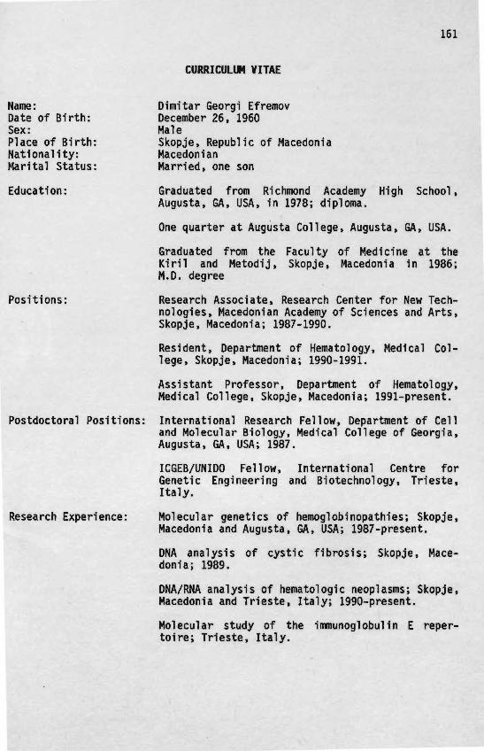
161
CURRICULUM VITAE
Name:Date of Birth:Sex:Place of Birth:Nationality:Marital Status:
Education:
Positions:
Postdoctoral Positions:
Research Experience:
Dimitar Georgi EfremovDecember 26, 1960MaleSkopje, Republic of MacedoniaMacedonianMarried, one son
Graduated from Richmond Academy High School,Augusta, GA, USA, in 1978; diploma.
One quarter at Augusta College, Augusta, GA, USA.
Graduated from the Faculty of Medicine at theKiril and Metodij, Skopje, Macedonia in 1986;M.D. degree
Research Associate, Research Center for New Tech-nologies, Macedonian Academy of Sciences and Arts,Skopje, Macedonia; 1987-1990.
Resident, Department of Hematology, Medical Col-lege, Skopje, Macedonia; 1990-1991.
Assistant Professor, Department of Hematology,Medical College, Skopje, Macedonia; 1991-present.
International Research Fellow, Department of Celland Molecular Biology, Medical College of Georgia,Augusta, GA, USA; 1987.
ICGEB/UNIDO Fellow, International Centre forGenetic Engineering and Biotechnology, Trieste,Italy.
Molecular genetics of hemoglobinopathies; Skopje,Macedonia and Augusta, GA, USA; 1987-present.
DNA analysis of cystic fibrosis; Skopje, Mace-donia; 1989.
DNA/RNA analysis of hématologie neoplasms; Skopje,Macedonia and Trieste, Italy; 1990-present.
Molecular study of the immunoglobulin E reper-toire; Trieste, Italy.

-•3.