Chemotherapeutic potential of 17-AAG against cutaneous leishmaniasis caused by Leishmania (Viannia)...
9
Chemotherapeutic Potential of 17-AAG against Cutaneous Leishmaniasis Caused by Leishmania (Viannia) braziliensis Diego M. Santos 1¤ , Antonio L. O. A. Petersen 1 , Fabiana S. Celes 1 , Valeria M. Borges 1,2 , Patricia S. T. Veras 1 , Camila I. de Oliveira 1,2 * 1 Centro de Pesquisas Gonc ¸alo Moniz, Fundac ¸a ˜o Oswaldo Cruz (FIOCRUZ), Salvador, Bahia, Brazil, 2 Instituto Nacional de Cie ˆncia e Tecnologia de Investigac ¸a ˜o em Imunologia (iii-INCT), Salvador, Bahia, Brazil Abstract Background: Leishmaniasis remains a worldwide public health problem. The limited therapeutic options, drug toxicity and reports of resistance, reinforce the need for the development of new treatment options. Previously, we showed that 17- (allylamino)-17-demethoxygeldanamycin (17-AAG), a Heat Shock Protein 90 (HSP90)-specific inhibitor, reduces L. (L.) amazonensis infection in vitro. Herein, we expand the current knowledge on the leishmanicidal activity of 17-AAG against cutaneous leishmaniasis, employing an experimental model of infection with L. (V.) braziliensis. Methodology/Principal findings: Exposure of axenic L. (V.) braziliensis promastigotes to 17-AAG resulted in direct dose- dependent parasite killing. These results were extended to L. (V.) braziliensis-infected macrophages, an effect that was dissociated from the production of nitric oxide (NO), superoxide (O 22 ) or inflammatory mediators such as TNF-a, IL-6 and MCP-1. The leishmanicidal effect was then demonstrated in vivo, employing BALB/c mice infected with L. braziliensis. In this model, 17-AAG treatment resulted in smaller skin lesions and parasite counts were also significantly reduced. Lastly, 17-AAG showed a similar effect to amphotericin B regarding the ability to reduce parasite viability. Conclusion/Significance: 17-AAG effectively inhibited the growth of L. braziliensis, both in vitro and in vivo. Given the chronicity of L. (V.) braziliensis infection and its association with mucocutaneous leishmaniasis, 17-AAG can be envisaged as a new chemotherapeutic alternative for cutaneous Leishmaniasis. Citation: Santos DM, Petersen ALOA, Celes FS, Borges VM, Veras PST, et al. (2014) Chemotherapeutic Potential of 17-AAG against Cutaneous Leishmaniasis Caused by Leishmania (Viannia) braziliensis. PLoS Negl Trop Dis 8(10): e3275. doi:10.1371/journal.pntd.0003275 Editor: Ben L. Kelly, Louisiana State University, United States of America Received March 11, 2014; Accepted September 16, 2014; Published October 23, 2014 Copyright: ß 2014 Santos et al. This is an open-access article distributed under the terms of the Creative Commons Attribution License, which permits unrestricted use, distribution, and reproduction in any medium, provided the original author and source are credited. Data Availability: The authors confirm that all data underlying the findings are fully available without restriction. All data are included within the manuscript. Funding: This work was funded by grants from CNPq and FIOCRUZ. DMS and ALOAP received CAPES fellowships. FSC received a PIBIC/FIOCRUZ fellowship. VMB, PSTV and CIdO are senior investigators from CNPq. The funders had no role in study design, data collection and analysis, decision to publish, or preparation of the manuscript. Competing Interests: The authors have declared that no competing interests exist. * Email: [email protected] ¤ Current address: Universidade Federal de Sergipe, Departamento de Educac ¸a ˜o em Sau ´ de, Lagarto, Sergipe, Brazil Introduction Leishmaniasis is a widespread group of parasitic diseases caused by protozoa of the genus Leishmania, that is transmitted by the bite of female sand flies. Currently, about 12 million people are at risk of leishmaniasis and there are an estimated 1.5–2 million new cases each year [1]. There are two main clinical manifestations: visceral leishmaniasis, affecting mainly the spleen and liver and cutaneous leishmaniasis, affecting the skin. CL caused by Leishmania (V.) braziliensis is particularly distinguished from other leishmaniasis by its chronicity, latency and tendency to metastasize in the human host [2]. In 1–5% of patients, mucocutaneous leishmaniasis may develop due to the ability of L. (V.) braziliensis to persist within lesion scars after spontaneous or chemotherapy-mediated healing and to its ability to metastasize to the nasal mucosal [3,4]. In this case, extensive tissue destruction is observed, resulting from the potent cell-mediated immune response triggered by parasite replication [5]. More rarely, parasite invasion of the bloodstream results in disseminated skin lesions [6]. Brazil along with nine other countries account for 70– 75% of the global estimated CL incidence [7]. The drugs of first choice for leishmaniasis chemotherapy are Pentavalent Antimonials (Sb +5 ) [8], which interfere with the oxidative metabolism of intracellular Leishmania [5,9,10]. These compounds are significantly toxic and have been associated with drug resistance [11,12]. Amphotericin B and Paramomycin, two other drugs available [13–15], also display limitations with regards to toxicity, cost and/or duration of treatment [16]. In the current scenario, the identification of new chemotherapeutic compounds is urgently needed, especially since vaccines against leishmaniasis are not yet available. Heat Shock Proteins (HSPs) form complexes that act as chaperones, binding other proteins, denominated client proteins. These multimolecular complexes are involved in regulating PLOS Neglected Tropical Diseases | www.plosntds.org 1 October 2014 | Volume 8 | Issue 10 | e3275
-
Upload
independent -
Category
Documents
-
view
1 -
download
0
Transcript of Chemotherapeutic potential of 17-AAG against cutaneous leishmaniasis caused by Leishmania (Viannia)...

Chemotherapeutic Potential of 17-AAG againstCutaneous Leishmaniasis Caused by Leishmania(Viannia) braziliensisDiego M. Santos1¤, Antonio L. O. A. Petersen1, Fabiana S. Celes1, Valeria M. Borges1,2, Patricia S. T. Veras1,
Camila I. de Oliveira1,2*
1 Centro de Pesquisas Goncalo Moniz, Fundacao Oswaldo Cruz (FIOCRUZ), Salvador, Bahia, Brazil, 2 Instituto Nacional de Ciencia e Tecnologia de Investigacao em
Imunologia (iii-INCT), Salvador, Bahia, Brazil
Abstract
Background: Leishmaniasis remains a worldwide public health problem. The limited therapeutic options, drug toxicity andreports of resistance, reinforce the need for the development of new treatment options. Previously, we showed that 17-(allylamino)-17-demethoxygeldanamycin (17-AAG), a Heat Shock Protein 90 (HSP90)-specific inhibitor, reduces L. (L.)amazonensis infection in vitro. Herein, we expand the current knowledge on the leishmanicidal activity of 17-AAG againstcutaneous leishmaniasis, employing an experimental model of infection with L. (V.) braziliensis.
Methodology/Principal findings: Exposure of axenic L. (V.) braziliensis promastigotes to 17-AAG resulted in direct dose-dependent parasite killing. These results were extended to L. (V.) braziliensis-infected macrophages, an effect that wasdissociated from the production of nitric oxide (NO), superoxide (O22) or inflammatory mediators such as TNF-a, IL-6 andMCP-1. The leishmanicidal effect was then demonstrated in vivo, employing BALB/c mice infected with L. braziliensis. In thismodel, 17-AAG treatment resulted in smaller skin lesions and parasite counts were also significantly reduced. Lastly, 17-AAGshowed a similar effect to amphotericin B regarding the ability to reduce parasite viability.
Conclusion/Significance: 17-AAG effectively inhibited the growth of L. braziliensis, both in vitro and in vivo. Given thechronicity of L. (V.) braziliensis infection and its association with mucocutaneous leishmaniasis, 17-AAG can be envisaged asa new chemotherapeutic alternative for cutaneous Leishmaniasis.
Citation: Santos DM, Petersen ALOA, Celes FS, Borges VM, Veras PST, et al. (2014) Chemotherapeutic Potential of 17-AAG against Cutaneous LeishmaniasisCaused by Leishmania (Viannia) braziliensis. PLoS Negl Trop Dis 8(10): e3275. doi:10.1371/journal.pntd.0003275
Editor: Ben L. Kelly, Louisiana State University, United States of America
Received March 11, 2014; Accepted September 16, 2014; Published October 23, 2014
Copyright: � 2014 Santos et al. This is an open-access article distributed under the terms of the Creative Commons Attribution License, which permitsunrestricted use, distribution, and reproduction in any medium, provided the original author and source are credited.
Data Availability: The authors confirm that all data underlying the findings are fully available without restriction. All data are included within the manuscript.
Funding: This work was funded by grants from CNPq and FIOCRUZ. DMS and ALOAP received CAPES fellowships. FSC received a PIBIC/FIOCRUZ fellowship. VMB,PSTV and CIdO are senior investigators from CNPq. The funders had no role in study design, data collection and analysis, decision to publish, or preparation of themanuscript.
Competing Interests: The authors have declared that no competing interests exist.
* Email: [email protected]
¤ Current address: Universidade Federal de Sergipe, Departamento de Educacao em Saude, Lagarto, Sergipe, Brazil
Introduction
Leishmaniasis is a widespread group of parasitic diseases caused
by protozoa of the genus Leishmania, that is transmitted by the
bite of female sand flies. Currently, about 12 million people are at
risk of leishmaniasis and there are an estimated 1.5–2 million new
cases each year [1]. There are two main clinical manifestations:
visceral leishmaniasis, affecting mainly the spleen and liver and
cutaneous leishmaniasis, affecting the skin. CL caused by
Leishmania (V.) braziliensis is particularly distinguished from
other leishmaniasis by its chronicity, latency and tendency to
metastasize in the human host [2]. In 1–5% of patients,
mucocutaneous leishmaniasis may develop due to the ability of
L. (V.) braziliensis to persist within lesion scars after spontaneous
or chemotherapy-mediated healing and to its ability to metastasize
to the nasal mucosal [3,4]. In this case, extensive tissue destruction
is observed, resulting from the potent cell-mediated immune
response triggered by parasite replication [5]. More rarely,
parasite invasion of the bloodstream results in disseminated skin
lesions [6]. Brazil along with nine other countries account for 70–
75% of the global estimated CL incidence [7].
The drugs of first choice for leishmaniasis chemotherapy are
Pentavalent Antimonials (Sb+5) [8], which interfere with the
oxidative metabolism of intracellular Leishmania [5,9,10]. These
compounds are significantly toxic and have been associated with
drug resistance [11,12]. Amphotericin B and Paramomycin, two
other drugs available [13–15], also display limitations with regards
to toxicity, cost and/or duration of treatment [16]. In the current
scenario, the identification of new chemotherapeutic compounds is
urgently needed, especially since vaccines against leishmaniasis are
not yet available.
Heat Shock Proteins (HSPs) form complexes that act as
chaperones, binding other proteins, denominated client proteins.
These multimolecular complexes are involved in regulating
PLOS Neglected Tropical Diseases | www.plosntds.org 1 October 2014 | Volume 8 | Issue 10 | e3275

protein folding, intracellular protein transport and repair or
degradation of proteins partially denatured due to stress, for
example [17,18]. Among the HSPs, HSP90 is one of the most
abundant cellular chaperones and many of its client proteins are
involved in cell signaling, proliferation and survival [19]. It is
essential for oncogenic transformation and exploited by malignant
cells to support cancer-associated kinases and transcription factors
[20]. HSP90 also plays an important role in protozoans such as
Leishmania and Trypanosoma, which critically rely on HSP90 for
survival in alternating environments associated with their complex
life cycles [21]. Therefore, HSP90-inhibitors become interesting
candidates for leishmaniasis chemotherapy.
Treatment of L. donovani parasites with geldanamycin (GA), a
HSP90-specific inhibitor, arrested promastigote growth and
differentiation into amastigotes [22]. It also reduced gluthathione
levels, increasing the production of reactive oxygen species (ROS)
and promoting apoptosis [23]. Recently, we reported on the effects
of 17-(allylamino)-17-demethoxygeldanamycin (17-AAG) on L.(L.) amazonensis [24]. 17-AAG is a HSP90-specific inhibitor
analogous to geldanamycin (GA) [25]. Macrophages infected with
L. (L.) amazonensis and treated with a low dose of 17-AAG
displayed significantly smaller parasite loads, an effect that was not
mediated by activation of the macrophage inflammatory response
[24].
In the present work, we expanded our previous observations to
the effects of 17-AAG on L. (V.) braziliensis, the etiological agent
of both cutaneous and mucocutaneous leishmaniasis in Brazil.
Experiments were performed in vitro and in vivo, employing an
experimental model [26]. 17-AAG was efficient at reducing L. (V.)braziliensis promastigote growth and macrophage infection. More
importantly, 17-AAG was equally efficient in vivo, highlighting its
potential as a novel chemotherapy agent against CL caused by L.(V.) braziliensis.
Methods
Ethics statementFemale BALB/c mice, 6–8 weeks of age, were obtained from
CPqGM/FIOCRUZ animal facility where they were maintained
under pathogen-free conditions. All animal work was conducted
according to the Guidelines for Animal Experimentation of the
Colegio Brasileiro de Experimentacao Animal and of the
Conselho Nacional de Controle de Experimentacao Animal.
The local Ethics Committee on Animal Care and Utilization
(CEUA) approved all procedures involving animals (CEUA-L001/
12-CPqGM/FIOCRUZ).
17-AAG and amphotericin B17-AAG (17-(allylamino)-17-demethoxygeldanamycin) (Invivo-
gen) was dissolved in Dimethyl sulfoxide (DMSO) (SIGMA) to a
5 mM stock solution, stored at -20uC in aliquots. For in vitro use,
the stock solution was diluted in cell culture medium to the desired
concentration at the time of use. For in vivo treatments, a stock
solution was prepared at 100 mg/ml and diluted to 20 mg/kg at
the time of use. Amphotericin B (Fungizone, Life Technologies)
was dissolved in DMEM medium to a 250 ug/ml stock solution.
The stock solution was diluted in cell culture medium to the
desired concentration at the time of use.
Parasite cultureL. (V.) braziliensis (MHOM/BR/01/BA788) [26] was cultured
at 26uC in Schneider’s insect medium (Invitrogen) supplemented
with 10% inactive Fetal Bovine Serum (FBS), 2 mM L-glutamine,
100 U/ml penicillin, and 100 mg/ml streptomycin (all from
Invitrogen).
L. (V.) braziliensis promastigotes viability assayAxenic L. (V.) braziliensis promastigotes (16106 parasites/ml),
cultivated in supplemented Schneider medium, were treated with
increasing concentrations of 17-AAG (25, 75, 125, 250, 500 or
625 nM). After 48 h, parasite viability was evaluated by direct
counting of live motile parasites using a Neubauer chamber. In
some experiments, promastigotes were treated with the half
maximal inhibitory concentration (IC50) (65 nM). After 48 h,
promastigotes were washed three times with PBS and were further
cultured for 24 and 48 h in supplemented Schneider medium,
devoid of 17-AAG. The number of viable promastigotes was
determined by direct counting.
Macrophage infection with L. (V.) braziliensis andtreatment with 17-AAG
BALB/c mice were injected i.p. with 3% thioglycolate. Five
days after injection, peritoneal lavage was performed using 8 ml
DMEM medium supplemented with 10% Fetal Calf Serum (FCS),
2 mM L-glutamine, 100 U/ml penicillin and 100 mg/ml strepto-
mycin (all from Invitrogen). To obtain monolayers, cells (66105
cells/ml) were place into glass coverslips within the wells of a 24-
well plate and were left to adhere for 2 h, at 37uC and 5% CO2.
Non-adherent cells were removed by gentle and extensive washing
with PBS; purity was routinely above 99%. Remaining cells
(36105 cells/ml) received 36106 cells/ml of stationary-phase L.(V.) braziliensis promastigotes and were incubated at 37uC in
supplemented DMEM medium. After 24 h of infection, glass
coverslips containing infected macrophages were washed to
remove non-internalized parasites and cells were treated with
different concentrations of 17-AAG (25, 100, 250 and 500 nM) for
12–72 h. Control groups were incubated in supplemented DMEM
medium containing DMSO only. Glass coverslips were washed
and stained with H&E and the intracellular amastigotes were
counted by light microscopy. The results are shown as the
percentage of infected cells and the number of intracellular
amastigotes was counted in 400 macrophages. Cultures were
performed in quintuplicate. Alternatively, infected macrophages
were washed extensively and the medium was replaced with
Author Summary
Antimony-containing compounds are the main drugs usedto treat leishmaniasis but the severe associated side effectspose the need for alternative chemotherapeutic options.Herein, we evaluated the ability of 17-AAG (a Heat ShockProtein 90 inhibitor) to kill Leishmania (Viannia) braziliensisparasites, a species that causes both cutaneous andmucocutaneous Leishmaniasis in Brazil. Heat Shock Protein90 (HSP90) is associated with important biologicalprocesses; inhibition of this molecule interferes withparasite survival and, hence, it can be exploited as achemotherapeutic target. We show that exposure to 17-AAG induced killing of L. brazilensis parasites in both itsextracellular and intracellular forms. This effect was notdependent on the activation of the host cell. Moreimportantly, treatment of mice infected with L. (V.)braziliensis also modulated lesion development anddecreased parasite growth at the infection site. Collective-ly, our results show that targeting HSP90 is a promisingalternative for development of novel chemotherapeuticoptions for leishmaniasis.
17-AAG Controls Infection by L. (V.) braziliensis
PLOS Neglected Tropical Diseases | www.plosntds.org 2 October 2014 | Volume 8 | Issue 10 | e3275

0.5 ml of supplemented Schneider medium, devoid of 17-AAG.
Cells were cultured at 26uC for an additional 5 days and the
number of viable parasites was determined by direct counting. In
some experiments, infected macrophages were treated with the
half maximal inhibitory concentration (IC50) (220 nM) of 17-AAG
or with amphotericin B (0.25 mg/ml; 0.27 mM) for 24 h. Parasite
viability was determined by direct counting.
Viability of 17-AAG-treated murine macrophagesMacrophages (26105 cells/ml), obtained as above, were treated
increasing concentrations of 17-AAG (39–20,000 nM) or with
DMSO for 72 h. Next, cultures were washed twice cells were
incubated with supplemented DMEM containing 10% Alamar-
Blue (Invitrogen). Cells were incubated for another 4 h and
reagent absorbance was measured at the wavelengths of 570 nm
and 600 nm using a spectrophotometer (SPECTRA Max 340 PC).
Ethanol-fixed cells were used as positive controls.
Detection of NO, reactive oxygen species, cytokines andchemokines
Macrophages (36106 cells/ml), obtained as above, were
stimulated with IFN-c (100 UI/ml) (Sigma) and were infected
with L. (V.) braziliensis (36107 cells/ml) for 24 h. Macrophage
cultures were then washed to remove non-internalized parasites
and fresh culture medium containing IFN-c and 220 nM of 17-
AAG was added. Cultures supernatants were collected 48 h later.
Griess reaction was used to measure nitric oxide (NO) production
by determining concentration of its stable reaction product nitrite
(NO22) [27]. Superoxide (SO) production was determined by
adding hydroxylamine (Sigma) (0.5 mM) [28,29] to infected
macrophages. Hydroxylamine converts superoxide into nitrite,
which is then be quantitated by the Griess reaction, as described
above. Background levels of nitrite generated by the release of NO
were determined in parallel, without the addition of hydroxyl-
amine. Production of TNF-a, IL-6, IL-10 and CCL2/MCP-1 was
evaluated using an inflammatory Cytometric Bead Array (BD
Biosciences) following the manufacturer’s instructions. Data were
acquired and analyzed using a FACSort flow cytometer and FCAP
Array (V.3.0) (BD Biosciences).
Intradermal infection with L. (V.) braziliensis and in vivotreatment with 17-AAG
BALB/c mice were inoculated with stationary-phase L. (V.)braziliensis promastigotes (105 parasites in 10 ml of saline) in the
left ear dermis using a 27.5-gauge needle. Four weeks post-
infection, mice (n = 10) were treated 3 times/wk for 3 weeks with
17-AAG (20 mg/kg of 17-AAG diluted in DMSO i.p.). The
control group (n = 10) received i.p. injections of DMSO in the
same concentrations used in 17-AAG treated animals (n = 10).
Lesion size was monitored weekly for 10 weeks using a digital
caliper (Thomas Scientific). Parasite load was determined using a
quantitative limiting-dilution assay as described elsewhere [26].
Briefly, infected ears and lymph nodes draining the infection site
were aseptically excised six weeks post-infection and homogenized
in Schneider medium. Homogenates were serially diluted in
supplemented Schneider complete and seeded into 96-well plates.
The number of viable parasites was determined from the highest
dilution at which the promastigotes could be grown after up to 2
weeks of incubation at 26uC.
Statistical analysisThe half maximal inhibitory concentration (IC50) of 17-AAG on
L. braziliensis promastigotes and on intracellular L. braziliensisamastigotes were determined from sigmoidal regression of the
concentration-responses curves, respectively, using Prism (Graph-
Pad Prism V. 6.0). The selectivity index of 17-AAG was calculated
as the ratio between the CC50 for murine macrophages and the
IC50 for intracellular L. braziliensis amastigotes.
Data are presented as the mean 6 standard error of the mean.
Kolmogorov-Smirnov was used for normality analysis. Parametric
(One-way ANOVA followed by Dunnett’s Multiple Comparison
Test or post-test for linear trend or by Bonferroni) or non-
parametric analysis (Mann-Whitney) tests were also performed
using Prism (GraphPad software, V. 6.0) To evaluate disease
burden in mice, ear thickness of mice treated with 17-AAG or
DMSO was recorded weekly for each individual mouse. The
course of disease for 17-AAG-treated and control mice was plotted
individually, and the area under each resulting curve was
calculated using a non-parametric test (Mann-Whitney). p-values
# 0.05 were considered significant.
Results
Exposure to 17-AAG reduces the viability of L. (V.)braziliensis promastigotes
Initially, we investigated the effects of 17-AAG on axenic L. (V.)braziliensis promastigotes. Parasites were incubated with increas-
ing concentrations of 17-AAG for 48 h and viability was
quantified by direct counting. All 17-AAG-treated cultures showed
a significantly lower number of parasites in comparison to the
control, treated with vehicle (DMSO) alone (Figure 1A) (One-way
ANOVA, p,0.001). After 48 h of treatment, 17-AAG (25 nM)
reduced parasite viability by 13% (Figure 1A) (compared to
DMSO-treated cultures) and increasing concentrations of 17-AAG
(75–625 nM), maximized killing effects. At the highest concentra-
tion tested (625 nM) parasite viability was reduced by 98% when
compared to DMSO-treated cultures (Figure 1A). These results
also indicate that parasite viability was reduced in a dose-
dependent effect (One-way ANOVA, p,0.001 followed by test for
linear trend p,0.001). The DMSO concentration used was not
toxic as parasite viability was similar in cultures left untreated (Lb)
or treated with DMSO only (Figure 1A). Based on these results,
IC50, after 48 h of 17-AAG treatment, was established at 65 nM
(Figure S1). To evaluate whether the effect of 17-AAG on L. (V.)braziliensis promastigotes was reversible, parasites were treated
with 17-AAG (65 nM) for 48 h, washed and subsequently re-
incubated in 17-AAG-free medium, for an additional 24 and 48 h.
Parasite numbers were significantly reduced (p,0.01) in cultures
kept for both 24 h (Figure 1B) and 48 h (Figure 1C). These results
show that the effect of 17-AAG on L. (V.) braziliensis promas-
tigotes is irreversible.
17-AAG reduces parasite load in L. (V.) braziliensis-infected macrophages
Next, we investigated the effect of 17-AAG on intracellular L.(V.) braziliensis amastigotes. BALB/c macrophages were infected
with L. (V.) braziliensis and cells were treated with a range of 17-
AAG concentrations (25–500 nM) for 12–72 h. At each time
point, cells were fixed and the parasite load was assessed by light
microscopy. At the initial time points (12 and 24 h), we did not
detect significant alterations in treated cultures versus control
cultures (DMSO-treated) (Figure 2A and B). After 48 h, 17-AAG
(25 nM) reduced the infection rate to 85% (Figure 2A) and
intracellular amastigotes were reduced to 82% (compared to the
percentages obtained in DMSO-treated cultures) (Figure 2B).
With increasing concentrations of 17-AAG (100–500 nM), these
effects became more pronounced and, at 500 nM, the percentage
of infected cells was reduced to 63% (Figure 2A) and of
17-AAG Controls Infection by L. (V.) braziliensis
PLOS Neglected Tropical Diseases | www.plosntds.org 3 October 2014 | Volume 8 | Issue 10 | e3275

intracellular amastigotes to 43% (Figure 2B) (One-way ANOVA,
p,0.001) (again compared to the percentages obtained in DMSO-
treated cultures). After 72 h of treatment, these effects were
maximal: 500 nM of 17-AAG decreased the infection rate to 20%
(Figure 2A) whereas intracellular amastigotes were reduced to
11% (Figure 2B). The absolute percentages of infection, following
treatment with different concentrations of17-AAG and the
absolute numbers of amastigotes/100 cells over time are shown
in Figures S2A and S2B, respectively. As with promastigotes
(Figure 1A), effects observed with 17-AAG on intracellular
macrophages were dose-dependent (One-way ANOVA, p,
0.001 followed by test for linear trend p,0.001). IC50, after
72 h of 17-AAG treatment, was determined as 220 nM (Figure
S3); additionally, 17-AAG employed at different concentrations
(125, 220 and 500 nM) did not compromise macrophage viability
as assayed by MTT (Figure S4). Cytotoxicity against murine
macrophages was determined upon treatment of non-infected
macrophage cultures with 17-AAG with a calculated CC50 of
3.6 nM. The selectivity index of 17-AAG was established at 16.6.
17-AAG reduces the viability of intracellular L. (V.)braziliensis amastigotes
Although 17-AAG treatment of L. (V.) braziliensis-infected
macrophages for 24 h did not significantly modify the infection
rate (Figure 2), we asked whether it would alter amastigote
viability. Cells were infected and treated with a range of 17-AAG
concentrations (25–500 nM). Intracellular parasite survival was
determined following the replacement of DMEM for Schneider
culture medium and direct counting of surviving L. (V.)braziliensis. Five days after medium replacement, treatment with
increasing concentrations of 17-AAG for 24 h significantly
reduced the number of viable L. (V.) braziliensis parasites
(Figure 3A) (One-way ANOVA, p,0.001), indicating once more
a dose-dependent effect (test for linear trend p,0.001). This effect
was also time-dependent as exposure to 220 nM (IC50) of 17-AAG
for longer periods (48 and 72 h) also significantly decreased the
number of L. (V.) braziliensis promastigotes (Figure 3B). There-
fore, exposure of infected macrophages to 17-AAG negatively
impacted on the survival of L. (V.) braziliensis.
Figure 1. 17-AAG induces killing of Leishmania (V.) braziliensis promastigotes in a dose-dependent and irreversible manner. L. (V.)braziliensis promastigotes were exposed to increasing concentrations of 17-AAG, to vehicle alone (DMSO) or were left unexposed (Lb) for 48 h. (A)The number of viable parasites was evaluated by direct counting. L.(V) braziliensis promastigotes were treated with 65 nM (IC50) of 17-AAG for 24 h(B) and (C) 48 h. After washing, promastigotes were cultured for additional 48 h and the number of viable parasites was evaluated. Data, shown asmean 6SEM, are from one of two independent repeats (**p,0.01 and ***p,0.001).doi:10.1371/journal.pntd.0003275.g001
Figure 2. Treatment with 17-AAG controls L. (V.) braziliensis replication inside macrophages. L. (V.) braziliensis- infected macrophages weretreated with increasing concentrations of 17-AAG or with vehicle alone (DMSO). After 12–72 h, glass coverslips were stained with H&E and assessedfor the percentage of infected macrophages (A) and the number of amastigotes per 100 macrophages (B) by light microscopy. Data, shown as mean6SEM, are shown as the percentage of DMSO -treated cultures, from one of three independent repeats (*p,0.05; **p,0.01 and ***p,0.001).doi:10.1371/journal.pntd.0003275.g002
17-AAG Controls Infection by L. (V.) braziliensis
PLOS Neglected Tropical Diseases | www.plosntds.org 4 October 2014 | Volume 8 | Issue 10 | e3275

17-AAG down-modulates the production of inflammatorymediators by L. (V.) braziliensis -infected macrophages
Macrophage activation and production of nitric oxide and
superoxide are key steps towards elimination of intracellular
Leishmania [30]. In macrophages infected with L. (V.) braziliensis
and treated with 220 nM 17-AAG (IC50), production of nitric oxide
(Figure 4A) and superoxide (Figure 4B) were lower compared to
cells exposed to DMSO. 17-AAG-treatment also down-modulated
the production of TNF-a (Figure 4C), IL-6 (Figure 4D) and CCL2/
MCP-1 (Figure 4E) by L. (V.) braziliensis-infected macrophages.
Figure 3. Treatment with 17-AAG reduces intracellular L. (V.) braziliensis viability. L. (V.) braziliensis- infected macrophages were treated for24 h with increasing concentrations of 17-AAG or with vehicle alone (DMSO). The number of viable parasites was evaluated by further culture (5 days)in Schneider medium, free of 17-AAG (A). Infected macrophages were treated with 220 nM (IC50) of 17-AAG for 24–72 h. The number of viableparasites was evaluated by further culture for five days in Schneider medium, free of 17-AAG (B). Data, shown as mean 6SEM, are from one of twoindependent repeats (**p,0.01 and *** p,0.001).doi:10.1371/journal.pntd.0003275.g003
Figure 4. Treatment with 17-AAG down regulates ROS and cytokine production in L. (V.) braziliensis-infected cells. L. (V.) braziliensis-infected macrophages were treated with 17-AAG (220nM) + IFN- . After 48h, supernatants were assayed for nitrite production (A) and for presence of(B) superoxide, following addition of hydroxylamine. The presence of secreted (C) TNF-a, (D) IL-6 and (E) CCL2 was determined in culture supernatantsby Cytometric Bead Array, after 24 h of treatment. Data, shown as mean 6 SEM, are from one of two independent repeats (**p,0.01; *p,0.05).doi:10.1371/journal.pntd.0003275.g004
17-AAG Controls Infection by L. (V.) braziliensis
PLOS Neglected Tropical Diseases | www.plosntds.org 5 October 2014 | Volume 8 | Issue 10 | e3275
Therefore, the leishmanicidal effect of 17-AAG is uncoupled from
the production of microbicidal molecules and from the production
of pro-inflammatory cytokines.
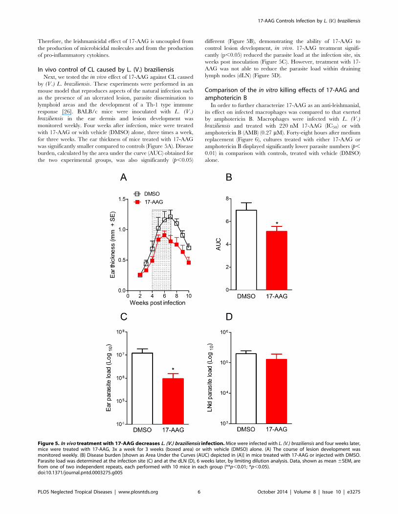
In vivo control of CL caused by L. (V.) braziliensisNext, we tested the in vivo effect of 17-AAG against CL caused
by (V.) L. braziliensis. These experiments were performed in an
mouse model that reproduces aspects of the natural infection such
as the presence of an ulcerated lesion, parasite dissemination to
lymphoid areas and the development of a Th-1 type immune
response [26]. BALB/c mice were inoculated with L. (V.)braziliensis in the ear dermis and lesion development was
monitored weekly. Four weeks after infection, mice were treated
with 17-AAG or with vehicle (DMSO) alone, three times a week,
for three weeks. The ear thickness of mice treated with 17-AAG
was significantly smaller compared to controls (Figure 5A). Disease
burden, calculated by the area under the curve (AUC) obtained for
the two experimental groups, was also significantly (p,0.05)
different (Figure 5B), demonstrating the ability of 17-AAG to
control lesion development, in vivo. 17-AAG treatment signifi-
cantly (p,0.05) reduced the parasite load at the infection site, six
weeks post inoculation (Figure 5C). However, treatment with 17-
AAG was not able to reduce the parasite load within draining
lymph nodes (dLN) (Figure 5D).
Comparison of the in vitro killing effects of 17-AAG andamphotericin B
In order to further characterize 17-AAG as an anti-leishmanial,
its effect on infected macrophages was compared to that exerted
by amphotericin B. Macrophages were infected with L. (V.)braziliensis and treated with 220 nM 17-AAG (IC50) or with
amphotericin B (AMB) (0.27 mM). Forty-eight hours after medium
replacement (Figure 6), cultures treated with either 17-AAG or
amphotericin B displayed significantly lower parasite numbers (p,
0.01) in comparison with controls, treated with vehicle (DMSO)
alone.
Figure 5. In vivo treatment with 17-AAG decreases L. (V.) braziliensis infection. Mice were infected with L. (V.) braziliensis and four weeks later,mice were treated with 17-AAG, 3x a week for 3 weeks (boxed area) or with vehicle (DMSO) alone. (A) The course of lesion development wasmonitored weekly. (B) Disease burden [shown as Area Under the Curves (AUC) depicted in (A)] in mice treated with 17-AAG or injected with DMSO.Parasite load was determined at the infection site (C) and at the dLN (D), 6 weeks later, by limiting dilution analysis. Data, shown as mean 6SEM, arefrom one of two independent repeats, each performed with 10 mice in each group (**p,0.01; *p,0.05).doi:10.1371/journal.pntd.0003275.g005
17-AAG Controls Infection by L. (V.) braziliensis
PLOS Neglected Tropical Diseases | www.plosntds.org 6 October 2014 | Volume 8 | Issue 10 | e3275

Discussion
HSP90 is a molecular chaperone fundamental for the life cycle
of a variety or protozoa [31] and, as such, inhibitors of HSP90
have been suggested as novel chemotherapeutic agents against
malaria [32], filariasis [33,34] and schistosomiasis [34]. Recently,
we showed that 17-AAG, a HSP90 inhibitor, reduced L. (L.)amazonensis infection in vitro [24]. Herein, we investigated the
potential of 17-AAG as a chemotherapeutic agent against L. (V.)braziliensis, the main etiological agent of CL and MCL in Brazil
[35]. We confirmed the effects of 17-AAG against this L. (V.)braziliensis promastigotes and we extended these findings to a pre-
clinical model of CL.
Initially, we investigated the in vitro effects of 17-AAG, against
both axenic promastigotes and intracellular amastigotes. Treat-
ment of L. braziliensis promastigotes with the lower dose of 17-
AAG (25 nM) already decreased promastigote viability. Herein,
the IC50 determined for L. (V). braziliensis was comparable to that
described for L. (L). amazonensis (65 nM) whereas in experiments
performed with L. (L). major, the IC50 was established at 80 nM
[24]. 17-AAG was equally effective at reducing intracellular
amastigote numbers and the viability of surviving L. (V).braziliensis promastigotes. These effects were not associated with
an increase in the microbicidal functions of macrophages as levels
of NO, superoxide and TNF-a were diminished in the presence of
17-AAG. These results are in accordance with our previous report
[24]. Additionally, the lack of amastigote replication in control
macrophages could be attributed to innate microbocidal proper-
ties of macrophages that allow L. (V). braziliensis killing, as
observed with L. (V). guyanensis and L. (L.) major [36–38]. In CL
patients, an exacerbated inflammatory immune response is
associated with the development of mucocutaneous leishmaniasis
(rev. in [39]) whereas subclinical patients, who do not develop the
disease, have a more controlled immune response [40]. Therefore,
the possibility of selectively inducing parasite killing without
contributing to overt inflammation is an important advantage for
the treatment of CL using 17-AAG. Of note, macrophages treated
with 17-DMAG alone displayed reduced production of IL-6,
TNF-a and NO [41] whereas 17-AAG prevented iNOS expression
upon stimulation with LPS or IFN-g [42].
Geldanamycin (GA), a HSP90 inhibitor analogous to 17-AAG,
induces an anti-oxidative and attenuated inflammatory response in
sepsis [43], autoimmune encephalitis [44], experimental athero-
sclerosis [45] and endotoxin-induced uveitis [46]. The proposed
mechanism for these effects is the reduced nuclear translocation of
NF-kB, reflecting in decreased production of IL-6, TNF-a and
NO [41]. Although we cannot extrapolate the complexities of invivo situations cited above, L. (L.) amazonensis-infected macro-
phages treated with 17-AAG displayed parasite killing, in spite of a
diminished production of inflammatory mediators [24]. It has
been shown that in BALB/c mice infected with L. (V). braziliensis,the density of INOS+ cells was higher when compared to L. (L).amazonensis-infected mice [47]. So, different responses to NO
between L. (V.) braziliensis and L. (L.) amazonensis could also
impact on the killing effect exerted by 17-AAG. In our previous
work, [24], we showed that L. (L) amazonensis amastigotes
displayed structural alterations following exposure of infected
macrophages to 17-AAG. Visible alterations in the cytoplasm of
parasites such as the presence of myelin figures, vesicles with
double-layered membranes and mitochondrial segments inside
membrane-bounded structures were the suggestive indications of
autophagy, a process that naturally occurs in Leishmania and
which plays an important role in the transition from promastigote
to amastigote [48]. It is possible that inhibition of HSP90 activity
interferes with cell cycle progression, blocking differentiation or
expression of stage specific protein and, consequently, affecting
survival in the intracellular environment.
17-AAG was also effective in vivo: mice infected with L. (V.)braziliensis and treated with 17-AAG showed a significantly
smaller disease burden in parallel to a smaller parasite load at the
infection site. However, 17-AAG was not able to alter parasite
load at the draining lymph nodes (dLN), a site where L. (V.)braziliensis parasites persist following lesion healing [26]. In this
experimental model, parasite persistence is associated with the
presence of regulatory T cells (Tregs) that accumulate within dLNs
of L. (V.) braziliensis-infected mice [49] and these Tregs control
Th1 responses by IL-10-dependent mechanisms [50]. Although
17-AAG treatment controlled parasite replication at the infection
site and promoted lesion healing, parasite persistence within distal
sites such as the dLNs may have important effects with regards to
maintenance of immunity to Leishmania [51] and/or development
of mucotutaneous leishmaniasis, deserving further investigation.
Currently, the drugs available for the treatment of CL are
limited and among them, pentavalent antimonials have been the
choice for over 60 years. However, treatment is long (20-30 days),
patients develop several side effects and, in the recent years, the
number of cases refractory to treatment has increased [52,53]. In
the case of therapeutic failure, second-line drugs such as
amphotericin B can be employed as well as combination of two
available drugs [54]. Advantages of a combination treatment
include increased efficacy, less drug resistance, lower drug dosage
and a general decrease in side effects [55]. Herein, 17-AAG was as
effective as amphotericin B at decreasing the parasite load within
infected macrophages. Experimentally, the treatment of L.infantum and L. panamensis promastigotes with 17-AAG plus
edelfosine improved the anti-leishmanicidal activity of the latter
[56]. In vitro synergism was also observed for the combinations of
paramomycin and amphotericin B against L. (V.) braziliensis [57].
In vivo, association of tamoxifen with amphotericin B yielded an
additive effect in mice infected with L. (L.) amazonensis [58]. The
combination of GA with fluconazole showed synergistic activity
against Candida albicans isolates resistant to fluconazole alone
[59]. Thus, we propose that combinations of 17-AAG and
Figure 6. Comparison of the effects of 17-AAG and amphoter-icin B on the growth of intracellular L. (V.) braziliensis. L. (V.)braziliensis-infected macrophages were treated with 17-AAG or withamphotericin B (AMB) for 48 h. The number of viable parasites wasevaluated by further culture in Schneider medium, free of 17-AAG. Data,shown as mean 6SEM, are from one of two independent repeats(***p,0.001). (ND, not detected).doi:10.1371/journal.pntd.0003275.g006
17-AAG Controls Infection by L. (V.) braziliensis
PLOS Neglected Tropical Diseases | www.plosntds.org 7 October 2014 | Volume 8 | Issue 10 | e3275
amphotericin B may be further investigated for the treatment of
CL caused by L. (V.) braziliensis.Herein, we reported that 17-AAG reduces L. (V.) braziliensis
infection in vitro and in vivo. 17-AAG shows excellent bioavail-
ability when given to mice by the i.p. route [60]. At 60 mg/kg, 17-
AAG caused no changes in appearance, appetite, waste elimina-
tion, or survival of treated animals as compared to vehicle-treated
controls. We employed with 20 mg/kg, a dose well below that
reported as having any harmful effects, as those decribed by Solit
et al. (equal or above 75 mg/kg) [61]. Given that HSP90
inhibitors, analogous to 17-AAG, have entered clinical trials with
cancer patients [62], we propose that 17-AAG could be further
investigated as a novel target for chemotherapy against cutaneous
leishmaniasis.
Supporting Information
Figure S1 Determination of IC50 values for 17-AAG in L.braziliensis promastigotes. Cells were treated in sextuplicate
with 17-AAG for 48 hours with varying concentrations of 17-
AAG. Following treatment, parasite viability was evaluated by
direct counting. IC50 values (nM) were determined using
GraphPad Prism.
(TIF)
Figure S2 Treatment with 17-AAG controls L. brazilien-sis replication inside macrophages. L. braziliensis- infected
macrophages were treated with increasing concentrations of 17-
AAG or with vehicle alone (DMSO). After 12–72 h, glass
coverslips were stained with H&E and assessed for the percentage
of infected macrophages (A) and the number of amastigotes per
100 macrophages (B) by light microscopy. Data, shown as mean
6SEM, are from one of three independent repeats (*p,0.05;
**p,0.01 and ***p,0.001).
(TIF)
Figure S3 Determination of IC50 values for 17-AAG inmacrophages infected with L. braziliensis promasti-gotes. Infected macrophages were treated in sextuplicate with 17-
AAG for 72 hours with varying concentrations of 17-AAG.
Following treatment, glass coverslips were stained with H&E and
assessed for the presence of amastigotes by light microscopy. IC50
values (nM) were determined using GraphPad Prism.
(TIF)
Figure S4 Cell viability following macrophage exposureto 17-AAG. Thioglycolate-elicited macrophages wereexposed to with different concentrations of 17-AAG orto DMSO (vehicle) alone for 24 h. Cell viability was evaluated
by MTT assay.
(TIF)
Acknowledgments
The authors are grateful to Lucas Menezes Moura for excellent technical
assistance. The authors are also thankful to Diego Menezes and Ricardo
Khouri for helpful comments and discussions.
Author Contributions
Conceived and designed the experiments: DMS ALOAP FSC VMB PSTV
CIdO. Performed the experiments: DMS ALOAP FSC. Analyzed the data:
DMS ALOAP VMB PSTV FSC CIdO. Contributed reagents/materials/
analysis tools: VMB PSTV CIdO. Wrote the paper: DMS ALOAP VMB
PSTV CIdO.
References
1. den Boer M, Argaw D, Jannin J, Alvar J (2011) Leishmaniasis impact and
treatment access. Clin Microbiol Infect 17: 1471–1477.
2. Bittencourt A, Silva N, Straatmann A, Nunes VL, Follador I, et al. (2003) Post-
kala-azar dermal leishmaniasis associated with AIDS. Braz J Infect Dis 7: 229–
233.
3. Schubach A, Marzochi MC, Cuzzi-Maya T, Oliveira AV, Araujo ML, et al.
(1998) Cutaneous scars in American tegumentary leishmaniasis patients: a site of
Leishmania (Viannia) braziliensis persistence and viability eleven years after
antimonial therapy and clinical cure. Am J Trop Med Hyg 58: 824–827.
4. Mendonca MG, de Brito ME, Rodrigues EH, Bandeira V, Jardim ML, et al.
(2004) Persistence of leishmania parasites in scars after clinical cure of American
cutaneous leishmaniasis: is there a sterile cure? J Infect Dis 189: 1018–1023.
5. Marsden PD (1986) Mucosal leishmaniasis (‘‘espundia’’ Escomel, 1911).
Trans R Soc Trop Med Hyg 80: 859–876.
6. Costa JM, Marsden PD, Llanos-Cuentas EA, Netto EM, Carvalho EM, et al.
(1986) Disseminated cutaneous leishmaniasis in a field clinic in Bahia, Brazil: a
report of eight cases. J Trop Med Hyg 89: 319–323.
7. Alvar J, Velez ID, Bern C, Herrero M, Desjeux P, et al. (2012) Leishmaniasis
worldwide and global estimates of its incidence. PLoS One 7: e35671.
8. Croft SL, Coombs GH (2003) Leishmaniasis–current chemotherapy and recent
advances in the search for novel drugs. Trends Parasitol 19: 502–508.
9. Berman JD (1997) Human leishmaniasis: clinical, diagnostic, and chemother-
apeutic developments in the last 10 years. Clin Infect Dis 24: 684–703.
10. Chulay JD, Oster CN, McGreevy PB, Hendricks LD, Kreutzer RD (1988)
American cutaneous leishmaniasis: presentation and problems of patient
management. Rev Soc Bras Med Trop 21: 165–172.
11. Amato VS, Tuon FF, Siqueira AM, Nicodemo AC, Neto VA (2007) Treatment
of mucosal leishmaniasis in Latin America: systematic review. Am J Trop Med
Hyg 77: 266–274.
12. Modabber F, Buffet PA, Torreele E, Milon G, Croft SL (2007) Consultative
meeting to develop a strategy for treatment of cutaneous leishmaniasis. Institute
Pasteur, Paris. 13–15 June, 2006. Kinetoplastid Biol Dis 6: 3.
13. Llanos-Cuentas A, Tulliano G, Araujo-Castillo R, Miranda-Verastegui C,
Santamaria-Castrellon G, et al. (2008) Clinical and parasite species risk factors
for pentavalent antimonial treatment failure in cutaneous leishmaniasis in Peru.
Clinical infectious diseases: an official publication of the Infectious Diseases
Society of America 46: 223–231.
14. Machado P, Araujo C, Da Silva AT, Almeida RP, D’Oliveira Jr A, et al. (2002)
Failure of early treatment of cutaneous leishmaniasis in preventing the
development of an ulcer. Clinical infectious diseases: an official publication of
the Infectious Diseases Society of America 34: E69–73.
15. Oliveira LF, Schubach AO, Martins MM, Passos SL, Oliveira RV, et al. (2011)
Systematic review of the adverse effects of cutaneous leishmaniasis treatment in
the New World. Acta Tropica 118: 87–96.
16. Mishra J, Saxena A, Singh S (2007) Chemotherapy of leishmaniasis: past,
present and future. Curr Med Chem 14: 1153–1169.
17. Morimoto RI, Kline MP, Bimston DN, Cotto JJ (1997) The heat-shock response:
regulation and function of heat-shock proteins and molecular chaperones. Essays
Biochem 32: 17–29.
18. Hartl FU, Hayer-Hartl M (2002) Molecular chaperones in the cytosol: from
nascent chain to folded protein. Science 295: 1852–1858.
19. Goetz MP (2003) The Hsp90 chaperone complex as a novel target for cancer
therapy. Annals of Oncology 14: 1169–1176.
20. Neckers L, Workman P (2012) Hsp90 molecular chaperone inhibitors: are we
there yet? Clinical cancer research: an official journal of the American
Association for Cancer Research 18: 64–76.
21. Rochani AK, Singh M, Tatu U (2013) Heat shock protein 90 inhibitors as broad
spectrum anti-infectives. Current pharmaceutical design 19: 377–386.
22. Wiesgigl M, Clos J (2001) The heat shock protein 90 of Leishmania donovani.
Medical microbiology and immunology 190: 27–31.
23. Li Q, Zhou Y, Yao C, Ma X, Wang L, et al. (2009) Apoptosis caused by Hsp90
inhibitor geldanamycin in Leishmania donovani during promastigote-to-
amastigote transformation stage. Parasitology research 105: 1539–1548.
24. Petersen AL, Guedes CE, Versoza CL, Lima JG, de Freitas LA, et al. (2012) 17-
AAG kills intracellular Leishmania amazonensis while reducing inflammatory
responses in infected macrophages. PLoS One 7: e49496.
25. Schulte TW, Neckers LM (1998) The benzoquinone ansamycin 17-allylamino-
17-demethoxygeldanamycin binds to HSP90 and shares important biologic
activities with geldanamycin. Cancer Chemother Pharmacol 42: 273–279.
26. de Moura TR, Novais FO, Oliveira F, Clarencio J, Noronha A, et al. (2005)
Toward a novel experimental model of infection to study American cutaneous
leishmaniasis caused by Leishmania braziliensis. Infect Immun 73: 5827–5834.
27. Miranda KM, Espey MG, Wink DA (2001) A rapid, simple spectrophotometric
method for simultaneous detection of nitrate and nitrite. Nitric Oxide 5: 62–71.
28. Khouri R, Bafica A, Silva Mda P, Noronha A, Kolb JP, et al. (2009) IFN-beta
impairs superoxide-dependent parasite killing in human macrophages: evidence
for a deleterious role of SOD1 in cutaneous leishmaniasis. J Immunol 182:
2525–2531.
17-AAG Controls Infection by L. (V.) braziliensis
PLOS Neglected Tropical Diseases | www.plosntds.org 8 October 2014 | Volume 8 | Issue 10 | e3275
29. Elstner EF, Heupel A (1976) Inhibition of nitrite formation from hydroxylam-
moniumchloride: a simple assay for superoxide dismutase. Anal Biochem 70:616–620.
30. Liese J, Schleicher U, Bogdan C (2008) The innate immune response against
Leishmania parasites. Immunobiology 213: 377–387.31. Roy N, Nageshan RK, Ranade S, Tatu U (2012) Heat shock protein 90 from
neglected protozoan parasites. Biochimica et biophysica acta 1823: 707–711.32. Mout R, Xu Z-D, Wolf AKH, Jo Davisson V, Jarori GK (2012) Anti-malarial
activity of geldanamycin derivatives in mice infected with Plasmodium yoelii.
Malaria journal 11: 54.33. Devaney E, O’Neill K, Harnett W, Whitesell L, Kinnaird JH (2005) Hsp90 is
essential in the filarial nematode Brugia pahangi. International journal forparasitology 35: 627–636.
34. Wenkert D, Ramirez B, Shen Y, Kron MA (2010) In Vitro Activity ofGeldanamycin Derivatives against Schistosoma japonicum and Brugia malayi.
Journal of parasitology research 2010: 716498–716498.
35. Costa JML (2005) Epidemiologia das Leishmanioses do Brasil. Gazeta Medicada Bahia 75: 3–18.
36. Gomes IN, Calabrich AF, Tavares Rda S, Wietzerbin J, de Freitas LA, et al.(2003) Differential properties of CBA/J mononuclear phagocytes recovered from
an inflammatory site and probed with two different species of Leishmania.
Microbes Infect 5: 251–260.37. Sousa-Franco J, Araujo-Mendes E, Silva-Jardim I, J LS, Faria DR, et al. (2006)
Infection-induced respiratory burst in BALB/c macrophages kills Leishmaniaguyanensis amastigotes through apoptosis: possible involvement in resistance to
cutaneous leishmaniasis. Microbes Infect 8: 390–400.38. Matta NE, Cysne-Finkelstein L, Machado GM, Da-Cruz AM, Leon L (2010)
Differences in the antigenic profile and infectivity of murine macrophages of
Leishmania (Viannia) parasites. J Parasitol 96: 509–515.39. Carvalho LP, Passos S, Schriefer A, Carvalho EM (2012) Protective and
pathologic immune responses in human tegumentary leishmaniasis. Frontiers inimmunology 3: 301.
40. Follador I, Araujo C, Bacellar O, Araujo CB, Carvalho LP, et al. (2002)
Epidemiologic and immunologic findings for the subclinical form of Leishmaniabraziliensis infection. Clin Infect Dis 34: E54–58.
41. Shimp SK, Parson CD, Regna NL, Thomas AN, Chafin CB, et al. (2012)HSP90 inhibition by 17-DMAG reduces inflammation in J774 macrophages
through suppression of Akt and nuclear factor-kB pathways. Inflammationresearch 61: 521–533.
42. Luo S, Wang T, Qin H, Lei H, Xia Y (2011) Obligatory role of heat shock
protein 90 in iNOS induction. Am J Physiol Cell Physiol 301: C227–233.43. Chatterjee A, Dimitropoulou C, Drakopanayiotakis F, Antonova G, Snead C, et
al. (2007) Heat shock protein 90 inhibitors prolong survival, attenuateinflammation, and reduce lung injury in murine sepsis. American journal of
respiratory and critical care medicine 176: 667–675.
44. Dello Russo C, Polak PE, Mercado PR, Spagnolo A, Sharp A, et al. (2006) Theheat-shock protein 90 inhibitor 17-allylamino-17-demethoxygeldanamycin
suppresses glial inflammatory responses and ameliorates experimental autoim-mune encephalomyelitis. J Neurochem 99: 1351–1362.
45. Madrigal-Matute J, Lopez-Franco O, Blanco-Colio LM, Munoz-Garcıa B,Ramos-Mozo P, et al. (2010) Heat shock protein 90 inhibitors attenuate
inflammatory responses in atherosclerosis. Cardiovascular research 86: 330–337.
46. Poulaki V, Iliaki E, Mitsiades N, Mitsiades CS, Paulus YN, et al. (2007)Inhibition of Hsp90 attenuates inflammation in endotoxin-induced uveitis.
FASEB journal: official publication of the Federation of American Societies for
Experimental Biology 21: 2113–2123.
47. Carvalho AK, Silveira FT, Passero LF, Gomes CM, Corbett CE, et al. (2012)
Leishmania (V.) braziliensis and L. (L.) amazonensis promote differential
expression of dendritic cells and cellular immune response in murine model.
Parasite Immunol 34: 395–403.
48. Besteiro S, Williams RA, Morrison LS, Coombs GH, Mottram JC (2006)
Endosome sorting and autophagy are essential for differentiation and virulence
of Leishmania major. J Biol Chem 281: 11384–11396.
49. Falcao S, de Moura TR, Clarencio J, Brodskyn C, Barral A, et al. (2012) The
presence of Tregs does not preclude immunity to reinfection with Leishmania
braziliensis. Int J Parasitol 42: 771–780.
50. Belkaid Y, Hoffmann KF, Mendez S, Kamhawi S, Udey MC, et al. (2001) The
role of interleukin (IL)-10 in the persistence of Leishmania major in the skin after
healing and the therapeutic potential of anti-IL-10 receptor antibody for sterile
cure. J Exp Med 194: 1497–1506.
51. Belkaid Y, Piccirillo CA, Mendez S, Shevach EM, Sacks DL (2002) CD4+CD25+ regulatory T cells control Leishmania major persistence and immunity.
Nature 420: 502–507.
52. Stauch A, Duerr HP, Dujardin JC, Vanaerschot M, Sundar S, et al. (2012)
Treatment of visceral leishmaniasis: model-based analyses on the spread of
antimony-resistant L. donovani in Bihar, India. PLoS Negl Trop Dis 6: e1973.
53. Tuon FF, Amato VS, Graf ME, Siqueira AM, Nicodemo AC, et al. (2008)
Treatment of New World cutaneous leishmaniasis–a systematic review with a
meta-analysis. Int J Dermatol 47: 109–124.
54. van Griensven J, Balasegaram M, Meheus F, Alvar J, Lynen L, et al. (2010)
Combination therapy for visceral leishmaniasis. Lancet Infect Dis 10: 184–194.
55. Haldar AK, Sen P, Roy S (2011) Use of antimony in the treatment of
leishmaniasis: current status and future directions. Molecular biology interna-
tional 2011: 571242–571242.
56. Varela MR, Mollinedo-Gajate C, Muro A, Mollinedo F (2013) The HSP90
inhibitor 17-AAG potentiates the antileishmanial activity of the ether lipid
edelfosine. Acta Trop 131C: 32–36.
57. de Morais-Teixeira E, Gallupo MK, Rodrigues LF, Romanha AJ, Rabello A
(2014) In vitro interaction between paromomycin sulphate and four drugs with
leishmanicidal activity against three New World Leishmania species. J Antimi-
crob Chemother 69: 150–154.
58. Trinconi CT, Reimao JQ, Yokoyama-Yasunaka JK, Miguel DC, Uliana SR
(2014) Combination therapy with tamoxifen and amphotericin B in experimen-
tal cutaneous leishmaniasis. Antimicrob Agents Chemother.
59. Zhang J, Liu W, Tan J, Sun Y, Wan Z, et al. (2013) Antifungal activity of
geldanamycin alone or in combination with fluconazole against Candida species.
Mycopathologia 175: 273–279.
60. Egorin MJ, Zuhowski EG, Rosen DM, Sentz DL, Covey JM, et al. (2001)
Plasma pharmacokinetics and tissue distribution of 17-(allylamino)-17-de-
methoxygeldanamycin (NSC 330507) in CD2F1 mice1. Cancer Chemother
Pharmacol 47: 291–302.
61. Solit DB, Zheng FF, Drobnjak M, Munster PN, Higgins B, et al. (2002) 17-
Allylamino-17-demethoxygeldanamycin induces the degradation of androgen
receptor and HER-2/neu and inhibits the growth of prostate cancer xenografts.
Clin Cancer Res 8: 986–993.
62. Usmani SZ, Bona R, Li Z (2009) 17 AAG for HSP90 inhibition in cancer–from
bench to bedside. Curr Mol Med 9: 654–664.
17-AAG Controls Infection by L. (V.) braziliensis
PLOS Neglected Tropical Diseases | www.plosntds.org 9 October 2014 | Volume 8 | Issue 10 | e3275