
Low Resolution Structural Studies Indicate that the Activator of Hsp90 ATPase 1 (Aha1) of Leishmania...
14
Low Resolution Structural Studies Indicate that the Activator of Hsp90 ATPase 1 (Aha1) of Leishmania braziliensis Has an Elongated Shape Which Allows Its Interaction with Both N- and M-Domains of Hsp90 Thiago V. Seraphim 1 , Marina M. Alves 1,2 , Indjara M. Silva 1 , Francisco E. R. Gomes 1 , Kelly P. Silva 1 , Silvane M. F. Murta 3 , Leandro R. S. Barbosa 4 , Ju ´ lio C. Borges 1 * 1 Instituto de Quı ´mica de Sa ˜o Carlos, Universidade de Sa ˜o Paulo - USP, Sa ˜o Carlos, SP, Brazil, 2 Centro de Cie ˆncias Biolo ´ gicas e da Sau ´ de, Universidade Federal de Sa ˜o Carlos, Sa ˜o Carlos, SP, Brazil, 3 Centro de Pesquisa Rene ´ Rachou, Fiocruz, Belo Horizonte, MG, Brazil, 4 Departamento de Fı ´sica Geral, Instituto de Fı ´sica, Universidade de Sa ˜o Paulo - USP, Sa ˜o Paulo, SP, Brazil Abstract The Hsp90 molecular chaperone is essential for protein homeostasis and in the maturation of proteins involved with cell- cycle control. The low ATPase activity of Hsp90 is critical to drive its functional cycle, which is dependent on the Hsp90 cochaperones. The Activator of Hsp90 ATPase-1 (Aha1) is a protein formed by two domains, N- and C-terminal, that stimulates the Hsp90 ATPase activity by several folds. Although the relevance of Aha1 for Hsp90 functions has been proved, as well as its involvement in the desensitization to inhibitors of the Hsp90, the knowledge on its overall structure and behavior in solution is limited. In this work we present the functional and structural characterization of Leishmania braziliensis Aha1 (LbAha1). This protozoan is the causative agent of cutaneous and mucocutaneous leishmaniasis, a neglected disease. The recombinant LbAha1 behaves as an elongated monomer and is organized into two folded domains interconnected by a flexible linker. Functional experiments showed that LbAha1 interacts with L. braziliensis Hsp90 (LbHsp90) with micromolar dissociation constant in a stoichiometry of 2 LbAha1 to 1 LbHsp90 dimer and stimulates 10-fold the LbHsp90 ATPase activity showing positive cooperativity. Furthermore, the LbHsp90::LbAha1 complex is directed by enthalphy and opposed by entropy, probably due to the spatial freedom restrictions imposed by the proteins’ interactions. Small-angle X-ray scattering data allowed the reconstruction of low resolution models and rigid body simulations of LbAha1, indicating its mode of action on LbHsp90. Western blot experiments allowed Aha1 identification (as well as Hsp90) in three Leishmania species at two temperatures, suggesting that Aha1 is a cognate protein. All these data shed light on the LbAha1 mechanism of action, showing that it has structural dimensions and flexibility that allow interacting with both N- terminal and middle domains of the LbHsp90. Citation: Seraphim TV, Alves MM, Silva IM, Gomes FER, Silva KP, et al. (2013) Low Resolution Structural Studies Indicate that the Activator of Hsp90 ATPase 1 (Aha1) of Leishmania braziliensis Has an Elongated Shape Which Allows Its Interaction with Both N- and M-Domains of Hsp90. PLoS ONE 8(6): e66822. doi:10.1371/ journal.pone.0066822 Editor: Claudine Mayer, Institut Pasteur, France Received January 8, 2013; Accepted May 13, 2013; Published June 24, 2013 Copyright: ß 2013 Seraphim et al. This is an open-access article distributed under the terms of the Creative Commons Attribution License, which permits unrestricted use, distribution, and reproduction in any medium, provided the original author and source are credited. Funding: This work was supported by Fundac ¸a ˜ o de Amparo a ` pesquisa do Estado de Sa ˜o Paulo (http://www.fapesp.br/): grants #2007/05001-4; #2008/09025-8; #2010/19242-6; #2011/23110-0; #2012/50161-8; #2012/01953-9; and Conselho Nacional de Pesquisa e Desenvolvimento (http://www.cnpq.br): grants #303792/2009-4 and #479229/2011-2. The funders had no role in study design, data collection and analysis, decision to publish, or preparation of the manuscript. Competing Interests: The authors have declared that no competing interests exist. * E-mail: [email protected] Introduction The molecular chaperones of the Hsp90 family are essential for the growth of many organisms [1,2], including protozoans such as Plasmodium falciparum, Leishmania donovani and L. amazonensis where they work in the heat stress response for the cellular differentiation in the parasite life cycle [3–7]. In addition, this protein family assists in the protein folding and mediates protein homeostasis. The Hsp90s are among the most abundant proteins in unstressed cells (,2%) (reviewed in [2,8,9]). The Hsp90 are 82–96 kDa proteins that form homodimers where each protomer can be divided into 3 domains as follow: N- terminal domain (ND), middle domain (MD) and C-terminal dimerization domain [1,2]. The Hsp90 ND has an ATP binding site and presents a weak ATPase activity [1,2]. This domain can also bind to client proteins, Hsp90 cochaperones and some Hsp90 inhibitors, such as geldanamycin, 17-AAG and radicicol. Besides, the Hsp90 ND can also dimerize during the Hsp90 functional cycle [1,2,10]. The Hsp90 MD participates of the ATPase activity of the Hsp90 ND and interacts with client proteins and Hsp90 cochaperones [11]. It should be highlighted that the modulation of the Hsp90 ATPase activity by its cochaperone interactions can compromise the inhibitor therapeutic response of Hsp90 [12]. It is suggested that the binding of some Hsp90 cochaperones, such as Aha1 (Activator of Hsp90 ATPase 1), can reverse, in vivo, the Hsp90 inhibitor effects [13]. The Hsp90 ATPase cycle is assisted by various cochaperones [2], one of which is the Aha1, which has molecular mass of around PLOS ONE | www.plosone.org 1 June 2013 | Volume 8 | Issue 6 | e66822
Transcript of Low Resolution Structural Studies Indicate that the Activator of Hsp90 ATPase 1 (Aha1) of Leishmania...

Low Resolution Structural Studies Indicate that theActivator of Hsp90 ATPase 1 (Aha1) of Leishmaniabraziliensis Has an Elongated Shape Which Allows ItsInteraction with Both N- and M-Domains of Hsp90Thiago V. Seraphim1, Marina M. Alves1,2, Indjara M. Silva1, Francisco E. R. Gomes1, Kelly P. Silva1,
Silvane M. F. Murta3, Leandro R. S. Barbosa4, Julio C. Borges1*
1 Instituto de Quımica de Sao Carlos, Universidade de Sao Paulo - USP, Sao Carlos, SP, Brazil, 2 Centro de Ciencias Biologicas e da Saude, Universidade Federal de Sao
Carlos, Sao Carlos, SP, Brazil, 3 Centro de Pesquisa Rene Rachou, Fiocruz, Belo Horizonte, MG, Brazil, 4 Departamento de Fısica Geral, Instituto de Fısica, Universidade de
Sao Paulo - USP, Sao Paulo, SP, Brazil
Abstract
The Hsp90 molecular chaperone is essential for protein homeostasis and in the maturation of proteins involved with cell-cycle control. The low ATPase activity of Hsp90 is critical to drive its functional cycle, which is dependent on the Hsp90cochaperones. The Activator of Hsp90 ATPase-1 (Aha1) is a protein formed by two domains, N- and C-terminal, thatstimulates the Hsp90 ATPase activity by several folds. Although the relevance of Aha1 for Hsp90 functions has been proved,as well as its involvement in the desensitization to inhibitors of the Hsp90, the knowledge on its overall structure andbehavior in solution is limited. In this work we present the functional and structural characterization of Leishmaniabraziliensis Aha1 (LbAha1). This protozoan is the causative agent of cutaneous and mucocutaneous leishmaniasis, aneglected disease. The recombinant LbAha1 behaves as an elongated monomer and is organized into two folded domainsinterconnected by a flexible linker. Functional experiments showed that LbAha1 interacts with L. braziliensis Hsp90(LbHsp90) with micromolar dissociation constant in a stoichiometry of 2 LbAha1 to 1 LbHsp90 dimer and stimulates 10-foldthe LbHsp90 ATPase activity showing positive cooperativity. Furthermore, the LbHsp90::LbAha1 complex is directed byenthalphy and opposed by entropy, probably due to the spatial freedom restrictions imposed by the proteins’ interactions.Small-angle X-ray scattering data allowed the reconstruction of low resolution models and rigid body simulations ofLbAha1, indicating its mode of action on LbHsp90. Western blot experiments allowed Aha1 identification (as well as Hsp90)in three Leishmania species at two temperatures, suggesting that Aha1 is a cognate protein. All these data shed light on theLbAha1 mechanism of action, showing that it has structural dimensions and flexibility that allow interacting with both N-terminal and middle domains of the LbHsp90.
Citation: Seraphim TV, Alves MM, Silva IM, Gomes FER, Silva KP, et al. (2013) Low Resolution Structural Studies Indicate that the Activator of Hsp90 ATPase 1(Aha1) of Leishmania braziliensis Has an Elongated Shape Which Allows Its Interaction with Both N- and M-Domains of Hsp90. PLoS ONE 8(6): e66822. doi:10.1371/journal.pone.0066822
Editor: Claudine Mayer, Institut Pasteur, France
Received January 8, 2013; Accepted May 13, 2013; Published June 24, 2013
Copyright: � 2013 Seraphim et al. This is an open-access article distributed under the terms of the Creative Commons Attribution License, which permitsunrestricted use, distribution, and reproduction in any medium, provided the original author and source are credited.
Funding: This work was supported by Fundacao de Amparo a pesquisa do Estado de Sao Paulo (http://www.fapesp.br/): grants #2007/05001-4; #2008/09025-8;#2010/19242-6; #2011/23110-0; #2012/50161-8; #2012/01953-9; and Conselho Nacional de Pesquisa e Desenvolvimento (http://www.cnpq.br): grants#303792/2009-4 and #479229/2011-2. The funders had no role in study design, data collection and analysis, decision to publish, or preparation of themanuscript.
Competing Interests: The authors have declared that no competing interests exist.
* E-mail: [email protected]
Introduction
The molecular chaperones of the Hsp90 family are essential for
the growth of many organisms [1,2], including protozoans such as
Plasmodium falciparum, Leishmania donovani and L. amazonensis where
they work in the heat stress response for the cellular differentiation
in the parasite life cycle [3–7]. In addition, this protein family
assists in the protein folding and mediates protein homeostasis.
The Hsp90s are among the most abundant proteins in unstressed
cells (,2%) (reviewed in [2,8,9]).
The Hsp90 are 82–96 kDa proteins that form homodimers
where each protomer can be divided into 3 domains as follow: N-
terminal domain (ND), middle domain (MD) and C-terminal
dimerization domain [1,2]. The Hsp90 ND has an ATP binding
site and presents a weak ATPase activity [1,2]. This domain can
also bind to client proteins, Hsp90 cochaperones and some Hsp90
inhibitors, such as geldanamycin, 17-AAG and radicicol. Besides,
the Hsp90 ND can also dimerize during the Hsp90 functional
cycle [1,2,10]. The Hsp90 MD participates of the ATPase activity
of the Hsp90 ND and interacts with client proteins and Hsp90
cochaperones [11]. It should be highlighted that the modulation of
the Hsp90 ATPase activity by its cochaperone interactions can
compromise the inhibitor therapeutic response of Hsp90 [12]. It is
suggested that the binding of some Hsp90 cochaperones, such as
Aha1 (Activator of Hsp90 ATPase 1), can reverse, in vivo, the
Hsp90 inhibitor effects [13].
The Hsp90 ATPase cycle is assisted by various cochaperones
[2], one of which is the Aha1, which has molecular mass of around
PLOS ONE | www.plosone.org 1 June 2013 | Volume 8 | Issue 6 | e66822

38 kDa and binds to the Hsp90 MD [11]. The intrinsic ATPase
activity of Hsp90 in relatively weak [14–16] and Aha1 cochaper-
one can stimulate it, indicating an important role in the Hsp90
ATP-driven cycle [2,17]. The ATPase function of the Hsp90
depends on the Arg380 (numbering of yeast Hsp90) in the loop
(370–390) located in the Hsp90 MD [11,18] and the Aha1
interaction assists in the Arg380 stabilization in the ATP c-
phosphate coordination process [19].
Aha1 can be divided into two domains, N- and C-terminal
[11,20,21]. However, in spite of the availability of tridimensional
structures of the yeast Aha1 (yAha1) N-terminal domain [19] and
of the human Aha1 (hAha1) C-terminal domain (PDB: 1X53),
understanding the functionality of each domain is limited.
Although the full length yAha1 is needed to promote a maximum
stimulation of Hsp90 ATPase activity, it is known that a
considerable stimulation can be obtained with high concentrations
of the yAha1 N-terminal domain, suggesting that this region is
responsible for the Hsp90 ATPase activity stimulation [17,19,21].
However, it has been shown that the isolated C-terminal domain is
also able to stimulate the Hsp90 ATPase activity, while the N-
terminal domain only binds to the Hsp90 MD [20]. The core of
Aha1-Hsp90 interaction lies on the Aha1 N-terminal domain and
Hsp90 MD, but the Aha1 C-terminal domain can interact with
the Hsp90 ND in its dimerized state [19,22]. While some works
has shown that the N-terminal domain of hAha1 binds to the
Hsp90 MD to stimulate its ATPase activity, other studies have
indicated the Aha1 C-terminal domain is also required for that
stimulation, and both domains bind cooperatively to the Hsp90
dimer interface [20,22].
Considering that little is known about the Aha1 structure,
mainly in protozoan, and the relevance of this protein to the
Hsp90 functional cycle, we present the biochemical and biophys-
ical characterization of Leishmania braziliensis Aha1 (LbAha1). This
protozoan is the causative agent of cutaneous and mucocutaneous
Leishmaniasis, which according to the World Health Organiza-
tion, it is a neglected disease [23,24]. The drugs currently used in
chemotherapy have various deficiencies, such as toxicity, high cost
and emerging resistance [25]. Consequently, the development of
novel drug targets is an urgent priority. Here, we present data
showing that LbAha1 is composed of two domains that are
organized in a high elongated protein. Through small angle X-ray
scattering (SAXS) data, homology molecular modeling, ab initio
modeling and rigid body simulation, we propose a structural
organization for LbAha1. This model suggests that LbAha1 can
interact with both MD and ND of L. braziliensis Hsp90 (LbHsp90).
LbAha1 was able to stimulate the weak ATPase activity of
LbHsp90 by around 10-fold exhibiting a cooperative behavior
according to the model that two LbAha1 molecules can act on one
LbHsp90 dimer. Additionally, Aha1 and Hsp90 were identified in
three Leishmania species (including L. braziliensis) at two growth
temperatures, suggesting that Aha1 as well as Hsp90 are cognate
proteins.
Materials and Methods
Sequence Analysis and Homology Molecular ModelingLbAha1 amino acid sequence (GenPept ID: XP_001563948.1)
was aligned with yAha1 (GenPept ID: Q12449.1) and hAha1
(GenPept ID: O95433.1) using the Clustal W program (http://
www.ebi.ac.uk/Tools/msa/clustalw2/). The Sednterp program
(http://www.jphilo.mailway.com/download.htm) was used to
estimate some of the LbAha1 physicochemical parameters. The
homology-modeling of LbAha1 N- and C-terminal domains were
automatically built by the Swiss Model server [26] using as
templates the structures of the yAha1 N-terminal domain (PDB:
1USV) and hAha1 C-terminal domain (PDB: 1X53). The overall
stereochemical quality of the LbAha1 N- and C-terminal domains
were investigated by the Procheck [27] and Verify 3-D programs
[28].
Cloning, Expression and PurificationThe DNA sequence coding for LbAha1 was amplified by PCR
from L. braziliensis genomic DNA using the specific primers: 59-
AATCATATGGCTAAGGTCGGCGAGG-39 and 59-ATA-
GAATTCAGATGTACTCGAGGGAG-39. The PCR product
was inserted in the pET23a vector between the Nde I and EcoR I
restriction sites, yielding the pET23a::LbAha1 expression vector.
The cloning process was verified by automatic DNA sequencing.
The recombinant LbAha1 was produced in Escherichia coli cells
Bl21(DE3) pLysS strain at 30uC (200 rpm) by 0.1 mM of IPTG
for 4 h. After centrifugation, the cells were resuspended in the
buffer 25 mM Tris-HCl (pH 8.0), 20 mM NaCl, 2 mM EDTA
and 20 mg.mL21 of lysozyme (Sigma) and 5 units of DNase
(Promega). After 30 min of incubation on ice, the cells were
disrupted by sonication and centrifuged for 20 min at 20,000 x g.
The supernatant was filtered using a 0.45 mm filter membrane and
submitted to an ion exchange chromatography, using a HighQ
Support column (Bio-Rad) in the buffer 25 mM Tris-HCl
(pH 8.0), 20 mM NaCl, 2 mM EDTA. LbAha1 was eluted with
a linear gradient of buffer 25 mM Tris-HCl, 500 mM NaCl,
2 mM EDTA. The fractions containing LbAha1were dialyzed
overnight against 10 mM sodium phosphate (pH 7.4) buffer and
submitted to a calcium affinity chromatography in a Ceramic
Hydroxyapatite Type II (Bio-Rad) resin. LbAha1 elution was
performed by a linear gradient of 10–500 mM sodium phosphate
(pH 7.4). The final step of LbAha1 purification was done by
preparative size exclusion chromatography (SEC), using a Super-
dex 200 16/60 pg column (GE Healthcare), previously equilibrat-
ed with the 25 mM sodium phosphate (pH 7.0) buffer, containing
50 mM NaCl, 2 mM EDTA and 1 mM b-mercaptoethanol. All
purification steps were performed using an Akta Prime device (GE
Healthcare). LbHsp90 purification was performed as previously
described [16]. The protein concentrations were measured under
denaturing conditions.
Spectroscopy StudiesCircular dichroism experiments were carried out in a J-810
spectropolarimeter (Jasco) coupled to a Peltier-type system for
temperature control. The CD spectra were collected in a 0.2 mm
path length quartz cell containing 0.5 mg.mL21 of LbAha1 in the
25 mM sodium phosphate (pH 7.0) buffer, which contained
50 mM NaCl, 2 mM EDTA and 1 mM b-mercaptoethanol.
The secondary structure estimation for LbAha1 was performed
using the DichroWeb server [29]. LbAha1 chemical-induced
unfolding, followed by CD signal at 220 nm, was carried out using
a 1 mm path length quartz cuvette with 0.25 mg.mL21 of protein
in the same buffer, as described earlier, after incubation at room
temperature for 1 hour. All CD data were normalized to residual
molar ellipticity ([h]).
The intrinsic fluorescence emission measurements were per-
formed in an F-4500 fluorescence spectrophotometer (Hitachi) at
20uC. The fluorescence emission spectra of 5 mM of LbAha1 in
the buffer described above were read in a 160.2 cm quartz cuvette
after sample excitation at 280 nm. The chemical-induced unfold-
ing experiments followed by intrinsic fluorescence emission were
performed with LbAha1 5 mM prepared in the buffer described
above containing the indicated denaturant agent. The fluores-
cence emission spectra were recorded after incubation at room
LbAha1 Low Resolution Structural Studies
PLOS ONE | www.plosone.org 2 June 2013 | Volume 8 | Issue 6 | e66822
temperature for 1 hour and the data were quantified by the center
of spectral mass (,l.), as follow:
vlw~X
liFi
.XFi ð1Þ
where, Fi is the fluorescence intensity measured at each
wavelength (li).
The chemical-induced unfolding data of both CD and
fluorescence data were fitted by a sigmoidal double-Boltzmann
function in order to obtain the Cm-values for each transition,
which is the chemical-concentration of the midpoint transition.
Hydrodynamic ExperimentsAnalytical SEC (aSEC) experiments were conducted as
previously described [16] using the column Superdex 200 10/
300 GL (GE Healthcare) equilibrated in the 25 mM sodium
phosphate (pH 7.0) buffer, containing 50 mM NaCl, 2 mM
EDTA and 1 mM b-mercaptoethanol.
Sedimentation velocity experiments were performed in an
Optima XL-A analytical ultracentrifuge (Beckman) with the AN-
60 Ti rotor set at 30,000 rpm at 20 oC. The LbAha1
sedimentation data were monitored by absorbance at 286 nm
and the experiments were performed with protein concentrations
of 300 to 1,000 mg.mL21 solved in the buffer described above. The
AUC data were treated by the SedFit 12.2 software [30] using the
frictional ratio (e/e0) as a regularization parameter, which was
allowed to float freely. The s-values, obtained from the peak of the
continuous c(S) distributions, were normalized to standard
conditions (s20,w – sedimentation coefficient at 20uC, in water).
Buffer density (1.00369 g.mL21) and viscosity and (0.00102 Poise),
and the partial specific volume of the LbAha1 (0.7346 mL.g21)
were estimated by the Sednterp program (http://www.jphilo.
mailway.com/download.htm). The s020,w-value (s20,w at
0 mg.mL21 of protein) was estimated using the s20,w graph as a
function of protein concentration, which is an intrinsic property of
the particle [31].
Isothermal Titration Calorimetry ExperimentsIsothermal titration calorimetry (ITC) experiments were
performed at range temperature of 20oC to 37oC in an iTC200
microcalorimeter (GE Healthcare Life Sciences). Both LbHsp90
and LbAha1 were dialyzed extensively against the 40 mM HEPES
buffer (pH 7.5) containing 5 mM KCl. Twenty-three aliquots of
1.65 mL of LbAha1 at 130–150 mM were injected into 203.8 uL of
LbHsp90 at around 5.5 mM (considering dimeric species). The
enthalpy change for each injection was calculated by integrating
the area under the peaks of the recorded time course of the change
of power. The data were analyzed by the Microcal Origin software
using the One Set of Sites curve fitting model in order to calculate
the apparent binding enthalpy change (DHapp), binding stoichi-
ometry (n), and association constant (KA). The heat of injectant
dilution was determined from the baseline at the end of titration
and subtracted of the data. The apparent Gibbs energy (DGapp)
and apparent binding entropy change (DSapp) were calculated by
the following equation:
DGapp~{RT ln KA~DHapp{TDSapp ð2Þ
Where T is the absolute temperature (in Kelvin) and R is the gas
constant (in cal.K21.mol21). The apparent binding heat capacity
change (DCpapp) was estimated by the dependence on the DHapp
with the temperature (Equation 3).
DCpapp~d(DHapp)�
dT ð3Þ
ATPase MeasurementsThe ATPase activity measurements were performed spectro-
photometrically by using the EnzChek Phosphate Assay kit
(Invitrogen) as previously shown [16]. Summarily, LbHsp90
(1 mM of dimers) was incubated, at 37 oC, with 0.5 mM of ATP
in the presence of increasing concentrations of LbAha1 (0–
20 mM). All samples were prepared in 40 mM HEPES (pH 7.5)
buffer, containing 5 mM KCl. The hydrolyzed Pi was quantified
as described above and the ATP hydrolysis rate was converted into
relative ATPase activity.
Small Angle X-ray ScatteringThe LbAha1 SAXS data collection was performed at the D02A-
SAXS2 beamline in the Laboratorio Nacional de Luz Sıncrotron
(LNLS, Campinas-SP, Brazil). The X-ray scattering data were
recorded using a two-dimensional position-sensitive MARCCD
detector. The measurements were done with a monochromatic X-
ray beam (l= 1.488 A) and a sample-to-detector distance of
,1000 mm, which corresponds to the scattering vector range of
0.015, q ,0.35 A21, where q is the magnitude of the q-vector
defined by q = (4p/l).sin h (2h is the scattering angle). The samples
were measured in a 1 mm path length cell formed by two mica
windows and the scattering patterns were collected in 300 s frames
at several protein concentrations (1.1 mg.mL21, 1.9 mg.mL21
and 3.2 mg.mL21 in the buffer 25 mM Sodium phosphate
(pH 7.0), 50 mM NaCl, 2 mM EDTA, 1 mM b-mercaptoetha-
nol). The SAXS curves were corrected for detector response and
scaled by the incident beam intensity and the attenuation of the
sample. The corrected buffer SAXS curve was subtracted from the
sample scattering. The SAXS curves were normalized by the
protein concentration and data quality, as well as Rg and Porod’s
volume, were calculated by the Primus program [32]. GNOM
software [33] was used for calculating the particle distance
distribution function, p(r).
It is well-known that the scattering intensity can be represented
by a Gaussian-like function in the small q-range, such approxi-
mation is known as Guinier’s law [34]. According to this
methodology it is possible to evaluate the scattering particle radius
of gyration, Rg, and the forward scattering intensity, I(qR0),
which is related to the protein molecular weight [35,36].
The LbAha1 shape ab initio models (DAM model) were
constructed using dummy atoms in a simulated annealing method
performed by the DAMMIN program [37]. To compute the most
probable ab initio model, a minimum of 10 individual runs were
used and averaged using DAMAVER software [38]. Due to the
LbAha1 domain organization, a multi-domain simulation was
performed using the Ensemble Optimization Method (EOM)
approach [39], applying the LbAha1 individual domain structures,
which were obtained by molecular homology. This ensemble
optimization involves 2 steps: 1) generation of 10,000 random
conformers of the interconnected domains using the RanCh
program; 2) the GAJOE program was used for selecting the best
conformers that have an ensemble scattering curve consistent with
experimental SAXS data, based on the smallest x-value.
The DAM model and all EOM selected conformers, which
presented the best ensemble among 10,000 conformers, were
analyzed by the HydroPro software [40] in order to predict their
hydrodynamic properties. The parameters of MM (38 kDa) and
Vbar (0.7346 cm3.g21) were estimated from the amino acid
sequence of LbAha1 using the Sednterp software, as well as the
parameters r and g (for standard conditions), at the temperature
of 20 oC.
LbAha1 Low Resolution Structural Studies
PLOS ONE | www.plosone.org 3 June 2013 | Volume 8 | Issue 6 | e66822

Parasites Growth, Protein Extraction and Western BlotAnalysis
In this study, we used promastigote forms of three different New
World Leishmania species: L. (V.) braziliensis (MHOM/BR/75/
M2904); L. (V.) guyanensis (IUMB/BR/85/M9945) and L. (L.)
infantum chagasi (MHOM/BR/74/PP75). Parasites were grown at
26 oC in M199 medium supplemented with 10% heat-inactivated
fetal calf serum, 50 mg.mL21 gentamicin, 40 mM HEPES
(pH 7.4), 1 mg.mL21 biotin, 14 mg.mL21 hypoxanthine,
0.36 mg.mL21 sodium bicarbonate, 0.1 mM adenine, 6 mM
biopterin and 250 mg.mL21 hemin.
Parasites in the logarithmic growth phase (around 107
parasites.mL21 of culture) were divided into two tubes containing
30 mL of culture and incubated at 26uC or 37uC. After 2 h, 4 h
and 6 h of incubation, the parasite aliquots (10 mL) were collected
and centrifuged at 8006g, 4uC for 10 min and washed three times
with RPMI medium (Sigma). The cell pellets were suspended in
the 20 mM Tris-HCl buffer (pH 8.0) containing 5 mM NaCl, 1%
Nonidet P-40 and protease inhibitor cocktail tablets (Roche),
followed by incubation on ice for 10 min. Three freeze-thaw cycles
were done to disrupt the parasites (N2: 2196uC – Water bath:
37uC), the samples were centrifuged at 3506g, 4uC for 10 min,
and the supernatant was stored at 270uC until use.
Figure 1. Structural features, acquisition and spectroscopic analysis of the LbAha1. A) Through homology-modeling, the LbAha1domains were modeled using as templates the N- and C-terminal domains of the yAha1 (PDB: 1USV) and hAha1 (PDB: 1X53), respectively. The barscheme represents the domain organization of LbAha1 and the regions highlighted in gray are represented by the structural domains shown. Thefirst residues of the LbAha1 N-terminal domain, as well as the linker region and the last residues of the C-terminal domain, are represented by dottedlines. LbAha1 has 9 Trp residues along its structure (4 in the N-terminal domain and 5 in the C-terminal domain), which are shown as blue sticks, andat the beginning of the N-terminal domain, an absent and unstructured region in the template, the Trp residues are represented by circles. B) TheLbAha1 protein was expressed in E. coli cells and purified by three chromatographic steps. All procedures were followed by SDS-PAGE, and the finalpurity of the target protein was higher than 95%. M: MM marker; 1 and 2: cell lysate before and after induction; 3, 4 and 5: LbAha1 after the anionicexchange, calcium affinity chromatography and preparative SEC, respectively. C) The CD spectra of LbAha1 were collected in 25 mM sodiumphosphate (pH 7.0), 50 mM NaCl, 2 mM EDTA, 1 mM b-mercaptoethanol. The LbAha1 CD spectrum was compatible with those for proteinsconstituted by a-helix and b-sheet conformation, as estimated by the DichroWeb server (see text for details). Inset: Intrinsic fluorescence spectra ofthe LbAha1 at native (solid line) and denatured (dashed line) condition were acquired in the same buffer of the CD analysis, but the latter contained6 M of GndHCl. Moreover, the recombinant LbAha1 protein was purified in the folded state.doi:10.1371/journal.pone.0066822.g001
LbAha1 Low Resolution Structural Studies
PLOS ONE | www.plosone.org 4 June 2013 | Volume 8 | Issue 6 | e66822

Western blot analysis were carried out in a nitrocellulose
0.22 mm membrane using rabbit-produced polyclonal antibodies
against recombinant LbHsp90 (1:2,000) and LbAha1 (1:250)
proteins (Celula B – Servico de Producao de Anticorpos), and the
monoclonal anti-a-tubulin mouse-produced antibody (1:30,000)
(clone B5-1-2, Sigma). The alkaline phosphatase-conjugated
secondary antibodies were goat anti-mouse IgG (1:30,000) and
goat anti-rabbit IgG (1:30,000) (Sigma).
Results and Discussion
LbAha1 Shares Low Sequence Identity with OrthologousProteins
Aha1 was first identified in yeast and was identified in S. pombe,
A. thaliana, D. melanogaster, C. elegans and H. sapiens [17]. LbAha1 has
23% and 30% of identity with the yAha1 and hAha1, respectively
(Fig. S1– Supporting Information) suggesting low conservation
degree. The Aha1 and Hsp90 proteins interact mainly by contacts
involving the Aha1 N-terminal domain and the Hsp90 MD
[19,21], but there are additional contacts in the Aha1 C-terminal
and the Hsp90 [20,22]. The low conservation between LbAha1
and orthologous proteins suggests that the mechanism of
interactions of LbAha1 with LbHsp90 can present some peculiar-
ities.
In spite of the low sequential identity, the available structures of
the yAha1 N-terminal domain (22% of identity) and hAha1 C-
terminal domain (32% of identity) were used as templates for
LbAha1 comparative-modeling (Fig. 1A). The generated struc-
tures were validated using the Procheck and Verify 3-D programs
and both N- and C-terminal domains presented sufficient
stereochemical quality for tryptophan identification and EOM
simulations. For instance, LbAha1 N- and C-terminal domains
models presented 100% and more than 97%, respectively, of their
amino acids residues on favorable or allowed regions in the
Ramachandran plot. These models allowed the localization of the
Trp residues in the LbAha1 domains and also to perform rigid
body simulations. The modeled domains of LbAha1 are shown in
Figure 1A, where the linker between the N- and C-terminal
domains is presented by a dotted line. LbAha1 has 9 Trp residues
along its structure: 4 in the N-terminal domain and 5 in the C-
terminal domain. The relative position of the residues is indicated
in the LbAha1 modeled domains (Fig. 1A, dark blue). The first
three Trp residues in the N-terminal region of LbAha1, which
share high conservation degree with hAha1, are represented by a
dashed line since they are in a unstructured region in the yAha1
N-terminal domain template [19].
Recombinant LbAha1 was Obtained Soluble, with HighPurity and Folded
In order to study the biochemical and biophysical properties of
LbAha1, the recombinant protein was expressed in the soluble
fraction, which allowed its purification (Fig. 1B), as described in
the Material and methods section. The secondary and tertiary
structures of LbAha1 were inspected by circular dichroism and
fluorescence spectroscopy, respectively (Fig. 1C). LbAha1 present-
ed a CD spectrum compatible with those for the proteins
containing a-helix (,17%) and b-sheet (,31%) conformations,
which are similar to the CD spectra of other Aha1 proteins
[20,22]. These amounts are in agreement with those found for the
yAha1 N-terminal [19] and hAha1 C-terminal (PDB: 1X53), as
well as with the homology models (Fig. 1A).
The local tertiary structure of LbAha1 was investigated by
intrinsic fluorescence emission spectroscopy in the absence and
presence of large amounts of Gnd-HCl (Fig. 1C, inset). The lmax of
native and unfolded LbAha1 were 33861 nm and 34961 nm,
respectively. Since the LbAha1 presents 9 Trp residues, each one
contributing to the fluorescence spectrum in a particular way, the
,l. was calculated, resulting in values of 345.960.1 nm and
355.660.1 nm for the native and unfolded protein, respectively.
These results suggest that the recombinant LbAha1 has a folded
conformation, once the lmax and ,l. for the native protein was
at lower wavelengths than for the protein in the presence of
denaturant agent.
LbAha1 is Shared into Two Domains with DissimilarChemical Stabilities
The structural organization of LbAha1 was investigated by
means of chemical-induced unfolding experiments using GndHCl
as chemical denaturant, and followed by CD at 220 nm (Fig. 2A)
and fluorescence spectroscopy (Fig. 2B). With the CD probing,
LbAha1 presented two apparent transitions with Cms at 0.961
and at 2.961 M of GndHCl. Similar results were observed using
,l.-signal as probe with Cms at 1.061 and 2.861 M of
GndHCl. A clear difference holds in the second unfolding
transition of the GndHCl-induced unfolding followed by fluores-
cence, which was more cooperative when compared to the
CD220nm curve.
Comparing the denaturant profiles, it was observed that the loss
of the secondary structure of LbAha1 occurred almost concom-
itantly with the changes in the tertiary structure. Moreover,
analyzing the amino acid sequences of LbAha1, yAha1 and hAha1
(Fig. S1– Supporting Information), as well as the modeled domains
of LbAha1 (Fig. 1A), it was possible to elaborate a hypothesis
about the chemical LbAha1 unfolding pathway (Fig. 2C). The
LbAha1 N-terminal domain, which has 3 exposed and one
partially exposed Trp residues (see Fig. 1A), lost its tridimensional
structure first, causing a subtle red-shift in the ,l.-signal (from
346 nm to 349 nm). The 5 tryptophan residues along the LbAha1
C-terminal domain seem to be buried or little exposed to the
solvent (see Fig. 1A), resulting in a higher red-shift in the ,l.-
signal (from 349 nm to 356 nm) when this domain unfolds. These
data corroborate with the dissimilar thermal stabilities of yAha1
isolated domains, suggesting that the yAha1 N-terminal domain is
less stable than the C-terminal domain [22].
LbAha1 Behaves As an Elongated Monomer in SolutionTo study the hydrodynamic parameters of LbAha1 and
therefore to investigate its oligomeric state, shape, among other
properties in solution, aSEC and AUC experiments were
performed. In the aSEC, LbAha1 eluted as a single peak between
globular proteins with MM of 45 kDa and 67 kDa (Fig. 3A). Since
the LbAha1 as a monomer presents a theoretical MM of 38 kDa,
the elution profile of the protein as a dimer does not explain the
observed result. The Rs of LbAha1 was then estimated, resulting in
a value of 3262 A (Fig. 3A, inset), which also allowed estimating
the e/e0 of 1.560.1 (from the ratio of the RS and R0 of a globular
protein of 38 kDa). This result suggested that LbAha1 behaves as
an elongated monomer in solution. Sedimentation velocity AUC
experiments (Fig. 3B) revealed that LbAha1 behaves as a
monomeric species with s020,w of 2.6260.02 S and MM of
4262 kDa. Furthermore, the e/e0 for LbAha1 was 1.6560.04,
a value that also indicated the elongated shape of the protein in
solution (Table 1). Taken together, the hydrodynamic properties
of LbAha1 are compatible with previous structural studies
regarding yAha1 and hAha1, which suggested that they are also
elongated monomers in solution [19,20,22].
LbAha1 Low Resolution Structural Studies
PLOS ONE | www.plosone.org 5 June 2013 | Volume 8 | Issue 6 | e66822

LbAha1 Interacts with LbHsp90 and Stimulates its ATPaseActivity Around 10-fold
LbAha1 interaction with LbHsp90 was monitored by means of
ITC (Fig. 4A). The dissociation constant obtained was of
1.060.1 mM, the same range reported for orthologous Aha1 of
human and yeast [19,22,41]. The thermodynamic data show an
LbAha1:LbHsp90 stoichiometry of 2:1 (2 molecules of LbAha1
per 1 LbHsp90 dimer), which is in agreement with the model
where each Aha1 molecule associates the opposite interfaces in the
Hsp90 molecule, at the middle domains. At 20uC, the DHapp was
218,0006400 cal.mol21 and the calculated DSapp was 23462
cal.mol21.K21, suggesting that the LbHsp90-LbAha1 interaction
is enthalpically driven and entropy-opposing. These data suggest-
ed that the complex formation led to an entropic cost probably
due to the freedom degree restriction of LbHsp90 in few
conformation states, from several initial conformation states [2].
The interaction of LbAha1 and LbHsp90 was also tested by
ITC in the temperature range of 293–310 K, and the thermo-
dynamic data observed are depicted in the Figure 4B. The binding
DGapp-values remained constant at the temperatures tested
(Fig. 4B) and, based on the Equation 2, the affinity between
LbAha1 and LbHsp90 decreased as a function of the temperature.
The measured binding DHapp was largely negative at all
temperatures, which drive the interaction, but showed a slightly
positive slope as a function of the temperature. Using the Equation
3, which takes into account the dependence on the binding DHapp
and temperature, the binding DCpapp of +260680 cal.mol21.K21
was calculated (Fig. 4B). Moreover, the thermodynamic term
TDSapp, in spite of the negative values at all temperatures tested
indicating the entropy-opposite contribution (as asserted above),
also presented a slightly positive slope as a function of the
temperature (Fig. 4B) in a clear entropy-enthalpy compensation to
maintain the binding DGapp independent on the temperature. The
binding DCpapp is an important thermodynamic parameter that
can supply information about the interaction mechanism and
positive binding DCpapp have been commonly attributed to
hydration events, hydrogen bounds and/or long-range electro-
static interactions arisen from binding of the interactors [42,43].
Interestingly, the co-crystallographic structure of yAha1 N-
terminal and yHsp90 MD (PDB: 1USV) exhibits an extensive
network contacts involving amino acids that interact by electro-
static interactions and hydrogen bounds, including water-mediated
interactions [19]. Therefore, interactions involving charged amino
acids and hydrogen bounds may also have considerable contribu-
tion to the interaction between LbAha1 and LbHsp90. The
sequence analysis of the LbAha1 showed that, in the known
mapped regions of yAha1 that interact with yHsp90 MD, the
identity and similarity between LbAha1 and yAha1 are 28% and
51%, respectively; values slightly higher than in the rest of the
proteins (Fig. S1B – Supporting Information). These characteris-
tics suggest that LbAha1::LbHsp90 interaction may present similar
molecular mechanism to the yAha1::yHsp90 system, despite the
low conservation degree, in the amino acid sequence, between
LbAha1 and yAha1 (Fig. S1B – Supporting Information).
It is known that Aha1 proteins are able to interact with Hsp90
and stimulate its ATPase activity [17,20]. The maximum
stimulation of Hsp90 ATPase activity occurs with the full length
Aha1, although some studies reported that a substantial activation
of Hsp90 could be achieved with the N-terminal domain alone
[17,22]. The precise role of the C-terminal domain is still
controversial, but it is suggested that additional contacts between
the Aha1 C-terminal and Hsp90 N-domain participate in the
Hsp90 activation in a cooperative way with the Aha1 N-terminal
domain [20,22].
Figure 2. Chemical stability studies of the LbAha1. A) Thechemical-induced unfolding experiments for LbAha1 were carried outusing GndHCl as the denaturing agent and changes in the secondarystructure of the protein were monitored by the CD220nm signal,normalized to [h]. Two defined transitions were observed with Cm-values centered at 1.060.1 and 2.860.1 M of GndHCl. B) Changes inthe tertiary structure of the LbAha1 during its chemical-inducedunfolding by GndHCl were monitored by fluorescence. The sampleswere excited at 280 nm, the fluorescence emission spectra werenormalized to ,l.. The Cm-values determined for each transition were1.060.1 and 2.860.1 M of GndHCl. Altogether, these results showedthat LbAha1 has two relatively independent domains with differentchemical stabilities. C) Chemical-unfolding model pathway proposedfor LbAha1 based on its structural organization and chemical stabilities(see text for details).doi:10.1371/journal.pone.0066822.g002
LbAha1 Low Resolution Structural Studies
PLOS ONE | www.plosone.org 6 June 2013 | Volume 8 | Issue 6 | e66822

Enzyme kinetic experiments were performed to check the ability
of the LbAha1 to stimulate the LbHsp90 ATPase activity. We
have shown that LbHsp90 has a weak ATPase activity
(0.320 min21), which is in the same range of both yHsp90 and
hHsp90 [16]. The results indicated that LbAha1 stimulated
LbHsp90 ATPase activity around 10-fold (Fig. 4C). Furthermore,
Figure 3. Characterization of the hydrodynamic properties of the LbAha1. A) Analytical SEC experiments showed that LbAha1 eluted asone peak between 45 kDa and 67 kDa. The MM of the standard proteins is displayed by arrows. Inset: estimation of the LbAha1 Rs, which was3262 A. B) Sedimentation velocity experiments showing the c(S) distribution of LbAha1, which behaved as a monomeric species with s0
20,w of2.6260.02 S, MM of 4262 kDa and e/e0 of 1.6560.04 also at higher concentrations. Inset: determination of the s0
20,w of the LbAha1 by linearregression analysis. In summary, the LbAha1 protein behaved as an elongated monomer in solution.doi:10.1371/journal.pone.0066822.g003
LbAha1 Low Resolution Structural Studies
PLOS ONE | www.plosone.org 7 June 2013 | Volume 8 | Issue 6 | e66822

the enzyme kinetics curve presented a sigmoidal format, suggesting
a cooperative behavior among LbAha1-LbHsp90 interactions.
The fitting of the curve data using the Hill equation resulted in a
Hill coefficient of 1.760.2, which represents a mechanism of
positive cooperativity for LbAha1 stimulating the LbHsp90
ATPase activity. This behavior was observed in previous reports
for hHsp90, although it has never been undertaken [20,44]. A
dissociation constant of 1–2 mM has been described for the Aha1-
Hsp90 interaction [17,20,22] (see above) and, in saturated
conditions, it was reported that yAha1 can bind to yHsp90 in a
2:1 stoichiometry [22], as also shown for LbAha1 by ITC
experiments. Within this context, the functional cooperativity
effect seen on the LbHsp90 ATPase activity experiments could be
due to the interactions among two units of LbAha1 with one unit
of LbHsp90 (2 monomers of LbAha1 per 1 dimer of LbHsp90),
considering the high molar ratios achieved in these kind of
experiments.
Low Resolution Structural Models Based on SAXS DataShow the LbAha1 Structural Organization as a HighlyElongated Monomer
The available structural information on Aha1 proteins comes
from crystallographic and NMR studies with the yAha1 N-
terminal domain [19] and the hAha1 C-terminal domain (PDB
ID: 1X53– unpublished data). However, these studies investigated
the structure of the individual domains of Aha1, yet none of them
revealed how the entire protein behaves in solution. To address
this question, SAXS experiments were performed with LbAha1
(Fig. 5) and low resolution structural models were reconstructed.
The Guinier region of the LbAha1 scattering curve was analyzed,
and displayed no features that characterize protein aggregation
(Fig. 5A, inset), yielding Rg of 3662 A. Moreover, using the I(0),
the value of LbAha1 MM was estimated in 4765 kDa, in
agreement with a monomeric LbAha1. Figure 5B depicts the
LbAha1 p(r) distribution function pointing a Dmax of 140610 A
and a curve shape similar to those of prolate-shaped particles [45].
In addition, the Kratky plot indicated that LbAha1 do not have
random or unfolded structure. Since LbAha1 consist of two
domains with different stabilities, they should present a folded and
compact structure probably linked by a flexible linker (Fig. 5B,
inset). These data are in good agreement with LbAha1 as an
elongated monomer, as observed in the hydrodynamic results
presented above.
Based on the SAXS data, an LbAha1 ab initio model was
reconstructed using the DAMMIN program (see Material and
methods section for details). A total of 12 independent recon-
structions were ran and the DAMAVER suite was used to average
the models, considering only ab initio models with low NSD value
(normalized spatial discrepancy), as follows: NSD,Mean +26Variation (Mean = 0.552; Variation = 0.029). The merged DAM
model is shown in Figure 6, which emphasizes the LbAha1
elongated shape. This model was validated by comparing its
predicted hydrodynamic properties, evaluated by the HydroPro
program, with the experimental data. The values found were in
excellent agreement with the experimental hydrodynamic data
obtained for LbAha1 protein (Table 1).
Rigid body fitting based on SAXS data is an attractive strategy
for interpreting the inner organization of multi-domain proteins
when high resolution structural models of the individual domains
are available [46,47]. Due to the presence of a linker between the
LbAha1 domains, the domain simulation was performed using the
EOM approach [39,47] and the homology models generated for
LbAha1 domains. The EOM strategy takes into account the
flexibility and allows the coexistence of different conformers that
contribute to the final scattering pattern. It is important to
mention that SAXS is a low resolution technique and this
simulation considers the scattering amplitude of each atom to
calculate the SAXS pattern to position both domains spatially.
The EOM routine generated 10,000 random conformers covering
the LbAha1 configurational space and selected 18 conformers (Fig.
S3– Supporting Information), which represent an ensemble of the
LbAha1 conformers in solution, based on goodness-of-fit of the
ensemble conformers to the experimental scattering pattern (Fig.
S2– Supporting Information). All selected LbAha1 conformers
were also submitted to the HydroPro analysis, as well as the DAM
model, and presented hydrodynamic and structural properties that
matched with the experimental ones (Table 1), which also validate
the ensemble conformers. One of them is presented superimposed
into the envelope of the DAM model and showed a good match
(Figure 6B). As asserted above, the EOM routine takes into
account the flexibility of the protein since SAXS data is a temporal
scattering average of all conformations of all scattering particles.
Therefore, structural properties like Rg and Dmax of the EOM
Table 1. Summary of the hydrodynamic and structural data for the LbAha1.
Technique Hydrodynamic and structural properties
RS (A) Rg (A) V (61023 A3) e/e0 MM (kDa) s020,w (S) Dmax (A)
Predicted data 22 – "61 – 38 4.1 –
Analytical SEC 3262 – – %1.560.1 – – –
AUC – – – 1.6560.04 4262 {2.6260.02 –
SAXS – 3662 17064 – 4765 – 140610
HydroPro DAM model 34 38 95 – – 2.59 147
Selected EOMconformers`
3663 3768 7962 – – 2.560.2 120620
RS predicted for a hollow sphere with the same MM of the target protein."Volume estimation based on Vbar and MM relationship, considering a hydration of 0.3 g/g.%Calculated from the ratio between the experimental and predicted RS.{Value of s0
20,w determined by linear regression of the AUC data at various protein concentrations.1Estimated from Porod’s law.`The values are an average of the HydroPro analyses of 18 EOM selected conformers (Figure S3– see text for details).doi:10.1371/journal.pone.0066822.t001
LbAha1 Low Resolution Structural Studies
PLOS ONE | www.plosone.org 8 June 2013 | Volume 8 | Issue 6 | e66822
conformers should be similar to the experimental values. The
Figure 6C-D shows a broad Rg and Dmax distribution values and
compared to the pool of 10,000 EOM conformers, the selected
ensemble presents a similar Gaussian distribution. These data
indicate that the LbAha1 linker is almost completely flexible and
does not interfere with the relative orientation of both N- and C-
terminal domains. Once the selected ensemble presents a
scattering pattern that showed a good match to the experimental
scattering data, the conformers have consistent size/shape to the
population of LbAha1 molecules in the solution. Interestingly, the
LbAha1 hydrated volume was estimated from SAXS data and it
was similar to that calculated for a hydrated particle of 38 kDa
(Table 1). However, the hydrated volumes estimated for EOM
molecules as well as for DAM model were slightly higher and
could be related to the protein flexibility. These data indicate that
LbAha1 domains may present a distention-contraction conforma-
tion changes and are interconnected by a flexible linker.
Interestingly, SAXS data analysis of LbAha1 confirmed its
elongated shape in solution, revealing its domains arrangement,
also suggesting a relative independence of both C- and N-terminal
domains. Furthermore, the LbAha1 low resolution models
presented a coherent shape with the mechanism of interaction
proposed between Aha1 and Hsp90 proteins [10]. In this model,
the Aha1 N-terminal domain interacts with the Hsp90 MD and
additional contacts are made between the Aha1 C-terminal
domain and the dimerized ND of Hsp90, inducing conformational
changes that facilitate the ATP hydrolysis in the Hsp90 [10,20,22].
Furthermore, the Dmax observed for the C-domain deletion
mutant of LbHsp90 was 120620 A (unpublished data), which fits
well with the Dmax of 140610 A observed for LbAha1, allowing
those proteins to interact.
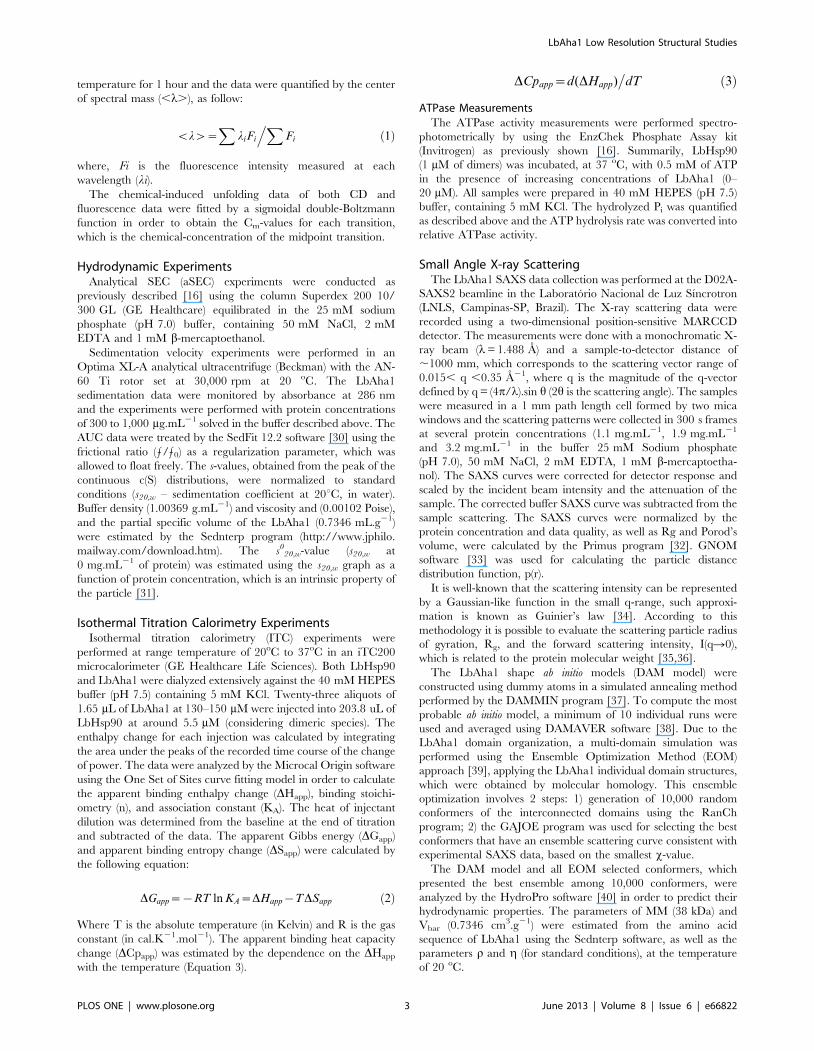
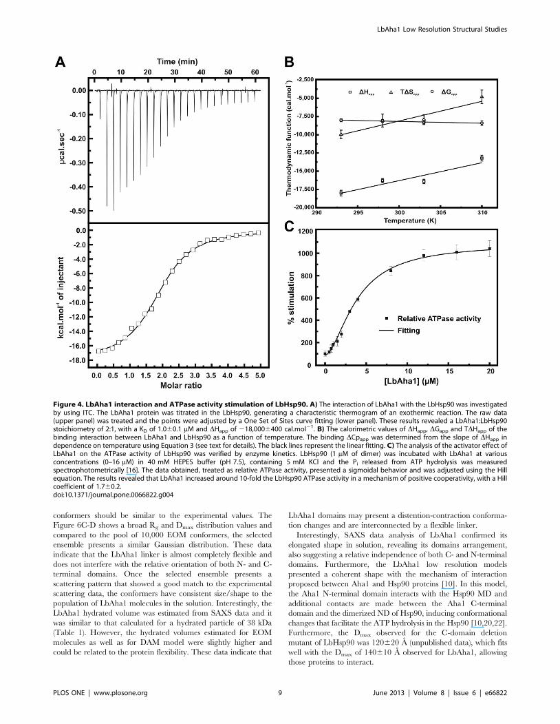
Figure 4. LbAha1 interaction and ATPase activity stimulation of LbHsp90. A) The interaction of LbAha1 with the LbHsp90 was investigatedby using ITC. The LbAha1 protein was titrated in the LbHsp90, generating a characteristic thermogram of an exothermic reaction. The raw data(upper panel) was treated and the points were adjusted by a One Set of Sites curve fitting (lower panel). These results revealed a LbAha1:LbHsp90stoichiometry of 2:1, with a KD of 1.060.1 mM and DHapp of 218,0006400 cal.mol21. B) The calorimetric values of DHapp, DGapp and TDHapp of thebinding interaction between LbAha1 and LbHsp90 as a function of temperature. The binding DCpapp was determined from the slope of DHapp independence on temperature using Equation 3 (see text for details). The black lines represent the linear fitting. C) The analysis of the activator effect ofLbAha1 on the ATPase activity of LbHsp90 was verified by enzyme kinetics. LbHsp90 (1 mM of dimer) was incubated with LbAha1 at variousconcentrations (0–16 mM) in 40 mM HEPES buffer (pH 7.5), containing 5 mM KCl and the Pi released from ATP hydrolysis was measuredspectrophotometrically [16]. The data obtained, treated as relative ATPase activity, presented a sigmoidal behavior and was adjusted using the Hillequation. The results revealed that LbAha1 increased around 10-fold the LbHsp90 ATPase activity in a mechanism of positive cooperativity, with a Hillcoefficient of 1.760.2.doi:10.1371/journal.pone.0066822.g004
LbAha1 Low Resolution Structural Studies
PLOS ONE | www.plosone.org 9 June 2013 | Volume 8 | Issue 6 | e66822

Aha1 is a Cognate Protein in Three Leishmania SpeciesThe presence and importance of Hsp90 in trypanosomatids has
been shown [3–6]. However little is known about the Hsp90 co-
chaperones, including their in vivo presence and roles. To identify
and investigate the expression profile of the Aha1 protein in
function of time and temperature, western blot analysis was
carried out using three Leishmania species: L. braziliensis, L. guyanensis
and L. chagasi incubated at 26uC or 37uC. Western blotting results
showed that the anti-LbAha1 and anti-LbHsp90 antibodies
recognized native proteins with expected size of 38 kDa and
Figure 5. LbAha1 SAXS curves and data analysis. A) The final scattering curve (open circles) of the LbAha1 was the average of the SAXS curvescollected at various protein concentrations. The solid line represents the SAXS curve fitting until q = 0 A21 performed by the GNOM program duringthe distance distribution function generation. Inset: Guinier analysis of the LbAha1 SAXS curve showing its linearity. B) The particle distancedistribution function of the LbAha1 was constructed by the GNOM program using the SAXS curve shown in A). LbAha1 showed a Dmax of 140610 Aand the p(r) revealed a prolate shape. Inset: Kratky plot showing that LbAha1 is a compact protein.doi:10.1371/journal.pone.0066822.g005
LbAha1 Low Resolution Structural Studies
PLOS ONE | www.plosone.org 10 June 2013 | Volume 8 | Issue 6 | e66822

Figure 6. Low resolution structure and rigid body modeling of the LbAha1. A) DAM model and B) LbAha1 EOM conformer (superposed tothe DAM model) in different views displaying its domain arrangement and the relative independence between them. This EOM conformer, which isone of the 18 selected conformers (Figure 6E and Figure S3– Supporting Information) of the best ensemble simulated for LbAha1, was selected based
LbAha1 Low Resolution Structural Studies
PLOS ONE | www.plosone.org 11 June 2013 | Volume 8 | Issue 6 | e66822

90 kDa, respectively (Fig. 7). As controls, L. braziliensis recombi-
nant proteins were used as standards, both Aha1 and Hsp90
proteins were also identified. Surprisingly, the Aha1 proteins were
found being expressed at normal parasite growth conditions (26uC)
and at heat-shock conditions (37uC). These results clearly show
that Aha1 is a cognate and constitutively expressed protein, as well
as the Hsp90 molecular chaperone.
ConclusionsWe produced the recombinant LbAha1 protein and performed
several experiments in order to characterize its structure-function
relationship. To the best of our knowledge this is the first work that
has characterized the Aha1 protein of a trypanosomatid organism.
We identified Aha1 as well as Hsp90 proteins in three Leishmania
species, including L. braziliensis. The protein expression levels were
similar in parasites incubated at 26 oC and 37 oC, suggesting that
Aha1 is a cognate protein in protozoa. This is interesting data,
since there is increasing interest regarding the Hsp90 molecular
chaperone family of protozoa as a target for inhibition and
treatment of protozoa diseases [4,48]. According to the World
Health Organization, Leishmaniasis is a neglected disease. It is
estimated that worldwide 12 million people are infected and 350
million are at risk of infection [23,24], representing a challenge to
health care, including the need for new medicines [25].
It has been shown that orthologous Aha1 are modular proteins
where the N-terminal domain perform the main interaction
contact with the Hsp90 MD helping to align the Arg380 with the
c-phosphate of ATP [19]. However, the role of the C-terminal,
which influences the Hsp90 ATPase activity, remained in second
plan. Two recent works have shown that the Aha1 C-terminal
domain directly interacts with the Hsp90 ND [20,22]. Here, we
observed that for LbAha1 these two important structural
characteristics are critical for its functional activity: 1) two
relatively independent domains connected by a flexible linker,
and 2) highly elongated shape. The dimension observed allows
on the best superposition with the shape of the DAM model. C) Rg and D) Dmax relative distributions of the LbAha1 conformers calculated by theEOM approach considering the experimental SAXS pattern. The solid lines represent the distribution within the pool of 10,000 conformers and thedotted lines, the Rg- and Dmax-values observed for the best ensemble conformers. E) Representative LbAha1 conformers present in the bestensemble obtained by the EOM. The first conformer was used for superposing to the DAM model in the panel B. The N-terminal and C-terminaldomains are shown in green and red, respectively; the reconstructed missing regions are displayed in blue.doi:10.1371/journal.pone.0066822.g006
Figure 7. Western blotting analysis of Aha1 and Hsp90 molecular chaperones. The expression levels of LbAha1 and LbHsp90 proteins wereanalyzed in three Leishmania species: L. braziliensis, L. guyanensis and L. chagasi cultured at 26uC or 37uC during 2, 4 or 6 h. Rabbit polyclonalantibodies anti-Aha1 and anti-Hsp90 recognized the native and recombinant proteins. Monoclonal anti-a-tubulin antibody was used as loadingcontrol. The arrows indicate the protein bands.doi:10.1371/journal.pone.0066822.g007
LbAha1 Low Resolution Structural Studies
PLOS ONE | www.plosone.org 12 June 2013 | Volume 8 | Issue 6 | e66822

LbAha1 to interact with both Hsp90 ND and Hsp90 MD, either
symmetrically as well as asymmetrically, while inducing a dual
effect. The domain independence pointed out that LbAha1 is a
modular protein which allows the Aha1 C-terminal domain to
dock on the Hsp90 ND after the first contact has been established
between Aha1 N-terminal and Hsp90 MD. These characteristics
allow LbAha1 (and orthologous proteins) to synergistically act on
stimulating the Hsp90 ATPase activity by influencing both Arg380
(numbering of yHsp90) in the Hsp90 MD and some catalytic
residues in the Hsp90 ND.
Functionally, LbAha1 interacted with LbHsp90 dimer in a 2:1
stoichiometry and with dissociation constant at the micromolar
range. The thermodynamic data also indicate that the interaction
was guided by enthalpy and with an entropy-opposing interaction
probably due to the freedom degree restriction of LbHsp90.
Moreover, LbAha1 stimulated LbHsp90 ATPase activity 10-fold
in a sigmoidal curve suggesting a positive cooperative mechanism
on the LbHsp90 ATPase activity stimulation. Taken all together,
LbAha1 shares several structural and functional properties with
the human and yeast orthologues, suggesting similar functional
mechanism among these proteins despite the low conservation
degree in the amino acid sequence.
Supporting Information
Figure S1 Sequence alignment analysis of LbAha1. The
LbAha1 was compared with the hAha1 (A) and yAha1 (B) proteins
in order to investigate its evolutionary conservation. LbAha1
shares 30/49% and 23/41% of identity/similarity with hAha1 and
yAha1, respectively. These values indicate a low conservation
among the amino acid sequences, which could lead to peculiarities
in the mechanism of action of the LbAha1 in the LbHsp90
ATPase activity stimulation. The red boxes represent the already
indentified amino acids involved in the yAha1 N-terminal domain
interaction with the yHsp90 MD (PDB: 1USV). In these regions,
LbAha1 presents 28/51% of identity/similarity to the yAha1
protein, slightly higher than in the rest of the protein sequences.
The N- and C-terminal domains are underlined, and the linker
region is indicated by the box. The conserved amino acid residues
(*), the residues with strongly similar properties (:) and the residues
with weakly similar properties (.) are showed.
(TIF)
Figure S2 Ab initio DAMMIN and EOM simulationsfrom small angle X-ray scattering data. A) The DAMMIN
program generated the ab initio models of the LbAha1 by adjusting
the simulated curve to the experimental SAXS curve in a
simulated annealing method. The average of the simulated curves
of the ab initio models used to reconstruct the low resolution
structure of the LbAha1 is showed (solid line), as well as the
experimental SAXS curve (open circles). The averaged x for these
adjustments was 1.3460.08. B) The EOM routine was used the
RanCh program to create 10,000 random LbAha1 models and the
best ensemble based on the experimental SAXS curve of the
protein (open circles) was selected by the GAJOE program. The
ensemble with the better curve adjustment (solid line) was selected
and presented a x-value of about 1.96.
(TIF)
Figure S3 The best LbAha1 conformers generated bythe EOM routine. The RanCh program generated 10,000
random models of LbAha1, from which 18 models were selected
by the GAJOE program based on the fitting goodness to the
experimental SAXS curve (Fig. S2B). All the models above
represent some possible LbAha1 conformers that reflect the
experimental SAXS data. The N- and C-terminal domains are
showed in green and red, respectively. The missing regions
reconstructed by the RanCh program are shown in blue.
(TIF)
Acknowledgments
We acknowledge the Spectroscopy and Calorimetry Laboratory at
Brazilian Biosciences National Laboratory (LNBio/CNPEM-ABTLuS,
Campinas, Brazil) for making the AUC device available. We also thank the
Brazilian Synchrotron Light Laboratory (LNLS/CNPEM-ABTLuS, Cam-
pinas, Brazil) for the use of the SAXS beamline. We thank Prof. Marcel
Tabak for making the fluorimeter available.
Author Contributions
Conceived and designed the experiments: JCB LRSB SMFM TVS.
Performed the experiments: JCB TVS MMA IMS KPS SMFM FERG.
Analyzed the data: JCB TVS LRSB SMFM. Contributed reagents/
materials/analysis tools: JCB SMFM. Wrote the paper: JCB TVS LRSB
SMFM.
References
1. Li J, Soroka J, Buchner J (2012) The Hsp90 chaperone machinery:
Conformational dynamics and regulation by co-chaperones. BBA-Mol Cell
Res 1823: 624–635. doi: 10.1016/j.bbamcr.2011.09.003.
2. Krukenberg KA, Street TO, Lavery LA, Agard DA (2011) Conformational
dynamics of the molecular chaperone Hsp90. Q Rev Biophys 44: 229–255.
3. Wiesgigl M, Clos J (2001) Heat shock protein 90 homeostasis controls stage
differentiation in Leishmania donovani. Mol Biol Cell 12: 3307–3316.
4. Shonhai A, Maier A, Przyborski J, Blatch G (2011) Intracellular Protozoan
Parasites of Humans: The Role of Molecular Chaperones in Development and
Pathogenesis. Protein Pept Lett 18: 143–157. 10.2174/092986611794475002.
5. Pavithra SR, Banumathy G, Joy O, Singh V, Tatu U (2004) Recurrent fever
promotes Plasmodium falciparum development in human erythrocytes. J Biol
Chem 279: 46692–46699.
6. Petersen AL, Guedes CE, Versoza CL, Lima JGB, de Freitas LA, et al. (2012)
17-AAG Kills Intracellular Leishmania amazonensis while Reducing Inflam-
matory Responses in Infected Macrophages. PLoS ONE 7: e49496.
doi:10.1371/journal.pone.0049496.
7. Roy N, Nageshan RK, Ranade S, Tatu U (2012) Heat shock protein 90 from
neglected protozoan parasites. BBA - Mol Cell Res 1823: 707–711. doi:
10.1016/j.bbamcr.2011.12.003.
8. Wandinger SK, Richter K, Buchner J (2008) The Hsp90 chaperone machinery.
J Biol Chem 283: 18473–18477.
9. Welch WJ, Feramisco JR (1982) Purification of the Major Mammalian Heat-
Shock Proteins. J Biol Chem 257: 4949–4959.
10. Hessling M, Richter K, Buchner J (2009) Dissection of the ATP-induced
conformational cycle of the molecular chaperone Hsp90. Nat Struct Mol Biol
16: 287–293. 10.1038/nsmb.1565.
11. Meyer P, Prodromou C, Hu B, Vaughan C, Roe SM, et al. (2003) Structural
and Functional Analysis of the Middle Segment of Hsp90: Implications for ATP
Hydrolysis and Client Protein and Cochaperone Interactions. Mol Cell 11: 647–
658. doi: 10.1016/S1097–2765(03)00065–0.
12. Holmes JL, Sharp SY, Hobbs S, Workman P (2008) Silencing of HSP90
cochaperone AHA1 expression decreases client protein activation and increases
cellular sensitivity to the HSP90 inhibitor 17-allylamino-17-demethoxygeldana-
mycin. Cancer Res 68: 1187–1196.
13. Zurawska A, Urbanski J, Matuliene J, Baraniak J, Klejman MP, et al. (2010)
Mutations that increase both Hsp90 ATPase activity in vitro and Hsp90 drug
resistance in vivo. BBA-Mol Cell Res 1803: 575–583. doi: 10.1016/
j.bbamcr.2010.03.002.
14. Obermann WMJ, Sondermann H, Russo AA, Pavletich NP, Hartl FU (1998) In
Vivo Function of Hsp90 Is Dependent on ATP Binding and ATP Hydrolysis.
J Cell Biol 143: 901–910.
15. Panaretou B, Prodromou C, Roe SM, O’Brien R, Ladbury JE, et al. (1998) ATP
binding and hydrolysis are essential to the function of the Hsp90 molecular
chaperone in vivo. EMBO J 17: 4829–4836. 10.1093/emboj/17.16.4829.
16. Silva KP, Seraphim TV, Borges JC (2013) Structural and functional studies of
Leishmania braziliensis Hsp90. BBA - Proteins Proteom 1834: 351–361. doi:
10.1016/j.bbapap.2012.08.004.
LbAha1 Low Resolution Structural Studies
PLOS ONE | www.plosone.org 13 June 2013 | Volume 8 | Issue 6 | e66822

17. Panaretou B, Siligardi G, Meyer P, Maloney A, Sullivan JK, et al. (2002)
Activation of the ATPase activity of Hsp90 by the stress-regulated cochaperone
Aha1. Mol Cell 10: 1307–1318.
18. Hawle P, Siepmann M, Harst A, Siderius M, Reusch HP, et al. (2006) The
Middle Domain of Hsp90 Acts as a Discriminator between Different Types of
Client Proteins. Mol Cell Biol 26: 8385–8395.
19. Meyer P, Prodromou C, Liao C, Hu B, Mark Roe S, et al. (2004) Structural
basis for recruitment of the ATPase activator Aha1 to the Hsp90 chaperone
machinery. EMBO J 23: 511–519. 10.1038/sj.emboj.7600060.
20. Koulov AV, LaPointe P, Lu BW, Razvi A, Coppinger J, et al. (2010) Biological
and Structural Basis for Aha1 Regulation of Hsp90 ATPase Activity in
Maintaining Proteostasis in the Human Disease Cystic Fibrosis. Mol Biol Cell
21: 871–884.
21. Lotz GP, Lin H, Harst A, Obermann WMJ (2003) Aha1 Binds to the Middle
Domain of Hsp90, Contributes to Client Protein Activation, and Stimulates the
ATPase Activity of the Molecular Chaperone. J Biol Chem 278: 17228–17235.
22. Retzlaff M, Hagn F, Mitschke L, Hessling M, Gugel F, et al. (2010) Asymmetric
Activation of the Hsp90 Dimer by Its Cochaperone Aha1. Mol Cell 37: 344–
354.
23. WHO (2007) World Heald Organisation: Control of leishmaniasis. Report by
the Secretariat.
24. Gonzalez U, Pinart M, Rengifo-Pardo M, Macaya A, Alvar J, et al. (2009)
Interventions for American cutaneous and mucocutaneous leishmaniasis
( R e v i e w ) . C o c h r a n e D b S y s t R e v C D 0 0 4 8 3 4 . 1 0 . 1 0 0 2 /
14651858.CD004834.pub2.
25. Croft SL, Olliaro P (2011) Leishmaniasis chemotherapy - challenges and
opportunities. Clin Microbiol Infec 17: 1478–1483. 10.1111/j.1469–
0691.2011.03630.x.
26. Arnold K, Bordoli L, Kopp J, Schwede T (2006) The SWISS-MODEL
workspace: a web-based environment for protein structure homology modelling.
Bioinformatics 22: 195–201.
27. Laskowski RA, MacArthur MW, Smith DK, Jones DT, Hutchinson EG, et al
(1994) PROCHECK v.3.0 - Program to Check the Stereochemistry Quality of
Protein structures - Operating Instructions., version 3.0.
28. Kabsch W, Sander C (1983) Dictionary of Protein Secondary Structure -
Pattern-Recognition of Hydrogen-Bonded and Geometrical Features. Biopoly-
mers 22: 2577–2637.
29. Whitmore L, Wallace BA (2008) Protein secondary structure analyses from
circular dichroism spectroscopy: Methods and reference databases. Biopolymers
89: 392–400. 10.1002/bip.20853.
30. Brown PH, Balbo A, Schuck P (2009) On the analysis of sedimentation velocity
in the study of protein complexes. Eur Biophys J Biophys Lett 38: 1079–1099.
31. Borges JC, Ramos CHI (2011) Analysis of molecular targets of mycobacterium
tuberculosis by analytical ultracentrifugation. Curr Med Chem 18: 1276–1285.
32. Konarev PV, Volkov VV, Sokolova AV, Koch MHJ, Svergun DI (2003)
PRIMUS: a Windows PC-based system for small-angle scattering data analysis.J App Crystallogr 36: 1277–1282.
33. Svergun DI (1992) Determination of the Regularization Parameter in Indirect-
Transform Methods Using Perceptual Criteria. J Appl Cryst 25: 495–503.34. Fournet G, Guinier A (1955) Small angle scattering of X-rays. Translated by
Walker, C.B. and Yudowitch, K.L. In: New York: John Wiley & Sons. 7–78.35. Mylonas E, Svergun DI (2007) Accuracy of molecular mass determination of
proteins in solution by small-angle X-ray scattering. J Appl Cryst 40: S245-S249.
36. Orthaber D, Bergmann A, Glatter O (2000) SAXS experiments on absolutescale with Kratky systems using water as a secondary standard. J Appl Cryst 33:
218–225.37. Svergun DI (1999) Restoring low resolution structure of biological macromol-
ecules from solution scattering using simulated annealing. Biophys J 76: 2879–2886.
38. Volkov VV, Svergun DI (2003) Uniqueness of ab initio shape determination in
small-angle scattering. J Appl Cryst 36: 860–864.39. Petoukhov MV, Franke D, Shkumatov AV, Tria G, Kikhney AG, et al. (2012)
New developments in the ATSAS program package for small-angle scatteringdata analysis. Journal of Applied Crystallography 45: 342–350.
40. de la Torre JG, Huertas ML, Carrasco B (2000) Calculation of hydrodynamic
properties of globular proteins from their atomic-level structure. Biophys J 78:719–730.
41. Siligardi G, Hu B, Panaretou B, Piper PW, Pearl LH, et al. (2004) Co-chaperoneregulation of conformational switching in the Hsp90 ATPase cycle. J Biol Chem
279: 51989–51998.42. Niedzwiecka A, Stepinski J, Darzynkiewicz E, Sonenberg N, Stolarski R (2002)
Positive Heat Capacity Change upon Specific Binding of Translation Initiation
Factor eIF4E to mRNA 59 Cap. Biochemistry 41: 12140–12148. doi: 10.1021/bi0258142.
43. Zhou YL, Liao JM, Du F, Liang Y (2005) Thermodynamics of the interaction ofxanthine oxidase with superoxide dismutase studied by isothermal titration
calorimetry and fluorescence spectroscopy. Thermochimica Acta 426: 173–178.
44. Pullen L, Bolon DN (2011) Enforced N-domain Proximity Stimulates Hsp90ATPase Activity and Is Compatible with Function in Vivo. J Biol Chem 286:
11091–11098.45. Svergun DI, Koch MHJ (2003) Small-angle scattering studies of biological
macromolecules in solution. Rep Prog Phys 66: 1735–1782.46. Silva JC, Borges JC, Cyr DM, Ramos CHI, Torriani IL (2011) Central domain
deletions affect the SAXS solution structure and function of Yeast Hsp40
proteins Sis1 and Ydj1. BMC Struct Biol 11: 40. 10.1186/1472–6807–11–40.47. Bernado P, Mylonas E, Petoukhov MV, Blackledge M, Svergun DI (2007)
Structural Characterization of Flexible Proteins Using Small-Angle X-rayScattering. J Am Chem Soc 129: 5656–5664. doi: 10.1021/ja069124n.
48. Shonhai A (2010) Plasmodial heat shock proteins: targets for chemotherapy.
FEMS Immunol Med Mic 58: 61–74. 10.1111/j.1574–695X.2009.00639.x.
LbAha1 Low Resolution Structural Studies
PLOS ONE | www.plosone.org 14 June 2013 | Volume 8 | Issue 6 | e66822


![Discovery of (2,4-Dihydroxy-5-isopropylphenyl)-[5-(4-methylpiperazin-1-ylmethyl)-1,3-dihydroisoindol-2-yl]methanone (AT13387), a Novel Inhibitor of the Molecular Chaperone Hsp90 by](https://static.fdokumen.com/doc/165x107/6337b1196f78ac31240eb3a7/discovery-of-24-dihydroxy-5-isopropylphenyl-5-4-methylpiperazin-1-ylmethyl-13-dihydroisoindol-2-ylmethanone.jpg)