Terazosin activates Pgk1 and Hsp90 to promote stress resistance
10
© 2014 Nature America, Inc. All rights reserved. NATURE CHEMICAL BIOLOGY | ADVANCE ONLINE PUBLICATION | www.nature.com/naturechemicalbiology 1 ARTICLE PUBLISHED ONLINE: 10 NOVEMBER 2014 | DOI: 10.1038/NCHEMBIO.1657 S ymptoms of a wide variety of human diseases, such as neurode- generative diseases, autoimmune diseases, heart failure, stroke and sepsis, are caused by the overwhelming activation of cell death and tissue damage. Sepsis, for example, is initiated by bacte- rial infection, which triggers massive apoptosis in immune systems and the failure of multiple organ functions 1 . However, the clinical treatment of sepsis has extensively relied on antimicrobial treatment and supportive care, which have limited efficacy owing to compli- cations such as drug resistance, antibiotic toxicity, endotoxins, cell death and organ dysfunction 2,3 . Similarly, brain stroke, an age- related vascular disease involving massive cell death in the brain, is another leading cause of death worldwide. Currently, no effective neuroprotective therapy has been developed beyond thrombolytic therapy to restore clotted blood vessels 4 . Apoptosis, which is mediated by caspases, was discovered in the 1990s 5 . Since then, no caspase inhibitor has passed clinical trials, and the discovery of new inhibitors remains a major challenge in biomedical research 6 . To manipulate apoptosis, efforts have been focused on the endogenous regulatory factors, including the inhibi- tor of apoptosis family proteins, Bcl-2 family proteins and chaperone proteins 7,8 . For example, the inhibitor of apoptosis family proteins are heavily pursued drug targets for cancer therapeutics 9 . Hsp90, a taxonomically highly conserved chaperone, has been reported to have important roles in the regulation of cellular homeostasis and stress response 10 . It has been reported that Hsp90 has many biologically important client proteins, especially kinases and hor- mone receptors 11,12 . Hsp90 is highly dynamic, and its conforma- tional change is regulated by its ATPase activity, which stabilizes its interactions with client proteins 10 . Considering its prominent anti- apoptotic and protective effects in cancer cells, many Hsp90 inhibi- tors have been developed as potential anticancer drugs to attenuate cancer cell survival. However, no Hsp90 activator that may protect cells from stresses and cell death has been reported. Here, we devised a screen to search for new antiapoptotic drugs from a pool of bioactive small molecules 13 . Once a candidate com- pound is identified, investigations of its targets and mode of action may enable its rapid translation to clinical applications. We found that TZ could rescue rodent models of sepsis and stoke in vivo. Notably, it inhibited apoptosis in macrophages, most likely through activation of Pgk1 and Hsp90. RESULTS TZ rescued reaper-mediated apoptosis in Drosophila We designed our search for apoptosis inhibitors in Drosophila on the rationale that its apoptotic pathway is conserved in mammals 14 and that flies are economical for compound screening. To induce apoptosis, the expression of reaper (rpr) was driven by a heat- activated promoter, HS-Gal4 (we refer to their progeny as HS>rpr). Consistent with the previous report that ectopically expressed rpr can induce widespread apoptosis and organ death in Drosophila 15 , we observed that approximately 75% of the progeny was dead after 24 h upon heat shock at 37 °C for 45 min (Supplementary Results, Supplementary Fig. 1a,b). To screen for apoptosis inhibitors, we first assessed global tran- scriptional alteration during apoptosis using Affymetrix microarrays. Comparing the HS>rpr flies to the genetic background–matched con- trol HS × y w 67c23 flies (progeny of HS-Gal4 flies crossed with y w 67c23 flies), 2,169 genes showed more than 50% altered expression levels (genes identified from the microarrays are listed in Supplementary Data Set 1). Then, these genes were converted to homologous human genes (the apoptotic signature genes after conversion into human homologs are listed in Supplementary Data Set 2) and entered into Cmap for compound matching 13 (matched drugs are listed in Supplementary Data Set 3). Twenty-five compounds were selected (Supplementary Data Set 4) and were fed to the HS>rpr flies with the concentrations indicated in Supplementary 1 State Key Laboratory of Biomembrane and Membrane Biotechnology, School of Life Sciences, Peking University, Beijing, China. 2 Beijing Institute for Brain Disorder and Beijing Tiantan Hospital, Capital Medical University, Beijing, China. 3 School of Life Sciences, University of Science and Technology of China, Hefei, China. 4 School of Life Science, Peking University, Beijing, China. 5 Key Laboratory of Bioorganic Chemistry and Molecular Engineering, College of Chemistry and Molecular Engineering, Peking University, Beijing, China. 6 National Institute of Biological Sciences, Beijing, China. 7 Dana-Farber Cancer Institute, Harvard Medical School, Boston, Massachusetts, USA. 8 Key Laboratory of Chemical Genomics, School of Chemical Biology and Biotechnology, Peking University Shenzhen Graduate School, Shenzhen, China. 9 These authors contributed equally to this work. *e-mail: [email protected], [email protected] or [email protected] Terazosin activates Pgk1 and Hsp90 to promote stress resistance Xinping Chen 1,2,9 , Chunyue Zhao 1,2,9 , Xiaolong Li 3,4,9 , Tao Wang 1,2,9 , Yizhou Li 5 , Cheng Cao 5 , Yuehe Ding 6 , Mengqiu Dong 6 , Lorenzo Finci 4 , Jia-huai Wang 3,4,7 *, Xiaoyu Li 5,8 * & Lei Liu 2 * Drugs that can protect against organ damage are urgently needed, especially for diseases such as sepsis and brain stroke. We discovered that terazosin (TZ), a widely marketed 1-adrenergic receptor antagonist, alleviated organ damage and improved survival in rodent models of stroke and sepsis. Through combined studies of enzymology and X-ray crystallography, we dis- covered that TZ binds a new target, phosphoglycerate kinase 1 (Pgk1), and activates its enzymatic activity, probably through 2,4-diamino-6,7-dimethoxyisoquinoline′s ability to promote ATP release from Pgk1. Mechanistically, the ATP generated from Pgk1 may enhance the chaperone activity of Hsp90, an ATPase known to associate with Pgk1. Upon activation, Hsp90 promotes multistress resistance. Our studies demonstrate that TZ has a new protein target, Pgk1, and reveal its corresponding biological effect. As a clinical drug, TZ may be quickly translated into treatments for diseases including stroke and sepsis.
Transcript of Terazosin activates Pgk1 and Hsp90 to promote stress resistance
©20
14 N
atu
re A
mer
ica,
Inc.
All
rig
hts
res
erve
d.
nature CHeMICaL BIOLOGY | AdvAnce online publicAtion | www.nature.com/naturechemicalbiology 1
articlepuBLIsHed OnLIne: 10 nOveMBer 2014 | dOI: 10.1038/nCHeMBIO.1657
Symptoms of a wide variety of human diseases, such as neurode-generative diseases, autoimmune diseases, heart failure, stroke and sepsis, are caused by the overwhelming activation of cell
death and tissue damage. Sepsis, for example, is initiated by bacte-rial infection, which triggers massive apoptosis in immune systems and the failure of multiple organ functions1. However, the clinical treatment of sepsis has extensively relied on antimicrobial treatment and supportive care, which have limited efficacy owing to compli-cations such as drug resistance, antibiotic toxicity, endotoxins, cell death and organ dysfunction2,3. Similarly, brain stroke, an age- related vascular disease involving massive cell death in the brain, is another leading cause of death worldwide. Currently, no effective neuroprotective therapy has been developed beyond thrombolytic therapy to restore clotted blood vessels4.
Apoptosis, which is mediated by caspases, was discovered in the 1990s5. Since then, no caspase inhibitor has passed clinical trials, and the discovery of new inhibitors remains a major challenge in biomedical research6. To manipulate apoptosis, efforts have been focused on the endogenous regulatory factors, including the inhibi-tor of apoptosis family proteins, Bcl-2 family proteins and chaperone proteins7,8. For example, the inhibitor of apoptosis family proteins are heavily pursued drug targets for cancer therapeutics9. Hsp90, a taxonomically highly conserved chaperone, has been reported to have important roles in the regulation of cellular homeostasis and stress response10. It has been reported that Hsp90 has many biologically important client proteins, especially kinases and hor-mone receptors11,12. Hsp90 is highly dynamic, and its conforma-tional change is regulated by its ATPase activity, which stabilizes its interactions with client proteins10. Considering its prominent anti-apoptotic and protective effects in cancer cells, many Hsp90 inhibi-tors have been developed as potential anticancer drugs to attenuate cancer cell survival. However, no Hsp90 activator that may protect cells from stresses and cell death has been reported.
Here, we devised a screen to search for new antiapoptotic drugs from a pool of bioactive small molecules13. Once a candidate com-pound is identified, investigations of its targets and mode of action may enable its rapid translation to clinical applications. We found that TZ could rescue rodent models of sepsis and stoke in vivo. Notably, it inhibited apoptosis in macrophages, most likely through activation of Pgk1 and Hsp90.
RESULTSTZ rescued reaper-mediated apoptosis in DrosophilaWe designed our search for apoptosis inhibitors in Drosophila on the rationale that its apoptotic pathway is conserved in mammals14 and that flies are economical for compound screening. To induce apoptosis, the expression of reaper (rpr) was driven by a heat- activated promoter, HS-Gal4 (we refer to their progeny as HS>rpr). Consistent with the previous report that ectopically expressed rpr can induce widespread apoptosis and organ death in Drosophila15, we observed that approximately 75% of the progeny was dead after 24 h upon heat shock at 37 °C for 45 min (Supplementary Results, Supplementary Fig. 1a,b).
To screen for apoptosis inhibitors, we first assessed global tran-scriptional alteration during apoptosis using Affymetrix microarrays. Comparing the HS>rpr flies to the genetic background–matched con-trol HS × y w67c23 flies (progeny of HS-Gal4 flies crossed with y w67c23 flies), 2,169 genes showed more than 50% altered expression levels (genes identified from the microarrays are listed in Supplementary Data Set 1). Then, these genes were converted to homologous human genes (the apoptotic signature genes after conversion into human homologs are listed in Supplementary Data Set 2) and entered into Cmap for compound matching13 (matched drugs are listed in Supplementary Data Set 3). Twenty-five compounds were selected (Supplementary Data Set 4) and were fed to the HS>rpr flies with the concentrations indicated in Supplementary
1State Key laboratory of biomembrane and Membrane biotechnology, School of life Sciences, peking university, beijing, china. 2beijing institute for brain disorder and beijing tiantan Hospital, capital Medical university, beijing, china. 3School of life Sciences, university of Science and technology of china, Hefei, china. 4School of life Science, peking university, beijing, china. 5Key laboratory of bioorganic chemistry and Molecular engineering, college of chemistry and Molecular engineering, peking university, beijing, china. 6national institute of biological Sciences, beijing, china. 7dana-Farber cancer institute, Harvard Medical School, boston, Massachusetts, uSA. 8Key laboratory of chemical Genomics, School of chemical biology and biotechnology, peking university Shenzhen Graduate School, Shenzhen, china. 9these authors contributed equally to this work. *e-mail: [email protected], [email protected] or [email protected]
terazosin activates pgk1 and Hsp90 to promote stress resistanceXinping Chen1,2,9, Chunyue Zhao1,2,9, Xiaolong Li3,4,9, tao Wang1,2,9, Yizhou Li5, Cheng Cao5, Yuehe ding6, Mengqiu dong6, Lorenzo Finci4, Jia-huai Wang3,4,7*, Xiaoyu Li5,8* & Lei Liu2*
Drugs that can protect against organ damage are urgently needed, especially for diseases such as sepsis and brain stroke. We discovered that terazosin (TZ), a widely marketed 1-adrenergic receptor antagonist, alleviated organ damage and improved survival in rodent models of stroke and sepsis. Through combined studies of enzymology and X-ray crystallography, we dis-covered that TZ binds a new target, phosphoglycerate kinase 1 (Pgk1), and activates its enzymatic activity, probably through 2,4-diamino-6,7-dimethoxyisoquinoline′s ability to promote ATP release from Pgk1. Mechanistically, the ATP generated from Pgk1 may enhance the chaperone activity of Hsp90, an ATPase known to associate with Pgk1. Upon activation, Hsp90 promotes multistress resistance. Our studies demonstrate that TZ has a new protein target, Pgk1, and reveal its corresponding biological effect. As a clinical drug, TZ may be quickly translated into treatments for diseases including stroke and sepsis.
©20
14 N
atu
re A
mer
ica,
Inc.
All
rig
hts
res
erve
d.
2 nature CHeMICaL BIOLOGY | AdvAnce online publicAtion | www.nature.com/naturechemicalbiology
article NATURE cHEMicAL biOLOgy dOI: 10.1038/nCHeMBIO.1657
Data Set 5. From this screen, we identified TZ, an α1-adrenergic receptor antagonist and a widely marketed antihypertension drug, as the only compound that significantly (P = 0.0018) improved sur-vival of the HS>rpr flies (Supplementary Fig. 1b).
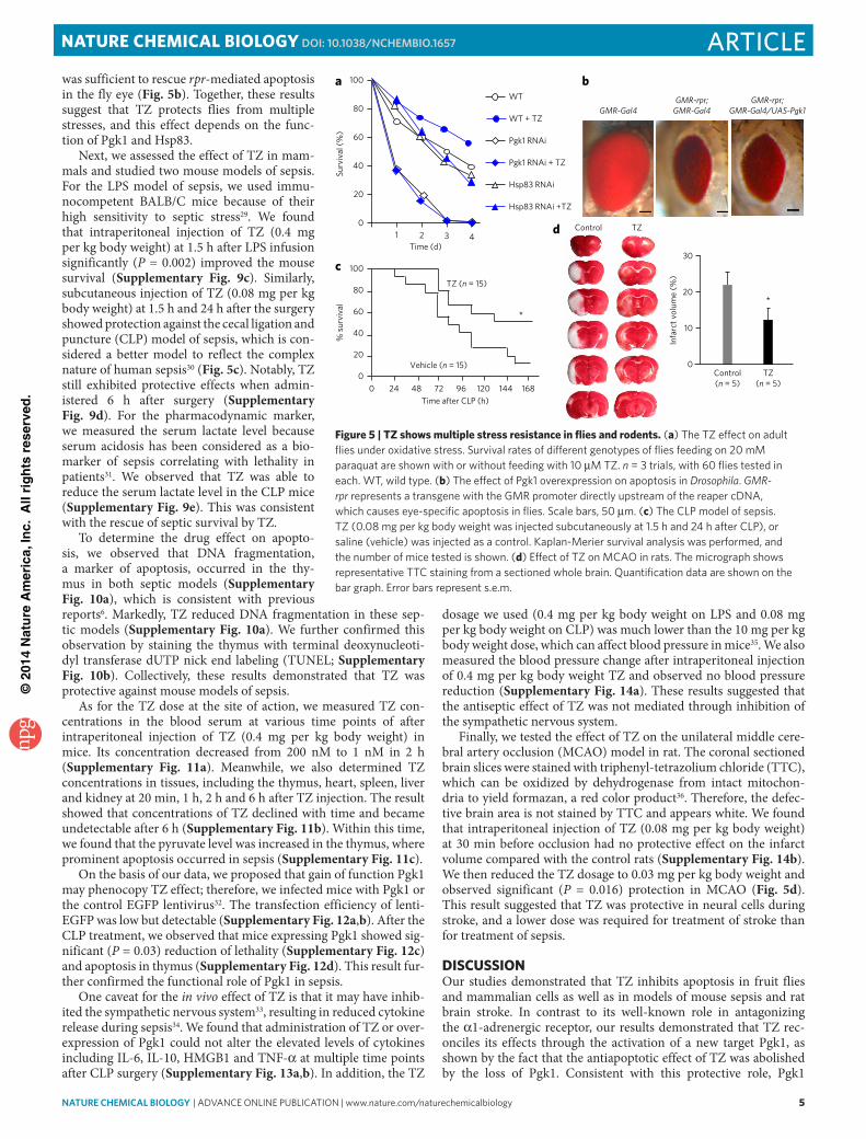
TZ inhibited apoptosis in cultured mammalian cellsTo examine whether TZ inhibits apoptosis in cultured mammalian cells, RAW 264.7 cells were pretreated for 18–24 h before induction of apoptosis by lipopolysaccharide (LPS) and interferon-γ (IFN-γ)16. After treatment for 18–24 h, apoptosis was determined by annexin V staining. The result showed that TZ suppressed apoptosis in these cells (Fig. 1a and Supplementary Fig. 2a), and these results were further verified by the lactate dehydrogenase (LDH) assay (Supplementary Fig. 2b). Furthermore, to test whether TZ blocks apoptosis induced by a different stressor, hydrogen peroxide (H2O2) was applied, which induces caspase-mediated apoptosis, a type of cell death that can be blocked by zVad, a pan-caspase inhibitor (Supplementary Fig. 2c). Again, TZ exhibited a protective effect against H2O2-induced apop-tosis by an LDH release assay (Fig. 1b), active caspase-3 western blot (Fig. 1c and Supplementary Fig. 2d) and caspase activity assay (Fig. 1d). These results demonstrated that TZ inhibited caspase- mediated apoptosis triggered by diverse inducers in macrophages.
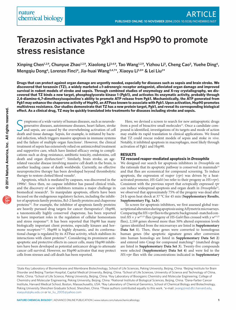
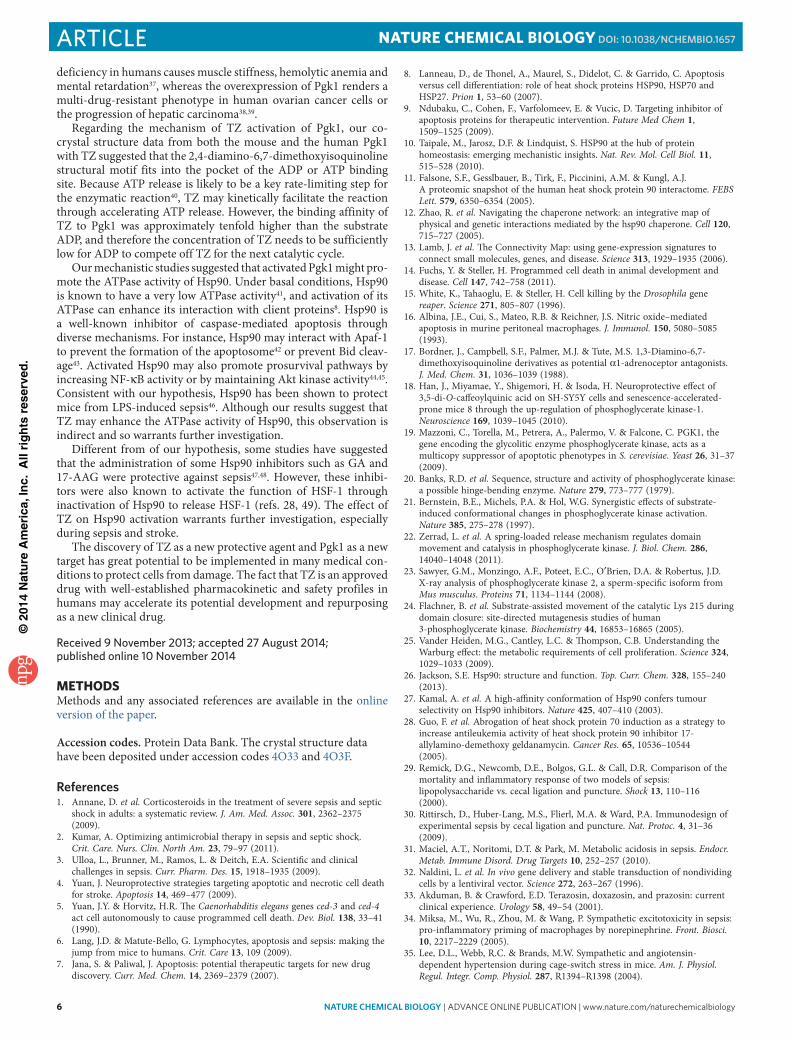
TZ was an activator of Pgk1To test whether the effect of TZ on apoptosis was mediated through the α1-adrenergic receptor, we modified TZ by replacing a nitrogen positioned on the quinazoline ring with a carbon (Fig. 2a; TZ-md, 2), which is known to reduce its binding to the α1-adrenoceptor by approximately 1,000-fold17. Indeed, TZ-md could not block intra-cellular calcium elevation induced by metaraninol, an α1 receptor agonist (Supplementary Fig. 3a). However, TZ-md still had an antiapoptotic effect in RAW 264.7 cells (Supplementary Fig. 3b). This result suggested that TZ might exert an antiapoptotic effect via an unknown target.
To search for potential new targets, we synthesized a TZ derivative, ‘TZ-TA’ (3), by conjugating a chemical handle at the 4-amino position of the quinazoline motif (Fig. 2a). TZ-TA had preserved the antiapoptotic property of TZ in RAW 264.7 cells (Supplementary Fig. 3b). Then, TZ-TA was immobilized onto an Affi-Gel support to immobilize any binding proteins (Supplementary Note 1). Comparing the pulldown results from the RAW 264.7 cell lysate and the lysate containing saturated TZ as a soluble competitor, we observed a distinct protein band specifi-cally bound to the Affi-Gel–TZ-TA beads, with a molecular weight of ~46 kDa (Fig. 2b). This band was extracted and identified by MS as Pgk1 (Supplementary Fig. 3c and Supplementary Note 2). Moreover, the purified His-tagged mouse Pgk1 could be pulled down by the Affi-Gel–TZ-TA beads (Fig. 2c), suggesting that TZ directly bound Pgk1.
As one of the core enzymes in glycolysis, Pgk1 had been reported to act as a suppressor of apoptosis in yeast and improve the via-bility in the human SH-SY5Y cell line upon amyloid stimulus18,19. To test the effect of TZ on Pgk1 activity, we performed an in vitro assay by providing the purified Pgk1 with substrates (ADP and 1,3-bisphosphoglycerate) and measured the forward reaction in which NADH accumulated (Online Methods). Notably, at higher dosages (2.5 μM and 25 μM), TZ inhibited Pgk1, whereas at lower dosages (0.5 μM to 2.5 nM) it activated Pgk1 (Fig. 2d). To further validate the TZ effect on Pgk1, we measured ATP (a product of Pgk1) and pyruvate (a downstream product of glycolysis) levels in the cell lysate. The ATP level was transiently elevated by nearly 40% in the first minute of the reaction, and the pyruvate level was stably increased by approximately 30% (Fig. 2e). Together, these results suggested that TZ activated Pgk1 at low concentrations.
Furthermore, we determined the effective dosages of TZ to inhibit H2O2-induced apoptosis in macrophages. The results
showed that TZ suppressed apoptosis with concentrations as low as 0.1 μM (Supplementary Fig. 4a).
To further explore the functional role of Pgk1 in modulating apoptosis, we knocked down Pgk1 in RAW 264.7 cells by stably expressing Pgk1 short hairpin RNA (shRNA). The knockdown effi-ciency was shown (Supplementary Fig. 4b). Although knocking down Pgk1 did not affect cell survival, the protective effect of TZ against H2O2-induced apoptosis was abolished by the Pgk1 shRNA, as determined by LDH release assays (Fig. 2f); caspase 3 activa-tion and PARP-1 cleavage were determined by western blotting (Supplementary Fig. 4c). These results indicated that the antiapop-tosis activity of TZ required Pgk1. Consistently, stable expression of Pgk1 in the RAW 264.7 cells by lentiviral transfection reduced PARP-1 cleavage and LDH release after H2O2 treatment (Fig. 2g, and Supplementary Fig. 4d,e). Collectively, these results indicated that Pgk1 was required for the protective effect of TZ.
crystallization of Pgk1 with TZTo investigate how TZ interacts with Pgk1, we crystallized mouse and human Pgk1 (Supplementary Fig. 5 shows protein purifica-tion of Pgk1). Pgk1 consists of N-terminal and C-terminal domains of roughly equal size and a similar Rossman fold. Human Pgk1 (hPgk1) has been crystallized in open20 and closed21 forms. The enzyme spends most of its time in a fully open and resting con-formation for substrate binding and product release, with a short period of time in a closed form, which is used for quick catalysis22. Although the mPgk2 structure has been resolved23, there have been no reports on mPgk1 structure. First, we grew mPgk1 crystals in
Cas
pase
/act
in(f
old
chan
ge)
*
c
*
0
0.5
1.0
1.5
H2O2 H2O2 +Z-vad
H2O2 +TZ
Control H2O2 H2O2 +Z-vad
H2O2 +TZ
d
DEV
Das
e ac
tivity
(fol
d ch
ange
)
0
0.5
1.0
1.5
2.0
*** ***
b
0
10
20
30
40
50
% o
f cel
l dea
th(L
DH
ass
ay)
Control H2O2 H2O2 +Z-vad
H2O2 +TZ
* *
a Control LPS + IFN-γLPS + IFN-γ
+ TZ
Brig
ht fi
eld
Ann
exin
V
Figure 1 | TZ blocks apoptosis in cultured mammalian cells. (a) examples of bright-field and fluorescent images of the RAW 264.7 cells stained with annexin v after induction by lpS and iFn-γ. Scale bar, 25 μm. three independent experiments were performed. (b) the ldH assay of cell death induced by H2o2. RAW 264.7 cells were treated with 0.1% dMSo (control), H2o2 (1 mM), H2o2 plus Z-vad (100 μM) or H2o2 plus tZ (10 μM) in 0.1% dMSo. the percentage of cell death = ldHmedium/(ldHmedium + ldHcell) × 100%; n = 3. All data are presented as the mean + s.e.m., unless otherwise indicated. the P values of a two-tailed t-test for the comparison of the two data sets, the one-way analysis of variance with post-hoc multiple comparison Sidak for three or more data sets and the Kaplan-Meier survival analysis with a log-rank algorithm are ***P < 0.001, **P < 0.01 and *P < 0.05, respectively, throughout all figures. (c) the statistical results of western blotting to detect the active form of caspase 3 are shown. conditions are the same as in b. the relative ratio of caspase 3/β-actin from the H2o2 treatment was set as 1, and the relative ratio of other treatments were shown; n = 3. (d) the effect of tZ on the activity of caspase 3 and caspase 7, measured by a devdase activity assay. the data were normalized to the control conditions (without treatment); n = 3.
©20
14 N
atu
re A
mer
ica,
Inc.
All
rig
hts
res
erve
d.
nature CHeMICaL BIOLOGY | AdvAnce online publicAtion | www.nature.com/naturechemicalbiology 3
articleNATURE cHEMicAL biOLOgy dOI: 10.1038/nCHeMBIO.1657
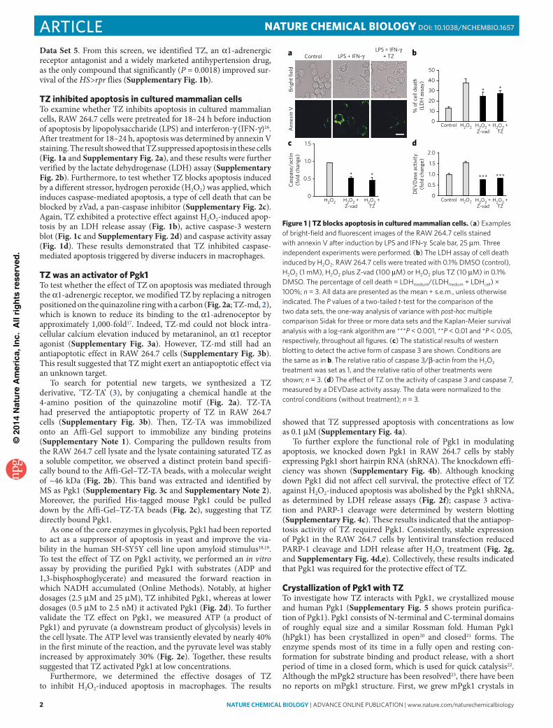
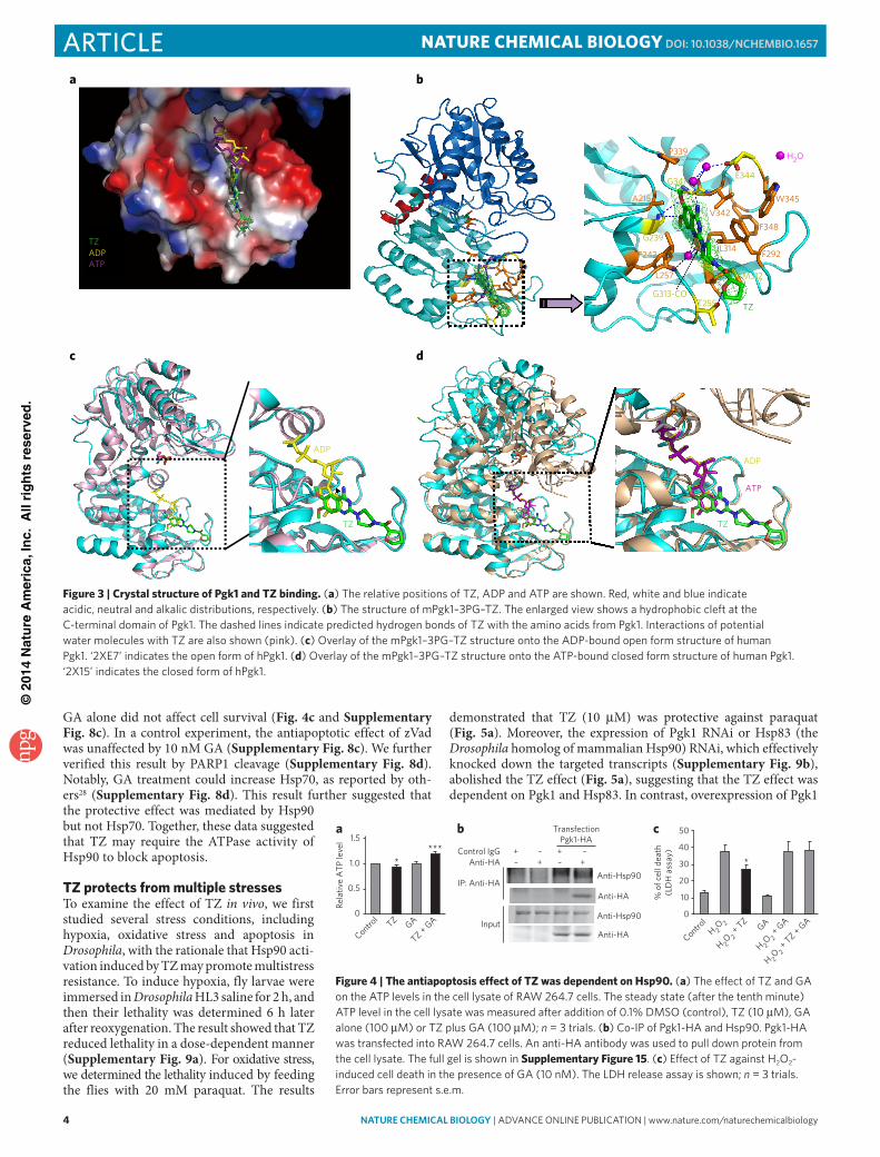
the presence of 3-phosphoglycerate (3PG), a substrate of Pgk1. Next, we obtained the ter-nary co-crystallization complexes for both mouse and human Pgk1–3PG–TZ, with the addition of a saturating amount of TZ (10 mM). The structures were determined using molecular replacement to 2.1 Å. Consistent with the 98% sequence identity between Pgk1 of these two species (Supplementary Fig. 6a), the structure of hPgk1–3PG–TZ could be superposed onto mPgk1–3PG–TZ well, indicating an identical TZ-binding mode to hPgk1 and mPgk1 (Fig. 3a and Supplementary Fig. 6b–g). The only differ-ence between these two structures occurred within a small disordered loop region between residues Thr378 and Asp387 in mPgk1 that was located near the C terminus but was structurally located at the junction between the N- and C-terminal domains (Supplementary Fig. 6c).
From the structure of mPgk1–3PG–TZ, we observed that the three hydrophobic rings of the TZ molecule were surrounded by many hydrophobic residues of the Pgk1 molecule, including the aromatic side chains of Phe242, Phe292, Phe348, Trp345 as well as Leu257, Met312 and Leu314 (Fig. 3b). Markedly, the oxygen and nitrogen atoms of TZ formed a network of hydrogen bonds directly or through water molecules to Pgk1, indicating that drug and enzyme recognition was quite specific (Fig. 3b). We displayed the electrostatic sur-face of the mPgk1–3PG–TZ structure to dem-onstrate the relative position of ADP, ATP and TZ (Fig. 3a). We observed that the phosphate groups of ADP and ATP were oriented toward a positively charged well, whereas the TZ mole-cule extended its hydrophobic tail further away on the opposite direction toward the end of the domain (Fig. 3a). Further, we overlaid the mPgk1–3PG–TZ structure onto the structures of the ADP-bound open form of the hPgk1 (Fig. 3c) and the ATP-bound closed form of the hPgk1 (Fig. 3d). These results indicated that the six-membered benzene ring of TZ’s quinazoline core has the same site as the six-membered pyrimidine ring of the purine in ADP and ATP. Together, our results demonstrated that the TZ binding site overlaps with the ADP and ATP binding site to the enzyme, and they were all inserted deeply into the same hydro-phobic cleft at the C-terminal domain of Pgk1.
The binding mode revealed by the Pgk1–3PG–TZ structures raises a paradox: in vitro enzymatic activity assays (Fig. 2d) indicated that TZ was stimulatory for Pgk1 at lower concentrations; however, as TZ occupies the binding site of ADP and ATP, it should be a com-petitive inhibitor instead of an activator. We rationalize that, on the basis of the chemical equilibrium principle, the effective TZ dos-age for either inhibition or activation should depend on the relative affinity and the concentration of TZ, ADP and ATP for Pgk1. The Pgk1 binding affinity (Kd) of ADP and ATP is 2.9 × 10⁻⁵ M and 3.3 × 10⁻⁴ M, respectively24. According to our measurement by isothermal titration calorimetry (ITC), TZ and TZ-TA bind to Pgk1 with a Kd of 2.78 × 10⁻⁶ M and 2.5 × 10⁻⁶ M, respectively (Supplementary Fig. 7a,b), which is approximately 10 times and 100 times stronger than ADP and ATP, respectively.
The antiapoptotic effect of TZ might depend on Hsp90Although upregulation of glycolytic pathway has been reported to benefit cell survival25, the mechanism for the antiapoptotic activity
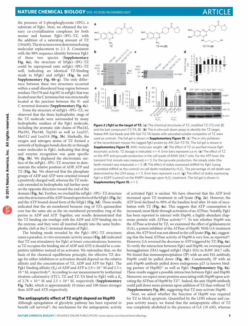
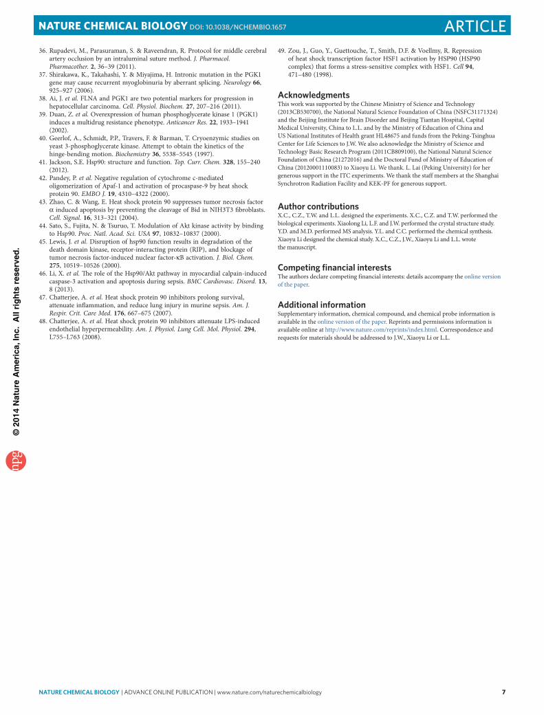
of activated Pgk1 is unclear. We have observed that the ATP level increased upon TZ treatment in cell lysate (Fig. 2e). However, the ATP level declined to 90% of the baseline level after 10 min of incu-bation with TZ (Fig. 4a). This suggests that ATP consumption is increased, most likely through activation of an ATPase. Notably, Pgk1 has been reported to interact with Hsp90, a highly abundant chap-erone protein with ATPase activity11,12. To test whether Hsp90 was the ATPase activated by TZ, we examined the effect of geldanamycin (GA), a potent inhibitor of the ATPase of Hsp90. With GA treatment alone, the ATP level was not altered in the cell lysate (Fig. 4a), suggest-ing that the basal ATPase activity of Hsp90 is very low, as reported26. However, GA reversed the decrease in ATP triggered by TZ (Fig. 4a). To verify the interaction between Pgk1 and Hsp90, we overexpressed hemagglutinin (HA)-tagged mPgk1 (Pgk1-HA) in RAW 264.7 cells. We found that immunoprecipitation (IP) with an anti-HA antibody, Hsp90 could be pulled down (Fig. 4b). Consistently, IP with an anti-Hsp90 antibody pulled down endogenous p23 (a known bind-ing partner of Hsp90)27 as well as Pgk1 (Supplementary Fig. 8a). These results suggest a possible interaction between Pgk1 and Hsp90. Moreover, we expect more proteins associating with Hsp90 upon acti-vating the ATPase of Hsp90 by TZ27. Indeed, the anti-Hsp90 antibody could pull down more proteins upon addition of TZ than without TZ (Supplementary Fig. 8b), suggesting that TZ may activate Hsp90.
Next, we asked whether the function of Hsp90 was required for TZ to block apoptosis. Quantified by the LDH release and cas-pase activity assays, we found that the antiapoptotic effect of TZ was completely abolished in the presence of GA (10 nM), whereas
f
*
% c
ell d
eath
(LD
H a
ssay
)
H2O2 H2O2
0
20
Sh-scramble
Sh-Pgk1
Sh-Pgk1
Sh-Pgk1
+TZ
Sh-scramble
Sh-scramble
+TZ
40
60NS
80
*
0
1.01.5
0.5
2.0
Pyru
vate
(fol
d ch
ange
)
Vehicle TZ(10 µM)
e
0
0.5
1.0
1.5
2.0
ATP
leve
l(f
old
chan
ge)
Vehicle TZ(10 µM)
**
a
N
N
N
O
O
NH2
MeO
MeO
TZ-md (2)
N
N
N
N
O
O
HN
MeO
MeO
TZ-TA (3)
OOH
N
N
N
N
O
O
NH2
MeO
MeO
TZ
g GFP GFP + H2O2 Pgk1 Pgk1 + H2O2
PARP1
HSP 70
β-actin
HSP 90
c
His-Pgk143
55A�-G
el
A�-Gel–T
Z-TA+TZ
A�-Gel–T
Z-TA
MW(kDa)
43
MW(kDa)
55 Pgk1
A�-Gel
A�-Gel-T
Z-TA+TZ
A�-Gel-T
Z-TAb d
–8–6–4–20246
–60
NA
DH
cha
nge
(%)
***
***
* ** ** ** *
25 µ
M2.
5 µM
250
pM
0.5
µM
0.25
µM
50 n
M
25 n
M
2.5
nM
Figure 2 | Pgk1 as the target of TZ. (a) the chemical structures of tZ, modified tZ (tZ-md; 2) and the bait compound (tZ-tA; 3). (b) the in vitro pull-down assay to identify the tZ target. naked Affi-Gel beads and Affi-Gel–tZ-tA beads with saturated soluble competitor of tZ were used as controls. the full gel is shown in Supplementary Figure 15. (c) the in vitro pulldown of the recombinant mouse His-tagged pgk1 protein by Affi-Gel–tZ-tA. the full gel is shown in Supplementary Figure 15. MW, molecular weight. (d) the effect of tZ on purified mouse pgk1 enzymatic activity. tZ dosage is indicated; n = 4. error bars represent s.e.m. (e) the effect of tZ on the Atp and pyruvate production in the cell lysate of RAW 264.7 cells. For the Atp level, the transient first minute was measured; n = 5. For the pyruvate production, the steady state (the tenth minute) was measured; n = 3. (f) the effect of stably expressed shRnA for pgk1 (using scrambled shRnA as the control) on cell death mediated by H2o2. the percentage of cell death was determined by the ldH assay; n = 5. error bars represent s.e.m. (g) the effect of stably expressing pgk1 or eGFp (control) on the pARp1 cleavage upon H2o2 treatment. the full gel is shown in Supplementary Figure 15; n = 3.
©20
14 N
atu
re A
mer
ica,
Inc.
All
rig
hts
res
erve
d.
4 nature CHeMICaL BIOLOGY | AdvAnce online publicAtion | www.nature.com/naturechemicalbiology
article NATURE cHEMicAL biOLOgy dOI: 10.1038/nCHeMBIO.1657
GA alone did not affect cell survival (Fig. 4c and Supplementary Fig. 8c). In a control experiment, the antiapoptotic effect of zVad was unaffected by 10 nM GA (Supplementary Fig. 8c). We further verified this result by PARP1 cleavage (Supplementary Fig. 8d). Notably, GA treatment could increase Hsp70, as reported by oth-ers28 (Supplementary Fig. 8d). This result further suggested that the protective effect was mediated by Hsp90 but not Hsp70. Together, these data suggested that TZ may require the ATPase activity of Hsp90 to block apoptosis.
TZ protects from multiple stressesTo examine the effect of TZ in vivo, we first studied several stress conditions, including hypoxia, oxidative stress and apoptosis in Drosophila, with the rationale that Hsp90 acti-vation induced by TZ may promote multistress resistance. To induce hypoxia, fly larvae were immersed in Drosophila HL3 saline for 2 h, and then their lethality was determined 6 h later after reoxygenation. The result showed that TZ reduced lethality in a dose-dependent manner (Supplementary Fig. 9a). For oxidative stress, we determined the lethality induced by feeding the flies with 20 mM paraquat. The results
demonstrated that TZ (10 μM) was protective against paraquat (Fig. 5a). Moreover, the expression of Pgk1 RNAi or Hsp83 (the Drosophila homolog of mammalian Hsp90) RNAi, which effectively knocked down the targeted transcripts (Supplementary Fig. 9b), abolished the TZ effect (Fig. 5a), suggesting that the TZ effect was dependent on Pgk1 and Hsp83. In contrast, overexpression of Pgk1
a b
ADP
TZ
c d
ADP
ATP
TZ
TZADPATP
H2OP339
G341E344
G239
G313-COT255
M312
TZ
A215
F242
L257
L314
V342F348
F292
W345
Figure 3 | crystal structure of Pgk1 and TZ binding. (a) the relative positions of tZ, Adp and Atp are shown. Red, white and blue indicate acidic, neutral and alkalic distributions, respectively. (b) the structure of mpgk1–3pG–tZ. the enlarged view shows a hydrophobic cleft at the c-terminal domain of pgk1. the dashed lines indicate predicted hydrogen bonds of tZ with the amino acids from pgk1. interactions of potential water molecules with tZ are also shown (pink). (c) overlay of the mpgk1–3pG–tZ structure onto the Adp-bound open form structure of human pgk1. ‘2Xe7’ indicates the open form of hpgk1. (d) overlay of the mpgk1–3pG–tZ structure onto the Atp-bound closed form structure of human pgk1. ‘2X15’ indicates the closed form of hpgk1.
Rela
tive
ATP
leve
l
***
0
Control
TZ GA
TZ + GA
1.0
0.5
1.5
*
a
Anti-Hsp90
Anti-HA
Anti-Hsp90
Anti-HAInput
IP: Anti-HA
TransfectionPgk1-HA
Control IgGAnti-HA
+ – + –– + – +
b c
% o
f cel
l dea
th(L
DH
ass
ay)
0
10
20
Control
H 2O 2
H 2O 2
+ TZ
H 2O 2
+ GA
H 2O 2
+ TZ + GAGA
30
40
50
*
Figure 4 | The antiapoptosis effect of TZ was dependent on Hsp90. (a) the effect of tZ and GA on the Atp levels in the cell lysate of RAW 264.7 cells. the steady state (after the tenth minute) Atp level in the cell lysate was measured after addition of 0.1% dMSo (control), tZ (10 μM), GA alone (100 μM) or tZ plus GA (100 μM); n = 3 trials. (b) co-ip of pgk1-HA and Hsp90. pgk1-HA was transfected into RAW 264.7 cells. An anti-HA antibody was used to pull down protein from the cell lysate. the full gel is shown in Supplementary Figure 15. (c) effect of tZ against H2o2-induced cell death in the presence of GA (10 nM). the ldH release assay is shown; n = 3 trials. error bars represent s.e.m.
©20
14 N
atu
re A
mer
ica,
Inc.
All
rig
hts
res
erve
d.
nature CHeMICaL BIOLOGY | AdvAnce online publicAtion | www.nature.com/naturechemicalbiology 5
articleNATURE cHEMicAL biOLOgy dOI: 10.1038/nCHeMBIO.1657
was sufficient to rescue rpr-mediated apoptosis in the fly eye (Fig. 5b). Together, these results suggest that TZ protects flies from multiple stresses, and this effect depends on the func-tion of Pgk1 and Hsp83.
Next, we assessed the effect of TZ in mam-mals and studied two mouse models of sepsis. For the LPS model of sepsis, we used immu-nocompetent BALB/C mice because of their high sensitivity to septic stress29. We found that intraperitoneal injection of TZ (0.4 mg per kg body weight) at 1.5 h after LPS infusion significantly (P = 0.002) improved the mouse survival (Supplementary Fig. 9c). Similarly, subcutaneous injection of TZ (0.08 mg per kg body weight) at 1.5 h and 24 h after the surgery showed protection against the cecal ligation and puncture (CLP) model of sepsis, which is con-sidered a better model to reflect the complex nature of human sepsis30 (Fig. 5c). Notably, TZ still exhibited protective effects when admin-istered 6 h after surgery (Supplementary Fig. 9d). For the pharmacodynamic marker, we measured the serum lactate level because serum acidosis has been considered as a bio-marker of sepsis correlating with lethality in patients31. We observed that TZ was able to reduce the serum lactate level in the CLP mice (Supplementary Fig. 9e). This was consistent with the rescue of septic survival by TZ.
To determine the drug effect on apopto-sis, we observed that DNA fragmentation, a marker of apoptosis, occurred in the thy-mus in both septic models (Supplementary Fig. 10a), which is consistent with previous reports6. Markedly, TZ reduced DNA fragmentation in these sep-tic models (Supplementary Fig. 10a). We further confirmed this observation by staining the thymus with terminal deoxynucleoti-dyl transferase dUTP nick end labeling (TUNEL; Supplementary Fig. 10b). Collectively, these results demonstrated that TZ was protective against mouse models of sepsis.
As for the TZ dose at the site of action, we measured TZ con-centrations in the blood serum at various time points of after intraperitoneal injection of TZ (0.4 mg per kg body weight) in mice. Its concentration decreased from 200 nM to 1 nM in 2 h (Supplementary Fig. 11a). Meanwhile, we also determined TZ concentrations in tissues, including the thymus, heart, spleen, liver and kidney at 20 min, 1 h, 2 h and 6 h after TZ injection. The result showed that concentrations of TZ declined with time and became undetectable after 6 h (Supplementary Fig. 11b). Within this time, we found that the pyruvate level was increased in the thymus, where prominent apoptosis occurred in sepsis (Supplementary Fig. 11c).
On the basis of our data, we proposed that gain of function Pgk1 may phenocopy TZ effect; therefore, we infected mice with Pgk1 or the control EGFP lentivirus32. The transfection efficiency of lenti-EGFP was low but detectable (Supplementary Fig. 12a,b). After the CLP treatment, we observed that mice expressing Pgk1 showed sig-nificant (P = 0.03) reduction of lethality (Supplementary Fig. 12c) and apoptosis in thymus (Supplementary Fig. 12d). This result fur-ther confirmed the functional role of Pgk1 in sepsis.
One caveat for the in vivo effect of TZ is that it may have inhib-ited the sympathetic nervous system33, resulting in reduced cytokine release during sepsis34. We found that administration of TZ or over-expression of Pgk1 could not alter the elevated levels of cytokines including IL-6, IL-10, HMGB1 and TNF-α at multiple time points after CLP surgery (Supplementary Fig. 13a,b). In addition, the TZ
dosage we used (0.4 mg per kg body weight on LPS and 0.08 mg per kg body weight on CLP) was much lower than the 10 mg per kg body weight dose, which can affect blood pressure in mice35. We also measured the blood pressure change after intraperitoneal injection of 0.4 mg per kg body weight TZ and observed no blood pressure reduction (Supplementary Fig. 14a). These results suggested that the antiseptic effect of TZ was not mediated through inhibition of the sympathetic nervous system.
Finally, we tested the effect of TZ on the unilateral middle cere-bral artery occlusion (MCAO) model in rat. The coronal sectioned brain slices were stained with triphenyl-tetrazolium chloride (TTC), which can be oxidized by dehydrogenase from intact mitochon-dria to yield formazan, a red color product36. Therefore, the defec-tive brain area is not stained by TTC and appears white. We found that intraperitoneal injection of TZ (0.08 mg per kg body weight) at 30 min before occlusion had no protective effect on the infarct volume compared with the control rats (Supplementary Fig. 14b). We then reduced the TZ dosage to 0.03 mg per kg body weight and observed significant (P = 0.016) protection in MCAO (Fig. 5d). This result suggested that TZ was protective in neural cells during stroke, and a lower dose was required for treatment of stroke than for treatment of sepsis.
DiScUSSiONOur studies demonstrated that TZ inhibits apoptosis in fruit flies and mammalian cells as well as in models of mouse sepsis and rat brain stroke. In contrast to its well-known role in antagonizing the α1-adrenergic receptor, our results demonstrated that TZ rec-onciles its effects through the activation of a new target Pgk1, as shown by the fact that the antiapoptotic effect of TZ was abolished by the loss of Pgk1. Consistent with this protective role, Pgk1
Pgk1 RNAi + TZ
Pgk1 RNAi
WT
WT + TZ
0
20
40
60
80
100
2 431
a
Hsp83 RNAi +TZ
Hsp83 RNAi
Surv
ival
(%)
% s
urvi
val
Time (d)
b
GMR-Gal4GMR-rpr;
GMR-Gal4GMR-rpr;
GMR-Gal4/UAS-Pgk1
c
Time after CLP (h)120
80
60
40
20
00 24 48 72 96 144 168
100
TZ (n = 15)
Vehicle (n = 15)
*
Infa
rct v
olum
e (%
)
Control TZ
0
10
20
30
Control(n = 5)
TZ(n = 5)
*
d
Figure 5 | TZ shows multiple stress resistance in flies and rodents. (a) the tZ effect on adult flies under oxidative stress. Survival rates of different genotypes of flies feeding on 20 mM paraquat are shown with or without feeding with 10 μM tZ. n = 3 trials, with 60 flies tested in each. Wt, wild type. (b) the effect of pgk1 overexpression on apoptosis in Drosophila. GMR-rpr represents a transgene with the GMR promoter directly upstream of the reaper cdnA, which causes eye-specific apoptosis in flies. Scale bars, 50 μm. (c) the clp model of sepsis. tZ (0.08 mg per kg body weight was injected subcutaneously at 1.5 h and 24 h after clp), or saline (vehicle) was injected as a control. Kaplan-Merier survival analysis was performed, and the number of mice tested is shown. (d) effect of tZ on McAo in rats. the micrograph shows representative ttc staining from a sectioned whole brain. Quantification data are shown on the bar graph. error bars represent s.e.m.
©20
14 N
atu
re A
mer
ica,
Inc.
All
rig
hts
res
erve
d.
6 nature CHeMICaL BIOLOGY | AdvAnce online publicAtion | www.nature.com/naturechemicalbiology
article NATURE cHEMicAL biOLOgy dOI: 10.1038/nCHeMBIO.1657
deficiency in humans causes muscle stiffness, hemolytic anemia and mental retardation37, whereas the overexpression of Pgk1 renders a multi-drug-resistant phenotype in human ovarian cancer cells or the progression of hepatic carcinoma38,39.
Regarding the mechanism of TZ activation of Pgk1, our co-crystal structure data from both the mouse and the human Pgk1 with TZ suggested that the 2,4-diamino-6,7-dimethoxyisoquinoline structural motif fits into the pocket of the ADP or ATP binding site. Because ATP release is likely to be a key rate-limiting step for the enzymatic reaction40, TZ may kinetically facilitate the reaction through accelerating ATP release. However, the binding affinity of TZ to Pgk1 was approximately tenfold higher than the substrate ADP, and therefore the concentration of TZ needs to be sufficiently low for ADP to compete off TZ for the next catalytic cycle.
Our mechanistic studies suggested that activated Pgk1 might pro-mote the ATPase activity of Hsp90. Under basal conditions, Hsp90 is known to have a very low ATPase activity41, and activation of its ATPase can enhance its interaction with client proteins8. Hsp90 is a well-known inhibitor of caspase-mediated apoptosis through diverse mechanisms. For instance, Hsp90 may interact with Apaf-1 to prevent the formation of the apoptosome42 or prevent Bid cleav-age43. Activated Hsp90 may also promote prosurvival pathways by increasing NF-κB activity or by maintaining Akt kinase activity44,45. Consistent with our hypothesis, Hsp90 has been shown to protect mice from LPS-induced sepsis46. Although our results suggest that TZ may enhance the ATPase activity of Hsp90, this observation is indirect and so warrants further investigation.
Different from of our hypothesis, some studies have suggested that the administration of some Hsp90 inhibitors such as GA and 17-AAG were protective against sepsis47,48. However, these inhibi-tors were also known to activate the function of HSF-1 through inactivation of Hsp90 to release HSF-1 (refs. 28, 49). The effect of TZ on Hsp90 activation warrants further investigation, especially during sepsis and stroke.
The discovery of TZ as a new protective agent and Pgk1 as a new target has great potential to be implemented in many medical con-ditions to protect cells from damage. The fact that TZ is an approved drug with well-established pharmacokinetic and safety profiles in humans may accelerate its potential development and repurposing as a new clinical drug.
received 9 November 2013; accepted 27 august 2014; published online 10 November 2014
METHODSMethods and any associated references are available in the online version of the paper.
Accession codes. Protein Data Bank. The crystal structure data have been deposited under accession codes 4O33 and 4O3F.
references1. Annane, D. et al. Corticosteroids in the treatment of severe sepsis and septic
shock in adults: a systematic review. J. Am. Med. Assoc. 301, 2362–2375 (2009).
2. Kumar, A. Optimizing antimicrobial therapy in sepsis and septic shock. Crit. Care. Nurs. Clin. North Am. 23, 79–97 (2011).
3. Ulloa, L., Brunner, M., Ramos, L. & Deitch, E.A. Scientific and clinical challenges in sepsis. Curr. Pharm. Des. 15, 1918–1935 (2009).
4. Yuan, J. Neuroprotective strategies targeting apoptotic and necrotic cell death for stroke. Apoptosis 14, 469–477 (2009).
5. Yuan, J.Y. & Horvitz, H.R. The Caenorhabditis elegans genes ced-3 and ced-4 act cell autonomously to cause programmed cell death. Dev. Biol. 138, 33–41 (1990).
6. Lang, J.D. & Matute-Bello, G. Lymphocytes, apoptosis and sepsis: making the jump from mice to humans. Crit. Care 13, 109 (2009).
7. Jana, S. & Paliwal, J. Apoptosis: potential therapeutic targets for new drug discovery. Curr. Med. Chem. 14, 2369–2379 (2007).
8. Lanneau, D., de Thonel, A., Maurel, S., Didelot, C. & Garrido, C. Apoptosis versus cell differentiation: role of heat shock proteins HSP90, HSP70 and HSP27. Prion 1, 53–60 (2007).
9. Ndubaku, C., Cohen, F., Varfolomeev, E. & Vucic, D. Targeting inhibitor of apoptosis proteins for therapeutic intervention. Future Med Chem 1, 1509–1525 (2009).
10. Taipale, M., Jarosz, D.F. & Lindquist, S. HSP90 at the hub of protein homeostasis: emerging mechanistic insights. Nat. Rev. Mol. Cell Biol. 11, 515–528 (2010).
11. Falsone, S.F., Gesslbauer, B., Tirk, F., Piccinini, A.M. & Kungl, A.J. A proteomic snapshot of the human heat shock protein 90 interactome. FEBS Lett. 579, 6350–6354 (2005).
12. Zhao, R. et al. Navigating the chaperone network: an integrative map of physical and genetic interactions mediated by the hsp90 chaperone. Cell 120, 715–727 (2005).
13. Lamb, J. et al. The Connectivity Map: using gene-expression signatures to connect small molecules, genes, and disease. Science 313, 1929–1935 (2006).
14. Fuchs, Y. & Steller, H. Programmed cell death in animal development and disease. Cell 147, 742–758 (2011).
15. White, K., Tahaoglu, E. & Steller, H. Cell killing by the Drosophila gene reaper. Science 271, 805–807 (1996).
16. Albina, J.E., Cui, S., Mateo, R.B. & Reichner, J.S. Nitric oxide–mediated apoptosis in murine peritoneal macrophages. J. Immunol. 150, 5080–5085 (1993).
17. Bordner, J., Campbell, S.F., Palmer, M.J. & Tute, M.S. 1,3-Diamino-6,7-dimethoxyisoquinoline derivatives as potential α1-adrenoceptor antagonists. J. Med. Chem. 31, 1036–1039 (1988).
18. Han, J., Miyamae, Y., Shigemori, H. & Isoda, H. Neuroprotective effect of 3,5-di-O-caffeoylquinic acid on SH-SY5Y cells and senescence-accelerated-prone mice 8 through the up-regulation of phosphoglycerate kinase-1. Neuroscience 169, 1039–1045 (2010).
19. Mazzoni, C., Torella, M., Petrera, A., Palermo, V. & Falcone, C. PGK1, the gene encoding the glycolitic enzyme phosphoglycerate kinase, acts as a multicopy suppressor of apoptotic phenotypes in S. cerevisiae. Yeast 26, 31–37 (2009).
20. Banks, R.D. et al. Sequence, structure and activity of phosphoglycerate kinase: a possible hinge-bending enzyme. Nature 279, 773–777 (1979).
21. Bernstein, B.E., Michels, P.A. & Hol, W.G. Synergistic effects of substrate-induced conformational changes in phosphoglycerate kinase activation. Nature 385, 275–278 (1997).
22. Zerrad, L. et al. A spring-loaded release mechanism regulates domain movement and catalysis in phosphoglycerate kinase. J. Biol. Chem. 286, 14040–14048 (2011).
23. Sawyer, G.M., Monzingo, A.F., Poteet, E.C., O′Brien, D.A. & Robertus, J.D. X-ray analysis of phosphoglycerate kinase 2, a sperm-specific isoform from Mus musculus. Proteins 71, 1134–1144 (2008).
24. Flachner, B. et al. Substrate-assisted movement of the catalytic Lys 215 during domain closure: site-directed mutagenesis studies of human 3-phosphoglycerate kinase. Biochemistry 44, 16853–16865 (2005).
25. Vander Heiden, M.G., Cantley, L.C. & Thompson, C.B. Understanding the Warburg effect: the metabolic requirements of cell proliferation. Science 324, 1029–1033 (2009).
26. Jackson, S.E. Hsp90: structure and function. Top. Curr. Chem. 328, 155–240 (2013).
27. Kamal, A. et al. A high-affinity conformation of Hsp90 confers tumour selectivity on Hsp90 inhibitors. Nature 425, 407–410 (2003).
28. Guo, F. et al. Abrogation of heat shock protein 70 induction as a strategy to increase antileukemia activity of heat shock protein 90 inhibitor 17-allylamino-demethoxy geldanamycin. Cancer Res. 65, 10536–10544 (2005).
29. Remick, D.G., Newcomb, D.E., Bolgos, G.L. & Call, D.R. Comparison of the mortality and inflammatory response of two models of sepsis: lipopolysaccharide vs. cecal ligation and puncture. Shock 13, 110–116 (2000).
30. Rittirsch, D., Huber-Lang, M.S., Flierl, M.A. & Ward, P.A. Immunodesign of experimental sepsis by cecal ligation and puncture. Nat. Protoc. 4, 31–36 (2009).
31. Maciel, A.T., Noritomi, D.T. & Park, M. Metabolic acidosis in sepsis. Endocr. Metab. Immune Disord. Drug Targets 10, 252–257 (2010).
32. Naldini, L. et al. In vivo gene delivery and stable transduction of nondividing cells by a lentiviral vector. Science 272, 263–267 (1996).
33. Akduman, B. & Crawford, E.D. Terazosin, doxazosin, and prazosin: current clinical experience. Urology 58, 49–54 (2001).
34. Miksa, M., Wu, R., Zhou, M. & Wang, P. Sympathetic excitotoxicity in sepsis: pro-inflammatory priming of macrophages by norepinephrine. Front. Biosci. 10, 2217–2229 (2005).
35. Lee, D.L., Webb, R.C. & Brands, M.W. Sympathetic and angiotensin-dependent hypertension during cage-switch stress in mice. Am. J. Physiol. Regul. Integr. Comp. Physiol. 287, R1394–R1398 (2004).
©20
14 N
atu
re A
mer
ica,
Inc.
All
rig
hts
res
erve
d.
nature CHeMICaL BIOLOGY | AdvAnce online publicAtion | www.nature.com/naturechemicalbiology 7
articleNATURE cHEMicAL biOLOgy dOI: 10.1038/nCHeMBIO.1657
36. Rupadevi, M., Parasuraman, S. & Raveendran, R. Protocol for middle cerebral artery occlusion by an intraluminal suture method. J. Pharmacol. Pharmacother. 2, 36–39 (2011).
37. Shirakawa, K., Takahashi, Y. & Miyajima, H. Intronic mutation in the PGK1 gene may cause recurrent myoglobinuria by aberrant splicing. Neurology 66, 925–927 (2006).
38. Ai, J. et al. FLNA and PGK1 are two potential markers for progression in hepatocellular carcinoma. Cell. Physiol. Biochem. 27, 207–216 (2011).
39. Duan, Z. et al. Overexpression of human phosphoglycerate kinase 1 (PGK1) induces a multidrug resistance phenotype. Anticancer Res. 22, 1933–1941 (2002).
40. Geerlof, A., Schmidt, P.P., Travers, F. & Barman, T. Cryoenzymic studies on yeast 3-phosphoglycerate kinase. Attempt to obtain the kinetics of the hinge-bending motion. Biochemistry 36, 5538–5545 (1997).
41. Jackson, S.E. Hsp90: structure and function. Top. Curr. Chem. 328, 155–240 (2012).
42. Pandey, P. et al. Negative regulation of cytochrome c-mediated oligomerization of Apaf-1 and activation of procaspase-9 by heat shock protein 90. EMBO J. 19, 4310–4322 (2000).
43. Zhao, C. & Wang, E. Heat shock protein 90 suppresses tumor necrosis factor α induced apoptosis by preventing the cleavage of Bid in NIH3T3 fibroblasts. Cell. Signal. 16, 313–321 (2004).
44. Sato, S., Fujita, N. & Tsuruo, T. Modulation of Akt kinase activity by binding to Hsp90. Proc. Natl. Acad. Sci. USA 97, 10832–10837 (2000).
45. Lewis, J. et al. Disruption of hsp90 function results in degradation of the death domain kinase, receptor-interacting protein (RIP), and blockage of tumor necrosis factor-induced nuclear factor-κB activation. J. Biol. Chem. 275, 10519–10526 (2000).
46. Li, X. et al. The role of the Hsp90/Akt pathway in myocardial calpain-induced caspase-3 activation and apoptosis during sepsis. BMC Cardiovasc. Disord. 13, 8 (2013).
47. Chatterjee, A. et al. Heat shock protein 90 inhibitors prolong survival, attenuate inflammation, and reduce lung injury in murine sepsis. Am. J. Respir. Crit. Care Med. 176, 667–675 (2007).
48. Chatterjee, A. et al. Heat shock protein 90 inhibitors attenuate LPS-induced endothelial hyperpermeability. Am. J. Physiol. Lung Cell. Mol. Physiol. 294, L755–L763 (2008).
49. Zou, J., Guo, Y., Guettouche, T., Smith, D.F. & Voellmy, R. Repression of heat shock transcription factor HSF1 activation by HSP90 (HSP90 complex) that forms a stress-sensitive complex with HSF1. Cell 94, 471–480 (1998).
acknowledgmentsThis work was supported by the Chinese Ministry of Science and Technology (2013CB530700), the National Natural Science Foundation of China (NSFC31171324) and the Beijing Institute for Brain Disorder and Beijing Tiantan Hospital, Capital Medical University, China to L.L. and by the Ministry of Education of China and US National Institutes of Health grant HL48675 and funds from the Peking-Tsinghua Center for Life Sciences to J.W. We also acknowledge the Ministry of Science and Technology Basic Research Program (2011CB809100), the National Natural Science Foundation of China (21272016) and the Doctoral Fund of Ministry of Education of China (20120001110083) to Xiaoyu Li. We thank. L. Lai (Peking University) for her generous support in the ITC experiments. We thank the staff members at the Shanghai Synchrotron Radiation Facility and KEK-PF for generous support.
author contributionsX.C., C.Z., T.W. and L.L. designed the experiments. X.C., C.Z. and T.W. performed the biological experiments. Xiaolong Li, L.F. and J.W. performed the crystal structure study. Y.D. and M.D. performed MS analysis. Y.L. and C.C. performed the chemical synthesis. Xiaoyu Li designed the chemical study. X.C., C.Z., J.W., Xiaoyu Li and L.L. wrote the manuscript.
Competing financial interestsThe authors declare competing financial interests: details accompany the online version of the paper.
additional informationSupplementary information, chemical compound, and chemical probe information is available in the online version of the paper. Reprints and permissions information is available online at http://www.nature.com/reprints/index.html. Correspondence and requests for materials should be addressed to J.W., Xiaoyu Li or L.L.
©20
14 N
atu
re A
mer
ica,
Inc.
All
rig
hts
res
erve
d.
nature CHeMICaL BIOLOGY doi:10.1038/nchembio.1657
ONLiNE METHODSFly stocks and compound screen. UAS-rpr and HS-Gal4 flies were obtained from Bloomington Stock Center. The F1 progenies (HS>rpr) from the cross of these two lines were raised at 18 °C for the compound screen. The Pgk1 transgenic fly was generated using a pUAST vector. For the compound screen, chemicals (purchased from Sigma and Santa Cruz; the purity of each chemi-cal was shown in Supplementary Data Set 4) were dissolved in 5% sucrose at the concentrations listed in the Cmap. The compound solution (150 μl) was added to a vial containing three layers of filter papers. Twenty adult HS>rpr flies aged 1–3 d were then placed into each vial for 24 h. Expression of rpr was subsequently induced by placing the vials in a 37 °C incubator for 45 min. These flies were then placed at 25 °C for another 24 h before calculation of the survival rate.
Antibody information. Caspase 3 (9662, Cell Signaling, 1:1,000); PARP1 (9532, Cell Signaling, 1:1,000); HSP70 (4872, Cell Signaling, 1:1,000); HSP90 for IP (ab1429, Abcam, 1:50); β-actin (P30002, Abmart, 1:1,000); HA (H6908, sigma, 1:1,000); Hsp90 for WB (4874, Cell Signaling, 1:1,000); Pgk1 for IP (sc-17943, Santa Cruz, 1:50); P23 (sc-376725, Santa Cruz, 1:200); GFP (BE2001, Easybio, 1:1,000); Pgk1 for WB (sc-48342, Santa Cruz, 1:200); HSF-1 (12972, Cell Signaling, 1:1,000); IgG (Invitrogen, 1:150).
Microarray processing and data analysis. Ten vials of HS>rpr flies and prog-eny of HS-Gal4 and yw67c23 (a genetic background–matched control line) aged 1–3 d were collected and frozen in liquid nitrogen before heat shock (as time 0), and after heat shock at 1 h, 2 h, 3 h, 4 h, 5 h, 6 h, 7 h, 8 h and 12 h. Total RNA was prepared by RNeasy mini kit (Qiagen). Drosophila 2.0 DNA microarrays from Affymetrix were used (Affymetrix). Data analysis was performed using the Arrayassist 5.0 (Affymetrix). The algorithm of ‘RMA’ was used to analyze the data. The fold changes of transcripts were listed in Supplementary Table 1.
Exploration of candidate compounds by Cmap. We converted upregulated and downregulated genes from the Drosophila microarray to mammalian homologous genes based on an online database (http://www.affymetrix.com/) (Supplementary Table 2). The signature files were then generated on the basis of the protocol provided by Cmap, and the list of predicted compounds with the best positive or negative correlations was generated (Supplementary Table 3). The 25 tested compounds are listed in Supplementary Table 4, and their concentrations used in the microarrays are listed in Supplementary Table 5.
Cell culture and apoptosis induction. The RAW 264.7 cells and 293T cells were cultured in DMEM medium (Gibco) supplemented with 10% FBS (Gibco), streptomycin (0.1 mg/ml) and penicillin (0.06 mg/ml) (Gibco). To induce apoptosis, RAW 264.7 cells at 40% confluence were incubated in DMEM (10% FBS) containing 2 μg/ml LPS (2 μg/ml LPS; Escherichia coli O111:B4, Sigma-Aldrich, >500,000 EU/mg) and mouse IFN-γ in 0.1% DMSO (50 U/ml, Peprotech, ≥98%), or LPS and IFN-γ plus TZ in 0.1% DMSO (10 μM) for 18 h to 30 h. For H2O2 treatment, the final concentration of H2O2 was 1 mM. For GA (Biovision, ≥99%) treatment, the concentration was 10 nM. Z-Val-Ala-D/L-Asp (OMe)-fluoromethylketone (Z-vad, Bachem) was used at the final concentra-tion of 100 μM.
Drugs. Terazosin was purchased from Sigma-Aldrich (T4680).
Annexin V staining and analysis in the RAW 264.7 cells. After incubating with LPS and IFN-γ, the cells were stained with anti–annexin V antibody (Invitrogen) for 15 min. The cells were then washed once with annexin- binding buffer and imaged with a confocal microscope (TCS SP5, Leica). The percentage of apoptotic cells was calculated as the number of annexin V–positive cells to the number of total cells.
LDH assay. The LDH assay (Promega) was performed according to the manufacturer′s instructions with minor modifications. Cells were plated in 96-well plates and treated as indicated in Figures 1b, 2f and 4c and Supplementary Figures 2b, 3b and 4a–e for 24 h. LDH levels in both medium and cells were measured. The absorbance at 490 nm was obtained from SpectraMax M2 Microplate Reader (Molecular Devices). The cytotoxic-ity is calculated as LDHmedium/(LDHmedium + LDHcell).
Western blotting and co-IP. Proteins were extracted with RIPA buffer (50 mM Tris-HCl, pH 7.8, 150 mM NaCl, 1% NP-40, 0.25% sodium deoxycholate and 1 mM EDTA), and then protease inhibitors were added (Roche). After disrupting on ice for 30 min, the cell lysates were centrifuged at 13,000g and boiled with SDS sample buffer at 95 °C for 5 min.
For the co-IP experiment, Pgk1-HA was transfected in the RAW264.7 cells for 36 h. The cells were lysed in the lysis buffer (20 mM Tris-HCl, pH 8.0, 130 mM NaCl, 10 mM KCl, 1.5 mM MgCl2, 10% glycerol, 0.25% Triton X-100 and protease inhibitors). After centrifugation at 13,000g for 10 min at 4 °C, the supernatant was incubated with HA antibody or control IgG at 4 °C overnight. The protein A/G-agarose beads were added to precipitate the complexes. After washing with lysis buffer six times, the agarose beads were boiled in a 2× SDS loading buffer. For endogenous co-IP, the same sample preparation protocol was followed.
Activity assay of caspase 3/7. Cell lysates were analyzed with a caspase-GloR 3/7 Assay kit (Promega).
Target identification of TZ with affinity chromatography. RAW 264.7 cells were sonicated in modified RIPA buffer (50 mM Tris-HCl, pH 7.8, 150 mM NaCl, 1% NP-40, 0.25% sodium deoxycholate and 1 mM EDTA) supplemented with a protease inhibitor cocktail (Roche). For the control experiment, 50 μl of naked Affi-Gel 102 (Bio-Rad) beads were added to 1 ml of 4 mg/ml cytosolic extracts. For the soluble competitor experiment, 10 mg of TZ was incubated with 1 ml of cell lysates for 1 h before being combined with 50 μl of TZ-TA immobilized Affi-Gel (Affi-Gel–TZ-TA). For the bait beads experiment, 50 μl of Affi-Gel–TZ-TA was directly incubated with 1 ml of cell lysates. The incuba-tion lasted overnight at 4 °C. The beads were then washed with the RIPA buffer three times. Bound proteins were eluted by boiling the beads in 0.1% SDS and were analyzed on a 12% SDS-PAGE gel and visualized by Coomassie brilliant blue staining.
MS analysis. The bands on the protein gels were excised and digested using the standard tryptic digestion method (from ABRF). The peptides from digestion were analyzed on an LTQ-Orbitrap mass spectrometer (Thermo Scientific). The analytical reverse phase column was 100 μm (ID) × 8 cm (length) with a pulled tip, packed with 3 μm, 125 Å Aqua C18 resin (Phenomenex). Gradients from 5% to 80% of ACN were used. The MS2 data was acquired in data- dependent mode, as one full scan followed by eight CID MS2 scans for the top eight peaks. The MS2 spectra were searched against an NCBI-mouse database using Prolucid and filtered with DTASelect2.
Measurement of intracellular calcium level. The intracellular Ca2+ concentra-tion ([Ca2+]i) was measured using the Fura-2 staining kit (Invitrogen). Cells were loaded with Fura 2-a.m. (2 μM) for 30 min at 37 °C in the dark and then washed three times in standard external solution (141 mM NaCl, 2.5 mM KCl, 2.4 mM CaCl2, 1.3 mM MgCl2, 1.25 mM NaH2PO4, 10 mM HEPES and 11 mM glucose). Imaging was performed on an inverted Olympus microscope, and Fura-2 was excited at 340 nm and 380 nm. The emission was then collected with a 480- to 540-nm bandpass filter. The 340 nm/380 nm ratio was calcu-lated and normalized to the baseline ratio at the beginning of the experiment. The Ca2+ signals are plotted as relative Fura-2 intensity, which denotes the 340 nm/380 nm ratio (background intensity was subtracted). Metaraminol (Sigma, ≥98%) was used at 200 μM. TZ and TZ-md were used at 4 μg/ml.
Pgk1 activity assay. To induce Pgk1 expression in vitro, mouse Pgk1 cDNA was cloned into a pEASYTM-E2 vector containing a His tag (TransGen Biotech). Protein expression was induced with 0.5 mM IPTG in BL21 (DE3) chemically competent cells (TransGen Biotech). After the cells were lysed in an extrac-tion buffer (20 mM sodium phosphate, 500 mM NaCl, 5 mM imidazole, 5% glycerol, protease inhibitor cocktail, pH 7.9), the supernatant was purified with nickel beads (AdarBiotech). Elution buffer (20 mM sodium phosphate, 500 mM NaCl, 80 mM imidazole, protease inhibitor cocktail, pH 7.9) was used to elute His-tagged Pgk1.
To measure the Pgk1 activity, a saturated amount of the substrates (1.6 mM GAP, 1 mM β-NAD, 1 mM ADP and 20 ng/μl GAPDH) was mixed with purified recombinant mouse Pgk1-His protein (2 μg/ml), and the reac-tion was conducted in buffer (20 mM Tris, 100 mM NaCl, 0.1 mM MgSO4, 10 mM Na2HPO4, 2 mM DTT at pH 8.6). The forward reaction of Pgk1 can lead to a reduction of ADP level, and this will promote GAPDH to produce

©20
14 N
atu
re A
mer
ica,
Inc.
All
rig
hts
res
erve
d.
nature CHeMICaL BIOLOGYdoi:10.1038/nchembio.1657
more NADH. NADH can be detected at an absorbance wavelength of 339 nm. Therefore, the levels of relative NADH changes to the baseline can indicate Pgk1 activity.
Affinity assay of TZ and Pgk1. To examine whether TZ and Pgk1 bind directly, naked Affi-Gel, Affi-Gel–TZ-TA with competitor TZ or Affi-Gel–TZ-TA alone was incubated with His-tagged Pgk1 overnight at 4 °C. The proteins on the beads were eluted and analyzed on a 15% SDS-PAGE.
Cloning expression and purification. Recombinant mPgk1 was produced in BL21 E. coli cells harboring the mPgk1-PET28a expression plasmid with a TEV cleavage sequence inserted. Protein expression was induced with 0.5 mM IPTG overnight at 16 °C. After the cells were lysed in the extraction buffer (20 mM sodium phosphate, 500 mM NaCl, 5 mM imidazole, 5% glycerol, pH 7.9), the supernatant was loaded onto a nickel affinity column (GE Healthcare) and washed with the buffer containing 10 mM imidazole. The bound protein to the column was eluted using 100 mM imidazole in the same buffer. The elu-tion was then mixed with TEV enzyme and dialyzed in the buffer containing 20 mM sodium phosphate, 500 mM NaCl, pH 7.9. After dialysis, the nickel affinity column was used to remove the peptide with the His tag and the TEV sequence. The mPgk1 without the His tag was concentrated and further purified by size-exclusion chromatography (GE Healthcare), and final frac-tions containing pure mPgk1 in the buffer containing 20 mM Tris-HCl, pH 7.0, 100 mM NaCl, 2 mM DTT were pooled and concentrated to 20–30 mg/ml.
Crystallization. The mPgk1–3PG crystals were crystallized at room tempera-ture using the hanging drop vapor diffusion method from 25–30% PEG 4000 0.2 M sodium acetate trihydrate and 0.1 M Tris-HCl, pH 8.0. The drop con-tained 1 μl of the protein solution mixed with 2 μl of the reservoir solution. The protein solution (10 mg/ml) consisting of Tris-HCl, pH 7.0, contained 25 mM MgCl2, 50 mM PG, 10 mM DTT and 0.02% NaN3.
To obtain the Pgk1–3PG–TZ complex, both co-crystallization and soak-ing methods were used. The mPgk1–3PG–TZ co-crystals were grown from the same crystallization condition described previously. The protein solution contained the same components as mPgk1–3PG solution, with the addition of 10 mM TZ. Meanwhile, hPgk1–3PG–TZ co-crystals were obtained from 25–30% PEG 4000, 0.1 M Tris-HCl, pH 8.0. To ensure complex formation, mPgk1–3PG crystals were soaked in the buffer containing 30% PEG 4000 0.1M Tris-HCl, pH 8.0, 25 mM MgCl2, 50 mM PG and 10 mM TZ.
Cryoprotection. Crystallization buffer contained 30% PEG 4,000, which was sufficient for cryoprotection in both human and mouse Pgk. Therefore, no additional cryoprotectant was needed to freeze the crystals. Individual crystals were mounted in a cryoloop (Hampton), followed by flash cooling in liquid nitrogen for data collection.
Data collection and refinement of crystal structures. Diffraction data of mPgk1–3PG–TZ was collected from cryo-cooled crystals to a resolution of 2.1 Å (Supplementary Table 2) at the KEK Photon Factory Synchrotron located in Japan, using the BL17A beamline using the Q270 X-ray diffrac-tometer. Diffraction data for hPgk1–3PG–TZ was collected at the Shanghai Synchrotron Radiation Facility, BL17U1 beamline, with a resolution of 2.1 Å (Structure Statistic Table, Supplementary Data Set 6). Data were processed with the program HKL2000, and molecular replacement was carried out using MolRep in the CCP4 Program Suite. The search model for the mPgk1–3-PG–TZ structure has PDB code 3C39, and the structure with PDB code 2XE6 was used to determine hPgk1–3PG–TZ structure. Refinement was performed in PHENIX with rigid body refinement and alternating cycles of TLS, posi-tional, and individual B-factor refinement. The resulting model was manually inspected and modified with the program COOT.
ITC. The raw heat values were measured over a series of injections of TZ or TZ-TA into mPgk1 at 25 °C by MicroCal iTC200 (GE Healthcare).
RT-qPCR. The method for qRT-PCR followed the ‘relative study’ standard protocol from Applied Biosystem (7500 real-time PCR system, ABI Inc.). Actin was used as a reference for total RNA quantity.
Lentivirus package. The coding sequences of EGFP and mouse Pgk1 were amplified by PCR and separately cloned into the pLentiCMVMCSSVBsd vector.
Lenti-EGFP and Lenti-Pgk-1 viruses were produced by cotransfection with the VSV-G/pMD2G and pCMV-dR8.74 plasmids into the packaging cell line HEK 293T by Lipofectamine 2000 (Invitrogen). The shRNA lentiviruses were packaged in a similar fashion.
Infection of cells and mice with lentivirus. RAW264.7 cells were incubated with the virus and the polybrene (8 μg/ml) overnight. On second day, stable cell lines were selected in a growth medium supplemented with 5–10 μg/ml blasticidin for 1 week. To infect a mouse, 2 × 10⁷ infection units (IU) of virus were diluted in 200 μl saline and injected (subcutaneous) twice on day 1 and 4 before CLP. One week later, the mice were subjected to CLP.
Detection of ATP and pyruvate in cell lysate and mouse tissue. The ATP level was detected using the kit from Promega. The cell lysates from RAW 264.7 cells were prepared as described previously. After adding DMSO or TZ (10 μM) into the cell lysates, the total ATP in the reaction system was detected immedi-ately or 10 min after the reaction system was established. For the GA treatment, it was added directly into the reaction system, and the final concentration was 100 μM. Pyruvate measurement was followed a protocol from a kit (EnzyChrom pyruvate Assay Kit, BioAssay Systems).
Drosophila hypoxia assay. The third instar larvae were immersed in a 1.5-ml tube filled with HL3 saline for 2 h. Then, the saline was removed to allow reoxygenation of the larvae. After 6 h recovery, their survival rate was determined.
Drosophila oxidative stress assay. The wild type, UAS-Pgk1RNAi and UAS-Hsp83RNAi flies were crossed with the Actin5c-Gal4 promoter line. Their prog-eny male flies were collected at age 1–3 d. The 20 flies then were put into a testing vial containing 450 μl of solutions (20 mM paraquat in 5% of sucrose) on three layers of filter papers. Their lethality was recorded each day for 4 d.
Mouse models of sepsis. Male BALB/C mice from the Vital River (Beijing, China) were housed in the Animal Center of Peking University. The animal studies were approved by the Institutional Animal Care and Use Committee (IACUC) of the Peking University (Approve number: LSC-LiuL-1 and LSC-LiuL-2). For the LPS model of sepsis, each mouse was injected with LPS (13.5 mg per kg body weight, intraperitoneal injection) once. TZ (0.4 mg per kg body weight) or saline was then injected (intraperitoneally) 1.5 h after LPS infusion. Lethality was recorded every 12 h for 7 d.
For the CLP model of sepsis, mice were anesthetized with Avertin (240 mg per kg body weight, intraperitoneally). Then, a 1.5- to 2.0-cm-long abdominal midline incision was made, and the cecum was exposed. After ligation at 5 mm from the cecal tip, the cecum was punctured once with a 22-gauge needle, and about 1 mm of stool was squeezed out. TZ (0.08 mg per kg body weight) or saline was injected after 1.5 h and 24 h after surgery. Lethality was recorded every 12 h for 7 d.
Rat permanent MCAO model. We used adult male Sprague-Dawley rats (250–270 g, Vital River, Beijing). The animal studies were approved by the Institutional Animal Care and Use Committee (IACUC) of Peking University. Briefly, a suture was inserted through the left external carotid artery of the anesthetized rats. A laser Doppler blood flow monitor was used to ensure suc-cessful occlusion (>80% drop from prestroke baseline). Then, TZ or saline (control) was injected intraperitoneally at a concentration of 0.08 mg per kg body weight and 0.03 mg per kg body weight, respectively, 30 min before occlu-sion. The infarct volume was determined 4 h later by TTC staining (1%, at 37 °C for 15 min).
DNA fragmentation in mouse thymus. The thymi were collected from each mouse, 6–48 h after CLP. The genomic DNA of thymus was then extracted with a genomic DNA purification kit (Biofuture). DNA fragmentation was assessed on a 1% agarose gel.
TUNEL assay. The assay was performed according to the manufacturer’s protocol (Millipore).
Cytokine detection. Blood samples were collected from mice 6–48 h after LPS infusion or CLP surgery. The samples were then incubated at room tempera-ture for 45 min, and serum was obtained by centrifugation at 1,900g for 15 min.
©20
14 N
atu
re A
mer
ica,
Inc.
All
rig
hts
res
erve
d.
nature CHeMICaL BIOLOGY doi:10.1038/nchembio.1657
extracts were combined (600 μl of acetic ether total), and the samples were analyzed by a WATERS UPLC system (Phenomenex Kinetex) coupled with an AB API4000Q mass spectrometer. Mass signals were collected under positive mode.
Statistical analyses. A one-way ANOVA test was used for group comparison (Figs. 1b–d, 2d,f and 4a,c and Supplementary Figs. 1b, 2a,b, 3a,b, 4a,e, 8c, 9a,e, 10b, 11a–c, 12d, 13a,b and 14a). Student t-tests were used to compare two data sets (Figs. 2e and 5d and Supplementary Fig. 9b). Kaplan-Meier tests were used under the log-rank algorithm for survival analysis (Fig. 5c and Supplementary Figs. 9c,d and 12c).
The serum samples were then analyzed by TNF-α, IL-6 and IL-10 ELISA kits (Boster) and a mouse HMGB-1 ELISA kit (SUNBIO).
Pharmacokinetics of TZ. After intraperitoneal injection of TZ, blood or tissue samples were collected at various time points. The tissues were ground and sonicated in RIPA buffer (50 mM Tris-HCl, pH 7.8, 150 mM NaCl, 1% NP-40, 0.25% sodium deoxycholate and 1 mM EDTA). For the stand-ard curve of TZ, 30 μl of TZ (200 nM) was added to a 100-μl blood sample and mixed with 40 μl of 1 M NaOH. Then, 200 μl of acetic ether was added and mixed for 2 min. After centrifugation at 2,000g for 3 min, the superna-tant was harvested, and 200 μl of acetic ether was added again. Then, the