Involvement of ceramide in hyperosmotic shock-induced death of erythrocytes
13
Involvement of ceramide in hyperosmotic shock- induced death of erythrocytes KS Lang 1 , S Myssina 1 , V Brand 1 , C Sandu 1 , PA Lang 1 , S Berchtold 1 , SM Huber 1 , F Lang* ,1 and T Wieder 1 1 Department of Physiology, University of Tu ¨ bingen, Gmelinstr. 5, 72076 Tu ¨ bingen, Germany * Corresponding author: F Lang, Physiologisches Institut der Universita ¨t Tu ¨ bingen, Gmelinstr. 5, Tu ¨ bingen D-72076, Germany. Tel: þ 49 7071 29 72194; Fax: þ 49 7071 29 5618; E-mail: fl[email protected] Received 20.11.02; revised 27.5.03; accepted 04.7.03; published online 14.11.03 Edited by RA Kolesnick Abstract Erythrocytes lack nuclei and mitochondria, the organelles important for apoptosis of nucleated cells. However, following increase of cytosolic Ca 2 þ activity, erythrocytes undergo cell shrinkage, cell membrane blebbing and breakdown of phosphatidylserine asymmetry, all features typical for apoptosis in nucleated cells. The same events are observed following osmotic shock, an effect mediated in part by activation of Ca 2 þ -permeable cation channels. However, erythrocyte death following osmotic shock is blunted but not prevented in the absence of extracellular Ca 2 þ pointing to additional mechanisms. As shown in this study, osmotic shock (950 mOsm) triggers sphingomyelin breakdown and formation of ceramide. The stimulation of annexin binding following osmotic shock is mimicked by addition of ceramide or purified sphingomyelinase and significantly blunted by genetic (aSM-deficient mice) or pharmacologic (50 lM 3,4- dichloroisocoumarin) knockout of sphingomyelinase. The effect of ceramide is blunted but not abolished in the absence of Ca 2 þ . Conversely, osmotic shock-induced annexin bind- ing is potentiated in the presence of sublethal concentrations of ceramide. In conclusion, ceramide and Ca 2 þ entry through cation channels concert to trigger erythrocyte death during osmotic shock. Cell Death and Differentiation (2004) 11, 231–243. doi:10.1038/ sj.cdd.4401311 Published online 14 November 2003 Keywords: cell volume; osmotic cell shrinkage; annexin; sphin- gomyelinase; calcium Abbreviations: C 6 -ceramide, D-erythro-N-hexanoylsphingo- sine; DMSO, dimethyl sulfoxide; EGTA, ethylene glycol-bis (b- aminoethyl ether)-N,N,N 0 N 0 -tetraacetic acid; FCS, fetal calf serum; FITC, fluorescein isothiocyanate; HEPES, N-2-hydro- xyethylpiperazine-N-2-ethanesulfonic acid; PBS, phosphate-buf- fered saline; SM, sphingomyelin; SMase, sphingomyelinase Introduction Apoptosis is a physiological form of death in which cells turn on a program eventually leading to self-destruction. It has been shown that the loss of cell volume is an early and fundamental feature of this form of cell death (for reviews, see Gomez-Angelats et al. 1 and Gomez-Angelats and Cidlows- ki 2 ). Among the well-known triggers of apoptotic cell death is osmotic shock, that is, exposure to hypertonic extracellular fluid. 3–5 The cellular mechanisms invoked in the triggering of apoptosis following osmotic shock have been intensively studied in nucleated cells. These mechanisms include ligand- independent clustering of multiple growth factor and cytokine receptors, such as tumor necrosis factor receptor, by physical stress, 6 upregulation of tumor necrosis factor-alpha expres- sion 3 and triggering of death receptor membrane trafficking. 7 These plasma membrane-located signals are then trans- duced by a network of different pathways, for example, activation of the phosphatidylinositol 3-kinase/Akt pathway, 8 induction of transcriptionally active p53 4 and activation of the heat-shock transcription factor 1. 9 Further studies pointed to a significant role of mitochondria during osmotic stress, either as a stress-sensing module 10 or as part of the apoptotic pathway itself. 11 This plasticity of effects involved in apoptotic pathways stimulated the search for less complex systems that are able to operate in the absence of intracellular organelles or gene transcription. Erythrocytes are devoid of nuclei and mitochondria, intracellular organelles involved in the induction of apoptosis of nucleated cells. 12,13 Nevertheless, elevation of intracellular Ca 2 þ activity by treatment of erythrocytes with the Ca 2 þ ionophore ionomycin induces erythrocyte shrinkage, mem- brane blebbing and breakdown of cell membrane phospha- tidylserine asymmetry, 14–16 all typical features of apoptosis in nucleated cells. Thus, the postulate has been made that those events reflect erythrocyte apoptosis. 14–16 The exposure of phosphatidylserine at the surface is evidenced from binding of annexin. 14,15 Most recently, erythrocytes have similarly been shown to bind annexin following osmotic shock, 17 which activates a Ca 2 þ -permeable cation channel, thus leading to increase of cytosolic Ca 2 þ activity. 17 Intracellular Ca 2 þ then activates a scramblase that translocates phosphatidylserine to the outer leaflet of the cell membrane. 18,19 However, erythrocyte annexin binding following osmotic shock is only partially inhibited in the nominal absence of Ca 2 þ or in the presence of a cation channel inhibitor. 17 Thus, osmotic shock is likely to trigger additional mechanisms eventually leading to breakdown of phosphatidylserine asymmetry. The present study aimed to disclose those additional mechanisms. As activation of acidic sphingomyelinase (aSMase) with subse- quent formation of ceramide participates in the signaling of CD95-induced apoptosis of nucleated cells 20,21 and as CD95 has also been implicated in the osmotic stress response, 7 we specifically explored the influence of ceramide on erythrocyte apoptosis. Cell Death and Differentiation (2004) 11, 231–243 & 2004 Nature Publishing Group All rights reserved 1350-9047/04 $25.00 www.nature.com/cdd
-
Upload
independent -
Category
Documents
-
view
1 -
download
0
Transcript of Involvement of ceramide in hyperosmotic shock-induced death of erythrocytes
Involvement of ceramide in hyperosmotic shock-induced death of erythrocytes
KS Lang1, S Myssina1, V Brand1, C Sandu1, PA Lang1,
S Berchtold1, SM Huber1, F Lang*,1 and T Wieder1
1 Department of Physiology, University of Tubingen, Gmelinstr. 5, 72076Tubingen, Germany
* Corresponding author: F Lang, Physiologisches Institut der UniversitatTubingen, Gmelinstr. 5, Tubingen D-72076, Germany.Tel: þ 49 7071 29 72194; Fax: þ 49 7071 29 5618;E-mail: [email protected]
Received 20.11.02; revised 27.5.03; accepted 04.7.03; published online 14.11.03
Edited by RA Kolesnick
AbstractErythrocytes lack nuclei and mitochondria, the organellesimportant for apoptosis of nucleated cells. However, followingincrease of cytosolic Ca2þ activity, erythrocytes undergo cellshrinkage, cell membrane blebbing and breakdown ofphosphatidylserine asymmetry, all features typical forapoptosis in nucleated cells. The same events are observedfollowing osmotic shock, an effect mediated in part byactivation of Ca2þ -permeable cation channels. However,erythrocyte death following osmotic shock is blunted but notprevented in the absence of extracellular Ca2þ pointing toadditional mechanisms. As shown in this study, osmoticshock (950 mOsm) triggers sphingomyelin breakdown andformation of ceramide. The stimulation of annexin bindingfollowing osmotic shock is mimicked by addition of ceramideor purified sphingomyelinase and significantly blunted bygenetic (aSM-deficient mice) or pharmacologic (50 lM 3,4-dichloroisocoumarin) knockout of sphingomyelinase. Theeffect of ceramide is blunted but not abolished in the absenceof Ca2þ . Conversely, osmotic shock-induced annexin bind-ing is potentiated in the presence of sublethal concentrationsof ceramide. In conclusion, ceramide and Ca2þ entry throughcation channels concert to trigger erythrocyte death duringosmotic shock.Cell Death and Differentiation (2004) 11, 231–243. doi:10.1038/sj.cdd.4401311Published online 14 November 2003
Keywords: cell volume; osmotic cell shrinkage; annexin; sphin-
gomyelinase; calcium
Abbreviations: C6-ceramide, D-erythro-N-hexanoylsphingo-
sine; DMSO, dimethyl sulfoxide; EGTA, ethylene glycol-bis (b-
aminoethyl ether)-N,N,N0N0-tetraacetic acid; FCS, fetal calf
serum; FITC, fluorescein isothiocyanate; HEPES, N-2-hydro-
xyethylpiperazine-N-2-ethanesulfonic acid; PBS, phosphate-buf-
fered saline; SM, sphingomyelin; SMase, sphingomyelinase
Introduction
Apoptosis is a physiological form of death in which cells turnon a program eventually leading to self-destruction. It hasbeen shown that the loss of cell volume is an early andfundamental feature of this form of cell death (for reviews, seeGomez-Angelats et al.1 and Gomez-Angelats and Cidlows-ki2). Among the well-known triggers of apoptotic cell death isosmotic shock, that is, exposure to hypertonic extracellularfluid.3–5 The cellular mechanisms invoked in the triggering ofapoptosis following osmotic shock have been intensivelystudied in nucleated cells. These mechanisms include ligand-independent clustering of multiple growth factor and cytokinereceptors, such as tumor necrosis factor receptor, by physicalstress,6 upregulation of tumor necrosis factor-alpha expres-sion3 and triggering of death receptor membrane trafficking.7
These plasma membrane-located signals are then trans-duced by a network of different pathways, for example,activation of the phosphatidylinositol 3-kinase/Akt pathway,8
induction of transcriptionally active p534 and activation of theheat-shock transcription factor 1.9 Further studies pointed to asignificant role of mitochondria during osmotic stress, eitheras a stress-sensing module10 or as part of the apoptoticpathway itself.11 This plasticity of effects involved in apoptoticpathways stimulated the search for less complex systems thatare able to operate in the absence of intracellular organelles orgene transcription.
Erythrocytes are devoid of nuclei and mitochondria,intracellular organelles involved in the induction of apoptosisof nucleated cells.12,13 Nevertheless, elevation of intracellularCa2þ activity by treatment of erythrocytes with the Ca2þ
ionophore ionomycin induces erythrocyte shrinkage, mem-brane blebbing and breakdown of cell membrane phospha-tidylserine asymmetry,14–16 all typical features of apoptosis innucleated cells. Thus, the postulate has been made that thoseevents reflect erythrocyte apoptosis.14–16 The exposure ofphosphatidylserine at the surface is evidenced from binding ofannexin.14,15 Most recently, erythrocytes have similarly beenshown to bind annexin following osmotic shock,17 whichactivates a Ca2þ -permeable cation channel, thus leading toincrease of cytosolic Ca2þ activity.17 Intracellular Ca2þ thenactivates a scramblase that translocates phosphatidylserineto the outer leaflet of the cell membrane.18,19 However,erythrocyte annexin binding following osmotic shock is onlypartially inhibited in the nominal absence of Ca2þ or in thepresence of a cation channel inhibitor.17 Thus, osmotic shockis likely to trigger additional mechanisms eventually leading tobreakdown of phosphatidylserine asymmetry. The presentstudy aimed to disclose those additional mechanisms. Asactivation of acidic sphingomyelinase (aSMase) with subse-quent formation of ceramide participates in the signaling ofCD95-induced apoptosis of nucleated cells20,21 and as CD95has also been implicated in the osmotic stress response,7 wespecifically explored the influence of ceramide on erythrocyteapoptosis.
Cell Death and Differentiation (2004) 11, 231–243& 2004 Nature Publishing Group All rights reserved 1350-9047/04 $25.00
www.nature.com/cdd

Results
Ca2þ ionophore ionomycin and hyperosmolaritytrigger erythrocyte annexin binding
Treatment of erythrocytes with the Ca2þ ionophore ionomycin(1mM) increases, within 1 h, the percentage of annexinbinding cells from 1.470.4 to 73.477.7% (n¼4). Thus,increase of intracellular calcium leads to breakdown ofphosphatidylserine asymmetry. As illustrated in Figure 1a,annexin binding of erythrocytes is further increased followingan 8-h exposure to hypertonic extracellular fluid (600 and950 mOsm). In the following experiments, we routinely used950 mOsm to induce annexin binding. In contrast to Jurkat T-cells, phosphatidylserine exposure in erythrocytes is notcaspase-dependent. The pan-caspase inhibitor zVAD-fmkreduces annexin binding after osmotic shock in Jurkat cells(Figure 1c) but not in erythrocytes (Figure 1b). Furthermore,although erythrocytes contain significant amounts of procas-pase-3, limited proteolysis of the enzyme to the active p17subunit of caspase-3 did not occur after osmotic shock (Figure2b, c). Again, Jurkat T-cells served as a positive control andthese cells clearly displayed osmotic shock-induced formationof the active p17 subunit (Figure 2a). The morphology oferythrocytes exposed to osmotic shock is shown in Figure 3a.The cells are shrunken, annexin-positive and show invagina-tions of the plasma membrane.
If erythrocyte membrane integrity is disrupted, then annexincould bind to phosphatidylserine facing the cytosol. Disruptionof the erythrocyte cell membrane should lead to release ofcellular proteins including hemoglobin. To test for thispossibility, we determined hemoglobin release. An 8-hexposure to 950 mOsm led to the release of hemoglobin fromonly 3.870.6% (n¼4) of the erythrocytes. Thus, the erythro-cyte cell membrane is not disrupted and annexin would nothave access to the intracellular leaflet of the cell membrane.This was further confirmed in erythrocytes infected withPlasmodium falciparum and counterstained with propidiumiodide: after an 8-h treatment with 950 mOsm 78.272.3%(n¼3) of the infected cells were annexin-positive and82.277.3% (n¼3) were propidium iodide-negative.
We then investigated the Ca2þ dependence of erythrocytedeath. In the nominal absence of extracellular Ca2þ annexinbinding following osmotic shock has been significantlydecreased but not abolished. The respective values were3.170.4% (n¼4) for controls, 58.574.0% (n¼4) for cells
treated with 950 mOsm in the presence of Ca2þ and33.876.1% (n¼4) for cells treated with 950 mOsm in theabsence of Ca2þ (Figure 3b). As illustrated in Figure 3b, theannexin binding triggered by osmotic shock in the presence of
Figure 1 Hyperosmotic shock induces caspase-independent annexin bindingof erythrocytes. (a) Annexin binding of erythrocytes at different osmolarities after8 h of incubation. Arithmetic means7S.E.M. (n¼3) of annexin-positive cells aregiven in % of the total population. (b) Annexin binding of erythrocytes after 8 h ofincubation with 950 mOsm in the absence or presence of 25 or 50 mM zVAD-fmk.Control refers to erythrocytes exposed to an isotonic buffer solution (300 mOsm)for 8 h. Arithmetic means7S.E.M. (n¼3) of annexin-positive cells are given in %of the total population. (c) Annexin binding of Jurkat T-cells after 8 h of incubationwith 950 mOsm in the absence or presence of 25 or 50 mM zVAD-fmk. Controlrefers to Jurkat T-cells exposed to isotonic medium (300 mOsm) for 8 h.Arithmetic means7S.E.M. (n¼3) of annexin-positive/propidium iodide-negativecells are given in % of the total population. ***Significantly different from cellstreated with 950 mOsm in the absence of zVAD-fmk (Po0.01)
Ceramide triggering erythrocyte apoptosisKS Lang et al
232
Cell Death and Differentiation

extracellular Ca2þ was only partially inhibited by the cationchannel blocker amiloride. In cells depleted from Ca2þ by a 1-h exposure to Ca2þ chelators (EGTA), the application ofionomycin for another 1 h remained without any increase ofannexin binding (6.571.1%, n¼5), but the subsequentexposure to osmotic shock for 8 h still led to significanttriggering of annexin binding (80.5710.1%, n¼3). These dataindicate that osmotic shock-induced erythrocyte death is notexclusively mediated by a Ca2þ increase originating fromintra- or extracellular sources.
Ceramide induces annexin binding of erythrocytesand cell shrinkage but unlike osmotic shock doesnot increase Ca2þ uptake into erythrocytes
Treatment of erythrocytes with 50 mM C6-ceramide or additionof 0.01 U/ml SMase to the culture medium for 4 h stronglyenhanced the number of annexin binding cells (Figure 4a–c).The effect of both C6-ceramide and SMase was blunted in thenominal absence of extracellular Ca2þ by 66 and 77%,respectively (Figure 4d). Thus, the presence of Ca2þ
sensitizes the erythrocytes against ceramide, but is notrequired for the cell death-inducing effect of ceramide. Wealso used long-chain natural ceramide to induce annexinbinding of erythrocytes. In these experiments, annexin bindingof erythrocytes after treatment with 0.1 and 0.25mM D-erythro-N-palmitoylsphingosine (C16-ceramide) approached56.773.8% (n¼3) and 53.9710.2% (n¼3), respectively, ascompared with 7.072.6% (n¼3) in Ringer’s solution-treatedcontrols. However, the ethanol/dodecane vehicle also had aslight effect and annexin binding was increased to25.4710.5% (n ¼3).
Ceramide-induced cell death was investigated in moredetail and forward scatter analysis revealed that all maneu-vers (osmotic shock, C6-ceramide and SMase treatment) led
to a significant decrease of the forward scatter indicating thatthe cells were shrunken (Figure 5a, b). Consistent with theabsence of hemolysis, the cell number was not reduced afterosmotic shock, C6-ceramide nor SMase treatment within the8-h time period (Figure 6a). However, at later time points (24 hof incubation), all maneuvers led to a significant reduction ofthe cell number (Figure 6b) indicating that the erythrocytes areterminally damaged.
In the next series of experiments, we measured intracellularCa2þ during ceramide-mediated erythrocyte death. Neither
Figure 2 Hyperosmotic shock does not induce caspase-3 activation inerythrocytes. (a) Jurkat T-cells were treated either with isotonic medium(300 mOsm) or with medium adjusted to 700 mOsm. Then, 25 mg of cellularextract was loaded per lane and activation of caspase-3 was analyzed byWestern blot as described in Materials and Methods. (b) Erythrocytes weretreated either with isotonic medium (300 mOsm) or with medium adjusted to 700or 950 mOsm. Then, 300 mg of cellular extract was loaded per lane and activationof caspase-3 was analyzed by Western blot. (c) Erythrocytes were treated eitherwith isotonic medium (300 mOsm) or with medium adjusted to 700 or 950 mOsm.Then, 500mg of cellular extract was loaded per lane and activation of caspase-3was analyzed by Western blot. Positions of molecular mass markers areindicated at the left. Arrows indicate the positions of procaspase-3 (ProC-3) andthe 17 kDa active subunit of caspase-3 (p17)
Figure 3 Hyperosmotic shock induces morphological changes and Ca2þ -dependent and Ca2þ -independent annexin-binding of erythrocytes. (a)Transmission light illustrations of erythrocytes treated with 300 mOsm for 8 h(upper left) or 950 mOsm for 8 h (lower left). The corresponding fluorescenceillustrations for 300 mOsm are shown on the upper right and for 950 mOsm on thelower right. Bar¼10 mm. (b) Annexin binding of erythrocytes after 8 h ofincubation with 950 mOsm in the absence or presence of 1 mM amiloride, or inthe absence of extracellular Ca2þ (Ca2þ -free). Control refers to erythrocytesexposed to an isotonic buffer solution (300 mOsm) for 8 h. Arithmeticmeans7S.E.M. (n¼3) of annexin-positive cells are given in % of the totalpopulation. ***Significantly different from cells treated with 950 mOsm in theabsence of amiloride (Po0.01)
Ceramide triggering erythrocyte apoptosisKS Lang et al
233
Cell Death and Differentiation

C6-ceramide nor SMase enhanced intracellular Ca2þ activityin erythrocytes. [CaT]i values were 1.4170.13 mmol/1013 cells(n¼4) for control cells, 1.2070.22 mmol/1013 cells (n¼4) forcells treated with 50 mM C6-ceramide and 1.3070.29 mmol/1013 cells (n¼4) for cells treated with 0.01 U/ml SMase(Figure 7a). Even high concentrations of C6-ceramide(100 mM) did not increase intracellular Ca2þ concentration(Figure 7b). In contrast, hyperosmotic shock by exposure ofthe cells to 950 mOsm significantly enhanced erythrocyteCa2þ concentration (Figure 7a), a finding confirming earlierobservations.17 [CaT]i values were 1.2670.10 mmol/1013 cells(n¼4) for control cells and 3.170.14 mmol/1013 cells (n¼4) forcells exposed to 950 mOsm. To further determine the role ofcation channels, osmotic shock was applied in the presence ofthe cation channel blocker amiloride. The blocker partiallyblunted phosphatidylserine exposure after osmotic shock(Figure 7c). However, neither C6-ceramide- nor SMase-induced annexin binding was significantly reduced by 1 mMamiloride (Figure 7c), indicating that cation channels do notplay an essential role in ceramide-induced death signaling.
Osmotic shock stimulates the formation ofceramide
As ceramide stimulates annexin binding, it might participate inthe breakdown of phosphatidylserine asymmetry followingosmotic shock. We thus tested whether exposure to hyper-tonic extracellular fluid influences ceramide formation. As
shown in Figure 8a, exposure of erythrocytes to osmoticshock (950 mOsm) increased binding of anti-ceramide anti-body, which is reflected by a significant shift of thefluorescence of the cells. The fluorescence shift was alreadyobserved after 30 min and persisted for 15 h. For comparisonand as a positive control, the effect of a 5-min exposure oferythrocytes to 1 U/ml SMase is shown (Figure 8a). Thefluorescence shift after SMase treatment was in the samerange as in the case of osmotic shock. Furthermore,nonspecific binding of antibodies to erythrocytes after osmoticshock was ruled out by the use of an isotype matchedunspecific antibody that did not show enhanced bindingto erythrocytes (Figure 8b). Detailed analysis revealedthat the mean fluorescence of anti-ceramide FITC stainingafter treatment of erythrocytes with 950 mOsm wasincreased approximately four-fold as compared with controlerythrocytes that were incubated in the presence of300 mOsm (Figure 8c).
Osmotic shock induces sphingomyelin breakdown
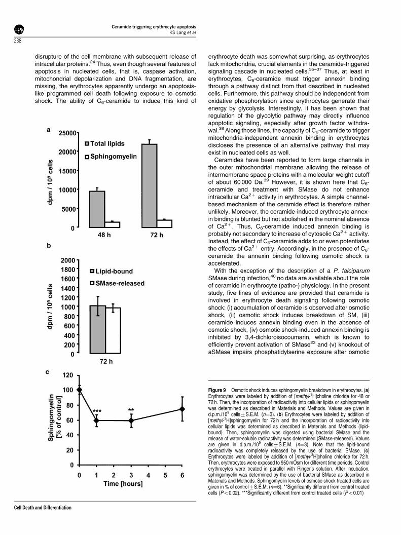
Formation of ceramide by a sphingomyelinase should beparalleled by a decline of cellular sphingomyelin content, asde novo synthesis of ceramide in erythrocytes is quiteunlikely.22 To test for sphingomyelin breakdown, we labelederythrocytes with radioactive choline and measured theincorporation of [methyl-3H]choline into erythrocyte lipids. Asshown in Figure 9a, significant label was detected in
Figure 4 Ceramide and sphingomyelinase induce annexin binding of erythrocytes. (a) Histograms of representative experiments demonstrating annexin binding incontrol cells (left) or cells treated for 4 h with 50 mM C6-ceramide (middle) or 0.01 U/ml SMase (right). (b) Annexin binding of erythrocytes after a 4-h treatment withdifferent concentrations of ceramide. Arithmetic means7S.E.M. (n¼3) of apoptotic cells are given in % of the total population. (c) Annexin binding of erythrocytes after a4-h treatment with different concentrations of SMase. Arithmetic means7S.E.M. (n¼3) of apoptotic cells are given in % of the total population. (d) Annexin binding oferythrocytes after a 4-h treatment with Ringer’s solution (control), 50 mM C6-ceramide or 0.01 U/ml SMase in the presence (black columns) or absence (open columns) of1 mM Ca2þ in the extracellular fluid. Arithmetic means7S.E.M. (n¼3) of apoptotic cells are given in % of the total population. ***Significantly different from cells treatedwith C6-ceramide or SMase in the presence of Ca2þ (Po0.01)
Ceramide triggering erythrocyte apoptosisKS Lang et al
234
Cell Death and Differentiation

erythrocyte lipids after 48 h of incubation, which reached2180071200 d.p.m./109 cells after 72 h of incubation. Moreimportantly, we could demonstrate that approximately 10% ofthe total lipid-bound radioactivity (19807100 d.p.m./109 cells)could be removed by the use of bacterial SMase (Figure 9a),thereby confirming that labeled choline was incorporated intoSM of erythrocytes. Additionally, the efficiency of the bacterialSMase was checked. For this purpose, erythrocyte mem-branes were labeled with radioactive SM, and lipid-boundradioactivity was removed by incubation for 2 h with SMase.
Figure 5 Osmotic shock, ceramide and sphingomyelinase induce erythrocyteshrinkage. (a) After 8 h of incubation, cells were measured in the FACS calibur forforward scatter and side scatter. Dot plots of representative experiments forcontrol cells (upper left) or cells treated for 8 h with 950 mOsm (upper right),50 mM C6-ceramide (lower left) or 0.01 U/ml SMase (lower right) are shown.Geometric mean values are given for the single experiment. (b) Forward scatteranalysis of erythrocytes after an 8-h treatment with 950 mOsm, 50 mM C6-ceramide or 0.01 U/ml SMase. Control refers to erythrocytes exposed to anisotonic buffer solution (300 mOsm) for 8 h. Arithmetic means7S.E.M. (n¼3) ofthe geometric mean of the forward scatter are given. **Significantly different fromcontrol treated cells (Po0.02). ***Significantly different from control treated cells(Po0.01)
Figure 6 Influence of osmotic shock, ceramide and sphingomyelinase onerythrocyte cell number after 8 or 24 h of incubation. (a) After the 8-h treatmentwith 950 mOsm, 50 mM C6-ceramide or 0.01 U/ml SMase, cells were counted in ahemocytometer. Control refers to erythrocytes exposed to an isotonic buffersolution (300 mOsm) for 8 h. (b) After 24-h treatment with 950 mOsm, 50 mM C6-ceramide or 0.01 U/ml SMase, cells were counted in a hemocytometer. Controlrefers to erythrocytes exposed to an isotonic buffer solution (300 mOsm) for 24 h.Arithmetic means7S.E.M. (n¼3) of the cell numbers are given in % of control.Erythrocyte numbers under control conditions were (1.370.1)� 107 cells/ml.**Significantly different from control treated cells (Po0.02). ***Significantlydifferent from control treated cells (Po0.01)
Ceramide triggering erythrocyte apoptosisKS Lang et al
235
Cell Death and Differentiation

As expected, 96% of the radioactivity of the total lipids wasremoved by SMase treatment (Figure 9b).
We now tested whether treatment of erythrocytes with950 mOsm induced SM breakdown. As shown in Figure 9c,SM breakdown already occurred after 1 h of hypertonic shockwhen SM levels reached approximately 60% of control. SMlevels increased again after 6 h but failed to reach the controllevel. This might be attributed to the low metabolic activity oferythrocytes.
3,4-Dichloroisocoumarin and knockout of aSMasebut not fumonisin B1 blunts the annexin bindinginduced by osmotic shock
3,4-Dichloroisocoumarin has been shown to inhibit activationof SMase and apoptosis triggered by daunorubicin.23 We thusused this inhibitor to further investigate the involvement ofSMase and ceramide in osmotic shock-induced annexinbinding of erythrocytes. As shown in Figure 10, annexinbinding induced by osmotic shock was concentration-depen-dently blunted by 3,4-dichloroisocoumarin. At a concentrationof 50 mM, annexin binding in the presence and absence ofCa2þ was reduced by 35 and 33%, respectively. In contrast,the annexin binding of erythrocytes following exposure to 1 mMionomycin or 50 mM C6-ceramide was not significantlymodified in the presence of 50–200 mM 3,4-dichloroisocou-marin, thereby ruling out that the inhibitor interfered with theCa2þ pathway or signaling downstream of ceramide forma-tion.
In a second approach, we used erythrocytes from aSMaseknockout mice. Osmotic shock-induced phosphatidylserineexposure was significantly reduced by 42% in erythrocytesfrom aSMase (�/�) mice as compared with the erythrocytesfrom wild-type littermates (Figure 11a). These data confirmthe hypothesis that breakdown of sphingomyelin plays a rolein osmotic shock-induced erythrocyte death.
Fumonisin B1 is an inhibitor of ceramide synthesis and hasbeen shown to antagonize ceramide-mediated apoptosis atconcentrations of 20–50mM.24 However, fumonisin B1 did notinfluence annexin binding of erythrocytes following treatmentwith 950 mOsm (Figure 11b), thereby ruling out that theelevated ceramide originates from the biosynthetic pathway.
Ceramide sensitizes the erythrocytes to osmoticshock
Since ceramide is released upon osmotic shock and triggersannexin binding, we investigated the combined effect ofosmotic shock and ceramide on erythrocyte apoptosis. Asillustrated in Figure 12a, the sublethal concentration of 20 mMC6-ceramide clearly potentiated annexin binding induced by950 mOsm after 4 h of combined treatment. Similarly,
Figure 7 Osmotic shock, but neither ceramide nor sphingomyelinase increaseintracellular Ca2þ of erythrocytes. (a) 45Ca2þ entry into erythrocytes wasmeasured as described in Materials and Methods. Erythrocytes were bathed inuptake solution without (control) or with 50 mM C6-ceramide (C6-cer) or 0.01 U/mlsphingomyelinase (SMase). Additionally, erythrocytes were exposed to uptakesolution adjusted to 950 mOsm. Arithmetic means7S.E.M. (n¼3) of intracellularcalcium are given in mmol/1013 cells. ***Significantly different from control treatedcells (Po0.01). (b) 45Ca2þ entry into erythrocytes was measured in thepresence of different concentrations of C6-ceramide. Arithmetic means7S.E.M.(n¼3) of intracellular calcium are given in mmol/1013 cells. (c) Annexin binding oferythrocytes after 8 h of incubation with 50 mM C6-ceramide (C6-cer), 0.01 U/mlsphingomyelinase (SMase) or 950 mOsm in the absence (black bars) orpresence of 1 mM amiloride (open bars). Control refers to erythrocytes exposedto an isotonic buffer solution (300 mOsm) for 8 h. Arithmetic means7S.E.M.(n¼3) of annexin-positive cells are given in % of the total population.**Significantly different from cells treated with 950 mOsm in the absence ofamiloride (Po0.02)
Ceramide triggering erythrocyte apoptosisKS Lang et al
236
Cell Death and Differentiation

formation of intracellular ceramide by addition of 0.005 U/mlSMase to the culture solution sensitized erythrocytes toosmotic shock after 4 h of combined treatment (Figure 12b).
Discussion
The breakdown of phosphatidylserine asymmetry is a well-known signal for the elimination of erythrocytes by macro-phages in vivo.25,26 Phosphatidylserine exposure itself isstimulated by elevation of intracellular Ca2þ 14,15,17 andsubsequent activation of a Ca2þ -dependent scram-blase.18,19,27 The present study confirms that osmotic shocktriggers erythrocyte shrinkage and annexin binding. Theosmolarity utilized is similar to the osmolarity prevailing inkidney medulla. On an average, each erythrocyte passes thehypertonic renal medulla 10 times a day. However, thedwelling time is normally too short to trigger significantbreakdown of phosphatidylserine asymmetry. This may bedifferent in acute renal failure where erythrocytes are indeedtrapped in the kidney medulla.28 Thus, at least in someconditions, hyperosmolarity may play a role in erythrocytedeath. More importantly, though, similar mechanisms aretriggered by other challenges of erythrocyte survival, such asoxidative stress and energy depletion.17,29
The effects of osmotic shock, oxidative stress and energydepletion have been shown to be partially mediated byunselective cation channels allowing the passage ofCa2þ .30,31 However, we show here that osmotic shock-induced erythrocyte annexin binding is not completely blockedin the absence of extracellular Ca2þ . These data point to theexistence of additional signaling mechanisms leading toannexin binding of erythrocytes. First of all, by using a pan-caspase inhibitor and by Western blot analysis of caspase-3activation, we ruled out a functional role of caspases inerythrocyte death following osmotic shock, which is inaccordance with the results of other groups showing thatcaspases in erythrocytes are not activated during cellularstress.14,15 Since activation of SMase with subsequentformation of ceramide has been demonstrated as animportant cellular response to different stress condi-tions,20,32–34 we specifically investigated the effect of cer-amide. Indeed, we could demonstrate that C6-ceramide aswell as treatment with bacterial SMase trigger annexinbinding.
Hyperosmotic shock and ceramide induce erythrocyteshrinkage and phosphatidylserine exposure, both clear hall-marks of apoptosis in nucleated cells. The absence ofhemoglobin release points to the integrity of the cellmembrane, another crucial feature of apoptosis. In contrast,necrosis typically leads to cell swelling and eventual
Figure 8 Stimulation of ceramide formation in erythrocytes by osmotic shock. (a) Ceramide was detected by anti-ceramide antibody and FACS analysis as described inMaterials and Methods. Histograms of representative experiments of control cells treated with Ringer’s solution (black line in all histograms) and cells exposed for 30 min(grey line, left histogram) or for 15 h (grey line, middle histogram) to osmotic shock of 950 mOsm are shown. As a positive control, erythrocytes were exposed for 5 min to1 U/ml SMase (grey line, right histogram). (b) Histogram of a representative experiment demonstrating the binding of isotype matched anti-mouse antibody to controlcells treated with Ringer’s solution (black line) and cells exposed for 15 h to osmotic shock of 950 mOsm (grey line). (c) Arithmetic means7S.E.M. (n¼4) of the meanfluorescence of erythrocytes after treatment with Ringer’s solution (300 mOsm) or after osmotic shock (950 mOsm). ***Significantly different from cells treated with300 mOsm (Po0.01)
Ceramide triggering erythrocyte apoptosisKS Lang et al
237
Cell Death and Differentiation
disrupture of the cell membrane with subsequent release ofintracellular proteins.24 Thus, even though several features ofapoptosis in nucleated cells, that is, caspase activation,mitochondrial depolarization and DNA fragmentation, aremissing, the erythrocytes apparently undergo an apoptosis-like programmed cell death following exposure to osmoticshock. The ability of C6-ceramide to induce this kind of
erythrocyte death was somewhat surprising, as erythrocyteslack mitochondria, crucial elements in the ceramide-triggeredsignaling cascade in nucleated cells.35–37 Thus, at least inerythrocytes, C6-ceramide must trigger annexin bindingthrough a pathway distinct from that described in nucleatedcells. Furthermore, this pathway should be independent fromoxidative phosphorylation since erythrocytes generate theirenergy by glycolysis. Interestingly, it has been shown thatregulation of the glycolytic pathway may directly influenceapoptotic signaling, especially after growth factor withdra-wal.38 Along those lines, the capacity of C6-ceramide to triggermitochondria-independent annexin binding in erythrocytesdiscloses the presence of an alternative pathway that mayexist in nucleated cells as well.
Ceramides have been reported to form large channels inthe outer mitochondrial membrane allowing the release ofintermembrane space proteins with a molecular weight cutoffof about 60 000 Da.39 However, it is shown here that C6-ceramide and treatment with SMase do not enhanceintracellular Ca2þ activity in erythrocytes. A simple channel-based mechanism of the ceramide effect is therefore ratherunlikely. Moreover, the ceramide-induced erythrocyte annex-in binding is blunted but not abolished in the nominal absenceof Ca2þ . Thus, C6-ceramide induced annexin binding isprobably not secondary to increase of cytosolic Ca2þ activity.Instead, the effect of C6-ceramide adds to or even potentiatesthe effects of Ca2þ entry. Accordingly, in the presence of C6-ceramide the annexin binding following osmotic shock isaccelerated.
With the exception of the description of a P. falciparumSMase during infection,40 no data are available about the roleof ceramide in erythrocyte (patho-) physiology. In the presentstudy, five lines of evidence are provided that ceramide isinvolved in erythrocyte death signaling following osmoticshock: (i) accumulation of ceramide is observed after osmoticshock, (ii) osmotic shock induces breakdown of SM, (iii)ceramide induces annexin binding even in the absence ofosmotic shock, (iv) osmotic shock-induced annexin binding isinhibited by 3,4-dichloroisocoumarin, which is known toefficiently prevent activation of SMase23 and (v) knockout ofaSMase impairs phosphatidylserine exposure after osmotic
Figure 9 Osmotic shock induces sphingomyelin breakdown in erythrocytes. (a)Erythrocytes were labeled by addition of [methyl-3H]choline chloride for 48 or72 h. Then, the incorporation of radioactivity into cellular lipids or sphingomyelinwas determined as described in Materials and Methods. Values are given ind.p.m./109 cells7S.E.M. (n¼3). (b) Erythrocytes were labeled by addition of[methyl-3H]sphingomyelin for 72 h and the incorporation of radioactivity intocellular lipids was determined as described in Materials and Methods (lipid-bound). Then, sphingomyelin was digested using bacterial SMase and therelease of water-soluble radioactivity was determined (SMase-released). Valuesare given in d.p.m./109 cells7S.E.M. (n¼3). Note that the lipid-boundradioactivity was completely released by the use of bacterial SMase. (c)Erythrocytes were labeled by addition of [methyl-3H]choline chloride for 72 h.Then, erythrocytes were exposed to 950 mOsm for different time periods. Controlerythrocytes were treated in parallel with Ringer’s solution. After incubation,sphingomyelin was determined by the use of bacterial SMase as described inMaterials and Methods. Sphingomyelin levels of osmotic shock-treated cells aregiven in % of control7S.E.M. (n¼6). **Significantly different from control treatedcells (Po0.02). ***Significantly different from control treated cells (Po0.01)
Ceramide triggering erythrocyte apoptosisKS Lang et al
238
Cell Death and Differentiation

shock. These data clearly demonstrate that erythrocytes areable to generate ceramide, presumably via breakdown of SM.Under normal conditions, erythrocytes lack considerableSMase activity, at least when enzyme activity is assayed incell-free extracts.41 Distinct stress conditions, however, suchas increase of extracellular osmolarity, lead to activation ofSMase and generation of ceramide.
The effect of osmotic shock is blunted by the sphingomye-linase inhibitor 3,4-dichloroisocoumarin suggesting that theeffect of osmotic shock is sensitive to ceramide release. Incontrast, the effects of neither C6-ceramide nor ionomycin areinhibited by 3,4-dichloroisocoumarin, indicating that the drugexerts its inhibitory effect by interference with the signalingupstream of ceramide and cytosolic Ca2þ . Thus, ceramideformation could play a mediating or a permissive role in thetriggering of annexin binding of erythrocytes. The experimentsutilizing the ceramide antibody demonstrate that osmotic cellshrinkage stimulates the formation of ceramide. Theseobservations strongly suggest the presence of a cellvolume-sensitive SMase in erythrocytes that forms ceramideand thus sensitizes the erythrocytes for apoptosis.
Among the functions of ceramide is clustering of receptors.It has been shown that clustering of the CD95 receptor ismediated via ceramide-rich membrane rafts.42 As a matter offact, apoptosis of nucleated cells following osmotic shock3–5
has been attributed to ligand-independent clustering ofmultiple growth factor and cytokine receptors, such as tumornecrosis factor receptor.6 Thus, it seems reasonable that
ceramide might play a similar role in erythrocytes, forexample, in the clustering of channels or enzymes, such asscramblase.
In summary, similar to nucleated cells, erythrocytes under-go annexin binding following exposure to C6-ceramide.Moreover, ceramide represents an important signaling mole-cule after hyperosmotic shock and sensitizes the erythrocytesfor proapoptotic stimuli. The generation of ceramide maytherefore participate in the regulation of erythrocyte survival.
Figure 10 3,4-Dichloroisocoumarin blunts osmotic shock-induced annexin-binding, but not ionomycin- or C6-ceramide-induced annexin-binding oferythrocytes. Erythrocytes were exposed to 950 mOsm in the presence (opencolumns) or absence (striped columns) of 1 mM Ca2þ , to 50 mM C6-ceramide(filled columns), to 1 mM ionomycin (black and white columns) or treated withRinger’s solution (control, hatched columns) for 8 h. Annexin-binding wasmeasured in the absence or presence of different concentrations of 3,4-dichloroisocoumarin. Arithmetic means7S.E.M. (n¼3) of annexin-positive cellsare given in % of the total population. *Significantly different from cells treatedwith 950 mOsm (Ca2þ -free) in the absence of 3,4-dichloroisocoumarin(Po0.05). **Significantly different from cells treated with 950 mOsm (Ca2þ -free) in the absence of 3,4-dichloroisocoumarin (Po0.02). ***Significantlydifferent from cells treated with 950 mOsm in the absence of 3,4-dichloroiso-coumarin (Po0.01)
Figure 11 Knockout of aSMase but not treatment with fumonisin B1 bluntsosmotic shock-induced annexin binding of erythrocytes. (a) Erythrocytes fromaSMase knockout mice (aSMase (�/�), open bars) or corresponding littermates(aSMase (þ /þ ), black bars) were exposed to 300 or 700 mOsm for 24 h.Arithmetic means7S.E.M. (n¼3) of annexin-positive cells are given in % of thetotal population. ***Significantly different from aSMase (þ /þ ) cells treated with700 mOsm (Po0.01). (b) Annexin binding of erythrocytes after 8 h of incubationwith 950 mOsm in the absence or presence of 25 or 50 mM fumonisin B1. Controlrefers to erythrocytes exposed to an isotonic buffer solution (300 mOsm) for 8 h.Arithmetic means7S.E.M. (n¼3) of annexin-positive cells are given in % of thetotal population
Ceramide triggering erythrocyte apoptosisKS Lang et al
239
Cell Death and Differentiation

Materials and methods
Cells
Erythrocytes were drawn from healthy volunteers or from aSMaseknockout mice and the corresponding wild-type littermates. aSMaseknockout mice43,44 and littermates were a kind gift of Dr VerenaJendrossek (University of Tubingen, Germany) and were originallyobtained from Dr R Kolesnick (Sloan Kettering Cancer Memorial Center,NY, USA). Erythrocytes were either used without purification or afterseparation by centrifugation for 25 min; 2000 g over Ficoll (Biochrom KG,Berlin, Germany). Experiments with nonpurified or experiments with Ficoll-separated erythrocytes yielded the same results (data not shown).
In some control experiments, erythrocytes were infected with P.falciparum as described previously.45
Jurkat T-cells were grown in RPMI 1640 medium supplemented with10% fetal calf serum (FCS), 0.56 g/l L-glutamine, 100 000 U/l penicillin and0.1 g/l streptomycin. Where indicated, osmolarity was increased to 700 or950 mOsm by adding sucrose.
Solutions
Experiments were performed at 371C in Ringer’s solution containing125 mM NaCl, 5 mM KCl, 1 mM MgSO4, 32 mM N-2-hydroxyethylpiper-azine-N-2-ethanesulfonic acid (HEPES), 5 mM Glucose, 1 mM CaCl2,pH¼7.4. For the nominally calcium-free solution CaCl2 was replaced by1 mM ethylene glycol-bis (b-aminoethyl ether)-N,N,N0,N0-tetraacetic acid(EGTA). Where indicated, osmolarity was increased to 950 mOsm byadding sucrose. Ionomycin (Sigma; Taufkirchen, Germany) was used at aconcentration of 1mM. D-erythro-N-hexanoylsphingosine (C6-ceramide)was dissolved in dimethyl sulfoxide (DMSO) to give a 50 mM stock solutionand further diluted in Ringer’s solution containing 0.1% bovine serumalbumin. The maximum concentration of DMSO was in all cases 0.1%, aconcentration that did not induce annexin binding (data not shown).Fumonisin B1 was dissolved in methanol to give a 50 mM stock solutionand further diluted in Ringer’s solution containing 0.1% bovine serumalbumin. D-erythro-N-palmitoylsphingosine (C16-ceramide) was dissolvedin ethanol/dodecane (98 : 2, v/v) at a concentration of 177 mM. Thesolution was then added to Ringer’s solution and sonicated for 30 minbefore use. Vehicle was present at 0.3% and was added to controls.Sphingomyelinase from Staphylococcus aureus, rabbit polyclonal anti-caspase-3 antibody (raised against human recombinant caspase-3 andrecognizing the proenzyme and the active 17 kDa subunit), C6-ceramideand 3,4-dichloroisocoumarin were purchased from Biomol GmbH(Hamburg, Germany). C16-ceramide, fumonisin B1, monoclonal antibodyto ceramide (clone MID 15B4; isotype IgM) and isotype pure mouse IgMwere from Alexis (Grunberg, Germany). The pan-caspase inhibitor zVAD-fmk (in which z stands for benzyloxycarbonyl and fmk for fluoromethylketone) was from Calbiochem (Bad Soden, Germany) and was dissolvedin DMSO to give a 20 mM stock solution. This inhibitor was synthesized asa methyl ester to enhance cell permeability. 45Ca2þ was from ICNBiomedicals GmbH (Eschwege, Germany) and delivered as CaCl2 inaqueous solution (specific activity: 0.185–1.11 TBq/g Ca).
Determination of cell numbers and hemolysis
Erythrocytes were suspended at 0.15% hematocrit and incubated underdifferent control and stress conditions (osmotic stress, C6-ceramide andSMase treatment). After incubation, the cell number was determined usinga hemocytometer as described previously.46 Additionally, hemolysis was
Figure 12 Ceramide and sphingomyelinase sensitize the erythrocytes to theapoptotic action of osmotic shock. (a) Annexin binding of erythrocytes after a 4-htreatment with Ringer’s solution (control), 20 mM C6-ceramide (C6-Cer), exposureto hypertonic solution (950 mOsm) or combined treatment with 20 mM C6-ceramide and hypertonic solution. Arithmetic means7S.E.M. (n¼3) of apoptoticcells are given in % of the total population. (b) Annexin binding of erythrocytesafter a 4-h treatment with Ringer’s solution (control), 0.005 U/ml SMase,exposure to hypertonic solution (950 mOsm) or combined treatment with 0.005 U/ml SMase and hypertonic solution. Arithmetic means7S.E.M. (n¼3) of apoptoticcells are given in % of the total population
Ceramide triggering erythrocyte apoptosisKS Lang et al
240
Cell Death and Differentiation

determined by photometric measurement of hemoglobin release. Thehemoglobin concentration in the supernatant was determined quantita-tively by photometry (absorbance at 546 nm after oxidation to cyanomet-hemoglobin). The absorbance of the supernatant of completely lysederythrocytes was set as 100% hemolysis.
FACS analysis
FACS analysis was performed essentially as described.47 After incubation,cells were washed in Annexin-binding buffer containing 125 mM NaCl,10 mM HEPES (pH¼7.4), 5 mM CaCl2. Erythrocytes were stained withAnnexin-Fluos (Bohringer Mannheim, Germany) at a 1 : 100 dilution. After15 min, samples were diluted 1 : 5 and measured by flow cytometricanalysis on a FACS-Calibur (Becton Dickinson; Heidelberg, Germany).Cells were analyzed by forward and side scatter and annexin-fluorescenceintensity was measured in FL-1. In the case of Jurkat cells and P.falciparum-infected erythrocytes, the cells were counterstained usingpropidium iodide (5 mg/ml). Propidium iodide-fluorescence was measuredin FL-3.
For determination of ceramide, cells were stained for 1 h at 41C with1mg/ml anti-ceramide antibody or 1 mg/ml isotype matched pure mouseantibody in phosphate-buffered saline (PBS) containing 1% FCS at adilution of 1 : 5 as described recently.42 After three washes with PBS/1%FCS, cells were stained with polyclonal fluorescein isothiocyanate (FITC)-conjugated goat anti-mouse Ig specific antibody (Pharmingen, Hamburg,Germany) in PBS/1% FCS at a dilution of 1 : 50 for 30 min. Unboundsecondary antibody was removed by washing the cells two times withPBS/1% FCS and samples were analyzed by flow cytometric analysis on aFACS-Calibur. FITC-fluorescence intensity was measured in FL-1.
Western blot analysis
Western blot analysis was performed as described.48 After incubation,cells were washed twice with PBS and lysed in buffer containing 20 mMTris/HCl, pH 7.4, 300 mM NaCl, 1% Triton X-100, 1% Na deoxycholate,0.1% Na dodecylsulfate (SDS), 2.5 mM EDTA, 10 mg/ml pepstatin A,10mg/ml leupeptin, 5 mg/ml aprotinin and 0.1 mM phenylmethylsulfonylfluoride (PMSF). Protein concentration was determined using the Bio-Radprotein assay from Biorad (Munchen, Germany) and equal amounts ofprotein were separated by SDS-PAGE.49 Then, blotting of proteins ontonitrocellulose membranes (Schleicher and Schuell, Dassel, Germany) wasperformed exactly as described.48 After blotting, the membrane wasblocked for 1 h in PBST (PBS, 0.05% Tween-20) containing 3% nonfat drymilk and incubated with primary anti-caspase-3 antibody at a dilution of1 : 1000 for 1 h. After the membrane had been washed three times inPBST, secondary donkey anti-rabbit horseradish peroxidase-linked wholeantibody (Amersham Biosciences; Freiburg, Germany) at a dilution of1 : 1000 in PBST was applied for 1 h. Finally, the membrane was washedin PBST again and the ECL enhanced chemiluminescence system fromAmersham Biosciences was used to visualize the protein bands inquestion.
Microscopy
Fluorescence microscopy was performed essentially as described.24 Afterincubation, erythrocytes were stained with Annexin-Fluos as describedabove. After washing the cells with annexin buffer, 10 ml of the suspensionwere applied to a slide and covered with a cover glass. Note that thestaining and washing solutions after hyperosmotic shock were alsoadjusted to 950 mOsm by addition of sucrose. Finally, the cells were
analyzed under a fluorescence microscope (Nikon; Dusseldorf, Germany)and digital pictures were taken using a digital imaging system (VisitronSystems; Puchheim, Germany) equipped with the Metaview software.
Calcium measurements
Intracellular calcium was measured as described in detail elsewhere.50,51
Erythrocytes were washed four times by centrifugation (2000 g for 5 min)and resuspension in five volumes of solution A containing in mM: 80 KCl,70 NaCl, 10 HEPES, 0.2 MgCl2, 0.1 EGTA; pH 7.5 to remove extracellularCa2þ . The cell pellet was then washed twice in solution B to removeEGTA from the medium. Solution B had the same composition as solutionA but without EGTA. The cells were suspended at 10% hematocrit andpreincubated for 20 min at 371C in the final incubation solution Bsupplemented with 10 mM inosine and 1 mM sodium orthovanadate. Then45Ca2þ was added from a 100 mM CaCl2 stock solution with a specificactivity of about 107 c.p.m./mmol to reach a final concentration of 150 mM.After 10 min, 100 ml aliquots were delivered into 1.2 ml of ice-cold solutionB with 0.2 mM CoCl2 and 1 mM amiloride. The cells were collected bycentrifugation in an Eppendorf centrifuge (14 000 r.p.m. for 0.5 min, 41C)and the cell pellet was washed twice using 1 ml of the same medium. Thesupernatant was discarded and the cells were lysed and the proteinsprecipitated by addition of 0.6 ml 6% trichloroacetic acid. After a furtherspin (14 000 r.p.m. for 2 min, 41C), 0.5 ml of clear supernatant was usedfor measuring 45Ca2þ radioactivity by scintillation counting. 45Ca2þ
specific activity was determined by addition of 0.6 ml 6% trichloroaceticacid to 100 ml suspension samples and centrifugation as described above.Then, 100 ml of clear supernatant were taken for scintillation counting. Thetotal calcium content of the cells [CaT]i was calculated by dividing theactivity of the samples by the specific activity of 45Ca2þ and by thenumber of cells. Different concentrations of C6-ceramide or sphingomye-linase were added to the cell suspensions together with 45Ca2þ .Exposure of erythrocytes to 950 mOsm was achieved by the addition ofsucrose to solution B during 20 min of preincubation and 10 min of 45Ca2þ
uptake. Note that the delivery medium for washing the cells afterradioactive labeling was also adjusted to 950 mOsm by the addition ofsucrose.
Choline labeling of erythrocytes
Erythrocytes were washed two times with Ringer’s solution and thengrown at 10% hematocrit in RPMI 1640 (Life Technologies; Karlsruhe,Germany) containing 50 mg/l gentamycin (Life Technologies), 2 mM L-glutamine, 20 mM HEPES, 20 mg/l hypoxanthine (Sigma; Taufkirchen,Germany), 1 mM glucose monohydrate, 5 g/l Albumax II (Life Technol-ogies) and 10% heat-inactivated human serum ABþ . The cells wereincubated for 72 h in the presence of 7.4� 104 Bq/ml [methyl-3H]cholinechloride (Amersham Pharmacia Biotech; Braunschweig, Germany).Postlabeling, cells were washed twice with Ringer’s solution, reseededat 2� 108 cells/ml in control Ringer’s solution or in Ringer’s solutionadjusted to 950 mOsm by adding sucrose for 1, 3 and 6 h. After incubation,cells were harvested by centrifugation in the Eppendorf centrifuge(14 000 r.p.m. for 0.5 min, 41C) and the cell pellet was washed twice using1 ml of the same medium. Then, lipids were extracted by a modifiedmethod of Bligh and Dyer as described earlier.52 Briefly, cell pellets wereresuspended in 50 ml methanol, 25 ml chloroform and 20 ml water.Samples were stirred for 10 min on a vortex mixer and centrifuged at13 000� g for 2 min. Phase separation was accomplished by the additionof 25ml chloroform and 25ml water. The suspension and centrifugationsteps were repeated. Volumes of 20 ml of the chloroform phases were
Ceramide triggering erythrocyte apoptosisKS Lang et al
241
Cell Death and Differentiation

taken for scintillation counting, and 50 ml of the chloroform phases weredried under nitrogen and used for SM measurement as described below.
Bacterial SMase
SM was quantified using bacterial sphingomyelinase to release[3H]phosphocholine as described.53 Briefly, cellular lipid was resuspendedin 100ml of assay buffer (100 mM Tris-HCl, pH 7.4, 6 mM MgCl2, 0.1%Triton X-100). Samples were sonicated for 5 min and 1 U/ml Streptomycessp. SMase (Sigma; Taufkirchen, Germany) was added. Reaction mixtureswere incubated for 2 h at 371C. Reactions were stopped by addition of1.0 ml of chloroform/methanol (2 : 1, v/v). Phase separation wascompleted by addition of 100 ml of water. SM was quantitated by countingthe upper, aqueous phase, containing the liberated [3H]phosphocholineand phosphatidylcholine was quantitated by drying and counting the lower,organic phase. Where appropriate, SM was normalized using phospha-tidylcholine measurements. Blank reactions contained no SMase. Theradioactivity of control samples normally reached 1500 d.p.m./109 cellsand was set as 100%. Subsequently, SM in the samples of hyperosmoticshock-treated cells was calculated as % of control.
Optimization studies illustrated that the above conditions yieldedmaximal SM hydrolysis (100%) (Figure 9b).
Statistics
Data are expressed as arithmetic means7SEM and statistical analysiswas made by paired or unpaired t-test, where appropriate.
Acknowledgements
We authors acknowledge the technical assistance of E Faber and themeticulous preparation of the manuscript by Leijla Subasic and TanjaLoch. We thank Dr R Kolesnick (Sloan Kettering Cancer Memorial Center,NY, USA) and Dr Verena Jendrossek (University of Tubingen, Germany)for kindly providing aSMase knockout mice. This study was supported bythe Deutsche Forschungsgemeinschaft, Nr. La 315/4-3 and La 315/6-1,the Bundesministerium fur Bildung, Wissenschaft, Forschung undTechnologie (Center for Interdisciplinary Clinical Research) 01 KS 9602and the Biomed program of the EU (BMH4-CT96-0602).
References
1. Gomez-Angelats M, Bortner CD and Cidlowski JA (2000) Cell volumeregulation in immune cell apoptosis. Cell Tissue Res. 301: 33–42
2. Gomez-Angelats M and Cidlowski JA (2002) Cell volume control and signaltransduction in apoptosis. Toxicol. Pathol. 30: 541–551
3. Lang KS, Fillon S, Schneider D, Rammensee HG and Lang F (2002)Stimulation of TNF alpha expression by hyperosmotic stress. Pflugers Arch.443: 798–803
4. Dmitrieva N, Kultz D, Michea L, Ferraris J and Burg M (2000) Protection of renalinner medullary epithelial cells from apoptosis by hypertonic stress-induced p53activation. J. Biol. Chem. 275: 18243–18247
5. Stoothoff WH and Johnson GV (2001) Hyperosmotic stress-induced apoptosisand tau phosphorylation in human neuroblastoma cells. J. Neurosci. Res. 65:573–582
6. Rosette C and Karin M (1996) Ultraviolet light and osmotic stress: activation ofthe JNK cascade through multiple growth factor and cytokine receptors.Science 274: 1194–1197
7. Reinehr R, Graf D, Fischer R, Schliess F and Haussinger D(2002) Hyperosmolarity triggers CD95 membrane trafficking and
sensitizes rat hepatocytes toward CD95L-induced apoptosis. Hepatology 36:602–614
8. Terada Y, Inoshita S, Hanada S, Shimamura H, Kuwahara M, Ogawa W,Kasuga M, Sasaki S and Marumo F (2001) Hyperosmolality activates Akt andregulates apoptosis in renal tubular cells. Kidney Int. 60: 553–567
9. Lu J, Park JH, Liu AY and Chen KY (2000) Activation of heat shock factor 1 byhyperosmotic or hypo-osmotic stress is drastically attenuated in normal humanfibroblasts during senescence. J. Cell Physiol. 184: 183–190
10. Desai BN, Myers BR and Schreiber SL (2002) FKBP12-rapamycin-associatedprotein associates with mitochondria and senses osmotic stress viamitochondrial dysfunction. Proc. Natl. Acad. Sci. USA 99: 4319–4324
11. Kabsch K and Alonso A (2002) The human papillomavirus type 16 (HPV-16) E5protein sensitizes human keratinocytes to apoptosis induced by osmotic stress.Oncogene 21: 947–953
12. Green DR and Reed JC (1998) Mitochondria and apoptosis. Science 281:1309–1312
13. Gulbins E, Jekle A, Ferlinz K, Grassme H and Lang F (2000) Physiology ofapoptosis. Am. J. Physiol. Renal Physiol. 279: F605–F615
14. Bratosin D, Estaquier J, Petit F, Arnoult D, Quatannens B, Tissier JP,Slomianny C, Sartiaux C, Alonso C, Huart JJ, Montreuil J and Ameisen JC(2001) Programmed cell death in mature erythrocytes: a model for investigatingdeath effector pathways operating in the absence of mitochondria. Cell DeathDiffer. 8: 1143–1156
15. Berg CP, Engels IH, Rothbart A, Lauber K, Renz A, Schlosser SF, Schulze-Osthoff K and Wesselborg S (2001) Human mature red blood cells expresscaspase-3 and caspase-8, but are devoid of mitochondrial regulators ofapoptosis. Cell Death Differ. 8: 1197–1206
16. Daugas E, Cande C and Kroemer G (2001) Erythrocytes: death of a mummy.Cell Death Differ. 8: 1131–1133
17. Lang KS, Duranton C, Poehlmann H, Myssina S, Bauer C, Lang F, Wieder Tand Huber SM (2003) Cation channels trigger apoptotic death of erythrocytes.Cell Death Differ. 10: 249–256
18. Woon LA, Holland JW, Kable EP and Roufogalis BD (1999) Ca2+ sensitivity ofphospholipid scrambling in human red cell ghosts. Cell Calcium 25: 313–320
19. Dekkers DW, Comfurius P, Bevers EM and Zwaal RF (2002) Comparisonbetween Ca2+-induced scrambling of various fluorescently labelled lipidanalogues in red blood cells. Biochem. J. 362: 741–747
20. Gulbins E, Bissonnette R, Mahboubi A, Martin S, Nishioka W, Brunner T, BaierG, Baier-Bitterlich G, Byrd C, Lang F, Kolesnick R, Altman A and Green D(1995) FAS-induced apoptosis is mediated via a ceramide-initiated RASsignaling pathway. Immunity 2: 341–351
21. Lepple-Wienhues A, Belka C, Laun T, Jekle A, Walter B, Wieland U, Welz M,Heil L, Kun J, Busch G, Weller M, Bamberg M, Gulbins E and Lang F (1999)Stimulation of CD95 (Fas) blocks T lymphocyte calcium channels throughsphingomyelinase and sphingolipids. Proc. Natl. Acad. Sci. USA 96:13795–13800
22. Elabbadi N, Ancelin ML and Vial HJ (1997) Phospholipid metabolism of serinein Plasmodium-infected erythrocytes involves phosphatidylserine and directserine decarboxylation. Biochem J. 324 (Part 2): 435–445
23. Mansat V, Bettaieb A, Levade T, Laurent G and Jaffrezou JP (1997) Serineprotease inhibitors block neutral sphingomyelinase activation, ceramidegeneration, and apoptosis triggered by daunorubicin. FASEB J. 11: 695–702
24. Wieder T, Orfanos CE and Geilen CC (1998) Induction of ceramide-mediatedapoptosis by the anticancer phospholipid analog, hexadecylphosphocholine. J.Biol. Chem. 273: 11025–11031
25. Boas FE, Forman L and Beutler E (1998) Phosphatidylserine exposure and redcell viability in red cell aging and in hemolytic anemia. Proc. Natl. Acad. Sci.USA 95: 3077–3081
26. Eda S and Sherman IW (2002) Cytoadherence of malaria-infected red bloodcells involves exposure of phosphatidylserine. Cell Physiol. Biochem. 12:373–384
27. Kamp D, Sieberg T and Haest CW (2001) Inhibition and stimulation ofphospholipid scrambling activity. Consequences for lipid asymmetry,echinocytosis, and microvesiculation of erythrocytes. Biochemistry 40:9438–9446
28. Mason J (1986) The pathophysiology of ischaemic acute renal failure. A newhypothesis about the initiation phase. Renal Physiol. 9: 129–147
29. Lang KS, Roll B, Myssina S, Schittenhelm M, Scheel-Walter HG, Kanz L, FritzJ, Lang F, Huber SM and Wieder T (2002) Enhanced erythrocyte apoptosis in
Ceramide triggering erythrocyte apoptosisKS Lang et al
242
Cell Death and Differentiation

sickle cell anemia, thalassemia and glucose-6-phosphate dehydrogenasedeficiency. Cell Physiol. Biochem. 12: 365–372
30. Huber SM, Gamper N and Lang F (2001) Chloride conductance and volume-regulatory nonselective cation conductance in human red blood cell ghosts.Pflugers Arch. 441: 551–558
31. Duranton C, Huber SM and Lang F (2002) Oxidation induces aCl(�)-dependent cation conductance in human red blood cells. J. Physiol.539: 847–855
32. Geilen CC, Wieder T and Orfanos CE (1997) Ceramide signalling: regulatoryrole in cell proliferation, differentiation and apoptosis in human epidermis. Arch.Dermatol. Res. 289: 559–566
33. Sawai H and Hannun YA (1999) Ceramide and sphingomyelinases in theregulation of stress responses. Chem. Phys. Lipids 102: 141–147
34. Gulbins E, Szabo I, Baltzer K and Lang F (1997) Ceramide-induced inhibition ofT lymphocyte voltage-gated potassium channel is mediated by tyrosinekinases. Proc. Natl. Acad. Sci. USA 94: 7661–7666
35. Ghafourifar P, Klein SD, Schucht O, Schenk U, Pruschy M, Rocha S andRichter C (1999) Ceramide induces cytochrome c release from isolatedmitochondria. Importance of mitochondrial redox state. J. Biol. Chem. 274:6080–6084
36. Raisova M, Bektas M, Wieder T, Daniel P, Eberle J, Orfanos CE and Geilen CC(2000) Resistance to CD95/Fas-induced and ceramide-mediated apoptosis ofhuman melanoma cells is caused by a defective mitochondrial cytochrome crelease. FEBS Lett. 473: 27–32
37. von Haefen C, Wieder T, Gillissen B, Starck L, Graupner V, Dorken B andDaniel PT (2002) Ceramide induces mitochondrial activation and apoptosis viaa Bax-dependent pathway in human carcinoma cells. Oncogene 21: 4009–4019
38. Vander Heiden MG, Plas DR, Rathmell JC, Fox CJ, Harris MH and ThompsonCB (2001) Growth factors can influence cell growth and survival through effectson glucose metabolism. Mol. Cell. Biol. 21: 5899–5912
39. Siskind LJ, Kolesnick RN and Colombini M (2002) Ceramide channels increasethe permeability of the mitochondrial outer membrane to small proteins. J. Biol.Chem. 277: 26796–26803
40. Hanada K, Palacpac NM, Magistrado PA, Kurokawa K, Rai G, Sakata D, HaraT, Horii T, Nishijima M and Mitamura T (2002) Plasmodium falciparumphospholipase C hydrolyzing sphingomyelin and lysocholinephospholipids is apossible target for malaria chemotherapy. J. Exp. Med. 195: 23–34
41. Hanada K, Mitamura T, Fukasawa M, Magistrado PA, Horii T and Nishijima M(2000) Neutral sphingomyelinase activity dependent on Mg2+ and anionic
phospholipids in the intraerythrocytic malaria parasite Plasmodium falciparum.Biochem. J. 346 (Part 3): 671–677
42. Grassme H, Jekle A, Riehle A, Schwarz H, Berger J, Sandhoff K, Kolesnick Rand Gulbins E (2001) CD95 signaling via ceramide-rich membrane rafts. J. Biol.Chem. 276: 20589–20596
43. Horinouchi K, Erlich S, Perl DP, Ferlinz K, Bisgaier CL, Sandhoff K,Desnick RJ, Stewart CL and Schuchman EH (1995) Acid sphingomyelinasedeficient mice: a model of types A and B Niemann–Pick disease. Nat. Genet.10: 288–293
44. Lin T, Genestier L, Pinkoski MJ, Castro A, Nicholas S, Mogil R, Paris F, Fuks Z,Schuchman EH, Kolesnick RN and Green DR (2000) Role of acidicsphingomyelinase in Fas/CD95-mediated cell death. J. Biol. Chem. 275:8657–8663
45. Huber SM, Uhlemann AC, Gamper NL, Duranton C, Kremsner PG and Lang F(2002) Plasmodium falciparum activates endogenous Cl(�) channels ofhuman erythrocytes by membrane oxidation. EMBO J. 21: 22–30
46. Wieder T, Perlitz C, Wieprecht M, Huang RT, Geilen CC and Orfanos CE(1995) Two new sphingomyelin analogues inhibit phosphatidylcholinebiosynthesis by decreasing membrane-bound CTP: phosphocholinecytidylyltransferase levels in HaCaT cells. Biochem. J. 311 (Part 3): 873–879
47. Andree HA, Reutelingsperger CP, Hauptmann R, Hemker HC, Hermens WTand Willems GM (1990) Binding of vascular anticoagulant alpha (VAC alpha) toplanar phospholipid bilayers. J. Biol. Chem. 265: 4923–4928
48. Wieder T, Essmann F, Prokop A, Schmelz K, Schulze-Osthoff K, Beyaert R,Dorken B and Daniel PT (2001) Activation of caspase-8 in drug-inducedapoptosis of B-lymphoid cells is independent of CD95/Fas receptor–ligandinteraction and occurs downstream of caspase-3. Blood 97: 1378–1387
49. Laemmli UK (1970) Cleavage of structural proteins during the assembly of thehead of bacteriophage T4. Nature 227: 680–685
50. Tiffert T and Lew VL (1997) Cytoplasmic calcium buffers in intact human redcells. J. Physiol. 500 (Part 1): 139–154
51. Tiffert T, Staines HM, Ellory JC and Lew VL (2000) Functional state of theplasma membrane Ca2+ pump in Plasmodium falciparum-infected human redblood cells. J. Physiol. 525 (Part 1): 125–134
52. Haase R, Wieder T, Geilen CC and Reutter W (1991) The phospholipidanalogue hexadecylphosphocholine inhibits phosphatidylcholine biosynthesisin Madin–Darby canine kidney cells. FEBS Lett. 288: 129–132
53. Jayadev S, Linardic CM and Hannun YA (1994) Identification of arachidonicacid as a mediator of sphingomyelin hydrolysis in response to tumor necrosisfactor alpha. J. Biol. Chem. 269: 5757–5763
Ceramide triggering erythrocyte apoptosisKS Lang et al
243
Cell Death and Differentiation