ATP-dependent sugar transport complexity in human erythrocytes
Upload
independentCategory
view
0download
0
Manoj T.Duraisingh, Tony Triglia,Stuart A.Ralph1, Julian C.Rayner2,John W.Barnwell2, Geoffrey I.McFadden1
and Alan F.Cowman3
The Walter and Eliza Hall Institute of Medical Research, Melbourne3050, 1Plant Cell Biology Research Centre, School of Botany,University of Melbourne 3010, Australia and 2Division of ParasiticDiseases, National Center for Infectious Diseases, Centers for DiseaseControl and Prevention, Chamblee, GA 30341, USA
3Corresponding authore-mail: [email protected]
M.T.Duraisingh and T.Triglia contributed equally to this work
The members of the phylum Apicomplexa parasitize awide range of eukaryotic host cells. Plasmodium falci-parum, responsible for the most virulent form ofmalaria, invades human erythrocytes using severalspeci®c and high af®nity ligand±receptor interactionsthat de®ne invasion pathways. We ®nd that membersof the P.falciparum reticulocyte-binding homologprotein family, PfRh2a and PfRh2b, are expressedvariantly in different lines. Targeted gene disruptionshows that PfRh2b mediates a novel invasion pathwayand that it functions independently of other relatedproteins. Phenotypic variation of the PfRh proteinfamily allows P.falciparum to exploit different pat-terns of receptors on the erythrocyte surface andthereby respond to polymorphisms in erythrocytereceptors and to evade the host immune system.Keywords: invasion/malaria/merozoite/P.falciparum/targeted gene disruption
Introduction
Plasmodium falciparum belongs to the phylumApicomplexa, a group characterized by a highly special-ized apical complex, including pedunculate organellesknown as rhoptries that play a central role in the invasionof host cells. Plasmodium species infect a very restrictedhost range in the bloodstream; this speci®city is deter-mined by speci®c receptor±ligand interactions that occurbetween invasive forms of the parasite and the hosterythrocyte (Butcher et al., 1973; Gratzer and Dluzewski,1993). This is in contrast to some other members ofApicomplexa, such as Toxoplasma gondii, that invade awide range of cell types from many different species.Despite the host cell speci®city of P.falciparum, it hasdeveloped the ability to invade human erythrocytes usingmultiple parasite ligand±receptor interactions that havebecome known as alternative invasion pathways (Dolanet al., 1994; Okoyeh et al., 1999).
Erythrocyte invasion is a multistep process involvingspeci®c ligand±receptor interactions that mediate apicalreorientation and adhesion of the parasite to the host cell(Gratzer and Dluzewski, 1993). A well-de®ned ligand±receptor interaction is erythrocyte-binding antigen 175(EBA-175) and glycophorin A on the red blood cellsurface (Dolan et al., 1994; Sim et al., 1994). Otherinvasion pathways have been de®ned using mutanterythrocytes and enzyme treatments, that use receptorssuch as glycophorin B and an unknown receptor termed`X' (Dolan et al., 1994). The interactions of parasiteligands with glycophorin A and B depend on sialic acidpresent on these receptors (Miller et al., 1977; Howardet al., 1982; Pasvol et al., 1982; Pasvol, 1984). Truncationof EBA-175 in a parasite line highly dependent on sialicacid residues was associated with a switch towardsinvasion via a sialic acid-independent receptor of un-known identity (Reed et al., 2000). Recently, a number ofEBA-175 homologs have been identi®ed, and some bind tothe erythrocyte surface in a sialic acid-dependent manner(Mayer et al., 2001; Thompson et al., 2001; Narum et al.,2002). The functions of these proteins in merozoiteinvasion are yet to be determined.
A second family of high molecular weight proteins thatbind to erythrocytes with high af®nity has been identi®edin the related human malaria Plasmodium vivax (Galinskiet al., 1992) and the rodent malaria Plasmodium yoelii(Ogun and Holder, 1996). The P.vivax proteins bindselectively to reticulocytes and have been termed reticul-ocyte-binding protein-1 and -2 (RBP-1 and RBP-2)(Galinski et al., 1992). They are located at the apical endof merozoites and have been hypothesized to play a role inthe recognition of reticulocytes by merozoites. Merozoiteproteins in other species of Plasmodium, that are related tothe RBPs of P.vivax, have been designated as members ofa reticulocyte binding-like or RBL family of adhesiveinvasion proteins (Rayner et al., 2001; Miller et al., 2002).The P.falciparum RBL homologs (PfRh) of the P.vivaxRBPs and the P.yoelii Py235 family have been identi®edand characterized (Rayner et al., 2000, 2001; Taylor et al.,2001; Triglia et al., 2001; Kaneko et al., 2002). There arefour members of the PfRh or NBP protein family, encodedby separate genes, and these have been named PfRh1,PfRh2a, PfRh2b and PfRh4, or NBP-1, NBP-2a, NBP-2band NBP-4 (Rayner et al., 2000, 2001; Triglia et al., 2001;Kaneko et al., 2002). A ®fth gene, PfRh3 (Taylor et al.,2001), has been identi®ed, and it has been suggested to bea pseudogene; however, proteomic analysis shows that itmay be expressed in sporozoites (Florens et al., 2002).PfRh1/NBP-1 binds to erythrocytes via a neuraminidase-sensitive and trypsin-resistant receptor, and some evidencesuggests that PfRh2b may form a complex with PfRh1(Rayner et al., 2001).
Phenotypic variation of Plasmodium falciparummerozoite proteins directs receptor targeting forinvasion of human erythrocytes
The EMBO Journal Vol. 22 No. 5 pp. 1047±1057, 2003
ã European Molecular Biology Organization 1047
The P.yoelii Py235 family is encoded by perhaps up to14 genes, and individual merozoites originating from asingle schizont transcribe distinct members of this genefamily (Preiser et al., 1999). This clonal phenotypicvariation has been suggested to provide this murinemalaria with a survival strategy in the host by expressionof a single but different Py235 protein in each merozoite,allowing the parasite to respond ef®ciently to variations inthe host environment. However, the functional conse-quence of this transcriptional variation remains unclear.
Here we have analyzed the P.falciparum PfRh proteinsand have found a remarkable degree of variant expressionin different parasite lines. Using gene disruptions, we haveidenti®ed a novel erythrocyte invasion pathway, and showthat variation in expression of these proteins alters thepattern of receptor usage for invasion of human erythro-cytes. This provides a unique strategy for parasite invasionin the face of erythrocyte receptor polymorphism and hostimmune responses.
Results
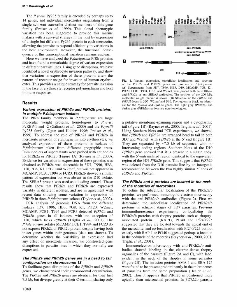
Variant expression of PfRh2a and PfRh2b proteinsin multiple P.falciparum isolatesThe PfRh family members in P.falciparum are largemolecular weight proteins, homologous to P.vivaxPvRBP-1 and -2 (Galinski et al., 2000) and the P.yoeliiPy235 family (Ogun and Holder, 1996; Preiser et al.,1999). To address the role of PfRh2a and PfRh2b inmerozoite invasion of P.falciparum into erythrocytes, weanalyzed expression of these proteins in isolates ofP.falciparum taken from different geographic areas.Immunoblots of supernatants were probed with antibodiesfor PfRh2a or PfRh2b (Figure 1A) (Rayner et al., 2000).Evidence for variation in expression of these proteins wasobtained as PfRh2a was detectable in 3D7, T996, HB3,D10, 7G8, K1, Pf120 and W2mef, but was not apparent inMCAMP, FCB1, T994 or FCR3. PfRh2b showed a similarpattern of expression but was absent in the D10 isolate.The SERA5 protein was used as a loading control. Theseresults show that PfRh2a and PfRh2b are expressedvariably in different isolates, and are in agreement withrecent data showing some variation in expression ofPfRh2b in three P.falciparum isolates (Taylor et al., 2002).
PCR analysis of genomic DNA from the differentisolates 3D7, T996, HB3, 7G8, K1, Pf120, W2mef,MCAMP, FCB1, T994 and FCR3 detected PfRh2a andPfRh2b genes in all isolates, with the exception ofD10, which lacks PfRh2b (Triglia et al., 2001). TheP.falciparum isolates MCAMP, FCB1, T994 and FCR3 donot express PfRh2a or PfRh2b protein despite having bothintact genes within their genomes (data not shown). Todetermine whether the variability in expression hadany effect on merozoite invasion, we constructed genedisruptions in parasite lines in which they normally areexpressed.
The PfRh2a and PfRh2b genes are in a head to tailcon®guration on chromosome 13To facilitate gene disruptions of the PfRh2a and PfRh2bgenes, we characterized their chromosomal organization.The PfRh2a and PfRh2b genes are identical for their ®rst7.5 kb, but diverge greatly at their C-termini, sharing only
a putative membrane-spanning region and a cytoplasmictail (Figure 1B) (Rayner et al., 2000; Triglia et al., 2001).Using Southern blots and PCR experiments, we showedthat PfRh2b and PfRh2a are arranged head to tail in both3D7 and W2mef, with PfRh2b at the 5¢ end (Figure 1B).They are separated by ~7.0 kb of sequence, with nointervening coding regions. Southern blots of the D10PfRh2a gene showed that it is present in a single copy,with the 5¢-untranslated region identical to the equivalentregion of the 3D7 PfRh2b gene. This suggests that PfRh2bwas deleted from the D10 parasite line by an intergenicrecombination between the two highly similar 5¢ ends ofPfRh2a and PfRh2b.
The PfRh2a and b proteins are located in the neckof the rhoptries of merozoitesTo de®ne the subcellular localization of the PfRh2a/bproteins, we performed immunogold-electron microscopywith the anti-PfRh2a/b antibodies (Figure 2). First wedetermined the subcellular localization of PfRh2a/bproteins in schizont stages of 3D7 parasites. Previousimmuno¯uorescence experiments co-localizing thePfRh2a/b proteins with rhoptry proteins such as rhoptry-associated protein 1 (RAP1), Pf140 and Pf240/225suggested that they are located towards the apical end ofthe merozoite, and co-localization with Pf240/225 but notexactly with RAP-1 or Pf140 suggested perhaps a locationin the peduncle of the rhoptries (Rayner et al., 2000, 2001;Triglia et al., 2001).
Immunoelectron microscopy with anti-PfRh2a/b anti-bodies showed labeling in the electron-dense rhoptryorganelles of the parasite (Figure 2A and C), with labelevident in the neck of the rhoptry in some parasites(Figure 2B). The invasion proteins AMA-1 and EBA-175were found to be present predominantly in the micronemesof parasites from the same preparation (Healer et al.,2002). Thus it appears that PfRh2b is positioned moreapically than micronemal proteins. In 3D7D2b parasite
Fig. 1. Variant expression, subcellular localization and structureof the PfRh2a and PfRh2b genes and proteins in P.falciparum.(A) Supernatants from 3D7, T996, HB3, D10, MCAMP, 7G8, K1,Pf120, FCB1, T994, FCR3 and W2mef were probed with anti-PfRh2a,anti-PfRh2b or anti-SERA5 antibodies. The position of the 200 kDamolecular weight marker is shown. (B) Structure of the PfRh2a andPfRh2b locus in 3D7, W2mef and D10. The regions in black are identi-cal for the PfRh2b and PfRh2a genes. The light gray (PfRh2b) anddarker gray (PfRh2a) sections are non-homologous.
M.T.Duraisingh et al.
1048
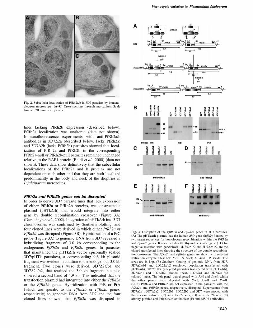
lines lacking PfRh2b expression (described below),PfRh2a localization was unaltered (data not shown).Immuno¯uorescence experiments with anti-PfRh2a/bantibodies in 3D7D2a (described below, lacks PfRh2a)and 3D7D2b (lacks PfRh2b) parasites showed that local-ization of PfRh2a and PfRh2b in the correspondingPfRh2a-null or PfRh2b-null parasites remained unchangedrelative to the RAP1 protein (Baldi et al., 2000) (data notshown). These data show de®nitively that the subcellularlocalizations of the PfRh2a and b proteins are notdependent on each other and that they are both localizedpredominantly in the body and neck of the rhoptries inP.falciparum merozoites.
PfRh2a and PfRh2b genes can be disruptedIn order to derive 3D7 parasite lines that lack expressionof either PfRh2a or PfRh2b proteins, we constructed aplasmid (pHTkDrh) that would integrate into eithergene by double recombination crossover (Figure 3A)(Duraisingh et al., 2002). Integration of pHTkDrh into 3D7chromosomes was con®rmed by Southern blotting, andfour cloned lines were derived in which either PfRh2a orPfRh2b was disrupted (Figure 3B). Hybridization of a PrCprobe (Figure 3A) to genomic DNA from 3D7 revealed ahybridizing fragment of 3.0 kb corresponding to theendogenous PfRh2a and PfRh2b genes. In parasitesthat maintained the pHTkDrh vector episomally (called3D7/pHTk parasites), a corresponding 9.6 kb plasmidfragment was evident in addition to the endogenous 3.0 kbfragment. Two clones were derived, 3D7D2a/b1 and3D7D2a/b2, that retained the 3.0 kb fragment but alsoshowed a second band of 4.9 kb. This indicated that thetransfection plasmid had integrated into either the PfRh2aor the PfRh2b genes. Hybridization with PrB or PrA(which are speci®c to the PfRh2b or PfRh2a genes,respectively) to genomic DNA from 3D7 and the fourcloned lines showed that PfRh2b was disrupted in
Fig. 3. Disruption of the PfRh2b and PfRh2a genes in 3D7 parasites.(A) The pHTkDrh plasmid has the human dhfr gene (hdhfr) ¯anked bytwo target sequences for homologous recombination within the PfRh2aand PfRh2b genes. It also includes the thymidine kinase gene (Tk) fornegative selection with ganciclovir. 3D7D2b1/2 and 3D7D2a1/2 are thecloned transfected lines showing the structure of the double recombina-tion crossovers. The PfRh2a and PfRh2b genes are shown with relevantrestriction enzyme sites: Sw, SwaI; S, SacI; A, AvaII; P, PvuII. Thesizes are in kbp. (B) Southern blotting of genomic DNA from 3D7,3D7D2a/b1 and 3D7D2a/b2 (uncloned population transfected withpHTkDrh), 3D7/pHTk (uncycled parasites transfected with pHTkDrh),3D7D2b1 and 3D7D2b2 (cloned lines), 3D7D2a1 and 3D7D2a1/a2(cloned lines). The left panel was digested with PstI and SwaI, whilstthe other panels were digested with SacI, AvaII and PvuII.(C±F) PfRh2a and PfRh2b are not expressed in the parasites with thePfRh2a and PfRh2b genes, respectively, disrupted. Supernatants from3D7D2a1, 3D7D2a2, 3D7D2b1, 3D7D2b2 and 3D7 were probed withthe relevant antisera: (C) anti-PfRh2a sera; (D) anti-PfRh2b sera; (E)af®nity-puri®ed anti-PfRh2a/2b antibodies; (F) anti-MSP3 antibodies.
Fig. 2. Subcellular localization of PfRh2a/b in 3D7 parasites by immuno-electron microscopy. (A±C) Cross-sections through merozoites. Scalebars are 200 nm in all panels.
Phenotypic variation in Plasmodium falciparum
1049
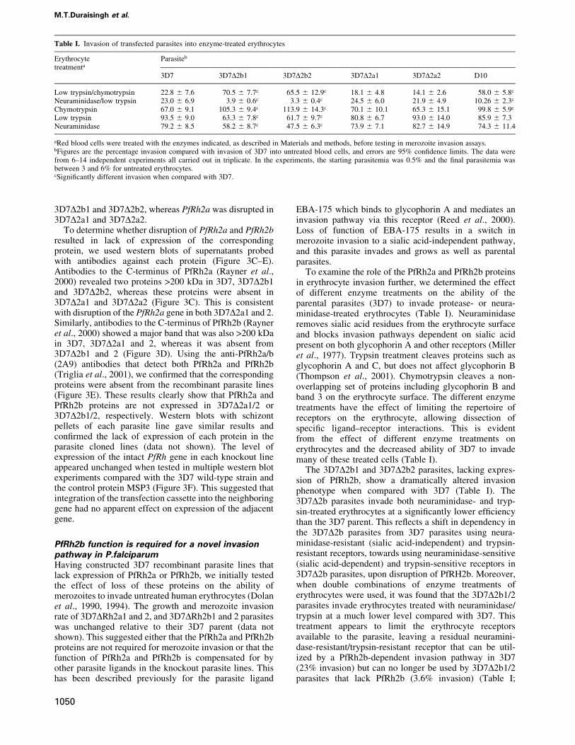
3D7D2b1 and 3D7D2b2, whereas PfRh2a was disrupted in3D7D2a1 and 3D7D2a2.
To determine whether disruption of PfRh2a and PfRh2bresulted in lack of expression of the correspondingprotein, we used western blots of supernatants probedwith antibodies against each protein (Figure 3C±E).Antibodies to the C-terminus of PfRh2a (Rayner et al.,2000) revealed two proteins >200 kDa in 3D7, 3D7D2b1and 3D7D2b2, whereas these proteins were absent in3D7D2a1 and 3D7D2a2 (Figure 3C). This is consistentwith disruption of the PfRh2a gene in both 3D7D2a1 and 2.Similarly, antibodies to the C-terminus of PfRh2b (Rayneret al., 2000) showed a major band that was also >200 kDain 3D7, 3D7D2a1 and 2, whereas it was absent from3D7D2b1 and 2 (Figure 3D). Using the anti-PfRh2a/b(2A9) antibodies that detect both PfRh2a and PfRh2b(Triglia et al., 2001), we con®rmed that the correspondingproteins were absent from the recombinant parasite lines(Figure 3E). These results clearly show that PfRh2a andPfRh2b proteins are not expressed in 3D7D2a1/2 or3D7D2b1/2, respectively. Western blots with schizontpellets of each parasite line gave similar results andcon®rmed the lack of expression of each protein in theparasite cloned lines (data not shown). The level ofexpression of the intact PfRh gene in each knockout lineappeared unchanged when tested in multiple western blotexperiments compared with the 3D7 wild-type strain andthe control protein MSP3 (Figure 3F). This suggested thatintegration of the transfection cassette into the neighboringgene had no apparent effect on expression of the adjacentgene.
PfRh2b function is required for a novel invasionpathway in P.falciparumHaving constructed 3D7 recombinant parasite lines thatlack expression of PfRh2a or PfRh2b, we initially testedthe effect of loss of these proteins on the ability ofmerozoites to invade untreated human erythrocytes (Dolanet al., 1990, 1994). The growth and merozoite invasionrate of 3D7DRh2a1 and 2, and 3D7DRh2b1 and 2 parasiteswas unchanged relative to their 3D7 parent (data notshown). This suggested either that the PfRh2a and PfRh2bproteins are not required for merozoite invasion or that thefunction of PfRh2a and PfRh2b is compensated for byother parasite ligands in the knockout parasite lines. Thishas been described previously for the parasite ligand
EBA-175 which binds to glycophorin A and mediates aninvasion pathway via this receptor (Reed et al., 2000).Loss of function of EBA-175 results in a switch inmerozoite invasion to a sialic acid-independent pathway,and this parasite invades and grows as well as parentalparasites.
To examine the role of the PfRh2a and PfRh2b proteinsin erythrocyte invasion further, we determined the effectof different enzyme treatments on the ability of theparental parasites (3D7) to invade protease- or neura-minidase-treated erythrocytes (Table I). Neuraminidaseremoves sialic acid residues from the erythrocyte surfaceand blocks invasion pathways dependent on sialic acidpresent on both glycophorin A and other receptors (Milleret al., 1977). Trypsin treatment cleaves proteins such asglycophorin A and C, but does not affect glycophorin B(Thompson et al., 2001). Chymotrypsin cleaves a non-overlapping set of proteins including glycophorin B andband 3 on the erythrocyte surface. The different enzymetreatments have the effect of limiting the repertoire ofreceptors on the erythrocyte, allowing dissection ofspeci®c ligand±receptor interactions. This is evidentfrom the effect of different enzyme treatments onerythrocytes and the decreased ability of 3D7 to invademany of these treated cells (Table I).
The 3D7D2b1 and 3D7D2b2 parasites, lacking expres-sion of PfRh2b, show a dramatically altered invasionphenotype when compared with 3D7 (Table I). The3D7D2b parasites invade both neuraminidase- and tryp-sin-treated erythrocytes at a signi®cantly lower ef®ciencythan the 3D7 parent. This re¯ects a shift in dependency inthe 3D7D2b parasites from 3D7 parasites using neura-minidase-resistant (sialic acid-independent) and trypsin-resistant receptors, towards using neuraminidase-sensitive(sialic acid-dependent) and trypsin-sensitive receptors in3D7D2b parasites, upon disruption of PfRH2b. Moreover,when double combinations of enzyme treatments oferythrocytes were used, it was found that the 3D7D2b1/2parasites invade erythrocytes treated with neuraminidase/trypsin at a much lower level compared with 3D7. Thistreatment appears to limit the erythrocyte receptorsavailable to the parasite, leaving a residual neuramini-dase-resistant/trypsin-resistant receptor that can be util-ized by a PfRh2b-dependent invasion pathway in 3D7(23% invasion) but can no longer be used by 3D7D2b1/2parasites that lack PfRh2b (3.6% invasion) (Table I;
Table I. Invasion of transfected parasites into enzyme-treated erythrocytes
Erythrocytetreatmenta
Parasiteb
3D7 3D7D2b1 3D7D2b2 3D7D2a1 3D7D2a2 D10
Low trypsin/chymotrypsin 22.8 6 7.6 70.5 6 7.7c 65.5 6 12.9c 18.1 6 4.8 14.1 6 2.6 58.0 6 5.8c
Neuraminidase/low trypsin 23.0 6 6.9 3.9 6 0.6c 3.3 6 0.4c 24.5 6 6.0 21.9 6 4.9 10.26 6 2.3c
Chymotrypsin 67.0 6 9.1 105.3 6 9.4c 113.9 6 14.3c 70.1 6 10.1 65.3 6 15.1 99.8 6 5.9c
Low trypsin 93.5 6 9.0 63.3 6 7.8c 61.7 6 9.7c 80.8 6 6.7 93.0 6 14.0 85.9 6 7.3Neuraminidase 79.2 6 8.5 58.2 6 8.7c 47.5 6 6.3c 73.9 6 7.1 82.7 6 14.9 74.3 6 11.4
aRed blood cells were treated with the enzymes indicated, as described in Materials and methods, before testing in merozoite invasion assays.bFigures are the percentage invasion compared with invasion of 3D7 into untreated blood cells, and errors are 95% con®dence limits. The data werefrom 6±14 independent experiments all carried out in triplicate. In the experiments, the starting parasitemia was 0.5% and the ®nal parasitemia wasbetween 3 and 6% for untreated erythrocytes.cSigni®cantly different invasion when compared with 3D7.
M.T.Duraisingh et al.
1050
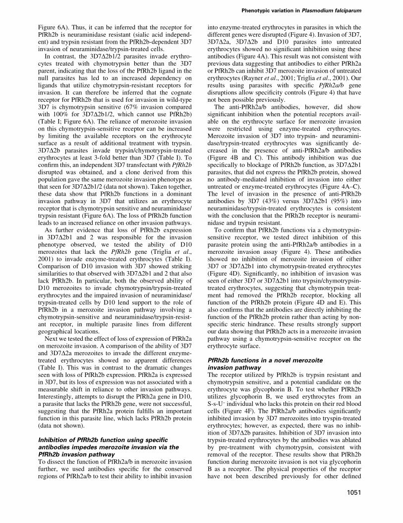
Figure 6A). Thus, it can be inferred that the receptor forPfRh2b is neuraminidase resistant (sialic acid independ-ent) and trypsin resistant from the PfRh2b-dependent 3D7invasion of neuraminidase/trypsin-treated cells.
In contrast, the 3D7D2b1/2 parasites invade erythro-cytes treated with chymotrypsin better than the 3D7parent, indicating that the loss of the PfRh2b ligand in thenull parasites has led to an increased dependency onligands that utilize chymotrypsin-resistant receptors forinvasion. It can therefore be inferred that the cognatereceptor for PfRh2b that is used for invasion in wild-type3D7 is chymotrypsin sensitive (67% invasion comparedwith 100% for 3D7D2b1/2, which cannot use PfRh2b)(Table I; Figure 6A). The reliance of merozoite invasionon this chymotrypsin-sensitive receptor can be increasedby limiting the available receptors on the erythrocytesurface as a result of additional treatment with trypsin.3D7D2b parasites invade trypsin/chymotrypsin-treatederythrocytes at least 3-fold better than 3D7 (Table I). Tocon®rm this, an independent 3D7 transfectant with PfRh2bdisrupted was obtained, and a clone derived from thispopulation gave the same merozoite invasion phenotype asthat seen for 3D7D2b1/2 (data not shown). Taken together,these data show that PfRh2b functions in a dominantinvasion pathway in 3D7 that utilizes an erythrocytereceptor that is chymotrypsin sensitive and neuraminidase/trypsin resistant (Figure 6A). The loss of PfRh2b functionleads to an increased reliance on other invasion pathways.
As further evidence that loss of PfRh2b expressionin 3D7D2b1 and 2 was responsible for the invasionphenotype observed, we tested the ability of D10merozoites that lack the PfRh2b gene (Triglia et al.,2001) to invade enzyme-treated erythrocytes (Table I).Comparison of D10 invasion with 3D7 showed strikingsimilarities to that observed with 3D7D2b1 and 2 that alsolack PfRh2b. In particular, both the observed ability ofD10 merozoites to invade chymotrypsin/trypsin-treatederythrocytes and the impaired invasion of neuraminidase/trypsin-treated cells by D10 lend support to the role ofPfRh2b in a merozoite invasion pathway involving achymotrypsin-sensitive and neuraminidase/trypsin-resist-ant receptor, in multiple parasite lines from differentgeographical locations.
Next we tested the effect of loss of expression of PfRh2aon merozoite invasion. A comparison of the ability of 3D7and 3D7D2a merozoites to invade the different enzyme-treated erythrocytes showed no apparent differences(Table I). This was in contrast to the dramatic changesseen with loss of PfRh2b expression. PfRh2a is expressedin 3D7, but its loss of expression was not associated with ameasurable shift in reliance to other invasion pathways.Interestingly, attempts to disrupt the PfRh2a gene in D10,a parasite that lacks the PfRh2b gene, were not successful,suggesting that the PfRh2a protein ful®lls an importantfunction in this parasite line, which lacks PfRh2b protein(data not shown).
Inhibition of PfRh2b function using speci®cantibodies impedes merozoite invasion via thePfRh2b invasion pathwayTo dissect the function of PfRh2a/b in merozoite invasionfurther, we used antibodies speci®c for the conservedregions of PfRh2a/b to test their ability to inhibit invasion
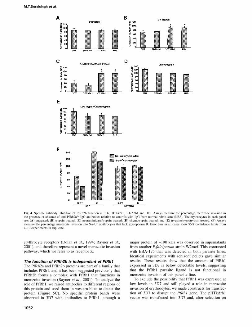
into enzyme-treated erythrocytes in parasites in which thedifferent genes were disrupted (Figure 4). Invasion of 3D7,3D7D2a, 3D7D2b and D10 parasites into untreatederythrocytes showed no signi®cant inhibition using theseantibodies (Figure 4A). This result was not consistent withprevious data suggesting that antibodies to either PfRh2aor PfRh2b can inhibit 3D7 merozoite invasion of untreatederythrocytes (Rayner et al., 2001; Triglia et al., 2001). Ourresults using parasites with speci®c PfRh2a/b genedisruptions allow speci®city controls (Figure 4) that havenot been possible previously.
The anti-PfRh2a/b antibodies, however, did showsigni®cant inhibition when the potential receptors avail-able on the erythrocyte surface for merozoite invasionwere restricted using enzyme-treated erythrocytes.Merozoite invasion of 3D7 into trypsin- and neuramini-dase/trypsin-treated erythrocytes was signi®cantly de-creased in the presence of anti-PfRh2a/b antibodies(Figure 4B and C). This antibody inhibition was duespeci®cally to blockage of PfRh2b function, as 3D7D2b1parasites, that did not express the PfRh2b protein, showedno antibody-mediated inhibition of invasion into eitheruntreated or enzyme-treated erythrocytes (Figure 4A±C).The level of invasion in the presence of anti-PfRh2bantibodies by 3D7 (43%) versus 3D7D2b1 (95%) intoneuraminidase/trypsin-treated erythrocytes is consistentwith the conclusion that the PfRh2b receptor is neurami-nidase and trypsin resistant.
To con®rm that PfRh2b functions via a chymotrypsin-sensitive receptor, we tested direct inhibition of thisparasite protein using the anti-PfRh2a/b antibodies in amerozoite invasion assay (Figure 4). These antibodiesshowed no inhibition of merozoite invasion of either3D7 or 3D7D2b1 into chymotrypsin-treated erythrocytes(Figure 4D). Signi®cantly, no inhibition of invasion wasseen of either 3D7 or 3D7D2b1 into trypsin/chymotrypsin-treated erythrocytes, suggesting that chymotrypsin treat-ment had removed the PfRh2b receptor, blocking allfunction of the PfRh2b protein (Figure 4D and E). Thisalso con®rms that the antibodies are directly inhibiting thefunction of the PfRh2b protein rather than acting by non-speci®c steric hindrance. These results strongly supportour data showing that PfRh2b acts in a merozoite invasionpathway using a chymotrypsin-sensitive receptor on theerythrocyte surface.
PfRh2b functions in a novel merozoiteinvasion pathwayThe receptor utilized by PfRh2b is trypsin resistant andchymotrypsin sensitive, and a potential candidate on theerythrocyte was glycophorin B. To test whether PfRh2butilizes glycophorin B, we used erythrocytes from anS-s-U± individual who lacks this protein on their red bloodcells (Figure 4F). The PfRh2a/b antibodies signi®cantlyinhibited invasion by 3D7 merozoites into tryspin-treatederythrocytes; however, as expected, there was no inhib-ition of 3D7D2b parasites. Inhibition of 3D7 invasion intotrypsin-treated erythrocytes by the antibodies was ablatedby pre-treatment with chymotrypsin, consistent withremoval of the receptor. These results show that PfRh2bfunction during merozoite invasion is not via glycophorinB as a receptor. The physical properties of the receptorhave not been described previously for other de®ned
Phenotypic variation in Plasmodium falciparum
1051
erythrocyte receptors (Dolan et al., 1994; Rayner et al.,2001), and therefore represent a novel merozoite invasionpathway, which we refer to as receptor Z.
The function of PfRh2b is independent of PfRh1The PfRh2a and PfRh2b proteins are part of a family thatincludes PfRh1, and it has been suggested previously thatPfRh2b forms a complex with PfRh1 that functions inmerozoite invasion (Rayner et al., 2001). To analyze therole of PfRh1, we raised antibodies to different regions ofthis protein and used them in western blots to detect theprotein (Figure 5C). No speci®c protein bands wereobserved in 3D7 with antibodies to PfRh1, athough a
major protein of ~190 kDa was observed in supernatantsfrom another P.falciparum strain W2mef. This contrastedwith EBA-175 that was detected in both parasite lines.Identical experiments with schizont pellets gave similarresults. These results show that the amount of PfRh1expressed in 3D7 is below detectable levels, suggestingthat the PfRh1 parasite ligand is not functional inmerozoite invasion of this parasite line.
To exclude the possibility that PfRh1 was expressed atlow levels in 3D7 and still played a role in merozoiteinvasion of erythrocytes, we made constructs for transfec-tion of 3D7 to disrupt the PfRh1 gene. The pHTkDrh1vector was transfected into 3D7 and, after selection on
Fig. 4. Speci®c antibody inhibition of PfRh2b function in 3D7, 3D7D2a1, 3D7D2b1 and D10. Assays measure the percentage merozoite invasion inthe presence or absence of anti-PfRh2a/b IgG antibodies relative to controls with IgG from normal rabbit sera (NRS). The erythrocytes in each panelare: (A) untreated; (B) trypsin treated; (C) neuraminidase/trypsin treated; (D) chymotrypsin treated; and (E) trypsin/chymotrypsin treated. (F) Assaysmeasure the percentage merozoite invasion into S-s-U± erythrocytes that lack glycophorin B. Error bars in all cases show 95% con®dence limits from4±10 experiments in triplicate.
M.T.Duraisingh et al.
1052
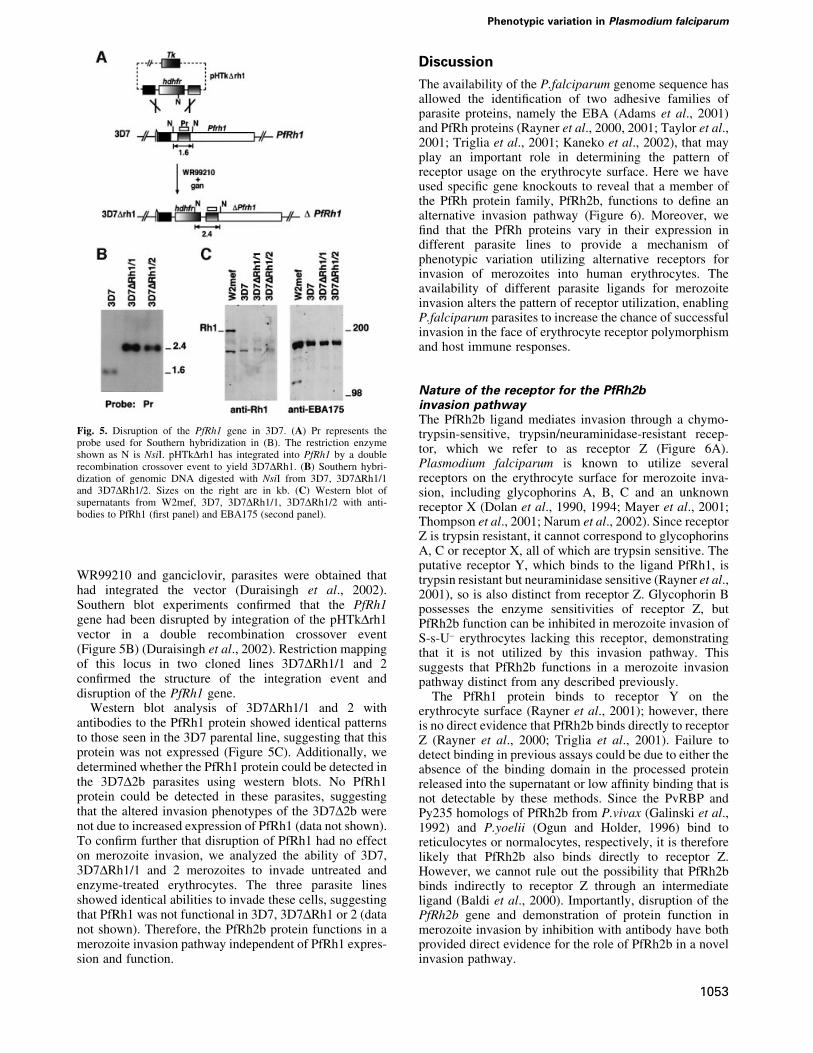
WR99210 and ganciclovir, parasites were obtained thathad integrated the vector (Duraisingh et al., 2002).Southern blot experiments con®rmed that the PfRh1gene had been disrupted by integration of the pHTkDrh1vector in a double recombination crossover event(Figure 5B) (Duraisingh et al., 2002). Restriction mappingof this locus in two cloned lines 3D7DRh1/1 and 2con®rmed the structure of the integration event anddisruption of the PfRh1 gene.
Western blot analysis of 3D7DRh1/1 and 2 withantibodies to the PfRh1 protein showed identical patternsto those seen in the 3D7 parental line, suggesting that thisprotein was not expressed (Figure 5C). Additionally, wedetermined whether the PfRh1 protein could be detected inthe 3D7D2b parasites using western blots. No PfRh1protein could be detected in these parasites, suggestingthat the altered invasion phenotypes of the 3D7D2b werenot due to increased expression of PfRh1 (data not shown).To con®rm further that disruption of PfRh1 had no effecton merozoite invasion, we analyzed the ability of 3D7,3D7DRh1/1 and 2 merozoites to invade untreated andenzyme-treated erythrocytes. The three parasite linesshowed identical abilities to invade these cells, suggestingthat PfRh1 was not functional in 3D7, 3D7DRh1 or 2 (datanot shown). Therefore, the PfRh2b protein functions in amerozoite invasion pathway independent of PfRh1 expres-sion and function.
Discussion
The availability of the P.falciparum genome sequence hasallowed the identi®cation of two adhesive families ofparasite proteins, namely the EBA (Adams et al., 2001)and PfRh proteins (Rayner et al., 2000, 2001; Taylor et al.,2001; Triglia et al., 2001; Kaneko et al., 2002), that mayplay an important role in determining the pattern ofreceptor usage on the erythrocyte surface. Here we haveused speci®c gene knockouts to reveal that a member ofthe PfRh protein family, PfRh2b, functions to de®ne analternative invasion pathway (Figure 6). Moreover, we®nd that the PfRh proteins vary in their expression indifferent parasite lines to provide a mechanism ofphenotypic variation utilizing alternative receptors forinvasion of merozoites into human erythrocytes. Theavailability of different parasite ligands for merozoiteinvasion alters the pattern of receptor utilization, enablingP.falciparum parasites to increase the chance of successfulinvasion in the face of erythrocyte receptor polymorphismand host immune responses.
Nature of the receptor for the PfRh2binvasion pathwayThe PfRh2b ligand mediates invasion through a chymo-trypsin-sensitive, trypsin/neuraminidase-resistant recep-tor, which we refer to as receptor Z (Figure 6A).Plasmodium falciparum is known to utilize severalreceptors on the erythrocyte surface for merozoite inva-sion, including glycophorins A, B, C and an unknownreceptor X (Dolan et al., 1990, 1994; Mayer et al., 2001;Thompson et al., 2001; Narum et al., 2002). Since receptorZ is trypsin resistant, it cannot correspond to glycophorinsA, C or receptor X, all of which are trypsin sensitive. Theputative receptor Y, which binds to the ligand PfRh1, istrypsin resistant but neuraminidase sensitive (Rayner et al.,2001), so is also distinct from receptor Z. Glycophorin Bpossesses the enzyme sensitivities of receptor Z, butPfRh2b function can be inhibited in merozoite invasion ofS-s-U± erythrocytes lacking this receptor, demonstratingthat it is not utilized by this invasion pathway. Thissuggests that PfRh2b functions in a merozoite invasionpathway distinct from any described previously.
The PfRh1 protein binds to receptor Y on theerythrocyte surface (Rayner et al., 2001); however, thereis no direct evidence that PfRh2b binds directly to receptorZ (Rayner et al., 2000; Triglia et al., 2001). Failure todetect binding in previous assays could be due to either theabsence of the binding domain in the processed proteinreleased into the supernatant or low af®nity binding that isnot detectable by these methods. Since the PvRBP andPy235 homologs of PfRh2b from P.vivax (Galinski et al.,1992) and P.yoelii (Ogun and Holder, 1996) bind toreticulocytes or normalocytes, respectively, it is thereforelikely that PfRh2b also binds directly to receptor Z.However, we cannot rule out the possibility that PfRh2bbinds indirectly to receptor Z through an intermediateligand (Baldi et al., 2000). Importantly, disruption of thePfRh2b gene and demonstration of protein function inmerozoite invasion by inhibition with antibody have bothprovided direct evidence for the role of PfRh2b in a novelinvasion pathway.
Fig. 5. Disruption of the PfRh1 gene in 3D7. (A) Pr represents theprobe used for Southern hybridization in (B). The restriction enzymeshown as N is NsiI. pHTkDrh1 has integrated into PfRh1 by a doublerecombination crossover event to yield 3D7DRh1. (B) Southern hybri-dization of genomic DNA digested with NsiI from 3D7, 3D7DRh1/1and 3D7DRh1/2. Sizes on the right are in kb. (C) Western blot ofsupernatants from W2mef, 3D7, 3D7DRh1/1, 3D7DRh1/2 with anti-bodies to PfRh1 (®rst panel) and EBA175 (second panel).
Phenotypic variation in Plasmodium falciparum
1053
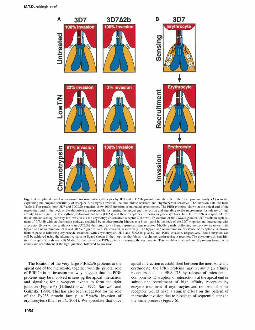
The location of the very large PfRh2a/b proteins at theapical end of the merozoite, together with the pivotal roleof PfRh2b in an invasion pathway, suggest that the PfRhproteins may be involved in sensing the apical interactionand signaling for subsequent events to form the tightjunction (Figure 6) (Galinski et al., 1992; Barnwell andGalinski, 1998). This has also been suggested for the roleof the Py235 protein family in P.yoelii invasion oferythrocytes (Khan et al., 2001). We speculate that once
apical interaction is established between the merozoite anderythrocyte, the PfRh proteins may recruit high af®nityreceptors such as EBA-175 by release of micronemalcomponents. Disruption of interactions at the apical end orsubsequent recruitment of high af®nity receptors byenzyme treatment of erythrocytes and removal of somereceptors would have a similar effect on the pattern ofmerozoite invasion due to blockage of sequential steps inthe same process (Figure 6).
Fig. 6. A simpli®ed model of merozoite invasion into erythrocytes by 3D7 and 3D7D2b parasites and the role of the PfRh protein family. (A) A modelexplaining the enzyme sensitivity of receptor Z as trypsin resistant, neuraminidase resistant and chymotrypsin sensitive. The invasion data are fromTable I. Top panels: both 3D7 and 3D7D2b parasites show 100% invasion of untreated erythrocytes. The PfRh proteins (shown at the apical end of themerozoites and in the neck of the rhoptries) are responsible for sensing the apical end interaction and signaling to the micronemes for release of highaf®nity ligands (see B). The eythrocyte-binding antigens (EBAs) and their receptors are shown as green symbols. In 3D7, PfRh2b is responsible forthe dominant sensing pathway for invasion via the chymotrypsin-sensitive receptor Z (brown). Disruption of the PfRh2b gene in 3D7 results in replace-ment of PfRh2b with an alternative pathway speci®ed by another protein [shown as a blue ligand in the neck of the 3D7 rhoptries and interacting witha receptor (blue) on the erythrocyte in 3D7D2b] that binds to a chymotrypsin-resistant receptor. Middle panels: following erythrocyte treatment withtrypsin and neuraminidase, 3D7 and 3D7D2b give 23 and 3% invasion, respectively. The trypsin and neuraminidase resistance of receptor Z is shown.Bottom panels: following erythrocyte treatment with chymotrypsin, 3D7 and 3D7D2b give 67 and 100% invasion, respectively. Some invasion canstill be achieved using the alternative parasite ligand shown in the rhoptries that binds to a chymotrypsin-resistant receptor. The chymotrypsin sensitiv-ity of receptor Z is shown. (B) Model for the role of the PfRh proteins in sensing the erythrocyte. This would activate release of proteins from micro-nemes and recruitment at the tight junction, followed by invasion.
M.T.Duraisingh et al.
1054
Compensation for loss of PfRh2b function by otherparasite ligands?Binding and entry of P.falciparum invasive merozoitesinto human erythrocytes require speci®c ligand±receptorinteractions. The observed strain variation in theutilization of these invasion pathways is very stableand does not change in the course of routine culture(Dolan et al., 1994; Okoyeh et al., 1999). Merozoitesrequire suf®cient af®nity of ligand binding to cognatereceptors to activate subsequent steps in the invasionprocess (see Barnwell and Galinski, 1998). It is likelythat the area available for interaction between the apicalend of the merozoite and the erythrocyte is limited, andtherefore the number of ligands occupying that spacewould also be limited (Figure 6). Even in the absence ofPfRh2b expression, no difference in invasion and growthrates is observed, implying that a suf®cient af®nity stillexists between the remaining ligands and their cognatereceptors for ef®cient invasion of normal erythrocytes.Nevertheless, a large change in receptor usage doesresult from the loss of PfRh2b expression, indicating thata signi®cant shift in dependency to other ligands withdifferent erythrocyte receptor speci®cities has occurred.This shift in receptor usage may either result from theupregulation of ligands, existing or novel, to compensatefor the missing Rh2b ligand or, alternatively, be due tothe redeployment of existing ligands from a poolcontaining an excess of ligands. In either case, theapparent shift in the usage of pathways implies that lossof PfRh2b expression necessitated an increased relianceon other ligands to provide suf®cient af®nity for ef®cientinvasion (Figure 6).
PfRh proteins function independently ofeach otherPrevious data has suggested that PfRh2b and PfRh1 mightform a functional complex (Rayner et al., 2001), and thiswould be consistent with the suggested association ofPvRBP1 and PvRBP2 in P.vivax (Galinski et al., 1992).However, our data show that PfRh2b functions inmerozoite invasion in the 3D7 parasite line in the absenceof PfRh1 and PfRh2a expression. Furthermore, immuno-precipitation experiments did not co-precipitate otherPfRh proteins using antibodies to PfRh2b, PfRh2a andPfRh1 (data not shown). The functional independence ofthe PfRh proteins is consistent with the expression of onlyone Py235 gene from a large gene family in a singlemerozoite of P.yoelii (Preiser et al., 1999). The role of afourth member of the PfRh family, PfRh4 (Kaneko et al.,2002), in merozoite invasion remains to be determined, butit is likely that this protein is involved in specifying adifferent invasion pathway.
Although PfRh2b functions in merozoite invasion, wefound no invasion role for the closely related proteinPfRh2a in the 3D7 parasite line. PfRh2a and PfRh2b arevirtually identical for a large part of the N-terminus, anddiffer at the C-terminus that includes a transmembraneregion and a short cytoplasmic tail. Although PfRh2a doesnot play an important role in 3D7 parasites, it does notexclude a role in other parasite lines in a different geneticbackground. Indeed, attempts to disrupt the PfRh2a genein D10 parasites, that lack PfRh2b, were unsuccessful,suggesting that the encoded protein plays an important role
in this parasite line. The differences in function betweenPfRh2b and PfRh2a must be speci®ed by the codingdivergence in their C-termini. It is possible that thecytoplasmic tail of PfRh2b is responsible for activatingsignals for release of micronemes onto the erythrocyte andsubsequent steps in tight junction formation (Galinskiet al., 1992; Barnwell and Galinski, 1998). Alternatively,the unique region in PfRh2b N-terminal to the transmem-brane domain may be responsible for interacting with asecond protein that is involved in receptor binding on theerythrocyte.
Variation in expression of PfRh proteins:a selective advantage for P.falciparumDifferent patterns of invasion phenotypes betweenisolates of P.falciparum can be explained in part byvariation in expression of the PfRh protein family. Thisstudy has identi®ed many parasite isolates that do notexpress PfRh2a or PfRh2b, and, similarly, 3D7 does notexpress detectable levels of PfRh1. We have evidencesuggesting that there are many parasite isolates that donot express PfRh1 (data not shown). Loss of expressionof the PfRh proteins in any merozoite is compensated byother parasite ligands interacting with available recep-tors. The presence of non-functional parasite ligands atthe apical end of some merozoites, because of blockadeby either antibodies or receptor polymorphism, decreasesthe ef®ciency of merozoite invasion. It follows thatvariation in expression of different parasite ligandsacross parasite populations would overcome this forindividual merozoites, since they would lack expressionof these non-functional ligands. By concentrating func-tional ligands at the apical end in place of PfRh2b, thesemerozoites could achieve the required af®nity foref®cient invasion. Obviously, suppression of expressionof proteins such as PfRh2b or PfRh1 could result in thepermanent loss of these proteins within the parasitepopulation. However, genetic recombination duringmeiosis between frequent mixed infections would con-stantly generate a pool of parasites that either express ordo not express particular PfRh proteins and invasionpathways. Hence, the parasites appear to maintain avaried repertoire of ligands, but probably concentrate onexpressing a limited suite at any one time in order toachieve a threshold of binding necessary for invasion.
A number of P.falciparum isolates do not express thePfRh2a or PfRh2b proteins despite having apparentlyintact genes within their genomes. The mechanism for lackof expression of these proteins is not yet understood, butthere are several possibilities. First, there may be muta-tions that block transcription or translation. Secondly, it ispossible that the genes are silenced, and this would providea mechanism to switch the expression of these genes onand off, allowing the parasite access to different invasionpathways when required. This would be analogous to theantigenic switching of the var gene family of P.falciparum(Smith et al., 1995; Su et al., 1995). Expression of theequivalent of the PfRh family in P.yoelii (Py235) islimited to only one gene per merozoite, suggesting someform of regulated control of this gene family (Preiser et al.,1999).
Phenotypic variation in Plasmodium falciparum
1055
Biological signi®cance for P.falciparum invasion ofhuman erythrocytesMultiple parasite ligands and phenotypic variation inexpression of the PfRh family in P.falciparum haveimportant implications for the parasite. Merozoite inva-sion requires suf®cient ligand±receptor interactions on theerythrocyte surface, and there are circumstances wherereceptor availability is limited. First, the molecules on theerythrocyte are highly polymorphic in the human popu-lation, and this heterogeneity has probably been selectedby P.falciparum and other infectious agents to protectagainst severe disease (Miller et al., 2002). The absence ormutation of receptors would limit those available on theerythrocyte for interaction with parasite ligands, and thiswould decrease invasion ef®ciency. Examples are popu-lations in East Africa that lack glycophorin B, andMelanesian populations that express a mutant form ofglycophorin C on the erythrocyte surface (Patel et al.,2001). Secondly, during development and aging of thehuman erythrocyte, different receptors such as glyco-phorin A are not present or vary in their quantity andpresentation on the surface of this cell (Issitt and Anstee,1998). Thirdly, host immune responses to one or more ofthe parasite ligands involved in merozoite invasion wouldblock utilization of particular alternative invasion path-ways, thus limiting the pathways available. Multipleparasite ligands and phenotypic variation of PfRh proteinexpression provide a mechanism for the parasite to evadethese situations by the presence of merozoites within thepopulation that have different dominant invasion pathwayspeci®cities, increasing the ability of the parasite tomaintain infection and its potential for transmission tosubsequent hosts. It will be important to consider theability of the parasite to vary expression of parasite ligandsfor merozoite invasion when developing vaccine strategiesdirected against invasion-related proteins.
Materials and methods
Plasmids and parasitesGenomic DNA from the 3D7 parasite line was used as template in thePCR of two ~1 kb fragments corresponding to a region conservedbetween the PfRh2a and PfRh2b genes: Flank 1 (5¢-GGACCGC-CGCGGAGTCATGAGCATTTTGTTGGACAA-3¢) and Flank 2n(5¢-GGACCGGTTAACTTCGTCATGTATATATCCAATAGA-3¢) usingthe primers 5¢-GGACCGGGCGCCCGGGTGATGTATTATGTACAG-CTAAAGC-3¢ and 5¢-GGACCGCCTAGGGAAAAAGCCTCTATGG-ATGAAATG-3¢. Similarly, two ¯anks from the PfRh1 gene were clonedinto a vector derived from pHTk, which had a modi®ed multiple cloningsite. Flank 1 was ampli®ed as above using the primers 5¢-GATCAC-TAGTTTATGAACCTACCCCTTCAT and 5¢-GATCAGATCTATGA-AAGTAGGTTTAATGTA, and Flank 2 was amplifed using the primers5¢-GATCGAATTCGATTTACAAGATCTCTTACT and 5¢-GATCCC-ATGGTTTGGATATATTTTCCCTTA.
Plasmodium falciparum asexual stages were maintained in human 0+erythrocytes. 3D7 is a cloned line derived from NF54 and was obtainedfrom David Walliker at Edinburgh University. W2mef is a cloned linederived from the Indochina III/CDC strain. The parasites Tak996 andTak994 from South-East Asia, and FCB1 of African origin were providedby David Baker from the London School of Hygiene and TropicalMedicine. The FCR3 cloned line is of African origin. HB3 and 7G8 areSouth American cloned lines, while D10 was cloned from a Papua NewGuinean isolate. Malayan Camp, K1 and Pf120 are all SouthEast Asianlines. Transfection with 80 mg of puri®ed plasmid DNA (Qiagen) andselection for stable transfectants with double recombination crossoverwere carried out as described previously (Duraisingh et al., 2002).
SDS±PAGE and immunoblot analysisParasite pellets were collected from parasites 40 h after synchronization.Parasite supernatants were obtained from cultures following treatment ofthe erythrocytes with trypsin (1.0 mg/ml) and neuraminidase (25 mU/ml).Proteins were separated on 6% SDS±polyacrylamide gels. Westernblotting onto nitrocellulose (0.45 mm; Schleicher and Schuell, Germany)was performed according to standard protocols.
Erythrocyte invasion assayUninfected erythrocytes were treated with enzymes as describedpreviously (Thompson et al., 2001). The invasion assay was carried outessentially as previously described (Reed et al., 2000). Erythrocytesinfected with ring stages were synchronized twice, followed by pre-treatment with trypsin and neuraminidase to prevent reinvasion.Experiments were carried out with the addition of new erythrocytestreated with trypsin (66.7 mg/ml), neuraminidase (66.7 mU/ml) andchymotrypsin (1.0 mg/ml) or a combination of these enzyme treatments(Thompson et al., 2001). Enzyme-treated or normal erythrocytes at 4%hematocrit were inoculated with infected erythrocytes to give aparasitemia of 0.5% and hematocrit of 4% in a volume of 100 ml.Parasites were incubated for 48 h followed by addition of[3H]hypoxanthine (Amersham) to 1 mCi/well. After 16 h, the cells wereharvested onto glass ®lters with a cell harvester (Packard). Incorporated[3H]hypoxanthine was determined in a scintillation counter. Percentageinvasion was calculated in comparison with invasion of the same parasiteline into untreated erythrocytes, which was set to 100%.
Antibody inhibition assayAntibodies were puri®ed using protein G. Antibody inhibition assayswere performed in the same way as the erythrocyte invasion assay exceptthat the hematocrit was kept at 2% with an initial parasitemia of 0.5%.Antibodies were added to a ®nal concentration of 0.5 mg/ml 24 hfollowing the setting up of the assay, prior to reinvasion. Invasion in thepresence of speci®c anti-PfRh2a/b antibodies was compared withinvasion in the presence of control antibodies from pre-immune serum.
Electron microscopyImmunolabeling for electron microscopy was performed on ultrathinsections of glutaraldehyde-®xed parasites as described previously (Healeret al., 2002). Micrographs were obtained using a Philips CM120BioTWIN transmission electron microscope at 120 kV.
Acknowledgements
We thank the Red Cross Blood Service (Melbourne, Australia) for supplyof red cells and serum, and Dr Brendan Crabb and Dr Mark Wickham forcritical reading of the manuscript. This work was supported by theNational Health and Medical Research Council of Australia. M.T.D. isfunded by a Wellcome Trust Advanced Training Fellowship (TropicalMedicine). S.A.R. is funded by a Melbourne Research Scholarship.G.I.M. and A.F.C. are Howard Hughes International Scholars.
References
Adams,J.H., Blair,P.L., Kaneko,O. and Peterson,D.S. (2001) Anexpanding ebl family of Plasmodium falciparum. Trends Parasitol.,17, 297±299.
Baldi,D.L., Andrews,K.T., Waller,R.S., Roos,D., Crabb,B.S. andCowman,A.F. (2000) RAP1 controls rhoptry targeting of RAP2 inthe malaria parasite Plasmodium falciparum. EMBO J., 19, 1±9.
Barnwell,J.W. and Galinski,M.R. (1998) Invasion of vertebrate cells:erythrocytes. In Sherman,I.W. (ed.), Malaria: Parasite Biology,Pathogenesis and Protection. ASM Press, Washington, DC,pp. 93±120.
Butcher,G.A., Mitchell,G.H. and Cohen,S. (1973) Mechanism of hostspeci®city in malarial infection. Nature, 244, 40±41.
Dolan,S.A., Miller,L.H. and Wellems,T.E. (1990) Evidence for aswitching mechanism in the invasion of erythrocytes byPlasmodium falciparum. J. Clin. Invest., 86, 618±624.
Dolan,S.A., Proctor,J.L., Alling,D.W., Okubo,Y., Wellems,T.E. andMiller,L.H. (1994) Glycophorin B as an EBA-175 independentPlasmodium falciparum receptor of human erythrocytes. Mol.Biochem. Parasitol., 64, 55±63.
Duraisingh,M.T., Triglia,T. and Cowman,A.F. (2002) Negative selection
M.T.Duraisingh et al.
1056
of Plasmodium falciparum reveals targeted gene deletion by doublecrossover recombination. Int. J. Parasitol., 32, 81±89.
Florens,L. et al. (2002) A proteomic view of the Plasmodium falciparumlife cycle. Nature, 419, 520±526.
Galinski,M.R., Medina,C.C., Ingravallo,P. and Barnwell,J.W. (1992) Areticulocyte-binding protein complex of Plasmodium vivaxmerozoites. Cell, 69, 1213±1226.
Galinski,M.R., Mengyao,X. and Barnwell,J.W. (2000) Plasmodiumvivax reticulocyte binding protein-2 (PvRBP-2) shares structuralfeatures with PvRBP-1 and the Plasmodium yoelii 235kDa rhoptryprotein family. Mol. Biochem. Parasitol., 108, 257±262.
Gratzer,W.B. and Dluzewski,A.R. (1993) The red blood cell and malariaparasite invasion. Semin. Hematol., 30, 232±247.
Healer,J., Crawford,S., Ralph,S., McFadden,G. and Cowman,A.F. (2002)Independent translocation of two micronemal proteins in developingPlasmodium falciparum merozoites. Infect. Immun., 70, 5751±5758.
Howard,R.J., Haynes,J.D., McGinniss,M.H. and Miller,L.H. (1982)Studies on the role of red blood cell glycoproteins as receptors forinvasion by Plasmodium falciparum merozoites. Mol. Biochem.Parasitol., 6, 303±315.
Issitt,P.D. and Anstee,D.J. (1998) Applied Blood Group Serology.Montgomery Scienti®c Publications, Durham.
Kaneko,O., Mu,J., Tsuboi,T., Su,X. and Torii,M. (2002) Gene structureand expression of a Plasmodium falciparum 220-kDa proteinhomologous to the Plasmodium vivax reticulocyte binding proteins.Mol. Biochem. Parasitol., 121, 275±278.
Khan,S.M., Jarra,W. and Preiser,P.R. (2001) The 235 kDa rhoptryprotein of Plasmodium (yoelii) yoelii: function at the junction. Mol.Biochem. Parasitol., 117, 1±10.
Mayer,D.C., Kaneko,O., Hudson-Taylor,D.E., Reid,M.E. andMiller,L.H. (2001) Characterization of a Plasmodium falciparumerythrocyte-binding protein paralogous to EBA-175. Proc. Natl Acad.Sci. USA, 98, 5222±5227.
Miller,L.H., McAuliffe,F.M. and Mason,S.J. (1977) Erythrocytereceptors for malaria merozoites. Am. J. Trop. Med. Hyg., 26,204±208.
Miller,L.H., Baruch,D.I., Marsh,K. and Doumbo,O.K. (2002) Thepathogenic basis of malaria. Nature, 415, 673±679.
Narum,D.L., Fuhrmann,S.R., Luu,T. and Sim,B.K. (2002) A novelPlasmodium falciparum erythrocyte binding protein-2 (EBP2/BAEBL) involved in erythrocyte receptor binding. Mol. Biochem.Parasitol., 119, 159±168.
Ogun,S.A. and Holder,A.A. (1996) A high molecular mass Plasmodiumyoelii rhoptry protein binds to erythrocytes. Mol. Biochem. Parasitol.,76, 321±324.
Okoyeh,J.N., Pillai,C.R. and Chitnis,C.E. (1999) Plasmodium falciparum®eld isolates commonly use erythrocyte invasion pathways that areindependent of sialic acid residues of glycophorin A. Infect. Immun.,67, 5784±5791.
Pasvol,G. (1984) Receptors on red cells for Plasmodium falciparum andtheir interaction with merozoites. Philos. Trans. R. Soc. Lond. B Biol.Sci., 307, 189±200.
Pasvol,G., Jungery,M., Weatherall,D.J., Parsons,S.F., Anstee,D.J. andTanner,M.J.A. (1982) Glycophorin as a possible receptor forPlasmodium falciparum. Lancet, 2, 947±950.
Patel,S.S., Mehlotra,R.K., Kastens,W., Mgone,C.S., Kazura,J.W. andZimmerman,P.A. (2001) The association of the glycophorin C exon 3deletion with ovalocytosis and malaria susceptibility in the Wosera,Papua New Guinea. Blood, 98, 3489±3491.
Preiser,P.R., Jarra,W., Capiod,T. and Snounou,G. (1999) A rhoptry-protein-associated mechanism of clonal phenotypic variation in rodentmalaria. Nature, 398, 618±622.
Rayner,J.C., Galinski,M.R., Ingravallo,P. and Barnwell,J.W. (2000) TwoPlasmodium falciparum genes express merozoite proteins that arerelated to Plasmodium vivax and Plasmodium yoelii adhesive proteinsinvolved in host cell selection and invasion. Proc. Natl Acad. Sci.USA, 97, 9648±9653.
Rayner,J.C., Vargas-Serrato,E., Huber,C.S., Galinski,M.R. andBarnwell,J.W. (2001) A Plasmodium falciparum homologue ofPlasmodium vivax reticulocyte binding protein (PvRBP1) de®nes atrypsin-resistant erythrocyte invasion pathway. J. Exp. Med., 194,1571±1581.
Reed,M.B., Caruana,S.R., Batchelor,A.H., Thompson,J.K., Crabb,B.S.and Cowman,A.F. (2000) Targeted disruption of an erythrocytebinding antigen in Plasmodium falciparum is associated with a switchtoward a sialic acid independent pathway of invasion. Proc. NatlAcad. Sci. USA, 97, 7509±7514.
Sim,B.K.L., Chitnis,C.E., Wasniowska,K., Hadley,T.J. and Miller,L.H.(1994) Receptor and ligand domains for invasion of erythrocytes byPlasmodium falciparum. Science, 264, 1941±1944.
Smith,J.D., Chitnis,C.E., Craig,A.G., Roberts,D.J., Hudson-Taylor,D.E.,Peterson,D.S., Pinches,R., Newbold,C.I. and Miller,L.H. (1995)Switches in expression of Plasmodium falciparum var genescorrelate with changes in antigenic and cytoadherent phenotypes ofinfected erythrocytes. Cell, 82, 101±110.
Su,X.Z., Heatwole,V.M., Wertheimer,S.P., Guinet,F., Herrfeldt,J.A.,Peterson,D.S., Ravetch,J.A. and Wellems,T.E. (1995) The largediverse gene family var encodes proteins involved in cytoadherenceand antigenic variation of Plasmodium falciparum-infectederythrocytes. Cell, 82, 89±100.
Taylor,H.M., Triglia,T., Thompson,J., Sajid,M., Fowler,R.,Wickham,M.E., Cowman,A.F. and Holder,A.A. (2001) Plasmodiumfalciparum homologue of the genes for Plasmodium vivax andPlasmodium yoelii adhesive proteins, which is transcribed but nottranslated. Infect. Immun., 69, 3635±3645.
Taylor,H.M., Grainger,M. and Holder,A.A. (2002) Variation in theexpression of a Plasmodium falciparum protein family implicated inerythrocyte invasion. Infect. Immun., 70, 5779±5789.
Thompson,J.K., Triglia,T., Reed,M.B. and Cowman,A.F. (2001) A novelligand from Plasmodium falciparum that binds to a sialic acid-containing receptor on the surface of human erythrocytes. Mol.Microbiol., 41, 47±58.
Triglia,T., Thompson,J., Caruana,S.R., Delorenzi,M., Speed,T. andCowman,A.F. (2001) Identi®cation of proteins from Plasmodiumfalciparum that are homologous to reticulocyte binding proteins inPlasmodium vivax. Infect. Immun., 69, 1084±1092.
Received November 5, 2002; revised January 2, 2003;accepted January 6, 2003
Phenotypic variation in Plasmodium falciparum
1057
Copyright © 2022 FDOKUMEN