Exported Proteins Required for Virulence and Rigidity of Plasmodium falciparum-Infected Human...
24
Exported Proteins Required for Virulence and Rigidity of Plasmodium falciparum-Infected Human Erythrocytes Alexander G. Maier 1 , Melanie Rug 1 , Matthew T. O'Neill 1 , Monica Brown 1 , Srabasti Chakravorty 2 , Tadge Szestak 2 , Joanne Chesson 1 , Yang Wu 2 , Katie Hughes 2 , Ross L. Coppel 3 , Chris Newbold 4 , James G. Beeson 1 , Alister Craig 2 , Brendan S. Crabb 1 , and Alan F. Cowman 1∗ 1 The Walter and Eliza Hall Institute of Medical Research, Melbourne 3050, Australia. 2 Liverpool School of Tropical Medicine, Liverpool L3 5QA, UK. 3 Monash University, Department of Microbiology, Clayton 3800, Australia. 4 University of Oxford, Weatherall Institute of Molecular Medicine, John Radcliffe Hospital, Oxford OX3 9DS, UK. Summary A major part of virulence for Plasmodium falciparum malaria infection, the most lethal parasitic disease of humans, results from increased rigidity and adhesiveness of infected host red cells. These changes are caused by parasite proteins exported to the erythrocyte using novel trafficking machinery assembled in the host cell. To understand these unique modifications, we used a large-scale gene knockout strategy combined with functional screens to identify proteins exported into parasite- infected erythrocytes and involved in remodeling these cells. Eight genes were identified encoding proteins required for export of the parasite adhesin PfEMP1 and assembly of knobs that function as physical platforms to anchor the adhesin. Additionally, we show that multiple proteins play a role in generating increased rigidity of infected erythrocytes. Collectively these proteins function as a pathogen secretion system, similar to bacteria and may provide targets for antivirulence based therapies to a disease responsible for millions of deaths annually. Keywords HUMDISEASE; CELLBIO Introduction Plasmodium falciparum causes the most severe form of malaria in humans with 1 to 3 million deaths annually. Once in the blood, multiplication of the parasite inside erythrocytes is responsible for associated morbidity and mortality. Profound structural and morphological changes occur in erythrocytes after parasite invasion, dramatically altering their physical properties and impairing circulation in vivo (Cooke et al., 2004). In contrast to normal erythrocytes, parasitised cells are rigid and adhere to host endothelium as well as other cell © 2008 ELL & Excerpta Medica ∗Corresponding author [email protected]. This document was posted here by permission of the publisher. At the time of deposit, it included all changes made during peer review, copyediting, and publishing. The U.S. National Library of Medicine is responsible for all links within the document and for incorporating any publisher-supplied amendments or retractions issued subsequently. The published journal article, guaranteed to be such by Elsevier, is available for free, on ScienceDirect. Sponsored document from Cell Published as: Cell. 2008 July 11; 134(1): 48–61. Sponsored Document Sponsored Document Sponsored Document
Transcript of Exported Proteins Required for Virulence and Rigidity of Plasmodium falciparum-Infected Human...

Exported Proteins Required for Virulence and Rigidity ofPlasmodium falciparum-Infected Human Erythrocytes
Alexander G. Maier1, Melanie Rug1, Matthew T. O'Neill1, Monica Brown1, SrabastiChakravorty2, Tadge Szestak2, Joanne Chesson1, Yang Wu2, Katie Hughes2, Ross L.Coppel3, Chris Newbold4, James G. Beeson1, Alister Craig2, Brendan S. Crabb1, and AlanF. Cowman1∗1The Walter and Eliza Hall Institute of Medical Research, Melbourne 3050, Australia.
2Liverpool School of Tropical Medicine, Liverpool L3 5QA, UK.
3Monash University, Department of Microbiology, Clayton 3800, Australia.
4University of Oxford, Weatherall Institute of Molecular Medicine, John Radcliffe Hospital, Oxford OX3 9DS,UK.
SummaryA major part of virulence for Plasmodium falciparum malaria infection, the most lethal parasiticdisease of humans, results from increased rigidity and adhesiveness of infected host red cells. Thesechanges are caused by parasite proteins exported to the erythrocyte using novel trafficking machineryassembled in the host cell. To understand these unique modifications, we used a large-scale geneknockout strategy combined with functional screens to identify proteins exported into parasite-infected erythrocytes and involved in remodeling these cells. Eight genes were identified encodingproteins required for export of the parasite adhesin PfEMP1 and assembly of knobs that function asphysical platforms to anchor the adhesin. Additionally, we show that multiple proteins play a role ingenerating increased rigidity of infected erythrocytes. Collectively these proteins function as apathogen secretion system, similar to bacteria and may provide targets for antivirulence basedtherapies to a disease responsible for millions of deaths annually.
KeywordsHUMDISEASE; CELLBIO
IntroductionPlasmodium falciparum causes the most severe form of malaria in humans with 1 to 3 milliondeaths annually. Once in the blood, multiplication of the parasite inside erythrocytes isresponsible for associated morbidity and mortality. Profound structural and morphologicalchanges occur in erythrocytes after parasite invasion, dramatically altering their physicalproperties and impairing circulation in vivo (Cooke et al., 2004). In contrast to normalerythrocytes, parasitised cells are rigid and adhere to host endothelium as well as other cell
© 2008 ELL & Excerpta Medica∗Corresponding author [email protected] document was posted here by permission of the publisher. At the time of deposit, it included all changes made during peer review,copyediting, and publishing. The U.S. National Library of Medicine is responsible for all links within the document and for incorporatingany publisher-supplied amendments or retractions issued subsequently. The published journal article, guaranteed to be such by Elsevier,is available for free, on ScienceDirect.
Sponsored document fromCell
Published as: Cell. 2008 July 11; 134(1): 48–61.
Sponsored Docum
ent Sponsored D
ocument
Sponsored Docum
ent

types (Barnwell, 1989). The increased rigidity and adhesiveness of P. falciparum-infectederythrocytes result in augmented haemodynamic resistance in the microvasculature (Raventoset al., 1985) and play an important role in the pathogenesis of malaria.
Adherence of infected red cells to vascular endothelium is mediated by P. falciparumerythrocyte membrane protein (PfEMP1) (Leech et al., 1984), an antigenically diverse proteinfamily trafficked to the infected red cell surface (Baruch et al., 1995; Smith et al., 1995; Suet al., 1995). This in turn is anchored at the red cell membrane skeleton by knobs,macromolecular complexes consisting of knob associated histidine-rich protein (KAHRP)(Crabb et al., 1997). In the absence of knobs, PfEMP1 cannot form adhesive interactions ofsufficient strength to withstand disruption by forces of blood flow (Crabb et al., 1997). KAHRPbinding with the membrane skeleton leads to an increased rigidity, blockage of blood vesselsand resistance to flow (Pei et al., 2005). The parasite proteins involved are transported throughhost cells without trafficking machinery and inserted into a highly organized membraneskeleton structure. The formation of a de novo transport system and trafficking of parasiteproteins to diverse locations in the host cell is unique in cell biology (Marti et al., 2005).
Parasite proteins such as PfEMP1 and KAHRP have to traverse several membranes to reachtheir destination (Marti et al., 2005). A pentameric sequence has been identified required fortranslocation of proteins across the parasitophorous vacuole membrane termed the P.falciparum Export Element (PEXEL) (Marti et al., 2004) or Vacuolar Targeting Signal (VTS)(Hiller et al., 2004). Indeed, a similar sequence has been identified in the parasitic fungiPhytophtora infestans that is required for export of proteins into infected plant cells (Whissonet al., 2007). Searching of the P. falciparum genome sequence has revealed 8% of P.falciparum genes contain this sequence (Hiller et al., 2004; Marti et al., 2004; Sargeant et al.,2006). Many of these are likely to encode proteins that play an important role in remodellinginfected erythrocytes (Marti et al., 2005).
Translocation across the parasitophorous vacuole membrane via a PEXEL motif is functionallyconserved across all Plasmodium species. However the ‘exportome’ for P. falciparum is 5-10times larger than that of other malaria parasites partly because of radiation and expansion ofgene families including those containing DnaJ domains (Walsh et al., 2004) and other noveldomains called PHIST (Plasmodium helical interspersed subtelomeric family) (Sargeant et al.,2006). One explanation for increased number of proteins exported to the host erythrocyte inP. falciparum is they are necessary for export of P.falciparum specific PfEMP1 to the parasite-infected erythrocyte surface (Marti et al., 2005). Once across the parasitophorous vacuole,many exported proteins interact with novel structures in the red cell cytoplasm called Maurer'sclefts, structures that serve as a sorting point from which P. falciparum proteins are depositedunderneath or into the erythrocyte membrane (Wickham et al., 2001). At least one of theproteins resident in clefts, the skeleton binding protein 1 (SBP1) has been shown to be requiredfor transport of PfEMP1 to the red cell membrane (Cooke et al., 2006; Maier et al., 2007).
To identify proteins involved in this process we used functional screens by constructing loss-of-function mutants of genes encoding proteins predicted to be exported. We were particularlyinterested in finding proteins required for trafficking PfEMP1 to the infected erythrocytesurface, correct assembly of knobs and those involved in rigidification of infected red cells, allprocesses associated with virulence in malaria infection. The scale of these studies is an orderof magnitude greater than previously attempted in the field of malaria. This allowed us toidentify previously unknown proteins exported to the P. falciparum-infected erythrocyte,responsible for establishment of the parasite in its intracellular environment and providingessential functions for assembly and localization of virulence determinants.
Maier et al. Page 2
Published as: Cell. 2008 July 11; 134(1): 48–61.
Sponsored Docum
ent Sponsored D
ocument
Sponsored Docum
ent
ResultsGeneration of Loss-of-Function Parasites Lacking Expression of Exported Proteins
We scanned the P. falciparum genome to generate a list that included known exported proteins,as well as those with a PEXEL motif (Hiller et al., 2004; Marti et al., 2004; Sargeant et al.,2006). Using these criteria we compiled a list of 83 candidate genes of which 46 had PEXELmotifs (Figure 1, shaded blue). Five genes were chosen that do not have a PEXEL but areexported including SURFIN (Winter et al., 2005), FIRA (Stahl et al., 1987), FEST (Kun et al.,1997), PIESP1 (Florens et al., 2004) and Pf332 (Mattei and Scherf, 1992) (Figure 1, shadedgray). Together, these 51 exported proteins constitute a representative subset of the exportomemanageable in terms of a P. falciparum gene knockout screen. In addition, we included 32genes encoding proteins with a signal sequence and gene transcription in blood-stages toprovide a comparison with respect to essentiality (Figure 1, shaded green). The original listwas made before identification of the PEXEL motif. The latter gene set were identified aspotentially exported as they fitted bioinformatic criteria including a signal sequence andtranscription in early rings. Subsequently, upon identification of the PEXEL the list wasrefined; however, we retained the 32 genes classed as not exported. Most genes within theexported set were transcribed either in ring stages soon after invasion and/or in schizont stageswhen the invasive merozoite is formed (Figure 1). This is consistent with these proteins playinga role in repairing or remodelling the host erythrocyte after invasion of the merozoite.
To disrupt the function of these genes in P. falciparum, we constructed plasmids that integrateinto targeted genes by double crossover homologous recombination using plasmid pHHT-TK(Duraisingh et al., 2002) (Figure 2A). During this work we developed improved plasmids(pCC1, see supplementary methods) for negative selection using the Saccharomycescerevisiae cytosine deaminase/uracil phosphoribosyl transferase (CDUP) gene (Figure 2A)(Maier et al., 2006). The plasmids were transfected into CS2, a strain of P. falciparumconferring adhesion of infected erythrocytes to chondroitin sulfate A (CSA) via a PfEMP1encoded by var2csa (Salanti et al., 2004). This parasite line was chosen because expression ofPfEMP1 encoded by var2csa is very stable over time. As most PfEMP1 genes undergo rapidtranscriptional switches to other family members as a means of immune evasion theseswitching events could confound our subsequent analysis, the choice of var2csa minimizesthis problem.
In P. falciparum the transfected plasmids are maintained as episomal circles and integrationby double crossover homologous recombination occurs at low frequency (Maier et al., 2006).Growth on WR99210 (positive selection) and 5′-fluorocytosine (negative selection) favors thesurvival of transfected parasites with homologous integration into the target gene and loss ofepisomal plasmids (Maier et al., 2006). Gene disruption was analyzed by Southern blots thatindicated the plasmid had integrated by double-crossover homologous recombination into 53of 83 genes (Figures 2B and S1). To show that gene disruption results in loss of proteinexpression we generated antibodies to a subset and analyzed them by Western blots todemonstrate loss of protein expression (Figure 2C). Although transfection of the plasmids wassuccessful for the other 30 genes, it was not possible to derive parasites in which the constructshad integrated. While the inability to select for double crossover homologous recombinationfor some genes is not definitive proof that they are essential under laboratory conditions it isconsistent with the proposition that they serve an important function in growth of the parasitein the host erythrocyte.
“Essentiality” of Exported Proteins in P. falciparumWe propagated P. falciparum in human erythrocytes in vitro and under these circumstances,genes that may be essential for survival in vivo (e.g., in the presence of the immune system),
Maier et al. Page 3
Published as: Cell. 2008 July 11; 134(1): 48–61.
Sponsored Docum
ent Sponsored D
ocument
Sponsored Docum
ent

such as those involved in the transport of PfEMP1 and its display on the parasite-infectederythrocyte surface, may not be required. Therefore we expected fewer exported proteins tobe essential for in vitro growth when compared to non-exported blood-stage proteins, many ofwhich presumably function to maintain normal erythrocytic growth. Overall, 53 of the P.falciparum genes tested could be disrupted and classified as non-essential for erythrocyticgrowth (64% of those tested) (Figure 3A). Consistent with our hypothesis, fewer exportedproteins were likely to be essential (23.5%) than those not exported from the parasite (43.7%).Genes encoding proteins annotated as having a probable metabolic role were over-representedamong “essential” genes whereas other annotated classes or those with no obvious functionalhomologs (hypothetical proteins) were present in similar proportions in the gene knockout andessential groups (Figure 3B).
Interestingly, for genes PFD0095c, MAL7P1.149 and MAL8P1.153 we were able to disruptthe endogenous loci but this was accompanied by a duplication event maintaining expressionof the gene (Figure S2). We concluded these genes are essential for in vitro growth. No matterwhich sub-classification was used to group different genes, a higher proportion of non-exportedproteins were considered essential (Figure 3C). Among the genes encoding exported proteins,both disruptable and non-disruptable examples were found within the PHIST family and thosecontaining DnaJ domains. The latter suggests that some co-chaperone functions may beessential, while others may not (Walsh et al., 2004) (Figure 2D).
Identification of Genes Required for PfEMP1 Surface ExpressionTo identify genes required for trafficking, display and function of PfEMP1 on the surface ofP. falciparum-infected erythrocytes we screened mutant lines for recognition of surfaceantigens by antibodies from malaria-exposed individuals (Beeson et al., 2006). The CS2parasite line used here expresses the var2csa gene (PFL0030c) (Duffy et al., 2006), whichencodes a PfEMP1 responsible for adhesion to CSA (Salanti et al., 2004). In vivo, parasitesexpressing this var gene tend to be found in primigravid women and very rarely in males ormutligravid women who have developed antibodies specific for the var2csa PfEMP1. We canthus use sera from multigravid women to detect surface expressed PfEMP1 on CS2-infectederythrocytes (Beeson et al., 2006). An initial screen with such sera showed that 10 of the 53parasite-infected erythrocyte lines had a decrease in reactivity of ≥ 70% compared to parentalCS2-infected red cells (Figure 4A) and we chose these as a cut off for further analyses.
To confirm the reduced level of PfEMP1 on the surface of these cells we used an assay in whichsurface exposed protein is cleaved by trypsin and the conserved C terminus of the proteindetected by western blot with antibodies against the acidic terminal segment (ATS) (Waterkeynet al., 2000). This differentiates surface exposed PfEMP1 from the intracellular pool. Surfaceexposed PfEMP1 in parental line CS2 results in cleavage products of 90 and 70 kDa (Figure 4B)whereas the internal pool of PfEMP1 in these cells migrates at approximately 300 kDa. Thisantibody also shows crossreaction with host spectrin (Rug et al., 2006). Four of the mutant celllines, in which the genes PFB0106c, MAL7P1.172, PF13_0076 and PF14_0758 had beendisrupted, showed none or very low levels of surface expressed PfEMP1 as evidenced by theabsence of cleaved fragments of any size. The CS2ΔMAL7P1.172 cells also showed greatlyreduced levels of total PfEMP1. The parasite lines CS2ΔMAL7P1.171 and CS2ΔPF10_0025showed consistently reduced surface expression of PfEMP1, in multiple independentexperiments, in comparison with parental wild-type cells. These results suggest that proteinsencoded by PFB0106c, MAL7P1.172, PF13_0076, PF14_0758, MAL7P1.171 andPF10_0025 play a role in trafficking and display of the virulence protein PfEMP1 on the hosterythrocyte (Figure 7).
Although the expression of the var2csa gene is very stable in comparison to other members ofthe var gene family, switching to other var genes does occur at low frequency and these other
Maier et al. Page 4
Published as: Cell. 2008 July 11; 134(1): 48–61.
Sponsored Docum
ent Sponsored D
ocument
Sponsored Docum
ent
var gene products would not react with the sera that we used. In order to eliminate such falsepositives, we screened all knockout parasites for their ability to bind to CSA, which is a uniquefeature of the var2sa gene product. Four parasite lines CS2ΔPFA0620c, CS2ΔPFB0090c,CS2ΔPFE0060w and CS2ΔMAL7P1.91 showed decreased levels of adherence to CSA and atrypsin-cleaved C terminus of PfEMP1 of altered size suggesting a switch to an alternativePfEMP1-encoding gene. These lines were subjected to selection for CSA binding to recoverparasites in which var2csa was the dominant var gene expressed. Following selection, anincreased reactivity with human serum from multigravid females compared to unselected lineswas observed (Figure 4A PFA0620c up, PFB0090c up, PFE0060w up). Additionally, the sizeof the trypsin-cleaved PfEMP1 was now the same as the parental CS2 line (Fig. S3 comparedto before CSA selection Figure 4C). We conclude that these lines are false positives due toantigenic switching.
Identification of Mutant P. falciparum Lines that Show Altered Adherence PropertiesTo confirm the lines in which the genes PFB0106c, MAL7P1.172, PF14_0758, MAL7P1.171,PF10_0025, and PF13_0076 were disrupted had altered adherence properties, and to identifyothers in which adherence had been affected, we used flow based cytoadherence assays withCSA. The parasite lines CS2ΔPFB0106c, CS2ΔMAL7P1.172, CS2ΔPF14_0758 show noadherence to CSA under flow conditions (Figure 4C) consistent with absence of PfEMP1 onthe surface of parasite-infected host cells (Figure 4B). Additionally, the parasite linesCS2ΔMAL7P1.171, CS2ΔPF10_0025 and CS2ΔPF13_0076 showed greatly reduced levels ofadherence which provides functional evidence of decreased levels of PfEMP1 on the infectederythrocyte surface (Figure 4B). Similar results were obtained using static adhesion assays toCSA (Figure S4). These results provide further evidence that the proteins encoded byPFB0106c, MAL7P1.172, PF14_0758, MAL7P1.171, PF10_0025 and PF13_0076 play a rolein trafficking and display of PfEMP1 on the host cell surface (Figure 7).
To determine if loss of function in mutant parasite lines had an effect on distribution of PfEMP1,KAHRP, PfEMP3 or SBP1 in host erythrocytes we performed immunofluorescenceexperiments with antibodies (Figures 5, S5, and S7). None of the cell lines showed anytrafficking defects of PfEMP3 or SBP1 (Figures S5 and S7). CS2ΔPFB0106c andCS2ΔMAL7P1.171 infected erythrocytes showed normal KAHRP distribution; however,PfEMP1 was primarily concentrated in the parasite with little detected within infected-erythrocytes (Figure 5A) suggesting the defect was a decreased efficiency of transfer ofPfEMP1 to Maurer's clefts. PFB0106c protein in parental CS2-infected erythrocytes wasdistributed in the erythrocyte cytoplasm as well as localized to Maurer's clefts (Figure 5B)suggesting it is exported to the erythrocyte cytoplasm and interacts with Maurer's clefts as hasbeen reported for KAHRP and PfEMP3 (Knuepfer et al., 2005; Wickham et al., 2001).Localization of PFB0106c protein to Maurer's clefts and the fact that PfEMP1 trafficking isblocked early within the parasite suggests this protein plays a role in transfer of this virulenceprotein to Maurer's clefts. The proteins MAL7P1.171 and PF10_0025 are likely to play asimilar role; however, some PfEMP1 can be trafficked to Maurer's clefts and the infectederythrocyte surface by the mutant parasite suggesting they have an overlapping function withother protein(s) (Figure 7).
In contrast, PfEMP1 in both CS2ΔMAL7P1.172 and CS2ΔPF14_0758 infected erythrocytesshowed localization to Maurer's clefts (Figure 5A), but not on the surface of infectederythrocytes (Figure 4B). The MAL7P1.172 protein seems to be mainly localized on Maurer'sclefts in parental CS2-infected erythrocytes (Figure 5B and Movie S1). The movie in Fig. S8shows that the protein appears to be localized within the lumen of the Maurer's cleft and isalways surrounded by the membrane bound Maurer's clefts resident protein SBP1. In contrast,the PF14_0758 protein is distributed throughout the cytoplasm of infected erythrocytes with
Maier et al. Page 5
Published as: Cell. 2008 July 11; 134(1): 48–61.
Sponsored Docum
ent Sponsored D
ocument
Sponsored Docum
ent

no major concentration on Maurer's clefts in parental CS2-infected erythrocytes (Figure 5B).Both of these mutant cell lines show a similar distribution of PfEMP1 to the CS2 parental line(Figure 5A). The mutant parasite line CS2ΔPF13_0076 also showed a normal distribution ofPfEMP1 in the infected erythrocyte suggesting that any effect on trafficking of PfEMP1 isoccurring at transfer from Maurer's clefts to the erythrocyte membrane. Overall these resultsidentified exported proteins playing a role in trafficking of PfEMP1 to the host erythrocyte andprovided evidence these proteins function at specific points in the pathway of trafficking(Figure 7).
PFD1170c and PF10_0381 Are Required for Formation of KnobsTwo mutant parasite lines CS2ΔPFD1170c and CS2ΔPF10_0381 had reduced binding to CSAunder static (Fig. S4) and flow conditions (Figure 4C). Interestingly, both lines expressed wild-type levels of var2csa PfEMP1 (Figure 4A). Additionally, transport of PfEMP1 to theerythrocyte surface was normal as measured by sensitivity of the exposed ectodomain to trypsin(Figure 4B) (Waterkeyn et al., 2000). Such behavior has previously been reported in knobnegative parasites in which the major structural component of knobs, the KAHRP gene, hadbeen disrupted (Crabb et al., 1997). We therefore determined the subcellular localization ofPfEMP1 and KAHRP in CS2ΔPFD1170c and CS2ΔPF10_0381 (Figure 5C). TheCS2ΔPF10_0381 infected erythrocytes showed similar localization of PfEMP1 as seen in theparent CS2 consistent with normal expression of this protein on the surface of host cells.KAHRP appeared to be in more localized punctate collections in CSΔPF10_0381 comparedto the more uniform pattern observed in parental parasites. In contrast, CS2ΔPFD1170c-infected erythrocytes did not show the typical rim fluorescence when compared to parentalcells suggesting a defect in movement of KAHRP from Maurer's clefts to the underside of theerythrocyte and assembly of the knob structure (Figure 7).
Knob morphology was examined by scanning electron microscopy in the two mutant lines(Figure 5D). Both CS2ΔPF10_0381 and CS2ΔPFD1170c parasite-infected red cells displayeddramatically altered knob morphology. CS2ΔPFD1170c showed a lack of knobs on the surfaceof infected red blood cells despite the fact that KAHRP was expressed and exported to the hosterythrocyte. In contrast, erythrocytes parasitized with CS2ΔPF10_0381 had rudimentaryknobs, which were significantly smaller and less protrusive compared to wild-type knobs(Fig. S6). Therefore the proteins encoded by PFD1170c and PF10_0381 are required for knobformation in P. falciparum-infected erythrocytes (Figure 7).
Identification of Genes that Affect Deformability of P. falciparum-Infected ErythrocytesUpon infection with P. falciparum, erythrocytes become rigid, most likely due to export ofparasite-derived proteins and cross-linking with the red blood cell cytoskeleton (Cooke et al.,2001). To determine if proteins encoded by the targeted genes have any influence onerythrocyte membrane rigidity, we assessed the deformability of infected red blood cells witha laser-assisted optical rotational cell analyzer (LORCA) (Hardeman et al., 1998) (Figures 6Aand 6B). The deformability ratio of erythrocytes infected with wild-type parasite toerythrocytes infected with mutant parasites for the four highest shear stresses was calculatedand plotted to compare the influence of the deleted protein on the rigidity of the infectederythrocyte (Figure 6A). The average ratio for uninfected erythrocytes was 0.67. Many of themutant lines demonstrated small alterations in rigidity of the infected erythrocyte suggestinga large number of proteins potentially have a minor effect on this host cell property.
However a number of mutant cell lines had a significantly reduced level of rigidity and weused CS2ΔPFA0110w as the cut off for significance (−0.13 ± 0.02) compared to CS2 asdisruption of the RESA gene has been shown previously to affect rigidity (Silva et al., 2005)(Figure 7). Four cell lines CS2ΔPFB0920w, CS2ΔPF10_0159, CS2ΔPF13_0073 and
Maier et al. Page 6
Published as: Cell. 2008 July 11; 134(1): 48–61.
Sponsored Docum
ent Sponsored D
ocument
Sponsored Docum
ent

CS2ΔPF14_0758 showed a significant increase in membrane rigidity (Figure 6A, Figure 7).Interestingly, CS2ΔPFB0920w, CS2ΔPF10_0159 and CS2ΔPF13_0073 were also highbinders in the CS2 adhesion assay (Figure 4C) and in contrast, CS2ΔPF14_0758 lackederythrocyte surface PfEMP1 (Figure 4B). These results suggest that a number of P.falciparum proteins combine to determine the overall rigidity of the parasite-infectederythrocyte (Figure 7).
DiscussionThe P. falciparum-infected erythrocyte undergoes a series of modifications after invasionconverting a terminally differentiated cell into one in which the parasite can access nutrientsand grow within a niche relatively protected from host responses. The mediators responsiblefor remodeling the erythrocyte are most likely exported proteins (Sargeant et al., 2006).However, there is information on specific roles for only a handful of these proteins. In orderto address the function of exported proteins we used a gene knockout strategy combined withfunctional assays. Using this approach we identified exported proteins required for trafficking,display and function of the cytoadherence protein PfEMP1, assembly of knobs andrigidification of the infected red cell, properties that are all thought to be important in malariapathogenesis (Figure 7).
The virulence protein PfEMP1 is expressed early post invasion; however, it does not appearon the P. falciparum-infected erythrocyte surface until 16 hr after invasion when the hostcells become adherent (Kriek et al., 2003). The mechanism and proteins required for traffickingof PfEMP1 through the parasitophorous vacuole membrane into Maurer's clefts and to theerythrocyte membrane are unknown. In this study we have identified six proteins that have aneffect on normal trafficking of PfEMP1 (Figures 6C, 6D, and 7). Disruption of function forPFB0106c, MAL7P1.172, PF14_0758, and PF13_0076 resulted in a complete lack or greatlyreduced levels of PfEMP1 on the parasite-infected erythrocyte suggesting they are requiredfor subcellular localization of this virulence protein. Trafficking of other exported proteinssuch as the classical PEXEL-containing proteins KAHRP and PfEMP3 (Figures 5A and S5)and the non-PEXEL containing exported protein SBP1 (Figure S5) is not affected suggestingthat the proteins identified are specifically required for localization of PfEMP1.
The gene products of PFB0106c, MAL7P1.171 and PF10_0025 seem to interfere with earlysteps of PfEMP1 transport, since less PfEMP1 is detected in erythrocytes infected withparasites deficient of these molecules. In parasite lines deficient in either MAL7P1.172,PF14_0758 or PF13_0076, PfEMP1 was trafficked to Maurer's clefts suggesting the functionof the relevant proteins is in transfer from this parasite structure to the erythrocyte membrane(Figure 6D). Previous studies have identified SBP1 as functioning at or just prior to this point(Cooke et al., 2006; Maier et al., 2007) and additional molecular players in this step are nowrevealed. The precise interplay between these proteins will require further studies. In contrast,PfEMP1 in CS2ΔPFB0106c does not appear to be transferred to Maurer's clefts suggestingthis protein functions early when PfEMP1 is loaded into these structures (Figure 6D).Interestingly, in CS2ΔMAL7P1.172 parasites PfEMP1 was not readily detected on Westernblots using the standard solubilization procedure for this protein. One explanation could bethat PfEMP1 in this line has different solubility characteristics perhaps due to its blockage atMaurer's clefts in its trafficking route. This is consistent with previous data showing that thesolubility of PfEMP1 changes during its transport pathway (Papakrivos et al., 2005). Consistentwith the hypothesis that expansion of the exportome in P. falciparum is primarily for traffickingand function of PfEMP1 and human specific pathogenicity mechanisms is the observation thatthe identified molecules are either P. falciparum specific or found exclusively in the otherPlasmodia of primates.
Maier et al. Page 7
Published as: Cell. 2008 July 11; 134(1): 48–61.
Sponsored Docum
ent Sponsored D
ocument
Sponsored Docum
ent

Our screen revealed that disruption of PFD1170c and PF10_0381 protein function leads toabsent or greatly decreased knob structures with an abnormal distribution. These samedisruptants also showed reduced cytoadherence providing a phenotype similar to that observedfor KAHRP disruption (Crabb et al., 1997) (Figure 4C) and suggesting the proteins encodedby these genes are required for correct assembly of KAHRP into knobs (Figures 6D and 7).Interestingly, when P. falciparum isolates with different adhesion properties were comparedin a proteomic analysis, PFD1170c was identified as being expressed at 3-fold increased levelsin the membrane of infected erythrocytes of different strains (Florens et al., 2004). In light ofour results, it is plausible that the increased expression of the PFD1170c protein results in ahigher density of knob structures and therefore increased adherence.
An interesting family of proteins that are exported in P. falciparum are the DnaJ proteins andthese are likely to function as co-chaperones with HSP70 to fold and assemble proteinstructures within the parasite-infected erythrocyte. Eleven of these were not essential for invitro growth and are likely to be involved in overlapping functions. Interestingly, three of thegenes with DnaJ domains could not be disrupted and presumably are involved in essentialfunctions. One is a DnaJ type I protein and conserved across all Plasmodium spp. and is likelyto be required as a cochaperone for a conserved set of protein(s). The PF10_0381 protein hasa DnaJ domain and is classified as HSP40-like, providing a clue to its function in knob assembly(Figure 6D). Recently, it has been suggested that the type III class of Hsp40 proteins shouldbe divided into a new type IV class that exhibit variations in the HDP catalytic motif withinthe conserved J domain (Botha et al., 2007) and PF10_0381 can be classified in this group. Ingeneral Hsp40 proteins can serve two roles; first, targeting protein substrates to Hsp70 forfolding and second, stabilization of Hsp70 in a substrate-bound form. However, as yet type IIIand IV Hsp40 proteins have not been shown to bind polypeptide substrates and it has beensuggested they may not have chaperone activity. They may serve a specialized role inrecruitment of Hsp70 for folding of specific substrates and PF10_0381 may play a direct rolein assembly of KAHRP within knob structures.
Severe malaria caused by P. falciparum can involve multiple organ failure and this is associatedwith increased rigidity of parasite-infected erythrocytes that can contribute to blockage ofmicro-capillaries (Nash et al., 1989). Normal erythrocytes are highly deformable allowing themto flow through the smallest capillaries and this property is due to their low internal viscosity,high-surface-area to volume ratio, and the elastic nature of the erythrocyte membrane andunderlying cytoskeleton. As the P. falciparum parasite grows within the erythrocyte it losesits deformability and becomes spherocytic and more rigid (Cooke et al., 2004). These propertiesare thought to contribute to the pathogenesis of malaria, in addition to vascular adhesion ofparasitised erythrocytes. The altered deformability is manifested by export of proteins intoerythrocytes that interact with the host cell cytoskeleton and insert into the membrane. Usingmicro-pipetting techniques it has been shown KAHRP and PfEMP3 contribute to alteredmembrane rigidity of P. falciparum-infected erythrocytes (Cooke et al., 2006). In this study,it was not possible to use micropipetting methods on such a large number of mutant cell linesand we therefore used LORCA, which has allowed a higher throughput analysis (Hardemanet al., 1998). However, the LORCA has disadvantages in that the sensitivity is not the same asmicro-pipetting and as a result we may have missed identifying some mutant lines with rigidityphenotypes. Nevertheless, it was clear that a number of mutant cell lines had altered erythrocyterigidity compared to the parental line, suggesting that a large number of exported proteinscontribute to the overall rigidity of the erythrocyte.
It is interesting that in S. cerevisiae 19% of genes are essential and under experimentalconditions the functions of most are not required (Giaever et al., 2002). In P. falciparum, atleast for the gene set we have chosen, 36% appeared to be essential suggesting that there maybe less redundancy in function for this protozoan parasite (Figure 3A). However, this figure
Maier et al. Page 8
Published as: Cell. 2008 July 11; 134(1): 48–61.
Sponsored Docum
ent Sponsored D
ocument
Sponsored Docum
ent

may be somewhat high due to the fact that genetic tools in this parasite are not as well developedand therefore efficiency of targeting may be less optimal (Maier et al., 2006). However, it isclear that the genes encoding exported proteins are generally dispensable for in vitro growthwith only 23.5% of these apparently essential using current genetic tools.
The exported proteome is predicted to comprise 455 proteins (∼8% of the genome) and ofthese, 256 code for the variant proteins PfEMP1 (59), stevor (32) and rifins (165) (Sargeantet al., 2006). The remaining 199 consist of unique genes and a number of gene families that,for example, encode proteins that have a DnaJ or a PHIST domain. The reasons for the greatlyexpanded exported proteome in P. falciparum are not clear, however, this organism is uniquein its expression of PfEMP1. We have suggested previously that a proportion of the exportedproteins would be required for trafficking and function of this complex protein (Sargeant et al.,2006). Consistent with this hypothesis is the identification of eight genes that encode proteinsinvolved in either PfEMP1 function or act as ancillary proteins required for assembly of knobs.It is likely that there will be other genes involved in these functions that are yet to be identified.Additionally, many proteins may have overlapping functions and this redundancy would notbe detected in our gene knockout screen.
A core set of 36 exported proteins has been defined that are conserved in the genusPlasmodium, i.e., they can be found in at least two Plasmodium species (Sargeant et al.,2006). We were able to disrupt 7 out of 9 attempted core set genes, all of which are specific tothe primate lineage (Figure 3D). One exception is PFD0495c, which is one of only two coremolecules where orthologs can be found in all primate and mouse malaria parasites examined(Sargeant et al., 2006). The fact that a majority of this core set is dispensable is rather surprising,since a broader distribution of genes within the genus could implicate a more fundamentalimportance. Interestingly, three of the gene deletions resulted in PfEMP1 transport defects(MAL7P1.172, PF10_0025 and PF13_0076). Since PfEMP1 does not have any orthologs inmost other Plasmodium species the proteins encoded by these genes are likely to be involvedin trafficking and export of a number of proteins.
In summary, we have used a gene knockout approach on a scale not previously attempted inthis organism to address the role of P. falciparum proteins that are exported into the parasite-infected erythrocyte. Collectively these proteins act like the secretion systems seen in bacteriain which pathogenicity arises from secreted proteins that interact with host cells by directinjection or by their presence in the extracellular milieu (Abdallah et al., 2007). The complexityof the secreted protein repertoire and the number of membranes that must be crossed make thePlasmodium secretion system more comparable to secretion systems of Gram negative bacteria(Christie et al., 2005; Cornelis and Van Gijsegem, 2000), however they appear significantlymore complex due to the reconstitution of a protein trafficking system within the red cell andinvolvement of multiple chaperone molecules. Although it may not be worthwhile labelingthem as a Type VIII secretion system, it may be valuable to adopt approaches being tested inbacteria in which these systems are the target of new therapeutic approaches aimed atminimizing pathogen virulence (Cegelski et al., 2008). This study significantly extends ourunderstanding the role of exported proteins in host/parasite interactions essential for survivalof P. falciparum in vivo and defines a group of potentially novel therapeutic targets.
Experimental ProceduresCulture, Parasite Strains, Plasmid Constructs and Immunoblots
CS2 wild-type parasites, a clone of the It isolate, adheres to chondroitin sulfate A (CSA) andhyaluronic acid in vitro. Constructs were assembled in pHHT-TK (Duraisingh et al., 2002) orpCC1 (Maier et al., 2006) and transfected as described (Crabb et al., 1997). To generateantibodies either GST-fusion proteins or KLH-coupled fusion peptides were synthesized
Maier et al. Page 9
Published as: Cell. 2008 July 11; 134(1): 48–61.
Sponsored Docum
ent Sponsored D
ocument
Sponsored Docum
ent

(Invitrogen) and injected into rabbits and IgG purified. Immunoblots were performed asdescribed (Maier et al., 2007).
Adherence and Trypsin Cleavage AssaysAdherence assays under static and flow conditions to CSA were performed using P.falciparum –infected erythrocytes at 3% parasitemia and 1% hematocrit (Crabb et al., 1997).For trypsin cleavage, trophozoite stage parasites were either incubated in TPCK-treated trypsin(Sigma) (1 mg/ml in PBS), in PBS alone or in trypsin plus soybean trypsin inhibitor (5 mg/mlin PBS, Worthington, Lakewood, NJ, USA) at 37°C for 1 hr and analyzed as described(Waterkeyn et al., 2000).
Laser-Assisted Optical Rotational Cell AnalysisTo measure deformability the infected red cells were analyzed via a laser-assisted opticalrotational cell analyzer (LORCA). Two independent measurements were taken and repeatedin independent experiments. Each experiment included measurement of CS2 cultured in anidentical red cell batch and uninfected erythrocytes (see supplementary procedures).
Electron Microscopy and Immunofluorescence MicroscopyFor scanning electron microscopy parasite-infected red blood cells were tightly synchronisedand processed as described (Rug et al., 2006). For immunofluoresence analysis, acetone/methanol (90%/10%) fixed smears of asynchronous parasites of CS2Δ- and/or CS2WT-infected erythrocytes were probed with rabbit anti-ATS (1:50), preabsorbed mouse anti-ATS(1:50), rabbit anti-KAHRP (1:200), mouse anti-KAHRP (His; 1:50), rabbit anti-SBP1 (1:500),mouse anti-SBP1 (1:500), mouse anti-PfEMP3 (1:2000), rabbit anti-PfEMP3 (1:1000), rabbitanti-PF14_0758 (1:125), rabbit anti-MAL7P1.172 (1:250), rabbit anti-PFB0106c (1:50) andconsequently incubated with Alexa Fluor 488 conjugated anti-rabbit IgG (Molecular Probes)and Alexa Fluor 488 conjugated anti-mouse IgG (Molecular Probes). See SupplementalExperimental Procedures for more detail.
Antibodies to the Surface of P. falciparum Infected ErythrocytesSerum samples were tested for IgG binding to the surface of trophozoite-infected erythrocytesat 3%–4% parasitemia, 0.2% hematocrit, using flow cytometry, as described (Beeson et al.,2006). All samples were tested in duplicate. Sera were collected from malaria-exposedpregnant residents of Madang Province, Papua New Guinea, presenting for routine antenatalcare at the Modilon Hospital, Madang (Beeson et al., 2007). Written informed consent wasgiven by donors and ethical clearance obtained from the Medical Research AdvisoryCommittee, Department of Health, PNG, and Walter and Eliza Hall Institute Ethics Committee.Serum samples collected from Melbourne residents were used as controls.
Supplemental DataRefer to Web version on PubMed Central for supplementary material.
Supplemental DataRefer to Web version on PubMed Central for supplementary material.
AcknowledgmentsWe are grateful to Peter Maltezos, Paul Gilson, and Brigitte Jordanidis for illustrations, Simon Crawford (Universityof Melbourne), and Stephen Firth (Monash University) for assistance with microscopy. We thank Duncan Craig fordata analysis, the WEHI Monoclonal Facility for antibodies, donors and Red Cross Blood Service (Melbourne,
Maier et al. Page 10
Published as: Cell. 2008 July 11; 134(1): 48–61.
Sponsored Docum
ent Sponsored D
ocument
Sponsored Docum
ent

Australia) for erythrocytes and serum. AGM is an ARC Australian Research Fellow, AFC and BSC are HHMIInternational Research Scholars. This work was supported by Wellcome Trust, NIH (RO1 AI44008), the NHMRC ofAustralia and the Australian Research Council.
ReferencesAbdallah et al, 2007. Abdallah A.M. Gey van Pittius N.C. Champion P.A. Cox J. Luirink J.
Vandenbroucke-Grauls C.M. Appelmelk B.J. Bitter W. Type VII secretion–mycobacteria show theway. Nat. Rev. Microbiol. 2007;5:883–891. [PubMed: 17922044]
Barnwell, 1989. Barnwell J.W. Cytoadherence and sequestration in falciparum malaria. Exp. Parasitol.1989;69:407–412. [PubMed: 2680570]
Baruch et al, 1995. Baruch D.I. Pasloske B.L. Singh H.B. Bi X. Ma X.C. Feldman M. Taraschi T.F.Howard R.J. Cloning the P.falciparum gene encoding PfEMP1, a malarial variant antigen andadherence receptor on the surface of parasitized human erythrocytes. Cell 1995;82:77–87.[PubMed: 7541722]
Beeson et al, 2006. Beeson J.G. Mann E.J. Byrne T.J. Caragounis A. Elliott S.R. Brown G.V. RogersonS.J. Antigenic differences and conservation among placental Plasmodium falciparum-infectederythrocytes and acquisition of variant-specific and cross-reactive antibodies. J. Infect. Dis.2006;193:721–730. [PubMed: 16453269]
Beeson et al, 2007. Beeson J.G. Ndungu F. Persson K.E. Chesson J.M. Kelly G.L. Uyoga S. HallamoreS.L. Williams T.N. Reeder J.C. Brown G.V. Antibodies among men and children to placental-binding Plasmodium falciparum-infected erythrocytes that express var2csa. Am. J. Trop. Med. Hyg.2007;77:22–28. [PubMed: 17620626]
Botha et al, 2007. Botha M. Pesce E.R. Blatch G.L. The Hsp40 proteins of Plasmodium falciparum andother apicomplexa: regulating chaperone power in the parasite and the host. Int. J. Biochem. CellBiol. 2007;39:1781–1803. [PubMed: 17428722]
Bozdech et al, 2003. Bozdech, Z., Llinás, M., Pulliam, B.L., Wong, E.D., Zhu, J., and DeRisi, J.L. (2003).The transcriptome of the intraerythrocytic developmental cycle of Plasmodium falciparum.
Cegelski et al, 2008. Cegelski L. Marshall G.R. Eldridge G.R. Hultgren S.J. The biology and futureprospects of antivirulence therapies. Nat. Rev. Microbiol. 2008;6:17–27. [PubMed: 18079741]
Christie et al, 2005. Christie P.J. Atmakuri K. Krishnamoorthy V. Jakubowski S. Cascales E. Biogenesis,architecture, and function of bacterial type IV secretion systems. Annu. Rev. Microbiol.2005;59:451–485. [PubMed: 16153176]
Cooke et al, 2006. Cooke B.M. Buckingham D.W. Glenister F.K. Fernandez K.M. Bannister L.H. MartiM. Mohandas N. Coppel R.L. A Maurer's cleft-associated protein is essential for expression of themajor malaria virulence antigen on the surface of infected red blood cells. J. Cell Biol.2006;172:899–908. [PubMed: 16520384]
Cooke et al, 2001. Cooke B.M. Mohandas N. Coppel R.L. The malaria-infected red blood cell: structuraland functional changes. Adv. Parasitol. 2001;50:1–86. [PubMed: 11757330]
Cooke et al, 2004. Cooke B.M. Mohandas N. Coppel R.L. Malaria and the red blood cell membrane.Semin. Hematol. 2004;41:173–188. [PubMed: 15071793]
Cornelis and Van Gijsegem, 2000. Cornelis G.R. Van Gijsegem F. Assembly and function of type IIIsecretory systems. Annu. Rev. Microbiol. 2000;54:735–774. [PubMed: 11018143]
Crabb et al, 1997. Crabb B.S. Cooke B.M. Reeder J.C. Waller R.F. Caruana S.R. Davern K.M. WickhamM.E. Brown G.V. Coppel R.L. Cowman A.F. Targeted gene disruption shows that knobs enablemalaria-infected red cells to cytoadhere under physiological shear stress. Cell 1997;89:287–296.[PubMed: 9108483]
Duffy et al, 2006. Duffy M.F. Maier A.G. Byrne T.J. Marty A.J. Elliott S.R. O'Neill M.T. Payne P.D.Rogerson S.J. Cowman A.F. Crabb B.S. VAR2CSA is the principal ligand for chondroitin sulfateA in two allogeneic isolates of Plasmodium falciparum. Mol. Biochem. Parasitol. 2006;149:117–124. [PubMed: 16765464]
Duraisingh et al, 2002. Duraisingh M.T. Triglia T. Cowman A.F. Negative selection of Plasmodiumfalciparum reveals targeted gene deletion by double crossover recombination. Int. J. Parasitol.2002;32:81–89. [PubMed: 11796125]
Maier et al. Page 11
Published as: Cell. 2008 July 11; 134(1): 48–61.
Sponsored Docum
ent Sponsored D
ocument
Sponsored Docum
ent

Florens et al, 2004. Florens L. Liu X. Wang Y. Yang S. Schwartz O. Peglar M. Carucci D.J. Yates J.R.Wub Y. Proteomics approach reveals novel proteins on the surface of malaria-infected erythrocytes.Mol. Biochem. Parasitol. 2004;135:1–11. [PubMed: 15287581]
Giaever et al, 2002. Giaever G. Chu A.M. Ni L. Connelly C. Riles L. Veronneau S. Dow S. Lucau-DanilaA. Anderson K. Andre B. Functional profiling of the Saccharomyces cerevisiae genome. Nature2002;418:387–391. [PubMed: 12140549]
Hardeman et al, 1998. Hardeman M.R. Meinarde M.M. Ince C. Vreeken J. Red blood cell rigidificationduring cyclosporin therapy: a possible early warning signal for adverse reactions. Scand. J. Clin.Lab. Invest. 1998;58:617–623. [PubMed: 10088198]
Hiller et al, 2004. Hiller N.L. Bhattacharjee S. van Ooij C. Liolios K. Harrison T. Lopez-Estrano C.Haldar K. A host-targeting signal in virulence proteins reveals a secretome in malarial infection.Science 2004;306:1934–1937. [PubMed: 15591203]
Knuepfer et al, 2005. Knuepfer E. Rug M. Klonis N. Tilley L. Cowman A.F. Trafficking determinantsfor PfEMP3 export and assembly under the P. falciparum-infected red blood cell membrane. MolMicro 2005;58:1039–1053.
Kriek et al, 2003. Kriek N. Tilley L. Horrocks P. Pinches R. Elford B.C. Ferguson D.J. Lingelbach K.Newbold C.I. Characterization of the pathway for transport of the cytoadherence-mediating protein,PfEMP1, to the host cell surface in malaria parasite-infected erythrocytes. Mol. Microbiol.2003;50:1215–1227. [PubMed: 14622410]
Kun et al, 1997. Kun J.F.J. Hibbs A.R. Saul A. McColl D.J. Coppel R.L. Anders R.F. A putativePlasmodium falciparum exported serine/threonine protein kinase. Mol. Biochem. Parasitol.1997;85:41–51. [PubMed: 9108547]
Leech et al, 1984. Leech J.H. Barnwell J.W. Miller L.H. Howard R.J. Identification of a strain-specificmalarial antigen exposed on the surface of Plasmodium falciparum-infected erythrocytes. J. Exp.Med. 1984;159:1567–1575. [PubMed: 6374009]
Maier et al, 2006. Maier A.G. Braks J.A. Waters A.P. Cowman A.F. Negative selection using yeastcytosine deaminase/uracil phosphoribosyl transferase in Plasmodium falciparum for targeted genedeletion by double crossover recombination. Mol. Biochem. Parasitol. 2006;150:118–121.[PubMed: 16901558]
Maier et al, 2007. Maier A.G. Rug M. O'Neill M.T. Beeson J.G. Marti M. Reeder J. Cowman A.F.Skeleton-binding protein 1 functions at the parasitophorous vacuole membrane to traffic PfEMP1to the Plasmodium falciparum-infected erythrocyte surface. Blood 2007;109:1289–1297. [PubMed:17023587]
Marti et al, 2005. Marti M. Baum J. Rug M. Tilley L. Cowman A.F. Signal-mediated export of proteinsfrom the malaria parasite to the host erythrocyte. J. Cell Biol. 2005;171:587–592. [PubMed:16301328]
Marti et al, 2004. Marti M. Good R.T. Rug M. Knuepfer E. Cowman A.F. Targeting malaria virulenceand remodeling proteins to the host erythrocyte. Science 2004;306:1930–1933. [PubMed:15591202]
Mattei and Scherf, 1992. Mattei D. Scherf A. The Pf332 gene of Plasmodium falciparum codes for agiant protein that is translocated from the parasite to the membrane of infected erythrocytes. Gene1992;110:71–79. [PubMed: 1544579]
Nash et al, 1989. Nash G.B. O'Brien E. Gordon-Smith E.C. Dormandy J.A. Abnormalities in themechanical properties of red blood cells caused by Plasmodium falciparum. Blood 1989;74:855–861. [PubMed: 2665857]
Papakrivos et al, 2005. Papakrivos J. Newbold C.I. Lingelbach K. A potential novel mechanism for theinsertion of a membrane protein revealed by a biochemical analysis of the Plasmodium falciparumcytoadherence molecule PfEMP-1. Mol. Microbiol. 2005;55:1272–1284. [PubMed: 15686570]
Pei et al, 2005. Pei X. An X. Guo X. Tarnawski M. Coppel R. Mohandas N. Structural and FunctionalStudies of Interaction between Plasmodium falciparum Knob-associated Histidine-rich Protein(KAHRP) and Erythrocyte Spectrin. J. Biol. Chem. 2005;280:31166–31171. [PubMed: 16006556]
Raventos et al, 1985. Raventos S.C. Kaul D.K. Macaluso F. Nagel R.L. Membrane knobs are requiredfor the microcirculatory obstruction induced by Plasmodium falciparum-infected erythrocytes.Proc. Natl. Acad. Sci. USA 1985;82:3829–3833. [PubMed: 3889917]
Maier et al. Page 12
Published as: Cell. 2008 July 11; 134(1): 48–61.
Sponsored Docum
ent Sponsored D
ocument
Sponsored Docum
ent

Rug et al, 2006. Rug M. Prescott S.W. Fernandez K.M. Cooke B.M. Cowman A.F. The role of KAHRPdomains in knob formation and cytoadherence of P. falciparum-infected human erythrocytes. Blood2006;108:370–378. [PubMed: 16507777]
Salanti et al, 2004. Salanti A. Dahlback M. Turner L. Nielsen M.A. Barfod L. Magistrado P. Jensen A.T.Lavstsen T. Ofori M.F. Marsh K. Evidence for the involvement of VAR2CSA in pregnancy-associated malaria. J. Exp. Med. 2004;200:1197–1203. [PubMed: 15520249]
Sargeant et al, 2006. Sargeant T.J. Marti M. Caler E. Carlton J.M. Simpson K. Speed T.P. Cowman A.F.Lineage-specific expansion of proteins exported to erythrocytes in malaria parasites. Genome Biol.2006;7:R12. [PubMed: 16507167]
Silva et al, 2005. Silva M.D. Cooke B.M. Guillotte M. Buckingham D.W. Sauzet J.P. Le Scanf C.Contamin H. David P. Mercereau-Puijalon O. Bonnefoy S. A role for the Plasmodium falciparumRESA protein in resistance against heat shock demonstrated using gene disruption. Mol. Microbiol.2005;56:990–1003. [PubMed: 15853885]
Smith et al, 1995. Smith J.D. Chitnis C.E. Craig A.G. Roberts D.J. Hudson-Taylor D.E. Peterson D.S.Pinches R. Newbold C.I. Miller L.H. Switches in expression of Plasmodium falciparum var genescorrelate with changes in antigenic and cytoadherent phenotypes of infected erythrocytes. Cell1995;82:101–110. [PubMed: 7606775]
Stahl et al, 1987. Stahl H.D. Crewther P.E. Anders R.F. Kemp D.J. Structure of the FIRA gene ofPlasmodium falciparum. Mol. Biol. Med. 1987;4:199–211. [PubMed: 2444858]
Su et al, 1995. Su X.Z. Heatwole V.M. Wertheimer S.P. Guinet F. Herrfeldt J.A. Peterson D.S. RavetchJ.A. Wellems T.E. The large diverse gene family var encodes proteins involved in cytoadherenceand antigenic variation of Plasmodium falciparum-infected erythrocytes. Cell 1995;82:89–100.[PubMed: 7606788]
Walsh et al, 2004. Walsh P. Bursac D. Law Y.C. Cyr D. Lithgow T. The J-protein family: modulatingprotein assembly, disassembly and translocation. EMBO Rep. 2004;5:567–571. [PubMed:15170475]
Waterkeyn et al, 2000. Waterkeyn J.F. Wickham M.E. Davern K. Cooke B.M. Reeder J.C. Culvenor J.G.Waller R.F. Cowman A.F. Targeted mutagenesis of Plasmodium falciparum erythrocyte membraneprotein 3 (PfEMP3) disrupts cytoadherence of malaria-infected red blood cells. EMBO J.2000;19:2813–2823. [PubMed: 10856227]
Whisson et al, 2007. Whisson S.C. Boevink P.C. Moleleki L. Avrova A.O. Morales J.G. Gilroy E.M.Armstrong M.R. Grouffaud S. van West P. Chapman S. A translocation signal for delivery ofoomycete effector proteins into host plant cells. Nature 2007;450:115–118. [PubMed: 17914356]
Wickham et al, 2001. Wickham M.E. Rug M. Ralph S.A. Klonis N. McFadden G.I. Tilley L. CowmanA.F. Trafficking and assembly of the cytoadherence complex in Plasmodium falciparum-infectedhuman erythrocytes. EMBO J. 2001;20:1–14. [PubMed: 11226149]
Winter et al, 2005. Winter G. Kawai S. Haeggstrom M. Kaneko O. von Euler A. Kawazu S. Palm D.Fernandez V. Wahlgren M. SURFIN is a polymorphic antigen expressed on Plasmodium falciparummerozoites and infected erythrocytes. J. Exp. Med. 2005;201:1853–1863. [PubMed: 15939796]
Maier et al. Page 13
Published as: Cell. 2008 July 11; 134(1): 48–61.
Sponsored Docum
ent Sponsored D
ocument
Sponsored Docum
ent

Figure 1.Expression and Homology of Genes Selected for Gene Knockout Screen in P. falciparumThe P. falciparum genome was scanned to generate a list that included known exportedproteins, as well as those with a PEXEL motif (Hiller et al., 2004; Marti et al., 2004; Sargeantet al., 2006). Using these criteria we compiled a list of 83 candidate genes of which 46 hadPEXEL motifs (shaded blue). Those not containing a PEXEL but known to be exported areshaded gray. Genes shaded green are not known to be exported or are present on theparasitophorous vacuole membrane (PVM) e.g. Exp-1 (PF11_0224). Shown in the first columnis the gene number which can be found at http://www.plasmodb.org. The second column refersto a known name of the corresponding protein. The third column shows a transcription profile
Maier et al. Page 14
Published as: Cell. 2008 July 11; 134(1): 48–61.
Sponsored Docum
ent Sponsored D
ocument
Sponsored Docum
ent

where red is an increased period of transcription and green a decreased period or notranscription. Gene transcription was obtained from the microarray data available onhttp://malaria.ucsf.edu/ (Bozdech et al., 2003). In the fourth column proteomic data is shownand + refers to peptides detected in ring (R), trophozoite (T) or schizont (S) asexual life cyclestages. Proteomic data is available on www.plasmodb.org. The fifth column refers to thepresence (Yes) or absence (No) of a Plasmodium export element or PEXEL (Hiller et al., 2004;Marti et al., 2004; Sargeant et al., 2006). The sixth column refers to the subcellular localizationof the protein in the P. falciparum-infected erythrocyte if known and if not known shown asND. The seventh column shows the homology detected and in some cases putative function ofthe protein and where none was detected is shown as ND. The eighth column shows theconservation of genes within different Plasmodia spp.: Pf, P. falciparum; Pv, P. vivax; Pb, P.berghei; Pc, P. chabaudi; Py, P. yoelii. The ninth column refers to whether the gene can begenetically disrupted (Yes) or not (No).
Maier et al. Page 15
Published as: Cell. 2008 July 11; 134(1): 48–61.
Sponsored Docum
ent Sponsored D
ocument
Sponsored Docum
ent

Figure 2.Genetic Disruption of Genes Encoding Exported Proteins(A) Strategy to delete genes as exemplified for PF14_0758. The vectors used were pHHT-TK(TK) or pCC1 (CDUP). The restriction enzyme used for PF14_0758 was EcoRI (E). The flanksfor recombination are shaded gray or black. These flanks were probes for Southern blots andthe size of DNA fragments based on 3D7 sequence are indicated in kilobases (kb).(B) Example of a Southern blot to verify disruption for PF14_0758 (all disrupted genes areshown in Fig. S1). (C) Western blots to confirm absence of protein expression in parasites linesin which the genes PFA0110w, PFB0106c, PFE0060w, MAL7P1.172, PF10_0159,PF11_0037, PF13_0275, PF14_0018 and PF14_0758 had been disrupted. CS2 is shown ineach panel and equal loading demonstrated with Hsp70 antibodies (bottom panel).
Maier et al. Page 16
Published as: Cell. 2008 July 11; 134(1): 48–61.
Sponsored Docum
ent Sponsored D
ocument
Sponsored Docum
ent

Figure 3.Essentiality of Genes in P. falciparum(A) Comparison of essentiality for genes judged by ability to genetically disrupt them.Comparison is shown for all genes (Overall), the exported and PEXEL containing proteins(Exported/PEXEL) and those not exported (non-PEXEL). Unsuccessful disruptions are in graywhile successful ones are in green.(B) Comparison of non-essential proteins to those not disrupted. The proteins are divided intohypothetical (blue), annotated (red) and those assigned to a metabolic pathway (white)(PlasmoDB, www.plasmodb.org).(C) Essentiality of different gene groups. The essentiality of the genes was compared withrespect to transcription profile, homologies, chromosomal position and allelic variability. The
Maier et al. Page 17
Published as: Cell. 2008 July 11; 134(1): 48–61.
Sponsored Docum
ent Sponsored D
ocument
Sponsored Docum
ent
bars show essentiality (as determined by the percentage of unsuccessful gene knock-outs foreach group) of exported (red) and non-exported gene products (yellow). n = number ofattempted gene disruption for exported (red) and non-exported proteins (yellow) in each group.(D) The essentiality of gene families as shown by the ability to generate a genetic disruption.n = number of attempted gene disruption/total members in gene family. PEXEL-containingfamilies are shown in red and non-PEXEL families are depicted in yellow.
Maier et al. Page 18
Published as: Cell. 2008 July 11; 134(1): 48–61.
Sponsored Docum
ent Sponsored D
ocument
Sponsored Docum
ent

Figure 4.Identification of Proteins Required for Display and Function of PfEMP1 on the Surface of P.falciparum-Infected Erythrocytes(A) Screening of mutant parasite strains to identify those with altered reactivity to anti-var2csaantibodies by FACS. Mutant parasite lines with specific gene disruptions were tested forreactivity with serum antibodies from malaria-exposed multigravid women (blue bars) andnon-exposed controls (red bars). Reactivity was expressed as relative to the parental line CS2,which was set at 100%. Gene names in blue signify candidates for a trypsin cleavage assay(Figure 3B) and gene names in italics indicate a subsequently identified influence on PfEMP1transport (Figure 7). Error bars indicate % range.
Maier et al. Page 19
Published as: Cell. 2008 July 11; 134(1): 48–61.
Sponsored Docum
ent Sponsored D
ocument
Sponsored Docum
ent

(B) Trypsin treatment of P. falciparum-infected erythrocytes to determine presence of PfEMP1on the host erythrocyte. The full-length PfEMP1 and cytoplasmic tail were detected usingantibodies to the cytoplasmic acidic terminal segment (ATS). Full-length PfEMP1 was a > 300kDa band. Surface pool of PfEMP1 was detected by a trypsin-resistant band between 70 and90 kDa. The lanes in each panel show Triton-X insoluble/SDS soluble extractions of parasite-infected erythrocytes: first lane, untreated (−); second lane, trypsin-treated (+); third lane,trypsin plus soybean trypsin inhibitor (i). The parasite lines shown are those that when screenedby antibodies against var2csa PfEMP1 were less than 30% reactive compared to CS2 or knob-deficient (panel A). The red cell control is shown in the last panel. The anti-ATS antibodycross-reacts with spectrin (Maier et al., 2007; Rug et al., 2006). Lack of a band between 70 and90 kDa in the trypsin-treated lanes shows absence of PfEMP1 on the erythrocyte surface. Full-length PfEMP1 is observed because there is a large pool of internal protein resistant to trypsin(Waterkeyn et al., 2000).(C) Adherence of mutant P. falciparum-infected erythrocytes to CSA under flow conditions.Each of the P. falciparum mutant strains was tested for binding to CSA under flow conditionsand parasitised cells counted as bound infected red blood cells/mm2. 95% confidence intervalsfor CS2 wild-type binding is presented (dashed lines).
Maier et al. Page 20
Published as: Cell. 2008 July 11; 134(1): 48–61.
Sponsored Docum
ent Sponsored D
ocument
Sponsored Docum
ent

Figure 5.Microscopic Analysis of Mutants with Export Defects(A) Localization of PfEMP1 and KAHRP in mutant P. falciparum-infected erythrocytes. Theparasite lines shown are those with either no PfEMP1 or reduced levels on the surface ofinfected erythrocytes determined by FACS and trypsin analysis. The first panel depictslocalization of PfEMP1 and the second panel shows localization of KAHRP. The first columnof each panel shows a bright-field image, the second panel specific antibody (either anti-PfEMP1 or anti-KAHRP) and the third panel overlay of the previous two and a nuclear stain(DAPI).(B) Localization pattern of three proteins which when deleted ablate surface exposure ofPfEMP1. These proteins were detected with specific antibodies raised against the gene productsof PFB0106c, MAL7P1.172 and PF14_0758 (see Figure 2C). The first panel shows a bright-field image, followed by a DAPI image, then the specific antibody (green), then antibodiesagainst the Maurer's cleft resident protein SBP1 (red) and an overlay of the specific antibodywith SBP1 localization. (C) Localization of PfEMP1 (first panel) and KAHRP (second panel)for parasite lines CS2ΔPF10_0381 and CS2ΔPFD1170c. Shown are a brightfield image,specific antibody (either anti-PfEMP1 or anti-KAHRP) and an overlay of the two with a nuclearstain (DAPI).(D) Scanning electron microscopy of CS2ΔPF10_0381 and CS2ΔPFD1170c infectederythrocytes. The first panel shows parental CS2-infected erythrocytes with normal knobscompared to the two mutant lines in which knobs are absent (PFD1170c) or greatly reducedin size (PF10_0381). The scale bar represents 2 μm.
Maier et al. Page 21
Published as: Cell. 2008 July 11; 134(1): 48–61.
Sponsored Docum
ent Sponsored D
ocument
Sponsored Docum
ent
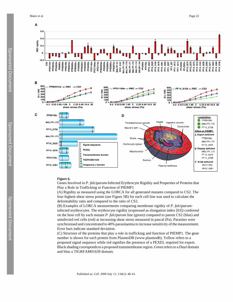
Figure 6.Genes Involved in P. falciparum-Infected Erythrocyte Rigidity and Properties of Proteins thatPlay a Role in Trafficking or Function of PfEMP1(A) Rigidity as measured using the LORCA for all generated mutants compared to CS2. Thefour highest shear stress points (see Figure 5B) for each cell line was used to calculate thedeformability ratio and compared to the ratio of CS2.(B) Examples of LORCA measurements comparing membrane rigidity of P. falciparum-infected erythrocytes. The erythrocyte rigidity (expressed as elongation index [EI]) conferredon the host cell by each mutant P. falciparum line (green) compared to parent CS2 (blue) anduninfected red cells (red) at increasing shear stress measured in pascal (Pa). Parasites weresynchronised and concentrated to 40% parasitaemia to increase sensitivity of the measurement.Error bars indicate standard deviation.(C) Structure of the proteins that play a role in trafficking and function of PfEMP1. The genenumber is shown for each protein from PlasmoDB (www.plasmodb). Yellow refers to aproposed signal sequence while red signifies the presence of a PEXEL required for export.Black shading corresponds to a proposed transmembrane region. Green refers to a DnaJ domainand blue a TIGRFAM01639 domain.
Maier et al. Page 22
Published as: Cell. 2008 July 11; 134(1): 48–61.
Sponsored Docum
ent Sponsored D
ocument
Sponsored Docum
ent

(D) Diagrammatic representation of a P. falciparum-infected erythrocyte signifying thelocalization of the protein (green symbols) or their functional position with respect to effectson PfEMP1 trafficking when disrupted (yellow letters).
Maier et al. Page 23
Published as: Cell. 2008 July 11; 134(1): 48–61.
Sponsored Docum
ent Sponsored D
ocument
Sponsored Docum
ent

Figure 7.Function and Properties of Selected Proteins Identified by the P. falciparum Gene KnockoutScreenThese proteins play a role in function or trafficking of the major virulence protein PfEMP1 ora putative role in determination of rigidity. The full description of the genes and proteins canbe found at http://plasmodb.org/. Most proteins have a PEXEL, which is required for exportto the infected erythrocyte. Changes in rigidity conferred on the host cell by each mutant P.falciparum line was compared to parent CS2 and uninfected red cells at increasing shear stressand are relative to CS2-infected erythrocytes. Those shown as a positive number have asignificantly increased rigidity whilst those that are negative have a decrease in rigidity that ismore than the CS2ΔPFA0110w (RESA) parasite line that has been shown previously to havean altered rigidity (Silva et al., 2005). The TIGR FAM01639 domain in MAL7P1.172represents a conserved sequence of about 60 amino acids found in over 40 predicted proteinsof P. falciparum. It is not found elsewhere, including closely related species such as P.yoelii. The PHIST proteins share a homologous domain unique to Plasmodium of 150 aminoacids and have been divided into a number of subfamilies (a, b, and c) (Sargeant et al., 2006).The skeleton binding protein 1 is included as a reference protein that is involved in PfEMP1trafficking (Maier et al., 2007; Cooke et al., 2006).
Maier et al. Page 24
Published as: Cell. 2008 July 11; 134(1): 48–61.
Sponsored Docum
ent Sponsored D
ocument
Sponsored Docum
ent