Modulation of Eukaryotic mRNA Stability via the Cap-binding Translation Complex eIF4F
Upload
independentCategory
view
3download
0
10.1128/JB.183.4.1346-1358.2001.
2001, 183(4):1346. DOI:J. Bacteriol. Gidekel and Alejandro VenegasFrancisco Uribe, Patricio Opazo, Teresa Soci?as, Manuel José Luis Palacios, Isabel Zaror, Patricio Marti?nez,
Erwinia chrysanthemiExported by the Type I Secretion System of Subset of Hybrid Eukaryotic Proteins Is
http://jb.asm.org/content/183/4/1346Updated information and services can be found at:
These include:
REFERENCEShttp://jb.asm.org/content/183/4/1346#ref-list-1at:
This article cites 63 articles, 36 of which can be accessed free
CONTENT ALERTS more»articles cite this article),
Receive: RSS Feeds, eTOCs, free email alerts (when new
http://journals.asm.org/site/misc/reprints.xhtmlInformation about commercial reprint orders: http://journals.asm.org/site/subscriptions/To subscribe to to another ASM Journal go to:
on April 7, 2014 by guest
http://jb.asm.org/
Dow
nloaded from
on April 7, 2014 by guest
http://jb.asm.org/
Dow
nloaded from
JOURNAL OF BACTERIOLOGY,0021-9193/01/$04.0010 DOI: 10.1128/JB.183.4.1346–1358.2001
Feb. 2001, p. 1346–1358 Vol. 183, No. 4
Copyright © 2001, American Society for Microbiology. All Rights Reserved.
Subset of Hybrid Eukaryotic Proteins Is Exported by the Type ISecretion System of Erwinia chrysanthemi
JOSE LUIS PALACIOS,1 ISABEL ZAROR,2 PATRICIO MARTINEZ,1 FRANCISCO URIBE,1
PATRICIO OPAZO,1 TERESA SOCIAS,1 MANUEL GIDEKEL,3†AND ALEJANDRO VENEGAS1*
Departamento de Genetica Molecular y Microbiologıa, Facultad de Ciencias Biologicas, Pontificia UniversidadCatolica de Chile, Santiago,1 and Instituto Nacional de Investigaciones Agropecuarias, Estacion
Carillanca, Temuco,3 Chile, and Chiron Corporation, Emeryville, California 946082
Received 27 June 2000/Accepted 22 November 2000
Erwinia chrysanthemi exports degradative enzymes by using a type I protein secretion system. The proteasessecreted by this system lack an N-terminal signal peptide but contain a C-terminal secretion signal. To explorethe substrate specificity of this system, we have expressed the E. chrysanthemi transporter system (prtDEFgenes) in Escherichia coli and tested the ability of this ABC transporter to export hybrid proteins carryingC-terminal fragments of E. chrysanthemi protease B. The C terminus contains six glycine-rich repeated motifs,followed by two repeats of the sequences DFLV and DIIV. Two types of hybrid proteins were assayed fortransport, proteins with the 93-residue-protease-B C terminus containing one glycine-rich repeat and bothhydrophobic terminal repeats and proteins with the 181-residue C terminus containing all repeat motifs.Although the shorter C terminus is unable to export the hybrids, the longer C terminus can promote thesecretion of hybrid proteins with N termini as large as 424 amino acids, showing that the glycine-rich motifsare required for the efficient secretion of these hybrids. However, the secretion of hybrids occurs only if theseproteins do not carry disulfide bonds in their mature structures. These latter results suggest that disulfidebond formation can occur prior to or during the secretion. Disulfide bonds may prevent type I secretion ofhybrids. One simple hypothesis to explain these results is that the type I channel is too narrow to permit theexport of proteins with secondary structures stabilized by disulfide bonds.
Nonpathogenic strains of Escherichia coli, such as the labo-ratory strain E. coli K-12, export few proteins to the externalmedium (53). The secretion of proteins in E. coli depends ona type II (Sec-dependent) mechanism in which unfolded pro-teins carrying an N-terminal signal sequence are transportedacross the inner membrane to the periplasm and then pro-cessed and folded in the periplasm prior to their translocationacross the outer membrane. To initiate type II secretion, it isthought that a polypeptide and its signal peptide must be fullyextended to cross the inner membrane (3). After crossing theinner membrane, proteins are folded in the periplasm in aprocess assisted by chaperones. In addition, the periplasm con-tains the enzymes required for the correct assembly of disulfidebonds to complete protein folding (5, 48).
In contrast, many gram-negative pathogens including enter-opathogenic E. coli (38), Yersinia spp. (36), Salmonella entericaserovar Typhimurium (39), and Vibrio cholerae (35) exportvirulence factors required for host colonization and survival.These virulence factors are secreted by mechanisms that op-erate independently from the type II system, mechanisms thatinvolve specialized membrane transport apparatuses (64). Asubset of virulence factors is exported by a type I mechanism inwhich three proteins assemble to form a transmembrane struc-ture that couples the export of protein substrates with ATP
hydrolysis. These ABC transporters include the type I mecha-nisms involved in the secretion of a Serratia marcescens met-alloprotease (43), E. coli beta-hemolysin (30, 61), Bordetellapertussis adenylate cyclase (29), Pseudomonas aeruginosa alka-line protease (23), and Pseudomonas fluorescens lipase (1). Allof the substrates for these ABC transport systems have C-terminal signal sequences.
Many phytopathogens, including members of the genusErwinia, also export proteins critical for virulence by usingdifferent secretion mechanisms. Among these, the secretion ofmetalloproteases in Erwinia chrysanthemi also proceeds by atype I mechanism (20). E. chrysanthemi secretes four proteases(19, 27, 28) to the external medium. Secretion requires theproducts of the prtD, prtE, and prtF genes, which also recognizea C-terminal signal sequence in their substrates (20). Likeother ABC transporters, the products of the prtD, prtE, andprtF genes are thought to form a channel that allows the exportof proteases directly from the cytoplasm to the external me-dium. The PrtD and PrtE proteins are associated with the in-ner membrane, and the PrtF protein is associated with theouter membrane (64). The large (ca. 60-kDa) PrtD protein hasan N terminus with six transmembrane segments and a hydro-philic C terminus with a classical ATP binding motif (42).Studies with PrtD in vitro show that its P-type ATPase activ-ity is inhibited by peptides carrying the C-terminal signal se-quence, arguing that substrate translocation by this type I sys-tem requires the hydrolysis of ATP (18). PrtE, which belongsto the family of the membrane fusion proteins (MFP), has ashort N terminus presumably anchored in the inner membrane,followed by a large hydrophilic periplasmic domain and a hy-
* Corresponding author: Mailing address: Departamento de Gene-tica Molecular y Microbiologıa, Facultad de Ciencias Biologicas, Ponti-ficia Universidad Catolica de Chile, Alameda 340, Santiago, Chile. Phone:(56-2)686-2661. Fax: (56-2)222-2810. E-mail: [email protected].
† Present address: Universidad de La Frontera, Laboratorio de Fisi-ologia y Biologıa Molecular, Instituto de Agroindustria, Temuco, Chile.
1346
on April 7, 2014 by guest
http://jb.asm.org/
Dow
nloaded from
drophobic C terminus thought to interact with the outer mem-brane (22). It has been suggested that proteins in the MFPfamily may form transmembranous pores for their substratesthat traverse the periplasmic space (22). The function of PrtF,associated with the outer membrane, is not well understood, asis the case for other ABC transporters (9).
Based on studies on the C-terminal secretion signal ofE. chrysanthemi proteases, this signal has been located in thelast 50 amino acids (20). Using an E. chrysanthemi protease,PrtG, it has been shown that the smallest C-terminal sequenceallowing efficient secretion contains the last 29 amino acids ofPrtG. This region contains two four-amino-acid motifs whichare essential for protease secretion (28). The E. chrysanthemiC-terminal protease signal can promote the specific secretionof fused passenger polypeptides. However, the four-amino-acid terminal motifs are not sufficient to promote secretion.Studies on fusion protein secretion have revealed the role of adomain located just upstream from the C-terminal signal onmost of the proteases and lipases secreted by the type I sys-tem. These proteins carry a domain of glycine-rich sequences(GGXGXD) that is repeated several times depending on theprotein (66). It has been shown that these repeats play a criticalrole in the secretion of some polypeptide passengers, suggest-ing that they may act as internal chaperones (41).
Because E. chrysanthemi is closely related to E. coli, we aretesting whether proteins expressed in E. coli hosts may betransported efficiently using the Erwinia chrysanthemi type Isecretion system. Understanding the substrate requirementsfor the secretion of proteins expressed in E. coli by this systemwould facilitate the production and purification of recombi-nant proteins of medical relevance, as well as have other im-portant biotechnological applications. In this work, we extendthe results of previous work showing that the signal sequencerequired for the secretion of hybrid eukaryotic proteins carry-ing the C terminus of E. chrysanthemi protease B is larger thanthat required for the secretion of protease B hybrids made byfusions to prokaryotic proteins (41) and showing that similarrules appear to apply to the transport of hybrids made betweeneukaryotic proteins and protease B. Surprisingly, we find thatthe E. chrysanthemi PtrDEF ABC transport system cannottransport hybrid eukaryotic proteins that have disulfide bondsin their mature structures.
MATERIALS AND METHODS
Bacterial strains and plasmids. Bacterial strains used in this work are listed inTable 1. AD494(DE3) cells were from Novagen and were grown according to the
vendor’s instructions. E. coli JM105 and DH5a were from Pharmacia, andTOP10 was from Invitrogen. E. coli and Erwinia carotovora strains were grown inLuria-Bertani (LB) medium at 37 and 30°C, respectively, with constant shaking(200 rpm). Plasmids are listed in Table 2. Plasmids pRUW4 and pRUW500 (20)were kindly provided by P. Delepelaire. Isolation of recombinant plasmids wasdone by using the alkaline lysis method of Birnboim and Doly (10), with slightmodifications. Bacterial cells carrying recombinant plasmids were grown in me-dium supplemented with the required antibiotics (100 mg of ampicillin/ml, 30 mgof chloramphenicol/ml, or 40 mg of kanamycin/ml).
DNA manipulations. Ligations and transformations were done using standardmethods (54). Restriction fragments and linearized plasmids were purified by theGeneClean kit (Bio 101) or the Wizard kit (Promega). Vector DNAs wereusually dephosphorylated with calf intestine alkaline phosphatase as described byChaconas and van de Sande (13). Dephosphorylation reactions were stoppedwith phenol extraction, and dephosphorylated DNA was purified after chloro-form-isoamyloalcohol (24:1) extraction followed by ethanol precipitation. Bac-terial transformation of E. coli and Erwinia strains was done by electroporation(47). In some cases, E. coli cells were transformed by using the calcium chloridecell permeation method (67). For electroporation, a Bio-Rad gene pulser appa-ratus, model 2-89, coupled to a pulse controller was used. A high voltage (2,500V) was applied to 40 ml of electrocompetent cells contained in a cuvette with a0.2-cm electrode separation. Electrocompetent cells were prepared as describedby Miller (47). Cells were recovered in SOC medium (54) at 30°C for 60 min, andelectroporants were selected on LB agar plates with appropriate antibiotics.DNA sequencing was done by the dideoxy chain termination method (55),following the procedure of Chen and Seeburg (15) for double-stranded plasmidDNA templates. The Sequenase kit version 2.0 (Amersham Pharmacia Biotech)was used according to the instructions, with [a-35S]dATP as labeled substrate.Gels were exposed for 1 to 2 days to X-OmatAR Kodak film.
PCR amplifications. PCRs were performed in thin-walled Eppendorf tubes, ina final volume of 100 ml containing 10 ml of 103 PCR buffer (200 mM Tris HCl[pH 8.4], 500 mM KCl), 3 ml of 50 mM MgCl2, 16 ml of 1.25 mM deoxynucleotidetriphosphate, 50 to 75 pmol of each primer, and 2.5 U of AmpliTaq DNApolymerase (Perkin-Elmer). Standard conditions were as follows: 30 to 35 cyclesof 2 min at 94°C, 1 min at 55°C, and 1 min at 72°C, followed by a terminalelongation step at 72°C for 7 min. Primers for amplification of various fragmentsare listed in Table 3.
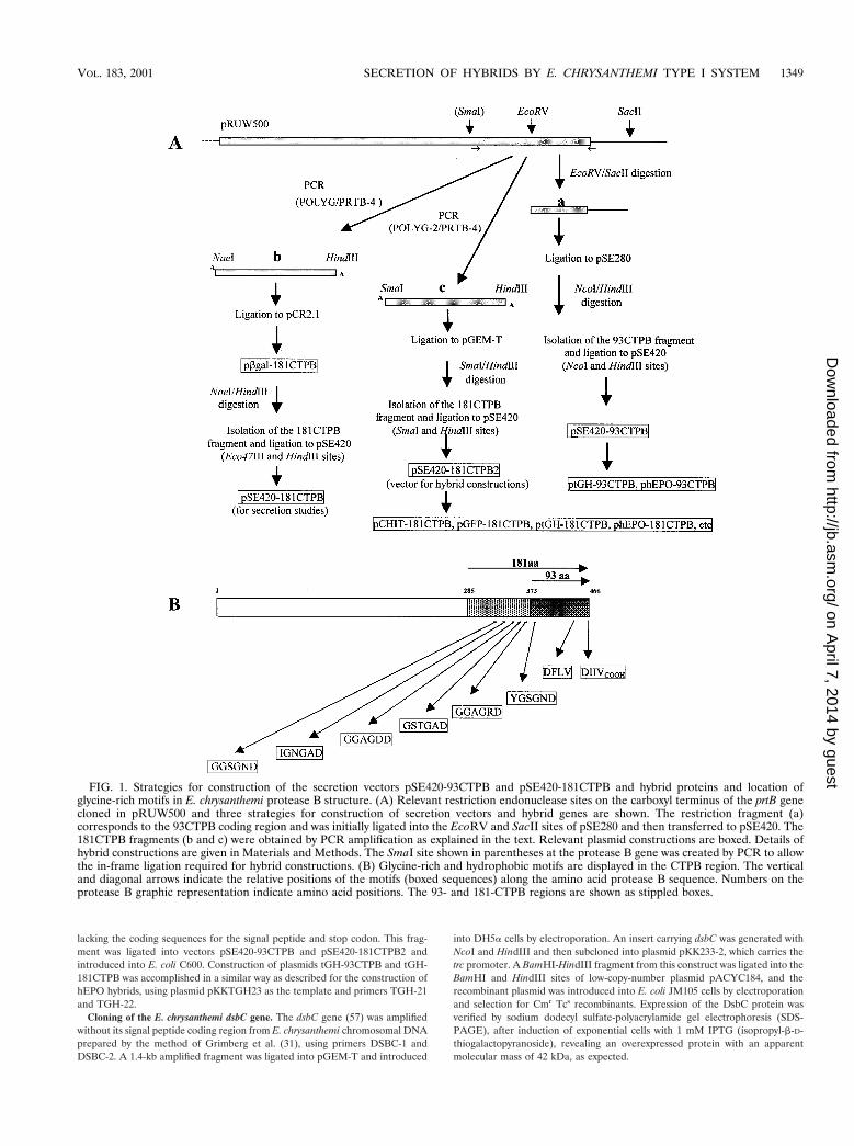
Construction of secretion vectors pSE420-93CTPB and pSE420-181CTPB.For the vector pSE420-93CTPB, a fragment containing the protease B codingregion between residues Tyr 373 and Val 466 was released from plasmidpRUW500 by EcoRV and SacII digestions (Fig. 1) and inserted at the same sitesin the pSE280 vector. Then, because of an additional EcoRV site present inpSE420, the 93CTPB region was transferred to pSE420 as a NcoI-HindIII frag-ment. The 181CTPB coding sequence was amplified by PCR from plasmidpRUW500 in two ways, using the primers POLYG and PRTB-4 (providing aNaeI site to be ligated in frame for the hybrid fusion at the Eco47III site of thevector) or using the pair POLYG-2 and PRTB-4 (providing an SmaI site forin-frame hybrid fusions) (see strategies in Fig. 1). Both amplified fragments wereseparately ligated to the pCR2.1 and the pGEM-T vectors. The first constructionhad the initial b-galactosidase residues in frame with the 181CTPB region andresulted in plasmid pbGAL-181CTPB. A fragment containing the 181CTPBregion was isolated from pbGAL-181CTPB after digestion with NaeI andHindIII, and the same fragment was obtained from the pGEM-T constructionafter digestion with SmaI and HindIII. Each 181CTPB fragment was indepen-dently ligated into the vector pSE420 that had been previously digested withEco47III and HindIII or SmaI and HindIII. The first construction, in which the
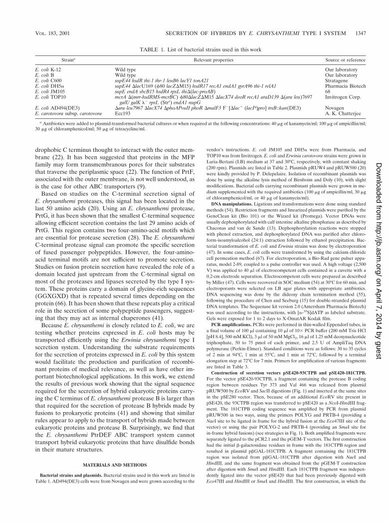
TABLE 1. List of bacterial strains used in this work
Straina Relevant properties Source or reference
E. coli K-12 Wild type Our laboratoryE. coli B Wild type Our laboratoryE. coli C600 supE44 hsdR thi-1 thr-1 leuB6 lacY1 tonA21 StratageneE. coli DH5a supE44 DlacU169 (f80 lacZDM15) hsdR17 recA1 endA1 gyrA96 thi-1 relA1 Pharmacia BiotechE. coli JM105 supE endA sbcB15 hsdR4 rpsL thiD(lac-proAB) 68E. coli TOP10 mcrA D(mrr-hsdRMS-mcrBC) f80DlacZDM15 DlacX74 deoR recA1 araD139 D(ara leu)7697
galU galK l2 rpsL (Strr) endA1 nupGInvitrogen Corp.
E. coli AD494(DE3) Dara leu7967 DlacX74 DphoAPvuII phoR DmalF3 F2[Dlac1 (lacIq)pro] trxB::kan(DE3) NovagenE. carotovora subsp. carotovora Ecc193 A. K. Chatterjee
a Antibiotics were added to plasmid-transformed bacterial cultures or when required at the following concentrations: 40 mg of kanamycin/ml; 100 mg of ampicillin/ml;30 mg of chloramphenicol/ml; 50 mg of tetracycline/ml.
VOL. 183, 2001 SECRETION OF HYBRIDS BY E. CHRYSANTHEMI TYPE I SYSTEM 1347
on April 7, 2014 by guest
http://jb.asm.org/
Dow
nloaded from
plasmid once ligated has lost both the NaeI and the Eco47III sites, was usedfor secretion studies of the carboxyl terminus. The second construction,pSE420-181CTPB2, preserved intact the SmaI site after ligation, and this wasutilized to generate the hybrids with different passenger proteins, as shown inFig. 1.
Construction of protease B hybrid genes. To construct plasmids expressingendochitinase hybrids, the endochitinase gene was amplified by PCR using theECh42 cDNA as the template (33) and primers CHIT-1 and CHIT-4 (Table 3).The use of primer CHIT-4 allowed the fusion in frame to the vector pSE420-181CTPB-2 after SmaI and NcoI digestions of the purified PCR fragment andligation to the NcoI and SmaI sites of the vector. Ligation products were trans-formed into E. coli HB101 cells. Two pCHIT-181CTPB recombinant plasmidswere selected for further studies. Plasmids expressing green fluorescent protein
(GFP) hybrids were made using a similar strategy. A PCR fragment containingfull-length GFP (238 codons) was amplified using template plasmid pGFP andprimers GFP-NH3 and GFP-CT4. After NcoI and SmaI digestions, the fragmentwas ligated to pSE420-181CTPB2. The OmpC-181CTPB hybrid was constructedby amplifying the first 204 codons of the ompC coding region, using plasmidpOmpCBgl-2 as the template and primers OC-1 and OC-51. The PCR fragmentwas digested with NcoI and SmaI and ligated to pSE420-181CTPB2.
Plasmids hEPO-93CTPB and hEPO-181CTPB were constructed by amplifyinga purified BglII restriction fragment obtained from plasmid pEry720 carrying asynthetic human erythropoietin (hEPO) gene (A. Venegas, unpublished results)as the PCR template and primers EPO-1 and EPO-2. The 518-bp PCR fragmentwas ligated to pGEM-T to make plasmid pGEM-hEPO-1, which was digestedwith NcoI and EcoRV to generate a fragment with the hEPO coding region
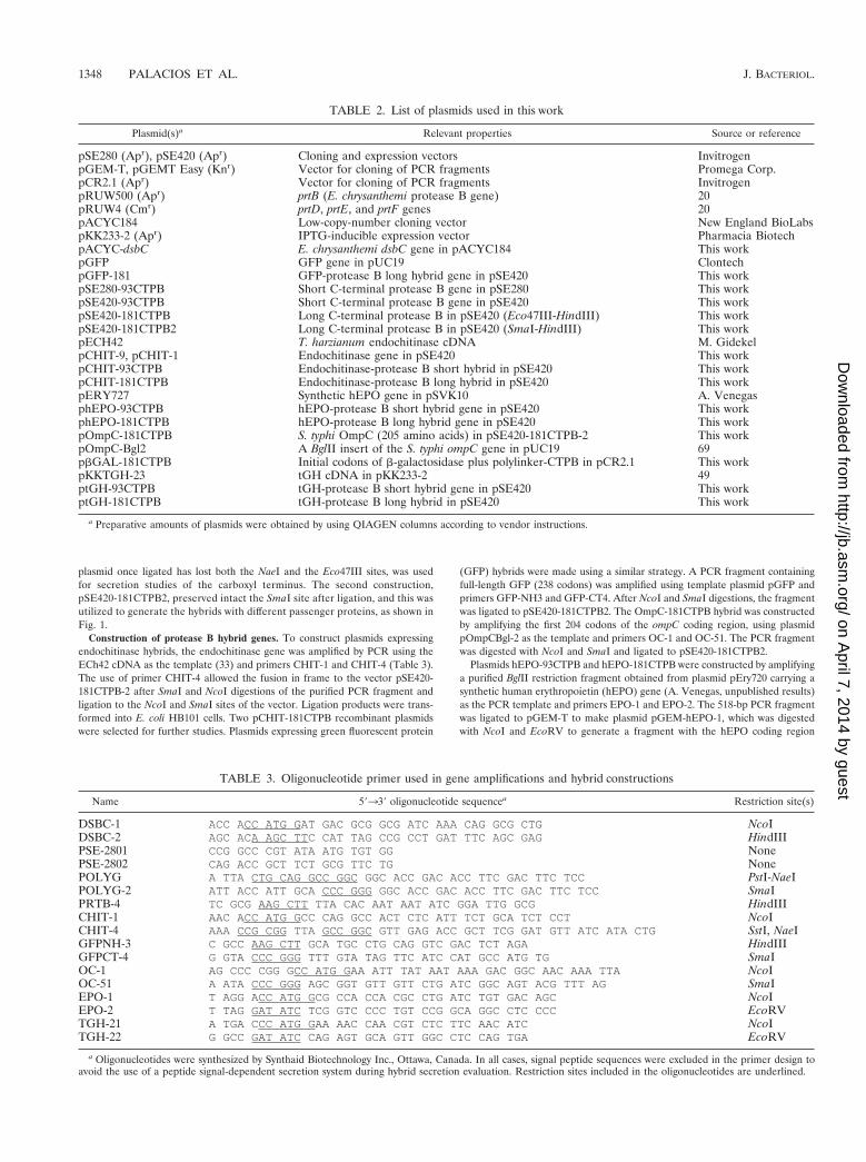
TABLE 2. List of plasmids used in this work
Plasmid(s)a Relevant properties Source or reference
pSE280 (Apr), pSE420 (Apr) Cloning and expression vectors InvitrogenpGEM-T, pGEMT Easy (Knr) Vector for cloning of PCR fragments Promega Corp.pCR2.1 (Apr) Vector for cloning of PCR fragments InvitrogenpRUW500 (Apr) prtB (E. chrysanthemi protease B gene) 20pRUW4 (Cmr) prtD, prtE, and prtF genes 20pACYC184 Low-copy-number cloning vector New England BioLabspKK233-2 (Apr) IPTG-inducible expression vector Pharmacia BiotechpACYC-dsbC E. chrysanthemi dsbC gene in pACYC184 This workpGFP GFP gene in pUC19 ClontechpGFP-181 GFP-protease B long hybrid gene in pSE420 This workpSE280-93CTPB Short C-terminal protease B gene in pSE280 This workpSE420-93CTPB Short C-terminal protease B gene in pSE420 This workpSE420-181CTPB Long C-terminal protease B in pSE420 (Eco47III-HindIII) This workpSE420-181CTPB2 Long C-terminal protease B in pSE420 (SmaI-HindIII) This workpECH42 T. harzianum endochitinase cDNA M. GidekelpCHIT-9, pCHIT-1 Endochitinase gene in pSE420 This workpCHIT-93CTPB Endochitinase-protease B short hybrid in pSE420 This workpCHIT-181CTPB Endochitinase-protease B long hybrid in pSE420 This workpERY727 Synthetic hEPO gene in pSVK10 A. VenegasphEPO-93CTPB hEPO-protease B short hybrid gene in pSE420 This workphEPO-181CTPB hEPO-protease B long hybrid gene in pSE420 This workpOmpC-181CTPB S. typhi OmpC (205 amino acids) in pSE420-181CTPB-2 This workpOmpC-Bgl2 A BglII insert of the S. typhi ompC gene in pUC19 69pbGAL-181CTPB Initial codons of b-galactosidase plus polylinker-CTPB in pCR2.1 This workpKKTGH-23 tGH cDNA in pKK233-2 49ptGH-93CTPB tGH-protease B short hybrid gene in pSE420 This workptGH-181CTPB tGH-protease B long hybrid in pSE420 This work
a Preparative amounts of plasmids were obtained by using QIAGEN columns according to vendor instructions.
TABLE 3. Oligonucleotide primer used in gene amplifications and hybrid constructions
Name 59339 oligonucleotide sequencea Restriction site(s)
DSBC-1 ACC ACC ATG GAT GAC GCG GCG ATC AAA CAG GCG CTG NcoIDSBC-2 AGC ACA AGC TTC CAT TAG CCG CCT GAT TTC AGC GAG HindIIIPSE-2801 CCG GCC CGT ATA ATG TGT GG NonePSE-2802 CAG ACC GCT TCT GCG TTC TG NonePOLYG A TTA CTG CAG GCC GGC GGC ACC GAC ACC TTC GAC TTC TCC PstI-NaeIPOLYG-2 ATT ACC ATT GCA CCC GGG GGC ACC GAC ACC TTC GAC TTC TCC SmaIPRTB-4 TC GCG AAG CTT TTA CAC AAT AAT ATC GGA TTG GCG HindIIICHIT-1 AAC ACC ATG GCC CAG GCC ACT CTC ATT TCT GCA TCT CCT NcoICHIT-4 AAA CCG CGG TTA GCC GGC GTT GAG ACC GCT TCG GAT GTT ATC ATA CTG SstI, NaeIGFPNH-3 C GCC AAG CTT GCA TGC CTG CAG GTC GAC TCT AGA HindIIIGFPCT-4 G GTA CCC GGG TTT GTA TAG TTC ATC CAT GCC ATG TG SmaIOC-1 AG CCC CGG GCC ATG GAA ATT TAT AAT AAA GAC GGC AAC AAA TTA NcoIOC-51 A ATA CCC GGG AGC GGT GTT GTT CTG ATC GGC AGT ACG TTT AG SmaIEPO-1 T AGG ACC ATG GCG CCA CCA CGC CTG ATC TGT GAC AGC NcoIEPO-2 T TAG GAT ATC TCG GTC CCC TGT CCG GCA GGC CTC CCC EcoRVTGH-21 A TGA CCC ATG GAA AAC CAA CGT CTC TTC AAC ATC NcoITGH-22 G GCC GAT ATC CAG AGT GCA GTT GGC CTC CAG TGA EcoRV
a Oligonucleotides were synthesized by Synthaid Biotechnology Inc., Ottawa, Canada. In all cases, signal peptide sequences were excluded in the primer design toavoid the use of a peptide signal-dependent secretion system during hybrid secretion evaluation. Restriction sites included in the oligonucleotides are underlined.
1348 PALACIOS ET AL. J. BACTERIOL.
on April 7, 2014 by guest
http://jb.asm.org/
Dow
nloaded from
lacking the coding sequences for the signal peptide and stop codon. This frag-ment was ligated into vectors pSE420-93CTPB and pSE420-181CTPB2 andintroduced into E. coli C600. Construction of plasmids tGH-93CTPB and tGH-181CTPB was accomplished in a similar way as described for the construction ofhEPO hybrids, using plasmid pKKTGH23 as the template and primers TGH-21and TGH-22.
Cloning of the E. chrysanthemi dsbC gene. The dsbC gene (57) was amplifiedwithout its signal peptide coding region from E. chrysanthemi chromosomal DNAprepared by the method of Grimberg et al. (31), using primers DSBC-1 andDSBC-2. A 1.4-kb amplified fragment was ligated into pGEM-T and introduced
into DH5a cells by electroporation. An insert carrying dsbC was generated withNcoI and HindIII and then subcloned into plasmid pKK233-2, which carries thetrc promoter. A BamHI-HindIII fragment from this construct was ligated into theBamHI and HindIII sites of low-copy-number plasmid pACYC184, and therecombinant plasmid was introduced into E. coli JM105 cells by electroporationand selection for Cmr Tcs recombinants. Expression of the DsbC protein wasverified by sodium dodecyl sulfate-polyacrylamide gel electrophoresis (SDS-PAGE), after induction of exponential cells with 1 mM IPTG (isopropyl-b-D-thiogalactopyranoside), revealing an overexpressed protein with an apparentmolecular mass of 42 kDa, as expected.
FIG. 1. Strategies for construction of the secretion vectors pSE420-93CTPB and pSE420-181CTPB and hybrid proteins and location ofglycine-rich motifs in E. chrysanthemi protease B structure. (A) Relevant restriction endonuclease sites on the carboxyl terminus of the prtB genecloned in pRUW500 and three strategies for construction of secretion vectors and hybrid genes are shown. The restriction fragment (a)corresponds to the 93CTPB coding region and was initially ligated into the EcoRV and SacII sites of pSE280 and then transferred to pSE420. The181CTPB fragments (b and c) were obtained by PCR amplification as explained in the text. Relevant plasmid constructions are boxed. Details ofhybrid constructions are given in Materials and Methods. The SmaI site shown in parentheses at the protease B gene was created by PCR to allowthe in-frame ligation required for hybrid constructions. (B) Glycine-rich and hydrophobic motifs are displayed in the CTPB region. The verticaland diagonal arrows indicate the relative positions of the motifs (boxed sequences) along the amino acid protease B sequence. Numbers on theprotease B graphic representation indicate amino acid positions. The 93- and 181-CTPB regions are shown as stippled boxes.
VOL. 183, 2001 SECRETION OF HYBRIDS BY E. CHRYSANTHEMI TYPE I SYSTEM 1349
on April 7, 2014 by guest
http://jb.asm.org/
Dow
nloaded from
Detection of SH residues in hybrid proteins by covalent modification withiodoacetic acid. Six milliliters of fresh overnight cultures was centrifuged at2,000 3 g for 5 min in an Eppendorf microcentrifuge. Cell pellets were sus-pended in double-distilled H2O (1/10 initial volume) and boiled for 5 min toinactivate disulfide bond formation enzymes. Cells were lysed by adding 300 mlof 25 mM Tris HCl (pH 8.0), 10 mM EDTA, and 50 mM glucose, containing 4mg of lysozyme/ml, and vortexing, followed by incubation for 5 min at 25°C. Totalproteins were precipitated with 10% trichloroacetic acid (TCA), and the pelletswere washed twice with acetone, dried, and then suspended in 120 ml of 125 mMTris HCl–6 M guanidinium chloride (pH 8.0) containing 20 mM iodoacetic acidand incubated for 10 min at 25°C. In order to remove iodoacetic acid remnants,a 50-ml aliquot was filtered through a 0.5-ml Sephadex G-25 minicolumn set ona disposable blue tip and previously equilibrated with 125 mM Tris HCl (pH 8.0).Proteins with modified SH residues change their electrophoretic mobility on an8 M urea-polyacrylamide gel. The gel was prepared as described by Laemmli (40)but containing urea instead of SDS. No upper gel was used. An 8% polyacryl-amide gel was run at 25°C using 150 V. The gel was made in 350 mM Tris HClbuffer (pH 8.9). Electrophoresis running buffer was 125 mM Tris-glycine (pH9.0) without SDS or b-mercaptoethanol (b-ME). All samples were boiled for5 min and some of them were reduced with 10 mM b-ME.
Purification of protease B and preparation of polyclonal antibodies. The52-kDa protease B protein from E. chrysanthemi cloned in plasmid pRUW500(20) was expressed in E. coli C600 after 1 mM IPTG induction of an overnightculture for 6 h. The protein band was purified by SDS-PAGE (7% polyacryl-amide), and an appropriate gel slice was electroeluted with a Bio-Rad electro-eluter as described in the vendor instructions. Anti-protease B polyclonal anti-body was obtained as described previously(16), using three 300-mg injections ofprotease B on rabbit pads. Sera from two rabbits were tested by an enzyme-linked immunosorbent assay, and each milliliter of a 1:10 dilution of serum indistilled water was adsorbed twice against a 5-ml extract of sonicated E. coli C600cells previously immobilized on a 5-cm2 nitrocellulose filter.
Protein gel electrophoresis and Western blotting for detection of protease Bhybrids. Proteins with a molecular mass larger than 15 kDa were separated usingSDS-PAGE (12 to 15% polyacrylamide) (40). Protein staining was done with0.1% Coomassie blue R-250 in a 50% methanol–10% acetic acid solution for 1 to2 h at 25°C. Gel destaining was done in 10% methanol–10% acetic acid at 25°C.Western blotting using anti-protease B polyclonal antibodies was performed bythe method of Towbin et al. (63) with few modifications. After transfer, nitro-cellulose filters were blocked with phosphate-buffered saline (PBS)–2% bovineserum albumin (BSA) for 2 h at 25°C. Prior to its use, anti-protease B antibodywas preadsorbed with E. coli C600 total protein extract as described by Zaror(69). The antibody was added to a final dilution of 1:1,000 in PBS–2% BSA, andthe filters were incubated for 1 h and then washed in PBS–0.1% Tween 20 threetimes for 5 min each. The filters were then incubated with anti-rabbit immuno-globulin G (diluted 1:1,000 in PBS–2% BSA) conjugated to horseradish perox-idase or alkaline phosphatase (Bio-Rad). For peroxidase reactions, the filterswere developed in 25 ml of 50 mM Tris HCl (pH 7.4)–200 mM NaCl containing15 mg of 4-chloro-1-naphthol (previously dissolved in 5 ml of methanol) and 60ml of 30% hydrogen peroxide. For phosphatase reactions, the filters were incu-bated in 10 ml of a mixture of 100 mM Tris HCl (pH 9.5), 100 mM NaCl, 5 mMMgCl2, 66 ml of a 50-mg/ml concentration of nitroblue tetrazolium, and 33 ml ofa 50-mg/ml concentration of 5-bromo-4-chloro-3-indolylphosphate. Western blotanalyses of protease B hybrids were carried out using a 1:1,000 anti-protease Bantibody dilution. To detect hybrids in supernatants, supernatants were concen-trated 10-fold by precipitation with 10% TCA (32) prior to electrophoreticseparation. In some cases, to standardize protein loading, the protein content ofextracts was determined by the method of Bradford (11). Chemiluminescenceassays were done with a Renaissance kit from NEN Life Science Products(Boston, Mass.).
RESULTS
Construction of plasmid vectors with E. chrysanthemi PrtBC-terminal signal sequences. E. chrysanthemi secretes fourproteases using its type I secretion system, proteases A, B, C,and G. These four proteases terminate with a similar 4-amino-acid motif, represented by the sequence Dhhh, in which h rep-resents a hydrophobic amino acid. Initial studies have shownthat this motif is critical for the secretion of protease G, be-cause the addition of a single amino acid residue to the Cterminus of protease G prevents its export by the type I mech-
anism (20, 28). Although fusion of the coding sequence for the40 C-terminal residues of protease B to the coding region forthe first 200 residues of amylomaltase results in a hybrid pro-tein that is secreted in E. coli, other larger fusion proteins thatcarry only this motif are not secreted efficiently by the type Imechanism (20). In addition to this C-terminal motif, it hasbecome clear that an additional glycine-rich motif, repeatedimmediately upstream of the C terminus of the E. chrysanthemiproteases, is required for the efficient type I transport of hy-brids formed between heterologous proteins and the C terminiof the secreted proteases (41). This additional motif has theconsensus sequence GGXGXD, in which X represents anyamino acid, and is repeated several times in all proteins se-creted by a type I mechanism.
To explore the C-terminal requirements for type I secretionby the E. chrysanthemi system in greater detail, we construct-ed plasmid vectors carrying sequences encoding two differentlengths of the C terminus of protease B. The PrtB sequences inthese vectors correspond to the 93 or 181 C-terminal residuesof protease B. Both sequences include the C-terminal signalessential for type I secretion, whereas only the latter vectorencodes the glycine-rich repeats required for the secretion oflarger, hybrid proteins. To construct these vectors, we obtaineddifferent portions of the 39 end of the prtB gene and subclonedthese products into plasmid expression vector pSE420, as de-scribed in Materials and Methods. These constructions allowedus to fuse part of the polylinker region of pSE420 in frameto the protease B C termini. This part of the polylinkerregion presumably is translated as a tract of 46 amino acidresidues.
We tested the ability of the E. chrysanthemi type I secretionsystem to export the small fusion proteins expressed from plas-mid vectors pSE420-93CTPB and pSE420-181CTPB in E. coliC600 cells. These proteins are predicted to be 139 and 229amino acids in length, respectively. Western blot analysis of theproteins present in the total induced lysates of E coli C600carrying these plasmids probed with anti-protease B antibodiesreveals bands corresponding to fusion products with apparentmolecular masses of 17 (Fig. 2A) and 27 (Fig. 2B) kDa, asexpected. Plasmid pRUW4, which expresses the E. chrysan-themi prtD, prtE, and prtF genes, was introduced into thesestrains, and the ability of the PrtDEF transport system tosecrete these proteins was tested by assaying for the presenceof secreted proteins in cell supernatants. We note that theexpression of proteins that are not secreted is higher in thepresence of plasmid pRUW4 (Fig. 2A, compare lanes 3 and 5).At present, we have no explanation for this result. Figure 2Bshows that only the 27-kDa fusion product (with the longerC-terminal signal sequence) is present in the supernatant frac-tion of cells (secretion scored 20 and 31% in two clones). Nosecretion was observed in the absence of plasmid pRUW4 (Fig.2B, lanes 2 and 6). Similar results were obtained when theplasmid vectors were introduced into an E. carotovora Ecc193host (data not shown). This result shows that the longer 181-amino-acid C terminus is essential for the efficient secretion ofthe short hybrid product formed between the polylinker se-quence on expression plasmid pSE420 in an E. coli C600 hostand thereby confirms that the glycine-rich repeats are requiredfor the efficient export of hybrid passenger proteins.
1350 PALACIOS ET AL. J. BACTERIOL.
on April 7, 2014 by guest
http://jb.asm.org/
Dow
nloaded from

The E. chrysanthemi type I secretion system permits the trans-port of hybrids between eukaryotic proteins and protease B.To test whether hybrids between eukaryotic passenger proteinsand the C-terminal signal sequence of PrtB can be secreted inE. coli by the E. chrysanthemi type I system, plasmids to expresshybrid proteins formed between a variety of eukaryotic pro-teins and the C terminus of protease B were constructed.These proteins include Trichoderma harzianum endochitinase(42 kDa), GFP from Aequorea victoria (28.5 kDa), (hEPO 20kDa), and trout growth hormone (tGH, 22.6 kDa). For pro-karyotic controls, we also constructed hybrids with segmentsof the E. coli lacZ (b-galactosidase segment, 3.6 kDa) andSalmonella typhi ompC porin genes (OmpC segment, 24.5 kDa)and included the protease B gene (full-length product, 56kDa).
Each gene was cloned upstream of, and in frame with,the 181-amino-acid C-terminal segment of PrtB present inpSEB420-181CTPB2. Hybrid constructions were tested for se-cretion after overnight incubation at 37°C in LB broth contain-ing 100 mg of ampicillin/ml and 50 mg of chloramphenicol/ml,followed by induction with 1 mM IPTG for 3 to 4 h. Figures 3Aand 3B show secretion of GFP and chitinase hybrids, respec-tively. It was found that the GFP-181CTPB hybrid (55 kDa)was secreted in about 32 to 56% of the total amount of thesynthesized hybrid depending on the particular clone, as shownin Fig. 3A (compare lanes 5 and 6 and and lanes 7 and 8).The chitinase-protease B hybrid (72 kDa) was modestly se-creted to the medium at about 4 to 8% depending on thespecific isolated clone (Fig. 3B, compare lanes 7 and 8 andlanes 9 and 10).
As expected, the b-galactosidase hybrid used as the controlgave between 60 and 75% secretion for two clones (Fig. 3C).The OmpC-181CTPB hybrid (46 kDa) was also secreted to asubstantial level (24 to 48%), even though this protein (whencarrying its signal peptide and the entire coding sequence) isnormally assembled in the outer membrane. For controls, pro-tease B secretion assays in E. coli DH5a (Fig. 3D) as well as inC600 cells (Fig. 3E) were included. These results show that a
variety of eukaryotic proteins can be secreted from an E. colihost by the E. chrysanthemi type I mechanism.
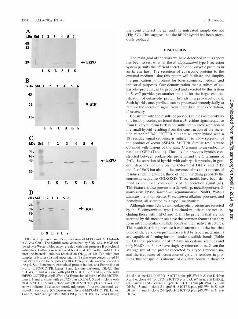
The E. chrysanthemi type I secretion system does not permitthe transport of hybrids between eukaryotic proteins that formdisulfide bridges in their native forms and protease B. Two ofthe hybrid proteins we tested are not secreted by the E. chry-santhemi type I system. The hybrids with hEPO and tGH wereassayed with both the 93CTPB and 181CTPB secretion signals.Both of these proteins are unique among the proteins thathave been tested for secretion by a type I system because theyare capable of forming disulfide bonds in their native struc-tures. The hEPO hormone contains two disulfide bonds, andbecause its active form is highly glycosylated (34), it has notbeen expressed previously in E. coli. In contrast, tGH, with amolecular mass of 22 kDa, contains two disulfide bonds, is notglycosylated, and has been expressed intracellularly in E. coli(49). Both tGH-93CTPB and hEPO-93CTPB hybrids ap-peared in E. coli C600 lysates as the corresponding 32- and35-kDa bands after separation by SDS-PAGE and Westernblot assays. Thus, hybrids with both proteins are expressed inE. coli. However, these proteins were absent in supernatantfractions of cells that also carry plasmid pRUW4, which en-codes the PrtDEF ABC transporter (Fig. 4A and B). In spite ofpositive secretion results with the other eukaryotic hybrids, thehEPO-181CTPB and tGH-181CTPB hybrids are not secretedwhen expressed in host E. coli DH5a cells carrying pRUW4(Fig. 4C and D). The same results were obtained when the93CTPB or 181CTPB hybrid contructions and plasmidpRUW4 were transferred to E. coli HB101 (results not shown).These results argue that the PrtDEF type I secretion systemcannot facilitate the secretion of passenger proteins that canform disulfide bonds. A summary of the results obtained forsecretion of different hybrid constructions by the type I systemin E. coli is shown in Table 4.
Cytoplasmic enhanced expression of E. chrysanthemi disul-fide isomerase C (DsbC) in E. coli does not improve the se-cretion of proteins carrying disulfide bonds. For some mech-anisms of secretion, it has been proposed that a passenger
FIG. 2. Expression and secretion of the carboxyl-terminal signals (93 and 181 amino acids) of the E. chrysanthemi protease B in E. coli C600.The hybrids were detected by SDS–12% PAGE followed by Western blot analysis. (A) The 93CTPB signal immunoblot revealed with alkalinephosphatase; (B) the 181CTPB signal immunoblot revealed by chemiluminescence with horseradish peroxidase. Cultures were induced with 1 mMIPTG for 4 h, when bacterial growth reached an optical density at 600 nm (OD600) of 0.5. Ten-microliter samples were applied to the gel fromlysates (L) and supernatants (S). (A) Lanes: Std, prestained low-molecular-mass standard (GIBCO-BRL); 1 and 2, clone with pSE420 andpRUW4; 3 and 4, clone with pSE420-93CTPB; 5 and 6, clone with pSE420-93CTPB and pRUW4. Supernatants were concentrated 10 times withrespect to the lysates by 10% TCA precipitation. (B) Lanes: 1 and 2, lysate and supernatant from control E. coli C600 cells; 3 and 4, lysate andsupernatant from clone 8 (pSE420-181CTPB in E. coli C600); 5 and 6, lysate and supernatant from clone 8-1 (pSE420-181CTPB plus pRUW4 inE. coli C600); 7 and 8, lysate and supernatant from clone 4 (pSE420-181CTPB in E. coli C600); 9 and 10, lysate and supernatant from clone 4-1(pSE420-181CTPB plus pRUW4 in E. coli C600). Std, migration of bands corresponding to low-molecular-mass standard (GIBCO-BRL). Su-pernatants were concentrated two times with respect to the lysates by 10% TCA precipitation.
VOL. 183, 2001 SECRETION OF HYBRIDS BY E. CHRYSANTHEMI TYPE I SYSTEM 1351
on April 7, 2014 by guest
http://jb.asm.org/
Dow
nloaded from
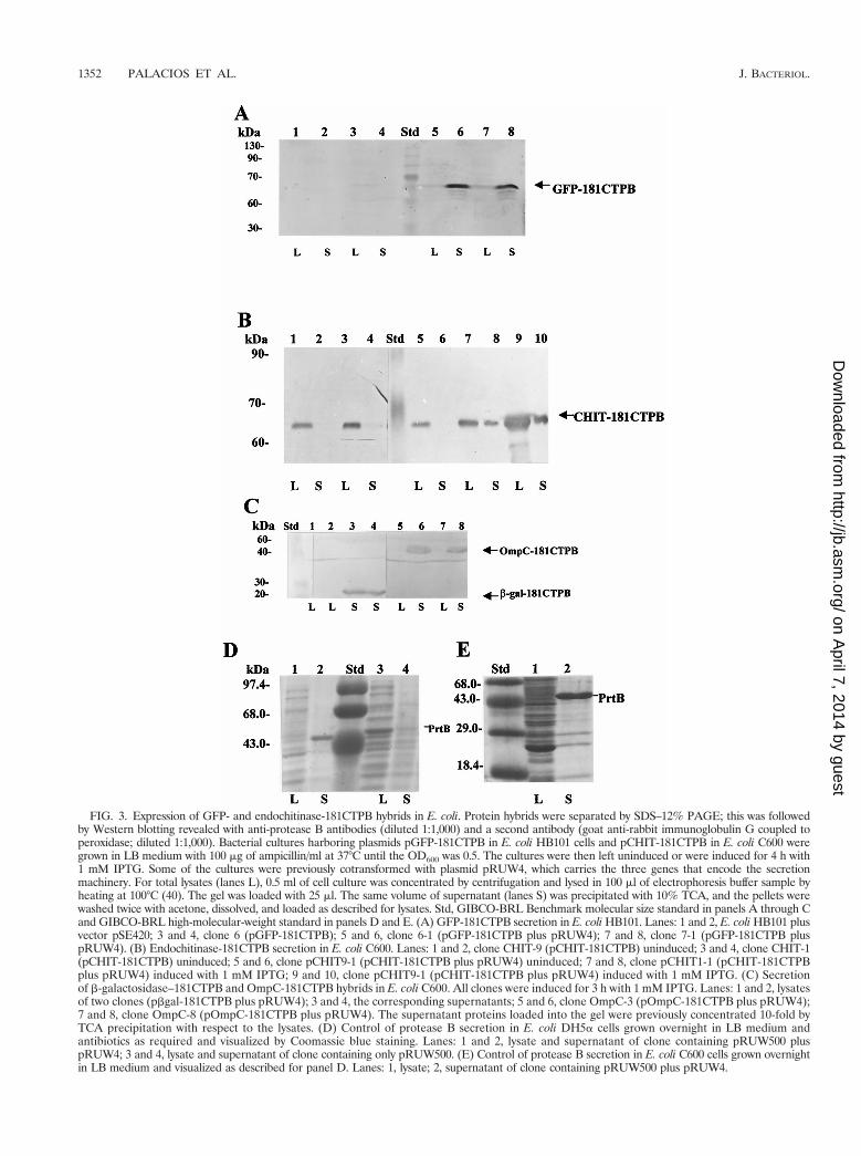
FIG. 3. Expression of GFP- and endochitinase-181CTPB hybrids in E. coli. Protein hybrids were separated by SDS–12% PAGE; this was followedby Western blotting revealed with anti-protease B antibodies (diluted 1:1,000) and a second antibody (goat anti-rabbit immunoglobulin G coupled toperoxidase; diluted 1:1,000). Bacterial cultures harboring plasmids pGFP-181CTPB in E. coli HB101 cells and pCHIT-181CTPB in E. coli C600 weregrown in LB medium with 100 mg of ampicillin/ml at 37°C until the OD600 was 0.5. The cultures were then left uninduced or were induced for 4 h with1 mM IPTG. Some of the cultures were previously cotransformed with plasmid pRUW4, which carries the three genes that encode the secretionmachinery. For total lysates (lanes L), 0.5 ml of cell culture was concentrated by centrifugation and lysed in 100 ml of electrophoresis buffer sample byheating at 100°C (40). The gel was loaded with 25 ml. The same volume of supernatant (lanes S) was precipitated with 10% TCA, and the pellets werewashed twice with acetone, dissolved, and loaded as described for lysates. Std, GIBCO-BRL Benchmark molecular size standard in panels A through Cand GIBCO-BRL high-molecular-weight standard in panels D and E. (A) GFP-181CTPB secretion in E. coli HB101. Lanes: 1 and 2, E. coli HB101 plusvector pSE420; 3 and 4, clone 6 (pGFP-181CTPB); 5 and 6, clone 6-1 (pGFP-181CTPB plus pRUW4); 7 and 8, clone 7-1 (pGFP-181CTPB pluspRUW4). (B) Endochitinase-181CTPB secretion in E. coli C600. Lanes: 1 and 2, clone CHIT-9 (pCHIT-181CTPB) uninduced; 3 and 4, clone CHIT-1(pCHIT-181CTPB) uninduced; 5 and 6, clone pCHIT9-1 (pCHIT-181CTPB plus pRUW4) uninduced; 7 and 8, clone pCHIT1-1 (pCHIT-181CTPBplus pRUW4) induced with 1 mM IPTG; 9 and 10, clone pCHIT9-1 (pCHIT-181CTPB plus pRUW4) induced with 1 mM IPTG. (C) Secretionof b-galactosidase–181CTPB and OmpC-181CTPB hybrids in E. coli C600. All clones were induced for 3 h with 1 mM IPTG. Lanes: 1 and 2, lysatesof two clones (pbgal-181CTPB plus pRUW4); 3 and 4, the corresponding supernatants; 5 and 6, clone OmpC-3 (pOmpC-181CTPB plus pRUW4);7 and 8, clone OmpC-8 (pOmpC-181CTPB plus pRUW4). The supernatant proteins loaded into the gel were previously concentrated 10-fold byTCA precipitation with respect to the lysates. (D) Control of protease B secretion in E. coli DH5a cells grown overnight in LB medium andantibiotics as required and visualized by Coomassie blue staining. Lanes: 1 and 2, lysate and supernatant of clone containing pRUW500 pluspRUW4; 3 and 4, lysate and supernatant of clone containing only pRUW500. (E) Control of protease B secretion in E. coli C600 cells grown overnightin LB medium and visualized as described for panel D. Lanes: 1, lysate; 2, supernatant of clone containing pRUW500 plus pRUW4.
1352 PALACIOS ET AL. J. BACTERIOL.
on April 7, 2014 by guest
http://jb.asm.org/
Dow
nloaded from
protein must be completely folded to permit translocationthrough the secretion channel or outer membrane (52, 25).In E. coli, the majority of intramolecular disulfide bonds areformed in the periplasm (59), and it is believed that interme-diates translocated by the E. chrysanthemi type I secretionsystem are not in contact with the periplasm during secretion(64). Because the bacterial cytoplasm is a reducing environ-ment, and most disulfide bonds are formed in the periplasm(53), we reasoned that proteins secreted by this pathway do nothave the opportunity to form disulfide bridges. If such proteinsmust be folded prior to transport, the failure to form disulfidebonds may account for their failure to be transported by thetype I mechanism. Therefore, we tested whether the formationof disulfide bonds in these proteins prior to transport might in-crease their efficiency of transport in two different ways. First,we expressed the hEPO and the tGHII hybrid proteins in a trxBmutant deficient in thioredoxin production to increase the oxi-dizing potential of the cytoplasm, and thereby to favor cyto-plasmic disulfide bond formation (59). Second, we expressedthe hEPO and the tGHII hybrids in a strain of E. coli in whicha soluble form of the dsbC gene product (disulfide isomer-ase) is also expressed in the cytoplasm, to promote the en-zyme-catalyzed formation of disulfide bonds prior to secre-tion.
Figure 5 shows the results of a Western blot assay for ex-pression of the tGH-93CTPB hybrid in total lysates of thetrxB-deficient E. coli host strain AD494, with or without b-ME
included in the sample prior to the protein separation by elec-trophoresis. We did not visualize any change in the electro-phoretic mobility of this protein under the two conditions,indicating that the absence of thioredoxin does not enhancethe formation of disulfide bonds in the hybrid protein. Wesuspect that this is due to the fact that disulfide bonds can formin this hybrid protein in the E. coli cytoplasm both in the pres-ence and absence of thioredoxin (see Discussion). Similar re-sults were obtained with the hEPO-93CTPB hybrid and bothconstructions carrying the 181CTPB region.
To overexpress a soluble form of DsbC in the cytoplasm, wecloned a version of the E. chrysanthemi dsbC gene without thecoding region for its signal sequence in plasmid pACYC184 asdescribed in Materials and Methods and expressed this trun-cated form of dsbC from a strong IPTG-inducible trc promoter.The plasmid pACYC-dsbC was transferred by electroporationto an E. coli DH5a host carrying plasmids phEPO-181CTPBand pRUW4. When we assayed for the expression and secre-tion of the hEPO hybrid as described previously, we found thatoverexpression of DsbC activity in the cytoplasm did not im-prove secretion of the hEPO-181CTPB hybrid (data not shown).In this case, the redox state of the hybrids was evaluated bymodification of the SH residues by iodoacetic acid (see Mate-rials and Methods). An electrophoretic mobility change forthe EPO hybrid was noticed when the cell extract previouslytreated with 20 mM iodoacetic acid was reduced by the addi-tion of b-ME, showing that the sample treated with the reduc-
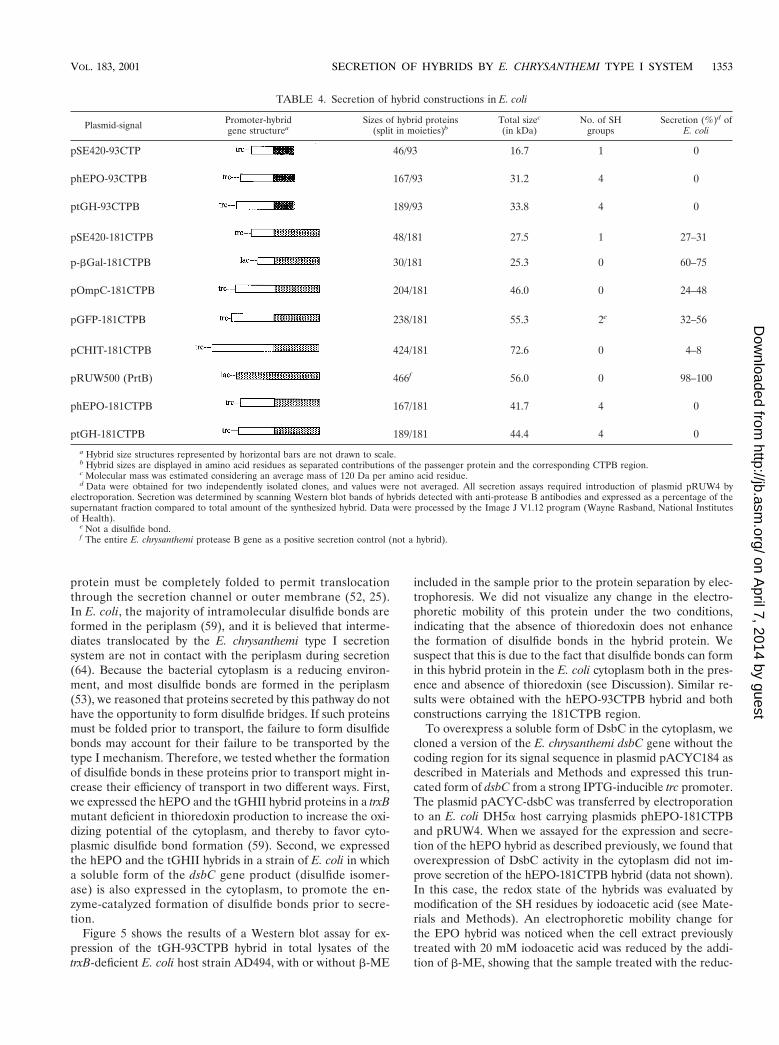
TABLE 4. Secretion of hybrid constructions in E. coli
Plasmid-signal Promoter-hybridgene structurea
Sizes of hybrid proteins(split in moieties)b
Total sizec
(in kDa)No. of SH
groupsSecretion (%)d of
E. coli
pSE420-93CTP 46/93 16.7 1 0
phEPO-93CTPB 167/93 31.2 4 0
ptGH-93CTPB 189/93 33.8 4 0
pSE420-181CTPB 48/181 27.5 1 27–31
p-bGal-181CTPB 30/181 25.3 0 60–75
pOmpC-181CTPB 204/181 46.0 0 24–48
pGFP-181CTPB 238/181 55.3 2e 32–56
pCHIT-181CTPB 424/181 72.6 0 4–8
pRUW500 (PrtB) 466f 56.0 0 98–100
phEPO-181CTPB 167/181 41.7 4 0
ptGH-181CTPB 189/181 44.4 4 0
a Hybrid size structures represented by horizontal bars are not drawn to scale.b Hybrid sizes are displayed in amino acid residues as separated contributions of the passenger protein and the corresponding CTPB region.c Molecular mass was estimated considering an average mass of 120 Da per amino acid residue.d Data were obtained for two independently isolated clones, and values were not averaged. All secretion assays required introduction of plasmid pRUW4 by
electroporation. Secretion was determined by scanning Western blot bands of hybrids detected with anti-protease B antibodies and expressed as a percentage of thesupernatant fraction compared to total amount of the synthesized hybrid. Data were processed by the Image J V1.12 program (Wayne Rasband, National Institutesof Health).
e Not a disulfide bond.f The entire E. chrysanthemi protease B gene as a positive secretion control (not a hybrid).
VOL. 183, 2001 SECRETION OF HYBRIDS BY E. CHRYSANTHEMI TYPE I SYSTEM 1353
on April 7, 2014 by guest
http://jb.asm.org/
Dow
nloaded from
ing agent entered the gel and the untreated sample did not(Fig. 5C). This suggests that the hEPO hybrid has been previ-ously oxidized.
DISCUSSION
The main goal of the work we have described in this reporthas been to test whether the E. chrysanthemi type I secretionsystem permits the efficient secretion of eukaryotic proteins inan E. coli host. The secretion of eukaryotic proteins to theexternal medium using this system will facilitate and simplifythe purification of proteins for basic scientific, medical, andindustrial purposes. Our demonstration that a subset of eu-karyotic proteins can be produced and excreted by this systemin E. coli provides yet another method for the large-scale pu-rification of eukaryotic protein hybrids in a prokaryotic host.Such hybrids, once purified, can be processed proteolytically toremove the secretion signal from the hybrid after exportation,if necessary.
Consistent with the results of previous studies with prokary-otic fusion proteins, we found that a 93-residue signal sequencefrom E. chrysanthemi PrtB is not sufficient to allow secretion ofthe small hybrid resulting from the construction of the secre-tion vector pSE420-93CTPB but that a larger hybrid with a181-residue signal sequence is sufficient to allow secretion ofthe product of vector pSE420-181CTPB. Similar results wereobtained with fusions of the same C termini to an endochiti-nase and GFP (Table 4). Thus, as for previous hybrids con-structed between prokaryotic proteins and the C terminus ofPrtB, the secretion of hybrids with eukaryotic proteins, in gen-eral, depends not only on the C-terminal DFLV and DIIVmotifs of PrtB but also on the presence of six short repeats ofresidues rich in glycines, three of them matching precisely theconsensus sequence GGXGXD. These motifs have been de-fined as additional components of the secretion signal (41).This feature is also present in a Serratia sp. metalloprotease, S.marcescens lipase, Rhizobium leguminosarum NodO, Proteusmirabilis metalloprotease, P. aeruginosa alkaline protease, andhemolysin, all secreted by a type I mechanism.
Although some hybrids with eukaryotic proteins are secretedby the E. chrysanthemi type I mechanism, others are not, in-cluding those with hEPO and tGH. The proteins that are notsecreted by this mechanism have the common feature that theyform intramolecular disulfide bonds in their native structures.This result is striking because it calls attention to the fact thatnone of the 22 known proteins secreted by type I mechanismsare capable of forming intramolecular disulfide bonds (Table5). Of these proteins, 20 of 22 have no cysteine residues andonly NodO and PllktA have single cysteine residues. Given theaverage size of the proteins secreted by a type I mechanism,and the frequency of occurrence of cysteine residues in pro-teins, this conspicuous absence of disulfide bonds in these 22
FIG. 4. Expression and secretion assays of hEPO and tGH hybridsin E. coli C600. The hybrids were visualized by SDS–12% PAGE fol-lowed by a Western blot assay revealed with anti-protease B polyclonalantibodies. Cultures were induced for 4 h at 37°C with 1 mM IPTGafter the bacterial cultures reached an OD600 of 1.0. Ten-microlitersamples of lysates (L) and supernatants (S) that were concentrated 10times with respect to the lysates by 10% TCA precipitation were loaded inthe gel. Std, Benchmark prestained protein ladder. (A) Expression ofhybrid hEPO-93CTPB. Lanes: 1 and 2, clone harboring pSE420 pluspRUW4; 3 and 4, clone with phEPO-93CTPB; 5 and 6, clone withphEPO-93CTPB plus pRUW4. (B) Expression of hybrid tGH2-93CTPB.Lanes: 1 and 2, clone with pSE420 plus pRUW4; 3 and 4, clone withptGH2-93CTPB; 5 and 6, clone with ptGH2-93CTPB plus pRUW4. Thearrows indicate the electrophoretic migration of the protein bands ex-pected in each case. (C) Expression of hybrid hEPO-181CTPB. Lanes:1 and 2, clone 3-1 (phEPO-181CTPB plus pRUW4 in E. coli DH5a);
3 and 4, clone 12-1 (phEPO-181CTPB plus pRUW4 in E. coli DH5a);5 and 6, clone 4-1 (phEPO-181CTPB plus pRUW4 in E. coli DH5a).(D) Lanes: 1 and 2, clone 6-1 (ptGH-181CTPB plus pRUW4 in E. coliDH5a); 3 and 4, clone 5-1 (ptGH-181CTPB plus pRUW4 in E. coliDH5a); 5 and 6, clone 2-1 (ptGH-181CTPB plus pRUW4 in E. coliDH5a).
1354 PALACIOS ET AL. J. BACTERIOL.
on April 7, 2014 by guest
http://jb.asm.org/
Dow
nloaded from

proteins cannot be due to mere coincidence. Furthermore, wehave shown that fusions with GFP are secreted by the type Iapparatus. GFP has two cysteine residues that do not partici-pate in disulfide bond formation because they are sufficientlydistant from one another on the surface of the GFP monomerto exclude their direct intramolecular interaction (51). Thus,the simplest hypothesis that can account for our results is thatthe formation of disulfide bonds, or the potential to formdisulfide bonds, precludes the secretion of a subset of proteinsby the E. chrysanthemi type I mechanism.
Initially, we considered the latter hypothesis because the E.coli cytoplasm is a reducing environment and most disulfidebond formation in E. coli occurs in the periplasm, due to theenzymatic activity of the Dsb proteins. According to this hy-pothesis, the potential to form disulfide bonds would precludethe secretion of a subset of proteins by the E. chrysanthemi typeI mechanism, presumably because these proteins would needto be folded prior to transport. By this hypothesis, then, thelack of secretion for hEPO and tGH would be due to theincomplete folding of these hybrids because of the lack ofdisulfide bonds in their structure. However, we find that this isunlikely to be the case because overexpression of a solubleform of active DsbC in the cytoplasm does not increase theefficiency of export of proteins with disulfide bonds in theirmature structures. This strategy of overexpressing members ofthe DsbC family to catalyze the formation of disulfide bonds inthe cytoplasm has been used successfully to improve the pro-duction of apo-retinol-binding protein (56), mouse urokinase,and human tissue plasminogen activator (6). Given that we candetect cytoplasmic activity of the expressed E. chrysanthemiDsbC protein without its signal peptide, we presume that thisprotein can function to stimulate disulfide bond formation inthe cytoplasm like its homologs. It is quite clear that the hEPO-181CTPB and tGH-181CTPB hybrids are sufficiently small tobe secreted by the type I mechanism, because the endochiti-
FIG. 5. (A) Western blot assay carried out in bacterial lysates todetect cytoplasmic formation of disulfide bonds in the tHG-93CTPBhybrid expressed in a trxB mutant. All E. coli AD494 and DH5a strainscarrying both ptGH-93CTPB and pRUW4 plasmids were grown over-night and induced with 1 mM IPTG for 3 h. Bacterial cells (1 ml) werecentrifuged in a microcentrifuge, and pellets were lysed by boiling for5 min in 100 ml of Laemmli sample buffer (40). Aliquots of 30 ml wereloaded in an SDS–12% polyacrylamide gel. Lanes: Std, GIBCO-BRLmolecular mass standard; 1, DH5a/ptGH-93CTPB plus 25 mM b-ME;2, DH5a/ptGH-93CTPB without b-ME; 3, AD494/ptGH-93CTPB plus25 mM b-ME; 4, AD494/ptGH-93CTPB without b-ME. (B) Detection ofchange in electrophoretic mobility of the tGH-181CTPB hybrid in DH5acell extracts after treatment with b-ME. Proteins were resolved by 8 Murea–8% polyacrylamide gel electrophoresis (see Materials and Meth-ods), and the hybrid (arrows) was detected by Western blotting asdescribed in Materials and Methods. Lanes: 1, ptGH-181CTPB with-out b-ME; 2, ptGH-181CTPB with 10 mM b-ME; 3, ptGH-181CTPBplus pRUW4, without b-ME added; 4, ptGH-181CTPB plus pRUW4with 10 mM b-ME. Loaded samples contained 10 ml of cell extractsand 10 ml of Laemmli sample buffer without SDS or b-ME. Hybridsloaded on lanes 1 and 3 did not enter the gel and were lost prior to orduring transfer to the nitrocellulose filter. (C) Detection of change inelectrophoretic mobility of hEPO-181 hybrid in DH5a cells overex-pressing the dsbC gene by b-ME treatment of extracts previouslyblocked with 20 mM iodoacetic acid. Reaction with iodoacetic acid wasdone as described in Materials and Methods. Proteins were resolved by8 M urea–8% polyacrylamide gel electrophoresis, and the hybrid (ar-rows) was detected by a chemiluminescence assay (see Materials andMethods). Loaded samples contained 10 ml of cell extracts and 10 mlof Laemmli sample buffer without SDS or b-ME. Lanes: 1, phEPO-181CTPB plus pRUW4 plus pACYC-dsbC, without b-ME added; 2,phEPO-181CTPB plus pRUW4 plus pACYC-dsbC with 10 mM b-ME.
TABLE 5. List of proteins secreted by type I secretion system and content of cysteine residues
Secreted protein (gene) Microorganism Components of type I secretion system No. of cysteine residues Reference
Protease AB374 (prtA) Erwinia chrysanthemi PrtD, PrtE, PrtF 0 27Protease B (prtB) Erwinia chrysanthemi PrtD, PrtE, PrtF 0 19Protease CE374 (prtC) Erwinia chrysanthemi PrtD, PrtE, PrtF 0 20Protease CEc16 (prtC) Erwinia chrysanthemi PrtD, PrtE, PrtF 0 17Protease G (prtG) Erwinia chrysanthemi PrtD, PrtE, PrtF 0 28Protease A (prtA) Erwinia amylovora PrtD, PrtE, PrtF 0 70Protease W (prtW) Erwinia carotovora Not yet identified 0 46Adenylate cyclase (cyaA) Bordetella pertussis CyaB, CyaD, CyaE 0 29Alpha-hemolysin (hlyA) Escherichia coli HlyB, HlyD, TolC 0 61Alpha-hemolysin (hlyA) Escherichia coli HlyB, HlyD, TolC 0 60Leukotoxin (pllktA) Pasteurella haemolytica PllktB, PllktD 1 14Metalloprotease (prt) Serratia sp. strain E-15 PrtD, PrtE 0 50Hb binding protein (hasA) Serratia marcescens PrtDSM, PrtESMa 0 44Protease SM (prtSM) Serratia marcescens PrtDSM, PrtESM, PrtFSM 0 12S-layer protein (rsaA) Caulobacter crescentus RsaD, RsaE 0 4Lipase A (lipA) Serratia marcescens Sr41 LipB, LipC, LipD 0 2Lipase A (lipA) Pseudomonas strain LS107d2 Not reported yet 0 37Thermostable lipase (tliA) Pseudomonas fluorescens tLiD, tLiE, tLiF 0 1Protease A (prtA) Pseudomonas fluorescens PrtD, PrtE, PrtF 0 1Alkaline protease (aprA) Pseudomonas aeruginosa AprD, AprE, AprF 0 23Metalloprotease (zapA) Proteus mirabilis PrtD, PrtE, PrtF 0 65Nodulating protein (nodO) Rhizobium leguminosarum Not yet reported 1 24
a PrtDSM and PrtESM components of the HasA secretion machinery have been renamed as HasB and HasD, respectively, and HasF, a TolC analog, has been cloned (8).
VOL. 183, 2001 SECRETION OF HYBRIDS BY E. CHRYSANTHEMI TYPE I SYSTEM 1355
on April 7, 2014 by guest
http://jb.asm.org/
Dow
nloaded from
nase hybrid with 605 amino acid residues, almost twice the sizeof these proteins, is secreted to the same extent.
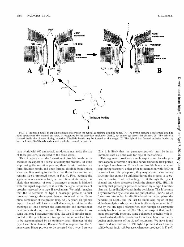
Thus, it appears that the formation of disulfide bonds per seexcludes the export of a subset of eukaryotic proteins. At somestep during the secretion process, these hybrid proteins canform disulfide bonds, and once formed, disulfide bonds blocksecretion. It is inviting to speculate that this is the case for tworeasons (see a proposed model in Fig. 6). First, because thesignal sequence essential for type I secretion is C-terminal, it islikely that transport of type I passenger proteins is initiatedwith this signal sequence, as it is with the signal sequences ofproteins secreted by a type II mechanism. We might imaginethat the C terminus of type I passenger proteins is firstthreaded through the export channel, followed by the N-ter-minal remainder of the protein (Fig. 6A). A priori, an optimalexport channel will have a small diameter, to minimize theexchange of ions between the extracellular and intracellularenvironments during transport. Thus, it is reasonable to as-sume that type I passenger proteins, like type II proteins trans-ported to the periplasm, are transported in an unfolded formto be accommodated by an optimally small diameter of thetype I secretion channel. Because SecB is required for the S.marcescens HasA protein to be secreted via a type I system
(21), it is likely that the passenger protein must be in anunfolded state as is the case for type II mechanisms.
This argument provides a simple explanation for why pro-teins capable of forming disulfide bonds cannot be transportedby a type I mechanism. If they form disulfide bonds at somestep during transport, either prior to interaction with PrtD orin contact with the periplasm, they may acquire a secondarystructure that cannot be unfolded during the process of secre-tion, a structure that is too large to fit through the type Ichannel and which therefore blocks the channel (Fig. 6B). It isunlikely that passenger proteins secreted by a type I mecha-nism can form disulfide bonds in the periplasm. This is becausea hybrid formed by E. coli alkaline phosphatase (PhoA), whichforms two intramolecular disulfide bonds in the periplasm de-pendent on DsbC, and the last 60-amino-acid region of thealpha-hemolysin carboxyl terminus is efficiently secreted in E.coli by the Hly type I transporter, even though no enzymaticactivity has been reported (26). Thus, we suspect that, unlikemany prokaryotic proteins, some eukaryotic proteins with in-tramolecular disulfide bonds can form these bonds in the re-ducing environment of the E. coli cytoplasm. We have founddirect evidence that our hEPO hybrid protein does form di-sulfide bonds in E. coli, because, when overproduced in E. coli,
FIG. 6. Proposed model to explain blockage of secretion for hybrids containing disulfide bonds. (A) The hybrid carrying a preformed disulfidebond approaches the channel entrance, is recognized by the secretion machinery (PrtD), but cannot go across the channel. (B) The hybrid isstacked inside the channel during secretion. Disulfide bonds may be formed at this stage. (C) The hybrid has formed inclusion bodies byintermolecular SOS bonds and cannot reach the channel or enter it.
1356 PALACIOS ET AL. J. BACTERIOL.
on April 7, 2014 by guest
http://jb.asm.org/
Dow
nloaded from
a fraction of the hEPO hybrid protein aggregates by formingdisulfide bonds that are sensitive to reduction with b-ME (Fig.5C). Also, the folding of growth hormone occurs independent-ly of the formation of at least one of its two disulfide bridges,to position two cysteine side chains sufficiently close to oneanother to permit spontaneous, base-catalyzed disulfide bondformation (62). In addition, formation of prochymosin inclu-sion bodies in E. coli cytoplasm by cross-linking of disulfidebonds has been reported (58). Formation of inclusion bodiesin the cytoplasm could also restrict secretion of hybrid proteinsby the type I secretion system (Fig. 6C).
Currently, we are testing the hypothesis that the formationof intramolecular disulfide bonds in the cytoplasm by passen-ger proteins can block their secretion by a type I mechanismdirectly, by determining whether E. coli strains with the E. chry-santhemi type I secretion system are capable of exporting pro-teins that form disulfide bonds in their mature structures de-pendent on the activity of cytoplasmically expressed DsbC.
ACKNOWLEDGMENTS
This work was funded by the Comision Nacional Cientıfica y Tec-nologica de Chile (grant FONDECYT 1971010) and Fondo de Desar-rollo Innovativo (CORFO, Santiago, Chile; grant FDI AT-1).
E. carotovora subsp. carotovora Ecc193 was kindly provided by A. K.Chatterjee. We gratefully acknowledge P. Delepelaire for providingplasmids pRUW4 and pRUW500 and P. Youderian for critical readingof the manuscript.
REFERENCES
1. Ahn, J., J. Pan, and J. Rhee. 1999. Identification of the tliDEF ABC trans-porter specific for lipase in Pseudomonas fluorescens SIK W1. J. Bacteriol.181:1847–1852.
2. Akatsuka, H., E. Kawai, K. Omori, and T. Shibatani. 1995. The three geneslipB, lipC, and lipD involved in the extracellular secretion of Serratia mar-cescens lipase which lacks an N-terminal signal peptide. J. Bacteriol. 177:6381–6389.
3. Arkowitz, R. J. Joly, and W. Wickner. 1993. Translocation can drive theunfolding of a preprotein domain. EMBO J. 12:243–253.
4. Awram, P., and J. Smith. 1998. The Caulobacter crescentus paracrystallineS-layer protein is secreted by an ABC transporter type I secretion apparatus.J. Bacteriol. 180:3062–3069.
5. Bardwell, J. 1994. Building bridges: disulfide bond formation in the cell. Mol.Microbiol. 14:199–205.
6. Bessette, P., F. Aslund, J. Beckwith, and G. Georgiou. 1999. Efficient foldingof proteins with multiple disulfide bonds in the Escherichia coli cytoplasm.Proc. Natl. Acad. Sci. USA 96:13703–13708.
7. Binet, R., and C. Wandersman. 1995. Protein secretion by hybrid bacterialABC-transporters: specific functions of the membrane ATPase and themembrane fusion protein. EMBO J. 14:2298–2306.
8. Binet, R., and C. Wandersman. 1996. Cloning of the Serratia marcescens hasFgene encoding the Has ABC exporter outer membrane component: a TolCanalog. Mol. Microbiol. 22:265–273.
9. Binet, R., S. Letoffe, J. Ghigo, P. Delepelaire, and C. Wandersman. 1997.Protein secretion by Gram negative bacterial ABC export: a review. Gene192:7–11.
10. Birnboim, H. C., and J. Doly. 1979. A rapid alkaline extraction procedure forscreening recombinant plasmid DNA. Nucleic Acids Res. 7:1513–1523.
11. Bradford, M. M. 1976. A rapid and sensitive method for the quantitation ofmicrogram quantities of protein utilizing the principle of protein-dye bind-ing. Anal. Biochem. 72:248–254.
12. Braunagel, S. C., and M. J. Benedik. 1990. The metalloprotease gene ofSerratia marcescens strain SM6. Mol. Gen. Genet. 222:446–451.
13. Chaconas, G., and J. van de Sande. 1990. 59-32P labeling of RNA and DNArestriction fragments. Methods Enzymol. 65:75–85.
14. Chang, Y., D. Ma, J. Shi, and M. Chengappa. 1993. Molecular character-ization of a leukotoxin gene from a Pasteurella haemolytica-like organism,encoding a new member of the RTX toxin family. Infect. Immun. 61:2089–2095.
15. Chen, E., and P. Seeburg. 1985. Supercoil sequencing: a fast and simplemethod for sequencing plasmid DNA. DNA 4:165–170.
16. Coligan, J. 1994. In J. Coligan, A. Kruisbeck, D. Margulies, E. Shevach, andW. Strober (ed.), Current protocols in immunology, 2nd ed., p. 1.6.1–1.7.8.Wiley Liss Inc., New York, N.Y.
17. Dahler, G. S., F. Barras, and N. T. Keen. 1990. Protease C cloning of genesencoding extracellular metalloproteases from Erwinia chrysanthemi EC16.J. Bacteriol. 172:5803–5815.
18. Delepelaire, P. 1994. PrtD, the integral membrane ATP-binding cassettecomponent of the Erwinia chrysanthemi metalloprotease secretion system,exhibits a secretion signal-regulated ATPase activity. J. Biol. Chem. 269:27952–27957.
19. Delepelaire, P., and C. Wandersman. 1989. Protease secretion by Erwiniachrysanthemi. Proteases B and C are synthesized and secreted as zymogenswithout a signal peptide. J. Biol. Chem. 264:9083–9089.
20. Delapelaire, P., and C. Wandersman. 1990. Protein secretion in gram-neg-ative bacteria. The extracellular metalloprotease B from Erwinia chrysan-themi contains a C-terminal secretion signal analogous to that of Escherichiacoli a-hemolysin. J. Biol. Chem. 265:17118–17125.
21. Delepelaire, P., and C. Wandersman. 1998. The SecB chaperone is involvedin the secretion of the Serratia marcescens HasA protein through an ABCtransporter. EMBO J. 17:936–944.
22. Dinh, T., I. Paulsen, and M. Saier. 1994. A family of extracytoplasmicproteins that allow transport of large molecules across the outer membranesof gram-negative bacteria. J. Bacteriol. 176:3825–3831.
23. Duong, F., A. Lazdunski, B. Cami, and M. Murgier. 1992. Sequence of acluster of genes controlling synthesis and secretion of alkaline protease inPseudomonas aeruginosa: relationships to other secretory pathways. Gene121:47–54.
24. Economou, A., W. Hamilton, A. Johnston, and J. Downie. 1990. The Rhizo-bium nodulation gene nodO encodes a Ca2(1)-binding protein that is ex-ported without N-terminal cleavage and is homologous to haemolysin andrelated proteins. EMBO J. 9:349–354.
25. Fujishige, A., K. Smith, J. Silen, and D. Agard. 1992. Correct folding ofa-lytic protease is required for extracellular secretion from Escherichia coli.J. Cell Biol. 118:33–42.
26. Gentschev, I., J. Hess, and W. Goebel. 1990. Change in the cellular local-ization of alkaline phosphatase by alteration of its carboxy-terminal se-quence. Mol. Gen. Genet. 222:211–216.
27. Ghigo, J., and C. Wandersman. 1992. Cloning, nucleotide sequence andcharacterization of the gene encoding the Erwinia chrysanthemi B374 PrtAmetalloprotease: a third metalloprotease secreted via a C-terminal secretionsignal. Mol. Gen. Genet. 236:135–144.
28. Ghigo, J., and C. Wandersman. 1994. A carboxyl-terminal four-amino acidmotif is required for secretion of the metalloprotease PtrG through theErwinia chrysanthemi protease secretion pathway. J. Biol. Chem. 269:8979–8985.
29. Glasser, P., H. Sakamoto, J. Bellalou, A. Ullman, and A. Danchin. 1988.Secretion of cyclolysin, the calmodulin-sensitive adenylate cyclase-haemoly-sin bifunctional protein of Bordetella pertussis. EMBO J. 7:3997–4004.
30. Gray, L., B. Kenny, N. Mackman, R. High, and I. Holland. 1989. A novelC-terminal signal sequence targets Escherichia coli haemolysin directly to themedium. J. Cell Sci. 11(Suppl.):45–57.
31. Grimberg, J., S. Maguire, and L. Belluscio. 1989. A simple method for thepreparation of plasmid and chromosomal Escherichia coli DNA. NucleicAcids Res. 17:8893.
32. Hames, B. 1981. Trichloroacetic acid precipitation, p. 71–73. In D. Bollagand S. Edelstein (ed.), Protein methods. Wiley Liss Inc., New York, N.Y.
33. Hayes, C. K., S. Klemsdal, M. Lorito, A. Di Pietro, C. Peterbauer, J. P.Nakas, A. Tronsmo, and G. E. Harman. 1994. Isolation and sequence of anendochitinase-encoding gene from a cDNA library of Trichoderma harzia-num. Gene 138:143–148.
34. Higuchi, M., M. Oh-eda, H. Kuboniwa, K. Tomonoh, Y. Shimonaka, and N.Ochi. 1992. Role of sugar chains in the expression of the biological activityof human erythropoietin. J. Biol. Chem. 267:7703–7709.
35. Hirst, T., and J. Holmgren. 1987. Conformation of protein secreted acrossbacterial outer membranes: a study of enterotoxin translocation from Vibriocholerae. Proc. Natl. Acad. Sci. USA 84:7418–7422.
36. Huek, C. 1998. Type III protein secretion systems in bacterial pathogens inplants and animals. Microbiol. Mol. Biol. Rev. 62:379–433.
37. Johnson, L. A., I. R. Beacham, I. C. MacRae, and M. L. Free. 1992. Degra-dation of triglycerides by a pseudomonad isolated from milk: molecularanalysis of a lipase-encoding gene and its expression in Escherichia coli.Appl. Environ. Microbiol. 58:1776–1779.
38. Kenny, B., and B. Finlay. 1995. Protein secretion by enteropathogenic Esch-erichia coli is essential for transducing signals to epithelial cells. Proc. Natl.Acad. Sci. USA 92:7991–7995.
39. Kubori, T., Y. Matsushima, D. Nakamura, J. Uralil, M. Lara-Tejero, A.Sukhan, J. Galan, and S. Aizawa. 1998. Supramolecular structure of theSalmonella typhimurium type III protein secretion system. Science 280:602–605.
40. Laemmli, U. K. 1970. Cleavage of structural proteins during the assembly ofthe head of bacteriophage T4. Nature 227:680–685.
41. Letoffe, S., and C. Wandersman. 1992. Secretion of Cya-PrtB and Hly-PrtBfusion proteins in Escherichia coli: involvement of the glycine-rich repeatdomain of Erwinia chrysanthemi protease B. J. Bacteriol. 174:4920–4927.
42. Letoffe, S., P. Delapelaire, and C. Wandersman. 1990. Protease secretion by
VOL. 183, 2001 SECRETION OF HYBRIDS BY E. CHRYSANTHEMI TYPE I SYSTEM 1357
on April 7, 2014 by guest
http://jb.asm.org/
Dow
nloaded from
Erwinia chrysanthemi: the specific secretion functions are analogous to thoseof Escherichia coli a-hemolysin. EMBO J. 9:1375–1382.
43. Letoffe, S., P. Delepelaire, and C. Wandersman. 1991. Cloning and expres-sion in Escherichia coli of the Serratia marcescens metalloprotease gene:secretion of the protease from E. coli in the presence of the Erwinia chry-santhemi protease functions. J. Bacteriol. 173:2160–2166.
44. Letoffe, S., J. Ghigo, and C. Wandersman. 1994. Secretion of the Serratiamarcescens HasA protein by an ABC transporter. J. Bacteriol. 176:5372–5377.
45. Letoffe, S., J. Ghigo, and C. Wandersman. 1994. Iron acquisition from hemeand hemoglobin by a Serratia marcescens extracellular protein. Proc. Natl.Acad. Sci. USA 91:9876–9880.
46. Marits, R., V. Koiv, E. Laasik, and A. Mae. 1999. Isolation of an extracellularprotease gene of Erwinia carotovora subsp. carotovora strain SCC3193 bytransposon mutagenesis and the role of protease in phytopathogenicity.Microbiology 145:1959–1966.
47. Miller, J. 1994. Bacterial transformation by electroporation. Methods Enzy-mol. 235:375–385.
48. Missiakas, D., C. Georgopoulos, and S. Raina. 1994. The Escherichia colidsbC (xprA) gene encodes a periplasmic protein involved in disulfide bondformation. EMBO J. 13:2013–2020.
49. Muller, I. 1991. Expression of trout growth hormone tGHII in Escherichiacoli. Biochemist professional title thesis. University of Concepcion, Concep-cion, Chile.
50. Nakahama, K., K. Yoshimura, R. Marumoto, M. Kikuchi, I. S. Lee, T. Hase,and H. Matsubara. 1986. Cloning and sequencing of Serratia protease gene.Nucleic Acids Res. 14:5843–5855.
51. Ormo, M., A. B. Cubitt, K. Kallio, L. A. Gross, R. Y. Tsien, and S. J.Remington. 1996. Crystal structure of the aequorea victoria green fluorescentprotein. Science 273:1392–1295.
52. Pugsley, A. 1992. Translocation of a folded protein across the outer mem-brane in Escherichia coli. Proc. Natl. Acad. Sci. USA 89:12058–12062.
53. Pugsley, A. 1993. The complete general secretory pathway in gram-negativebacteria. Microbiol. Rev. 57:50–108.
54. Sambrook, J., E. F. Fritsch, and T. Maniatis. 1989. Molecular cloning: alaboratory manual, 2nd ed. Cold Spring Harbor Laboratory Press, ColdSpring Harbor, N.Y.
55. Sanger, F., S. Nicklen, and A. R. Coulson. 1977. DNA sequencing withchain-terminating inhibitors. Proc. Natl. Acad. Sci. USA 74:5463–5467.
56. Schmidt, A., I. Bloss, and A. Skerra. 1998. Improved folding of apo-retinol-binding protein in the periplasm of Escherichia coli: positive influences ofdsbC coexpression and of an amino acid exchange in the vitamin A bindingsite. Protein Eng. 11:601–607.
57. Shevchik, V., G. Condemine, and J. Robert-Baudouy. 1994. Characterizationof DsbC, a periplasmic protein of Erwinia carotovora and Escherichia coliwith disulfide isomerase activity. EMBO J. 13:2007–2012.
58. Shoemaker, J. M., A. H. Brasnett, and F. A. Marston. 1985. Examination ofcalf prochymosin accumulation in Escherichia coli: disulphide linkages are astructural component of prochymosin-containing inclusion bodies. EMBO J.4:775–780.
59. Stewart, E., F. Aslund, and J. Beckwith. 1998. Disulfide bond formation inEscherichia coli cytoplasm and in vivo role reversal for the thioredoxinsEMBO J. 17:5543–5550.
60. Taneike, I., N. Wakisaka-Saito, Y. Harada, H.-M. Zhang, and T. Yamamoto.The enterohemorrhagic Escherichia coli (EHEC)-hemolysin genes of a Shigatoxin 1 (Stx1)- and Stx2-producing, serotype O128 Escherichia coli strain witha greatest hemolytic activity. Acta Med. Biol., in press.
61. Thomas, W., S. Wagner, and R. Welch. 1992. A heterologous membraneprotein domain fused to the C-terminal ATP-binding domain of HlyB canexport Escherichia coli hemolysin. J. Bacteriol. 174:6771–6779.
62. Tokunaga, T., T. Tanaka, M. Ikehara, and E. Ohtsuka. 1985. Synthesis andexpression of a human growth hormone (somatotropin) gene mutated tochange cysteine-165 to alanine. Eur. J. Biochem. 153:445–449.
63. Towbin, H., T. Staehelin, and J. Gordon. 1979. Electrophoretic transfer ofproteins from polyacrylamide gels to nitrocellulose sheets: procedure andsome applications. Proc. Natl. Acad. Sci. USA 76:4350–4354.
64. Wandersman, C. 1992. Secretion across the bacterial outer membrane.Trends Genet. 8:317–322.
65. Wassif, C., D. Cheek, and R. Belas. 1995. Molecular analysis of a metallo-protease from Proteus mirabilis. J. Bacteriol. 177:5790–5798.
66. Welch, R. A. 1991. Pore-forming cytolysins of gram-negative bacteria. Mol.Microbiol. 5:521–528.
67. Weston, A., M. Brow, H. Perkins, J. Saunders, and G. Humphreys. 1981.Transformation of Escherichia coli with plasmid deoxyribonucleic acid: cal-cium-induced binding of deoxyribonucleic acid to whole cells and to isolatedmembrane fractions. J. Bacteriol 145:780–787.
68. Yanisch-Perron, C., J. Vieira, and J. Messing. 1985. Improved M13 phagecloning vectors and host strains: nucleotide sequences of the M13mp18 andpUC19 vectors. Gene 33:103–119.
69. Zaror, M. 1989. Isolation and characterization of the Salmonella typhi ompCgene. Structural and functional studies of the OmpC porin. Ph.D. thesis.Catholic University of Chile, Santiago.
70. Zhang, Y., D. D. Bak, H. Heid, and K. Geider. 1999. Molecular character-ization of a protease secreted by Erwinia amylovora. J. Mol. Biol. 289:1239–1251.
1358 PALACIOS ET AL. J. BACTERIOL.
on April 7, 2014 by guest
http://jb.asm.org/
Dow
nloaded from
Copyright © 2022 FDOKUMEN