Sequence and structure-based prediction of eukaryotic protein phosphorylation sites1
12
Sequence and Structure-based Prediction of Eukaryotic Protein Phosphorylation Sites Nikolaj Blom 1 , Steen Gammeltoft 2 and Søren Brunak 1 * 1 Center for Biological Sequence Analysis, Department of Biotechnology, The Technical University of Denmark, DK- 2800 Lyngby, Denmark 2 Department of Clinical Biochemistry, Glostrup Hospital, DK-2600 Glostrup Denmark Protein phosphorylation at serine, threonine or tyrosine residues affects a multitude of cellular signaling processes. How is specificity in substrate recognition and phosphorylation by protein kinases achieved? Here, we present an artificial neural network method that predicts phosphorylation sites in independent sequences with a sensitivity in the range from 69 % to 96 %. As an example, we predict novel phosphorylation sites in the p300/CBP protein that may regulate interaction with transcription factors and histone acetyltransferase activity. In addition, serine and threonine residues in p300/CBP that can be modified by O-linked glycosylation with N-acetylglucosamine are identified. Glycosylation may prevent phosphorylation at these sites, a mechanism named yin-yang regulation. The prediction server is available on the Internet at http:// www.cbs.dtu.dk/services/NetPhos/or via e-mail to [email protected]. # 1999 Academic Press Keywords: phosphorylation; kinase specificity; prediction; protein structure; transcriptional regulation *Corresponding author Introduction Protein kinases catalyze phosphorylation events that are essential for the regulation of cellular pro- cesses like metabolism, proliferation, differen- tiation, and apoptosis (Koliba & Druker, 1997; Hunter, 1998; Johnson et al., 1996, 1998; Pinna & Ruzzene, 1996; Graves et al., 1997). This very large family of enzymes share homologous catalytic domains and the mechanism of substrate recog- nition may be similar despite large variation in sequence. Crystallization studies indicate that a region, between seven and 12 residues in size, sur- rounding the acceptor residue contacts the kinase active site (Songyang et al., 1994). The specificity of protein kinases is dominated by acidic, basic, or hydrophobic residues adjacent to the phosphorylated residue, but the large variation makes it difficult manually to inspect protein sequences and predict the location of biologically active sites. This prompted us to investigate if the fuzzy sequence patterns can be recognized using artificial neural networks techniques. Neural net- works are capable of classifying even highly com- plex and non-linear biological sequence patterns, where correlations between positions are important. The network recognizes the patterns seen during training, and retains the ability to generalize and recognize similar, but non-identical patterns. Artifi- cial neural networks have been extensively used in biological sequence analysis (Wu, 1997; Baldi & Brunak, 1998). Since determinants of phosphoryl- ation sites probably are no longer than about ten residues, most local sequence alignment tools, such as BLAST and FASTA, will not be useful for detect- ing phosphorylation sites due to a large number of irrelevant hits in the protein databases, even to non- phosphorylated proteins. The related proteins p300 and CBP (CREB (cAMP-response-element-binding)-binding protein) integrate molecular signals at the level of gene transcription and chromatin modification. p300 and CBP interact with transcription factors CREB, Jun and Fos, viral oncoproteins E1a and SV40 large T antigen, and kinases pp90 RSK and cyclin E-com- plexed cyclin-dependent kinase (CDK)-2 (Shikama et al., 1997; Ait-Si-Ali et al., 1998). These inter- actions may possibly be regulated by reversible phosphorylation of p300/CBP. We demonstrate that regions of p300/CBP, which have been shown to interact with other molecules, contain probable phosphorylation sites. In addition, we describe sites that possibly are regulated by both phos- phorylation and glycosylation by N-acetylglucosa- mine (GlcNac), a regulatory mechanism described as a yin-yang dynamic phosphorylation/glycosyla- tion (Hart et al., 1995; Hart, 1997). E-mail address of the corresponding author: [email protected] Article No. jmbi.1999.3310 available online at http://www.idealibrary.com on J. Mol. Biol. (1999) 294, 1351–1362 0022-2836/99/501351–12 $30.00/0 # 1999 Academic Press
-
Upload
independent -
Category
Documents
-
view
1 -
download
0
Transcript of Sequence and structure-based prediction of eukaryotic protein phosphorylation sites1

Article No. jmbi.1999.3310 available online at http://www.idealibrary.com on J. Mol. Biol. (1999) 294, 1351±1362
Sequence and Structure-based Prediction ofEukaryotic Protein Phosphorylation Sites
Nikolaj Blom1, Steen Gammeltoft2 and Sùren Brunak1*
1Center for Biological SequenceAnalysis, Department ofBiotechnology, The TechnicalUniversity of Denmark, DK-2800 Lyngby, Denmark2Department of ClinicalBiochemistry, GlostrupHospital, DK-2600 GlostrupDenmark
E-mail address of the [email protected]
0022-2836/99/501351±12 $30.00/0
Protein phosphorylation at serine, threonine or tyrosine residues affects amultitude of cellular signaling processes. How is speci®city in substraterecognition and phosphorylation by protein kinases achieved? Here, wepresent an arti®cial neural network method that predicts phosphorylationsites in independent sequences with a sensitivity in the range from 69 %to 96 %. As an example, we predict novel phosphorylation sites in thep300/CBP protein that may regulate interaction with transcription factorsand histone acetyltransferase activity. In addition, serine and threonineresidues in p300/CBP that can be modi®ed by O-linked glycosylationwith N-acetylglucosamine are identi®ed. Glycosylation may preventphosphorylation at these sites, a mechanism named yin-yang regulation.
The prediction server is available on the Internet at http://www.cbs.dtu.dk/services/NetPhos/or via e-mail to [email protected].
# 1999 Academic Press
Keywords: phosphorylation; kinase speci®city; prediction; proteinstructure; transcriptional regulation
*Corresponding authorIntroduction
Protein kinases catalyze phosphorylation eventsthat are essential for the regulation of cellular pro-cesses like metabolism, proliferation, differen-tiation, and apoptosis (Koliba & Druker, 1997;Hunter, 1998; Johnson et al., 1996, 1998; Pinna &Ruzzene, 1996; Graves et al., 1997). This very largefamily of enzymes share homologous catalyticdomains and the mechanism of substrate recog-nition may be similar despite large variation insequence. Crystallization studies indicate that aregion, between seven and 12 residues in size, sur-rounding the acceptor residue contacts the kinaseactive site (Songyang et al., 1994).
The speci®city of protein kinases is dominated byacidic, basic, or hydrophobic residues adjacent tothe phosphorylated residue, but the large variationmakes it dif®cult manually to inspect proteinsequences and predict the location of biologicallyactive sites. This prompted us to investigate if thefuzzy sequence patterns can be recognized usingarti®cial neural networks techniques. Neural net-works are capable of classifying even highly com-plex and non-linear biological sequence patterns,where correlations between positions are important.The network recognizes the patterns seen during
ing author:
training, and retains the ability to generalize andrecognize similar, but non-identical patterns. Arti®-cial neural networks have been extensively used inbiological sequence analysis (Wu, 1997; Baldi &Brunak, 1998). Since determinants of phosphoryl-ation sites probably are no longer than about tenresidues, most local sequence alignment tools, suchas BLAST and FASTA, will not be useful for detect-ing phosphorylation sites due to a large number ofirrelevant hits in the protein databases, even to non-phosphorylated proteins.
The related proteins p300 and CBP (CREB(cAMP-response-element-binding)-binding protein)integrate molecular signals at the level of genetranscription and chromatin modi®cation. p300and CBP interact with transcription factors CREB,Jun and Fos, viral oncoproteins E1a and SV40 largeT antigen, and kinases pp90RSK and cyclin E-com-plexed cyclin-dependent kinase (CDK)-2 (Shikamaet al., 1997; Ait-Si-Ali et al., 1998). These inter-actions may possibly be regulated by reversiblephosphorylation of p300/CBP. We demonstratethat regions of p300/CBP, which have been shownto interact with other molecules, contain probablephosphorylation sites. In addition, we describesites that possibly are regulated by both phos-phorylation and glycosylation by N-acetylglucosa-mine (GlcNac), a regulatory mechanism describedas a yin-yang dynamic phosphorylation/glycosyla-tion (Hart et al., 1995; Hart, 1997).
# 1999 Academic Press
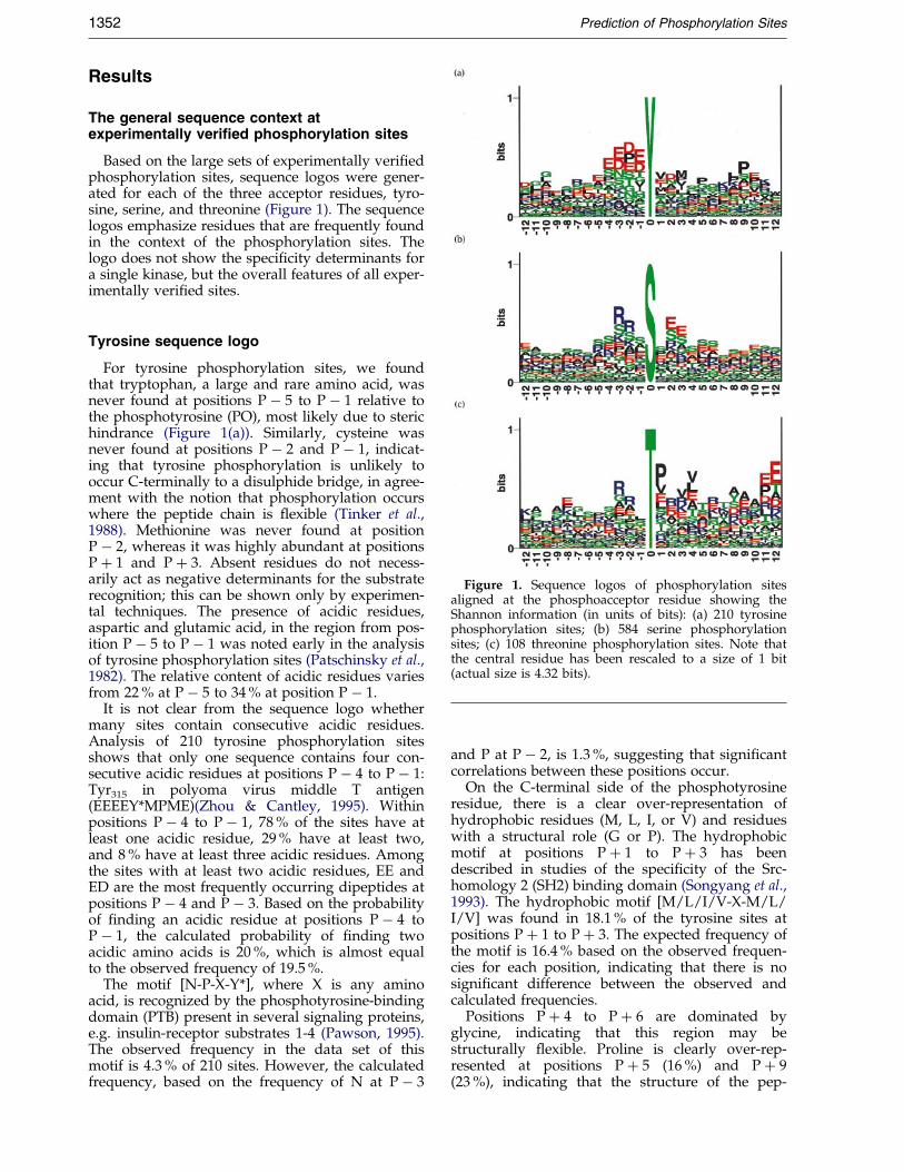
Figure 1. Sequence logos of phosphorylation sitesaligned at the phosphoacceptor residue showing theShannon information (in units of bits): (a) 210 tyrosinephosphorylation sites; (b) 584 serine phosphorylationsites; (c) 108 threonine phosphorylation sites. Note thatthe central residue has been rescaled to a size of 1 bit(actual size is 4.32 bits).
1352 Prediction of Phosphorylation Sites
Results
The general sequence context atexperimentally verified phosphorylation sites
Based on the large sets of experimentally veri®edphosphorylation sites, sequence logos were gener-ated for each of the three acceptor residues, tyro-sine, serine, and threonine (Figure 1). The sequencelogos emphasize residues that are frequently foundin the context of the phosphorylation sites. Thelogo does not show the speci®city determinants fora single kinase, but the overall features of all exper-imentally veri®ed sites.
Tyrosine sequence logo
For tyrosine phosphorylation sites, we foundthat tryptophan, a large and rare amino acid, wasnever found at positions P ÿ 5 to P ÿ 1 relative tothe phosphotyrosine (PO), most likely due to sterichindrance (Figure 1(a)). Similarly, cysteine wasnever found at positions P ÿ 2 and P ÿ 1, indicat-ing that tyrosine phosphorylation is unlikely tooccur C-terminally to a disulphide bridge, in agree-ment with the notion that phosphorylation occurswhere the peptide chain is ¯exible (Tinker et al.,1988). Methionine was never found at positionP ÿ 2, whereas it was highly abundant at positionsP � 1 and P � 3. Absent residues do not necess-arily act as negative determinants for the substraterecognition; this can be shown only by experimen-tal techniques. The presence of acidic residues,aspartic and glutamic acid, in the region from pos-ition P ÿ 5 to P ÿ 1 was noted early in the analysisof tyrosine phosphorylation sites (Patschinsky et al.,1982). The relative content of acidic residues variesfrom 22 % at P ÿ 5 to 34 % at position P ÿ 1.
It is not clear from the sequence logo whethermany sites contain consecutive acidic residues.Analysis of 210 tyrosine phosphorylation sitesshows that only one sequence contains four con-secutive acidic residues at positions P ÿ 4 to P ÿ 1:Tyr315 in polyoma virus middle T antigen(EEEEY*MPME)(Zhou & Cantley, 1995). Withinpositions P ÿ 4 to P ÿ 1, 78 % of the sites have atleast one acidic residue, 29 % have at least two,and 8 % have at least three acidic residues. Amongthe sites with at least two acidic residues, EE andED are the most frequently occurring dipeptides atpositions P ÿ 4 and P ÿ 3. Based on the probabilityof ®nding an acidic residue at positions P ÿ 4 toP ÿ 1, the calculated probability of ®nding twoacidic amino acids is 20 %, which is almost equalto the observed frequency of 19.5 %.
The motif [N-P-X-Y*], where X is any aminoacid, is recognized by the phosphotyrosine-bindingdomain (PTB) present in several signaling proteins,e.g. insulin-receptor substrates 1-4 (Pawson, 1995).The observed frequency in the data set of thismotif is 4.3 % of 210 sites. However, the calculatedfrequency, based on the frequency of N at P ÿ 3
and P at P ÿ 2, is 1.3 %, suggesting that signi®cantcorrelations between these positions occur.
On the C-terminal side of the phosphotyrosineresidue, there is a clear over-representation ofhydrophobic residues (M, L, I, or V) and residueswith a structural role (G or P). The hydrophobicmotif at positions P � 1 to P � 3 has beendescribed in studies of the speci®city of the Src-homology 2 (SH2) binding domain (Songyang et al.,1993). The hydrophobic motif [M/L/I/V-X-M/L/I/V] was found in 18.1 % of the tyrosine sites atpositions P � 1 to P � 3. The expected frequency ofthe motif is 16.4 % based on the observed frequen-cies for each position, indicating that there is nosigni®cant difference between the observed andcalculated frequencies.
Positions P � 4 to P � 6 are dominated byglycine, indicating that this region may bestructurally ¯exible. Proline is clearly over-rep-resented at positions P � 5 (16 %) and P � 9(23 %), indicating that the structure of the pep-

Prediction of Phosphorylation Sites 1353
tide chain is important. Position P � 7 is domi-nated by the positively charged basic residues(K/R [28 %]) and position P � 12 by the rareand bulky tryptophan residue (10.8 % comparedto 1.2 % on average in natural proteins).
We examined the sites containing either prolineat position P � 9 or tryptophan at P � 12 andobserved that many sites contained both residues.Most of the sites were known auto-phosphoryl-ation sites in Src-related kinases (e.g. Src, Hck, Tec,Fyn) or in receptor tyrosine kinases (insulin-recep-tor, IGF-1-receptor, PGDF-receptor, NGF-receptor).In the crystal structure of the insulin receptorkinase domain, the proline and tryptophan resi-dues are located in a buried coil region C-termin-ally of the activation loop containing the auto-phosphorylated tyrosine residues (Tyr1158,1162,1163).
Serine sequence logo
In the sequence logo of serine phosphorylationsites (Figure 1(b)), the basic motif recognized byPKA and PKG at positions Pÿ3 and Pÿ2 wasreadily observable (31 % and 27 % R � K at pos-itions P ÿ 3 and P ÿ 2, respectively). In the motifof protein kinase C (PKC), basic residues at P � 2and P � 3 could also be identi®ed. However, thesepositions were dominated by glutamic acid, whichis part of the acidic motif recognized by the caseinkinase 2 (CK-2) type kinases. Proline-directedkinases have a preference for proline at positionP � 1 (13 % of the residues at P � 1), which couldbe seen readily from the logo. Rare residuesaround the serine residue include tryptophan andcysteine. Tryptophan is not observed at positionsP ÿ 3 to P ÿ 1 and P � 2, and cysteine never atpositions P ÿ 7 and P ÿ 5.
As described earlier for O-glycosylation sites,serine residues have a tendency to cluster, and thisis observed for the neighboring positions P ÿ 1 andP � 1 also, where serine is the most abundant resi-due (Wilson et al., 1991; Hansen et al., 1998).
Threonine sequence logo
In the sequence logo of threonine phosphoryl-ation sites (Figure 1(c)), the basic motif at positionsP ÿ 3 and P ÿ 2 was readily observable, as was theproline-directed motif at P � 1. The PKC prefer-ence for basic residues at positions P � 2 andP � 3, and the CK-2 preference for acidic residuesat the same positions could be observed also.
Tryptophan was never found at positions P ÿ 9to P ÿ 5 or P � 1 to P � 6, but was over-represented at P � 7 (11 % of the residues).Cysteine was never found at positions P ÿ 5, P ÿ 3to P � 1 and P � 4 to P � 12, while asparagine wasnever found at positions P ÿ 3, P ÿ 2 or P � 1.These observations could be due to a smalleramount of data (108 sites), but for cysteine andtryptophan these probably re¯ect the fact thatmany phosphorylation sites are located in ¯exibleregions of the target molecule.
The frequent occurrence of valine or leucine atpositions P � 3 and P � 4 was investigated furtherby analyzing sites containing these features. Eightsites contained valine or leucine at both P � 3 andP � 4 (data not shown). We noted that these areinvolved in cell-cycle-dependent phosphorylationand some of them are known as targets of theCDK-activating kinase. Two of the sites containedtyrosine phosphorylation sites either at positionP � 1 or P � 2 relative to the phosphothreonineresidue, indicating that they may be targets ofdual-speci®city kinases. However, not all of thesites contained adjacent tyrosine residues, andtherefore the double V/L motif may be importantfor target recognition of CDK-activating andrelated kinases. Other kinases that prefer hydro-phobic residues at P � 4 include AMP-activatedprotein kinase and calmodulin-dependent proteinkinase I (Dale et al., 1995).
Another motif found in protein kinases involvedin regulation of cell-cycle was the frequent occur-rence of proline at P � 11 and glutamic acid atP � 12. Eleven phosphorylation sites, all in CDKsor related proteins, contained the consensussequence (T*xxV[V/A]TxxYR[A/S]PE), where the®rst T indicates the acceptor residue. These sitesare clearly related and the conserved residuesmight re¯ect the conservation of structural featuresrather than conservation of speci®city determi-nants.
Phosphorylation sites predicted byneural networks
Sequence motifs composing functional sites inpolypeptides can be complex in the sense that pos-itional correlations may play an important role.The amino acids surrounding a phosphorylatedresidue may not contribute independently as towhether a particular site is activated. This meansthat simple local alignment methods, or linearweight matrices based on consensus patterns, maybe unable with acceptable accuracy to separatetrue sites from a control set of non-phosphorylatedsites. Rules like ``an acidic residue at P ÿ 2 and nocysteine at P � 1 will make this site a probablephosphorylation site'' cannot be taken intoaccount. Two positions can make only independentcontributions to the overall score.
Training of the neural networks, using differentcombinations of input window sizes and trainingsets, showed that networks containing no hiddenunits (i.e. linear networks), performed worse thannetworks containing hidden units (i.e. non-linearnetworks). This clearly indicated that correlationsbetween the amino acids surrounding a phos-phorylated residue are signi®cant in determiningwhether a particular site is phosphorylated. Thiswas evident also when we compared the exper-imentally veri®ed data to the kinase patterns inthe Prosite database (Bairoch et al., 1997). Prositepatterns describe the speci®city pattern of a fewwell-characterized kinases and were never

1354 Prediction of Phosphorylation Sites
designed for obtaining a proteome-wide predictionof phosphorylation sites. However, since Prositepatterns are the most widely used approach forpredicting phosphorylation sites in novel proteins,we found that it was relevant to compare themwith our newly developed method.
As shown in Table 1, the tyrosine-speci®c Prositepattern matched only 10 % of the tyrosine phos-phorylation sites, indicating that the divergence oftyrosine phosphorylation site motifs is quite high,and hard to describe by a single consensus pattern.The serine and threonine phosphorylation siteswere somewhat better identi®ed by the Prosite pat-terns (sensitivity of 48 % and 38 %, respectively).
With non-linear networks, we were able toobtain much better results. Performance values forthe standard approach for each of the three accep-tor types are shown in Table 1. Between 65 % and89 % of the positive sites and 78 % to 86 % of thenegative sites were correctly predicted. The corre-lation coef®cient ranged from 0.44 for threonine to0.47 for tyrosine, and 0.75 for the serine sites. How-ever, a signi®cant number of seemingly false posi-tive predictions had very high scores, indicatingthat these sites actually had properties similar tothose of true phosphorylation sites.
Since the annotation of negative phosphorylationsites is a problem in the databases, and since somenegative sites eventually will be shown experimen-tally to be true phosphorylation sites, we con-
Table 1. Predictive performance of the sequence and structur
Residue Method C Sn (%
YProsite(PDOC0007) 0.28 10NN-std. 0.47 70NN-augm. 0.59 87NN-augm. 0.92 87Structural-NN 0.46 87
SProsite(PDOC0004-6) 0.67 48NN-std. 0.75 89NN-augm. 0.85 96NN-augm. 0.97 96Structural-NN 0.50 85
TProsite(PDOC0004-6) 0.60 38NN-std. 0.44 65NN-augm. 0.52 69NN-augm. 0.82 69Structural-NN 0.38 87
For each of the three residue types (Y, S or T), the predictive perfoProsite patterns, indicated by their relevant codes, and neural netwo(augm.) data sets, are compared. C indicates the correlation coef®ccorrectly predicted (or true positives tp), the speci®city Sp indicatestn indicates the number of true negative sites. The number of phosptive sites in the data sets was (Y 319/940), (S 584/3266), and (T 38The Prosite patterns represent PDOC0004 (cAMP and cGMP-depen[S/T*]x[R/K]; PDOC0006 (CK-2) [S/T*]xx[D/E]; and PDOC0007E]xxY*.
structed an augmented negative data set. We usedthe optimal neural network parameters found bythe standard approach and selected negative sitesfrom the entire set of acceptor sites that would notcon¯ict with the known experimentally veri®edphosphorylation sites when initially training thenetworks.
The augmented data sets contained three to ®vetimes more unique negative sites than used in thestandard approach. Still, the trained networks hada signi®cantly increased ability to detect the truesites with an improvement of the order of 10-20 %.This indicates clearly that there are false negativesin the publicly available data, and that their effectcan be suppressed by this approach. The corre-lation coef®cients now range from 0.82 to 0.97,approaching the optimal value of 1.00. The generaltest performance on novel data will fall in betweenthese values, and those obtained when the net-works trained on the augmented data sets predictthe phosphorylation status of residues in the stan-dard unmodi®ed set, see Table 1.
The optimal window sizes were found to benine residues for tyrosine and threonine, and 11residues for serine (data not shown). These valuesare in agreement with the general consensus thatthe kinase physically contacts a stretch of 7-12 resi-dues surrounding the acceptor residue (Songyanget al., 1994).
e-based methods
) Sp (%) tn (%) Data set
100 100 augm.68 78 std.69 74 std.
100 100 augm.40 71 augm.
100 100 augm.86 86 std.90 89 std.
100 100 augm.65 64 augm.
100 100 augm.52 83 std.58 86 std.
100 100 augm.37 59 augm.
rmance of the sequence and structure-based methods is shown.rks (NN), trained using either the standard (std.) or augmented
ient, the sensitivity Sn indicates the proportion of positive sitesthe proportion of all positive classi®cations that are correct andhorylation sites was (Y 210; S 584; T 108). The number of nega-0/1283) in the standard and augmented versions, respectively.dent kinase) [R/K][R/K]x[S/T*]; PDOC0005 (protein kinase C)(general tyrosine kinase) [R/K]xx[D/E]xxxY* or [R/K]xxx[D/
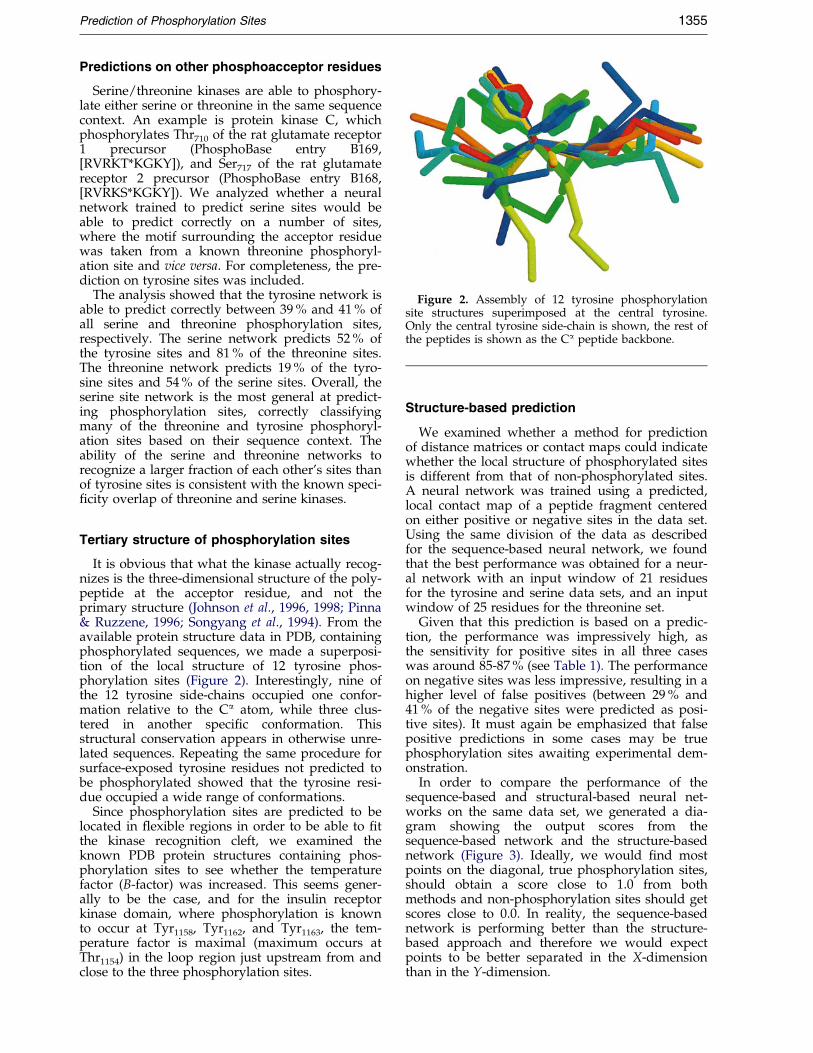
Figure 2. Assembly of 12 tyrosine phosphorylationsite structures superimposed at the central tyrosine.Only the central tyrosine side-chain is shown, the rest ofthe peptides is shown as the Ca peptide backbone.
Prediction of Phosphorylation Sites 1355
Predictions on other phosphoacceptor residues
Serine/threonine kinases are able to phosphory-late either serine or threonine in the same sequencecontext. An example is protein kinase C, whichphosphorylates Thr710 of the rat glutamate receptor1 precursor (PhosphoBase entry B169,[RVRKT*KGKY]), and Ser717 of the rat glutamatereceptor 2 precursor (PhosphoBase entry B168,[RVRKS*KGKY]). We analyzed whether a neuralnetwork trained to predict serine sites would beable to predict correctly on a number of sites,where the motif surrounding the acceptor residuewas taken from a known threonine phosphoryl-ation site and vice versa. For completeness, the pre-diction on tyrosine sites was included.
The analysis showed that the tyrosine network isable to predict correctly between 39 % and 41 % ofall serine and threonine phosphorylation sites,respectively. The serine network predicts 52 % ofthe tyrosine sites and 81 % of the threonine sites.The threonine network predicts 19 % of the tyro-sine sites and 54 % of the serine sites. Overall, theserine site network is the most general at predict-ing phosphorylation sites, correctly classifyingmany of the threonine and tyrosine phosphoryl-ation sites based on their sequence context. Theability of the serine and threonine networks torecognize a larger fraction of each other's sites thanof tyrosine sites is consistent with the known speci-®city overlap of threonine and serine kinases.
Tertiary structure of phosphorylation sites
It is obvious that what the kinase actually recog-nizes is the three-dimensional structure of the poly-peptide at the acceptor residue, and not theprimary structure (Johnson et al., 1996, 1998; Pinna& Ruzzene, 1996; Songyang et al., 1994). From theavailable protein structure data in PDB, containingphosphorylated sequences, we made a superposi-tion of the local structure of 12 tyrosine phos-phorylation sites (Figure 2). Interestingly, nine ofthe 12 tyrosine side-chains occupied one confor-mation relative to the Ca atom, while three clus-tered in another speci®c conformation. Thisstructural conservation appears in otherwise unre-lated sequences. Repeating the same procedure forsurface-exposed tyrosine residues not predicted tobe phosphorylated showed that the tyrosine resi-due occupied a wide range of conformations.
Since phosphorylation sites are predicted to belocated in ¯exible regions in order to be able to ®tthe kinase recognition cleft, we examined theknown PDB protein structures containing phos-phorylation sites to see whether the temperaturefactor (B-factor) was increased. This seems gener-ally to be the case, and for the insulin receptorkinase domain, where phosphorylation is knownto occur at Tyr1158, Tyr1162, and Tyr1163, the tem-perature factor is maximal (maximum occurs atThr1154) in the loop region just upstream from andclose to the three phosphorylation sites.
Structure-based prediction
We examined whether a method for predictionof distance matrices or contact maps could indicatewhether the local structure of phosphorylated sitesis different from that of non-phosphorylated sites.A neural network was trained using a predicted,local contact map of a peptide fragment centeredon either positive or negative sites in the data set.Using the same division of the data as describedfor the sequence-based neural network, we foundthat the best performance was obtained for a neur-al network with an input window of 21 residuesfor the tyrosine and serine data sets, and an inputwindow of 25 residues for the threonine set.
Given that this prediction is based on a predic-tion, the performance was impressively high, asthe sensitivity for positive sites in all three caseswas around 85-87 % (see Table 1). The performanceon negative sites was less impressive, resulting in ahigher level of false positives (between 29 % and41 % of the negative sites were predicted as posi-tive sites). It must again be emphasized that falsepositive predictions in some cases may be truephosphorylation sites awaiting experimental dem-onstration.
In order to compare the performance of thesequence-based and structural-based neural net-works on the same data set, we generated a dia-gram showing the output scores from thesequence-based network and the structure-basednetwork (Figure 3). Ideally, we would ®nd mostpoints on the diagonal, true phosphorylation sites,should obtain a score close to 1.0 from bothmethods and non-phosphorylation sites should getscores close to 0.0. In reality, the sequence-basednetwork is performing better than the structure-based approach and therefore we would expectpoints to be better separated in the X-dimensionthan in the Y-dimension.

Figure 3. Sequence (x-axis) and structure-based (y-axis) scores on tyrosine phosphorylation sites. True phosphoryl-ation sites are shown as red diamonds (210 sites), while non-phosphorylation sites are shown as blue crosses(940 sites).
1356 Prediction of Phosphorylation Sites
Interestingly, a few of the positive sites incor-rectly predicted as negative sites by the sequence-based method were correctly predicted by thestructural method. For the tyrosine data set, 17 outof 27 phosphorylation sites incorrectly classi®ed bythe sequence-based network were correctly classi-®ed by the structure-based network. Some of thesites having a very low sequence score, but a struc-ture score above the threshold of 0.5, include theautophosphorylation site Tyr1361 [EHIPY*THMN]of the insulin receptor, Tyr762 [QRSLY*DRPA] ofthe platelet-derived growth factor receptor andTyr826 [VGPGY*LGSG] of the Ret receptor tyrosinekinase. These sites do not match well with the typi-cal consensus features of tyrosine phosphorylationsites, such as an acidic region upstream of theacceptor residue or a strong hydrophobic motif atP � 1 and P � 3. Nevertheless, the predictions bythe structural network indicate that these tyrosinephosphorylation sites have structural featuresresembling those of other tyrosine phosphorylationsites.
Putative phosphorylation sites in p300/CBP
p300/CBP is a transcriptional adaptor that inter-acts with transcription factors and is also a histoneacetyltransferase (HAT) (Shikama et al., 1997; Ait-Si-Ali et al., 1998). Especially in the ®rst third andthe last third of the 2400� residues, p300/CBP pro-teins many protein-protein interactions have beenshown to occur, and some of these are believed tobe regulated by phosphorylation. Considering thelarge number of potential phosphoacceptor resi-dues in p300/CBP (CBP contains 394 serine, threo-
nine and tyrosine residues, 16 % of the totalnumber of residues) it would be helpful to identifythe most probable sites before conducting exper-iments involving site-directed mutagenesis or inanalysis of mass spectra of the modi®ed proteins.Using the prediction networks, we have foundputative phosphorylation sites. The amino acidsequences of p300 (accession number Q09472) andCBP (Q92793) are 63 % identical, which providedus with an opportunity to compare putative sitesin one protein with putative sites at the samealigned position in the other. We concentrated onhomologous sites in p300 and CBP with a strongprediction score above 0.9 (Table 2).
Several of the putative phosphorylation sites arecompletely conserved in both molecules, e.g.Tyr620, Ser1497, and Thr1684 (numbering refers top300). Interestingly, three sites utilize serine as theacceptor residue in one protein and threonine inthe other, e.g. Thr992, Ser1295 and Thr1533 (Table 2).
In the N-terminal third of the molecule, a con-served tyrosine site at position 620 (641 in CBP)may be of importance in the binding of CREB,since interaction in the region from positions 590-669 of CBP has been reported, see Shikama et al.(1997) and references therein.
The central part of the molecule (residues 800-1600) includes the catalytic domain of HAT activityof p300/CBP. The putative phosphorylation sitesfound in this region may be related to regulationof HAT activity.
The C-terminal part includes a region that inter-acts with viral oncoproteins E1A and SV40 large T,transcription factors c-Fos, c-Jun, JunB, YY1 amongothers, (Shikama et al., 1997) and references therein.

Table 2. Putative phosphorylation sites in p300/CBP
Protein Position NetPhos Score Acc.res. Sequence
P300 24 0.984 S SPALSASAS
CBP 23 0.987 S SPGFSANDS
p300 620 0.977 Y EGDMYESAN
CBP 641 0.977 Y EGDMYESAN
p300 959 0.946 S PSTSSTEVN
CBP 980 0.953 S SSVASAETN
p300 992 0.961 T EPADTQPED
CBP 1013 0.993 S DPGESKGEP
p300 1135 0.983 T YNRKTSRVY
CBP 1171 0.983 T YNRKTSRVY
p300 1289 0.991 S ENKFSAKRL
CBP 1325 0.975 S ENKFSAKRL
p300 1295 0.984 S KRLPSTRLG
CBP 1331 0.972 T KRLQTTRLG
p300 1346 0.988 S RFVDSGEMA
CBP 1382 0.993 S RFVDSGEMA
p300 1396 0.992 S RVYISYLDS
CBP 1432 0.988 S RVYISYLDS
p300 1446 0.979 Y ECDDYIFHC
CBP 1482 0.979 Y EGDDYIFHC
p300 1497 0.998 S DRLTSAKEL
CBP 1533 0.998 S DRLTSAKEL
p300 1516 0.993 S VLEESIKEL
CBP 1552 0.993 S VLEESIKEL
p300 1533 0.955 T REENTSNES
CBP 1568 0.987 S KKEESTAAS
p300 1684 0.956 T RWHCTVCED
CBP 1721 0.956 T RWHCTVCED
p300 1726 0.995 S AATQSPGDS
CBP 1763 0.995 S PQSKSPQES
p300 1734 0.993 S SRRLSIQRC
CBP 1771 0.992 S SRRVSIQRC
p300 1868 0.935 T TTPQTPQPT
CBP 1902 0.925 T STPQTPQPP
p300 2315 0.992 S QPVPSPRPQ
CBP 2351 0.935 S APVQSPRPQ
p300 2320 0.959 S PRPQSQPPH
CBP 2356 0.959 S PRPQSQPPH
p300 2325 0.984 S QPPHSSPSP
CBP 2361 0.984 S QPPH5SPSP
p300 2328 0.996 S HSSPSPRMQ
CBP 2364 0.993 S HSSPSPRIQ
NetPhos score is the output score from the ensemble of neural networks trained on that acceptorresidue type. The sequence shows the context of the acceptor residue � four residues.
Prediction of Phosphorylation Sites 1357
This region is phosphorylated by cyclin E-Cdk2,and thereby regulating the HAT activity of CBP(Ait-Si-Ali et al. , 1998). The region investigatedspanned residues 1890-2441 of CBP, therebyincluding the putative phosphorylation sites atThr1902, Ser2351, Ser2356, Ser2361 and Ser2364. Thesesites all contain an SP or SXP motif, the ®rst ofwhich is a well-known motif for Cdk-related pro-line-directed kinases.
Putative yin-yang sites in p300/CBP
The reversible and dynamic modi®cation of aparticular serine or threonine residue by eitherphosphorylation or GlcNac-glycosylation has beennamed yin-yang regulation (Hart et al., 1995). The
{ Accessible at http://www.cbs.dtu.dk/services/DictyOGlyc/
addition of the GlcNac sugar moiety prevents theacceptor residue from being phosphorylated andrepresents one way of inhibiting signals that mayotherwise cause abnormal growth or apoptosis.This mechanism is involved in regulation of thetumor suppressor protein p53 and the AP1 tran-scription factor complex.
Based on known sites of reciprocal phosphoryl-ation/GlcNac-glycosylation, we are currentlydeveloping a neural network-based method for theprediction of GlcNac sites on intracellular proteins(R. Gupta, unpublished results), analogous to themethods available for predicting GalNac-O-glyco-sylation sites (Hansen et al., 1998) and GlcNac-O-glycosylation sites in Dictyostelium discoideum pro-teins (R. Gupta et al., unpublished results). At pre-sent, the latter method, named DictyOGlyc { isprobably the method that most closely identi®essites with features similar to known intracellularGlcNac-sites.
1358 Prediction of Phosphorylation Sites
To investigate whether some of the putativephosphorylation sites in p300/CBP might also beregulated by GIcNac-glycosylation, we comparedthe predictions by the phosphorylation site neuralnetworks with the output from the DictyOGlyc ser-ver. The sites that have high scores from bothmethods and that have homologous sites in p300/CBP are listed in Table 3.
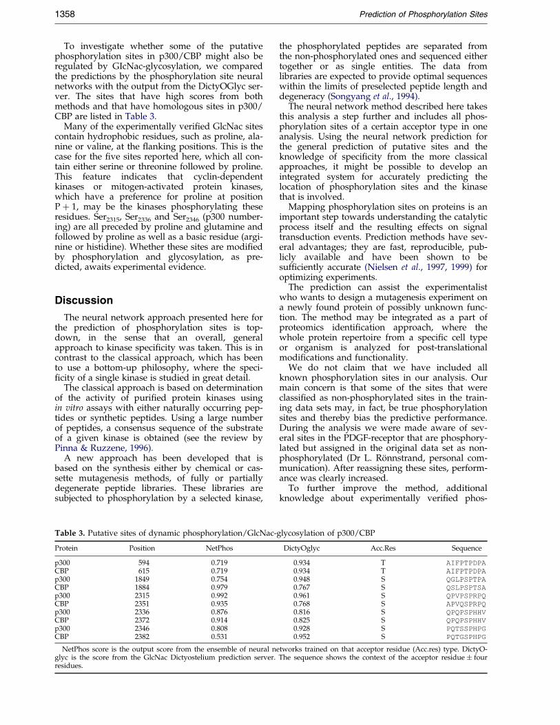
Many of the experimentally veri®ed GlcNac sitescontain hydrophobic residues, such as proline, ala-nine or valine, at the ¯anking positions. This is thecase for the ®ve sites reported here, which all con-tain either serine or threonine followed by proline.This feature indicates that cyclin-dependentkinases or mitogen-activated protein kinases,which have a preference for proline at positionP � 1, may be the kinases phosphorylating theseresidues. Ser2315, Ser2336 and Ser2346 (p300 number-ing) are all preceded by proline and glutamine andfollowed by proline as well as a basic residue (argi-nine or histidine). Whether these sites are modi®edby phosphorylation and glycosylation, as pre-dicted, awaits experimental evidence.
Discussion
The neural network approach presented here forthe prediction of phosphorylation sites is top-down, in the sense that an overall, generalapproach to kinase speci®city was taken. This is incontrast to the classical approach, which has beento use a bottom-up philosophy, where the speci-®city of a single kinase is studied in great detail.
The classical approach is based on determinationof the activity of puri®ed protein kinases usingin vitro assays with either naturally occurring pep-tides or synthetic peptides. Using a large numberof peptides, a consensus sequence of the substrateof a given kinase is obtained (see the review byPinna & Ruzzene, 1996).
A new approach has been developed that isbased on the synthesis either by chemical or cas-sette mutagenesis methods, of fully or partiallydegenerate peptide libraries. These libraries aresubjected to phosphorylation by a selected kinase,
Table 3. Putative sites of dynamic phosphorylation/GlcNac-g
Protein Position NetPhos
p300 594 0.719CBP 615 0.719p300 1849 0.754CBP 1884 0.979p300 2315 0.992CBP 2351 0.935p300 2336 0.876CBP 2372 0.914p300 2346 0.808CBP 2382 0.531
NetPhos score is the output score from the ensemble of neural neglyc is the score from the GlcNac Dictyostelium prediction server.residues.
the phosphorylated peptides are separated fromthe non-phosphorylated ones and sequenced eithertogether or as single entities. The data fromlibraries are expected to provide optimal sequenceswithin the limits of preselected peptide length anddegeneracy (Songyang et al., 1994).
The neural network method described here takesthis analysis a step further and includes all phos-phorylation sites of a certain acceptor type in oneanalysis. Using the neural network prediction forthe general prediction of putative sites and theknowledge of speci®city from the more classicalapproaches, it might be possible to develop anintegrated system for accurately predicting thelocation of phosphorylation sites and the kinasethat is involved.
Mapping phosphorylation sites on proteins is animportant step towards understanding the catalyticprocess itself and the resulting effects on signaltransduction events. Prediction methods have sev-eral advantages; they are fast, reproducible, pub-licly available and have been shown to besuf®ciently accurate (Nielsen et al., 1997, 1999) foroptimizing experiments.
The prediction can assist the experimentalistwho wants to design a mutagenesis experiment ona newly found protein of possibly unknown func-tion. The method may be integrated as a part ofproteomics identi®cation approach, where thewhole protein repertoire from a speci®c cell typeor organism is analyzed for post-translationalmodi®cations and functionality.
We do not claim that we have included allknown phosphorylation sites in our analysis. Ourmain concern is that some of the sites that wereclassi®ed as non-phosphorylated sites in the train-ing data sets may, in fact, be true phosphorylationsites and thereby bias the predictive performance.During the analysis we were made aware of sev-eral sites in the PDGF-receptor that are phosphory-lated but assigned in the original data set as non-phosphorylated (Dr L. RoÈnnstrand, personal com-munication). After reassigning these sites, perform-ance was clearly increased.
To further improve the method, additionalknowledge about experimentally veri®ed phos-
lycosylation of p300/CBP
DictyOglyc Acc.Res Sequence
0.934 T AIFPTPDPA
0.934 T AIFPTPDPA
0.948 S QGLPSPTPA
0.767 S QSLPSPTSA
0.961 S QPVPSPRPQ
0.768 S APVQSPRPQ
0.816 S QPQPSPHHV
0.825 S QPQPSPHHV
0.928 S PQTSSPHPG
0.952 S PQTGSPHPG
tworks trained on that acceptor residue (Acc.res) type. DictyO-The sequence shows the context of the acceptor residue � four
Prediction of Phosphorylation Sites 1359
phorylation sites will be needed. Many of the phos-phorylation sites described in the literature havenot yet been included in PhosphoBase, and therebynot in this analysis. A search in Medline for thekeyword phosphorylation yields 69,286 hits (phos-phorylation AND site yields 9044 hits; October1999). A reasonable estimate of the number ofpapers reporting novel phosphorylation sites maybe of the order of 1000-5000. Thus, the continuousincrease of phosphorylation site entries in Phos-phoBase will make it likely that further improve-ment of the prediction method can be achieved.
In addition to predicting putative phosphoryl-ation sites, it would be of great value to get a hintas to which kinase is likely to interact at this par-ticular site. We are currently considering differentapproaches to achieve this goal. The most obviousapproach is a simple alignment against extendedversions of the known phosphorylation sites inPhosphoBase, from which the phosphorylation siteannotation might provide information about thekinase or other interaction partners. A moreadvanced approach is to generate sequence pro®lesfor all phosphorylation sites being modi®ed by acertain kinase. A sequence pro®le describes, foreach position, the probability of ®nding each of the20 amino acids. Predicting the most probablekinase for a novel phosphorylation site would thenbe a matter of aligning the sequence against thekinase sequence pro®les and report the best match.The same type of approach could be used to pre-dict functional domains interacting with phosphor-esidues, such as SH2 and PcTB domains.
A third approach would involve training a newneural network to classify substrate sites of knownkinases. For example, PhosphoBase contains infor-mation about 160 sites for protein kinase A (PKA),174 sites for PKC and 25 sites for the EGF-receptortyrosine kinase. This method will then beimplemented as a post-processing function to theexisting phosphorylation site prediction method.Thus, the initial putative sites predicted in thenovel protein will be fed to the kinase-classifyingnetwork, which will then output a prediction ofthe most probable kinase.
Another very important constraint of the highlycompartmentalized eukaryotic cell is that manypotential protein-protein interactions may nevertake place because of topological factors, e.g. oneprotein being membrane-bound, and the otherlocalized in the nucleus. The role of compartmenta-lized processes for the regulation of the signaltransduction kinase cascades has been shown to bevery important for the correct target-substrateinteractions to occur. During speci®c phases of thecell cycle, several kinases and phosphatases occurin speci®c cellular compartments, such as cyto-plasmic or nuclear/chromosomal regions (Inagakiet al., 1994). Thus, methods available for predictingprotein cellular localization might aid in decidingwhich of the putative sites are most biologicallyrelevant (Andrade et al., 1998; Chou & Elrod, 1999;Nakai & Horton, 1999).
Materials & Methods
Data sets extracted from PhosphoBase
Experimentally veri®ed phosphorylation sites wereextracted mainly from PhosphoBase (Kreegipuu et al.,1999), which is available from http://www.cbs.dtu.dk/databases/PhosphoBase/.
The phosphoproteins were mostly from mammaliansources, with a few examples from viruses or plants. Thedata set consisted of 584 serine sites (251 protein entries),108 threonine sites (85 protein entries), and 210 tyrosinesites (98 protein entries). No sites were identical within a9-mer sequence. Negative examples of phosphorylationsites were assigned by two approaches.
(1) The standard approach. For each of the threeacceptor types, a subset of the protein entries were cate-gorized as being well characterized. All acceptor resi-dues in the selected subsets, not reported as beingphosphorylated, were assigned as negative sites.
(2) The augmented approach. All acceptor residues inthe entire set of protein entries not reported as beingphosphorylated, were assigned as negative sites a priori.Subsequently, during initial neural network training ses-sions, all negative sites predicted as positive sites wereexcluded. The resulting data set thus obtained was usedfor the ®nal neural network training sessions.
Sequence logos
Sequence logos were used for displaying the position-speci®c features of complex sequence alignments asdescribed earlier (Schneider & Stephens, 1990; Blom et al.,1996). Since phosphorylation sites are quite divergent,sequence logos are better suited at emphasizing the con-served positions than a multiple alignment.
Neural networks and cross-validation
The neural networks were of the standard feed-for-ward type (Minsky & Papert, 1988; Hertz et al., 1991).Details of sequence encoding, error functions, etc., maybe found elsewhere (Blom et al., 1996). The predictiveperformance was monitored using the Mathews corre-lation coef®cient (Mathews, 1975) during training andtest of the networks.
Phylogenetic trees, indicating the relationship betweenthe proteins in each of the three data sets (serine, threo-nine or tyrosine), were constructed using multiple align-ments and neighbour-joining algorithms of the ClustalWpackage (Thompson et al., 1994) and visualized using theDrawtree program of the Phylip package (Felsenstein,1989), as illustrated for the tyrosine data set (Figure 4).Based on the trees, each of the three data sets wasdivided into ®ve parts in order to allow for cross-vali-dated testing. This procedure used four of the ®ve sub-groups for training, while the last subgroup (non-sequence similar to the training sets) was used for testingthe performance. Five different networks resulted fromthis approach for each of the three phosphoresidues.
The subgroups included related proteins like e.g. theMAP-kinase family or the Src-related tyrosine kinases(Hck, Blk, Abl, etc.), ensuring that test performance wasmeasured on proteins non-homologous to the trainingset.
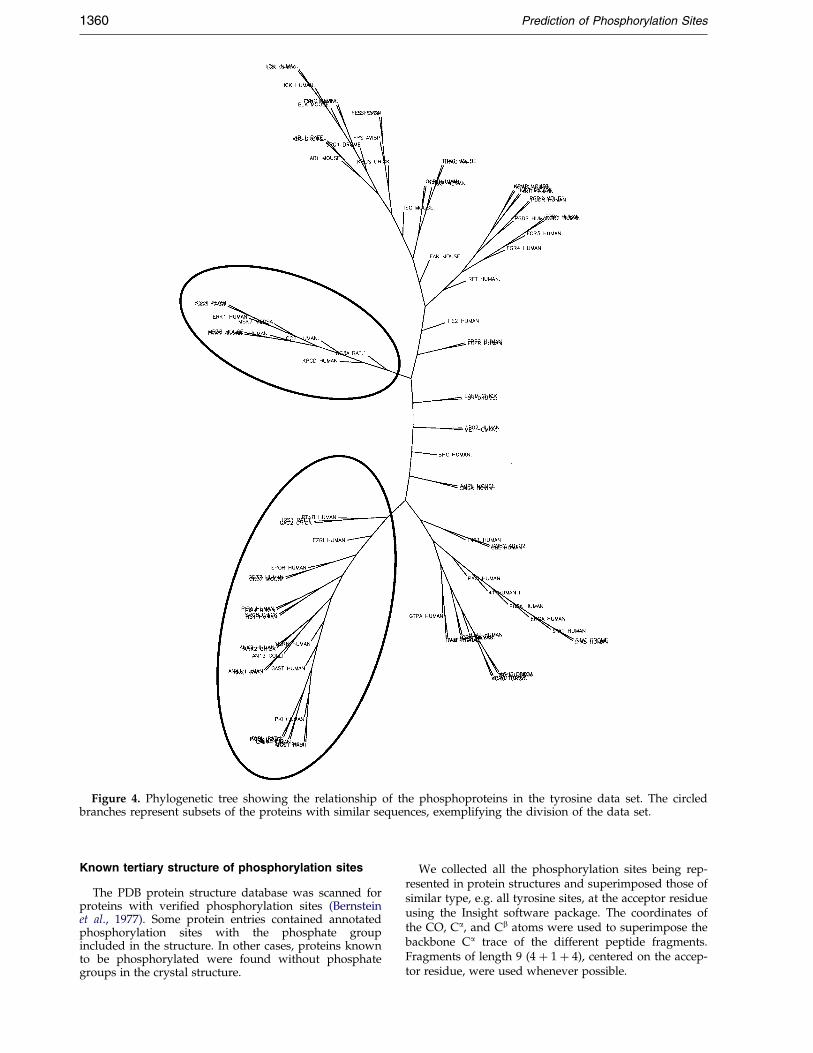
Figure 4. Phylogenetic tree showing the relationship of the phosphoproteins in the tyrosine data set. The circledbranches represent subsets of the proteins with similar sequences, exemplifying the division of the data set.
1360 Prediction of Phosphorylation Sites
Known tertiary structure of phosphorylation sites
The PDB protein structure database was scanned forproteins with veri®ed phosphorylation sites (Bernsteinet al., 1977). Some protein entries contained annotatedphosphorylation sites with the phosphate groupincluded in the structure. In other cases, proteins knownto be phosphorylated were found without phosphategroups in the crystal structure.
We collected all the phosphorylation sites being rep-resented in protein structures and superimposed those ofsimilar type, e.g. all tyrosine sites, at the acceptor residueusing the Insight software package. The coordinates ofthe CO, Ca, and Cb atoms were used to superimpose thebackbone Ca trace of the different peptide fragments.Fragments of length 9 (4 � 1 � 4), centered on the accep-tor residue, were used whenever possible.

Prediction of Phosphorylation Sites 1361
Neural network based on local tertiary structure
A neural network predicting Ca contact maps for pep-tide sequence inputs was used on peptide fragmentsfrom the data sets, up to a length of 33 residues centeredat the phosphorylation sites (Lund et al., 1997). For eachpair of residues in the input sequence (e.g. residues i andi � 5), the output from this method gives a probabilityscore indicating the distance between the two residuescompared to the average distance for all pairs at thissequence separation. The output was processed in orderto extract probabilities pertaining to 9, 13, 17, 21 or 25residues centered on the phosphoresidue. The same pro-cedure was performed for the non-phosphorylation sites.The resulting data sets consisted of a number of prob-ability parameters for each peptide (indirectly de®ningits local structure) for different fragment sizes.
Electronic access
The prediction method can be accessed on the Internetat http://www.cbs.dtu.dk/services/NetPhos/ or viae-mail by sending the word `help' to [email protected].
The PhosphoBase database is also available on theInternet at http://www.cbs.dtu.dk/databases/Phospho-Base/.
Acknowledgments
We thank Kristoffer Rapacki for competent computerassistance and Jan Hansen for friendly and helpful dis-cussions. This work was supported by the DanishNational Research Foundation.
References
Ait-Si-Ali, S., Ramirez, S., Barre, F., Dkhissi, F.,Magnaghi-Jaulin, L., Girault, J., Robin, P.,Knibiehler, M., Pritchard, L., Ducommun, B.,Trouche, D. & Harel-Bellan, A. (1998). Histoneacetyl-transferase activity of CBP is controlled bycycle- dependent kinases and oncoprotein E1A.Nature, 396, 184-186.
Andrade, M., O'Donoghue, S. & Rost, B. (1998). Adap-tation of protein surfaces to subcellular location.J. Mol. Biol. 276, 517-525.
Bairoch, A., Bucher, P. & Hofmann, K. (1997). The PRO-SITE database, its status in 1997. Nucl. Acids Res.25, 217-221.
Baldi, P. & Brunak, S. (1998). Bioinformatics: The MachineLearning Approach, MIT Press, Cambridge, MA.
Bernstein, F. C., Koetzle, T. F., Williams, G. J. B., Jr, E,F. M., Brice, M. D., Rodgers, J. R., Kennard, O.,Shimanouchi, T. & Tasumi, M. (1977). The ProteinData Bank: a computer-based archival ®le formacromolecular structures. J. Mol. Biol. 112, 535-542.
Blom, N., Hansen, J., Blaas, D. & Brunak, S. (1996). Clea-vage site analysis in picornaviral polyproteins: dis-covering cellular targets by neural networks. ProteinSci. 5, 2203-2216.
Chou, K. & Elrod, D. (1999). Protein subcellular locationprediction. Protein Eng. 12, 107-118.
Dale, S., Wilson, W. A., Edelman, A. M. & Hardie, D. G.(1995). Similar substrate recognition motifs formammalian amp-activated protein kinase, higher
plant hmg-coa reductase kinase-a, yeast snf1, andmammalian calmodulin-dependent protein kinase i.FEBS Letters, 361, 191-195.
Felsenstein, J. (1989). Phylogeny inference package (ver-sion 3.2). Cladistics, 5, 164-166.
Graves, L., Bornfeldt, K. & Krebs, E. (1997). Historicalperspectives and new insights involving the MAPkinase cascades. Advan. Sec. Mess. Phos. Res. 31, 49-62.
Hansen, J. E., Lund, O., Tolstrup, N., Gooley, A. A.,Williams, K. L. & Brunak, S. (1998). NetOglyc: pre-diction of mucin type O-glycosylation sites basedon sequence context and surface accessibility. Glyco-conj. J. 15, 115-130.
Hart, G. (1997). Dynamic O-linked glycosylation ofnuclear and cytoskeletal proteins. Annu. Rev. Bio-chem. 66, 315-335.
Hart, G., Greis, K., Dong, L., Blomberg, M., Chou, T.,Jiang, M., Roquemore, E., Snow, D., Kreppel, L. &Cole, R. (1995). O-Linked N-acetylglucosamine: theyin-yang of Ser/Thr phosphorylation? Nuclear andcytoplasmic glycosylation. Advan. Exp. Med. Biol.376, 115-123.
Hertz, J., Krogh, A. & Palmer, R. (1991). Introduction tothe Theory of Neural Computation, Addison-Wesley,Redwood City, CA.
Hunter, T. (1998). The Croonian Lecture 1997. The phos-phorylation of proteins on tyrosine: its role in cellgrowth and disease. Phil. Trans. Roy. Soc. ser. B, 353,583-605.
Inagaki, N., Ito, M., Nakano, T. & Inagaki, M. (1994).Spatiotemporal distribution of protein kinase andphosphatase activities. Trends Biochem. Sci. 19, 448-452.
Johnson, L., Noble, M. & Owen, D. (1996). Active andinactive protein kinases: structural basis for regu-lation. Cell, 85, 149-158.
Johnson, L., Lowe, E., Noble, M. & Owen, D. (1998).The eleventh datta lecture. the structural basis forsubstrate recognition and control by proteinkinases. FEBS Letters, 430, 1-11.
Kolibaba, K. & Druker, B. (1997). Protein tyrosinekinases and cancer. Biochim. Biophys. Acta, 1333,F217-F248.
Kreegipuu, A., Blom, N. & Brunak, S. (1999). Phospho-Base, a database of phosphorylation sites: release2.0. Nucl. Acids Res. 27, 237-239.
Lund, O., Frimand, K., Gorodkin, J., Bohr, H., Bohr, J.,Hansen, J. & Brunak, S. (1997). Protein distance con-straints predicted by neural networks and prob-ability density functions. Protein Eng. 10, 1241-1248.
Mathews, B. (1975). Comparison of the predicted andobserved secondary structure of T4 phage lyso-zyme. Biochim. Biophys. Acta, 405, 442-451.
Minsky, M. & Papert, S. (1969, 1988). Perceptrons, MITPress, Cambridge, MA.
Nakai, K. & Horton, P. (1999). PSORT: a program fordetecting sorting signals in proteins and predictingtheir subcellular localization. Trends Biochem. Sci. 24,34-36.
Nielsen, H., Brunak, S. & von Heijne, G. (1999). Machinelearning approaches for the prediction of signalpeptides and other protein sorting signals. ProteinEng. 12, 3-9.
Nielsen, H., Engelbrecht, J., Brunak, S. & von Heijne, G.(1997). Identi®cation of prokaryotic and eukaryoticsignal peptides and prediction of their cleavagesites. Protein Eng. 10, 1-6.
1362 Prediction of Phosphorylation Sites
Patschinsky, T., Hunter, T., Esch, F. S., Cooper, J. A. &Sefton, B. M. (1992). Analysis of the sequence ofamino acids surrounding sites of tyrosine phos-phorylation. Proc. Natl Acad. Sci. USA, 79, 973-977.
Pawson, T. (1995). Protein modules and signalling net-works. Nature, 373, 573-80.
Pinna, L. A. & Ruzzene, M. (1996). How do proteinkinases recognize their substrates? Biochim. Biophys.Acta, 1314, 191-225.
Schneider, T. D. & Stephens, R. M. (1990). Sequencelogos: a new way to display consensus sequences.Nucl. Acids Res, 18, 6097-6100.
Shikama, N., Lyon, J. & NB, L. T. (1997). The p300/CBPfamily: integrating signals with transcription factorsand chromatin. Trends Cell Biol. 7, 230-236.
Songyang, Z., Shoelson, S. E., Chaudhuri, M., Gish, G.,Pawson, T., Haser, W. G., King, F., Roberts, T.,Ratnofsky, S., Lechleider, R. J., Neel, B. G., Birge,R. B., Fajardo, J. E., Chou, M. M. & Hanafusa, H.,et al. (1993). SH2 domains recognize speci®c phos-phopeptide sequences. Cell, 72, 767-778.
Songyang, Z., Blechner, S., Hoagland, N., Hoekstra,M. F., Piwnica-Worms, H. & Cantley, L. C. (1994).Use of an oriented peptide library to determine theoptimal substrates of protein kinases. Curr. Biol. 4,973-982.
Thompson, J., Higgins, D. & Gibson, T. (1994). CLUS-TAL W: improving the sensitivity of progressivemultiple sequence alignment through sequenceweighting, position-speci®c gap penalties andweight matrix choice. Nucl. Acids Res. 22, 4673-4680.
Tinker, D. A., Krebs, E. A., Feltham, I. C., Attah-Poku,S. K. & Ananthanarayanan, V. S. (1988). Syntheticbeta-turn peptides as substrates for a tyrosine pro-tein kinase. J. Biol. Chem. 263, 5024-5026.
Wilson, I. B. H., Gavel, Y. & von Heijne, G. (1991).Amino acid distributions around O-linked glycosy-lation sites. Biochem. J. 275, 529-534.
Wu, C. H. (1997). Arti®cial neural networks for molecu-lar sequence analysis. Comput. Chem. 21, 237-256.
Zhou, S. & Cantley, L. (1995). Recognition and speci-®city in protein tyrosine kinase-mediated signalling.Trends Biochem. Sci. 20, 470-475.
Edited by F. E. Cohen
(Received 16 July 1999; received in revised form 12 October 1999; accepted 12 October 1999)