Evidence for an unfolded border-collision bifurcation in paced cardiac tissue

Intracellular Eukaryotic Parasites Have a DistinctUnfolded Protein ResponseSara J. C. Gosline1,2,3., Mirna Nascimento4., Laura-Isobel McCall4, Dan Zilberstein5, David Y. Thomas6,
Greg Matlashewski4*, Michael Hallett1,2,3,6*
1 McGill Centre for Bioinformatics, McGill University, Montreal, Quebec, Canada, 2 McGill School of Computer Science, McGill University, Montreal, Quebec, Canada,
3 Rosalind and Morris Goodman Cancer Centre, McGill University, Montreal, Quebec, Canada, 4 Department of Microbiology and Immunology, McGill University, Montreal,
Quebec, Canada, 5 Faculty of Biology, Technion-Israel Institute of Technology, Haifa, Israel, 6 Department of Biochemistry, McGill University, Montreal, Quebec, Canada
Abstract
Insult to the endoplasmic reticulum (ER) activates the Unfolded Protein Response (UPR), a set of signaling pathways thatprotect the cell from the potential damage caused by improperly folded proteins. Accumulation of misfolded proteins in theER lumen initiates a series of signal transduction events via activation of three transmembrane ER proteins: Ire1, Atf6 andPERK. Activation of these proteins results in the transcriptional up-regulation of the components of the folding, traffickingand degradation machinery in the ER. PERK further reduces the load on the ER via the phosphorylation of eIF2a, attenuatinggeneral protein translation. It is believed that the UPR evolved as a transcriptional response that up-regulates proteinfolding machinery in the ER and later gained the ability to decrease ER load by attenuating general protein translation inmetazoa. However, our in silico analyses of protozoan parasites revealed an absence of proteins involved in thetranscriptionally mediated UPR and the presence of both PERK and its target eIF2a. Consistent with these observations,stimulation of the UPR in Leishmania donovani identified an absence of up-regulation of the ER chaperone BiP, the canonicalER chaperone modulated by the UPR in higher eukaryotes, while exhibiting increased phosphorylation of eIF2a which hasbeen shown to attenuate protein translation. We further observed that L. donovani is more sensitive to UPR inducing agentsthan host macrophages, suggesting that the less evolved stress response could provide a new avenue for therapeutictreatment of parasitic infections.
Citation: Gosline SJC, Nascimento M, McCall L-I, Zilberstein D, Thomas DY, et al. (2011) Intracellular Eukaryotic Parasites Have a Distinct Unfolded ProteinResponse. PLoS ONE 6(4): e19118. doi:10.1371/journal.pone.0019118
Editor: Volker Theo Heussler, University of Bern, Switzerland
Received November 19, 2010; Accepted March 25, 2011; Published April 29, 2011
Copyright: � 2011 Gosline, et al. This is an open-access article distributed under the terms of the Creative Commons Attribution License, which permitsunrestricted use, distribution, and reproduction in any medium, provided the original author and source are credited.
Funding: This research was supported by the Canadian Institutes of Health Research (DYT, GM, MN), the Natural Sciences and Engineering Research Council ofCanada (Discovery Grant, MH; PGS D, SJCG), and by grant number 33928 from the Israel Ministry of Health Chief Scientist Foundation (DZ). The funders had norole in study design, data collection and analysis, decision to publish, or preparation of the manuscript.
Competing Interests: The authors have declared that no competing interests exist.
* E-mail: [email protected] (GM); [email protected] (MH)
. These authors contributed equally to this work.
Introduction
The Unfolded Protein Response (UPR) is a set of signaling
pathways that protect the cell from stress imposed on the
endoplasmic reticulum (ER). In metazoa, the accumulation of
misfolded proteins in the ER causes the chaperone BiP to
disassociate from and subsequently activate three signal transduc-
ers: Ire1, PERK and Atf6. Figure 1A shows the signaling pathways
initiated by each protein. Inositol Requiring 1 (Ire1a and Ire1b) is
a transmembrane kinase/ribonuclease that induces the non-
conventional splicing of X box Binding Protein 1 (XBP1, HAC1
in yeast) mRNA. This splicing increases the amount of Xbp1p
transcription factor which leads to the up-regulation of protein
chaperones, most notably BiP and Protein Disulfide Isomerase
(PDI) [1]. PRKR-like Endoplasmic Reticulum Kinase (PERK)
phosphorylates the a subunit of eIF2, which causes global
translation attenuation by preventing the formation of the 80S
complex at the AUG initiator codon [2]. Phosphorylated eIF2aselectively increases the translation of Atf4, a basic-leucine zipper
(bZIP) transcription factor that up-regulates ER-resident chaper-
ones [1,2]. Activating Transcription Factor 6-like proteins (Atf6a,
Atf6b, CREB3L2) transcriptionally initiate a gene expression
program that includes cell cycle arrest [3]. Together, the inhibition
of protein synthesis (by PERK activation) combined with the
increase in ER chaperone production (including that of BiP)
decrease the accumulation of unfolded proteins in the ER. While
all UPR pathways have been implicated in many diseases [4,5],
the individual pathways have been shown to act independently
when faced with varying kinds of stress [6,7].
Knowing how pathways have evolved can provide valuable
insight into their individual function. However, the evolution of
the UPR, depicted as a dendrogram in Figure 1B, is not
straightforward. The assumption that the Ire1 pathway is the
most ancient of the UPR [8,9] conflicts with evidence of Atf6 in
plant [10] and PERK in Apicomplexa [11]. Also, the apparent
absence of a transcriptional response in Giardia lamblia [12] and
Trypanosoma brucei [13] suggests that the transcriptional response
may be absent in many protozoan parasites, though T. brucei is
able to mount a UPR-like response through an organism-specific
form of mRNA regulation when treated with very high amounts of
UPR-inducing chemicals [14]. Though PERK has been identified
in T. brucei [15], it was found to reside outside the ER in the
PLoS ONE | www.plosone.org 1 April 2011 | Volume 6 | Issue 4 | e19118

flagellar pocket. Thus, despite the sequence similarity this protein
is unable to act as a functional ortholog because it cannot sense ER
stress. While there is evidence of a PERK-based translational UPR
in Toxoplasma gondii [11], the apparent absence of this pathway in
T. brucei makes it difficult to assume that this pathway exists in
other protozoan parasites.
In this study, we developed a computational model to charac-
terize the UPR across eukaryotes that is able to identify a PERK
associated pathway and confirm the absence of a UPR transcrip-
tional response in some protozoa. We validated this model by in vivo
induction of the UPR in cultured Leishmania donovani (L. donovani) to
measure BiP chaperone levels and eIF2a phosphorylation during
experimentally-induced ER stress. We further examined whether L.
donovani was more sensitive to ER stress than host macrophages. Our
results suggest L. donovani possess a translationally mediated UPR
pathway but no change in UPR-specific protein expression, making
it more sensitive to ER stress-inducing drugs than its host. The
computational model indicates that a transcriptionally-mediated
UPR may be absent across parasitic protozoa, suggesting that ER
stress could be a therapeutic target in these organisms. Furthermore,
our approach can be used to identify other cyto-protective
metazoan pathways in an effort to identify new therapeutic targets
in parasitic infections.
Results
Domain analysis shows absence of UPR-specifictranscriptional machinery
The use of standard ortholog detection tools (see Materials and
Methods) was unable to identify many important UPR proteins in
protozoan parasites including those required for transcriptional
regulation of UPR targets (shown in Tables 1 and S1). To
investigate whether the transcriptional machinery existed in a
highly diverged form (e.g. metazoan Xbp1 and yeast Hac1[16]),
we developed a computational model that relies on conservation of
protein domains instead of entire amino acid sequences. We
collected domains from all proteins involved in the UPR across the
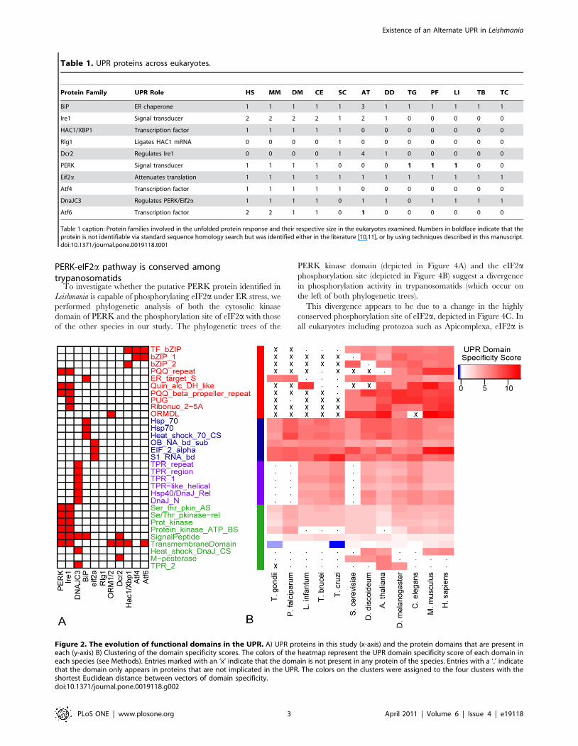
12 eukaryotes in this study. Figure 2A shows the UPR proteins and
their respective domains (collected from Interpro [17]). We
calculated the specificity value (see Methods) to measure how
frequently each domain occurs in UPR proteins relative to non-
UPR proteins. Hierarchical clustering of these values shown in
Figure 2B reveals clusters of protein domains that have evolved
similarly across eukaryotes, indicated by the colors of each cluster
of domains.
The blue and purple clusters include domains that occur within
well-conserved proteins such as BiP, eIF2a and DNAJC3 while the
green cluster contains domains that have lower specificity values as
they reside within a diverse array of proteins across the cell. The
red cluster is enriched in protein domains that are present in all
species but absent in the protozoan parasites. Surprisingly, these
domains are specifically those required in the UPR transcriptional
response: the ribonuclease and PUG domains found exclusively
within Ire1 and the bZIP domains found in XBP1, Atf6 and Atf4
transcription factors. At least one of these domains is required for
proper transcriptional binding to initiate the canonical UPR
transcriptional response. While sporadic absence of some domains
is expected across such a diverse set of species, the wholesale
absence of functionally similar domains suggests that the entire
pathway could be absent in protozoan parasites.
Naive Bayes’ classifier identifies a putative PERK in lowereukaryotes
Because we did not see an absence of domains required for the
PERK pathway and there has been evidence of this pathway in T.
gondi [11], we developed a naıve Bayes’ classifier to search for
PERK in L. donovani (and other missing proteins, see Methods). We
were able to identify several putative PERK proteins. We then
aligned the kinase domains of these proteins to known eIF2akinase structures in the Protein Data Bank (PDB) [18] (see
Methods) using PSI-BLAST [19] to arrive at a single protein in L.
infantum, T. cruzi, and T. brucei, the latter of which has been
characterized in [15] (Table S2). Consistent with previous findings
in T. gondii [11], we identified two such proteins in P. falciparum,
although none were identified in D. discoideum. Figure 3 shows the
domain structure of the PERK identified in L. infantum compared
with the domain structure of validated PERK orthologs in human,
mouse, C. elegans and T. gondii. The full alignment of L. infantum
PERK with other known and putative PERK molecules is
depicted in Figure S2.
Figure 1. The Unfolded Protein Response across eukaryotesprior to this study. A) The accumulation of unfolded proteins (left)causes BiP (in red) to disassociate from the three transmembraneproteins leading to their subsequent activation: activation of Ire1 (blue)leads to the splicing of XBP1 (or HAC1 in yeast) mRNA; activation ofPERK (yellow) results in the phosphorylation of eIF2a which in turnattenuates general cellular translation and up-regulates Atf4 (green);activation of Atf6 (orange) causes it to travel first to the Golgi where it iscleaved and then to the nucleus. Xbp1, Atf4 and Atf6 enter the nucleuswhere they bind to specific UPR Element (UPRE) binding motifs and up-regulate hundreds of proteins such as BiP. B) Phylogenetic tree of the12 organisms in this study. Colors of the branches indicate that theproteins with the same color in (A) are present in that organism.Checkered branches indicate results determined by this study.doi:10.1371/journal.pone.0019118.g001
Existence of an Alternate UPR in Leishmania
PLoS ONE | www.plosone.org 2 April 2011 | Volume 6 | Issue 4 | e19118
PERK-eIF2a pathway is conserved amongtrypanosomatids
To investigate whether the putative PERK protein identified in
Leishmania is capable of phosphorylating eIF2a under ER stress, we
performed phylogenetic analysis of both the cytosolic kinase
domain of PERK and the phosphorylation site of eIF2a with those
of the other species in our study. The phylogenetic trees of the
PERK kinase domain (depicted in Figure 4A) and the eIF2aphosphorylation site (depicted in Figure 4B) suggest a divergence
in phosphorylation activity in trypanosomatids (which occur on
the left of both phylogenetic trees).
This divergence appears to be due to a change in the highly
conserved phosphorylation site of eIF2a, depicted in Figure 4C. In
all eukaryotes including protozoa such as Apicomplexa, eIF2a is
Table 1. UPR proteins across eukaryotes.
Protein Family UPR Role HS MM DM CE SC AT DD TG PF LI TB TC
BiP ER chaperone 1 1 1 1 1 3 1 1 1 1 1 1
Ire1 Signal transducer 2 2 2 2 1 2 1 0 0 0 0 0
HAC1/XBP1 Transcription factor 1 1 1 1 1 0 0 0 0 0 0 0
Rlg1 Ligates HAC1 mRNA 0 0 0 0 1 0 0 0 0 0 0 0
Dcr2 Regulates Ire1 0 0 0 0 1 4 1 0 0 0 0 0
PERK Signal transducer 1 1 1 1 0 0 0 1 1 1 0 0
Eif2a Attenuates translation 1 1 1 1 1 1 1 1 1 1 1 1
Atf4 Transcription factor 1 1 1 1 1 0 0 0 0 0 0 0
DnaJC3 Regulates PERK/Eif2a 1 1 1 1 0 1 1 0 1 1 1 1
Atf6 Transcription factor 2 2 1 1 0 1 0 0 0 0 0 0
Table 1 caption: Protein families involved in the unfolded protein response and their respective size in the eukaryotes examined. Numbers in boldface indicate that theprotein is not identifiable via standard sequence homology search but was identified either in the literature [10,11], or by using techniques described in this manuscript.doi:10.1371/journal.pone.0019118.t001
Figure 2. The evolution of functional domains in the UPR. A) UPR proteins in this study (x-axis) and the protein domains that are present ineach (y-axis) B) Clustering of the domain specificity scores. The colors of the heatmap represent the UPR domain specificity score of each domain ineach species (see Methods). Entries marked with an ‘x’ indicate that the domain is not present in any protein of the species. Entries with a ‘.’ indicatethat the domain only appears in proteins that are not implicated in the UPR. The colors on the clusters were assigned to the four clusters with theshortest Euclidean distance between vectors of domain specificity.doi:10.1371/journal.pone.0019118.g002
Existence of an Alternate UPR in Leishmania
PLoS ONE | www.plosone.org 3 April 2011 | Volume 6 | Issue 4 | e19118

phosphorylated at Ser51 (highlighted in black) which is surrounded
by a highly conserved motif [20]. However, when eukaryotic
phosphorylation sites are aligned with the putative L. infantum
phosphorylation site, there is a threonine (highlighted in grey) in
place of the serine (Figure 4C) suggesting that Thr166 is phosphory-
lated in this species. Additionally, the conserved amino acids in
close proximity to Thr166 differ from higher eukaryotes; leucine
and methionine upstream of Thr166 are replaced by proline and
tyrosine and the leucine downstream of Thr166 has been replaced
by valine. Lastly, while eIF2a contains ,340 amino acids in most
eukaryotes, copies of the protein identified in three Leishmania
species were each over 400 amino acids in length.
Despite this divergence, recent studies in both T. brucei [15] and
L. donovani [21] reported that eIF2a is phosphorylated at Thr169
and Thr166 in both species respectively. This phosphorylation was
shown to decrease protein translation in both organisms [15,21].
BiP protein levels in L. donovani do not change inresponse to UPR stress
While the absence of transcriptional control is not uncommon
among trypanosomatids [13,22], recent evidence of a UPR
mediated via post-transcriptional mRNA regulation in T. brucei
[14] raised the question of whether or not Leishmania species could
mount a UPR at the protein level despite the absence of
transcriptional regulation. To investigate this, we examined BiP
protein levels in response to treatment by tunicamycin and
dithiothreitol (DTT), two compounds commonly used to induce
the UPR [1]. BiP is known to be highly up-regulated by the UPR
transcriptional response across metazoan and plants [23] and up-
regulated post-transcriptionally in T. brucei [14]. We included
tubulin and A2 proteins as controls. While the precise role of A2 is
unknown, it is present in low levels in cultured promastigotes and
has been shown to be expressed in response to cellular stress, such
as increased temperature, as well as in response to stimulation of
differentiation [24,25,26].
As shown in the Western blot in Figure 5A, BiP and tubulin
protein levels in L. donovani did not change in response to treatment
with tunicamycin or DTT. As expected, BiP protein levels in host
cell macrophages increased in response to DTT treatment
(Figure 5B). In comparison, DTT induced expression of the A2
family of proteins in promastigotes to levels similar to those
observed in heat differentiated axenic amastigotes indicating that
the chemical is causing stress to the organism (Figure 5C). The
weak induction of A2 proteins by tunicamycin indicates that this
chemical does not cause high levels of stress in cultured L. donovani
promastigotes. A2 proteins migrate as a ladder in SDS-PAGE
since it is a multigene family in L. donovani where each member has
a different number of a ten amino acid repeat sequences [24].
Because L. donovani cultures failed to proliferate at the concentra-
tions of DTT used to induce a change in BiP protein expression in
T. brucei[14] (shown in Figure S3, Panel A) A2 served as a way to
illustrate that DTT was inducing a stress response in L. donovani.
The results demonstrate that DTT induced a stress response in L.
donovani, as determined by increased A2 protein expression, but
this did not result in the induction of BiP.
Phosphorylation of eIF2a upon UPR induction providesevidence of translational control originating from the ERin Leishmania
To assess whether ER stress activates eIF2a phosphorylation in
L. donovani, we used nano-LC-multiple reaction monitoring
(MRM)-MS ([21], see Methods) to measure eIF2a phosphoryla-
tion levels in promastigotes exposed to DTT and tunicamycin. In
this method, an aliquot of a heavy phosphopeptide isotope was
used to identify and quantitate the level of phosphorylated Thr166
in L. donovani promastigotes (see Methods). This experiment
Figure 3. PERK domain structure. The domain structure of the PERK-like protein identified in L. infantum compared to validated PERK proteins inHuman, Mouse, C. elegans and T. gondii. The legend describing the protein domains is below.doi:10.1371/journal.pone.0019118.g003
Existence of an Alternate UPR in Leishmania
PLoS ONE | www.plosone.org 4 April 2011 | Volume 6 | Issue 4 | e19118

Existence of an Alternate UPR in Leishmania
PLoS ONE | www.plosone.org 5 April 2011 | Volume 6 | Issue 4 | e19118

revealed increased phosphorylation of eIF2a upon DTT treatment
(Figure 4D). Together with the high degree of similarity with the
T. brucei PERK-eIF2a pathway [15] and direct evidence that this
phosphorylation attenuates translation in L. donovani [21], this
increased phosphorylation illustrates that ER stress activates a
PERK-eIF2a associated translational attenuation pathway in these
organisms.
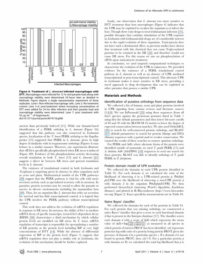
Induction of the UPR by DTT leads to reduced viability ofLeishmania donovani
To determine if the UPR in Leishmania is sufficient to protect the
organism from ER stress, we treated Leishmania within host
macrophages with DTT and then measured the viability of both
the macrophage cell and the intra-macrophage amastigote. As
shown in Figure 6, intracellular amastigotes were more sensitive to
DTT than the host macrophage, suggesting that the translational
response present in Leishmania is not as effective at protecting the
parasite as the UPR present in metazoa. These results were
confirmed in axenic cultures of L. donovani promastigotes (Figure
S3A) that were also significantly more sensitive to DTT than were
uninfected macrophages (Figure S3B).
Discussion
The UPR is an important set of signaling pathways that protects
cells from pharmacological and environmental insults that affect the
performance of the ER. Studying the evolution of the individual
pathways within the ER can shed light on their importance in
disease. However, the inability of bioinformatic tools such as
BLAST [27] to identify known orthologs in the UPR (e.g. Xbp1/
Hac1) have made it difficult to characterize the UPR in organisms
more ancient than yeast such as Leishmania. The identification of
Atf6 in plant [10] and PERK in Apicomplexa [11] failed to resolve
the current models of pathway evolution proposing that the Ire1
transcriptional pathway predates the Atf6 and PERK pathways
[8,9]. The work presented here provides a more comprehensive
view of UPR evolution.
The UPR was discovered in yeast as a transcriptional response
to the accumulation of misfolded proteins in the ER and has since
been identified in metazoa and plant [8]. Given the divergent
transcriptional machinery in protozoa [13,22], the absence of a
transcriptional response to misfolded proteins in organisms such as
G. lamblia [12] and T. brucei [13] is not surprising. Our model of
protein domains in the UPR (Figure 2) shows that this absence
occurs throughout protozoan parasites. However, the recent
identification of a UPR-like response at the protein level through
mRNA regulation [14] suggests that ER chaperones such as BiP
could still be up-regulated at the protein level in response to ER
stress. We illustrated in Figure 5 that this is not the case in L.
donovani, warranting further exploration of this mechanism in other
organisms.
Evidence of translational attenuation in response to ER stress
was first identified in C. elegans [8] and later in metazoa. In yeast
and plant, translational attenuation is activated in a similar
manner but by a cytosolic protein, Gcn2, thus making it
independent of ER stress [8]. In T. gondii, activation of a PERK
ortholog via disassociation with BiP upon ER stress was shown to
decrease protein translation, suggesting that this pathway is more
Figure 4. Evolution of PERK pathway. A) Sequence alignment (see Methods) of PERK kinase domains and B) eIF2a phosphorylation sites acrosseukaryotes. C) Alignment of phosphorylation site of eIF2a across the 12 Eukaryotes in this study. Phosphorylated serine is highlighted in black, whilethe phosphorylated threonine is highlighted in grey. D) Relative values of phosphorylated threonine in untreated amastigotes (first column),untreated promastigotes (second column) and promastigotes treated with tunicamycin (third column) and DTT (fourth column). Values represent theZ-score of each treatment for each of the three biological replicates.doi:10.1371/journal.pone.0019118.g004
Figure 5. Western blot analysis. Western blot analysis of BiP and A2 protein expression in promastigote and amastigote cultures treated for8 hours with UPR inducing agents, tunicamycin and DTT. The same L. donovani cell lysates were used for the BiP, A2 and a-tubulin blots. A) The levelsof BiP protein expression is not affected by the presence of either tunicamycin (lane 2) or DTT (lane 3) when compared to untreated promastigotes(lane 1) or amastigotes (lane 4). B) BiP protein levels in host macrophages (top panel) with b-actin as a control (bottom panel) C) The A2 protein familyexpression in promastigotes treated with tunicamycin of DTT and in amastigotes as indicated.doi:10.1371/journal.pone.0019118.g005
Existence of an Alternate UPR in Leishmania
PLoS ONE | www.plosone.org 6 April 2011 | Volume 6 | Issue 4 | e19118
ancient than previously believed [11]. While our domain-based
identification of a PERK ortholog in L. donovani (Figure 3A)
suggested that this pathway was also conserved in Leishmania
species, localization of the T. brucei PERK ortholog to the flagellar
pocket [15] suggested that PERK in L. donovani, given its high
degree of similarity with its trypanosome orthologs (Figures 4) may
behave in a similar manner. However, our experiments illustrate
that eIF2a is specifically phosphorylated when ER stress is induced
(Figure 4D). Evidence of this phosphorylation activity decreasing
overall translation in both T. brucei [15] and L. donovani [21]
suggests a direct tie between ER stress and general translation
levels in L. donovani.
The existence of translational control in both Leishmania and
Toxoplasma is surprising given its absence in other organisms such
as yeast and plant. Mathematical models of the UPR pathways
[28] suggest that the PERK pathway is vital for cells with more
secretory activity such as specialized secretory cells in metazoa. In
parasites, protein secretion may be crucial to allow the parasite to
survive in diverse environments including the mammalian host
[29]. Thus, for an organism like L. donovani that relies on secretion
for survival and has little transcriptional control, it is logical that
the UPR involves the PERK pathway without transcriptional
control.
This work does not address the evolution of mRNA regulation
in response to ER stress. In metazoa, the ability of Ire1 to mediate
mRNA decay of specific transcripts, termed Ire1-dependent decay
(RIDD) [30] characterizes a third mechanism by which cellular
protein levels are modified via ER stress. In T. brucei, mRNA
regulation of transcripts is responsible for up-regulating a number
of ER proteins at the protein level including BiP at very high
concentrations of DTT [14]. While the absence of differential
expression of BiP at the protein level (Figure 5) suggests that
mRNA regulation does not play a similar role in Leishmania, the
evolution of this mechanism should be further explored.
Lastly, our observation that L. donovani was more sensitive to
DTT treatment than host macrophages (Figure 6) indicates that
the UPR may be exploited to weaken the organism as it infects the
host. Though there exist drugs to treat leishmaniasis infection [31],
possible therapies that combine stimulation of the UPR response
in Leishmania with leishmanicidal drugs are of considerable interest
due to the rapid evolution of these organisms. Tunicamycin does
not have such a detrimental effect, as previous studies have shown
that treatment with this chemical does not cause N-glycosylated
proteins to be retained in the ER [32] and therefore would not
cause ER stress. For this reason we saw no phosphorylation of
eIF2a upon tunicamycin treatment.
In conclusion, we used targeted computational techniques to
characterize the evolution of the UPR in eukaryotes. We provided
evidence for the existence of a PERK translational control
pathway in L. donovani as well as an absence of UPR mediated
transcriptional or post-transcriptional control. This alternate UPR
in Leishmania makes it more sensitive to ER stress, providing a
novel approach to drug development that can be exploited in
other parasites that possess a similar UPR.
Materials and Methods
Identification of putative orthologs from sequence dataWe collected a list of human, yeast and plant proteins involved
in UPR signaling from various reviews and literature sources
[1,8,9]. We ran InParanoid [33] using these sequences (from all
three species) against the protozoan genomes listed in Table 1
using first the default parameters and then lower bit-score cutoffs
of 40 and 30 with the BLOSUM 45 matrix to lower the degree of
expected conservation between matches. We also used OrthoMCL
[34] to search for well-conserved protein orthologs, and BLAST
[27] (default parameters) to search for protein (blastp) and DNA
(tblastn) sequences with a partial and/or weaker conservation. We
curated the results of these searches to remove spurious orthologs.
For PERK and Atf6, where alternate forms of the protein were
identified outside of mammals, we used T. gondii PERK [11] and
A. thaliana Atf6 (AtbZIP60) [10] sequences to seed our search for
these proteins. BLAST was able to identify orthologs of T. gondii
PERK in P. falciparum.
Protein domain model of UPR evolutionWe collected the domains in each UPR protein identified in
Table S1. For each domain d, we calculated the ratio of the
likelihood of observing d in a UPR-related protein p, Pr(d[pjp[UPR) over the likelihood of observing a non-UPR protein p
with domain d in the organism Pr(d[pjp=[UPR). We then
performed hierarchical clustering (Ward’s algorithm, Euclidean
distance) and plotted in R/Bioconductor (http://www.bioconduc
tor.org) (Figure 2). Exact specificity measurements are in Table S3.
Naıve Bayes’ classifierWe collected the domains for each of the proteins in Table S1.
For each protein that was missing orthologs, we constructed a
naıve Bayes’ classifier that gives a score to each functional domain
d that is present in the Interpro database [17]. The classifier scores
each domain d with a score of s(d) which represents the mean
value of s(d)~logPr(p~PROT jd)Pr(p=PROT jd)
� �as measured in all species in
which protein of interest PROT has been identified. s(d) represents
posterior log-odds ratio of a protein being protein PROT given the
presence of domain d in a particular species s. If a domain d is not
found in protein PROT, then s(d) = 0. For a particular protein p
with domain set D, we calculated the total log likelihood that p is
Figure 6. Treatment of L. donovani infected macrophages withDTT. Macrophages were infected for 12 hr and parasite load along withmacrophage viability were determined 24 hours later as detailed inMethods. Figure depicts a single representative experiment of threereplicates. Lane1: Non-infected macrophage cells. Lane 2: No-treatmentcontrol. Lane 3–6: post-treatment where increasing concentrations ofDTT were added for 24 hrs after infection and then parasite load andmacrophage viability were determined. Lane 7: post treatment with50 mg ml21 of hygromycin.doi:10.1371/journal.pone.0019118.g006
Existence of an Alternate UPR in Leishmania
PLoS ONE | www.plosone.org 7 April 2011 | Volume 6 | Issue 4 | e19118
an ortholog as:Pd[D
s(d). The high-scoring results for each PERK,
along with additional curation described in Methods, are in Table
S2. We searched for orthologs of Ire1, Atf6, Xbp1 and DNAJC3
but found none.
Curation and structural alignmentWe ran PSI-BLAST [19] across the high-scoring PERK
proteins (results in Table S2) to identify those whose kinase
domain most closely resembled the eIF2 kinase domain structure
available in PDB: 2a19/2a1a [35] and 1zy4/1zyD/1zyC [36].
Through this search, we were able to identify a putative PERK in
L. Infantum that shares close homology with other trypanosomatid
PERK proteins. We were also able to identify two proteins in P.
falciparum and no proteins in D. discoideum whose best PDB
structure hit was one of the eIF2 kinase domains. Lastly, we
collected transmembrane domain predictions for the final PERK
candidates from a number of sources to account for the fact that
no single predictive tool is perfect [37]. All candidate transmem-
brane domains are depicted in Figure S1. The full alignment of
putative and validated PERK proteins is depicted in Figure S2.
The final proteins are listed in Table S1.
Parasite CulturesThe Leishmania donovani 1S/Cl2D promastigotes were routinely
cultured as previously described [38]. L. donovani promastigotes
were induced to differentiate into axenic amastigotes by
incubation overnight in amastigote culture medium (37uC, pH
5.5 in RPMI 1640 plus 25% fetal bovine serum, [39]).
Tunicamycin (1–100 mg ml21) and DTT (0.1 mM–10 mM) were
added to the growth medium. The L. donovani 1S2D [40]
engineered to express an ectopic luciferase gene (provided by
Dr. Martin Olivier) as a marker for viability was cultured in
Leishmania media [38] supplemented with 38 mg/ml of G418.
Luciferase activity was determined in either L. donovani or L.
donovani-infected macrophage cells as previously described [40].
Western blot analysisPromastigote cultures, tunicamycin or DTT-treated promasti-
gote cultures and amastigotes were washed two times with chilled
PBS, re-suspended to 5.06106cells/10 ml, and immediately lysed
with boiling 26SDS-PAGE sample buffer, as previously described
in [41]. Detection of A2 proteins was performed as described
previously with the anti-A2 monoclonal antibody [24]. For the BiP
detection, anti-BiP antibodies kindly provided by Dr. J. Bangs,
were used in a 1:1000 dilution and the secondary antibody was
donkey anti-rabbit IgG (Amersham). To insure equal loading of
protein in each lane, and as a negative control for the UPR, cells
were also blotted with anti-tubulin antibodies (Oncogene). The
same L. donovani cell lysates were used for the BiP, A2, and tubulin
Western blots.
Determination of UPR-induced eIF2a phosphorylationLogarithmic phase L. donovani promastigotes (4.46107 cells/ml)
were treated with 0.5 mM DTT or 50 mg ml21 tunicamycin for
eight hours as described above. Following treatment, 26109 cells
were collected, washed three times with ice cold PBS supplement-
ed with phosphatase inhibitors (1 mM Sodium orthovanadate
(Na3VO4), 50 mM NaF and 5 mM beta-glycerophosphate), and
divided into two aliquots of 16109 cells. Cell pellets were then
lysed using a buffer containing 1% w/v sodium deoxycholate,
25 mM ammonium bicarbonate, and three phosphatase inhibitors
(5 mM NaF, 5 mM Na3VO4, and 10 mM b-glycerophosphate).
One mg of protein from each sample was reduced with DTT, and
cysteine sulfhydryls alkylated with iodoacetamide and then
subjected to trypsin (20 ug) digestion for 16 h at 37uC.
A heavy version of the EGIIPYTEV(pT)R phosphopeptide
(+10 Da) was spiked into samples and the resulting peptide mixes
were mixed with TiO2 beads and phosphopeptides eluted in two
steps, using 30 and 50% ACN in 0.5% NH4OH. The enriched
phosphopeptides were subjected to nano-LC-multiple reaction
monitoring (MRM)-MS analysis at the Genome BC Proteomics
Centre at the University of Victoria [42]. All data was analyzed
using MultiQuant 1.1 (Applied Biosystems). The ratio of
endogenous EGIIPYTEV(pT)R phosphopeptide levels in the
samples to those of the heavy phosphopeptide (averaged from five
MRM transitions) is then normalized to a Z-score across all
conditions for each of the three samples and reported in
Figure 4D.
Macrophage infection with L. donovani and treatmentwith DTT
Murine macrophages derived from raw 264.7 (ATCC TIB-71)
cells (16105 ml21/well) were infected with stationary phase L.
donovani promastigotes containingan ectopic firefly luciferase gene
in a 20:1 ratio of parasite to macrophage cells for 12 hrs. Free
extracellular parasites were washed away from the adherent
macrophages and cells were treated with either DTT concentra-
tions ranging from 0.5–5 mM or with 50 mg ml21 hygromicin for
24 h. Treated infected macrohages were then harvested and
macrophage viability was assessed by the AlamarBlue H bioassay
(Invitrogen) and parasite viability determined by measuring
luciferase activity.
Supporting Information
Figure S1 Full domain characterization of putative PERK
proteins in metazoa, Apicomplexa and trypanosomatids. We used
a combination of tools to predict transmembrane domains and
signal peptides as described in the Methods to account for
differences between prediction tools. The legend describing the
protein domains is on the right-hand side.
(PDF)
Figure S2 Results of ClustalW alignment of putative PERK
proteins from each species evaluated in this study (plus two
additional Leishmania species). Rows are described by species and,
in parenthesis, protein identifier for each putative PERK protein.
Due to the excessive size of the Toxoplasma gondii PERK the first
part of this protein (amino acids 1–3276) was removed. Jalview
(www.jalview.org) was used to visualize the alignment. Numbers
on either side of the sequence indicate the position in the protein,
and coloring indicates degree of sequence conservation where
darker purple reflects more highly conserved amino acids.
(PDF)
Figure S3 (A) Proliferation and viability analysis of L. donovani
promastigotes in the presence of DTT. (B) Proliferation and
viability analysis of macrophages in the presence of DTT.
(PDF)
Table S1 Protein identifiers of the UPR proteins in the 12
species in this study. For each protein family (indicated by a row
describing the family name in boldface), each row indicates a
different species identifier. In families for which there are multiple
paralogs in a single species, (e.g. Atf6) paralogous genes are
replicated in the columns. Absent entries indicate that no ortholog
was found for that particular species and protein family.
(XLS)
Existence of an Alternate UPR in Leishmania
PLoS ONE | www.plosone.org 8 April 2011 | Volume 6 | Issue 4 | e19118

Table S2 Putative PERK orthologs identified by the Naıve
bayes’ classifier. Column 1 indicates the species in which the
protein was identified, column 2 indicates the Uniprot identifier,
column 3 indicates the log-likelihood score and column 4 indicates
the domains present on the protein. Columns 5 and 6 indicate the
PSI-BLAST E-values against the known eIF2a kinase structures in
PDB.
(XLS)
Table S3 UPR specificity scores of each protein domain in each
species.
(XLS)
Author Contributions
Conceived and designed the experiments: SJCG MN DZ GM MH.
Analyzed the data: SJCG MN LIM DZ DYT GM MH. Contributed
reagents/materials/analysis tools: DYT DZ. Wrote the paper: SJCG GM
MH.
References
1. Schroder M, Kaufman RJ (2005) The mammalian unfolded protein response.
Annu Rev Biochem 74: 739–789.2. Shi Y, Vattem KM, Sood R, An J, Liang J, et al. (1998) Identification and
characterization of pancreatic eukaryotic initiation factor 2 alpha-subunit kinase,
PEK, involved in translational control. Mol Cell Biol 18: 7499–7509.3. Haze K, Yoshida H, Yanagi H, Yura T, Mori K (1999) Mammalian
transcription factor ATF6 is synthesized as a transmembrane protein andactivated by proteolysis in response to endoplasmic reticulum stress. Mol Biol
Cell 10: 3787–3799.4. Marciniak SJ, Ron D (2006) Endoplasmic reticulum stress signaling in disease.
Physiol Rev 86: 1133–1149.
5. Boelens J, Lust S, Offner F, Bracke ME, Vanhoecke BW (2007) Review. Theendoplasmic reticulum: a target for new anticancer drugs. In Vivo 21: 215–226.
6. Koong AC, Chauhan V, Romero-Ramirez L (2006) Targeting XBP-1 as a novelanti-cancer strategy. Cancer Biol Ther 5: 756–759.
7. Fels DR, Koumenis C (2006) The PERK/eIF2alpha/ATF4 module of the UPR
in hypoxia resistance and tumor growth. Cancer Biol Ther 5: 723–728.8. Bernales S, Papa FR, Walter P (2006) Intracellular signaling by the unfolded
protein response. Annu Rev Cell Dev Biol 22: 487–508.9. Mori K (2009) Signaling pathways in the unfolded protein response:
development from yeast to mammals. J Biochem.
10. Iwata Y, Koizumi N (2005) An Arabidopsis transcription factor, AtbZIP60,regulates the endoplasmic reticulum stress response in a manner unique to
plants. Proc Natl Acad Sci U S A 102: 5280–5285.11. Narasimhan J, Joyce BR, Naguleswaran A, Smith AT, Livingston MR, et al.
(2008) Translation regulation by eukaryotic initiation factor-2 kinases in thedevelopment of latent cysts in Toxoplasma gondii. J Biol Chem 283:
16591–16601.
12. Reiner DS, McCaffery JM, Gillin FD (2001) Reversible interruption of Giardialamblia cyst wall protein transport in a novel regulated secretory pathway. Cell
Microbiol 3: 459–472.13. Koumandou VL, Natesan SK, Sergeenko T, Field MC (2008) The trypanosome
transcriptome is remodelled during differentiation but displays limited
responsiveness within life stages. BMC Genomics 9: 298.14. Goldshmidt H, Matas D, Kabi A, Carmi S, Hope R, et al. (0731) Persistent ER
stress induces the spliced leader RNA silencing pathway (SLS), leading toprogrammed cell death in Trypanosoma brucei. PLoS Pathog 6: e1000731.
15. Moraes MC, Jesus TC, Hashimoto NN, Dey M, Schwartz KJ, et al. (2007)Novel membrane-bound eIF2alpha kinase in the flagellar pocket of Trypano-
soma brucei. Eukaryot Cell 6: 1979–1991.
16. Yoshida H, Matsui T, Yamamoto A, Okada T, Mori K (2001) XBP1 mRNA isinduced by ATF6 and spliced by IRE1 in response to ER stress to produce a
highly active transcription factor. Cell 107: 881–891.17. Mulder NJ, Apweiler R, Attwood TK, Bairoch A, Bateman A, et al. (2007) New
developments in the InterPro database. Nucleic Acids Res 35: D224–D228.
18. Berman HM, Battistuz T, Bhat TN, Bluhm WF, Bourne PE, et al. (2002) TheProtein Data Bank. Acta Crystallogr D Biol Crystallogr 58: 899–907.
19. Altschul SF, Madden TL, Schaffer AA, Zhang J, Zhang Z, et al. (1997) GappedBLAST and PSI-BLAST: a new generation of protein database search
programs. Nucleic Acids Res 25: 3389–3402.20. Dever TE, Feng L, Wek RC, Cigan AM, Donahue TF, et al. (1992)
Phosphorylation of initiation factor 2 alpha by protein kinase GCN2 mediates
gene-specific translational control of GCN4 in yeast. Cell 68: 585–596.21. Lahav T, Sivam D, Volpin H, Ronen M, Tsigankov P, Anderson-Green A,
Holland N, Kuzyk M, Borchers C, Zilberstein D, Myler PJ (2010) Multiple levelsof gene regulation mediate differentiation of the intracellular pathogen
Leishmania donovani. FASEB J.
22. Clayton CE (2002) Life without transcriptional control? From fly to man andback again. EMBO J 21: 1881–1888.
23. Martinez IM, Chrispeels MJ (2003) Genomic analysis of the unfolded proteinresponse in Arabidopsis shows its connection to important cellular processes.
Plant Cell 15: 561–576.
24. Zhang WW, Charest H, Ghedin E, Matlashewski G (1996) Identification and
overexpression of the A2 amastigote-specific protein in Leishmania donovani.Mol Biochem Parasitol 78: 79–90.
25. McCall LI, Matlashewski G (2010) Localization and induction of the A2virulence factor in Leishmania: evidence that A2 is a stress response protein. Mol
Microbiol 77: 518–530.
26. Charest H, Matlashewski G (1994) Developmental gene expression in
Leishmania donovani: differential cloning and analysis of an amastigote-stage-
specific gene. Mol Cell Biol 14: 2975–2984.
27. Altschul SF, Gish W, Miller W, Myers EW, Lipman DJ (1990) Basic local
alignment search tool. J Mol Biol 215: 403–410.
28. Trusina A, Papa FR, Tang C (2008) Rationalizing translation attenuation in the
network architecture of the unfolded protein response. Proc Natl Acad Sci U S A105: 20280–20285.
29. Silverman JM, Chan SK, Robinson DP, Dwyer DM, Nandan D, et al. (2008)Proteomic analysis of the secretome of Leishmania donovani. Genome Biol 9:
R35.
30. Hollien J, Weissman JS (2006) Decay of endoplasmic reticulum-localized
mRNAs during the unfolded protein response. Science 313: 104–107.
31. Murray HW, Berman JD, Davies CR, Saravia NG (2005) Advances in
leishmaniasis. Lancet 366: 1561–1577.
32. Funk VA, Jardim A, Olafson RW (1994) An investigation into the significance of
the N-linked oligosaccharides of Leishmania gp63. Mol Biochem Parasitol 63:
23–35.
33. Remm M, Storm SEV, Sonnhammer ELL (2001) Automatic Clustering of
Orthologs and In-paralogs from Pairwise Species Comparisons. Journal ofMolecular Biology 314: 1041–1052.
34. Li L, Stoeckert CJ, Roos DS (2003) OrthoMCL: identification of orthologgroups for eukaryotic genomes. Genome Res 13: 2178–2189.
35. Dar AC, Dever TE, Sicheri F (2005) Higher-order substrate recognition ofeIF2alpha by the RNA-dependent protein kinase PKR. Cell 122: 887–900.
36. Padyana AK, Qiu H, Roll-Mecak A, Hinnebusch AG, Burley SK (2005)Structural basis for autoinhibition and mutational activation of eukaryotic
initiation factor 2alpha protein kinase GCN2. J Biol Chem 280: 29289–29299.
37. Elofsson A, von Heijne G (2007) Membrane Protein Structure: Prediction vs
Reality. Annu Rev Biochem.
38. Zhang WW, Mendez S, Ghosh A, Myler P, Ivens A, et al. (2003) Comparison of
the A2 gene locus in Leishmania donovani and Leishmania major and its controlover cutaneous infection. J Biol Chem 278: 35508–35515.
39. Barak E, Amin-Spector S, Gerliak E, Goyard S, Holland N, et al. (2005)Differentiation of Leishmania donovani in host-free system: analysis of signal
perception and response. Mol Biochem Parasitol 141: 99–108.
40. Roy G, Dumas C, Sereno D, Wu Y, Singh AK, et al. (2000) Episomal and stable
expression of the luciferase reporter gene for quantifying Leishmania spp.
infections in macrophages and in animal models. Mol Biochem Parasitol 110:195–206.
41. Nascimento M, Abourjeily N, Ghosh A, Zhang WW, Matlashewski G (2003)Heterologous expression of a mammalian protein tyrosine phosphatase gene in
Leishmania: effect on differentiation. Mol Microbiol 50: 1517–1526.
42. Lange V, Picotti P, Domon B, Aebersold R (2008) Selected reaction monitoring
for quantitative proteomics: a tutorial. Mol Syst Biol 4: 222.
Existence of an Alternate UPR in Leishmania
PLoS ONE | www.plosone.org 9 April 2011 | Volume 6 | Issue 4 | e19118
Copyright © 2022 FDOKUMEN