Study of Aminoglycoside Antibiotics Interaction with Eukaryotic ...
69
Zurich Open Repository and Archive University of Zurich University Library Strickhofstrasse 39 CH-8057 Zurich www.zora.uzh.ch Year: 2010 Study of aminoglycoside antibiotics interaction with eukaryotic ribosomal decoding sites using bacterial hybrid ribosomes Kalapala, Sarath Kumar Abstract: Anti-infective agents targeting the bacterial translation machinery must diferentiate between prokaryotic and eukaryotic ribosomes. This discrimination provides the basis for the selectivity and toxicity of ribosomal drugs. Despite the use of ribosomal drugs for decades, we still do only in part understand the principles governing selectivity and toxicity of these agents. Most antibiotics that bind to the ribosome have been shown to interact directly with the ribosomal RNA (rRNA). rRNA is involved in all essential steps of translation e.g. selection of cognate aminoacyl-tRNA, peptide bond formation. The site of codon-anticodon interaction is located in the small ribosomal subunit and termed “decoding region” or A (aminoacyl)-site. It is mainly composed of small-subunit rRNA, i.e., helix 44 of bacterial 16S rRNA. The decoding region is also the binding site for the 2-deoxystreptamine aminoglycosides. Recent advances in X-ray crystallography have greatly contributed to the understanding of the structural inter- actions between aminoglycosides and the ribosomal decoding A-site. Aminoglycosides are polycationic amino sugars which are composed of a common core, termed neamine, in which a glycopyranosyl ring (ring I) is attached to position 4 of a 2-deoxystreptamine ring (ring II). Toxicity of 2-deoxystreptamines, i.e., ototoxicity and nephrotoxicity, limits their clinical use. The toxicity of aminoglycosides has been attributed in part to inhibition of mitochondrial protein synthesis. A single nucleotide polymorphism between bacterial 16S rRNA and its eukaryotic cytoplasmic counterpart, i.e., corresponding 16S rRNA position 1408A versus G (an adenine is found in prokaryotic ribosomes versus a guanine in eukaryotic cytoribosomes), has been suggested to form the basis for the selectivity of aminoglycoside antibiotics. The mitochondrial rRNA carries a susceptible adenine at the corresponding position providing a molecular explanation for the toxicity of these drugs. Toxicity of aminoglycosides seems also to be associated with genetic alterations of the human mitochondrial rRNA. Investigation of rRNA structure-function relation- ships in most organisms is complicated by the presence of a multitude of chromosomal rrn genes encoding rRNA. The multiplicity of genes encoding cytoplasmic and mitochondrial rRNA in higher eukaryotes does not allow for genetic manipulation of genes encoding ribosomal nucleic acids. Likewise, most bacterial species carry multiple rrn operons in the chromosome. The presence of several operons hampers the isolation of rrn mutants due to a recessive phenotype. In addition, a mixture of wild-type and mutated ribosomes complicates biochemical investigations. These limitations in investigating structure-function relationships of bacterial ribosomal nucleic acids at a genetic level were in part met by generating My- cobacterium smegmatis ΔrrnB, the frst eubacterial organism carrying a single functional rrn operon which is amenable to genetic manipulations. Mutant ribosomal RNA operons were introduced into M. smegmatis ΔrrnB resulting in bacterial/eukaryotic hybrid ribosomes. 1) Hybrid ribosomes carrying the rRNA decoding A-site of higher eukaryotic cytoribosomes show pronounced resistance to aminoglycoside antibiotics, equivalent to that of rabbit reticulocyte ribosomes. This fnding suggests that helix 44 of the rRNA decoding A-site behaves as an autonomous domain, which can be exchanged between ribosomes of diferent phylogenetic domains for study of function. 2) Compared to hybrid ribosomes with the A-site of human cytosolic ribosomes, aminoglycoside susceptibility of hybrid ribosomes with the A-site of hu- man mitochondrial ribosomes was found to be variable and to correlate with the relative cochleotoxicity of these drugs. This result provides experimental support for aminoglycoside-induced dysfunction of mitochondrial protein synthesis. 3) Recent reports on an interplay of S12 on 16S rRNA function and
-
Upload
khangminh22 -
Category
Documents
-
view
1 -
download
0
Transcript of Study of Aminoglycoside Antibiotics Interaction with Eukaryotic ...

Zurich Open Repository andArchiveUniversity of ZurichUniversity LibraryStrickhofstrasse 39CH-8057 Zurichwww.zora.uzh.ch
Year: 2010
Study of aminoglycoside antibiotics interaction with eukaryotic ribosomaldecoding sites using bacterial hybrid ribosomes
Kalapala, Sarath Kumar
Abstract: Anti-infective agents targeting the bacterial translation machinery must differentiate betweenprokaryotic and eukaryotic ribosomes. This discrimination provides the basis for the selectivity andtoxicity of ribosomal drugs. Despite the use of ribosomal drugs for decades, we still do only in partunderstand the principles governing selectivity and toxicity of these agents. Most antibiotics that bindto the ribosome have been shown to interact directly with the ribosomal RNA (rRNA). rRNA is involvedin all essential steps of translation e.g. selection of cognate aminoacyl-tRNA, peptide bond formation.The site of codon-anticodon interaction is located in the small ribosomal subunit and termed “decodingregion” or A (aminoacyl)-site. It is mainly composed of small-subunit rRNA, i.e., helix 44 of bacterial 16SrRNA. The decoding region is also the binding site for the 2-deoxystreptamine aminoglycosides. Recentadvances in X-ray crystallography have greatly contributed to the understanding of the structural inter-actions between aminoglycosides and the ribosomal decoding A-site. Aminoglycosides are polycationicamino sugars which are composed of a common core, termed neamine, in which a glycopyranosyl ring(ring I) is attached to position 4 of a 2-deoxystreptamine ring (ring II). Toxicity of 2-deoxystreptamines,i.e., ototoxicity and nephrotoxicity, limits their clinical use. The toxicity of aminoglycosides has beenattributed in part to inhibition of mitochondrial protein synthesis. A single nucleotide polymorphismbetween bacterial 16S rRNA and its eukaryotic cytoplasmic counterpart, i.e., corresponding 16S rRNAposition 1408A versus G (an adenine is found in prokaryotic ribosomes versus a guanine in eukaryoticcytoribosomes), has been suggested to form the basis for the selectivity of aminoglycoside antibiotics. Themitochondrial rRNA carries a susceptible adenine at the corresponding position providing a molecularexplanation for the toxicity of these drugs. Toxicity of aminoglycosides seems also to be associated withgenetic alterations of the human mitochondrial rRNA. Investigation of rRNA structure-function relation-ships in most organisms is complicated by the presence of a multitude of chromosomal rrn genes encodingrRNA. The multiplicity of genes encoding cytoplasmic and mitochondrial rRNA in higher eukaryotes doesnot allow for genetic manipulation of genes encoding ribosomal nucleic acids. Likewise, most bacterialspecies carry multiple rrn operons in the chromosome. The presence of several operons hampers theisolation of rrn mutants due to a recessive phenotype. In addition, a mixture of wild-type and mutatedribosomes complicates biochemical investigations. These limitations in investigating structure-functionrelationships of bacterial ribosomal nucleic acids at a genetic level were in part met by generating My-cobacterium smegmatis ΔrrnB, the first eubacterial organism carrying a single functional rrn operonwhich is amenable to genetic manipulations. Mutant ribosomal RNA operons were introduced into M.smegmatis ΔrrnB resulting in bacterial/eukaryotic hybrid ribosomes. 1) Hybrid ribosomes carrying therRNA decoding A-site of higher eukaryotic cytoribosomes show pronounced resistance to aminoglycosideantibiotics, equivalent to that of rabbit reticulocyte ribosomes. This finding suggests that helix 44 of therRNA decoding A-site behaves as an autonomous domain, which can be exchanged between ribosomes ofdifferent phylogenetic domains for study of function. 2) Compared to hybrid ribosomes with the A-siteof human cytosolic ribosomes, aminoglycoside susceptibility of hybrid ribosomes with the A-site of hu-man mitochondrial ribosomes was found to be variable and to correlate with the relative cochleotoxicityof these drugs. This result provides experimental support for aminoglycoside-induced dysfunction ofmitochondrial protein synthesis. 3) Recent reports on an interplay of S12 on 16S rRNA function and
susceptibility to 2-deoxystreptamine aminoglycosides prompted us to study the role, if any, between rpsLK42R and alterations in 16S-rRNA helix 44 in more detail. We find that the non-restrictive rpsL K42Rmutation does not affect the 2-deoxystreptamine susceptibility of various rRNA mutations in H44. Thedata presented in this thesis demonstrate that the model system developed properly reflects the situ-ation in eukaryotic ribosomes and is useful to study the interaction of aminoglycoside antibiotics witheukaryotic ribosomes. With this, we can identify residues critical for the selectivity of aminoglycosides.Ultimately, a detailed understanding of the structure-function relationships will lead to the improve-ment of existing antimicrobials and the rational design of effective novel compounds. AntiinfektiöseSubstanzen, welche an der bakteriellen Translationsmaschinerie angreifen, müssen zwischen prokaryon-tischen und eukaryontischen Ribosomen unterscheiden. Eine solche Unterscheidung ist die Grundlage fürdie Selektivität und Toxizität von Antibiotika. Obwohl derartige Antibiotika seit Jahrzehnten eingesetztwerden, sind die Prinzipien, welche Selektivität und Toxizität dieser Substanzen bestimmen, noch immernur teilweise verstanden. Für die meisten Antibiotika, die am Ribosom angreifen, wurde gezeigt, dasssie direkt mit der ribosomalen RNA (rRNA) interagieren. Die rRNA ist an allen wesentlichen Schrit-ten der Translation beteiligt, z. B. an der Auswahl passender Aminoacyl-tRNA und der Bildung vonPeptidbindungen. Der Ort der Codon-Anticodon-Wechselwirkung befindet sich in der kleinen riboso-malen Untereinheit und wird als „Decodierungsregion“ oder A-Stelle (Aminoacylstelle) bezeichnet. Erbesteht hauptsächlich aus rRNA der kleinen Untereinheit, d. h. Helix 44 der bakteriellen 16S-rRNA. DieDecodierungsregion ist auch die Bindestelle für 2-Desoxystreptamin- Aminoglykoside. Aminoglykosidesind polykationische Aminozucker, die aus einem gemeinsamen Grundgerüst, dem Neamin, bestehen,in dem ein Glycopyranosylring (Ring I) an Position 4 eines 2-Desoxystreptaminrings (Ring II) gebun-den ist. Die Toxizität von 2-Desoxystreptaminen, - und hier besonders die Ototoxizität -, schränkt ihreklinische Anwendung stark ein. Die Toxizität von Aminoglykosiden wird zum Teil auf eine Hemmungder mitochondrialen Proteinsynthese zurückgeführt. Hierbei wird postuliert, dass ein einzelner Nuk-leotidpolymorphismus zwischen der bakteriellen 16S-rRNA und ihrem eukaryontischen zytoplasmatischenPendant an Position 1408 (ein Adenin findet man in prokaryontischen Ribosomen, ein Guanin dagegenin eukaryontischen Zytoribosomen) für die Selektivität von Aminoglykosidantibiotika verantwortlich ist.Demgegenüber enthält die mitochondriale rRNA an der entsprechenden Position ein Adenin, was einemögliche molekulare Erklärung für die Toxizität dieser Arzneimittel liefert. Darüber hinaus scheint einZusammenhang zwischen der Toxizität von Aminoglykosiden und gewissen genetischen Veränderungender humanen mitochondrialen rRNA zu bestehen. Die Vielzahl von Genen, welche für zytoplasmatischeund mitochondriale rRNA in höheren Eukaryonten kodieren, macht ihre genetische Manipulation prak-tisch unmöglich. Auch die meisten Bakterienarten tragen mehrere rrn-Operons auf dem Chromosom. DieGegenwart mehrerer Operons erschwert die Isolation von rrn-Mutanten aufgrund eines rezessiven Phäno-typs. Darüber hinaus erschwert eine Mischung aus Wildtyp- und mutierten Ribosomen biochemischeUntersuchungen. Diese Einschränkungen bei der Untersuchung von Struktur- Funktionsbeziehungen vonbakteriellen ribosomalen Nukleinsäuren auf genetischer Ebene werden durch Mycobacterium smegmatisΔrrnB umgangen. M. smegmatis ΔrrnB ist der erste eubakterielle Organismus, der ein einziges funk-tionelles rrn-Operon trägt, das für genetische Manipulationen zugänglich ist. 1) Hybridribosomen, welchedie rRNA-Decodierungsstelle (A-Stelle) der Zytoribosomen höherer Eukaryonten aufweisen, zeigen eineausgeprägte Resistenz gegenüber Aminoglykosidantibiotika – gleichwertig mit der von Ribosomen ausKaninchenretikulozyten. Dieses Ergebnis weist darauf hin, dass Helix 44 der rRNA-Decodierungsstelle(A-Stelle) sich als eine autonome Domäne verhält, die zur Funktionsanalyse zwischen Ribosomen ver-schiedenen phylogenetischen Ursprungs ausgetauscht werden kann. 2) Im Vergleich zu Hybridribosomenmit der A-Stelle humaner zytosolischer Ribosomen zeigen Hybridribosomen mit der A-Stelle humanermitochondrialer Ribosomen unterschiedliche Aminoglykosid-Empfindlichkeit, welche mit der relativenCochleotoxizität dieser Substanzen korreliert. Dieses Ergebnis liefert experimentelle Hinweise auf eineAminoglykosid-induzierte Fehlfunktion der mitochondrialen Proteinsynthese. 3) Neuere Berichte überein Zusammenspiel des ribosomalen Proteins S12 und der 16S-rRNA im Hinblick auf die Empfindlichkeitgegenüber 2-Desoxystreptamin-Aminoglykosiden veranlassten uns zu untersuchen, ob eine Funktions-beziehung zwischen rpsL K42R und Veränderungen in der Helix 44 der 16S-rRNA besteht. Dabei kon-nten wir zeigen, dass die nichtrestriktive rpsL-K42R- Mutation die 2-Desoxystreptamin-Empfindlichkeitverschiedener rRNA-Mutationen in H44 nicht beeinflusst. Die in dieser Dissertation erbrachten Datenzeigen, dass das entwickelte Modellsystem die Situation in eukaryontischen Ribosomen korrekt wider-spiegelt und zur Untersuchung der Wechselwirkung von Aminoglykosidantibiotika mit eukaryontischenRibosomen geeignet ist. Auf diese Weise können für die Selektivität von Aminoglykosiden entscheidendeStrukturen identifiziert werden. Letztlich ist ein genaues Verständnis der Struktur-Funktionsbeziehungenentscheidend zur Verbesserung bestehender antimikrobieller Substanzen wie auch zur rationalen Entwick-lung neuartiger Verbindungen.
2
Posted at the Zurich Open Repository and Archive, University of ZurichZORA URL: https://doi.org/10.5167/uzh-46499DissertationPublished Version
Originally published at:Kalapala, Sarath Kumar. Study of aminoglycoside antibiotics interaction with eukaryotic ribosomaldecoding sites using bacterial hybrid ribosomes. 2010, University of Zurich, Faculty of Science.
3

Study of Aminoglycoside Antibiotics Interaction with Eukaryotic Ribosomal Decoding Sites Using Bacterial
Hybrid Ribosomes
Dissertation
zur
Erlangung der naturwissenschaftlichen Doktorwürde
(Dr. sc. nat.)
vorgelegt der
Mathematisch-naturwissenschaftlichen Fakultät
der
Universität Zürich
von
Sarath Kumar Kalapala
aus
Indien
Promotionskomitee
Prof. Dr. Erik C. Böttger (Vorsitz)
Prof. Dr. Leo Eberl
Dr. Sven N. Hobbie
Zürich, 2010

To my parents

Acknowledgements
I would like to express my heartfelt and sincere thanks to those who have extended their
help, guidance and inspiration to me in pursuit of my doctoral studies and thesis work.
At the outset I wish to convey my deepest sense of gratitude to my supervisor Prof. Dr. Erik
C. Böttger for having offered me the opportunity to pursue my research work at the University
of Zurich which is one of the world’s most reputed institutions. Erik, your scientific expertise
and vast experience in the arena of research have immensely helped me put my work in the
right perspective. Our fruitful discussions and deliberations during lab meetings have been of
great help in preparing this thesis.
I am grateful to Prof. Dr. Leo Eberl from the Department of Microbiology, Institute of Plant
Biology, University of Zurich for generously examining this thesis.
I owe my sincere thanks to Prof. Dr. Roland K. O. Sigel who was kind enough to
accommodate me in The Graduate School of Chemical and Molecular Sciences Zurich
(CMSZH). My thanks also go to all the members of CMSZH for conducting monthly seminars
and yearly retreat programmes which helped me to interact with them and share my
experiences with regards to my research work.
My earnest thanks are due to Dr. Sven N. Hobbie, co-examiner, for his constant inspiration
and support in accomplishing my goal. It was a great privilege and honour to benefit from his
skills and prowess. I fondly reminisce my association with Sven both inside and outside the
lab.
I owe my sincere thanks to Dr. Dimitri Shcherbakov and Dr. Rashid Akbergenov, for they
have repeatedly and patiently helped me in thesis preparation.
I would like to make a special mention about Dr. Peter Pfister for cordially introducing me into
Ribosome group.
I am indebted to Dr. Christian M. Brüll for imparting the specialized techniques in the lab at
the initial stages and for his invaluable contribution to this work.
I thank our lab technician Tanja Janušić for her technical expertise and lab organization.
I am hugely grateful to all of my colleagues at the Institute of Medical Microbiology for
providing a stimulating and hilarious environment. My special thanks go to the current
(Akshay, Mihai, Tanja Matt, Stefan, Martin) and former (Susi, Sebastian, Michael, Stefanie,
Susanne, Markus) “Ribosome Group” members.
Thanks to Prof. Dr. Peter Sander and his present (Thomas, Silke, Andreas, Beat, Tobias,
Petra, Agnese, Reto, Bernadette) and past (Mandana, Martin, Corrado, Janine) group
members who have indulged in interdisciplinary discussions.
Special thanks are due to Dr. Peter Keller who stood by me whenever I sought his help and
guidance.

Thanks to Prof. Dr. Brigitte Berger-Baechi and her present and past lab associates (Diana,
Chantal, Benjamin, Dominic) for their cordial interaction during departmental seminars.
I am very grateful to Susanna, Dagmer, Magi, Hermann, Nguyen for their fantastic
administration work and for their encouraging and cheerful disposition.
Many, many thanks to all my friends here in Zurich (Sravan, Pavani, Raj, Kalpana, Gopi,
Srinivas, Nag, Leela, Naresh, Sunil, Sashi, Shiva, Pavani, Sekhar, Pallavi, Srihari, Manohar,
Srinath, Mini, Prasanna, Joshiya, Payal, Isai, Ajit, Anshu, Alok, Ashok, Michelle) and
Lausanne (Rajesh, Abhijit, Sathish, Kadi, Chandra, Srinivas, Thresen) for all the good times
that we spent together.
I owe my deepest love and gratitude to this land of Switzerland and people as well for
keeping me always happy and healthy all these years. It is remarkable that I find people at
the University of Zurich so hospitable and accommodative with their love and affection.
I am very much indebted to Prof. Umesh Varshney, my mentor who had given an opportunity
to work under him at the Indian Institute of Science. My sincere thanks are due to Prof. D.N.
Rao and Prof. Vijaya for constantly encouraging and influencing me in the direction of
scientific outlook.
Special thanks are due to Dr. Sathish who had extended enormous support and helping
hand in pursuit of my career.
Very special thanks are due to Dr. Ravindra who sparked interest and inclination in the
direction of research by guiding me at the right time.
I thank my frinds and class mates (Rama Krishna, Ajay, Lakshmikanth, Mangareddy, Koti,
Karuna, Bhanu) who encouraged and supported me throughout my academic career.
Lastly, but most importantly, I would like to thank my mother (Suma), father
(Ramanjaneyulu), sister (Sree Devi), brother (Sesh Kumar) and other family members for
your invaluable support and love which have caused enormous energy and determination in
achieving my goal. You have always believed in me and my capabilities. You never hesitated
to endorse my way with whatever I asked for. Without your generous support, help and
encouragement I would not be at the stage where I now find myself. Parents, it is my
cherished wish to dedicate this thesis to you.
This work was funded in part by the Swiss National Science Foundation.

Table of contents Summary I Zusammenfassung III 1.Introduction:
1.1 Mechanisms of Translation 1
1.2 Ribosome: composition and structure 3
1.2.1 Small subunit ribosomal proteins 4
1.2.2 Small subunit ribosomal RNA 6
1.3 Eukaryotic ribosomes 7
1.3.1 Cytoplasm and cytoplasmic ribosomes 7
1.3.2 Mitochondria and mitochondrial ribosomes 7
1.4 Ribosome as a drug target 8
1.4.1 Aminoglycosides 9
1.4.2 Selectivity and toxicity of Aminoglycosides 12
1.5 Ribosomal dysfunction in Mitochondria 13
1.6 Model system 14
References 15
2.Results:
Project 1: Engineering the rRNA decoding site of eukaryotic cytosolic 18
ribosomes in bacteria.
Project 2: Genetic analysis of interactions with eukaryotic rRNA identify the 28
mitoribosome as target in aminoglycoside ototoxicity
Project 3: Mutation K42R in ribosomal protein S12 does not affect 40
susceptibility of Mycobacterium smegmatis 16S rRNA A-site mutants
to 2-deoxystreptamine aminoglycosides.
Personal contributions 52
3. Publications 53
4. Conference presentations 54
Curriculum vitae 56

I
I. Summary
Anti-infective agents targeting the bacterial translation machinery must differentiate between
prokaryotic and eukaryotic ribosomes. This discrimination provides the basis for the selectivity
and toxicity of ribosomal drugs. Despite the use of ribosomal drugs for decades, we still do only in
part understand the principles governing selectivity and toxicity of these agents.
Most antibiotics that bind to the ribosome have been shown to interact directly with the ribosomal
RNA (rRNA). rRNA is involved in all essential steps of translation e.g. selection of cognate
aminoacyl-tRNA, peptide bond formation. The site of codon-anticodon interaction is located in the
small ribosomal subunit and termed “decoding region” or A (aminoacyl)-site. It is mainly
composed of small-subunit rRNA, i.e., helix 44 of bacterial 16S rRNA. The decoding region is
also the binding site for the 2-deoxystreptamine aminoglycosides. Recent advances in X-ray
crystallography have greatly contributed to the understanding of the structural interactions
between aminoglycosides and the ribosomal decoding A-site.
Aminoglycosides are polycationic amino sugars which are composed of a common core, termed
neamine, in which a glycopyranosyl ring (ring I) is attached to position 4 of a 2-deoxystreptamine
ring (ring II). Toxicity of 2-deoxystreptamines, i.e., ototoxicity and nephrotoxicity, limits their
clinical use. The toxicity of aminoglycosides has been attributed in part to inhibition of
mitochondrial protein synthesis. A single nucleotide polymorphism between bacterial 16S rRNA
and its eukaryotic cytoplasmic counterpart, i.e., corresponding 16S rRNA position 1408A versus
G (an adenine is found in prokaryotic ribosomes versus a guanine in eukaryotic cytoribosomes),
has been suggested to form the basis for the selectivity of aminoglycoside antibiotics. The
mitochondrial rRNA carries a susceptible adenine at the corresponding position providing a
molecular explanation for the toxicity of these drugs. Toxicity of aminoglycosides seems also to
be associated with genetic alterations of the human mitochondrial rRNA.
Investigation of rRNA structure-function relationships in most organisms is complicated by the
presence of a multitude of chromosomal rrn genes encoding rRNA. The multiplicity of genes
encoding cytoplasmic and mitochondrial rRNA in higher eukaryotes does not allow for genetic
manipulation of genes encoding ribosomal nucleic acids. Likewise, most bacterial species carry
multiple rrn operons in the chromosome. The presence of several operons hampers the isolation
of rrn mutants due to a recessive phenotype. In addition, a mixture of wild-type and mutated
ribosomes complicates biochemical investigations. These limitations in investigating structure-
function relationships of bacterial ribosomal nucleic acids at a genetic level were in part met by
generating Mycobacterium smegmatis ΔrrnB, the first eubacterial organism carrying a single
functional rrn operon which is amenable to genetic manipulations. Mutant ribosomal RNA
operons were introduced into M. smegmatis ΔrrnB resulting in bacterial/eukaryotic hybrid
ribosomes.
II
1) Hybrid ribosomes carrying the rRNA decoding A-site of higher eukaryotic cytoribosomes show
pronounced resistance to aminoglycoside antibiotics, equivalent to that of rabbit reticulocyte
ribosomes. This finding suggests that helix 44 of the rRNA decoding A-site behaves as an
autonomous domain, which can be exchanged between ribosomes of different phylogenetic
domains for study of function.
2) Compared to hybrid ribosomes with the A-site of human cytosolic ribosomes, aminoglycoside
susceptibility of hybrid ribosomes with the A-site of human mitochondrial ribosomes was found to
be variable and to correlate with the relative cochleotoxicity of these drugs. This result provides
experimental support for aminoglycoside-induced dysfunction of mitochondrial protein synthesis.
3) Recent reports on an interplay of S12 on 16S rRNA function and susceptibility to 2-
deoxystreptamine aminoglycosides prompted us to study the role, if any, between rpsL K42R and
alterations in 16S-rRNA helix 44 in more detail. We find that the non-restrictive rpsL K42R
mutation does not affect the 2-deoxystreptamine susceptibility of various rRNA mutations in H44.
The data presented in this thesis demonstrate that the model system developed properly reflects
the situation in eukaryotic ribosomes and is useful to study the interaction of aminoglycoside
antibiotics with eukaryotic ribosomes. With this, we can identify residues critical for the selectivity
of aminoglycosides. Ultimately, a detailed understanding of the structure-function relationships
will lead to the improvement of existing antimicrobials and the rational design of effective novel
compounds.

III
II. Zusammenfassung
Antiinfektiöse Substanzen, welche an der bakteriellen Translationsmaschinerie angreifen,
müssen zwischen prokaryontischen und eukaryontischen Ribosomen unterscheiden. Eine solche
Unterscheidung ist die Grundlage für die Selektivität und Toxizität von Antibiotika. Obwohl
derartige Antibiotika seit Jahrzehnten eingesetzt werden, sind die Prinzipien, welche Selektivität
und Toxizität dieser Substanzen bestimmen, noch immer nur teilweise verstanden.
Für die meisten Antibiotika, die am Ribosom angreifen, wurde gezeigt, dass sie direkt mit der
ribosomalen RNA (rRNA) interagieren. Die rRNA ist an allen wesentlichen Schritten der
Translation beteiligt, z. B. an der Auswahl passender Aminoacyl-tRNA und der Bildung von
Peptidbindungen. Der Ort der Codon-Anticodon-Wechselwirkung befindet sich in der kleinen
ribosomalen Untereinheit und wird als „Decodierungsregion“ oder A-Stelle (Aminoacylstelle)
bezeichnet. Er besteht hauptsächlich aus rRNA der kleinen Untereinheit, d. h. Helix 44 der
bakteriellen 16S-rRNA. Die Decodierungsregion ist auch die Bindestelle für 2-Desoxystreptamin-
Aminoglykoside.
Aminoglykoside sind polykationische Aminozucker, die aus einem gemeinsamen Grundgerüst,
dem Neamin, bestehen, in dem ein Glycopyranosylring (Ring I) an Position 4 eines 2-
Desoxystreptaminrings (Ring II) gebunden ist. Die Toxizität von 2-Desoxystreptaminen, - und hier
besonders die Ototoxizität -, schränkt ihre klinische Anwendung stark ein. Die Toxizität von
Aminoglykosiden wird zum Teil auf eine Hemmung der mitochondrialen Proteinsynthese
zurückgeführt. Hierbei wird postuliert, dass ein einzelner Nukleotidpolymorphismus zwischen der
bakteriellen 16S-rRNA und ihrem eukaryontischen zytoplasmatischen Pendant an Position 1408
(ein Adenin findet man in prokaryontischen Ribosomen, ein Guanin dagegen in eukaryontischen
Zytoribosomen) für die Selektivität von Aminoglykosidantibiotika verantwortlich ist.
Demgegenüber enthält die mitochondriale rRNA an der entsprechenden Position ein Adenin, was
eine mögliche molekulare Erklärung für die Toxizität dieser Arzneimittel liefert. Darüber hinaus
scheint ein Zusammenhang zwischen der Toxizität von Aminoglykosiden und gewissen
genetischen Veränderungen der humanen mitochondrialen rRNA zu bestehen.
Die Vielzahl von Genen, welche für zytoplasmatische und mitochondriale rRNA in höheren
Eukaryonten kodieren, macht ihre genetische Manipulation praktisch unmöglich. Auch die
meisten Bakterienarten tragen mehrere rrn-Operons auf dem Chromosom. Die Gegenwart
mehrerer Operons erschwert die Isolation von rrn-Mutanten aufgrund eines rezessiven
Phänotyps. Darüber hinaus erschwert eine Mischung aus Wildtyp- und mutierten Ribosomen
biochemische Untersuchungen. Diese Einschränkungen bei der Untersuchung von Struktur-
Funktionsbeziehungen von bakteriellen ribosomalen Nukleinsäuren auf genetischer Ebene
werden durch Mycobacterium smegmatis ΔrrnB umgangen. M. smegmatis ΔrrnB ist der erste
eubakterielle Organismus, der ein einziges funktionelles rrn-Operon trägt, das für genetische
Manipulationen zugänglich ist.
IV
1) Hybridribosomen, welche die rRNA-Decodierungsstelle (A-Stelle) der Zytoribosomen höherer
Eukaryonten aufweisen, zeigen eine ausgeprägte Resistenz gegenüber Aminoglykosidantibiotika
– gleichwertig mit der von Ribosomen aus Kaninchenretikulozyten. Dieses Ergebnis weist darauf
hin, dass Helix 44 der rRNA-Decodierungsstelle (A-Stelle) sich als eine autonome Domäne
verhält, die zur Funktionsanalyse zwischen Ribosomen verschiedenen phylogenetischen
Ursprungs ausgetauscht werden kann.
2) Im Vergleich zu Hybridribosomen mit der A-Stelle humaner zytosolischer Ribosomen zeigen
Hybridribosomen mit der A-Stelle humaner mitochondrialer Ribosomen unterschiedliche
Aminoglykosid-Empfindlichkeit, welche mit der relativen Cochleotoxizität dieser Substanzen
korreliert. Dieses Ergebnis liefert experimentelle Hinweise auf eine Aminoglykosid-induzierte
Fehlfunktion der mitochondrialen Proteinsynthese.
3) Neuere Berichte über ein Zusammenspiel des ribosomalen Proteins S12 und der 16S-rRNA im
Hinblick auf die Empfindlichkeit gegenüber 2-Desoxystreptamin-Aminoglykosiden veranlassten
uns zu untersuchen, ob eine Funktionsbeziehung zwischen rpsL K42R und Veränderungen in der
Helix 44 der 16S-rRNA besteht. Dabei konnten wir zeigen, dass die nichtrestriktive rpsL-K42R-
Mutation die 2-Desoxystreptamin-Empfindlichkeit verschiedener rRNA-Mutationen in H44 nicht
beeinflusst.
Die in dieser Dissertation erbrachten Daten zeigen, dass das entwickelte Modellsystem die
Situation in eukaryontischen Ribosomen korrekt widerspiegelt und zur Untersuchung der
Wechselwirkung von Aminoglykosidantibiotika mit eukaryontischen Ribosomen geeignet ist. Auf
diese Weise können für die Selektivität von Aminoglykosiden entscheidende Strukturen
identifiziert werden. Letztlich ist ein genaues Verständnis der Struktur-Funktionsbeziehungen
entscheidend zur Verbesserung bestehender antimikrobieller Substanzen wie auch zur rationalen
Entwicklung neuartiger Verbindungen.

1. Introduction
1.1 Mechanisms of Translation
Decades of genetic, biochemical, and biophysical characterization have established our current
understanding of translation as a complex, multistep, and multicomponent process that requires
intricate communication to achieve high levels of speed, accuracy, and regulation. Translation
converts genetic information into proteins that execute the myriad tasks necessary for life. It is
estimated that, in the simplest prokaryotic organisms, nearly half the dry weight of the cell and
more than 80% of its energy are used to drive the synthesis of proteins.
Protein synthesis can be divided into four distinct phases: initiation, elongation, termination and
recycling (Fig. 1). Each stage requires the coordination of multiple components of the
translational machinery and the precise timing of molecular events. The role of the initiation
phase is to position the ribosome correctly on the mRNA so that protein synthesis initiates at the
right place in the correct reading frame. The result is a 70S ribosome programmed with the start
codon of the mRNA and the initiator-tRNA located at the P-site of the ribosome, a so-called 70S
initiation complex. In principle, there are three binding sites for the tRNA on the ribosome. The A-
site binding aminoacylated t-RNA, the P-site holding peptidyl t-RNA, and the E-site as exit site for
hydrolyzed t-RNA. The elongation phase involves the movement of tRNAs in a cyclic fashion
through the three tRNA binding sites A → P → E, where the number of cycles is dictated by the
length of the polypeptide being synthesized. The first step in the cycle involves binding of the aa-
tRNA to the A-site, which is facilitated by a protein factor EF-Tu. EF-Tu hydrolyzes GTP and
dissociates from the ribosome, allowing the A-tRNA to accommodate on the large subunit (Fig.
1). Peptide-bond formation proceeds, transferring the entire polypeptide chain from the peptidyl-
tRNA in the P-site to the aminoacyl moiety of the tRNA A-site. Now the ribosome has a peptidyl-
tRNA at the A-site and a deacylated-tRNA at the P-site. This ribosomal state is highly dynamic
and the tRNAs move back and forth into so-called A/P (A/P denotes that the tRNA is in the A-site
on the 30S and P-site on the 50S) and P/E (P/E denotes that the tRNA is in the P-site on the 30S
and E-site on the 50S) hybrid states. Next, translocation of the tRNAs occurs, a process that is
catalyzed by a second elongation factor, EF-G. Binding of EF-G to the ribosome locks the tRNAs
in hybrid states and the subsequent translocation reaction shifts the peptidyl-tRNA from the A/P
hybrid state to the P-site and the deacylated tRNA from the P/E to the E-site – the outcome being
that the A-site is free to bind the next incoming aa-tRNA. When a stop signal in the mRNA enters
the A-site, the ribosome is then channeled into termination and recycling phases. The stop
signals are recognized by protein termination factors, RF1 and RF2, which function to hydrolyze
the peptidyl-tRNA bond and release the translated polypeptide chain from the ribosome. RF1 and
RF2 are recycled from the ribosome by a third release factor RF3, in a GTP-dependent fashion.
The post-termination ribosome complexes are then split into subunits by the concerted action of
1

EF-G and the ribosome recycling factor RRF and the components recycled for the next round of
translation (Fig. 1).
Figure 1. The prokaryotic translation cycle. Shown is the current model, which has been derived from biochemical and biophysical studies. Initiation, mediated by initiation factors 1,2, and 3 (green-shaded circles), culminates in the joining of 30S (gray) and 50S (purple) subunits on the mRNA message primed with initiator tRNA (gray line with red circle) in the P site. This complex, aided by the elongation factors Tu and G (blue-shaded circles), subsequently undergoes multiple rounds of elongation. Termination, under the control of release factors 1, 2 and 3 (red-shaded circles), frees the newly synthesized polypeptide upon recognition of the stop codon. Ribosomal recycling factor (yellow circle) and elongation factor G then prepare the translational machinery for subsequent initiation events. Abbreviations: A, ribosomal aminoacyl-tRNA site; P, ribosomal peptidyl-tRNA site; E, ribosomal exit site; G, elongation factor G; RRF, ribosome recycling factor; Tu, elongation factor Tu. Figure taken from reference (29).
2

1.2 Ribosome: composition and structure
The central component in translation is the ribosome, a massive (megadaltons), multisubunit
biomolecular machine. The ribosome is approximately globular, its average diameter ranging
from 2.5nm (Escherichia coli) to 2.8nm (mammalian). Mammalian ribosomes exist in two forms:
cytoplasmic ribosomes and mitochondrial ribosomes (mitoribosomes). The sizes and molecular
weights of ribosomes from three domains of life are listed in Table 1.
Table 1: Ribosome components from the three domains of life. Table taken from reference (41). Ribosomes are made up of two subunits, both of which consist of ribosomal RNA (rRNA) and
many specific ribosomal proteins (r-proteins). Ribosomes translate genetic information stored on
the messenger RNA (mRNA) into polypeptides. Figure 2A shows the crystal structure of 70S
ribosome with large (50S) and small (30S) ribosomal subunits from the thermophilic bacterium
Thermus thermophilus. The small subunit contains the decoding site (Fig. 2B) where the mRNA
sequence is read in blocks of three nucleotides, called codons. Each codon denotes one of
twenty different amino acids, and each amino acid is ferried to the ribosome by its own transfer
RNA (tRNA) or set of tRNAs. Every tRNA has an anticodon sequence that makes a specific
match with the corresponding mRNA codon. The mRNA passes through two narrow channels on
the 30S subunit to be displayed at the interface decoding site, where it interacts with the tRNA
anticodon (10, 33, 44, 45). The basepairing match between the tRNA anticodon and mRNA
codon is checked by the decoding site within the A site of the small subunit.
3

A
B
Figure 2. Crystal structure of the ribosome. (A) 70S ribosome complexed with mRNA and tRNA.(B) Exploded view of the 30S subunit in the 70S ribosome, showing the locations of A-(red), P-(green), and E-(gold) site tRNAs. Figure taken from reference (44).
1.2.1 Small subunit ribosomal proteins
The structural studies of the ribosome confirmed the predominance of rRNA at the ribosomal
active sites, but also revealed that a number of r-proteins were located in positions of functional
importance, for example, S12 at the decoding centre. Both proteins and rRNA are essential for
optimal ribosome function. There are 21 r-proteins (S1–S21) in the small subunit of E. coli
ribosomes and they are universally conserved through all three domains of life (Table 2).
Table 2: Concordance between the universally conserved ribosomal proteins in small subunit from the three kingdoms of life. Table taken from reference (58).
4

Binding of cognate tRNA to the A site induces a transition in the 30S subunit from the open to the
closed form. This involves a rotation of the head and movement of body (see red arrows in Fig.
7A) towards the decoding site. The closed form brings elements of S12 and 16S rRNA into
contact. In particular, the interactions of G530, A1492, and A1493 with the codon-anticodon helix
minor groove make the transition to the closed form favorable for cognate but not near-cognate
tRNA. Binding of near-cognate tRNA to the ribosome indicates that it associates weakly with the
A-site codon, the 30S remains in an open conformation, similar to when the decoding centre is
unoccupied.
Ribosomal protein (r-protein) S12 encoded by rpsL gene is a critical component of the decoding
center of the 30S ribosomal subunit and involved in recognizing the codon-anticodon positions at
the A-site. Mutations in the r-protein S12 are known to affect ribosomal accuracy to various
extents, resulting in what is known as restrictive or non-restrictive rpsL mutations. Mutations in
S12 that block salt-bridge formation between S12 and nucleotides of the 16S may destabilize the
closed form and result in restrictive (hyperaccurate) ribosomes (9). The classical S12 mutations
were isolated as streptomycin resistance mutants (5).
Crystal structures of streptomycin bound to the small ribosomal subunit of T. thermophilus have
revealed that the lysine residue 42 (E. coli numbering) of ribosomal protein S12 forms contact to
the phosphate backbone of 16S-rRNA helix 27 (H27) via a salt bridge to the phosphate group of
residue A913. K42 also forms two hydrogen bonds with streptomycin (Fig. 3).
Figure 3. A model of three-dimensional crystal structure of T. thermophilus ribosomal decoding A-site with bound streptomycin (Protein Data Bank, 1FJG.pdb). Amino-acid residues of S12 are shown labelled according to atoms: carbon – green, nitrogen – blue, oxygen – red. H44 (pink), H27 (violet), streptomycin (light blue), Hydrogen bonds (black dotted lines) and salt bridge (red dotted line) are shown.
5

A number of the mutations in the r-protein S12 that confer resistance to, and in some cases even
dependence on streptomycin, map within a loop of S12 that directly contacts the drug molecule (Fig.
7A) (26). Of these mutations, positions Lys42 and Lys87 directly interact with streptomycin, suggesting
that the other mutations confer resistance indirectly by altering in the loop conformation.
1.2.2 Small subunit ribosomal RNA
The ribosomal RNAs (rRNAs) are at the core of the protein synthesis machinery. These RNAs
were long regarded as mere scaffolds for the r-proteins but recent work has shown that the
rRNAs in fact carry out the key reactions in translation. A major function of the r-proteins is
ensuring the correct structure of the rRNA, allowing its tight packing around the active centres of
the ribosome. In almost all organisms the small ribosomal subunit contains a single RNA species
(the 18S rRNA in eukaryotes, the 12S rRNA in mitochondria and the 16S rRNA in prokaryotes).
The 16S rRNA can be divided into four domains, - the decoding region is located at helix 44
(H44) of Domain IV (Fig. 4)
Figure 4. Secondary structure of 16S rRNA from E. coli showing decoding region at helix 44. Domain I (blue), Domain II (violet), Domain III (pink), and Domain IV (yellow) are shown.
6

1.3 Eukaryotic Ribosomes
The eukaryotic ribosomes come in two flavors: the cytoplasmic and the mitochondrial ribosomes.
1.3.1 Cytoplasm and cytoplasmic ribosomes
The cytoplasm is the part of a cell that is enclosed within the cell membrane. In eukaryotic cells,
the contents of the cell nucleus are not part of the cytoplasm and are instead called the
nucleoplasm. In eukaryotic cells, the cytoplasm contains organelles, such as mitochondria, which
are filled with liquid that is kept separate from the rest of the cytoplasm by biological membranes.
The cytoplasm is the site where most cellular activities occur, such as many metabolic pathways
like glycolysis, and processes such as cell division. The inner, granular mass is called the
endoplasm and the outer, clear and glassy layer is called the cell cortex or the ectoplasm.
Cytoplasmic ribosomes are present in two forms, free floating and membrane bound (bound to
the rough endoplasmic reticulum). Human 80S cytoplasmic ribosomes contain 2 subunits i.e.,
small (40S) and large (60S). The small ribosomal subunit contains 18S rRNA and 32 proteins and
the large subunit comprises the 28S, 5.8S, 5S rRNA and 46 proteins (Table 1).
1.3.2 Mitochondria and mitochondrial ribosomes
Mitochondria are found in all nucleated cells and evolved from a symbiotic relationship between
aerobic bacteria and primordial eukaryotic cells (56). As the principal generators of cellular ATP
by oxidative phosphorylation (OXPHOS), these double-membrane organelles provide a highly
efficient route for eukaryotic cells to generate ATP from energy-rich molecules. Electrons from
oxidative substrates are transferred to oxygen, via a series of redox reactions. In the process,
protons are pumped from the matrix across the mitochondrial inner membrane through
respiratory complexes I, III, and IV. When protons return to the mitochondrial matrix down their
electrochemical gradient, ATP is synthesized via complex V (ATP synthase). Mitochondria are
the only location of extra-chromosomal DNA within the eukaryotic cell (except in plant
chloroplasts), and they are under the dual genetic control of both nuclear DNA and the
mitochondrial genome. Although the vast majority of mitochondrial proteins (about 900) are
encoded by the nuclear genome and imported into the mitochondria, mitochondria nevertheless
maintain a genome that is essential for their respiratory function (56). The mitochondrial genome
consists of a multicopy, circular dsDNA (mtDNA) molecule (16.6 kb in humans) that contains 37
genes. Thirteen of these genes encode protein subunits of respiratory complexes I, III, IV, and V;
only complex II is solely composed of proteins encoded by nuclear genes. The mtDNA genome
also encodes 22 mitochondrial tRNAs and 2 rRNAs that are essential for translation of mtDNA
transcripts within the organelle.
7

Human 55S mitochondrial ribosomes contain 2 subunits i.e., small (28S) and large (39S). The
small ribosomal subunit contains 12S rRNA and 33 proteins and the large subunit comprises the
16S rRNA and 52 proteins Table 1. Since mitochondria are believed to have arisen from
endosymbiosis of a eubacterium, it had been assumed that the mitoribosome would be
structurally closely related to bacterial ribosomes. However, mitochondrial ribosomes have a
protein-to-RNA ratio of 69% protein:31% RNA, almost a complete reversal of the 33%
protein:67% RNA in bacterial ribosomes.
1.4 Ribosome as a drug target
Protein synthesis is one of the fundamental processes in all living cells, and therefore, it is not
surprising that the RNA and protein machinery of the prokaryotic ribosomes are the target of
about half of the antibiotics characterized thus far (54). Among the different classes of clinically
important antibiotics that interfere with protein synthesis (Fig. 5) via this target (e.g.
aminoglycosides, macrolides, ketolides, lincosamides, oxazolidinones and tetracyclines),
aminoglycosides (Fig. 6) represent gold standard drugs for the treatment of Gram-negative
pathogens. Streptomycin, the first representative of this class of antibiotics, was discovered by
Waksman et al. in 1944 and was the first effective antibiotic against Mycobacterium tuberculosis.
In the following decades several milestone drugs, such as neomycin, kanamycin, tobramycin and
others, were isolated from soil bacteria by intense search for natural products with antibacterial
activity (52, 55, 57).
Figure 5. Sites of antibiotic action during protein synthesis. Schematic showing the sites of antibiotic action for the different stages of protein synthesis. Figure taken from reference (50).
8
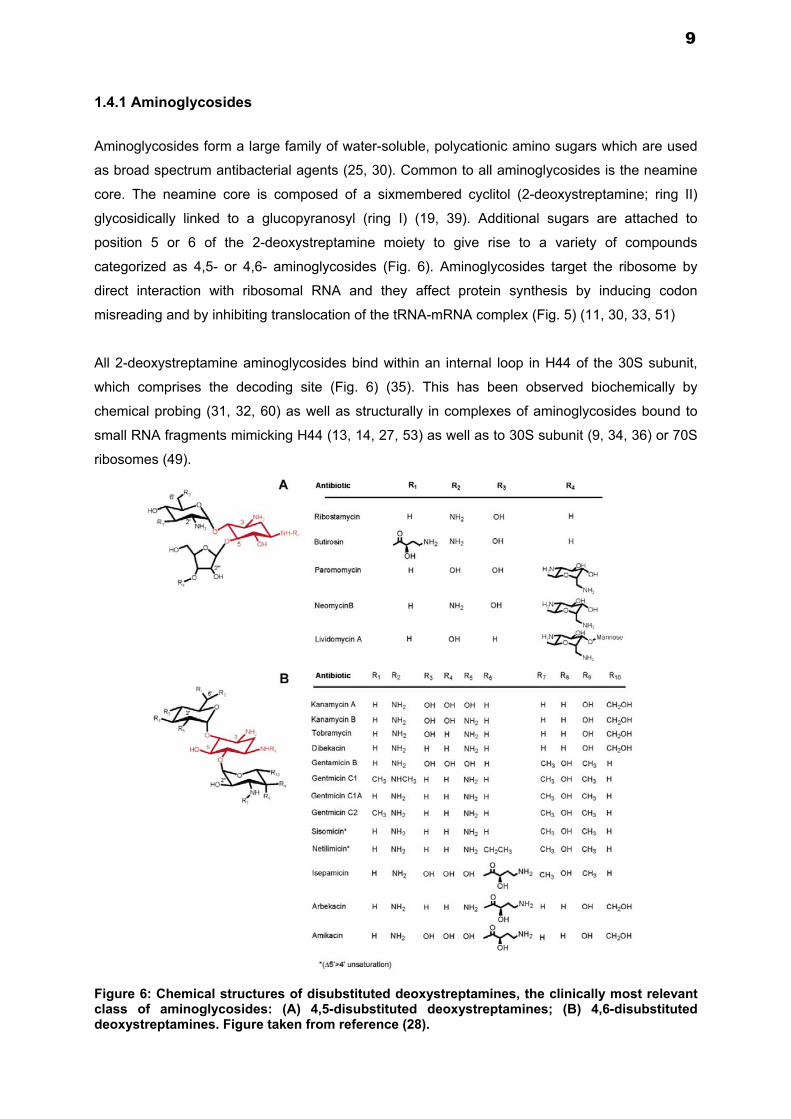
1.4.1 Aminoglycosides
Aminoglycosides form a large family of water-soluble, polycationic amino sugars which are used
as broad spectrum antibacterial agents (25, 30). Common to all aminoglycosides is the neamine
core. The neamine core is composed of a sixmembered cyclitol (2-deoxystreptamine; ring II)
glycosidically linked to a glucopyranosyl (ring I) (19, 39). Additional sugars are attached to
position 5 or 6 of the 2-deoxystreptamine moiety to give rise to a variety of compounds
categorized as 4,5- or 4,6- aminoglycosides (Fig. 6). Aminoglycosides target the ribosome by
direct interaction with ribosomal RNA and they affect protein synthesis by inducing codon
misreading and by inhibiting translocation of the tRNA-mRNA complex (Fig. 5) (11, 30, 33, 51)
All 2-deoxystreptamine aminoglycosides bind within an internal loop in H44 of the 30S subunit,
which comprises the decoding site (Fig. 6) (35). This has been observed biochemically by
chemical probing (31, 32, 60) as well as structurally in complexes of aminoglycosides bound to
small RNA fragments mimicking H44 (13, 14, 27, 53) as well as to 30S subunit (9, 34, 36) or 70S
ribosomes (49).
Figure 6: Chemical structures of disubstituted deoxystreptamines, the clinically most relevant class of aminoglycosides: (A) 4,5-disubstituted deoxystreptamines; (B) 4,6-disubstituted deoxystreptamines. Figure taken from reference (28).
9

The A site of the 30S ribosomal subunit (the decoding centre), in which the codon and anticodon
pair, is made up of four different domains: the head, shoulder, platform and H44 (Fig. 7A).
A
Figure 7A: The bacterial decoding center in the 30S ribosomal subunit. Overview of the 30S subunit structure, in complex with A-site tRNA anticodon stem–loop (ASL, gold). Red arrows indicate the movement of domains during the transition to the closed 30S conformation. P-site codon and tRNA-ASL (mimicked by the 3′ end of the 16S RNA and the ‘spur’ stem–loop of a symmetry-related molecule in the crystal) are dark grey, helices H44 cyan, and H27 yellow. In the shoulder domain, H18 with the 530-loop is turquoise, and proteins S12 (orange), S4 (violet) and S5 (dark blue, on the back of the subunit) are highlighted in space-filling representation. In the head domain, H34 is blue. Close-up of selected 30S elements around the decoding center, showing the A-site codon (purple), 16S RNA nucleotides G530 in the 530-loop (turquoise), and A1492 and 1493 in H44 (cyan) the positions of paromomycin (green) and streptomycin (pink). Remaining colors as in panel A (proteins S4 and S5 not shown) (35).
B
Figure 7B. The bacterial A-site (E. coli numbering) is a well known target for antibiotics. View of the three-dimensional structure of the A-site complexed with 4,5- and 4,6-disubstituted 2-deoxystreptamines: the common neamine core is denoted in yellow; ring III of the 4,6-compounds (tobramycin) is denoted in red; and rings III and IV of the 4,5-compounds (paromomycin) are denoted in blue. Key nucleotides of the binding site are given in bold. Figure taken from reference (7).
10

During the last decade, several achievements in bacterial ribosome structure determination (9)
(47) along with crystal and NMR structures of bacterial A-site oligonucleotide models (14, 54, 59)
have provided fascinating insights into our understanding of the decoding mechanism in
prokaryotic cells and of how 2-DOS aminoglycosides induce the deleterious misreading of the
genetic code. During decoding, a critical step in aminoacyl-tRNA selection is based on the
formation of a minihelix between the codon of the mRNA and the anti-codon of the cognate
aminoacyl-tRNA. In this process, the conformation of the A-site is changed from an “off” state,
where the two conserved adenines A1492 and A1493 are folded back within the helix, to an “on”
state, where A1492 and A1493 are flipped out from the A-site and interact with the cognate
codon–anticodon mini-helix (34, 36). This conformational change is a molecular switch that
irreversibly determines on the continuation of translation. The binding of aminoglycosides such as
paromomycin to the bacterial A-site changes the conformation equilibrium of the conserved
adenines A1492 and A1493 by stabilizing the “on” state conformation even in the absence of
cognate tRNA–mRNA complex (Fig. 8). Thus, the affinity of the A-site for a non-cognate mRNA–
tRNA complex is increased upon aminoglycoside binding, preventing the ribosome from efficiently
discriminating between near-cognate and cognate complexes and leading to the assembly of
proteins of incorrect sequence (37).
Figure 8. The molecular basis of the aminoglycoside-induced miscoding as resolved by X-ray crystal structures. At the bacterial decoding site (A-site), two flexible adenines A1492 and A1493 are in conformational equilibrium with a predominance of an intrahelical “off state” conformation. The binding of 2-DOS aminoglycoside paromomycin (green) shifts the equilibrium by stabilizing the “on state” conformation even in the absence of mRNA or tRNA. In the “on state” conformation the A1492 and A1493 are able to create hydrogen bonds with the bases of the mini-helix formed by the near-cognate tRNA anticodon (cyan) and the mRNA codon (magenta) leading to miscoding (16).
11

1.4.2 Selectivity and toxicity of Aminoglycosides
Drugs targeting the ribosome are characterized by two features: specificity and toxicity (6, 23, 24,
38, 40). During the past decades, considerable evidence has accumulated demonstrating that the
nucleic acid component of the ribosome is key to binding many of the ribosomal drugs, rather
than the numerous ribosomal proteins (15, 31, 33). Recent data from X-ray crystallography have
not only confirmed this suggestion but also provided details of drug-target interactions at the
atomic level by revealing how antibiotic binding occurs (4, 17, 48, 59, 61).
The components of the cytoribosome are encoded by chromosomal genes as are the
mitoribosomal proteins. However, the rRNA components of the mitoribosome are encoded by the
mitochondrial genome. Although the basis for drug related toxicity of the ribosomal inhibitors is
unknown, several lines of evidence point to mitoribosomes as the Achilles heel of ribosomal
antibiotics, because: (i) mitochondrial ribosomes are more related to the prokaryotic ribosome
than to the eukaryotic cytoplasmic ribosome; (ii) toxicity in-vivo correlates with activity in-vitro, i.e.,
those antibiotics which exhibit in vitro activity on mitoribosomes are associated with toxicity in
vivo; (iii) familial hypersensitivity to aminoglycosides (drug-induced deafness) is associated with
specific mutations in mitochondrial rRNA (43).
It has been suggested that the analysis of drug resistance mutations in bacteria allows one to
understand the basis of specificity for drugs targeting the ribosome (8). Central to this hypothesis
is the concept of ‘informative sequence positions’ _ i.e., the identification of polymorphic
nucleotides as a determinant of drug resistance in bacteria. The identification of a polymorphic
residue as determinant of ribosomal resistance provides information about the selectivity of a
ribosomal antibiotic, i.e., whether a drug affects the prokaryotic as opposed to the eukaryotic
ribosome. The basis for this hypothesis was initially established by investigating bacterial
alterations within the ribosome mediating resistance to aminoglycosides. The conclusion from
these studies was that the selectivity of these agents is largely due to a single nucleotide position
within the rRNA, e.g., the identity of the base at 16S rRNA position 1408. According to this
hypothesis, selectivity of the aminoglycosides is due to the natural insensitivity of eukaryotic
cytoplasmic ribosomes conferred by a guanine at 16S rRNA position 1408; conversely the toxicity
of aminoglycosides (at least irreversible ototoxicity) is due to the natural susceptibility of
mitoribosomes, which carry a susceptible bacterial adenine at this sequence position (Fig. 9) (8).
12

Figure 9. 16S rRNA decoding region: secondary structures.
1.5 Ribosomal dysfunction in Mitochondria
Mitochondria are vital components of all nucleated cells. Mitochondrial diseases are a clinically
heterogeneous group of disorders that arise as a result of dysfunction of the mitochondrial
respiratory chain. They can be caused by mutations of nuclear DNA or mitochondrial DNA
(mtDNA). Nuclear gene defects may be inherited in an autosomal recessive manner or an
autosomal dominant manner. Mitochondrial DNA defects are transmitted by maternal inheritance.
A male does not transmit the mtDNA mutation to his offspring. Some mitochondrial disorders only
affect a single organ (such as the eye in Leber hereditary optic neuropathy [LHON]), but many
involve multiple organ systems and often present with prominent neurologic and myopathic
features. Mitochondrial dysfunction has pleiotropic effects in multicellular organisms.
Mitochondrial disorders may present at any age. In general terms, nuclear DNA mutations
present in childhood and mtDNA mutations (primary or secondary to a nuclear DNA abnormality)
present in late childhood or adult life. Common clinical features of mitochondrial disease include
ptosis, external ophthalmoplegia, proximal myopathy and exercise intolerance, cardiomyopathy,
sensorineural deafness, optic atrophy, pigmentary retinopathy, and diabetes mellitus.
Mutations in the decoding A-site of mitochondrial 12S rRNA have been associated with deafness
(2, 3). In particular, the single-nucleotide alterations A1555G and C1494U have been identified as
a major source of nonsyndromic deafness (43, 62). By themselves, these mutations produce a
clinical phenotype that may range from severe congenital deafness through moderate
progressive hearing loss of later onset to normal hearing (12). Interestingly, and in addition to the
genetic predisposition, mutations A1555G and C1494U render affected individuals
hypersusceptible to aminoglycoside autotoxicity.
13

1.6 Model system
Investigations on structure-function relationships of mitochondrial rRNA in higher and lower
eukaryotes are mainly hampered by the lack of experimental genetic models (18, 19, 21, 22).
Genetic manipulation of the rRNA component of eukaryotic ribosomes has proven exceedingly
difficult due to the high copy number of corresponding operons. Higher eukaryotes have so far
resisted any genetic manipulation of their ribosomal nucleic acids, virtually abolishing the
possibility to test hypotheses by experimentation. Even most eubacteria harbor multiple rRNA
operons (e.g. E. coli: 7; Bacillus subtilis: 10; Streptomyces ambofaciens: 4) making genetic
studies of rRNA difficult. In the mid 90s, genetic procedures were developed which permitted the
construction of eubacteria carrying a single functional rRNA operon (46) (1). These single rRNA
allelic microorganisms allow mutagenesis of their ribosomal nucleic acids to result in cells
containing homogeneous populations of mutant ribosomes (42).
In the present thesis, and with the view to study the interaction of aminoglycoside antibiotics with
eukaryotic ribosomes, we replaced a central 34-nucleotide part of the bacterial drug binding
pocket in 16S rRNA H44 of Mycobacterium smegmatis with its eukaryotic counterpart, resulting in
bacterial hybrid ribosomes with a fully functional eukaryotic rRNA decoding site. For this, the
recently described M. smegmatis mc2 155 ΔrrnB (20) was used for all genetic manipulations.
Using a strategy of unmarked deletion of chromosomal rRNA operons combined with RecA
mediated site-directed mutagenesis, the decoding region was engineered.
For functional characterization of these hybrid ribosomes and to study a possible cause-effect
relationship of disease-associated mitochondrial rRNA mutations, we assessed drug susceptibility
in-vivo, by minimal inhibitory concentration assays. We also studied translational activity, drug
susceptibility, drug induced misreading, and decoding accuracy in-vitro by cell free translation
assays.
The model system was used to:
i) test the effect of ribosomal antibiotics on translation.
ii) identify mode of action of drugs and their interaction with ribosomes.
iii) study structure-function relationships within a defined region of rRNA.
iv) investigate the influence of rRNA mutations on protein synthesis.
v) investigate disease associated alterations in the small subunit rRNA.
vi) determine residues critical for selectivity and specificity of aminoglycosides.
vii) compare the functionality of humanized hybrid ribosomes to that of eukaryotic
cytoplasmic ribosomes.
viii) study the functionality of hybrid protozoan parasitic ribosomes
14

REFERENCES:
1. Asai, T., D. Zaporojets, C. Squires, and C. L. Squires. 1999. An Escherichia coli strain with
all chromosomal rRNA operons inactivated: complete exchange of rRNA genes between bacteria. Proc. Natl. Acad. Sci. USA 96:1971-1976.
2. Bacino, C., T. R. Prezant, X. Bu, P. Fournier, and N. Fischel-Ghodsian. 1995. Susceptibility mutations in the mitochondrial small ribosomal RNA gene in aminoglycoside induced deafness. Pharmacogenetics 5:165-72.
3. Ballana, E., E. Morales, R. Rabionet, B. Montserrat, M. Ventayol, O. Bravo, P. Gasparini, and X. Estivill. 2006. Mitochondrial 12S rRNA gene mutations affect RNA secondary structure and lead to variable penetrance in hearing impairment. Biochem Biophys Res Commun 341:950-7.
4. Ban, N., P. Nissen, J. Hansen, P. B. Moore, and T. A. Steitz. 2000. The complete atomic structure of the large ribosomal subunit at 2.4 A resolution. Science 289:905-20.
5. Bilgin, N., A. A. Richter, M. Ehrenberg, A. E. Dahlberg, and C. G. Kurland. 1990. Ribosomal RNA and protein mutants resistant to spectinomycin. EMBO J 9:735-9.
6. Böttger, E. C. 2007. Antimicrobial agents targeting the ribosome: the issue of selectivity and toxicity - lessons to be learned. Cell Mol Life Sci 64:791-5.
7. Böttger, E. C. 2006. The ribosome as a drug target. Trends in Biotechnology 24:145-147. 8. Böttger, E. C., B. Springer, T. Prammananan, Y. Kidan, and P. Sander. 2001. Structural
basis for selectivity and toxicity of ribosomal antibiotics. EMBO Rep. 2:318-323. 9. Carter, A. P., W. M. Clemons, D. E. Brodersen, R. J. Morgan-Warren, B. T. Wimberly, and
V. Ramakrishnan. 2000. Functional insights from the structure of the 30S ribosomal subunit and its interactions with antibiotics. Nature 407:340-348.
10. Dahlberg, A. E. 1989. The functional role of ribosomal RNA in protein synthesis. Cell 57:525-529.
11. Davies, J., L. Gorini, and B. D. Davis. 1965. Misreading of RNA codewords induced by aminoglycoside antibiotics. Mol Pharmacol 1:93-106.
12. Estivill, X., N. Govea, E. Barcelo, C. Badenas, E. Romero, L. Moral, R. Scozzri, L. D'Urbano, M. Zeviani, and A. Torroni. 1998. Familial progressive sensorineural deafness is mainly due to the mtDNA A1555G mutation and is enhanced by treatment of aminoglycosides. Am J Hum Genet 62:27-35.
13. Fourmy, D., M. I. Recht, S. C. Blanchard, and J. D. Puglisi. 1996. Structure of the A site of Escherichia coli 16S ribosomal RNA complexed with an aminoglycoside antibiotic. Science 274:1367-71.
14. Fourmy, D., S. Yoshizawa, and J. D. Puglisi. 1998. Paromomycin binding induces a local conformational change in the A-site of 16 S rRNA. J Mol Biol 277:333-45.
15. Gale, E. F., E. Cundliffe, P. E. Reynolds, M. H. Richmond, and J. M. Waring. 1981. The molecular basis of antibiotic action. John Wiley & Sons, Inc., London.
16. Hainrichson, M., I. Nudelman, and T. Baasov. 2008. Designer aminoglycosides: the race to develop improved antibiotics and compounds for the treatment of human genetic diseases. Org Biomol Chem 6:227-39.
17. Harms, J., F. Schluenzen, R. Zarivach, A. Bashan, S. Gat, I. Agmon, H. Bartels, F. Franceschi, and A. Yonath. 2001. High resolution structure of the large ribosomal subunit from a mesophilic eubacterium. Cell 107:679-88.
18. Hobbie, S. N., S. Akshay, S. K. Kalapala, C. M. Bruell, D. Shcherbakov, and E. C. Böttger. 2008. Genetic analysis of interactions with eukaryotic rRNA identify the mitoribosome as target in aminoglycoside ototoxicity. Proc Natl Acad Sci U S A 105:20888-93.
19. Hobbie, S. N., C. Bruell, S. Kalapala, S. Akshay, S. Schmidt, P. Pfister, and E. C. Böttger. 2006. A genetic model to investigate drug-target interactions at the ribosomal decoding site. Biochimie 88:1033-43.
20. Hobbie, S. N., C. Bruell, S. K. Kalapala, S. Akshay, S. Schmidt, P. Pfister, and E. C. Böttger. 2006. A genetic model to investigate structural drug-target interactions at the ribosomal decoding site. Biochimie 88:1033-1043.
21. Hobbie, S. N., C. M. Bruell, S. Akshay, S. K. Kalapala, D. Shcherbakov, and E. C. Böttger. 2008. Mitochondrial deafness alleles confer misreading of the genetic code. Proc Natl Acad Sci U S A 105:3244-9.
22. Hobbie, S. N., S. K. Kalapala, S. Akshay, C. Bruell, S. Schmidt, S. Dabow, A. Vasella, P. Sander, and E. C. Böttger. 2007. Engineering the rRNA decoding site of eukaryotic cytosolic ribosomes in bacteria. Nucleic Acids Res. 35:6086-6093.
15

23. Hobbie, S. N., P. Pfister, C. Bruell, P. Sander, B. Francois, E. Westhof, and E. C. Böttger. 2006. Binding of neomycin-class aminoglycoside antibiotics to mutant ribosomes with alterations in the A-site of 16S rRNA. Antimicrob. Agents Chemother. 50:1489-1496.
24. Hobbie, S. N., P. Pfister, C. Brull, E. Westhof, and E. C. Böttger. 2005. Analysis of the contribution of individual substituents in 4,6-aminoglycoside-ribosome interaction. Antimicrob. Agents Chemother. 49:5112-5118.
25. Kotra, L. P., J. Haddad, and S. Mobashery. 2000. Aminoglycosides: perspectives on mechanisms of action and resistance and strategies to counter resistance. Antimicrob Agents Chemother 44:3249-56.
26. Kurland, C. G., D. Hughes, and M. Ehrenberg. 1996. Limitations of translational accuracy, p. 979-1004. In F. C. Neidhardt, R. Curtiss, J. L. Ingraham, E. C. C. Lin, K. B. Low, B. Magasanik, W. S. Reznikoff, M. Riley, M. Schaechter, and H. E. Umbarger (ed.), Escherichia coli and Salomonella typhimurium: Cellular and Molecular Biology, 2 ed, vol. 1. American Society for Microbiology Press, Washington, DC.
27. Lynch, S. R., R. L. Gonzalez, and J. D. Puglisi. 2003. Comparison of X-Ray Crystal Structure of the 30S Subunit-Antibiotic Complex with NMR Structure of Decoding Site Oligonucleotide-Paromomycin Complex. Structure (Camb) 11:43-53.
28. Magnet, S., and J. S. Blanchard. 2005. Molecular insights into aminoglycoside action and resistance. Chem. Rev. 105:477-498.
29. Marshall, R. A., C. E. Aitken, M. Dorywalska, and J. D. Puglisi. 2008. Translation at the single-molecule level. Annu Rev Biochem 77:177-203.
30. Mingeot-Leclercq, M. P., Y. Glupczynski, and P. M. Tulkens. 1999. Aminoglycosides: activity and resistance. Antimicrob Agents Chemother 43:727-37.
31. Moazed, D., and H. F. Noller. 1987. Interaction of antibiotics with functional sites in 16S ribosomal RNA. Nature 327:389-94.
32. Moazed, D., and H. F. Noller. 1989. Interaction of tRNA with 23S rRNA in the ribosomal A, P, and E sites. Cell 57:585-97.
33. Noller, H. F. 1991. Ribosomal RNA and translation. Annu Rev Biochem 60:191-227. 34. Ogle, J. M., D. E. Brodersen, W. M. Clemons, Jr., M. J. Tarry, A. P. Carter, and V.
Ramakrishnan. 2001. Recognition of cognate transfer RNA by the 30S ribosomal subunit. Science 292:897-902.
35. Ogle, J. M., A. P. Carter, and V. Ramakrishnan. 2003. Insights into the decoding mechanism from recent ribosome structures. Trends Biochem Sci 28:259-66.
36. Ogle, J. M., F. V. Murphy, M. J. Tarry, and V. Ramakrishnan. 2002. Selection of tRNA by the ribosome requires a transition from an open to a closed form. Cell 111:721-732.
37. Ogle, J. M., and V. Ramakrishnan. 2005. Structural insights into translational fidelity. Annu Rev Biochem 74:129-77.
38. Pfister, P., S. Hobbie, C. Brull, N. Corti, A. Vasella, E. Westhof, and E. C. Böttger. 2005. Mutagenesis of 16S rRNA C1409-G1491 base-pair differentiates between 6'OH and 6'NH3
+
aminoglycosides. J. Mol. Biol. 346:467-475. 39. Pfister, P., S. Hobbie, Q. Vicens, E. C. Böttger, and E. Westhof. 2003. The molecular basis
for A-Site mutations conferring aminoglycoside resistance: relationship between ribosomal susceptibility and X-ray crystal structures. ChemBioChem. 4:1078-1088.
40. Pfister, P., M. Risch, D. E. Brodersen, and E. C. Böttger. 2003. Role of 16S rRNA helix 44 in ribosomal resistance to hygromycin B. Antimicrob. Agents Chemother. 47:1496-1502.
41. Poehlsgaard, J., and S. Douthwaite. 2005. The bacterial ribosome as a target for antibiotics. Nat Rev Microbiol 3:870-81.
42. Prammananan, T., P. Sander, B. Springer, and E. C. Böttger. 1999. RecA-mediated gene conversion and aminoglycoside resistance in strains heterozygous for rRNA. Antimicrob. Agents Chemother. 43:447-453.
43. Prezant, T. R., J. V. Agapian, M. C. Bohlman, X. Bu, S. Oztas, W. Q. Qiu, K. S. Arnos, G. A. Cortopassi, L. Jaber, J. I. Rotter, and et al. 1993. Mitochondrial ribosomal RNA mutation associated with both antibiotic-induced and non-syndromic deafness. Nat Genet 4:289-94.
44. Ramakrishnan, V. 2002. Ribosome structure and the mechanism of translation. Cell 108:557-72.
45. Ramakrishnan, V., and P. B. Moore. 2001. Atomic structures at last: the ribosome in 2000. Curr. Opin. Struct. Biol. 11:144-154.
46. Sander, P., T. Prammananan, and E. C. Böttger. 1996. Introducing mutations into a chromosomal rRNA gene using a genetically modified eubacterial host with a single rRNA operon. Mol. Microbiol. 22:841-848.
16

47. Schluenzen, F., A. Tocilj, R. Zarivach, J. Harms, M. Gluehmann, D. Janell, A. Bashan, H. Bartels, I. Agmon, F. Franceschi, and A. Yonath. 2000. Structure of functionally activated small ribosomal subunit at 3.3 angstroms resolution. Cell 102:615-23.
48. Schuwirth, B. S., M. A. Borovinskaya, C. W. Hau, W. Zhang, A. Vila-Sanjurjo, J. M. Holton, and J. H. Cate. 2005. Structures of the bacterial ribosome at 3.5 A resolution. Science 310:827-34.
49. Selmer, M., C. M. Dunham, F. V. t. Murphy, A. Weixlbaumer, S. Petry, A. C. Kelley, J. R. Weir, and V. Ramakrishnan. 2006. Structure of the 70S ribosome complexed with mRNA and tRNA. Science 313:1935-1942.
50. Sohmen, D., J. M. Harms, F. Schlunzen, and D. N. Wilson. 2009. Enhanced SnapShot: Antibiotic inhibition of protein synthesis II. Cell 139:212-212 e1.
51. Spahn, C. M., and C. D. Prescott. 1996. Throwing a spanner in the works: antibiotics and the translation apparatus. J Mol Med 74:423-39.
52. Umezawa, H. 1958. Kanamycin: its discovery. Ann N Y Acad Sci 76:20-6. 53. Vicens, Q., and E. Westhof. 2001. Crystal structure of paromomycin docked into the
eubacterial ribosomal decoding A site. Structure 9:647-58. 54. Vicens, Q., and E. Westhof. 2003. RNA as a drug target: the case of aminoglycosides.
Chembiochem 4:1018-23. 55. Waksman, S. A., H. A. Lechevalier, and D. A. Harris. 1949. Neomycin; production and
antibiotic properties. J Clin Invest 28:934-9. 56. Wallace, D. C. 2005. A mitochondrial paradigm of metabolic and degenerative diseases,
aging, and cancer: a dawn for evolutionary medicine. Annu Rev Genet 39:359-407. 57. Weinstein, M. J., G. M. Luedemann, E. M. Oden, G. H. Wagman, J. P. Rosselet, J. A.
Marquez, C. T. Coniglio, W. Charney, H. L. Herzog, and J. Black. 1963. Gentamicin, a New Antibiotic Complex from Micromonospora. J Med Chem 6:463-4.
58. Wilson, D. N., and K. H. Nierhaus. 2005. Ribosomal proteins in the spotlight. Crit Rev Biochem Mol Biol 40:243-67.
59. Wimberly, B. T., D. E. Brodersen, W. M. Clemons, Jr., R. J. Morgan-Warren, A. P. Carter, C. Vonrhein, T. Hartsch, and V. Ramakrishnan. 2000. Structure of the 30S ribosomal subunit. Nature 407:327-39.
60. Woodcock, J., D. Moazed, M. Cannon, J. Davies, and H. F. Noller. 1991. Interaction of antibiotics with A- and P-site-specific bases in 16S ribosomal RNA. EMBO J 10:3099-103.
61. Yusupov, M. M., G. Z. Yusupova, A. Baucom, K. Lieberman, T. N. Earnest, J. H. Cate, and H. F. Noller. 2001. Crystal structure of the ribosome at 5.5 A resolution. Science 292:883-96.
62. Zhao, H., R. Li, Q. Wang, Q. Yan, J. H. Deng, D. Han, Y. Bai, W. Y. Young, and M. X. Guan. 2004. Maternally inherited aminoglycoside-induced and nonsyndromic deafness is associated with the novel C1494T mutation in the mitochondrial 12S rRNA gene in a large Chinese family. Am J Hum Genet 74:139-52.
17

6086–6093 Nucleic Acids Research, 2007, Vol. 35, No. 18 Published online 30 August 2007doi:10.1093/nar/gkm658
Engineering the rRNA decoding site of eukaryoticcytosolic ribosomes in bacteria
Sven N. Hobbie1,*, Sarath K. Kalapala1, Subramanian Akshay1, Christian Bruell1,
Sebastian Schmidt1, Sabine Dabow1, Andrea Vasella2, Peter Sander1 and Erik C. Bottger1
1Institut fur Medizinische Mikrobiologie, Universitat Zurich and 2Laboratorium fur Organische Chemie, ETH Zurich,
Switzerland
Received July 10, 2007; Revised August 7, 2007; Accepted August 8, 2007
ABSTRACT
Structural and genetic studies on prokaryotic
ribosomes have provided important insights into
fundamental aspects of protein synthesis and
translational control and its interaction with riboso-
mal drugs. Comparable mechanistic studies in
eukaryotes are mainly hampered by the absence of
both high-resolution crystal structures and efficient
genetic models. To study the interaction of amino-
glycoside antibiotics with selected eukaryotic ribo-
somes, we replaced the bacterial drug binding site
in 16S rRNA with its eukaryotic counterpart, result-
ing in bacterial hybrid ribosomes with a fully
functional eukaryotic rRNA decoding site. Cell-free
translation assays demonstrated that hybrid ribo-
somes carrying the rRNA decoding site of higher
eukaryotes show pronounced resistance to amino-
glycoside antibiotics, equivalent to that of rabbit
reticulocyte ribosomes, while the decoding sites of
parasitic protozoa show distinctive drug suscept-
ibility. Our findings suggest that phylogenetically
variable components of the ribosome, other than the
rRNA-binding site, do not affect aminoglycoside
susceptibility of the protein-synthesis machinery.
The activities of the hybrid ribosomes indicate
that helix 44 of the rRNA decoding site behaves as
an autonomous domain, which can be exchanged
between ribosomes of different phylogenetic
domains for study of function.
INTRODUCTION
Accurate decoding of genetic information is a crucial stepin protein synthesis. Genetic, biochemical and structuraldata provide evidence for a functional role of ribosomalRNA in mRNA decoding and tRNA selection (1–3).
The functional relevance of rRNA residues incodon–anticodon recognition and stabilization is reflectedin their universal conservation throughout the threephylogenetic domains of life. While this holds true formost nucleotides in helices 18, 34 and 44 of the smallsubunit rRNA that form the aminoacyl-tRNA acceptorsite (A site), critical variations have evolved betweendifferent phylogenetic domains, most prominent in helix44, the penultimate stem of 16S rRNA (4). Thesevariations most likely account for a decoding region thathas been highly optimized in the context of evolutionarydifferentiation.
Aminoglycosides are a class of structurally relatedantibiotics which interfere with decoding by binding tothe A site of small subunit rRNA (5). These antibioticspreferentially target prokaryotic over eukaryotic ribo-somes and affect protein synthesis by inducing codonmisreading and inhibiting tRNA translocation (6–8).The binding site is located within a conserved loop ofhelix 44, which in part shows phylogenetic sequencevariability, e.g. at position 1408 and at base pair1409–1491 (9) (rRNA residues are numbered accordingto Escherichia coli nomenclature; see also Figure 2).Crystal structures of various bacterial 30S ribosomalparticles in complex with different ligands, e.g. tRNA andantibiotics, have revealed the molecular mechanisms of thedecoding step at atomic resolution (10,11). In the absenceof high-resolution X-ray crystal structures of eukaryoticribosomes, model oligonucleotides mimicking the riboso-mal decoding site have been used for structural analysisof the A site in human cytosolic ribosomes (12).
Site-directed mutagenesis has been used in prokaryotesto study the functional relevance of individual drug–nucleotide contacts in aminoglycoside–ribosome interac-tion (13). A detailed analysis of the aminoglycosidetarget site in ribosomes of higher and lower eukaryotesis complicated by the complexity of eukaryotic rRNAgenetics. Genetic manipulation of the rRNA componentof eukaryotic ribosomes has proven exceedingly difficult
*To whom correspondence should be addressed. Tel: +41 44 634 2664; Email: [email protected]
The authors wish it to be known that, in their opinion, the first three authors should be regarded as joint First Authors.
� 2007 The Author(s)
This is an Open Access article distributed under the terms of the Creative Commons Attribution Non-Commercial License (http://creativecommons.org/licenses/
by-nc/2.0/uk/) which permits unrestricted non-commercial use, distribution, and reproduction in any medium, provided the original work is properly cited.
18

due to the problem of the high copy number ofcorresponding operons. Higher eukaryotes have so farresisted any genetic manipulation of their ribosomalnucleic acids, virtually abolishing the possibility to testhypotheses by experimentation. Even in lower eukaryoticorganisms, such as yeast, the presence of 100–200tandemly repeated copies at the RDN locus, has madegenetic studies of rRNA difficult (14–16).
The eukaryotic ribosome differs significantly fromthe prokaryotic one (17). It is impossible to predict thecontribution of additional, phylogenetically diverse ribo-somal elements and characteristics, as in the eukaryoticribosome, to aminoglycoside susceptibility. It is, forinstance, commonly assumed that translation in mamma-lian cells functions with higher fidelity than that ofbacteria (18). To address this question experimentally,we have performed domain shuffling experiments inrRNA. Using genetic techniques, we successfully engi-neered a functionally critical domain in the ribosome byreplacing the bacterial A-site rRNA of helix 44 with itscounterpart from higher and lower eukaryotes. Forfunctional characterization of these hybrid ribosomes,we studied their susceptibility to antibiotics targeting theribosomal A site and which are known to affect bacterialversus eukaryotic ribosomes to different extents.
MATERIALS AND METHODS
Construction of the rrn plasmid exchange system
Starting with Mycobacterium smegmatis mc2-155 SMR5(19), a single-rRNA allelic strain bearing a deletion of�5 kb in the chromosomal rrnA operon was constructedby gene replacement. The deletion strategy for chromo-somal rRNA operons is shown in SupplementaryFigure 1. Subsequently and following integration of therrnB-replacement vector into the chromosomal rrnB locus,the strain was transformed with integration-proficientvector pMIH-rrnB2058G for rRNA complementation.pMIH-rrnB2058G is a derivative of plasmid pMV361-rrnB2058G (20), in which the kanamycin resistance cassettehas been replaced with a hygromycin resistance cassette.Counterselection and genetic screening for deletion of thechromosomal rrnB locus resulted in strain M. smegmatis�rrn pMIH-rrnB2058G bearing unmarked full deletionsof the chromosomal rrnA and rrnB loci. In this strain,ribosomal RNA is exclusively transcribed from thecomplementation vector (Figure. 1).To establish a genetic system for efficient plasmid
exchange, an integration-proficient complementationvector pMIG-rrnB-sacB was designed that carries therrnB wild-type operon, a gentamicin resistance cassette,
TTrrsA rrlA ogtmurA rrfA
PPrrsB rrlB 3760tyrS rrfB
PP TTattB
TTmurA‘rrfA ogt
murA‘rrfA ogt
murA‘rrfA ogt
murA‘rrfA ogt
murA‘rrfA ogt
rrsB rrlB 3760tyrS rrfBPP TT
attB
TTrrsB rrlB 3760tyrS rrfB
PP TTrrs rrlint rrf Hygr attB
rrf Hygr attB
rrf Hygr attB
PP TTattB
TT
tyrS ‘3760PP
rrs rrlintPP TT
attB
TTtyrS ‘3760
PPrrs rrlint rrf Gmr sacB attB
PP TTattB
TTtyrS ‘3760
PPhybrid rrs rrlint
PP TTattB
M. smegmatis
∆rrnA
∆rrnA attB::pMIH-rrnB2058G
∆rrnA ∆rrnB attB::pMIH-rrnB2058G
∆rrnA ∆rrnB attB::pMIG-sacB-rrnB2058G
∆rrnA ∆rrnB attB::pMIH-rrnBhybrid
rrnA rrnB attB
Chromosomal locus
Figure 1. Sequential strategy for the generation of a plasmid-rRNA exchange system. From top: Following deletion of chromosomal rrnA,a complementation vector pMIH-rrnB2058G carrying a functional rrn operon was introduced to the chromosomal attB site. Subsequent deletion ofrrnB resulted in M. smegmatis �rrnA �rrnB attB::pMIH-rrnB2058G, in which ribosomal RNA is exclusively transcribed from the plasmid. Fromthere, a plasmid-rRNA exchange system was established by replacing pMIH-rrnB2058G with pMIG-rrnB-sacB. Transformation with hybrid rRNAgenes pMIH-rrnBhybrid and selection on sucrose resulted in M. smegmatis �rrnA �rrnB attB::pMIH-rrnBhybrid with homogenous populationsof hybrid ribosomes.
Nucleic Acids Research, 2007, Vol. 35, No. 18 6087
19

and the sucrose sensitivity marker sacB. This vector wasused to replace pMIH-rrnB2058G in strain M. smegmatis�rrn pMIH-rrnB2058G, resulting in strain M. smegmatis�rrn pMIG-rrnB-sacB. The latter was used as plasmidexchange system in which the rrnB wild-type operon canbe efficiently replaced with an rrnB mutant operon bytransformation and selection on sucrose-containing media(Figure. 1).
Construction of mutant strains with hybrid ribosomes
Site-directed mutagenesis of rRNA genes was performedby PCR mutagenesis as described previously (21). Inbrief, hybrid rDNA oligonucleotides comprising theeukaryotic helix 44 decoding site sequence were used forgene amplification. Using restriction endonuclease recog-nition sites present in the operon, rRNA gene fragments inplasmid pMIH-rrnB were replaced to construct pMIH-rrnBhybrid, the plasmids coding for hybrid 16S rRNAgenes. Mycobacterium smegmatis �rrn pMIG-rrnB-sacBwas transformed with the various pMIH-rrnBhybrid
constructs and selected on hygromycin plates containingsucrose (Figure 1). Successful plasmid exchange wascontrolled by sequence analysis.
Minimal inhibitory concentrations
Broth microdilution tests were performed in a microtiterplate format as described previously (22). In brief,bacterial strains were cultured on Luria–Bertani (LB)agar plates at 378C. Freshly grown cultures wereresuspended in LB broth supplemented with 0.05% ofTween 80, diluted to an absorbance at 600 nm of 0.025,and incubated in the presence of 2-fold serial dilutionsof the following 2-deoxystreptamine aminoglycosides:paromomycin, neomycin, geneticin (G418), gentamicin,netilmicin, tobramycin and kanamycin A (Sigma). Afterincubation at 378C for 72 h, the minimal inhibitoryconcentration (MIC) was recorded as the lowest concen-tration of drug inhibiting visible growth.
Ribosome purification
Approximately 8 g of wet bacterial cell mass wereresuspended in 25ml of homogenization buffer (HB;20mM Tris–HCl pH 7.4, 100mM NH4Cl, 10.5mMMgCl2, 0.5mM EDTA and 3mM 2-mercaptoethanol)and lyzed with a French Pressure Cell (AmericanInstrument Company, Maryland) at 16 000 psi, asdescribed previously (23). The lysate was supplementedwith DNase (RQ1 RNase-free, Promega, 2 U per gramcell mass), incubated on ice for 10min, and adjusted withHB to a volume of 70ml. Pre-cooled alumina (1.5 g pergram cell mass) was added to the cell lysate, stirred on icefor 20min, and removed by low-speed centrifugation(10min at 2000g; all centrifugations were performed at48C). Cell debris was sedimented by centrifugation for30min at 10 000g and the lysate was passed through a teafilter. Centrifugation of the lysate for 60min at 32 500gresulted in the S-30 supernatant. Sixteen milliliter portionsof the supernatant were layered on 9ml sucrose cushions(SC; 20mM Tris–HCl pH 7.4, 350mM NH4Cl, 10.5mMMgCl2, 0.5mM EDTA, 1.1M sucrose and 3mM
2-mercaptoethanol) and centrifuged for 15 h at 110 000g(Beckman Coulter OptimaTM L-80 XP Ultracentrifuge) toseparate ribosomal particles from the S-100 supernatant.The S-100 fraction was dialyzed for 24 h (6000–8000MWCO, Spectra/Por, Serva) with three intermittentchanges of buffer A (50mM Tris–HCl pH 7.5, 70mMNH4Cl, 30mM KCl and 7mM MgCl2), concentrated(Viva spin, 10 000 MWCO, Viva Science), and stored at�808C. Ribosome pellets from the S-100 preparation wereresuspended in washing buffer (WB; 20mM Tris–HCl pH7.4, 350mM NH4Cl, 10.5mM MgCl2, 0.5mM EDTA and7mM 2-mercaptoethanol) and passed twice throughsucrose cushions (6 h at 180 000g and 16 h at 83 000g).The final ribosome pellets were resuspended in buffer A,incubated for 30min at 48C, dispensed into aliquots, andstored at �80C after shock freezing in liquid nitrogen. 70Sribosome concentrations were determined by absorptionmeasurements on the basis of 23 pmol per A260 unit.
Qualitative evaluation of ribosome preparations
Integrity of 70S ribosomes was determined by analyticalultracentrifugation (14 h at 70 000g) through a 10–40%sucrose gradient in both association buffer (buffer A) anddissociation buffer (50mM Tris–HCl pH 7.5, 70mMNH4Cl and 30mM KCl). 70S, 50S and 30S ribosomefractions in different gradient layers were detected andquantified by absorption at 254 nm. Functional activity ofpurified ribosomes was determined by assessing theircapacity to form initiation complexes. 70S ribosomes(0.5 mM) were incubated in buffer A with m022 SD-MFTI-mRNA at 1.5 mM (50-GGCAAGGAGGUAAAUAAUGUUCACGAUC-30; obtained from Dharmacon), initiationfactors (IF) 1 and 3 from M. smegmatis, IF-2 from E. coli(1 mM each), GTP (1mM), and [3H]-fMet-tRNAfMet
(1 mM) for 60min at 378C; purified tRNA and IF2from E. coli were a kind gift from Marina Rodnina.The initiation complexes formed were bound to nitrocel-lulose filters (Sartorius, pore size 0.45mm), washed with15ml cold buffer A, dissolved in 10ml scintillationcocktail (Filtersafe, Zinsser Analytic) and quantified in aLiquid Scintillation Analyzer (Tri-Carb 2900 TR,PerkinElmer).
Cell-free (UUU)12 translation assays
Cell-free translation reactions in buffer A (pH 7.5) wereprepared on ice and contained M. smegmatis tRNAbulk
(0.5mg/ml), amino acids mixture (30mM each) lackingphenylalanine and/or leucine, 10% (v/v) S100 extract,energy mix [DTT (1mM), GTP (1mM), ATP (4mM),PEP (5mM)], pyruvate kinase (0.1mg/ml), and polya-mines [spermidine (2mM) and putrescine (8mM)].The reaction mixture was preincubated with radiolabeledphenylalanine and/or leucine (30mM; obtained fromAmersham) at 378C for 15min. The translation reactionwas started by addition of ribosomes to a final concentra-tion of 0.25mM and (UUU)12-mRNA (50-GCGGCAAGGAGGUAAAUA AUG (UUU)12 UAA GCAGG-30,obtained from Dharmacon) to 1 mM; serially dilutedaminoglycoside antibiotics were added simultaneously.Following incubation at 378C for 60min, the reaction was
6088 Nucleic Acids Research, 2007, Vol. 35, No. 1820

stopped by addition of KOH to 0.5M and subsequenthydrolysis at 378C for 30min. Synthesized polypeptideswere precipitated with 200 ml of 5% tri-chloro-acetic acid(TCA) for 10min on ice and bound to nitrocellulose filters(Sartorius, pore size 0.45 mm). Filter-bound polypeptideswere washed with cold 30% 2-propanol and quantifiedin 10ml of scintillation cocktail. Data analysis wasperformed with Prism (GraphPad Software Inc.). Best-fitnonlinear regression was used to define the 100% value ofpolypeptide synthesis and to calculate the aminoglycosideIC50 values of dose-dependent inhibition (Figure 3and Table 2).
Cell-free luciferase translation assays
Purified 70S hybrid ribosomes were used in a coupledtranscription–translation reaction of firefly luciferase(plasmid pBESTluc, Promega). A typical reaction(15 ml volume) contained 0.25 mM 70S ribosomes, 300 ngDNA, 40% (v/v) of M. smegmatis S100 extract, 100 mMamino acid mixture, and RNasin (40 U, Promega). rNTPs,tRNAs and energy were supplied by addition of commer-cial S30 Premix Without Amino Acids (Promega). Seriallydiluted aminoglycosides were added and the reactionmixture was incubated at 378C for 60min. The reactionwas stopped on ice, 100 ml of luciferase assay substrate(Promega) was added, and the bioluminescence wasmeasured in a luminometer (Bio-Tek instruments,FLx800). Data analysis was performed with Prism(GraphPad Software Inc.). Best-fit nonlinear regressionwas used to define the 100% value of luciferase synthesisand to calculate the aminoglycoside IC50 values of dose-dependent inhibition (Figure 4 and Table 3).
Rabbit reticulocyte translation assays
Cytosolic ribosomes present in commercially availablerabbit reticulocyte lysate (Promega) were used for in vitrotranslation of a firefly luciferase control mRNA(Promega). A standard 30 ml reaction contained 20 mlreticulocyte lysate, 500 ng of luciferase mRNA, an aminoacid mixture (100mM each), and RNasin. Serially dilutedaminoglycosides were added and the reaction mixture wasincubated at 308C for 90min. After incubation, 100 mlof luciferase assay substrate (Promega) was added andluciferase activity was determined.
RESULTS AND DISCUSSION
The two chromosomal rrn operons in M. smegmatis wereinactivated to produce a strain in which ribosomal RNAis exclusively transcribed from plasmid-encoded rRNAgenes. Operon inactivation was achieved by means ofunmarked deletion mutagenesis spanning the completerRNA gene sequences and part of the promoter region(Supplementary Figure 1). For complementation, a fullyfunctional wild-type rrnB operon encompassing promoterand termination sequences was cloned into the integra-tion-proficient plasmid pMIG-sacB. In M. smegmatis�rrn pMIG-sacB-rrnB, the wild-type rRNA operoncarried on pMIG-sacB is efficiently replaced with amutant rRNA operon by plasmid exchange following
transformation with integration-proficient plasmid pMIH-rrnBmut (for details, see the Materials and Methods sectionand Figure 1). The rRNA plasmid exchange system wasused to replace the bacterial 16S rRNA helix 44 within thedecoding region with its homologous structures fromparasitic kinetoplastid protozoa. The kinetoplastid generaLeishmania and Trypanosoma comprise species that areresponsible for leishmaniasis and trypanosomiasis,diseases that are widespread in many tropical and sub-tropical countries. The insect trypanosome Blastocrithidiais characterized by an rRNA decoding site with an adenineat the position homologous to 16S rRNA residue 1408,and therefore represents a very rare trait within theeukaryotic domain. We also constructed human–bacterialhybrid ribosomes by introducing the human helix 44rRNA homolog into functional ribosomes of strainM. smegmatis �rrnB by a method described previously(13). Both approaches resulted in bacterial mutant strainswith homogenous populations of hybrid ribosomes inwhich a 34 nucleotide portion of helix 44 was substitutedwith its counterpart of eukaryotic small-subunit rRNA(Figure 2). Together with A-site residues in helices 18, 34and 44 that are universally conserved throughout thephylogenetic domains, the engineered decoding siteresembles that of eukaryotic cytosolic ribosomes.Engineering the eukaryotic decoding sites into bacterialribosomes had relatively little effect on cell growth, withonly slightly increased generation times as compared towild-type cells (generation times: wild-type 3.7� 0.3 h;Leishmania hybrids 4.8� 0.5 h; Blastocrithidia hybrids4.2� 0.1 h; H. sapiens hybrids 5.1� 0.6 h).A bacterial oligoribonucleotide analog of 16S rRNA
helix 44 has been shown to interact with both antibioticand RNA ligands of the 30S subunit in a manner thatcorrelates with normal subunit function (24). Since thedecoding-site rRNA in the hybrid ribosomes is identical tothat of eukaryotic ribosomes, the hybrid ribosomes mayserve as a model to investigate specific features associatedwith the eukaryotic A-site rRNA structure. Towards thisend, we investigated the susceptibility of the hybridribosomes to the 2-deoxystreptamine antibiotics paromo-mycin, neomycin, geneticin (G418), gentamicin, netilmi-cin, tobramycin and kanamycin A (see SupplementaryFigure 2 for chemical structures). In MIC assays, whichdetermine growth inhibition at the whole cell level,M. smegmatis mutants carrying human–bacterial hybridribosomes were highly resistant to all aminoglycosideantibiotics tested except for geneticin (Table 1). Relativeresistance of the human hybrids to geneticin, calculatedas the ratio of mutant to wild-type MIC, was 8 to 16 ascompared to � 1024 for paromomycin, neomycin, genta-micin, netilmicin, tobramycin, and kanamycin. TheLeishmania hybrids showed high-level resistance toneomycin, gentamicin, netilmicin, tobramycin and kana-mycin, with relative resistance levels of �1024. However,the Leishmania hybrids showed distinct susceptibility toparomomycin and geneticin, i.e. aminoglycosides witha hydroxyl group at position 60 of ring I. Mycobacteriumsmegmatis hybrids with a Blastocrithidia decoding regionwere susceptible to all aminoglycoside antibiotics;the minimal inhibitory concentrations of the 4,6- and
Nucleic Acids Research, 2007, Vol. 35, No. 18 6089
21

4,5-disubstituted deoxystreptamines againstBlastocrithidia hybrids were similar to those observed forthe wild-type M. smegmatis strain (Table 1).To analyze the antibiotic effects on translation in more
detail, we determined the IC50 values for selectedaminoglycosides in (UUU)12-mRNA-directed phenylala-nine incorporation in cell-free translation assays. In vitrotranslation assays were established with purified 70Sribosomes of wild-type and mutant M. smegmatis strains.A polyphenylalanine mRNA with a ribosomal-bindingsite, twelve consecutive (UUU) triplets coding forphenylalanine, and a stop codon was used to assessaminoglycoside-induced inhibition of [14C]-phenylalanine
incorporation in translating wild-type and hybrid ribo-somes. Bacterial wild-type ribosomes were highly suscep-tible to all aminoglycosides tested, with IC50 values forpoly-Phe synthesis below 3 mM (Figure 3 and Table 2).Hybrid ribosomes with a Blastocrithidia decoding regionwere likewise susceptible to low aminoglycoside concen-trations. In contrast, hybrid ribosomes with the humandecoding site were resistant, exhibiting IC50 values that aremore than 500-fold higher than those observed in bacterialwild-type ribosomes. Leishmania hybrid ribosomes wereresistant to all aminoglycosides tested except paromomy-cin. In order to assess aminoglycoside-stimulated mis-reading (25) in hybrid ribosomes, paromomycin was used
U
A
A
A
AC
U
G C
C
C G
G C
U
C G
CA
AA
A
C G
G C
A
G
U A
GU ·
AC
1410 1490
1420 1480
1400
1500
C G
G C
C G
G U
A G
U G
A U
G
A GUA
C
·
·
U
A
A
A
AC
U
G C
C
C G
G C
U
C G
CA
AA
G
C G
G C
A
G
A U
GU ·
AC
1410 1490
1420 1480
1400
1500
C A
U A
C G
C G
U A
A A
C G
G
A GUA
C
C G
GC
UA
U
A
A
A
AC
U
G C
C
C G
G C
U
C G
CA
AA
G
C G
G C
A
G
G C
GU ·
AC
1410 1490
1420 1480
1400
1500
U A
U A
U G
U A
U A
G
A G
C
·
C G
GC
U
A
A
A
AC
U
G C
C
C G
G C
U
C G
CA
AA
A
C G
G C
A
G
G C
GU ·
AC
1410 1490
1420 1480
1400
1500
U A
U A
U G
U A
U A
G
A GUA
C
·
A B C D
M. smegmatis Homo sapiens Leishmania Trypanosoma
Blastocrithidia
Figure 2. 16S rRNA sequence within helix 44 of M. smegmatis wild-type and hybrid ribosomes after transplanting the A-site rRNA of eukaryoticribosomes. (A) Mycobacterium smegmatis. (B) Human–bacterial hybrid ribosomes. (C) Hybrid ribosomal RNA containing the decoding-site rRNAof the protozoan Leishmania, which is also identical to Trypanosoma; and (D) Blastocrithidia. Base substitutions rendering the bacterial 16S rRNAeukaryotic are depicted in blue; the transplanted region is boxed. rRNA residues are numbered according to the nucleotide numbering used in E. coli16S rRNA.
Figure 3. Kanamycin-induced inhibition of polypeptide synthesis using (UUU)12-directed phenylalanine incorporation. (A) Homo sapiens cytosolichybrid ribosomes (closed circles) versus M. smegmatis wild-type ribosomes (open circles). (B) Leishmania (closed squares) versus Blastocrithidia (opensquares) hybrid ribosomes. The relative amount of [14C]-phenylalanine incorporated by 5 pmol of purified 70S ribosomes after 60min. incubation inthe presence of varying concentrations of kanamycin A is shown. SEs are indicated. The corresponding IC50 values for kanamycin A and selectedaminoglycoside antibiotics are given in Table 2.
6090 Nucleic Acids Research, 2007, Vol. 35, No. 1822

to measure drug-induced incorporation of leucine in acell-free (UUU)12 translation assay. In line with the otherassays used to determine ribosomal drug susceptibility,the Leishmania hybrids were significantly more susceptible
to paromomycin-induced mistranslation than the humanhybrids (data not shown).We next wished to validate the hybrid ribosome
approach by comparing the human–bacterial hybridribosomes to rabbit reticulocyte ribosomes (the decoding-site rRNA of human cytosolic ribosomes is identical tothat of most vertebrates, including rabbit). In addition,with a view to define the selectivity of aminoglycosidecompounds for protozoan versus human ribosomes moreaccurately, we wished to use an assay which is not limitedby its sensitivity. Note that both the whole-cell determina-tion of minimal inhibitory concentrations (MIC; Table 1)and the (UUU)12 assay using purified ribosomes (Table 2)are limited by the amount of drug which can be applied.Towards this end we analyzed synthesis of luciferase basedupon a coupled transcription–translation assay. Figure 4shows the effect of paromomycin on protein synthesisin bacterial versus human–bacterial hybrid ribosomesand compared to rabbit reticulocyte ribosomes translatinga luciferase mRNA. The paromomycin concentrationrequired to inhibit synthesis of active luciferase in human-hybrid ribosomes to 50% (IC50) closely resembled the IC50
Table 3. Aminoglycoside-induced inhibition of luciferase synthesis
Compound IC50 (mM)a
Mycobacterium
smegmatis
Homo sapiens Leishmania
Trypanosoma
Blastocrithidia Rabbit reticulocytelysateb
Paromomycin 0.03 4.7 0.18 0.05 9.2Neomycin 0.04 13 6.7 0.05 18Geneticin 0.03 0.8 0.02 0.02 0.2Netilmicin 0.05 64 127 0.11 58Tobramycin 0.02 21 60 0.05 38Kanamycin A 0.05 116 204 0.03 67
aAminoglycoside concentrations required to inhibit synthesis of active luciferase to 50 percent (IC50). Inhibition kinetics are exemplified by the graphsfor paromomycin presented in Figure 4. Best-fit nonlinear regression was used to define the 100% value and to calculate the IC50.bThe rRNA decoding region of rabbit reticulocyte ribosomes is identical to that of human cytosolic ribosomes shown in Figure 2B.
Table 2. Aminoglycoside-induced inhibition of (UUU)12-directed
phenylalanine incorporation
Compound IC50 (mM)a
Mycobacteriumsmegmatis
Homosapiens
LeishmaniaTrypanosoma
Blastocrithidia
Paromomycin 0.9 >500 96 2.7Neomycin 0.4 >500 >500 0.7Gentamicin 0.7 >500 >500 0.7Netilmicin 0.7 >500 >500 1.1Tobramycin 1.0 >500 >500 2.3Kanamycin A 2.5 >500 >500 7.5
aAminoglycoside concentrations required to inhibit [14C]-phenylalanineincorporation to 50 percent (IC50). Inhibition kinetics are exemplifiedby the graphs for kanamycinA presented in Figure 3. Best-fit nonlinearregression was used to define the 100% value and to calculate the IC50.An antibiotic concentration of 1 mM corresponds to approximately fouraminoglycoside molecules per ribosome.
Figure 4. Paromomycin-induced inhibition of protein synthesismeasured as luciferase activity in cell-free translation assays of fireflyluciferase mRNA. Rabbit reticulocyte (closed triangles) versus human-bacterial hybrid (closed circles) and wild-type M. smegmatis (opencircles) ribosomes; error bars represent the SEM (n=3). Thecorresponding IC50 values for paromomycin and the aminoglycosidestested are given in Table 3.
Table 1. Activity of aminoglycoside antibiotics against cells carrying
hybrid ribosomes
Compound MIC (mg/ml)
Mycobacteriumsmegmatis
Homosapiens
LeishmaniaTrypanosoma
Blastocrithidia
Paromomycin 1 �1024 128 4Neomycin 0.5 >1024 >1024 1Geneticin 8 128 4 4Gentamicin 1 >1024 >1024 1Netilmicin 2 >1024 >1024 8Tobramycin 1 1024 1024 2Kanamycin A 1 >1024 >1024 1
Nucleic Acids Research, 2007, Vol. 35, No. 18 6091
23

in rabbit reticulocyte lysate. In fact, the IC50 values of thehuman hybrid ribosomes are virtually identical to the IC50
values determined for rabbit cytosolic ribosomes for allaminoglycosides tested in this study (Table 3). In contrast,the IC50 values in bacterial ribosomes were two to threeorders of magnitude lower. Thus, transplanting helix 44 ofthe eukaryotic decoding region into bacterial ribosomestestified to be a valid approach for studying the specificityof aminoglycoside antibiotics, compounds that selectivelyaffect prokaryotic versus eukaryotic ribosomes.Analysis of aminoglycoside-induced inhibition of
luciferase synthesis in Leishmania hybrid ribosomes andcomparison to human hybrid ribosomes also allowedquantifying the differences in drug susceptibility.Leishmania hybrid ribosomes are �20- to 30-foldmore susceptible to the 4,5-disubstituted aminoglycosideparomomycin than human hybrid ribosomes. In line withthe results from MIC determinations (Table 1) andaminoglycoside-induced inhibition of (UUU)12-drivenphenylalanine incorporation (Table 2), there is a significantdifference in drug susceptibility between Leishmania hybridand human hybrid ribosomes for aminoglycosides with a60-hydroxyl group. This is in contrast to aminoglycosideswith a 60-amino group where no such difference is present.The basic mechanisms of translation of mRNA to
protein are conserved throughout all organisms (17,26).Experimental data suggest that the selectivity of amino-glycoside antibiotics for bacterial ribosomes is due todefined phylogenetic variations of ribosomal RNA in anotherwise highly conserved binding site (13). The resultsobtained with the hybrid ribosomes are in excellentagreement with the drug susceptibilities determined inrabbit reticulocyte lysate (Table 3) and with data reportedin the literature on complete eukaryotic ribosomes (25,27),demonstrating that the model system adequately reflectsthe situation in eukaryotic cytosolic ribosomes. Drugstargeting protein synthesis are infrequently used fortreatment of infections with lower eukaryotes such asprotozoan parasites, helminths or fungi, although thepotential for selective activity appears to exist (28–33).Using various assays of drug activity, we demonstratedthat the 4,5-disubstituted aminoglycoside paromomycinexhibits a distinct activity towards hybrid ribosomescarrying the cytosolic decoding site of Leishmania andTrypanosoma. These results provide an explanation for theactivity of paromomycin in treatment of leishmaniasis(34,35), by pointing to the cytosolic ribosome as a drugtarget. Little treatment options involving quite toxicdrugs, such as the trivalent arsenic compound melarso-prol, exist for late stage T. brucei rhodiense sleepingsickness (36). The availability of compounds with anti-trypanosomal activity would be most significant, if only assecond-line option for patients who experience relapse.Defined point mutations have been introduced pre-
viously into the bacterial rRNA A site to study individualdrug—nucleotide interactions of aminoglycosides (13).These studies demonstrated the role of nucleotides 1408,1409 and 1491 in drug binding. A base substitution ofadenine at position 1408 with a guanine interrupts thepseudo-base-pair formation of the adenine with ring I ofthe disubstituted 2-deoxystreptamines and alterations of
the 1409–1491 bp interfere with the stacking interactionbetween 1491 and ring I. Our study complements thisreductionistic approach by transferring the completeeukaryotic binding site into bacterial ribosomes. Giventhe complex structure of the ribosome and its variouscomponents that contribute to translational fidelity, e.g.ribosomal proteins S4, S5 and S12 (3), it is remarkablethat a hybrid bacterial ribosome carrying the eukaryoticH44 shows a drug-susceptibility pattern identical to thatof complete rabbit reticulocyte ribosomes. From this weconclude that additional phylogenetically variable riboso-mal structures do not affect aminoglycoside susceptibilityof the complete eukaryotic ribosome. In line with previousfindings (13,37–39), a more general picture emerges, whereribosomes with a 1408 guanine in small subunit rRNA areresistant to aminoglycosides carrying an amino group atposition 60 of ring I (neomycin, gentamicin, netilmicin,tobramycin, kanamycin), while A sites with a non-canonical base-pair interaction between residues 1409and 1491 are predominantly resistant to geneticin andparomomycin, drugs which carry a hydroxyl group at the60 position of ring I. Together, these results suggest thatparomomycin and geneticin have a specific activity againstribosomes that are characterized by a canonical 1409–1491 bp; that neomycin, gentamicin, netilmicin, tobramy-cin and kanamycin are active against ribosomes with a1408 adenine; and that metazoan cytosolic ribosomes aregenerally resistant to 2-deoxystreptamines.
Successful crystallizations of the ribosome and ribo-some–drug complexes have initiated a rebirth of interest inthe ribosome as drug target (40–43). Apparently, helix 44of the rRNA decoding A site behaves as an autonomousdomain, which can be dissected from the remaining part ofthe ribosome and shuffled between ribosomes of differentphylogenetic domains in vivo while retaining its functionalcharacteristics. It is likely that this possibility will helpto study mechanisms of the various eukaryotic rRNAdecoding sites in the future and to test hypotheses builtupon X-ray structures. The construction of hybridribosomes, as demonstrated in this study, should alsohelp characterizing the specificity of ribosomal inhibitorsalready available and of future compounds to come.
SUPPLEMENTARY DATA
Supplementary Data are available at NAR Online.
ACKNOWLEDGEMENTS
The authors thank Peter Pfister for thoughtful commentsin the initial phase of the project, Marina Rodnina forgenerously providing E. coli IF2 and [3H]-fMet-tRNAfMet,and Tanja Janusic for expert technical assistance. Fundingwas provided by the Swiss National Science Foundation(grant 3200BO-100780 to E.C.B. and P.S.) and theBonizzi-Theler Foundation (grant to S.N.H. andE.C.B.). Funding to pay the Open Access publicationcharges for this article was provided by the University ofZurich.
Conflict of interest statement. None declared.
6092 Nucleic Acids Research, 2007, Vol. 35, No. 1824

REFERENCES
1. Dahlberg,A.E. (1989) The functional role of ribosomal RNA inprotein synthesis. Cell, 57, 525–529.
2. Noller,H.F. (1991) Ribosomal RNA and translation. Annu. Rev.Biochem., 60, 191–227.
3. Ogle,J.M. and Ramakrishnan,V. (2005) Structural insights intotranslational fidelity. Annu. Rev. Biochem., 74, 129–177.
4. Gutell,R.R. (1994) Collection of small subunit (16S- and 16S-like)ribosomal RNA structures: 1994. Nucleic Acids Res., 22, 3502–3507.
5. Moazed,D. and Noller,H.F. (1987) Interaction of antibiotics withfunctional sites in 16S ribosomal RNA. Nature, 327, 389–394.
6. Davies,J., Gorini,L. and Davis,B.D. (1965) Misreading of RNAcodewords induced by aminoglycoside antibiotics. Mol. Pharmacol.,1, 93–106.
7. Campuzano,S., Vazquez,D. and Modolell,J. (1979) Functionalinteraction of neomycin B and related antibiotics with 30S and 50Sribosomal subunits. Biochem. Biophys. Res. Commun., 87, 960–966.
8. Peske,F., Savelsbergh,A., Katunin,V.I., Rodnina,M.V. andWintermeyer,W. (2004) Conformational changes of the smallribosomal subunit during elongation factor G-dependenttRNA-mRNA translocation. J. Mol. Biol., 343, 1183–1194.
9. Pfister,P., Hobbie,S., Vicens,Q., Bottger,E.C. and Westhof,E. (2003)The molecular basis for A-Site mutations conferring aminoglycosideresistance: relationship between ribosomal susceptibility and X-raycrystal structures. ChemBioChem, 4, 1078–1088.
10. Carter,A.P., Clemons,W.M., Brodersen,D.E., Morgan-Warren,R.J.,Wimberly,B.T. and Ramakrishnan,V. (2000) Functional insightsfrom the structure of the 30S ribosomal subunit and its interactionswith antibiotics. Nature, 407, 340–348.
11. Ogle,J.M., Brodersen,D.E., Clemons,W.M. Jr, Tarry,M.J.,Carter,A.P. and Ramakrishnan,V. (2001) Recognition of cognatetransfer RNA by the 30S ribosomal subunit. Science, 292, 897–902.
12. Kondo,J., Urzhumtsev,A. and Westhof,E. (2006) Two conforma-tional states in the crystal structure of the Homo sapienscytoplasmic ribosomal decoding A site. Nucleic Acids Res., 34,676–685.
13. Hobbie,S.N., Bruell,C., Kalapala,S.K., Akshay,S., Schmidt,S.,Pfister,P. and Bottger,E.C. (2006) A genetic model to investigatestructural drug-target interactions at the ribosomal decoding site.Biochimie, 88, 1033–1043.
14. Musters,W., Venema,J., van der Linden,G., van Heerikhuizen,H.,Klootwijk,J. and Planta,R.J. (1989) A system for the analysis ofyeast ribosomal DNA mutations. Mol. Cell. Biol., 9, 551–559.
15. Chernoff,Y.O., Vincent,A. and Liebman,S.W. (1994) Mutations ineukaryotic 18S ribosomal RNA affect translational fidelity andresistance to aminoglycoside antibiotics. EMBO J., 13, 906–913.
16. Wai,H.H., Vu,L., Oakes,M. and Nomura,M. (2000) Completedeletion of yeast chromosomal rDNA repeats and integration ofa new rDNA repeat: use of rDNA deletion strains for functionalanalysis of rDNA promoter elements in vivo. Nucleic Acids Res., 28,3524–3534.
17. Kapp,L.D. and Lorsch,J.R. (2004) The molecular mechanics ofeukaryotic translation. Annu. Rev. Biochem., 73, 657–704.
18. Weinstein,I.B., Ochoa,M.Jr and Friedman,S.M. (1966) Fidelityin the translation of messenger ribonucleic acids in mammaliansubcellular systems. Biochemistry, 5, 3332–3339.
19. Sander,P., Meier,A. and Bottger,E.C. (1995) rpsL+: a dominantselectable marker for gene replacement in mycobacteria. Mol.Microbiol., 16, 991–1000.
20. Sander,P., Prammananan,T., Meier,A., Frischkorn,K. andBottger,E.C. (1997) The role of ribosomal RNAs in macrolideresistance. Mol. Microbiol., 26, 469–480.
21. Pfister,P., Hobbie,S., Brull,C., Corti,N., Vasella,A., Westhof,E. andBottger,E.C. (2005) Mutagenesis of 16S rRNA C1409-G1491base-pair differentiates between 6’OH and 6’NH3
+ aminoglycosides.J. Mol. Biol., 346, 467–475.
22. Pfister,P., Corti,N., Hobbie,S., Bruell,C., Zarivach,R., Yonath,A.and Bottger,E.C. (2005) 23S rRNA base pair 2057-2611 determinesketolide susceptibility and fitness cost of the macrolide resistancemutation 2058A->G. Proc. Natl Acad. Sci. USA, 102, 5180–5185.
23. Beringer,M., Bruell,C., Xiong,L., Pfister,P., Bieling,P., Katunin,V.I.,Mankin,A.S., Bottger,E.C. and Rodnina,M.V. (2005) Essentialmechanisms in the catalysis of peptide bond formation on theribosome. J. Biol. Chem., 280, 36065–36072.
24. Purohit,P. and Stern,S. (1994) Interactions of a small RNA withantibiotic and RNA ligands of the 30S subunit. Nature, 370,659–662.
25. Wilhelm,J.M., Jessop,J.J. and Pettitt,S.E. (1978) Aminoglycosideantibiotics and eukaryotic protein synthesis: stimulation of errors inthe translation of natural messengers in extracts of cultured humancells. Biochemistry, 17, 1149–1153.
26. Liebman,S.W., Chernoff,Y.O. and Liu,R. (1995) The accuracycenter of a eukaryotic ribosome. Biochem. Cell Biol., 73, 1141–1149.
27. Kurtz,D.I. (1974) Fidelity of protein synthesis with chicken embryomitochondrial and cytoplasmic ribosomes. Biochemistry, 13,572–577.
28. Botero,D. (1970) Paromomycin as effective treatment of Taeniainfections. Am. J. Trop. Med. Hyg., 19, 234–237.
29. Araujo,F.G., Guptill,D.R. and Remington,J.S. (1988)Azithromycin, a macrolide antibiotic with potent activity againstToxoplasma gondii. Antimicrob. Agents Chemother., 32, 755–757.
30. Edlind,T.D. (1989) Susceptibility of Giardia lamblia to aminogly-coside protein synthesis inhibitors: correlation with rRNA structure.Antimicrob. Agents Chemother., 33, 484–488.
31. Beckers,C.J., Roos,D.S., Donald,R.G., Luft,B.J., Schwab,J.C.,Cao,Y. and Joiner,K.A. (1995) Inhibition of cytoplasmic andorganellar protein synthesis in Toxoplasma gondii. Implicationsfor the target of macrolide antibiotics. J. Clin. Invest., 95, 367–376.
32. Mathis,A., Wild,P., Deplazes,P. and Boettger,E.C. (2004) Themitochondrial ribosome of the protozoan Acanthamoeba castellaniiis the target for macrolide antibiotics. Mol. Biochem. Parasitol., 135,225–229.
33. Mathis,A., Wild,P., Boettger,E.C., Kapel,C.M. and Deplazes,P.(2005) Mitochondrial ribosome as the target for the macrolideantibiotic clarithromycin in the helminth Echinococcusmultilocularis. Antimicrob. Agents Chemother., 49, 3251–3255.
34. Moskalenko,N. and Pershin,G.N. (1966) Comparative study of thechemotherapeutic effect of paromomycin and monomycin inexperimental cutaneous leischmaniasis in albino mice. Farmakol.Toksikol., 29, 90–94.
35. Murray,H.W., Berman,J.D., Davies,C.R. and Saravia,N.G. (2005)Advances in leishmaniasis. Lancet, 366, 1561–1577.
36. Stanghellini,A. and Josenando,T. (2001) The situation of sleepingsickness in Angola: a calamity. Trop Med Int Health, 6, 330–334.
37. Sander,P., Prammananan,T. and Bottger,E.C. (1996) Introducingmutations into a chromosomal rRNA gene using a geneticallymodified eubacterial host with a single rRNA operon. Mol.Microbiol., 22, 841–848.
38. Recht,M.I., Douthwaite,S. and Puglisi,J.D. (1999) Basis forprokaryotic specificity of action of aminoglycoside antibiotics.EMBO J., 18, 3133–3138.
39. De Stasio,E.A., Moazed,D., Noller,H.F. and Dahlberg,A.E. (1989)Mutations in 16S ribosomal RNA disrupt antibiotic—RNAinteractions. EMBO J., 8, 1213–1216.
40. Yonath,A. (2005) Antibiotics targeting ribosomes: resistance,selectivity, synergism and cellular regulation. Annu. Rev. Biochem.,74, 649–679.
41. Poehlsgaard,J. and Douthwaite,S. (2005) The bacterial ribosome asa target for antibiotics. Nat. Rev. Microbiol., 3, 870–881.
42. Hermann,T. (2005) Drugs targeting the ribosome. Curr. Opin.Struct. Biol., 15, 355–366.
43. Sutcliffe,J.A. (2005) Improving on nature: antibiotics that target theribosome. Curr. Opin. Microbiol., 8, 534–542.
Nucleic Acids Research, 2007, Vol. 35, No. 18 6093
25

�
�
�
������������ ������������������������� ������������������������������
� ���� �� �������� ������������� ����������������������������� ������������������
�������� �������������������������� ����� ��!��� �������� �� ������� �� ������������
"#$���� ������ ���� %������������������������������������������&#$���� ���
�������������������������������������������������������� ���������� ��'����
��������������������������(�)�����*�������������������� ��� �����������%�
������� �������� �������� �� ����������������� ���������������������������������
������+������������������������������� � � ����� ����������������%������� ���������������
�����������
�
�
rrsB rrlB 3760tyrS rrfBPP
sacB
TT
‘3760‘tyrSPP
Ampr
Kmr
rrsB rrlB 3760tyrS rrfBPP
sacBTT
‘3760 ‘tyrSPP
Ampr Kmr
tyrS ‘3760PP
A
B
C
D
26

�
�
�
�
������������ ������������������� �� ������������ ���� ����������������������
����������� ����������� ����
�
���
���
�
��
����
��
�
��
��
���
�
�
�
����
���
�
���
��
��
�
��
����
��
�
���
�
��
���
�
�
�
����
���
�
�
�����
� � �
�
�
�
�
�
�
��
��
�
��
�
�
��
���
�
�
���
�
�
��
�
���� ��
��
��
���
��
����
���
�
���
��
���
��
� � �
�
�
�
�
�
���
��
�
�
��
��
��
��
� � �
�
�
�
�
�
���
��
�
� � �
�
�
�
�
�
� � �
�
�
�
�
�
���
��
�
�
��
��
��
��
�
�
��
���
�
�
���
�
�
��
�
���� ��
��
��
���
��
����
���
�
���
�
��
���
��
� � �
�
�
�
�
�
���
��
�
�
��
��
��
��
� � �
�
�
�
�
�
���
��
�
� � �
�
�
�
�
�
� � �
�
�
�
�
�
���
��
�
�
��
��
��
��
��
��
���
���
���
�
��
����
��
�
���
�
��
���
�
�
�
����
��
�
��
���
� � �
�
�
�
�
�
�
��
��
�
��
Geneticin
���
���
�
��
����
��
�
���
�
��
���
�
�
�
����
���
�
�
��
���
� � �
�
�
�
�
�
�
��
��
�
��
Gentamicin C1a
��
��
��
�
��
����
��
�
���
�
��
���
�
�
�
��
���
��
�����
� � �
�
�
�
�
�
�
��
��
�
��
Kanamycin A Tobramycin
NeomycinParomomycin
Netilmicin
�
����
��
� ��
��
���
���
���
��
�
�
�
����
�
�
��
27

Genetic analysis of interactions with eukaryoticrRNA identify the mitoribosome as targetin aminoglycoside ototoxicitySven N. Hobbie1, Subramanian Akshay1, Sarath K. Kalapala1, Christian M. Bruell, Dmitry Shcherbakov,and Erik C. Bottger2
Institut fur Medizinische Mikrobiologie, Universitat Zurich, Gloriastrasse 32, CH-8006 Zurich, Switzerland
Communicated by Ada Yonath, Weizmann Institute of Science, Rehovot, Israel, November 7, 2008 (received for review July 28, 2008)
Aminoglycoside ototoxicity has been related to a surprisingly large
number of cellular structures and metabolic pathways. The finding
that patients with mutations in mitochondrial rRNA are hypersus-
ceptible to aminoglycoside-induced hearing loss has indicated a
possible role for mitochondrial protein synthesis. To study the
molecular interaction of aminoglycosides with eukaryotic ribo-
somes, we made use of the observation that the drug binding site
is a distinct domain defined by the small subunit rRNA, and
investigated drug susceptibility of bacterial hybrid ribosomes car-
rying various alleles of the eukaryotic decoding site. Compared to
hybrid ribosomes with the A site of human cytosolic ribosomes,
susceptibility of mitochondrial hybrid ribosomes to various ami-
noglycosides correlated with the relative cochleotoxicity of these
drugs. Sequence alterations that correspond to the mitochondrial
deafness mutations A1555G and C1494T increased drug-binding
and rendered the ribosomal decoding site hypersusceptible to
aminoglycoside-induced mistranslation and inhibition of protein
synthesis. Our results provide experimental support for aminogly-
coside-induced dysfunction of the mitochondrial ribosome. We
propose a pathogenic mechanism in which interference of amino-
glycosides with mitochondrial protein synthesis exacerbates the
drugs’ cochlear toxicity, playing a key role in sporadic dose-
dependent and genetically inherited, aminoglycoside-induced
deafness.
decoding � mitochondria � ribosomes � toxicity � translation
Low cost and high efficacy make aminoglycosides a commonchoice for treatment of serious infections caused by gram-
negative bacilli, including endocarditis, sepsis, pneumonia, py-elonephritis, and multidrug-resistant tuberculosis (1). Unfortu-nately, aminoglycosides are both nephrotoxic and ototoxic.Although renal impairment is in general mild and reversible,ototoxicity results from drug-induced apoptosis of cochlear andvestibular hair cells and is irreversible (2, 3). Ototoxicity ofaminoglycoside antibiotics occurs both in a dose-dependent andin an inherited idiosyncratic fashion. Despite attempts to limitdrug doses and to monitor blood levels carefully, measurablesigns of hearing loss are found in 20% of patients receivingaminoglycosides (2). Familial cases of aminoglycoside-induceddeafness are maternally transmitted and linked to mutations inmitochondrial DNA (mtDNA) (4–6).
The mechanisms by which aminoglycoside antibiotics exerttheir toxic effects are controversial. A surprisingly large anddiverse number of effects have been associated with aminogly-cosides. Aminoglycosides have been reported to affect DNA,RNA, and protein synthesis; energy metabolism and ion trans-port; and synthesis or degradation of prostaglandins, ganglio-sides, mucopolysaccharides and lipids (2). In addition, it hasbeen hypothesized that aminoglycosides may form cochleotoxicmetabolites. Antioxidants apparently attenuate aminoglycoside-induced hearing loss, pointing to a role of the mitochondrion, anorganelle involved in oxidation, as a target of ototoxic drugs (7,
8). Genetic analyses of individuals hypersensitive to aminogly-cosides have identified mutations in mitochondrial rRNA. Tran-sition mutations in the mitochondrial small ribosomal RNAgene, namely A1555G and, less frequently C1494T, have beenidentified as primary genetic traits in aminoglycoside-induceddeafness (4, 6, 9). A1555G and C1494T both map to theaminoacyl-tRNA acceptor site (A site) of the small ribosomalsubunit. The bacterial A-site rRNA is target for aminoglycosideantibiotics, which exert their antibacterial effect at the level ofthe prokaryotic ribosome (10–13). Aminoglycosides affect pro-tein synthesis by inducing codon misreading and by inhibitingtranslocation of the tRNA-mRNA complex (14, 15). The basisfor aminoglycoside selectivity is presumably their preferentialbinding to the bacterial as opposed to eukaryotic ribosomes (13,16, 17).
The high copy number of mtDNA in mitochondria and the vastnumber of mitochondria in a single cell have frustrated anyattempt of genetic manipulation of mitochondrial rRNA in lowerand higher eukaryotes. Model oligonucleotides designed tomimic the drug-binding site have been used to investigate variousaspects of aminoglycoside-ribosome interaction (18–22). How-ever, conclusions derived from the study of model A-site oligo-nucleotides are compromised by several findings: (i) in contrastto drug susceptibility of complete ribosomes, binding affinities ofaminoglycosides to prokaryotic decoding region constructs arenot very sensitive to mutations within the RNA-binding region(23); (ii) in vivo drug susceptibilities of mutant ribosomes and invitro binding affinities using variants of model A-site oligonu-cleotides may or may not correlate (24–26); (iii) the exquisitespecificity of aminoglycosides for the prokaryotic as opposed tothe eukaryotic cytosolic ribosome contrasts with the observationthat these drugs bind to eukaryotic decoding-site constructs withapproximately the same affinity as found for their prokaryoticcounterpart (23, 24); and (iv) while there is evidence thatmitochondrial ribosomes are susceptible to aminoglycosides (13,27), oligonucleotides mimicking the mitochondrial A site do notbind aminoglycosides to any significant extent (24, 28).
Using gene-shuffling experiments, we have previously re-placed the A-site residues of helix 44 (H44) in bacterial 16SrRNA with various eukaryotic homologues, demonstrating thatthe A-site rRNA behaves as an autonomous domain, which canbe exchanged between different species for study of function (29,
Author contributions: E.C.B. designed research; S.N.H., S.A., S.K.K., C.M.B., and D.S. per-
formed research; S.N.H., S.A., S.K.K., C.M.B., D.S., and E.C.B. analyzed data; and S.N.H. and
E.C.B. wrote the paper.
The authors declare no conflict of interest.
1S.N.H., S.A., and S.K.K. contributed equally to this work.
2To whom correspondence should be addressed. E-mail: [email protected] (until
December 31, 2008) and [email protected] (after January 1, 2009).
This article contains supporting information online at www.pnas.org/cgi/content/full/
0811258106/DCSupplemental.
© 2008 by The National Academy of Sciences of the USA
20888–20893 � PNAS � December 30, 2008 � vol. 105 � no. 52 www.pnas.org�cgi�doi�10.1073�pnas.0811258106
28
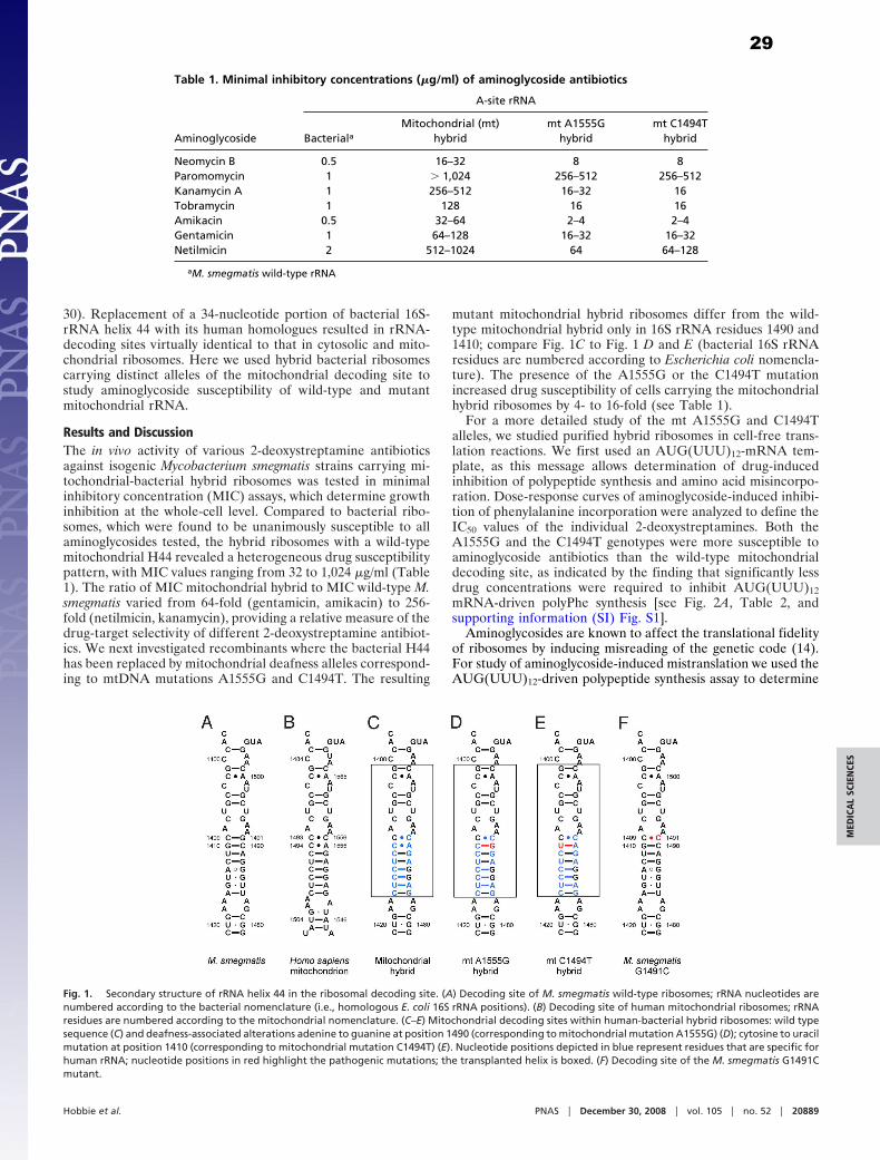
30). Replacement of a 34-nucleotide portion of bacterial 16S-rRNA helix 44 with its human homologues resulted in rRNA-decoding sites virtually identical to that in cytosolic and mito-chondrial ribosomes. Here we used hybrid bacterial ribosomescarrying distinct alleles of the mitochondrial decoding site tostudy aminoglycoside susceptibility of wild-type and mutantmitochondrial rRNA.
Results and Discussion
The in vivo activity of various 2-deoxystreptamine antibioticsagainst isogenic Mycobacterium smegmatis strains carrying mi-tochondrial-bacterial hybrid ribosomes was tested in minimalinhibitory concentration (MIC) assays, which determine growthinhibition at the whole-cell level. Compared to bacterial ribo-somes, which were found to be unanimously susceptible to allaminoglycosides tested, the hybrid ribosomes with a wild-typemitochondrial H44 revealed a heterogeneous drug susceptibilitypattern, with MIC values ranging from 32 to 1,024 �g/ml (Table1). The ratio of MIC mitochondrial hybrid to MIC wild-type M.smegmatis varied from 64-fold (gentamicin, amikacin) to 256-fold (netilmicin, kanamycin), providing a relative measure of thedrug-target selectivity of different 2-deoxystreptamine antibiot-ics. We next investigated recombinants where the bacterial H44has been replaced by mitochondrial deafness alleles correspond-ing to mtDNA mutations A1555G and C1494T. The resulting
mutant mitochondrial hybrid ribosomes differ from the wild-type mitochondrial hybrid only in 16S rRNA residues 1490 and1410; compare Fig. 1C to Fig. 1 D and E (bacterial 16S rRNAresidues are numbered according to Escherichia coli nomencla-ture). The presence of the A1555G or the C1494T mutationincreased drug susceptibility of cells carrying the mitochondrialhybrid ribosomes by 4- to 16-fold (see Table 1).
For a more detailed study of the mt A1555G and C1494Talleles, we studied purified hybrid ribosomes in cell-free trans-lation reactions. We first used an AUG(UUU)12-mRNA tem-plate, as this message allows determination of drug-inducedinhibition of polypeptide synthesis and amino acid misincorpo-ration. Dose-response curves of aminoglycoside-induced inhibi-tion of phenylalanine incorporation were analyzed to define theIC50 values of the individual 2-deoxystreptamines. Both theA1555G and the C1494T genotypes were more susceptible toaminoglycoside antibiotics than the wild-type mitochondrialdecoding site, as indicated by the finding that significantly lessdrug concentrations were required to inhibit AUG(UUU)12
mRNA-driven polyPhe synthesis [see Fig. 2A, Table 2, andsupporting information (SI) Fig. S1].
Aminoglycosides are known to affect the translational fidelityof ribosomes by inducing misreading of the genetic code (14).For study of aminoglycoside-induced mistranslation we used theAUG(UUU)12-driven polypeptide synthesis assay to determine
Table 1. Minimal inhibitory concentrations (�g/ml) of aminoglycoside antibiotics
Aminoglycoside
A-site rRNA
BacterialaMitochondrial (mt)
hybrid
mt A1555G
hybrid
mt C1494T
hybrid
Neomycin B 0.5 16–32 8 8
Paromomycin 1 � 1,024 256–512 256–512
Kanamycin A 1 256–512 16–32 16
Tobramycin 1 128 16 16
Amikacin 0.5 32–64 2–4 2–4
Gentamicin 1 64–128 16–32 16–32
Netilmicin 2 512–1024 64 64–128
aM. smegmatis wild-type rRNA
Fig. 1. Secondary structure of rRNA helix 44 in the ribosomal decoding site. (A) Decoding site of M. smegmatis wild-type ribosomes; rRNA nucleotides are
numbered according to the bacterial nomenclature (i.e., homologous E. coli 16S rRNA positions). (B) Decoding site of human mitochondrial ribosomes; rRNA
residues are numbered according to the mitochondrial nomenclature. (C–E) Mitochondrial decoding sites within human-bacterial hybrid ribosomes: wild type
sequence (C) and deafness-associated alterations adenine to guanine at position 1490 (corresponding to mitochondrial mutation A1555G) (D); cytosine to uracil
mutation at position 1410 (corresponding to mitochondrial mutation C1494T) (E). Nucleotide positions depicted in blue represent residues that are specific for
human rRNA; nucleotide positions in red highlight the pathogenic mutations; the transplanted helix is boxed. (F) Decoding site of the M. smegmatis G1491C
mutant.
Hobbie et al. PNAS � December 30, 2008 � vol. 105 � no. 52 � 20889
MED
ICA
LS
CIE
NC
ES
29

the relative amount of near-cognate leucine incorporation com-pared to incorporation of the cognate amino acid phenylalaninein the presence of various concentrations of gentamicin. Relativeincorporation of [3H]-labeled leucine versus [14C]-labeled phe-nylalanine was determined and plotted against gentamicin con-centration. Introduction of the A1555G and C1494T alterationrendered the mitochondrial hybrid ribosomes highly susceptibleto aminoglycoside-induced misreading (Fig. 2B; see also Fig.S2). In quantitative terms, the amount of gentamicin-inducedmisreading (calculated as leucine per phenylalanine incorpora-tion) in A1555G and C1494T mutant hybrid ribosomes was upto 1.75 leucine per phenylalanine, as compared to a maximum of0.2 leucine per phenylalanine for hybrid ribosomes with awild-type mitochondrial decoding site.
To study the effect of aminoglycoside antibiotics on translationof a more natural mRNA template, we tested wild-type and mutantmitochondrial hybrid ribosomes in a cell-free luciferase synthesisassay. As depicted in Fig. 2C, Table 3, and Fig. S3, the allele- anddrug-specific inhibition of luciferase synthesis essentially correlatedwith the results of the MIC and AUG(UUU)12 assays. Drug-mediated inhibition of luciferase synthesis was significantly in-creased in A1555G and C1494T mutant ribosomes.
The basis for the selectivity of aminoglycosides is presumablytheir preferential binding to bacterial as opposed to eukaryoticribosomes (10, 13, 16, 17). In particular, 16S rRNA nucleotides1408, 1409, and 1491 of helix 44 have been shown to be criticalfor drug-binding by forming direct contacts with ring I of the2-deoxystreptamines (12, 13, 16, 17, 26, 31–38) (see Fig. S4 forthe chemical structures of aminoglycosides used in this study). Inthe absence of X-ray structures for aminoglycosides complexed
to the mitochondrial ribosome, we can rationalize our findingson data invoked from the study of bacterial ribosome-drugcomplexes (12, 39). The rRNA secondary structure of the drugbinding site in mitochondrial A1555G and C1494T mutantribosomes resembles that of bacterial ribosomes with a G1491Calteration in that the C1409-C1491 opposition is accompanied bya 1410–1490 Watson–Crick pair (see Fig. 1). The bacterialG1491C ribosome shows a drug-susceptibility phenotype that isvirtually superimposable on that found for the mitochondrialdeafness alleles (see Table S1 for comparison). To determinewhether the affinity of aminoglycosides to mutant mitochondrialdecoding sites corresponds to that of the bacterial G1491Cdecoding site, we probed gentamicin binding by chemical foot-printing experiments. Bacterial wild-type ribosomes showeddrug-mediated protection from dimethyl sulfate (DMS) modi-fication at G1405 (N-7), which is in good agreement withprevious reports on aminoglycoside protection in bacterial 16SrRNA (34). Wild-type mitochondrial hybrid ribosomes showedlittle protection, while mutant mitochondrial A1555G andC1494T hybrid ribosomes showed a concentration-dependentprotection of G1405 that resembles the dose-response curveobserved with bacterial G1491C ribosomes (Fig. 3). Thus, bind-ing of aminoglycosides to ribosomes with an adenine at 16SrRNA position 1408, appears to be mainly determined by thestructural geometry of base pairs 1409–1491 and 1410–1490.
When studying drug-induced miscoding, we found that thedecoding accuracy of bacterial G1491C ribosomes is barelyaffected by aminoglycoside antibiotics. In absolute terms and incontrast to the mitochondrial A1555G and C1494T deafnessmutants, the bacterial G1491C ribosomes showed little drug-
Fig. 2. Aminoglycoside susceptibility of mutant and wild-type mitochondrial hybrid ribosomes. Dose-response curves of wild-type mitochondrial (red squares),
mutant A1555G (green triangles), and mutant C1494T (blue inverted triangles) hybrid ribosomes; bacterial ribosomes (black circles) are included for comparison.
(A) Gentamicin-induced inhibition of [14C]-phenylalanine incorporation (n � 3; � SD.). The 100% value corresponds to 25 to 30 pmol Phe incorporation.
Corresponding IC50 values of gentamicin and selected aminoglycoside antibiotics are presented in Table 2. (B) Gentamicin-induced increase in misincorporation
of the near-cognate [3H]-leucine relative to the drug-free control (n � 3; � SD.). (C) Gentamicin-induced inhibition of luciferase synthesis relative to the drug-free
control (n � 3; � SD.). Corresponding IC50 values for gentamicin and selected aminoglycoside antibiotics are presented in Table 3.
Table 2. Aminoglycoside-induced inhibition of AUG(UUU)12-driven phenylalanine
incorporation (IC50, �M)
Aminoglycoside
A-site rRNA
BacterialaMitochondrial (mt)
hybrid
mt A1555G
hybrid
mt C1494T
hybrid
Neomycin B 0.2 � 0.0 209 � 61 3.1 � 0.3 3.7 � 0.7
Paromomycin 0.7 � 0.1 � 500 124 � 35 116 � 20
Kanamycin A 1.2 � 0.2 � 500 20 � 2 95 � 29
Tobramycin 0.7 � 0.1 � 500 15 � 3 70 � 23
Amikacin 0.8 � 0.1 � 500 7.1 � 0.8 13 � 3
Gentamicin 1.3 � 0.2 � 500 13 � 2 62 � 16
Netilmicin 1.2 � 0.3 � 500 71 � 10 331 � 84
IC50 values represent the drug concentrations in �M that are required to inhibit AUG(UUU)12-driven phenyl-
alanine incorporation to half-maximal extent. A representative graph showing phenylalanine incorporation
plotted against gentamicin concentration is shown in Fig. 2A.aM. smegmatis wild-type rRNA
20890 � www.pnas.org�cgi�doi�10.1073�pnas.0811258106 Hobbie et al.
30

induced misreading (see Fig. S2). The bacterial G1491C mutantand the hybrid deafness ribosomes differ primarily in 16S rRNAresidues 1413 to 1415 and 1485 to 1487, which form the lowerstem of helix 44 (see Fig. 1). At the structural level, helix 44interacts with helix 27. By modeling, nucleotide alterations in thelower stem of H44 have been suggested to affect this interaction
and the relative movement between these two helices as part ofthe conformational change required in decoding (30). Appar-ently, the nature of the lower stem plays an important role inboth spontaneous and drug-aggravated miscoding and deter-mines the translational accuracy of the mutant decoding sites.Thus, susceptibility of A1555G and C1494T deafness mitoribo-somes is the result of two mechanisms, which act in concert:increased drug binding to its target and excessive aggravation ofthe mutants’ inherent deficiency in ribosomal accuracy.
Several lines of evidence link aminoglycoside ototoxicity to themitochondrial ribosome: (i) mitochondrial ribosomes are struc-turally more similar to their prokaryotic ancestor than to theeukaryotic cytosolic homologues; (ii) compared to cytosolicribosomes, the mitochondrial ribosomes of higher eukaryotesexhibit a remarkable degree of aminoglycoside susceptibility(27) [see Table S2, which compares the drug susceptibility ofhybrid bacterial ribosomes with the A site (H44) of humancytosolic ribosomes to that of hybrid ribosomes with the mitochon-drial decoding site]; and (iii) idiosyncratic drug susceptibility isassociated with genetic predisposition, in particular mutations inmtDNA: 20 to 40% of patients with aminoglycoside-inducedototoxicity either carry the A1555G or the C1494T mutation inthe 12S rRNA gene (6, 40).
To further assess whether aminoglycoside-induced ototoxicityis a result of the drugs’ anti-mitoribosomal activity, we comparedthe potencies of a series of aminoglycosides to inhibit mitoribo-some function with their relative cochleotoxicity in humans (41).The correlation between these two measures (Fig. 4) is consis-tent with the hypothesis that aminoglycoside-induced cochleo-toxicity relates to the drugs’ activity against mitochondrialribosomes. Further evidence for this hypothesis is provided byour finding that netilmicin, which displays the least cochleartoxicity of the clinical aminoglycosides (reviewed in ref. 42), issignificantly less active against hybrid mitochondrial ribosomesthan gentamicin, tobramycin, or amikacin.
In summary, we provide experimental evidence for a mech-anistic linkage between the mitochondrial A1555G and C1494Tmutations and hypersusceptibility to aminoglycosides, althoughthe exquisite tissue-specific action of aminoglycoside toxicity(that is, ototoxicity) is likely to involve additional factors (e.g.,reactive oxygen species, drug uptake, or polyamine-like activa-tion of NMDA receptors) (2, 43). Our results provide experi-mental support for aminoglycoside-induced dysfunction of themitochondrial ribosome. We propose a pathogenic mechanism,in which interference of aminoglycosides with mitochondrialprotein synthesis exacerbates the drugs’ cochlear toxicity, play-ing a key role in sporadic dose-dependent and geneticallyinherited, aminoglycoside-induced deafness. Based upon ourexperiments, we suggest a scenario of aminoglycoside hearing
Table 3. Aminoglycoside-induced inhibition of luciferase synthesis (IC50, �M)
Aminoglycoside
A-site rRNA
BacterialaMitochondrial (mt)
hybrid
mt A1555G
hybrid
mt C1494T
hybrid
Neamine 1.4 131 32 35
Neomycin B 0.04 0.7 0.5 0.4
Paromomycin 0.03 33 2.4 3.1
Kanamycin A 0.05 15.7 1.2 1.1
Tobramycin 0.02 7.8 0.8 0.9
Amikacin 0.02 7.0 0.4 0.6
Gentamicin 0.03 5.7 0.6 0.7
Netilmicin 0.05 17.6 0.8 2.6
IC50 values represent the drug concentrations in �M that are required to inhibit synthesis of functional firefly
luciferase to 50%. Relative luciferase activity plotted against aminoglycoside concentration is shown in Fig. 2C.aM. smegmatis wild-type rRNA
Fig. 3. Chemical footprints of gentamicin binding to wild-type and mutant
mitochondrial decoding sites in comparison to bacterial wild-type and G1491C
ribosomes. (A) Gentamicin-dependent protection of G1405 in wild-type mi-
tochondrial (red squares), mutant mt A1555G (green triangles), mutant mt
C1494T (blue inverted triangles), and bacterial G1491C (purple diamonds)
decoding sites; wild-type bacterial ribosomes (black circles) are included for
comparison. (B) Corresponding footprinting blots showing primer extensions
starting with U1420. i, bacterial wild type; ii, mt wild type; iii, mt A1555G; iv,
mt C1494T; v, bacterial G1491C.
Hobbie et al. PNAS � December 30, 2008 � vol. 105 � no. 52 � 20891
MED
ICA
LS
CIE
NC
ES
31

loss, which is initiated by mitoribosomal misreading, subse-quently via activation of downstream signaling pathways, such asMAPK and JNK (44, 45), misreading results in hair cell deaththrough apoptosis.
Materials and Methods
Construction of Mutant Strains with Hybrid Ribosomes. The recently described
M. smegmatis mc2 155 SmS �rrnB (38) was used for all genetic manipulations.
Site-directed mutagenesis of its single rRNA operon was done by PCR mu-
tagenesis using hybrid rDNA oligonucleotides comprising the wild-type or
mutant mitochondrial helix 44 decoding-site sequence. The resulting hybrid
gene fragment was cloned into an integration-proficient plasmid used to
transform M. smegmatis �rrnB. Transformants were selected on LB agar plates
containing 20 �g/ml paromomycin for gene replacement by homologous
recombination. Resulting recombinant M. smegmatis cells had the central
34-nucleotide part of the bacterial H44 replaced by its mitochondrial coun-
terpart. Successful replacement of the bacterial decoding-site sequence with
the mitochondrial sequence was controlled by sequence analysis of the chro-
mosomal rrnA locus.
Minimal Inhibitory Concentration Assays. Minimal inhibitory concentrations of
neomycin B, paromomycin, kanamycin A, tobramycin, amikacin, gentamicin,
netilmicin (all Sigma), and neamine were determined by broth microdilution
assays as described previously (46). Neamine was a kind gift of Andrea Vasella,
ETH Zurich. The gentamicin used in this study is a mixture of gentamicin C1,
gentamicin C1a, and gentamicin C2 in a 45:35:30 ratio.
Isolation and Purification of Ribosomes. Ribosomes were purified from bacte-
rial cell pellets as described previously (30). In brief, ribosome particles were
isolated by successive centrifugations and fractionated by sucrose gradient
(10–40%) centrifugation. The 70S ribosome-enriched fraction was pelleted,
resuspended in association buffer, incubated for 30 min at 4 °C, dispensed into
aliquots, and stored at �80 °C following shock freezing in liquid nitrogen.
Ribosome concentrations of 70S were determined by absorption measure-
ments on the basis of 23 pmol ribosomes per A260 unit. Integrity and functional
activity of purified 70S ribosomes was determined by analytical ultra-
centrifugation and by assessing their capacity to form initiation complexes, as
described previously (29).
Cell-free AUG(UUU)12 Translation Assays. Cell-free translation reactions were
done as described previously (30). A reaction mixture containing M. smegma-
tis tRNAbulk, amino acids, S100 extract, energy mix, pyruvate kinase, and
polyamines was preincubated with 30 �M [14C]-phenylalanine (110 mCi/
mmol) and/or 30 �M [3H]-leucine (500 mCi/mmol) at 37 °C for 15 min. The
translation reaction was started by addition of ribosomes to a final concen-
tration of 0.25 �M, AUG(UUU)12-mRNA (5�-GCGGCAAGGAGGUAAAUA AUG
(UUU)12 UAA GCAGG-3�, obtained from Dharmacon) to 1 �M, and aminogly-
coside antibiotics in serial dilutions. Following incubation for 60 min at 37 °C,
the reaction was stopped by addition of KOH, precipitated polypeptides were
collected on filters, and [14C]-phenylalanine or [3H]-leucine were quantified.
Background values for Phe and Leu incorporation were 0.4 to 0.5 pmol at time
zero; the background was not subtracted from the experimental values
determined.
Cell-Free Luciferase Translation Assays. Purified 70S hybrid ribosomes were
used in a coupled transcription-translation reaction as described previously
(29). The reaction mixture was incubated for 60 min at 37 °C, stopped on ice,
and luciferase assay substrate (Promega) was added. Functional protein was
quantified by measuring bioluminescence in a luminometer (Bio-Tek instru-
ments, FLx800).
Footprinting Analyses. DMS modification of 70S ribosomes (20 pmol) was per-
formedin100�lbuffercontaining80mMpotassiumcacodylate (pH6.5),100mM
ammonium chloride, 20 mM magnesium chloride, 1 mM DTT, and 0.5 mM EDTA.
Following ribosome activation for 15 min at 37 °C, gentamicin was added and the
reaction mixture was incubated for another 15 min before addition of DMS (6 �l,
1:10 inethanol).Followinga30-minincubationat37 °C, thereactionwasstopped
by addition of 100 �l DMS Stop solution (50 mM Tris, pH 7.5, 300 mM sodium
chloride, 1% SDS, 200 mM �-mercaptoethanol). Ribosomes were precipitated
with ethanol, pelleted, resuspended in 200 �l 50 mM Tris pH 8, 0.5% SDS, and
extracted with phenol/chloroform. DMS-modified RNA was precipitated with
ethanol and sodium borohydride reduction and aniline-induced strand scission
was performed as described previously (47). Primer extension of 16S rRNA was
performed as described (48), using DNA oligonucleotides complementary to
16S-rRNA nucleotides 1445 to 1421. Air-dried gels were scanned and quantified
using the STORM PhosphorImaging System with ImageQuant 5.2 Software (Am-
ersham Bioscience).
ACKNOWLEDGMENTS. The authors thank Tanja Janusic (Institut fur Mediz-inische Mikrobiologie) for expert technical assistance, Andrea Vasella (SwissFederal Institute of Technology, Zurich) for kindly providing neamine, andAlexander Mankin for helpful comments on the manuscript. This work wassupported by grants from the Swiss National Science Foundation (to E.C.B.)and from the Bonizzi-Theler-Stiftung (to S.N.H.).
1. Chambers HF, Sande MA (1996) in Goodman and Gilman’s The Pharmacological Basis
of Therapeutics, ed. Goodman LS, Limbird LE, Milinoff PB, Gilman AG, Hardman JG.
(McGraw-Hill, New York), pp. 1103–1121.
2. Forge A, Schacht J (2000) Aminoglycoside antibiotics. Audiol Neurootol 5:3–22.
3. Matsui JI, Cotanche DA (2004) Sensory hair cell death and regeneration: two halves of
the same equation. Curr Opin Otolaryngol Head Neck Surg 12:418–425.
4. Prezant TR, Agapian JV, Bohlman MC, Bu X, Oztas S (1993) Mitochondrial ribosomal
RNA mutation associated with both antibiotic-induced and non-syndromic deafness.
Nat Genet 4:289–294.
5. Fischel-Ghodsian N (1999) Mitochondrial deafness mutations reviewed. Hum Mutat
13:261–270.
6. Zhao H, Li R, Wang Q, Yan Q, Deng JH (2004) Maternally inherited aminoglycoside-
induced and nonsyndromic deafness is associated with the novel C1494T mutation in
the mitochondrial 12S rRNA gene in a large Chinese family. Am J Hum Genet 74:139–
152.
7. Chen Y, Huang WG, Zha DJ, Qiu JH, Wang JL (2007) Aspirin attenuates gentamicin
ototoxicity: from the laboratory to the clinic. Hear Res 226:178–182.
8. Sha SH, Qiu JH, Schacht J (2006) Aspirin to prevent gentamicin-induced hearing loss.
N Engl J Med 354:1856–1857.
9. Hutchin T, Cortopassi G (1994) Proposed molecular and cellular mechanism for ami-
noglycoside ototoxicity. Antimicrob Agents Chemother 38:2517–2520.
10. Gale EF, Cundliffe E, Reynolds PE, Richmond MH, Waring JM (1981) The Molecular Basis
of Antibiotic Action (John Wiley & Sons, Inc., London).
11. Moazed D, Noller HF (1987) Interaction of antibiotics with functional sites in 16S
ribosomal RNA. Nature 327:389–394.
12. Carter AP, Clemens WM, Brodersen DE, Morgan-Warren EJ, Wimberly BT (2000)
Functional insights from the structure of the 30S ribosomal subunit and its interactions
with antibiotics. Nature 407:340–348.
13. Bottger EC, Springer B, Prammananan T, Kidan Y, Sander P (2001) Structural basis for
selectivity and toxicity of ribosomal antibiotics. EMBO Rep 2:318–323.
14. Davies J, Gorini L, Davis BD (1965) Misreading of RNA codewords induced by amino-
glycoside antibiotics. Mol Pharmacol 1:93–106.
15. Campuzano S, Vazquez D, Modolell J (1979) Functional interaction of neomycin B and
related antibiotics with 30S and 50S ribosomal subunits. Biochem Biophys Res Commun
87:960–966.
16. Sander P, Prammananan T, Bottger EC (1996) Introducing mutations into a chromo-
somal rRNA gene using a genetically modified eubacterial host with a single rRNA
operon. Mol Microbiol 22:841–848.
Fig. 4. Relationship between inhibition of protein synthesis in mitochondrial
hybrid ribosomes and relative in vitro cochleotoxicity of aminoglycoside
antibiotics. The potencies of a series of cochleotoxic aminoglycosides (Ak,
amikacin; Gm, gentamicin; Ne, neamine; Nm, neomycin) in inhibiting protein
synthesis in hybrid mitochondrial ribosomes (see Table 3) correlates with the
relative cochleotoxicity previously reported by Kotecha and Richardson (41).
20892 � www.pnas.org�cgi�doi�10.1073�pnas.0811258106 Hobbie et al.
32

17. Recht MI, Douthwaite S, Puglisi JD (1999) Basis for prokaryotic specificity of action of
aminoglycoside antibiotics. EMBO J 18:3133–3138.
18. Purohit P, Stern S (1994) Interactions of a small RNA with antibiotic and RNA ligands of
the 30S subunit. Nature 370:659–662.
19. Griffey RH, Hofstadler SA, Sannes-Lowery KA, Ecker DJ, Crooke ST (1999) Determinants
of aminoglycoside-binding specificity for rRNA by using mass spectrometry. Proc Natl
Acad Sci USA 96:10129–10133.
20. Shandrick S, et al. (2004) Monitoring molecular recognition of the ribosomal decoding
site. Angew Chem Int Ed Engl 43:3177–3182.
21. Kaul M, Barbieri CM, Pilch DS (2006) Aminoglycoside-induced reduction in nucleotide
mobility at the ribosomal RNA A-site as a potentially key determinant of antibacterial
activity. J Am Chem Soc 128:1261–1271.
22. Kondo J, Westhof E (2008) The bacterial and mitochondrial ribosomal A-site molecular
switches possess different conformational substates. Nucleic Acids Res 36:2654–2666.
23. Ryu DH, Rando RR (2001) Aminoglycoside binding to human and bacterial A-Site rRNA
decoding region constructs. Bioorg Med Chem 9:2601–2608.
24. Hamasaki K, Rando RR (1997) Specific binding of aminoglycosides to a human rRNA
construct based on a DNA polymorphism which causes aminoglycoside-induced deaf-
ness. Biochemistry 36:12323–12328.
25. Kaul M, Barbieri CM, Pilch DS (2005) Defining the basis for the specificity of amino-
glycoside-rRNA recognition: a comparative study of drug binding to the A sites of
Escherichia coli and human rRNA. J Mol Biol 346:119–134.
26. Pfister P, et al. (2005) Mutagenesis of 16S rRNA C1409–G1491 base-pair differentiates
between 6�OH and 6�NH3� aminoglycosides. J Mol Biol 346:467–475.
27. Kurtz DI (1974) Fidelity of protein synthesis with chicken embryo mitochondrial and
cytoplasmic ribosomes. Biochemistry 13:572–577.
28. Ryu DH, Rando RR (2002) Decoding region bubble size and aminoglycoside antibiotic
binding. Bioorg Med Chem Lett 12:2241–2244.
29. Hobbie SN, et al. (2007) Engineering the rRNA decoding site of eukaryotic cytosolic
ribosomes in bacteria. Nucleic Acids Res 35:6086–6093.
30. Hobbie SN, et al. (2008) Mitochondrial deafness alleles confer misreading of the
genetic code. Proc Natl Acad Sci USA 105:3244–3249.
31. De Stasio EA, Moazed D, Noller HF, Dahlberg AE (1989) Mutations in 16S ribosomal RNA
disrupt antibiotic-RNA interactions. EMBO J 8:1213–1216.
32. De Stasio EA, Dahlberg AE (1990) Effects of mutagenesis of a conserved base-paired site
near the decoding region of Escherichia coli 16 S ribosomal RNA. J Mol Biol 212:127–
133.
33. Prammananan T, Sander P, Springer B, Bottger EC (1999) RecA-mediated gene con-
version and aminoglycoside resistance in strains heterozygous for rRNA. Antimicrob
Agents Chemother 43:447–453.
34. Recht MI, Puglisi JD (2001) Aminoglycoside resistance with homogeneous and heter-
ogeneous populations of antibiotic-resistant ribosomes. Antimicrob Agents Che-
mother 45:2414–2419.
35. Pfister P, Hobbie S, Vicens Q, Bottger EC, Westhof E (2003) The molecular basis for
A-Site mutations conferring aminoglycoside resistance: relationship between ribo-
somal susceptibility and X-ray crystal structures. ChemBioChem 4:1078–1088.
36. Hobbie SN, Pfister P, Brull C, Westhof E, Bottger EC (2005) Analysis of the contribution
of individual substituents in 4,6-aminoglycoside-ribosome interaction. Antimicrob
Agents Chemother 49:5112–5118.
37. Hobbie SN, et al. (2006) Binding of neomycin-class aminoglycoside antibiotics to
mutant ribosomes with alterations in the A-site of 16S rRNA. Antimicrob Agents
Chemother 50:1489–1496.
38. Hobbie SN, et al. (2006) A genetic model to investigate structural drug-target inter-
actions at the ribosomal decoding site. Biochimie 88:1033–1043.
39. Vicens Q, Westhof E (2001) Crystal structure of paromomycin docked into the eubac-
terial ribosomal decoding A site. Structure (Camb) 9:647–658.
40. Estivill X, et al. (1998) Familial progressive sensorineural deafness is mainly due to the
mtDNA A1555G mutation and is enhanced by treatment of aminoglycosides. Am J Hum
Genet 62:27–35.
41. Kotecha B, Richardson GP (1994) Ototoxicity in vitro: effects of neomycin, gentamicin,
dihydrostreptomycin, amikacin, spectinomycin, neamine, spermine and poly-L-lysine.
Hear Res 73:173–184.
42. Begg EJ, Barclay ML (1995) Aminoglycosides-50 years on. Br J Clin Pharmacol 39:597–
603.
43. Basile AS, et al. (1996) N-methyl-D-aspartate antagonists limit aminoglycoside antibi-
otic-induced hearing loss. Nat Med 2:1338–1343.
44. Wang J, et al. (2003) A peptide inhibitor of c-Jun N-terminal kinase protects against
both aminoglycoside and acoustic trauma-induced auditory hair cell death and hear-
ing loss. J Neurosci 23:8596–8607.
45. Kalinec GM, et al. (2005) Pivotal role of Harakiri in the induction and prevention of
gentamicin-induced hearing loss. Proc Natl Acad Sci USA 102:16019–16024.
46. Pfister P, et al. (2005) 23S rRNA base pair 2057–2611 determines ketolide susceptibility
and fitness cost of the macrolide resistance mutation 2058A-�G. Proc Natl Acad Sci
USA 102:5180–5185.
47. Peattie DA (1979) Direct chemical method for sequencing RNA. Proc Natl Acad Sci USA
76:1760–1764.
48. Merryman C, Noller HF (1998) in RNA:Protein Interactions: A Practical Approach, ed.
Smith CWJ. (Oxford Univ Press, Oxford, UK), pp. 237–253.
Hobbie et al. PNAS � December 30, 2008 � vol. 105 � no. 52 � 20893
MED
ICA
LS
CIE
NC
ES
33

Supporting Information
Hobbie et al. 10.1073/pnas.0811258106
Fig. S1. Aminoglycoside susceptibility of bacterial hybrid ribosomes in AUG(UUU)12-directed polypeptide synthesis. Dose-response curves of bacterial (black
circles), mitochondrial (red squares), mutant A1555G (green triangles), and mutant C1494T (blue inverted triangles) hybrid ribosomes. Corresponding IC50 values
are given in Table 2.
Hobbie et al. www.pnas.org/cgi/content/short/0811258106 1 of 6
34

Fig. S2. Aminoglycoside-induced miscoding relative to a drug-free control in a M. smegmatis G1491C mutant in comparison to mutant mitochondrial decoding
sites. Dose-response curves of M. smegmatis G1491C (purple diamonds), mitochondrial (red squares), mutant A1555G (green triangles), and mutant C1494T (blue
inverted triangles) hybrid ribosomes. Bacterial wild-type ribosomes (black circles) are included for comparison.
Hobbie et al. www.pnas.org/cgi/content/short/0811258106 2 of 6
35

Fig. S3. Aminoglycoside-induced inhibition of luciferase synthesis. Dose-response curves of bacterial (black circles), mitochondrial (red squares), mutant
A1555G (green triangles), and mutant C1494T (blue inverted triangles) hybrid ribosomes. Corresponding IC50 values are given in Table 3.
Hobbie et al. www.pnas.org/cgi/content/short/0811258106 3 of 6
36

Fig. S4. Chemical structures of 2-deoxystreptamine antibiotics used in this study.
Hobbie et al. www.pnas.org/cgi/content/short/0811258106 4 of 6
37

Table S1. Minimal inhibitory concentrations (�g/ml) against M. smegmatis G1491C mutant in comparison to mutant mitochondrial
hybrids
A-site rRNA
M. smegmatis Mitochondrial (mt) hybrid mt A1555G hybrid mt C1494T hybrid M. smegmatis G1491C
Neomycin B 0.5 16–32 8 8 16
Paromomycin 1 � 1,024 256–512 256–512 512
Kanamycin A 1 256–512 16–32 16 16–32
Tobramycin 1 128 16 16 16
Amikacin 0.5 32–64 2–4 2–4 4
Gentamicin 1 64–128 16–32 16–32 16–32
Netilmicin 2 512–1,024 64 64–128 128
Hobbie et al. www.pnas.org/cgi/content/short/0811258106 5 of 6
38

Table S2. Aminoglycoside susceptibility of the mitochondrial
versus the cytosolic decoding site
MIC (�g/ml)
Mitochondrial hybrid Cytosolic hybrid
Neomycin B 16–32 � 1,024
Kanamycin A 256–512 � 1,024
Tobramycin 128 1,024
Amikacin 32–64 512–1,024
Gentamicin 64–128 � 1,024
Netilmicin 512–1,024 � 1,024
Hobbie et al. www.pnas.org/cgi/content/short/0811258106 6 of 6
39

1
2
3
4
5
6 7 8 9
10
11
12 13 14 15 16
17
18
19
20
21
22
23
24
25
26
27
28
29
30
31
32
33
34
35
36
37
38
39
40
41
(Manuscript in preparation)
Mutation K42R in Ribosomal Protein S12 Does Not Affect Susceptibility of Mycobacterium smegmatis 16S rRNA A- site Mutants to 2-deoxystreptamine Aminoglycosides.
Sarath K. Kalapala1, Sven N. Hobbie
1,2, Erik C. Böttger
1 and Dmitry Shcherbakov
1*
1 Institut für Medizinische Mikrobiologie, Universität Zürich, Gloriastrasse 32, CH-8006 Zürich, Switzerland.
2 Present address: Singapore-MIT Alliance for Research and Technology (SMART),
Centre for Life Sciences, 28 Medical Drive, Singapore.
*Corresponding author. Mailing address: Institut für Medizinische Mikrobiologie, Universität Zürich, Gloriastrasse 32, CH-8006 Zürich, Switzerland. Phone: +41 44 634 2672. FAX: +41 44 634 49 06. E-Mail: [email protected]
Abstract
Recent studies have suggested that ribosomal protein S12 modulates 16S rRNA
function and susceptibility to 2-deoxystreptamine aminoglycosides. To study whether
the non-restrictive K42R mutation in RpsL affects 2-deoxystreptamine susceptibility in
Mycobacterium smegmatis, we studied the drug susceptibility pattern of various
mutants with genetic alterations in the 16S rRNA decoding A-site in the context of
wild-type and mutant protein S12. RpsL K42R substitution was found not to affect the
drug resistance pattern associated with mutational alterations in 16S rRNA H44.
Introduction
Ribosomal protein S12 is a critical component of the A-site of the 30S ribosomal subunit and
is involved in both tRNA selection and resistance to streptomycin [1,2]. Mutations in rpsL
coding for ribosomal protein S12 are known to affect ribosomal accuracy to various extents,
resulting in what is characterized as error-restrictive or non-restrictive S12 alterations [3].
Streptomycin inhibits protein synthesis and makes ribosomes error prone by affecting initial
tRNA selection and proof-reading [4]. Mutations in S12 confer streptomycin resistance by
preventing streptomycin binding and/or conferring ribosomal hyperaccuracy; a strongly
hyperaccurate phenotype may even manifest as streptomycin-dependence [2]. Various
substitutions at positions 42 and 87 are associated with streptomycin resistance. In
particular, mutations Lys42 → Arg, Ala or Thr promote high levels of streptomycin resistance;
among those Lys42Arg has a non-restrictive phenotype [3], whereas Lys42Ala and Lys42Thr
are strongly error-restrictive [3,5,6]. S12 substitutions Lys87 → Gln or Gly confer different
degrees of streptomycin resistance [7,8]. The homologous mammalian ribosomal proteins
40

42
43
44
45
46
47
48
49
50
51
52
53
54
55
56
57
58
59
60
61
62
63
64
65
66
67
68
69
70
71
72
73
74
75
76
77
78
carry Gln-87 (human mitochondrial ribosomes) or Gly-87 (human cytoplasmic ribosomes) in
part accounting for the drugs’ prokaryotic specificity [7].
More recently it has been suggested that ribosomal protein S12 modulates 16S-rRNA
function and susceptibility to 2-deoxystreptamine aminoglycosides [5,6]. 2-deoxystreptamine
aminoglycosides are composed of a common core, termed neamine, in which position 4 of a
2-deoxystreptamine ring (ring II) is attached to a glycopyranosyl ring (ring I). Additional
sugars are attached to position 5 or 6 of the 2-deoxystreptamine moiety to give rise to 4,5- or
4,6- aminoglycosides (see Supplementary Data Fig. S1). In 4,5- aminoglycosides, the core is
further substituted by one (ribostamycin), two (neomycin, paromomycin) or three
(lividomycin) additional sugars attached to position 5 of ring II, whereas in 4,6-
aminoglycosides (gentamicin, tobramycin, kanamycin, etc.) the core is further substituted by
one additional sugar attached to position 6 of ring II. The drug binding pocket for these
compounds consists of an internal loop of 16S-rRNA helix 44 – the decoding A-site of the
ribosome [9]. We have previously performed extensive genetic studies of 16S-rRNA helix 44
in Mycobacterium smegmatis to address the role of individual rRNA residues in drug binding
(reviewed in [10]). These studies have been conducted mainly in the genetic background of a
non-restrictive K42R mutation in ribosomal protein S12 [10-16]. The K42R mutation confers
high-level resistance to streptomycin and was used as counter-selectable marker in strain
construction [15]. The recent reports on the interplay of S12 on 16S rRNA function and
susceptibility to 2-deoxystreptamine aminoglycosides prompted us to study in detail the role,
if any, between RpsL K42R and mutational alterations in 16S-rRNA helix 44 conferring
resistance to 2-deoxystreptamines. We find that the non-restrictive RpsL K42R mutation
does not affect the 2-deoxystreptamine susceptibility of various rRNA mutations in
M. smegmatis H44.
Results and Discussion
Generation of M. smegmatis strains with mutations in S12 and 16S rRNA
M. smegmatis was the first eubacterial model organism that was made single rRNA allelic by
means of deletion mutagenesis and that allowed for genetic studies of its ribosomal RNA
[16]. As a gram-positive mesophilic bacterium, M. smegmatis is susceptible to a number of
ribosomal antibiotics and sets itself apart from other model organisms by being a close
representative of clinically relevant pathogenic bacteria. Early genetic studies on
susceptibility to 2-deoxystreptamine aminoglycoside antibiotics relied on M. smegmatis
strains with a K42R mutation in ribosomal protein S12 [10-16]. To substantiate the results
and conclusions drawn from these early studies, we constructed a comprehensive set of
M. smegmatis rRNA mutants in the context of both a wild-type and a mutant S12 (Table S1).
41

79
80
81
82
83
84
85
86
87
88
89
90
91
The set of 16S rRNA mutants includes alterations of positions 1408 (A1408G), 1491
(G1491C; G1491U; G1491A) and 1409 (C1409U; C1409G).
Aminoglycoside susceptibility of M. smegmatis mutants
Aminoglycoside susceptibility of the mutant strains was determined by minimal inhibitory
concentration (MIC) assays. Representatives for each of the two disubstituted 2-
deoxystreptamine subclasses were included: the 4,5-disubstituted 2-deoxystreptamines
paromomycin and neomycin and the 4,6-disubstituted 2-deoxystreptamines gentamicin,
tobramycin and kanamycin. The results are presented in Table 1.
Table 1. Drug susceptibility of M. smegmatis 16S rRNA mutants: wild-type S12 versus K42R
16S rRNA residues S12 MIC (μg/mL) Reference
1408 1409–1491 Pm Nm Gm Tb Km
A C≡G wt 1 0.5 1 1 0.5-1 this study
A C≡G K42R 1 1 1 1 0.5-1 [12]
G C≡G wt 64 > 1024 > 1024 > 1024 > 1024 this study
G C≡G K42R 64 ≥ 1024 > 1024 > 1024 > 1024 [12]
A C � C wt 512 16 16-32 16 16-32 this study
A C � C K42R 512 16-32 16-32 16-32 16-32 [12]
A C � U wt 512-1024 8-16 32-64 64 64-128 this study
A C � U K42R 512 8-16 32 32-64 128 [12]
A C � A wt 32-64 2 2 2 1-2 this study
A C � A K42R 32 4 2 4 2 [12]
A U · G wt 4-8 0.5-1 8 8-16 8-16 this study
A U · G K42R 4-8 1 8-16 8-16 16 [12]
A G � G wt 32-64 4-8 1-2 8-16 32-64 this study
A G � G K42R 16-32 4 2-4 8-16 16-32 [12]
92 93 94
95
96
Pm, paromomycin; Nm, neomycin; Gm, gentamicin; Tb, tobramycin; Km, kanamycin A.
42

97
98
99
100
101
102
103
104
105
106
107
108
109
110
111
112
113
114
115
116
117
118
119
120
121
122
123
124
125
126
127
128
129 130 131 132
133
134
Among all A-site mutations that confer aminoglycoside resistance, the A1408G mutation is
the most significant. In M. smegmatis the A1408G mutation confers moderate resistance to
paromomycin (6′ OH) but high level resistance to 2-deoxystreptamine aminoglycosides with
an amino group at the 6′ position of ring I. A key element in drug binding is the pseudo base-
pair interaction between the aminoglycosides’ ring I and A 1408 [13,17,18]. In case of an
adenine, the oxygen of ring I accepts a hydrogen bond from the N6 of A1408, and the amino
or hydroxyl group at position 6′ donates a hydrogen bond to the N1 of adenine, accounting
for two direct hydrogen bonds between ring I and A1408 (Fig. 1). In case of a 1408 guanine,
the 6′ amino group of ring I can no longer form an H bond with the Watson-Crick edge of
residue 1408. Additionally, the positive charge of the 6′ amino group creates repulsion
against the N1 and N2 amino groups of guanine. As a consequence, the A to G mutation
prevents aminoglycoside binding by precluding the proper insertion of ring I into the binding
site. In contrast, a 6′- hydroxyl group, as in paromomycin could still become an acceptor of
an H bond from N1 or N2 of the guanine, although the resulting pseudo base pair does not
appear to promote optimal insertion of ring I, as indicated by decreased ribosomal drug
susceptibility.
Figure 1. Stacking interaction of ring I with G1491 and pseudo-base pairing of ring I with A1408. The hydrogen bonding contacts between ring I and A1408 are indicated by black broken lines
43

135
136
137
138
139
140
141
142
143
144
145
146
147
148
149
150
151
152
153
154
155
156
157
158
159
160
161
162
163
164
165
166
167
168
169
170
171
The Watson–Crick base pair C1409–G1491 forms the base of the drug binding pocket. In the
crystal structures [17,18], G1491 provides a stacking interaction with ring I of the
aminoglycosides, thereby stabilizing the pseudo base-pair interaction of ring I with A1408
(Fig. 1). Among all mutations investigated affecting base-pair interaction C1409-G1491, the
transversion mutations G1491C and G1491U (resulting in pyrimidine-pyrimidine oppositions)
confer the highest level of resistance, in particular to paromomycin. Presumably, a
pyrimidine-pyrimidine opposition provides a conformation that sterically hinders the correct
positioning of ring I. A pyrimidine-purine opposition is retained following transition of G1491
to A resulting in 1409C–A1491. This mutational alteration apparently interferes less with drug
binding, as indicated by the mutants’ drug susceptibility pattern. The C1409U mutant shows
little resistance to aminoglycosides. Nucleotide C1409 is not involved directly in drug binding,
but is responsible for the correct orientation of nucleotide 1491, as 1409 and 1491 form a
Watson–Crick base pair. The mutant wobble-base pair interaction 1409U–G1491 is likely to
show conformational characteristics resembling those of the wild-type C–G. Mutation
C1409G leads to a purine–purine opposition 1409G–G1491 in which the exact nature of
interaction is difficult to predict. Surprisingly, the resistance levels conferred by this mutation
are low to moderate. In general, sequence alterations in C1409-G1491 while mostly affecting
the 6′ OH paromomycin do not discriminate between 4,5- and 4,6-aminoglycosides [10]. This
is in agreement with the structural observation that ring I binds in the same orientation
irrespective of the substituents at the 2-deoxystreptamine ring [18].
Strains with mutations in 16S rRNA residues 1408, 1409 and 1491 show a mutation-specific
drug susceptibility pattern that was independent of the amino acid residue 42 in ribosomal
protein S12 (wt vs K42R, see Table 1) and that corresponded to previously published data
[12]. These results demonstrate that the K42R mutation in ribosomal protein S12 does not
affect the susceptibility of M. smegmatis H44 mutants to 2-deoxystreptamines. Notably, this
finding is not fully congruent with a previous finding in E. coli where K42R (the rpsL226
allele) reportedly modulated the level of paromomycin resistance of a G1491U mutant [19].
To study whether the different findings reported for E. coli are due to phylogenetic
differences in the decoding-site rRNA, we constructed M. smegmatis with a proteobacteria-
like helix 44 (Fig. 2). M. smegmatis and E. coli differ in 16S rRNA residues 1410-1490 and
1411-1489: 1410-1490 G-C (M. smegmatis) versus A-U (E. coli), 1411-1489 U-A (M.
smegmatis) versus C-G (E. coli). We found that the various H44 mutations (A1408G,
G1491A, G1491C, G1491U) resulted in identical drug susceptibility patterns regardless of a
mycobacterial and proteobacterial H44 sequence context at residues 1410-1490 and 1411-
1489 (data not shown).
44

172
173
174
175
176
177
178
179
180
181
182
183
184
185
186 187 188 189 190 191
192
193
194
195
196
197
198
199
200
201
202
203
204
205
206
207
208
209
210
Figure 2. Secondary structure of 16S rRNA helix 44 in the ribosomal decoding site. (A) Decoding site of E. coli wild-type (Proteobacteria). Four nucleotide positions depicted in green represent residues that are specific for E. coli 16S rRNA. (B) Decoding site of M. smegmatis wild-type (Mycobacteria) (C) Decoding site of M. smegmatis mutagenised to correspond to the polymorphism observed in E. coli i.e., Eco4.
Structural analysis of K42 mutations
Crystal structures of streptomycin bound to the small ribosomal subunit of T. thermophilus
[17] have revealed two direct hydrogen bonds between streptomycin and the lysine residue
42 of ribosomal protein S12 (Fig. 3A, 3B). K42 forms an additional contact to the phosphate
backbone of 16S-rRNA helix 27 (H27) via a salt bridge to the phosphate group of residue
A913. Superimposition of the K42R substitution disrupts the hydrogen bonding to
streptomycin (Fig. 3C), accounting for the streptomycin resistance of K42R mutants.
However, amino-acid substitution K42R leaves the salt bridge to H27 intact (Fig. 3C). Thus,
the general structure of the A-site remains intact and rate and fidelity of translation remains
unaffected [17]. This is in agreement with the observation that K42R is the only known
mutation in S12 that confers streptomycin resistance but at the same time does not result in
a hyper-accurate (i.e. restrictive) phenotype [3].
In contrast to K42R, mutations K42A or K42T disrupt the salt bridge to H27 (Fig. 3D, 3E).
This mostly accounts for the restrictive phenotype of these mutations [17]. While K42A does
not interfere with the binding of paromomycin directly [5], the hyperaccurate phenotype of the
variant K42A ribosome in part functionally antagonizes aminoglycoside-induced misreading.
Thus, these ribosomes show paromomycin-induced misreading only at much higher drug
concentrations compared to wild-type [5].
45

211
212
213
214
215
216
217
218
219
220
221
222
223
224
225
226
227
228
229
230
231
A
B
C
D
E
46
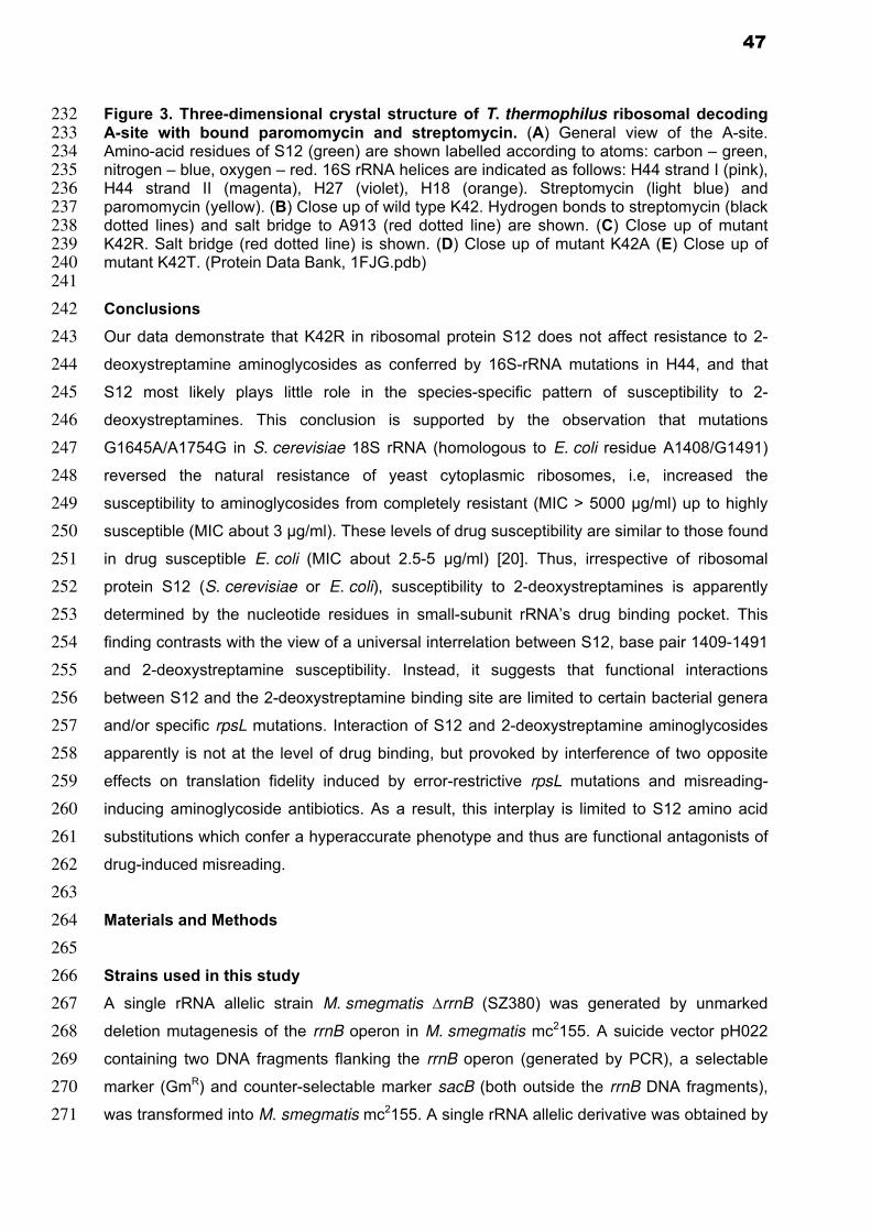
232
233 234 235 236 237 238 239 240
241
242
243
244
245
246
247
248
249
250
251
252
253
254
255
256
257
258
259
260
261
262
263
264
265
266
267
268
269
270
271
Figure 3. Three-dimensional crystal structure of T. thermophilus ribosomal decoding A-site with bound paromomycin and streptomycin. (A) General view of the A-site. Amino-acid residues of S12 (green) are shown labelled according to atoms: carbon – green, nitrogen – blue, oxygen – red. 16S rRNA helices are indicated as follows: H44 strand I (pink), H44 strand II (magenta), H27 (violet), H18 (orange). Streptomycin (light blue) and paromomycin (yellow). (B) Close up of wild type K42. Hydrogen bonds to streptomycin (black dotted lines) and salt bridge to A913 (red dotted line) are shown. (C) Close up of mutant K42R. Salt bridge (red dotted line) is shown. (D) Close up of mutant K42A (E) Close up of mutant K42T. (Protein Data Bank, 1FJG.pdb)
Conclusions
Our data demonstrate that K42R in ribosomal protein S12 does not affect resistance to 2-
deoxystreptamine aminoglycosides as conferred by 16S-rRNA mutations in H44, and that
S12 most likely plays little role in the species-specific pattern of susceptibility to 2-
deoxystreptamines. This conclusion is supported by the observation that mutations
G1645A/A1754G in S. cerevisiae 18S rRNA (homologous to E. coli residue A1408/G1491)
reversed the natural resistance of yeast cytoplasmic ribosomes, i.e, increased the
susceptibility to aminoglycosides from completely resistant (MIC > 5000 µg/ml) up to highly
susceptible (MIC about 3 µg/ml). These levels of drug susceptibility are similar to those found
in drug susceptible E. coli (MIC about 2.5-5 µg/ml) [20]. Thus, irrespective of ribosomal
protein S12 (S. cerevisiae or E. coli), susceptibility to 2-deoxystreptamines is apparently
determined by the nucleotide residues in small-subunit rRNA’s drug binding pocket. This
finding contrasts with the view of a universal interrelation between S12, base pair 1409-1491
and 2-deoxystreptamine susceptibility. Instead, it suggests that functional interactions
between S12 and the 2-deoxystreptamine binding site are limited to certain bacterial genera
and/or specific rpsL mutations. Interaction of S12 and 2-deoxystreptamine aminoglycosides
apparently is not at the level of drug binding, but provoked by interference of two opposite
effects on translation fidelity induced by error-restrictive rpsL mutations and misreading-
inducing aminoglycoside antibiotics. As a result, this interplay is limited to S12 amino acid
substitutions which confer a hyperaccurate phenotype and thus are functional antagonists of
drug-induced misreading.
Materials and Methods
Strains used in this study
A single rRNA allelic strain M. smegmatis ∆rrnB (SZ380) was generated by unmarked
deletion mutagenesis of the rrnB operon in M. smegmatis mc2155. A suicide vector pH022
containing two DNA fragments flanking the rrnB operon (generated by PCR), a selectable
marker (GmR) and counter-selectable marker sacB (both outside the rrnB DNA fragments),
was transformed into M. smegmatis mc2155. A single rRNA allelic derivative was obtained by
47

272
273
274
275
276
277
278
279
280
281
282
283
284
285
286
287
288
289
290
291
292
293
294
295
296
297
298
299 300 301 302 303 304 305 306 307 308 309 310 311 312 313
a two-step selection procedure: selection of transformants on agar plates containing
gentamicin followed by a counter-selection step on agar plates with sucrose. Deletion of rrnB
was confirmed by Southern blot analysis and partial sequencing.
Mutagenesis of the H44 decoding-site RNA was performed in strain M. smegmatis ∆rrnB
(SZ0380) and in strain M. smegmatis ∆rrnB rpsL K42R (SZ0004) [12]. rRNA mutations were
generated by PCR, cloned into vector pMIH-rrnB and introduced into the 16S rRNA A-site of
the single rRNA allelic strain SZ0380 by RecA-mediated homologous recombination as
described [12]. For a list of strains and plasmids see Tables S1 and S2 in supplementary
data.
Minimal inhibitory concentration (MIC) assay
Drug susceptibility was studied by determining Minimal Inhibitory Concentrations (MIC). MIC
tests were performed in a microtiter plate format as described [12]. In brief, freshly grown
M. smegmatis cultures were resuspended in LB broth supplemented with 0.05% of Tween
80, diluted to an absorbance at 600 nm of 0.025 and incubated in the presence of 2-fold
serial dilutions of 2-deoxystreptamine aminoglycosides. After incubation at 37°C for 72 h, the
MIC was recorded as the lowest concentration of drug inhibiting visible growth.
Structural modelling
PyMol (DeLano Scientific) was used to render the structure of the A-site of 30S ribosomal
subunit from T. thermophilus (Protein Data Bank, 1FJG.pdb) [21]
Acknowledgements
The authors thank Tanja Janušić for expert technical assistance and Björn Oettinghaus for
contributing to plasmid constructions.
References
1. Allen PN, Noller HF (1989) Mutations in ribosomal proteins S4 and S12 influence the higher order structure of 16 S ribosomal RNA. J Mol Biol 208: 457-468.
2. Funatsu G, Wittmann HG (1972) Ribosomal proteins. 33. Location of amino-acid replacements in protein S12 isolated from Escherichia coli mutants resistant to streptomycin. J Mol Biol 68: 547-550.
3. Kurland CG, Hughes D, Ehrenberg M (1996) Limitations of translational accuracy. In: Neidhardt FC, Curtiss R, Ingraham JL, Lin ECC, Low KB et al., editors. Escherichia coli and Salomonella typhimurium: Cellular and Molecular Biology. 2 ed. Washington, DC: American Society for Microbiology Press. pp. 979-1004.
4. Peske F, Savelsbergh A, Katunin VI, Rodnina MV, Wintermeyer W (2004) Conformational changes of the small ribosomal subunit during elongation factor G-dependent tRNA-mRNA translocation. J Mol Biol 343: 1183-1194.
5. Sharma D, Cukras AR, Rogers EJ, Southworth DR, Green R (2007) Mutational analysis of S12 protein and implications for the accuracy of decoding by the ribosome. J Mol Biol 374: 1065-1076.
48

314 315 316 317 318 319 320 321 322 323 324 325 326 327 328 329 330 331 332 333 334 335 336 337 338 339 340 341 342 343 344 345 346 347 348 349 350 351 352 353 354 355 356 357 358 359 360 361 362 363
364
365
366
6. Vila-Sanjurjo A, Lu Y, Aragonez JL, Starkweather RE, Sasikumar M, et al. (2007) Modulation of 16S rRNA function by ribosomal protein S12. Biochim Biophys Acta 1769: 462-471.
7. Böttger EC, Springer B, Prammananan T, Kidan Y, Sander P (2001) Structural basis for selectivity and toxicity of ribosomal antibiotics. EMBO Rep 2: 318-323.
8. Toivonen JM, Boocock MR, Jacobs HT (1999) Modelling in Escherichia coli of mutations in mitoribosomal protein S12: novel mutant phenotypes of rpsL. Mol Microbiol 31: 1735-1746.
9. Moazed D, Noller HF (1987) Interaction of antibiotics with functional sites in 16S ribosomal RNA. Nature 327: 389-394.
10. Hobbie SN, Bruell C, Kalapala S, Akshay S, Schmidt S, et al. (2006) A genetic model to investigate drug-target interactions at the ribosomal decoding site. Biochimie 88: 1033-1043.
11. Hobbie SN, Pfister P, Brull C, Westhof E, Böttger EC (2005) Analysis of the contribution of individual substituents in 4,6-aminoglycoside-ribosome interaction. Antimicrob Agents Chemother 49: 5112-5118.
12. Pfister P, Hobbie S, Brull C, Corti N, Vasella A, et al. (2005) Mutagenesis of 16S rRNA C1409-G1491 base-pair differentiates between 6'OH and 6'NH3
+ aminoglycosides. J Mol Biol 346: 467-475.
13. Pfister P, Hobbie S, Vicens Q, Böttger EC, Westhof E (2003) The molecular basis for A-site mutations conferring aminoglycoside resistance: relationship between ribosomal susceptibility and X-ray crystal structures. Chembiochem 4: 1078-1088.
14. Prammananan T, Sander P, Springer B, Böttger EC (1999) RecA-mediated gene conversion and aminoglycoside resistance in strains heterozygous for rRNA. Antimicrob Agents Chemother 43: 447-453.
15. Sander P, Meier A, Böttger EC (1995) rpsL+: a dominant selectable marker for gene replacement in mycobacteria. Mol Microbiol 16: 991-1000.
16. Sander P, Prammananan T, Böttger EC (1996) Introducing mutations into a chromosomal rRNA gene using a genetically modified eubacterial host with a single rRNA operon. Mol Microbiol 22: 841-848.
17. Carter AP, Clemons WM, Brodersen DE, Morgan-Warren RJ, Wimberly BT, et al. (2000) Functional insights from the structure of the 30S ribosomal subunit and its interactions with antibiotics. Nature 407: 340-348.
18. Francois B, Russell RJ, Murray JB, Aboul-ela F, Masquida B, et al. (2005) Crystal structures of complexes between aminoglycosides and decoding A site oligonucleotides: role of the number of rings and positive charges in the specific binding leading to miscoding. Nucleic Acids Res 33: 5677-5690.
19. O'Connor M, De Stasio EA, Dahlberg AE (1991) Interaction between 16S ribosomal RNA and ribosomal protein S12: differential effects of paromomycin and streptomycin. Biochimie 73: 1493-1500.
20. Fan-Minogue H, Bedwell DM (2008) Eukaryotic ribosomal RNA determinants of aminoglycoside resistance and their role in translational fidelity. RNA 14: 148-157.
21. DeLano WL (2002) The PyMol molecular graphics system. . San Carlos, Calif.: DeLano Scientific.
22. Hobbie SN, Kalapala SK, Akshay S, Bruell C, Schmidt S, et al. (2007) Engineering the rRNA decoding site of eukaryotic cytosolic ribosomes in bacteria. Nucleic Acids Res 35: 6086-6093.
49

367
368
369
370
371
372
373
374
375
376
377
378
379
380
381
382
383
384
385
386
387
388
389
390
391
392
393
394
395
396
397
398
399
400
401
402
403
404
405
406
407 408 409
410
Supplementary data
Figure S1. Chemical structures of disubstituted 2-deoxystreptamine antibiotics used in this study.
50

411
412
Table S1. Strains used in this study
M. smegmatis strain Parental Strain rpsL 16S rRNA mutation
Mutagenesis Reference
SZ0380 ∆rrnB rrnA+ --- wt wt --- [22]
SZ0004 ∆rrnB rrnA+ --- K42R wt Spont. [12]
∆rrnB rrnA(G1491A) SZ0380 wt G1491A Recomb. this study
∆rrnB rrnA(G1491A) SZ0004 K42R G1491A Recomb. [12]
∆rrnB rrnA(G1491C) SZ0380 wt G1491C Recomb. this study
∆rrnB rrnA(G1491C) SZ0004 K42R G1491C Recomb. [12]
∆rrnB rrnA(G1491U) SZ0380 wt G1491U Recomb. this study
∆rrnB rrnA(G1491U) SZ0004 K42R G1491U Recomb. [12]
∆rrnB rrnA(C1409G) SZ0380 wt C1409G Recomb. this study
∆rrnB rrnA(C1409G) SZ0004 K42R C1409G Recomb. [12]
∆rrnB rrnA(C1409U) SZ0380 wt C1409U Recomb. this study
∆rrnB rrnA(C1409U) SZ0004 K42R C1409U Recomb. [12]
∆rrnB rrnA(A1408G) SZ0380 wt A1408G Recomb. this study
∆rrnB rrnA(A1408G) SZ0004 K42R A1408G Recomb. [12]
413 414 415
416
417 418 419
420
Spont., spontaneous point mutation; Recomb., mutagenesis by RecA-mediated gene conversion.
Table S2. Plasmids used in this study
Number Plasmid Marker 16S rRNA mutations
Reference
pH022 pGEM7-rrnB5’3’-sacB-aph Gm - this study
PZ176 pMV361∆aph-hyg-1491A Hyg G1491A [12]
PZ178 pMV361∆aph-hyg-1491C Hyg G1491C [12]
PZ177 pMV361∆aph-hyg-1491U Hyg G1491U [12]
PZ191 pMV361∆aph-hyg-1409G Hyg C1409G [12]
PZ175 pMV361∆aph-hyg-1409U Hyg C1409U [12]
pH128 pMV361∆aph-hyg-1408G Hyg A1408G [13]
421 422 423
424
Hyg, hygromycin; Gm, gentamicin.
51
Personal contributions
My personal contributions to the projects described in the results section were as follows
Project 1 - Engineering the rRNA decoding site of eukaryotic cytosolic ribosomes in bacteria. (2007). Nucleic Acids Res. 35(18):6086-93.
Hobbie, S. N.*, Kalapala, S. K.*, Akshay, S.*, Bruell, C. M., Schmidt, S., Dabow, S., Vasella, A., Sander, P., Böttger, E. C.
* Equal contribution
Prepared ribosomes for in-vitro studies
Generated aminoglycoside-induced inhibition of AUG(UUU)12-driven phenylalanine incorporation data to calculate the IC50 values
Developed TnT assay for mycobacterial ribosomes
Generated aminoglycoside-induced inhibition of luciferase synthesis data by TnT assay to calculate the IC50 values
Project 2 – Genetic analysis of interactions with eukaryotic rRNA identify the mitoribosome as target in aminoglycoside ototoxicity. (2008). Proc Natl Acad Sci U S A. 105(52):20888-93. Hobbie, S. N.*, Akshay, S.*, Kalapala, S. K.*, Bruell, C. M., Shcherbakov, D., Böttger, E. C. * Equal contribution
Prepared ribosomes for in-vitro studies
Generated aminoglycoside-induced inhibition of AUG(UUU)12-driven phenylalanine incorporation data to calculate the IC50 values
Generated aminoglycoside-induced inhibition of luciferase synthesis data by TnT assay to calculate the IC50 values
Participated in manuscript writing Project 3 – Mutation K42R in ribosomal protein S12 does not affect susceptibility of Mycobacterium smegmatis 16S rRNA A- site mutants to 2-deoxystreptamine aminoglycosides. (manuscript in preparation) Kalapala, S. K., Hobbie, S. N., Böttger, E. C., Shcherbakov, D.
Constructed recombinant mutant strains
Generated MIC data
Wrote manuscript together with Shcherbakov, D., Hobbie, S. N., and Böttger, E. C.
52

3. Publications 1.
Kalapala, S. K., Hobbie, S. N., Böttger, E. C., Shcherbakov, D. Mutation K42R in ribosomal
protein S12 does not affect susceptibility of Mycobacterium smegmatis 16S rRNA A-site
mutants to 2-deoxystreptamine aminoglycosides. (manuscript in preparation)
2.
Hobbie, S. N.*, Akshay, S.*, Kalapala, S. K.*, Bruell, C. M., Shcherbakov, D., Böttger, E. C.
(2008). Genetic analysis of interactions with eukaryotic rRNA identify the mitoribosome as
target in aminoglycoside ototoxicity. Proc Natl Acad Sci U S A. 105(52):20888-93.
3.
Pathak, R., Perez-Fernandez, D., Nandurdikar, R., Kalapala, S.K., Böttger, E.C., Vasella, A.
(2008). Synthesis and Evaluation of Paromomycin Derivatives Modified at C(4'). Helv. Chim.
Acta. 91, 1533-1552.
4.
Hobbie, S. N.*, Bruell, C. M.*, Akshay, S., Kalapala, S. K., Shcherbakov, D., Böttger, E. C.
(2008). Mitochondrial deafness alleles confer misreading of the genetic code. Proc Natl Acad
Sci U S A. 105(9):3244-49.
5.
Hobbie, S. N.*, Kalapala, S. K.*, Akshay, S.*, Bruell, C. M., Schmidt, S., Dabow, S., Vasella,
A., Sander, P., Böttger, E. C. (2007). Engineering the rRNA decoding site of eukaryotic
cytosolic ribosomes in bacteria. Nucleic Acids Res. 35(18):6086-93.
6.
Hobbie, S. N., Bruell, C. M., Kalapala, S., Akshay, S., Schmidt, S., Pfister, P., Böttger, E. C.
(2006). A genetic model to investigate drug-target interactions at the ribosomal decoding site.
Biochimie 88(8):1033-43.
* These authors contributed equally to this work
53

4. Conference presentations 2009
EMBO Conference on Protein Synthesis and Translational Control, Heidelberg, GermanyrRNA sequence polymorphism within the bacterial domain and susceptibility to drugs targeting protein synthesis R. Akbergenov, A. Subramanian, S. K. Kalapala, M. Bertea, M.K.A. Kulstrunk and E.C. Böttger EMBO Conference on Ribosome Synthesis, Regensburg, Germany Probing of aminoglycoside interaction with the ribosomal decoding A-site by site-directed mutagenesis S.K. Kalapala, S. Akshay, D. Scherbakov, R. Akbergenov and E.C. Böttger 12S rRNA deafness alleles and aminoglycoside induced ototoxicity converge on mitochondrial mistranslation as mechanism of disease pathogenesis S. Akshay, S.K. Kalapala, R. Akbergenov, D. Shcherbakov and E.C. Böttger 68th Annual Assembly of the Swiss Society for Microbiology (SSM), Lausanne, Switzerland Synthesis of novel aminoglycoside compounds by a synthetic biology approach E.C. Böttger, D.P. Fernandez, I. Kudyba, R. Pathak, S. Harish, S.R. Dubakka, S.K. Kalapala, D. Scherbakov, R. Akbergenov and A. Vasella Aminoglycosides: Structural analyses of interactions with eukaryotic A-sites point to the mitoribosome as target and identify mechanisms of toxicity D. Scherbakov, S. Akshay, S.K. Kalapala, M.K.A. Kulstrunk, R. Akbergenov and E.C. Böttger
2008
60th
Annual Meeting of the German Society for Microbiology (DGHM), Dresden, Germany Synthesis of novel aminoglycoside compounds by a synthetic biology approach E.C. Böttger, D.P. Fernandez, I. Kudyba, R. Pathak, S. Harish, S.R. Dubakka, S.K. Kalapala, S.N. Hobbie and A. Vasella Aminoglycosides: Structural analyses of interactions with eukaryotic A-sites point to the mitoribosome as target and identify mechanisms of toxicity S.N. Hobbie, S. Akshay, S.K. Kalapala, C.M. Bruell, D. Scherbakov and E.C. Böttger 13th Annual Meeting of the RNA Society, Berlin, Germany Mutations A1555G and C1494U in mitochondrial 12S rRNA affect the accuracy of mRNA translation S. Metze, S.K. Kalapala, S.N. Hobbie and E.C. Böttger IUBMB Symposium on Mitochondrial Physiology and Pathology, Bari, Italy Mitochondrial deafness alleles affect the accuracy of mRNA translation S.N. Hobbie, S. Akshay, S.K. Kalapala, C.M. Bruell, D. Scherbakov and E.C. Böttger EMBO Conference on RNA and Disesse, Rome, Italy Mitochondrial 12S rRNA mutations increase ribosomal susceptibility to aminoglycoside-induced mistranslation D. Scherbakov, S.N. Hobbie, S.K. Kalapala, S. Akshay and E.C. Böttger
54

67th Annual Assembly of the Swiss Society for Microbiology (SSM) 2008, Interlaken, Switzerland Transplanting eukaryotic decoding sites into bacterial hybrid ribosomes S.K. Kalapala, S. Akshay, C.M. Bruell, S.N. Hobbie and E.C. Böttger
2007
EMBO Conference on Protein Synthesis and Translational Control, Heidelberg, Germany Genetic modeling of Mycobacterium smegmatis ribosomal RNA S.K. Kalapala, S.N. Hobbie and E.C. Böttger Transplanting human and protozoan decoding sites into bacterial hybrid ribosomes S.N. Hobbie, S.K. Kalapala, S. Akshay, C.M. Bruell and E.C. Böttger
4th
International Conference on the Ribosome, Cape Cod, USA Modeling the rRNA decoding site of eukaryotic cytosolic ribosomes in bacteria S.N. Hobbie, C.M. Bruell, S. K. Kalapala, S. Akshay and E.C. Böttger
2006
2nd
FEMS Congress of European Microbiologists, Madrid, Spain Genetic analysis of aminoglycoside binding to the ribosomal decoding site S.N. Hobbie, S.K. Kalapala, A. Subramanian, B. François, E. Westhof and E.C. Böttger
55

CURRICULUM VITAE
Full Name: Sarath Kumar Kalapala Date of birth: 02.04.1978 Place of birth: Vijayawada, India Nationality: Indian
EDUCATION AND RESEARCH ACTIVITIES
2005 – Present: Doctoral studies under the supervision of Prof. Dr. Erik C. Böttger, Institute of Medical Microbiology, University of Zurich, Zurich, Switzerland. “Study of Aminoglycoside Antibiotics Interaction with Eukaryotic Ribosomal Decoding Sites Using Bacterial Hybrid Ribosomes” 2001 – 2004: I had worked as a Research assistant under the supervision of Prof. Umesh Varshney, Department of Microbiology and Cell Biology, Indian Institute of Science, Bangalore, India “Studies on ribosome recycling factor from mycobacteria” 1999 – 2001: Graduate studies in Microbiology, Nagarjuna University, Guntur, India Graduate Thesis under the supervision of Dr. Ashlesha Deshpande, Indian Institute of Chemical Technology, Hyderabad, India. “Enzyme immobilization / purification: a novel approach on protease, an extra cellular enzyme produced by Bacillus licheniformis”. 1996 – 1999: Undergraduate studies in Microbiology, Biochemistry, Chemistry and Aqua Culture, Nagarjuna University, Guntur, India. 1993 – 1996: Intermediate education in Botany, Zoology, Physics and Chemistry, SRR & CVR Govt. College, Vijayawada, India. 1983 – 1993: Primary and Secondary school education at Tagore, Jaikisan and Gowtham public schools, Vijayawada, India.
56

LIST OF PUBLICATIONS
2010
Kalapala, S. K., Hobbie, S. N., Böttger, E. C., Shcherbakov, D. Mutation K42R in ribosomal protein S12 does not affect susceptibility of Mycobacterium smegmatis 16S rRNA A-site mutants to 2-deoxystreptamine aminoglycosides. (manuscript in preparation)
2008
Hobbie, S. N.*, Akshay, S.*, Kalapala, S. K.*, Bruell, C. M., Shcherbakov, D., Böttger, E. C. (2008). Genetic analysis of interactions with eukaryotic rRNA identify the mitoribosome as target in aminoglycoside ototoxicity. Proc Natl Acad Sci U S A. 105(52):20888-93.
2008
Pathak, R., Perez-Fernandez, D., Nandurdikar, R., Kalapala, S.K., Böttger, E.C., Vasella, A. (2008). Synthesis and Evaluation of Paromomycin Derivatives Modified at C(4'). Helv. Chim. Acta. 91, 1533-1552.
2008
Hobbie, S. N.*, Bruell, C. M.*, Akshay, S., Kalapala, S. K., Shcherbakov, D., Böttger, E. C. (2008). Mitochondrial deafness alleles confer misreading of the genetic code. Proc Natl Acad Sci U S A. 105(9):3244-49.
2007
Hobbie, S. N.*, Kalapala, S. K.*, Akshay, S.*, Bruell, C. M., Schmidt, S., Dabow, S., Vasella, A., Sander, P., Böttger, E. C. (2007). Engineering the rRNA decoding site of eukaryotic cytosolic ribosomes in bacteria. Nucleic Acids Res. 35(18):6086-93.
2006
Hobbie, S. N., Bruell, C. M., Kalapala, S., Akshay, S., Schmidt, S., Pfister, P., Böttger, E. C. (2006). A genetic model to investigate drug-target interactions at the ribosomal decoding site. Biochimie 88(8):1033-43.
2005
Saikrishnan, K., Kalapala, S. K., Varshney, U., Vijayan, M. (2005) X-ray structural studies of Mycobacterium tuberculosis RRF and a comparative study of RRFs of known structure. Molecular plasticity and biological implications. J Mol Biol.345(1):29-38.
2004
Saikrishnan, K., Kalapala, S. K., Bidya Sagar, M., Rao, A.R., Varshney, U., Vijayan, M. (2004) Purification, crystallization and preliminary X-ray studies of Mycobacterium tuberculosis RRF. JActa Crystallogr. D Biol. Crystallogr. 60(Pt2):368-70.
* These authors contributed equally to this work
57