ANTIBIOTICS - Annamalai University
41
ANTIBIOTICS INTRODUCTION The term antibiotic has its origin in the word antibiosis (i.e. against life). Antibiotics are chemical substances obtained from various species of microorganisms (bacteria, fungi, actinomycetes) that suppress the growth of other microorganisms and eventually may destroy them. The probable points of difference amongst the antibiotics may be physical, chemical, pharmacological properties, antibacterial spectra, and mechanism of action. They have made it possible to cure diseases caused by bacteria, such as pneumonia, tuberculosis, and meningitis, and they save the lives of millions of people around the world. CLASSIFICATION Antibiotics are classified on the basis of their mechanism of action and by its chemical nature. Classification Based on Mechanism of Action 1. Agents that inhibit the synthesis of bacterial cell wall: These include the penicillins and cephalosporins that are structurally similar and dissimilar agents, such as cycloserine, vancomycin, bacitracin and the imidazole antifungal agents. 2. Agents that act directly on the cell membrane of the microorganisms, affecting permeability, and leading to leakage of intracellular compounds: These include polymyxin, polyene antifungal agents, nystatin, and amphotericin B that bind to cell wall sterols. 3. Agents that affect the function of 30s and 50s ribosomal subunits to cause reversible inhibition of protein synthesis: These include tetracyclines, erythromycins, chloramphenicol, and clindamycin. 4. Agents that bind to the 30s ribosomal subunit and alter protein synthesis: These include aminoglycosides that leads to cell deaths eventually. 5. Agents that affect nucleic acid metabolism: Such as rifamycins, which inhibit DNA dependent RNA polymerase.
-
Upload
khangminh22 -
Category
Documents
-
view
2 -
download
0
Transcript of ANTIBIOTICS - Annamalai University

ANTIBIOTICS
INTRODUCTION
The term antibiotic has its origin in the word antibiosis (i.e. against life). Antibiotics are chemical substances obtained from various species of microorganisms (bacteria, fungi, actinomycetes) that suppress the growth of other microorganisms and eventually may destroy them. The probable points of difference amongst the antibiotics may be physical, chemical, pharmacological properties, antibacterial spectra, and mechanism of action. They have made it possible to cure diseases caused by bacteria, such as pneumonia, tuberculosis, and meningitis, and they save the lives of millions of people around the world.
CLASSIFICATION
Antibiotics are classified on the basis of their mechanism of action and by its chemical nature.
Classification Based on
Mechanism of Action
1. Agents that inhibit the synthesis of bacterial cell wall: These include the penicillins and cephalosporins that are structurally similar and dissimilar agents, such as cycloserine, vancomycin, bacitracin and the imidazole antifungal agents.
2. Agents that act directly on the cell membrane of the microorganisms, affecting permeability, and leading to leakage of intracellular compounds: These include polymyxin, polyene antifungal agents, nystatin, and amphotericin B that bind to cell wall sterols.
3. Agents that affect the function of 30s and 50s ribosomal subunits to cause reversible inhibition of protein synthesis: These include tetracyclines, erythromycins, chloramphenicol, and clindamycin.
4. Agents that bind to the 30s ribosomal subunit and alter protein synthesis: These include aminoglycosides that leads to cell deaths eventually.
5. Agents that affect nucleic acid metabolism: Such as rifamycins, which inhibit DNA dependent RNA polymerase.

Classification Based on Chemical Structure
1. β-lactam antibiotics
2. Aminoglycoside antibiotics
3. Tetracycline antibiotics
4. Polypeptide antibiotics
5. Macrolide antibiotics
6. Lincomycins
7. Other antibiotics
1. β-lactam antibiotics
These consists of two major class of agents, that is penicillins and cephalosporins.
a. Penicillins:
Penicillin, the most important antibiotic, was first extracted from the mould Penicillium notatum. Subsequently, a mutant of a related mould, P. chrysogenum, was found to give the highest yield of penicillin and is employed for the commercial production of this antibiotic. Penicillin belongs to a group of antibiotics called β-lactam antibiotics . The basic structure of the penicillins consists of a thiazolidine ring fused with a β-lactam ring, which is essential for antibacterial activity. These two rings constitute the fundamental nucleus of all the penicillins, namely, 6-amino penicillanic-acid (6-APA) A variety of semisynthetic penicillins are produced by altering the composition of the side chain attached to 6-APA nucleus. Both the 6-APA nucleus and side chain are essential for the antibacterial activity.

Nomenclature
Penicillins are named in the following ways:
a. Chemical abstract
1. The penicillins are described as 4-thia-1-azabicyclo (3.2.0) heptanes.
2. Benzylpenicillin is 6-(2-phenylacetamido)-3, 3-dimethyl-7-oxo-4-thia-1-azabiclo(3.2.0)heptane2-carboxylic acid.
b. Penam: In order to simplify the unsubstituted bicylic ring system of penicillin, it is given the name penam. Accordingly, the penicillins are 6-acylamino-2, 2-dimethyl penam-3-carboxylates.
CLASSIFICATION

I. Penicillinase-susceptible penicillins
The general impact on antibacterial activity is as follows:
• Good gram-positive potency against susceptible Staphylococci and Streptococci
• Useful against some gram-positive cocci
• Not effective against gram-negative bacilli

II. Penicillinase-resistant penicillins
General impact on antibacterial activity is as follows:
• Decreased susceptibility to many penicillinase.
• Active against microrganisms, resistant to early penicillin.
• Oxacillins offer good oral activity.
III. Aminopenicillins
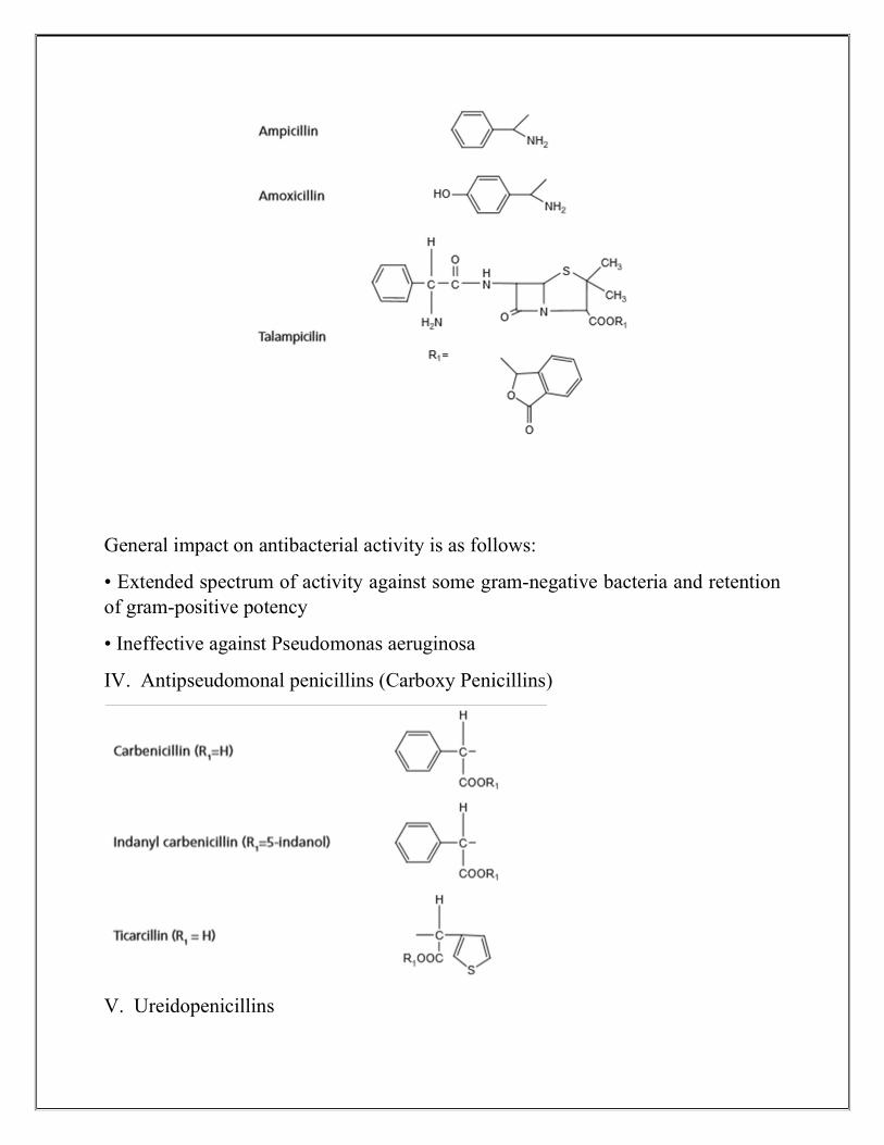
General impact on antibacterial activity is as follows:
• Extended spectrum of activity against some gram-negative bacteria and retention of gram-positive potency
• Ineffective against Pseudomonas aeruginosa
IV. Antipseudomonal penicillins (Carboxy Penicillins)
V. Ureidopenicillins

Mode of action:
The cell wall of bacteria is essential for the normal growth and development. Peptidoglycan is a heteropolymeric component of the cell wall that provides rigid mechanism for stability by virtue of its highly cross-linked lattice-wise structure. The peptidoglycan is composed of glycan chains, which are linear strands of two alternating amino sugars (N-acetyl glucosamine and N-acetylmuramic acid) that are cross-linked by peptide chains of an enzyme, transpeptidase. Penicillins inhibit the transpeptidase activity to the synthesis of cell walls. They also block cleavage of terminal D-alanine during the cell wall synthesis. The biosynthesis of peptidoglycan involves three stages Β-lactam antibiotics inhibit the last step in peptidoglycan synthesis. The transpeptidase enzyme that contains serine is probably acylated by β-lactam antibiotics with the cleavage of -CO-N-bond of the βlactam ring. This renders the enzyme inoperative and inhibits peptidoglycan synthesis.
SAR :
6-Acyl side chain:

The substitution of R on the primary amine with an electron withdrawing group decreases the electron density on the side chain and protects from acid degradation. Substituents on the α-carbon of the side chain, such as amino (ampicillin), chloro, and guanidine exerts good resistance to inactivation by acids. Benzyl penicillin undergoes acid and alkali degradation and is susceptible to all known DŽ-lactamase. The increased latitude in varying the acyl amino side chain through acylation of 6APA results with superior biological activity. Substitution of α-aryl of the alkyl group in the side chain gives increased stability and oral absorption.
1. Substitution of bulky groups on α-carbon of the side chain confers β-lactamase resistance. Examples: methicillin, nafcillin, oxacillin, etc. In all these penicillins, an aromatic ring is attached directly to the side chain amide carbonyl, and there is substitution at both positions ortho to the point of attachment. The size of the ring systems play an important role in determining the ability of the ortho substitutent to confer penicillinase resistance.
2. The isomeric forms of penicillins differs in their activity. Example: D-isomer is 2–8 times more active than L-isomer of amoxicillin. The introduction of polar group or ionized molecule into the α-position of the side chain in the benzyl carbon atom of penicillin-G confers against the gram-negative bacilli. Amino, hydroxyl, carboxyl, and sulphonyl increases gram-negative activity. Example: ampicillin and carbenicillin.
3. Replacement of acyl side chain with hydroxymethyl groups shows improved gram-negative activity and introduction of C-6 α-methoxy group produces greater stability against β-lactamase. N-acylated ampicillins (ureidopenicillins) have increased activity against Pseudomonas.
4. Many esters of the carboxyl group attached to C-3 have been prepared as prodrugs to increase lipophilicity and acid stability. Example: Acetoxymethyl ester derivatives are used for preparing prodrugs.
5. The sulphur of the thiazolidine ring with O, CH2, and CH-β-CH3 gives broad-spectrum antibacterial activity. The geminal dimethyl group at C-2 position is a characteristic of the penicillin. In general, derivatization of the C-3 carboxylic acid functionality is not tolerated unless the free penicillin carboxylic acid can be generated in vivo. Doubly activated penicillin esters, undergo rapid cleavage in vivo to generate active penicillin. Example: pivampicillin and becampicillin. The antibacterial activity is evidented by N-4 atom at ring junction.

6. In vitro degradation is retarded by keeping the pH of the solution between 6.0 and 8.0. More lipophilic side chain increases the plasma protein binding. Example: Ampicillin: 25% plasma protein bound and phenoxy methyl penicillin: 75% plasma protein bound.
I. Penicillinase resistant penicillins
i. Methicillin
ii. Oxacillins (Isoxazolyl penicillins)
Properties and uses:
Oxacillin sodium monohydrate is a white powder, soluble in water and methanol, insoluble in methylene chloride. The use of oxacillin and other isoxazolyl penicillins should be restricted to the treatment of infections caused by Staphylococci that are resistant to penicillin G, although their spectrum of activity is similar to that of penicillin G.

CEPHALOSPORINS
The cephalosporins were isolated from the fungus Cephalosporium acremonium in 1948 by Pro Tzu, Newton, and Abraham (1953). The main product being cephalosporin-C, the molecular modifi cation of cephalosporin-C gave origin to semisynthetic substances. They are β-lactam antibiotics with same fundamental structural requirements as penicillins, the main difference between the two is that cephalosporins contain dihydrometathiazine ring, while penicillin contains a tetrahydrothiazole (thiazolidine) ring. The cephalosporins are much more acid stable than the corresponding penicillins and also have a mechanism of action similar to that of penicillins; they mainly inhibit the cross-linking of the peptidoglycan units in bacterial cell walls by inhibiting transpeptidase enzyme. However, they bind in the target proteins other than penicillins binding proteins.
Cephalosporins can be divided into three classes:

1. Cephalosporin N: It has a penicillin-like structure being a derivative of 6-aminopenicillanic acid.
2. Cephalosporin P: An acidic antibiotic, which is steroidal in nature.
3. Cephalosporin-C: It is a true cephalosporin and it is a derivative of 7 amino-cephalosporanic acid.
Nomenclatures
Cephalosporins are named in the following ways:
1. Chemical abstracts: 5-Thia-1-azobicyclo (4.2.0) octanes. Accordingly, cephalothin is 3-(Acetoxy methyl)-8-oxo-7-(2-thienyl) acetamido-5thia-1-aza-bicyclo[4.2.0]-oct-2ene-2-carboxylic acid.
2. Cepham derivatives: Cepham is the name given to the unsubstituted bicyclic lactam.
Classification Cephalosporins are classifi ed on the basis of their chemical structure, clinical pharmacology, antibacterial spectrum, or penicillinase resistance.
a. Orally administered: cephalexin, cephradine, and cefaclor
b. Parentrally administered: cephalothin, cephapirin, cephacetrile, and cefazedone. These agents are sensitivity to β-lactamase
c. Resistant to β-lactamase and parentrally administered: cefuroxime, cefamandole, cefoxitin
d. Metabolically unstable: cephalothin and cephapirin
Clinically used cephalosporins:

I. First-generation cephalosporins
These drugs have the highest activity against gram-positive bacteria and the lowest activity against gram negative bacteria
II. Second-generation cephalosporins
These drugs are more active against gram-negative bacteria and less active against gram-positive bacteria than first-generation members
III. Third-generation cephalosporins
These drugs are less active than first-generation drugs against gram-positive organisms, but have a much expanded spectrum of activity against gram-negative organisms
IV. Fourth-generation cephalosporins
Cefepime and cefpirome are new fourth-generation parenteral cephalosporins with a spectrum of activity which makes them suitable for the treatment of infections caused by a wide variety of bacteria

Properties and uses:
Cephalexin monohydrate is a white crystalline powder, sparingly soluble in water, and practically insoluble in alcohol. The α-amino group of cephalexin renders it acid stable. The 3-methyl group is responsible for the metabolic stability. It is particularly recommended for urinary tract infection.
Dose: The oral dose for adults is 250–500 mg every 6 h, for children, the dose is 18–25mg/kg every 6 h.
ii. Cefadroxil (Cefadrox, Droxyl, Codroxil)

Properties and uses:
Cefadroxil monohydrate is a white or almost white powder, slightly soluble in water, and sparingly soluble in ethanol. The antibacterial spectrum of action and therapeutic indications of cefadroxil are very similar to those of cephalexin and cephradine. The D-p-hydroxyphenylglycyl isomer is much more active than the L-isomer.
Assay:
It is assayed by adopting liquid chromatography technique.
Dosage forms:

Cefadroxil capsules I.P., B.P., Cefadroxil oral suspension I.P., B.P. Cefadroxil tablets I.P.
Dose:
In the case of uncomplicated lower urinary tract infections: For adults, the dose is 1–2 g daily as a single or 2 divided doses. For children more than 6 years, the dose is 500 mg twice a day, that is, 1–6 years, 250 mg twice a day; for children less than 1 year, the dose is 25 mg/kg daily in divided doses. In the case of skin and skin structure infections: For adults, the dose is 1g per day in single or divided doses. For children, the dose is 30 mg/kg per day in equally divided doses every 12 h. In the case of pharyngitis and tonsilitis: For adults in the treatment of group A beta-haemolytic streptococcal pharyngitis and tonsilitis, the dose is 1 g per day in single or divided doses for 10 days. In the case of children, 30 mg/kg per day in equally divided doses every 12 h for at least 10 days.
Properties:
Cephalothin is a white, odourless, crystalline powder, insoluble in most organic solvents, soluble in organic solvents and it is acid stable. It is hygroscopic and decomposes on heating, and it has been described as broad-spectrum antibacterial compound, it is not in the same class as the tetracyclines. Its spectrum of activity is broader than that of penicillin G and more similar to that of ampicillin.
Dose:
The dose for adults given IM or IV is equivalent to 500 mg–1 g every 4 to 6 h; children the dose is 13–26 mg/kg of body weight every 4 h.

Adverse reactions of the cephalosporins
The cephalosporins produce a number of adverse effects. Examples are the following:
i. Allergic manifestation: The cephalosporins should be avoided or used with caution in individuals allergic to penicillins. When cefamandole or cefoperazone is ingested with alcohol, a disulphiramlike effect is seen, because these cephalosporins block the second step in alcohol oxidation, which results in the accumulation of acetaldehyde.
ii. Bleeding: Bleeding can occur with cefemandole or ceforperazone because of antivitamin K effects. But the administration of the vitamin overcomes this problem.
2. AMINOGLYCOSIDE ANTIBIOTICS
The aminoglycoside antibiotics contain one or more amino sugars linked to an aminocytitol ring by glycosidic bonds. These are broad-spectrum antibiotics; in general, they have greater activity against gram-negative than gram-positive

bacteria. The development of streptomycin, the fi rst antibiotic of this group, was a well-planned work of Waksman (1944) and his associates, who isolated it from a strain of Streptomyces griseus. The aminoglycoside can produces severe adverse effects, which include nephrotoxity, ototoxicity, and neuro effects. These properties have limited the use of aminoglycoside chemotherapy to serious systemic indications. Some aminoglycosides can be administered for ophthalmic and topical purposes.
Mode of action:
The aminoglycosides exhibit bactericidal effects as a result of several phenomena. Ribosomal binding on 30s and 50s subunits as well as the interface produces misreading; this disturbs the normal protein synthesis. Cell membrane damage also plays an integral part in ensuring bacterial cell death.
SAR of Aminoglycoside Antibiotics
The aminoglycosides consist of two or more amino sugars joined in glycoside linkage to a highly substituted 1,3-diaminocyclo hexane (aminocyclitol), which is a centrally placed ring. The ring is a 2-deoxy streptamine in all aminoglycosides except streptomycin and dihydrostreptomycin, where it is streptidine.
Thus,
• In kanamycin and gentamycin families, two amino sugars are attached to 2-deoxy streptamine.
• In streptomycin, two amino sugars are attached to strepidine.

• In neomycin family, there are amino sugars attached to 2-deoxy streptamine. The aminoglycoside antibiotics contain two important structural features. They are amino sugar portion and centrally placed hexose ring, which is either 2-deoxystreptamine or streptidine.
TETRACYCLINE ANTIBIOTICS
Tetracyclines have a ring system of four linear annelated six-membered rings and are characterized by a common octahydronaphthacenes skeleton. They are potent, broad-spectrum antibacterial agents effective against gram-positive and gram-negative aerobic and anaerobic bacteria. As a result, the tetracyclines are drugs of choice or well-accepted alternatives for a variety of infectious diseases. Among these, they also play a role in the treatment of sexually transmitted and gonococcal diseases, urinary tract infections, bronchitis, and sinusitis remain prominent. The majority of the marketed tetracyclines (tetracycline, chlorotetracycline, oxytetracycline, and demeclocycline) are naturally occurring compounds obtained by the fermentation of Streptomyces spp. broths. The semisynthetic tetracyclines (methacycline, doxycycline, minocycline) have the advantage of longer duration of antibacterial action. However, all these tetracyclines exhibit a similar profile in terms of antibacterial potency. In general, their activity encompasses many strains of gram-negative E. coli, Proteus, Klebsiella, Enterobacter, Niesseria, and Serratia spp., as well as gram-negative Streptococci and Staphylococci of particular interest is the potency of tetracylines against Haemophilus, Legionella, Chlamydia, and Mycoplasma.
Classes of tetracyclines
I. Natrual tetracyclines (biosynthetic)
II. Semisynthetic tetracyclines
III. Protetracyclines
I. Natrual tetracyclines (biosynthetic) :

Properties and uses:
Methacycline is a yellow crystalline powder, sparingly soluble in water. It is obtained by the chemical modification of oxytetracycline. It has an antibiotic

spectrum similar to tetracyclines, but greater potency; about 600 mg of methacycline is equivalent to 1 g of tetracycline.
Properties and uses:
It was first obtained in small yields by a chemical transformation of oxytetracycline. The 6α-methyl epimer is more than three times as active as its β epimer.
Dose:
In adults, the oral dosage is 100 mg every 12 h.
Dosage forms:
Doxcycline HCl capsules I.P., Doxcycline HCl tablets I.P.
SAR OF TETRACYCLINES
The key structural feature is a linearly fused tetracyclic nucleus and each ring needs to be six membered and purely carbocyclic. A tetracyclic backbone skeleton is essential for activity.
• The D-ring needs to be aromatic and the A-ring must be appropriately substituted at each of its carbon atoms for notable activity.

• The B-ring and the C-ring tolerate certain substitutent changes as long as the keto-enol systems (at C-11, 12, 12a) remain intact and conjugated to the phenolic D-ring.
• The D, C, B-ring phenol, keto-enol system is imperative and the A-ring must also contain a conjugated keto enol system.
• Specifically, the A-ring contains a tricarbonyl derived keto-enol array at positions C-1, 2, and -3. Other structural requirements for good antibacterial activity include a basic amine function at C-4 position of the A-ring.
Modification of C-1 and C-3 position: The keto-enol tautomerism of ring A in carbon atom 1 and 3 is a common feature to all biologically active tetracyclines, blocking this system by forming derivatives at C-1 and C-3 results in loss of antibacterial activity A–C = O, a function of C-1 and C-3 is essential for activity. In addition, equilibrium between non-ionized and Zwitterionic structure of tetracycline is essential for activity.
Modification of C-2 position: The antibacterial activity resides on the carboxamide moiety. The amide is best left unsubstituted or monosubstitution is acceptable in the form of activated alkylaminomethyl amide (Mannich bases). An example includes rolitetracycline large alkyl group on the carboxamide that may alter the normal keto-enol equilibrium of the C-1, 2, and 3 conjugated systems and diminishes inherent antibacterial activity. The replacement of carboxamide group or dehydration of carboxamide to the corresponding nitrile results in a loss of activity.
Modification of C-4 position: The keto-enolic character of the A-ring is due to the α-C-4 dimethyl amino substituent. Loss of activity is exerted when dimethyl amino group is replaced with hydrazone oxime or hydroxyl group.
Modification of C-4a position: The α-hydrogen at C-4a position of tetracyclines is necessary for useful antibacterial activity.
Modification of the C-5 and C-5a positions: Alkylation of the C-5 hydroxyl group results in loss of activity. Naturally occurring antibacterial tetracyclines have an unsubstituted methylene moiety at the C-5 position. However, oxytetracycline contains C-5 α-hydroxyl group, was found to be a potent compound, and has been modified chemically to some semisynthetic tetracyclines. Esterification is only acceptable if the free oxytetracycline can be liberated in vivo; only small alkyl esters are useful. Epimerization is detrimental to antibacterial activity.
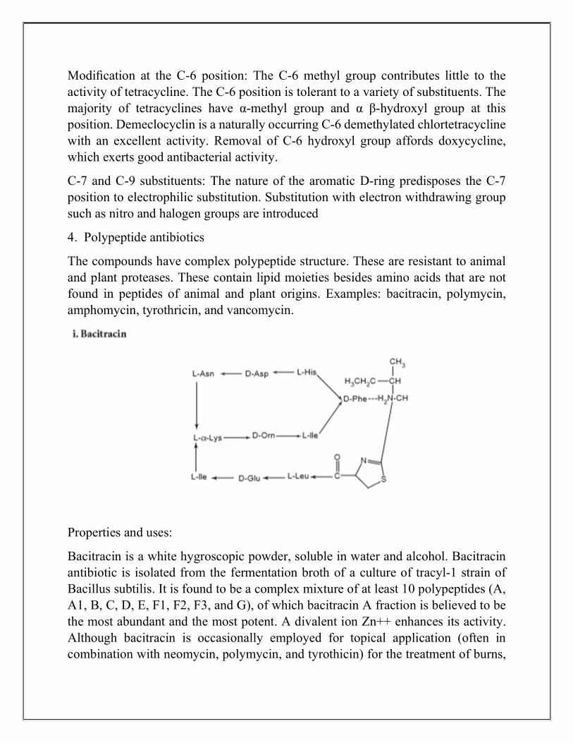
Modification at the C-6 position: The C-6 methyl group contributes little to the activity of tetracycline. The C-6 position is tolerant to a variety of substituents. The majority of tetracyclines have α-methyl group and α β-hydroxyl group at this position. Demeclocyclin is a naturally occurring C-6 demethylated chlortetracycline with an excellent activity. Removal of C-6 hydroxyl group affords doxycycline, which exerts good antibacterial activity.
C-7 and C-9 substituents: The nature of the aromatic D-ring predisposes the C-7 position to electrophilic substitution. Substitution with electron withdrawing group such as nitro and halogen groups are introduced
4. Polypeptide antibiotics
The compounds have complex polypeptide structure. These are resistant to animal and plant proteases. These contain lipid moieties besides amino acids that are not found in peptides of animal and plant origins. Examples: bacitracin, polymycin, amphomycin, tyrothricin, and vancomycin.
Properties and uses:
Bacitracin is a white hygroscopic powder, soluble in water and alcohol. Bacitracin antibiotic is isolated from the fermentation broth of a culture of tracyl-1 strain of Bacillus subtilis. It is found to be a complex mixture of at least 10 polypeptides (A, A1, B, C, D, E, F1, F2, F3, and G), of which bacitracin A fraction is believed to be the most abundant and the most potent. A divalent ion Zn++ enhances its activity. Although bacitracin is occasionally employed for topical application (often in combination with neomycin, polymycin, and tyrothicin) for the treatment of burns,

ulcer, and wounds, it can cause serious necrosis of the kidney tubules; if it is given systematically (i.e. I.V route) an oral administration is not feasible due to its lack of absorption from the GI tract. A variety of gram-positive cocci and bacilli are sensitive to bacitracin. It should be stored in airtight containers due to its hygroscopic nature.
Assay:
It is assayed by microbiological method.
Properties and uses:
Polymycin sulphate is a white hygroscopic powder, soluble in water, and slightly soluble in ethanol. The polymyxins are cyclic peptides holding a fatty acid side chain. This is a group of relatively simple basic, cationic, detergent peptides that are produced by Bacillus polymyxia. At least, fi ve polymyxins (A, B, C, D, and E) are known, but only polymyxin B and polymyxin E are of clinical utility. Both polymyxin B and polymyxin E (colistin) are mixtures of two components and is used in the treatment of bacterial meningitis, urinary tract infection, burns, wounds, and gastroenteritis. Polymyxin may affect renal tubules and central nervous system (CNS), and because of their nephrotoxicity associated with their systemic use, they are primarily employed to treat topical infections.
Assay:
It is assayed by adopting liquid chromatography technique.
MACROLIDE ANTIBIOTICS

The macrolide antibacterial agents are extremely useful chemotherapeutic agents for the treatment of a variety of infectious disorders and diseases caused by a host of gram-positive bacteria, both cocci and bacilli; they also exhibit useful effectiveness against gram-negative cocci, specially, neisseria spp. The macrolides are commonly administered for respiratory, skin, tissue, and genitourinary infections caused by these pathogens.
Chemistry:
They are characterized by five common chemical features.
1. A macrocyclic lactone usually has 12–17 atoms, hence the name macrolide.
2. A ketone group.
3. One or two amino sugars glycosidically linked to the nucleus.
4. A neutral sugar linked either to amine sugar or to nucleus.
5. The presence of dimethyl amino moiety on the sugar residue, which explains the basicity of these compounds, and consequently the formation of salts. The antibacterial spectrum of activity of the more potent macrolides resembles that of penicillin. Examples: erythromycin, oleandomycin, clarithromycin, flurithromycin, dirithomycin, azithromycin.
Properties and uses:
Azithromycin is a white powder, practically insoluble in water, soluble in anhydrous ethanol and methylene chloride. It is very stable under acidic conditions, is less

active against Streptococci and Staphylococci than erythromycin, and is far more active against respiratory infections due to H. influenzae and Chlamydia trachomatis
Mode of action:
Macrolide antibiotics are bacteriostatic agents that inhibit protein synthesis by binding irreversibly to a site on the 50S subunits of the bacterial ribosome. Thus, inhibiting the translocation steps of protein synthesis at varying stages of peptide chain elongation (hinder the translocation of elongated peptide chain back from ‘A’ site to ‘P’ site). The macrolides inhibit ribosomal peptidyl transferase activity. Some macrolides also inhibit the translocation of the ribosome along with the mRNA template.
Properties and uses:
The antibiotic lincomycin is obtained from Actinomycetes, Streptomyces, and Lincolnensis. The ability of lincomycin to penetrate into bones, adds to its qualities and it gets promoted in the chemotherapy of bone and joint infections by penicillin resistant strains of S. aureus. Variation of the substituents on pyrrolidine portion and C-5 side chain affects the activity.
Some of the examples are as follows:
i. N-demethylation imparts activity against gram-negative bacteria.
ii. Increase in the chain length of the propyl substitutent at C-4 position in pyrrolidine moiety up to n-hexyl increase in vivo activity.
iii. The thiomethyl ether of α-thiolincosamide moiety is essential for activity.

iv. Structural modifications at C-7 position, such as introduction of 7S chloro or 7R-OCH3, change the physiochemical parameters of the drug (i.e. partition coefficient), and thus, alter the activity spectrum and pharmacokinetic properties. The usual side effects include skin rashes, nausea, vomiting, and diarrhoea. Dosage forms: Lincomycin HCl capsules I.P.
7. OTHER ANTIBIOTICS
Examples of other antibiotics are chloramphenicol, rifampicin and mupirocin.
Chloramphenicol or chloromycetin Chloramphenicol has a spectrum of activity resembling that of the tetracyclines except that it exhibits a bit less activity against some gram-positive bacteria. It is isolated from Salmonella venezuelae by Ehrlich et al in 1947. It contains chlorine and is obtained from an actinomycete, and thus, named as chloromycetin. It is specifically recommended for the treatment of serious infections caused by H. influnzae, S. typhi (typhoid), S. pneumoniae, and N. meningitides. Its ability to penetrate into the CNS presents an alternative therapy for meningitis and exhibits antirickettsial activity.
Structure
Properties and uses:
Chlorampenicol is a white or greyish-white or yellowish-white crystalline powder or fi ne crystals, slightly soluble in water, soluble in alcohol and propylene glycol. It was the first, and still is the only therapeutically important antibiotic to be produced in competition with microbiological processes. It contains a nitrobenzene moiety and is a derivative of dichloroacetic acid. Since it has two chiral centres, four isomers are possible. The D-(-) threo is the biologically active form. It is used in the treatment of typhoid fever caused by S. typhi. The most serious adverse effect of chloramphenicol is bone marrow depression and fatal blood dyscrasias.

Assay:
Dissolve the sample in water, dilute with the same solvent, and measure the absorbance at the maximum of 278 nm using ultraviolet spectrophotometer.
Dose: Usual adult dose is 500 mg every 6 h.
Dose: Usual adult dose is 500 mg every 6 h.
Dosage forms: Chloramphenicol capsules I.P., B.P., Chloramphenicol ear drops I.P., B.P., Chloramphenicol eye ointment I.P., B.P., Chloramphenicol eye drops B.P

ANTINEOPLASTIC AGENTS, CLASSIFICATION, ALKYLATING AGENTS
SYNOPSIS
INTRODUCTION CLASSIFICATION ALKYLATING AGENTS Types synthesis mechanism uses dose adverse effect
INTRODUCTION
Antineoplastic agents are drugs used for the treatment of cancer, malignancy,
tumour, carcinoma, sarcoma, leukaemia, or neoplasm (Greek neo = new,
Plasm = formation).
Neoplasm refers to a group of diseases caused by several agents, namely,
chemical compounds and radiant energy.
Cancer is characterized by an abnormal and uncontrolled, division of cells,
which produces tumours and invades adjacent normal tissues.
Often, cancer cells separate themselves from the primary tumour, and are
carried by the lymphatic system to reach distant sites of the organs, where they
divide and form secondary tumours (metastasis).
Cell Cycle Kinetics Two key aspects of cellular life are the following:

1. DNA synthesis and mitosis to produce new cells.
2. Cell differentiation that produces specialized cells.
Limitations of Therapy
• Cancer cells very rapidly develop resistance to antineoplastic drugs.
• Differences between normal and neoplastic human cells are merely quantitative.
• Biochemical and morphological differences between normal and neoplastic cells are slight; therefore, antineoplastic agents are devoid of selective toxicity to tumour cells.
• Antineoplastic agents kill cells by first -order kinetics, that is, they kill a constant fraction of cells. However, some of the cancer cells elude killing and one of these cells may restablize the tumour. It is extremely difficult to kill all the malignant cells.
• Most antineoplastic drugs are highly toxic to the patients.
Adverse Effects
The prominent adverse effects of antineoplastic drugs are exerted on rapidly proliferating normal tissues, in addition, to their chronic and cumulative toxicities.
• Bone marrow toxicity: Bleomycin, L-asparaginase.
• Hair follicle toxicity: Methotrexate, Vincristine, Cyclophosphamide, and Doxoroubicin.
• Hepatotoxicity: Azathiopurine, Mercaptopurine, and L-asparaginase.
• Skin rashes: Vinca alkaloids, Nitrosourea, Anthracyclins, and Mitomycin C.
• Pulmonary toxicity: Bleomycin, Methotrexate, and Busulfan.
• Cardiac toxicity: Doxorubicin, Daunorubicin, and Anthracyclins.
• Other toxicities: Intestinal epithelium, central nervous system (CNS) toxicity, nephrotoxicity, immunosuppression, fever, anaphylaxis, cataracts, haemolytic anaemia, pancreatitis, pituitary insufficiency, adrenal insufficiency, coagulation problems, suppression of growth, and carcinogenicity.
CLASSIFICATION
Antineoplastic agents are classified as follows:

Alkylating agents
Antimetabolites
Antibiotics
Plant products
Enzymes
Hormones
Immuno therapy
Monoclonal antibodies
Radio-therapeutic agents
Cyto-protective agents
Miscellaneous.
Alkylating agent
alkylating agents
Nitrogen mustrads
Alkyl Sulphonate
Nitrosoureas
Aziridines
altretamine
methylhydrazines

ALKYLATING AGENT
STRUCTURE :
Nitrogen mustards
b.Alkyl Sulphonate
Busulfan CH3SO2O(CH2)4OSO2CH3

c. Nitrosoureas
d. Aziridines
1,-(2,4-dinitro phenyl) aziridine
e. Altretamine

4 (1-aziridinyl)-2,6-dimethoxy triazine
Triethylene melamine
f. Methylhydrazines
Procarbazine
Dacarbazine
SYNTHESIS :
I. Alkylating agents
Step I: Intramoleular cyclization

Step II: Nucleophilic attack of unstable aziridine
Mode of action: These compounds produce highly reactive carbonium ion intermediates that transfer alkyl group to cellular macromolecules by forming covalent bonds. It alkylates the 7th nitrogen atom of guanine residue in DNA, and results in cross-linking or abnormal base pairing. Initially, one of the 2-chloro ethyl side chain undergoes a fi rst-order (SN1) intramolecular cyclization with the release of Cl– and formation of highly reactive ethyleniminium intermediate (Step I) By this reaction, tertiary amine is converted to an unstable quaternary ammonium compound, which react by forming carbonium ion. This precedes a second-order reaction (SN2) nucleophilic substitution and alkylates the 7th N atom in guanine (Step II).
I. a. Nitrogen mustards Mechlorethamine (Mustargen, Nitrogen mustard, Mustine)

Properties and uses: It is a white crystalline hygroscopic powder, soluble in water and in alcohol. It is used in Hodgkin’s disease in combination with vincristine, procarbazine, and prednisone. Most serious toxic reaction is bone marrow depression, which results in leukopenic and thrombocytopenia.
Dose: Single doses of 400 μg per kg body weight or a course of 4 daily doses of 100 μg per kg is normally administered by intravenous (IV) injection in a strength of 1 mg per ml in sodium chloride injection.
Melphalan
Properties and uses:
Melphalan is a white powder, practically insoluble in water and ether, slightly soluble in methanol and dissolves in dilute mineral acids. Melphalan is active against multiple myeloma, breast, testicular, and ovarian carcinoma.
Assay:
To the sample add 20% w/v solution of potassium hydroxide, heat on a water bath, add water and nitric acid, cool, and titrate with 0.1 M silver nitrate. Determine the end point potentiometrically.
Dose:

Dose orally is 150 μg per kg body weight daily for 4–7 days combined with prednisone 40–60 mg daily; 250 mg per kg daily for 4–5 days; or 6 mg daily by 2–3 weeks. Dosage forms: Melphalan injection I.P., B.P., Melphalan tablets I.P., B.P.
Synthesis
I. b. Alkyl sulphones
Busulfan (mylearn)
CH3SO2O(CH2)4OSO2CH3
1,4-Bis(methanesulphonyloxy) butane
Synthesis

Metabolism:
Busulfan undergoes sulphur stripping due to interaction with thiol compounds such as glutathione or cysteine and leads to loss of two equivalents of methosulphonic acid and formation of cyclic sulphonium intermediates, which is then converted into a metabolite 3-hydroxythiolane-1, 1-dioxide.
Properties and uses:
Busulfan is a white crystalline powder, very slightly soluble in water and alcohol, soluble in acetone and acetonitrile. It is used in the treatment of chronic granulocytic leukaemia.
Assay:
To the sample add water, boil under a refl ux condenser, cool and titrate with 0.1 M sodium hydroxide using phenolphthalein as indicator until a pink colour is obtained.
Dose:
For granulocytic leukaemia, the daily oral dose is 60 μg per kg body weight, up to a maximum single daily dose of 4 mg, and to be continued till the white cell count falls between 15,000 and 25,000 per mm3
Dosage forms:
Busulfan tablets I.P., B.P.
I.c. Nitrosourea
Carmustine

Properties and uses:
Carmustine is a yellowish granular powder, very slightly soluble in water, very soluble in methylene chloride, and soluble in ethanol. It is used against brain tumours and leukaemia, which have metastasized to the brain, and these multiple states respond to a combination of carmustine and prednisone.
Assay:
Dissolve the sample in ethanol, dilute with water, and measure the absorbance at the maxima at 230 nm using ultraviolet spectrophotometer
I. d. Aziridines
Thiotepa

Metabolism:
Thiotepa undergoes oxidative desulphuration forming an active cytotoxic metabolite known as Triethylene phosphoramide (TEPA). Aziridine metabolism occurs, with liberation of ethanolamine. Properties and uses: Thiotepa exists as white crystalline flakes, freely soluble in water, chloroform, and ethanol. It is used as cytotoxic alkylating agent.
Assay:
Transfer the sample to an iodine fl ask with the aid of 20% w/v solution of sodium thiosulphate and titrate immediately with 0.1 M hydrochloric acid, using methyl orange as indicator, until a faint red colour persists for 10 sec. Stopper the flask, allow to stand for 30 min, and titrate with 0.1 M sodium hydroxide using phenolphthalein as indicator. Subtract the volume of 0.1 M sodium hydroxide used from the volume of 0.1 M hydrochloric acid used.
Dosage forms:
Thiotepa injection I.P., B.P.
I.e Altretamine

I.e. Methyl hydrazines
Procarbazine
Properties and uses:
It exists as white to pale yellow crystalline powder with a slight odour and a bitter taste, soluble in water or alcohol, slightly soluble in chloroform, but insoluble in ether. Solutions are acid to litmus, stable in light, slowly oxidized in air, and stable at room temperature (in the presence of oxygen, oxidation is accelerated by increased temperature).
Synthesis

THANK YOU
REFERENCE:
Textbook of medicinal chemistry alagarsamy
Medicinal chemistry pharm.D Ilango valentina