Autologous Osteochondral Transplantation to Treat Patellar Chondral Injuries

ROSETTING OF ACTIVATED HUMAN T LYMPHOCYTESWITH AUTOLOGOUS ERYTHROCYTES
Definition of the Receptor and Ligand Molecules as CD2 andLymphocyte Function-associated Antigen 3 (LFA-3)
By MARIAN L. PLUNKETT, MARTIN E. SANDERS,* PERIASAMY SELVARAJ,MICHAEL L . DUSTIN, AND TIMOTHY A . SPRINGER
From the Laboratory ofMembrane Immunochemistry, Dana-Farber Cancer Institute, andDepartment ofPathology, Harvard Medical School, Boston, Massachusetts 02115 and the
*Immunology Branch, National Cancer Institute, National Institutes ofHealth,Bethesda, Maryland 20892
CD2 (known also as T11 [1], LFA-2 [2, 3], and the E rosette receptor [4]) is aT lymphocyte surface glycoprotein of 50-58 kD that appears early in thymocyteontogeny and is present on all mature T cells (5) . mAbs to CD2 inhibit Th cellantigen-stimulated proliferation and cytolytic T lymphocyte-mediated killing(2-4, 6) . CD2 mAbs inhibit an early step in T lymphocyte-mediated killing,adhesion of the T lymphocyte to the target cell (7) . The findings suggest thatCD2-mediated adhesion facilitates interaction of T lymphocytes with other cells.
Curiously, CD2 mAbs also inhibit rosetting of T lymphocytes with E (4, 8).When human T lymphocytes are centrifuged with E of certain species and heldat 4'C, they are found to adhere to multiple E in delicate rosettes (9, 10) .Rosetting has long been used to purify T lymphocytes from peripheral blood,and clinically to enumerate T lymphocyte subpopulations (11) . Rosetting doesnot involve the T cell antigen receptor . Sheep E rosette with essentially all humanT lymphocytes in peripheral blood and with thymocytes and activated T lym-phocytes (9) . A small percentage of human peripheral blood T lymphocytes willform autologous rosettes with human E (12-14) . Rosetting with autologous andallogeneic E is equivalent . The subpopulation that forms autologous rosettesmediates the autologous MLR (12, 15) and may thus be activated . Activatedhuman T lymphocytes, i.e ., thymocytes, T lymphoblasts, and T cell tumor lines,vigorously rosette with human E (16). Rosetting of human T lymphocytes withE from sheep, human, pig, and rabbit is inhibited by CD2 mAbs (8, 13, 17, 18),and thus it has been inferred that CD2 is an E rosette receptor . We recentlypurified CD2 to homogeneity and found that it inhibited sheep E rosetting andwas absorbed by sheep E (19) . This suggested to us that CD2 might bind to asimilar ligand on human E.We have previously defined (3, 20) an antigen-independent pathway of T
lymphocyte adhesion to target cells that involves CD2 on the T lymphocyte and
This work was supported by National Institutes ofHealth grant CA-31798 and an American CancerSociety Faculty Award to T. A. Springer .
664
Journal of Experimental Medicine - Volume 165, March 1987
664-676
on Novem
ber 30, 2015jem
.rupress.orgD
ownloaded from
Published March 1, 1987
PLUNKETT ET AL .
665
lymphocyte function-associated antigen 3 (LFA-3)' on the target cell . LFA-3 isa glycoprotein of 55-70 kD that is broadly distributed on nonhematopoietic aswell as hematopoietic cells (2, 3) . mAbs to LFA-3 and CD2 inhibit a similarspectrum of antigen-specific T cytolytic and Th cell responses (3) . Furthermore,thymocyte rosetting with thymic epithelial cells is dependent on CD2 on thethymocyte, and LFA-3 on the thymic epithelial cell (21) . These findings led usto question whether autologous E rosetting was mediated by a CD2/LFA-3interaction similar to that which is so important in physiologic T cell responses .We have confirmed this hypothesis, and have used autologous E rosetting as a
model system to examine molecular interactions between CD2 and LFA-3. Wedemonstrate that purified CD2 binds to LFA-3 on E. Furthermore, purified CD2can mediate cell-cell adhesion (aggregation) of E by binding to LFA-3.
Materials and MethodsCells.
Fresh PBL or thymocytes were separated on lymphocyte separation medium(Ficoll and diatrizoate salts, density 1 .077-1 .080 g/ml at 20°C) (Bionetics, Kensington,MD). T lymphoblasts were obtained by stimulating PBL with 1 Ag/ml PHA (SigmaChemical Co., St . Louis, MO) in RPMI 1640/10% FBS for 5 d, and they were washed inRPMI 1640/10% FBS three times before use . Identical rosetting results were obtainedwith PHA-PBL that had been washed free of PHA and cultured 1 d before use . TheJurkat T lymphoma cell line was maintained in RPMI 1640 with 10% FBS . Whereindicated, lymphocytes (5 X 10⁶/ml) in RPMI 1640/5% FBS were pretreated with 0.1%bromelain or 0.2 U/ml neuraminidase (Calbiochem-Behring Corp., La Jolla, CA) for 1 hat 37°C . Cells were washed three times in RPMI 1640/10% FBS before addition to therosetting assay .Monoclonal Antibodies.
mAbs were TS2/18 (CD2), TS2/9 (LFA-3), TS1/22 (LFA-1)(2), control P3X63 (myeloma IgGI), E3 (human glycophorin) (22), D44 (complementreceptor type 1 [CR11) (23), and IAIO (decay-accelerating factor [DAF]) (24) . A goatanti-human glycophorin serum (Dr . CathyrnJohn, Children's Hospital, Boston, MA) wasused at a dilution of 1 :100 .Membrane Proteins. CD2 was purified to homogeneity from Jurkat or SKW3 T
lymphoma lines by mAb affinity chromatography as previously described (19) . CD2 waseluted from a TS2/18 CD2 mAb Sepharose column with 0.1 M Glycine HCI buffer, pH2.75, containing 0.2 M NaCl and 0.2% Triton X-100 . The eluate was immediatelyneutralized to pH 7 .4 with 0.1 volume of 1 M Tris/HCI, pH 9.0, and used for furtherstudies after appropriate dilution . Protein was determined on ethanol-precipitated CD2according to Lowry (24a) . LFA-1 was purified from the same SKW3 cell lysate using aTS1/22 (2) mAb Sepharose column linked in series to the CD2 mAb Sepharose columnunder identical chromatography and elution conditions .
' 25I-CD2.
Purified CD2 was labeled with ' 211 using 1,3,4,6-tetrachloro-3a,6a-diphen-ylglycoluril (25) and extensively dialyzed against 10 mM Tris-HCI,-pH 8 .0, 0.14 M NaCl,0.02% NaN3 . The sp act was 3.8 X 10 8 cpm/nmol .
Immunofluorescent Flow Cytometry (26).
Cells were stained with undiluted culture su-pernatants containing mAbs, purified mAbs, or a subclass-matched irrelevant antibody(Ab) . The second-step fluoresceinated sandwich reagent was goat F(ab')2 anti-mouse IgG(H+L) (Cappel Laboratories, Cochranville, PA) . When flow microfluorometry was usedto analyze inhibition of anti-LFA-3 mAb binding by CD2, cells were preincubated withCD2, LFA-1, or control buffers for 1 h at 4°C in 20 ul of HBSS with 15% BSA. Allsamples receiving membrane proteins, including the zero point oftitrations, were adjustedto the same detergent and buffer concentrations . mAbs were added in an additional 20dal and incubated another 30 min . mAb concentrations were the lowest, that gave optimal
'Abbreviations used in this paper :
Ab, antibody ; CR1, complement receptor type 1 ; DAF, decay-accelerating factor; LFA, lymphocyte function-associated antigen ; TUTS, TI I target structure.
on Novem
ber 30, 2015jem
.rupress.orgD
ownloaded from
Published March 1, 1987
666
MOLECULAR BASIS OF AUTOLOGOUS ROSETTING
Expression of LFA-3
Ez
400
300
200
100
0
w100
0a
0
CONTROL
-TS 2/9
B
1 10 100 1,000
Results
HRBC
SRBC
LCL
FIGURE 1 .
Flow microfluorimetry analysis of LFA-3 expression . (A) Human E (gain = 1 .0) ;(B) sheep E (gain = 1 .0) ; (C) lymphoblastoid cell line B17B from a normal human donor (gain= 0.25) .
staining (2 pg/ml for TS2/9 LFA-3 mAb), and comparable concentrations were used forthe nonbinding control IgGI . The cells were then washed and stained with the sandwichreagent as above .
Rosetting assay.
Cells were washed three times in RPMI 1640/10% FBS before use inthe rosetting assay . Sheep E or human E were mixed with lymphocytes at a ratio of 100E per T cell in a total volume of 200 ul RPMI 1640/10% FBS. The cells were kept on iceand centrifuged for 2 min at 200 g, and they were incubated on ice for 60 min . The tubeswere gently rocked to resuspend the pellet, and the rosettes were counted in a hemocy-tometer (150-200 T cells were counted) .
Flow cytometry confirmed the predicted expression of LFA-3 on human E(Fig . 1A) and revealed no crossreactivity of the LFA-3 mAb on sheep E (Fig.1B). Calculation of fluorescence intensity (adjusted for differences in gain be-tween samples) showed anti-LFA-3 binding on human Eat a level of one-fourtieththe level of binding on a lymphoblastoid cell line from a normal donor (Fig. 1 C) .
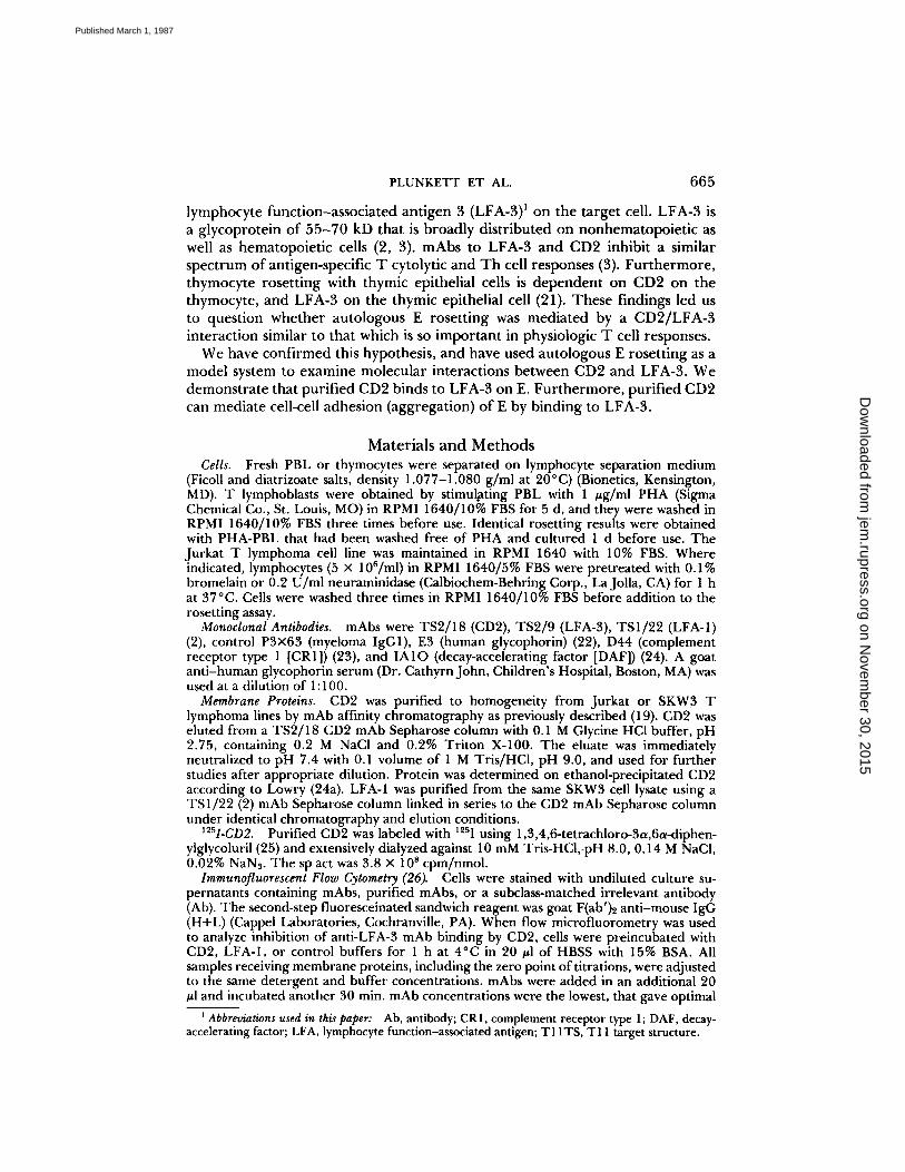
Peripheral blood human T lymphocytes rosetted with sheep E but not withhuman Eas previously reported (9, 10) (Table I) . ThehumanJurkat T lymphomacell line (which expresses 1-2 x 10⁵ molecules of CD2/cell [19]) was much moreefficient than PBL at rosetting with human E (Table 1) . CD2 mAb inhibitedautologous rosetting (Table I, Exp. 2), as previously described (13, 18) . ControlmAbs to the T lymphocyte surface proteins T4, T8, T3, and LFA-1 did notinhibit human E rosetting (data not shown) . Strikingly, LFA-3 mAb completely
on Novem
ber 30, 2015jem
.rupress.orgD
ownloaded from
Published March 1, 1987
PLUNKETT ET AL .
667
TABLE IEffect ofmAb on Rosetting ofHuman TCells with Human or
Sheep E
Cells were pretreated with 5 wg/ml mAbfor 30 min on ice, then washedthree times before addition to the rosetting assay.
* 5 pg/ml mAb was present throughout the rosetting assay.
inhibited rosetting of human T cells with human E (Table 1, Exp. 2) . Controlmabto glycophorin and CRI on human E had no effect . mAb to CD2 completelyinhibited Jurkat T cell rosetting with human and sheep E, while the mAb toLFA-3 inhibited rosetting with human but not sheep E. The lack of effect ofanti-human LFA-3 mAb on rosetting with sheep E was expected from its lack ofreactivity with sheep E (Fig . 1) .The cell type on which CD2 and LFA-3 were functionally important in E
rosetting was tested by pretreating only one cell type with the mAb (Table 1,Exp. 3) . CD2 mAb was inhibitory only when T cells were pretreated, and LFA-3mAb was inhibitory only when human E were pretreated .These findings with a T cell tumor line were confirmed and extended with
human thymocytes and T lymphoblasts (Table 11) . In contrast to peripheral Tlymphocytes, thymocytes and PHA blasts rosetted efficiently with human E.Rosetting of these cells with human E was inhibited at the level of the T cell withCD2 mAb and at the level of the human E with LFA-3 mAb (Table II, Exps . 1and 2) .Removal of cell surface sialic acid with neuraminidase, and partial removal of
cell surface glycoproteins with proteases have previously been shown (18, 27) toenhance a variety of cell-cell interactions, including E rosetting, possibly byreducing charge-charge repulsion due to negative surface changes. Bromelainandneuraminidase treatment of PBL unmasked rosetting activity (Table 11, Exp.
Exp. PretreatmentRosettes
Human E Sheep E
1 PBL E0 34
2* Jurkat EControl Control 30 73Anti-CD2 Anti-CD2 0 0Anti-LFA-3 Anti-LFA-3 0 67Anti-Glyco- Anti-Glyco- 32 63
phorin phorin 28 66Anti-CRI Anti-CRI
3 Jurkat EControl - 21 73Anti-CD2 - 0 0Anti-LFA-3 - 18 74- Control 23 74- Anti-CD2 23 75- Anti-LFA-3 0 76
on Novem
ber 30, 2015jem
.rupress.orgD
ownloaded from
Published March 1, 1987

668
MOLECULAR BASIS OF AUTOLOGOUS ROSETTING
TABLE IIEffect of mAb on Rosetting ofHuman TCells with Human E
Pretreatment
Percent Rosettes with Human E
Cells were pretreated with mAb as described in Table I .
3), and bromelain treatment of PHA-stimulated PBL increased the efficiency ofrosetting with human E (Table II, Exps . 2 and 3) . Pretreatment experimentsagain showed that CD2 mAb inhibited at the level of the T lymphocyte andLFA-3 mAb inhibited at the level of the human E. Bromelain or neuraminidasetreatment of human E rendered them agglutinatable by LFA-3 mAb (data notshown); in no other case were cells agglutinated by LFA-3 mAb or CD2 mAb.To determine if CD2 was functioning as a receptor for human E, we tested
whether the purified receptor could competitively inhibit the T lymphocyte-Einteraction . CD2 was solubilized with Triton X-100 from human T lymphoblastcell lines and purified by mAb affinity chromatography as described in Materialsand Methods. BSA was added to CD2 preparations to bind detergent (28) andprevent damage to cells. Purified CD2 inhibited Jurkat T cell rosetting withhuman E (Table III), and inhibited Jurkat T cell rosetting with sheep E aspreviously described (19) .
Inhibition of autologous rosetting with purified CD2 suggested solubilizedCD2 could bind to E. Purified, 125I-labeled CD2 bound to human and sheep E(3-10% of input counts in different experiments) . We tested whether pretreat-ment of human E with LFA-3 mAb would inhibit CD2 binding. '25I-labeled CD2binding to human E was inhibited 99% by anti-LFA-3 (Table IV). '25I-CD2binding to sheep E was not inhibited by LFA-3 mAb, as expected from the lackof reactivity of LFA-3 mAb with sheep E. Binding to either sheep or human Ewas unaffected by control mAb to LFA-1, glycophorin, and CR1 . Inclusion ofCD2 mAb inhibited binding of ' 25I-CD2 to both cell types, showing that labeledCD2 binding was specific .
Reciprocal experiments were carried out to determine whether CD2 couldcompetitively inhibit binding of LFA-3 mAb. E were preincubated with unlabeled
T cell E
Exp. I
Thymo-cytes
Exp.
PHA-PBL
2Brome-lainPHA-PBL
Exp.
Neura-minidasePBL
3
Brome-lainPBL
Control - 61 34 95 10 14Anti-CD2 - 0 0 0 0 0Anti-LFA-3 - 60 34 96 10 11Anti-Glyco- - 59 ND ND ND NDphorin - 59 ND ND ND ND
Anti-CR 1- Control 60 31 91 8 ND- Anti-CD2 61 26 92 8 ND- Anti-LFA-3 0 0 2 0 ND- Anti-Glyco- 60 ND ND ND ND- phorin 58 ND ND ND ND
Anti-CR1
on Novem
ber 30, 2015jem
.rupress.orgD
ownloaded from
Published March 1, 1987

PLUNKETT ET AL .
TABLE III
Inhibition by Purified CD2 ofHuman E and Sheep E Rosettes
Purified CD2 antigen in 30 A1 or a buffer control was mixed with 100 ,1of 30% BSA.Jurkat cells in 70 jal were added to give a final total volumeof 200 pl with the indicated finalconcentration of CD2and were incubatedfor 30 min at 4°C before the E rosette assay. Exp. 1: SKW3 CD2 in 0.2%Triton X-100, 0.01 M Tris, pH 7.2, 0.15 M NaCl . Exp. 2: Jurkat CD2 in0.1% Triton X-100, 0.01 M Tris, pH 7.2, 0.15 M NaCl . Controls hadidentical detergent and buffer concentrations .
TABLE IV
Inhibition of '25I-CD2 Binding by Anti-LFA-3 Antibody
5 X 10 6 human or sheep E were incubated with 50 gl of mAb (eitherhybridoma culture supernatant anti-CR1, anti-glycophorin, anti-CD2 andanti-LFA-3, or 1 :100 diluted ascites X63 and anti LFA-1) for 45 min at4°C. Then 50 ul of .Y5í-CD2 (diluted to 4,000 cpm/,ul with 10% FBS/RPMI 1640/2 mM Hepes, pH 7.4/3% BSA) was added and the incubationwas continued for another 2 h at 4°C. After incubation, the cells werewashed three times with 10% FBS/RPMI 1640/2 mM Hepes, pH 7.4 andwere counted in a gamma counter.
669
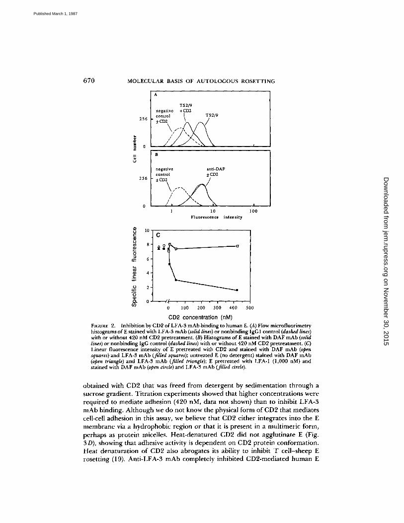
CD2 or control purified protein (LFA-1), then treated with the TS2/9 mAb toLFA-3, washed, and stained with FITC-conjugated second antibody for analysisby flow cytometry . Purified CD2 inhibited in a dose-dependent fashion thebinding of anti-LFA-3 to human E (Fig . 2) . At 420 nM, CD2 reduced anti-LFA-3 binding to human E by 79% (Fig . 2A) . Half-maximal inhibition was attainedat ^-30 nM CD2 (Fig . 2C) . The binding of a control antibody (anti-DAF) wasunaffected by CD2 (Fig . 2, B and C) . As another control, preincubation withpurified LFA-1 (1,000 nM) had no affect on anti-LFA-3 binding to human E(Fig. 2 C) . These experiments strongly suggest that CD2 binds directly to LFA-3on human E.
Functional studies showed that purified C132-mediated cell-cell adhesion (ag-glutination) of both human and sheep E (Fig . 3, B and F). Similar results were
Exp. CD2Jurkat cells
rosetting withhuman E
Jurkat cellsrosetting with
sheep EKg/ml
1 0 76 760.5 ND 325 12 4
2 0 65 805 16 13
Antibodyi2 'I-CD2 Bound (cpm
Human E
±SD)
SpecificityDonor 1 Donor 2
SheepE
X63 3,697 ± 55 5,329 ± 79 10,392 t 143Anti-LFA-1 3,898 t 40 5,547 t 56 11,168 ± 837Anti-CRI 3,205 t 79 5,135 ± 97 11,375 ± 267Anti-glycophorin 3,687 ± 20 5,524 ± 131 12,268 ± 32Anti-LFA-3 42 t 24 57 t 26 10,660 ± 402Anti-CD2 53 ± 14 34 ± 6 69 ± 6
on Novem
ber 30, 2015jem
.rupress.orgD
ownloaded from
Published March 1, 1987
670
MOLECULAR BASIS OF AUTOLOGOUS ROSETTING
daEre
uU
256
0
256
0
U 10-Ca)
.a)
8 -d
N 4~C
C
1
10
100Fluorescence intensity
2U0-
0L0 100 200 300 400 500
CD2 concentration (nM)
a
FIGURE 2.
Inhibition by CD2 ofLFA-3 mAb binding to humanE. (A) Flow microfluorimetryhistograms ofE stained with LFA-3 mAb (solid lines) or nonbinding IgGI control (dashed lines)with or without 420 nM CD2 pretreatment . (B) Histograms of E stained with DAF mAb (solidlines) or nonbinding IgG control (dashed lines) with or without 420 nM CD2pretreatment. (C)Linear fluorescence intensity of E pretreated with CD2 and stained with DAF mAb (opensquares) and LFA-3 mAb (filled squares); untreated E (no detergent) stained with DAF mAb(open triangle) and LFA-3 mAb (filled triangle) ; E pretreated with LFA-1 (1,000 nM) andstained with DAFmAb(open circle) and LFA-3 mAb (filled circle) .
obtained with CD2 that was freed from detergent by sedimentation through asucrose gradient . Titration experiments showed that higher concentrations wererequired to mediate adhesion (420 nM, data not shown) than to inhibit LFA-3mAb binding. Although we do not know the physical form of CD2 that mediatescell-cell adhesion in this assay, we believe that CD2 either integrates into the Emembrane via a hydrophobic region or that it is present in a multimeric form,perhaps as protein micelles . Heat-denatured CD2 did not agglutinate E (Fig .3D), showing that adhesive activity is dependent on CD2 protein conformation .Heat denaturation of CD2 also abrogates its ability to inhibit T cell-sheep Erosetting (19) . Anti-LFA-3 mAb completely inhibited CD2-mediated human E
on Novem
ber 30, 2015jem
.rupress.orgD
ownloaded from
Published March 1, 1987

PLUNKETT ET AL .
6'71
FIGURE 3.
CD2 induced aggregation of E. 107 E were mixed with 20 #g/ml CD2 with orwithout 5,ug/ml mAb in the presence of 15% BSA. The mixture was centrifuged for 2 minat 200 gand incubated on ice for 2 h. The cells were gently resuspended by rotating the tubeand photographed with a Nikon inverted microscope at an original magnification of x 100(x 77 shown here), except for anti-glycophorin induced aggregation of human E which wasphotographed at x 40 (x 31 shown here) . (A) human E; (B) human E + CD2; (C) human E +CD2 + LFA-3 mAb; (D) human E + CD2 denatured at 100°C for 15 min; (E) sheep E; (F)sheep E+ CD2; (G) sheep E+ CD2 + LFA-3 mAb; (H) human E + anti-human glycophorinserum + LFA-3mAb.
on Novem
ber 30, 2015jem
.rupress.orgD
ownloaded from
Published March 1, 1987
672
MOLECULAR BASIS OF AUTOLOGOUS ROSETTING
aggregation (Fig . 3 C), strongly suggesting that aggregation of human E by CD2is mediated by direct binding of CD2 to LFA-3. As expected, anti-LFA-3 had noeffect on aggregation of sheep E by CD2 (Fig . 3 G) . As a specificity control,agglutination induced by a polyclonal serum to human glycophorin was unaf-fected by anti-LFA-3 (Fig . 3 H) .
DiscussionWe have found that autologous rosetting of human T cells with E is due to
binding of CD2 on the T lymphocyte to LFA-3 on the erythrocyte, and haveused this system to explore the interactions between these molecules . Antibodiesto CD2 and LFA-3 inhibited rosetting by binding to the T lymphocyte and theE, respectively . These mAbs inhibited rosetting by thymocytes, mitogen-activatedlymphocytes, and a T lymphoma tumor cell line . Peripheral blood T lymphocytesdo not rosette, but rosetting activity that was unmasked by neuraminidase orbromelain treatment was CD2 and LFA-3 dependent . Detergent-solubilized,purified CD2 bound to LFA-3 on E, as demonstrated by inhibition by LFA-3mAb of CD2 binding and reciprocal inhibition by CD2 of LFA-3 mAb binding.Higher concentrations of purified CD2 (400 nM) aggregated E and this aggre-gation was inhibited by LFA-3 mAb. These results show that CD2 binds to LFA-3 on E, and that this interaction is sufficient to mediate cell-cell adhesion .CD2 and LFA-3 previously have been shown to participate in a pathway of
antigen-independent adhesion of T cells to B lymphoblastoid target cells (20),and to be important in both T cytolytic and Th cell function (1-7) . In studiesanalogous to those reported here (29), purified CD2 has been shown to bind toLFA-3 on a B lymphoblastoid cell line . Antigen-specific cytolytic T lymphocyte-mediated killing of this cell line has been shown to be inhibited by CD2 mAband LFA-3 mAb (2) . Thus, functionally important T cell-target cell interactionsand autologous rosetting involve similar interactions at the molecular levelbetween CD2 and LFA-3. In further similarity to the human E rosetting systemstudied here, CD2- and LFA-3-dependent (but not LFA-1-dependent) antigen-independent conjugate formation can occur at 4°C (20) . The concentration atwhich CD2 inhibited LFA-3 mAb binding suggests that the affinity of CD2 forLFA-3 on E is -3 X 10'/M; this is similar to its affinity for LFA-3 on Blymphoblastoid cells of 1 .9 X 107/M (29) . Autologous rosetting is thus not anaberrant crossreaction ; rather, it is an interesting model system for studying aphysiologically relevant molecular interaction .A particularly interesting feature of autologous rosetting is its dependence on
T cell activation . Thymocytes and T cell blasts, but not peripheral bloodlymphocytes, form autologous rosettes, correlating with expression of activationepitopes on the CD2 molecule (1, 30) . Whether CD2 from activated cells has ahigher affinity for LFA-3 than CD2 from resting peripheral T lymphocytes is aninteresting question for further work . Activation epitopes can be induced onCD2 on resting T lymphocytes at 4 ° C in 2 min (1, 30, 31), raising the possibilitythat detergent solubilization could also change the conformation of CD2 andalter its affinity for LFA-3. We found that latent CD2- and LFA-3-dependentrosetting by peripheral T lymphocytes could be unmasked by protease orneuraminidase treatment . A reduction of net negative surface charge, lessening
on Novem
ber 30, 2015jem
.rupress.orgD
ownloaded from
Published March 1, 1987

PLUNKETT ET AL .
673
charge repulsion between the T lymphocyte and E would be sufficient to explainthese effects, but it is possible that modification of CD2 is also important .Neuraminidase treatment of T cells has been shown to unmask CD2 activationepitopes (30) .Although we found no rosetting of peripheral blood T lymphocytes with
human E, under less stringent conditions a small percentage of peripheral bloodT lymphocytes are found to form autologous rosettes (12-I4). Removal of thesecells abrogates the autologous mixed lymphocyte reaction (12-14). The corre-lation between autologous rosetting and activation suggests these may be a subsetof activated T lymphocytes. We speculate that the CD2-LFA-3 interaction mayhelp mediate localization and extravasation of activated T lymphocytes at sitesof immune reaction . LFA-3 is expressed on endothelial cells (3, 32). Lympho-blasts extravasate throughout the circulation, in contrast to small lymphocytes,which selectively recirculate through high endothelial venules (33) . This en-hanced adhesion of activated T lymphocytes may explain why the majority ofactivated T lymphocytes are not found in the circulation but are present inlymph nodes or sites of antigenic stimulation .
Purification of T lymphocytes by sheep E rosetting has been known to causefunctional alterations . Larsson et al . (34) found that E rosetting primes Tlymphocytes to proliferate in response to lymphokines. Although this is anartificial system, it suggests that interaction of CD2 with the LFA-3 ligand maybe a physiologically relevant pathway of T lymphocyte stimulation . Interactionof CD2' thymocytes with LFA-3 + thymic epithelial cells via CD2/LFA-3 recep-tor/ligand interactions appears of great importance in functional responses ofseveral different thymocyte subsets (21, 35).
Molecules similar to LFA-3 are present on E in other species. Human Tlymphocytes form CD2-dependent rosettes with E from human, sheep, dog,horse, pig, and rabbit (8, 13, 17, 18, 36). Autologous rosetting of thymocyteshas been demonstrated in the human, rabbit, rat, and pig (13, 16, 27, 37). A 42kD surface glycoprotein termed the T11 target structure (T 11TS) has beendefined on sheep E (38, 39). mAb to T11TS inhibit rosetting of sheep E witheither humanT cells or sheep T lymphocytes and inhibit a range of T lymphocyteresponses in sheep similar to those inhibited by LFA-3 mAb in humans. Further-more, the purified TIITS molecule competitively inhibits CD2 mAb binding.Comparative studies suggest LFA-3 and T11ITS are functional and structuralhomologues, and the much higher density of T11ITS on sheep E than LFA-3 onhuman E may explain the much more avid rosetting of resting human T cellswith sheep than human E (Hunig, T., M . Plunkett, M . Dustin, P. Selvaraj, andT. Springer, unpublished observations) . While the significance of the expressionof LFA-3 on human E is still not fully appreciated, the conservation of expressionof similar structures on E in other mammals that can bind to human CD2 pointsto its evolutionary importance .
SummaryCD2, also known as LFA-2, T11, and the E rosette receptor, is a T lymphocyte
surface protein functionally important in adhesion to target cells and T celltriggering . LFA-3 is a widely distributed cell surface protein that functions in
on Novem
ber 30, 2015jem
.rupress.orgD
ownloaded from
Published March 1, 1987
674
MOLECULAR BASIS OF AUTOLOGOUS ROSETTING
adhesion on target cells . We find that LFA-3 is expressed on human E, and thatCD2 is a receptor for LFA-3 that mediates T cell adhesion to human E.Pretreatment of T lymphocytes with CD2 mAb or of E with LFA-3 mAb inhibitsrosetting. Purified CD2 molecules bind to human E and inhibit rosetting . 12s1-
CD2 binding to E is inhibited by LFA-3 mAb; reciprocally, binding of LFA-3mAb to human E is inhibited by pretreatment with purified CD2. Higherconcentrations of CD2 aggregate human E; aggregation is inhibited by mAb toLFA-3.
Receivedfor publication 6 November 1986 .
References1 . Meuer, S. C., R . E. Hussey, M. Fabbi, D. Fox, O . Acuto, K. A . Fitzgerald, J . C .
Hodgdon, J . P . Protentis, S . F . Schlossman, and E. L . Reinherz . 1984 . An alternativepathway of T-cell activation : a functional role for the 50 kd TI 1 sheep erythrocytereceptor protein . Cell. 36:897 .
2 . Sanchez-Madrid, F., A . M . Krensky, C. F . Ware, E . Robbins, J . L . Strominger, S . J .Burakoff, and T. A. Springer . 1982 . Three distinct antigens associated with humanT lymphocyte-mediated cytolysis : LFA-1, LFA-2, and LFA-3 . Proc . Nati . Acad. Sci.USA . 79:7489 .
3 . Krensky, A. M ., F . Sanchez-Madrid, E . Robbins, J . Nagy, T. A . Springer, and S . J .Burakoff. 1983 . The functional significance, distribution, and structure of LFA-1,LFA-2, and LFA-3 : cell surface antigens associated with CTL-target interactions . J.Immunol. 131 :611 .
4 . Van Wauwe, J., J . Goossens, W . Decock, P. Kung, and G. Goldstein . 1981 . Suppres-sion ofhuman T-cell mitogenesis and E-rosette formation by the monoclonal antibodyOKT11A. Immunology. 44:865 .
5 . Haynes, B . F . 1981 . Human T lymphocyte antigens as defined by monoclonalantibodies . Immunol. Rev. 57:127 .
6 . Palacios, R., and O. Martinez-Maza . 1982 . Is the E receptor on human T lymphocytesa "negative signal receptor'?J . Immunol . 129:2479 .
7 . Krensky, A . M., E . Robbins, T. A . Springer, and S . J . Burakoff 1984 . LFA-I, LFA-2 and LFA-3 antigens are involved in CTL-target conjugation .J. Immunol. 132:2180 .
8 . Kamoun, M., P . J . Martin, J . A . Hansen, M. A. Brown, A . W. Siadak, and R. C .Nowinski . 1981 . Identificatio n of a human T lymphocyte surface protein associatedwith the E-rosette receptor . J. Exp. Med. 153:207 .
9 . Lay, W. H., N . F. Mendes, C . Bianco, and V . Nussenzweig . 1971 . Binding of sheepred blood cells to a large population of human lymphocytes . Nature (tond.) . 230:531 .
10 . Froland, S . S . 1972 . Binding ofsheep erythrocytes to human lymphocytes . A probablemarker of T lymphocytes . Scand . J. Immunol . 1:269.
11 . Ross, G . D ., and R. J. Winchester. 1980 . Methods for enumerating lymphocytepopulations . In Manual ofClinical Immunology . 2nd ed. N. R . Rose and H. Friedman,editors . American Society of Microbiology, Washington, DC . 213-228 .
12 . Tomonari, K., A. Wakisaka, and M . Aizawa . 1980 . Self recognition by autologousmixed lymphocyte reaction-primed cells.J. Immunol . 125:1596 .
13 . Nalet, V., and C . Fournier. 1985 . Human autologous rosette-forming cells 111 .Binding oferythrocytes from different species to the T-cell receptors for autologousred blood cells . Cell. Immunol. 96:126 .
14 . Palacios, R., L . Llorente, D . Alarcon-Segovia, A. Ruiz-Arguelles, and E . Diaz-Jouanen . 1980 . Autologous rosette-forming T cells as the responding cells in humanautologous mixed-lymphocyte reaction . J. Clin . Invest. 65:1527 .
on Novem
ber 30, 2015jem
.rupress.orgD
ownloaded from
Published March 1, 1987
PLUNKETT ET AL.
675
15. Yang, S . Y ., S . Rhee, G . Angelos, and B. Dupont . 1987 . Functional analysis of CD2(T,p50) epitopes detected by 24 anti-CD2 antibodies. In Leukocyte Typing III . A . J.McMichael, editor . Springer-Verlag New York Inc., New York . In press .
16 . Baxley, G., G . B . Bishop, A . G . Cooper, and H. H . Wortis. 1973 . Rosetting of humanred blood cells to thymocytes and thymus-derived cells . Clin . Exp. Immunol. 15:385 .
17 . Verbi, W., M. F. Greaves, C . Schneider, K . Koubek, G. Janossey, H . Stein, P . C.Kung, and G . Goldstein . 1982 . Monoclonal antibodies OKTI1 and OKTIIa havepan-T reactivity and block sheep erythrocyte receptors . Eur. J. Immunol. 12 :81 .
18 . Scheffel, J . W., and S . J . Swartz . 1982 . Inhibition of autologous rosette formation bymonoclonal antibody to the sheep erythrocyte receptor. J. Immunol. 128:1930 .
19 . Plunkett, M . L ., and T. A. Springer. 1986 . Purification and characterization of thelymphocyte function-associated-2 (LFA-2) molecule .J. Immunol. 136:4181 .
20 . Shaw, S., G . E . G . Luce, R . Quinones, R . E. Gress, T . A . Springer, and M. E. Sanders .1986 . Two antigen-independent adhesion pathways used by human cytotoxic T cellclones . Nature (Loud.) . 323 :262 .
21 . Wolf, L . S ., D . T . Tuck, T. A . Springer, B . F . Haynes, and K . H . Singer . 1986 .Thymocyte binding to human thymic epithelial cells is inhibited by monoclonalantibodies to CD-2 and LFA-3 antigens .J. Immunol. 138:358 .
22 . Nichols, M. E ., R . E . Rosenfield, and P . Rubinstein . 1985 . Two blood group Mepitopes disclosed by monoclonal antibodies. VoxSang. 49:138 .
23 . Iida, K., R . Mornaghi, and V. Nussenzweig . 1982 . Complement receptor (CRI)deficiency in erythrocytes from patients with systemic lupus erythematosus. J. Exp.Med. 155 :1427 .
24 . Kinoshita, T., M . E . Medof, R. Silber, and V . Nussenzweig . 1985 . Distribution ofdecay-acclerating factor in the peripheral blood of normal individuals and patientswith paroxysmal nocturnal hemoglobinuria. J. Exp. Med. 162:75 .
24a.Lowry, O. H., N . J . Rosebrough, A. L . Fair, and R . J . Randall . 1951 . Proteinmeasurement with the Folin-phenol reagent . J. Biol. Chem. 193:265 .
25 . Fraker, P . J ., and J . C. Speck . 1978 . Protein and cell membrane iodinations with asparingly soluble chloroamide, 1,3,4,6-tetrachloro-3a,6a-diphenyl glycoluril .Biochem. Biophys. Res. Commun . 80:849 .
26 . Kilrzinger, K., T . Reynolds, R. N . Germain, D . Davignon, E. Martz, and T. A .Springer . 1981 . A novel lymphocyte function-associated antigen (LFA-1): cellulardistribution, quantitative expression, and structure . J. Immunol. 127:596 .
27 . Scheffel, J . W., and Y. B . Kim . 1981 . Characterization of autologous erythrocyterosette-forming cells in Minnesota miniature swine. Cell. Immunol. 57:175 .
28 . Springer, T . A ., D . L . Mann, A . L . DeFranco, andJ . L . Strominger . 1977 . Detergentsolubilization, purification, and separation of specificities of HLA antigens from acultured human lymphoblastoid line, RPMI 4265.J. Biol . Chem . 252:4682 .
29 . Selvaraj, P., M . L . Plunkett, M. E . Sanders, M. Dustin, S . Shaw, and T. A . Springer .1987 . The T lymphocyte glycoprotein CD2 (LFA-2/T11/E-rosette receptor) bindsthe cell surface ligand LFA-3. Nature (Loud.) . In press .
30 . Bernard, A., C . Gelin, B . Raynal, D. Pham, C . Gosse, and L . Boumsell . 1982 .Phenomenon of human T cells rosetting with sheep erythrocytes analyzed withmonoclonal antibodies . "Modulation" of a partially hidden epitope determining theconditions of interaction between T cells and erythrocytes . J. Exp. Med. 155:1317 .
31 . Yang, S . Y ., S. Chouaib, and B . Dupont. 1986 . A common pathway for T lymphocyteactivation involving both the CD3-Ti complex and CD2 sheep erythrocyte receptordeterminants . J. Immunol. 137:1097 .
32 . Collins, T., A . M . Krensky, C . Clayberger, W. Fiers, M. A . Gimbrone Jr ., S . J .Burakoff, and J . S . Pober . 1984 . Human cytolytic T lymphocyte interactions with
on Novem
ber 30, 2015jem
.rupress.orgD
ownloaded from
Published March 1, 1987

676
MOLECULAR BASIS OF AUTOLOGOUS ROSETTING
vascular endothelium and fibroblasts : Role of effector and target cell molecules . J.Immunol . 133:1878 .
33 . Gowans, J . L ., and E . J . Knight . 1964 . The route of re-circulation of lymphocytes inthe rat . Proc. R . Soc. Lond . B Biol. Sci. 159:257 .
34 . Larsson, E.-L ., J . Andersson, and A. Coutinho . 1978 . Functional consequences ofsheep red blood cell rosetting for human T cells : gain of reactivity to mitogenicfactors . Eur. J. Immunol. 8 :693 .
35 . Haynes, B . F . 1986 . The role of the thymic microenvironment in promotion ofearlystages of human T cell maturation . Clin . Res. 34:422 .
36 . Braganza, C . M ., G . Stathopoulos, A. J. S . Davies, E . V . Elliott, R . S . Kerbel, M.Papamichail, and E. J . Holborow. 1975 . Lymphocyte : erythrocyte (L.E .) rosettes asindicators of the heterogeneity of lymphocytes in a variety of mammalian species .Cell. 4:103 .
37 . Sandilands, G., K . Gray, A . Cooney, J . D . Browning, and J . R . Anderson . 1974 .Auto-rosett e formation by human thymocytes and lymphocytes . Lancet. i :27 .
38 . Hünig, T . 1985 . The cell surface molecule recognized by the erythrocyte receptorof T lymphocytes .J. Exp . Med. 162:890 .
39 . Hünig, T. R . 1986 . The ligand of the erythrocyte receptor of T lymphocytes :expression on white blood cells and possible involvement in T cell activation . J.Immunol . 136:2103 .
on Novem
ber 30, 2015jem
.rupress.orgD
ownloaded from
Published March 1, 1987
Copyright © 2022 FDOKUMEN