Autologous and Cancer Stem Cell Gene Therapy (488 Pages)
489
干细胞之家www.stemcell8.cn ←点击进入
-
Upload
khangminh22 -
Category
Documents
-
view
0 -
download
0
Transcript of Autologous and Cancer Stem Cell Gene Therapy (488 Pages)

AUTOLOGOUS AND CANCER STEM CELL GENE THERAPY
Progress in Gene Therapy — Vol. 3
干细胞之家www.stemcell8.cn ←点击进入

This page intentionally left blankThis page intentionally left blank
干细胞之家www.stemcell8.cn ←点击进入

N E W J E R S E Y • L O N D O N • S I N G A P O R E • B E I J I N G • S H A N G H A I • H O N G K O N G • TA I P E I • C H E N N A I
World Scientific
AUTOLOGOUS AND CANCER STEM CELL GENE THERAPY
Progress in Gene Therapy — Vol. 3
EditorsRoger BERTOLOTTI
University of Nice–Sophia Antipolis, France Keiya OZAWA
Jichi Medical University, Japan
干细胞之家www.stemcell8.cn ←点击进入

Library of Congress Cataloging-in-Publication DataAutologous and cancer stem cell gene therapy / edited by Roger Bertolotti, Keiya Ozawa.
p. ; cm. -- (Progress in gene therapy ; v. 3)Includes bibliographical references and index.ISBN-13: 978-981-277-586-3 (hardcover : alk. paper)ISBN-10: 981-277-586-2 (hardcover : alk. paper)
1. Stem cells--Therapeutic use. 2. Stem cells--Transplantation. 3. Gene therapy.4. Cancer--Gene therapy. 5. Autotransplantation. I. Bertolotti, R. (Roger) II. Ozawa,Keiya, 1953- III. Series.[DNLM: 1. Gene Therapy. 2. Stem Cells. 3. Regenerative Medicine. 4. Stem CellTransplantation. 5. Transplantation, Autologous. 6. Tumor Stem Cells. QZ 325 A939 2007]QH588.S83A98 2007615.8'95--dc22
2007043710
British Library Cataloguing-in-Publication DataA catalogue record for this book is available from the British Library.
For photocopying of material in this volume, please pay a copying fee through the Copyright Clearance Center,Inc., 222 Rosewood Drive, Danvers, MA 01923, USA. In this case permission to photocopy is not required fromthe publisher.
Typeset by Stallion PressEmail: [email protected]
All rights reserved. This book, or parts thereof, may not be reproduced in any form or by any means, electronic ormechanical, including photocopying, recording or any information storage and retrieval system now known or tobe invented, without written permission from the Publisher.
Copyright © 2008 by World Scientific Publishing Co. Pte. Ltd.
Published by
World Scientific Publishing Co. Pte. Ltd.
5 Toh Tuck Link, Singapore 596224
USA office: 27 Warren Street, Suite 401-402, Hackensack, NJ 07601
UK office: 57 Shelton Street, Covent Garden, London WC2H 9HE
Printed in Singapore.
SC - Autologous and Cancer.pmd 10/25/2007, 12:25 PM1
干细胞之家www.stemcell8.cn ←点击进入

November 5, 2007 14:50 WSPC/SPI-B559: Autologous & Cancer Stem Cell Gene Therapy fm FA
Preface
Adult/Postnatal stem cells provide for life-long cell replacement in tissuesand organs. They have an inherent ability for self-renewal and differentia-tion into multiple cell types that are characteristic of their tissue of origin,and have been identified in an increasing number of tissues/organs evenin brain and myocardium in which cell turnover was reputedly absent. Inaddition, many stem cells have inherent homing abilities that are instru-mental in therapeutic applications. Stem cells are also the driving force ofcancer, where genetic/epigenetic alterations culminate in uncontrolled self-renewal and tumorigenesis either in tissue stem cells or in some of theirprogenitor/differentiated derivatives. As a rare subset of the tumor, can-cer stem cells are the only drive of tumor initiation/propagation and, upontransplantation, have been shown to recapitulate the hierarchical clonogenicdifferentiating/differentiated organization of the original tumor cell popu-lation. Stem cells are thus the key targets of 1) long-term gene therapy andbroad/synergistic transient regenerative/epigenetic gene therapy for bothinherited diseases and acquired/aging disorders on their autologous side,and 2) effective anti-cancer therapy on their dark side.
Autologous stem cells have been instrumental in the first unequivocalsuccesses for gene therapy (2000–2004), whereby ex vivo retrovirally cor-rected hematopoietic stem cells have been returned to the patients. Sucha stem cell gene therapy achievement that relies on random integration oftherapeutic transgenes into host chromosomes is presented together withemerging experimental approaches aimed at eliminating random integra-tion oncogenic hazards through site-specific integration or gene target-ing. Breakthrough endonuclease-boosted gene targeting for gene correction(inherited diseases) or targeted integration of therapeutic transgene (otherpathologies), culminating in an efficiency compatible with clinical correc-tion of a disease gene, is one of the highlights of the book. Other highlightsinclude the pioneering transplantation of adult pluripotent stem cells asa substitute for tissue-specific stem cells, thereby pinpointing the break-through potential of such autologous cells able to contribute to all three
v
干细胞之家www.stemcell8.cn ←点击进入

November 5, 2007 14:50 WSPC/SPI-B559: Autologous & Cancer Stem Cell Gene Therapy fm FA
vi Preface
germ layers both for multi-systemic diseases and for the development of auniversal stem cell gene therapy platform. The autologous side is thus dis-cussed in terms of magnifying stem cell therapeutic homing/regenerativecapabilities through transient regenerative gene therapy, and in terms oftackling most pathologies (including mitochondrial DNA diseases andaging disorders) through stem cell repopulation dynamics into appropri-ate niches (long-term engraftment) and tissues (cell turnover). Regardingthe dark side, focus is on both the increasing number of identified tissue-specific cancer stem cells as the ultimate targets for recurrence-free cancertherapy and on the development of armed stem cells as tumor-homing vec-tors for targeted anti-cancer stem cell gene therapy.
Roger BertolottiKeiya Ozawa
干细胞之家www.stemcell8.cn ←点击进入

November 5, 2007 14:50 WSPC/SPI-B559: Autologous & Cancer Stem Cell Gene Therapy fm FA
Contents
Preface vRoger Bertolotti & Keiya Ozawa
List of Contributors xi
Introduction
Toward a Universal Platform for Autologous Stem Cell Gene Therapy xxiRoger Bertolotti
Long-term Stem Cell Gene Therapy: From Current ClinicalRandom–Integration Achievements to Stem Cell Gene
Targeting/Cybridization
I. From pioneering clinical retroviral gene therapy toexperimental site-specific integrative gene therapy
1 Stem Cell Gene Therapy for ADA-Deficiency withoutMyelopreparative Conditioning 1Makoto Otsu and Tadashi Ariga
2 Targeted Insertion of Transgene into a Specific Site onChromosome 19 by Using Adeno-Associated VirusIntegration Machinery 19Masashi Urabe, Yoko Obara, Takayuki Ito, Hiroaki Mizukami,Akihiro Kume and Keiya Ozawa
3 Herpes Simplex Virus Type 1/Adeno-Associated VirusHybrids as Site-Specific Integrating Vectors 47Cornel Fraefel, Daniel L. Glauser, Thomas Heisterand Mathias Ackermann
vii
干细胞之家www.stemcell8.cn ←点击进入

November 5, 2007 14:50 WSPC/SPI-B559: Autologous & Cancer Stem Cell Gene Therapy fm FA
viii Contents
II. Stem cell gene targeting/cybridization: towardendonuclease-boosted gene repair/alteration, customsite-specific integrative gene therapyand transmitochondrial therapy/rejuvenation
4 Endonuclease-Boosted Gene Targeting and Cybridizationfor Long-Term Stem Cell Gene Therapy 81Roger Bertolotti
5 Cell-Free Protein-Evolution Systems for Engineeringof Novel Sequence-Specific DNA-Binding and -ModifyingActivities 115Armin Sepp, Farid Ghadessy and Yen Choo
6 Advances in Engineering Homing Endonucleases forGene Targeting: Ten Years After Structures 135Barry L. Stoddard, Andrew M. Scharenberg andRaymond J. Monnat, Jr
7 Gene Targeting Mediated by Helper-dependentAdenoviral Vectors 169Kohnosuke Mitani
Adult Pluripotent Stem Cells: Emerging Stem Cell/GeneTherapy Breakthrough
8 Multipotent Progenitor Cells: A New Era in StemCell-Mediated Gene Therapy? 187Marta Serafini and Catherine M. Verfaillie
Cancer Stem Cell Gene Therapy
I. Cancer stem cells as breakthrough targets of cancergene therapy
9 Leukemia Stem Cells, A Pioneering Paradigmatic Model 203Daniel J. Pearce and Dominique Bonnet
10 Mammary Stem Cells 261Mark Shackleton, Marie-Liesse Asselin-Labat, François Vaillant,Geoffrey J. Lindeman and Jane E. Visvader
干细胞之家www.stemcell8.cn ←点击进入

November 5, 2007 14:50 WSPC/SPI-B559: Autologous & Cancer Stem Cell Gene Therapy fm FA
Contents ix
11 Human Brain Tumor Stem Cells 299Sara G. M. Piccirillo and Angelo L. Vescovi
12 CD44 as a Functional Cancer Stem Cell Markerand a Potential Therapeutic Target 317Lubna Patrawala and Dean G. Tang
II. Armed stem cells as tumor-homing vectors for cancergene therapy
13 Neural Stem Cell-mediated Therapy of Primary andMetastatic Solid Tumors 335Joseph Najbauer, Mary K. Danks, Nils-Ole Schmidt,Seung U. Kim and Karen S. Aboody
14 Antitumor Activity of Adenovirally Transduced CD34+
Cells Expressing Membrane-bound TRAIL 373Carmelo Carlo-Stella, Cristiana Lavazza, Antonino Carbone andAlessandro M. Gianni
15 Mesenchymal Stem Cells as Vehicles for DeliveringTherapeutics and Oncolytic Viruses 393Larisa Pereboeva and Justin Roth
Index 445
干细胞之家www.stemcell8.cn ←点击进入

November 5, 2007 14:50 WSPC/SPI-B559: Autologous & Cancer Stem Cell Gene Therapy fm FA
This page intentionally left blankThis page intentionally left blank
干细胞之家www.stemcell8.cn ←点击进入

November 5, 2007 14:50 WSPC/SPI-B559: Autologous & Cancer Stem Cell Gene Therapy fm FA
List of Contributors
Karen S. AboodyDivision of Hematology/HematopoieticCell Transplantation, City of HopeNational Medical Center andBeckman Research InstituteDuarte, CA, USAandDivision of NeurosciencesCity of Hope NationalMedical Center and Beckman Research InstituteDuarte, CA, USA
Mathias AckermannInstitute of VirologyUniversity of ZurichSwitzerland
Tadashi ArigaDepartment of PediatricsHokkaido University Graduate School of MedicineSapporo, 060-8638Hokkaido, Japan
Marie-Liesse Asselin-LabatThe Walter and Eliza Hall Institute of Medical ResearchParkvilleVictoria 3050, Australia
xi
干细胞之家www.stemcell8.cn ←点击进入

November 5, 2007 14:50 WSPC/SPI-B559: Autologous & Cancer Stem Cell Gene Therapy fm FA
xii List of Contributors
Roger BertolottiCNRS, Gene Therapy and Regulation ResearchFaculty of MedicineUniversity of Nice Sophia Antipolis06107 Nice, France
Dominique BonnetHematopoietic Stem Cell LaboratoryCancer Research UKLondon Research InstituteLondon, WC2A 3PX, UK
Antonino CarboneDepartment of PathologyFondazione IRCCS Istituto Nazionale TumoriMilano, Italy
Carmelo Carlo-Stella“Cristina Gandini” Medical Oncology UnitFondazione IRCCS Istituto Nazionale TumoriMilano, ItalyandMedical OncologyUniversity of MilanoMilano, Italy
Yen ChooPlasticell LtdImperial BioincubatorBessemer Building (RSM)Prince Consort RoadSouth KensingtonLondon SW7 2BP, UK
干细胞之家www.stemcell8.cn ←点击进入

November 5, 2007 14:50 WSPC/SPI-B559: Autologous & Cancer Stem Cell Gene Therapy fm FA
List of Contributors xiii
Mary K. DanksDepartment of Molecular PharmacologySt. Jude Children’s Research HospitalMemphis, TN, USA
Cornel FraefelInstitute of VirologyUniversity of ZurichSwitzerland
Farid GhadessyInstitute of Molecular and Cell Biology61 Biopolis DriveSingapore 138673
Alessandro M. Gianni“Cristina Gandini” Medical Oncology UnitFondazione IRCCS Istituto Nazionale TumoriMilano, ItalyandMedical OncologyUniversity of MilanoMilano, Italy
Daniel L. GlauserInstitute of VirologyUniversity of ZurichSwitzerland
Thomas HeisterInstitute of VirologyUniversity of ZurichSwitzerland
干细胞之家www.stemcell8.cn ←点击进入

November 5, 2007 14:50 WSPC/SPI-B559: Autologous & Cancer Stem Cell Gene Therapy fm FA
xiv List of Contributors
Takayuki ItoDivision of Genetic TherapeuticsCenter for Molecular MedicineJichi Medical UniversityTochigi 329-0498, Japan
Seung U. KimDivision of NeurologyUBC HospitalUniversity of British ColumbiaVancouver, CanadaandInstitute for Regenerative MedicineGachon UniversityInchon, Korea
Akihiro KumeDivision of Genetic TherapeuticsCenter for Molecular MedicineJichi Medical UniversityTochigi 329-0498, Japan
Cristiana Lavazza“Cristina Gandini” Medical Oncology UnitFondazione IRCCS Istituto Nazionale TumoriMilano, ItalyandMedical OncologyUniversity of MilanoMilano, Italy
Geoffrey J. LindemanThe Walter and Eliza Hall Institute of Medical ResearchParkvilleVictoria 3050, Australia
干细胞之家www.stemcell8.cn ←点击进入

November 5, 2007 14:50 WSPC/SPI-B559: Autologous & Cancer Stem Cell Gene Therapy fm FA
List of Contributors xv
Kohnosuke MitaniGene Therapy DivisionResearch Center for Genomic MedicineSaitama Medical UniversityHidaka, Saitama 350-1241, Japan
Hiroaki MizukamiDivision of Genetic TherapeuticsCenter for Molecular MedicineJichi Medical UniversityTochigi 329-0498, Japan
Raymond J Monnat, JrDepartments of Pathology and Genome SciencesUniversity of WashingtonSeattle WA 98195, USA
Joseph NajbauerDivision of Hematology/HematopoieticCell Transplantation, City of HopeNational Medical Center andBeckman Research InstituteDuarte, CA, USA
Yoko ObaraDivision of Genetic TherapeuticsCenter for Molecular MedicineJichi Medical UniversityTochigi 329-0498, Japan
Makoto OtsuLaboratory of Stem Cell TherapyCenter for Experimental MedicineInstitute of Medical ScienceUniversity of Tokyo108-8639 Tokyo, Japan
干细胞之家www.stemcell8.cn ←点击进入

November 5, 2007 14:50 WSPC/SPI-B559: Autologous & Cancer Stem Cell Gene Therapy fm FA
xvi List of Contributors
Keiya OzawaDivision of Genetic TherapeuticsCenter for Molecular MedicineJichi Medical UniversityTochigi 329-0498, Japan
Lubna PatrawalaDepartment of CarcinogenesisThe University of Texas MD Anderson Cancer CenterScience Park–Research DivisionSmithville, TX 78957, USA
Daniel J. PearceHematopoietic Stem Cell LaboratoryCancer Research UKLondon Research InstituteLondon, WC2A 3PX, UK
Larisa PereboevaDivision of Human Gene TherapyDepartments of Medicine, Obstetrics and Gynecology,Pathology, and Surgery and the Gene Therapy CenterUniversity of Alabama at BirminghamBirmingham, AL 35294-2172, USAandDivision of Molecular and Cellular PathologyDepartment of PathologyUniversity of Alabama at BirminghamBirmingham, AL 35294, USA
Sara G. M. PiccirilloDepartment of Biotechnology and BiosciencesUniversity of Milan-BicoccaMilan, Italy
干细胞之家www.stemcell8.cn ←点击进入

November 5, 2007 14:50 WSPC/SPI-B559: Autologous & Cancer Stem Cell Gene Therapy fm FA
List of Contributors xvii
Justin RothDivision of Molecular and Cellular PathologyDepartment of PathologyUniversity of Alabama at BirminghamBirmingham, AL 35294, USA
Andrew M ScharenbergDepartment of PediatricsUniversity of WashingtonSeattle WA 98195, USAandChildren’s Hospital and RegionalMedical CenterSeattle WA 98109, USA
Nils-Ole SchmidtDepartment of NeurosurgeryUniversity Medical Center Hamburg-EppendorfHamburg, Germany
Armin Sepp43 Bullen CloseCambridge CB1 8YU, UK
Marta SerafiniCentro Ricerca M. TettamantiClinica Pediatrica Università Milano-BicoccaOspedale San GerardoMonza, Italy
干细胞之家www.stemcell8.cn ←点击进入

November 5, 2007 14:50 WSPC/SPI-B559: Autologous & Cancer Stem Cell Gene Therapy fm FA
xviii List of Contributors
Mark ShackletonLife Sciences InstituteUniversity of MichiganAnn Arbor, MI, USAandThe Walter and Eliza Hall Institute of Medical ResearchParkvilleVictoria 3050, Australia
Barry L StoddardDivision of Basic SciencesFred Hutchinson Cancer Research CenterSeattle WA 981089, USA
Dean G. TangDepartment of CarcinogenesisThe University of Texas MD Anderson Cancer CenterScience Park — Research DivisionSmithville, TX 78957, USAandProgram in Environmental & Molecular CarcinogenesisGraduate School of Biomedical Sciences (GSBS)Houston, TX 77030, USA
Masashi UrabeDivision of Genetic TherapeuticsCenter for Molecular MedicineJichi Medical UniversityTochigi 329-0498, Japan
François VaillantThe Walter and Eliza Hall Institute of Medical ResearchParkvilleVictoria 3050, Australia
干细胞之家www.stemcell8.cn ←点击进入

November 5, 2007 14:50 WSPC/SPI-B559: Autologous & Cancer Stem Cell Gene Therapy fm FA
List of Contributors xix
Catherine M. VerfaillieStamcelinstituut LeuvenKatholieke Universiteit LeuvenLeuven, Belgium
Angelo L. VescoviDepartment of Biotechnology and BiosciencesUniversity of Milan-Bicocca, Milan, ItalyandStemGen Spa, Milan, Italy
Jane E. VisvaderThe Walter and Eliza Hall Institute of Medical ResearchParkvilleVictoria 3050, Australia
干细胞之家www.stemcell8.cn ←点击进入

November 5, 2007 14:50 WSPC/SPI-B559: Autologous & Cancer Stem Cell Gene Therapy fm FA
This page intentionally left blankThis page intentionally left blank
干细胞之家www.stemcell8.cn ←点击进入

November 5, 2007 14:50 WSPC/SPI-B559: Autologous & Cancer Stem Cell Gene Therapy fm FA
Introduction
Toward a Universal Platformfor Autologous Stem Cell
GeneTherapy
Roger Bertolotti∗
Adult/Postnatal stem cells provide for life-long cell replacement in tissues andorgans. They have an inherent ability for self-renewal and differentiation into mul-tiple cell types that are characteristic of their tissue of origin, and have been iden-tified in an increasing number of tissues/organs even in brain and myocardium inwhich cell turnover was reputedly absent. They are thus the obvious targets of bothlong-term and transient-regenerative gene therapy, whereby they either provide forlife-long transgenic cell turnover (e.g., gene-corrected hematopoietic stem cells inSCID patients) or are mobilized/recruited in a regenerative process (e.g., vasculoge-nesis in patients incurring critical limb ischemia). In both cases, their inherent hom-ing ability is essential to their therapeutic repopulation dynamics into appropriateniches (long-term engraftment) and tissues (cell turnover), culminating in synergis-tic combinations aimed at magnifying their homing/regenerative/differentiativepotential. Importantly enough, the homing ability of relevant stem/progenitor cellsis a unique opportunity to substitute stem cell-mediated delivery for transienttopical gene therapy both for regenerative medicine (e.g., aforementioned vas-culogenesis) and for cancer therapy. Indeed, as “never-healing wounds”, tumorsmobilize neovascularizing and connective-mesenchymal stem cells that are indis-pensable to their growth, thereby prompting stem cell-mediated tumor-targetedgene therapy protocols for maximized efficiency and minimized off-target sideeffects. Unexpectedly, neural stem/progenitor cells have been found to have sucha tumor-homing propensity that they are currently used to track disseminatedmetastatic cells. Therapeutic homing is thus the driving force of our autologousstem cell gene therapy platform that comprises 1) a long-term gene therapy branchthat relies on endonuclease-boosted gene targeting and cybridization, and 2) a tran-sient regenerative/epigenetic/cancer gene therapy branch. Endonuclease-boostedgene targeting and ex vivo cybridization are true genomic homeostatic tools forgene repair/alteration (nuclear inherited diseases) and whole wild-type mito-chondrial genome (mtDNA) transfer (mtDNA diseases including aging disorders),
∗Corresponding author. [email protected], Gene Therapy and Regulation Research, Faculty of Medicine, University of Nice SophiaAntipolis, 06107 Nice, France
xxi
干细胞之家www.stemcell8.cn ←点击进入

November 5, 2007 14:50 WSPC/SPI-B559: Autologous & Cancer Stem Cell Gene Therapy fm FA
xxii R. Bertolotti
respectively, thereby eliminating random-integration hazards (dysregulations andadverse oncogenic events) that hamper current clinical gene therapy trials. Thesame holds true for emerging custom site-specific integrative gene therapy medi-ated by endonuclease-boosted gene targeting (mtDNA-independent acquired anddegenerative/aging disorders). Transient regenerative gene therapy is a critical armof our platform, since it is aimed both at driving regenerative medicine sensu stricto(wound healing and aging/degenerative pathologies) and at synergizing long-termgene therapy, thereby magnifying the repopulating/regenerative ability of trans-fected/transduced stem cells and cybrids (inherited/acquired disorders includingmtDNA diseases). The transient epigenetic and cancer gene therapy arms are dis-cussed in terms of 1) potential long-term inactivation/activation of endogenousgenes through transient expression of promoter-specific siRNAand 2) breakthroughtargeting of cancer stem cells for recurrence-free cancer therapy, respectively. Impor-tantly enough, emerging adult/postnatal pluripotent stem cells that have an exten-sive/clonogenic ex vivo growth potential and are able to differentiate into cells ofthe three germ lineages (pluripotency) are presented as the ultimate autologousdrive of our universal stem cell gene therapy platform, culminating in strategiesaimed at selective ex vivo amplification of engineered stem cells and cybrids and attackling both tissue-specific and multisystemic pathologies/regenerative needs.
Keywords: Autologous pluripotent stem cells; cybrid stem cells; cancer stem cells; tissue-and tumor-homing; endonuclease-boosted gene targeting; gene repair/alteration; targetedtransgene integration; long-term and transient gene therapy.
1. Therapeutic Homing as theVery Drive of Both Long-termandTransient-Regenerative Stem Cell GeneTherapy
Adult/Postnatal stem cells have both self-renewing and differentiative capa-bilities, thereby providing for life-long cell replacement in tissues andorgans. In addition, many stem cells have inherent homing abilities thatare instrumental in therapeutic applications (see below). They are thus theobvious target of both transient-regenerative and long-term gene therapy(see Bertolotti, 2001, 2003a and 2003b).
As illustrated by the first unequivocal successes for gene therapy, theselective engraftment and repopulating abilities of ex vivo gene-correctedhematopoietic stem cells (HSCs) have been instrumental in the clinicaloutcome of patients with X-linked severe combined immunodeficiency(X-SCID) and adenosine deaminase (ADA)-deficient SCID (Cavazzana-Calvo et al., 2000; Aiuti et al., 2002; Gaspar et al., 2004). In these trials, efficientex vivo retroviral transduction of bone marrow (BM) HSCs has been syner-gized to the BM niche-homing ability of these transduced stem cells andto the selective growth advantage of their lymphoid progenitor derivativesover mutant SCID cognates. The self-renewing and differentiative capabili-ties of engrafted gene-corrected stem cells thus secure long-term transgenic
干细胞之家www.stemcell8.cn ←点击进入

November 5, 2007 14:50 WSPC/SPI-B559: Autologous & Cancer Stem Cell Gene Therapy fm FA
Toward a Universal Platform for Autologous Stem Cell GeneTherapy xxiii
lymphoid cell turnover (Hacein-Bey-Abina et al., 2002; see Bertolotti, 2003aand 2003b) and, like in conventional BM transplantations, are expected toresult in a life-long cure for these inherited diseases, thereby establishingthe very paradigm of life-long therapeutic gene expression mediated bytargeted autologous stem cells.
Stem cells are also the targets of transient gene therapy protocols, wherethey are mobilized and recruited in a regenerative process such as the for-mation of new blood vessels (see Bertolotti, 2001, 2002 and 2003c). Indeed,pioneering vascular endothelial growth factor (VEGF) gene therapy trialswith topical transgene expression drove Isner and co-workers to the iden-tification of circulating endothelial progenitor cells (EPCs or angioblasts)in peripheral blood (Asahara et al., 1997) and to the demonstration thatneovascularization in adult ischemic tissue is not restricted to angiogene-sis (sprouting of endothelial cells [ECs] from pre-existing vessels; Folkman,1971) but also involves vasculogenesis (Asahara et al., 1997), where mobi-lization of BM EPCs (Takahashi et al., 1999; Asahara et al., 1999a) is increasedby VEGF gene therapy (Asahara et al., 1999b; Kalka et al., 2000a and 2000c),culminating in the incorporation of EPCs into foci of neovascularization andin their differentiation into mature ECs (Asahara et al., 1997 and 1999b; Kalkaet al., 2000a and 2000c). Importantly enough, mobilization of BM EPCs is cor-related to the transient expression of the therapeutic VEGF transgene andsubsequent transient increase in plasma VEGF (Kalka et al., 2000a and 2000c),indicating that one of the main targets of VEGF gene therapy is indeed BMstem/progenitor cells, i.e., EPC precursors such as putative adult heman-gioblasts (precursors of both HSCs and angioblasts; see Reyes et al., 2002),multipotent lineage-negative c-kit-positive (lin− c-kit+) stem cells (Orlicet al., 2001) and/or pluripotent mesenchymal stem cells (MSCs)/pre-MSCs(Reyes et al., 2002; Jiang et al., 2002; Anjos-Afonso and Bonnet, 2007).
As shown by Isner and co-workers, a key feature of circulating EPCsis their propensity to home into ischemic tissues (Asahara et al., 1997and 1999b; Kalka et al., 2000a and 2000c). Such a regenerative homingof EPCs prompted the development of 1) stem cell-mediated gene deliv-ery to ischemic/neovascularizating tissues (Asahara et al., 1997; Iwaguroet al., 2002) and 2) autologous vasculogenic stem cell therapy. The firstapproach is a breakthrough for both regenerative medicine and cancer genetherapy (see below). The second approach was initiated on experimentalmodels for both critical limb ischemia (Kalka et al., 2000b) and myocar-dial ischemia (Kawamoto et al., 2001; Kocher et al., 2001), and then readilymoved from bench to bedside because stem cell transplantation was already
干细胞之家www.stemcell8.cn ←点击进入

November 5, 2007 14:50 WSPC/SPI-B559: Autologous & Cancer Stem Cell Gene Therapy fm FA
xxiv R. Bertolotti
a well-established medical practice (BM transplantation) (see: Matsubara,2003; Brehm et al., 2003; Assmus et al., 2003; Bertolotti, 2003c).
Such a stimulation of the homing ability of EPCs to ischemic foci bytransient topical gene therapy prompted us to devise a synergistic approachfor long-term stem cell gene therapy, in which ex vivo protocols are com-bined with transient topical gene therapy in order to maximize the homing,regenerative and differentiative capabilities of the autologous therapeuticstem cells when they are returned to the patients (Bertolotti, 2001, 2003aand 2003b). The same synergistic combination with transient topical genetherapy applies to straight stem cell regenerative therapy too, as initiallyproposed for cardiovascular stem cell gene therapy (Bertolotti, 2002 and2003c). Importantly enough, the intensive investigations that are aimed atunraveling the molecular mechanisms driving stem cell homing in a vari-ety of niches and tissues/organs will be instrumental in the developmentof highly specific transient gene therapy protocols (see Bertolotti, 2003a and2003b).
2. Stem/Progenitor Cells as Tissue- or Tumor-homingVectors forTargetedTransient Regenerative/Cancer GeneTherapy
2.1. Autologous Stem/Progenitor Cells asTissue-Homing VectorsforTargetedTransient Regenerative GeneTherapy
The homing ability of EPCs has been pioneered by Isner and co-workersas a substitute for topical VEGF gene therapy in order to overcome autol-ogous stem cell scarcity in aged patients through an increase of the in vivorevascularizative action of adult EPCs. Indeed, in this experimental trialon nude mice, ex vivo expansion and subsequent adenoviral transductionwith a VEGF transgene strongly increased both the in vitro proliferativeindex and the in vivo revascularizative action of adult human EPCs. VEGFtransduction was such a powerful booster that it reduced the effective ther-apeutic dose of EPCs to one thirtieth of its original value (Iwaguro et al.,2002; Asahara et al., 2002). The relative contribution of VEGF-induced angio-genesis, VEGF-induced mobilization/vasculogenesis of endogenous EPCsand transplanted-EPC–mediated vasculogenesis remains to be evaluated.Importantly enough, in this case homing does not apply to stem cells thatare bound to engraft in their specific niche for life-long transgene expression(see HSCs in above SCID trials), but to progenitors cells that are bound tothe tissue in which they incorporate through differentiation and in which
干细胞之家www.stemcell8.cn ←点击进入

November 5, 2007 14:50 WSPC/SPI-B559: Autologous & Cancer Stem Cell Gene Therapy fm FA
Toward a Universal Platform for Autologous Stem Cell GeneTherapy xxv
they will incur basic cell turnover (see Iwaguro et al., 2002). Under theseconditions, transient expression of the transgene is granted because aden-oviral transgenes do not integrate into host chromosomal DNA and are thuslost by dilution through the last bursts of EPC divisions that precede finaldifferentiation.
Such a transient stem cell gene therapy has obviously many applicationsin regenerative medicine, and is part of the transient regenerative gene ther-apy arm of our universal platform (Bertolotti, 2007; see below). It has beenpioneered with EPCs, but also with MSCs and with neural stem cells (NSCs)that might work as progenitors and/or real stem cells (see below).
2.2. Autologous and Immortalized Stem/Progenitor Cells asTumor-homing Vectors forTargeted Cancer GeneTherapy
Like train servicing a combat unit, angiogenesis/vasculogenesis is essentialboth to most regenerative processes and to tumor growth/development. Asexpected, mobilization of BM EPCs to the tumor bed has been shown to beessential to tumor angiogenesis/vasculogenesis and growth (Lyden et al.,2001). In fact, the formation of tumor stroma closely resembles wound heal-ing and is so tightly associated to cancer pathological growth that tumorsare seen as “never-healing wounds” (Dvorak, 1986). Increased neovascu-larization and turnover/proliferation of connective stromal cells in tumorsappear thus to rely on both EPCs and MSC regenerative potential, therebypaving the way to EPC-mediated (Ferrari et al., 2003) and MSC-mediatedtargeted cancer therapy (Studeny et al., 2002). The propensity of both EPCsand MSCs to home into the tumor bed is the very drive of an emerging EPC-and MSC-based strategy for targeted delivery of therapeutics and oncolyticviruses to primary and metastatic tumors (see: Ferrari et al., 2003; Mooreet al., 2004; Hall et al., 2007; Pereboeva and Roth, this volume).
Importantly enough, neural stem/progenitor cells (NSCs) have beenfound to display extensive tropism for pathology (Aboody et al., 2000) and,in experimental models, appear to be fairly efficient for targeted cancergene therapy both for intracranial gliomas and for a variety of primaryand metastatic tumors (see Yip et al., 2006; Aboody et al., 2006; Shah, 2007;Neujbauer et al., this volume). On the other hand, CD34+ HSCs have beensuccessfully used to target leukemias in another experimental system (Carlo-Stella et al., 2006 and this volume).
For cancer therapy, we are dealing with a transient gene therapy protocoleven though armed stem cells can be transduced with an integrating vec-tor. Therefore, the main point is to have an efficient homing into the tumor
干细胞之家www.stemcell8.cn ←点击进入

November 5, 2007 14:50 WSPC/SPI-B559: Autologous & Cancer Stem Cell Gene Therapy fm FA
xxvi R. Bertolotti
beds or nodules in order to topically deliver a transient transgene product,thereby maximizing the therapeutic effect and minimizing off-target sideeffects. Such a strategy is well fitted to autologous stem cells that are easy toobtain for ex vivo expansion and transfection/transduction (MSCs, EPCsand possibly circulating CD34+ HSCs), but cannot apply to autologousNSCs and many other tissue-stem cells. For these reasons and for the sakeof standardization and easiness, off-the-shelf cell lines or preconditionedheterologous stem/progenitor cells are currently favored over autologousstem/progenitor cells (see: Hall et al., 2007; Neujbauer et al., this volume).This is why cancer stem cell gene therapy has not been formally included inour autologous stem cell gene therapy platform (see below).
2.3. Cancer Stem Cells as BreakthroughTargets for StemCell-mediated GeneTherapy and Other CancerTherapies
Cancer stem cell gene therapy has a double meaning. It covers either theaforedescribed approach in which armed stem cells are used for tumor-targeted delivery of a transgene or an oncolytic virus (see above), oremerging gene therapy approaches aimed at targeting cancer stem cells.However, we favor a third option conceptualizing an emerging break-through approach based on armed tumor-homing stem cells to specificallytarget cancer stem cells, thereby opening a promising avenue for recurrence-free cancer therapy.
Cancer stem cells are indeed the driving force of cancer (or at least ofmany of them), where genetic/epigenetic alterations have culminated inuncontrolled self-renewal and tumorigenesis either in tissue stem cells or insome of their progenitor/differentiated derivatives (see: Pearce and Bonnet,this volume; Piccirillo and Vescovi, this volume; Shackleton et al., this vol-ume; Patrawala and Tang, this volume). As a rare subset of the tumor, can-cer stem cells are the only drive of tumor initiation/propagation and, upontransplantation, have been shown to recapitulate the hierarchical clonogenicdifferentiating/differentiated organization of the original tumor cell popu-lation (Bonnet and Dick, 1997; Reya et al., 2001). Cancer stem cells have beenidentified in leukemia (Lapidot et al., 1994; Bonnet and Dick, 1997) and inan increasing number of solid tumors such as breast cancer (Al-Hajj et al.,2003), brain cancer (Singh et al., 2003 and 2004; Galli et al., 2004), prostatecancer (Collins et al., 2005; Patrawala et al., 2007), colon cancer (O’Brien et al.,2007; Ricci-Vitiani et al., 2007) and pancreatic cancer (Li et al., 2007).
Cancer stem cells have the exclusive ability to reinitiate tumors(tumor-initiating cells) and are thus the very targets of a tantalizing
干细胞之家www.stemcell8.cn ←点击进入

November 5, 2007 14:50 WSPC/SPI-B559: Autologous & Cancer Stem Cell Gene Therapy fm FA
Toward a Universal Platform for Autologous Stem Cell GeneTherapy xxvii
progression-free, recurrence-free therapy. Intensive investigations are thusaimed both at their identification in an exhaustive number of tumor types(Clarke et al., 2006) and at developing cancer stem cell-specific therapeuticregimens (see: Piccirillo and Vescovi, this volume; Patrawala and Tang, thisvolume; Bao et al., 2006; Piccirillo et al., 2006; Jin et al., 2006; Krause et al.,2006). In this respect, we believe that cancer stem cell gene therapy proto-cols capable of efficiently driving a stem cell-mediated destruction of specificcancer stem cells while sparing their normal tissue-stem cell counterpartswill soon be available.
3. Therapeutic Homing and Autologous Stem Cells as theMain Drive of Our Stem Cell GeneTherapy Platform
As schematized in Fig. 1, autologous stem cells are the universal drive of ourstem cell gene therapy platform (Bertolotti, 2007). They are the targets of bothlong-term and transient gene therapy protocols that culminate in synergis-tic combinations (Bertolotti, 2001, 2003a and 2003b). As discussed above,therapeutic homing drives both long-term engraftment of gene-correctedautologous stem cells into their original niche when they are returned tothe patient (e.g., CD34+ HSCs into the BM for life-long supply of trans-genic lymphoid cells in the aforedescribed SCID patients) and tissue-specificintegration of stem/progenitor cells that have been mobilized by transientgene therapy (e.g., EPCs in transient topical VEGF gene therapy in theaforementioned critical limb ischemic patients). It also drives the tissue-specific migration of transgenic stem cells aimed at transient topical regen-erative gene therapy (e.g., aforedescribed EPC-mediated VEGF gene ther-apy for experimental critical limb ischemia) or the tumor-targeted armedstem cell delivery of transgene products for maximized efficiency of tran-sient cancer gene therapy (e.g., Aboody et al., 2000; Studeny et al., 2002;Shah et al., 2005; Carlo-Stella et al., 2006; Komarova et al., 2006). As dis-cussed above, cancer stem cell gene therapy has not been formally includedin our autologous stem cell gene therapy platform because, for the sakeof standardization and easiness/availability, off-the-shelf cell lines or pre-conditioned heterologous stem/progenitor cells are currently favored overautologous stem/progenitor cells (see: Hall et al., 2007; Neujbauer et al., thisvolume).
As schematized in Fig. 1, the long-term gene therapy branch of ourplatform also has a transmitochondrial arm aimed at tackling mitochon-drial DNA (mtDNA) diseases (Bertolotti, 2005). In this case, autologous
干细胞之家www.stemcell8.cn ←点击进入

Novem
ber5,2007
14:50W
SPC/
SPI-B559:A
utologous&
Cancer
StemC
ellGene
Therapy
fmFA
xxviiiR.Bertolotti
Fig. 1. Stem cell gene therapy: a universal platform (Poster Exhibition of June 2, 2005, ASGT 8th Annual Meeting, Saint-Louis: Bertolotti,2005a). A schematic overview of our universal platform in which autologous stem cells are the targets of both long-term (left: endonuclease-boosted gene targeting and cybridization; Bertolotti, 2004a and 2005) and transient (right: regenerative medicine and epigenetic therapy*;Bertolotti, 2000c, 2001 and 2003b) gene therapy protocols culminating in synergistic combinations (Bertolotti, 2001 and 2003a). Cancer stem cellgene therapy has not been included because off-the-shelf stem cell lines or preconditioned heterologous stem/progenitor cells are currentlyfavored over autologous stem/progenitor cells for tumor-targeted stem cell gene therapy (see text). Autologous adult/neonatal stem cellsinclude emerging multipotent/pluripotent stem cells with an extensive ex vivo growth potential (e.g., MAPCs, MIAMI cells, hBMSCs andmaGSCs; see text), umbilical cord derivatives and potential ES-like cells that might arise from intensive cell reprogramming investigations(see Bertolotti, 2001). Such a universal platform is aimed at eliminating hazardous random integration of therapeutic DNA, at reversinginherited diseases by re-establishing wild-type genomic homeostasis, and at tackling most pathologies through stem cell repopulationdynamics into appropriate niches (long-term engraftment) and tissues (cell turnover). *Although the experimental work of Kawasaki andTaira is under fraud suspicion investigations (Fuyuno and Cyranoski, 2006), siRNA-mediated transcriptional gene silencing in human cellshas been also described by Morris et al. (2004) and more recent data (see text); on the other hand, siRNA-mediated gene activation has nowbeen shown to occur too (Li et al., 2006; Janowski et al., 2007). (Based on Fig. 1 of: Bertolotti R., Gene Therapy and Regulation, 3: 1–14 (2007),with permission of WSPC.)
干细胞之家www.stemcell8.cn ←点击进入

November 5, 2007 14:50 WSPC/SPI-B559: Autologous & Cancer Stem Cell Gene Therapy fm FA
Toward a Universal Platform for Autologous Stem Cell GeneTherapy xxix
transmitochondrial stem cells obtained through ex vivo cybridization(see below) are returned to the patient like a conventional long-term genetherapy trial (e.g., aforedescribed SCID trials). However, in this case, one ofthe first potential targets might be the heart instead of the BM, and efficientengraftment of relevant cybrid stem cells might need the synergistic magni-fication of their homing/repopulating ability by the transient regenerativegene therapy arm of our platform (Bertololotti, 2005 and this volume).
Importantly enough, transient regenerative gene therapy is a criticalarm of our platform since it is aimed both at driving regenerative medicinesensu stricto (healing disorders and aging/degenerative pathologies) andat synergizing long-term gene therapy, thereby magnifying the repopulat-ing/regenerative ability of autologous native ex vivo expanded stem cells,transfected/transduced stem cells and cybrids.
4. Autologous Pluripotent Stem Cells: Toward a UniversalStem Cell GeneTherapy Platform
Using the therapeutic homing ability of adult/postnatal stem cells, we havethus designed an autologous stem cell gene therapy platform aimed at tack-ling most diseases (Bertolotti, 2005 and 2007). As schematized in Fig. 1, thelong-term gene therapy branch comprises a gene targeting arm aimed attackling 1) nuclear inherited diseases through gene repair/alteration and2) mtDNA-independent acquired/degenerative/aging disorders throughcustom site-specific integration of therapeutic transgenes (see below). Thelong-term gene therapy branch also has a cybridization arm in whichautologous transmitochondrial stem cells are used to treat all mtDNAdiseases (Bertolotti, 2005). On the other hand, as emphasized above, thetransient gene therapy branch comprises a regenerative arm for regener-ative medicine sensu stricto and for the synergistic magnification of thehoming/regenerative/differentiative potential of ex vivo engineered stemcells from the long-term gene therapy arm (see above), and an epigeneticarm for long-term inactivation/activation of endogenous genes throughtransient expression of promoter-specific siRNAs and antisense RNA (seebelow). In addition, the transient gene therapy arm of our stem cell genetherapy platform includes a potential cancer gene therapy arm for stemcell-mediated, tumor-targeted delivery of transgenic products or oncolyticviruses (see above). Some of the main features of our platform are detailedbelow.
干细胞之家www.stemcell8.cn ←点击进入

November 5, 2007 14:50 WSPC/SPI-B559: Autologous & Cancer Stem Cell Gene Therapy fm FA
xxx R. Bertolotti
4.1. GeneTargeting and Cybridization as GenomicHomeostaticTools
4.1.1. Endonuclease-boosted gene targeting
Pioneering gene therapy clinical trials relied on retrovirus-mediated trans-duction of transgenes encoding the wild-type product of the disease genes(Blaese et al., 1995; Kohn et al., 1995; Bordignon et al., 1995). Such an approachculminated in the early 2000s with the first unequivocal successes for genetherapy (Cavazzana-Calvo et al., 2000; Aiuti et al., 2002; Gaspar et al., 2004).Unfortunately, this therapeutic breakthrough is plagued by adverse onco-genic events resulting from the fact that retroviral vectors integrate at ran-dom into host chromosomal DNA (Hacein-Bey-Abina et al., 2003b; Baum,2007). Random integration of transgenic DNA into host chromosomal DNAis an insertional mutagenesis event that can hit a cancer-prone gene (e.g.,oncogene, tumor suppressor or DNA-repair gene), thereby promoting a truelong-term carcinogenesis hazard as experienced by patients from the firstunequivocal successful gene therapy trial (Cavazzana-Calvo et al., 2000;Hacein-Bey-Abina et al., 2003b). In this trial, in which four out of eightsuccessfully treated X-SCID patients incurred retroviral vector-mediatedrandom-insertional T cell lymphomas, the first lymphoma case occurredalmost three years after successful therapeutic gene transfer (Hacein-Bey-Abina et al., 2003a) and the fourth case a few months ago (Baum, 2007).
Although a theoretically low-probability risk, random insertional onco-genesis is a true safety hazard of retroviral vectors. In addition, random inte-gration into host chromosomal DNAdoes not provide for optimal transgeneexpression/regulation, a major concern when, unlike with SCID patients,we deal with a tightly regulated function (see Bertolotti, 1998 and 2000a).Site-specific integrating vectors are thus under intensive investigations, andare now emerging for both viral and nonviral gene therapy protocols (see:Olivares and Calos, 2003; Allen and Samulski, 2003; Wilkinson et al., 2005;Urabe et al. and Fraefel et al., this volume). However, these site-specific inte-grating vectors lack chromosomal target flexibility and might thus incurdifferentiation background trouble if one wishes to target a broad spectrumof tissue stem cells. Unlike these approaches, integration of exogenous DNAmediated by gene targeting is driven by homologous recombination withchromosomal DNA (see Capecchi, 1989) and has thus no target restriction,since the integration site is not specified by the DNA-binding domain of aprotein but by the very DNA sequence of the targeting vector that is homol-ogous to target chromosomal DNA. Gene targeting mediates flexible DNA
干细胞之家www.stemcell8.cn ←点击进入

November 5, 2007 14:50 WSPC/SPI-B559: Autologous & Cancer Stem Cell Gene Therapy fm FA
Toward a Universal Platform for Autologous Stem Cell GeneTherapy xxxi
exchanges between chromosomal DNAand transfecting/transducing DNA,thereby providing the means to modify at will the sequence of target chro-mosomal DNA (Capecchi, 1989). Gene targeting stands thus not only as theultimate process for site-specific (i.e., targeted) transgene integration, butalso for gene repair/alteration (Bertolotti, 1996 and 1999). Therefore, clini-cal gene targeting is discussed both in terms of gene repair/alteration forinherited diseases/anti-viral therapy and in terms of custom site-specificintegrative gene therapy for acquired/degenerative and aging disorders(Bertolotti, 1999, 2000a and 2004a).
Conventional gene targeting is overwhelmed by random integration andis inefficient unless a double-strand break (DSB) hits target chromosomalDNA (see Bertolotti, this volume). Emerging DSB-boosted gene targetingrelies on chimeric zinc-finger endonucleases (ZFNs) and customized hom-ing endonucleases that create site-specific DSBs (see Bertolotti, 2007). Opti-mization of zinc-finger DNA-binding domains culminates now in target-ing efficiencies compatible with clinical single-base correction: homologousrecombination mediates strand exchanges between chromosomal and trans-fecting therapeutic DNAunder negligible random integration of both thera-peutic DNAand ZFN vectors (Urnov et al, 2005). Hot-off-the-press data showthat the same holds true for targeted integration of up to ≈ 8 kb transgenes(Moehle et al., 2007), thereby opening a promising custom site-specific inte-grative gene therapy era in which therapeutic transgenes can be integratedat selected hazard-free locations for optimized regulated expression.
Endonuclease-boosted gene targeting is thus a breakthrough for clinicalgene therapy (see Bertolotti, 2004a and this volume). However, customiza-tion of both ZFNs and homing endonucleases for clinical applications isstill a tedious and time-consuming process, thereby limiting their availabil-ity (see Bertolotti, 2007 and this volume). In addition, either with ZFNs oremerging custom homing endonucleases, efficiency is excellent when thedistance between the target bases and the DSB is short (less than ∼50 bp),but quickly drops when the distance increases (Elliott et al., 1998; Milleret al., 2005; Porteus, 2006). Such a limitation is a major hurdle for the clinicalmanagement of most multi-base mutations. In addition, it pinpoints the per-sonalized facet of current endonuclease-boosted gene repair since most sitesof a disease gene have to be targeted with specific sets of designed nucle-ases. Therefore, current strategic choices focus on polyvalent ZFNs/homingendonucleases that target mutation hotspots and nearby mutation sites. Inorder to reach full clinical potentialities, vectorization of therapeutic DNA
干细胞之家www.stemcell8.cn ←点击进入

November 5, 2007 14:50 WSPC/SPI-B559: Autologous & Cancer Stem Cell Gene Therapy fm FA
xxxii R. Bertolotti
might need optimization (see Bertolotti, 2004a and 2004b). An alternateapproach is related to our autologous cybridization arm (see below).
4.1.2. Cybridization: transmitochondrial stem cells for mtDNA diseases
Due to the current lack of effective techniques for intramitochondrial geneticengineering, gene targeting is not amenable to mtDNA and wild-typemtDNAsequence cannot be restored in mtDNAmutants. Therefore, we havedevised a strategy in which autologous transmitochondrial stem cells aregenerated ex vivo and then returned to the patient as in standard long-termstem cell gene therapy (Bertolotti, 2005). Current approaches rely on nucleartransgenes for allotopic expression of mtDNA-encoded proteins (e.g., Owenet al., 2000; Manfredi et al., 2002; Guy et al., 2002) or for mutant mtDNAcleavage/methylation (e.g., Srivastava and Moraes, 2001; Tanaka et al., 2002;Minczuk et al., 2006), culminating in the mtDNA-free mitochondria conceptin which all mtDNA-encoded proteins are converted into allotopic geneproducts (Zullo, 2001). Unlike these approaches, stem cell cybrid gene ther-apy can accommodate all types of mtDNAmutations and is not hampered byrandom-integration hazards and transgene dysregulations associated withcurrent long-term nuclear gene therapy (Bertolotti, 2005).
In this approach, patients’ stem cells are cured from their mtDNA con-tent by a transient growth period in the presence of rhodamine 6G andthen repopulated by wild-type mitochondria through fusion with relevantenucleated cells (cytoplasts), thereby generating cybrids which are truetransmitochondrial stem cells (Bertolotti, 2005). Together with their differen-tiating/differentiated progeny, cybrid stem cells are thus expected to converttheir gain in energy metabolism efficiency into a selective in vivo growthadvantage over resident mtDNA mutant cognate cells. Like the nuclearγC transgene in the seminal X-linked SCID trial (Cavazzana-Calvo et al.,2000), wild-type mtDNAcould thus drive effective therapeutic repopulationdynamics of autologous engineered stem cells and differentiated derivativesinto appropriate niches and relevant tissues/organs of patients, respectively.Importantly enough, magnification of this repopulating/regenerative capa-bility will be under the control of the transient regenerative gene therapyarm of our stem cell gene therapy platform (see above).
4.1.3. Genomic homeostasis and hazard-free custom site-specific integrativegene therapy
The aforedescribed approaches are thus ideal to tackle inherited diseases,since the mutant nuclear genome can be repaired by gene targeting and
干细胞之家www.stemcell8.cn ←点击进入

November 5, 2007 14:50 WSPC/SPI-B559: Autologous & Cancer Stem Cell Gene Therapy fm FA
Toward a Universal Platform for Autologous Stem Cell GeneTherapy xxxiii
the wild-type mitochondrial genome (mtDNA) substituted for its mutantcounterpart by cybridization, thereby restoring a perfect wild-type genomichomeostasis in both cases (Bertolotti, 1996, 2005 and 2007). Such an approacheliminates dysregulations and oncogenic hazards that hamper random inte-gration of conventional or allotopic transgenes into host chromosomalDNA (Bertolotti, 1998 and 2005). Importantly enough, for other long-termstem cell gene therapy applications (mtDNA-independent acquired anddegenerative/aging disorders), the hot-off-the-press gene targeting break-through achievement that culminated in high-efficiency integration of an≈8 kb transgene (Moehle et al., 2007) opens a promising custom site-specific integrative gene therapy era in which therapeutic transgenes canbe integrated at selected hazard-free locations for optimized regulatedexpression.
4.2. EpigeneticTherapy: Potential Long-Term Gene Inactivation/ActivationThroughTransient siRNA GeneTherapy
The epigenetic arm of our transient gene therapy branch is aimed at long-term inactivation/activation of endogenous genes through transient expres-sion of promoter-specific siRNAs (Morris et al., 2004; Li et al., 2006; Janowskiet al., 2007). Transcriptional gene silencing mediated by siRNAs is tightlysequence-specific (Morris et al., 2004; Castanotto et al., 2005; Suzuki et al.,2005; Ting et al., 2005; Weinberg et al., 2006; Han et al., 2007), thereby neces-sitating potential patient personalization and, possibly, allele adjustment inheterozygote patients. On the other hand, siRNA (Li et al., 2006; Janowskiet al., 2007) and noncoding antisense RNAs (Imamura et al., 2004) are alsoamenable to converse epigenetic endogenous gene activation. Whether long-term gene silencing (Suzuki et al., 2005) or activation (Janowski et al., 2007)can be achieved under current conditions is not yet clear, and may be asso-ciated with efficient promoter methylation (Suzuki et al., 2005).
4.3. Emerging Pluripotent Stem Cells as Potential Universal DrivesforTissue-Specific and Multisystemic GeneTherapy
Emerging adult/postnatal multi/pluripotent stem cells such as MAPCs(multipotent adult progenitor cells; Jiang et al., 2002), MIAMI cells (marrow-isolated adult multilineage inducible cells; D’Ippolito et al., 2004), hBMSCs(human bone marrow multipotent stem cells; Yoon et al., 2005) or maGSCs(multipotent adult germ stem cells; Guan et al., 2006) have an extensive/clonogenic ex vivo growth potential, and are therefore amenable to drastic
干细胞之家www.stemcell8.cn ←点击进入

November 5, 2007 14:50 WSPC/SPI-B559: Autologous & Cancer Stem Cell Gene Therapy fm FA
xxxiv R. Bertolotti
selective growth conditions. They are thus instrumental in our transmi-tochondrial stem cell production platform for the selective cybrid rescuestep (Bertolotti, 2005 and this volume). Such an extended in vitro growthpotential also opens a promising avenue for emerging selective gene target-ing, whereby relevant engineered autologous stem cells are selected/sortedand amplified ex vivo in order to compensate for nonclinical gene targetingfrequencies incurred when the mutation track is too long or too far fromthe endonuclease target site (Bertolotti, 2006 and 2007; see Bertolotti, thisvolume).
Importantly enough, the breakthrough ability of the aforementionedadult/postnatal stem cells to differentiate into cells of the three germ lin-eages (pluripotency) is an important parameter in the development of ouruniversal autologous stem cell gene therapy platform, since it opens excit-ing avenues to tackle all tissue-specific pathologies and tissue regenerationneeds with a single stem cell population (Bertolotti, 2007; see Serafini andVerfaillie, this volume). In this respect, the pioneering transplantation ofMAPCs as a substitute for tissue-specific stem cells (HSCs) with full func-tional reconstitution of the hematopoietic system stands as a promisingproof of concept (Serafini et al., 2007). However, one of the most excitingfeatures of autologous pluripotent cells is that they should be amenableto multisystemic diseases such as progeria or most of the mtDNA dis-eases (see Bertolotti, this volume). Importantly enough, in order to takefull advantage of the extraordinary potential of these cells culminating inmultisystemic applications, magnification/optimization of their homing,regenerative, and differentiative abilities will exhaustively depend on thetransient regenerative arm of our universal platform. Therefore, thanks tosynergistic combinations between the different arms of our autologous stemcell gene therapy platform, we believe that both tissue-specific and multi-systemic pathologies/regenerative needs (including mtDNA diseases andaging disorders) can be tackled through pluripotent stem cell repopulationdynamics into appropriate niches (long-term engraftment) and tissues (cellturnover) (Bertolotti, 2005 and 2007).
References
Aboody KS, Brown A, Rainov NG, et al. (2000) Neural stem cells display extensivetropism for pathology in adult brain: evidence from intracranial gliomas. ProcNatl Acad Sci USA 97: 12846–51.
Aboody KS, Bush RA, Garcia E, et al. (2006) Development of a tumor-selectiveapproach to treat metastatic cancer. PLoS ONE 1: e23.
干细胞之家www.stemcell8.cn ←点击进入

November 5, 2007 14:50 WSPC/SPI-B559: Autologous & Cancer Stem Cell Gene Therapy fm FA
Toward a Universal Platform for Autologous Stem Cell GeneTherapy xxxv
Aiuti A, Slavin S, Aker M, et al. (2002) Correction of ADA-SCID by stem cell genetherapy combined with nonmyeloablative conditioning. Science 296: 2410–13.
Al-Hajj M, Wicha MS, Benito-Hernandez A, et al. (2003) Prospective identificationof tumorigenic breast cancer cells. Proc Natl Acad Sci USA 100: 3983–88.
Allen NA, Samulski RJ. (2003) The pros and cons of using the mechanism of AAVsite-specific recombination in gene delivery. Gene Ther Regul 2: 121–38.
Anjos-Afonso F, Bonnet D. (2007) Non-hematopoietic/endothelial SSEA-1+ cellsdefines the most primitive progenitors in the adult murine bone marrow mes-enchymal compartment. Blood 109: 1298–306.
Asahara T, Murohara T, Sullivan A, et al. (1997) Isolation of putative progenitorendothelial cells for angiogenesis. Science 275: 964–67.
Asahara T, Masuda H, Takahashi T, et al. (1999a) Bone marrow origin of endothelialprogenitor cells responsible for postnatal vasculogenesis in physiological andpathological neovascularization. Circ Res 85: 221–28.
Asahara T, Takahashi T, Masuda H, et al. (1999b) VEGF contributes to postnatalneovascularization by mobilizing bone marrow-derived endothelial progenitorcells. EMBO J 18: 3964–72.
Asahara T, Kawamoto A, Kalka C, Masuda H. (2002) Therapeutic potential of bonemarrow-derived endothelial progenitor cells for cardiovascular ischemic dis-eases. Gene Ther Regul 1: 361–74.
Assmus B, Schächinger V, Dimmeler S, Zeiher AM. (2003) Transplantation of adultprogenitor cells in ischemic heart disease: TOPCARE-AMI trial and other currentclinical trials. In: Progress in Gene Therapy: Pioneering Stem Cell/Gene Therapy Trials,Bertolotti R, Ozawa K, Hammond HK (eds.), VSP, Utrecht, NL, Vol. 2, pp. 233–52.
Bao S, Wu Q, McLendon RE, et al. (2006) Glioma stem cells promote radioresistanceby preferential activation of the DNA damage response. Nature 444: 756–60.
Baum C. (2007) Fourth case of leukaemia in the first SCID-X1 gene therapy trial, andthe diversity of gene therapy. http://www.esgct.org/upload/4th_CaseofLeukemial.pdf
Bertolotti R. (1996) Recombinase-mediated gene therapy: strategies based on Lesch-Nyhan mutants for gene repair/inactivation using human RAD51 nucleoproteinfilaments. Biogenic Amines 12: 487–98.
Bertolotti R. (1998) Gene therapy 1998: transient or stable minigene expression andgene repair/inactivation. Biogenic Amines 14: 389–406.
Bertolotti R. (1999) Recombinase-DNA nucleoprotein filaments as vectors for generepair/inactivation and targeted integration of minigenes. Biogenic Amines 15:169–95.
Bertolotti R. (2000a) RNA and gene repair/alteration: from inherited diseases toacquired disorders and tantalizing applications for non-disease conditions. GeneTher Regul 1: 115–22.
Bertolotti R. (2000b) Gene therapy: dsDNA-cored presynaptic filaments as vec-tors for gene repair and targeted integration of transgenes. In: Progress in GeneTherapy — Basic and Clinical Frontiers, Bertolotti R, Parvez H, Nagatsu T (eds.),VSP, Utrecht, NL, pp. 513–49.
干细胞之家www.stemcell8.cn ←点击进入

November 5, 2007 14:50 WSPC/SPI-B559: Autologous & Cancer Stem Cell Gene Therapy fm FA
xxxvi R. Bertolotti
Bertolotti R. (2001) Adult and embryonic-like stem cells: toward a major gene ther-apy breakthrough relying on autologous multipotent stem cells. Gene Ther Regul1: 207–12.
Bertolotti R. (2002) From therapeutic angiogenesis to myocardium regeneration.Gene Ther Regul 1: 287–95.
Bertolotti R. (2003a) Stem cell gene therapy: breakthrough culminating in combi-nation of ex vivo protocols with transient topical gene therapy. Gene Ther Regul 2:91–102.
Bertolotti R. (2003b) Stem cell gene therapy: a breakthrough combination magni-fied by therapeutic stem cell homing. In: Progress in Gene Therapy: PioneeringStem Cell/Gene Therapy Trials, Bertolotti R, Ozawa K, Hammond HK (eds.), VSP,Utrecht, NL, Vol. 2, pp. 1–31.
Bertolotti R. (2003c) From therapeutic angio/vasculogenesis toward myocardiumregeneration. In: Progress in Gene Therapy: Pioneering Stem Cell/Gene Therapy Trials,Bertolotti R, Ozawa K, Hammond HK (eds.), VSP, Utrecht, NL, Vol. 2, pp. 253–73.
Bertolotti R. (2004a) Zinc finger nuclease-boosted gene targeting and synergistictransient regenerative gene therapy for long-term stem cell gene therapy. BiogenicAmines 18: 503–38.
Bertolotti R. (2004b) Zinc finger nuclease-boosted gene targeting: toward clinicalgene repair/alteration and custom site-specific integrative gene therapy. GeneTher Regul 2: 177–89.
Bertolotti R. (2005) Transmitochondrial stem cells: from mouse models of mtDNAdiseases to autologous cybrid stem cell gene therapy. Gene Ther Regul 2: 275–82.
Bertolotti R. (2005a) Zinc finger nuclease-boosted gene targeting and syner-gistic transient regenerative stem cell gene therapy: toward clinical generepair/alteration and custom site-specific integrative gene therapy. Mol Ther 11:S148–49.
Bertolotti R. (2006) Stem cell gene therapy: toward a universal platform relying oncustom endonuclease-boosted gene targeting, cybridization, transient regenera-tive/epigenetic gene therapy and synergistic combinations. Mol Ther 13: S392.
Bertolotti R. (2007) Autologous stem cell gene therapy: toward a universal platformfor personalized therapy. Gene Ther Regul 3: 1–14.
Blaese RM, Culver K, Miller AD, et al. (1995) T lymphocyte-directed gene therapyfor ADA-SCID: initial trial results after 4 years. Science 270: 475–80.
Bonnet D, Dick JE. (1997) Human acute myeloid leukemia is organized as a hierar-chy that originates from a primitive hematopoietic cell. Nat Med 3: 730–37.
Borchiellini P, Angulo JF, Bertolotti R. (1997) Genes encoding mammalian recombi-nases: cloning approach with anti-RecA antibodies. Biogenic Amines 13: 195–215.
Bordignon C, Notarangelo LD, Nobili N, et al. (1995) Gene therapy in periph-eral blood lymphocytes and bone marrow for ADA-immunodeficient patients.Science 270: 470–75.
干细胞之家www.stemcell8.cn ←点击进入

November 5, 2007 14:50 WSPC/SPI-B559: Autologous & Cancer Stem Cell Gene Therapy fm FA
Toward a Universal Platform for Autologous Stem Cell GeneTherapy xxxvii
Brehm M, Zeus T, Strauer BE. (2003) Autologous mononuclear bone marrowcell transplantation for myocardial infarction: first pilot study and other pio-neering trials. In: Progress in Gene Therapy: Pioneering Stem Cell/Gene TherapyTrials, Bertolotti R, Ozawa K, Hammond HK (eds.), VSP, Utrecht, NL, Vol. 2,pp. 203–31.
Capecchi M. (1989) The new mouse genetics: altering the genome by gene targeting.Trends Genet 5: 70–76.
Carlo-Stella C, Lavazza C, Di Nicola M, et al. (2006) Antitumor activity ofhuman CD34(+) cells expressing membrane-bound tumor necrosis factor-relatedapoptosis-inducing ligand. Hum Gene Ther 17: 1225–40.
Castanotto D, Tommasi S, Li M, et al. (2005) Short hairpin RNA-directed cytosine(CpG) methylation of the RASSF1A gene promoter in HeLa cells. Mol Ther 12:179–83.
Cavazzana-Calvo M, Hacein-Bey S, de Saint Basile G, et al. (2000) Gene therapyof human severe combined immunodeficiency (SCID)-X1 disease. Science 288:669–72.
Clarke MF, Dick JE, Dirks PB, et al. (2006) Cancer stem cells — perspectives oncurrent status and future directions: AACR Workshop on Cancer Stem Cells.Cancer Res 66: 9339–44.
Collins AT, Berry PA, Hyde C, et al. (2005) Prospective identification of tumorigenicprostate cancer stem cells. Cancer Res 65: 10946–51.
D’Ippolito G, Diabira S, Howard GA, et al. (2004) Marrow-isolated adult multilin-eage inducible (MIAMI) cells, a unique population of postnatal young and oldhuman cells with extensive expansion and differentiation potential. J Cell Sci 117:2971–81.
Dvorak HF. (1986) Tumors: wounds that do not heal: similarities between tumorstroma generation and wound healing. N Engl J Med 315: 1650–59.
Elliott B, Richardson C, Winderbaum J, et al. (1998) Gene conversion tracts fromdouble-strand break repair in mammalian cells. Mol Cell Biol 18: 93–101.
Ferrari N, Glod J, Lee J, et al. (2003) Bone marrow-derived, endothelial progenitor-like cells as angiogenesis-selective gene-targeting vectors. Gene Ther 10: 647–56.
Folkman J. (1971) Tumor angiogenesis: therapeutic implications. N Engl J Med 285:1182–86.
Fuyuno I, Cyranoski D. (2006) Doubts over biochemist’s data expose holes inJapanese fraud laws. Nature 439: 514.
Galli R, Binda E, Orfanelli U, et al. (2004) Isolation and characterization of tumori-genic, stem-like neural precursors from human glioblastoma. Cancer Res 64:7011–21.
Gaspar HB, Parsley KL, Howe S, et al. (2004) Gene therapy of X-linked severecombined immunodeficiency by use of a pseudotyped gammaretroviral vector.Lancet 364: 2181–87.
干细胞之家www.stemcell8.cn ←点击进入

November 5, 2007 14:50 WSPC/SPI-B559: Autologous & Cancer Stem Cell Gene Therapy fm FA
xxxviii R. Bertolotti
Guan K, Nayernia K, Maier LS, et al. (2006) Pluripotency of spermatogonial stemcells from adult mouse testis. Nature 440: 1199–203.
Guy J, Qi X, Pallotti F, et al. (2002) Rescue of a mitochondrial deficiency causingLeber hereditary optic neuropathy. Ann Neurol 52: 534–42.
Hacein-Bey-Abina S, Le Deist F, Carlier F, et al. (2002) Sustained correction ofX-linked severe combined immunodeficiency by ex vivo gene therapy. N EnglJ Med 346: 1185–93.
Hacein-Bey-Abina S, von Kalle C, Schmidt M, et al. (2003a) A serious adverse eventafter successful gene therapy for X-linked severe combined immunodeficiency.N Engl J Med 348: 255–56.
Hacein-Bey-Abina S, Von Kalle C, Schmidt M, et al. (2003b) LMO2-associated clonalT cell proliferation in two patients after gene therapy for SCID-X1. Science 302:415–19.
Hall B, Dembinski J, Sasser AK, et al. (2007) Mesenchymal stem cells in cancer:tumor-associated fibroblasts and cell-based delivery vehicles. Int J Hematol 86:8–16.
Han J, Kim D, Morris KV. (2007) Promoter-associated RNA is required for RNA-directed transcriptional gene silencing in human cells. Proc Natl Acad Sci USA104: 12422–27.
Imamura T, Yamamoto S, Ohgane J, et al. (2004) Non-coding RNA directed DNAdemethylation of Sphk1 CpG island. Biochem Biophys Res Commun 322: 593–600.
Iwaguro H, Yamaguchi J, Kalka C, et al. (2002) Endothelial progenitor cell vascularendothelial growth factor gene transfer for vascular regeneration. Circulation 105:732–38.
Janowski BA, Younger ST, Hardy DB, et al. (2007) Activating gene expressionin mammalian cells with promoter-targeted duplex RNAs. Nat Chem Biol 3:166–73.
Jiang Y, Jahagirdar BN, Reinhardt RL, et al. (2002) Pluripotency of mesenchymalstem cells derived from adult marrow. Nature 418: 41–49.
Jin L, Hope KJ, Zhai Q, et al. (2006) Targeting of CD44 eradicates human acutemyeloid leukemic stem cells. Nat Med 12: 1167–74.
Kalka C, Masuda H, Takahashi T, et al. (2000a) Vascular endothelial growth fac-tor(165) gene transfer augments circulating endothelial progenitor cells in humansubjects. Circ Res 86: 1198–202.
Kalka C, Masuda H, Takahashi T, et al. (2000b) Transplantation of ex vivo expandedendothelial progenitor cells for therapeutic neovascularization. Proc Natl AcadSci USA 97: 3422–27.
Kalka C, Tehrani H, Laudenberg B, et al. (2000c) VEGF gene transfer mobilizesendothelial progenitor cells in patients with inoperable coronary disease. AnnThorac Surg 70: 829–34.
Kawamoto A, Gwon H-C, Iwaguro H, et al. (2001) Therapeutic potential of ex vivoexpanded endothelial progenitor cells for myocardial ischemia. Circulation 103:634–37.
干细胞之家www.stemcell8.cn ←点击进入

November 5, 2007 14:50 WSPC/SPI-B559: Autologous & Cancer Stem Cell Gene Therapy fm FA
Toward a Universal Platform for Autologous Stem Cell GeneTherapy xxxix
Kawasaki H, Taira K. (2004) Induction of DNA methylation and gene silencing byshort interfering RNAs in human cells. Nature 431: 211–17.
Kocher AA, Schuster MD, Szabolcs MJ, et al. (2001) Neovascularization of ischemicmyocardium by human bone-marrow-derived angioblasts prevents cardiomy-ocyte apoptosis, reduces remodeling and improves cardiac function. Nat Med 7:430–36.
Kohn DB, Weinberg KI, Nolta JA, et al. (1995) Engraftment of gene-modified cellsfrom umbilical cord blood in neonates with adenosine deaminase deficiency. NatMed 1: 1017–26.
Komarova S, Kawakami Y, Stoff-Khalili MA, et al. (2006) Mesenchymal progenitorcells as cellular vehicles for delivery of oncolytic adenoviruses. Mol Cancer Ther5: 755–66.
Krause DS, Lazarides K, von Andrian UH, Van Etten RA. (2006) Requirement forCD44 in homing and engraftment of BCR-ABL-expressing leukemic stem cells.Nat Med 12: 1175–80.
Lapidot T, Sirard C, Vormoor J, et al. (1994) A cell initiating human acute myeloidleukaemia after transplantation into SCID mice. Nature 367: 645–48.
Li C, Heidt DG, Dalerba P, et al. (2007) Identification of pancreatic cancer stem cells.Cancer Res 67: 1030–37.
Li LC, Okino ST, Zhao H, et al. (2006) Small dsRNAs induce transcriptional activa-tion in human cells. Proc Natl Acad Sci USA 103: 17337–42.
Lyden D, Hattori K, Dias S, et al. (2001) Impaired recruitment of bone-marrow-derived endothelial and hematopoietic precursor cells blocks tumor angiogene-sis and growth. Nat Med 7: 1194–201.
Manfredi G, Fu J, Ojaimi J, et al. (2002) Rescue of a deficiency in ATP synthesis bytransfer of MTATP6, a mitochondrial DNA-encoded gene, to the nucleus. NatGenet 30: 394–99.
Matsubara H. (2003) Therapeutic angiogenesis for cardiac and peripheral vasculardiseases by autologous bone marrow cell transplantation. In: Progress in GeneTherapy, Vol. 2: Pioneering Stem Cell/Gene Therapy Trials, Bertolotti R, Ozawa K,Hammond HK (eds.), VSP, Utrecht, NL, Vol. 2, pp. 181–201.
Miller JC, Urnov FD, Lee Y-L, et al. (2005) Development of zinc finger nucleases fortherapeutic gene correction of sickle cell anemia. Mol Ther 11: S35.
Minczuk M, Papworth MA, Kolasinska P, et al. (2006) Sequence-specific modifica-tion of mitochondrial DNA using a chimeric zinc finger methylase. Proc NatlAcad Sci USA 103: 19689–94.
Moehle EA, Rock JM, Lee YL, et al. (2007) Targeted gene addition into a specifiedlocation in the human genome using designed zinc finger nucleases. Proc NatlAcad Sci USA 104: 3055–60.
Moore XL, Lu J, Sun L, et al. (2004) Endothelial progenitor cells’ "homing" specificityto brain tumors. Gene Ther 11: 811–18.
Morris KV, Chan SW, Jacobsen SE, Looney DJ. (2004) Small interfering RNA-induced transcriptional gene silencing in human cells. Science 305: 1289–92.
干细胞之家www.stemcell8.cn ←点击进入

November 5, 2007 14:50 WSPC/SPI-B559: Autologous & Cancer Stem Cell Gene Therapy fm FA
xl R. Bertolotti
O’Brien CA, PollettA, Gallinger S, Dick JE. (2007)Ahuman colon cancer cell capableof initiating tumour growth in immunodeficient mice. Nature 445: 106–10.
Olivares EC, Calos MP. (2003) Phage φC31 integrase-mediated site-specific integra-tion for gene therapy. Gene Ther Regul 2: 103–20.
Orlic D, Kajstura J, Chimenti S, et al. (2001) Bone marrow cells regenerate infarctedmyocardium. Nature 410: 701–705.
Owen R IV, Lewin AP, Peel A, et al. (2000) Recombinant adeno-associated virusvector-based gene transfer for defects in oxidative metabolism. Hum Gene Ther11: 2067–78.
Patrawala L, Calhoun-Davis T, Schneider-Broussard R, Tang DG. (2007) Hierarchi-cal organization of prostate cancer cells in xenograft tumors: the CD44+α2β1+
cell population is enriched in tumor-initiating cells. Cancer Res 67: 6796–805.Piccirillo SGM, Reynolds BA, Zanetti N, et al. (2006) Bone morphogenetic proteins
inhibit the tumorigenic potential of human brain tumour-initiating cells. Nature444: 761–65.
Porteus MH. (2006) Mammalian gene targeting with designed zinc finger nucleases.Mol Ther 13: 438–46.
Reya T, Morrison SJ, Clarke MF, Weissman IL. (2001) Stem cells, cancer, and cancerstem cells. Nature 414: 105–11.
Reyes M, Dudek A, Jahagirdar B, et al. (2002) Origin of endothelial progenitors inhuman postnatal bone marrow. J Clin Invest 109: 337–46.
Ricci-Vitiani L, Lombardi DG, Pilozzi E, et al. (2007) Identification and expansionof human colon-cancer-initiating cells. Nature 445: 111–15.
Serafini M, Dylla SJ, Oki M, et al. (2007) Hematopoietic reconstitution by multipotentadult progenitor cells: precursors to long-term hematopoietic stem cells. J ExpMed 204: 129–39.
Shah K. (2007) Neural stem cells and armed derivatives: fate and therapeutic poten-tial in the brain. Gene Ther Regul 3: 91–109.
Shah K, Bureau E, Kim DE, et al. (2005) Glioma therapy and real-time imaging ofneural precursor cell migration and tumor regression. Ann Neurol 57: 34–41.
Singh SK, Clarke ID, Terasaki M, et al. (2003) Identification of a cancer stem cell inhuman brain tumors. Cancer Res 63: 5821–28.
Singh SK, Hawkins C, Clarke ID, et al. (2004) Identification of human brain tumourinitiating cells. Nature 432: 396–401.
Srivastava S, Moraes CT. (2001) Manipulating mitochondrial DNAheteroplasmy bya mitochondrially targeted restriction endonuclease. Hum Mol Genet 10: 3093–99.
Studeny M, Marini FC, Champlin RE, et al. (2002) Bone marrow-derived mesenchy-mal stem cells as vehicles for interferon-beta delivery into tumors. Cancer Res 62:3603–608.
Suzuki K, Shijuuku T, Fukamachi T, et al. (2005) Prolonged transcriptional silencingand CpG methylation induced by siRNAs targeted to the HIV-1 promoter region.J RNAi Gene Silencing 1: 66–78.
干细胞之家www.stemcell8.cn ←点击进入

November 5, 2007 14:50 WSPC/SPI-B559: Autologous & Cancer Stem Cell Gene Therapy fm FA
Toward a Universal Platform for Autologous Stem Cell GeneTherapy xli
Takahashi T, Kalka C, Masuda H, et al. (1999) Ischemia- and cytokine-induced mobi-lization of bone marrow-derived endothelial progenitor cells for neovasculariza-tion. Nat Med 5: 434–38.
Tanaka M, Borgeld HJ, Zhang J, et al. (2002) Gene therapy for mitochondrial diseaseby delivering restriction endonuclease SmaI into mitochondria. J Biomed Sci 9:534–41.
Ting AH, Schuebel KE, Herman JG, Baylin SB. (2005) Short double-stranded RNAinduces transcriptional gene silencing in human cancer cells in the absence ofDNA methylation. Nat Genet 37: 906–10.
Urnov FD, Miller JC, Lee YL, et al. (2005) Highly efficient endogenous human genecorrection using designed zinc-finger nucleases. Nature 435: 646–51.
Weinberg MS, Villeneuve LM, Ehsani A, et al. (2006) The antisense strand of smallinterfering RNAs directs histone methylation and transcriptional gene silencingin human cells. RNA 12: 256–62.
Wilkinson TA, Tan W, Chow SA. (2005) Safe delivery of therapeutic genes intospecific chromosomal sites using engineered retroviral integrase. Gene Ther Regul2: 369–85.
Yip S, Sabetrasekh R, Sidman RL, Snyder EY. (2006) Neural stem cells as novelcancer therapeutic vehicles. Eur J Cancer 42: 1298–308.
Yoon YS, Wecker A, Heyd L, et al. (2005) Clonally expanded novel multipotentstem cells from human bone marrow regenerate myocardium after myocardialinfarction. J Clin Invest 115: 326–38.
Zullo SJ. (2001) Gene therapy of mitochondrial DNA mutations: a brief, biasedhistory of allotopic expression in mammalian cells. Semin Neurol 21: 327–35.
干细胞之家www.stemcell8.cn ←点击进入

November 5, 2007 14:50 WSPC/SPI-B559: Autologous & Cancer Stem Cell Gene Therapy fm FA
This page intentionally left blankThis page intentionally left blank
干细胞之家www.stemcell8.cn ←点击进入

October 30, 2007 12:29 WSPC/SPI-B559: Autologous & Cancer Stem Cell Gene Therapy ch01 FA
Chapter 1
Stem Cell GeneTherapy forADA-Deficiency without
Myelopreparative Conditioning
Makoto Otsu∗,† and Tadashi Ariga∗,‡
Severe combined immunodeficiency (SCID) due to adenosine deaminase (ADA)-deficiency has been one of the best target diseases for clinical gene therapy. Stem cellgene therapy (SCGT) mediated by ex vivo transduction of hematopoietic stem cellswith an ADA transgene can be curative for ADA-SCID patients, having a potentialto enable life-long immune reconstitution. We have treated twoADA-SCID patientsby retroviral-mediated gene transfer into bone marrow CD34+ cells. Of note, wedid not give any myelopreparative conditioning to our patients before cell infusion,while the concurrent successful SCGT trials utilized chemotherapy with the aim ofenhancing engraftment of infused gene-corrected cells. At ∼3 years post SCGT,clinical benefits are evident for both patients even in the absence of myeloprepara-tive conditioning. Gene-corrected CD34+ cells have engrafted in both patients andstill keep producing mature hematopoietic cells expressing ADA levels sufficient toallow systemic detoxification. Our SCGT trial has also been successful in providingprotective immunity to our two patients. The extent and kinetics of immune recon-stitution, however, are significantly different from those achieved in the SCGT tri-als with preconditioning. These results indicate that preconditioning-free SCGT forADA-deficiency may be further improved since it does not yet match the clinicaloutcomes of concurrent preconditioning-based SCGT trials. Unlike the pioneer-ing SCGT trial for X-SCID in which success has been plagued by some random-integration oncogenic cases, all ADA-SCID SCGT trials, including ours, are thusfar free from adverse events. However, myelopreparative conditioning might havea negative effect on the long-term repopulating potential of the HSC pool. There-fore, our protocol through worldwide cooperation will eventually culminate in the
∗Corresponding authors. [email protected]; [email protected]. ac.jp†Laboratory of Stem Cell Therapy, Center for Experimental Medicine, Institute of Medical Science,University of Tokyo, 108-8639 Tokyo, Japan‡Department of Pediatrics, Hokkaido University Graduate School of Medicine, Sapporo, 060-8638Hokkaido, Japan
1
干细胞之家www.stemcell8.cn ←点击进入

October 30, 2007 12:29 WSPC/SPI-B559: Autologous & Cancer Stem Cell Gene Therapy ch01 FA
2 M. Otsu andT. Ariga
establishment of a long-term risk-free SCGT protocol for ADA-deficiency, therebymaximizing the effectiveness of current successful myelopreparative-conditioningSCGT strategies.
Keywords: Adenosine deaminase; severe combine immunodeficiency; gene therapy;clinical trials; hematopoietic stem cells; myelopreparative conditioning.
1. Introduction
Severe combined immunodeficiency, SCID, is a heterogeneous group of dis-orders with which affected patients have profound defects in their immunefunctions (Buckley, 2004; Fischer et al., 2005). Among the currently rec-ognized SCID diseases, adenosine deaminase(ADA)-deficiency represents∼15% of all cases (Buckley et al., 1997; Stephan et al., 1993). ADA-deficiencyhas been one of the best target diseases for clinical gene therapy since thepioneering trial conducted in 1990, which was originally aimed at geneticcorrection of patients’ T cells (Blaese et al., 1995). Extensive efforts havebeen made to improve efficacy and safety of genetic correction for ADA-deficiency either by targeting patients’ T cells or hematopoietic stem cells(HSCs) (Onodera et al., 1998a; Hoogerbrugge et al., 1996; Kohn et al., 1995;Bordignon et al., 1995). These efforts have culminated in the recent successachieved in the stem cell gene therapy trial conducted by Aiuti et al., whichhas for the first time introduced nonmyeloablative conditioning for stemcell gene therapy (SCGT) (Aiuti et al., 2002a). In this trial, the use of a low-dose of busulfan as a cytoreductive reagent is believed to have played a keyrole for the beneficial immune reconstitution achieved in patients by help-ing efficient engraftment and expansion of gene-corrected HSCs. Anothersuccessful trial reported by Gasper et al. that used melphalan as a precondi-tioning drug has further supported the idea that creating some “space” inpatient’s marrow can lead to a better outcome in SCGT for ADA-deficiency(Gaspar et al., 2006). Although possibly in rare instances, however, thereseem to be some cases in which the use of chemotherapeutic reagents maynot be appropriate because of unpredictable hypersensitivity of patients’hematopoietic cells to the drugs (Engel et al., 2007). Moreover, since thelong-term outcomes of treated patients still remain to be determined, it maybe desirable to keep seeking for the ultimate treatment procedures that canprovide ADA-deficient SCID patients with life-long immune reconstitutionwhile simultaneously eliminating any treatment-related risks.
Of note, we have had our own clinical trial of SCGT in which two ADA-deficient SCID patients received autologous gene-corrected HSCs withoutany myelopreparative conditioning. Both patients clearly obtained clinical
干细胞之家www.stemcell8.cn ←点击进入

October 30, 2007 12:29 WSPC/SPI-B559: Autologous & Cancer Stem Cell Gene Therapy ch01 FA
Stem Cell GeneTherapy for ADA-SCID without Myelopreparative Conditioning 3
benefits and have no need for ADA enzyme replacement for more than 3years with partial, but still protective reconstitution of their immunity. Inthis chapter, we will summarize our clinical trial and try to make a compar-ison between different concurrent trials. We believe that the results of ourtrial can add important pieces of information to the field of SCGT and thatthe collective efforts will eventually lead to the establishment of an idealtreatment for ADA-deficiency.
2. Severe Combined Immunodeficiency due to ADA-deficiency
ADA is the purine salvage enzyme ubiquitously expressed in mammaliancells. Genetic defects in the ADA gene that is located on chromosome 20 leadto accumulation of cellular toxic metabolites, which can cause death oflymphocytes and consequently profound deficiency in immune systems(Markert, 1994; Hirschhorn, 1993; Giblett et al., 1972). Systemic involvementdue to metabolic toxicities of various non-lymphoid organs, including liver,kidney and gut is also a feature in ADA-deficient SCID patients, makingthis disorder distinctive from other major SCID diseases such as X-linkedSCID. Because ADA-deficiency is virtually a “metabolic” disease, the cura-tive treatment requires correction of not only immune deficiency, but alsogeneralized toxicities.
Allogeneic bone marrow transplantation (BMT) or HSC transplantation(HSCT) has been successful as a curative treatment forADA-deficiency whenHLA-identical sibling donors are available (Myers et al., 2002). Because thelack of such ideal donors is not uncommon for most patients, BMT fromHLA-haploidentical parental donors have been performed with limited suc-cess (Haddad et al., 1998; Buckley et al., 1999). HLA-matched unrelated donorprocedures are also shown to result in frequent transplantation failure, mak-ing ADA-deficiency one of hard-to-manage SCID phenotypes (Booth et al.,2007).
The presence of another life-saving option is one remarkable featureof ADA-deficiency; polyethylene glycol-modified ADA (PEG-ADA) wasdeveloped as an enzyme replacement reagent (Hershfield, 1995). PEG-ADAhas provided over 150 patients worldwide with life-saving effects, but recentreports have highlighted still protective, but somehow limited immunereconstitution observed in treated patients with long follow-up (Chan et al.,2005; Malacarne et al., 2005). Despite the above-mentioned limitation, it isstill true that PEG-ADA stands as one of the major life-saving treatmentoptions for ADA-SCID capable of stabilizing patients prior to other curativetreatments such as HSCT and SCGT (Booth et al., 2007).
干细胞之家www.stemcell8.cn ←点击进入

October 30, 2007 12:29 WSPC/SPI-B559: Autologous & Cancer Stem Cell Gene Therapy ch01 FA
4 M. Otsu andT. Ariga
3. GeneTherapyTrials for ADA-deficiency
3.1. T Cell-Directed GeneTransfer as aTreatmentfor ADA-deficiency
In the early clinical trials including ours (Onodera et al., 1998a), peripheralT lymphocytes were used as target cells for gene transduction. These trialshave demonstrated that retroviral-mediated gene transfer could be safelycompleted and lead to long-term gene marking/expression in patients’T cells (Blaese et al., 1995; Bordignon et al., 1995; Onodera et al., 1998a).Although limited ADA-transgene expression in the T cell compartmentseems sufficient to provide protective immunity with the concomitant useof PEG-ADA, it likely fails to supply ADA in sufficient amounts to detoxifysystemic organs (Aiuti et al., 2002b).
3.2. Early Stem Cell GeneTherapyTrials
To achieve life-long immune reconstitution by gene therapy, targeting HSCsis thought to be the ideal measure because of their ability to self-renew anddifferentiate into multiple hematopoietic lineages. Several pioneering trialsaimed at genetic correction of HSCs via retroviral-mediated gene transferhave been conducted (Bordignon et al., 1995; Kohn et al., 1995; Hoogerbruggeet al., 1996). These trials resulted in long-term engraftment of gene-markedcells, thereby establishing that hematopoietic progenitor/stem cells withrepopulating abilities can be targeted by retroviral-mediated gene transfer(Schmidt et al., 2003). Treated patients, however, did not gain clear clinicalbenefits from transduced cell populations. The reasons for poor immunerecovery in these trials are now thought to be largely attributable to insuffi-cient gene transduction into HSCs and to impairment due to continuous useof PEG-ADA of selective growth/survival advantages normally expectedfor gene-corrected cells.
3.3. Second Generation of Stem Cell GeneTherapyTrialsfor ADA-deficiency
Based on the cumulative experience of early clinical trials and the advance-ment in gene transfer techniques into HSCs, a team in Italy started the newgeneration of SCGT trials for ADA-deficiency (Aiuti et al., 2002a). In sharpcontrast to the early trials, the treatment provided most patients with sys-temic detoxification and good immune reconstitution, thereby eliminatingthe need for PEG-ADA administration. This trial has introduced two major
干细胞之家www.stemcell8.cn ←点击进入

October 30, 2007 12:29 WSPC/SPI-B559: Autologous & Cancer Stem Cell Gene Therapy ch01 FA
Stem Cell GeneTherapy for ADA-SCID without Myelopreparative Conditioning 5
amendments in its protocol, i.e., to treat patients who are not on PEG-ADAand to use a low-dose of busulfan as a preconditioning reagent. These twomodifications have most likely played important roles in the improvementof the clinical outcomes. Usefulness of mild preconditioning for SCGT hasbeen further confirmed by a report from Gaspar et al., which demonstratedmeaningful immune reconstitution in an ADA-SCID patient treated fol-lowing the Italian SCGT protocol but using Melphalan instead of busulfan(Gaspar et al., 2006). Together with two other clinical trials, one conductedin the USA (Engel et al., 2007) and the other by us in Japan (see below),SCGT is now establishing its status as a curative treatment option for ADA-deficiency. We believe that cumulative experience from different trials willhelp establish the best treatment protocol for SCGT for ADA-deficiency.
4. Stem Cell GeneTherapy for ADA-deficiency withoutMyelopreparative Conditioning
In 1995, we started the clinical trial that was aimed at full genetic correc-tion of patient T cells by retroviral-mediated gene transfer. Although thepatient clearly benefited from the treatment, we could not withdraw PEG-ADA replacement because gene-marked T cells alone seemed incapable ofproducing sufficientADAfor systemic detoxification (Kawamura et al., 1998;Onodera et al., 1998a; Kawamura et al., 1999). While seeking for a better treat-ment option for this patient, we happened to have another ADA-deficientSCID patient who did not have a HLA-identical sibling donor (Ariga et al.,2001). PEG-ADAhad helped her growth and raised her immune functions toa level compatible with a transition from hospital to day care, but she had tostay home most of the time in order to avoid severe infections. We thereforedecided to conduct the clinical trial of stem cell-directed gene therapy withthe aim of giving patients sustained immune reconstitution by eliminatingthe necessity for supportive treatment including PEG-ADA.
The above-mentioned two ADA-deficient SCID patients (pt 1, a 4-year-old girl and pt 2, a 13-year-old boy) have been enrolled in the trial. PEG-ADA was stopped ∼5 weeks prior to treatment. Bone marrow (BM) cellswere collected and then subjected to CD34+ cell-purification procedures.We then transduced CD34+ cells (∼1 × 10⁶ cells/kg) with the retroviral vec-tor GCsapM-ADA (Onodera et al., 1998b) pseudotyped with gibbon apeleukemia virus (GALV) envelope. We used for cell culture, a serum-freemedium supplemented with a combination of cytokines: SCF, TPO, Flt3-L, IL-6, and soluble IL-6R (sIL-6R). After 3 rounds of transduction, weobtained ∼1 × 10⁶ cells/kg of CD34+ cells that expressed high levels of ADA
干细胞之家www.stemcell8.cn ←点击进入

October 30, 2007 12:29 WSPC/SPI-B559: Autologous & Cancer Stem Cell Gene Therapy ch01 FA
6 M. Otsu andT. Ariga
with ∼50% of cells transduced. Gene-corrected autologous BM cells wereintravenously injected into pt 1 on December 2003 and into pt 2 on February2004 without any preconditioning. No acute adverse reaction was noted.
For the first several weeks in the absence of PEG-ADAreplacement, somesymptoms related to ADA-deficiency, including mild anorexia became evi-dent in both patients. Consistent with the clinical ADA-deficient state, thelevels of accumulated toxic metabolites measured in erythrocytes reachedthe levels of the untreated ADA-deficient SCID patients. Liver toxicity wasalso noted (Fig. 1). Starting from as early as 1 week post SCGT, however, theseADA-deficiency-related abnormalities gradually disappeared. Of note, liverenzyme values completely normalized for both patients (Fig. 1), and toxicmetabolite values continuously remain low for up to ∼3 years post treat-ment, the levels being similar to those achievable in successful HSCT cases.These results indicate that SCGT without myelopreparative preconditioningcan lead to systemic detoxification in ADA-deficient SCID patients.
Fig. 1. Effectiveness of stem cell gene therapy for ADA-deficiency without myeloprepara-tive preconditioning. Left: Serum levels of liver enzymes. Serum aspartate aminotransferase(AST, empty columns) and alanine aminotranferase (ALT, solid columns) activities are shownbefore cessation of PEG-ADA (on PEG-ADA), at peak levels (38 and 41 days after cessationof PEG-ADA for pt 1 and pt 2, respectively), and ∼3 years after gene therapy (3 y post GT).Right: Absolute lymphocyte counts (ALC). Time points are the same as for serum levels ofliver enzyme activities.
干细胞之家www.stemcell8.cn ←点击进入

October 30, 2007 12:29 WSPC/SPI-B559: Autologous & Cancer Stem Cell Gene Therapy ch01 FA
Stem Cell GeneTherapy for ADA-SCID without Myelopreparative Conditioning 7
Reconstitution of hematopoiesis and immune functions was alsoachieved, although with much slower kinetics than those observed in othertrials that used mild preconditioning (Aiuti et al., 2002a; Gaspar et al., 2006).Lymphocyte counts showed a clear increase from ∼4–6 months post treat-ment, then reached levels higher than those observed in their pre-SCGTstatus (300−400/ µ l for pt 1 and 500–700/ µ l for pt 2; Fig. 1). Immunopheno-typing of these lymphocytes revealed that T cells represented a major subsetof polyclonal nature as evidenced by TCR-spectratyping analysis. Accord-ingly, proliferative responses of T cells to mitogenic stimuli were remark-ably improved in both patients. In contrast, increase of B and NK cells didnot occur until ∼10 months after treatment for pt 1. Once developed, how-ever, these lymphocytes showed steady improvement of their counts. Thisdelayed reconstitution is in sharp contrast to that achieved in the Italian trialthat showed quick recovery of B and NK cells starting as early as 2 monthspost treatment (Aiuti et al., 2002a). For pt 2, NK cell counts increased,although to a lesser extent, while B cell counts remained low throughoutthe observation period. Because antibody production after SCGT is cur-rently considered insufficient, both patients are still on immunoglobulinreplacement therapy. Overall, SCGT has improved both patients’ immunityto levels compatible with protection from life-threatening infections withoutthe need for PEG-ADA administration.
Assessment of gene-marking levels revealed that virtually ∼100% of Tcells possessed the transgene in both patients. Similar analysis was avail-able for B and NK cells only for pt 1, and showed ∼60% and ∼80% markinglevels for B cells and NK cells, respectively. It is noteworthy that we couldconfirm the presence of transgene in ∼10% of colony-forming cells derivedfrom BM at ∼3 years post SCGT. Similar marking levels determined in BMCD34+ cells by quantitative PCR assay further support the idea that signifi-cant portions of hematopoietic stem/progenitor cells have been successfullytransduced and stably engrafted in patients’ marrow without cytoreductivepreconditioning.
We have carried out sequential analysis of retroviral integration sitesin peripheral blood mononuclear cell samples, and have demonstratedthe oligoclonal nature of transduced cells. During the ∼3 year-observationperiod, we have not observed in either patients any severe adverse events,including leukemic expansion of abnormal clones.At the time of writing, pt 1is an elementary school pupil while pt 2 is a high school student. Althoughwe have not achieved complete withdrawal of supportive treatment, e.g.,prophylactic antibiotics, it may be of great significance that both patientshave been off PEG-ADA for as long as three years without serious infec-tious episodes. Further follow-up is necessary to evaluate the long-term
干细胞之家www.stemcell8.cn ←点击进入

October 30, 2007 12:29 WSPC/SPI-B559: Autologous & Cancer Stem Cell Gene Therapy ch01 FA
8 M. Otsu andT. Ariga
safety and efficacy of SCGT for ADA-deficiency without myelopreparativeconditioning.
5. Factors that Likely Affect the Safety and Efficacy ofStem Cell GeneTherapy for ADA-deficiency
To establish the ultimate protocol with the maximal efficacy and minimalrisk, it is valuable to consider what factors likely affect patient outcomesafter SCGT. Although there should be more than those listed below, we willfocus on the five factors that are considered in particular to have influencedthe outcome of our patients.
5.1. Age of Patients
Our patients were relatively old (4-year-old and 13-year-old) at the begin-ning of SCGT. From the clinical experience obtained from either BMT (Myerset al., 2002; Buckley et al., 1999) or SCGT (Hacein-Bey-Abina et al., 2002;Cavazzana-Calvo et al., 2000) for SCID diseases, younger age of patientsseems critical for treatment effectiveness. This is probably due to severalreasons that include higher quality of “younger” HSCs and better patients’condition with less frequent histories of infections. Of note, SCGT trialsfor another SCID disease, i.e., X-linked severe combined immunodeficiency(X-SCID) indicated a possible age-related limitation for treatment efficacy(Thrasher et al., 2005). Although particularly young age (less than 6 month-old) may be correlated to the high incidence of leukemia-like adverse eventsreported in the French X-SCID gene therapy trial (Hacein-Bey-Abina et al.,2003a), younger age should be considered as one of the critical factors forsuccessful SCGT.
5.2. Transduction Procedures
Currently, all the clinical trials of SCGT for SCID have utilized retrovi-ral vectors for gene transfer. Because cell division is a prerequisite forretroviral-mediated gene transduction, various combinations of cytokinesare being used in each trial in order to stimulate BM CD34+ cell prolif-eration. Ideal transduction procedures, we believe, should preserve thelife-long repopulating/multi-differentiating capabilities of transduced stemcells without impairing good transduction efficiency. Our protocol is the soleone that utilizes IL-6 and sIL-6R besides the standard cytokines, SCF, TPO,and Flt-3L. Our protocol may thus be significantly different from all others.
干细胞之家www.stemcell8.cn ←点击进入

October 30, 2007 12:29 WSPC/SPI-B559: Autologous & Cancer Stem Cell Gene Therapy ch01 FA
Stem Cell GeneTherapy for ADA-SCID without Myelopreparative Conditioning 9
Addition of IL-6/sIL-6R was expected to enhance repopulating abilities ofhuman CD34+ cells based on experiments utilizing a NOD/SCID xenotrans-plantaion model (Ueda et al., 2000) and therefore might have contributedto the favorable clinical outcomes of our patients. As a newer xenotrans-plantation model, NOD/SCID/IL-2Rγ chain KO mice, has recently becomeavailable with its improved capability to allow human T cell development(Shultz et al., 2005; Ishikawa et al., 2005). Further refinements of cytokinecombinations may thus be experimented that might be instrumental for thefuture clinical trials.
5.3. Cell Doses
Accumulation of worldwide clinical experience has generally demonstrateda positive correlation between higher cell doses and better engraftment(Davies et al., 2000; Bittencourt et al., 2002). A large number of murine exper-iments have also supported this correlation (Rao et al., 1997; Mauch andHellman, 1989; Glass et al., 1993). The relatively low doses of CD34+ cells(∼1 × 10⁶ cells/kg) transplanted into our patients may thus explain, at leastpartially, the much slower hematopoietic recovery than those observed inthe other successful SCGT cases (Hacein-Bey-Abina et al., 2002; Aiuti et al.,2002a). In the SCGT settings, however, the cell dose effect on clinical outcomeshould be carefully discussed. During a 4–5 day course of gene transduc-tion, CD34+ hematopoietic cells usually expand to certain extents, whichwill vary according to the difference in cytokine cocktails. Extreme expan-sion of CD34+ cells, however, does not necessarily lead to HSC expansion butrather may compromise the quality and/or quantity of HSCs. It is thereforeimportant to consider the balance between maintenance of good repopulat-ing abilities and total cell expansion when evaluating the appropriatenessof culture conditions. Since single injections of relatively small numbers ofgene-corrected cells have led to the development of hematopoietic cells capa-ble of maintaining ADAlevels sufficient for systemic detoxification, we mayassume high repopulating abilities in the infused CD34+ cell populations.Obviously, higher numbers of transduced cells with a similar repopulationpotential would enhance hematopoietic and immunological reconstitutionwith our SCGT protocol.
5.4. Use of PEG-ADA
PEG-ADA has been, and will remain indispensable as a treatment optionfor ADA-deficiency. It especially has great significance in that the drug can
干细胞之家www.stemcell8.cn ←点击进入

October 30, 2007 12:29 WSPC/SPI-B559: Autologous & Cancer Stem Cell Gene Therapy ch01 FA
10 M. Otsu andT. Ariga
be used to promptly stabilize patients’ condition when HLA-matched suit-able donors turn out to be lacking (Booth et al., 2007). PEG-ADA, however,does have a disadvantage in terms of “selective growth/survival advan-tage for gene-corrected cells,” which must be taken into account in the set-tings of SCGT. Since PEG-ADA helps growth/survival of both uncorrectedand gene-corrected patient cells, concomitant use of the enzyme replace-ment is believed to lower the treatment efficacy by impairing the advan-tage otherwise assigned to ADA-normalized cells. This idea is supportedby the expansion of small numbers of spontaneously gene-corrected T cellsin the reported case of in vivo gene-reversion (Hirschhorn et al., 1996) andthe results of earlier clinical gene therapy trials for ADA-deficiency (Aiutiet al., 2002b; Kohn et al., 1998). The idea that we could expect better selec-tive expansion of gene-corrected cells if PEG-ADA is not given to patientsat the time of SCGT, does now translate into PEG-ADA-free SCGT trials forADA-deficiency.
5.5. Cytoreductive Preconditioning
Apparently, the most remarkable difference between our protocol and theother currently ongoing SCGT clinical studies is in the use of cytoreduc-tive preconditioning. The trials running in Italy, the UK, and the US areall utilizing a chemotherapeutic reagent before infusion of gene-transducedBM cells (Gaspar et al., 2006; Engel et al., 2007; Aiuti et al., 2002a), while wedid not give any preconditioning treatment to both patients enrolled in ourtrial. Although the above-mentioned factors, including age and cell dosesmay also have affected our patients’ outcome, it may be natural to considerthat the absence of preconditioning has significantly influenced the kineticsof hematopoietic reconstitution. Conditioning regimen, including “no pre-conditioning” as well, is a matter of debate in the SCGT field for primaryimmunodeficiency diseases that include not only ADA-deficiency but alsoother disorders. The conditioning issue will greatly affect the evaluation ofthe risk/benefit balance in each SCGT trial. We will, therefore, pay particularattention to this issue and discuss the significance of our own trial in lightof the current situation in the SCGT field in the following section.
6. MyelopreparativeTreatment: Roles in Stem CellGeneTherapy
6.1. Concept of Myelopreparation
It has been a widely accepted concept that the use of cytoreductive treatmentas conditioning makes “space” in recipient BM to give transfused HSCs a
干细胞之家www.stemcell8.cn ←点击进入

October 30, 2007 12:29 WSPC/SPI-B559: Autologous & Cancer Stem Cell Gene Therapy ch01 FA
Stem Cell GeneTherapy for ADA-SCID without Myelopreparative Conditioning 11
better chance to engraft (Vriesendorp, 2003). It is also true, however, thatengraftment of long-term repopulating HSCs does not necessarily requiremarrow-conditioning as shown in many murine experimental studies(Stewart et al., 1993; Bhattacharya et al., 2006). Quesenberry et al. have shownthat the replacement of HSCs likely occurs according to “equilibration”between host- and donor-cell populations in a setting of syngeneic BMT(Colvin et al., 2004). We thus may think that the main effect of condition-ing, i.e., total body irradiation or chemotherapeutic drugs, is not “to makespace” but “to reduce competitive counterparts” (Fig. 2). According to thistheory, the use of conditioning in SCGT may have particular significance.As shown in Fig. 2, we may regard SCGT after conditioning as “reductive”
Fig. 2. Schematic representation for modeling of replicative stresses forced on HSCs instem cell gene therapy. (A) Treatment with cytoreductive conditioning. The blue columnon the left represents a total pool of patient HSCs. A small portion of the pool (a thin sliceof blue column at the top) is removed from patient BM (Harvest), and then transducedwith the therapeutic vectors (Gene Td). Gene-corrected HSCs (a thin slice of red column)is given back to BM. Because of cytoreductive conditioning, total mass of the original HSCpool is reduced (smaller blue columns). As a consequence, gene-corrected HSCs are mostlikely forced to rapidly replicate, resulting in substantial loss of their repopulation abilities.(B) Treatment with no conditioning. In the absence of conditioning, the resident HSC poolremains relatively untouched (blue columns). Transfused gene-corrected HSCs (a thin sliceof red column) now have to compete for replication with resident HSCs, but are thought tobe less susceptible to a replication-related loss of repopulation abilities.
干细胞之家www.stemcell8.cn ←点击进入

October 30, 2007 12:29 WSPC/SPI-B559: Autologous & Cancer Stem Cell Gene Therapy ch01 FA
12 M. Otsu andT. Ariga
and SCGT with no conditioning as “non-reductive” with regard to the totalnumber of HSCs. In light of the recent advances in stem cell research, senes-cence of HSCs has become an important issue (Wang et al., 2006; Allsoppet al., 2003). As replicative stress is thought to be one of the major factorsthat promote HSC senescence in transplantation (Harrison et al., 1990), non-reductive SCGT may have an advantage in terms of the preservation oflong-term repopulation potential in HSCs.
6.2. Conditioning in SCGT: comparison betweenADA-SCID and X-SCID
Primary immunodeficiency represents the exceptional example regardingthe choice of conditioning in transplantation medicine, especially for thecases of SCID. First, in the settings of allogeneic HSCT, preconditioningas a measure of immune suppression can be precluded since the patients’immunity is already impaired. Second, as SCGT utilizes patient’s autolo-gous hematopoietic cells, it also precludes the need for immunosuppres-sive drugs unless the strong immune reaction against the transgene productbecomes a issue. Finally, selected SCID diseases such as X-SCID may notnecessitate a high chimerism of donor- or gene-corrected-cells from the use ofcytoreductive conditioning because of creditable selective growth/survivaladvantages assigned to normal or normalized cells over diseased cells. Infact, it is believed that such advantages have played a major role in achiev-ing successful reconstitution in patients’ immune in the X-SCID gene ther-apy trials (Cavazzana-Calvo et al., 2000; Gaspar et al., 2004). We may expectsimilar selective advantage in SCGT for ADA-deficiency, as well, consider-ing the reported ADA-SCID cases in which spontaneously gene-corrected(revertant) T cells showed preferential expansion in the context of ADA-deficient counterparts (Ariga et al., 2001; Hirschhorn et al., 1996). Recent stud-ies that usedADA-deficient mouse models also support this idea (Mortellaroet al., 2006). Nonetheless, T cell reconstitution after transplantation withoutconditioning seems less satisfactory in the cases of ADA-deficiency thanthose of X-SCID in both HSCT (Haddad et al., 1998; Buckley et al., 1999) andSCGT settings. As suggested in a recent report, this advantage for X-SCIDmay be attributed to the absence of resident T cell precursors and the intactthymic environment (Cavazzana-Calvo et al., 2007). Since the developmen-tal block of T cells is not a feature of ADA-deficiency while it is the case forX-SCID, resident T cell precursors likely exist in ADA-deficient thymic envi-ronment with the potential to compete with incoming “normal” precursors.In addition, the thymic environment itself may be somehow dysfunctional
干细胞之家www.stemcell8.cn ←点击进入

October 30, 2007 12:29 WSPC/SPI-B559: Autologous & Cancer Stem Cell Gene Therapy ch01 FA
Stem Cell GeneTherapy for ADA-SCID without Myelopreparative Conditioning 13
in the absence of ADA, thereby hampering the prompt repopulation ofincoming T cell precursors. Finally, sinceADA-transduced transplanted cellshave a systemic therapeutic action (toxic metabolite clearing) in addition totheir specific HSC repopulating activity (see above), slow initial kineticsof immune reconstitution might reflect a bi-phasic process in which earlysystemic detoxification interferes with full immune recovery. By contrast,for X-SCID, the selective advantage of gene-corrected cells should theoreti-cally remain constant throughout the treatment period after SCGT. For B celldevelopment, however, the selective advantage situation appears to be verydifferent between ADA-deficiency and X-SCID. In general, the B cell com-partment of X-SCID patients rarely shows high chimerism of donor-derivedor gene-corrected B cells without conditioning in both HSCT and SCGT situ-ations (Cavazzana-Calvo et al., 2000; Hacein-Bey-Abina et al., 2002; Buckleyet al., 1999; Haddad et al., 1998). This is most likely due to constraints of “nor-mal” B cell development in BM because of competition with resident X-SCIDB cell precursors as suggested in murine experiments (Liu et al., 2006; Otsuet al., 2000). In contrast, one patient in our trial has shown steady increase ofB lymphocytes, ∼60% of which were estimated to be gene-corrected. Thisobservation supports the idea that due to sufficient selective advantage forgene-corrected cells, a limited number of normalized HSCs still have thepotential to repopulate B cell compartments to meaningful levels in ADA-deficient SCID patients even in the absence of conditioning.
7. Future of SCGT for ADA-deficiency
Currently, for patients with ADA-deficiency who do not have a HLA-identical sibling donor, SCGT may be regarded as the first treatment optionto be tested either before or after stabilization with the use of PEG-ADA. Ashas been reported above, over 10 ADA-deficient SCID patients have beentreated worldwide with the latest SCGT protocols since the first success-ful cases in Italy, but there have been no treatment-related severe adverseevents reported so far. Insertional leukemogenesis has been recognized asan inherited risk of retroviral-mediated gene transfer as evidenced in theFrench X-SCID gene therapy trial (Hacein-Bey-Abina et al., 2003b and 2003a).In comparison to the cases of X-SCID gene therapy, gene-corrected HSCsexpressing the ADA transgene may be less prone to leukemogenic eventsthat are believed to occur due to cooperation between vector-insertionaleffects and the transgene expression (Thrasher et al., 2006). Careful follow-ups of treated patients are absolutely essential for further development ofADA-SCGT.
干细胞之家www.stemcell8.cn ←点击进入

October 30, 2007 12:29 WSPC/SPI-B559: Autologous & Cancer Stem Cell Gene Therapy ch01 FA
14 M. Otsu andT. Ariga
Establishment of the ideal treatment protocol for ADA-deficiency isobviously the ultimate goal. The recently developed gene-editing tech-nique using a set of zinc finger nucleases (Urnov et al., 2005) may beutilized for SCGT with the expectation that it can eliminate the risk ofinsertional leukemogenesis. Even if this becomes close to reality, however,the choice of conditioning will still remain the issue of debate. As long asex vivo manipulation is inevitable in the currently available SCGT proto-cols, balancing selective advantages given to gene-corrected cells againstcompetition between infused- and resident-HSCs continues to be criticalto maximize treatment effectiveness. With the advancement in decipher-ing HSC-homing/lodgment mechanisms (Quesenberry et al., 2005; Lapidotet al., 2005; Broxmeyer, 2005) and with the improvement in technologies ofHSC manipulation (Hofmeister et al., 2007; Zheng et al., 2003), we may expectfurther enhancement of treatment efficacy ofADA-SCGT even in the absenceof conditioning. We believe that worldwide cooperations will culminate inthe development of a risk-free SCGT protocol that can grant prompt, suf-ficient, and life-long immune reconstitution to all the ADA-deficient SCIDpatients in the not too far future.
References
Aiuti A, Slavin S, Aker M, et al. (2002a) Correction of ADA-SCID by stem cell genetherapy combined with nonmyeloablative conditioning. Science 296: 2410–13.
Aiuti A, Vai S, Mortellaro A, et al. (2002b) Immune reconstitution in ADA-SCIDafter PBL gene therapy and discontinuation of enzyme replacement. Nat Med 8:423–25.
Allsopp RC, Morin GB, DePinho R, et al. (2003) Telomerase is required to slowtelomere shortening and extend replicative lifespan of HSCs during serial trans-plantation. Blood 102: 517–20.
Ariga T, Oda N, Yamaguchi K, et al. (2001) T-cell lines from 2 patients with adenosinedeaminase (ADA) deficiency showed the restoration of ADA activity resultedfrom the reversion of an inherited mutation. Blood 97: 2896–99.
Bhattacharya D, Rossi DJ, Bryder D, Weissman IL. (2006) Purified hematopoieticstem cell engraftment of rare niches corrects severe lymphoid deficiencies with-out host conditioning. J Exp Med 203: 73–85.
Bittencourt H, Rocha V, Chevret S, et al. (2002) Association of CD34 cell dose withhematopoietic recovery, infections, and other outcomes after HLA-identical sib-ling bone marrow transplantation. Blood 99: 2726–33.
Blaese RM, Culver KW, Miller AD, et al. (1995) T lymphocyte-directed gene therapyfor ADA-SCID: initial trial results after 4 years. Science 270: 475–80.
干细胞之家www.stemcell8.cn ←点击进入

October 30, 2007 12:29 WSPC/SPI-B559: Autologous & Cancer Stem Cell Gene Therapy ch01 FA
Stem Cell GeneTherapy for ADA-SCID without Myelopreparative Conditioning 15
Booth C, Hershfield M, Notarangelo L, et al. (2007) Management options foradenosine deaminase deficiency, Proc EBMT Satellite Workshop, Hamburg, March2006. Clin Immunol 123: 139–47.
Bordignon C, Notarangelo LD, Nobili N, et al. (1995) Gene therapy in periph-eral blood lymphocytes and bone marrow for ADA- immunodeficient patients.Science 270: 470–75.
Broxmeyer HE. (2005) Biology of cord blood cells and future prospects for enhancedclinical benefit. Cytotherapy 7: 209–18.
Buckley RH. (2004) Molecular defects in human severe combined immunodefi-ciency and approaches to immune reconstitution. Annu Rev Immunol 22: 625–55.
Buckley RH, Schiff RI, Schiff SE, et al. (1997) Human severe combined immunod-eficiency: genetic, phenotypic, and functional diversity in one hundred eightinfants. J Pediatr 130: 378–87.
Buckley RH, Schiff SE, Schiff RI, et al. (1999) Hematopoietic stem-cell transplan-tation for the treatment of severe combined immunodeficiency. N Engl J Med340: 508–16.
Cavazzana-Calvo M, Carlier F, Le Deist F, et al. (2007) Long-term T-cell recon-stitution after hematopoietic stem-cell transplantation in primary T-cell-immunodeficient patients is associated with myeloid chimerism and possiblythe primary disease phenotype. Blood 109: 4575–81.
Cavazzana-Calvo M, Hacein-Bey S, de Saint Basile G, et al. (2000) Gene ther-apy of human severe combined immunodeficiency (SCID)-X1 disease. Science288: 669–72.
Chan B, Wara D, Bastian J, et al. (2005) Long-term efficacy of enzyme replacementtherapy for adenosine deaminase (ADA)-deficient severe combined immunod-eficiency (SCID). Clin Immunol 117: 133–43.
Colvin GA, Lambert JF, Abedi M, et al. (2004) Murine marrow cellularity and theconcept of stem cell competition: geographic and quantitative determinants instem cell biology. Leukemia 18: 575–83.
Davies SM, Kollman C, Anasetti C, et al. (2000) Engraftment and survival afterunrelated-donor bone marrow transplantation: a report from the NationalMarrow Donor Program. Blood 96: 4096–102.
Engel BC, Podsakoff GM, Ireland JL, et al. (2007) Prolonged pancytopenia in agene therapy patient with ADA-deficient SCID and trisomy 8 mosaicism: a casereport. Blood 109: 503–6.
Fischer A, Le Deist F, Hacein-Bey-Abina S, et al. (2005) Severe combined immunod-eficiency. A model disease for molecular immunology and therapy. Immunol Rev203: 98–109.
Gaspar HB, Bjorkegren E, Parsley K, et al. (2006) Successful reconstitution of immu-nity in ADA-SCID by stem cell gene therapy following cessation of PEG-ADAand use of mild preconditioning. Mol Ther 14: 505–13.
干细胞之家www.stemcell8.cn ←点击进入

October 30, 2007 12:29 WSPC/SPI-B559: Autologous & Cancer Stem Cell Gene Therapy ch01 FA
16 M. Otsu andT. Ariga
Gaspar HB, Parsley KL, Howe S, et al. (2004) Gene therapy of X-linked severecombined immunodeficiency by use of a pseudotyped gammaretroviral vector.Lancet 364: 2181–87.
Giblett ER, Anderson JE, Cohen F, et al. (1972) Adenosine-deaminase deficiency intwo patients with severely impaired cellular immunity. Lancet 2: 1067–69.
Glass B, Uharek L, Gaska T, et al. (1993) The influence of graft-versus-host reactivity,lymphocyte depletion, and cell dose on allogeneic bone marrow engraftment.Bone Marrow Transplant 12(3): S41–S47.
Hacein-Bey-Abina S, Le Deist F, Carlier F, et al. (2002) Sustained correction of X-linked severe combined immunodeficiency by ex vivo gene therapy. N Engl J Med346: 1185–93.
Hacein-Bey-Abina S, von Kalle C, Schmidt M, et al. (2003a) A serious adverse eventafter successful gene therapy for X-linked severe combined immunodeficiency.N Engl J Med 348: 255–56.
Hacein-Bey-Abina S, Von Kalle C, Schmidt M, et al. (2003b) LMO2-associated clonalT cell proliferation in two patients after gene therapy for SCID-X1. Science 302:415–19.
Haddad E, Landais P, Friedrich W, et al. (1998) Long-term immune reconstitutionand outcome after HLA-nonidentical T-cell-depleted bone marrow transplanta-tion for severe combined immunodeficiency: a European retrospective study of116 patients. Blood 91: 3646–53.
Harrison DE, Stone, M, Astle, CM. (1990) Effects of transplantation on the primitiveimmunohematopoietic stem cell. J Exp Med 172: 431–37.
Hershfield MS. (1995) PEG-ADA: an alternative to haploidentical bone marrowtransplantation and an adjunct to gene therapy for adenosine deaminase defi-ciency. Hum Mutat 5: 107–12.
Hirschhorn R. (1993) Overview of biochemical abnormalities and molecular genet-ics of adenosine deaminase deficiency. Pediatr Res 33: S35–S41.
Hirschhorn R, Yang DR, Puck JM, et al. (1996) Spontaneous in vivo reversion to nor-mal of an inherited mutation in a patient with adenosine deaminase deficiency.Nat Genet 13: 290–95.
Hofmeister CC, Zhang J, Knight KL, et al. (2007) Ex vivo expansion of umbilical cordblood stem cells for transplantation: growing knowledge from the hematopoieticniche. Bone Marrow Transplant 39: 11–23.
Hoogerbrugge PM, van Beusechem VW, Fischer A, et al. (1996) Bone marrowgene transfer in three patients with adenosine deaminase deficiency. Gene Ther3: 179–83.
Ishikawa F, Yasukawa M, Lyons B, et al. (2005) Development of functional humanblood and immune systems in NOD/SCID/IL2 receptor {gamma} chain (null)mice. Blood 106: 1565–73.
Kawamura N, Ariga T, Ohtsu M, et al. (1999) In vivo kinetics of transduced cellsin peripheral T cell-directed gene therapy: role of CD8+ cells in improved
干细胞之家www.stemcell8.cn ←点击进入

October 30, 2007 12:29 WSPC/SPI-B559: Autologous & Cancer Stem Cell Gene Therapy ch01 FA
Stem Cell GeneTherapy for ADA-SCID without Myelopreparative Conditioning 17
immunological function in an adenosine deaminase (ADA)-SCID patient.J Immunol 163: 2256–61.
Kawamura N,Ariga T, Ohtsu M, et al. (1998) Elevation of serum IgE level and periph-eral eosinophil count during T lymphocyte-directed gene therapy for ADA defi-ciency: implication of Tc2-like cells after gene transduction procedure. ImmunolLett 64: 49–53.
Kohn DB, Hershfield MS, Carbonaro D, et al. (1998) T lymphocytes with a nor-mal ADA gene accumulate after transplantation of transduced autologousumbilical cord blood CD34+ cells in ADA-deficient SCID neonates. Nat Med4: 775–80.
Kohn DB, Weinberg KI, Nolta JA, et al. (1995) Engraftment of gene-modified umbil-ical cord blood cells in neonates with adenosine deaminase deficiency. Nat Med1: 1017–23.
Lapidot T, Dar A, Kollet O. (2005) How do stem cells find their way home? Blood106: 1901–10.
LiuA, Vosshenrich CA, Lagresle-Peyrou C, et al. (2006) Competition within the earlyB-cell compartment conditions B-cell reconstitution after hematopoietic stem celltransplantation in nonirradiated recipients. Blood 108: 1123–28.
Malacarne F, Benicchi T, Notarangelo LD, et al. (2005) Reduced thymic output,increased spontaneous apoptosis and oligoclonal B cells in polyethylene glycol-adenosine deaminase-treated patients. Eur J Immunol 35: 3376–86.
Markert ML. (1994) Molecular basis of adenosine deaminase deficiency. Immunod-eficiency 5: 141–57.
Mauch P, Hellman S. (1989) Loss of hematopoietic stem cell self-renewal after bonemarrow transplantation. Blood 74: 872–75.
Mortellaro A, Hernandez RJ, Guerrini MM, et al. (2006) Ex vivo gene therapy withlentiviral vectors rescues adenosine deaminase (ADA)-deficient mice and cor-rects their immune and metabolic defects. Blood 108: 2979–88.
Myers LA, Patel DD, Puck JM, Buckley RH. (2002) Hematopoietic stem cell trans-plantation for severe combined immunodeficiency in the neonatal period leadsto superior thymic output and improved survival. Blood 99: 872–78.
Onodera M, Ariga T, Kawamura N, et al. (1998a) Successful peripheral T-lymphocyte-directed gene transfer for a patient with severe combined immunedeficiency caused by adenosine deaminase deficiency. Blood 91: 30–36.
Onodera M, Nelson DM, Yachie A, et al. (1998b) Development of improved adeno-sine deaminase retroviral vectors. J Virol 72: 1769–74.
Otsu M, Sugamura K, Candotti F. (2000) In vivo competitive studies between normaland common gamma chain-defective bone marrow cells: implications for genetherapy. Hum Gene Ther 11: 2051–56.
Quesenberry PJ, Colvin G, Abedi M. (2005) Perspective: fundamental and clinicalconcepts on stem cell homing and engraftment: a journey to niches and beyond.Exp Hematol 33: 9–19.
干细胞之家www.stemcell8.cn ←点击进入

October 30, 2007 12:29 WSPC/SPI-B559: Autologous & Cancer Stem Cell Gene Therapy ch01 FA
18 M. Otsu andT. Ariga
Rao SS, Peters SO, Crittenden RB, et al. (1997) Stem cell transplantation in the normalnonmyeloablated host: relationship between cell dose, schedule, and engraft-ment. Exp Hematol 25: 114–21.
Schmidt M, Carbonaro DA, Speckmann C, et al. (2003) Clonality analysis afterretroviral-mediated gene transfer to CD34+ cells from the cord blood of ADA-deficient SCID neonates. Nat Med 9: 463–68.
Shultz LD, Lyons BL, Burzenski LM, et al. (2005) Human lymphoid and myeloid celldevelopment in NOD/LtSz-scid IL2R gamma null mice engrafted with mobi-lized human hemopoietic stem cells. J Immunol 174: 6477–89.
Stephan JL, Vlekova V, Le Deist F, et al. (1993) Severe combined immunodeficiency:a retrospective single-center study of clinical presentation and outcome in 117patients. J Pediatr 123: 564–72.
Stewart FM, Crittenden RB, Lowry PA, et al. (1993) Long-term engraftment of nor-mal and post-5-fluorouracil murine marrow into normal nonmyeloablated mice.Blood 81: 2566–71.
Thrasher AJ, Gaspar HB, Baum C, et al. (2006) Gene therapy: X-SCID transgeneleukaemogenicity. Nature 443: E5–E6; discussion E6–E7.
Thrasher AJ, Hacein-Bey-Abina S, Gaspar HB, et al. (2005) Failure of SCID-X1 genetherapy in older patients. Blood 105: 4255–57.
Ueda T, Tsuji K, Yoshino H, et al. (2000) Expansion of human NOD/SCID-repopulating cells by stem cell factor, Flk2/Flt3 ligand, thrombopoietin, IL-6,and soluble IL-6 receptor. J Clin Invest 105: 1013–21.
Urnov FD, Miller JC, Lee YL, et al. (2005) Highly efficient endogenous human genecorrection using designed zinc-finger nucleases. Nature 435: 646–51.
Vriesendorp HM. (2003) Aims of conditioning. Exp Hematol 31: 844–54.Wang Y, Schulte BA, LaRue AC, et al. (2006) Total body irradiation selectively
induces murine hematopoietic stem cell senescence. Blood 107: 358–66.Zheng Y, Watanabe N, Nagamura-Inoue T, et al. (2003) Ex vivo manipulation of
umbilical cord blood-derived hematopoietic stem/progenitor cells with recom-binant human stem cell factor can up-regulate levels of homing-essentialmolecules to increase their transmigratory potential. Exp Hematol 31: 1237–46.
干细胞之家www.stemcell8.cn ←点击进入

October 30, 2007 12:29 WSPC/SPI-B559: Autologous & Cancer Stem Cell Gene Therapy ch02 FA
Chapter 2
Targeted Insertion of Transgene intoa Specific Site on Chromosome 19 by
Using Adeno-AssociatedVirusIntegration Machinery
Masashi Urabe∗,Yoko Obara,Takayuki Ito, Hiroaki Mizukami,Akihiro Kume and Keiya Ozawa∗
Site-specific integration of the therapeutic transgene is favorable for gene therapyapplications since it minimizes the risk of insertional mutagenesis and therebyprevents target cells from developing tumors. Adeno-associated virus (AAV), amember of parvovirus, is unique in that it integrates its genome into a specificsite termed the AAVS1 locus (19q13.4) in the human genome. A non-structuralreplication initiator protein of AAV, Rep78 or Rep68, binds the inverted terminalrepeat (ITR) sequence at either end of the AAV genome via tandem repeats ofthe GAGC/GCTC motif. A homologous sequence exists at the AAVS1 site. The Repprotein recognizes it and drives the integration of theAAV genome intoAAVS1. TheITR is a cis element sufficient for AAVS1-specific integration. The incorporation ofthe ITR sequence into plasmid DNAis thus discussed in terms of Rep-mediated site-specific integration and of AAVS1 as a hazard-free target for transgene integration.Therefore, the use of the AAV integration machinery should allow us to develop asafer gene delivery system.
Keywords: Site-specific transgene integration; adeno-associated virus; Rep protein; AAVS1locus.
∗Corresponding authors. [email protected], [email protected] of Genetic Therapeutics, Center for Molecular Medicine,Jichi Medical University, Tochigi 329-0498, Japan
19
干细胞之家www.stemcell8.cn ←点击进入

October 30, 2007 12:29 WSPC/SPI-B559: Autologous & Cancer Stem Cell Gene Therapy ch02 FA
20 M. Urabe et al.
1. Introduction
Stem cells such as hematopoietic stem cells, embryonic stem (ES) cells andmesenchymal stem cells (MSCs) are attractive targets for gene therapy sincethey replicate themselves and differentiate into various cell lineages. Tomanipulate genes in these cells, it is especially important to utilize a sys-tem to introduce therapeutic DNA with a minimal risk of insertional muta-genesis. Insertion of the gene of interest into a defined site in the humanchromosome is desirable. However, current strategies that achieve the inte-gration of transgene into host chromosomal DNA insert it randomly, whichis an insertional mutagenic/oncogenic hazard as shown with retroviral vec-tors. It is undoubtedly a milestone in gene therapy that 10 patients withX-linked severe combined immune deficiency, a lethal inherited diseasecharacterized by an early block in T and natural killer lymphocyte differ-entiation due to mutations of the gene encoding the γc cytokine receptorsubunit, were treated by infusion of autologous CD34+ hematopoietic stemcells transduced with a murine retrovirus vector encoding the common γ
chain (Cavazzana-Calvo et al., 2000; Hacein-Bey-Abina et al., 2002). Unfortu-nately T-cell leukemia developed in four patients approximately three yearsafter gene therapy (Hacein-Bey-Abina et al., 2003; Baum, 2007). The vectorsequence was integrated into the upstream region or the first intron of theLMO2 gene, which is known to be a T-cell proto-oncogene. The activation ofthe LMO2 gene by chromosomal translocation has been reported in patientswith T-cell acute leukemia (Nam and Rabbitts, 2006). The integrated vectorpromoter, the long terminal repeat (LTR) enhanced the activity of the LMO2promoter and resulted in aberrant LMO2 expression and premalignant cellproliferation (Hacein-Bey-Abina et al., 2003). To prevent such an adverseevent, it is absolutely necessary to employ a strategy that introduces foreignDNA specifically into a predefined safe region of chromosomal DNA.
Prokaryotic site-specific recombinases such as Cre (Sauer and Hender-son, 1988) or Flt (Andrews et al., 1985) are widely used to introduce DNAintoa site that their respective recognition sequence in eukaryotic cells as well asprokaryotic cells. However, the recognition sequence must be inserted intotarget chromosomal DNA in advance. Phage phiC31 integrase recognizingphage attP and attB sites has been shown to mediate site-specific DNA inte-gration in human genome at native “pseudo” attP sites (Thyagarajan et al.,2001).
AAV integrates its genome into a particular site in human chromosome19, termed AAVS1 (19q13.4) (Kotin et al., 1992; Samulski et al., 1991), throughthe activity of a specific replicase/integrase protein, Rep. Taking advantage
干细胞之家www.stemcell8.cn ←点击进入

October 30, 2007 12:29 WSPC/SPI-B559: Autologous & Cancer Stem Cell Gene Therapy ch02 FA
Targeted Insertion of Transgene into a Specific Site on Chromosome 19 21
of the AAV integration machinery, systems for AAVS1-specific integrationof therapeutic DNAhave been developed (Balague et al., 1997; Surosky et al.,1997). The systems are particularly valuable for ex vivo gene therapy appli-cations for stem cells. Here the authors will review the current trend of thedevelopment of Rep-mediated AAVS1-targeted integration as well as basicbiology of the site-specific integration of AAV.
2. Overview of AAV
The adeno-associated virus (AAV) belongs to the family Parvoviridae and isclassified into the genus Dependovirus.Anumber ofAAV serotypes have beenreported so far. The AAV serotype 2 was sequenced more than 20 years agoand has been most extensively studied (for general review, see: Muzyczka,2001; Smith, 2002). The type 2 is referred here as AAV. The AAV depends forits replication and propagation on a helper virus such as adenovirus, herpesvirus, papilloma virus and vaccinia virus. When the AAV infects cells alone,it enters a latent infection phase and integrates into the human genomepreferentially into theAAVS1 locus on chromosome 19 (19q14.2) (Kotin et al.,1992; Samulski et al., 1991). The integrated genome can be activated andrescued by subsequent superinfection by a helper virus (Fig. 1). There is nodisease reported associated with AAV infection. More than 80% of adultsare seropositive for antibodies against the AAV. The AAV is used as a gene-transfer vector particularly for long-term gene expression in livers, neuronsand muscles. However, AAV vectors that are devoid of the rep gene fail tointegrate into AAVS1 (see below for details).
The wild-type AAV has been reported to integrate into the AAVS1 sitein immortalized cell lines at a frequency of 68% (Kotin et al., 1990) or 94%(Kearns et al., 1996). The insertion of AAV DNA into the AAVS1 site wasalso reported in human testis tissue (Mehrle et al., 2004). The AAV vectorgenome has also been shown to persist extrachromosomally in cells (Afioneet al., 1996; Duan et al., 1998; Nakai et al., 2001). A recent study reportedthat the AAV genome in humans appears to persist as episomal forms, notas integrated forms; an attempt to detect a junction DNA sequence betweenthe AAV genome and host chromosome by PCR from clinical tonsil-adenoidsamples obtained from children was made. The junction sequence, however,could not be amplified, suggesting that the AAV genome existed in the res-piratory tissues as an episomal form (Schnepp et al., 2005). Thus, the modeof persistence of the AAV genome may be different among cell types.
The AAV genome is a single-stranded DNA of 4.8 kb in size. Either endof the genome shows a unique T-shaped hairpin configuration, which is
干细胞之家www.stemcell8.cn ←点击进入

October 30, 2007 12:29 WSPC/SPI-B559: Autologous & Cancer Stem Cell Gene Therapy ch02 FA
22 M. Urabe et al.
Fig. 1. Life cycle of AAV. Following infection, the AAV genome persists as an integrativeor episomal form. The AAV genome preferentially integrates into the AAVS1 site (19q13.4)in the presence of the Rep protein. When AAV and a helper virus (e.g., adenovirus) coinfectcells or a helper virus superinfects the cells harboring the AAV genome, burst replication ofAAV occurs.
called an inverted terminal repeat (ITR). The ITR serves as an origin ofAAV genome replication. Between the ITRs are two open reading framescorresponding to rep and cap [Fig. 2(A)]. The rep gene encodes four overlap-ping nonstructural proteins, Rep78, Rep68, Rep52 and Rep40. The cap genecodes for structural proteins, VP1, VP2 and VP3. On the genome are threepromoters, p5, p19, and p40, designated according to their map positions.The unspliced and spliced transcripts from the p5 promoter encode Rep78and Rep 68, respectively, while Rep52 and Rep40 are translated from p19-unspliced and -spliced transcripts. Rep78 or Rep68 is a replication initiator ofthe AAV genome, which possesses site-specific, strand-specific endonucle-ase activity, ATP-dependent helicase activity (Im and Muzyczka, 1990), andligase activity (Smith and Kotin, 2000). The Rep78 and Rep68 proteins bind
干细胞之家www.stemcell8.cn ←点击进入

October 30, 2007 12:29 WSPC/SPI-B559: Autologous & Cancer Stem Cell Gene Therapy ch02 FA
Targeted Insertion of Transgene into a Specific Site on Chromosome 19 23
Fig. 2. AAV genome and Rep proteins. (A) Map of the AAV genome. Three promoters,p5, p19, and p40 drive transcription of Rep78/68, Rep52/40, and VP proteins, respectively.Spliced transcript from p5 and p19 encode Rep68 and Rep40 proteins, respectively. (B) Mapof Rep proteins. The domains responsible for DNA binding/nicking, helicase and ATPasedomains are indicated. The C-terminal portion of Rep 78 or Rep52 comprises a Zn-finger likemotif. The tyrosine and histidine residues at position 156 and 340 are indicated, respectively.
the ITR via the Rep binding site (RBS) consisting of five tandem repeats of theGAGC/GCTC tetranucleotide, self-associate to form a hexameric complex(Hickman et al., 2004; Smith et al., 1997), extrude the terminal resolution site(trs), thereby forming a stem-loop structure via their helicase activity (Bristerand Muzyczka, 1999) and nicking at the trs between the thymidine residues.The protrusion of the trs is a prerequisite for nicking at this site by the Repprotein. The site-specific nicking event is followed by unwinding of the ter-minal hairpin. The smaller Rep52 and Rep40 proteins also have a helicaseactivity (Collaco et al., 2003; Smith and Kotin, 1998) and are involved in thepackaging of the AAV genome into viral capsids (King et al., 2001).
A number of mutational studies and the determination of the three-dimensional structure of the Rep protein (Hickman et al., 2002 and 2004)revealed that the N-terminal half of the large Rep polypeptide is responsiblefor DNA binding and endonuclease activity. The central portion is essentialfor helicase activity, ATPase activity and multimerization of Rep proteins.
干细胞之家www.stemcell8.cn ←点击进入

October 30, 2007 12:29 WSPC/SPI-B559: Autologous & Cancer Stem Cell Gene Therapy ch02 FA
24 M. Urabe et al.
The lysine residue at position 340 associates withATP. The tyrosine residue atposition 156 covalently links to the 5′-end of single stranded DNA, a productderived from the nicking reaction mediated by the Rep protein [Fig. 2(B)].Charged amino acids that are important for the site-specific integration wereidentified in the N-terminal half of Rep78 (Urabe et al., 1999).
Either large Rep protein is capable of supporting the replication ofthe AAV genome (Holscher et al., 1995). However, functional differencesbetween the two have been reported. Rep68 shows a stronger nickingactivity than its counterpart and is more efficient in processing dimers tomonomer duplex DNA (Ni et al., 1998 and 1994), which is an intermediatereplicative form of the AAV genome. The helicase activity of Rep78 has beenreported to be stronger (Wollscheid et al., 1997). Rep78 suppresses CREB-dependent transcription by the interaction of a domain unique to Rep78with protein kinase A (PKA) (Chiorini et al., 1998; Di Pasquale and Stacey,1998) which implies that Rep78 (or Rep52) indirectly inhibits adenoviralnuclear transport by PKA (Suomalainen et al., 2001) and CREB-dependentadenovirus promoters E1A, E2 and E4 (Leza and Hearing, 1989). Rep68, butnot Rep78, associates with 14-3-3 proteins through phosphorylated serine atposition 535 and its interaction may affect the life cycle of AAV (Han et al.,2004).
3. Mechanism of AAVS1-Targeted Integrationof the AAV Genome
There are similar RBS and trs sequences in the AAVS1, which correspondto the first exon and intron of the MBS85 gene (Fig. 3). The Rep proteinbinds the RBS, which is the first event during the replication and ampli-fication of the AAV genome. The Rep protein has been shown to mediatecomplex formation between AAV DNA and AAVS1 DNA (Weitzman et al.,1994). It has been reported that the Rep protein asymmetrically amplifiesthe AAVS1 sequence (Urcelay et al., 1995), suggesting that a similar eventobserved on the ITR during the replication of the AAV genome takes placeon the AAVS1 locus as well. A model of deletion-substitution mechanismhas been proposed for the mechanism of the site-specific integration of theAAV genome (Linden et al., 1996a).
The ITR sequence alone appears to enhance the integration into hostchromosomal DNA (Lieber et al., 1999; Philip et al., 1994), albeit not site-specifically. The minimal cis element for AAVS1-specific integration is con-troversial. A series of analyses of AAV integration events on an EBV-basedepisomal vector harboring the human AAVS1 sequence in the HEK293 cell
干细胞之家www.stemcell8.cn ←点击进入

October 30, 2007 12:29 WSPC/SPI-B559: Autologous & Cancer Stem Cell Gene Therapy ch02 FA
Targeted Insertion of Transgene into a Specific Site on Chromosome 19 25
Fig. 3. Comparison of the ITR, AAVS1 and p5 promoter. Terminal resolution site (trs) ornicking site is indicated by an arrowhead. Amotif consisting of five or four tandem repeats ofthe GAGC/GCTC tetranucleotide constitutes a Rep binding site (RBS). The initiation codonfor myosin binding subunit 85 (MBS85) or for Rep78/68 is indicated by an arrow. The TATAbox on the p5 promoter is indicated by a box.
line expressing EBNA-1, revealed that both the RBS and the trs sequencesare required for AAVS1-targeted integration (Linden et al., 1996a and 1996b).Removal of the sequence upstream of the trs appears to result in a decreasein the frequency of AAVS1 specific-integration (Linden et al., 1996a and1996b). The eight-base spacer sequence between the RBS and trs also appearsimportant for site-specific integration, as partial replacement with unrelatednucleotides in the spacer sequence greatly reduces the integration frequency(Meneses et al., 2000). On the other hand, a recent study reported that the RBSmotif alone is sufficient for the Rep-mediated AAVS1-specific insertion of aGFP/Neo plasmid into HEK293 cells (Feng et al., 2006). Seven out of 19 (37%)G418-resistant clones were considered to have the GFP/Neo transgene inte-grated at the AAVS1 site by Southern analysis. The RBS motif within the p5promoter could direct the GFP/Neo gene into AAVS1 albeit with a lowerfrequency (2 of 13 clones).
Although the RBS in the ITR is the primary binding site for the Repprotein, a tip of the T-shaped hairpinned structure of the ITR is identified asanother Rep association site (Ryan et al., 1996). It has been shown that theaffinity of the Rep protein for the RBS in the ITR is higher than for the RBSanalog in the p5 promoter, as revealed by electrophoretic mobility shift assay
干细胞之家www.stemcell8.cn ←点击进入

October 30, 2007 12:29 WSPC/SPI-B559: Autologous & Cancer Stem Cell Gene Therapy ch02 FA
26 M. Urabe et al.
(Glauser et al., 2005). The ITR thus is the perfect substrate for Rep bindingand seems to mediate AAVS1-specific integration most efficiently. The Repprotein is capable of associating with transcription factor Sp1 (Hermonatet al., 1996; Pereira and Muzyczka, 1997) and the TATAbinding protein (TBP)(Francois et al., 2005; Hermonat et al., 1998; Su et al., 2000), in both casesbinding to the p5 promoter region. The association of Rep with Sp1 andTBP may stabilize the binding of Rep to the p5 element, compensate forRep’s lower affinity for the DNA substrate, and enhance the site-specificintegration.
Besides driving transcription of Rep78 and Rep68 mRNA, the p5 pro-moter sequence also serves as an origin of replication that promotes theamplification of integrated AAV genome (Francois et al., 2005; Nony et al.,2001). The p5 sequence element also enhances the AAVS1-specific integra-tion and is sufficient for the site-specific integration (Philpott et al., 2002aand 2002b). The p5 promoter comprises a RBS and trs homolog like theITR and the AAVS1 site. The Rep protein binds the p5-RBS (Kyostio et al.,1995) and nicks at the trs homolog (Francois et al., 2005). The analysis ofjunction sequences between Rep-mediated integrants and host AAVS1 siterevealed that some break points occur at the p5 promoter (Tsunoda et al.,2000), which corroborates the presence of a cis element for AAVS1-specificintegration.
Cellular factors involved in Rep-mediated integration into AAVS1 havenot been fully identified although the Rep protein is a major player. A pro-tein capable of associating with the Rep protein may be involved in the tar-geted integration. The DNA-dependent protein kinase, which is involvedin the repair of double-stranded DNA break and in V(D)J recombinationby nonhomologous end-joining, is responsible for the circularization andconcatemerization of the AAV genome in cells and inhibits the site-specificintegration into host DNA(Song et al., 2004). Recently the TAR RNA-bindingprotein of 185 kDa (TRP-185), which was identified as a protein binding tothe TAR RNAloop of human immunodeficiency virus type 1 (Wu et al., 1991),has been shown to bind to the RBS region. Inhibiting the integration at theRBS of the AAV genome, TRP-185 appears to promote the hexamerizationof the Rep protein and to enhance the integration downstream of the RBS(Yamamoto et al., 2007).
4. AAVTarget Site: AAVS1 Locus (19q13.4)
The AAVS1 locus (19q13.4) overlaps with the first exon of the MBS85 geneencoding myosin binding subunit 85 or protein phosphatase 1 regulatory
干细胞之家www.stemcell8.cn ←点击进入

October 30, 2007 12:29 WSPC/SPI-B559: Autologous & Cancer Stem Cell Gene Therapy ch02 FA
Targeted Insertion of Transgene into a Specific Site on Chromosome 19 27
Fig. 4. Location of the AAVS1 site. The AAVS1 site of approximately 2 kb in length overlapswith the first exon and intron of MBS85, a hot spot of AAV integration. TNNI3, cardiactroponin I; TNNT1, slow skeletal troponin T1; MBS85, myosin binding subunit 85; EPS8L1,epidermal growth factor receptor pathway substrate 8-like protein 1. (Adapted from a figureby Dutheil et al. 2004.)
subunit 12C (Fig. 4). MBS85 is one of the effector molecules in Rho signalingpathway and is involved in the assembly of myosin chains (Tan et al., 2001).Rho is a small molecule with GTPase activity (Bishop and Hall, 2000). RhoAassociates with a kinase and the kinase phosphorylates MBS85. The phos-phorylated MBS85 inhibits the myosin light chain phosphatase and a netincrease in myosin light chain phosphorylation activates myosin and finallyinduces stress fiber formation. A region upstream of the initiation codon forMBS85 is homologous to the AAV origin of replication, at which are twoelements essential for AAV integration: a RBS homolog and a trs homolog.
A sequence homologous to human AAVS1 is also found in the simian(Amiss et al., 2003) as well as rodent genome (Dutheil et al., 2004). Integrationof the AAV genome into the simian AAVS1 ortholog was identified in a cellline, COS7 (Amiss et al., 2003). Rep68 that are derived from serotype 4 AAV,which was originally isolated from monkey, has a higher affinity for simianRBS than AAV2 Rep68. A survey in genomic banks revealed the presence ofan AAVS1 ortholog in the mouse genome too (Dutheil et al., 2004). In vitrostudies showed that a recombinant type 2 Rep68 can bind to mouse RBS andnick at trs, suggesting that the AAV genome would target the mouse AAVS1locus if the genome is delivered to the nucleus.
The integration of the AAV genome occurs mostly within approximately2 kb downstream of the RBS (Linden et al., 1996a). The integration of theAAV genome or a transgene into AAVS1 is accompanied by deletion ofthe target site. After nicking at the trs, the Rep protein unwinds target DNAprogressively and the DNAwith the ITR sequences replaces the existing hostDNA, which is contrasted to the microdeletion accompanying the retrovirus-mediated integration event.
干细胞之家www.stemcell8.cn ←点击进入

October 30, 2007 12:29 WSPC/SPI-B559: Autologous & Cancer Stem Cell Gene Therapy ch02 FA
28 M. Urabe et al.
5. Multifunctional Rep Protein
The Rep proteins are “sticky” and have been reported to associate witha number of cellular proteins, including TBP (Hermonat et al., 1998; Suet al., 2000); Sp1 (Hermonat et al., 1996; Pereira and Muzyczka, 1997); E2F-1(Batchu et al., 2001), p53 (Batchu et al., 1999), pRB(Batchu et al., 2002); a topoi-somerase I binding protein (Topors) (Weger et al., 2002); protein kinase A(PKA); a protein kinase (PrKX) (Chiorini et al., 1998; Di Pasquale and Stacey,1998); transcription-positive cofactor 4 (PC4) (Muramatsu et al., 1998; Wegeret al., 1999); 14-3-3 proteins (association with Rep68 via phosphrylated ser-ine at position 535) (Han et al.; 2004); UBC9 (E2 conjugating enzyme for thesmall ubiquitin-related polypeptide SUMO-1) (Weger et al., 2004); TRP-185(Yamamoto et al., 2007); and high mobility group chromosomal protein 1(HMG1) (Costello et al., 1997).
The anti-proliferative action of the Rep protein is partially explainedby the association of the Rep protein with TBP (Hermonat et al., 1998; Suet al., 2000), which is required for the assembly of the transcription initiationcomplex. The CREB protein (cAMP responsive element binding protein) isa transcription factor and is activated by PKA, which plays a central role incell growth and development. Thus, the inhibiton of PKA by the Rep pro-tein eventually suppresses the CREB-dependent transcriptional activation(Chiorini et al., 1998; Di Pasquale and Stacey, 1998). PC4 is involved in thedownregulation of the p5, p19, and p40 promoters in the absence of a helpervirus (Weger et al., 1999). Coinfection of adenovirus relieves the suppressionof the AAV promoters (Weger et al., 1999) and enhances the replication ofthe AAV genome (Muramatsu et al., 1998). UBC9 is involved in the attach-ment of small ubiquitin-like modifier (SUMO-1), which protects a proteinfrom ubiquitin-mediated degradation. A prolonged life span of the Rep pro-tein (being protected by UBC9) may establish AAV latency in cells (Wegeret al., 2004). The association of adenovirus E1b with p53 induces ubiquitin-mediated degradation of the p53 tumor suppressor gene product and thisdisturbs the cell cycle pathway (Ciechanover et al., 1994). AAV Rep78 asso-ciates with p53 and prevents it from being degradated (Batchu et al., 1999).Topors appears to enhance the expression of Rep78, Rep52 and capsid pro-teins as a transcriptional regulator in the absence of a helper virus (Wegeret al., 2002). The 14-3-3 proteins are associated with a number of cellular pro-teins and implicated in their modification (Fu et al., 2000). The interactionbetween Rep68 and 14-3-3 proteins results in reduced DNA binding activityof Rep68. However, the significance of the interaction has to be elucidated(Han et al., 2004). Atranscription factor E2F-1 is suppressed by its interaction
干细胞之家www.stemcell8.cn ←点击进入

October 30, 2007 12:29 WSPC/SPI-B559: Autologous & Cancer Stem Cell Gene Therapy ch02 FA
Targeted Insertion of Transgene into a Specific Site on Chromosome 19 29
with the retinoblastoma protein (pRB). Rep78 associates with E2F-1 and sta-bilizes the E2F-1-pRB complex (Batchu et al., 2002). The Rep protein hasbeen reported to interact with Sp1 and to inhibit a promoter bearing an Sp1binding motif (Hermonat et al., 1996). The Rep protein also binds to the E2transactivator of human papilloma virus (HPV) type 16, disrupts the bind-ing of E2 to the cellular transcriptional coactivator p300, and inhibits thereplication and transforming activities of HPV (Marcello et al., 2000).
Although the consensus Rep binding site on the ITRs is a five tandemrepeat of the GAGC/GCTC tetranucleotide, the Rep proteins can bind toimperfect RBS motifs (Chiorini et al., 1995). Within the AAV p5, p19, andp40 promoter regions, incomplete RBS motifs were identified and the Repprotein has been shown to bind there (Kyostio et al., 1995). The Rep proteinregulates the AAV p5, p19, and p40 promoter activity. In latent infection,the large Rep protein suppresses the p5 promoter, which is suitable for per-sistence of the AAV genome. In the presence of adenovirus, the large Repprotein trans-activates the p19 and p40 promoters.
Systematic in vitro studies indicated that the Rep protein can binds tomany cellular promoter regions, including the c-sis proto-oncogene, glu-cose transporter, and KIP2 (cyclin-dependent kinase inhibitor) (Wonderlingand Owens, 1996 and 1997). The Rep polypeptides have been shown todown-regulate the promoters of a number of genes such as c-H-ras , c-fos,c-myc (Batchu et al., 1994; Hermonat, 1991 and 1994); c-sis (Wonderling andOwens, 1996), E2F-1 (Batchu et al., 2001); and the LTR promoter of the humanimmunodeficiency virus 1 (Batchu and Hermonat, 1995; Oelze et al., 1994).A detailed study showed that Rep suppresses the HPV type 16 p97 pro-moter by inhibiting the binding of TBP to the p97 promoter (Su et al., 2000)or by directly binding the p97 promoter region (Zhan et al., 1999). Whilemost promoter activities are down-regulated by Rep78 or Rep68, some pro-moters, such as the human cytomegalovirus (CMV) immediate early pro-moter (Wonderling et al., 1997) and the c-sis promoter (Wonderling andOwens, 1996) are trans-activated. The Rep protein also suppresses trans-lation (Takeuchi et al., 2000).
AAV inhibits the replication of viruses (Bantel-Schaal and zur Hausen,1988) and cellular transformation by SV40 or Ad E1a plus ras oncogene(Khleif et al., 1991). Establishment of cellular transformants by plasmid trans-fection was also inhibited (Labow et al., 1987). A cellular factor induced byAAV infection that suppresses cell proliferation has been reported (Bantel-Schaal, 2001). It is reported that AAV infection results in an increase in p21level, a cyclin-dependent kinase inhibitor (Hermanns et al., 1997). Rep68and Rep78 induce cell cycle arrest in G1 and G2 phases and, in addition,
干细胞之家www.stemcell8.cn ←点击进入

October 30, 2007 12:29 WSPC/SPI-B559: Autologous & Cancer Stem Cell Gene Therapy ch02 FA
30 M. Urabe et al.
Rep78 inhibits S-phase progression by accumulating active hypophospho-rylated pRb (Saudan et al., 2000), a negative regulator of transcription factors(Weinberg, 1995). Infection with AAV suppresses indirectly the HPV18 pro-moter activity in transgenic mice (Walz et al., 2002). The Rep protein hasbeen reported to suppress the expression of c-myc, c-myb, and pRb whereasit up-regulates c-fos expression (Klein-Bauernschmitt et al., 1992).
6. The Use of the AAV Integration Machineryto Achieve Site-Specific Integration
Since the elements required forAAVS1-specific integration ofAAV are Rep78or Rep68, and the ITR sequences, it is possible to insert any DNA sequencelinked to an ITR sequence into AAVS1 if the Rep78 or Rep68 protein isexpressed simultaneously. Early studies demonstrated that a reporter genewas efficiently inserted into the AAVS1 site in cultured cells such as HeLacells and HEK293 cells, although the frequency of the site-specific integrationdiffered (Balague et al., 1997; Shelling and Smith, 1994; Surosky et al., 1997).Table 1 summarizes the studies of AAVS1-targeted integration in culturedcells.
Shelling and Smith inserted a Neo gene downstream of the p40 pro-moter, the original promoter for capsid protein expression (Shelling andSmith, 1994). The plasmid construct harbored the coding sequence for thep5-, p19-rep genes and a p40-driven Neo gene flanked by the ITRs (Tratschinet al., 1985). Since the open reading frames (ORFs) of rep78 and rep68 extendover the p40 promoter, the Rep proteins expressed from this plasmid wereC-terminally truncated. After transfecting it into HeLa cells and HEK293cells, they analyzed G418-resistant clones by Southern blot and showed thatnine out of 12 clones had the Neo gene integrated in AAVS1. They also gen-erated an AAV vector with the same rep and Neo genes. The AAVS1-specificintegration efficiency was 82% (nine out of 11 clones).
Balague et al. adopted a similar strategy, but the difference is that a rep78gene was placed outside the ITR-flanked GFP transgene portion on the plas-mid. One day after transfecting HEK293 cells, they sorted fluorescent cellsand plated them at 1 cell per well, thereby isolating single-cell clones withoutselection for stable transgene expression analysis. Forty five percent (103 outof 227) of the clones were GFP-positive as compared to eight out of 167 withcontrol rep78-free plasmid. Southern analysis showed that in five out of nine(55%) clones integration of the GFP gene was into AAVS1. The results indi-cated that Rep78 enhances the chromosomal integration of an ITR plasmidboth site-specifically and randomly (Balague et al., 1997).
干细胞之家www.stemcell8.cn ←点击进入

October
30,200712:29
WSPC
/SPI-B
559:Autologous
&C
ancerStem
CellG
eneT
herapych02
FA
TargetedInsertion
ofTransgene
intoa
SpecificSite
onChrom
osome
1931
Table 1 Studies of AAVS1-targeted Integration in Cultured Cells
Authors Year Cells Transfection1 Rep Expression: Transgene AASV1/Total3 Analysis of(n Plasmid2) Promoter (Protein) (cis Element) (% Targeted) Integration
Shelling et al. 1994 HeLa/293 C (1) p5 (Rep78/68) Neo (ITR) 9/12 (75) SouthernBalagué et al. 1997 293 C (1) p5 (Rep78) Neo (ITR) 5/9 (45) SouthernSurosky et al. 1997 293 C (2) RSV (Rep78/68) LacZ (ITR) 6/7 (86) Southern/FISHPieroni et al. 1998 HeLa C (1) p5 (Rep78/68) GFP/Neo (ITR) 15/100 (15) Southern/FISH
Huh-7 L (1) p5 (Rep78/68) GFP/Neo (ITR) 5/55 (9) Southern/PCRLamartina et al. 1998 HeLa L (1) CMV (Rep78) Neo (ITR) 9/37 (24) Southern
HeLa L (1) CMV (Rep68) Neo (ITR) 8/37 (22) SouthernHeLa L (1) Rep68 protein4 Neo (ITR) N.A.5 PCR
Tsunoda et al. 2000 HeLa L (1) p5 (Rep78/68) Neo (ITR/p5) 22/36 (61) Southern/PCR
Rinaudo et al. 2000 HeLa C (2) CMV (Rep78/68-PR6) Neo (ITR) 7/28 (25) SouthernKogure et al. 2001 K562 L (2) MMTV/IRES (Rep78) Neo (ITR) 6/17 (22) Southern/FISH
K562 E (2) CMV (Rep78) Neo (ITR) 8/25 (32) Southern/FISH
Huttner et al. 2003 HeLa C (rAAV7) p5 (Rep78/68) GFP/Hygro (ITR) 7/10 (70) Southern/FISHUrabe et al. 2003 293 C (2) CMV (Rep78) Neo (ITR) 4/17 (24) Southern/FISH
293 C (2) CMV (Rep68) Neo (ITR) 8/20 (40) Southern/FISHPhilpott et al. 2004 HeLa E (1) p5 (Rep78/68) GFP (ITR/p5) 82/95 (86) Southern
HeLa E (1) p5 (Rep78/68) GFP (p5) 44/47 (94) SouthernHeLa E (2) T7 (Rep78/68) CAT (p5) 9/10 (90) Southern
Wong, Jr. et al. 2006 293 C (rAAV27) CMV (Rep78/68-VP228) SEAP (ITR) N.A. PCRFeng et al. 2006 293 C (2) p5 (Rep78/Rep68) GFP/Neo (RBS) 7/19 (37) Southern/PCR
1Method used: C, calcium phosphate method; L, lipofection; E, electroporation.2Number of plasmids used for transfection. “1” indicates that a rep gene and a transgene cassette were on the same plasmid. “2” indicatesthat a rep plasmid and a transgene plasmid were cotransfected.3Number of clones with AAVS-targeted integration/total number of clones analyzed.4Purified recombinant Rep68 protein was transfected instead of a Rep68 expression cassette.5N.A., not available.6PR (ligand binding domain of the progesterone receptor) was fused to Rep78/68.7AAV vector transduction was used instead of plasmid transfection.8VP22 (a tegument protein of herpes simplex virus) was fused to Rep78/68.
干细胞之家www.stemcell8.cn ←点击进入

October 30, 2007 12:29 WSPC/SPI-B559: Autologous & Cancer Stem Cell Gene Therapy ch02 FA
32 M. Urabe et al.
In order to reduce the frequency of the chromosomal integration of therep gene, Surosky et al. used a two-plasmid system: one plasmid is for theexpression of the Rep protein and the other is an ITR-linked transgene plas-mid (Surosky et al., 1997). The idea is that the frequency of integration of a repplasmid is lower than that of an ITR-plasmid when the Rep protein is sup-plied in trans. They concluded that six out of seven LacZ-positive colonieshad the LacZ transgene into the AAVS1 site as revealed by Southern blotanalysis. Fluorescent in situ hybridization (FISH) confirmed that two outof six clones had a LacZ signal on chromosome 19. They also showed thatRep78 or Rep68 alone was sufficient for promoting AAVS1-specific integra-tion and that one ITR sequence could target integration to the AAVS1 site.The full sequence of one AAVS1-integrant was determined and it was thusshown that the whole plasmid was integrated at AAVS1 in tandem array.
The work by Pieroni et al. described the transfection of HeLa cells orHuh-7 cells with a plasmid harboring the p5-rep gene and an ITR-flankedGFP/Neo cassette, with the resulting generation of clones with the transgeneintegrated in AAVS1 (7 to 25%), which were then analyzed by Southernblot. AAVS1-specific integration was confirmed in one Huh-7 clone by PCRamplification of the ITR-AAVS1 junction sequence and in three HeLa cellclones by FISH colocalizaiton of the GFP/Neo and AAVS1 signals (Pieroniet al., 1998).
By using an AAV vector plasmid similar to that of Shelling and Smith,Tsunoda et al. tested site-specific integration in HeLa cells (Tsunoda et al.,2000). In addition, their plasmid had a hygromycin resistance gene besidethe ITR-flanked p5-rep and Neo gene cassette. Southern blot analysis showedthat 22 out of 36 (61%) clones had the GFP/Neo gene in AAVS1. The junc-tion sequence between the ITR and AAVS1 was amplified by PCR and thisshowed that the junction occurred in the p5 promoter region.ARBS homologand trs homolog exist in the p5 promoter region, where Rep78 or Rep68 bindsand regulates the p5 promoter activity. The Rep-mediated amplification ofthe AAV genome occurs via the RBS homolog (Nony et al., 2001; Tessieret al., 2001; Tullis and Shenk, 2000), which is important for efficient pro-duction of AAV progeny. In addition, the p5 element has been shown to besufficient for AAVS1-specific integration (Philpott et al., 2002a and 2002b).All the junction sequences amplified had a partially deleted p5 portion, notan ITR sequence (Tsunoda et al., 2000). This is probably partially becausecell survival depends on the inactivation of the rep gene. The Rep protein iscytotoxic and anti-proliferative (see above). Tsunoda et al. also speculatedthat the plasmid backbone was also simultaneously inserted into AAVS1 as
干细胞之家www.stemcell8.cn ←点击进入

October 30, 2007 12:29 WSPC/SPI-B559: Autologous & Cancer Stem Cell Gene Therapy ch02 FA
Targeted Insertion of Transgene into a Specific Site on Chromosome 19 33
well as the ITR-flanked rep/Neo cassette, an observation consistent with theresult of Surosky et al. (Surosky et al., 1997).
Kogure et al. applied the AAVS1-targeting system to K562 cells, ahematopoietic cell line (Kogure et al., 2001). To limit the expression of Rep78,they used a weak promoter derived from the long terminal repeat (LTR) ofmouse mammary tumor virus (MMTV) driving bicistronic expression ofGFP and Rep78. They transfected the rep plasmid and an ITR-Neo plasmidinto K562 cells, analyzed 17 clones by Southern blot and found that six clones(22%) harbored the Neo gene in AAVS1. FISH analysis confirmed that fiveout of six clones had a Neo gene on chromosome 19.
Urabe et al. used a CMV-driven rep78 gene plasmid for AAVS1-specificintegration in HEK 293 cells (Urabe et al., 2003). To regulate the expres-sion level of Rep78, they used decreasing amounts of rep78 plasmid. Theyobserved that approximately 20% of the clones analyzed by Southern blotshowed integration of the Neo gene into AAVS1. They also compared theefficiency of targeted integration mediated by Rep78 and Rep68, and foundthat Rep68 was superior to Rep78 in their experiment. Southern analysisof G418-resistant clones obtained from transfection with the rep68 plasmidshowed that eight out of 20 clones (40%) had the integration of the Neo geneinto AAVS1. The presence of the Neo gene on chromosome 19 was confirmedin all six clones.
Philpott et al. utilized the p5 promoter instead of the ITR as a cis ele-ment for AAVS1-specific integration and successfully introduced the GFPgene into AAVS1 at a frequency of 94% (out of 47 HeLa cell clones), aresult comparable to data obtained with an ITR-plasmid (86%) (Philpottet al., 2004). They observed that clones obtained by transfection of a plas-mid harboring both a rep cassette and a GFP cassette showed a grad-ual decrease in the transgene expression over 18 weeks, while clonesobtained from transfection with two plasmids (one for Rep expression,and the other for transgene delivery) stably expressed the transgeneover time.
Recombinant AAV vector is widely used as a gene transfer vector and isbeing evaluated for some human applications including coagulation factorIX deficiency, lipoprotein lipase deficiency, and Parkinson’s disease. Con-ventional AAV vectors harbor the gene of interest flanked by the ITRs andare thus devoid of the rep gene. Since the rep gene product is essential toAAVS1-specific integration, these AAV vectors are not able to preferentiallyintegrate into AAVS1. To achieve the AAVS1-specific integration of the AAVvector genome, Huttner et al. infected HeLa cells with an AAV vector after
干细胞之家www.stemcell8.cn ←点击进入

October 30, 2007 12:29 WSPC/SPI-B559: Autologous & Cancer Stem Cell Gene Therapy ch02 FA
34 M. Urabe et al.
transfection with a Rep-expression plasmid and reported that seven out of 10clones showed site-specific integration of the AAV vector genome (Huttneret al., 2003). Wong, Jr. et al. also performed Rep-mediated insertion of rAAVby using a fusion protein consisting of Rep and VP22, a tegument proteinof herpes simplex virus (Wong et al., 2006). VP22 traffics intercellularly andspreads over adjacent cells (Elliott and O’Hare, 1997). The Rep protein hasa nuclear localization signal (NLS) sequence, which inhibits the spread ofthe Rep-VP22 fusion protein. Removal of the NLS allowed the fusion pro-tein to traffic intercellularly. The advantage of this strategy is that it can beapplied to cells easy to transduce with AAV vectors and hard to transfectwith plasmid DNA.
7. How to Regulate Rep Expression
The Rep protein affects numerous cellular phenomena as mentioned above.In addition, infection with wild-type AAV or Rep-directed transgene inser-tion into AAVS1 appears to accompany the disruption and rearrangement ofthe AAVS1 locus (Balague et al., 1997; Hamilton et al., 2004; Kotin et al., 1990;Shelling and Smith, 1994; Urabe et al., 2003). Thus, it is necessary to regulatethe expression level of the Rep protein to as low as possible for applicationsaimed at AAVS1-specific integration.
The regulation of Rep protein expression at the DNA level includes thebacterial Cre/loxP system (Sauer and Henderson, 1988) and the yeast FLPsystem (O’Gorman et al., 1991). The former was applied to the regulationof the Rep protein (Satoh et al., 2000). The p5 promoter was moved down-stream of the rep ORF on a plasmid and a loxP sequence was placed upstreamof the rep ORF and another was placed downstream of the p5 promoter.Cre removes a stuffer sequence between the two loxP sites and the rep cas-sette is circularized such that the p5 promoter is placed just upstream ofthe rep ORF. Co-transfection of HEK293 cells was performed with a Rep-expression plasmid, a Cre expression plasmid, and an AAV vector plasmidon which a Neo gene and an expression cassette for a secreted form of alka-line phosphatase (SEAP) were placed between the ITRs. Only by the use ofa Cre-expression plasmid could they obtain G418-resistant clones where theNeo/SEAP gene was targeted into the AAVS1 locus.
Regulation of the rep gene at the transcription level includes the useof a weak promoter. The bacteriophage T7 promoter (Recchia et al., 1999)and the combination of the mouse mammary tumor virus (MMTV) LTRpromoter and internal ribosome entry site (IRES) sequence of the encephalo-myocarditis virus (Kogure et al., 2001) have been used for Rep expression.
干细胞之家www.stemcell8.cn ←点击进入

October 30, 2007 12:29 WSPC/SPI-B559: Autologous & Cancer Stem Cell Gene Therapy ch02 FA
Targeted Insertion of Transgene into a Specific Site on Chromosome 19 35
The native p5 promoter is weak enough for Rep expression. However, sincethe p5 promoter encompasses an imperfect RBS and is thus a cis element ofAAVS1-specific integration (see above), it is desirable to avoid using the p5promoter for Rep expression in order to minimize the frequency of rep geneintegration.
In general, RNA is more labile than DNA. Transfection of RNA encod-ing Rep proteins may thus reduce the prolonged cytostatic effects of theRep protein. The delivery of the Rep protein may limit the duration of Repcytostatic action even more. Lamartina et al. transfected HeLa cells with amixture of a recombinant Rep protein and an ITR-flanked plasmid by theuse of a lipid reagent (Lamartina et al., 1998). The analysis of pooled trans-fected cells by PCR showed that AAVS1 site-specific integration occured inmany cells. Three junctions were sequenced and confirmed the site-specificintegration.
Regulation of a protein function by a molecular switch is an attractivetool for Rep protein. Rinaudo et al. developed a chimeric protein betweenRep and the truncated form of the ligand binding domain (LBD) of the pro-gesterone receptor (Rinaudo et al., 2000). The progesterone receptor (PR)associates with heat shock proteins hsp70, hsp90, and several co-chaperoneproteins via its LBD. Binding of progesterone promotes conformationalchanges in PR, resulting in its release from the chaperone complex, and thenits nuclear transport (McKenna et al., 1999). The truncated LBD does notbind endogenous progesterone but a synthetic antagonist, RU486 (Rinaudoet al., 2000). In the absence of RU486, C-terminally truncated Rep (residue1-491) fused to the LBD is predominantly in the cytoplasm, whereas in thepresence of RU485, the fusion protein moves into the nucleus. Followingcotransfection of HeLa cells with a Rep-LBD expression plasmid and anITR-flanked Neo plasmid and a 24-hour treatment with RU486, they obtainedG418-resistant clones. Southern analysis showed that seven out of 28 clonesharbored the Neo gene in the AAVS1 site. In addition, generation of AAVS1rearrangement without insertion of the Neo gene was markedly reduced.Another regulation system for the Rep protein was reported. The Rep pro-tein functions as a hexameric complex. The Rep domain responsible for DNAbinding and nicking fused to a protein that multimerizes can target an ITR-linked DNA into AAVS1. Oligomerization of Rep molecules on the RBS isa prerequisite for Rep enzymatic activities, including nicking, helicase, andATPase activities. The N-terminal two thirds portion (1–224) of the Rep pro-tein is able to target site-specific integration when it is fused C-terminally toartificial multimerizing proteins (Cathomen et al., 2000).
干细胞之家www.stemcell8.cn ←点击进入

October 30, 2007 12:29 WSPC/SPI-B559: Autologous & Cancer Stem Cell Gene Therapy ch02 FA
36 M. Urabe et al.
8. Vehicles to Deliver the Rep Gene into Cells
A number of strategies have been developed to incorporate DNA into targetcells. Plasmid transfection with the calcium phosphate precipitation methodor with lipofection is the simplest way to introduce DNA as well as electro-poration. For “hard-to-transfect” cells, a viral vector is the second choice.
The Rep proteins are cytostatic as mentioned above and transient orregulated expression of the Rep protein is favored. A number of attemptshave been made to create an adenoviral vector harboring the rep gene, whichturned to be unsuccessful due to low yields and instability of recombinantrep-Ad vectors. AAV Rep proteins inhibit the replication of adenovirus atdifferent steps. The Rep78 and Rep68 proteins associate with the single-stranded DNAbinding protein, an E2Agene product of adenovirus (Strackeret al., 2004). Rep68 has been shown to bind the E2a promoter region (Casperet al., 2005) and suppresses transcription (Casper et al., 2005; Jing et al., 2001;Nada and Trempe, 2002). In addition Rep78 represses E1a, E2a, E4 promoteractivity, but trans-activates E1b and E3 promoters. By contrast, in the pres-ence of E1a protein, Rep78 repressed all the promoters (Jing et al., 2001).Rep78 and Rep68 inhibit the transcription from the Ad major late promoterby the association with the TATA-box binding protein and binding to sitesadjacent to the TATA box (Needham et al., 2006). The regulation of rep geneexpression by the Cre/LoxP system (Ueno et al., 2000) or the tetracyclineinducible system (Recchia et al., 2004) succeeded in generating an Ad vec-tor with the rep gene. In addition, a promoter derived from bacteriophageT7 (Recchia et al., 1999) and the locus control region (LCR) of the humanβ-globin gene (Wang and Lieber, 2006), which functions very weakly inmammalian cells, have been shown to drive expression levels of the Repprotein compatible with Ad vector production.
Another viral vector tested for incorporation of the Rep expression cas-sette is herpes simplex virus (HSV) vector. The details are presented byFraefel et al. in the following chapter. HSV is also a helper virus for AAV andsupport AAV replication. HSV appears to tolerate the anti-viral effect of theRep protein more than the adenovirus. A p5 promoter-driven Rep cassettecould be successfully packaged into an HSV mini-amplicon vector althoughthe titer of the recombinant HSVs was low. By using a hybrid amplicon vectorwith a rep gene and a transgene cassette between the ITR sequences, AAVS1-targeted integration of the transgene was achieved, which was confirmed byamplification of junction sequences in HEK293 cells (Heister et al., 2002) orby Southern blot and FISH analysis in fibroblasts obtained from transgenicemice bearing human AAVS1 sequence (Bakowska et al., 2003).
干细胞之家www.stemcell8.cn ←点击进入

October 30, 2007 12:29 WSPC/SPI-B559: Autologous & Cancer Stem Cell Gene Therapy ch02 FA
Targeted Insertion of Transgene into a Specific Site on Chromosome 19 37
Baculovirus, an invertebrate virus that is widely used for the productionof recombinant proteins is also able to harbor the rep gene and to mediateAAVS1-specific integration (Palombo et al., 1998). It was reported that therep gene and the ITRs were stable in the baculovirus genome and that titersof rep-baculovirus were comparable to wild-type ones (Urabe et al., 2002).
9. Future Direction
Insertion of foreign DNA into a specific chromosome at a predeterminedsite will become a prerequisite for human gene manipulation in the future.AAV offers an attractive tool to achieve site-specific integration. Currently itis impossible to insert transgene into AAVS1 in all transfected cells. Ex vivogene therapy is a practical strategy to apply for AAVS1-targeted integrationsince a cell clone that harbors the therapeutic transgene at the AAVS1 sitecan be selected and expanded for use.
Some challenges to develop better AAVS1-targeted integration sys-tems include: 1) increasing the frequency of AAVS1-specific integration;2) decreasing the frequency of disruption of non-AAVS1 sites; and 3) reduc-ing the cytotoxicity of the Rep protein. Better understanding of AAV biologywill help us to refine the system.
References
Afione SA, Conrad CK, Kearns WG, et al. (1996) In vivo model of adeno-associatedvirus vector persistence and rescue. J Virol 70(5): 3235–41.
Amiss TJ, McCarty DM, Skulimowski A, et al. (2003) Identification and characteri-zation of an adeno-associated virus integration site in CV-1 cells from the Africangreen monkey. J Virol 77(3): 1904–15.
Andrews BJ, Proteau GA, Beatty LG, et al. (1985) The FLP recombinase of the 2micron circle DNA of yeast: interaction with its target sequences. Cell 40(4): 795–803.
Bakowska JC, Di Maria MV, Camp SM, et al. (2003) Targeted transgene integrationinto transgenic mouse fibroblasts carrying the full-length human AAVS1 locusmediated by HSV/AAV rep(+) hybrid amplicon vector. Gene Ther 10(19): 1691–702.
Balague C, Kalla M, Zhang WW. (1997) Adeno-associated virus Rep78 protein andterminal repeats enhance integration of DNAsequences into the cellular genome.J Virol 71(4): 3299–306.
Bantel-Schaal U. (2001) Integration of adeno-associated virus 2 DNA in humanMKR melanoma cells induces a peptide with oncosuppressive properties. Int JCancer 92(4): 537–44.
干细胞之家www.stemcell8.cn ←点击进入

October 30, 2007 12:29 WSPC/SPI-B559: Autologous & Cancer Stem Cell Gene Therapy ch02 FA
38 M. Urabe et al.
Bantel-Schaal U, zur Hausen H. (1988) Adeno-associated viruses inhibit SV40DNA amplification and replication of herpes simplex virus in SV40-transformedhamster cells. Virology 164(1): 64–74.
Batchu RB, Hermonat PL. (1995) The trans-inhibitory Rep78 protein of adeno-associated virus binds to TAR region DNA of the human immunodeficiencyvirus type 1 long terminal repeat. FEBS Lett 367(3): 267–71.
Batchu RB, Kotin RM, Hermonat PL. (1994) The regulatory rep protein of adeno-associated virus binds to sequences within the c-H-ras promoter. Cancer Lett86(1): 23–31.
Batchu RB, Shammas MA, Wang JY, et al. (2002) Adeno-associated virus protectsthe retinoblastoma family of proteins from adenoviral-induced functional inac-tivation. Cancer Res 62(10): 2982–85.
Batchu RB, Shammas MA, Wang JY, et al. (1999) Interaction of adeno-associatedvirus Rep78 with p53: implications in growth inhibition. Cancer Res 59(15):3592–95.
Batchu RB, Shammas MA, Wang JY, et al. (2001) Dual level inhibition of E2F-1activity by adeno-associated virus Rep78. J Biol Chem 276(26): 24315–22.
Baum C. (2007) Fourth case of leukaemia in the first SCID-X1 gene therapy trial, andthe diversity of gene therapy. http://www.esgct.org/upload/4th_CaseofLeukemial.pdf
Bishop AL, Hall A. (2000) Rho GTPases and their effector proteins. Biochem J 348(Pt 2): 241–55.
Brister JR, Muzyczka N. (1999) Rep-mediated nicking of the adeno-associated virusorigin requires two biochemical activities, DNA helicase activity and transester-ification. J Virol 73(11): 9325–36.
Casper JM, Timpe JM, Dignam JD, et al. (2005) Identification of an adeno-associatedvirus Rep protein binding site in the adenovirus E2a promoter. J Virol 79(1):28–38.
Cathomen T, Collete D, Weitzman MD. (2000) A chimeric protein containing theN-terminus of the adeno-associated virus Rep protein recognizes its target sitein an in vivo assay. J Virol 74(5): 2372–82.
Cavazzana-Calvo M, Hacein-Bey S, de Saint Basile G, et al. (2000) Gene ther-apy of human severe combined immunodeficiency (SCID)-X1 disease. Science288(5466): 669–72.
Chiorini JA, Yang L, Safer B, et al. (1995) Determination of adeno-associated virusRep68 and Rep78 binding sites by random sequence oligonucleotide selection. JVirol 69(11): 7334–38.
Chiorini JA, Zimmermann B, Yang L, et al. (1998) Inhibition of PrKX, a novel proteinkinase, and the cyclic AMP-dependent protein kinase PKA by the regulatoryproteins of adeno-associated virus type 2. Mol Cell Biol 18(10): 5921–29.
CiechanoverA, Shkedy D, Oren M, et al. (1994) Degradation of the tumor suppressorprotein p53 by the ubiquitin-mediated proteolytic system requires a novel speciesof ubiquitin-carrier protein, E2. J Biol Chem 269(13): 9582–89.
干细胞之家www.stemcell8.cn ←点击进入

October 30, 2007 12:29 WSPC/SPI-B559: Autologous & Cancer Stem Cell Gene Therapy ch02 FA
Targeted Insertion of Transgene into a Specific Site on Chromosome 19 39
Collaco RF, Kalman-Maltese V, Smith AD, et al. (2003) A biochemical charac-terization of the adeno-associated virus Rep40 helicase. J Biol Chem 278(36):34011–17.
Costello E, Saudan P, Winocour E, et al. (1997) High mobility group chromoso-mal protein 1 binds to the adeno-associated virus replication protein (Rep) andpromotes Rep-mediated site-specific cleavage of DNA, ATPase activity and tran-scriptional repression. EMBO J 16(19): 5943–54.
Di Pasquale G, Stacey SN. (1998) Adeno-associated virus Rep78 protein interactswith protein kinase Aand its homolog PRKX and inhibits CREB-dependent tran-scriptional activation. J Virol 72(10): 7916–25.
Duan D, Sharma P, Yang J, et al. (1998) Circular intermediates of recombinant adeno-associated virus have defined structural characteristics responsible for long-termepisomal persistence in muscle tissue. J Virol 72(11): 8568–77.
Dutheil N, Yoon-Robarts M, Ward P, et al. (2004) Characterization of the mouseadeno-associated virus AAVS1 ortholog. J Virol 78(16): 8917–21.
Elliott G, O’Hare P. (1997) Intercellular trafficking and protein delivery by aherpesvirus structural protein. Cell 88(2): 223–33.
Feng D, Chen J, Yue Y, et al. (2006) A 16bp Rep binding element is sufficient formediating Rep-dependent integration into AAVS1. J Mol Biol 358(1): 38–45.
Francois A, Guilbaud M, Awedikian R, et al. (2005) The cellular TATA binding pro-tein is required for rep-dependent replication of a minimal adeno-associatedvirus type 2 p5 element. J Virol 79(17): 11082–94.
Fu H, Subramanian RR, Masters SC. (2000) 14-3-3 proteins: structure, function, andregulation. Ann Rev Pharmacol Toxicol 40: 617–47.
Glauser DL, Saydam O, Balsiger NA, et al. (2005) Four-dimensional visualizationof the simultaneous activity of alternative adeno-associated virus replicationorigins. J Virol 79(19): 12218–30.
Hacein-Bey-Abina S, Le Deist F, Carlier F, et al. (2002) Sustained correction of X-linked severe combined immunodeficiency by ex vivo gene therapy. N Engl J Med346(16): 1185–93.
Hacein-Bey-Abina S, Von Kalle C, Schmidt M, et al. (2003) LMO2-associated clonalT cell proliferation in two patients after gene therapy for SCID-X1. Science302(5644): 415–19.
Hamilton H, Gomos J, Berns KI, et al. (2004) Adeno-associated virus site-specificintegration and AAVS1 disruption. J Virol 78(15): 7874–82.
Han SI, Kawano MA, Ishizu K, et al. (2004) Rep68 protein of adeno-associated virustype 2 interacts with 14-3-3 proteins depending on phosphorylation at serine535. Virology 320(1): 144–55.
Heister T, Heid I, Ackermann M, et al. (2002) Herpes simplex virus type 1/adeno-associated virus hybrid vectors mediate site-specific integration at the adeno-associated virus preintegration site, AAVS1, on human chromosome 19. J Virol76(14): 7163–73.
干细胞之家www.stemcell8.cn ←点击进入

October 30, 2007 12:29 WSPC/SPI-B559: Autologous & Cancer Stem Cell Gene Therapy ch02 FA
40 M. Urabe et al.
Hermanns J, Schulze A, Jansen-Db1urr P, et al. (1997) Infection of primary cells byadeno-associated virus type 2 results in a modulation of cell cycle-regulatingproteins. J Virol 71(8): 6020–27.
Hermonat PL. (1991) Inhibition of H-ras expression by the adeno-associated virusRep78 transformation suppressor gene product. Cancer Res 51(13): 3373–77.
Hermonat PL. (1994) Down-regulation of the human c-fos and c-myc proto-oncogene promoters by adeno-associated virus Rep78. Cancer Lett 81(2): 129–36.
Hermonat PL, Santin AD, Batchu RB. (1996) The adeno-associated virus Rep78major regulatory/transformation suppressor protein binds cellular Sp1 in vitroand evidence of a biological effect. Cancer Res 56(22): 5299–304.
Hermonat PL, Santin AD, Batchu RB, et al. (1998) The adeno-associated virus Rep78major regulatory protein binds the cellular TATA-binding protein in vitro andin vivo. Virology 245(1): 120–27.
Hickman AB, Ronning DR, Kotin RM, et al. (2002) Structural unity among viralorigin binding proteins: crystal structure of the nuclease domain of adeno-associated virus Rep. Mol Cell 10(2): 327–37.
Hickman AB, Ronning DR, Perez ZN, et al. (2004) The nuclease domain of adeno-associated virus rep coordinates replication initiation using two distinct DNArecognition interfaces. Mol Cell 13(3): 403–14.
Holscher C, Kleinschmidt JA, Burkle A. (1995) High-level expression of adeno-associated virus (AAV) Rep78 or Rep68 protein is sufficient for infectious-particleformation by a rep-negative AAV mutant. J Virol 69(11): 6880–85.
Huttner NA, Girod A, Schnittger S, et al. (2003) Analysis of site-specific transgeneintegration following cotransduction with recombinant adeno-associated virusand a rep encodingplasmid. J Gene Med 5(2): 120–29.
Im DS, Muzyczka N. (1990) The AAV origin binding protein Rep68 is an ATP-dependent site-specific endonuclease with DNA helicase activity. Cell 61(3):447–57.
Jing XJ, Kalman-Maltese V, Cao X, et al. (2001) Inhibition of adenovirus cytotoxicity,replication, and E2a gene expression by adeno-associated virus. Virology 291(1):140–51.
Kearns WG,Afione SA, Fulmer SB, et al. (1996) Recombinant adeno-associated virus(AAV-CFTR) vectors do not integrate in a site-specific fashion in an immortalizedepithelial cell line. Gene Ther 3(9): 748–55.
Khleif SN, Myers T, Carter BJ, et al. (1991) Inhibition of cellular transformation bythe adeno-associated virus rep gene. Virology 181(2): 738–41.
King JA, Dubielzig R, Grimm D, et al. (2001) DNA helicase-mediated packaging ofadeno-associated virus type 2 genomes into preformed capsids. EMBO J 20(12):3282–91.
Klein-Bauernschmitt P, zur Hausen H, Schlehofer JR. (1992) Induction ofdifferentiation-associated changes in established human cells by infection withadeno-associated virus type 2. J Virol 66(7): 4191–200.
干细胞之家www.stemcell8.cn ←点击进入

October 30, 2007 12:29 WSPC/SPI-B559: Autologous & Cancer Stem Cell Gene Therapy ch02 FA
Targeted Insertion of Transgene into a Specific Site on Chromosome 19 41
Kogure K, Urabe M, Mizukami H, et al. (2001) Targeted integration of foreign DNAinto a defined locus on chromosome 19 in K562 cells using AAV-derived com-ponents. Int J Hematol 73(4): 469–75.
Kotin RM, Linden RM, Berns KI. (1992) Characterization of a preferred site onhuman chromosome 19q for integration of adeno-associated virus DNA by non-homologous recombination. EMBO J 11(13): 5071–78.
Kotin RM, Siniscalco M, Samulski RJ, et al. (1990) Site-specific integration by adeno-associated virus. Proc Natl Acad Sci USA 87(6): 2211–15.
Kyostio SR, Wonderling RS, Owens RA. (1995) Negative regulation of the adeno-associated virus (AAV) P5 promoter involves both the P5 rep binding site andthe consensus ATP-binding motif of the AAV Rep68 protein. J Virol 69(11):6787–96.
Labow MA, Graf LH Jr, Berns KI. (1987) Adeno-associated virus gene expressioninhibits cellular transformation by heterologous genes. Mol Cell Biol 7(4): 1320–25.
Lamartina S, Roscilli G, Rinaudo D, et al. (1998) Lipofection of purified adeno-associated virus Rep68 protein: toward a chromosome-targeting nonviralparticle. J Virol 72(9): 7653–58.
Leza MA, Hearing P. (1989) Independent cyclic AMP and E1A induction ofadenovirus early region 4 expression. J Virol 63(7): 3057–64.
Lieber A, Steinwaerder DS, Carlson CA, et al. (1999) Integrating adenovirus-adeno-associated virus hybrid vectors devoid of all viral genes. J Virol 73(11): 9314–24.
Linden RM, Ward P, Giraud C, et al. (1996a) Site-specific integration by adeno-associated virus. Proc Natl Acad Sci USA 93(21): 11288–94.
Linden RM, Winocour E, Berns KI. (1996b) The recombination signals for adeno-associated virus site-specific integration. Proc Natl Acad Sci USA 93(15): 7966–72.
Marcello A, Massimi P, Banks L, et al. (2000) Adeno-associated virus type 2 rep pro-tein inhibits human papillomavirus type 16 E2 recruitment of the transcriptionalcoactivator p300. J Virol 74(19): 9090–98.
McKenna NJ, Lanz RB, O’Malley BW. (1999) Nuclear receptor coregulators: cellularand molecular biology. Endocr Rev 20(3): 321–44.
Mehrle S, Rohde V, Schlehofer JR. (2004) Evidence of chromosomal integration ofAAV DNA in human testis tissue. Virus Genes 28(1): 61–69.
Meneses P, Berns KI, Winocour E. (2000) DNA sequence motifs which direct adeno-associated virus site-specific integration in a model system. J Virol 74(13): 6213–16.
Muramatsu S, Handa A, Kajigaya S, et al. (1998) Transcription-positive cofactor4 enhances rescue of adeno-associated virus genome from an infectious clone.J Gen Virol 79 (Pt 9): 2157–61.
Muzyczka N, Berns KI. (2001) Parvoviridae: the viruses and their replication. In:Fields’ Virology, Knipe DM, Howley PM, Griffin DE, et al. (eds.), LippincottWilliams & Wilkins, Philadelphia, 4th edn., pp. 2327–2359.
Nada S, Trempe JP. (2002) Characterization of adeno-associated virus rep proteininhibition of adenovirus E2a gene expression. Virology 293(2): 345–55.
干细胞之家www.stemcell8.cn ←点击进入

October 30, 2007 12:29 WSPC/SPI-B559: Autologous & Cancer Stem Cell Gene Therapy ch02 FA
42 M. Urabe et al.
Nakai H, Yant SR, Storm TA, et al. (2001) Extrachromosomal recombinant adeno-associated virus vector genomes are primarily responsible for stable liver trans-duction in vivo. J Virol 75(15): 6969–76.
Nam CH, Rabbitts TH. (2006) The role of LMO2 in development and in T cellleukemia after chromosomal translocation or retroviral insertion. Mol Ther 13(1):15–25.
Needham PG, Casper JM, Kalman-Maltese V, et al. (2006) Adeno-associated virusrep protein-mediated inhibition of transcription of the adenovirus major latepromoter in vitro. J Virol 80(13): 6207–17.
Ni TH, McDonald WF, Zolotukhin I, et al. (1998) Cellular proteins required foradeno-associated virus DNA replication in the absence of adenovirus coinfec-tion. J Virol 72(4): 2777–87.
Ni TH, Zhou X, McCarty DM, et al. (1994) In vitro replication of adeno-associatedvirus DNA. J Virol 68(2): 1128–38.
Nony P, Tessier J, Chadeuf G, et al. (2001) Novel cis-acting replication element in theadeno-associated virus type 2 genome is involved in amplification of integratedrep-cap sequences. J Virol 75(20): 9991–94.
O’Gorman S, Fox DT, Wahl GM. (1991) Recombinase-mediated gene activation andsite-specific integration in mammalian cells. Science 251(4999): 1351–55.
Oelze I, Rittner K, Sczakiel G. (1994) Adeno-associated virus type 2 rep gene-mediated inhibition of basal gene expression of human immunodeficiency virustype 1 involves its negative regulatory functions. J Virol 68(2): 1229–33.
Palombo F, Monciotti A, Recchia A, et al. (1998) Site-specific integration in mam-malian cells mediated by a new hybrid baculovirus-adeno-associated virus vec-tor. J Virol 72(6): 5025–34.
Pereira DJ, Muzyczka N. (1997) The adeno-associated virus type 2 p40 promoterrequires a proximal Sp1 interaction and a p19 CArG-like element to facilitateRep transactivation. J Virol 71(6): 4300–309.
Philip R, Brunette E, Kilinski L, et al. (1994) Efficient and sustained gene expressionin primary T lymphocytes and primary and cultured tumor cells mediated byadeno-associated virus plasmid DNA complexed to cationic liposomes. Mol CellBiol 14(4): 2411–18.
Philpott NJ, Giraud-Wali C, Dupuis C, et al. (2002a) Efficient integration of recom-binant adeno-associated virus DNA vectors requires a p5-rep sequence in cis. JVirol 76(11): 5411–21.
Philpott NJ, Gomos J, Berns KI, et al. (2002b) A p5 integration efficiency elementmediates Rep-dependent integration into AAVS1 at chromosome 19. Proc NatlAcad Sci USA 99(19): 12381–85.
Philpott NJ, Gomos J, Falck-Pedersen E. (2004) Transgene expression after rep-mediated site-specific integration into chromosome 19. Hum Gene Ther 15(1):47–61.
干细胞之家www.stemcell8.cn ←点击进入
October 30, 2007 12:29 WSPC/SPI-B559: Autologous & Cancer Stem Cell Gene Therapy ch02 FA
Targeted Insertion of Transgene into a Specific Site on Chromosome 19 43
Pieroni L, Fipaldini C, Monciotti A, et al. (1998) Targeted integration of adeno-associated virus-derived plasmids in transfected human cells. Virology 249(2):249–59.
Recchia A, Parks RJ, Lamartina S, et al. (1999) Site-specific integration mediated by ahybrid adenovirus/adeno-associated virus vector. Proc Natl Acad Sci USA 96(6):2615–20.
Recchia A, Perani L, Sartori D, et al. (2004) Site-specific integration of functionaltransgenes into the human genome by adeno/AAV hybrid vectors. Mol Ther10(4): 660–70.
Rinaudo D, Lamartina S, Roscilli G, et al. (2000) Conditional site-specific integra-tion into human chromosome 19 by using a ligand-dependent chimeric adeno-associated virus/Rep protein. J Virol 74(1): 281–94.
Ryan JH, Zolotukhin S, Muzyczka N. (1996) Sequence requirements for binding ofRep68 to the adeno-associated virus terminal repeats. J Virol 70(3): 1542–53.
Samulski RJ, Zhu X, Xiao X, et al. (1991) Targeted integration of adeno-associatedvirus (AAV) into human chromosome 19. EMBO J 10(12): 3941–50.
Satoh W, Hirai Y, Tamayose K, et al. (2000) Site-specific integration of an adeno-associated virus vector plasmid mediated by regulated expression of rep basedon Cre-loxP recombination. J Virol 74(22): 10631–38.
Saudan P, Vlach J, Beard P. (2000) Inhibition of S-phase progression by adeno-associated virus Rep78 protein is mediated by hypophosphorylated pRb. EMBOJ 19(16): 4351–61.
Sauer B, Henderson N. (1988) Site-specific DNA recombination in mammalian cellsby the Cre recombinase of bacteriophage P1. Proc Natl Acad Sci USA 85(14):5166–70.
Schnepp BC, Jensen RL, Chen CL, et al. (2005) Characterization of adeno-associatedvirus genomes isolated from human tissues. J Virol 79(23): 14793–803.
Shelling AN, Smith MG. (1994) Targeted integration of transfected and infectedadeno-associated virus vectors containing the neomycin resistance gene. GeneTher 1(3): 165–69.
Smith RH, Kotin, RM. (1998) The Rep52 gene product of adeno-associated virus isa DNA helicase with 3’-to-5’ polarity. J Virol 72(6): 4874–81.
Smith RH, Kotin RM. (2000) An adeno-associated virus (AAV) initiator protein,Rep78, catalyzes the cleavage and ligation of single-stranded AAV ori DNA. JVirol 74(7): 3122–29.
Smith RH, Kotin RM. (2002) Adeno-associated virus. In: Mobile DNA II, Craig N,Craigie R, Gellert M, Lambowitz AM (eds.), ASM Press, Washington, DC,pp. 905–923.
Smith RH, Spano AJ, Kotin RM. (1997) The Rep78 gene product of adeno-associatedvirus (AAV) self-associates to form a hexameric complex in the presence of AAVori sequences. J Virol 71(6): 4461–71.
干细胞之家www.stemcell8.cn ←点击进入

October 30, 2007 12:29 WSPC/SPI-B559: Autologous & Cancer Stem Cell Gene Therapy ch02 FA
44 M. Urabe et al.
Song S, Lu Y, Choi YK, et al. (2004) DNA-dependent PK inhibits adeno-associatedvirus DNA integration. Proc Natl Acad Sci USA 101(7): 2112–16.
Stracker TH, Cassell GD, Ward P, et al. (2004) The Rep protein of adeno-associatedvirus type 2 interacts with single-stranded DNA-binding proteins that enhanceviral replication. J Virol 78(1): 441–53.
Su PF, Chiang SY, Wu CW, et al. (2000) Adeno-associated virus major Rep78 pro-tein disrupts binding of TATA-binding protein to the p97 promoter of humanpapillomavirus type 16. J Virol 74(5): 2459–65.
Suomalainen M, Nakano MY, Boucke K, et al. (2001) Adenovirus-activated PKAandp38/MAPK pathways boost microtubule-mediated nuclear targeting of virus.EMBO J 20(6): 1310–19.
Surosky RT, Urabe M, Godwin SG, et al. (1997) Adeno-associated virus Rep pro-teins target DNA sequences to a unique locus in the human genome. J Virol71(10): 7951–59.
Takeuchi T, Kozuka T, Nakagawa K, et al. (2000) Adeno-associated virus type 2 non-structural protein Rep78 suppresses translation in vitro. Virology 266(1): 196–202.
Tan I, Ng CH, Lim L, et al. (2001) Phosphorylation of a novel myosin binding sub-unit of protein phosphatase 1 reveals a conserved mechanism in the regulationof actin cytoskeleton. J Biol Chem 276(24): 21209–16.
Tessier J, Chadeuf G, Nony P, et al. (2001) Characterization of adenovirus-induced inverted terminal repeat-independent amplification of integratedadeno-associated virus rep-cap sequences. J Virol 75(1): 375–83.
Thyagarajan B, Olivares EC, Hollis RP, et al. (2001) Site-specific genomic integrationin mammalian cells mediated by phage phiC31 integrase. Mol Cell Biol 21(12):3926–34.
Tratschin JD, Miller IL, Smith MG, et al. (1985) Adeno-associated virus vector forhigh-frequency integration, expression, and rescue of genes in mammalian cells.Mol Cell Biol 5(11): 3251–60.
Tsunoda H, Hayakawa T, Sakuragawa N, et al. (2000) Site-specific integration ofadeno-associated virus-based plasmid vectors in lipofected HeLa cells. Virology268(2): 391–401.
Tullis GE, Shenk T. (2000) Efficient replication of adeno-associated virus type 2 vec-tors: a cis-acting element outside of the terminal repeats and a minimal size. JVirol 74(24): 11511–21.
Ueno T, Matsumura H, Tanaka K, et al. (2000) Site-specific integration of a trans-gene mediated by a hybrid adenovirus/adeno-associated virus vector usingthe Cre/loxP-expression-switching system. Biochem Biophys Res Commun 273(2):473–78.
Urabe M, Ding C, Kotin RM. (2002) Insect cells as a factory to produce adeno-associated virus type 2 vectors. Hum Gene Ther 13(16): 1935–43.
干细胞之家www.stemcell8.cn ←点击进入

October 30, 2007 12:29 WSPC/SPI-B559: Autologous & Cancer Stem Cell Gene Therapy ch02 FA
Targeted Insertion of Transgene into a Specific Site on Chromosome 19 45
Urabe M, Hasumi Y, Kume A, et al. (1999) Charged-to-alanine scanning mutagene-sis of the N-terminal half of adeno-associated virus type 2 Rep78 protein. J Virol73(4): 2682–93.
Urabe M, Kogure K, Kume A, et al. (2003) Positive and negative effects of adeno-associated virus Rep onAAVS1-targeted integration. J Gen Virol 84(Pt 8): 2127–32.
Urcelay E, Ward P, Wiener SM, et al. (1995) Asymmetric replication in vitro from ahuman sequence element is dependent on adeno-associated virus Rep protein.J Virol 69(4): 2038–46.
Walz CM, Correa-Ochoa MM, Muller M, et al. (2002) Adenoassociated virus type2-induced inhibition of the human papillomavirus type 18 promoter in trans-genic mice. Virology 293(1): 172–81.
Wang H, Lieber A. (2006) A helper-dependent capsid-modified adenovirus vectorexpressing adeno-associated virus rep78 mediates site-specific integration of a27-kilobase transgene cassette. J Virol 80(23): 11699–709.
Weger S, Hammer E, Heilbronn R. (2002) Topors, a p53 and topoisomerase I bind-ing protein, interacts with the adeno-associated virus (AAV-2) Rep78/68 proteinsand enhances AAV-2 gene expression. J Gen Virol 83(Pt 3): 511–16.
Weger S, Hammer E, Heilbronn R. (2004) SUMO-1 modification regulates the pro-tein stability of the large regulatory protein Rep78 of adeno associated virus type2 (AAV-2). Virology 330(1): 284–94.
Weger S, Wendland M, Kleinschmidt JA, et al. (1999) The adeno-associated virustype 2 regulatory proteins rep78 and rep68 interact with the transcriptional coac-tivator PC4. J Virol 73(1): 260–69.
Weinberg RA. (1995) The retinoblastoma protein and cell cycle control. Cell 81(3):323–30.
Weitzman MD, Kyostio SR, Kotin RM, et al. (1994) Adeno-associated virus (AAV)Rep proteins mediate complex formation between AAV DNA and its integrationsite in human DNA. Proc Natl Acad Sci USA 91(13): 5808–12.
Wollscheid V, Frey M, Zentgraf H, et al. (1997) Purification and characterization ofan active form of the p78Rep protein of adeno-associated virus type 2 expressedin Escherichia coli. Protein Expr Purif 11(3): 241–49.
Wonderling RS, Kyostio SR, Walker SL, et al. (1997). The Rep68 protein of adeno-associated virus type 2 increases RNA levels from the human cytomegalovirusmajor immediate early promoter. Virology 236(1): 167–76.
Wonderling RS, Owens RA. (1996) The Rep68 protein of adeno-associated virustype 2 stimulates expression of the platelet-derived growth factor B c-sis proto-oncogene. J Virol 70(7): 4783–86.
Wonderling RS, Owens RA. (1997) Binding sites for adeno-associated virus Repproteins within the human genome. J Virol 71(3): 2528–34.
Wong KK Jr, Chatterjee S, Conrad J, et al. (2006) AAV2 Rep protein fusions.US Patent 7122348: http://www.uspto.gov/web/patents/patog/week42/OG/html/1311-3/US07122348-20061017.html.
干细胞之家www.stemcell8.cn ←点击进入

October 30, 2007 12:29 WSPC/SPI-B559: Autologous & Cancer Stem Cell Gene Therapy ch02 FA
46 M. Urabe et al.
Wu F, Garcia J, Sigman D, et al. (1991) Tat regulates binding of the human immunod-eficiency virus trans-activating region RNAloop-binding protein TRP-185. GenesDev 5(11): 2128–40.
Yamamoto N, Suzuki M, Kawano MA, et al. (2007) Adeno-associated virus site-specific integration is regulated by TRP-185. J Virol 81(4): 1990–2001.
Zhan D, Santin AD, Liu Y, et al. (1999) Binding of the human papillomavirus type16 p97 promoter by the adeno-associated virus Rep78 major regulatory proteincorrelates with inhibition. J Biol Chem 274(44): 31619–24.
干细胞之家www.stemcell8.cn ←点击进入

October 30, 2007 12:29 WSPC/SPI-B559: Autologous & Cancer Stem Cell Gene Therapy ch03 FA
Chapter 3
Herpes SimplexVirusType1/Adeno-AssociatedVirus Hybrids as
Site-Specific IntegratingVectors
Cornel Fraefel∗, Daniel L. Glauser, Thomas Heisterand Mathias Ackermann
Herpes simplex virus type 1 (HSV-1) amplicons can accommodate foreign DNA ofany size between 1 kbp and 150 kbp and, therefore, give room for extensive combi-nations of genetic elements. Genomic sequences as well as cDNA, large transcrip-tional regulatory sequences for cell type-specific expression, multiple transgenes,and genetic elements from other viruses to create hybrid vectors may be inserted ina modular fashion. Hybrid amplicons use genetic elements from HSV-1 that allowreplication and packaging of the vector DNA into HSV-1 virions, and genetic ele-ments from other viruses that either direct integration of transgene sequences intothe host genome or allow the vector to replicate autonomously as an episome. Thus,the advantages of the HSV-1 amplicon system, such as large transgene capacity,broad host range, strong nuclear localization, and availability of a helper virus-freepackaging system are retained and combined with those of heterologous viral ele-ments that confer genetic stability to the vector DNA within transduced cells. Inthis respect, adeno-associated virus (AAV) has the unique capability of integrat-ing its genome into a specific site, designated AAVS1, on human chromosome 19.The viral rep gene and the inverted terminal repeats (ITRs), that flank the AAVgenome, or the p5 promoter sequence, driving expression of rep, are sufficient forthis process. HSV-1 amplicons have thus been designed that incorporate the AAVrep gene on the backbone and the AAV ITRs on the boundaries of the transgenecassette. These HSV/AAV hybrid vectors direct site-specific integration of trans-gene sequences intoAAVS1 and support long-term transgene expression. However,production of these hybrid vectors is hampered by rep expression. Optimization ofthese Rep-dependent site-specific integrating vectors is discussed in light of themolecular mechanisms of AAV-mediated inhibition of HSV-1 replication and is
∗Corresponding author. [email protected] of Virology, University of Zurich, Switzerland
47
干细胞之家www.stemcell8.cn ←点击进入
October 30, 2007 12:29 WSPC/SPI-B559: Autologous & Cancer Stem Cell Gene Therapy ch03 FA
48 C. Fraefel et al.
aimed at eliminating both background random-integration and rep interference onvector packaging.
Keywords: Herpes simplex virus type 1; adeno-associated virus; HSV/AAV hybrid vector;long-term gene expression; site-specific integration.
1. Introduction
Viral vectors that exist as nonreplicating, extrachromosomal molecules, suchas herpes simplex virus type 1 (HSV-1) or adenovirus (Ad) derivatives, donot support long-term gene expression, at least in dividing cells. By con-trast, wild-type (wt) adeno-associated virus (AAV) has the unique ability topreferentially integrate its DNA into a site on human chromosome 19, des-ignated AAVS1, while causing no apparent cytopathic effects (Kotin et al.,1990; Linden et al., 1996a; Muzyczka and Berns, 2001). Such a property ishighly desirable in a long-term gene delivery vector, as it provides for stabletransgene expression (under appropriate design conditions) while avoidinghazardous insertional mutagenesis inherent to random-integrating vectors(retroviruses: see Thrasher et al., 2006). Two genetic elements from wt AAV,the inverted terminal repeats (ITRs) which flank the AAV genome and therep gene, in particular rep68/78, are sufficient to mediate this site-specifc inte-gration (Surosky et al., 1997). Alternatively, the p5 promoter sequence canalso serve as a substrate for Rep-mediated site-specific vector integration(Philpott et al., 2002b). Unfortunately, AAV vectors have a small transgenecapacity (∼4.6 kb), which, in general, does not allow the inclusion of therep gene in addition to a therapeutic transgene. In the absence of rep, AAVvectors do not integrate site-specifically (Balague et al., 1997).
A new generation of gene delivery viral vectors has been introducedto overcome the limitations of pre-existing viral vector systems. These so-called hybrid viral vectors are designed to combine selected properties ofthe current vector systems (e.g., Johnston et al., 1997; Palombo et al., 1998;Recchia et al., 1999), such as the large transgene capacity of HSV-1 (∼150 kbp),or of Ad (∼50 kbp), with the capability for site-specific genomic integrationof AAV.
Interestingly, the combination of genetic elements from different virusesto gain new biological properties may not be entirely artificial, but rathermimicking a naturally occurring process of virus-virus interaction in eukary-otic cells. For example, the human herpes virus 6 (HHV-6) U94 geneencodes a 490-amino-acid polypeptide homologous to AAV Rep (Thomsonet al., 1991). Moreover, HHV-6 is a helper forAAV replication and HHV-6 U94can complement replication of rep-deficient AAV (Thomson et al., 1994). As
干细胞之家www.stemcell8.cn ←点击进入

October 30, 2007 12:29 WSPC/SPI-B559: Autologous & Cancer Stem Cell Gene Therapy ch03 FA
HSV-1/AAV HybridsVectors 49
no cellular or (non-parvo-)viral homologues of rep are known, HHV-6 U94was perhaps acquired from AAV, and this acquisition likely had importantand advantageous consequences for the life cycle of HHV-6 (Thomson et al.,1991).
This chapter discusses aspects of HSV-1 and AAV biology that are rele-vant to the construction of vector systems, followed by a description of thedesign and biological properties of HSV/AAV hybrid vectors.
2. HSV-1 and HSV-basedVectors
2.1. HSV-1:Virion, Genome, and Life Cycle
All the viruses of the family Herpesviridae have a large genome of linear,double-stranded DNA (120 kbp–250 kbp) and share the ability to estab-lish both lytic and latent infections. Based on certain biological properties,including host range, duration of replication cycle, cytopathology and char-acteristics of latent infection, the family can be divided into three subfami-lies: Alpha-, Beta- and Gammaherpesvirinae (Roizman and Pellett, 2001). HSV-1belongs to the Alphaherpesvirinae and is one of the eight human herpesvirusesidentified to date.
The HSV-1 virion has a diameter of approximately 300 nm and consists ofan icosadeltahedral capsid containing the viral genome. The capsid, whichhas a diameter of approximately 100 nm, is surrounded by a proteinaceouslayer, the tegument, which is in turn enclosed by a lipid envelope carrying theviral glycoproteins [Fig. 1(A)]. The virus genome is approximately 152 kbpin size and is composed of two unique segments, UL and US, which are eachflanked by inverted repeats, IRL and IRS [Fig. 2(A)]. The HSV-1 genomecontains three origins of DNA replication, oriL, located within UL, and oriS,which is found within IRs and is therefore duplicated (Stow, 1982; Lockshonand Galloway, 1986). DNA cleavage/packaging signals (pac) are situated atboth termini of the genome, as well as at the junction between the long andthe short segments. Ori and pac are the sole cis-acting elements necessary forHSV-1 replication and packaging.
HSV-1 infection [Fig. 3(A)] starts with the attachment of virion glycopro-teins C and B (gC, gB) to glycosaminoglycans, preferentially heparan sul-fate, on the cellular surface of skin or mucosa (Campadelli-Fiume et al., 1990;Herold et al., 1991; Lycke et al., 1991; Shieh et al., 1992; Laquerre et al., 1998).Glycoproteins B, D, and the gH-gL complex mediate fusion of the virionenvelope with the cell membrane (Cai et al., 1988; Desai et al., 1988; Ligasand Johnson, 1988). At least 3–4 specific cell surface receptors that interact
干细胞之家www.stemcell8.cn ←点击进入

October 30, 2007 12:29 WSPC/SPI-B559: Autologous & Cancer Stem Cell Gene Therapy ch03 FA
50 C. Fraefel et al.
Fig. 1. Virion structures (not to scale). (A) The HSV-1 virion consists of a very large icosahe-dral capsid containing the linear, double-stranded DNA genome. The capsid is surroundedby the tegument, a proteinaceous layer, which in turn is enclosed by a lipid envelope thatcarries viral glycoptoteins. (B) The AAV virion consists of a tiny icosahedral capsid whichcontains the linear, single-stranded DNA genome. (Based on Fig. 1 of: Fraefel et al., Gene TherRegul 2: 7–28 (2003), with permission of VSP/Brill.)
with gD have been identified and designated herpes virus entry mediators(Montgomery et al., 1996; Geraghty et al., 1998; Terry-Allison et al., 1998;Shukla et al., 1999; Perez et al., 2005). After entry into the cytoplasm, the cap-sid and tegument proteins are transported along microtubules to the nuclearpores (Sodeik et al., 1997), where the DNA is released into the nucleoplasmand circularizes.
During lytic infection, approximately 80 HSV-1 genes are transcribed ina tightly regulated cascade with three temporal phases: immediate-early,early, and late (Roizman and Knipe, 2001). Transcription of the immediate-early genes is stimulated by virion tegument protein VP16, which enters thenucleus along with the viral genome. The immediate-early proteins haveregulatory functions and activate transcription of the early and late genes.Early genes encode some of the tegument proteins and envelope glycopro-teins, as well as enzymes required for DNA metabolism, including thymi-dine kinase, ribonucleotide reductase, and DNAploymerase. Accumulationof these enzymes initiates the replication of the virus genome, which in turnis required for the expression of some of the late genes. The late genes encodemost of the structural components of the virion.
HSV-1 replication occurs in nuclear domains termed replication com-partments (Maul et al., 1996; Sourvinos and Everett, 2002). Replication of the
干细胞之家www.stemcell8.cn ←点击进入

October 30, 2007 12:29 WSPC/SPI-B559: Autologous & Cancer Stem Cell Gene Therapy ch03 FA
HSV-1/AAV HybridsVectors 51
Fig. 2. Virus genomes (not to scale). (A) The HSV-1 genome (152 kbp) is composed of uniquelong (UL) and unique short (US) segments, which are each flanked by inverted repeats, IRL,IRS. The DNA cleavage/packaging signals (pac) and the origins of DNA replication (oriL,oriS) are indicated. (B) The AAV genome is a linear, single-stranded DNAof 4.7 kb containing145-base inverted terminal repeats (ITR) at both ends, flanking two clusters of genes, rep andcap. The ITRs contain the origin of DNA replication and the packaging signal. The rep genesencode four overlapping proteins, Rep78, 68, 52 and 40, from two different promoters, p5 andp19. The cap gene encodes three overlapping proteins, VP1, 2 and 3, from a single promoter,p40. Translation of VP2 starts from an ACG start codon located 66 codons upstream of theVP3 ATG start codon. All rep and cap transcripts share a common polyadenylation signal(pA). (Based on Fig. 2 of: Fraefel et al., Gene Ther Regul 2: 7–28 (2003), with permission ofVSP/Brill.)
HSV-1 genome starts at the origins of DNA replication and produces large,branched concatemers of head-to-tail linked genomes (Zhang et al., 1994;Severini et al., 1996). Concatemers are cleaved into unit-length genomes atthe pac signals after filling pre-formed capsids (Jacob and Roizman, 1977;Vlazny et al., 1982; Deiss et al., 1986). Different models of HSV-1 maturation,envelopment and egress have been proposed. One model suggests that thevirus acquires some tegument proteins and a primary envelope when bud-ding through the inner nuclear membrane. The enveloped virions fuse withthe outer nuclear envelope releasing free nucleocapsids at the cytoplasmthat will be re-enveloped in a Golgi-related compartment. Re-envelopedvirion particles are then secreted by a vesicular route (Mettenleiter, 2002).Another model proposes two distinct pathways: the first pathway involves
干细胞之家www.stemcell8.cn ←点击进入

October 30, 2007 12:29 WSPC/SPI-B559: Autologous & Cancer Stem Cell Gene Therapy ch03 FA
52 C. Fraefel et al.
Fig. 3. Lytic and latent infections. (A) Lytic and latent HSV-1 infection. HSV-1 envelope gly-coproteins mediate attachment and fusion of the virion membrane with the cell membrane.After entry into the cytoplasm, capsid and tegument are transported along microtubules tonuclear pores, where the virus genome is released into the nucleoplasm and circularizes. Dur-ing lytic infection, approximately 80 HSV-1 genes are transcribed; the genome is replicated,and progeny virus is produced. In neurons, HSV-1 can enter latency. Latent viral genomesexist as circular episomes, and gene expression is limited to the so-called latency-associatedtranscripts. Latent HSV-1 can periodically reactivate in response to a variety of stimuli. (B)Lytic and latent AAV-1 infection. Following receptor-mediated endocytosis, AAV virionsaccumulate in the perinuclear space and then in the nucleus. AAV can enter both latent (1)or productive (2) infections, depending on the presence or absence of a helper virus. In theabsence of helper virus, theAAV genome integrates into the human genome, preferentially atAAVS1 on chromosome 19. Superinfection of latently infected cells with helper virus inducesthe expression of the AAV genes, the excision of the AAV genome from the host genome, andthe onset of DNA replication and packaging into AAV virions. (Based on Fig. 3 of: Fraefelet al., Gene Ther Regul 2: 7–28 (2003), with permission of VSP/Brill.)
envelopment at the inner nuclear membrane followed by intraluminal trans-port to Golgi cisternae, where transport vacuoles are formed. Alternatively,capsids leave the nucleus via impaired nuclear pores and are enveloped atthe cytoplasmic membranes of the rough endoplasmic reticulum (Wild et al.,2002; Leuzinger et al., 2005).
Lytic HSV-1 infection usually causes only mild symptoms, such as coldsores, and is rapidly controlled by the immune system. As an alternativeto the lytic cycle, HSV-1 can enter latency, usually in sensory neurons inthe trigeminal ganglia, thereby avoiding elimination by the immune sys-tem (Stevens, 1975; Rock et al., 1987). Latent viral genomes exist as circularepisomes, and gene expression is limited to the so-called latency-associatedtranscripts (LAT). Latent HSV-1 can periodically reactivate in response toa variety of stimuli and enter a new lytic cycle, usually at the site of theprimary infection.
干细胞之家www.stemcell8.cn ←点击进入

October 30, 2007 12:29 WSPC/SPI-B559: Autologous & Cancer Stem Cell Gene Therapy ch03 FA
HSV-1/AAV HybridsVectors 53
2.2. HSV-1-Based Vector Systems
The development of vectors based on HSV-1 has been stimulated bythe need for methods to deliver therapeutic genes to cells of the ner-vous system. HSV-1 has many features that make it suitable for this task:(a) the entire sequence of the HSV-1 genome is known (McGeoch et al.,1988); (b) more than 20% of the viral genes are non-essential for repli-cation in cell culture and can, therefore, be replaced by foreign DNA;(c) HSV-1 has a wide host range and efficiently infects both dividing andnon-dividing cells; and (d) during latency in neurons, the HSV-1 genomeremains in a relatively stable state that supports at least some transcriptionalactivity.
Over the past two decades, two fundamentally different HSV-1-basedvector systems, recombinant and amplicon, have been developed to exploitthe numerous biological properties that make this virus such an efficientgene delivery vehicle.
Recombinant HSV-1 vectors are created by replacing one or several virusgenes with transgene sequences. Depending on the virus genes that arereplaced, recombinant HSV-1 vectors can be replication-competent, condi-tional (Forrester et al., 1992; Boviatsis et al., 1994a and 1994b; Coffin et al.,1996), or-defective (McCarthy et al., 1989; Samaniego et al., 1998). The choiceof the replicative state of a vector depends on the purpose of gene deliv-ery and the target tissue, e.g., replication-defective for gene replacementtherapy (Glorioso and Fink, 2002; Goss et al., 2002; Natsume et al., 2002) orreplication-competent/conditional for cancer gene therapy and vaccination(Herrlinger et al., 2000; Loudon et al., 2001; Markert et al., 2001; Pawlik et al.,2002; Rees et al., 2002).
2.3. HSV-1 Amplicon
In contrast to recombinant HSV-1 vectors, amplicons contain less than 1%of the HSV-1 genome, in particular an origin of DNA replication (ori) anda DNA cleavage/packaging signal [pac; Fig. 4(A)]. The idea of the ampli-con originated from the detection of defective viruses in HSV-1 stocks thathave been serially passaged at high multiplicities of infection (Spaete andFrenkel, 1982). The genomes of these naturally occurring defective viruseshad a size of ∼150 kbp and consisted of multiple reiterations of a small sub-set of viral sequences, including pac and ori. Transfection of a bacterial plas-mid that contained ori and pac, termed amplicon, into HSV-1 infected cells,resulted in the replication of the seed amplicon and subsequent packaging
干细胞之家www.stemcell8.cn ←点击进入

October 30, 2007 12:29 WSPC/SPI-B559: Autologous & Cancer Stem Cell Gene Therapy ch03 FA
54 C. Fraefel et al.
Fig. 4. Vector designs. (A) The standard HSV-1 amplicon is composed of three types ofgenetic elements: (i) sequences from bacteria, including an origin of DNA replication (colEI)and an antibiotic resistance gene (ampr); (ii) sequences from HSV-1, in particular an originof DNA replication (oriS) and a DNA cleavage/packaging signal (pac); and (iii) a transgenecassette with one or several genes of interest. (B) The rAAV vector is a bacterial plasmidthat contains a transgene cassette flanked by the AAV ITRs. (C) In addition to the standardamplicon elements, HSV/AAV hybrid vectors contain the AAV rep gene and a transgenecassette that is flanked by the AAV ITRs. (Based on Fig. 4 of: Fraefel et al., Gene Ther Regul 2:7–28 (2003), with permission of VSP/Brill.)
of 150 kbp, head-to-tail-linked, concatemeric replication products intoHSV-1 virions (Spaete and Frenkel, 1982).
The general design of the HSV-1 amplicon has not changed over the past20 years. However, safety issues have initiated the development of inno-vative means to provide helper functions required for amplicon replicationand packaging. First, wt HSV-1 has been replaced by replication-conditionalmutants of HSV-1 as helper viruses (Geller and Breakefield, 1988; Gelleret al., 1990; Johnson et al., 1992; During et al., 1994; Ho et al., 1995; Lim et al.,1996). This has increased the safety of the amplicon vector system, but manyhelper virus-associated problems remained and have limited the usefulnessof amplicon vectors for gene transfer/therapy experiments (Johnson et al.,1992; Ho et al., 1995). These problems include: (a) acute cytopathic effects andpronounced immune responses induced by gene expression from the helper
干细胞之家www.stemcell8.cn ←点击进入

October 30, 2007 12:29 WSPC/SPI-B559: Autologous & Cancer Stem Cell Gene Therapy ch03 FA
HSV-1/AAV HybridsVectors 55
virus; (b) reversion of the helper virus to wt HSV-1 phenotype; (c) potentialinteractions with endogenous viruses, such as reactivation and recombina-tion; and (d) instability of transgene expression. Many of these limitationshave been reduced or excluded by the development of helper virus-freepackaging systems (Fraefel et al., 1996; Saeki et al., 1998; Stavropoulos andStrathdee, 1998; Horsburgh et al., 1999; Saeki et al., 2001). Helper virus-freepackaging systems use replication-competent, packaging-defective HSV-1genomes to provide the functions necessary for replication and packaging ofamplicon DNA [Fig. 5(A)]. The prototype helper virus-free packaging sys-tem used a set of five cosmids that contained the entire HSV-1 genome, with
Fig. 5. Packaging of HSV-1 amplicons and rAAV. (A) Packaging of HSV-1 Amplicons.Cells that are permissive for HSV-1 replication are co-transfected with amplicon DNAand replication-competent, packaging-defective HSV-1 helper DNA (HSV-BAC-�pac). Thehelper DNA provides all the functions necessary for replication and packaging of the ampli-con DNA, but is itself not packaged owing to the absence of the pac signals. (B) Packagingof rAAV vectors. Conventionally, the rAAV plasmid and a second plasmid that provides repand cap in trans are co-transfected into helper virus (Ad or HSV-1) infected cells. As shown inthe figure, helper virus-free protocols are now available in which helper virus functions (e.g.,adenoviral E2A, VA and E4 genes) are provided by a third plasmid (triple transfection strat-egy) (Xiao et al., 1998) or incorporated in the rep-cap construct (double transfection strategy)(Grimm et al., 1998). The ITR-flanked transgene cassette is replicated and single-strandedreplication products are packaged into AAV capsids. (Based on Fig. 5 of: Fraefel et al., GeneTher Regul 2: 7–28 (2003), with permission of VSP/Brill.)
干细胞之家www.stemcell8.cn ←点击进入

October 30, 2007 12:29 WSPC/SPI-B559: Autologous & Cancer Stem Cell Gene Therapy ch03 FA
56 C. Fraefel et al.
pac signals deleted (Fraefel et al., 1996). Upon transfection into cells, thesefive cosmids can form a complete, replication-competent HSV-1 genome,via homologous recombination between the individual clones, and pro-vide all helper functions necessary for the replication and packaging of co-transfected amplicon DNA. Packaging of the reconstituted HSV-1 genomesis, however, prevented by the absence of the pac signals. To simplify thiscosmid-based packaging system, the entire HSV-1 genome, deleted for thepac signals, has been cloned as a bacterial artificial chromosome (BAC) inE. coli (Saeki et al., 1998; Stavropoulos and Strathdee, 1998; Horsburgh et al.,1999; Saeki et al., 2001). Amplicon vector stocks, produced by using helpervirus-free packaging systems, have titers of up to 107 transducing units (t.u.)per ml of cell culture medium (and up to 109 t.u./ml following purificationand concentration) and can efficiently transduce many different cell types,including post-mitotic neurons and non-dividing hepatocytes both in cul-ture and in vivo (Fraefel et al., 1997; Costantini et al., 1999; Persichetti et al.,1999; Wang et al., 2000; Hocknell et al., 2002; Wang et al., 2002b; Fraefel et al.,2005; Saydam et al., 2005). Furthermore, these vectors cause minimal to nocytopathic effects at multiplicities of infection of at least 50 t.u. per cell andno detectable immune responses at doses of at least 107 t.u. (Costantini et al.,1999).
A novel strategy for producing large amounts of amplicon stocks withlow helper virus contamination has recently been reported by Logvinoffand Epstein (Logvinoff and Epstein, 2001). This system does not depend onDNA transfection because it utilizes a recombinant HSV-1 helper virus thatcontains a pac signal flanked by loxP sites. This recombinant is replication-and packaging-competent in normal cells, but when amplicon packaging isperformed in cre-recombinase expressing cells, the pac signals are efficientlydeleted from the helper virus genome.
Numerous properties make the HSV-1 amplicon an attractive gene trans-fer vehicle: (a) this vector has a large transgene capacity (150 kbp) (Wade-Martins et al., 2001); (b) replication of amplicon DNA proceeds by a rollingcircle mechanism which creates long concatemers of head-to-tail linkedamplicon units. For packaging into virions, these concatemers are cleavedat pairs of pac signals that are separated by ∼150 kbp of DNA, which is thepackaging capacity of the HSV-1 capsid. Therefore, the size of the seed ampli-con determines the number of transgene copies packaged in one virion;(c) amplicon vectors can infect most cell types and various species, includ-ing humans, dogs, cats, elephants, and turtles; and (d) amplicon stocks canbe produced that contain essentially no helper virus.
干细胞之家www.stemcell8.cn ←点击进入

October 30, 2007 12:29 WSPC/SPI-B559: Autologous & Cancer Stem Cell Gene Therapy ch03 FA
HSV-1/AAV HybridsVectors 57
However, HSV-1 amplicons have one major limitation: transgene expres-sion is in general transient.
3. AAV and AAV-BasedVectors
3.1. AAV:Virion, Genome, and Life Cycle
The Parvoviridae have a virion diameter of 18–26 nm and are among thesmallest of the DNA animal viruses. The virus family can be divided intotwo subfamilies: the Parvovirinae, which infect vertebrates, and the Denso-virinae, which infect insects (Muzyczka and Berns, 2001). The Parvovirinaeinclude three genera: parvoviruses, erythroviruses, and dependoviruses,which depend on a helper virus for productive replication (AAVs). The AAVvirion consists of an icosahedral capsid, which is composed of a mixture ofvirion proteins (VP) 1, 2, and 3, and contains the virus genome [Fig. 1(B)]. Todate, there are 11 known serotypes of primate AAVs, with the human AAV2being the prototype species. The following section will focus on the biologyof AAV2, which has been the subject of intensive research and is one of thebest-characterized parvoviruses.
The AAV genome is a linear, single-stranded DNA of 4.7 kb contain-ing 145-base ITRs at both ends, flanking two clusters of genes, rep and cap[Fig. 2(B)] (Lusby et al., 1980; Srivastava et al., 1983). The ITRs can form T-shaped secondary structures and comprise the Rep-binding site (RBS) andthe terminal resolution site (TRS), which together act as a minimal origin ofDNAreplication and as packaging signal (Ward and Berns, 1995; Wang et al.,1996; Xiao et al., 1997; Ward et al., 2001). The rep genes encode four overlap-ping proteins, Rep78, 68, 52 and 40, from two different promoters, p5 andp19. In particular, transcription from the p5 promoter leads to synthesis ofthe large Rep proteins, Rep68/78, while transcription from the p19 promoterresults in the synthesis of the small Rep proteins, Rep40/52. Alternativesplicing of an intron in the C-terminal half of the rep ORF leads to the pro-duction of both spliced (Rep68 and Rep40) and unspliced (Rep78 and Rep52)variants from each promoter [Fig. 2(B)]. The p5 promoter also contains a RBS(McCarty et al., 1994), which is involved in the Rep-mediated regulation ofp5 activity (Kyostio et al., 1995; Pereira et al., 1997), as well as a functionalTRS (Wang and Srivastava, 1997), suggesting that it may contain an alter-native origin of DNA replication. Indeed, several groups have reported onthe replication origin activity of the p5 promoter sequence in the presence ofboth Ad and HSV-1 helper functions (Nony et al., 2001; Musatov et al., 2002;Glauser et al., 2005). In addition, the p5 promoter sequence can also mediate,
干细胞之家www.stemcell8.cn ←点击进入

October 30, 2007 12:29 WSPC/SPI-B559: Autologous & Cancer Stem Cell Gene Therapy ch03 FA
58 C. Fraefel et al.
albeit inefficiently, the packaging of single-stranded, ITR-deficient rep-capsequences into AAV particles (Nony et al., 2003). The cap gene is transcribedfrom the p40 promoter. All three capsid proteins VP1, VP2, and VP3 aretranslated from two alternatively spliced mRNA species (Muralidhar et al.,1994). The major spliced species is used to translate the major capsid proteinVP3 from a conventionalAUG codon and the minor capsid protein VP2 froman upstream in-frame ACG codon. The minor spliced species is translatedfrom an upstream AUG codon, allowing the translation of the entire capORF and leading to synthesis of the minor capsid protein VP1. All the repand cap transcripts share a common polyadenylation signal [Fig. 2(B)].
The first step of an AAV infection is the binding at cell surface recep-tors and entry into the cell by an endocytic pathway. AAV2, for instance,uses heparan sulfate proteoglycan as primary receptor for cell attachment(Summerford and Samulski, 1998), while fibroblast growth factor receptor-1(Qing et al., 1999), integrin alpha-V-beta-5 (Summerford et al., 1999), and hep-atocyte growth factor receptor (Kashiwakura et al., 2005) act as co-receptorsassisting virus internalization. Following receptor binding, AAV enters thecell via a dynamin-dependent clathrin-coated pit endocytosis pathway.AAVthen escapes from the late endosome and accumulates in a perinuclear pat-tern, before the genome is delivered to the nucleus (Weitzman et al., 1996;Duan et al., 1999; Bartlett et al., 2000; Hansen et al., 2000; Sanlioglu et al.,2000; Xiao et al., 2002). The ssDNA genome is subsequently converted intoa double-stranded template for transcription by the host cell DNA poly-merase, a process facilitated by the T-shaped hairpin structure of the ITR,which provides the primer for synthesis of the complementary DNAstrand.In the presence of a helper virus, the AAV genome is replicated followinga single-strand displacement mechanism, which is also called the “rollinghairpin model” of DNA replication (Straus et al., 1976; Tattersall and Ward,1976; Hauswirth and Berns, 1977). The ssDNA replication products are sub-sequently packaged into the AAV capsid.
AAV can enter both productive and latent infections, depending on thepresence or absence of a helper virus. ITRs and either Rep78 or Rep68 aresufficient for replication of theAAV genome in the presence of a helper virus,such as Ad or HSV-1 [Fig. 3(B)]. In the absence of the helper virus, ITRsand either Rep78 or 68 are sufficient to mediate the integration of the AAVgenome into a specific site, termedAAVS1, on chromosome 19 of human cells(Kotin et al., 1990; Samulski et al., 1991; Linden et al., 1996a; Surosky et al.,1997). Rep68/78 bind to the RBSs located on both the AAV genome andthe AAVS1 pre-integration site, and a nonhomologous deletion-insertion
干细胞之家www.stemcell8.cn ←点击进入

October 30, 2007 12:29 WSPC/SPI-B559: Autologous & Cancer Stem Cell Gene Therapy ch03 FA
HSV-1/AAV HybridsVectors 59
recombination results in the integration of the AAV genome (Giraud et al.,1994; Weitzman et al., 1994; Urcelay et al., 1995; Linden et al., 1996b; Dyalland Berns, 1998; Young et al., 2000; Young and Samulski, 2001). The findingthat p5 promoter sequences are frequently found in recombinant junctionsformed by site-specific integration of wt AAV or recombinant AAV (rAAV)vectors into AAVS1 indicate that the p5 promoter sequence may play anequally important role in site-specific integration as the ITRs (Samulski et al.,1991; Giraud et al., 1995; Linden et al., 1996a; Linden et al., 1996b; Tsunodaet al., 2000). Indeed, the p5 promoter sequence enhances Rep-mediated, site-specific integration of ITR-flanked transgene DNA(Philpott et al., 2002a) andis in fact, sufficient to allow for site-specific integration of transgene DNA inthe absence of ITRs (Philpott et al., 2002b). Superinfection of latently infectedcells with helper virus induces the expression of the AAV genes (Janik et al.,1981; Laughlin et al., 1982; Richardson and Westphal, 1984; Chang et al., 1989;Geoffroy et al., 2004), the rescue of the AAV genome from the host genomeby a process coupled to DNA replication (Samulski et al., 1983; Ward et al.,1994 and 2003), and the onset of viral DNA synthesis and packaging intoAAV virions.
3.2. Recombinant AAV Vectors
Many of its biological properties, in particular those regarding toxicity andimmune response, stability of gene expression, and simplicity, make AAVan interesting platform for vector development (Goncalves, 2005): (a) AAVhas not been associated with any human or animal disease (Muzyczkaand Berns, 2001); (b) in the absence of a helper virus, AAV is replication-defective, enters latency and, thereby, has the potential to maintain trans-gene sequences in a physical state that supports long-term gene expression;(c)AAV can infect many different cell types, both dividing and non-dividing,without causing cytopathic effects; (d) the AAV genome is relatively simple,can be cloned in bacterial plasmids and can be easily manipulated to makeroom for transgene sequences; and (e) in the presence of a helper virus, inte-grated, double-stranded AAV genomes are rescued, replicated, and pack-aged into AAV virions. More importantly for vector production, the AAVgenome is rescued not only from the genomes of latently infected cells, butalso from bacterial plasmids (Laughlin et al., 1983), and the ITRs are the onlycis elements required for rescue, replication, and packaging (Samulski et al.,1982 and 1983).
This last point allows the following overall strategy for rAAV vectordesign and production: the rAAV vector is a bacterial plasmid that contains
干细胞之家www.stemcell8.cn ←点击进入
October 30, 2007 12:29 WSPC/SPI-B559: Autologous & Cancer Stem Cell Gene Therapy ch03 FA
60 C. Fraefel et al.
a transgene flanked by the AAV ITRs [Fig. 4(B)]. Conventionally, the AAVreplicative and structural genes (rep and cap) are provided in trans from aseparate plasmid. Following co-transfection of the two plasmids into Ad orHSV-1 infected cells, the ITR-flanked transgene cassette is replicated andsingle-stranded replication products are packaged into AAV capsids. Infec-tious rAAV particles are separated from helper virus particles by densitygradient centrifugation. There are many variations to this general packag-ing strategy with respect to cellular delivery of the three components, rAAVplasmid vector, rep/cap, and helper virus functions (Xiao et al., 1998; Zhouand Muzyczka, 1998; Conway et al., 1999; Feudner et al., 2001; Zhang et al.,2001). Helper virus-free packaging protocols [Fig. 5(B)] (Grimm et al., 1998;Xiao et al., 1998) associated with viral particle purification using affinitychromatography based on identified cellular receptors (Summerford andSamulski, 1998; Kaludov et al., 2001) are becoming popular (e.g., Zolotukhinet al., 2002).
Recombinant AAV vectors have been used for delivery of therapeuticgenes into many different cell types. Studies in animals models of variousdiseases, including Parkinson’s disease, diabetes and hemophilia B (for areview see: Summerford et al., 2000) as well as in clinical trials (e.g., Wagneret al., 2002) have been most promising.
However, rAAV vectors have also limitations. The small packagingcapacity does not allow the insertion of large regulatory sequences, genomictransgene sequences, or even some cDNAs. Furthermore, rAAV vectorgenomes can persist for long periods and support long-term gene expres-sion, but this is not due to site-specific integration, as rep is in generalremoved from the ITR cassette to make room for transgene sequences. Inthe absence of rep, rAAV remains episomally or integrates randomly (Bal-ague et al., 1997).
4. HSV/AAV HybridVectors
4.1. Rationale and Design
The development of improved HSV-1 amplicon packaging systems hasgreatly reduced the toxicity and immunogenicity vectors, but had little effecton the stability of amplicon-mediated transgene expression, even if cell type-specific promoters were used (Fraefel et al., 1997). On the other hand, clas-sical rAAV vectors have a small transgene capacity (∼4.6 kb) and, due tothe replacement of the rep and cap genes by transgenic sequences, do notconserve the potential of the parent virus for site-specific integration. The
干细胞之家www.stemcell8.cn ←点击进入

October 30, 2007 12:29 WSPC/SPI-B559: Autologous & Cancer Stem Cell Gene Therapy ch03 FA
HSV-1/AAV HybridsVectors 61
rep gene can, however, be provided in trans and is included in the second-generation AAV-based vector systems (see Owens, 2002). HSV/AAV hybridvectors have been designed to overcome these limitations (Johnston et al.,1997; Heister et al., 2002; Wang et al., 2002a).
In addition to the standard HSV-1 amplicon elements, HSV/AAV hybridvectors incorporate the AAV rep gene and a transgene that is flanked by AAVITRs [Fig. 4(C)]. By placing the rep gene outside of the ITR cassette, it is notintegrated into the host genome. Loss of rep after integration of the ITR cas-sette eliminates a potential source of toxicity and the risk of rescue/excisionof integrated ITR cassettes. Because HSV/AAV hybrid vectors can be pack-aged into HSV-1 virions, they conserve the properties of the HSV-1 amplicon,including the high efficiency of gene transfer, the large transgene capacity,and availability of a helper virus-free packaging system. However, afterdelivery into the host cell nucleus, the vector has the potential to act likeAAV with rep-mediated site-specific integration of the ITR-flanked trans-gene cassette into the AAVS1 element of human chromosome 19 (Heisteret al., 2002).
4.2. Biological Properties
The initial study HSV/AAV hybrid vectors demonstrated that these vectorscan be packaged into HSV-1 virions by using either helper virus-dependentor helper virus-free packaging systems (Johnston et al., 1997). Moreover,ITR-flanked transgene cassettes could efficiently be rescued from the ampli-con backbone and packaged into AAV particles in the presence of a helpervirus and cap expression, indicating that rep and ITRs are functional in thecontext of the hybrid vector. Following infection of cultured human gliomacells, hybrid vectors supported transgene retention and expression signifi-cantly longer than standard amplicons (Johnston et al., 1997). Although thepossibility of transgene integration had not been specifically addressed inthat study, the percentage of cells expressing the transgene was consistentlyhigher with hybrid vectors that contained the rep gene than with those with-out rep, or with standard amplicons, especially in the long term.
Two other studies have specifically addressed the question whetherHSV/AAV hybrid vectors mediate genomic integration, both randomly orsite-specifically at the AAVS1 site on human chromosome 19 (Heister et al.,2002; Wang et al., 2002a).
Heister and colleagues constructed HSV/AAV hybrid vectors that con-tained rep68, rep78, both rep68 and rep78, all four rep “genes,” or no repgene, and a green fluorescent protein (GFP) reporter gene flanked by the
干细胞之家www.stemcell8.cn ←点击进入

October 30, 2007 12:29 WSPC/SPI-B559: Autologous & Cancer Stem Cell Gene Therapy ch03 FA
62 C. Fraefel et al.
AAV ITRs. Replication assays demonstrated that both the AAV elementsand the HSV-1 elements were functional in the context of the hybrid vector:in the presence of the HSV-1 helper DNA, all rep-containing hybrid vec-tors produced both monomeric and multimeric replication intermediates ofthe ITR-flanked transgene cassette and high molecular-weight concatemericproducts of replication from the HSV-1 origin of DNA replication (Fig. 6).Importantly enough, all hybrid vectors could be packaged into HSV-1 viri-ons, although those containing the rep sequences incurred a drastic (20 to2100-fold) reduction in titers. The highest titer among the rep-containinghybrid vectors was obtained with the amplicon vector that comprises thefour rep “genes.” Site-specific integration at AAVS1 was directly demon-strated by PCR and sequence analysis of the ITR-AAVS1 junctions in trans-duced human 293 cells. The junctions were similar to those that had beenidentified in cells infected with wt AAV (Kotin et al., 1990; Samulski et al.,1991; Kotin et al., 1992; Giraud et al., 1994 and 1995; Linden et al., 1996a and1996b); bacculovirus/AAV hybrid vectors (Palombo et al., 1998); orAd/AAV
Fig. 6. Biological properties of the HSV/AAV hybrid amplicon. In the presence of HSV-1helper DNA (HSV-BAC-�pac), rep-containing HSV/AAV hybrid vectors produce bothmonomeric and dimeric replication intermediates of the ITR-flanked transgene cassette andhigh molecular-weight amplicon concatemers. The amplicon concatemers are packaged intoHSV-1 particles. Upon infection of human cells, HSV/AAV hybrid vectors can mediate theintegration of the ITR cassette into the AAVS1 site on chromosome 19. Co-transfection ofHSV-1 helper DNA and a rep-expressing plasmid into cells with stably integrated ITR cas-settes, results in the rescue and replication of the ITR cassette. (Based on Fig. 5 of: Fraefelet al., Gene Ther Regul 2: 7–28, 2003, with permission of VSP/Brill.)
干细胞之家www.stemcell8.cn ←点击进入

October 30, 2007 12:29 WSPC/SPI-B559: Autologous & Cancer Stem Cell Gene Therapy ch03 FA
HSV-1/AAV HybridsVectors 63
hybrid vectors (Recchia et al., 1999). Cell clones that stably expressed thetransgene could be easily isolated without genetic selection. Under non-selective growth conditions, these cells maintained transgene expressionfor at least 12 months. Although not quantitative, this study showed thatclonal stability was usually associated with site-specific integration of thetransgene cassette and that sequences outside of the ITR cassette such asthe rep gene were usually absent. As expected, transfection of these cloneswith the HSV-1 helper DNA and a rep expressing plasmid resulted in therescue/replication of the integrated ITR cassettes (Fig. 6). However, someof the clones were characterized by random integration of the ITR cassettealone or along with sequences from the vector backbone.
Similar results were obtained by Wang and colleagues who have alsoused 293 cells and extended the study to other cell lines, including gliomacells (gli36) and primary myoblasts (Wang et al., 2002a). These investigatorsused HSV/AAV hybrid vectors that contained rep68 and rep78, or no rep,and an ITR-flanked transgene cassette that consisted of a GFP reporter geneand a neomycin resistance gene. In order to overcome the low-titer packag-ing problem inherent to the rep gene, they worked on position/orientationeffects and found that a decent amplicon vector titer is achieved whenthe rep68/78 “gene” is placed downstream of the ITR cassette in the for-ward orientation. Rep-dependent transduction studies were restricted to thisrep68/78-containing hybrid vector. It had a significantly improved efficiencyof stable tranduction in all human cells tested, including 293 cells, gliomacells and primary myoblasts. Although neomycin selection was employedfor cell cloning, 86% of the stably transduced glioma cells and 40% of thestably transduced 293 cells had the transgene sequences correctly integratedat the AAVS1 site.
In summary, inserting the AAV ITRs and rep “genes” into an HSV-1amplicon considerably improved the frequency of stable transgene expres-sion in various proliferating human cell types. Such a long-term transgeneexpression mediated by HSV/AAV hybrid vectors appears to rely on chro-mosomal integration of the transgene sequences, most frequently at theAAVS1 site.
5. Molecular Mechanisms of AAV-Mediated Inhibitionof HSV/AAV HybridVector Production
There are at least two potential mechanisms by which the genetic elementsand the gene products of the two hybrid vector partners may interfere dur-ing hybrid vector production. First, the multiple origins of DNA replication
干细胞之家www.stemcell8.cn ←点击进入

October 30, 2007 12:29 WSPC/SPI-B559: Autologous & Cancer Stem Cell Gene Therapy ch03 FA
64 C. Fraefel et al.
present on HSV/AAV hybrid vectors, specifically the HSV-1 oriS, the AAVITRs, and the AAV p5 promoter sequences, might compete for the HSV-1helper functions. If, for example, rescue and rolling-hairpin replication fromthe AAV ITR replication origins proceeded much more efficiently than therolling-circle replication from the HSV-1 oriS, this would inevitably resultin lower amounts of packagable hybrid vector DNA, as the rescued ITRcassettes are not packagable into HSV-1 virions. Second, the expression ofrep, which undoubtedly occurs during hybrid vector production, may havedirect inhibitory effects on the HSV-1 oriS replication. Although the molecu-lar mechanisms of AAV-mediated inhibition of HSV-1 replication are not yetfully understood, in recent years, some light has been shed on the interac-tion of the AAV and HSV-1 replication origins as well on the mechanisms bywhich AAV inhibits the replication of another helper virus, Ad. In addition,increasing knowledge about the pronounced effects of AAV on the host cell,in particular the DNA damage response and the cell-cycle (Saudan et al.,2000; Raj et al., 2001; Berthet et al., 2005; Jurvansuu et al., 2005), raises thequestion of whether these effects may also contribute to inhibition of HSV-1replication.
The mechanisms by which AAV Rep inhibits the replication of Ad havebeen elucidated to some extent. Trempe and coworkers have shown thattransfected AAV rep repressed expression from the Ad E1a, E2a, and E4 pro-moters but trans-activated the E1b and E3 promoters (Jing et al., 2001). WhenAAV rep was co-transfected with Ad E1a, rep repressed expression from allthe five promoters (Jing et al., 2001). They also demonstrated that repressionof E2a expression is due to Rep-mediated inhibition of Ad E2a transcriptionand that binding of Rep to a specific site in the E2a promoter contributes tothe effect (Nada and Trempe, 2002; Casper et al., 2005). The most recent workfrom the same group, however, suggested that the abovementioned effectsof Rep on Ad early genes may in fact not be the main factors responsible forAAV-mediated inhibition of Ad replication (Timpe et al., 2006). Specifically,this study demonstrated that AAV co-infection, in particular Rep78, mainlyinhibited E4 and late transcription of Ad, and to a lesser extent, E1a and E2a.Interestingly, transfected Rep78 did not reduce E2a and E4 transcript levelsprior to DNA replication nor did AAV co-infection affect E2a and E4 mRNAproduction in the presence of hydroxyurea. The authors concluded thatAAVreplication and/or rep gene expression inhibited Ad DNA replication andthat the reduced early gene expression was a consequence rather than thecause of inhibited DNA replication (Timpe et al., 2006). However, there arealso indirect mechanisms which account for Rep-mediated inhibition of Ad
干细胞之家www.stemcell8.cn ←点击进入

October 30, 2007 12:29 WSPC/SPI-B559: Autologous & Cancer Stem Cell Gene Therapy ch03 FA
HSV-1/AAV HybridsVectors 65
replication. Two independent groups reported the interaction of AAV Rep78and Rep52 proteins with the cellular cyclic AMP-dependent protein kinaseA(PKA) and the novel protein kinase PrKX (Chiorini et al., 1998; Di Pasqualeand Stacey, 1998). The interaction led to inhibition of PKA and PrKX kinaseactivity with Rep78 acting as a competitive inhibitor (Chiorini et al., 1998; DiPasquale and Stacey, 1998; Schmidt et al., 2002). Consistent with this find-ing, the PrKX and PKA inhibition domains of Rep78, which are located inthe putative C-terminal zinc finger domain, were demonstrated to displayhomology to a cellular pseudosubstrate inhibitor of PKA, the protein kinaseinhibitor PKI (Schmidt et al., 2002). Interestingly, the PKI-like domain ofRep78 was not directly necessary for AAV replication and packaging butstrongly contributed to AAV-mediated inhibition of Ad replication and con-sequently was important for AAV replication fitness during Ad co-infection(Di Pasquale and Chiorini, 2003).
Apart from the observations made in the context of HSV/AAV hybridvectors (Heister et al., 2002; Wang et al., 2002a), AAV-mediated inhibitionof HSV-1 replication was also observed in two earlier studies. In par-ticular, co-infection with AAV inhibited replication of HSV-1 in simianvirus 40-transformed hamster cells (Bantel-Schaal and zur Hausen, 1988),while co-transfection of AAV rep-expressing plasmids inhibited HSV-1 oriS-dependent DNAreplication (Heilbronn et al., 1990). In a recent study, Glauseret al., addressed the question whether theAAV ITR and p5 replication originscan be simultaneously active using live-cell visualization systems for ITR-and p5-mediated AAV replication (Glauser et al., 2005). The results demon-strated that in the presence of AAV Rep and HSV-1 helper functions, thesealternative replication origins are simultaneously active and form spatiallyseparate replication compartments recruiting differential amounts of Repprotein. The differential recruitment of Rep was due to an approximately100-fold lower affinity of Rep for the linear p5 sequence than for the hair-pinned ITR. Rolling-circle plasmid replication from the p5 replication originled to the accumulation of large, head-to-tail linked concatameric replica-tion products, which could readily be packaged into HSV-1 virions if theHSV-1 packaging/cleavage signal was included in the plasmid (Glauseret al., 2005). Taken together, these findings demonstrated, that the AAV ITRand p5 replication origins can be simultaneously active and that the AAV p5replication origin can even substitute for the HSV-1 oriS on the HSV/AAVhybrid vectors. In a subsequent study, the interaction between AAV andHSV-1 replication origins was addressed and some aspects ofAAV-mediatedinhibition of HSV-1 replication were elucidated (Glauser et al., 2007). Live
干细胞之家www.stemcell8.cn ←点击进入

October 30, 2007 12:29 WSPC/SPI-B559: Autologous & Cancer Stem Cell Gene Therapy ch03 FA
66 C. Fraefel et al.
cell visualization assays for AAV and HSV-1 DNA replication revealed theformation of separate AAV and HSV-1 replication compartments and theinhibition of HSV-1 replication compartment formation in the presence ofAAV. While AAV Rep proteins were recruited into AAV, but not HSV-1 repli-cation compartments, the single-stranded DNA-binding protein HSV-1 ICP8was found in both the AAV and HSV-1 compartments. The analysis of cellsco-infected with HSV-1 and AAV revealed inhibition of HSV-1 DNA repli-cation by wt AAV, but not by rep-negative rAAV. In addition, wt AAV wasfound to affect the levels of the HSV-1 immediate early and early gene prod-ucts only modestly, but strongly inhibited the accumulation of HSV-1 lategene products. Furthermore, the presence of Rep in the absence of AAVDNA replication was sufficient for the inhibition of HSV-1. In particular,Rep68/78 proteins severely inhibited the formation of mature HSV-1 repli-cation compartments and led to the accumulation of ICP8 at sites of cellularDNA synthesis, a phenomenon previously observed in the presence of viralpolymerase inhibitors (Fig. 7). Taken together, these results suggested that,although AAV and HSV-1 replicate in separate compartments, competitionfor HSV-1 helper functions likely did not represent the main mechanism ofinhibition, because the essential replication protein ICP8 was found in boththe AAV and HSV-1 replication domains. Rather, the AAV Rep proteins, inparticular Rep68/78, have an intrinsic ability to directly inhibit HSV-1 DNAreplication, and fairly high levels of Rep are required for inhibition of HSV-1replication. Specifically, expression of rep from its native promoters result-ing in initially low levels of Rep, which gradually increase after helper-virusinfection, is compatible with the co-existence of AAV and HSV-1 replica-tion domains. However, expression of Rep from the strong CMV promoteralmost completely precluded the formation of mature HSV-1 replicationcompartments (Glauser et al., 2007; Fig. 7). As such, these observations mayhave important implications for the design of improved HSV/AAV hybridvector systems as discussed in the following section.
6. Perspectives and Considerations for the Design of NovelHSV/AAV Hybrid Amplicons
The full potential of HSV/AAV hybrid vectors has now to be evaluatedfor site-specific integration of both large transgene constructs and genomicsequences. Importantly enough, such an evaluation is amenable to in vivotesting since transgenic animals are available that carry the human-specificAAVS1 genomic element (Rizzuto et al., 1999). Moreover, as murine (Dutheil
干细胞之家www.stemcell8.cn ←点击进入

October 30, 2007 12:29 WSPC/SPI-B559: Autologous & Cancer Stem Cell Gene Therapy ch03 FA
HSV-1/AAV HybridsVectors 67
Fig. 7. Influence ofAAV Rep68/78 proteins on HSV-1 replication compartment (RC) forma-tion. (A) Vero cells were transfected with plasmid pCMVrep68/78_AL, encoding Rep68/78under the control of the CMV promoter, or plasmid pEGFP-N3, containing the same vectorbackbone and encoding EGFP instead of Rep68/78. On the following day, the cells wereinfected with HSV-1 at an MOI of 10 PFU. Cells were fixed at 0, 4, 8, and 12 h post infection(p.i.) and stained with anti-ICP8 MAb 7381 and an AF594-conjugated secondary antibody(a to k; red). The cells in panels f to k were also stained with a rabbit serum specific forRep and a FITC-conjugated secondary antibody (f to k; green). pEGFP-N3-tranfected cellswere identified by EGFP fluorescence (a to e; green). Cells were observed by epifluorescencemicroscopy, and stages of HSV-1 replication were assessed according to the ICP8 stainingpattern, as previously described (Burkham et al., 1998; Lukonis et al., 1997). The numbersindicate the proportions of cells in the respective stages at 12 h p.i. and are means ± standarddeviations for triplicate experiments. (B) Time course of HSV-1 RC formation in transfectedcells expressing EGFP or Rep68/78. The bars show the proportions of cells in the respectivestages and represent the mean values for triplicate experiments. (Based on Fig. 7 of: Glauseret al., J Virol 81: 4732–4743, 2007, with permission of the American Society for Microbiology.)
et al., 2004) and simian (Amiss et al., 2003)AAVS1 orthologs have been found,AAV2 likely can mediate site-specific integration in other species as well.
On the other hand, potential improvements of the HSV/AAV ampli-con vectors may rely on the appropriate use of the p5 promoter sequence
干细胞之家www.stemcell8.cn ←点击进入

October 30, 2007 12:29 WSPC/SPI-B559: Autologous & Cancer Stem Cell Gene Therapy ch03 FA
68 C. Fraefel et al.
that has been shown to mediate Rep-dependent integration into the AAVS1site (Philpott et al., 2002b). Indeed, the p5 promoter driving the expres-sion of the Rep78 and Rep68 proteins in the aforedescribed HSV/AAVhybrid vectors (Heister et al., 2002; Wang et al., 2002a) may promote pro-hibitable vector-backbone integration (Philpott et al., 2002a) owing to itslocation outside of the therapeutic cassette. In addition, it may also inter-fere with site-specific integration of the p5-free ITR-flanked transgenes.Transferring the p5 promoter sequence from the Rep expression cassetteto the transgenic one may thus be a straightforward way to solve theproblem and thereby increase site-specific integration, while minimizingrandom integration (e.g., Ad/AAV hybrid vectors: Philpott et al., 2002aand 2002b).
Importantly, removing the p5 promoter from the Rep expression cassetteshould provide an additional improvement path for HSV/AAV hybrid vec-tors. Indeed, Rep proteins are cytotoxic (Berns and Linden, 1995) and inhibitboth Ad replication (Weitzman et al., 1996) and HSV-1 DNA amplification(Glauser et al., 2007; Heilbronn et al., 1990). In the presence of HSV-1 or AAVhelper viruses, the p5 promoter drives lytic/rescue expression levels of theRep proteins [see Figs. 3(B) and 6], thereby promoting a drastic drop inpackaging titers of both HSV/AAV (Heister et al., 2002; Wang et al., 2002a)and Ad/AAV (Recchia et al., 1999) hybrid vectors. In the aforedescribedHSV/AAV amplicon vectors, the packaging hurdle has been partially over-come by either using the full-length p5-p19 rep gene (Heister et al., 2002)or by working on position-orientation parameters of the p5-rep expressioncassette on the hybrid vectors (Wang et al., 2002a). In both cases, the vectorpackaging titers were far from optimal and therefore may request substantialimprovements. Replacement of the p5 promoter with the phage T7 promoterhas been successfully used to drive appropriate low level Rep expressionin Ad/AAV hybrid vectors (Recchia et al., 1999; Philpott et al., 2002a). Theuse of the phage T7 promoter or even a more sophisticated promoter maytherefore significantly contribute to the optimization of HSV/AAV hybridvector packaging processes.
Liu et al. developed a different strategy to overcome the negative effectof AAV Rep on hybrid vector replication and packaging (Liu et al., 2006).These investigators designed an HSV/AAV hybrid vector in such a waythat little or no rep was expressed during packaging. However, rep wasexpressed in transduced cells if Cre-recombinase was provided; followingsite-specific integration, rep was suppressed again. These vectors mediatedstable expression in 22% of transduced Cre-expressing 293 cells. Of those
干细胞之家www.stemcell8.cn ←点击进入

October 30, 2007 12:29 WSPC/SPI-B559: Autologous & Cancer Stem Cell Gene Therapy ch03 FA
HSV-1/AAV HybridsVectors 69
cells, approximately 70% transduction efficiency was achieved by Rep-mediated site-specific integration.
The finding that concatameric plasmid replication products from theAAV p5 replication origin are packaged into HSV-1 virions if HSV-1 pac isincluded on the plasmid (Glauser et al., 2005), suggests that it is in princi-ple possible to construct a novel generation of HSV/AAV hybrid ampliconvectors, which replicate from a heterologous origin of DNA replication, theAAV p5 replication origin, and still are packageable into HSV-1 particles.Such a vector system would have several advantages: first, as described byPhilpott and coworkers, the AAV p5 element can efficiently mediate site-specific vector integration into AAVS1 on human chromosome 19 and sup-port long term transgene expression (Philpott et al., 2002b and 2004). It cantherefore be expected that the potential for site-specific vector integration isalso conferred to p5-containing HSV/AAV hybrid amplicon vectors. Second,the AAV p5 replication origin is not inhibited by, but instead depends on thepresence of AAV Rep protein in the replication/packaging process (Nonyet al., 2001; Musatov et al., 2002; Glauser et al., 2005). Therefore, the incorpora-tion of an AAV rep gene, which is required for site-specific vector integrationin the target cell (Heister et al., 2002; Wang et al., 2002a), does not interferewith the vector replication/packaging process. However, expression ofAAVrep is expected to also inhibit replication of the packaging defective HSV-1helper genome used in the packaging process, resulting in suboptimal helperfunction. This problem may eventually be overcome by the incorporation ofan AAV p5 or ITR replication origin on the packaging defective helper virusgenome to facilitate its replication in presence of AAV rep.
The HSV-1 virion contains three proteinaceous compartments for deliv-ery — envelope, tegument, and capsid – which could all be used to deliverfunctional foreign proteins by fusion with virion components. For example,AAV Rep could be fused with VP16, an abundant HSV-1 tegument proteinthat enters the cell nucleus along with the virus genome. This would alloweliminating the rep gene from the HSV/AAV hybrid vector genome, as Repprotein could enter the cell nucleus as a fusion with VP16 and there maymediate efficient site-specific integration of the transgene sequences via p5or ITRs.
References
Amiss TJ, McCarty DM, Skulimowski A, Samulski RJ. (2003) Identification andcharacterization of an adeno-associated virus integration site in CV-1 cells fromthe African green monkey. J Virol 77: 1904–15.
干细胞之家www.stemcell8.cn ←点击进入

October 30, 2007 12:29 WSPC/SPI-B559: Autologous & Cancer Stem Cell Gene Therapy ch03 FA
70 C. Fraefel et al.
Balague C, Kalla M, Zhang WW. (1997) Adeno-associated virus Rep78 protein andterminal repeats enhance integration of DNAsequences into the cellular genome.J Virol 71: 3299–306.
Bantel-Schaal U, zur Hausen H. (1988) Adeno-associated viruses inhibit SV40 DNAamplification and replication of herpes simplex virus in SV40-transformed ham-ster cells. Virology 164: 64–74.
Bartlett JS, Wilcher R, Samulski RJ. (2000) Infectious entry pathway of adeno-associated virus and adeno-associated virus vectors. J Virol 74: 2777–85.
Berns KI, Linden RM. (1995) The cryptic life style of adeno-associated virus. Bioes-says 17: 237–45.
Berthet C, Raj K, Saudan P, Beard P. (2005) How adeno-associated virus Rep78protein arrests cells completely in S phase. Proc Natl Acad Sci USA 102: 13634–39.
Boviatsis EJ, Park JS, Sena-Esteves M, et al. (1994a) Long-term survival of rats har-boring brain neoplasms treated with ganciclovir and a herpes simplex virusvector that retains an intact thymidine kinase gene. Cancer Res 54: 5745–51.
Boviatsis EJ, Scharf JM, Chase M, et al. (1994b) Antitumor activity and reporter genetransfer into rat brain neoplasms inoculated with herpes simplex virus vectorsdefective in thymidine kinase or ribonucleotide reductase. Gene Ther 1: 323–31.
Burkham J, Coen DM, Weller SK. (1998) ND10 protein PML is recruited to herpessimplex virus type 1 prereplicative sites and replication compartments in thepresence of viral DNA polymerase. J Virol 72: 10100–107.
Cai WH, Gu B, Person S. (1988) Role of glycoprotein B of herpes simplex virus type 1in viral entry and cell fusion. [erratum appears in J Virol November 1988; 62(11):4438]. J Virol 62: 2596–604.
Campadelli-Fiume G, Stirpe D, Boscaro A, et al. (1990) Glycoprotein C-dependentattachment of herpes simplex virus to susceptible cells leading to productiveinfection. Virology 178: 213–22.
Casper JM, Timpe JM, Dignam JD, Trempe JP. (2005) Identification of an adeno-associated virus Rep protein binding site in the adenovirus E2a promoter. J Virol79: 28–38.
Chang LS, Shi Y, Shenk T. (1989) Adeno-associated virus P5 promoter contains anadenovirus E1A-inducible element and a binding site for the major late tran-scription factor. J Virol 63: 3479–88.
Chiorini JA, Zimmermann B, Yang L, et al. (1998) Inhibition of PrKX, a novel proteinkinase, and the cyclic AMP-dependent protein kinase PKA by the regulatoryproteins of adeno-associated virus type 2. Mol Cell Biol 18: 5921–29.
Coffin RS, MacLean AR, Latchman DS, Brown SM. (1996) Gene delivery to thecentral and peripheral nervous systems of mice using HSV1 ICP34.5 deletionmutant vectors. Gene Ther 3: 886–91.
Conway JE, Rhys CM, Zolotukhin I, et al. (1999) High-titer recombinant adeno-associated virus production utilizing a recombinant herpes simplex virus type Ivector expressing AAV-2 Rep and Cap. Gene Ther 6: 986–93.
干细胞之家www.stemcell8.cn ←点击进入

October 30, 2007 12:29 WSPC/SPI-B559: Autologous & Cancer Stem Cell Gene Therapy ch03 FA
HSV-1/AAV HybridsVectors 71
Costantini LC, Jacoby DR, Wang S, et al. (1999) Gene transfer to the nigrostriatalsystem by hybrid herpes simplex virus/adeno-associated virus amplicon vec-tors. [see comment][erratum appears in Hum Gene Ther April 10, 2000; 11(6): 981].Hum Gene Ther 10: 2481–94.
Deiss LP, Chou J, Frenkel N. (1986) Functional domains within the a sequenceinvolved in the cleavage-packaging of herpes simplex virus DNA. J Virol59: 605–18.
Desai PJ, Schaffer PA, Minson AC. (1988) Excretion of non-infectious virus parti-cles lacking glycoprotein H by a temperature-sensitive mutant of herpes sim-plex virus type 1: evidence that gH is essential for virion infectivity. J Gen Virol69: 1147–56.
Di Pasquale G, Chiorini JA. (2003) PKA/PrKX activity is a modulator ofAAV/adenovirus interaction. EMBO J 22: 1716–24.
Di Pasquale G, Stacey SN. (1998) Adeno-associated virus Rep78 protein interactswith protein kinase Aand its homolog PRKX and inhibits CREB-dependent tran-scriptional activation. J Virol 72: 7916–25.
Duan D, Li Q, Kao AW, et al. (1999) Dynamin is required for recombinant adeno-associated virus type 2 infection. J Virol 73: 10371–76.
During MJ, Naegele JR, O’Malley KL, Geller AI. (1994) Long-term behavioral recov-ery in Parkinsonian rats by an HSV vector expressing tyrosine hydroxylase.[see comment]. Science 266: 1399–403.
Dutheil N, Yoon-Robarts M, Ward P, et al. (2004) Characterization of the mouseadeno-associated virus AAVS1 ortholog. J Virol 78: 8917–21.
Dyall J, Berns KI. (1998) Site-specific integration of adeno-associated virus intoan episome with the target locus via a deletion-substitution mechanism. J Virol72: 6195–98.
Feudner E, de Alwis M, Thrasher AJ, et al. (2001) Optimization of recombinantadeno-associated virus production using an herpes simplex virus amplicon sys-tem. J Virol Meth 96: 97–105.
Forrester A, Farrell H, Wilkinson G, et al. (1992) Construction and properties of amutant of herpes simplex virus type 1 with glycoprotein H coding sequencesdeleted. J Virol 66: 341–48.
Fraefel C, Jacoby DR, Lage C, et al. (1997) Gene transfer into hepatocytes mediatedby helper virus-free HSV/AAV hybrid vectors. Mol Med 3: 813–25.
Fraefel C, Mendes-Madeira A, Mabon O, et al. (2005) In vivo gene transfer to the ratretina using herpes simplex virus type 1 (HSV-1)-based amplicon vectors. GeneTher 12: 1283–88.
Fraefel C, Song S, Lim F, et al. (1996) Helper virus-free transfer of herpes simplexvirus type 1 plasmid vectors into neural cells. J Virol 70: 7190–97.
Geller AI, Breakefield XO. (1988) A defective HSV-1 vector expresses Escherichia colibeta-galactosidase in cultured peripheral neurons. Science 241: 1667–69.
干细胞之家www.stemcell8.cn ←点击进入

October 30, 2007 12:29 WSPC/SPI-B559: Autologous & Cancer Stem Cell Gene Therapy ch03 FA
72 C. Fraefel et al.
Geller AI, Keyomarsi K, Bryan J, Pardee AB. (1990) An efficient deletionmutant packaging system for defective herpes simplex virus vectors: potentialapplications to human gene therapy and neuronal physiology. Proc Natl Acad SciUSA 87: 8950–54.
Geoffroy MC, Epstein AL, Toublanc E, et al. (2004) Herpes simplex virus type 1 ICP0protein mediates activation of adeno-associated virus type 2 rep gene expressionfrom a latent integrated form. J Virol 78: 10977–86.
Geraghty RJ, Krummenacher C, Cohen GH, et al. (1998) Entry of alphaherpesvirusesmediated by poliovirus receptor-related protein 1 and poliovirus receptor. Science280: 1618–20.
Giraud C, Winocour E, Berns KI. (1994) Site-specific integration by adeno-associatedvirus is directed by a cellular DNAsequence. Proc Natl Acad Sci USA 91: 10039–43.
Giraud C, Winocour E, Berns KI. (1995) Recombinant junctions formed by site-specific integration of adeno-associated virus into an episome. J Virol 69: 6917–24.
Glauser DL, Saydam O, Balsiger NA, et al. (2005) Four-dimensional visualizationof the simultaneous activity of alternative adeno-associated virus replicationorigins. J Virol 79: 12218–30.
Glauser DL, Strasser R, Laimbacher AS, et al. (2007) Live covisualization of com-peting adeno-associated virus and herpes simplex virus type 1 DNA replication:molecular mechanisms of interaction. J Virol 81: 4732–43.
Glorioso JC, Fink DJ. (2002) Use of HSV vectors to modify the nervous system. CurrOpin Drug Discov Dev 5: 289–95.
Goncalves MAFV. (2005) Adeno-associated virus: from defective virus to effectivevector. Virol J 2: 43.
Goss JR, Goins WF, Lacomis D, et al. (2002) Herpes simplex-mediated gene transferof nerve growth factor protects against peripheral neuropathy in streptozotocin-induced diabetes in the mouse. Diabetes 51: 2227–32.
Grimm D, Kern A, Rittner K, Kleinschmidt JA. (1998) Novel tools for productionand purification of recombinant adenoassociated virus vectors. Hum Gene Ther9: 2745–60.
Hansen J, Qing K, Kwon HJ, et al. (2000) Impaired intracellular trafficking of adeno-associated virus type 2 vectors limits efficient transduction of murine fibroblasts.J Virol 74: 992–96.
Hauswirth WW, Berns KI. (1977) Origin and termination of adeno-associated virusDNA replication. Virology 78: 488–99.
Heilbronn R, Burkle A, Stephan S, zur Hausen H. (1990) The adeno-associated virusrep gene suppresses herpes simplex virus-induced DNA amplification. J Virol64: 3012–18.
Heister T, Heid I, Ackermann M, Fraefel C. (2002) Herpes simplex virus type1/adeno-associated virus hybrid vectors mediate site-specific integration at theadeno-associated virus preintegration site, AAVS1, on human chromosome 19.J Virol 76: 7163–73.
干细胞之家www.stemcell8.cn ←点击进入

October 30, 2007 12:29 WSPC/SPI-B559: Autologous & Cancer Stem Cell Gene Therapy ch03 FA
HSV-1/AAV HybridsVectors 73
Herold BC, WuDunn D, Soltys N, Spear PG. (1991) Glycoprotein C of herpes simplexvirus type 1 plays a principal role in the adsorption of virus to cells and ininfectivity. J Virol 65: 1090–98.
Herrlinger U, Woiciechowski C, Sena-Esteves M, et al. (2000) Neural precursor cellsfor delivery of replication-conditional HSV-1 vectors to intracerebral gliomas.Mol Ther 1: 347–57.
Ho DY, Saydam TC, Fink SL, et al. (1995) Defective herpes simplex virus vec-tors expressing the rat brain glucose transporter protect cultured neurons fromnecrotic insults. J Neurochem 65: 842–50.
Hocknell PK, Wiley RD, Wang X, et al. (2002) Expression of human immunodefi-ciency virus type 1 gp120 from herpes simplex virus type 1-derived ampliconsresults in potent, specific, and durable cellular and humoral immune responses.J Virol 76: 5565–80.
Horsburgh BC, Hubinette MM, Qiang D, et al. (1999) Allele replacement: an appli-cation that permits rapid manipulation of herpes simplex virus type 1 genomes.Gene Ther 6: 922–30.
Jacob RJ, Roizman B. (1977) Anatomy of herpes simplex virus DNA VIII. Propertiesof the replicating DNA. J Virol 23: 394–411.
Janik JE, Huston MM, Rose JA. (1981) Locations of adenovirus genes required for thereplication of adenovirus-associated virus. Proc Natl Acad Sci USA 78: 1925–29.
Jing XJ, Kalman-Maltese V, Cao X, et al. (2001) Inhibition of adenovirus cytotoxicity,replication, and E2a gene expression by adeno-associated virus. Virology 291:140–51.
Johnson PA, Miyanohara A, Levine F, et al. (1992) Cytotoxicity of a replication-defective mutant of herpes simplex virus type 1. J Virol 66: 2952–65.
Johnston KM, Jacoby D, Pechan PA, et al. (1997) HSV/AAV hybrid amplicon vectorsextend transgene expression in human glioma cells. Hum Gene Ther 8: 359–70.
Jurvansuu J, Raj K, Stasiak A, Beard P. (2005) Viral transport of DNA damage thatmimics a stalled replication fork. J Virol 79: 569–80.
Kaludov N, Brown KE, Walters RW, et al. (2001) Adeno-associated virus serotype4 (AAV4) and AAV5 both require sialic acid binding for hemagglutinationand efficient transduction but differ in sialic acid linkage specificity. J Virol75: 6884–93.
Kashiwakura Y, Tamayose K, Iwabuchi K, et al. (2005) Hepatocyte growth fac-tor receptor is a coreceptor for adeno-associated virus type 2 infection. J Virol79: 609–14.
Kotin RM, Linden RM, Berns KI. (1992) Characterization of a preferred site onhuman chromosome 19q for integration of adeno-associated virus DNA by non-homologous recombination. EMBO J 11: 5071–78.
Kotin RM, Siniscalco M, Samulski RJ, et al. (1990) Site-specific integration by adeno-associated virus. Proc Natl Acad Sci USA 87: 2211–15.
干细胞之家www.stemcell8.cn ←点击进入

October 30, 2007 12:29 WSPC/SPI-B559: Autologous & Cancer Stem Cell Gene Therapy ch03 FA
74 C. Fraefel et al.
Kyostio SR, Wonderling RS, Owens RA. (1995) Negative regulation of the adeno-associated virus (AAV) P5 promoter involves both the P5 rep binding site andthe consensus ATP-binding motif of the AAV Rep68 protein. J Virol 69: 6787–96.
Laquerre S, Argnani R, Anderson DB, et al. (1998) Heparan sulfate proteoglycanbinding by herpes simplex virus type 1 glycoproteins B and C, which differin their contributions to virus attachment, penetration, and cell-to-cell spread.J Virol 72: 6119–30.
Laughlin CA, Jones N, Carter BJ. (1982) Effect of deletions in adenovirus early region1 genes upon replication of adeno-associated virus. J Virol 41: 868–76.
Laughlin CA, Tratschin JD, Coon H, Carter BJ. (1983) Cloning of infectious adeno-associated virus genomes in bacterial plasmids. Gene 23: 65–73.
Leuzinger H, Ziegler U, Schraner EM, et al. (2005) Herpes simplex virus 1 envelop-ment follows two diverse pathways. J Virol 79: 13047–59.
Ligas MW, Johnson DC. (1988)Aherpes simplex virus mutant in which glycoproteinD sequences are replaced by beta-galactosidase sequences binds to but is unableto penetrate into cells. J Virol 62: 1486–94.
Lim F, Hartley D, Starr P, et al. (1996) Generation of high-titer defective HSV-1vectors using an IE 2 deletion mutant and quantitative study of expression incultured cortical cells. Biotechniques 20: 460–69.
Linden RM, Ward P, Giraud C, et al. (1996a) Site-specific integration by adeno-associated virus. Proc Natl Acad Sci USA 93: 11288–94.
Linden RM, Winocour E, Berns KI. (1996b) The recombination signals for adeno-associated virus site-specific integration. Proc Natl Acad Sci USA 93: 7966–72.
Liu Q, Perez CF, Wang Y. (2006) Efficient site-specific integration of large transgenesby an enhanced herpes simplex virus/adeno-associated virus hybrid ampliconvector. J Virol 80: 1672–79.
Lockshon D, Galloway DA. (1986) Cloning and characterization of oriL2, a largepalindromic DNA replication origin of herpes simplex virus type 2. J Virol58: 513–21.
Logvinoff C, Epstein AL. (2001) A novel approach for herpes simplex virus type 1amplicon vector production, using the Cre-loxP recombination system to removehelper virus. Hum Gene Ther 12: 161–67.
Loudon PT, Blakeley DM, Boursnell ME, et al. (2001) Preclinical safety testing ofDISC-hGMCSF to support phase I clinical trials in cancer patients. J Gene Med3: 458–67.
Lukonis CJ, Burkham J, Weller SK. (1997) Herpes simplex virus type 1 prereplicativesites are a heterogeneous population: only a subset are likely to be precursors toreplication compartments. J Virol 71: 4771–81.
Lusby E, Fife KH, Berns KI. (1980) Nucleotide sequence of the inverted terminalrepetition in adeno-associated virus DNA. J Virol 34: 402–9.
Lycke E, Johansson M, Svennerholm B, Lindahl U. (1991) Binding of herpes simplexvirus to cellular heparan sulphate, an initial step in the adsorption process. J GenVirol 72: 1131–37.
干细胞之家www.stemcell8.cn ←点击进入

October 30, 2007 12:29 WSPC/SPI-B559: Autologous & Cancer Stem Cell Gene Therapy ch03 FA
HSV-1/AAV HybridsVectors 75
Markert JM, Parker JN, Gillespie GY, Whitley RJ. (2001) Genetically engineeredhuman herpes simplex virus in the treatment of brain tumours. Herpes 8: 17–22.
Maul GG, Ishov AM, Everett RD. (1996) Nuclear domain 10 as preexisting potentialreplication start sites of herpes simplex virus type-1. Virology 217: 67–75.
McCarthy AM, McMahan L, Schaffer PA. (1989) Herpes simplex virus type 1 ICP27deletion mutants exhibit altered patterns of transcription and are DNAdeficient.J Virol 63: 18–27.
McCarty DM, Pereira DJ, Zolotukhin I, et al. (1994) Identification of linear DNAsequences that specifically bind the adeno-associated virus Rep protein. J Virol68: 4988–97.
McGeoch DJ, Dalrymple MA, Davison AJ, et al. (1988) The complete DNA sequenceof the long unique region in the genome of herpes simplex virus type 1. J GenVirol 69: 1531–74.
Mettenleiter TC. (2002) Herpesvirus assembly and egress. J Virol 76: 1537–47.Montgomery RI, Warner MS, Lum BJ, Spear PG. (1996) Herpes simplex virus-1
entry into cells mediated by a novel member of the TNF/NGF receptor family.Cell 87: 427–36.
Muralidhar S, Becerra SP, Rose JA. (1994) Site-directed mutagenesis of adeno-associated virus type 2 structural protein initiation codons: effects on regulationof synthesis and biological activity. J Virol 68: 170–76.
Musatov S, Roberts J, Pfaff D, Kaplitt M. (2002) A cis-acting element that directscircular adeno-associated virus replication and packaging. J Virol 76: 12792–802.
Muzyczka N, Berns KI. (2001) Parvoviridae: the viruses and their replication. In:Fields Virology, Knipe DM, Howley PM (eds.), Lippincott Williams & Wilkins,Philadelphia, vol. 2, 4th edn., pp. 2327–46.
Nada S, Trempe JP. (2002) Characterization of adeno-associated virus rep proteininhibition of adenovirus E2a gene expression. Virology 293: 345–55.
Natsume A, Mata M, Wolfe D, et al. (2002) Bcl-2 and GDNF delivered by HSV-mediated gene transfer after spinal root avulsion provide a synergistic effect.J Neurotrauma 19: 61–68.
Nony P, Chadeuf G, Tessier J, et al. (2003) Evidence for packaging of rep-capsequences into adeno-associated virus (AAV) type 2 capsids in the absence ofinverted terminal repeats: a model for generation of rep-positive AAV particles.J Virol 77: 776–81.
Nony P, Tessier J, Chadeuf G, et al. (2001) Novel cis-acting replication element in theadeno-associated virus type 2 genome is involved in amplification of integratedrep-cap sequences. J Virol 75: 9991–94.
Owens RA. (2002) Second generation adeno-associated virus type 2-based genetherapy systems with the potential for preferential integration into AAVS1. CurrGene Ther 2: 145–59.
Palombo F, Monciotti A, Recchia A, et al. (1998) Site-specific integration in mam-malian cells mediated by a new hybrid baculovirus-adeno-associated virusvector. J Virol 72: 5025–34.
干细胞之家www.stemcell8.cn ←点击进入

October 30, 2007 12:29 WSPC/SPI-B559: Autologous & Cancer Stem Cell Gene Therapy ch03 FA
76 C. Fraefel et al.
Pawlik TM, Nakamura H, Mullen JT, et al. (2002) Prodrug bioactivation and oncol-ysis of diffuse liver metastases by a herpes simplex virus 1 mutant that expressesthe CYP2B1 transgene. Cancer 95: 1171–81.
Pereira DJ, McCarty DM, Muzyczka N. (1997) The adeno-associated virus (AAV)Rep protein acts as both a repressor and an activator to regulate AAV transcrip-tion during a productive infection. J Virol 71: 1079–88.
Perez A, Li QX, Perez-Romero P, et al. (2005) A new class of receptor for herpessimplex virus has heptad repeat motifs that are common to membrane fusionproteins. J Virol 79: 7419–30.
Persichetti F, Trettel F, Huang CC, et al. (1999) Mutant huntingtin forms in vivocomplexes with distinct context-dependent conformations of the polyglutaminesegment. Neurobiol Dis 6: 364–75.
Philpott NJ, Giraud-Wali C, Dupuis C, et al. (2002a) Efficient integration of recom-binant adeno-associated virus DNA vectors requires a p5-rep sequence in cis.J Virol 76: 5411–21.
Philpott NJ, Gomos J, Berns KI, Falck-Pedersen E. (2002b)Ap5 integration efficiencyelement mediates Rep-dependent integration into AAVS1 at chromosome 19.Proc Natl Acad Sci USA 99: 12381–85.
Philpott NJ, Gomos J, Falck-Pedersen E. (2004) Transgene expression after rep-mediated site-specific integration into chromosome 19. Hum Gene Ther 15: 47–61.
Qing K, Mah C, Hansen J, et al. (1999) Human fibroblast growth factor receptor 1 isa co-receptor for infection by adeno-associated virus 2. [see comment]. Nat Med5: 71–77.
Raj K, Ogston P, Beard P. (2001) Virus-mediated killing of cells that lack p53 activity.[see comment] [erratum appears in Nature March 14, 2002; 416(6877): 202]. Nature412: 914–17.
Recchia A, Parks RJ, Lamartina S, et al. (1999) Site-specific integration mediatedby a hybrid adenovirus/adeno-associated virus vector. Proc Natl Acad Sci USA96: 2615–20.
Rees RC, McArdle S, Mian S, et al. (2002) Disabled infectious single cycle-herpessimplex virus (DISC-HSV) as a vector for immunogene therapy of cancer. CurrOpin Mol Ther 4: 49–53.
Richardson WD, Westphal H. (1984) Requirement for either early region 1a or earlyregion 1b adenovirus gene products in the helper effect for adeno-associatedvirus. J Virol 51: 404–10.
Rizzuto G, Gorgoni B, Cappelletti M, et al. (1999) Development of animal modelsfor adeno-associated virus site-specific integration. J Virol 73: 2517–26.
Rock DL, NesburnAB, Ghiasi H, et al. (1987) Detection of latency-related viral RNAsin trigeminal ganglia of rabbits latently infected with herpes simplex virus type 1.J Virol 61: 3820–26.
Roizman B, Knipe DM. (2001) Herpes simplex viruses and their replication. In:Fields Virology, Knipe DM, Howley PM (eds.), Lippincott Williams & Wilkins,Philadelphia, Vol. 2, 4th edn., pp. 2399–459.
干细胞之家www.stemcell8.cn ←点击进入

October 30, 2007 12:29 WSPC/SPI-B559: Autologous & Cancer Stem Cell Gene Therapy ch03 FA
HSV-1/AAV HybridsVectors 77
Roizman B, Pellett PE. (2001) The family herpesviridae: a brief introduction. In:Fields Virology, Knipe DM, Howley PM (eds.), Lippincott Williams & Wilkins,Philadelphia, Vol. 2, 4th edn., pp. 2381–97.
Saeki Y, Fraefel C, Ichikawa T, et al. (2001) Improved helper virus-free packagingsystem for HSV amplicon vectors using an ICP27-deleted, oversized HSV-1 DNAin a bacterial artificial chromosome. Mol Ther: J Am Soc Gene Ther 3: 591–601.
Saeki Y, Ichikawa T, Saeki A, et al. (1998) Herpes simplex virus type 1 DNA ampli-fied as bacterial artificial chromosome in Escherichia coli: rescue of replication-competent virus progeny and packaging of amplicon vectors. Hum Gene Ther 9:2787–94.
Samaniego LA, Neiderhiser L, DeLuca NA. (1998) Persistence and expression ofthe herpes simplex virus genome in the absence of immediate-early proteins. JVirol 72: 3307–20.
Samulski RJ, Berns KI, Tan M, Muzyczka N. (1982) Cloning of adeno-associatedvirus into pBR322: rescue of intact virus from the recombinant plasmid in humancells. Proc Natl Acad Sci USA 79: 2077–81.
Samulski RJ, Srivastava A, Berns KI, Muzyczka N. (1983) Rescue of adeno-associated virus from recombinant plasmids: gene correction within the terminalrepeats of AAV. Cell 33: 135–43.
Samulski RJ, Zhu X, Xiao X, et al. (1991) Targeted integration of adeno-associatedvirus (AAV) into human chromosome 19. [erratum appears in EMBO J March1992; 11(3): 1228]. EMBO J 10: 3941–50.
Sanlioglu S, Benson PK, Yang J, et al. (2000) Endocytosis and nuclear trafficking ofadeno-associated virus type 2 are controlled by rac1 and phosphatidylinositol-3kinase activation. J Virol 74: 9184–96.
Saudan P, Vlach J, Beard P. (2000) Inhibition of S-phase progression by adeno-associated virus Rep78 protein is mediated by hypophosphorylated pRb. EMBOJ 19: 4351–61.
Saydam O, Glauser DL, Heid I, et al. (2005) Herpes simplex virus 1 amplicon vector-mediated siRNA targeting epidermal growth factor receptor inhibits growth ofhuman glioma cells in vivo. Mol Ther: J Am Soc Gene Ther 12: 803–12.
Schmidt M, Chiorini JA, Afione S, Kotin R. (2002) Adeno-associated virus type 2Rep78 inhibition of PKA and PRKX: fine mapping and analysis of mechanism.J Virol 76: 1033–42.
Severini A, Scraba DG, Tyrrell DL. (1996) Branched structures in the intracellularDNA of herpes simplex virus type 1. J Virol 70: 3169–75.
Shieh MT, WuDunn D, Montgomery RI, et al. (1992) Cell surface receptors for herpessimplex virus are heparan sulfate proteoglycans. J Cell Biol 116: 1273–81.
Shukla D, Liu J, Blaiklock P, et al. (1999)Anovel role for 3-O-sulfated heparan sulfatein herpes simplex virus 1 entry. Cell 99: 13–22.
Sodeik B, Ebersold MW, Helenius A. (1997) Microtubule-mediated transport ofincoming herpes simplex virus 1 capsids to the nucleus. J Cell Biol 136: 1007–21.
干细胞之家www.stemcell8.cn ←点击进入

October 30, 2007 12:29 WSPC/SPI-B559: Autologous & Cancer Stem Cell Gene Therapy ch03 FA
78 C. Fraefel et al.
Sourvinos G, Everett RD. (2002) Visualization of parental HSV-1 genomes and repli-cation compartments in association with ND10 in live infected cells. EMBO J 21:4989–97.
Spaete RR, Frenkel N. (1982) The herpes simplex virus amplicon: a new eucaryoticdefective-virus cloning-amplifying vector. Cell 30: 295–304.
Srivastava A, Lusby EW, Berns KI. (1983) Nucleotide sequence and organization ofthe adeno-associated virus 2 genome. J Virol 45: 555–64.
Stavropoulos TA, Strathdee CA. (1998) An enhanced packaging system for helper-dependent herpes simplex virus vectors. J Virol 72: 7137–43.
Stevens JG. (1975) Latent herpes simplex virus and the nervous system. Curr TopMicrobiol Immunol 70: 31–50.
Stow ND. (1982) Localization of an origin of DNA replication within the TRS/IRSrepeated region of the herpes simplex virus type 1 genome. EMBO J 1: 863–67.
Straus SE, Sebring ED, Rose JA. (1976) Concatemers of alternating plus and minusstrands are intermediates in adenovirus-associated virus DNA synthesis. ProcNatl Acad Sci USA 73: 742–46.
Summerford C, Bartlett JS, Samulski RJ. (1999) AlphaVbeta5 integrin: a co-receptorfor adeno-associated virus type 2 infection. [see comment]. Nat Med 5: 78–82.
Summerford C, Bartlett JS, Samulski RJ. (2000) Adeno-associated viral vectors andsuccessful gene therapy, the gap is closing. Gene Ther Regul 1: 9–32.
Summerford C, Samulski RJ. (1998) Membrane-associated heparan sulfate proteo-glycan is a receptor for adeno-associated virus type 2 virions. J Virol 72: 1438–45.
Surosky RT, Urabe M, Godwin SG, et al. (1997) Adeno-associated virus Rep pro-teins target DNA sequences to a unique locus in the human genome. J Virol 71:7951–59.
Tattersall P, Ward DC. (1976) Rolling hairpin model for replication of parvovirusand linear chromosomal DNA. Nature 263: 106–9.
Terry-Allison T, Montgomery RI, Whitbeck JC, et al. (1998) HveA(herpesvirus entrymediator A), a coreceptor for herpes simplex virus entry, also participates invirus-induced cell fusion. J Virol 72: 5802–10.
Thomson BJ, Efstathiou S, Honess RW. (1991) Acquisition of the human adeno-associated virus type-2 rep gene by human herpesvirus type-6. Nature 351: 78–80.
Thomson BJ, Weindler FW, Gray D, et al. (1994) Human herpesvirus 6 (HHV-6) is ahelper virus for adeno-associated virus type 2 (AAV-2) and the AAV-2 rep genehomologue in HHV-6 can mediate AAV-2 DNA replication and regulate geneexpression. Virology 204: 304–11.
Thrasher AJ, Gaspar HB, Baum C, et al. (2006) Gene therapy: X-SCID transgeneleukaemogenicity. Nature 443: E5–6; discussion E6–7.
Timpe JM, Verrill KC, Trempe JP. (2006) Effects of adeno-associated virus onadenovirus replication and gene expression during coinfection. J Virol 80:7807–15.
干细胞之家www.stemcell8.cn ←点击进入

October 30, 2007 12:29 WSPC/SPI-B559: Autologous & Cancer Stem Cell Gene Therapy ch03 FA
HSV-1/AAV HybridsVectors 79
Tsunoda H, Hayakawa T, Sakuragawa N, Koyama H. (2000) Site-specific integra-tion of adeno-associated virus-based plasmid vectors in lipofected HeLa cells.Virology 268: 391–401.
Urcelay E, Ward P, Wiener SM, et al. (1995) Asymmetric replication in vitro from ahuman sequence element is dependent on adeno-associated virus Rep protein.J Virol 69: 2038–46.
Vlazny DA, Kwong A, Frenkel N. (1982) Site-specific cleavage/packaging of herpessimplex virus DNA and the selective maturation of nucleocapsids containingfull-length viral DNA. Proc Natl Acad Sci USA 79: 1423–27.
Wade-Martins R, Smith ER, Tyminski E, et al. (2001) An infectious transfer andexpression system for genomic DNA loci in human and mouse cells. NatBiotechnol 19: 1067–70.
Wagner JA, Nepomuceno IB, Messner AH, et al. (2002) A phase II, double-blind,randomized, placebo-controlled clinical trial of tgAAVCF using maxillary sinusdelivery in patients with cystic fibrosis with antrostomies. Hum Gene Ther 13:1349–59.
Wang XS, Ponnazhagan S, Srivastava A. (1996) Rescue and replication of adeno-associated virus type 2 as well as vector DNA sequences from recombinantplasmids containing deletions in the viral inverted terminal repeats: selectiveencapsidation of viral genomes in progeny virions. J Virol 70: 1668–77.
Wang XS, Srivastava A. (1997) A novel terminal resolution-like site in the adeno-associated virus type 2 genome. J Virol 71: 1140–46.
Wang Y, Camp SM, Niwano M, et al. (2002a) Herpes simplex virus type 1/adeno-associated virus rep(+) hybrid amplicon vector improves the stability oftransgene expression in human cells by site-specific integration. J Virol 76:7150–62.
WangY, Fraefel C, Protasi F, et al. (2000) HSV-1 amplicon vectors are a highly efficientgene delivery system for skeletal muscle myoblasts and myotubes. Am J PhysiolCell Physiol 278: C619–26.
Wang Y, Mukherjee S, Fraefel C, et al. (2002b) Herpes simplex virus type 1 ampliconvector-mediated gene transfer to muscle. Hum Gene Ther 13: 261–73.
Ward P, Berns KI. (1995) Minimum origin requirements for linear duplex AAV DNAreplication in vitro. Virology 209: 692–95.
Ward P, Elias P, Linden RM. (2003) Rescue of the adeno-associated virus genomefrom a plasmid vector: evidence for rescue by replication. J Virol 77: 11480–90.
Ward P, Falkenberg M, Elias P, et al. (2001) Rep-dependent initiation of adeno-associated virus type 2 DNA replication by a herpes simplex virus type 1 repli-cation complex in a reconstituted system. J Virol 75: 10250–58.
Ward P, Urcelay E, Kotin R, et al. (1994) Adeno-associated virus DNA replicationin vitro: activation by a maltose binding protein/Rep68 fusion protein. J Virol 68:6029–37.
干细胞之家www.stemcell8.cn ←点击进入

October 30, 2007 12:29 WSPC/SPI-B559: Autologous & Cancer Stem Cell Gene Therapy ch03 FA
80 C. Fraefel et al.
Weitzman MD, Fisher KJ, Wilson JM. (1996) Recruitment of wild-type and recom-binant adeno-associated virus into adenovirus replication centers. J Virol 70:1845–54.
Weitzman MD, Kyostio SR, Kotin RM, Owens RA. (1994) Adeno-associated virus(AAV) Rep proteins mediate complex formation between AAV DNA and itsintegration site in human DNA. Proc Natl Acad Sci USA 91: 5808–12.
Wild P, Schraner EM, Cantieni D, et al. (2002) The significance of the Golgi complex inenvelopment of bovine herpesvirus 1 (BHV-1) as revealed by cryobased electronmicroscopy. Micron 33: 327–37.
Xiao W, Warrington KH Jr, Hearing P, et al. (2002) Adenovirus-facilitated nucleartranslocation of adeno-associated virus type 2. J Virol 76: 11505–17.
Xiao X, Li J, Samulski RJ. (1998) Production of high-titer recombinant adeno-associated virus vectors in the absence of helper adenovirus. J Virol 72: 2224–32.
Xiao X, Xiao W, Li J, Samulski RJ. (1997) A novel 165-base-pair terminal repeatsequence is the sole cis requirement for the adeno-associated virus life cycle.J Virol 71: 941–48.
Young SM Jr, McCarty DM, Degtyareva N, Samulski RJ. (2000) Roles of adeno-associated virus Rep protein and human chromosome 19 in site-specific recom-bination. J Virol 74: 3953–66.
Young SM Jr, Samulski RJ. (2001) Adeno-associated virus (AAV) site-specific recom-bination does not require a Rep-dependent origin of replication within the AAVterminal repeat. Proc Natl Acad Sci USA 98: 13525–30.
Zhang HG, Wang YM, Xie JF, et al. (2001) Recombinant adenovirus expressingadeno-associated virus cap and rep proteins supports production of high-titerrecombinant adeno-associated virus. Gene Ther 8: 704–12.
Zhang X, Efstathiou S, Simmons A. (1994) Identification of novel herpes simplexvirus replicative intermediates by field inversion gel electrophoresis: implica-tions for viral DNA amplification strategies. Virology 202: 530–39.
Zhou X, Muzyczka N. (1998) In vitro packaging of adeno-associated virus DNA.J Virol 72: 3241–47.
Zolotukhin S, Potter M, Zolotukhin I, et al. (2002) Production and purification ofserotype 1, 2, and 5 recombinant adeno-associated viral vectors. Methods 28:158–67.
干细胞之家www.stemcell8.cn ←点击进入

October 30, 2007 12:29 WSPC/SPI-B559: Autologous & Cancer Stem Cell Gene Therapy ch04 FA
Chapter 4
Endonuclease-Boosted GeneTargetingand Cybridization for Long-Term
Stem Cell GeneTherapy
Roger Bertolotti
Endonuclease-boosted gene targeting and ex vivo cybridization are the drivingforce of the long-term gene therapy arm of our universal stem cell gene therapyplatform. They are ideal to tackle inherited diseases, since the mutant nucleargenome can be repaired by gene targeting and the wild-type mitochondrialgenome (mtDNA) substituted for its mutant counterpart by cybridization, therebyrestoring wild-type genomic homeostasis in both cases. Such an approach elimi-nates random-integration hazards of therapeutic transgenes, i.e., dysregulationsand adverse oncogenic events that hamper current clinical gene therapy trials.Importantly enough, for other long-term stem cell gene therapy applications(mtDNA-independent acquired and degenerative/aging disorders), endonuclease-boosted gene targeting opens a promising custom site-specific integrative genetherapy era in which therapeutic transgenes can be integrated at selected hazard-free locations for optimized regulated expression. Our long-term gene therapy armand synergistic transient regenerative gene therapy aimed at magnifying stem celltherapeutic homing/regenerative capabilities are thus discussed in terms of 1) thebreakthrough development of custom site-specific endonucleases able to raise genetargeting efficiency to a level compatible with clinical protocols for both single-point mutations and targeted transgene integration, 2) of intensive investigationsaimed at overcoming the current custom-endonuclease engineering bottleneckfor both pioneering zinc-finger nucleases and emerging homing endonucleases,and 3) the extensive/clonal ex vivo growth potential of emerging adult/postnatalmultipotent/pluripotent stem cells able to sustain the selective amplification oftransmitochondrial stem cells (cybrids) and of engineered autologous stem cellsunder conditions of low gene targeting efficiencies. Low gene targeting efficienciesare incurred when the mutation track is too long or too far from the endonuclease
CNRS, Gene Therapy and Regulation Research, Faculty of Medicine, University of Nice-SophiaAntipolis, 06107 Nice, France. [email protected]
81
干细胞之家www.stemcell8.cn ←点击进入

October 30, 2007 12:29 WSPC/SPI-B559: Autologous & Cancer Stem Cell Gene Therapy ch04 FA
82 R. Bertolotti
target site. Importantly enough, the breakthrough ability of the aforementionedadult/postnatal stem cells to contribute to all three germ layers (pluripotency) ispresented in light of the universality of our autologous stem cell gene therapyplatform, and in light of strategies aimed at tackling both tissue-specific and multi-systemic pathologies (including mtDNA diseases and aging disorders) throughstem cell repopulation dynamics into appropriate niches (long-term engraftment)and tissues (cell turnover).
Keywords: Autologous pluripotent stem cells; cybrid stem cells; endonuclease-boosted genetargeting; gene repair/alteration; targeted transgene integration; long-term gene therapy;transient regenerative gene therapy.
1. Introduction: Pioneering Clinical Retroviral GeneTherapyis Hampered by Random-Integration Oncogenic Hazards
The most obvious approach to tackle an inherited disease is to repair thedysfunctional gene in appropriate stem cells (Bertolotti, 1996a and 2000a).Re-establishing the wild type sequence of the mutant gene before or afterthe onset of the disease would prevent or reverse the genetic disorder,respectively. Prevention is of course the best way since complete pheno-typic reversion is not likely to be possible in many cases. Although idealfor inherited diseases, this method of gene therapy requires efficient tech-niques of in situ gene repair that were not available until recently (Bertolotti,1996a, 1998, 2004a and 2007). This is why pioneering gene therapy clinicaltrials relied on retrovirus-mediated transduction of transgenes encoding thewild-type product of the disease gene (Blaese et al., 1995; Kohn et al., 1995).Such an approach culminated in the early 2000s with the first unequivocalsuccesses for gene therapy (Cavazzana-Calvo et al., 2000; Aiuti et al., 2002;Gaspar et al., 2004).
Unfortunately, this therapeutic breakthrough is plagued by adverseoncogenic events resulting from the fact that retroviral vectors integrateat random into host chromosomal DNA (Hacein-Bey-Abina et al., 2003b;Baum, 2007). Random integration of transgenic DNAinto host chromosomalDNA is an insertional mutagenesis event that can hit a cancer-prone gene(e.g., oncogene, tumor suppressor or DNA-repair gene), thereby promotinga true long-term carcinogenesis hazard as experienced by patients fromthe first unequivocal successful gene therapy trial (Cavazzana-Calvo et al.,2000; Hacein-Bey-Abina et al., 2003b). In this trial, in which four out ofeight successfully treated severe combined immunodeficiency (SCID)-X1patients incurred retroviral vector-mediated random-insertional T cell lym-phomas, the first lymphoma case occurred almost three years after successfultherapeutic gene transfer (Hacein-Bey-Abina et al., 2003a) and the fourth
干细胞之家www.stemcell8.cn ←点击进入

October 30, 2007 12:29 WSPC/SPI-B559: Autologous & Cancer Stem Cell Gene Therapy ch04 FA
Endonuclease-Boosted GeneTargeting and Cybridization for Long-Term Stem Cell GeneTherapy 83
case a few months ago (Baum, 2007). Extensive analyses showed thatretroviral vector integration sites preferentially map to growth-regulatinggenes expressed in hematopoietic stem cell (HSC) targets, thereby high-lighting that integration occurs preferentially near/in active gene loci andsuggesting that insertional activation of growth-related genes mediated byretroviral long-terminal repeat (LTR) enhancers may confer an in vivo clonalselective advantage to the relevant transduced HSCs (Deichmann et al.,2007). Such an LTR enhancing effect may be a premalignant event as shownwith LMO2 proto-oncogene dysregulations in the aforedescribed T lym-phoma cases (Hacein-Bey-Abina et al., 2003b). Retroviral vector improve-ments are thus discussed in terms of control of LTR enhancers througheither postintegration inactivation or insulators (Yi et al., 2005; Deichmannet al., 2007).
The second successful X-SCID trial (Gaspar et al., 2004) is until nowfree from adverse events (Thrasher et al., 2006). The same holds true for theaforementioned ADA-SCID trial (Aiuti et al., 2002). However, the very firstsuccessful trial (Cavazzana-Calvo et al., 2000; Hacein-Bey-Abina et al., 2003b)has shown that, although a theoretically low-probability risk, random inser-tional oncogenesis is a true safety hazard of retroviral vectors. Such a hazardis all the more puzzling as retroviral vector integration has been shown tobe favored near transcriptional start sites (Wu et al., 2003; Mitchell et al.,2004), a key location for gene regulation. In addition, random integrationinto host chromosomal DNAdoes not provide for optimal transgene expres-sion/regulation, a major concern when, unlike with SCID patients, we dealwith a tightly regulated function (see Bertolotti, 1998 and 2000a). Site-specificintegrating vectors are thus under intensive investigations, and are nowemerging for both viral and nonviral gene therapy protocols (e.g., Olivaresand Calos, 2003; Allen and Samulski, 2003; Wilkinson et al., 2005). Althoughpromising (see Urabe et al. and Fraefel et al., this volume), these site-specificintegrating vectors lack chromosomal target flexibility and might thus incurdifferentiation background troubles if one wishes to target a broad spec-trum of tissue stem cells. Unlike these approaches, integration of exogenousDNA mediated by gene targeting is driven by homologous recombinationwith chromosomal DNA (see Capecchi, 1989) and has thus no target restric-tion, since the integration site is not specified by the DNA-binding domainof a protein but by the very DNA sequence of the targeting vector that ishomologous to target chromosomal DNA. Gene targeting mediates flexibleDNA exchanges between chromosomal DNA and transfecting/transducingDNA, thereby providing the means to modify at will the sequence of target
干细胞之家www.stemcell8.cn ←点击进入

October 30, 2007 12:29 WSPC/SPI-B559: Autologous & Cancer Stem Cell Gene Therapy ch04 FA
84 R. Bertolotti
chromosomal DNA (Capecchi, 1989). Gene targeting stands thus not only asthe ultimate process for site-specific (i.e., targeted) transgene integration, butalso for gene repair/alteration (Bertolotti, 1996a and 1999). Therefore, clin-ical gene targeting is discussed both in terms of gene repair/alteration forinherited diseases/anti-viral therapy and in terms of custom site-specificintegrative gene therapy for acquired/degenerative and aging disorders(Bertolotti, 1999, 2000a and 2004a).
2. Stem Cell GeneTargeting: Custom Site-SpecificEndonuclease Breakthrough
2.1. From Conventional to Endonuclease-Boosted GeneTargeting
Gene targeting is ideal to tackle inherited diseases, since mutated sequencescan be corrected and wild-type genomic homeostasis ideally restored(Bertolotti, 1996a, 2000a and 2007). Such a gene repair approach eliminatesdysregulations and oncogenic hazards that hamper random integration oftherapeutic DNA into host chromosomes (Bertolotti, 1998 and 2004a). How-ever, conventional gene targeting is a highly inefficient process (Capecchi,1989; Yanez and Porter, 1998). In addition, it is overwhelmed by randomintegration (Capecchi, 1989; Merrihew et al., 1996; Yanez and Porter, 1998),most likely reflecting the prevalence of the nonhomologous end joining(NHEJ) DNA repair pathway over the homologous recombination one inmammalian cells (Vasquez et al., 2001). For these reasons, we devised in1991 a new approach to gene targeting aimed at increasing its efficiency toa level compatible with gene therapy protocols (Bertolotti R., Grant appli-cation to the French Ministry of Research and Technology, 1991; Bertolotti,1996a and 1996b).
Further to our previous use of in vivo homologous recombination asa shuttle processing mechanism in both mammalian cells and fission yeast(Lutfalla et al., 1985; Bertolotti et al., 1995) as well as to our interest in eukary-otic RecA-like recombinases (Angulo et al., 1989; Borchiellini et al., 1997), wefirst focused on presynaptic filaments, i.e., the active recombinase–DNAnucleoprotein complexes that mediate the key reaction from homologousrecombination, and developed a strategy to master in vivo homologousrecombination through transfer of premade recombinase–DNA complexescomprising either a standard single-stranded DNA (ssDNA) backboneor designed double-stranded DNA (dsDNA)-cored backbones (Bertolotti,1996a, 1996b and 2000b). Chimeric zinc-finger nucleases (ZFNs) were thendesigned that create sequence-specific double-strand breaks (DSBs) in target
干细胞之家www.stemcell8.cn ←点击进入

October 30, 2007 12:29 WSPC/SPI-B559: Autologous & Cancer Stem Cell Gene Therapy ch04 FA
Endonuclease-Boosted GeneTargeting and Cybridization for Long-Term Stem Cell GeneTherapy 85
chromosomal DNA and stimulate gene targeting as expected (Bibikovaet al., 2003; Porteus and Baltimore, 2003). We thus devised a synergisticzinc-finger nuclease-boosted gene targeting approach, in which randomintegration may be blocked by the use of premade presynaptic complexes(stoichiometric coating of transfecting DNAby recombinase RAD51 protein)and homologous recombination promoted both by premade presynapticcomplexes/DNA backbones and by sequence-specific DSBs in target chro-mosomal DNA (Bertolotti, 2004a and 2004b). Exquisite optimization of ZFNspecificity and affinity culminates now in targeting efficiencies compatiblewith clinical practice (Urnov et al., 2005; Moehle et al., 2007), thereby pavingthe way to personalized stem cell gene targeting (Bertolotti, 2007).
2.2. Double-strand Break (DSB) inTarget Chromosomal DNAas a Dramatic Stimulator of GeneTargeting
Like in yeast in which homologous recombination drives the main DNADSB repair pathway, the genesis of a DSB in target chromosomal DNA isan efficient way to dramatically increase the frequency of gene targetingin mammalian cells (Smih et al., 1995; Choulika et al., 1995; Donoho et al.,1998). Such a stimulation was, however, initially achieved in artificial sys-tems in which prior modification of the chromosomal target was required tointroduce the 18-base pair recognition site of the rare-cutting endonucleaseI-SceI. An 18-bp sequence provides a remarkable degree of specificity in amammalian genome (3 × 10⁹ bp) since its predicted occurrence is once in6.9 × 1010 (i.e., 418) bp. Consistent with this calculation, expression of thisyeast meganuclease is nontoxic to the mammalian cells that have been usedin these experiments, presumably because there are no endogenous sites intheir genomes (Rouet et al., 1994; see below).
Although essential to the proof of principle, I-SceI and other well-characterized homing endonucleases, which cleave long (typically >18 bp)DNA target sites, were of course not an option for gene targeting applica-tions in the absence of site-specificity customization (see Bertolotti, 2004aand Stoddard et al., this volume). Therefore, the pioneering breakthroughhas been the design of ZFNs that provide the means to cleave DNA atvirtually any site of interest and stimulate gene targeting as expected(Bibikova et al., 2003; Porteus and Baltimore, 2003; Urnov et al., 2005).Intensive investigations aimed at homing endonuclease customization arenow eventually able to match the flexibility of the ZFN design (see Arnoultet al., 2007; Gimble, 2007; Stoddard et al., this volume). Therefore, bothZFNs and customized homing endonucleases designed to cut the human
干细胞之家www.stemcell8.cn ←点击进入

October 30, 2007 12:29 WSPC/SPI-B559: Autologous & Cancer Stem Cell Gene Therapy ch04 FA
86 R. Bertolotti
genome at a single specific location are now driving the endonuclease-boosted gene targeting breakthrough, thereby opening the clinical era forgene repair/alteration and custom site-specific integrative gene therapy(Bertolotti, 2007; see below).
3. Chimeric Zinc-finger Nucleases as Pioneering CustomGeneTargeting Boosters
3.1. Genesis of Sequence-specific DSBs with Designed Zinc-fingerNucleases
Chimeric zinc-finger nucleases are artificial proteins that comprise adesigned zinc-finger DNA-binding domain linked to the nonspecific cleav-age domain from the restriction enzyme FokI (see Fig. 1 and Kim et al., 1996).Like for the original FokI enzyme, the active form of the chimeric nucleaseis a dimer (Smith et al., 2000). Such a dimerization through the FokI cleavagedomain is promoted by the binding of the two zinc-finger domains to theirrespective DNA targets (see Fig. 1 and Smith et al., 2000). There are there-fore steric requirements both in the spacing of the two binding sites on targetDNAand in the length of the linker peptide that joins the zinc-finger domain
Fig. 1. Genesis of a site-specific DSB by a set of designed zinc-finger nucleases. The proto-typic pair of zinc-finger nucleases yA and yB designed to cleave at a unique site the yellow(y) gene of Drosophila is schematized together with its target genomic sequence (modifiedfrom Fig. 1 of Bibikova et al., 2002). The active form of the enzyme being a dimer, the unique18-bp recognition site is designed to be specified by a pair of DNA-binding domains. A 9-bpspecificity is achieved by the modular combination of three zinc fingers. The zinc-fingers rec-ognize the target sequence in the major groove of dsDNA while making contacts primarilywith bases in one strand, and are thus designed to provide close and anti-parallel binding tothe two chimeric nucleases. DNAis cleaved as indicated by carats. Reprinted from: BertolottiR., Biogenic Amines, 18: 503–538 (2004), with permission of VSP/Brill.
干细胞之家www.stemcell8.cn ←点击进入

October 30, 2007 12:29 WSPC/SPI-B559: Autologous & Cancer Stem Cell Gene Therapy ch04 FA
Endonuclease-Boosted GeneTargeting and Cybridization for Long-Term Stem Cell GeneTherapy 87
to the nucleasic one. Optimization of the design of ZFN prototypes has beenperformed by Carroll, Chandrasegaran and co-workers (Smith et al., 2000;Bibikova et al., 2001), and is illustrated in Fig. 1 with the yA-yB set that hasbeen designed for a target site in the drosophila yellow (y) gene (Bibikovaet al., 2002). The yA-yB set has been shown to mediate efficient targetedchromosomal cleavage in Drosophila (Bibikova et al., 2002) and to enhancegene targeting as expected (Bibikova et al., 2003), thereby validating the ZFNapproach in a true biological context (i.e., the target is a true endogenousgene in which the nuclease binding sites are not of transgenic origin). Thesame validation has been concurrently achieved in human cells in an arti-ficial genomic context, where gene targeting is directed at an engineeredgreen fluorescent protein (GFP) transgene stably integrated in the genomeof a human cell line (Porteus and Baltimore, 2003).
3.2. Pioneering DSB-boosted GeneTargeting with DesignedZinc-finger Nucleases in Human Cells
Consistent with the optimization of Bibikova et al. (2001; see Fig. 1), thebinding sites for the two ZFNs were spaced by a 6-bp insert in the tar-get GFP transgene (Porteus and Baltimore, 2003). Gene targeting was thusperformed with a set of appropriate ZFNs and transfecting dsDNA com-prising a stretch of 2700 bp homologous to target GFP gene sequences.This transfecting dsDNA was the substrate of homologous recombinationwith the genomic target; it carried a heterologous core sequence for generepair/modification. The reaction was initiated by the co-transfection ofsubstrate DNA and minigenes encoding the two ZFNs, and culminated inthe genesis of easily scorable GFP gene targeting events. As anticipated, adramatic increase in gene targeting frequencies was associated to the ZFNsand fairly matched the effect of endonuclease I-SceI in concurrent controlexperiments (Porteus and Baltimore, 2003). However, contrary to I-SceI, theset of ZFNs used in this experiment exhibited some toxicity, and extrap-olation based on control I-SceI results indicated that, upon optimization,gene repair/modification rates of 3%–5% could be achievable (Porteus andBaltimore, 2003, supporting online material).
3.3. Zinc Finger Nuclease-boosted GeneTargeting: PioneeringSingle-base Correction Achievement
Exquisite optimization of ZFNs has indeed been shown to be the criticalpoint for highly efficient DSB-boosted gene targeting. With such an
干细胞之家www.stemcell8.cn ←点击进入

October 30, 2007 12:29 WSPC/SPI-B559: Autologous & Cancer Stem Cell Gene Therapy ch04 FA
88 R. Bertolotti
optimized set of ZFNs, DSB-boosted gene targeting culminated in a single-base correction frequency of ≈20% on human lymphoblastoid K562 cells andof ≈5% on primary human CD4+ T cells (Urnov et al., 2005). Under theseconditions, random integration was negligible for both therapeutic DNAand ancillary vectors encoding the ZFNs (Urnov et al., 2005, supplementalonline material). The experimental target was an X-linked SCID mutationhotspot, thereby opening a new avenue for the very disease that has beenthe subject of both the first unequivocal successful gene therapy trial andthe first random-insertional oncogenic cases (Cavazzano-Calvo et al., 2000;Hacein-Bey-Abina et al., 2003b). Such a single-base correction approach isamenable to hematopoietic stem cell gene therapy, even though CD34+ stemcells are still in the low-frequency ranges for clinical gene targeting (Holmeset al., 2004).Anew seminal clinical trial can thus be envisioned in which generepair could substitute for retrovirus-mediated hazardous random integra-tion of therapeutic transgenes.
3.4. From Single-base Correction to Multi-base Modification
Such a high gene targeting efficiency is compatible with the synthesis-dependent strand annealing (SDSA) mechanism of the homologous recom-bination DSB repair pathway (Fig. 2; see Ira et al., 2006). Strand invasionmediated by presynaptic filaments resulting from recombinase RAD51 poly-merization onto 3′ overhangs of resected DSB ends results in pairing ofhomologous strands, thereby initiating DNA synthesis on the donor plas-mid DNA template (Fig. 2). The invading strand is then displaced from thetemplate sequence and anneals to the second resected end of the DSB. Thesecond strand is then synthesized, using the first, newly copied strand asthe template (Fig. 2 and Ira et al., 2006). Such a gene conversion mechanismappears to drive mitotic DSB repair in yeast (Ira et al., 2006) and is thusexpected to be active in mitotic mammalian cells.
The SDSA mechanism can easily explain why sequence modificationefficiency is excellent when the distance between the target bases andthe DSB is short (less than ∼50 bp), but quickly drops when the dis-tance increases (Elliott et al., 1998; Miller et al., 2005; Porteus, 2006). Sucha limitation is a major hurdle for the clinical management of most multi-base mutations. In addition, it pinpoints the personalized facet of cur-rent endonuclease-boosted gene repair since most sites of a disease genehave to be targeted with specific sets of designed nucleases. Therefore,in order to reach full clinical potentialities, vectorization of therapeutic
干细胞之家www.stemcell8.cn ←点击进入

October 30, 2007 12:29 WSPC/SPI-B559: Autologous & Cancer Stem Cell Gene Therapy ch04 FA
Endonuclease-Boosted GeneTargeting and Cybridization for Long-Term Stem Cell GeneTherapy 89
Fig. 2. Model of DSB-boosted gene targeting mediated by the synthesis-dependent strand-annealing (SDSA) mechanism. Like in the seminal DSB repair model of Szostak et al. (1983),the ends of a DSB are resected by a 5′-to-3′ exonuclease, producing 3′-ended ssDNA (3′overhangs). Stoichiometric polymerization of recombinase RAD51 and ancillary proteinsonto 3’ overhangs generates presynaptic filaments able to invade donor plasmid dsDNA(5497-bp circle carrying a 1.5-kb donor fragment; Urnov et al., 2005) and to prime new DNAsynthesis (dashed line) upon pairing with the homologous donor sequence. The invadingstrand is then displaced from the donor template sequence and anneals to the second resectedend of the DSB. The second strand is then synthesized, using the first, newly copied strandas the template (Ira et al., 2006). In this figure, the mutation is located near the DSB, on itsleft side.
DNA might need optimization based on our recombinase–DNA nucleo-protein complexes or DNA backbones (Bertolotti, 1996a, 2004a and 2004b)or on viral gene targeting constructs (Hendrie and Russell, 2005). Interest-ingly enough, an alternate approach is related to the mitochondrial DNA(mtDNA) arm of the stem cell gene therapy platform we have devised(Bertolotti, 2005 and 2006). It involves autologous multipotent/pluripotentstem cells that have an extensive ex vivo growth potential such as MAPCs,MIAMIs, hBMSCs or maGSCs (Jiang et al., 2002; D’Ippolito et al., 2004;Yoon et al., 2005; Guan et al., 2006), and are therefore amenable toin vitro post-gene targeting selective amplification in order to compensatefor nonclinical gene targeting frequencies (Bertolotti, 2006 and 2007; seebelow).
干细胞之家www.stemcell8.cn ←点击进入

October 30, 2007 12:29 WSPC/SPI-B559: Autologous & Cancer Stem Cell Gene Therapy ch04 FA
90 R. Bertolotti
3.5. Custom Site-specific Integrative GeneTherapy: The ZFNBreakthrough
The aforedescribed SDSA mechanism suggests that targeted integration ofsome base pairs should be efficiently achieved using the previous set ofZFNs optimized for an X-linked SCID mutation hotspot (Urnov et al., 2005).Unexpectedly, this set has been shown to drive high-efficiency site-specificintegration of an 8-kb transgene construct in human lymphoblastoid K562cells: targeted integration occurred at a frequency of 6% without any selec-tion (Moehle et al., 2007). Such a high frequency was slightly lower than fora 12-bp sequence (15%), but fairly matched the 5%–6% obtained with 900-bpand 1.5-kb sequences. This surprising result is however consistent with bio-chemical data and a SDSA mechanism in which both 3′ overhangs invadedonor dsDNA and, after extensive DNA synthesis, anneal to each other anduse each other as templates to complete DNA synthesis (Moehle et al., 2007).The same efficiency appears to apply to two other loci and to a variety ofhuman primary and stem cells, thereby opening a new era of site-specificintegrative gene therapy in which therapeutic transgenes can be integratedin their endogenous counterparts for optimized regulated expression or atselected hazard-free locations (see below).
4. Customized Homing Endonucleases: New Impulse toEndonuclease-Boosted GeneTargeting
Unlike the aforedescribed unoptimized ZFNs, I-SceI as a prototypic yeasthoming endonuclease is devoid of cytotoxicity in mammalian cells and isused as a reference in ZFN-mediated gene targeting experiments (Rouetet al., 1994; Porteus and Baltimore, 2003; Urnov et al., 2005; Alwin et al.,2005; Porteus, 2006). Homing endonucleases have long and highly specificDNA-binding recognition sites (14–40 bp) that generate site-specific DSBsinto intron- and intein-less targets, thereby initiating the transfer of the veryintrons or intein-DNAs that encode them (see Gimble, 2007 and Stoddardet al., this volume). Such an extremely high DNA-binding specificity andthe prototypic efficiency of I-SceI in mammalian cell gene targeting havebeen instrumental in the development of homing endonuclease engineer-ing aimed at customizing their binding sites (see Gimble, 2007 and Stoddardet al., this volume). Engineering involves enzymes that act as monomers (e.g.,I-SceI) or dimers (e.g., I-CreI). Such an approach culminates now with theisolation of a prototypical set of engineered I-CreI homing endonucleasesthat has been shown with transgenic CHO-K1 cells to be a perfect reagent
干细胞之家www.stemcell8.cn ←点击进入

October 30, 2007 12:29 WSPC/SPI-B559: Autologous & Cancer Stem Cell Gene Therapy ch04 FA
Endonuclease-Boosted GeneTargeting and Cybridization for Long-Term Stem Cell GeneTherapy 91
to efficiently boost gene targeting in the human xeroderma pigmentosum com-plementation group C (XPC) gene (Arnould et al., 2007).
5. Customization of Site-Specific Endonucleases asthe Limiting Factor of Clinical GeneTargeting
5.1. Chimeric Zinc-finger Nucleases: Polydactyl Assemblyof the DNA-binding Domain as the Critical Pointof ZFN Customization
As previously discussed, the critical point of zinc-finger nuclease design isthe assembly of the Cys2His2 zinc-finger modules that comprise the DNA-binding domain (Bertolotti, 2004a and 2004b). Each finger module comprisesabout 30 amino acids that adopt a compact DNA-binding structure on chelat-ing a zinc ion (Miller et al., 1985), and primarily recognizes a specific DNAtriplet sequence by forming base-specific contacts through the binding ofits α helix into the major groove of dsDNA (Pavletich and Pabo, 1991).A panel of individual zinc fingers specific to each of the 64 possible DNAbase triplets should theoretically cover all tandem-array assembling needs.However, straightforward modular assembly is hampered by cooperativeand context-dependent contacts between neighboring fingers/neighboringDNA sites that interfere with the binding specificity of each finger module,thereby requiring sophisticated optimization strategies relying on randomzinc-finger libraries (see Hirsch and Joung, 2004). New strategies are nowemerging that might directly sustain simultaneous randomized selectionof the zinc finger modules comprising the three-finger domains requestedfor ZFN site-specificity (Hurt et al., 2003; Sepp and Choo, 2005), the lat-ter involving breakthrough emulsion-based in vitro compartmentalization(Tawfik and Griffith, 1998; Sepp et al., this volume).
Such an exquisite optimization of the DNA-binding domains is criti-cal to the clinical use of ZFNs, since it maximizes gene targeting efficiencyand minimizes/eliminates potential off-target DSB cleavages. Importantlyenough, off-target DSBs are mutagenic/oncogenic hazards since they arenot provided with therapeutic repair DNA (see Bibikova et al., 2002) and,together with random integration of therapeutic DNA/ZFN vectors, aretherefore a major long-term safety concern for the ZFN-boosted gene tar-geting approach. Custom assembly of predefined zinc-finger modules fromvarious data sets stands as an attractive way to obtain active ZFNs for exper-imental gene targeting (Porteus and Baltimore, 2003; Bibikova et al., 2003;Alwin et al., 2005; Porteus, 2006). In this respect, online modular assembly
干细胞之家www.stemcell8.cn ←点击进入

October 30, 2007 12:29 WSPC/SPI-B559: Autologous & Cancer Stem Cell Gene Therapy ch04 FA
92 R. Bertolotti
is now available (Zinc Finger Targeter: Sander et al., 2007), whereby pre-existing individual zinc fingers are sorted from the pooled archives of thethree research groups that have designed/identified their own module setsfor constructing multi-finger arrays: the Barbas laboratory at The ScrippsResearch Institute (Segal et al., 1999; Dreier et al., 2001 and 2005), SangamoBioSciences Inc. (Zhang et al., 2000; Liu et al., 2001 and 2002) and ToolGenInc. (Bae et al., 2003); an alternate option is restricted to the Barbas labo-ratory modules (Zinc Finger Tools: Mandell and Barbas, 2006). However,even if specificity/functionality tests are used (Wright et al., 2006; Carrollet al., 2006), such a modular assembly approach is not currently amenableto the clinic without the aforedescribed exquisite optimization: the seminalachievement of Urnov et al. on the X-linked SCID model relies on proprietaryoptimization of the zinc-finger DNA-binding domains (Urnov et al., 2005).Indeed, the final optimization step resulted both in a five-fold increase ingene targeting frequencies and in the disappearance of cytotoxic side-effects(Urnov et al., 2005).
Cytotoxic side-effects are associated to unexquisite ZFN optimizationand therefore appear to result from off-target DSBs (Porteus and Baltimore,2003; Alwin et al., 2005; Porteus, 2006). Hot-off-the-press data show that off-target DSBs are associated both to homodimer formation and to low affinityoff-target sites, thereby validating the use of engineered variants in whichnuclease domains are unable to homodimerize and have weakened dimer-ization affinity requiring stronger interactions between each monomer andits target DNAsequence, thereby minimizing off-target dimerization (Milleret al., 2007; Szczepek et al., 2007). Importantly enough, the destabilization ofthe dimer interface should be particularly efficient in preventing single-binding site cleavage that occurs when only one monomer of the activedimer is bound to DNA; such off-target DSBs are expected to appear at highZFN concentrations, typically with unoptimized reagents (Mani et al., 2005;Carroll and Porteus, 2005).
Another potential way to reduce off-target DSBs is to increase the lengthof the zinc-finger DNA-binding domain. DSB site specificity relies on apair of matched ZFNs, i.e., a set of elements with nonunique DNA-bindingspecificity. The zinc-finger number of the DNA-binding domain was orig-inally set to three in order to have an 18-bp recognition site, long enoughto specify a unique genomic address in mammals (Smith et al., 2000). How-ever, each three-finger domain primarily recognizes a 9-base sequence andtherefore statistically has ≈22, 000 binding sites in the human genome. Six-finger DNA-binding domains are currently used for designed transcription
干细胞之家www.stemcell8.cn ←点击进入

October 30, 2007 12:29 WSPC/SPI-B559: Autologous & Cancer Stem Cell Gene Therapy ch04 FA
Endonuclease-Boosted GeneTargeting and Cybridization for Long-Term Stem Cell GeneTherapy 93
factors that have an exquisite specificity (Tan et al., 2003), and could thus beamenable to ZFN design. However, the seminal achievement of Urnov et al.relied on four-finger domains, thereby suggesting that four-finger arrayscould be a good safety compromise since gross genomic alterations wereundetectable (Urnov et al., 2005, supplemental online material). Shifting toa four-finger domain drastically reduces the statistical binding site num-ber to ≈300 and, interestingly enough, may promote an attractive assemblymethod that uses optimized two-finger units instead of basic zinc-fingermodules (Moore et al., 2001). However, even with this assembly method,an ultimate exquisite optimization step is not dispensable (Urnov et al.,2005).
In summary, emerging clinical ZFN design is thus discussed in termsof exquisite optimization of the zinc-finger domain, and of improved FokIcleavage domains for asymmetic and low-energy dimerization.
5.2. Emerging Customized Homing Endonucleases
As shown above, the exquisite optimization of the zinc-finger DNA-bindingdomain of clinical grade ZFNs is a tedious and time-consuming process andis essential to the genesis of efficient, cytotoxic-free, hazard-free reagents(e.g., Urnov et al., 2005). Therefore, modular assembly of designed poly-dactyl zinc-finger DNA binding domains is an illusive advantage when onedeals with clinical-grade ZFNs. Such a situation is a fantastic booster forhoming endonuclease engineering. As detailed by Stoddard et al. (this vol-ume), the LAGLIDADG homing endonuclease family to which belongs pro-totypical I-SceI has many advantageous features, two of which being criticalfor the development of the appropriate reagents: they are the most specificof all known homing endonucleases, typically recognizing 19 bp to 22 bpDNA target sites, and employ a DNA-binding mechanism that is directlyamenable to design and engineering. Intensive investigations are thus cur-rently aimed at customizing the binding sites of prototypical LAGLIDADGhoming endonuclease for both genomic and clinical gene targeting applica-tions (see Gimble, 2007 and Stoddard et al., this volume). High-throughputscreening has already resulted in the isolation of customized I-CreI endonu-cleases that efficiently cleave two sequences from the human XPC gene andinduce high gene targeting frequencies in transgenic CHO-K1 cells, therebymatching on a human target sequence the efficiency of prototypical I-SceI(Arnoult et al., 2007). The above high-throughput technology developedby Cellectis S.A. (France) is still time-demanding, but new technical break-throughs are arising (e.g., surface display of homing endonucleases: Volna
干细胞之家www.stemcell8.cn ←点击进入

October 30, 2007 12:29 WSPC/SPI-B559: Autologous & Cancer Stem Cell Gene Therapy ch04 FA
94 R. Bertolotti
et al., 2007) that may ease the current custom site-specific endonuclease bot-tleneck for clinical gene targeting (see below).
5.3. Engineered Group II Intron Ribonucleoproteinsas Potential Site-specific Endonucleases or Site-specificIntegrating Vectors
Group II introns are mobile, retroposable genetic elements that have aDNA target recognition site compatible with site specificity in a mammaliangenome (Guo et al., 2000). Their mobility is driven by retrohoming, wherebythe excised intron RNA inserts directly into one strand of a dsDNA tar-get site by reverse splicing and is then reverse-transcribed by the intron-encoded protein (IEP) (see Lambowitz and Zimmerly, 2004). IEP acts asa ribonucleoprotein complex with the excised intron RNA for both DNAtarget site recognition and retrohoming catalysis. However, most of thetarget specificity comes from base pairing of the intron RNA to the DNAtarget sequence, thereby opening simple intron RNA sequence modifica-tion to designed retrohoming vector engineering. Prototypical Lactococcuslactis group II intron Ll.LtrB has been subjected to a detailed analysis ofits DNA target site recognition rules and is currently used for the produc-tion of designed bacterial targetrons (Guo et al., 2000; Karberg et al., 2001).Interestingly enough, group II introns straightforwardly convert into site-specific endonucleases when a reverse transcriptase-deficient IEP mutant isused. With such a mutant, reverse splicing in one strand of target DNAand cleavage of the opposite strand occur; however, in the absence ofcDNA synthesis, the inserted intron RNA is apparently degraded, leav-ing a DSB at the target site that is readily amenable to gene targeting(Karberg et al., 2001). Unfortunately, group II introns are restricted to bac-teria and organelles, and, until now, engineered introns have been inactiveon genomic DNA in mammalian cells (Jones et al., 2005). Converting theIEP ribonucleoprotein complex into an active mammalian targeting vec-tor stands as a true challenge, since promising results on episomic DNAwere published in 2000 that did not yet translate into success (Guo et al.,2000).
Interestingly enough, group II introns can accommodate sizable exoge-nous sequences; therefore, in addition to gene knock-out disruption, theydrive site-specific integration of intron-embedded sequences culminatingin targeted transgene integration (Guo et al., 2000; Karberg et al., 2001). Thehigh targeting efficiency of engineered Ll.LtrB introns in bacteria is a strongincentive for their conversion into mammalian cell vectors; investigations
干细胞之家www.stemcell8.cn ←点击进入

October 30, 2007 12:29 WSPC/SPI-B559: Autologous & Cancer Stem Cell Gene Therapy ch04 FA
Endonuclease-Boosted GeneTargeting and Cybridization for Long-Term Stem Cell GeneTherapy 95
are all the more intensive as these vectors are amenable to cis-splicing genetherapy, whereby a mutation is reversed by mRNA cis-splicing correctionmediated by Ll.LtrB intron-embedded surrogate exon modules targetedinto a disease gene intron (Jones et al., 2005). However, although attrac-tive for gene regulation and safety purposes, cis-splicing gene therapy doesnot re-establish wild-type genomic homeostasis, an ultimate feature of genetargeting.
5.4. Custom Site-specific Endonucleases as the UltimatePersonalizedTools
Zinc-finger nucleases, customized homing endonucleases and reversetranscriptase-deficient group II intron derivatives are designed not to toler-ate a single-base variation in their recognition sequence. The corollary is thatsuch a design is ultimately patient-specific and eventually mutation-specific.Mutation specificity is an attractive feature for improved DSB-boosted genetargeting, since inclusion of the mutated base(s) in the recognition sequenceof a customized endonuclease prevents unwanted postrepair nucleasicactivity and protects the wild-type allele when the target is a dominant muta-tion. Although dispensable in the aforedescribed pioneering achievement(Urnov et al., 2005), such a desirable and ultimate personalization shouldimprove both gene targeting efficiency (no therapeutic DNA cleavage andno postrepair cleavage) and long-term safety (postrepair cleavage increasesmutagenic hazards culminating, possibly, in translocations; see Richardsonand Jasin, 2000). However, it requires facilities that might accommodate site-specificity engineering relying mainly on RNA–DNA base pairing (group IIribonucleoproteins, see above) but that are incompatible with current cus-tomization technology of ZFNs or homing endonucleases. Indeed, for bothZFNs and homing endonucleases, a single base change in the recognitionsite requires us to start over exquisite optimization and selective isolation,i.e., time-consuming and labor-intensive processes under present method-ologies. Therefore, strategic choices currently focus on highly conservedgenomic sequences and on target sites located near the most common muta-tions of disease genes (e.g., X-linked SCID mutation hotspot chosen byUrnov et al., 2005), thereby approaching all mutations from a single hotspotand nearby mutation sites with a single ZFN set or a single (single setof) custom homing endonuclease(s). However, silent sequence variations(e.g., no amino-acid change in a coding sequence) and other single nucleotidepolymorphisms are likely to interfere in many cases.
干细胞之家www.stemcell8.cn ←点击进入

October 30, 2007 12:29 WSPC/SPI-B559: Autologous & Cancer Stem Cell Gene Therapy ch04 FA
96 R. Bertolotti
6. Ex vivo and in vivo Stem Cell GeneTherapy:Transfection (Plasmid dsDNA) orTransduction(AAV ssDNA and HD-Ad dsDNA)
Initiated on an easily transfectable human cell line (293) with the calciumphosphate co-precipitate method, zinc-finger nuclease-boosted gene target-ing mediated by plasmid DNAculminated in gene repair/modification ratesof 1%–3%, i.e., the very level of random integration frequency (Porteus andBaltimore, 2003). In their seminal achievement, Urnov et al. (2005) usedeither lipofection (LipofectAMINE 2000) or nucleofection (Amaxa Nucle-ofector) on the human lymphoblastoid K562 cell line and primary CD4+
T cells and raised the gene repair/alteration frequency to ≈20% (K562cells) and ≈5.3% (CD4+ T cells), i.e., far above the random integrationlevel (≈1%). The same successful conditions applied to custom site-specificintegrative gene therapy too (Moehle et al., 2007). Importantly enough, theapproach is also working with CD34+ hematopoietic stem cells, howeverwith a fair but currently lower efficiency (≈1%–3%; Holmes et al., 2004).It should therefore be soon amenable to ex vivo clinical gene therapy forboth X-SCID and ADA-SCID, and also for a series of other monogenicdiseases.
Although promising for ex vivo gene therapy, the plasmid DNAtransfec-tion approach may eventually be challenged byAAV transduction for in vivoprotocols. Indeed, the gene targeting ability of recombinantAAV vectors hasbeen recently established (Russell and Hirata, 1998) and shown to be muchmore efficient than conventional technology relying on dsDNA(Russell andHirata, 1998; Inoue et al., 1999; Hirata et al., 2002). Importantly enough, likeconventional gene targeting, AAV-mediated gene targeting has been shownto be dramatically stimulated by a designed DSB in target chromosomalDNA on two I-SceI artificial transgene models (Miller et al., 2003; Porteuset al., 2003). Such a stimulation reduces the critical AAV multiplicity of infec-tion (MOI) from 10,000–400,000 to 100–1000 viral genomes/cell, i.e., to a levelthat might be compatible with a clinical trial (Porteus et al., 2003; Miller et al.,2003). Under optimal conditions, the gene targeting frequency is raised tothe random-integration level, i.e., ≈1%–3% of transduced cells. Therefore,this approach that has been already used in pioneering in vivo gene target-ing experiments (Miller et al., 2006) is promising enough to be amenable tocustom site-specific endonuclease-boosted protocols. The same holds truefor helper-dependent adenoviral (HD-Ad) vectors that are able to transducea wider spectrum of stem cells than AAV vectors (see Ohbayashi et al., 2005;Mitani, this volume).
干细胞之家www.stemcell8.cn ←点击进入

October 30, 2007 12:29 WSPC/SPI-B559: Autologous & Cancer Stem Cell Gene Therapy ch04 FA
Endonuclease-Boosted GeneTargeting and Cybridization for Long-Term Stem Cell GeneTherapy 97
Interestingly enough, the active substrate for the AAV-mediated genetargeting reaction appears to be the very genomic DNA from recombinantAAV, i.e., ssDNA(Hirata and Russell, 2000). Such a single-stranded genomicDNAis converted to dsDNAin a host-cell–mediated limiting step during thestandard viral transducing process (see McCarty et al., 2003 and Wang et al.,2003). This host-cell–mediated synthesis step is apparently not involved inAAV-mediated gene targeting. The same appears to hold true for the minutevirus of mouse (MVM), the second parvovirus that has been used in genetargeting experiments (Hendrie et al., 2003). Such an observation suggeststhat, unlike standard homologous recombination DSB repair (see Fig. 2),the invading strands are not resected chromosomal DNA but AAV genomicssDNA (see Miller et al., 2003; Bertolotti, 2004a; Hendrie and Russell, 2005),thereby opening a potential new avenue for vector improvement (Bertolotti,2004a and 2004b).
7. Endonuclease-Boosted Stem Cell GeneTargeting:From Highly Personalized Single-Base Correction to BroadSelective GeneTherapy
As discussed above, current gene targeting efficiency is restricted to a ≈50 bpdistance from the site-specific DSB, thereby restricting clinical gene targetingto short-mutation correction and limiting the use of a ZFN set or a customhoming endonuclease to a short portion of a disease gene. Such a tendencyto mutation-site specificity and to short DNA-track exchange is currentlyaddressed by vectorization approaches (see above; Bertolotti, 2004a and2004b; Hendrie and Russell, 2005), while long deletions are now the perfecttargets for endonuclease-boosted integrative gene therapy (Moehle et al.,2007). Importantly enough, the stem cell gene therapy platform we havedevised provides another way to cope with low gene targeting frequenciesincurred when the target is distant from the DSB or the mutation track toolong (Bertolotti, 2006 and 2007). As illustrated below with the mitochondrialarm of our platform, multipotent/pluripotent stem cells such as the afore-described MAPCs, MIAMIs and maGSCs have an extended in vitro growthpotential and can thus accommodate a drastic selective/amplificative step(e.g., cybrid rescue) before being returned to the patient (Bertolotti, 2005).Such a selective stem cell gene therapy strategy is also amenable to gene tar-geting, whereby properly engineered autologous cells are selected or sortedand amplified in vitro in order to increase their proportion/number to alevel compatible with a clinical trial (Bertolotti, 2006 and 2007). This selectiveamplification strategy is less drastic than conventional gene targeting aimed
干细胞之家www.stemcell8.cn ←点击进入

October 30, 2007 12:29 WSPC/SPI-B559: Autologous & Cancer Stem Cell Gene Therapy ch04 FA
98 R. Bertolotti
at selecting discrete ES cell clones for the production of transgenic animals(see Capecchi, 1989; Abuin and Bradley, 1996; Sligh et al., 2000) and is thusmore appropriate for clinical applications. Such an approach is consistentwith the breakthrough emergence of ex vivo amplification technologies fortissue-stem cells (e.g., Zhang et al., 2006), and is therefore expected to extendsoon to a variety of autologous stem cells including hematopoietic ones.
8. Cybridization as the Driving Force of Long-TermStem Cell GeneTherapy for mtDNA Diseases
For inherited diseases, gene targeting provides the means to correctmutated sequences, thereby ideally restoring wild-type genomic home-ostasis (Bertolotti, 1996a, 2000a and 2007). Gene targeting is, however, notamenable to mtDNA diseases (see Bertolotti, 2005). Indeed, due to the cur-rent lack of effective techniques for intramitochondrial genetic engineer-ing, wild-type mtDNA sequence cannot be restored in mtDNA mutants (seebelow). Therefore, we have devised a strategy in which autologous transmi-tochondrial stem cells are generated ex vivo and then returned to the patientas in standard stem cell gene therapy (see above and Bertolotti, 2005). Asdetailed below, current approaches rely on nuclear transgenes for allotopicexpression of mtDNA-encoded proteins or for mutant-mtDNA cleavage,culminating in the mtDNA-free mitochondria concept in which all mtDNA-encoded proteins are converted into allotopic gene products (Zullo, 2001).Unlike these approaches, stem cell cybrid gene therapy can accommodate alltypes of mtDNAmutations and is not hampered by random-integration haz-ards and transgene dysregulations associated to current long-term nucleargene therapy (Bertolotti, 2005).
8.1. Mitochondrial Genome (mtDNA) Mutants: From HumanDiseases toTransmitochondrial Mouse Models
With its ≈17, 000 base pairs comprised in a self-replicating DNA circle(Anderson et al., 1981; Bibb et al., 1981), the mitochondrial genome is smalland can be easily handled in bacterial plasmids (see Chang et al., 1975;Lutfalla et al., 1985; Yoon and Koob, 2003). Yet, mtDNA is the target ofmutations that lead to a variety of diseases in particular neuromusculardegenerative disorders (Wallace, 1999 and 2005; DiMauro and Schon, 2001;http://www.mitomap.org, 2007). The large number of identified mtDNAmutants is, however, restricted to humans. As emphasized by Cannon et al.(2005), mouse mtDNA mutants are few and difficult to generate due to high
干细胞之家www.stemcell8.cn ←点击进入

October 30, 2007 12:29 WSPC/SPI-B559: Autologous & Cancer Stem Cell Gene Therapy ch04 FA
Endonuclease-Boosted GeneTargeting and Cybridization for Long-Term Stem Cell GeneTherapy 99
cellular mtDNA copy number and the current lack of effective techniquesfor intramitochondrial genetic engineering. Therefore, in order to producemouse models of mtDNA diseases, an approach has been successfullydevised in which exogenous mitochondria are used to create transmito-chondrial animals (Sligh et al., 2000; see Cannon et al., 2005). Unfortunately,the human repertoire of mtDNA mutants cannot be used because the mito-chondrial entity involves a series of nuclear genes in addition to mtDNA,thereby restricting functional integration of mtDNA and nuclear mitochon-drial gene products to xenogeneic combinations between closely relatedspecies (e.g., human and nonhuman primates: Kenyon and Moraes, 1997).However, xenogeneic combinations between different mouse species culmi-nate in slight dysregulations between mtDNA and nuclear mitochondrialgene products that mimic true mtDNA mutations (McKenzie et al., 2004;Trounce et al., 2004). A new approach for the production of animal models ofmtDNAdiseases is thus currently emerging and relies on xenomitochondrialmice (see Cannon et al., 2005).
8.2. Homoplasmic Embryonic Stem (ES) Cybrids as BasicToolsfor the Production of Transmitochondrial Animals
Either with allo- or xeno-mitochondria, transmitochondrial mice are pro-duced, like standard transgenic animals, by injecting engineered embryonicstem (ES) cells into normal mouse blastocysts and by using the resultingchimeric mice to obtain transgenic founders. In this case, the relevant EScells are transmitochondrial cybrids obtained upon fusing a mtDNA-freefemale ES cell to an enucleated cell (cytoplast) derived either from a homo-plasmic mtDNA mutant or from a xenogeneic cell. Initiated on somatic ρ0
mutant cells that are devoid of mtDNA (King and Attardi, 1989) and thenon rhodamine-6G–treated (ρ0-like) cells (Trounce and Wallace, 1996; Levyet al., 1999), such a repopulation of mtDNA-free cells by exogenous mito-chondria results in homoplasmic cybrids, i.e., cells containing a pure popu-lation of mtDNA. Homoplasmic ES cybrids have been instrumental in thederivation of the first homoplasmic mouse model for a mtDNA disease,i.e., chimeric females able to transmit the chloramphenicol-resistant (CAPR)mtDNA in homoplasmic state to their progeny (Sligh et al., 2000). Unlikeheteroplasmic cybrids obtained by fusing somatic cytoplasts either with EScells (Marchington et al., 1999) or with single-cell embryos that generated thefirst heteroplasmic mouse model for a mtDNA disease (Inoue et al., 2000),transmitochondrial cybrids derived from rhodamine-6G–treated ES cells arethus the basic tools for the production of homoplasmic mouse models for
干细胞之家www.stemcell8.cn ←点击进入

October 30, 2007 12:29 WSPC/SPI-B559: Autologous & Cancer Stem Cell Gene Therapy ch04 FA
100 R. Bertolotti
mtDNAdiseases, as illustrated by current xenomitochondrial mice (McKen-zie et al., 2004; Trounce et al., 2004; Cannon et al., 2005). Importantly enough,mtDNA being maternally inherited (Giles et al., 1981), transmitochondrialmouse production relies on female ES cells.
8.3. Cybrid Stem Cells: From Homoplasmic ES Cybridsto Autologous AdultTransmitochondrialStem Cells
8.3.1. Transmitochondrial engineering does not alter the ES epigenotype
Transmitochondrial ES cells are cybrids resulting from the fusion of a somaticcytoplast with a totipotent ES cell which has been cured from its mtDNAcontent by a transient growth period in the presence of rhodamine 6G (Levyet al., 1999). Pioneering experiments (Sligh et al., 2000; McKenzie et al., 2004)have shown that neither the elimination of mtDNA mediated by rhodamine6G nor the fusion with an enucleated somatic cell appear to interfere with thetotipotentiality of the original ES cell. Such a result is consistent with basicsomatic cell genetics (Ephrussi, 1972) and the chromosomal basis of epigeno-type stability (Klebe et al., 1970; Bertolotti, 1977): cybrids maintain the dif-ferentiated state of the nucleated parental cell even though they may incura transient (few hours) extinction of the differentiated phenotype (Kahnet al., 1981). Importantly enough, cloning experiments stand as a notableexception since cytoplast-mediated reprogramming eventually occurs at avery low frequency in oocyte cybrids, i.e., highly peculiar fusion productsof somatic cells with overwhelming oocyte cytoplasts (see Wilmut et al.,1997). Like ES cells, adult stem cells are thus expected to maintain theirpotentialities under appropriate growth conditions when fused to somaticcytoplasts, thereby opening mtDNA diseases to transmitochondrial stemcell gene therapy.
8.3.2. Therapeutic autologous cybrid stem cells
Therapeutic transmitochondrial stem cells should thus be obtained by fus-ing donor enucleated cells with rhodamine-6G–treated patient stem cells,preferentially multipotent/pluripotent ones that can be extensively grownand expanded in vitro (e.g., aforedescribed MAPCs, MIAMIs, hBMSCs ormaGSCs). Such an extended in vitro expansion should be critical for theresulting cybrids in order to eliminate nuclear-encoded donor immuno-gens. True autologous stem cell cybrids would comprise wild-type mtDNAoriginating from the mother side of the patient’s kindred (e.g., mother,
干细胞之家www.stemcell8.cn ←点击进入

October 30, 2007 12:29 WSPC/SPI-B559: Autologous & Cancer Stem Cell Gene Therapy ch04 FA
Endonuclease-Boosted GeneTargeting and Cybridization for Long-Term Stem Cell GeneTherapy 101
mother’s sisters and brothers, sisters and brothers, sisters’ children, etc.)since mtDNA is maternally inherited (see above). Some flexibility is, how-ever, expected for wild-type mtDNA, since the major concern of nonselfmitochondrial protein immunogenicity has been successfully addressedin a bovine therapeutic cloning model (Lanza et al., 2002). As schema-tized in Fig. 3, a clinical platform could thus be envisioned for the pro-duction of autologous stem cell cybrids, whereby a carefully selectedhazard-free human ρ0 cell line with a nuclear gene marker could be usedas a mitochondria repopulating mediator like in homoplasmic ES cybrid
Fig. 3. Autologous transmitochondrial stem cells. Left: production platform. Wild-type(wt) mtDNA is amplified through cybridization of donor cytoplasts with a carefully selectedhazard-free human ρ0 cell line. The resulting homoplasmic cybrids are expanded underin vitro growth conditions and then enucleated before being fused to ρ0-like patient stem cellsobtained by a transient sub-lethal growth period in the presence of rhodamine 6G (R6G).While repopulating stem cells with wt mtDNA mitochondria, this second cybridization steprescues ρ0-like cells that would otherwise die (see: Trounce and Wallace, 1996; Cannon et al.,2005). In both cybridization steps, a selective marker such as the hypoxanthine guanine phos-phoribosyl transferase (HPRT) gene is used to eliminate residual unenucleated “cytoplasts”either in thioguanine (TG, negative selection) or in HAT (positive selection; Szybalska andSzybalski, 1962; Littlefield, 1964) medium. Depending on the donor source, the ρ0 cell lineand its nuclear marker are dispensable. Adult/Postnatal stem cells (including umbilical cordderivatives) with an extensive in vitro growth potential should be amenable to this transmi-tochondrial platform. Based on Fig. 1 of: Bertolotti R., Gene Therapy and Regulation, 2: 275–282(2005), with permission of VSP/Brill. Right: regenerative cybrid stem cell gene therapy. Seetext for details.
干细胞之家www.stemcell8.cn ←点击进入
October 30, 2007 12:29 WSPC/SPI-B559: Autologous & Cancer Stem Cell Gene Therapy ch04 FA
102 R. Bertolotti
experiments (Sligh et al., 2000; McKenzie et al., 2004). With emergingmultipotent/pluripotent adult stem cells like the aforedescribed MAPCs,MIAMIs and maGSCs that have an extensive growth potential, such a plat-form could culminate in a cybrid production efficiency compatible withclinical trials. It could also accommodate autologous ES-like cells that mightarise from research on oocyte-free epigenotype-remodeling methods (seeBertolotti, 2001).
8.4. TowardTransmitochondrial Stem Cell GeneTherapyfor mtDNA diseases
In vivo fusion of transplanted somatic cytoplasts with resident cells from tar-get tissues/organs has been proposed for the treatment of mtDNA diseases(Kagawa and Hayashi, 1997; Kagawa et al., 2001). However, we believe thatthe most promising approach is to transplant autologous cybrid stem cells,thereby relying on a true regenerative stem cell gene therapy approach thatcould culminate in a synergistic association with transient regenerative genetherapy (Bertolotti, 2005).
Autologous transmitochondrial stem cells are designed to avoid nonselfdonor protein immunogenicity (see above) and, together with their differen-tiating/differentiated progeny, could possibly convert their gain in energymetabolism efficiency into a selective in vivo growth advantage over residentmtDNA mutant cognate cells. Like the nuclear γC transgene in the seminalX-SCID trial (Cavazzana-Calvo et al., 2000), wild-type mtDNA could thusdrive effective therapeutic repopulation dynamics of autologous engineeredstem cells and differentiated derivatives into appropriate niches (long-termengraftment) and relevant tissues/organs (cell turnover) of patients, respec-tively. Importantly enough, magnification of this repopulating/regenerativecapability will be under the control of the transient regenerative gene ther-apy arm of our stem cell gene therapy platform (Bertolotti, 2005 and 2007).
Unlike current approaches that rely on nuclear transgenes for allotopicexpression of mtDNA-encoded proteins (e.g., Owen et al., 2000; Manfrediet al., 2002; Guy et al., 2002) or for mutant mtDNA cleavage/methylation(e.g., Srivastava and Moraes, 2001; Tanaka et al., 2002; Minczuk et al.,2006), transmitochondrial stem cell gene therapy is amenable to all typesof mtDNA mutations and is not hampered by random-integration safety/dysregulation concerns. On the other hand, rhodamine-6G–treated ES cellsdid not display any obvious carcinogenic/teratogenic activity in the afore-described experiments (Levy et al., 1999; Sligh et al., 2000; McKenzie et al.,2004; Trouce et al., 2004), thereby indicating that the putative equivocal
干细胞之家www.stemcell8.cn ←点击进入

October 30, 2007 12:29 WSPC/SPI-B559: Autologous & Cancer Stem Cell Gene Therapy ch04 FA
Endonuclease-Boosted GeneTargeting and Cybridization for Long-Term Stem Cell GeneTherapy 103
carcinogenic hazard of rhodamine 6G (National Toxicology Program, 1989)will most likely be easily cleared under transient mtDNA-curing condi-tions with both ES and various somatic cells. Transmitochondrial stem cellgene therapy stands thus as an attractive potential therapeutic approchfor mtDNA diseases affecting targetable tissues (e.g., homoplasmic hyper-trophic cardiomyopathy, Taylor et al., 2003; see multisystemic/tissue-specificmorbidity map of mtDNA in DiMauro and Schon, 2001) and, possibly, alsofor degenerative aging disorders linked to the accumulation of somaticmtDNA mutations (Wallace, 1992 and 2005; Michikawa et al., 1999; Ruiz-Pesini et al., 2007) when relevant autologous wild-type stem cells are notavailable. On the other hand, although tissue-specific mtDNA diseases arepriority targets for the sake of easiness, the multisystemic prevalence ofmtDNA pathologies for both inherited and aging disorders (DiMauro andSchon, 2001; Wallace, 2005) deserves strategies aimed at taking full advan-tage of the differentiative ability of adult/postnatal pluripotent stem cells.Such stem cells are able to contribute to all three germ layers and might thusbe amenable to multi-organ transmitochondrial stem cell therapy.
9. Endonuclease-Boosted GeneTargeting and Cybridization asthe Long-Term GeneTherapy Arm of our StemCell GeneTherapy Platform
As emphasized in our Introduction (Bertolotti, this volume), endonuclease-boosted gene targeting and ex vivo cybridization are the driving force ofthe long-term gene therapy arm of our universal stem cell gene therapyplatform (Bertolotti, 2004a, 2005 and 2007). They are ideal to tackle inher-ited diseases, since the mutant nuclear genome can be repaired by genetargeting and the wild-type mitochondrial genome (mtDNA) substitutedfor its mutant counterpart by cybridization, thereby restoring a perfectwild-type genomic homeostasis in both cases (Bertolotti, 1996a, 2005 and2007). Such an approach eliminates dysregulations and oncogenic hazardsthat hamper the random integration of conventional or allotopic transgenesinto host chromosomes (Bertolotti, 1998 and 2005). Importantly enough, forother long-term stem cell gene therapy applications (mtDNA-independentacquired and degenerative/aging disorders), the hot-off-the-press gene tar-geting breakthrough achievement that culminated in high-efficiency inte-gration of an ≈8 kb transgene (Moehle et al., 2007) opens a promising customsite-specific integrative gene therapy era, in which therapeutic transgenescan be integrated at selected hazard-free locations for optimized regulatedexpression.
干细胞之家www.stemcell8.cn ←点击进入

October 30, 2007 12:29 WSPC/SPI-B559: Autologous & Cancer Stem Cell Gene Therapy ch04 FA
104 R. Bertolotti
For gene repair/alteration, the personalized features of site-specificendonucleases culminates with mutation specificity, whereby inclusion ofthe mutated base(s) in the recognition sequence of a customized endonucle-ase prevents unwanted postrepair nucleasic activity and protects the wild-type allele when the target is a dominant mutation (see above). Such adesirable and ultimate personalization should improve both gene target-ing efficiency (no therapeutic DNA cleavage and no postrepair cleavage)and long-term safety (postrepair cleavage increases mutagenic hazards cul-minating, possibly, in translocations; see Richardson and Jasin, 2000). How-ever, customization of both ZFNs and homing endonucleases for clinicalapplications is still a tedious and time-consuming process, thereby focusingcurrent strategic choices on polyvalent ZFNs/homing endonucleases thattarget mutation hotspots and nearby mutation sites (see above). Anotherstrategy, at least for a transient time period, is to tackle nuclear inherited dis-eases with the emerging custom site-specific integrative gene therapy, i.e., todesign a single set of ZFNs/homing endonuclease(s) for a disease gene andto treat most of its dysfunctions with a single transgene (see Moehle et al.,2007). Being targeted into a hazard-free site of its endogenous counterpart,the therapeutic transgene can be designed for optimized regulation.
The aforedescribed current strategies aimed at overcoming the customsite-specific endonuclease bottleneck are attractive. However, we believethat through vector improvement (see above and Bertolotti, 2004a and2004b) and, most importantly, through the extensive/clonal growth prop-erties of emerging autologous multipotent/pluripotent stem cells (e.g.,aforementioned MAPCs, MIAMIs and maGSC) we will be able to over-come the current limited availability of custom site-specific endonucleases,thereby relying on ex vivo selective stem cell gene therapy to isolate/sort andamplify in vitro both autologous cybrid stem cells and stem cells incurringlow-frequency gene targeting conditions. Then, like for standard selection-free processing, the homing/repopulating potential of the therapeutic stemcells will be magnified by the synergistic action of the transient regen-erative gene therapy arm of our platform (Bertolotti, 2003a, 2003b, 2005and 2007).
The breakthrough ability of the aforementioned adult/postnatal stemcells to contribute to all three germ layers (pluripotency) is an importantparameter in the development of our universal autologous stem cell genetherapy platform, and opens exiting avenues to tackle both tissue-specificand multisystemic pathologies (including mtDNA diseases and aging dis-orders) through stem cell repopulation dynamics into appropriate niches
干细胞之家www.stemcell8.cn ←点击进入

October 30, 2007 12:29 WSPC/SPI-B559: Autologous & Cancer Stem Cell Gene Therapy ch04 FA
Endonuclease-Boosted GeneTargeting and Cybridization for Long-Term Stem Cell GeneTherapy 105
(long-term engraftment) and tissues (cell turnover). Interestingly enough,the long-term gene therapy arm of our universal platform stands as themain personalized facet of our approach, and is expected to significantlycontribute to the rise of tantalizing personalized medicine in the not-too-distant future (Bertolotti, 2007).
References
Abuin A, Bradley A. (1996) Recycling selectable markers in mouse embryonic stemcells. Mol Cell Biol 16: 1851–56.
Aiuti A, Slavin S, Aker M, et al. (2002) Correction of ADA-SCID by stem cell genetherapy combined with nonmyeloablative conditioning. Science 296: 2410–13.
Allen NA, Samulski RJ. (2003) The pros and cons of using the mechanism of AAVsite-specific recombination in gene delivery. Gene Ther Regul 2: 121–38.
Alwin S, Gere MB, Guhl E, et al. (2005) Custom zinc-finger nucleases for use inhuman cells. Mol Ther 12: 610–17.
Anderson S, Bankier AT, Barrell BG, et al. (1981) Sequence and organization of thehuman mitochondrial genome. Nature 290: 457–65.
Angulo J, Moreau P, Maunoury R, et al. (1989) Kin, a mammalian nuclear proteinimmunologically related to E. coli RecA protein. Mutation Res 217: 123–34.
Arnould S, Chames P, Perez C, et al. (2006). Engineering of large numbers of highlyspecific homing endonucleases that induce recombination on novel DNAtargets.J Mol Biol 355: 443–58.
Arnould S, Perez C, Cabaniols JP, et al. (2007) Engineered I-CreI derivatives cleavingsequences from the human XPC gene can induce highly efficient gene correctionin mammalian cells. J Mol Biol 371: 49–65.
Bae KH, Kwon YD, Shin HC, et al. (2003) Human zinc fingers as building blocks inthe construction of artificial transcription factors. Nat Biotechnol 21: 275–80.
Baum C. (2007) Fourth case of leukaemia in the first SCID-X1 gene therapy trial, andthe diversity of gene therapy. http://www.esgct.org/upload/4th_CaseofLeukemial.pdf
Bertolotti R. (1977) Expression of differentiated functions in hepatoma cell hybrids:selection in glucose-free media of segregated hybrid cells which reexpress glu-coneogenic enzymes. Somatic Cell Genet 3: 579–602.
Bertolotti R. (1996a) Recombinase-mediated gene therapy: strategies based onLesch-Nyhan mutants for gene repair/inactivation using human RAD51 nucle-oprotein filaments. Biogenic Amines 12: 487–98.
Bertolotti R. (1996b) Recombinase-mediated gene therapy: targeting single strandedDNA to chromosomal DNA with human RAD51 presynaptic fibers. Hepatology24: 484A.
Bertolotti R. (1998) Gene therapy 1998: transient or stable minigene expression andgene repair/inactivation. Biogenic Amines 14: 389–406.
干细胞之家www.stemcell8.cn ←点击进入

October 30, 2007 12:29 WSPC/SPI-B559: Autologous & Cancer Stem Cell Gene Therapy ch04 FA
106 R. Bertolotti
Bertolotti R. (1999) Recombinase-DNA nucleoprotein filaments as vectors forgene repair/inactivation and targeted integration of minigenes. Biogenic Amines15: 169–95.
Bertolotti R. (2000a) RNA and gene repair/alteration: from inherited diseases toacquired disorders and tantalizing applications for non-disease conditions. GeneTher Regul 1: 115–22.
Bertolotti R. (2000b) Gene therapy: dsDNA-cored presynaptic filaments as vectorsfor gene repair and targeted integration of transgenes. In: Progress in Gene Ther-apy — Basic and Clinical Frontiers, Bertolotti P, Parvez H, Nagatsu T (eds.), VSP,Utrecht, NL, pp. 513–49.
Bertolotti R. (2001) Adult and embryonic-like stem cells: toward a major gene ther-apy breakthrough relying on autologous multipotent stem cells. Gene Ther Regul1: 207–12.
Bertolotti R. (2003a) Stem cell gene therapy: breakthrough culminating in combi-nation of ex vivo protocols with transient topical gene therapy. Gene Ther Regul 2:91–102.
Bertolotti R. (2003b) Stem cell gene therapy: a breakthrough combination magnifiedby therapeutic stem cell homing. In: Progress in Gene Therapy — 2: PioneeringStem Cell/Gene Therapy Trials, Bertolotti R, Ozawa K, Hammond HK (eds.), VSP,Utrecht, NL, pp. 1–31.
Bertolotti R. (2004a) Zinc finger nuclease-boosted gene targeting and synergistictransient regenerative gene therapy for long-term stem cell gene therapy. BiogenicAmines 18: 503–38.
Bertolotti R. (2004b) Zinc finger nuclease-boosted gene targeting: toward clinicalgene repair/alteration and custom site-specific integrative gene therapy. GeneTher Regul 2: 177–89.
Bertolotti R. (2005) Transmitochondrial stem cells: from mouse models of mtDNAdiseases to autologous cybrid stem cell gene therapy. Gene Ther Regul 2: 275–82.
Bertolotti R. (2006) Stem cell gene therapy: toward a universal platform relyingon custom endonuclease-boosted gene targeting, cybridization, transient regen-erative/epigenetic gene therapy and synergistic combinations. Mol Ther 13:S392.
Bertolotti R. (2007) Autologous stem cell gene therapy: toward a universal platformfor personalized therapy. Gene Ther Regul 3: 1–14.
Bertolotti R, Armbruster-Hilbert L, Okayama H. (1995) Liver fructose-1,6-bisphosphatase cDNA: trans-complementation of fission yeast and character-ization of two human transcripts. Differentiation 59: 51–60.
Bibb MJ, Van Etten RA, Wright CT, et al. (1981) Sequence and gene organization ofmouse mitochondrial DNA. Cell 26: 167–80.
Bibikova M, Carroll D, Segal DJ, et al. (2001) Stimulation of homologous recombi-nation through targeted cleavage by chimeric nucleases. Mol Cell Biol 21: 289–97.
干细胞之家www.stemcell8.cn ←点击进入

October 30, 2007 12:29 WSPC/SPI-B559: Autologous & Cancer Stem Cell Gene Therapy ch04 FA
Endonuclease-Boosted GeneTargeting and Cybridization for Long-Term Stem Cell GeneTherapy 107
Bibikova M, Golic M, Golic KG, Carroll D. (2002) Targeted chromosomal cleav-age and mutagenesis in Drosophila using zinc-finger nucleases. Genetics 161:1169–75.
Bibikova M, Beumer K, Trautman JK, Carroll D. (2003) Enhancing gene targetingwith designed zinc finger nucleases. Science 300: 764.
Blaese RM, Culver K, Miller AD, et al. (1995) T lymphocyte-directed gene therapyfor ADA- SCID: initial trial results after 4 years. Science 270: 475–80.
Borchiellini P, Angulo JF, Bertolotti R. (1997) Genes encoding mammalian recombi-nases: cloning approach with anti-RecA antibodies. Biogenic Amines 13: 195–215.
Cannon MV, Pinkert CA, Trounce IA. (2005) Xenomitochondrial embryonic stemcells and mice: modeling human mitochondrial biology and disease. Gene TherRegul 2: 283–300.
Capecchi M. (1989) The new mouse genetics: altering the genome by gene targeting.Trends Genet 5: 70–76.
Carroll D, Morton JJ, Beumer KJ, Segal DJ. (2006) Design, construction and in vitrotesting of zinc finger nucleases. Nat Protoc 1: 1329–41.
Castanotto D, Tommasi S, Li M, et al. (2005) Short hairpin RNA-directed cytosine(CpG) methylation of the RASSF1A gene promoter in HeLa cells. Mol Ther 12:179–83.
Cavazzana-Calvo M, Hacein-Bey S, de Saint Basile G, et al. (2000) Gene therapyof human severe combined immunodeficiency (SCID)-X1 disease. Science 288:669–72.
Chames P, Epinat JC, Guillier S, et al. (2005) In vivo selection of engineered homingendonucleases using double-strand break induced homologous recombination.Nucl Acids Res 33: e178.
Chang AC, Lansman RA, Clayton DA, Cohen SN. (1975) Studies of mouse mito-chondrial DNA in Escherichia coli: structure and function of the eukaryotic–prokaryotic chimeric plasmids. Cell 6: 231–44.
ChoulikaA, PerrinA, Dujon B, Nicolas JF. (1995) Induction of homologous recombi-nation in mammalian chromosomes by using the I-SceI system of Saccharomycescerevisiae. Mol Cell Biol 15: 1968–73.
D’Ippolito G, Diabira S, Howard GA, et al. (2004) Marrow-isolated adult multilin-eage inducible (MIAMI) cells, a unique population of postnatal young and oldhuman cells with extensive expansion and differentiation potential. J Cell Sci 117:2971–81.
Deichmann A, Hacein-Bey-Abina S, Schmidt M, et al. (2007) Vector integration isnonrandom and clustered and influences the fate of lymphopoiesis in SCID-X1gene therapy. J Clin Invest 117: 2225–32.
DiMauro S, Schon EA. (2001) Mitochondrial DNA mutations in human disease.Am J Med Genet 106: 18–26.
Donoho G, Jasin M, Berg P. (1998) Analysis of gene targeting and intrachromoso-mal homologous recombination stimulated by genomic double-strand breaks inmouse embryonic stem cells. Mol Cell Biol 18: 4070–78.
干细胞之家www.stemcell8.cn ←点击进入

October 30, 2007 12:29 WSPC/SPI-B559: Autologous & Cancer Stem Cell Gene Therapy ch04 FA
108 R. Bertolotti
Dreier B, Beerli RR, Segal DJ, et al. (2001) Development of zinc finger domainsfor recognition of the 5’-ANN-3’ family of DNA sequences and their use in theconstruction of artificial transcription factors. J Biol Chem 276: 29466–78.
Dreier B, Fuller RP, Segal DJ, et al. (2005) Development of zinc finger domainsfor recognition of the 50-CNN-30 family DNA sequences and their use in theconstruction of artificial transcription factors. J Biol Chem 280: 35588–97.
Elliott B, Richardson C, Winderbaum J, et al. (1998) Gene conversion tracts fromdouble-strand break repair in mammalian cells. Mol Cell Biol 18: 93–101.
Ephrussi B. (1972) Hybridization of Somatic Cells. Princeton University Press, NJ.Gaspar HB, Parsley KL, Howe S, et al. (2004) Gene therapy of X-linked severe
combined immunodeficiency by use of a pseudotyped gammaretroviral vector.Lancet 364: 2181–87.
Giles RE, Blanc H, Cann HM, Wallace DC. (1981) Maternal inheritance of humanmitochondrial DNA. Proc Natl Acad Sci USA 77: 6715–19.
Gimble FS. (2007) Engineering homing endonucleases to modify complex genomes.Gene Ther Regul 3: 33–50.
Guan K, Nayernia K, Maier LS, et al. (2006) Pluripotency of spermatogonial stemcells from adult mouse testis. Nature 440: 1199–203.
Guo H, Karberg M, Long M, et al. (2000) Group II introns designed to insert intotherapeutically relevant DNA target sites in human cells. Science 289: 452–57.
Guy J, Qi X, Pallotti F, et al. (2002) Rescue of a mitochondrial deficiency causingLeber Hereditary Optic Neuropathy. Ann Neurol 52: 534–42.
Hacein-Bey-Abina S, von Kalle C, Schmidt M, et al. (2003a) A serious adverse eventafter successful gene therapy for X-linked severe combined immunodeficiency.N Engl J Med 348: 255–56.
Hacein-Bey-Abina S, Von Kalle C, Schmidt M, et al. (2003b) LMO2-associated clonalT cell proliferation in two patients after gene therapy for SCID-X1. Science 302:415–19.
Hendrie PC, Russell DW. (2005) Gene targeting with viral vectors. Mol Ther 12: 9–17.Hendrie PC, Hirata RK, Russell DW, (2003) Chromosomal integration and
homologous gene targeting by replication-incompetent vectors based on theautonomous parvovirus minute virus of mice. J Virol 77: 13136–45.
Hirata RK, Russell DW, (2000) Design and packaging of adeno-associated virusgene targeting vectors. J Virol 74: 4612–20.
Hirata R, Chamberlain J, Dong R, Russell DW. (2002) Targeted transgene insertioninto human chromosomes by adeno-associated virus vectors. Nat Biotechnol 20:735–38.
HirshAS, Joung JK. (2004) Engineered Cys2His2 zinc finger DNA-binding domains.Gene Ther Regul 2: 191–206.
Holmes MC, Urnov FD, Lee YL, et al. (2004) Gene correction therapy using designedzinc finger-based endonucleases. Mol Ther 9: S272.
干细胞之家www.stemcell8.cn ←点击进入

October 30, 2007 12:29 WSPC/SPI-B559: Autologous & Cancer Stem Cell Gene Therapy ch04 FA
Endonuclease-Boosted GeneTargeting and Cybridization for Long-Term Stem Cell GeneTherapy 109
Hurt JA, Thibodeau SA, Hirsh AS, et al. (2003) Highly specific zinc finger proteinsobtained by directed domain shuffling and cell-based selection. Proc Natl AcadSci USA 100: 12271–76.
Imamura T, Yamamoto S, Ohgane J, et al. (2004) Non-coding RNA directed DNAdemethylation of Sphk1 CpG island. Biochem Biophys Res Commun 322: 593–600.
Inoue N, Hirata RK, Russell DW. (1999) High-fidelity correction of mutations atmultiple chromosomal positions by adeno-associated virus vectors. J Virol 73:7376–80.
Inoue K, Nakada K, Ogura A, et al. (2000) Generation of mice with mitochondrialdysfunction by introducing mouse mtDNA carrying a deletion into zygotes. NatGenet 26: 176–81.
Ira G, Satory D, Haber JE. (2006) Conservative inheritance of newly synthe-sized DNA in double-strand break-induced gene conversion. Mol Cell Biol 26:9424–29.
Jiang Y, Jahagirdar BN, Reinhardt RL, et al. (2002) Pluripotency of mesenchymalstem cells derived from adult marrow. Nature 418: 41–49.
Jones JP, 3rd, Kierlin MN, Coon RG, et al. (2005) Retargeting mobile group II intronsto repair mutant genes. Mol Ther 11: 687–94.
Kagawa Y, Hayashi JI. (1997) Gene therapy of mitochondrial diseases using humancytoplasts. Gene Ther 4: 6–10.
Kagawa Y, Inoki Y, Endo H. (2001) Gene therapy by mitochondrial transfer. AdvDrug Deliv Rev 49: 107–19.
Kahn CR, Bertolotti R, Ninio M, Weiss MC. (1981) Short-lived cytoplasmic regula-tors of gene expression in cell cybrids. Nature 290: 717–20.
Karberg M, Guo H, Zhong J, et al. (2001) Group II introns as controllablegene targeting vectors for genetic manipulation of bacteria. Nat Biotechnol 19:1162–67.
Kenyon L, Moraes CT. (1997) Expanding the functional human mitochondrial DNAdatabase by the establishment of primate xenomitochondrial cybrids. Proc NatlAcad Sci USA 94: 9131–35.
Kim YG, Cha J, Chandrasegaran S. (1996) Hybrid restriction enzymes: zinc fingerfusions to Fok I cleavage domain. Proc Natl Acad Sci USA 93: 1156–60.
King MP, Attardi G. (1989) Human cells lacking mtDNA: repopulation with exoge-nous mitochondria by complementation. Science 246: 500–503.
Klebe RJ, Chen T, Ruddle RH. (1970) Mapping of a human genetic regulator elementby somatic cell genetic analysis. Proc Natl Acad Sci USA 66: 1220–27.
Kohn DB, Weinberg KI, Nolta JA, et al. (1995) Engraftment of gene-modified cellsfrom umbilical cord blood in neonates with adenosine deaminase deficiency. NatMed 1: 1017–26.
Lambowitz AM, Zimmerly S. (2004) Mobile group II introns. Ann Rev Genet 38,1–35.
干细胞之家www.stemcell8.cn ←点击进入

October 30, 2007 12:29 WSPC/SPI-B559: Autologous & Cancer Stem Cell Gene Therapy ch04 FA
110 R. Bertolotti
Lanza RP, Chung HY, Yoo JJ, et al. (2002) Generation of histocompatible tissues usingnuclear transplantation. Nat Biotechnol 20: 689–96.
Leamon JH, Braverman MS, Rothberg JM. (2006) High-throughput, Massively Par-allel DNA Sequencing Technology for the era of personalized medicine. GeneTher Regul 3: in press.
Levy SE, Waymire KG, Kim YL, et al. (1999) Transfer of chloramphenicol-resistantmitochondrial DNA into the chimeric mouse. Transgenic Res 8: 137–45.
Littlefied J. (1964) Selection of hybrids from mating of fibroblasts in vitro and theirpresumed recombinants. Science 145: 709–10.
Liu PQ, Rebar EJ, Zhang L, et al. (2001) Regulation of an endogenous locus using apanel of designed zinc finger proteins targeted to accessible chromatin regions.Activation of vascular endothelial growth factor A. J Biol Chem 276: 11323–34.
Liu Q, Xia Z, Zhong X, Case CC. (2002) Validated zinc finger protein designs for all16 GNN DNA triplet targets. J Biol Chem 277: 3850–56.
Lutfalla G, Blanc H, Bertolotti R. (1985) Shuttling of integrated vectors from mam-malian cells to E. coli is mediated by head-to-tail multimeric inserts. Somat CellMol Genet 11: 223–38.
Mandell JG, Barbas CF, 3rd. (2006) Zinc finger tools: custom DNA-binding domainsfor transcription factors and nucleases. Nucl Acids Res 34 (Web Server issue):W516–23.
Manfredi G, Fu J, Ojaimi J, et al. (2002) Rescue of a deficiency in ATP synthesis bytransfer of MTATP6, a mitochondrial DNA-encoded gene, to the nucleus. NatGenet 30: 394–99.
Mani M, Smith J, Kandavelou K, et al. (2005) Binding of two zinc finger nucle-ase monomers to two specific sites is required for effective double-strand DNAcleavage. Biochem Biophys Res Commun. 334: 1191–97.
Marchington DR, Barlow D, Poulton J. (1999) Transmitochondrial mice carry-ing resistance to chloramphenicol on mitochondrial DNA: developing the firstmouse model of mitochondrial DNA disease. Nat Med 5: 957–60.
McCarty DM, Fu H, Monahan PE, et al. (2003) Adeno-associated virus terminalrepeat (TR) mutant generates self-complementary vectors to overcome the rate-limiting step to transduction in vivo. Gene Ther 10: 2112–18.
McKenzie M, Trounce IA, Cassar CA, Pinkert CA. (2004) Production of homoplas-mic xenomitochondrial mice. Proc Natl Acad Sci USA 101: 1685–90.
Merrihew R, Marburger K, Pennington S, et al. (1996) High-frequency illegitimateintegration of transfected DNA at preintegrated target sites in a mammaliangenome. Mol Cell Biol 16: 10–18.
Michikawa Y, Mazzucchelli F, Bresolin N, et al. (1999) Aging-dependent large accu-mulation of point mutations in the human mtDNAcontrol region for replication.Science 286: 774–79.
Miller DG, Petek LM, Russell DW. (2003) Human gene targeting by adeno-associated virus vectors is enhanced by DNA double-strand breaks. Mol CellBiol 23: 3550–57.
干细胞之家www.stemcell8.cn ←点击进入

October 30, 2007 12:29 WSPC/SPI-B559: Autologous & Cancer Stem Cell Gene Therapy ch04 FA
Endonuclease-Boosted GeneTargeting and Cybridization for Long-Term Stem Cell GeneTherapy 111
Miller DG, Wang PR, Petek L, et al. (2006) Gene targeting in vivo by adeno-associatedvirus vectors. Nat Biotechnol 24: 1022–26.
Miller J, McLachlan AD, Klug A. (1985) Repetitive zinc-binding domains in theprotein transcription factor IIIA from Xenopus oocytes. EMBO J 4: 1609–14.
Miller JC, Urnov FD, Lee YL, et al. (2005) Development of Zinc finger nucleases fortherapeutic gene correction of sickle cell anemia. Mol Ther 11: S35.
Miller JC, Holmes MC, Wang J, et al. (2007) An improved zinc-finger nuclease archi-tecture for highly specific genome editing. Nat Biotechnol 25: 778–85.
Minczuk M, Papworth MA, Kolasinska P, et al. (2006) Sequence-specific modifica-tion of mitochondrial DNA using a chimeric zinc finger methylase. Proc NatlAcad Sci USA 103: 19689–94.
Mitchell RS, Beitzel BF, Schroder AR, et al. (2004) Retroviral DNAintegration: ASLV,HIV, and MLV show distinct target site preferences. PLoS Biol 2: E234.
Moehle EA, Rock JM, Lee YL, et al. (2007) Targeted gene addition into a specifiedlocation in the human genome using designed zinc finger nucleases. Proc NatlAcad Sci USA 104: 3055–60.
Moore M, Klug A, Choo Y. (2001) Improved DNA binding specificity from polyzincfinger peptides by using strings of two-finger units. Proc Natl Acad Sci USA 98:1437–41.
Morris KV, Chan SW, Jacobsen SE, Looney DJ. (2004) Small interfering RNA-induced transcriptional gene silencing in human cells. Science 305: 1289–92.
National Toxicology Program. (1989) NTP Toxicology and Carcinogenesis Studiesof Rhodamine 6G (C.I. Basic Red 1) (CAS No. 989-38-8) in F344/N Rats andB6C3F1 Mice (Feed Studies) Natl Toxicol Program Tech Rep Ser 364: 1–192.
Ohbayashi F, Balamotis MA, Kishimoto A, et al. (2005) Correction of chromoso-mal mutation and random integration in embryonic stem cells with helper-dependent adenoviral vectors. Proc Natl Acad Sci USA 102: 13628–33.
Olivares EC, Calos MP. (2003) Phage φC31 integrase-mediated site-specific integra-tion for gene therapy. Gene Ther Regul 2: 103–20.
Owen R, IV, Lewin AP, Peel A, et al. (2000) Recombinant adeno-associated virusvector-based gene transfer for defects in oxidative metabolism. Hum Gene Ther11: 2067–78.
Pavletich NP, Pabo CO. (1991) Zinc finger-DNA recognition: crystal structure of aZif268-DNA complex at 2.1 A. Science 252: 809–17.
Porteus MH. (2006) Mammalian gene targeting with designed zinc finger nucleases.Mol Ther 13: 438–46.
Porteus MH, Baltimore D. (2003) Chimeric nucleases stimulate gene targeting inhuman cells. Science 300: 763.
Porteus MH, Carroll D. (2005) Gene targeting using zinc finger nucleases. NatBiotechnol 23: 967–73.
Porteus MH, Cathomen T, Weitzman MD, Baltimore D. (2003) Efficient gene tar-geting mediated by adeno-associated virus and DNA double-strand breaks. MolCell Biol 23: 3558–65.
干细胞之家www.stemcell8.cn ←点击进入

October 30, 2007 12:29 WSPC/SPI-B559: Autologous & Cancer Stem Cell Gene Therapy ch04 FA
112 R. Bertolotti
Richardson C, Jasin M. (2000) Frequent chromosomal translocations induced byDNA double-strand breaks. Nature 405: 697–700.
Rouet P, Smih F, Jasin M. (1994) Expression of a site-specific endonuclease stimu-lates homologous recombination in mammalian cells. Proc Natl Acad Sci USA 91:6064–68.
Ruiz-Pesini E, Lott MT, Procaccio V, et al. (2007) An enhanced MITOMAP witha global mtDNA mutational phylogeny. Nucl Acids Res 35 (Database issue):D823–28.
Russell DW, Hirata RK. (1998) Human gene targeting by viral vectors. Nat Genet 18:325–30.
Sander JD, Zaback P, Joung JK, et al. (2007) Zinc Finger Targeter (ZiFiT): an engi-neered zinc finger/target site design tool. Nucl Acids Res 35 (Web Server issue):W599–605.
Segal DJ, Dreier B, Beerli RR, Barbas CF, 3rd. (1999) Toward controlling gene expres-sion at will: selection and design of zinc finger domains recognizing each of the50-GNN-30 DNA target sequences. Proc Natl Acad Sci USA 96: 2758–63.
SeppA, ChooY. (2005) Cell-free selection of zinc finger DNA-binding proteins usingin vitro compartmentalization. J Mol Biol 354: 212–19.
Sepp A, Ghadessy F, Choo Y. (2006) Cell-free selection of DNA-binding proteins forfuture gene therapy applications. Gene Ther Regul 3: in press.
Sligh JE, Levy SE, Waymire KG, et al. (2000) Maternal germ-line transmission ofmutant mtDNAs from embryonic stem cell-derived chimeric mice. Proc NatlAcad Sci USA 97: 14461–66.
Smih F, Rouet P, Romanienko PJ, Jasin M. (1995) Double-strand breaks at the tar-get locus stimulate gene targeting in embryonic stem cells. Nucl Acids Res 23:5012–19.
Smith J, Bibikova M, Whitby FG, et al. (2000) Requirements for double-strand cleav-age by chimeric restriction enzymes with zinc finger DNA–recognition domains.Nucl Acids Res 28: 3361–69.
Srivastava S, Moraes CT. (2001) Manipulating mitochondrial DNA heteroplasmyby a mitochondrially targeted restriction endonuclease. Hum Mol Genet 10:3093–99.
Stoddard BL. (2005) Homing endonuclease structure and function. Q Rev Biophys38: 49–95.
Suzuki K, Shijuuku T, Fukamachi T, et al. (2005) Prolonged transcriptional silencingand CpG methylation induced by siRNAs targeted to the HIV-1 promoter region.J RNAi Gene Silencing 1: 66–78.
Szczepek M, Brondani V, Buchel J, et al. (2007) Structure-based redesign of thedimerization interface reduces the toxicity of zinc-finger nucleases. Nat Biotech-nol. 25(7): 786–93.
Szostak JW, Orr-Weaver TL, Rothstein RJ, Stahl FW. (1983) The double-strand-breakrepair model for recombination. Cell 33: 25–35.
干细胞之家www.stemcell8.cn ←点击进入

October 30, 2007 12:29 WSPC/SPI-B559: Autologous & Cancer Stem Cell Gene Therapy ch04 FA
Endonuclease-Boosted GeneTargeting and Cybridization for Long-Term Stem Cell GeneTherapy 113
Szybalska E, Szybalski W. (1962) Genetics of human cell lines, IV. DNA-mediatedheritable transformation of a biochemical trait. Proc Natl Acad Sci USA 48:2026–34.
Tan S, Guschin D, Davalos A, et al. (2003) Zinc-finger protein-targeted gene regula-tion: genomewide single-gene specificity. Proc Natl Acad Sci USA 100: 11997–2002.
Tanaka M, Borgeld HJ, Zhang J, et al. (2002) Gene therapy for mitochondrial dis-ease by delivering restriction endonuclease SmaI into mitochondria. J Biomed Sci9: 534–41.
Tawfik DS, Griffiths AD. (1998) Man-made cell-like compartments for molecularevolution. Nat Biotechnol 16: 652–56.
Taylor RW, Giordano C, Davidson MM, et al. (2003) A homoplasmic mitochon-drial transfer ribonucleic acid mutation as a cause of maternally inherited hyper-trophic cardiomyopathy. J Am Coll Cardiol 41: 1786–96.
Ting AH, Schuebel KE, Herman JG, Baylin SB. (2005) Short double-stranded RNAinduces transcriptional gene silencing in human cancer cells in the absence ofDNA methylation. Nat Genet 37: 906–10.
Trounce I, Wallace DC. (1996) Production of transmitochondrial mouse cell linesby cybrid rescue of rhodamine-6G pre-treated L-cells. Somat Cell Mol Genet 22:81–85.
Trounce IA, McKenzie M, Cassar CA, et al. (2004) Development and initial charac-terization of xenomitochondrial mice. J Bioenerg Biomembr 36: 421–27.
Urnov FD, Miller JC, Lee YL, et al. (2005) Highly efficient endogenous human genecorrection using designed zinc-finger nucleases. Nature 435: 646–51.
Vasquez KM, Marburger K, Intody Z, Wilson JH. (2001) Manipulating the mam-malian genome by homologous recombination. Proc Natl Acad Sci USA 98: 8403–10.
Volna P, Jarjour J, Baxter S, et al. (2007) Flow cytometric analysis of DNA bindingand cleavage by cell surface-displayed homing endonucleases. Nucl Acids Res35: 2748–58.
Wallace DC. (1992) Mitochondrial genetics: a paradigm for aging and degenerativediseases? Science 256: 628–32.
Wallace DC. (1999) Mitochondrial diseases in man and mouse. Science 283: 1482–88.Wallace DC. (2005) A mitochondrial paradigm of metabolic and degenerative dis-
eases, aging, and cancer: a dawn for evolutionary medicine. Ann Rev Genet 39:359–407.
Wang Z, Ma HI, Li J, et al. (2003) Rapid and highly efficient transduction by double-stranded adeno-associated virus vectors in vitro and in vivo. Gene Ther 10: 2105–11.
Weinberg MS, Villeneuve LM, Ehsani A, et al. (2006) The antisense strand of smallinterfering RNAs directs histone methylation and transcriptional gene silencingin human cells. RNA 12: 256–62.
Wilkinson TA, Tan W, Chow SA. (2005) Safe delivery of therapeutic genes intospecific chromosomal sites using engineered retroviral integrase. Gene Ther Regul2: 369–85.
干细胞之家www.stemcell8.cn ←点击进入

October 30, 2007 12:29 WSPC/SPI-B559: Autologous & Cancer Stem Cell Gene Therapy ch04 FA
114 R. Bertolotti
Wilmut I, Schnieke AE, McWhir J, et al. (1997) Viable offspring derived from fetaland adult mammalian cells. Nature 385: 810–13.
Wright DA, Thibodeau-Beganny S, Sander JD, et al. (2006) Standardized reagentsand protocols for engineering zinc finger nucleases by modular assembly. NatProtoc 1: 1637–52.
Wu X, Li Y, Crise B, Burgess SM. (2003) Transcription start regions in the humangenome are favored targets for MLV integration. Science 300: 1749–51.
Yanez RJ, Porter AC. (1998) Therapeutic gene targeting. Gene Ther 5: 149–59.Yi Y, Hahm SH, Lee KH. (2005) Retroviral gene therapy: safety issues and possible
solutions. Curr Gene Ther 5: 25–35.YoonYG, Koob MD. (2003) Efficient cloning and engineering of entire mitochondrial
genomes in Escherichia coli and transfer into transcriptionally active mitochon-dria. Nucl Acids Res 31: 1407–15.
Yoon YS, Wecker A, Heyd L, et al. (2005) Clonally expanded novel multipotentstem cells from human bone marrow regenerate myocardium after myocardialinfarction. J Clin Invest 115: 326–38.
Zhang CC, Kaba M, Ge G, et al. (2006) Angiopoietin-like proteins stimulate ex vivoexpansion of hematopoietic stem cells. Nat Med 12: 240–45.
Zhang L, Spratt SK, Liu Q, et al. (2000) Synthetic zinc finger transcription factoraction at an endogenous chromosomal site. Activation of the human erythropoi-etin gene. J Biol Chem 275: 33850–60.
Zullo SJ. (2001) Gene therapy of mitochondrial DNA mutations: a brief, biasedhistory of allotopic expression in mammalian cells. Semin Neurol 21: 327–35.
干细胞之家www.stemcell8.cn ←点击进入

October 30, 2007 12:29 WSPC/SPI-B559: Autologous & Cancer Stem Cell Gene Therapy ch05 FA
Chapter 5
Cell-Free Protein-Evolution Systemsfor Engineering of Novel
Sequence-Specific DNA-Bindingand -Modifying Activities
Armin Sepp†, Farid Ghadessy‡ andYen Choo∗,§
Engineered DNA-binding proteins, in particular zinc finger proteins (ZFPs), com-prise a promising new class of therapeutic proteins for applications in gene therapythat range from regulation of gene expression to in vivo mutation correction. Allthese approaches depend on the availability of highly specific DNA-binding pro-teins for genome-wide accurate targeting of therapeutic constructs. Until now, thedesign of novel ZFPs has mainly been achieved by selection using phage display.This is a labor-intensive approach which allows working with only moderately-sized combinatorial libraries. We overcame these limitations by adapting a conve-nient cell-free selection method known as in vitro compartmentalization (IVC) forthe selection of ZFPs. In IVC, the expression libraries are assembled by PCR and upto 1010 independent clones can be selected conveniently in a single reaction. In addi-tion to ZFPs, IVC has previously been used to engineer DNA-modifying enzymes,e.g., polymerase and methyltransferase activities. In this chapter, we describe theIVC procedure and review the progress made.
Keywords: In vitro; selection; expression; emulsion; zinc finger; methyltransferase.
∗Corresponding author. [email protected]†43 Bullen Close, Cambridge CB1 8YU, UK‡Institute of Molecular and Cell Biology, 61 Biopolis Drive, Singapore 138673§Plasticell Ltd, Imperial Bioincubator, Bessemer Building (RSM), Prince Consort Road, SouthKensington, London SW7 2BP, UK
115
干细胞之家www.stemcell8.cn ←点击进入

October 30, 2007 12:29 WSPC/SPI-B559: Autologous & Cancer Stem Cell Gene Therapy ch05 FA
116 A. Sepp, F. Ghadessy and Y. Choo
1. Introduction
1.1. DNA-Binding Proteins: From Gene Regulation to GeneTherapy
Gene therapy may be achieved by the introduction, deletion, modificationor sequence editing of specific genes, and/or the exogenous regulationof their expression. In nature, these processes are frequently mediated byDNA-binding proteins and the ability to manipulate their activity is thusof key importance to future biotechnology in general and gene therapy inparticular.
There are an estimated 25,000 genes (International Human GenomeSequencing Consortium, 2004) in the 3 billion bp human genome (Venteset al., 2001; Lander et al., 2001), many of them encoding DNA-binding pro-teins from several structurally distinct protein families directly involvedin cell growth, differentiation and homeostasis. Although thousands ofdifferent DNA-binding proteins are known, it is unlikely that the naturally-occurring ones can be used to achieve therapeutic goals, e.g., the repressionof oncogenes and other disease-related genes, disruption of viral infectionand reproductive cycles, as well as for the control of the expression oftherapeutic transgenes delivered in gene therapy. The action of wild-typetranscription factors on genes is synergistic, hence no one protein may beexploited to control a given gene of interest. In order to achieve this, it isnecessary to engineer a novel DNA-binding protein that can bind specif-ically to a defined gene-associated DNA sequence typically 15–18 bp inlength.
1.2. Designed Zinc Finger Proteins
Such novel DNA-binding proteins can be engineered using zinc fingerproteins (ZFPs), small, Zn2+-coordinated, modular DNA-binding motifs dis-covered in Xenopus oocyte transcription factor IIIA (Miller et al., 1985). Ithas been demonstrated that the DNA-binding specificity of ZFPs may bealtered by mutating key DNA-contacting amino acids present on the helixof the motif (Nardelli et al., 1991). Although initial results suggested theexistence of a simple base recognition code for ZFPs (Choo and Klug, 1997),it soon became evident that combinatorial phage display methods, mod-eled on those established for antibody engineering (Osbourn et al., 2003),were far superior engineering strategies (Isalan and Choo, 2001; Isalan et al.,2001; Choo and Klug, 1995; Jamieson et al., 2003). These selected zinc fingerscould be assembled into strings of up to six or seven units, thus recogniz-ing long sequence motifs of at least 18 bp in length (Moore et al., 2001a;
干细胞之家www.stemcell8.cn ←点击进入

October 30, 2007 12:29 WSPC/SPI-B559: Autologous & Cancer Stem Cell Gene Therapy ch05 FA
Cell-Free Protein-Evolution Systems 117
Kim and Pabo, 1998; Moore et al., 2001b) — theoretically specific enoughto deliver a fused protein domain precisely to any desired location of thehuman genome. Engineered ZFPs have been used for the up- and down-regulation of plasmid-borne and chromosomally-embedded genes (Zhanget al., 2000; Choo et al., 1997; Beerli et al., 2000; Choo et al., 1994; Reynoldset al., 2003; Papworth et al., 2003) and even for the correction of a X-SCIDgene point mutation through targeted recombination induced by ZFP-FokI chimeric nuclease (Urnov et al., 2005). At least one engineered ZFP tran-scription factor, which targets the VEGF-A gene, is already in gene therapyclinical trials for the treatment of diabetic neuropathy and peripheral arterydisease (Yu et al., 2006; Rebar et al., 2002; Dai et al., 2004), while several more,also based on zinc finger scaffolds, are at the preclinical development stage(Graslund et al., 2005; Kim et al., 2005; Manwani et al., 2007).
1.3. Methods for Combinatorial Selections of Proteins
1.3.1. Phage display
In phage display, which is the most common method for combinatorial selec-tions of proteins, a ZFP library is expressed in fusion to the N-terminus ofphage p3 protein which is displayed on the viral capsid and is essentialfor bacterial infection (Smith, 1985; McCafferty et al., 1990). Although phagedisplay has been used successfully for the selection of novel ZFPs (Chooand Klug, 1994, 1995 and 1997; Jamieson et al., 1994; Rebar and Pabo, 1994),and of course for selection of antibodies from very large libraries (Ling,2003), the method has potential shortcomings related to library bias result-ing from cytotoxicity, secretion or folding problems of individual clones.Additionally, since the phage particles that carry the library are produced inbacteria, these need to be transformed with plasmid DNA, an inefficientprocess for a plasmid construct the size of bacteriophage (about 9 kb).The transformation efficiency is thus limited to between 106–108 clonesper µg of DNA, allowing full randomization of only 4–6 amino acids oronly one zinc finger at a time in libraries of rather modest size. Sincezinc fingers recognize DNA synergistically (Isalan et al., 2001), it would bebeneficial to simultaneously randomize more zinc fingers using a largerlibrary.
Furthermore, the selected library members must also be rescued andamplified in vivo, and although this procedure is generally more efficientthan bacterial transformation (and therefore does not further limit library
干细胞之家www.stemcell8.cn ←点击进入

October 30, 2007 12:29 WSPC/SPI-B559: Autologous & Cancer Stem Cell Gene Therapy ch05 FA
118 A. Sepp, F. Ghadessy and Y. Choo
size), the in vivo steps nevertheless further disadvantage the technique bymaking the entire process rather slow and cumbersome.
1.3.2. In vitro mRNA-based libraries: ribosome- and RNA-displays
There are several systems which can be used for the selection of pro-teins from large combinatorial libraries using completely in vitro condi-tions, thus bypassing the cell-based expression problems. Ribosome-display(Mattheakis et al., 1994) and mRNA-display (Nemoto et al., 1997; Robertsand Szostak, 1997) systems utilize RNAas the carrier of genetic information,while CIS-display (FitzGerald, 2000), CAD-display (Reiersen et al., 2005) andin vitro compartmentalization (IVC) (Tawfik and Griffiths, 1998) use DNA.These methods produce relatively large library sizes (about 1010–1012 inde-pendent clones per reaction) and allow convenient in vitro assembly andselection without the use of cells.
The most extensively used method is ribosome display (Mattheakis et al.,1994). In this approach, the mRNA-based library is translated in a cell extract(i.e. in vitro) from a construct that lacks a stop codon, resulting in stalling ofthe ribosome and the formation of a ternary complex between the nascentpolypeptide, coding mRNA and ribosome. These complexes, further stabi-lized by low temperature and Mg2+ ions, can be subsequently selected on thebasis of the binding characteristics of the nascent polypeptide. Single-chainantibodies have been selected successfully by ribosome display both fromnaive and affinity maturation libraries using bacterial S30 extract (Hanes andPluckthun, 1997; Hanes et al., 1998 and 2000; Groves and Osbourn, 2005) orrabbit reticulocyte extract (He and Taussig, 1997). Recently, this techniquehas been successfully applied to the first selection of three-finger ZFPs froma naive Zif268-based library (Ihara et al., 2006).
In mRNA display (in vitro virus), an alternative RNA-based system,the nascent polypeptides are covalently coupled to the encoding mRNAmolecules via a puromycin linker attached to the 3-end of the encodingnucleotide molecule. These libraries can be expected to be even larger thanthose made by ribosome display, since they are not limited by the num-ber of ribosomes available in the extract (Nemoto et al., 1997; Roberts andSzostak, 1997). Again, although the pioneering applications were in the fieldof protein-protein interactions, including fibronectin domain-based combi-natorial libraries (Lipovsek and Plückthun, 2004; Parker et al., 2005), themethod has now been applied to studying protein-DNA interactions in theform of genome-wide mapping of AP-1 class transcription factor regulatorynetworks (Tateyama et al., 2006).
干细胞之家www.stemcell8.cn ←点击进入

October 30, 2007 12:29 WSPC/SPI-B559: Autologous & Cancer Stem Cell Gene Therapy ch05 FA
Cell-Free Protein-Evolution Systems 119
1.3.3. In vitro DNA-based libraries: CIS/CAD displays and IVC
Both ribosome- and mRNA-display methods use RNA as the carrier ofgenetic information which needs to be transcribed in vitro from DNA atthe start of each round of selection, then reverse transcribed back into DNAat the end of each round of selection for PCR amplification of selected genesto occur. DNA-based display systems eliminate these “Heath Robinson”manipulations as well as the problem of nucleic acid instability, resultingin a more robust and convenient selection system. DNA-based selectionsystems are also a simple means of carrying out DNA-binding protein engi-neering, since the expressed proteins can bind directly to the encoding DNAmolecule if one or more target binding sites are incorporated into it, elimi-nating the need for bridging domains/ligands. They are also ideally suitedfor the selection and engineering of DNA-modifying enzymes which act ontheir encoding DNA. Alternatively, high-affinity DNA-binding proteins canbe used as simple tools in DNA-based display systems, for linking peptidesand other protein domains to their encoding molecules.
2. In Vitro Compartmentalization
A highly robust and convenient cell-free selection method for engineeringZFPs is the in vitro compartmentalization (IVC) procedure (Tawfik and Grif-fiths, 1998). IVC is based on the observation that E. coli coupled S30 in vitrotranscription-translation system retains its activity upon emulsification inmineral oil, as can be demonstrated by compartmentalized expression ofthe green fluorescent protein (GFP) from a PCR-fragment based gene (Fig. 1,Panels A and B). The proteins expressed within the compartments can inter-act with their encoding genes, enabling in vitro selection of proteins (Fig. 2),Recent comprehensive reviews of the method give details of experimentalprotocols (Miller et al., 2006) and applications (Griffiths and Tawfik, 2006).
Dispersion of the coupled in vitro transcription-translation mix contain-ing the PCR fragment-based gene library occurs through high-speed stir-ring in mineral oil with a magnetic bar or a homogenizer. In the presenceof detergents which act as surfactants, the resulting water-in-oil emulsion isstable for extended periods of time. Owing to the small size of the dropletsformed — 2.5 µm median diameter (Tawfik and Griffiths, 1998) — 50 µlof extract can be effectively dispersed into 1010 compartments per 1 ml ofemulsion or even more if the size of the compartments is further reducedto 1 µm through more vigorous stirring (Bertschinger and Neri, 2004). Forpractical purposes, these droplets can be thought of as artificial cells fully
干细胞之家www.stemcell8.cn ←点击进入

October 30, 2007 12:29 WSPC/SPI-B559: Autologous & Cancer Stem Cell Gene Therapy ch05 FA
120 A. Sepp, F. Ghadessy and Y. Choo
Fig. 1. Brightfield and fluorescent microphotographs of in vitro translation mix in water-in-oil emulsion with and without in situ expressed thermally stabilized GFP. The gene encod-ing the thermostable folding mutant GFP5 (Siemering et al., 1996) was a gift from Dr. J.Haseloff. (A) No DNA added to the emulsified translation mix. (B) The gene encoding theT7-controlled expression cassette was applied at 10 genes per droplet. (C) The same as B butat 0.1 genes/droplet. (Sepp et al., unpublished results).
equipped with ribosomes, tRNAs, amino acids, sources of energy and allthe required enzymes to carry out gene expression from suitably configuredDNA constructs. The average number of molecules expressed per droplet isrelated to the expression efficiency of the construct, the size of the dropletand the number of encapsulated genes. Nevertheless, even expression ofa single gene in a droplet is possible, as can be readily seen by expressinga single copy of the GFP gene in an IVC reaction [Fig. 1, panel (C)]. For a40-amino acid long peptide, the average yield per droplet was estimated tobe about 350 molecules per droplet from a single DNA molecule (Sepp et al.,2002). Owing to the small size and volume of the droplets, even a singlemolecule will have an effective concentration of 200 pM in 2.6 µm dropletand 3 nM in a 1 µm drop. A few hundred molecules in a 2.6 µm droplet willtherefore result in a concentration of about 50 nM.
IVC selection of DNA-binding proteins requires a ZFP expression libraryto be emulsified so that genes end up dispersed into separate dropletswherein they are expressed. Proteins and DNA do not cross into the
干细胞之家www.stemcell8.cn ←点击进入

October 30, 2007 12:29 WSPC/SPI-B559: Autologous & Cancer Stem Cell Gene Therapy ch05 FA
Cell-Free Protein-Evolution Systems 121
hydrophobic oil phase, remaining co-segregated within the aqueous interiorof the droplets where they may interact. If genes that encode desired proteinsbind or are modified by their products, then they may be isolated using a suit-able selection strategy. The gene pool enriched for those encoding proteinswith desired properties can then be re-amplified, and re-submitted for selec-tion and eventually subcloned for further characterization. The sequence ofexpression, selection and amplification constitutes the basic selection cyclecommon to most in vitro selection4 systems (Fig. 2).
In order for selection of ZFPs to take place, some form of genotype-phenotype linkage between the expressed protein and the gene must occurwithin the IVC compartment. This may be the formation of a protein-DNAcomplex as discussed above (Sepp and Choo, 2005) or enzymatic modifi-cation of the encoding DNA (Lee et al., 2002; Cohen et al., 2004 and 2002;
Fig. 2. IVC is based on the coupled transcription-translation occurring in an emulsifiedin vitro expression mix. The selection cycle starts with the emulsification of an in vitro expres-sion mix containing the PCR fragment-based library (1). The genes are expressed within theaqueous droplets (2). The expression products interact with or modify their co-segregatedgenes (3). The emulsion is broken (4) and the genes modified in the desired way are isolated(5) and then amplified for the next round of selection (6a) or cloned for further characteri-zation (6b). (Based on Fig. 1 of: Sepp et al., Gene Ther Regul, 3: 51–63 (2007), with permissionfrom World Scientific Publishing.)
干细胞之家www.stemcell8.cn ←点击进入

October 30, 2007 12:29 WSPC/SPI-B559: Autologous & Cancer Stem Cell Gene Therapy ch05 FA
122 A. Sepp, F. Ghadessy and Y. Choo
Ghadessy and Holliger, 2007; Ghadessy et al., 2004; Ghadessy and Hol-liger, 2004; Ghadessy et al., 2001). Efficient protein evolution depends onthe number of variant molecules that can be subjected to selection and itis to be expected that the higher this number is the greater the chance ofisolating improved function. In an ideal case, all the emulsified dropletsof the in vitro expression extract should contain one gene: this is to avoidany wasted extract in droplets with no DNA and conversely the loss ofgenotype-phenotype fidelity in droplets containing genes. However, thestatistical distribution of genes between the droplets makes this unlikelyto happen in practice. Assuming all droplets are of the same size (the emul-sions formed by stirring are actually quite heterogeneous in size; however,the size distribution is sufficiently narrow that for practical purposes theycan be treated as being homogeneous), the distribution of genes betweenthem is described by Poisson distribution whereby the largest number ofindividually segregated genes occurs when the number of genes is equal tothe number of compartments. However, even under these conditions, about38% of the compartments are still expected to be empty, 38% to contain onegene and the rest two or more (Sepp et al., 2007). The fraction of individuallysegregated genes can be increased but only at the cost of reduction in theirabsolute number (Fig. 3; Sepp et al., 2007).
3. Selection of Zinc Finger Proteins and Zinc Finger FusionProteins by IVC
Cell-free selections are increasingly seen as advantageous for combinatorialprotein design, as they allow large libraries to be assembled and selectedrapidly. IVC which relies on DNA (the relatively stable nucleic acid) as thecarrier of genetic information, is conveniently carried out at room tempera-ture and allows flexibility in choosing the valency of protein display. Thereare no in vivo steps involved, i.e., bacterial transformation, neither during theassembly nor selection of the library. Furthermore, IVC libraries are ideallysuited for the selection of DNA-binding proteins, as the encoding gene frag-ments can include a non-coding binding site recognized by the gene prod-uct. Any sufficiently stable protein-DNA complexes formed in the dropletscan be recovered by biopanning using a universal peptide tag fused to theprotein.
Using such a strategy we recently demonstrated in a model selectionexperiment that ZFPs can be selected by IVC (Sepp and Choo, 2005). TheZFPs were HA-tagged at the C-terminus, cloned in pET23a (Merck) E. coliexpression vector between the T7 promoter and terminator sequences. The
干细胞之家www.stemcell8.cn ←点击进入

October 30, 2007 12:29 WSPC/SPI-B559: Autologous & Cancer Stem Cell Gene Therapy ch05 FA
Cell-Free Protein-Evolution Systems 123
Fig. 3. The segregation of genes into droplets depends on the ratio of genes to droplets.(A) The predicted distribution of genes in droplets in a monodisperse emulsion according toPoisson statistics at the total gene/droplet ratios of 0.3 (dotted line), 1 (solid line), 2 (dashedline) and 5 (dotted/dashed line). (B) In a monodisperse emulsion, the percentage of dropletscontaining a single gene (filled circles) and the percentage of genes segregated individually(empty circles) depend on the ratio of total number of genes to droplets (gene-to-dropletratio). (C) The predicted distribution of genes in a polydisperse emulsion containing 7 × 10⁹
droplets with median diameter of 2.5 µm (Tawfik and Griffiths, 1998). Circles — 5×10⁸ genes,rectangles — 7 × 10⁹ genes, triangles — 1.2 × 10¹¹ genes. (D) The predicted distribution ofgenes in a polydisperse emulsion containing 1.2 × 10¹¹ droplets with median diameter of1 µm (Bertschinger and Neri, 2004). Circles — 5 × 10⁸ genes, rectangles — 7 × 10⁹ genes,triangles — 1.2 × 10¹¹ genes. (Based on Fig. 2 of: Sepp et al., Gene Ther Regul, 3: 51–63 (2007),with permission from World Scientific Publishing.)
HA tag was chosen because of commercial availability of a very high affin-ity anti-HA rat monoclonal antibody 3F10 (Roche). Two constructs wereprepared, both carrying multiple copies of a target DNA site upstream ofthe T7 promoter. Upon compartmentalized expression, those ZFPs whichrecognize the DNA binding sites form complexes with their gene, whilethe rest remain unbound in solution. After breaking of the emulsion in thepresence of competitor DNA, the in vitro expressed ZFPs are captured byimmobilized anti-HAmonoclonal antibody, co-precipitating a population of
干细胞之家www.stemcell8.cn ←点击进入

October 30, 2007 12:29 WSPC/SPI-B559: Autologous & Cancer Stem Cell Gene Therapy ch05 FA
124 A. Sepp, F. Ghadessy and Y. Choo
DNA molecules which is enriched with those encoding high affinity specificbinders. This captured fraction of the library is then PCR-amplified for thenext round of selection (Fig. 4).
We used two different ZFPs in the model selection. A target constructcomprised the first four zinc fingers of Xenopus TFIIIA fused to the threezinc fingers of mouse Zif268 — the resulting chimeric protein has very high(Kd = 5 pM) affinity towards its chimeric 18 bp cognate binding site (Mooreet al., 2001a). A competitor construct comprised two head-to-tail fusions ofengineered versions of mouse Zif268, previously designed to bind sequencesin the promoter region of human immunodeficiency virus type 1 (Reynoldset al., 2003). In order to simulate a realistic selection in our model, we usedtarget and competitor proteins whose C-terminal portions of the constructshave almost identical sequence specificity.
To test the display system we carried out a series of model selectionswhere the target gene construct was diluted 10,000-fold by the competitorgene construct and then selected by IVC at 5 × 10⁹ genes per reaction. Atthis target: competitor ratio, it took only two rounds of selection for thetarget ZFP genes to become overwhelmingly dominant in the population,thus demonstrating the feasibility of IVC for the selection of high affinityZFPs based on DNA-binding selectivity of a three-finger domain in a poly-dactyl protein (Fig. 5). We therefore conclude that selection of three-fingerunits attached to a three-finger fetter is possible, by analogy to previouslydiscussed selection strategies (Isalan et al., 2001).
4. Selection of DNA-Modifying Enzymes
The defining feature of IVC is its suitability for the selection of enzymeson the basis of catalytic turnover. DNA-modifying enzymes, in particular,are good selection candidates because the encoding gene can simultane-ously function as a substrate. Indeed, the principle of IVC was demon-strated with M. Hae III DNAmethyltransferase, which protected its encodingDNA fragment against cleavage by Hae III restriction endonuclease (Taw-fik and Griffiths, 1998). A similar experimental setup was used to studythe reaction mechanism and specificity of M. Hae III and M. Hha I methyl-transferases, both of which catalyze the transfer of a methyl group fromS-adenosylmethionine to the internal cytosine of their respective recognitionsequences at the C5 position (Cohen et al., 2004; Lee et al., 2002). In this exper-iment, a two-step mutagenesis strategy was employed, involving initial ran-domization of DNA-contacting residues followed by randomization of theloop that lies behind these residues. This yielded a mutant with a 670-fold
干细胞之家www.stemcell8.cn ←点击进入

October 30, 2007 12:29 WSPC/SPI-B559: Autologous & Cancer Stem Cell Gene Therapy ch05 FA
Cell-Free Protein-Evolution Systems 125
Fig. 4. ZFP selection by IVC. (A) Detailed scheme: DNA-binding protein moleculesexpressed within the vesicles form complexes with the PCR fragments carrying the encodinggenes if specific binding sequences upstream of the coding sequence are present; after break-ing of the emulsion, protein-gene complexes are captured using the HAepitope tag appendedto all ZFPs; selected gene expression cassettes are amplified by PCR. (B) Expression cassettesused in the selection of ZFPs: two constructs were prepared in which either the seven fin-ger protein ZNF-T-HA (TFIIIA-Zif268) or the six finger protein ZNF-C-HA (Zif268’-Zif268)was tagged with C-terminal HA peptide epitope; both constructs contain seven copies ofthe binding sequence for ZNF-T upstream of the T7 promoter; binding sites of nested PCRprimers used in gene amplification are indicated. (C) sequences of the DNA binding sitesfor ZNF-T-HA and ZNF-C-HA; the regions sharing substantial sequence homology are inbold. (Based on Fig. 1 of: Sepp and Choo, J Mol Biol 354: 212–219 (2005), with permissionfrom Elsevier.)
干细胞之家www.stemcell8.cn ←点击进入

October 30, 2007 12:29 WSPC/SPI-B559: Autologous & Cancer Stem Cell Gene Therapy ch05 FA
126 A. Sepp, F. Ghadessy and Y. Choo
Fig. 5. Isolation of target ZNF-T-HA gene. (A) Agarose gel electrophoresis showing therespective sizes of ZNF-T-HA(lane 1) and ZNF-C (lane 2) genes used in the model selections.(B) PCR amplification of DNA fragments recovered from the first round of selection. Lane 1:positive control ZNF-T-HA; Lane 2: model library; Lane 3: negative control ZNF-C-HA. (C)PCR amplification of DNA fragments recovered from the second round of selection. Lane 1:positive control ZNF-T-HA; Lane 2: model librar; Lane 3: negative control ZNF-C-HA. Notethe predominant band in the library after two rounds of selection is the ZNF-T-HA targetgene which was originally present at a frequency of 10−4. (D) PCR amplification of DNAfragments recovered from a mock round of selection in which T7 polymerase was omittedfrom the IVC reaction. Lane 1: ZNF-T-HA; Lane 2: ZNF-C-HA. (Based on Fig. 2 of: Sepp andChoo, J Mol Biol 354: 212–219 (2005), with permission from Elsevier.)
improvement in catalytic efficiency (kcat/Km DNA) using non-cognateAGCCand a preference for AGCC over the wild-type GGCC. The mutant methy-lates three of the sites efficiently (AGCC, CGCC and GGCC) but discrimi-nates against other noncanonical sites, including TGCC, as effectively as thewildtype enzyme. This proves that catalytic efficiency of a designed proteincan surpass that of the wild-type enzyme with the principal substrate.
Should it prove possible to evolve methyltransferases with even higherspecificity, it may be possible to use DNAmethylation for specific repressionor activation of genes through localized chromatin remodelling (Ballestarand Esteller, 2002). Indeed, this targeting approach has been explored withM. Hha I and M. Hpa II methyltransferases which were expressed and stud-ied (McNamara et al., 2002) in fusion with a man-made ZFP originallydesigned to bind specifically to a unique nine-base-pair region of a BCR-ABL fusion oncogene in preference to the parent genomic sequences (Chooet al., 1994). Of these two enzymes, M. Hpa II proved to be more suitablefor expression in fusion with the ZFP, as both domains of the fusion proteinretained their affinity and specificity towards the DNA. Targeted in vivomethylation of genomic sequences can be expected to be a valuable researchtool for studying the spread of methylation patterns within eukaryotes andare potentially of therapeutic value in suppressing the transcriptional activ-ity of targeted genes via methylation of the promoter region CpG island.Unfortunately, both wild-type methyltransferase domains displayed a highlevel of non-specific methylation activity which had to be countered throughintroduction of higher affinity and specificity ZFPs. Recently, adaptation of
干细胞之家www.stemcell8.cn ←点击进入

October 30, 2007 12:29 WSPC/SPI-B559: Autologous & Cancer Stem Cell Gene Therapy ch05 FA
Cell-Free Protein-Evolution Systems 127
the catalytic domain such that it is functionally dominated by the fusedzinc-finger component, produced more site-specific enzymes with signif-icantly reduced background methylation activity in the absence of targetsites (Smith and Ford, 2007). Gene silencing in mammalian cell lines andinterference with viral propagation has also been demonstrated in the caseof mouse Dnmt3a and Dnmt3b DNA methyltransferase fusions to an engi-neered zinc finger DNA-binding domain, which binds a single site in thepromoter region of HSV-1 gene IE175k, embedded in the cellular chromatin(Li et al., 2006).
IVC may also be exceptionally suitable for the engineering of endonu-cleases with novel specificities because novel restriction sites can be incor-porated into the DNAfragments used as genes. This has been demonstratedusing Fok I type IIS restriction enzyme, where three residues of the catalyticsite were randomized at once. Although no catalytically active variants tothe wild type enzyme were isolated, selection for the desired activity didoccur, emphasizing the high level of adaptation of these enzymes in nature(Doi et al., 2004).
5. New Developments and Perspectives
The steady progress made in the development and adaptation of in vitroselection systems for antibody engineering suggests that similar advanceswill eventually occur in the field of transcription factor design. A number ofpublications describing the first applications of in vitro systems to transcrip-tion factor design or selection have appeared, and it remains for “consensus”to develop regarding the optimal expression media, conditions and selectionstrategies.
Cell-free expression systems also have come a long way in recentyears and now many different formulations are available. For instance,many formulations containing RNA polymerase for coupled transcription-translation from DNA template are available. The most widely used in vitroexpression medium is perhaps the E. coli S30 extract, but rabbit reticulo-cyte and wheat germ based (Yonezawa et al., 2003 and 2004) systems arealso available to provide the means for optimal expression of particularproteins such as human telomerase which is a multi-subunit ribonucle-oprotein (Ghadessy et al., 2004) and Fok I endonuclease (Doi et al., 2004)in IVC.
Until recently, “cell-free” transcription-translation extracts were rathercrude mixtures of cellular proteins (some of which are wholly undesirable,
干细胞之家www.stemcell8.cn ←点击进入

October 30, 2007 12:29 WSPC/SPI-B559: Autologous & Cancer Stem Cell Gene Therapy ch05 FA
128 A. Sepp, F. Ghadessy and Y. Choo
e.g., RNases) and ribosomes supplemented with energy regeneration sys-tems in reducing buffer. However, Puresystem (PostGenome Institute Ltd.,Co., Japan) is unique in being a reconstituted cell-free protein synthesissystem which consists of purified components necessary for transcription,translation and energy regeneration (Shimizu et al., 2001) and consequentlyvery low nuclease activity. Puresystem is also available in an oxidizing for-mulation, as is the RTS Disulfide kit (Roche), which is a whole cell extract.Thus by careful optimization, both eukaryotic and bacterial in vitro expres-sion extracts retain their activity even when supplemented with 500 µMZnCl2 (required for the activity of ZFPs) (Sepp and Choo, 2005; Moore et al.,2001b), or oxidized glutathione (Ryabova et al., 1997; Coia et al., 2001; Irvinget al., 2001).
ZFPs are not the only DNA-binding proteins compatible with IVC. Bothsingle-chain Arc, an engineered version of Salmonella phage 22 Arc repres-sor, and E. coli Tus replication factor have been successfully expressed andused for the linking of fused single domain antibody fragments to theirencoding genes (Sepp and Griffiths, 2003; Sepp and Stoop, 2004). This is aprocess which requires very high affinity binding of the fusion protein tothe encoding DNA for the duration of the experiment so that biopanning onimmobilized antigen can be carried out. Even more stable complex for link-ing arbitrary protein domains to DNA has been achieved by using genet-ically engineered M. Hae III methyltranseferase to link protein fragmentscovalently to their encoding genes (Bertschinger and Neri, 2004).
Finally, while protein and nucleic acid molecules remain co-segregatedwithin the droplets in IVC because highly charged macromolecules do notpartition into hydrophobic mineral oil phase, the same is not true for lowmolecular weight uncharged substrates or ligands, which can be deliveredthrough the oil directly to droplets (Bernath et al., 2005). If the compounddelivered is a fluorogenic substrate, the droplets that express an activeenzyme capable of turning-over substrate become fluorescent and can beisolated from the rest of the population by flow cytometry after conversioninto a water-in-oil-in-water emulsion (Mastrobattista et al., 2005; Aharoniet al., 2005; Bernath et al., 2004).
6. Conclusion
In summary, a number of advanced in vitro selection methods have been val-idated that allow the formation of stable protein-DNA complexes and maybe used for the selection of novel DNA-binding and modifying proteins.The expressed protein may be conjugated either to its encoding mRNA, as in
干细胞之家www.stemcell8.cn ←点击进入

October 30, 2007 12:29 WSPC/SPI-B559: Autologous & Cancer Stem Cell Gene Therapy ch05 FA
Cell-Free Protein-Evolution Systems 129
ribosome or mRNAdisplay, or to the cDNA, as in IVC. In the case of conven-tional display methods, the formation of genotype-linkage and the selectionstep are two discrete events occurring via different mechanisms and carriedout separately. In IVC of DNA-binding proteins, these steps are unified, asDNA binding is a means of discriminating between active/inactive genesand also of forming a genotype-phenotype linkage. Different techniques andstrategies of engineering DNA-binding proteins, each with various advan-tages and disadvantages, are suggested by this possibility.
References
Aharoni A, Amitai G, Bernath K, et al. (2005) High-throughput screening of enzymelibraries: thiolactonases evolved by fluorescence-activated sorting of single cellsin emulsion compartments. Chem Biol 12: 1281–89.
Ballestar E, Esteller M. (2002) The impact of chromatin in human cancer: linkingDNA methylation to gene silencing. Carcinogenesis 23: 1103–109.
Beerli RR, Dreier B, Barbas CF. 3rd. (2000) Positive and negative regulation ofendogenous genes by designed transcription factors. Proc Natl Acad Sci USA97: 1495–500.
Bernath K, Hai M, Mastrobattista E. et al. (2004) In vitro compartmentalization bydouble emulsions: sorting and gene enrichment by fluorescence activated cellsorting. Anal Biochem 325: 151–57.
Bertschinger J, Neri D. (2004) Covalent DNA display as a novel tool for directedevolution of proteins in vitro. Protein Eng Des Sel 17: 699–707.
Choo Y, Castellanos A, Garcia-Hernandez B, et al. (1997) Promoter-specific activa-tion of gene expression directed by bacteriophage-selected zinc fingers. J MolBiol 273: 525–32.
Choo Y, Klug A. (1994) Selection of DNA binding sites for zinc fingers using ratio-nally randomized DNA reveals coded interactions. Proc Natl Acad Sci USA 91:11168–72.
Choo Y, Klug A. (1995) Designing DNA-binding proteins on the surface of filamen-tous phage. Curr Opin Biotechnol 6: 431–36.
Choo Y, Klug A. (1997) Physical basis of a protein-DNA recognition code. Curr OpinStruct Biol 7: 117–25.
Choo Y, Sanchez-Garcia I, Klug A. (1994) In vivo repression by a site-specific DNA-binding protein designed against an oncogenic sequence. Nature 372: 642–45.
Cohen HM, Tawfik DS, Griffiths AD. (2002) Promiscuous methylation of non-canonical DNA sites by HaeIII methyltransferase. Nucl Acids Res 30: 3880–85.
Cohen HM, Tawfik DS, Griffiths AD. (2004) Altering the sequence specificity ofHaeIII methyltransferase by directed evolution using in vitro compartmentaliza-tion. Protein Eng Des Sel 17: 3–11.
干细胞之家www.stemcell8.cn ←点击进入

October 30, 2007 12:29 WSPC/SPI-B559: Autologous & Cancer Stem Cell Gene Therapy ch05 FA
130 A. Sepp, F. Ghadessy and Y. Choo
Coia G, Pontes-Braz L, Nuttall SD, et al. (2001) Panning and selection of proteinsusing ribosome display. display. J Immunol Meth 254: 191–97.
Dai Q, Huang J, Klitzman B, et al. (2004) Engineered zinc finger-activating vascularendothelial growth factor transcription factor plasmid DNAinduces therapeuticangiogenesis in rabbits with hindlimb ischemia. Circulation 110: 2467–75.
Doi N, Kumadaki S, OishiY, et al. (2004) In vitro selection of restriction endonucleasesby in vitro compartmentalization. Nucl Acids Res 32: e95.
FitzGerald K. (2000) In vitro display technologies — new tools for drug discovery.Drug Discov Today 5: 253–58.
Ghadessy FJ, Holliger P. (2004) A novel emulsion mixture for in vitro compart-mentalization of transcription and translation in the rabbit reticulocyte system.Protein Eng Des Sel 17: 201–204.
Ghadessy FJ, Holliger P. (2007) Compartmentalized self-replication: a novel methodfor the directed evolution of polymerases and other enzymes. Meth Mol Biol 352:237–48.
Ghadessy FJ, Ong JL, Holliger P. (2001) Directed evolution of polymerase functionby compartmentalized self-replication. Proc Natl Acad Sci USA 98: 4552–57.
Ghadessy F J, Ramsay N, Boudsocq F, et al. (2004) Generic expansion of the substratespectrum of a DNA polymerase by directed evolution. Nat Biotechnol 22: 755–59.
Graslund T, Li X, Magnenat L, et al. (2005) Exploring strategies for the design of arti-ficial transcription factors: targeting sites proximal to known regulatory regionsfor the induction of gamma-globin expression and the treatment of sickle celldisease. J Biol Chem 280: 3707–14.
Griffiths AD, Tawfik DS. (2006) Miniaturising the laboratory in emulsion droplets.Trends Biotechnol 24: 395–402.
Groves MA, Osbourn JK. (2005) Applications of ribosome display to antibody drugdiscovery. Expert Opin Biol Ther 5: 125–35.
Hanes J, Jermutus L, Weber-Bornhauser S, et al. (1998) Ribosome display efficientlyselects and evolves high-affinity antibodies in vitro from immune libraries. ProcNatl Acad Sci USA 95: 14130–35.
Hanes J, Plückthun A. (1997) In vitro selection and evolution of functional proteinsby using ribosome display. Proc Natl Acad Sci USA 94: 4937–42.
Hanes J, Schaffitzel C, KnappikA, PlückthunA. (2000) Picomolar affinity antibodiesfrom a fully synthetic naive library selected and evolved by ribosome display.Nat Biotechnol 18: 1287–92.
He M, Taussig MJ. (1997) Antibody-ribosome-mRNA (ARM) complexes as efficientselection particles for in vitro display and evolution of antibody combining sites.Nucl Acids Res 25: 5132–34.
Ihara H, Mie M, Funabashi H, et al. (2006) In vitro selection of zinc finger DNA-binding proteins through ribosome display. Biochem Biophys Res Commun 345:1149–54.
International Human Genome Sequencing Consortium. (2004) Finishing theeuchromatic sequence of the human genome. Nature 431: 931–45.
干细胞之家www.stemcell8.cn ←点击进入

October 30, 2007 12:29 WSPC/SPI-B559: Autologous & Cancer Stem Cell Gene Therapy ch05 FA
Cell-Free Protein-Evolution Systems 131
Irving RA, Coia G, Roberts A, et al. (2001) Ribosome display and affinity matura-tion: from antibodies to single V-domains and steps towards cancer therapeutics.J Immunol Meth 248: 31–45.
Isalan M, Choo Y. (2001) Rapid, high-throughput engineering of sequence-specificzinc finger DNA-binding proteins. Meth Enzymol 340: 593–609.
Isalan M, Klug A, Choo Y. (2001) A rapid, generally applicable method to engineerzinc fingers illustrated by targeting the HIV-1 promoter. Nat Biotechnol 19: 656–60.
Jamieson AC, Kim SH, Wells JA. (1994) In vitro selection of zinc fingers with alteredDNA-binding specificity. Biochemistry 33: 5689–95.
Jamieson AC, Miller JC, Pabo CO. (2003) Drug discovery with engineered zinc-finger proteins. Nat Rev Drug Discov 2: 361–68.
Kim JS, Pabo CO. (1998) Getting a handhold on DNA: design of poly-zinc fin-ger proteins with femtomolar dissociation constants. Proc Natl Acad Sci USA 95:2812–17.
Kim YS, Kim JM, Jung DL, et al. (2005) Artificial zinc finger fusions targeting Sp1-binding sites and the trans-activator-responsive element potently repress tran-scription and replication of HIV-1. J Biol Chem 280: 21545–52.
Lander ES, Linton LM, Birren B, et al. (2001) Initial sequencing and analysis of thehuman genome. Nature 409: 860–921.
Lee YF, Tawfik DS, Griffiths AD. (2002) Investigating the target recognition of DNAcytosine-5 methyltransferase HhaI by library selection using in vitro compart-mentalisation. Nucl Acids Res 30: 4937–44.
Li F, Papworth M, Minczuk M, et al. (2006) Inhibition of herpes simplex virus 1 geneexpression by designer zinc-finger transcription factors. Nucl Acids Res.
Ling MM. (2003) Large antibody display libraries for isolation of high-affinity anti-bodies. Comb Chem High Throughput Screen 6: 421–32.
Lipovsek D, Pluckthun A. (2004) In vitro protein evolution by ribosome display andmRNA display. J Immunol Meth 290: 51–67.
Manwani D, Galdass M, Bieker JJ. (2007)Altered regulation of beta-like globin genesby a redesigned erythroid transcription factor. Exp Hematol 35: 39–47.
Mastrobattista E, Taly V, Chanudet E, et al. (2005) High-throughput screeningof enzyme libraries: in vitro evolution of a beta-galactosidase by fluorescence-activated sorting of double emulsions. Chem Biol 12: 1291–300.
Mattheakis LC, Bhatt RR, Dower WJ. (1994) An in vitro polysome display systemfor identifying ligands from very large peptide libraries. Proc Natl Acad Sci USA91: 9022–26.
McCafferty J, Griffiths AD, Winter G, Chiswell DJ. (1990) Phage antibodies: fila-mentous phage displaying antibody variable domains. Nature 348: 552–54.
McNamara AR, Hurd PJ, Smith AE, Ford KG. (2002) Characterisation of site-biasedDNAmethyltransferases: specificity, affinity and subsite relationships. Nucl AcidsRes 30: 3818–30.
Miller J, McLachlan AD, Klug A. (1985) Repetitive zinc-binding domains in theprotein transcription factor IIIA from Xenopus oocytes. EMBO J 4: 1609–14.
干细胞之家www.stemcell8.cn ←点击进入

October 30, 2007 12:29 WSPC/SPI-B559: Autologous & Cancer Stem Cell Gene Therapy ch05 FA
132 A. Sepp, F. Ghadessy and Y. Choo
Miller OJ, Bernath K, Agresti JJ, et al. (2006) Directed evolution by in vitro compart-mentalization. Nat Meth 3: 561–70.
Moore M, Choo Y, Klug A. (2001a) Design of polyzinc finger peptides with struc-tured linkers. Proc Natl Acad Sci USA 98: 1432–36.
Moore M, KlugA, ChooY. (2001b) Improved DNAbinding specificity from polyzincfinger peptides by using strings of two-finger units. Proc Natl Acad Sci USA 98:1437–41.
Nardelli J, Gibson TJ, Vesque C, Charnay P. (1991) Base sequence discrimination byzinc-finger DNA-binding domains. Nature 349: 175–78.
Nemoto N, Miyamoto-Sato E, Husimi Y, Yanagawa H. (1997) In vitro virus: bondingof mRNA bearing puromycin at the 3’-terminal end to the C-terminal end of itsencoded protein on the ribosome in vitro. FEBS Lett 414: 405–408.
Osbourn J, Jermutus L, Duncan A. (2003) Current methods for the generation ofhuman antibodies for the treatment of autoimmune diseases. Drug Discov Today8: 845–51.
Papworth M, Moore M, Isalan M, et al. (2003) Inhibition of herpes simplex virus 1gene expression by designer zinc-finger transcription factors. Proc Natl Acad SciUSA 100: 1621–26.
Parker MH, Chen Y, Danehy F, et al. (2005) Antibody mimics based on humanfibronectin type three domain engineered for thermostability and high-affinitybinding to vascular endothelial growth factor receptor two. Protein Eng Des Sel18: 435–44.
Rebar EJ, Huang Y, Hickey R, et al. (2002) Induction of angiogenesis in a mousemodel using engineered transcription factors. Nat Med 8 : 1427–32.
Rebar EJ, Pabo CO. (1994) Zinc finger phage: affinity selection of fingers with newDNA-binding specificities. Science 263: 671–73.
Reiersen H, Lobersli I, Loset GA, et al. (2005) Covalent antibody display — an invitro antibody-DNA library selection system. Nucl Acids Res 33: e10.
Reynolds L, Ullman C, Moore M, et al. (2003) Repression of the HIV-1 5’ LTR pro-moter and inhibition of HIV-1 replication by using engineered zinc-finger tran-scription factors. Proc Natl Acad Sci USA 100: 1615–20.
Roberts RW, Szostak JW. (1997) RNA-peptide fusions for the in vitro selection ofpeptides and proteins. Proc Natl Acad Sci USA 94: 12297–302.
Ryabova LA, Desplancq D, Spirin AS, Pluckthun A. (1997) Functional antibodyproduction using cell-free translation: effects of protein disulfide isomerase andchaperones. Nat Biotechnol 15: 79–84.
SeppA, ChooY. (2005) Cell-free selection of zinc finger DNA-binding proteins usingin vitro compartmentalization. J Mol Biol 354: 212–19.
Sepp A, Ghadessy F, Choo Y. (2007) Cell-free selection of DNA-binding proteins forfuture gene therapy applications. Gene Ther Regul 3: 51–63.
Sepp A, Griffiths AD. (2003) Method of in vitro polypeptide selection using the ArcDNA binding domain fused to the polypeptide selected. PCT/GB2005/003243(WO 2006/018650).
干细胞之家www.stemcell8.cn ←点击进入

October 30, 2007 12:29 WSPC/SPI-B559: Autologous & Cancer Stem Cell Gene Therapy ch05 FA
Cell-Free Protein-Evolution Systems 133
Sepp A, Stoop A. (2004) Method of selecting polypeptides. PCT/GB2005/004148(WO/2006/046042).
Sepp A, Tawfik DS, Griffiths AD. (2002) Microbead display by in vitro compartmen-talisation: selection for binding using flow cytometry. FEBS Lett 532: 455–58.
Shimizu Y, Inoue A, Tomari Y, et al. (2001) Cell-free translation reconstituted withpurified components. Nat Biotechnol 19: 751–55.
Siemering KR, Golbik R, Sever R, Haseloff J. (1996) Mutations that suppress thethermosensitivity of green fluorescent protein. Curr Biol 6: 1653–63.
Smith AE, Ford KG. (2007) Specific targeting of cytosine methylation to DNAsequences in vivo. Nucl Acids Res 35: 740–54.
Smith GP. (1985) Filamentous fusion phage: novel expression vectors that displaycloned antigens on the virion surface. Science 228: 1315–17.
Tateyama S, Horisawa K, Takashima H, et al. (2006) Affinity selection of DNA-binding protein complexes using mRNA display. Nucl Acids Res 34: e27.
Tawfik DS, Griffiths AD. (1998) Man-made cell-like compartments for molecularevolution. Nat Biotechnol 16: 652–56.
Urnov FD, Miller JC, Lee YL, et al. (2005) Highly efficient endogenous human genecorrection using designed zinc-finger nucleases. Nature 435: 646–51.
Venter JC, Adams MD, Myers EW, et al. (2001) The sequence of the human genome.Science 291: 1304–51.
Yonezawa M, Doi N, Higashinakagawa T, Yanagawa H. (2004) DNA display ofbiologically active proteins for in vitro protein selection. J Biochem (Tokyo) 135:285–88.
Yonezawa M, Doi N, Kawahashi Y, et al. (2003) DNA display for in vitro selection ofdiverse peptide libraries. Nucl Acids Res 31: E118.
Yu J, Lei L, Liang Y, et al. (2006) An engineered VEGF-activating zinc finger proteintranscription factor improves blood flow and limb salvage in advanced-age mice.FASEB J 20: 479–81.
Zhang L, Spratt SK, Liu Q, et al. (2000) Synthetic zinc finger transcription factoraction at an endogenous chromosomal site. Activation of the human erythropoi-etin gene. J Biol Chem 275: 33850–60.
干细胞之家www.stemcell8.cn ←点击进入

October 30, 2007 12:29 WSPC/SPI-B559: Autologous & Cancer Stem Cell Gene Therapy ch05 FA
This page intentionally left blankThis page intentionally left blank
干细胞之家www.stemcell8.cn ←点击进入

October 30, 2007 12:29 WSPC/SPI-B559: Autologous & Cancer Stem Cell Gene Therapy ch06 FA
Chapter 6
Advances in Engineering HomingEndonucleases for GeneTargeting:
TenYears After Structures
Barry L Stoddard∗,†, Andrew M Scharenberg‡
and Raymond J Monnat, Jr§
Homing endonucleases (HEs) are highly site-specific endonucleases that inducehomologous recombination or gene conversion in vivo by cleaving long (typi-cally >18 bp) DNA target sites. Homing endonucleases are under developmentas tools for targeted genetic engineering applications, ranging from therapeuticgene correction to metabolic and population engineering. The first structures ofhoming endonucleases were reported 10 years ago. Since that time, representativestructures from each of the known families of homing endonucleases have beendetermined, and the corresponding details of their mechanisms of DNA recogni-tion and cleavage have been elucidated. Using this information, the LAGLIDADGhoming endonuclease family has been identified as the most tractable for furthermodification by structure-based selection and/or engineering approaches. Mostrecently, successful redesign of the I-CreI endonuclease has led to the developmentof reagents that recognize and act on genes associated with monogenic diseases,including the human RAG1 and XPC genes. These studies demonstrate the feasi-bility of using engineered homing endonucleases to promote efficient and targetsite-specific modification of chromosomal loci. Current studies are rapidly improv-ing the throughput and efficiency of homing endonuclease design and selection,and aim to optimize the specificity and activity of the resulting endonucleases fortargeted genomic applications in medicine and biotechnology.
Keywords: Homing endonuclease; LAGLIDADG; recombination; gene therapy; gene correc-tion; protein engineering; genome engineering; DNA recognition.
∗Corresponding author. [email protected]†Division of Basic Sciences, Fred Hutchinson Cancer Research Center, Seattle WA 981089, USA‡Department of Pediatrics, University of Washington Seattle WA 98195 and Children’s Hospital andRegional Medical Center, Seattle WA 98109, USA§Departments of Pathology and Genome Sciences, University of Washington, Seattle WA 98195, USA
135
干细胞之家www.stemcell8.cn ←点击进入

October 30, 2007 12:29 WSPC/SPI-B559: Autologous & Cancer Stem Cell Gene Therapy ch06 FA
136 B. L. Stoddard, A. M. Scharenberg and R. J. Monnat
1. GenomicTargeting: From Monogenic Diseases to BroadGenome Engineering
1.1. Monogenic Disorders:A Case forTargeted Gene Repair
Over the last decade, analyses of US and global public health issues havehighlighted the increasing significance of monogenic diseases to the totalhealth care burden, particularly for children (Baird et al., 1988; Christiansonet al., 2006; McCandless et al., 2004). The individual and societal costsimposed by medical interventions and treatment of monogenic disordershave compelled a search for curative therapeutic options for these dis-eases, focusing on correcting the underlying genetic defect in a patients’cells. A variety of therapies based on either of two broad approaches havebeen developed: (1) allogeneic stem cell transplantation, a strategy aimed atreplacing a patient’s defective hematopoietic stem cells with those of a nor-mal donor; and (2) “traditional” gene therapy. In the latter case, a wild-typeversion of a defective gene is integrated into the genomes of a patient’s cellsin order to replace the function of a defective gene. In the case of diseasesaffecting lymphohematopoietic development and function, patient-derivedhematopoietic stem cell populations can be transfected in vitro with the wild-type gene and then reintroduced.
The current practice of gene replacement therapy has several attendantissues. First, gene therapy involves the random insertion of foreign DNAinto the genomes of stem cells, potentially resulting in the inactivation oractivation of endogenous genes. Widely publicized examples of this typeof problem include the development of T-cell lymphomas in four of 10patients treated in France for X-SCID, and the development of oligoclonalproliferation of neutrophils in two of three patients treated in Germany forchronic granulomatous disease (CGD) (Abbott, 2006; Baum, 2007; Hacein-Bey-Abina et al., 2003). In each of these cases, cellular proliferation appears tohave been driven by retroviral insertion near a growth-related gene (LMO2for the T-cell lymphomas in X-SCID, and MDS1/EV1 for clonal prolifera-tions in CGD patients). In addition, liver tumors have developed followinglentiviral vector-mediated factor IX hepatic gene transfer in mice (Themiset al., 2005). While these results are troubling, it is still important to note thatfour of the eight patients who have been successfully cured in the FrenchX-SCID trial are still doing well, and all the patients from a second X-SCIDtrial performed in London are well and free from adverse events (Baum,2007; Thrasher et al., 2006). In addition, new lentiviral vectors avoid the useof highly active LTR-based promoters, and thus may improve safety profiles
干细胞之家www.stemcell8.cn ←点击进入

October 30, 2007 12:29 WSPC/SPI-B559: Autologous & Cancer Stem Cell Gene Therapy ch06 FA
Advances in Engineering Homing Endonucleases for GeneTargeting:TenYears After Structures 137
(Griesenbach et al., 2006); however, others remain more pessimistic (Porteus,2006).
A second issue is that it is desirable in all cases, and required in many,to use lineage-specific transcriptional control elements. However, definingsuch control elements is non-trivial, and may require years of experimenta-tion. For the treatment of hemogoblinopathies such as thalassemias, nativehemoglobin regulatory elements are too large to be efficiently deliveredtogether with the relevant coding regions to hematopoietic stem cells. It hastaken an extraordinary effort, by many laboratories, to define smaller tran-scriptional regulatory elements and to devise ways to deliver these with thecurrent generation viral vectors. One recent successful development madeuse of a knowledge of β-globin transcriptional regulation together withlentiviral delivery technology to express artificial hemoglobin genes withapparently strict transcriptional control in murine hematopoietic stem cells(Malik and Arumugam, 2005; Malik et al., 2005; Puthenveetil et al., 2004).The extension of these approaches to large animal models remains to bedemonstrated.
Afinal issue with gene replacement therapy is that it is best suited to treatdiseases due to the complete deficiency or absence of a gene product. Genereplacement therapy is less well-suited or may not be applicable to diseasescaused by the presence of an aberrantly functioning protein that may inter-fere with the function of the replacement normal protein. These diseases mayrequire a more complicated approach in which simultaneous gene replace-ment and the suppression or knockdown of the defective gene product maybe required. One example of a disease where this combined approach may berequired is sickle cell disease (Sadelain, 2006; Samakoglu et al., 2006). How-ever, gene knockdown strategies involve highly conserved cellular RNAimechanisms whose function is required for normal hematopoietic develop-ment. The long-term safety of this type of combined approach is an openquestion.
1.2. Genome Engineering:ATargeted Approach
A potential solution to some of the drawbacks and hurdles facing tradi-tional gene replacement therapy is the possibility of “genome engineering,”which describes an emerging discipline in which genomes of target organ-isms or cells are manipulated in vivo, using site-specific recombination toalter or add genetic information. The concept of genome engineering is notnew, and dates back to experiments in the late 1970s in which ectopic DNAcould be incorporated into the genome of the budding yeast Saccharomyces
干细胞之家www.stemcell8.cn ←点击进入

October 30, 2007 12:29 WSPC/SPI-B559: Autologous & Cancer Stem Cell Gene Therapy ch06 FA
138 B. L. Stoddard, A. M. Scharenberg and R. J. Monnat
cerevisiae. The success of this approach depends on endogenous homolo-gous recombination pathways (Hinnen et al., 1978; Orr-Weaver et al., 1981).Depending on the exact methodology, individual yeast genes can be effi-ciently incorporated, deleted, mutated or corrected. However, while homol-ogous recombination is extremely efficient in yeast, in mammalian cells itoccurs at a very low frequency, often in the range of 10−5 to 10−7 per trans-formed cells (Doetschman et al., 1987; Koller and Smithies, 1989). As wedescribe below, this limitation can in part be overcome by using a highlysite-specific endonuclease to cleave the donor or recipient locus to stimulatetargeted homologous recombination. The development of these reagents hasallowed the field of genome engineering to progress dramatically over thepast five years, together with the pursuit of several specific genome engi-neering applications (Bullard and Weaver, 2002; Glaser et al., 2005; Goubleet al., 2006; Tzfira and White, 2005; Vasquez et al., 2001):
• Synthetic biology. DNA synthesis technology now allows the assemblyof large artificial DNA fragments — up to several tens of thousands ofbase pairs — which are of sufficient size to encode entire metabolicpathways. Embedding these new pathways in a “minimal” genome orother model organism for the purpose of creating new types of syn-thetic organisms is the goal of the emerging science of synthetic biol-ogy (Ghatge et al., 2006; Posfai et al., 1999). The use of site-specificnucleases or recombinases to embed synthetic genes at specific desiredtarget sites in model organisms represents a crucial enabling technol-ogy for synthetic biologists to create, manipulate, and control artificialgenomes.
• Pest control. Synthetic genes encoding artificial nucleases may be usedto create “selfish” genetic elements with the ability to integrate into andalter target genes while promoting their own transmission. This type ofgenetic drive system can strongly bias Mendelian inheritance to favor thegeneration of progeny that contain both the selfish genetic element andthe altered target gene(s) (Burt, 2003). In modeling experiments, completepenetration of such a “hyper-dominant” allele through a population canoccur in fewer than 20 generations. This strategy has been proposed as anovel means for genetic control of Anopheles-mediated malaria transmis-sion by dominant transmission and inheritance of traits correspondingto resistance against Plasmodium infection, or by reducing the lifespan orreproductive fitness of the insect host (Chase, 2006). Practical evaluationand implementation of such a strategy are presently the subjects of a
干细胞之家www.stemcell8.cn ←点击进入

October 30, 2007 12:29 WSPC/SPI-B559: Autologous & Cancer Stem Cell Gene Therapy ch06 FA
Advances in Engineering Homing Endonucleases for GeneTargeting:TenYears After Structures 139
Gates Grand Challenges project focused on the control of malaria andthe malaria vector Anopheles gambiae (Burt, 2005).
• Gene repair. By using standard transfection methods to introduce a site-specific nuclease and a modifying genomic template into primary lym-phocytes, it has been possible to modify up to≈5% of the target locus in thetransfected cell population (Urnov et al., 2005). This approach, when usedto modify a mutant gene so as to restore normal function, is often termed“gene repair” or “gene correction.” While gene repair has the same goalas traditional gene therapy approaches — restoration of the expressionof a normally functioning protein — it has many advantages (Bertolotti,1996). Since the endogenous gene’s function is restored, the protein isexpressed under the control of its natural regulatory elements, thus elim-inating potential problems with inappropriate or inadequate expressionof a transgene or transgene silencing. By targeting the repair with highefficiency to a single mutant locus, gene repair may also be able to dra-matically reduce mutagenesis due to random insertions at other genomiclocations.
2. Double Strand Break-Induced Gene Conversionand Gene-Specific Nucleases
Several different technologies have been developed to promote efficienttargeted gene correction in mammalian cells. These include gene-targetedtriplex forming oligonucleotides and hybrid RNA-DNA oligonucleotides(Kolb et al., 2005) and the use of highly site-specific recombinases and trans-posases (Coates et al., 2005). Each of these approaches has limitations relatedto the range of sequences that can be targeted (e.g., triplex-forming oligonu-cleotides), or the requirement for prior introduction of a target site (e.g., forsite-specific recombinase-mediated targeting). Potentially the most versatileof all genome engineering technologies are those that make use of DNAdou-ble strand break-targeted homologous recombination for gene modification.Recent achievements (Urnov et al., 2005; Arnould et al., 2007) indicate thatthis method allows a desired genomic sequence to be altered in a precisemanner, without the requirement for a selection marker or the introductionof additional exogenous DNA sequence(s).
Double strand break-targeted recombination requires the introductionor expression of a site-specific endonuclease in cells to generate a DNAdouble strand break at or near the desired modification site, together withthe presence of a DNA repair template. Repair templates typically flank
干细胞之家www.stemcell8.cn ←点击进入

October 30, 2007 12:29 WSPC/SPI-B559: Autologous & Cancer Stem Cell Gene Therapy ch06 FA
140 B. L. Stoddard, A. M. Scharenberg and R. J. Monnat
the DNA double strand break site and include sequence modificationsto be incorporated upon repair. Homologous recombination is a normalDNA repair pathway that makes use of homologous donor DNA to repairstrand breaks. This pathway is highly conserved, and is used in manycells and organisms to repair DNA double strand breaks with high effi-ciency and fidelity or accuracy. The machinery of homologous recombi-nation makes use of the DNA double strand break ends, together withthe repair template, to incorporate DNA sequence modification(s) in therepair template into the chromosome as part of the process of DNA breakrepair.
A significant practical barrier to a widespread application of this accu-rate and efficient gene repair mechanism in genome engineering has been therequirement for endonucleases that are able to induce DNA double strandbreaks at specific chromosomal target sites. Until very recently the technol-ogy to accomplish this has been lacking. However, over the past severalyears, two different approaches to creating enzymes capable of inducinghighly site-specific DNA double strand breaks have been developed: zincfinger nucleases (ZFNs) and homing endonucleases (HEs) (Fig. 1).
ZFN site-specific nuclease technology involves the creation of artificialnucleases by appending a non-specific nuclease domain (such as the cat-alytic domain of the FokI restriction endonuclease) to a DNA-recognitionand binding construct consisting of a tandem array of zinc fingers (Bibikovaet al., 2001; Porteus, 2006; Smith et al., 2000). As individual zinc fingers rec-ognize DNA triplets within the context of long cognate target sites (Beerliand Barbas, 2002; Bulyk et al., 2001; Segal et al., 1999), the concatenation of aseries of zinc fingers of defined triplet specificity provides the possibility tocreate ZFNs able to bind and cleave at rare DNA targets. ZFNs have beendemonstrated to induce gene correction/modification in both Drosophilaand mammalian cells (Bibikova et al., 2003; Porteus and Baltimore, 2003),and the highly efficient correction of disease-associated mutations in thehuman IL2Rγ gene (Urnov et al., 2005).
Zinc finger nucleases have the important advantage of some capacity formodular design, and therefore ZFN technology has been the subject of inten-sive study over the past ten years (Porteus, 2006). While ZFN technologyis clearly useful in designing nucleases able to cleave at diverse targetsites, it also has several important limitations (Segal et al., 2003). The mainone results from the fact that DNA-recognition by individual zinc fingersis context-dependent: the identity of neighboring zinc fingers and targetDNA sequences strongly influences their specificity and affinity towards
干细胞之家www.stemcell8.cn ←点击进入

October 30, 2007 12:29 WSPC/SPI-B559: Autologous & Cancer Stem Cell Gene Therapy ch06 FA
Advances in Engineering Homing Endonucleases for GeneTargeting:TenYears After Structures 141
Fig. 1. (A) A hypothetical model of a zinc finger nuclease dimer bound to a DNA targetsite. Each subunit consists of a tandem series of three zinc fingers (blue arrows) tethered tothe nuclease domain of the FokI restriction enzyme (red arrows). The model shown is basedon the crystal structure of the Zif 268 zinc finger-DNA cocrystal structure (PDB code 1AAY)and the crystal structure of the FokI endonuclease bound to its DNAtarget (PDB code 1FOK).As structure of a zinc finger nuclease chimera has not yet been solved, the model is for illus-trative purposes only. In the active complex, it is known that the nuclease domains form aclosely associated transient dimer interface during double strand cleavage. The individualzinc fingers are each associated with three basepairs of the DNA target site (18 bases total).The two cognate binding sites for these modules are separated by a nonspecific sequenceof DNA that harbors the sites of cleavage by the nuclease domains. (B) The crystal struc-ture of a monomeric LAGLIDADG homing endonuclease (I-AniI) bound to its 19 basepaircognate target site. The active sites and cleaved phosphates are located at the center of theendonuclease-DNA complex; the target site is 19 basepairs in length.
“cognate” DNA codons. Therefore, engineering is not a simple modularprocess of appending zinc fingers with the appropriate triplet recognitionspecificity — multiple steps of selection and optimization are required.Indeed, the exquisite specificity and affinity of clinical grade reagents is
干细胞之家www.stemcell8.cn ←点击进入

October 30, 2007 12:29 WSPC/SPI-B559: Autologous & Cancer Stem Cell Gene Therapy ch06 FA
142 B. L. Stoddard, A. M. Scharenberg and R. J. Monnat
a tedious and time-consuming process and is essential for the genesis ofefficient toxic-free zinc finger nucleases (e.g., Urnov et al., 2005). Therefore,modular assemby of designed polydactyl zinc-finger DNAbinding domainsis an illusive advantage when compared to emerging customization of clin-ical grade homing endonucleases.
Homing endonucleases are naturally occurring, highly site-specific DNAendonucleases that are encoded as open reading frames embedded withinintrons or inteins (Fig. 2) (Belfort and Roberts, 1997; Stoddard, 2005). Firstdiscovered in the 1980s as part of the mobile genetic elements in yeast(Jacquier and Dujon, 1985; Kostriken et al., 1983), homing endonucleasespromote the mobility of introns or inteins that contain the endonucleaseopen reading frame by generating a site-specific DNA double strand breakin a homologous gene or allele that lacks the intron or intein (Colleauxet al., 1986). DNA break repair leads to lateral transfer of the element viahomologous recombination to the cleaved allele, using the intron- or intein-containing allele as a repair template. Thus, homing endonuclease genes(HEGs) are selfish DNA sequences that are inherited in a dominant, non-Mendelian manner, and represent nature’s application of double strandbreak-induced gene modification. Since their discovery, one of the first wellcharacterized homing endonucleases (I-SceI protein from budding yeast)has been widely used to promote DNAdouble strand break-induced recom-bination in a wide variety of genomes. This work demonstrates the generalutility of using homing endonucleases to induce site-specific recombina-tion for gene repair, though with wide variations in the efficiency of repairand length of the repair template (commonly referred to as the “conversiontract”) that is incorporated into the repaired allele (reviewed in Pâques andDuchateau, 2007).
Homing endonucleases are widespread and are found encoded withinintrons and inteins in all biological super-Kingdoms. Based on primarysequence homology, five homing enzyme families have been identified(Fig. 3), each primarily associated with a unique location in the hostgenomes: the LAGLIDADG endonucleases (found in archaea and fungaland algal organellar genomes), His-Cys Box family (found in protist nucleargenomes), the HNH and GIY-YIG endonucleases (found in bacteriophage)and the PD-(D/E)XK family, similar to archael Holliday junction resolvasesand type II restriction endonucleases, as typified by I-SspI (found in bacte-rial genomes) (Stoddard, 2005). The His-Cys Box and HNH family appearto be descended from a common ancestral origin (which also gave rise tobacterial colicins and many additional DNA modifying enzymes), while the
干细胞之家www.stemcell8.cn ←点击进入

October 30, 2007 12:29 WSPC/SPI-B559: Autologous & Cancer Stem Cell Gene Therapy ch06 FA
Advances in Engineering Homing Endonucleases for GeneTargeting:TenYears After Structures 143
Fig. 2. Homing endonucleases and intron/intein mobility. Homing is the transfer of anintervening sequence (either an intron or intein) to a homologous allele that lacks thesequence, leading to gene conversion and dominant transmission and inheritance of themobile element. Invasion of ectopic sites, including transfer across biological kingdoms andbetween different genomic compartments, has also been documented. Homing is initiatedby an endonuclease, that is encoded within the mobile intervening sequence, that recognizesa DNA target site and generates a single or double strand break. Transfer of group I intronsand inteins is initiated solely by the endonuclease activity, and is completed by cellularmechanisms that repair the strand breaks via homologous recombination, using the intron-or intein-containing allele as a template. (Based on Fig. 1 of: Stoddard BL, Q Rev Biophys38: 49–95 (2005), with permission of Cambridge University Press.)
remaining three families each contain a unique catalytic scaffold and appearto have arisen independently of one another. The structure and mechanismsof representatives from each of these families have been extensively charac-terized over the past ten years.
干细胞之家www.stemcell8.cn ←点击进入

October 30, 2007 12:29 WSPC/SPI-B559: Autologous & Cancer Stem Cell Gene Therapy ch06 FA
144 B. L. Stoddard, A. M. Scharenberg and R. J. Monnat
Fig. 3. Representative members of the known group I homing endonuclease families. Eachparticular family of homing endonuclease is generally constrained to mobility within theircorresponding family of host organisms and genomes, presumably due to barriers imposedby the constraints of host toxicity and/or poor mobility when transferred into a foreigngenome. Of these homing endonuclease families, the LAGLIDADG endonucleases havebecome increasingly useful for engineering.
3. LAGLIDADG Homing Endonuclease Proteinsare Well-Suited for Protein Engineering and GenomicTargeting Applications
The LAGLIDADG homing endonucleases (“LHEs”) are the largest familyof such enzymes, which includes the first identified and biochemically char-acterized intron-encoded proteins (Dujon, 1980; Jacquier and Dujon, 1985;Kostriken et al., 1983; Lazowska et al., 1980). It has also been variously termedthe “DOD,” “dodecapeptide,” “dodecamer,” and “decapeptide” endonu-clease family, based on the conservation of a 10-residue sequence motif(Belfort et al., 1995; Belfort and Roberts, 1997; Chevalier and Stoddard, 2001;Dalgaard et al., 1997; Dujon, 1989; Dujon et al., 1989). LHEs are segregated intogroups that possess either one or two copies of the conserved LAGLIDADGmotif. Enzymes that contain a single copy of this motif act as homodimerson DNA targets sites that are palindromic or near-palindromic. In contrast,
干细胞之家www.stemcell8.cn ←点击进入

October 30, 2007 12:29 WSPC/SPI-B559: Autologous & Cancer Stem Cell Gene Therapy ch06 FA
Advances in Engineering Homing Endonucleases for GeneTargeting:TenYears After Structures 145
LHEs that have two copies of the motif are monomers that possess a pairof structurally similar nuclease domains on a single peptide chain. Theseendonucleases are not constrained to symmetric DNA target sites. The twodomains are linked in these monomeric proteins by flexible linker peptidesthat range from three residues to over 100 residues (Dalgaard et al., 1997).LHEs that are not covalently linked to additional functional domains rec-ognize DNA sites that range from 18 to 22 base pairs (Agaard et al., 1997;Dalgaard et al., 1994; Durrenberger and Rochaix, 1993; Perrin et al., 1993).They cleave both DNA strands to generate cohesive four-base 3′-overhangs(Colleaux et al., 1988; Durrenberger and Rochaix, 1993; Thompson et al.,1992). Like most nucleases, LHEs require divalent cations for activity.
As reviewed below and in Stoddard (2005), the LAGLIDADG familyappear to be homing endonucleases of choice for engineering and gene-specific applications:
(i) They represent the largest collection of known and characterized HEs,with a diverse biological host range that spans the genomes of plantand algal chloroplasts, fungal and protozoan mitochondria, bacteriaand Archaea (Dalgaard et al., 1997). One reason for the wide phyloge-netic distribution of LAGLIDADG genes appears to be their remarkableability to invade unrelated types of intervening sequences, includinggroup I introns, archaeal introns and inteins (Belfort and Roberts, 1997;Chevalier and Stoddard, 2001). Descendents of LAGLIDADG homingendonucleases also include the yeast HO mating type switch endonu-clease (Jin et al., 1997), and “maturases” that assist in group I intronsplicing (Delahodde et al., 1989; Geese and Waring, 2001; Lazowskaet al., 1989; Schafer et al., 1994).
(ii) They are the most specific of all known homing endonucleases, typ-ically recognizing 19 to 22 base-pair DNA target sites. Thus, LHEstarget DNA sites that are sufficiently long to ensure that they cleaveone or a small number of target sites, even within complex genomes.They display low- to sub-nanomolar dissocation constants against theircognate target sites. The specificity of cognate DNA recognition andcleavage, and the structural basis for this behavior, has been exten-sively described both for a homodimeric LHE (I-CreI: Chevalier et al.,2003) and for monomeric LHEs that are prototypes for genome engi-neering and correction applications (I-SceI: Gimble et al., 2003; andI-AniI: Scalley-Kim et al., 2007); under physiological conditions, themeasured specificity of DNA recognition for that latter enzyme is atleast 1 in 109 random sequences.
干细胞之家www.stemcell8.cn ←点击进入

October 30, 2007 12:29 WSPC/SPI-B559: Autologous & Cancer Stem Cell Gene Therapy ch06 FA
146 B. L. Stoddard, A. M. Scharenberg and R. J. Monnat
(iii) They possess a well-defined, relatively small and highly modularizedstructure, and employ a DNA-binding mechanism, described in detailbelow, that is amenable to design and engineering. Monomeric LHEs(those containing both active sites and DNA recognition regions on asingle peptide chain) are surprisingly small (ranging from ∼200 to 250amino acids total) relative to their long DNA target sites, thus simpli-fying the task of delivering a functional nuclease or its gene to targetcells (Dalgaard et al., 1997).
(iv) LHE’s have been shown to be highly effective in inducing markerlessmodification of genes without toxicity in both prokaryotic and eukary-otic organisms. Of note, the prototypical LHE I-SceI has been used as the“gold standard” against which the activity and specificity of engineeredZFN’s have been measured (Pâques and Duchateau, 2007; Porteus andBaltimore, 2003).
(v) LHE cleavage activity is tightly linked to site-specific binding of thecognate DNA target, significantly minimizing off-site cleavage activ-ity (Chevalier et al., 2004). Extensive experimentation by a wide vari-ety of laboratories with I-SceI and other LHEs, in which transfectedeukaryotic cells overexpress active endonuclease scaffolds, indicate lit-tle or no toxicity or off-site activity in a variety of experimental contexts,together with the ability to promote efficient, DNAdouble strand break-dependent gene conversion (Pâques and Duchateau, 2007).
(vi) LHE proteins have the potential for biochemical diversification, as theycan be converted to site-specific nicking enzymes (McConnell-Smithand Stoddard, unpublished data) and can be fused to additional func-tion protein domains at their N- and C-termini while retaining targetsite-specific binding affinity and activity.
3.1. The Structure, Specificity and Catalytic Activity ofLAGLIDADG Endonucleases
The structures of several LHEs bound to their DNA targets have beendetermined [Fig. 4(A) and 4(B)]. These include several homodimers [I-CreI(Chevalier et al., 2003 and 2001; Heath et al., 1997; Jurica et al., 1998), I-MsoI(Chevalier et al., 2003) and I-CeuI (Spiegel et al., 2006)], pseudosymmetricmonomers [I-AniI (Bolduc et al., 2003) and I-SceI (Moure et al., 2003)], anartificially engineered chimeric enzyme [H-DreI (Chevalier et al., 2002)] andan intein-associated endonuclease from yeast [PI-SceI (Moure et al., 2002)].Structures have also been determined of additional LHE’s in the absence
干细胞之家www.stemcell8.cn ←点击进入

October 30, 2007 12:29 WSPC/SPI-B559: Autologous & Cancer Stem Cell Gene Therapy ch06 FA
Advances in Engineering Homing Endonucleases for GeneTargeting:TenYears After Structures 147
Fig. 4. Structural features of LAGLIDADG homing endonucleases. (A) DNA bound com-plex of the I-CreI homodimer. (B) DNA bound complex of the I-AniI monomer. The peptidelinker connecting the two related domains is outlined in yellow. (C) The LAGLIDADG motifsform the helices at the domain interface of the I-CreI structure and serve a similar role inall known LAGLIDADG enzymes. (D) Structure and orientation of the DNA-binding anti-parallel β-sheets from I-CreI. (E) Summary of direct and water-mediated contacts betweenthe I-CreI enzyme and the bases of its DNA target site. Direct bonds are shown as solidarrows (red to bases, green to backbone atoms); water-mediated contacts are shown as bluedashed and solid lines. The scissile phosphates are indicated with black closed circles. (F) Pro-posed catalytic mechanism for I-CreI, as described in the text. Nucleophilic water is red; sur-rounding ordered water molecules are blue. (Based on Fig. 3 of: Stoddard BL, Q Rev Biophys38: 49–95 (2005), with permission of Cambridge University Press.)
干细胞之家www.stemcell8.cn ←点击进入

October 30, 2007 12:29 WSPC/SPI-B559: Autologous & Cancer Stem Cell Gene Therapy ch06 FA
148 B. L. Stoddard, A. M. Scharenberg and R. J. Monnat
of DNA. LHE domains form an elongated protein fold that consists of acore fold with mixed α/β topology (α-β-β-α-β-β-α: Heath et al., 1997). Theoverall shape of this domain is a half-cylindrical “saddle” that averagesapproximately 25 Å × 25 Å × 35 Å. The surface of the saddle is formed byan anti-parallel, three- or four-stranded β-sheet that presents a large num-ber of exposed basic and polar residues for DNA contacts and binding. Thecomplete DNA-binding surfaces of the full-length enzymes, generated bytwo-fold symmetry or pseudo-symmetry, are 70 to 85 Å long and thus canaccommodate DNA target sites of up to 24 base pairs.
The LAGLIDADG motifs [Fig. 4(C)] form the last two turns of theN-terminal helices in each folded domain or monomer and are packedagainst one another. They also contribute N-terminal, conserved acidicresidues to two active sites where they help coordinate divalent cations thatare essential for catalytic activity. The structure and packing of the parallel,two-helix bundle in the domain interface of the LAGLIDADG enzymes isstrongly conserved among the otherwise highly diverged members of thisenzyme family.
Two structurally independent, antiparallel β-sheets (one from each pro-tein domain) are used to contact nucleotide bases within the major groove,at positions flanking the central four base pairs [Fig. 4(D)]. Despite littleprimary sequence homology among the LHEs outside of the motif itself,the topologies of the endonuclease domains of the enzymes visualized todate, and the shape of their DNA-bound β-sheets, are remarkably similar.A structural alignment of several endonuclease domains and subunits intheir DNA-bound conformation indicates that the structure of the centralcore of the β-sheets is well conserved (Bolduc et al., 2003). The conforma-tions of the more distant ends of the β-strands and connecting turns aremore degenerate. Base pairs ±3 to ±7 in each DNA half-site are typicallyrecognized with higher specificity than base pairs in the less conserved, dis-tant flanks of the DNA target. In rare cases, the core fold of LHEs can betethered to additional functional domains involved in DNA binding (Sitbonand Pietrokovski, 2003). For example, a single copy of a canonical helix-turn-helix domain is found downstream (C-terminal) from the LAGLIDADG coreof the intron-associated gene product of ORF Q0255 in yeast. This motifis similar to a conserved region of the bacterial sigma54-activator DNA-binding protein (Wintjens and Rooman, 1996).
LHEs typically make contact with 65 to 75% of possible hydrogen-bonddonors and acceptors of the base pairs in the major groove. They makefew or no additional contacts in the minor groove, and contact approxi-mately one-third of the backbone phosphate groups across the homing site
干细胞之家www.stemcell8.cn ←点击进入

October 30, 2007 12:29 WSPC/SPI-B559: Autologous & Cancer Stem Cell Gene Therapy ch06 FA
Advances in Engineering Homing Endonucleases for GeneTargeting:TenYears After Structures 149
sequence [Fig. 4(E)]. These contacts are split evenly between direct andwater-mediated contacts. Target site DNA is progressively and graduallybent around the interface formed by antiparallel β-sheets to give an over-all curvature across the entire length of the site of ∼45◦. The informationcontent (specificity) of recognition and cleavage by these enzymes, at eachbase pair of their DNA target site, is correlated with the number and typeof intermolecular contacts made by the enzyme to each base pair. In addi-tion, specificity is increased at the individual base pairs, particularly nearthe center of the cleavage site, by the additional indirect contribution ofsequence-specific conformational preferences of the DNA itself.
The structures of several LHEs have been determined at relatively highresolution in complex with DNA (2.4 to 1.5 Å). These high resolution struc-tures demonstrate the presence of three bound divalent metal ions dis-tributed across a pair of overlapping active sites, with one central metalshared between the active sites. These enzymes appear to employ a canonicaltwo-metal mechanism for phosphodiester hydrolysis [Fig. 4(F); Chevalieret al., 2001]. The active site metal ions are coordinated by conserved acidicresidues from each LAGLIDADG motif, and by oxygen atoms from scis-sile phosphates on each DNA strand. Individual details of the structuralmechanism of nucleophilic activation appear to differ between enzyme sub-families. With the exception of the direct metal-binding residues from theLAGLIDADG motifs, the active site residues are only moderately conservedwithin the LAGLIDADG enzymes (Chevalier et al., 2001).
3.2. Engineering and Selection of LAGLIDADG HomingEndonucleases with Altered Structures and Specificities
The past 20 years of homing endonuclease research, including recentanalyses of their evolution and divergence that have been facilitated byhigh-throughput sequencing programs, have conclusively documented thedynamic evolution of these gene and protein families. Protein open readingframes have undergone rapid divergence, structural shuffling and recom-bination, continuous adaptation to and invasion of ectopic target inser-tion sites, rapid expansion throughout novel target lineages, and cyclicalacquisition and loss. As a result, it is widely believed that the actual —and potential — site recognition repertoires of homing endonucleases areextremely broad. These data argue that if the primary mechanisms by whichevolution has driven specificity changes in homing endonucleases could beduplicated in the laboratory, it should be possible to generate a wide variety
干细胞之家www.stemcell8.cn ←点击进入

October 30, 2007 12:29 WSPC/SPI-B559: Autologous & Cancer Stem Cell Gene Therapy ch06 FA
150 B. L. Stoddard, A. M. Scharenberg and R. J. Monnat
of HEs with diverse DNA target site specificities for genome engineeringapplications.
The structures of several hundred protein-DNA complexes have beendetermined and analyzed at high resolution, and there have been many cor-responding attempts to survey and catalogue the identity of contacts madein those complexes (Pabo and Nekludova, 2000). These studies have repeat-edly indicated that while certain DNA nucleotides display preferences forspecific residue contact patterns (such as the use of arginine side chainsto make direct contacts to guanine bases, or the use of the amides of glu-tamine and asparagine to make direct contacts to adenine bases), there isno simple one-to-one “code” describing and predicting such interactions,even for the most modular and simplest DNA recognition modules suchas zinc-fingers (Wolfe et al., 2000a and 2000b). This is due to the fact thatside-chain contacts to DNAbases, and their individual contribution to speci-ficity, are exquisitely sensitive to the surrounding structural and chemicalcontext of the protein-DNAinterface. This context is determined by the localbackbone conformation and the structure of both the protein and the corre-sponding DNA target site, as well as the conformational changes that occurduring complex formation and the interdependence of neighboring contactnetworks.
In addition to the context-dependent role of direct protein contacts, bind-ing specificity is also driven by the sequence-dependent conformationalpreferences of potential target sites, which can greatly influence and increasespecificity at the individual nucleotide positions (Lavery, 2005). This prop-erty is usually termed “indirect” readout of specificity, as contrasted to thespecificity created through direct contacts as described above. For exam-ple, the homing endonuclease I-CreI clearly displays very strong sequencepreferences in the DNA target at individual base pairs that are not directlycontacted by the enzyme, due to significant DNA bending induced acrossthe center of the target site (Chevalier et al., 2003).
The implication of these studies for the engineering of novel protein-DNA specificities is clear: that structure-based redesign of direct protein-DNA contact points, together with the generation or selection of equallycritical variants within the protein scaffold, will be required to produceproteins with the requisite target site specificity for genome engineeringapplications.
干细胞之家www.stemcell8.cn ←点击进入

October 30, 2007 12:29 WSPC/SPI-B559: Autologous & Cancer Stem Cell Gene Therapy ch06 FA
Advances in Engineering Homing Endonucleases for GeneTargeting:TenYears After Structures 151
3.3. Redesign of Homing Endonuclease Domain Architectureand Oligomery
Several independent studies have demonstrated that domains or subunitsfrom unrelated LHEs can be fused to create fully active, chimeric hom-ing endonucleases that recognize corresponding chimeric DNA target sites(Chevalier et al., 2002; Epinat et al., 2003; Steuer et al., 2004). This technologyrequires extensive repacking of the domain interface, and allows the creationof new protein scaffolds with novel specificities. These studies reinforce theidea that LAGLIDADG HEs display modularity of form and function, withindividual domains and subunits being largely or entirely reponsible for therecognition and binding of individual DNA target half-sites.
Of particular note, two separate groups have generated an artificialhighly specific endonuclease by fusing domains of homing endonucle-ases I-DmoI and I-CreI (Fig. 5); the novel proteins were termed “E-DreI”(now renamed H-DreI for Hybrid-Dmo/CreI) and “DmoCre,” respectively(Chevalier et al., 2002; Epinat et al., 2003). In the former study leading to thecreation of H-DreI, structure-based protein engineering was accomplishedby combining computational redesign and an in vivo protein folding andsolubility screen (Chevalier et al., 2002). The resulting enzyme binds a longchimeric DNA target site with nanomolar affinity, cleaving it precisely atthe same phosphate groups with a rate equivalent to its natural parents.The structure of the engineered protein in complex with its DNA targetdemonstrated the accuracy of the protein interface redesign algorithm, andrevealed how catalytic function was maintained by the creation of new,chimeric active sites. Most importantly, the mechanism of DNA recognitiondisplayed by the chimeric endonuclease, and the identity of its residues usedto make contacts to individual nucleotides, appeared to be indistinguish-able from the original parental enzymes. Thus, the individual domains ofLAGLIDADG endonucleases appear to be highly modular, and thus canbe shuffled and recombined in order to make large numbers of differentDNA-binding specificities.
Additional engineering experiments have reinforced this concept.Domains isolated from homing endonucleases and from inteins (self-splicing protein ligases, that are often associated with HEs) can be fusedand shuffled into artificial, bifunctional inteins with novel DNA-bindingspecificities and/or activities. One such construct was assembled by insert-ing a gene that expresses one of the two I-CreI subunits into the Mycobac-terium xenopi GyrAmini-intein (Fitzsimons-Hall et al., 2002). This engineeredintein displayed appropriate protein splicing, and produced a homodimeric
干细胞之家www.stemcell8.cn ←点击进入
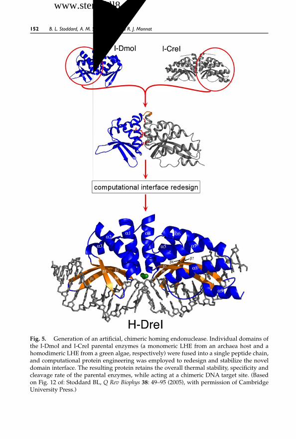
October 30, 2007 12:29 WSPC/SPI-B559: Autologous & Cancer Stem Cell Gene Therapy ch06 FA
152 B. L. Stoddard, A. M. Scharenberg and R. J. Monnat
Fig. 5. Generation of an artificial, chimeric homing endonuclease. Individual domains ofthe I-DmoI and I-CreI parental enzymes (a monomeric LHE from an archaea host and ahomodimeric LHE from a green algae, respectively) were fused into a single peptide chain,and computational protein engineering was employed to redesign and stabilize the noveldomain interface. The resulting protein retains the overall thermal stability, specificity andcleavage rate of the parental enzymes, while acting at a chimeric DNA target site. (Basedon Fig. 12 of: Stoddard BL, Q Rev Biophys 38: 49–95 (2005), with permission of CambridgeUniversity Press.)
干细胞之家www.stemcell8.cn ←点击进入

October 30, 2007 12:29 WSPC/SPI-B559: Autologous & Cancer Stem Cell Gene Therapy ch06 FA
Advances in Engineering Homing Endonucleases for GeneTargeting:TenYears After Structures 153
site-specific endonuclease activity identical to naturally occurring I-CreI.Separate experiments have also demonstrated that the PI-SceI protein splic-ing domain can be used as a site-specific DNA binding module in chimericprotein constructs: domain swapping between the PI-SceI and a homologuefrom Candida tropicalis (PI-CtrIP) was conducted to design altered specificityproteins (Steuer et al., 2004).
A related experiment has demonstrated that a single chain, monomericendonuclease can be generated from a homodimer predecessor, I-CreI(Epinat et al., 2003). This construct was shown to initiate homologous recom-bination in both yeast and mammalian cells. Finally, the role and mutabilityof LAGLIDADG interface residues has been examined by grafting sidechains from the homodimeric I-CreI into the corresponding positions in themonomeric I-DmoI enzyme, resulting in enzymes with novel nicking activ-ities and oligomeric properties (Silva and Belfort, 2004). Subsequent exper-iments with this same enzyme have demonstrated that individual domainsfrom the monomeric wild-type protein can be engineered to form stable andfunctional homodimers, again illustrating the modularity of LHE scaffolds(Silva et al., 2006).
3.4. Redesign of Homing Endonuclease DNA Contactsand Specificity
Several methods have been used to alter the DNA target site specificity ofLHE’s (Figs. 6 and 7). These have focused in large part on the mutationof individual DNA-protein interface side chains that contact specific DNAtarget site base pairs. These strategies can be broadly divided into:
(i) Protocols that rely on a selection for high affinity DNA binding activity.(ii) Protocols that rely on a selection for efficient cleavage activity. Exam-
ples have been described (summarized below) of methods that relyon the elimination of a gene (usually by destruction of a plasmid anda resident protein coding sequence) or on complementation of a gene(by cleavage-induced homologous recombination of two dysfunctionalenzyme alleles into a wild-type, functional reading frame). This lattermethod is the basis of the most successful “high-throughput” endonu-clease redesigns to date, which have progressed to targeting of physio-logically relevant gene sequences. Those experiments are summarizedat the end of this section.
(iii) Protocols that rely on structure-based computational redesign of DNA-protein interface contact surfaces and residues. It is important to note
干细胞之家www.stemcell8.cn ←点击进入

October 30, 2007 12:29 WSPC/SPI-B559: Autologous & Cancer Stem Cell Gene Therapy ch06 FA
154 B. L. Stoddard, A. M. Scharenberg and R. J. Monnat
Fig. 6. Selections and screens for altered specificity homing endonucleases. (A) Selectionfor DNA binding activity using a two-hybrid system (Gimble et al., 2003). Gal11P/PI-SceIvariants expressed from a plasmid library that bind to a homing site increase the expressionof HIS3 and aadA by recruiting the RNAPα/Gal4 fusion protein to the weak Plac promoteron the F′. Thus, cells that express a PI-SceI protein capable of binding a chosen DNA targetsequence grow on histidine-selective and spectinomycin-selective media. (B) Selection forDNA cleavage activity (Chen and Zhao, 2005; Doyon and Liu, 2006). Co-existence of twoplasmids kills bacterial cells because one expresses the toxic CcdB gene product. Selection canbe made for I-SceI enzyme variants that cleave I-SceI target sites on the CcdB plasmid becausethey eliminate it and allow the cells to survive. (C) Screen for homologous recombinationactivity (Arnould et al., 2006). Expression of I-CreI from a plasmid library cleaves a target sitewithin an interrupted lacZ gene located on a reporter plasmid. This DSB stimulates singlestrand annealing (SSA) of two direct repeats that flank the site, leading to restoration of afunctional lacZ gene and the appearance of a blue colony. (Figure and caption reproducedfrom: Gimble FS, Gene Ther Regul 3: 33–50 (2007), with permission of the author and of WorldScientific Publishing.)
干细胞之家www.stemcell8.cn ←点击进入

October 30, 2007 12:29 WSPC/SPI-B559: Autologous & Cancer Stem Cell Gene Therapy ch06 FA
Advances in Engineering Homing Endonucleases for GeneTargeting:TenYears After Structures 155
Fig. 7. From in vivo selection to in silico screening. (A) Screen for homing endonucle-ase based on cleavage and gene elimination (Seligman et al., 2002 and 2004). Plasmid-encoded I-CreI derivatives that bind and cleave an I-CreI homing site located on an F′lead to its elimination. Concomitant loss of an adjacent kanamycin antibiotic marker yieldskanamycin-sensitive cells. (B) Alteration of specificity of the I-MsoI homing endonucleaseby computational structure-based redesign (Ashworth et al., 2006). The four panels providea comparison of the experimentally determined interactions in wild-type and redesignedcognate complexes (upper left and lower right) and predicted interactions in non-cognatecomplexes, as well as predicted relative energies of the complexes.
干细胞之家www.stemcell8.cn ←点击进入

October 30, 2007 12:29 WSPC/SPI-B559: Autologous & Cancer Stem Cell Gene Therapy ch06 FA
156 B. L. Stoddard, A. M. Scharenberg and R. J. Monnat
that high resolution crystal structures of the wild-type protein-DNAcomplexes have been used in various contexts, either to enable the tar-geted redesign of homing endonuclease-DNA contacts at individualresidues (thus bypassing selection approaches altogether), or to facili-tate more efficient mutational screening of enzyme libraries (by identi-fying, and thus greatly reducing, the number of protein residues to berandomized). The use of such crystal structures is a prominent featureof the most successful LHE redesigns described to date.
3.4.1. Redesign of specificity at individual DNA base pairs
An early attempt to reprogram the DNA recognition specificity of a homingendonuclease involved an adaptation of a bacterial two-hybrid screeningstrategy [Gimble et al., 2003; Fig. 6(A)]. In this experiment, variants of theintein-encoded PI-SceI homing endonuclease with altered binding speci-ficities were selected, and then characterized for their ability to discriminatebetween the corresponding substrates in DNAcleavage reactions. The speci-ficities of the selected endonuclease variants ranged from being relaxed (i.e.,able to cleave the wild-type and mutant targets equally) to being shifted topreferring the selection targets. However, none of the variants displayed thesame discrimination as wild-type PI-SceI.
Two alternative strategies have used bacterial selection strategies basedon the cleavage and elimination of a reporter gene to isolate homingendonuclease derivatives with altered specificities. In the first, cleavage ofthe target site results in cells being converted from lac+ to lac−, allowingselection of desired activities based on a simple blue-white colony pheno-type screen [Rosen et al., 2006; Seligman et al., 2002; Sussman et al., 2004;Fig. 7(A)]. Undesirable activity (e.g., cleavage of the original wild-type site)can be suppressed through a secondary “negative” screen for eliminationof an essential reporter (such as an antibiotic resistance marker). Using thismethod, endonuclease mutants with single or double amino-acid substi-tutions, at positions predicted to make base-specific DNA contacts, wereassayed against appropriate DNAtarget site mutants. Enzyme variants withshifted specificities, but with reduced ability to discriminate between cog-nate and miscognate sites, were typically obtained. Crystallographic anal-yses of several of these altered mutants in complex with their new cognatetargets demonstrated that the “modularity” of protein-DNA recognition,previously described for entire protein domains, extends to the level of indi-vidual amino-acid side chains (Rosen et al., 2006; Sussman et al., 2004).
干细胞之家www.stemcell8.cn ←点击进入

October 30, 2007 12:29 WSPC/SPI-B559: Autologous & Cancer Stem Cell Gene Therapy ch06 FA
Advances in Engineering Homing Endonucleases for GeneTargeting:TenYears After Structures 157
A similar bacterial selection strategy has been described in which a toxicgene product results in cell death, unless a homing endonuclease variantthat cleaves a homing site within the toxin expression vector is also present[Fig. 6(B)]. Two versions of this screen have been described: the first utiliz-ing the nonspecific ribonuclease barnase (Gruen et al., 2002), and the otherdescribing a more easily controlled system based on the “control of cell deathB” (CcdB) protein (Doyon et al., 2006). In the latter screening strategy, CcdBexpression led to very low rates of background survival, without requiringadditional gene expression control elements such as those required to usethe more toxic barnase protein. As with the bacterial selection described inthe preceding paragraph, the CcdB system can be tailored as a positive selec-tion for cleavage of a desired target, or as a negative selection, to stronglydisfavor the recognition of a non-cognate DNA target site sequence. Thissystem, when optimized, can lead to nearly 100% survival of cells express-ing an active homing endonuclease, against a background survival of <1 in5 × 104 with an inactive enzyme.
In separate experiments that avoided all use of combinatorial mutage-nesis and screening methodologies, a successful computational redesign ofthe I-MsoI homing endonuclease was described using a physically realisticatomic level forcefield [Ashworth et al., 2006; Fig. 7(B)]. Using an in silicoscreen, investigators identified single base pair substitutions predicted todisrupt binding by the wild type enzyme, and then optimized the iden-tities and conformations of clusters of amino acids around each of theseunfavorable substitutions by using Monte Carlo sampling. A redesignedenzyme predicted to display altered target site specificity was identified thatmaintained wild-type binding affinity. The redesigned enzyme was foundto bind and cleave the redesigned recognition site more effectively than doesthe wild type enzyme, with a level of target discrimination comparable tothe original endonuclease. Determination of the structure of the redesignednuclease-recognition site complex by X-ray crystallography confirmed theaccuracy of the computationally-predicted interface.
3.4.2. High-throughput screening and targeting of genomic loci
Recently, a commercial research and development group in France (Cel-lectis, Inc.) has developed a powerful eukaryotic assay system that reportson the generation of double-strand break-induced homologous recombina-tion, rather than only DNA cleavage [Arnould et al., 2006; Chames et al.,2005; Fig. 6(C)]. In this assay, the function of a gene required for growthor for another easily scoreable phenotype is restored through the action
干细胞之家www.stemcell8.cn ←点击进入

October 30, 2007 12:29 WSPC/SPI-B559: Autologous & Cancer Stem Cell Gene Therapy ch06 FA
158 B. L. Stoddard, A. M. Scharenberg and R. J. Monnat
of a homing endonuclease. Prior to expression and action of the HE, thegene sequence is interrupted with an insert containing a desired HE cut siteflanked by two direct repeats. In the most recently described version of thisscreen, the endonuclease expression construct and the “reporter” constructare located in separate yeast strains, allowing the investigator to introducethe HE (or a library of HE variants) to a target site by mating. The mating ofthe two required yeast strains can be automated, and thus done as a high-throughput assay. Thus, the same library of endonuclease variants can beefficiently screened against multiple DNA target site variants, in parallel orrapid sequential experiments.
Using this method, the DNA recognition specificity of I-CreI LHE hasbeen thoroughly analyzed. Small endonuclease mutant libraries resultingfrom the randomization of two to four amino acids (corresponding to the“nearest protein neighbors” of individual DNA basepairs) were individ-ually screened against all potential variant cognate sequences. This anal-ysis identified individual mutations in the I-CreI protein scaffold thatwere associated with shifts in specificity at individual target site base pairpositions. From these studies, hundreds of endonuclease mutants withaltered specificies were identified, catalogued and archived (Arnould et al.,2006). Many of these variants displayed cleavage activities and levels ofsite discrimination that were at least equivalent to the wild-type endonu-clease, thus providing a starting point for more ambitious endonucleaseredesign to physiological targets in eukaryotic, mammalian and even humangenomes.
Using the approach summarized above, derivatives of the I-CreI LHEhave been generated that display sequence-specific cleavage and recom-bination activity against the human RAG1 gene (Smith et al., 2006) at thesite of mutations, giving rise to a rare subset of severe combined immun-odeficiency disease (or SCID) phenotypes. I-CreI variants directed at thehuman XPC (xeroderma pigmentosum complementation group C) gene(Arnould et al., 2007) were also generated. XPC, when a mutant, confersan extreme UV or sun-sensitivity phenotype together with a predisposi-tion to sunlight-induced skin cancer. Both of these human disease genes arecandidates for corrective genetic therapies. Furthermore, in the latter exper-iments, the modified I-CreI derivatives cleaving sequences from the XPCgene were found to induce a high level of gene targeting, similar to levelsobserved with the wild-type I-CreI or I-SceI scaffolds. This is the first timean engineered homing endonuclease has been used in mammalian cells totarget and modify a chromosomal target locus.
干细胞之家www.stemcell8.cn ←点击进入

October 30, 2007 12:29 WSPC/SPI-B559: Autologous & Cancer Stem Cell Gene Therapy ch06 FA
Advances in Engineering Homing Endonucleases for GeneTargeting:TenYears After Structures 159
3.4.3. Surface display of homing endonucleases: avoiding bottlenecksin mutagenesis and screening?
While the methodologies summarized above have shown great promise indelivering tailor-made gene-specific targeting reagents, they are limited bythe requirement to first generate and then screen libraries of endonucleasemutants. Moreover, the selections and screens discussed above requirethe generation of unique intracellular reporter constructs that must beredesigned and constructed for each target sequence of interest. In orderto address these issues, investigators have recently described a novel sys-tem where LHE proteins might be rapidly mutated in vivo and then screenedto identify and isolate endonuclease variants with a new DNA target speci-ficity (Fig. 8; Volna et al., 2007). This work has built on a recent study thatdescribes the evolution and selection of fluorescent protein spectral prop-erties using the somatic hypermutation machinery of lymphocytes (Wanget al., 2004).
Investigators demonstrated that LHEs can be expressed on the plasmamembrane of a lymphocyte cell line by targeting the expression of an LHE-CD80 transmembrane fusion protein to the secretory pathway. Surface-expressed LHEs faithfully recapitulate the properties of the native enzymesin solution, as assessed by flow cytometric analysis of both the binding andthe cleavage of fluorescently conjugated double-stranded DNA target siteoligonucleotides. Identification of endonuclease expression clones with thedesired DNA recognition properties was highly specific, allowing discrimi-nation of endonucleases with binding preferences differing by only a singlebase pair. Furthermore, target sequence-specific LHE interactions with theseDNA target site probes under conditions that limit substrate cleavage allowboth the identification and sorting or enrichment of clones expressing LHEvariants with the highest site specificity and affinity for further character-ization. This coupled rapid analysis of LHE-DNA interactions on the cellsurface, together with concurrent sorting or enrichment, should substan-tially accelerate the generation and isolation of novel endonuclease variantswith unique DNA target specificities.
4. Concluding Remarks
The first X-ray structures of homing endonuclease (I-CreI and PI-SceI) werereported within one month of each other in 1997 (Duan et al., 1997; Heathet al., 1997). These were followed a year later by the first DNA-bound cocrys-tal structure (again I-CreI) (Jurica et al., 1998). The subsequent 10 years have
干细胞之家www.stemcell8.cn ←点击进入

October 30, 2007 12:29 WSPC/SPI-B559: Autologous & Cancer Stem Cell Gene Therapy ch06 FA
160 B. L. Stoddard, A. M. Scharenberg and R. J. Monnat
Fig. 8. DNA binding by surface displayed homing endonucleases. (A) An HA-taggedLAGLIDADG homing endonuclease is expressed on the surface of vertebrate cells throughfusion with the transmembrane domain and tail of murine CD80. The expressed homingendonucleases I-AniI and H-DreI retain their DNArecognition, binding and cleavage activity,and binding can be quantitatively analyzed through the use of fluorescently labeled, double-stranded (ds) DNA oligonucletides and flow cytometry. “SP” = signal peptide. (B) Left: Thefluorescence staining intensity using dsDNA probes is dependent on the surface expressionand display of a homing endonuclease, and is capable of discriminating between the bind-ing of a wild-type target site vs. miscognate sites harboring single base pair substitutions.DT40: parental cell line without surface HE expression. I-Ani (B3) and (B10): two individualclones expressing wild-type I-AniI homing endonuclease on the surface. dsAni1 and dsAni2:distinct dsDNA probes encoding different I-AniI target sites that are efficiently recognized,bound, and cleaved by recombinant soluble I-AniI. dsAniI-6A and dsAni1-9A: two dsDNAprobes harboring I-AniI target sites that contain single base pair substitutions which elim-inate cleavage activity of the recombinant soluble I-AniI. Right: enrichment through flowsorting of rare cells expressing a desired binding specificity from an excess (approximately100–500 fold) of undesired HE binding activities.
produced a remarkable increase in our understanding of the biology, struc-ture, function and mechanism of homing endonucleases. Representativestructures from each of the major homing endonuclease families have beendetermined together with their catalytic mechanisms. At least six differentmethods for the modification and redesign of homing endonuclease DNA
干细胞之家www.stemcell8.cn ←点击进入

October 30, 2007 12:29 WSPC/SPI-B559: Autologous & Cancer Stem Cell Gene Therapy ch06 FA
Advances in Engineering Homing Endonucleases for GeneTargeting:TenYears After Structures 161
target specificity have also been developed. During this period, a small num-ber of homing endonuclease proteins have been assayed for activity andtoxicity in vivo, and have been used to investigate mechanistic aspects ofDNA double strand break repair and recombination. The first engineeredhoming endonuclease variants have also been described and characterized,and the most recent of these have been shown to induce DNA cleavage andrecombination at chromosomal target loci in vivo. This collective experiencehas established the feasibility of using homing endonuclease proteins forgenome engineering, together with the data needed to further develop hom-ing endonuclease proteins for basic science and engineering applications.
The development of engineered homing endonucleases as reagents forbasic biology (to facilitate the introduction or analysis of double- or single-strand breaks, covalent DNA modifications or trans-acting protein factorsat specific DNA target sites in living cells) has matured rapidly over thepast decade, and their further use and development in this area is virtuallyguaranteed. In contrast, the use of homing endonucleases as therapeuticreagents for disease treatment or prevention is still in its infancy and willrequire many years of dedicated work. Among the most important, unan-swered questions are: the specificity profile of such reagents in biologicallyrelevant contexts (e.g., stem cells), how efficient they are in generating targetsite-specific DNAdouble strand breaks in vivo, the range and efficiency withwhich individual DNA double-strand breaks lead to desired — and unde-sired — molecular outcomes in populations of cells, and the long-term toxic-ity, mutagenicity and immunogenicity of homing endonucleases when usedas genome engineering, therapeutic or disease prevention reagents. Thesequestions are pertinent now that the feasibility of using homing endonucle-ase proteins for genome engineering has been demonstrated, and when, asnoted by Pâques and Duchateau, “the time when therapeutic applicationswere pure fantasy is now gone” (Pâques and Duchateau, 2007).
References
Abbott A. (2006) Questions linger about unexplained gene-therapy trial death. NatMed 12: 597.
Agaard C, Awayez MJ, Garrett RA. (1997) Profile of the DNA recognition site of thearchaeal homing endonuclease I-DmoI. Nucl Acids Res 25:1523–30.
Arnould S, Chames P, Perez C, et al. (2006) Engineering of large numbers of highlyspecific homing endonucleases that induce recombination on novel DNAtargets.J Mol Biol 355: 443–58.
干细胞之家www.stemcell8.cn ←点击进入

October 30, 2007 12:29 WSPC/SPI-B559: Autologous & Cancer Stem Cell Gene Therapy ch06 FA
162 B. L. Stoddard, A. M. Scharenberg and R. J. Monnat
Arnould S, Perez C, Cabaniols J-P, et al. (2007) Engineered I-CreI derivatives cleavingsequences from the human XPC gene can induce highly efficient gene correctionin mammalian cells. J Mol Biol 371: 49–65.
Ashworth J, Havranek JJ, Duarte CM, et al. (2006) Computational redesign ofendonuclease DNA binding and cleavage specificity. Nature 441: 656–59.
Baird PA, Anderson TW, Newcombe HB, Lowry RB. (1988) Genetic disorders inchildren and young adults: a population study. Am J Hum Genet 42: 677–93.
Baum C. (2007) Fourth case of leukaemia in the first SCID-X1 gene ther-apy trial, and the diversity of gene therapy. http://www.esgct.org/upload/4th_CaseofLeukemial.pdf
Beerli RR, Barbas CF. (2002) Engineering polydactyl zinc-finger transcription fac-tors. Nat Biotechnol 20: 130–41.
Belfort M, Reaban ME, Coetzee T, Dalgaard JZ. (1995) Prokaryotic introns andinteins: a panoply of form and function. J Bacter 177: 3897–903.
Belfort M, Roberts RJ. (1997) Homing endonucleases — keeping the house in order.Nucl Acids Res 25: 3379–88.
Bertolotti R. (1996) Recombinase-mediated gene therapy: strategies based on Lesch-Nyhan mutants for gene repair/inactivation using human RAD51 nucleoproteinfilaments. Biogenic Amines 12: 487–98.
Bibikova M, Beumer K, Trautman JK, Carroll D. (2003) Enhancing gene targetingwith designed zinc finger nucleases. Science 300: 764.
Bibikova M, Carroll D, Segal DJ, et al. (2001) Stimulation of homologous recombi-nation through targeted cleavage by chimeric nucleases. Mol Cell Biol 21: 289–97.
Bolduc JM, Spiegel PC, Chatterjee P, et al. (2003) Structural and biochemical analysesof DNA and RNA binding by a bifunctional homing endonuclease and group Iintron splicing cofator. Genes Dev 17: 2875–88.
Bullard DC, Weaver CT. (2002) Cutting-edge technology: IV. Genomic engineeringfor studies of the gastrointestinal tract in mice. Am J Physiol Gastrointest LiverPhysiol 283: G1232–37.
Bulyk ML, Huang XH, Choo Y, Church GM. (2001) Exploring the DNA-bindingspecificities of zinc fingers with DNA microarrays. Proc Natl Acad Sci USA 98:7158–63.
Burt A. (2003) Site-specific selfish genes as tools for the control and genetic engi-neering of natural populations. Proc R Soc Lond B Biol Sci 270: 921–28.
Burt A. (2005) Gates Grand Challenge Project. In: Homing Endonuclease Genes: NewTools for Mosquito Population Engineering and Control. www.gcgh.org.
Chames P, Epinat JC, Guillier S, et al. (2005) In vivo selection of engineered homingendonucleases using double-strand break induced homologous recombination.Nucl Acids Res 33: e178.
Chase CD. (2006) Genetically engineered cytoplasmic male sterility. Trends Plant Sci11: 7–9.
Chevalier B, Sussman D, Otis C, et al. (2004) Analysis of the symmetric metal-dependent I-CreI homing endonuclease mechanism. Biochemistry 43: 4015–26.
干细胞之家www.stemcell8.cn ←点击进入

October 30, 2007 12:29 WSPC/SPI-B559: Autologous & Cancer Stem Cell Gene Therapy ch06 FA
Advances in Engineering Homing Endonucleases for GeneTargeting:TenYears After Structures 163
Chevalier B, Turmel M, Lemieux C, et al. (2003) Flexible DNA target site recognitionby divergent homing endonuclease isoschizomers I-CreI and I-MsoI. J Mol Biol329: 253–69.
Chevalier BS, Kortemme T, Chadsey MS, et al. (2002) Design, activity and structureof a highly specific artificial endonuclease. Mol Cell 10: 895–905.
Chevalier BS, Monnat RJ Jr, Stoddard BL. (2001) The homing endonuclease I-CreIuses three metals, one of which is shared between the two active sites. Nat StructBiol 8: 312–16.
Chevalier BS, Stoddard BL. (2001) Homing endonucleases: structural and functionalinsight into the catalysts of intron/intein mobility. Nucl Acids Res 29: 3757–74.
Christianson A, Howson CP, Modell B. (2006) Global Report on Birth Defects: TheHidden Toll of Dying and Disabled Children. March of Dimes, White Plains,New York.
Coates CJ, Kaminski JM, Summers JB, et al. (2005) Site-directed genome modifica-tion: derivatives of DNA-modifying enzymes as targeting tools. Trends Biotechnol23: 407–19.
Colleaux L, D’Auriol L, Betermier M, et al. (1986) Universal code equivalent of ayeast mitochondrial intron reading frame is expressed into E. coli as a specificdouble strand endonuclease. Cell 44: 521–33.
Colleaux L, D’Auriol L, Galibert F, Dujon B. (1988) Recognition and cleavage siteof the intron-encoded omega transposase. Proc Natl Acad Sci USA 85: 6022–26.
Dalgaard JZ, Garrett RA, Belfort M. (1994) Purification and characterization oftwo forms of I-DmoI, a thermophilic site-specific endonuclease encoded by anarchaeal intron. J Biol Chem 269: 28885–92.
Dalgaard JZ, Klar AJ, Moser MJ, et al. (1997) Statistical modeling and analysis ofthe LAGLIDADG family of site-specific endonucleases and identification of anintein that encodes a site-specific endonuclease of the HNH family. Nucl AcidsRes 25: 4626–38.
Delahodde A, Goguel V, Becam AM, et al. (1989) Site-specific DNA endonucleaseand RNA maturase activities of two homologous intron-encoded proteins fromyeast mitochondria. Cell 56: 431–41.
Doetschman T, Gregg RG, Maeda N, et al. (1987) Targetted correction of a mutantHPRT gene in mouse embryonic stem cells. Nature 330: 576–78.
Doyon JB, Pattanayak V, Meyer CB, Liu DR. (2006) Directed evolution and substratespecificity profile of homing endonuclease I-SceI. J Am Chem Soc 128: 2477–84.
Duan X, Gimble FS, Quiocho FA. (1997) Crystal structure of PI-SceI, a homingendonuclease with protein splicing activity. Cell 89: 555–64.
Dujon B. (1980) Sequence of the intron and flanking exons of the mitochondrial 21SrRNA gene of yeast strains having different alleles at the omega and rib-1 loci.Cell 20: 185–97.
Dujon B. (1989) Group I introns as mobile genetic elements: facts and mechanisticspeculations — a review. Gene 82: 91–114.
干细胞之家www.stemcell8.cn ←点击进入

October 30, 2007 12:29 WSPC/SPI-B559: Autologous & Cancer Stem Cell Gene Therapy ch06 FA
164 B. L. Stoddard, A. M. Scharenberg and R. J. Monnat
Dujon B, Belfort M, Butow RA, et al. (1989) Mobile introns: definition of terms andrecommended nomenclature. Gene 82: 115–18.
Durrenberger F, Rochaix J-D. (1993) Characterization of the cleavage site and therecognition sequence of the I-CreI DNAendonuclease encoded by the chloroplastribosomal intron of Chlamydomonas reinhardtii. Mol Gen Genet 236: 409–14.
Epinat JC, Arnould S, Chames P, et al. (2003) A novel engineered meganucleaseinduces homologous recombination in yeast and mammalian cells. Nucl AcidsRes 31: 2952–62.
Fitzsimons-Hall M, Noren CJ, Perler FB, Schildkraut I. (2002) Creation of an artificialbifunctional intein by grafting a homing endonuclease into a mini-intein. J MolBiol 323: 173–79.
Geese WJ, Waring RB. (2001) A comprehensive characterization of a group IBintron and its encoded maturase reveals that protein-assisted splicing requiresan almost intact intron RNA. J Mol Biol 308: 609–22.
Ghatge M, Palaniappan N, Das Choudhuri S, Reynolds K. (2006) Genetic manip-ulation of the biosynthetic process leading to phoslactomycins, potent proteinphosphatase 2A inhibitors. J Ind Microbiol Biotechnol 33: 589–99.
Gimble FS, Moure CM, Posey KL. (2003) Assessing the plasticity of DNA target siterecognition of the PI-SceI homing endonuclease using a bacterial two-hybridselection system. J Mol Biol 334: 993–1008.
Glaser S, Anastassiadis K, Stewart AF. (2005) Current issues in mouse genome engi-neering. Nat Genet 37: 1187–93.
Gouble A, Smith J, Bruneau S, et al. (2006) Efficient in toto targeted recombinationin mouse liver by meganuclease-induced double-strand break. J Gene Med 8:616–22.
Griesenbach U, Geddes DM,Alton EW. (2006) Gene therapy progress and prospects:cystic fibrosis. Gene Ther 13: 1061–67.
Gruen M, Chang K, Serbanescu I, Liu DR. (2002) An in vivo selection system forhoming endonuclease activity. Nucl Acids Res 30: 29–34.
Hacein-Bey-Abina S, Von Kalle C, Schmidt M, et al. (2003) LMO2–associated clonalT cell proliferation in two patients after gene therapy for SCID-X1. Science 302:415–19.
Heath PJ, Stephens KM, Monnat RJ, Stoddard BL. (1997) The structure of I-CreI, agroup I intron-encoded homing endonuclease. Nat Struct Biol 4: 468–76.
Hinnen A, Hicks JB, Fink GR. (1978) Transformation of yeast. Proc Natl Acad SciUSA 75: 1929–33.
Jacquier A, Dujon B. (1985) An intron-encoded protein is active in a gene conversionprocess that spreads an intron into a mitochondrial gene. Cell 41: 383–94.
Jin Y, Binkowski G, Simon LD, Norris D. (1997) Ho endonuclease cleaves MATDNA in vitro by an inefficient stoichiometric reaction mechanism. J Biol Chem272: 7352–59.
干细胞之家www.stemcell8.cn ←点击进入

October 30, 2007 12:29 WSPC/SPI-B559: Autologous & Cancer Stem Cell Gene Therapy ch06 FA
Advances in Engineering Homing Endonucleases for GeneTargeting:TenYears After Structures 165
Jurica MS, Monnat RJ Jr, Stoddard BL. (1998) DNA recognition and cleavage by theLAGLIDADG homing endonuclease I- CreI. Mol Cell 2: 469–76.
Kolb AF, Coates CJ, Kaminski JM, et al. (2005) Site-directed genome modification:nucleic acid and protein modules for targeted integration and gene correction.Trends Biotechnol 23: 399–406.
Koller BH, Smithies O. (1989) Inactivating the beta 2-microglobulin locus in mouseembryonic stem cells by homologous recombination. Proc Natl Acad Sci USA 86:8932–35.
Kostriken R, Strathern JN, Klar AJ, et al. (1983) Asite-specific endonuclease essentialfor mating-type switching in Saccharomyces cerevisiae. Cell 35: 167–74.
Lavery R. (2005) Recognizing DNA. Q Rev Biophys 38: 339–44.Lazowska J, Claisse M, Gargouri A, et al. (1989) Protein encoded by the third intron
of cytochrome b gene in Saccharomyces cerevisiae is an mRNA maturase. Analysisof mitochondrial mutants, RNA transcripts proteins and evolutionary relation-ships. J Mol Biol 205: 275–89.
Lazowska J, Jacq C, Slonimski PP. (1980) Sequence of introns and flanking exonsin wild-type and box3 mutants of cytochrome b reveals an interlaced splicingprotein coded by an intron. Cell 22: 333–48.
Malik P, Arumugam PI. (2005) Gene therapy for {beta}-thalassemia. Hematol Am SocHematol Educ Program: 45–50.
Malik P, Arumugam PI, Yee JK, Puthenveetil G. (2005) Successful correction of thehuman Cooley’s anemia beta-thalassemia major phenotype using a lentiviralvector flanked by the chicken hypersensitive site 4 chromatin insulator. Ann NY Acad Sci 1054: 238–49.
McCandless SE, Brunger JW, Cassidy SB. (2004) The burden of genetic disease oninpatient care in a children’s hospital. Am J Hum Genet 74: 121–27.
Moure C, Gimble F, Quiocho F. (2002) Crystal structure of the intein homing endonu-clease PI-SceI bound to its recognition sequence. Nat Struct Biol 9: 764–70.
Moure CM, Gimble FS, Quiocho FA. (2003) The crystal structure of the gene target-ing homing endonuclease I-SceI reveals the origins of its target site specificity.J Mol Biol 334: 685–96.
Orr-Weaver TL, Szostak JW, Rothstein RJ. (1981) Yeast transformation: a modelsystem for the study of recombination. Proc Natl Acad Sci USA 78: 6354–58.
Pabo CO, Nekludova L. (2000) Geometric analysis and comparison of protein-DNAinterfaces: Why is there no simple code for recognition? J Mol Biol 301: 597–624.
Pâques F, Duchateau P. (2007) Meganucleases and DNA double-strand break-induced recombination: perspectives for gene therapy. Curr Gene Ther 7: 49–66.
Perrin A, Buckle M, Dujon B. (1993) Asymmetrical recognition and activity of theI-SceI endonuclease on its site and on intron-exon junctions. EMBO J 12: 2939–47.
Porteus MH. (2006) Mammalian gene targeting with designed zinc finger nucleases.Mol Ther 13: 438–46.
干细胞之家www.stemcell8.cn ←点击进入

October 30, 2007 12:29 WSPC/SPI-B559: Autologous & Cancer Stem Cell Gene Therapy ch06 FA
166 B. L. Stoddard, A. M. Scharenberg and R. J. Monnat
Porteus MH, Baltimore D. (2003) Chimeric nucleases stimulate gene targeting inhuman cells. Science 300: 763.
Posfai G, Kolisnychenko V, Bereczki Z, Blattner FR. (1999) Markerless gene replace-ment in Escherichia coli stimulated by a double-strand break in the chromosome.Nucl Acids Res 27: 4409–15.
Puthenveetil G, Scholes J, Carbonell D, et al. (2004) Successful correction of thehuman beta-thalassemia major phenotype using a lentiviral vector. Blood 104:3445–53.
Rosen LE, Morrison HA, Masri S, et al. (2006) Homing endonuclease I-CreI deriva-tives with novel DNA target specificities. Nucl Acids Res 34: 4791–800.
Sadelain M. (2006) Recent advances in globin gene transfer for the treatment ofbeta-thalassemia and sickle cell anemia. Curr Opin Hematol 13: 142–48.
Samakoglu S, Lisowski L, Budak-Alpdogan T, et al. (2006) A genetic strategy totreat sickle cell anemia by coregulating globin transgene expression and RNAinterference. Nat Biotechnol 24: 89–94.
Scalley-Kim M, McConnell-Smith A, Stoddard BL. (2007) Coevolution of a homingendonuclease and its host target sequence. J Mol Biol, in press.
Schafer B, Wilde B, Massardo DR, et al. (1994) A mitochondrial group-I intron infission yeast encodes a maturase and is mobile in crosses. Curr Genet 25: 336–41.
Segal DJ, Beerli RR, Blancafort P, et al. (2003) Evaluation of a modular strategy for theconstruction of novel polydactyl zinc finger DNA-binding proteins. Biochemistry42: 2137–48.
Segal DJ, Dreier B, Beerli RR, Barbas CF. (1999) Toward controlling gene expressionat will: selection and design of zinc finger domains recognizing each of the 5’-GNN-3’ DNA target sequences. Proc Natl Acad Sci USA 96: 2758–63.
Seligman L, Chisholm KM, Chevalier BS, et al. (2002) Mutations altering the cleavagespecificity of a homing endonuclease. Nucl Acids Res 30: 3870–79.
Silva GH, Belfort M. (2004)Analysis of the LAGLIDADG interface of the monomerichoming endonuclease I-DmoI. Nucl Acids Res 32: 3156–68.
Silva GH, Belfort M, Wende W, Pingoud A. (2006) From monomeric to homod-imeric endonucleases and back: engineering novel specificity of LAGLIDADGenzymes. J Mol Biol 361: 744–54.
Sitbon E, Pietrokovski S. (2003) New types of conserved sequence domains in DNA-binding regions of homing endonucleases. Trends Biochem Sci 28: 473–77.
Smith J, Bibikova M, Whitby FG, et al. (2000) Requirements for double-strand cleav-age by chimeric restriction enzymes with zinc finger DNA-recognition domains.Nucl Acids Res 28: 3361–69.
Smith J, Grizot S,Arnould S, et al. (2006)Acombinatorial approach to create artificialhoming endonucleases cleaving chosen sequences. Nucl Acids Res 34: e149.
Spiegel PC, Chevalier B, Sussman D, et al. (2006) The structure of I-CeuI homingendonuclease: evolving asymmetric DNA recognition from a symmetric proteinscaffold. Structure 14: 869–80.
干细胞之家www.stemcell8.cn ←点击进入

October 30, 2007 12:29 WSPC/SPI-B559: Autologous & Cancer Stem Cell Gene Therapy ch06 FA
Advances in Engineering Homing Endonucleases for GeneTargeting:TenYears After Structures 167
Steuer S, Pingoud V, Pingoud A, Wende W. (2004) Chimeras of the homing endonu-clease PI-SceI and the homologous Candida tropicalis intein: a study to explorethe possibility of exchanging DNA-binding modules to obtain highly speciricendonucleases with altered specificity. Chembiochem 5: 206–13.
Stoddard BL. (2005) Homing endonuclease structure and function. Q Rev Biophys38: 49–95.
Sussman DJ, Chadsey M, Fauce S, et al. (2004) Isolation and characterization of newhoming endonuclease specificities at individual target site positions. J Mol Biol342: 31–41.
Themis M, Waddington SN, Schmidt M, et al. (2005) Oncogenesis following deliveryof a nonprimate lentiviral gene therapy vector to fetal and neonatal mice. MolTher 12: 763–71.
ThompsonAJ, Yuan X, Kudlicki W, Herrin DL. (1992) Cleavage and recognition pat-tern of a double-strand-specific endonuclease (I-CreI) encoded by the chloroplast23S rRNA intron of Chlamydomonas reinhardtii. Gene 119: 247–51.
Thrasher AJ, Gaspar HB, Baum C, et al. (2006) Gene therapy: X-SCID transgeneleukaemogenicity. Nature 443: E5–E6; discussion E6–E7.
Tzfira T, White C. (2005) Towards targeted mutagenesis and gene replacement inplants. Trends Biotechnol 23: 567–69.
Urnov FD, Miller JC, Lee YL, et al. (2005) Highly efficient endogenous human genecorrection using designed zinc-finger nucleases. Nature 435: 646–51.
Vasquez KM, Marburger K, Intody Z, Wilson JH. (2001) Manipulating the mam-malian genome by homologous recombination. Proc Natl Acad Sci USA 98:8403–10.
Volna P, Jarjour J, Baxter S, et al. (2007) Flow cytometric analysis of DNA bindingand cleavage by cell surface-displayed homing endonucleases. Nucl Acids Res35: 2748–58.
Wang L, Jackson WC, Steinbach PA, Tsien RY. (2004) Evolution of new nonanti-body proteins via iterative somatic hypermutation. Proc Natl Acad Sci USA 101:16745–49.
Wintjens R, Rooman M. (1996) Structural classification of HTH DNA-bindingdomains and protein-DNA interaction modes. J Mol Biol 262: 294–313.
Wolfe SA, Nekludova L, Pabo CO. (2000a) DNArecognition by Cys2His2 zinc fingerproteins. Ann Rev Biophys Biomol Struct 29:183–212.
Wolfe SA, Ramm EI, Pabo CO. (2000b) Combining structure-based design withphage display to create new Cys2His2 zinc finger dimers. Structure 8: 739–50.
干细胞之家www.stemcell8.cn ←点击进入

October 30, 2007 12:29 WSPC/SPI-B559: Autologous & Cancer Stem Cell Gene Therapy ch06 FA
This page intentionally left blankThis page intentionally left blank
干细胞之家www.stemcell8.cn ←点击进入

October 30, 2007 12:29 WSPC/SPI-B559: Autologous & Cancer Stem Cell Gene Therapy ch07 FA
Chapter 7
GeneTargeting Mediatedby Helper-dependentAdenoviralVectors
Kohnosuke Mitani
Recently reported serious adverse events in the French trial of gene therapy forX-linked severe combined immunodeficiency syndrome (SCID-X1) highlightedan issue of insertional mutagenesis of cancer-related host genes caused byrandom-integration into host chromosomal DNA of current vector systems, suchas retroviral and lentiviral vectors. For gene therapy of inherited diseases, targetedintegration/gene repair via homologous recombination (HR) between exogenousand chromosomal DNA would be an ideal strategy to avoid potentially seriousproblems of random integration, such as cellular transformation and gene silenc-ing. Efficient sequence-specific modification of chromosomes by HR would alsoadvance both biological studies and therapeutic applications of a variety of stemcells. Toward these goals, viral vector-mediated approaches for gene targeting havebeen explored. Among them, HR mediated by helper-dependent adenoviral vec-tors (HDAdVs) is promising and relatively safe, and, in conjunction with emergingcustom site-specific endonucleases, might be a viable and efficient option for ex vivogene repair therapy as well as a powerful tool for chromosomal manipulation ofvarious embryonic and adult stem cells.
Keywords: Gene therapy; helper-dependent adenoviral vector; homologous recombination;stem cell; gene repair; custom site-specific endonucleases.
1. Introduction
For gene therapy of inherited diseases, integrating vectors, such as retroviral,lentiviral and adeno-associated virus (AAV) vectors, have been utilized tointroduce a therapeutic gene into random sites on target cell chromosomes
Gene Therapy Division, Research Center for Genomic Medicine, Saitama Medical University, Hidaka,Saitama 350-1241, Japan. [email protected]
169
干细胞之家www.stemcell8.cn ←点击进入

October 30, 2007 12:29 WSPC/SPI-B559: Autologous & Cancer Stem Cell Gene Therapy ch07 FA
170 K. Mitani
(Kay et al., 2001; Verma and Weitzman, 2005). However, gene expression fromrandomly integrated vectors tends to be subject to integration site-specificeffects, often leading to gene silencing [Fig. 1(A)]. As realized in the caseof successful but controversial gene therapy for X-linked severe combinedimmunodeficiency syndrome (SCID-X1) in France, random integration hasa potential problem of insertional mutagenesis, which might lead to cancer(Cavazzana-Calvo et al., 2000; Hacein-Bey-Abina et al., 2003; Baum, 2007).To overcome these problems, homologous recombination (HR) is an idealmechanism to be exploited because it has the potential to repair mutant geneseven in the case of dominant mutations [Fig. 1(B)]. It can also be applied tointroduce a gene into safe and transcriptionally active chromosomal sitesin order to obtain a predictable level of expression. HR between transfectedexogenous DNA and chromosomal loci, termed gene targeting, has beenextensively applied in mouse embryonic stem (ES) cells to produce geneknock-out mice. However, in mammalian cells, chromosomal integrationof exogenous DNA is mainly random, presumably mediated by the non-homologous end-joining (NHEJ) pathway, and HR between exogenous andchromosomal DNA is extremely rare (Vasquez et al., 2001). Because of theirhigh efficiency in transducing a variety of cell types both in vitro and in vivo,viral vectors, such as AAV and adenoviral vectors (AdVs), have been usedas gene targeting vectors. In this review, recent studies on the random andtargeted chromosomal integration with helper-dependent (HD) AdVs, aswell potential utilities for basic and therapeutic applications are discussed.
2. E1-Deleted AdenoviralVectors
2.1. Chromosomal Integration of E1-deleted AdVs
In general, adenoviruses are considered as lytic viruses, and so wild-typeviruses usually kill host cells upon infection. However, in the past, chromo-somal integration of wild-type viruses under abortive conditions as well asthat of temperature-sensitive adenovirus mutants at non-permissive tem-peratures has been reported (Fisher et al., 1982; Hosel et al., 2003). Chromo-somal integration of an E1-deleted AdV has also previously been reported;the integration efficiencies were 0.4% of infected cells for rat Rat2 cellsand 0.75% for simian CV-1 cells (van Doren et al., 1984). In another study,both E1-deleted and HDAdVs achieved similar integration efficiencies of10−2–10−5 per cell in various cell lines, which were measured by using theneomycin-resistant gene as a marker (Harui et al., 1999). Efficient productionof transgenic mice by adenoviral gene transfer into fertilized eggs was also
干细胞之家www.stemcell8.cn ←点击进入

October 30, 2007 12:29 WSPC/SPI-B559: Autologous & Cancer Stem Cell Gene Therapy ch07 FA
GeneTargeting Mediated by Helper-dependent Adenoviral Vectors 171
Fig. 1. Mechanisms of chromosomal integration. (A) Random integration. Most vectorintegration events are into random chromosomal sites. In this case, the majority of vectorsequence (red bars and arrows) is integrated into naturally occurring chromosomal doublestrand breaks (DSBs) via the non-homologous end-joining (NHEJ) pathway. Gene expressiontends to be subject to the position effects of integration sites, often leading to gene silencing.For therapeutic applications, insertional mutagenesis of nearby cancer-related genes havebeen a major concern. (B) Targeted integration via homologous recombination (HR). Whena vector encodes sequences homologous to host chromosomes (blue bars), it might integrateinto the homologues region via gene targeting, i.e., a DSB repair mechanism mediated by thehomologous recombination pathway. In this case, vector end sequences are not co-integrated.(C) Targeted integration via chromatin tethering. When a vector encodes specific enhancersequences such as locus control regions (LCR, green bar), transcription factors in a target cellare supposed to bridge the LCRs of the vector and target chromosome. As a result, when aDSB occurs in one of the DSB-proned LCR-HS regions, the vector appears to integrate via theNHEJ pathway, resulting in co-integration of vector end sequences. GOI: Gene of interest.
干细胞之家www.stemcell8.cn ←点击进入

October 30, 2007 12:29 WSPC/SPI-B559: Autologous & Cancer Stem Cell Gene Therapy ch07 FA
172 K. Mitani
reported (Tsukui et al., 1996). Because chromosomal DNA double strandbreaks (DSBs) are involved both in random integration and gene target-ing, the above observations suggest that it might also be possible to utilizeAdVs for gene targeting with host chromosomal DNA. Furthermore, aden-oviral genome is transferred to and stays in the host nuclei as stable double-stranded linear episomal DNA (McConnell and Imperiale, 2004), and mightthus be an ideal substrate for gene targeting.
2.2. GeneTargeting with Replication-competent orE1-deleted AdVs
Subsequently, AdVs have been used to target extrachromosomal andchromosomal loci in mammalian cells by HR, albeit with limited success.Infection of a semipermissive hemizygous CHO adenine phosphoribosyltransferase (APRT)-defective cell line containing a 3-bp deletion in the Aprtgene with a recombinant adenovirus containing the wild-type gene resultedin restoration of the APRT(+) phenotype at a frequency of 10−6–10−5 perinfected cell (Wang and Taylor, 1993). Six percent to 20% of the transductantsappeared to result from a HR event. In another study, replication-defectiveE1-deleted AdVs were used for efficient delivery of donor neomycin-resistant gene for gene targeting in a mouse cell line harboring a targetmutant neomycin-resistant gene on a nuclear papillomavirus shuttle vector.The structure of the neomycin-resistant recombinants was analyzed afterrecovery in Escherichia coli. All the recombinants had gone through precisecorrection of the gene, with HR frequencies of 1.9–8.3 × 10⁻⁴ per cell (Fujitaet al., 1995). In both reports, however, either the vector (in CHO/Aprt sys-tem) or the target episomal DNA (in mouse cell line/papilloma system)replicates to high copy numbers in the corresponding target cells. Therefore,it was difficult to draw any conclusion of whether replication-incompetentAdVs could be used in HR with mammalian chromosomal DNA, which iscritical for future application of the technology in gene repair therapy.
The ability of an E1, E3-defective AdV to act as a substrate for HR withchromosomal DNAwas examined by including host chromosomal sequencefrom the mouse Fgr locus that also contained a neomycin-resistant geneexpression cassette (Mitani et al., 1995). After infection of mouse ES cells,stable integration was selected for drug resistance and the efficiency of HRwas evaluated by Southern analysis. Twenty-five percent to 40% of the inte-gration events resulted from HR, although the absolute efficiency of integra-tion was low, with frequencies of 1.9×10⁻⁷–1.2×10⁻⁵ per cell. These resultssuggested, for the first time, that it is possible to design an AdV to achieve a
干细胞之家www.stemcell8.cn ←点击进入

October 30, 2007 12:29 WSPC/SPI-B559: Autologous & Cancer Stem Cell Gene Therapy ch07 FA
GeneTargeting Mediated by Helper-dependent Adenoviral Vectors 173
high gene targeting efficiency to a host chromosomal target. More recently,an E1-deleted AdV was evaluated for its ability to introduce a mutationby HR in vivo in the MutaMouse line, a transgenic mouse which carries 40tandem copies of a bacteriophage λ shuttle vector comprising a wild-typebacterial lacZ transgene (Ino et al., 2005). An 8-kb DNA corresponding to asingle-point mutant LacZ transgene and surrounding bacteriophage λ DNAwas inserted into a replication-defective AdV. This recombinant adenoviruswas injected into the tail-vein of transgenic mice. Twenty-four hours later,genomic DNA was extracted from the liver and the λ shuttle was rescuedby in vitro packaging in a GalE-defective E. coli mutant which supports thegrowth of only lacZ-defective phages when plated on a p-gal–containingagar. However, the frequency of lacZ-negative recombinant phages rescuedfrom AdV-injected mice was the same as with control mice, and thus thefrequency of AdV-mediated gene targeting in the mouse liver was less than5 × 10−5 per copy of target gene.
These results suggest that, although replication-incompetent AdVsmight be able to deliver a substrate for HR with mammalian cell chromoso-mal DNA, the frequencies of gene targeting per cell are low. Interestingly,however, the ratios of gene targeting to random integration were high insuccessful cases.
3. Helper-Dependent AdenoviralVectors
3.1. Development of Helper-dependent Adenoviral Vector System
High-capacity HDAdVs were originally developed to overcome hostimmune responses against E1-deleted AdVs in vivo (Fig. 2; reviewed in:Palmer and Ng, 2005). Because all the viral genes are removed from the vec-tor genome, the vector possesses additional advantages, such as decreasedcytotoxicity and expanded cloning capacity, permitting insertion of largersegments of mammalian chromosomal DNA. These features of HDAdVsmight be advantageous to obtain highly efficient site-specific integrationinto host chromosomes via HR. Another potential advantage of a HDAdVover an E1-deletedAdV is its ease to switch the fiber to that of other serotypesin order to change tropisms. Recently, it has been shown that, by choosingappropriate fiber serotypes, AdVs can infect almost any cell types efficiently(Havenga et al., 2002). In the case of E1-deleted AdVs, vectors with dif-ferent serotypes have to be propagated from each plaque isolate becausethe vectors themselves encode the fiber gene. On the other hand, in thecase of HDAdVs, only one propagation with helper viruses with different
干细胞之家www.stemcell8.cn ←点击进入

October 30, 2007 12:29 WSPC/SPI-B559: Autologous & Cancer Stem Cell Gene Therapy ch07 FA
174 K. Mitani
Fig. 2. Helper-dependent adenoviral vector system. Conventional E1-deleted AdVs arepropagated in adenoviral E1-expressing cell lines, such as 293 cells. However, because otherviral genes (blue bars) are retained on the vector genome, the cloning capacity is limited(∼8.5 kb) and the vectors show cytotoxicity as well as strong immunogenicity. Helper-dependent adenoviral vectors encode only the inverted terminal repeats (white triangles)and the packaging signal (pac; red circles) as viral sequences. The vectors are propagatedin the presence of E1-deleted helper virus, which supplies all the viral proteins requiredfor propagation and packaging of the vector. The packaging signal sequence of the helpervirus is sandwiched by loxP sites (black triangles). When the vector and helper viruses arepropagated in cell lines which express both adenoviral E1 genes and P1 phage Cre genes,the helper packaging signal is excised and only the vector genome is selectively packagedinto virions. Because all the viral coding sequences are deleted from the vector, the cloningcapacity is large (∼36 kb) and the vector shows reduced cytotoxicity and immunogenicity.GOI, Gene of interest.
fiber serotypes is necessary to switch serotypes, because the helper viruses,not the vectors, encode the fiber gene. A variety of helper AdVs with dif-ferent fibers have recently become available (Parks et al., 1999; Biermannet al., 2001; Balamotis et al., 2004; Bramson et al., 2004; Shayakhmetov et al.,2004).
At present, there have been three reports on HDAdV-mediated genetargeting in mammalian cells (see below). The results are summarized inTable 1.
3.2. Attempts for GeneTargeting in Human Glioblastoma Cells
Hillgenberg et al. characterized the integration pattern of a HDAdVwith a neomycin-resistance gene inserted into a non-coding 27.4-kbgenomic sequence derived from the human X chromosome after infec-tion of a sex chromosome-aneuploid (X0) human glioblastoma cellline (Hillgenberg et al., 2001). The integration frequency at an MOI of 1
干细胞之家www.stemcell8.cn ←点击进入

October 30, 2007 12:29 WSPC/SPI-B559: Autologous & Cancer Stem Cell Gene Therapy ch07 FA
GeneTargeting Mediated by Helper-dependent Adenoviral Vectors 175
Table 1 Summary of HDAdv-mediated Gene Targeting/Tethering Experiments.
Mechanism Target Cells Random Targeted Integration Targeted toIntegration* Random Ratio
HR Humanglioblastomacell line†
1.7 × 10⁻⁴ (neo) ND (<8.5 × 10⁻⁷) <0.005
Mouse ES cells‡ 3.1 × 10⁻³ (neo) 2.2 × 10⁻³ 0.715.4 × 10−2 (PCR) 0.04
Tethering Humanerythroleukemiccell line§
1.6 × 10⁻¹ (GFP) 2.2 × 10⁻² (HR ND) 0.14
The highest integration frequencies in each study are shown. Random and targeted integration fre-quencies per infected cell are indicated.∗Parenthesis indicate the method by which the frequencies of random integration were measured. It isof note that random integration frequencies measured by marker gene expression, either by neomycin-resistance or by green fluorescent protein (GFP) expression, might be underestimated because someintegrated vectors are subject to gene silencing.†Hillgenberg et al., 2001.‡Ohbayashi et al., 2005.§Wang et al., 2005.ND: not detected.
infectious particle/cell was 1.7 × 10⁻⁴ per infected cell and it occurredprimarily by insertion of a monomer with no or little loss of sequencesat the vector ends. However, out of 200 neomycin-resistant colonies ana-lyzed, none was caused by HR (gene targeting efficiency per cell, thus,is calculated to be <8.5 × 10⁻⁷). Therefore, it was concluded that thepotential of HDAdVs as tools to obtain efficient HR in mammalian cellsis limited. Gene targeting in this human glioblastoma cell line studymight have been inhibited by sequence mismatches resulting from nat-urally occurring polymorphisms between the vector-encoding sequencesand the target chromosomal sequences. Indeed, mismatches between non-isogenic DNA have been shown to drastically inhibit gene targeting(Deng and Capecchi, 1992; Te Riele et al., 1992). It is also possible thatthe success of HDAdV-mediated gene targeting is dependent on targetcell types.
3.3. GeneTargeting with HDAdVs in Mouse ES Cells
The ability of HDAdVs to correct an insertional mutation at the endoge-nous Hprt (hypoxanthine phosphoribosyl transferase) locus was examined
干细胞之家www.stemcell8.cn ←点击进入

October 30, 2007 12:29 WSPC/SPI-B559: Autologous & Cancer Stem Cell Gene Therapy ch07 FA
176 K. Mitani
in male mouse ES cells (Ohbayashi et al., 2005). The vectors contained a wild-type exon 3 sequence of the mouse Hprt gene with variable lengths of iso-genic (ES cell line-derived) flanking intronic sequences ranging from 1.7 kbto 18.6 kb, either in E1-deleted or HDAdV backbone. HR between the vectorand the mutated Hprt locus would restore the HPRT activity and make thecells HAT-resistant. In addition, because the β-geo gene was encoded in theHDAdVs, the frequency of random integration was examined by measur-ing the number of G418-resistant colonies. While no HR event was observedwhen E1-deleted AdV encoding a 1.7-kb homologous sequence to the Hprtlocus or HDAdV with a 6.7-kb homology was used, the frequency obtainedwith a HDAdV with an 18.6-kb homology reached 0.22% per transduced cellat a surprisingly low MOI of 10 genomes per cell. Therefore, gene targetingwith HDAdVs in mouse ES cells was highly efficient, and the high cloningcapacity of HDAdVs was critical to achieve a successful HR. The HR fre-quency by electroporation of the parental plasmid DNAof the HDAdV withthe 18.6 homology was 23-fold less than that by vector infection, indicatingthe significance of AdV-mediated DNA delivery for an efficient HR. Inter-estingly, in the case of AAV-mediated gene targeting, it was also reportedthat cell entry of the targeting cassette as a virus but not as a naked viralDNA was critical (Liu et al., 2004). In a scaled-down experiment, it was alsodemonstrated that gene correction could be achieved relatively efficientlywith a smaller number of cells by using AdVs. As for random integration,regardless of the size of homologous sequences, HDAdVs achieved similarfrequencies of 10−4–10−3 per cell, which were measured as the frequencyof G418-resistant colonies. In contrast to the HR frequency, which showedan MOI-dependent increase, the random integration frequency reached aplateau at an MOI of 100. As a result, the ratio of HR to random integra-tion was increased at an MOI of 1000 to over 50%, which would be a greatadvantage of using this system.
The random integration frequencies, measured as the frequency to pro-duce neomycin-resistant colonies, might be underestimated, because inte-grated DNA is subject to transcriptional silencing. In order to directly detectboth transcriptionally active and inactive integrated vectors at the DNAlevel, a PCR was carried out to amplify the vector sequences in each EScolony formed after infection. As a result, HDAdV DNA was detected atthe surprisingly high frequency of 5.4% of colonies (i.e., one in every 20infected cells). These results indicate that the random integration frequencyof HDAdVs may be much higher than previously reported (Harui, et al.,1999; Hillgenberg, et al., 2001).
干细胞之家www.stemcell8.cn ←点击进入

October 30, 2007 12:29 WSPC/SPI-B559: Autologous & Cancer Stem Cell Gene Therapy ch07 FA
GeneTargeting Mediated by Helper-dependent Adenoviral Vectors 177
3.4. Targeted Integration Mediated by ChromatinTethering
Wang et al. reported a novel strategy to obtain site-specific chromosomalintegration using HDAdVs (Wang et al., 2005). They constructed a HDAdVcarrying the β-globin locus control region (LCR, 22-kb; Li et al., 2002) to drivegreen fluorescent protein (GFP) expression, whereby the LCR-GFP cassetteis flanked by AAV inverted terminal repeats. This vector was rescued with ahelper virus, which possesses the adenovirus type 35 fiber knob that allowsefficient infection of human hematopoietic cells (Shayakhmetov, et al., 2004).Initial studies on transduction and vector integration were performed inMO7e cells, a growth factor-dependent CD34+ human erythroleukemic cellline. Instead of drug selection, single cell sorting of the infected cells was per-formed, followed by analysis of GFP expression, to determine the frequen-cies of vector integration, which was detected in 18.6% of the cells infectedat an MOI of 300 genomes/cell. Interestingly, analysis of integration sitesindicated that integration of the HDAdV in MO7e cells was not randombut tethered to chromosome 11, specifically to the globin LCR: 11 out of 33integrants were localized to chromosome 11 and 4 out of this 11 sites werewithin the globin LCR. Thus, more than 10% of analyzed integration siteswere within the chromosomal β-globin LCR (targeted integration, Table 1).All the integrants retained vector sequences flanking the LCR sequences,indicating that site-specific integration was mediated by the NHEJ pathwaybut not by HR. It was speculated that both the LCR-carrying AdV and thegenomic β-globin LCR were brought to discrete intranuclear foci by cell-type specific transcription factors (“transcription factories”: Osborne et al.,2004) and that the susceptibility of the LCR-specific DNA hypersensitiveregions to DNAbreaks allowed the vector DNAto integrate into the β-globinLCR [Fig. 1(C)] (Wang et al., 2005). This very interesting speculation wassupported by the different integration pattern of another HDAdV whichcontained X-chromosomal heterochromatin-derived stuffer DNA, insteadof the β-globin LCR. In this case, out of 24 vector integration sites ana-lyzed, none of them were in chromosome 11 and only one (4.1%) was inchromosome X.
Significantly, similar studies were performed with human cord bloodCD34+ cells, and the integration frequency at an MOI of 4000 was ∼5%.Four integration sites were successfully mapped for HDAdV in GFP-positive burst-forming units — erythroid (BFU-E) colonies, among whichtwo were on chromosome 11 and the others were on other chromosomes-suggesting that chromatin tethering-mediated targeting might also beobtained in human hematopoietic progenitors. Although the number
干细胞之家www.stemcell8.cn ←点击进入

October 30, 2007 12:29 WSPC/SPI-B559: Autologous & Cancer Stem Cell Gene Therapy ch07 FA
178 K. Mitani
of well-characterized LCRs is limited (Li, et al., 2002), this site-specificintegration strategy might be a very powerful approach to introduce ther-apeutic gene into safe and transcriptionally active regions. Importantly,this report presents the most successful targeted integration/correction inhuman primary hematopoietic progenitor cells to date.
3.5. Integration Sites of HDAdVs
Although the above reports suggest that HDAdV-mediated gene targetingmight be an attractive strategy for safe and permanent gene therapy in inher-ited disorders, the frequencies of random integration are always higher thanthose of targeted integration, which might be unavoidable in mammaliancells. In two studies where both E1-deleted and HDAdVs were comparedfor random integration frequencies, HDAdVs showed even higher frequen-cies (Harui, et al., 1999; Hillgenberg, et al., 2001). This might be becauseadenoviral E4 genes, which inhibit double-strand break repair, are deletedfrom HDAdVs (Stracker et al., 2002; Weitzman and Ornelles, 2005). Alterna-tively, because E1-deleted AdVs can replicate in some cell lines, the vectorinfection might have resulted in cytotoxicity to the infected cells. In anycase, because frequencies of chromosomal integration are higher than 1% ofthe infected cells in some cases (Ohbayashi, et al., 2005; Wang, et al., 2005;Table 1), integration site preferences of HDAdVs could become an impor-tant issue. If random integration occurred at active gene loci at such a highfrequency, it is more likely to induce cellular mutagenesis, as is the case withretroviral vectors (Hacein-Bey-Abina et al., 2003; Baum, 2007). Althoughthe results differ depending on the cell lines used, representative reportsshowed that 75–80%, 61%, 57% and 47% of integration sites were into geneswhen transductions were performed with human immunodeficiency virus,murine leukemia virus, avian sarcoma-leukosis virus vectors and humanT-lymphotropic virus type I, respectively (Mitchell et al., 2004; Derse et al.,2007). In the case of AAV vectors, the results were quite different betweentwo experimental systems: 53% in mouse hepatocytes in vivo and 39% inhuman fibroblasts in vitro (Miller et al., 2005; Nakai et al., 2005). The analysisof integration sites in mouse ES cells showed that HDAdVs have a tendencyto integrate not in genes (5 of 18; 28%) but in intergenic regions, suggestingthat HDAdVs integrate into random sites in host chromosomes, in contrastto retroviral vectors (Ohbayashi, et al., 2005). In the human hematopoieticcell line, MO7e, it was reported that 10 out of 24 integrations (42%) of theHDAdV, which does not encode the β-globin LCR, were in genes (Wang,et al., 2005).
干细胞之家www.stemcell8.cn ←点击进入

October 30, 2007 12:29 WSPC/SPI-B559: Autologous & Cancer Stem Cell Gene Therapy ch07 FA
GeneTargeting Mediated by Helper-dependent Adenoviral Vectors 179
It was suggested that randomly integrated DNA viruses, such as AAVand hepadna virus, are located at sites of spontaneous DSB formation (Billand Summers, 2004; Miller et al., 2004). Thus, the modest preference for inte-gration of AdVs and AAV vectors at transcribed sequences (Miller, et al.,2004; Nakai, et al., 2005; Ohbayashi, et al., 2005; Wang, et al., 2005) couldbe explained by a greater likelihood of DSB formation during transcrip-tion or improved access to DSBs in transcribed regions of each cell type.In addition, we recently characterized integration site preferences of linearplasmid DNA electroporated in mouse ES cells and found a modest prefer-ence in genes (K. Mitani and co-workers, unpublished data). Therefore, theuse of DNA viral vectors for gene targeted chromosomal integration doesnot appear to add extra risks over the natural fate of exogenous DNA interms of insertional mutagenesis of cancer-related genes and of inductionof chromosomal abnormalities. Although more integration sites should becharacterized, HDAdVs, therefore, might be a safe option compared to otherviral vectors, even as an integrating vector.
4. Toward Endonuclease-boosted HDAdV GeneTargeting
Because the efficiency of HR mediated by HDAdVs is still in the order of 10−3
per cell, this strategy is currently applicable only to ex vivo gene therapy. Fur-thermore, because the frequency of random integration is higher than genetargeting, an appropriate strategy for negative selection, such as the onewith the herpes simplex virus thymidine kinase gene, has to be utilized. Asan attractive strategy to enhance HR-mediated gene repair at desired lociin mammalian cells, it was reported recently that zinc-finger proteins engi-neered to recognize a unique chromosomal site can be fused to a nucleasedomain and used as a pair to generate active dimers upon biding on tar-get DNA (Kim et al., 1996). A double-strand break induced by the resultingzinc-finger nucleases (ZFNs) can create specific sequence alterations by stim-ulating HR between the target chromosome and an extrachromosomal DNAdonor (Porteus and Baltimore, 2003; Bibikova et al., 2003). It was shown thatZFNs designed against a SCID-X1 mutation in the IL2Rγ gene yielded morethan 18% gene-modified human cells without selection (Urnov et al., 2005).Remarkably, about 7% of the cells acquired the desired genetic modificationon both X chromosomes, with cell genotype accurately reflected at the mes-senger RNA and protein levels. Comparably high frequencies (∼5%) wereobserved in human T cells, raising the possibility to use strategies basedon zinc-finger nucleases for the treatment of diseases. Indeed, by gener-ating a DSB in the very target sequence of chromosomal DNA, ZFNs and
干细胞之家www.stemcell8.cn ←点击进入

October 30, 2007 12:29 WSPC/SPI-B559: Autologous & Cancer Stem Cell Gene Therapy ch07 FA
180 K. Mitani
emerging customized homing endonucleases are driving conventional genetargeting to efficiency levels compatible with clinical trials (Urnov et al., 2005;Arnould et al., 2007).As shown above, by its safety profile and gene targetingproficiency, HDAdV transduction outpasses conventional gene targetingmediated by DNA transfection. Therefore, incorporating emerging customsite-specific endonucleases into HDAdV-mediated gene targeting proto-cols is an obvious priority. With its ∼36 kb space for transgenic sequences,HDAdVs can easily accomodate both the therapeutic DNAand the ancillarytransgene(s) encoding the appropriate customized homing endonuclease orthe set of paired ZFNs. Such an endonuclease-boosted HDAdV gene tar-geting strategy will, without a doubt, make gene correction therapy muchmore realistic. It is consistent with the endonuclease-boosted gene target-ing arm that has been proposed as the main long-term gene therapy branchof a universal stem cell gene therapy platform (Bertolotti, 2004 and 2007).The current limitation is the availability of both types of custom site-specificendonucleases and intensive investigations are aimed at breakthrough engi-neering improvements that would ease the genesis of clinical grade ZFNsor customized homing endonucleases (e.g., Sepp and Choo, 2005; Arnouldet al., 2007; Volna et al., 2007).
5. Conclusion
5.1. Advantages of HDAdV-mediated GeneTargeting
5.1.1. HDAdV transduction or conventional gene targeting
In summary, although it would be necessary to extend studies to severalother loci in various cell types, it was shown that HDAdV offers a numberof advantages over conventional gene targeting based on DNA transfec-tion, thereby offering attractive potentialities for gene therapy of inheriteddiseases. In comparison to non-viral methods, under reasonable MOI con-ditions, replication-deficient viral vectors are less toxic and less damagingto target cells, because they enter cells through cell surface receptors. Obvi-ously, one of the critical advantages of utilizing HDAdVs is their higher genetargeting efficiency as compared to conventional technology based on DNAtransfection. In mouse ES cells, even at an MOI of 10 vector genomes per cell,the vector achieved gene targeting at frequency of 2.2 × 10⁻³ per infectedcell, thereby outpassing conventional gene targeting efficiency (10−6–10−7;Vasquez et al., 2002). Importantly enough, this advantage is a true propertyof HDAdVs as it is lost when one shifts from transduction to transfectionof naked HDAdV DNA: in this case, gene targeting efficiency drops to the
干细胞之家www.stemcell8.cn ←点击进入

October 30, 2007 12:29 WSPC/SPI-B559: Autologous & Cancer Stem Cell Gene Therapy ch07 FA
GeneTargeting Mediated by Helper-dependent Adenoviral Vectors 181
low level of conventional technology (Ohbayashi, et al., 2005). The sameholds true for gene targeting mediated by AAV (Liu et al., 2004), indicat-ing that viral genomic DNA, as an infectious form, might somehow attractscellular DNA repair machinery. Interestingly enough, because gene trans-fer efficiency by viral vectors is not affected by the target cell number, itwould also be easy to scale-down the transduction protocol. Consideringdifficulties in obtaining and growing a sufficient number of some rare stemcells, feasibility of manipulating chromosomes of a small number of cellsis a big advantage of HDAdVs. Finally, unwanted random chromosomalintegration by HDAdVs tends to be located at intergenic regions, therebyminimizing random-insertional oncogenic hazards.
5.1.2. Viral gene targeting: HDAdV or AAV
AAV vectors have been pioneered and are widely utilized for gene target-ing in mammalian cells by Russell’s group (Hendrie and Russell, 2005). Thesingle-stranded nature of the viral genome appears to be the key to highlyefficient gene targeting (Hendrie and Russell, 2005). The reported target-ing frequencies were even higher than those with HDAdVs with almost1% of the entire unselected fibroblast population undergoing targeting atthe human HPRT locus under optimal conditions. HDAdV-mediated genetargeting might be still in its infancy, compared to AAV vectors. However,because AdVs and AAV vectors have distinct characteristics, they wouldbe utilized in different applications. For example, currently available AAVvectors are less efficient in transducing certain types of stem cells, such asES cells and hematopoietic stem cells, than AdVs (Smith-Arica et al., 2003;Zhong et al., 2004). In contrast, by choosing appropriate fiber serotypes,AdVs can infect almost any cell types efficiently (Havenga, et al., 2002). Thehigh capacity (∼36 kb) of HDAdVs over a relatively small capacity of AAV(∼5 kb) would also allow the delivery of a large DNA fragment for site-specific integration. On the other hand, because of its size, construction andpropagation of HDAdVs is more laborious than that of AAV. To solve thetechnical issues, an improved helper system was developed recently (Palmerand Ng, 2003).
5.2. Future Directions
5.2.1. From ex vivo to in vivo gene targeting
An AAV vector has been used to correct dominant mutations in mesenchy-mal stem cells from patients with osteogenesis imperfecta (Chamberlain
干细胞之家www.stemcell8.cn ←点击进入

October 30, 2007 12:29 WSPC/SPI-B559: Autologous & Cancer Stem Cell Gene Therapy ch07 FA
182 K. Mitani
et al., 2004). Because AdVs have wider tropisms than AAVs, this disease aswell as other monogenic disorders, especially SCID-X1, hereditary tyrosine-mia type I and Fanconi anemia might be strong candidates for therapeuticHDAdV-mediated stem cell gene targeting. These diseases are ideal can-didates, as corrected cells have a selectable advantage in vivo and there-fore, after ex vivo transgenesis, a small starting pool of repopulating cellscan be selectively amplified in vivo to a level consistent with a therapeu-tic effect (Overturf et al., 1996; Battaile et al., 1999; Cavazzana-Calvo et al.,2000). Although hematopoietic cells are relatively refractory to AAV vec-tors, HDAdVs with a fiber from human group B adenoviruses can efficientlytransduce human hematopoietic progenitors without affecting their clono-genic activities. More than 40% of CD34+ cells were transduced and ∼5% ofBFU-Es, among them 50% being mixed colonies, were marker gene-positivewithout selection (Wang, et al., 2005). These features of HDAdVs make themattractive candidates as vectors for gene repair therapy for safe and perma-nent correction of inherited hematopoietic diseases.
Interestingly enough, gene targeting in vivo in the livers of mice hasrecently been reported with AAV vectors (Miller et al., 2006), although thefrequencies were relatively low (<0.1% of hepatocytes). In another in vivostudy, no correction of a mutant GFP gene by an AAV vector after injectionof the tibialis muscle of transgenic mice was reported (Liu, et al., 2004).Nonetheless, these reports strongly indicate that AAV vectors can be usedto obtain HR in a variety of in vivo applications. Such an in vivo gene targetingtechnology perfectly fits into the broad transducing cell-specific repertoireof HDAdVs (see above) and to the low immunogenicity of these gutlessAdVs (Fig. 2).
5.2.2. Promises of endonuclease-boosted HDAdVs
Pioneering experiments have already shown that, like for conventional genetargeting, creating a DSB in target chromosomal DNA produces a dramaticincrease in AAV gene targeting efficiency (Miller et al., 2003; Porteus et al.,2003).AAV-mediated gene targeting frequency increased 60- to 100-fold and,under optimal conditions, was raised to the random-integration level, i.e.,∼1–3% of transduced cells. Importantly enough, unlike under standardAAVgene targeting conditions (see above), these high gene targeting frequen-cies were obtained with relatively low MOIs. These data clearly show thatendonuclease-boosted gene targeting is straightforwardly implementable toviral protocols and is therefore an emerging driving force for our HDAdV —mediated gene targeting strategy. Such an endonuclease-boosted HDAdV
干细胞之家www.stemcell8.cn ←点击进入

October 30, 2007 12:29 WSPC/SPI-B559: Autologous & Cancer Stem Cell Gene Therapy ch07 FA
GeneTargeting Mediated by Helper-dependent Adenoviral Vectors 183
Table 2 Potential Applications of HDAdV-Mediated Gene Targeting.
1. Gene Therapy• Ex vivo gene correction of inherited diseases (especially hematopoietic diseases)• Therapy of dominantly inherited diseases• Gene correction in vivo?
2. Stem Cell Research/Regenerative Medicine• Establishment of cells to assay culture conditions by knocking-in of marker genes into
differentiation-specific loci• Knock-out of immunogenic or tumorigenic genes for transplantation• Establishment of stem cell lines with disease-specific mutations
gene targeting technology is all the more timely as clinical grade ZFNs andcustomized homing endonucleases are now available (Urnov et al., 2005;Arnould et al., 2007).
Considering that a variety of embryonic and adult stem cells have beenisolated and characterized, the development of a general strategy to obtainefficient gene targeting in various cell types would be of paramount impor-tance for biological studies and clinical applications, such as gene therapyand regenerative medicine (Table 2). With the rapid progress in regenera-tive medicine, the necessity for developing an efficient tool for chromosomalmanipulation of these cells has been increased. However, applications of HRin many cell types, including human ES cells, are still hampered by a lackof a suitable gene delivery method (Zwaka and Thomson, 2003). HDAdV-mediated gene targeting provides the means to manipulate a chromosomein a variety of target cells and, in conjunction with emerging custom site-specific endonucleases, we believe that this approach will be very successfulboth in the clinic and in basic genomic engineering.
References
Arnould S, Perez C, Cabaniols J-P, et al. (2007) Engineered I-CreI derivatives cleavingsequences from the human XPC gene can induce highly efficient gene correctionin mammalian cells. J Mol Biol 371: 49–65.
Balamotis MA, Huang K, Mitani K. (2004) Efficient delivery and stable gene expres-sion in a hematopoietic cell line using a chimeric serotype 35 fiber pseudotypedhelper-dependent adenoviral vector. Virology 324: 229–37.
Battaile KP, Bateman RL, Mortimer D, et al. (1999) In vivo selection of wild-type hematopoietic stem cells in a murine model of Fanconi anemia. Blood 94:2151–58.
Baum C. (2007) Fourth case of leukaemia in the first SCID-X1 gene therapy trial, andthe diversity of gene therapy. http://www.esgct.org/upload/4th_CaseofLeukemial.pdf
干细胞之家www.stemcell8.cn ←点击进入

October 30, 2007 12:29 WSPC/SPI-B559: Autologous & Cancer Stem Cell Gene Therapy ch07 FA
184 K. Mitani
Bertolotti R. (2004) Zinc finger nuclease-boosted gene targeting and synergistictransient regenerative gene therapy for long-term stem cell gene therapy. BiogenicAmines 18: 503–38.
Bertolotti R. (2007) Autologous stem cell gene therapy: toward a universal platformfor personalized therapy. Gene Ther Regul 3: 1–14.
Bibikova M, Beumer K, Trautman JK, Carroll D. (2003) Enhancing gene targetingwith designed zinc finger nucleases. Science 300: 764.
Biermann V, Volpers C, Hussmann S, et al. (2001) Targeting of high-capacity aden-oviral vectors. Hum Gene Ther 12:1757–69.
Bill CA, Summers J. (2004) Genomic DNA double-strand breaks are targets forhepadnaviral DNA integration. Proc Natl Acad Sci USA 101: 11135–40.
Bramson JL, Grinshtein N, Meulenbroek RA, et al. (2004) Helper-dependent aden-oviral vectors containing modified fiber for improved transduction of developingand mature muscle cells. Hum Gene Ther 15: 179–88.
Cavazzana-Calvo M, Hacein-Bey S, de Saint Basile G, et al. (2000) Gene therapyof human severe combined immunodeficiency (SCID)-X1 disease. Science 288:669–72.
Chamberlain JR, Schwarze U, Wang PR, et al. (2004) Gene targeting in stem cellsfrom individuals with osteogenesis imperfecta. Science 303: 1198–201.
Derse D, Crise B, Li Y, et al. (2007) Human T-cell leukemia virus type 1 integrationtarget sites in the human genome: comparison with those of other retroviruses.J Virol 81: 6731–41.
Fisher PB, Babiss LE, Weinstein IB, Ginsberg HS. (1982) Analysis of type 5 aden-ovirus transformation with a cloned rat embryo cell line (CREF). Proc Natl AcadSci USA 79: 3527–31.
Fujita A, Sakagami K, Kanegae Y, et al. (1995) Gene targeting with a replication-defective adenovirus vector. J Virol 69: 6180–90.
Hacein-Bey-Abina S, Von Kalle C, Schmidt M, et al. (2003) LMO2-associated clonalT cell proliferation in two patients after gene therapy for SCID-X1. Science 302:415–19.
Harui A, Suzuki S, Kochanek S, Mitani K. (1999) Frequency and stability of chro-mosomal integration of adenovirus vectors. J Virol 73: 6141–46.
Havenga MJ, Lemckert AA, Ophorst OJ, et al. (2002) Exploiting the natural diver-sity in adenovirus tropism for therapy and prevention of disease. J Virol 76:4612–20.
Hendrie PC, Russell DW. (2005) Gene targeting with viral vectors. Mol Ther 12: 9–17.Hillgenberg M, Tonnies H, Strauss M. (2001) Chromosomal integration pattern of a
helper-dependent minimal adenovirus vector with a selectable marker insertedinto a 27.4-kilobase genomic stuffer. J Virol 75: 9896–908.
Hosel M, Webb D, Schroer J, Doerfler W. (2003) The abortive infection of Syrianhamster cells with human adenovirus type 12. Curr Top Microbiol Immunol 272:415–40.
干细胞之家www.stemcell8.cn ←点击进入

October 30, 2007 12:29 WSPC/SPI-B559: Autologous & Cancer Stem Cell Gene Therapy ch07 FA
GeneTargeting Mediated by Helper-dependent Adenoviral Vectors 185
Ino A, Naito Y, Mizuguchi H, et al. (2005) A trial of somatic gene targeting in vivowith an adenovirus vector. Genet Vaccines Ther 3: 8.
Kay MA, Glorioso JC, Naldini L. (2001) Viral vectors for gene therapy: the art ofturning infectious agents into vehicles of therapeutics. Nat Med 7: 33–40.
Kim YG, Cha J, Chandrasegaran S. (1996) Hybrid restriction enzymes: zinc fingerfusions to Fok I cleavage domain. Proc Natl Acad Sci USA 93: 1156–60.
Li Q, Peterson KR, Fang X, Stamatoyannopoulos G. (2002) Locus control regions.Blood 100: 3077–86.
Liu X, Yan Z, Luo M, et al. (2004) Targeted correction of single-base-pair mutationswith adeno-associated virus vectors under nonselective conditions. J Virol 78:4165–75.
McConnell MJ, Imperiale MJ. (2004) Biology of adenovirus and its use as a vectorfor gene therapy. Hum Gene Ther 15: 1022–33.
Miller DG, Petek LM, Russell DW. (2003) Human gene targeting by adeno-associated virus vectors is enhanced by DNA double-strand breaks. Mol CellBiol 23: 3550–57.
Miller DG, Petek LM, Russell DW. (2004) Adeno-associated virus vectors integrateat chromosome breakage sites. Nat Genet 36: 767–73.
Miller DG, Trobridge GD, Petek LM, et al. (2005) Large-scale analysis of adeno-associated virus vector integration sites in normal human cells. J Virol 79: 11434–42.
Miller DG, Wang PR, Petek LM, et al. (2006) Gene targeting in vivo by adeno-associated virus vectors. Nat Biotechnol 24: 1022–26.
Mitani K, Wakamiya M, Hasty P, et al. (1995) Gene targeting in mouse embryonicstem cells with an adenoviral vector. Somat Cell Mol Genet 21: 221–31.
Mitchell RS, Beitzel BF, Schroder AR, et al. (2004) Retroviral DNAintegration: ASLV,HIV, and MLV show distinct target site preferences. PLoS Biol 2: E234.
Nakai H, Wu X, Fuess S, et al. (2005) Large-scale molecular characterization ofadeno-associated virus vector integration in mouse liver. J Virol 79: 3606–14.
Ohbayashi F, Balamotis MA, Kishimoto A, et al. (2005) Correction of chromoso-mal mutation and random integration in embryonic stem cells with helper-dependent adenoviral vectors. Proc Natl Acad Sci USA 102: 13628–33.
Overturf K, Al-Dhalimy M, Tanguay R, et al. (1996) Hepatocytes corrected by genetherapy are selected in vivo in a murine model of hereditary tyrosinaemia typeI. Nat Genet 12: 266–73.
Palmer D, Ng P. (2003) Improved system for helper-dependent adenoviral vectorproduction. Mol Ther 8: 846–52.
Palmer DJ, Ng P. (2005) Helper-dependent adenoviral vectors for gene therapy.Hum Gene Ther 16: 1–16.
Parks R, Evelegh C, Graham F. (1999) Use of helper-dependent adenoviral vec-tors of alternative serotypes permits repeat vector administration. Gene Ther 6:1565–73.
干细胞之家www.stemcell8.cn ←点击进入

October 30, 2007 12:29 WSPC/SPI-B559: Autologous & Cancer Stem Cell Gene Therapy ch07 FA
186 K. Mitani
Porteus MH, Cathomen T, Weitzman MD, Baltimore D. (2003) Efficient gene tar-geting mediated by adeno-associated virus and DNA double-strand breaks. MolCell Biol 23: 3558–65.
SeppA, ChooY. (2005) Cell-free selection of zinc finger DNA-binding proteins usingin vitro compartmentalization. J Mol Biol 354: 212–19.
Shayakhmetov DM, Li ZY, Gaggar A, et al. (2004) Genome size and structure deter-mine efficiency of postinternalization steps and gene transfer of capsid-modifiedadenovirus vectors in a cell-type-specific manner. J Virol 78: 10009–22.
Smith-Arica JR, Thomson AJ, Ansell R, et al. (2003) Infection efficiency of humanand mouse embryonic stem cells using adenoviral and adeno-associated viralvectors. Cloning Stem Cells 5: 51–62.
Stracker TH, Carson CT, Weitzman MD. (2002) Adenovirus oncoproteins inactivatethe Mre11-Rad50-NBS1 DNA repair complex. Nature 418: 348–52.
Tsukui T, Kanegae Y, Saito I, Toyoda Y. (1996) Transgenesis by adenovirus-mediatedgene transfer into mouse zona-free eggs. Nat Biotechnol 14: 982–85.
Urnov FD, Miller JC, Lee YL, et al. (2005) Highly efficient endogenous human genecorrection using designed zinc-finger nucleases. Nature 435: 646–51.
van Doren K, Hanahan D, Gluzman Y. (1984) Infection of eucaryotic cells by helper-independent recombinant adenoviruses: early region 1 is not obligatory for inte-gration of viral DNA. J Virol 50: 606–14.
Vasquez KM, Marburger K, Intody Z, Wilson JH. (2001) Manipulating the mam-malian genome by homologous recombination. Proc Natl Acad Sci USA 98:8403–10.
Verma IM, Weitzman MD. (2005) Gene therapy: twenty-first century medicine. AnnRev Biochem 74: 711–38.
Volna P, Jarjour J, Baxter S, et al. (2007) Flow cytometric analysis of DNA bindingand cleavage by cell surface-displayed homing endonucleases. Nucl Acids Res35: 2748–58.
Wang H, Shayakhmetov DM, Leege T, et al. (2005) A capsid-modified helper-dependent adenovirus vector containing the beta-globin locus control regiondisplays a nonrandom integration pattern and allows stable, erythroid-specificgene expression. J Virol 79: 10999–11013.
Wang Q, Taylor MW. (1993) Correction of a deletion mutant by gene targeting withan adenovirus vector. Mol Cell Biol 13: 918–27.
Weitzman MD, Ornelles DA. (2005) Inactivating intracellular antiviral responsesduring adenovirus infection. Oncogene 24: 7686–96.
Zhong L, Li W, Yang Z, et al. (2004) Impaired nuclear transport and uncoating limitrecombinant adeno-associated virus 2 vector-mediated transduction of primarymurine hematopoietic cells. Hum Gene Ther 15: 1207–18.
Zwaka TP, Thomson JA (2003) Homologous recombination in human embryonicstem cells. Nat Biotechnol 21: 319–21.
干细胞之家www.stemcell8.cn ←点击进入

October 30, 2007 12:29 WSPC/SPI-B559: Autologous & Cancer Stem Cell Gene Therapy ch08 FA
Chapter 8
Multipotent Progenitor Cells:A NewEra In Stem Cell-Mediated Gene
Therapy?
Marta Serafini† and Catherine M.Verfaillie∗,‡
Over the last decade, a number of studies have provided evidence of the exis-tence of stem cells with pluripotency properties either in fresh bone marrow orfollowing in vitro culture. Numerous groups have isolated non-hematopoietic cellpopulations from bone marrow, umbilical cord blood, amniotic fluid or fetal tis-sue via in vitro culture, which possess some molecular and biological propertiescomparable to embryonic stem cells. Due to their differentiation capacity into cellswith features of the three germ layers, they are a novel cellular source for tissueregeneration. Because such stem cells of greater potency can be expanded for pro-longed periods ex vivo without evidence of senescence and can be easily geneti-cally manipulated, they can also be considered good candidates for gene therapyapproaches.
Keywords: Stem cells; multipotency; plasticity; gene therapy.
1. Introduction
In recent years, the interest of the scientific world has been focussed on thebiology of stem cells, the promise of stem cell technology and their futureclinical applications in regenerative medicine. Studies reported by severalscientists have led to the identification of various categories of stem cells,defined by their capacity for self-renewal and their differentiation potential.
∗Corresponding author. [email protected]†Centro Ricerca M. Tettamanti, Clinica Pediatrica Università Milano-Bicocca, Ospedale San Gerardo,Monza, Italy‡Stamcelinstituut Leuven, Katholieke Universiteit Leuven, Leuven, Belgium
187
干细胞之家www.stemcell8.cn ←点击进入

October 30, 2007 12:29 WSPC/SPI-B559: Autologous & Cancer Stem Cell Gene Therapy ch08 FA
188 M. Serafini and C. M.Verfaillie
These unique properties have defined stem cells as ideal targets for the per-manent correction of genetic defects, considering that multiple cell lineagesderived from stem cells can be genetically corrected. This notion is currentlybeing tested. For instance, hematopoietic stem cells(HSCs), the best charac-terized somatic stem cells have, since several decades, been used for trans-plantation in patients with a variety of acquired and congenital diseases. Ina number of trials, genetically modified HSCs have been used with variablelevels of success and sometimes with unexpected toxicity (Bordignon andRoncarolo, 2002; Hacein-Bey-Abina et al., 2003a and 2003b; Kohn et al., 2003;Dave et al., 2004). With the discovery of multipotent stem cells in differ-ent tissues, and even cells that may differentiate across tissue boundaries,alternative cell sources for cell and gene therapy are emerging. For instance,numerous groups have isolated nonhematopoietic cell populations frombone marrow (BM), umbilical cord blood or liver via in vitro culture, whichcan differentiate into cells with mesodermal, endodermal, and ectodermalcharacteristics (Jiang et al., 2002a and 2002b; Yoon et al., 2005; D’Ippolito et al.,2004; Anjos-Afonso and Bonnet, 2006; Kucia et al., 2006; Kogler et al., 2004;Kues et al., 2005; De Coppi et al., 2007). Because such more potent stem cellscan be expanded for prolonged periods ex vivo without evidence of senes-cence and can be easily genetically manipulated, they can be considered asa cell source for cell and gene therapy.
2. Methods Used for Genetic Modification
The ideal form of gene therapy, however, would be the correction of themutant gene, without causing changes in other genes (Bertolotti, 1996). Sucha strategy would overcome the difficulties encountered with gene therapyusing plasmid transfection or viral vector-based approaches, such as loss ofexpression of the randomly inserted gene due to lack of appropriate generegulation, loss of gene expression, mutagenesis resulting from randomintegration of the correcting gene into the host genome, and viral vector-mediated immunogenicity (Nienhuis et al., 2006; Cornetta et al., 1991; Daveet al., 2004; Noguchi et al., 2003; Berns, 2004; Naldini et al., 2006). Correction ofa mutant gene can be achieved by targeting a wild type sequence to preciselyrecombine with its homologous mutant gene, thereby correcting it (Bishopand Schiestl, 2000). This strategy is advantageous as random insertion ofthe targeting DNA into the genome is infrequent in cells that have under-gone homologous recombination. However, such efforts have been limitedso far due to the low frequency of homologous recombination in cells and
干细胞之家www.stemcell8.cn ←点击进入

October 30, 2007 12:29 WSPC/SPI-B559: Autologous & Cancer Stem Cell Gene Therapy ch08 FA
Multipotent Progenitor Cells in GeneTherapy 189
the feasibility of using the corrected cells clinically (Yanez and Porter, 1998;Urnov et al., 2005).
For this reason other means of gene insertion have been developed,wherein genes are inserted randomly into the genome. Although this canbe achieved by plasmid transfection, the efficiency of stable integration ofthe gene in the host cell genome using either lipofection or electroporationmethods is too low (in the order of 1/1000–10,000 cells) to allow efficientgene transfer (Nishikawa and Huang, 2001; Lechardeur and Lukacs, 2002).Therefore, most preclinical and clinical studies have exploited gene transferusing viral vectors, such as adeno-associated virus (AAV) as well as retro-viral vectors, as these allow stable integration in the host cell genome (Kayet al., 2001; Richter and Karlsson, 2001). Most ex vivo stem cell gene therapyapproaches exploit retroviruses (Kurre and Kiem, 2000; e.g., Cavazzana-Calvo et al., 2000; Aiuti et al., 2002; Aiuti, 2004).
Indeed, integration of proviral DNA into the host cell genome is anessential step in the life cycle of retroviruses. The retroviral RNA genome isreverse transcribed, and the proviral DNA is then inserted into the cellularchromosomal DNA via the viral encoded integrase, and the provirus is sta-bly transmitted to all progeny of transduced cells as an integral element ofthe host genome (Kay et al., 2001; Pages and Bru, 2004; Coffin, 1997). Thisfeature of retroviruses underlies the usefulness of retroviruses as vectorsfor gene therapy. Both murine leukemia virus (MLV)-derived and humanimmunodeficiency virus (HIV)-derived vectors are being evaluated (Suzukiand Craigie, 2007). Two major differences exist between the two vector sys-tems. Although retroviruses can enter the cytoplasm of non-dividing cells,they are not able to cross the nuclear membrane. During mitosis, the break-down of this membrane allows the entry of viral retrotranscribed DNA intothe nucleus (Roe et al., 1993). The integration phase of lentiviruses differsfrom that of MLV because the preintegration complex is actively transportedthrough the nucleopore upon recognition of the nuclear localization signals(NLS) on matrix protein, vpr and integrase by the nuclear import machin-ery (Zennou et al., 2000; Stevenson, 2000; Lewis and Emerman, 1994). Theability to infect non-dividing cells has encouraged the creation of lentivirus-based vectors for use in clinical gene transfer studies (Thomas et al., 2003;Yamashita and Emerman, 2006; Naldini et al., 1996; Woods et al., 2000; Salmonet al., 2000; Ailles et al., 2002). Hence, many important gene therapy targetssuch as hematopoietic stem cells, neurons and others should be suscepti-ble to lentiviral-mediated gene transfer, without requiring ex vivo strategiesto induce the cycling required for MLV transduction. On the other hand,
干细胞之家www.stemcell8.cn ←点击进入

October 30, 2007 12:29 WSPC/SPI-B559: Autologous & Cancer Stem Cell Gene Therapy ch08 FA
190 M. Serafini and C. M.Verfaillie
the greater complexity of lentiviruses with respect to other retroviruses andthe concern of their potential recombination with pathogenic viruses such asHIV-1 render the development of vectors and packaging cell lines more com-plicated. Secondly the insertion locations of the two vector systems differsignificantly. Insertion is not random, with, for instance, MLV-based vectorsinserting preferentially in the vicinity of transcription start sequences, andHIV vectors in gene rich regions of the host DNA, both of which may leadto unexpected toxicity, as will be discussed for MLV-based vectors below.
3. Hematopoietic Stem Cells in GeneTherapy Approaches
The objective of gene therapy is to deliver a therapeutic gene into cells with,in general, the goal of incorporating it into host chromosomal DNA in orderto achieve stable expression. The ideal cell target is therefore a stem cell.A stem cell is an undifferentiated cell capable of: 1) long term self-renewal,or the ability to generate at least one identical copy of the original cell; 2) dif-ferentiation at the single cell level into multiple, and in some instance, onlyone, specialized cell type; and 3) in vivo functional regeneration of tissues(Verfaillie, 2002). Over the last decade, studies by many scientists have led tothe identification of various categories of stem cells, defined by their capacityfor self-renewal and their differentiation potential, ranging from totipotent(ability to create any type of cells for embryonic and extraembryonic devel-opment), through pluripotent (embryonic stem cells, able to give rise to allcell types of the embryo), to multi-, oligo- and unipotent adult stem cells,defined by their ability to contribute to multiple, few or only one mature celltype (Wagers and Weissman, 2004). Recent reports have shown that adult,multipotent stem cells are present in several, if not most, adult tissues, andare not confined to tissues where rapid cell turnover requires an active stemcell and progenitor cell population such as the hematopoietic tissue (Weiss-man et al., 2001), skin and epithelial mucosa (Toma et al., 2001; Toma et al.,2005; Slack, 2000), but also in the central nervous system (CNS), where cellturnover is significantly lower (McKay, 1997; Gage, 2000).
HSCs have been considered a good target for gene therapy, as there isa longstanding expertise in transplantation of HSCs. The drawbacks arethat HSCs cannot be readily cultured ex vivo. For this reason, over thepast two decades, several protocols have been developed to obtain the bestbiological setting to culture HSCs (Bordignon, 2006; Ferguson et al., 2005).Theoretically, this procedure is relatively easy, and consists of HSCs enrich-ment step, wherein human cells expressing the CD34 cell surface antigenare selected, followed by culture of the selected cells in the presence of
干细胞之家www.stemcell8.cn ←点击进入

October 30, 2007 12:29 WSPC/SPI-B559: Autologous & Cancer Stem Cell Gene Therapy ch08 FA
Multipotent Progenitor Cells in GeneTherapy 191
cytokines to enhance susceptibility to gene transfer and then transplantation(Larochelle and Dunbar, 2004; Passegué et al., 2005). Unfortunately, ex vivoculture of human HSCs, even in the presence of cytokines thought to sup-port self-renewal and preservation of HSCs quality, such as FLT3 ligand andstem cell factor (SCF), along with either thrombopoietin or IL-3 and IL-6, isstill associated with loss of HSCs quality (Ferguson et al., 2005). Even whenHSCs are cultured on extracellular matrix components, such as fibronectinwhich may prevent differentiation of HSCs, engraftment remains low, dueto both commitment of HSCs to more mature daughter cells and difficul-ties of proliferating HSCs to home to the BM niche. Therefore, homologousrecombination as well as plasmid transfection of means to genetically cor-rect/alter HSCs is precluded. More recent approaches are starting to evalu-ate additional factors that may allow self-renewal of HSCs without loss ofengraftment ability, including activation of the Notch signalling pathway(Androutsellis-Theotokis et al., 2006); use of morphogens also active duringHSCs development, including bone morphogenetic protein (BMP)-4 (Hut-ton et al., 2006; Larsson and Karlsson, 2005); sonic or Indian hedgehog; orshort-term overexpression of certain key regulators of self-renewal, such asthe homeobox transcription factor, Hox-B4 (Antonchuk et al., 2002; Kroslet al., 2003; Zhang et al., 2006). However, these newer approaches are still inan experimental phase.
Therefore, preclinical and clinical studies of HSCs gene therapy haveexploited retroviral gene transfer. As discussed above, MLV-based vectorsrequire that cell division occurs for the proviral DNA to be inserted into thehost chromosomal DNA. Again, the need for cell division for gene inser-tion leads to loss of engraftable HSCs. Therefore HIV-based vectors maybe the preferred means for gene transfer in HSCs. The first clinical suc-cess of HSCs-mediated gene therapy has been obtained in the late 1990sby the group led by Alain Fisher, showing the therapeutic potential of thisapproach (Cavazzana-Calvo et al., 2000). In this trial, 11 children affected byX-linked SCID with no HLAidentical sibling received autologous bone mar-row CD34+ cells transduced with a classic MLV vector expressing a correc-tive γ-chain subunit gene.Although functional T-cell immune reconstitutionoccurred in 9/11 treated patients, four patients developed T-cell leukaemia(Hacein-Bey-Abina et al., 2003a and 2003b; Baum, 2007). Molecular studiesof the leukemic cells revealed that the retroviral vector used to deliver theγ-chain gene has integrated itself near the proto-oncogene LMO-2, a tran-scription factor associated with human T-cell leukemia, with resulting acti-vation of the oncogene (Kohn et al., 2003; Pike-Overzet et al., 2007). The risk
干细胞之家www.stemcell8.cn ←点击进入

October 30, 2007 12:29 WSPC/SPI-B559: Autologous & Cancer Stem Cell Gene Therapy ch08 FA
192 M. Serafini and C. M.Verfaillie
of insertional mutagenesis has been subsequently confirmed by studies inmice and primates (Kiem et al., 2004; Hematti et al., 2004; Calmels et al., 2005;Kustikova et al., 2005; Beard et al., 2007; Bushman et al., 2005).
4. Adult Stem Cells with Greater Potency
Over the last five years, a variety of studies have identified novel populationsof adult stem cells, that in contrast to classical somatic stem cells such asHSCs, have in common that they can be expanded ex vivo without signsof senescence, and may be capable of generating cell types other than theapparent tissue of origin. This phenomenon is referred to as adult stem cellplasticity or transdifferentiation (Wagers and Weissman, 2004; Theise, 2005;Serafini and Verfaillie, 2006). Under certain tissue-specific conditions, adultstem cells can acquire the fate of cell types different from the tissue of origin,even crossing germ layers. Such apparent lineage switch can be induced byadding certain growth factors to the medium when stem cells are culturedin vitro or by transplanting them into a different organ of the body. Thisconcept has led to speculations that we may one day be able to use thesenew found capabilities clinically. To truly prove that plasticity exists, oneneeds to demonstrate that a single stem cell can self-renew, can give rise tothe functional cell types from the tissue where the stem cell originated pluscell types of a non-related tissue, and can repopulate both tissues robustlyin vivo.
Many of the studies that have identified cells with apparent greaterpotency than classical somatic stem cells have used BM as the starting mate-rial. Different populations of stem cells can be isolated from fresh BM or afterculturing BM mononuclear cells for several weeks. Fresh BM is composedof a non-adherent hematopoietic and adherent stromal cell compartment,and contains multiple stem/progenitor cell populations, including HSCs,mesenchymal stem cells or marrow stromal cells (MSCs) and endothelialprecursors cells (EPC).
MSCs were initially named fibroblast colony forming cells (CFU-Fs),because they are rare adherent cells that can generate distinct coloniesof cells with fibroblast morphology when cultured with serum on plastic(Friedenstein et al., 1976). These CFU-Fs are able to give rise to osteoblasts,chondroblasts and adipocytes (Prockop, 1997). In the late 1990s, prospectiveisolation methods were developed first for MSCs from bone marrow, andsubsequently MSCs have also been isolated from a variety of other tissues,including umbilical cord blood (Lee et al., 2004), placental tissue (In’t Ankeret al., 2004; Yen et al., 2005), fetal tissue (Campagnoli et al., 2001 and 2000),
干细胞之家www.stemcell8.cn ←点击进入

October 30, 2007 12:29 WSPC/SPI-B559: Autologous & Cancer Stem Cell Gene Therapy ch08 FA
Multipotent Progenitor Cells in GeneTherapy 193
adipose tissue (Zuk et al., 2002), exfoliated deciduous teeth(Miura et al., 2003)and amniotic fluid (In’t Anker et al., 2003). MSCs can be expanded for 20–40doublings in culture and differentiate into lineages of mesenchymal tissues,including bone, cartilage, muscle, tendon, fat and marrow stroma (Prockop,1997; Oreffo et al., 2005; Rosen and MacDougald, 2006). When introducedin vivo, MSCs differentiate into the same array of cell types.
Over the last ten years, a number of studies have demonstrated that it ispossible to purify more potent adherent stem cell populations that generatecells with characteristics of mesodermal, endodermal and ectodermal lin-eage, through in vitro culture of skin, bone marrow, muscle, umbilical cordblood, human fetal liver and other fetal tissues. These in vitro isolated stemcells have been named MAPC (Jiang et al., 2002a and 2002b), hBMSC (Yoonet al., 2005), USSC (Kogler et al., 2004), FSSC (Kues et al., 2005),AFS (De Coppiet al., 2007), MIAMI cells (D’Ippolito et al., 2004), hFLMPC (Dan et al., 2006),and MASC (Beltrami et al., 2007). Many of them are reported to express theembryonic stem cells (ESC) specific transcription factor Oct4 (and Nanog);they lack HSC cell surface marker expression and express a variable com-plement of cell surface antigens found on MSC. Despite expression of Oct4and for some of the cells, Nanog, transcription factors known to be causallyinvolved in the pluripotency of ESC, these more potent adult stem cell lackother typical ESC pluripotency characteristics such as formation of embry-oid bodies (EBs) and teratomas. It is still unknown if MAPC, hBMSC, USSC,FSSC,AFS, MIAMI cells, hFLMPC, and MASC are a culture artefact or if theyexist in vivo. However, Kucia et al. recently demonstrated that cells, termedvery-small embryonic-like cells (VSEL), expressing the transcription factorOct4 as well as the SSEA1/SSEA4 antigen on the cell membrane, can be iso-lated from unmanipulated murine bone marrow (Kucia et al., 2006) and fromhuman cord blood (Kucia et al., 2007), suggesting that pluripotent stem cellssimilar to those isolated by cultivating BM may exist as such in vivo. Anotherrecent report by Anjos-Afonso et al. has shown that cells expressing the ESCmarker SSEA1 can be isolated from mesenchymal stem cell cultures afterone passage in vitro (Anjos-Afonso and Bonnet, 2007), which they termedpre-MSCs. Pre-MSCs express high levels of Oct-4 as well as Nanog, and candifferentiate into cells of the three lineages in vitro.
5. What are Multipotent Adult Progenitor Cells
In 2001 and 2002, our group described a new population of stem cells, namedmultipotent adult progenitor cells or MAPC, isolated through culture fromadult BM of humans, mouse and rat and more recently from mouse muscle
干细胞之家www.stemcell8.cn ←点击进入

October 30, 2007 12:29 WSPC/SPI-B559: Autologous & Cancer Stem Cell Gene Therapy ch08 FA
194 M. Serafini and C. M.Verfaillie
and brain and from swine BM. In the most updated protocol, MAPC isola-tion is performed under hypoxic conditions, on fibronectin coated plates, inthe presence of 2% fetal calf serum and a cocktail of reagents, such as EGF,PDGF and LIF (Breyer et al., 2006). The phenotype of mouse MAPC is B220,CD3, CD15, CD31, CD34, CD44, CD45, CD105, Thy1.1, Sca-1, E-cadherin,MHC class I and II, negative, EpCAMlow and c-Kit, VLA-6 and CD9 pos-itive. For rat MAPC, the phenotype is CD44, CD45, MHC class I and IInegative, but CD31 positive. It remains still unknown whether MAPCs existin vivo or are created in culture as isolation occurs by culturing the differ-ent tissues in vitro, subcloning, and identification of cells with phenotypicand functional characteristics of MAPC. Hence, it is possible that the iso-lation of MAPCs is the result of de-differentiation of an existing classicaladult stem cell to a cell with greater potency. MAPCs resemble ESCs intheir self-renewal ability without obvious senescence, in their capacity togive rise to cells of mesoderm, endoderm and ectoderm in vitro and in theexpression of ESC-specific transcription factors (Jiang et al., 2002a; Breyeret al., 2006). Mouse, rat, human and swine MAPCs can differentiate in vitrointo typical mesenchymal lineage cells, including osteoblasts, chondroblasts,adipocytes and skeletal myoblasts (Jiang et al., 2002a and 2002b; Zeng et al.,2006; Reyes et al., 2001). In addition, MAPCs can be induced to differen-tiate into cells with morphological, phenotypic and functional character-istics of endothelial cells, smooth muscle cells, neuroectodermal cells andhepatocytes in vitro (Reyes et al., 2002; Aranguren et al., 2006; Jiang et al.,2002a; Ross et al., 2006; Schwartz et al., 2002). The evidence that MAPCscan contribute to tissues in vivo when grafted in postnatal animals comesfrom studies wherein MAPCs predifferentiated into endothelial cells weregrafted. Reyes et al. reported that MAPCs could generate cells that expressendothelial markers in vitro and contribute to neoangiogenesis in vivo duringtumor angiogenesis (Reyes et al., 2002). Moreover, Aranguren et al. providedevidence of the ability of human MAPC, in contrast to human AC133, todifferentiate into functional arterial and venous endothelium in vitro andin vivo (Aranguren et al., 2006). In a recent study by Serafini et al., the initialevidence by Jiang et al. (Jiang et al., 2002a) that MAPCs might differentiateinto hematopoietic cells when grafted in sublethally irradiated animals wasfurther confirmed (Serafini et al., 2007). Tolar et al. reported that engraft-ment of MAPC that are MHC class-I negative, is inhibited by NK activ-ity (Tolar et al., 2006). Considering these findings, MAPCs were grafted insublethally irradiated NOD-SCID mice treated with anti-NK antibody forthe first three weeks. In 75% of transplanted animals, MAPCs functionally
干细胞之家www.stemcell8.cn ←点击进入

October 30, 2007 12:29 WSPC/SPI-B559: Autologous & Cancer Stem Cell Gene Therapy ch08 FA
Multipotent Progenitor Cells in GeneTherapy 195
reconstituted the hematopoietic system in vivo, without evidence of fusion(Serafini et al., 2007). High contribution from donor-derived cells could bedetected in the spleen, BM, peripheral blood and lymph nodes of recipi-ent mice. In all these tissues, the collected cells expressed appropriate B,T, and myeloid cell surface markers. Moreover, MAPC-derived progenitorswere capable of migrating to the thymus, where they underwent typical dif-ferentiation processes. MAPC alone were not radioprotective, despite thefact that the hematopoietic progeny they generated in vivo is radioprotec-tive. In fact, donor-derived KLS (c-Kit+ Lin– Sca-1+) cells purified from BMof primary recipients and transplanted into lethally irradiated secondaryC57Bl/6 mice, once again led to a long-term reconstitution of the myeloidand lymphoid lineages. This data demonstrates that MAPC populationscontribute to hematopoiesis in vivo and may be a precursors of HSCs inontogeny, given that they generate long-term repopulating HSCs and thefull complement of hematopoietic progenitors. As indicated earlier, similarin vivo results have been seen for the pre-MSC described by Anjos-Afonsoand Bonnet (Anjos-Afonso and Bonnet, 2007). They, too, found contributionof pre-MSC to endothelium and hematopoiesis, as well as to adipose, boneand cartilage compartments of the bone following intrafemoral injection ofthe cells. Hence, like MAPC, pre-MSCs appear to not only be the precursorfor BM derived MSCs but also HSCs.
6. Are Adult Stem Cells with Greater Potential GoodCandidates for Cellular GeneTherapy?
Why are MAPC, hBMSC, USSC, FSSC, AFS, MIAMI cells, hFLMPC, MASCor pre-MSC possibly superior candidates for cellular gene therapy com-pared with classical somatic stem cells? As discussed in the beginning of thechapter, limitations to current cellular gene therapy include the inability toculture somatic stem cells without loss of in vivo engraftment ability. Thisprecludes plasmid-based genetic modification, and to a lesser extent, viralvector-based gene transfer. It is obvious that MAPC, hBMSC, USSC, FSSC,AFS, MIAMI cells, hFLMPC, MASC or pre-MSC can be cultured for manycell doublings (ranging from > 40 – > 100 for human cell populations) with-out loss of differentiation ability in vitro. This should allow plasmid-basedgene transfer approaches.
In fact, MAPC can be genetically modified using nucleoporation, with-out loss of differentiation ability (Lakshmipathy et al., 2004; Table 1). Theability to expand MAPC, hBMSC, USSC, FSSC, AFS, MIAMI cells, hFLMPC,MASC or pre-MSC may also allow for homologous recombination (HR)
干细胞之家www.stemcell8.cn ←点击进入

October 30, 2007 12:29 WSPC/SPI-B559: Autologous & Cancer Stem Cell Gene Therapy ch08 FA
196 M. Serafini and C. M.Verfaillie
Table 1 Efficiency of Gene Delivery using Viral and Non-viralVectors
Category HSCs More Pluripotent Adult Stem Cells
RV moderate highLV high highNucleoporation low highElectroporation low highLipofection low highHR low possible?
Abbreviations: RV: retroviral vectors; LV: lentiviral vectors; HSCs: hematopoi-etic stem cells; HR: homologous recombination.
Fig. 1. Potential clinical application of genetically modified multipotent stem cells to treata genetic abnormality. Multipotent stem cells are isolated from the patient and maintainedin vitro for several passages. The population of cells obtained is able to differentiate into cellsof the three lineages. Genetic modification of the cells can be obtained using a gene transfervector that include a therapeutic gene. The resulting genetically modified stem cells can bepropagated in vitro and delivered to the patient or stored for future use.
and hence in situ correction of a gene defect, as — even though HR canbe achieved in HSCs — the very low frequency (<1/105−106 cells) pre-cludes this from being a clinically useful method. As MAPC, hBMSC, USSC,FSSC,AFS, MIAMI cells, hFLMPC, MASC or pre-MSC can be expanded, low
干细胞之家www.stemcell8.cn ←点击进入

October 30, 2007 12:29 WSPC/SPI-B559: Autologous & Cancer Stem Cell Gene Therapy ch08 FA
Multipotent Progenitor Cells in GeneTherapy 197
efficiency of HR should not be an impediment. Whether this will constitutea viable therapeutic approach still remains to be determined. The ability toclone and expand MAPC, hBMSC, USSC, FSSC,AFS, MIAMI cells, hFLMPC,MASC or pre-MSC may also be an advantage for viral vector-based genetherapy approaches. As discussed above, the MLV-based gene transfer isassociated with insertion of the gene in the vicinity of transcription startsequences, and HIV vectors in gene rich regions of the host DNA, whichcan lead to insertional mutagenesis and tumorigenesis. Although the viral-vector based gene delivery would still be associated with insertions in theselocations, it is not inconceivable, that following subcloning, clones of cellswith the gene inserted in a harmless location could be isolated and used fortransplantation.
The final conclusion that MAPC, hBMSC, USSC, FSSC, AFS, MIAMIcells, hFLMPC, MASC or pre-MSC are superior candidates for cellular genetherapy will, however, depend on the efficiency with which cells derivedfrom human tissues will be capable of repopulating tissues in vivo, a questionthat remains unanswered.
References
Ailles L, Schmidt M, Santoni de Sio FR, et al. (2002) Molecular evidence of lentiviralvector-mediated gene transfer into human self-renewing, multi-potent, long-term NOD/SCID repopulating hematopoietic cells. Mol Ther 6: 615–26.
Aiuti A, Slavin S, Aker M, et al. (2002) Correction of ADA-SCID by stem cell genetherapy combined with nonmyeloablative conditioning. Science 296: 2410–13.
Aiuti A. (2004) Gene therapy for adenosine-deaminase-deficient severe combinedimmunodeficiency. Best Pract Res Clin Haematol 17: 505–16.
Androutsellis-Theotokis A, Leker RR, Soldner F, et al. (2006) Notch signalling reg-ulates stem cell numbers in vitro and in vivo. Nature 442: 823–26.
Anjos-Afonso F, Bonnet D. (2007) Non-hematopoietic/endothelial SSEA-1pos cellsdefines the most primitive progenitors in the adult murine bone marrow mes-enchymal compartment. Blood 109:1298–306.
Antonchuk J, Sauvageau G, Humphries RK. (2002) HOXB4-induced expansion ofadult hematopoietic stem cells ex vivo. Cell 109: 39–45.
Aranguren XL, Luttun A, Clavel C, et al. (2007) In vitro and in vivo arterial differen-tiation of human multipotent adult progenitor cells. Blood 109: 2634–42.
Baum C. (2007) Fourth case of leukaemia in the first SCID-X1 gene therapy trial, andthe diversity of gene therapy. http://www.esgct.org/upload/4th_CaseofLeukemial.pdf
Beard BC, Keyser KA, Trobridge GD, Peterson LJ, et al. (2007) Unique integrationprofiles in a canine model of long-term repopulating cells transduced with gam-maretrovirus, lentivirus, or foamy virus. Hum Gene Ther 18: 423–34.
干细胞之家www.stemcell8.cn ←点击进入

October 30, 2007 12:29 WSPC/SPI-B559: Autologous & Cancer Stem Cell Gene Therapy ch08 FA
198 M. Serafini and C. M.Verfaillie
Beltrami AP, Cesselli D, Bergamin N, et al. (2007) Multipotent cells can be generatedin vitro from several adult human organs (heart, liver and bone marrow). Blood,in press.
Berns A. (2004) Good news for gene therapy. N Engl J Med 350: 1679–80.Bertolotti R. (1996) Recombinase-mediated gene therapy: strategies based on Lesch-
Nyhan mutants for gene repair/inactivation using human RAD51 nucleoproteinfilaments. Biogenic Amines 12: 487–98.
Bishop AJ, Schiestl RH. (2000) Homologous recombination as a mechanism forgenome rearrangements: environmental and genetic effects. Hum Mol Genet 9:2427–334.
Bordignon C. (2006) Stem-cell therapies for blood diseases. Nature 441: 1100–102.Bordignon C, Roncarolo MG. (2002) Therapeutic applications for hematopoietic
stem cell gene transfer. Nat Immunol 3: 318–21.Breyer A, Estharabadi N, Oki M, et al. (2006) Multipotent adult progenitor cell
isolation and culture procedures. Exp Hematol 34: 1596–601.Bryder D, Rossi DJ, Weissman IL. (2006) Hematopoietic stem cells: the paradigmatic
tissue-specific stem cell. Am J Pathol 169: 338–46.Bushman F, Lewinski M, Ciuffi A, et al. (2005) Genome-wide analysis of retroviral
DNA integration. Nat Rev Microbiol 3: 848–58.Calmels B, Ferguson C, Laukkanen MO, et al. (2005) Recurrent retroviral vector
integration at the Mds1/Evi1 locus in nonhuman primate hematopoietic cells.Blood 106: 2530–33.
Campagnoli C, Fisk N, Overton T, et al. (2000) Circulating hematopoietic progenitorcells in first trimester fetal blood. Blood 95: 1967–72.
Campagnoli C, Roberts IA, Kumar S, et al. (2001) Identification of mesenchy-mal stem/progenitor cells in human first-trimester fetal blood, liver, andbone marrow. Blood 98: 2396–402.
Cattoglio C, Facchini G, Sartori D, et al. (2007) Hot spots of retroviral integration inhuman CD34+ hematopoietic cells. Blood, in press.
Cavazzana-Calvo M, Hacein-Bey S, de Saint Basile G, et al. (2000) Gene therapyof human severe combined immunodeficiency (SCID)-X1 disease. Science 288:669–72.
Coffin JM. (1997) Retroviruses. Cold Spring Harbor Laboratory Press, NY, USA.Cornetta K, Morgan RA, Anderson WF. (1991) Safety issues related to retroviral-
mediated gene transfer in humans. Hum Gene Ther 2: 5–14.Dan YY, Riehle KJ, Lazaro C, et al. (2006) Isolation of multipotent progenitor cells
from human fetal liver capable of differentiating into liver and mesenchymallineages. Proc Natl Acad Sci USA 103: 9912–17.
Dave UP, Jenkins NA, Copeland NG. (2004) Gene therapy insertional mutagenesisinsights. Science 303: 333.
De Coppi P, Bartsch G, Siddiqui MM, et al. (2007) Isolation of amniotic stem celllines with potential for therapy. Nat Biotechnol 25: 100–106.
干细胞之家www.stemcell8.cn ←点击进入

October 30, 2007 12:29 WSPC/SPI-B559: Autologous & Cancer Stem Cell Gene Therapy ch08 FA
Multipotent Progenitor Cells in GeneTherapy 199
D’Ippolito G, Diabira S, Howard GA, et al. (2004) Marrow-isolated adult multilin-eage inducible (MIAMI) cells, a unique population of postnatal young and oldhuman cells with extensive expansion and differentiation potential. J Cell Sci 117:2971–81.
Ferguson C, LarochelleA, Dunbar CE. (2005) Hematopoietic stem cell gene therapy:dead or alive? Trends Biotechnol 23: 589–97
Fernandes KJ, McKenzie IA, Mill P, et al. (2004)Adermal niche for multipotent adultskin-derived precursor cells. Nat Cell Biol 6: 1082–93.
Friedenstein AJ, Gorskaja JF, Kulagina NN. (1976) Fibroblast precursors in normaland irradiated mouse hematopoietic organs. Exp Hematol 4: 267–74.
Gage FH. (2000) Mammalian neural stem cells. Science 287: 1433–38.Hacein-Bey-Abina S, Von Kalle C, Schmidt M, et al. (2003a) LMO2-associated clonal
T cell proliferation in two patients after gene therapy for SCID-X1. Science 302:415–19.
Hacein-Bey-Abina S, von Kalle C, Schmidt M, et al. (2003b) A serious adverse eventafter successful gene therapy for X-linked severe combined immunodeficiency.N Engl J Med 348: 255–56.
Hanenberg H, Xiao XL, Dilloo D, et al. (1996) Colocalization of retrovirus and targetcells on specific fibronectin fragments increases genetic transduction of mam-malian cells. Nat Med 2: 876–82.
Hematti P, Hong BK, Ferguson C, et al. (2004) Distinct genomic integration of MLVand SIV vectors in primate hematopoietic stem and progenitor cells. PLoS Biol 2:2183–90.
Hutton JF, Rozenkov V, Khor FS, et al. (2006) Bone morphogenetic protein 4 con-tributes to the maintenance of primitive cord blood hematopoietic progenitorsin an ex vivo stroma-noncontact co-culture system. Stem Cells Dev 15: 805–13.
In ’t Anker PS, Scherjon SA, Kleijburg-van der Keur C, et al. (2003) Amniotic fluid asa novel source of mesenchymal stem cells for therapeutic transplantation. Blood102: 1548–49.
In’t Anker PS, Scherjon SA, Kleijburg-van der Keur GM, et al. (2004) Isolation ofmesenchymal stem cells of fetal or maternal origin from human placenta. StemCells 22: 1338–45.
Jiang Y, Jahagirdar BN, Reinhardt RL, et al. (2002a) Pluripotency of mesenchymalstem cells derived from adult marrow. Nature 418: 41–49.
Jiang Y, Vaessen B, Lenvik T, et al. (2002b) Multipotent progenitor cells can be iso-lated from postnatal murine bone marrow, muscle, and brain. Exp Hematol 30:896–904.
Kay MA, Glorioso JC, Naldini L. (2001) Viral vectors for gene therapy: the art ofturning infectious agents into vehicles of therapeutics. Nat Med 7: 33–40.
Kiem HP, Sellers S, Thomasson B, et al. (2004) Long-term clinical and molecularfollow-up of large animals receiving retrovirally transduced stem and progenitorcells: no progression to clonal hematopoiesis or leukemia. Mol Ther 9: 389–95.
干细胞之家www.stemcell8.cn ←点击进入

October 30, 2007 12:29 WSPC/SPI-B559: Autologous & Cancer Stem Cell Gene Therapy ch08 FA
200 M. Serafini and C. M.Verfaillie
Kogler G, Sensken S, Airey JA, et al. (2004) A new human somatic stem cell fromplacental cord blood with intrinsic pluripotent differentiation potential. J ExpMed 200:123–35.
Kohn DB, Sadelain M, Glorioso JC. (2003) Occurrence of leukaemia following genetherapy of X-linked SCID. Nat Rev Cancer 3: 477–88.
Krosl J, Austin P, Beslu N, et al. (2003) In vitro expansion of hematopoietic stem cellsby recombinant TAT-HOXB4 protein. Nat Med 9:1428–32.
Kucia M, Reca R, Campbell FR, et al. (2006) A population of very small embryonic-like (VSEL) CXCR4(+)SSEA-1(+)Oct-4+ stem cells identified in adult bone mar-row. Leukemia 20: 857–69.
Kucia M, Halasa M, Wysoczynski M, et al. (2007) Morphological and molecularcharacterization of novel population of CXCR4(+) SSEA-4(+) Oct-4(+) very smallembryonic-like cells purified from human cord blood — preliminary report.Leukemia 21: 297–303.
Kues WA, Petersen B, Mysegades W, et al. (2005) Isolation of murine and porcinefetal stem cells from somatic tissue. Biol Reprod 72: 1020–28.
Kurre P, Kiem HP. (2000) Progress towards hematopoietic stem cell gene therapy.Curr Opin Mol Ther 2: 400–11.
Kustikova O, Fehse B, Modlich U, et al. (2005) Clonal dominance of hematopoieticstem cells triggered by retroviral gene marking. Science 308: 1171–74.
Lakshmipathy U, Pelacho B, Sudo K, et al. (2004) Efficient transfection of embryonicand adult stem cells. Stem Cells 22: 531–43.
Larochelle A, Dunbar CE. (2004) Genetic manipulation of hematopoietic stem cells.Semin Hematol 41: 257–71.
Larsson J, Karlsson S. (2005) The role of Smad signaling in hematopoiesis. Oncogene24: 5676–92.
Lechardeur D, Lukacs GL. (2002) Intracellular barriers to non-viral gene transfer.Curr Gene Ther 2: 183–94.
Lee OK, Kuo TK, Chen WM, et al. (2004) Isolation of multipotent mesenchymal stemcells from umbilical cord blood. Blood 103: 1669–75.
Lewis PF, Emerman M. (1994) Passage through mitosis is required for oncoretro-viruses but not for the human immunodeficiency virus. J Virol 68: 510–16.
McKay R. (1997) Stem cells in the central nervous system. Science 276: 66–71.Miura M, Gronthos S, Zhao M, et al. (2003) SHED: stem cells from human exfoliated
deciduous teeth. Proc Natl Acad Sci USA 100: 5807–12.Naldini L, Blomer U, Gallay P, et al. (1996) In vivo gene delivery and stable trans-
duction of nondividing cells by a lentiviral vector. Science 272: 263–67.Naldini L. (2006) Inserting optimism into gene therapy. Nat Med 12: 386–88.Nienhuis AW, Dunbar CE, Sorrentino BP. (2006) Genotoxicity of retroviral integra-
tion in hematopoietic cells. Mol Ther 13: 1031–49.Nishikawa M, Huang L. (2001) Nonviral vectors in the new millennium: delivery
barriers in gene transfer. Hum Gene Ther 12: 861–70.
干细胞之家www.stemcell8.cn ←点击进入

October 30, 2007 12:29 WSPC/SPI-B559: Autologous & Cancer Stem Cell Gene Therapy ch08 FA
Multipotent Progenitor Cells in GeneTherapy 201
Noguchi P. (2003) Risks and benefits of gene therapy. N Engl J Med 348: 193–94.Onodera M. (2004) Delivery of genes to hematopoietic stem cells. Meth Mol Biol 246:
527–39.Oreffo RO, Cooper C, Mason C, Clements M. (2005) Mesenchymal stem cells: lin-
eage, plasticity, and skeletal therapeutic potential. Stem Cell Rev 1: 169–78.Pages JC, Bru T. (2004) Toolbox for retrovectorologists. J Gene Med Suppl 1: S67–S82.Passegué E, Wagers AJ, Giuriato S, et al. (2005) Global analysis of proliferation and
cell cycle gene expression in the regulation of hematopoietic stem and progenitorcell fates. J Exp Med 202: 1599–611.
Pike-Overzet K, de Ridder D, Weerkamp F, et al. (2007) Ectopic retroviral expressionof LMO2, but not IL2Rgamma, blocks human T-cell development from CD34+cells: implications for leukemogenesis in gene therapy. Leukemia 21: 754–63.
Prockop DJ. (1997) Marrow stromal cells as stem cells for nonhematopoietic tissues.Science 276: 71–74.
Reyes M, Lund T, Lenvik T, et al. (2001) Purification and ex vivo expansion of post-natal human marrow mesodermal progenitor cells. Blood 98: 2615–25.
Reyes M, Dudek A, Jahagirdar B, et al. (2002) Origin of endothelial progenitors inhuman postnatal bone marrow. J Clin Invest 109: 337–46.
Richter J, Karlsson S. (2001) Clinical gene therapy in hematology: past and future.Int J Hematol 73: 162–69.
Roe T, Reynolds TC, Yu G, Brown PO. (1993) Integration of murine leukemia virusDNA depends on mitosis. EMBO J 12: 2099–108.
Rosen ED, MacDougald OA. (2006) Adipocyte differentiation from the inside out.Nat Rev Mol Cell Biol 7: 885–96.
Ross JJ, Hong Z, Willenbring B, et al. (2006) Cytokine-induced differentiation ofmultipotent adult progenitor cells into functional smooth muscle cells. J ClinInvest 116: 3139–49.
Salmon P, Kindler V, Ducrey O, et al. (2000) High-level transgene expression inhuman hematopoietic progenitors and differentiated blood lineages after trans-duction with improved lentiviral vectors. Blood 96: 3392–98.
Schwartz RE, Reyes M, Koodie L, et al. (2002) Multipotent adult progenitor cells frombone marrow differentiate into functional hepatocyte-like cells. J Clin Invest 109:1291–302.
Serafini M, Verfaillie CM. (2006) Pluripotency in adult stem cells: state of the art.Semin Reprod Med 24: 379–88.
Serafini M, Dylla SJ, Oki M, et al. (2007) Hematopoietic reconstitution by multipotentadult progenitor cells: precursors to long-term hematopoietic stem cells. J ExpMed 204: 129–39.
Slack JM. (2000) Stem cells in epithelial tissues. Science 287: 1431–33.Stevenson M. (2000) HIV nuclear import: What’s the flap? Nat Med 6: 626–28.Suzuki Y, Craigie R. (2007) The road to chromatin — nuclear entry of retroviruses.
Nat Rev Microbiol 5: 187–96.
干细胞之家www.stemcell8.cn ←点击进入

October 30, 2007 12:29 WSPC/SPI-B559: Autologous & Cancer Stem Cell Gene Therapy ch08 FA
202 M. Serafini and C. M.Verfaillie
Theise ND. (2005) On experimental design and discourse in plasticity research. StemCell Rev 1: 9–13.
Thomas CE, Ehrhardt A, Kay MA. (2003) Progress and problems with the use ofviral vectors for gene therapy. Nat Rev Genet 4: 346–58.
Tolar J, O’Shaughnessy MJ, Panoskaltsis-Mortari A, et al. (2006) Host factors thatimpact the biodistribution and persistence of multipotent adult progenitor cells.Blood 107: 4182–88.
Toma JG, Akhavan M, Fernandes KJ, et al. (2001) Isolation of multipotent adult stemcells from the dermis of mammalian skin. Nat Cell Biol 3: 778–84.
Toma JG, McKenzie IA, Bagli D, Miller FD. (2005) Isolation and characterization ofmultipotent skin-derived precursors from human skin. Stem Cells 23: 727–37.
Urnov FD, Miller JC, Lee YL, et al. (2005) Highly efficient endogenous human genecorrection using designed zinc-finger nucleases. Nature 435: 646–51.
VandenDriessche T, Collen D, Chuah MK. (2003) Biosafety of onco-retroviral vec-tors. Curr Gene Ther 3: 501–15.
Verfaillie CM. (2002) Adult stem cells: assessing the case for pluripotency. TrendsCell Biol 12: 502–508.
Wagers AJ, Weissman IL. (2004) Plasticity of adult stem cells. Cell 116: 639–48.Weissman IL, Anderson DJ, Gage F. (2001) Stem and progenitor cells: origins, phe-
notypes, lineage commitments, and transdifferentiations. Ann Rev Cell Dev Biol17: 387–403.
Woods NB, Fahlman C, Mikkola H, et al. (2000) Lentiviral gene transfer into primaryand secondary NOD/SCID repopulating cells. Blood 96: 3725–33.
Yanez R J, Porter AC. (1998) Therapeutic gene targeting. Gene Ther 5: 149–59.Yamashita M, Emerman M. (2006) Retroviral infection of non-dividing cells: old
and new perspectives. Virology 344: 88–93.Yen BL, Huang HI, Chien CC, et al. (2005) Isolation of multipotent cells from human
term placenta. Stem Cells 23: 3–9.Yokota T, Oritani K, Mitsui H, et al. (1998) Growth-supporting activities of
fibronectin on hematopoietic stem/progenitor cells in vitro and in vivo: structuralrequirement for fibronectin activities of CS1 and cell-binding domains. Blood 91:3263–72.
Yoon YS, Wecker A, Heyd L, et al. (2005) Clonally expanded novel multipotentstem cells from human bone marrow regenerate myocardium after myocardialinfarction. J Clin Invest 115: 326–38.
Zeng L, Rahrmann E, Hu Q, et al. (2006) Multipotent adult progenitor cells fromswine bone marrow. Stem Cells 24: 2355–66.
Zennou V, Petit C, Guetard D, et al. (2000) HIV-1 genome nuclear import is mediatedby a central DNA flap. Cell 101: 173–85.
Zhang XB, Beard BC, Beebe K, et al. (2006) Differential effects of HOXB4 on nonhu-man primate short- and long-term repopulating cells. PLoS Med 3: 687–98.
Zuk PA, Zhu M, Ashjian P, et al. (2002) Human adipose tissue is a source of multi-potent stem cells. Mol Biol Cell 13: 4279–95.
干细胞之家www.stemcell8.cn ←点击进入

October 30, 2007 12:29 WSPC/SPI-B559: Autologous & Cancer Stem Cell Gene Therapy ch09 FA
Chapter 9
Leukemia Stem Cells,A PioneeringParadigmatic Model
Daniel J. Pearce and Dominique Bonnet∗
A fundamental problem in cancer research is the identification of the cell type capa-ble of initiating and sustaining the growth of the neoplastic clone in vivo. The key tosolving this problem lies on the observation made over 40 years ago that tumors areheterogeneous and thus might be maintained only by a rare subset of cells called“cancer stem cells” (CSCs). However, the proof of this principle was only possi-ble after the development of modern research tools for investigating the behaviorof defined cell populations in vivo. The blood-related cancer leukemia was the firstdisease where human CSCs, or leukemic stem cells (LSCs), were isolated. The devel-opment of quantitative xenotransplantation assays using immune-deficient mouserecipients to detect primitive human HSCs with in vivo repopulating ability and theadaptation of this model to leukemia has been instrumental. Leukemia can now beviewed as aberrant hematopoietic processes initiated by rare LSCs that have main-tained or reacquired the capacity for indefinite proliferation through accumulatedmutations and/or epigenetic changes. Yet, despite their critical importance, muchremains to be learned about the developmental origin of LSC and the mechanismsresponsible for their emergence in the course of the disease. This chapter will reviewour current knowledge on normal and leukemic stem cell development and finallydemonstrate how these discoveries provide a paradigm for identification of cancerstem cell (CSC) from solid tumors.
Keywords: Hematopoietic stem cell (HSC); xenotransplantation model; leukemic stem cell(LSC); cancer stem cell (CSC); self-renewal.
∗Corresponding author. [email protected] Stem Cell Laboratory, Cancer Research UK, London Research Institute, London, WC2A3PX, UK
203
干细胞之家www.stemcell8.cn ←点击进入

October 30, 2007 12:29 WSPC/SPI-B559: Autologous & Cancer Stem Cell Gene Therapy ch09 FA
204 D. J. Pearce and D. Bonnet
1. Introduction
The hallmark properties of the hematopoietic stem cells (HSC) are theirability to balance self-renewal versus differentiation cell fate decisions toprovide sufficient primitive cells to sustain hematopoiesis, while generat-ing more mature cells with specialized capacities. Through the process ofasymmetric cell division, a single division can result in the formation of bothan identical stem cell and a more highly mature cell (Ho, 2005). In order toassure a persistent pool of regenerating cells without outgrowth of imma-ture cell types, a tight regulation of HSC division is required. Uncheckedgrowth of immature cells is thought to represent a paradigm for malignantoutgrowth, at least for acute myeloid leukemia (AML) and chronic myeloidleukemia (CML) (Passegue et al., 2003; Reya et al., 2001). Thus, determiningthe composition and relationship of the cell types that constitute the humanstem cell compartment may help both to identify the cellular and molecularfactors that govern normal and leukemic stem cell (LSC) development, andto advance clinical applications of transplantation, gene therapy, stem cellexpansion and tumor cell purging. This review will introduce the notionof LSCs, the potential origin of these cells with an emphasis on myeloidleukemia, the mechanisms that regulate normal hematopoietic stem cell,how dysregulation of these key processes are involved in leukemogenesisand finally, to examine the impacts these discoveries may have clinicallyand in understanding the organization of cancer of other tissues.
2. Normal Hematopoietic Stem Cell
Most mature blood cells have a short life (granulocytes — a few hours in cir-culation and a few days in tissues; erythrocytes — a few weeks). To maintaina sufficient number of mature cells, new cells must be produced to replacethem. The turnover is immense; a 70 kg man may produce almost 1 trillioncells per day, including approximately 200 billion erythrocytes and 70 billionneutrophils.At any one time during hematopoiesis, cells are at various stagesof development, from recognizable mature cell types to more primitiveundifferentiated precursor cells. One common cell is thought to give rise toall these precursors and is known as the pluripotent hematopoietic stem cell(HSC). Hematopoiesis is thought to occur via progressive lineage restrictionas a successive hierarchy of differentiation from a single pluripotent stem cellto multipotent progenitors to bipotent and unipotent progenitors and ulti-mately to many different committed mature cell types (see Fig. 1). It may beinferred that since hematopoiesis continues mostly unchanged throughout
干细胞之家www.stemcell8.cn ←点击进入

October 30, 2007 12:29 WSPC/SPI-B559: Autologous & Cancer Stem Cell Gene Therapy ch09 FA
Leukemia Stem Cells, a Pioneering Paradigmatic Model 205
Fig. 1. Diagrammatic representation of myeloid markers expressed in hematopoiesis andacute myeloid leukemia. This figure compares the phenotype of normal, non-malignanthematopoietic cells and AML cells. Phenotypes above the dotted line are capable of 12-weekengraftment in NOD/SCID mice. Normal non-malignant SRC are a heterogenous popula-tion. Most SRCs are CD34+, but a rare population of Lin−/CD34−/CD38− cells exist thatprecede CD34+ cells in the hierarchy of hematopoiesis. Lin−/CD34+/CD38− SRCs are aheterogenous population, which includes subsets that are positive or negative for CD33,CD123 and CD13. The number and size of cells in the diagram represent the relative fre-quencies of SRC subsets. The most prevalent subsets of Lin−/CD34+/CD38− SRCs areCD33+/CD123+/CD13+ and CD33+/CD123+/CD13−. The phenotype of AML-IC mostclosely resembles these two subsets of normal SRC. In contrast, AML-ICs do not resemblenormal progenitors as these co-expressed CD38. This data is consistent with a leukemictransformation event that causes most AMLs occurring in a subset of normal HSCs. SRC —SCID repopulating cells, CLP — common lymphoid progenitor, CMP — common myeloidprogenitor, MEP— megakaryocyte/erythroid progenitor, GMP— granulocyte/macrophageprogenitor. AML-IC — acute myeloid leukemia initiating cell. See main text for further infor-mation. (Based on Fig. 1 of: Pearce and Bonnet, Gene Ther Regul, 3: 65–90 (2007), with per-mission from World Scientific Publishing.)
life, the cells ultimately responsible for hematopoiesis are constantly beingrenewed.
This was first described by Till and McCulloch in 1961, who demon-strated that a single cell can repopulate the spleen of myeloablated micewith cells of all hematopoietic lineages (Till and McCulloch, 1961). Thesecolonies were deemed to have been derived from a single cell by Beckeret al. in 1963, who demonstrated that when radiation damaged stem cells(resulting in chromosomal abnormalities) were transplanted into lethallyirradiated mice, each spleen colony contained cells with only one chromo-somal abnormality (Becker et al., 1963). Till and McCulloch also proposed
干细胞之家www.stemcell8.cn ←点击进入

October 30, 2007 12:29 WSPC/SPI-B559: Autologous & Cancer Stem Cell Gene Therapy ch09 FA
206 D. J. Pearce and D. Bonnet
the aforementioned concept that is central to our definition of stem cells:self-renewal. They and colleagues observed that rare cells within the spleencolonies were capable of forming further spleen colonies and hence werestill spleen colony-forming cells (CFU-S) (Siminovitch et al., 1963).
2.1. Characterization of the Hematopoietic Stem Cell
Over the years, various assays have been developed to examine hematopoi-etic stem and progenitor cells. As these assays were improved and pro-gressively became more selective for stem cells, our definition of stem cellsand understanding of hematopoiesis has developed. Here, we describe theseassays, starting with those that assess the proliferation and differentiation ofmature progenitors before moving on to describe more recent HSC-specificassays.
The colony formation cell (CFC) assay involves the use of a semi-solid,methylcellulose-based medium supplemented with cytokines that promotedifferentiation and proliferation of hematopoietic progenitor cells. Long-term cultures of hematopoietic cells with stromal feeder layers are an attemptto recreate hematopoiesis in vitro. Within this system, committed cells andprogenitors exhaust their proliferative potential within a few weeks andare no longer part of the culture. More primitive cells attach to the feederlayer and some actually pass through to underneath the stromal layer. Thecells that grow underneath the layer have a simple morphology and areas ofgrowth of primitive cells under the feeder layers in a long-term culture areknown as cobblestone areas. The cells that produce these cobblestone areasare termed cobblestone area forming cells (CAFC) and are thought to bemore primitive than progenitor cells (Breems et al., 1994; Ploemacher et al.,1989). Later in the long-term culture, some CAFC go on to produce non-adherent cells that produce progenitor colonies in the CFC assay. Cells thatproduce CFC at 5 weeks of long-term culture are termed long-term cultureinitiating cells (LTC-IC) (Sutherland et al., 1989 and 1990). An extended LTC-IC has also been described that produces CFCs after 10 weeks of culture(Hao et al., 1996).
The long-term culture system assesses the proliferation and myeloid dif-ferentiation potential of hematopoietic cells. Unfortunately, lymphoid cellsdo not grow under the conditions of feeder layer long-term cultures or in thestandard CFC assay so the multilineage (lymphoid and myeloid) potentialof cells is not assessed. Furthermore, only limited self-renewal is requiredto produce colonies after 5 or even 10 weeks in long-term culture.
干细胞之家www.stemcell8.cn ←点击进入

October 30, 2007 12:29 WSPC/SPI-B559: Autologous & Cancer Stem Cell Gene Therapy ch09 FA
Leukemia Stem Cells, a Pioneering Paradigmatic Model 207
Culture systems have since been developed that support the growth ofboth myeloid and lymphoid cells and hence can examine the multilineagepotential of primitive hematopoietic cells. These systems involve the trans-fer of hematopoietic cells from stromal cells that support the growth of lym-phoid cells to a stromal cell line that support myeloid growth (Hao et al., 1998;Miller et al., 1999). Limited self-renewal of multilineage stem/progenitorcells may be demonstrated by performing secondary experiments on todetermine whether rare cells are capable of re-initiating such multilineagecultures (Punzel et al., 1999; Theunissen and Verfaillie, 2005).
Although self-renewal of multilineage cells may be demonstratedin vitro, cytokines are required to stimulate primitive cells to divide and cellsare quickly exhausted compared to their apparent endogenous potential.Since we do not know the concentrations or composition of all the cytokinessignals involved, this lack of long-term potency may be a due to the artificialnature of in vitro culture systems.
The ideal way to assess hematopoietic stem cells would be to observetheir growth in the endogeneous microenvironment where they wouldreceive the appropriate signals. With colonies of inbred mice it is possibleto myeloablate recipients and transplant hematopoietic cells from geneti-cally similar mice and observe the growth of primitive hematopoietic cells intheir endogenous environment for a significant proportion (10 months) of anorganisms lifespan (29 months average) (Osawa et al., 1996; Spangrude et al.,1988). It is not possible to perform these experiments in humans, but xeno-transplantation models may provide the closest paradigm for the humanhematopoietic environment.
The greatest challenge to the development of xenotransplantation mod-els is immune rejection of the transplanted cells. This may be overcome bytransplanting cells before the immune system has developed or by modu-lation of the hosts’ immune response. The injection of primitive cells intofoetal sheep that have a naive immune system has allowed the growth ofhematopoietic cells to be observed in an in vivo environment for over oneyear (Zanjani et al., 1999 and 1997). However, this model is technically diffi-cult, expensive to manage and hence is limited to small sample numbers.
Immuno-compromised mouse models offer the most suitable environ-ment available for the study of human hematopoietic cells. The SCID muta-tion in mice causes a lack of functional T and B lymphocytes and withcytokine stimulation, human cells may develop in the marrows of thesemice (Lapidot et al., 1992). When the SCID mutation is combined withthe NOD mutation, mice also have an impaired natural killer (NK) and
干细胞之家www.stemcell8.cn ←点击进入

October 30, 2007 12:29 WSPC/SPI-B559: Autologous & Cancer Stem Cell Gene Therapy ch09 FA
208 D. J. Pearce and D. Bonnet
antigen-presenting cell function and the growth of human cells in thesemice is possible without cytokine stimulation (Shultz et al., 1995; Cashmanet al., 1997; Larochelle et al., 1996). Given ideal conditions, the NOD/SCIDmodel can support multilineage (B-cell and myeloid) for over 3 months andcan support secondary engraftment (Hogan et al., 1997). In this way, theNOD/SCID assay can assess the self-renewal, proliferation and differentia-tion of human hematopoietic stem cells.
The ideal demonstration of human stem cell potential would be the abil-ity of a single cell to produce all the hematopoietic lineages for a significantperiod of time. Due to apparent inefficiencies in transplantation of humansingle cells, xenotransplantation studies are still elusive. Indeed, althoughtransplantation in a mouse-mouse setting is very efficient (Matsuzaki et al.,2004), transplantation is not efficient in the human-mouse situation. Thismay be partially due to an inefficiency in homing given that injection ofcells directly into the bone increases the frequency of repopulating cellswhen compared to intravenous administration (Yahata et al., 2003). In spiteof this, it has been demonstrated via viral tracking experiments that a sin-gle cell is capable of multi-lineage differentiation in the NOD/SCID model(Guenechea et al., 2001).
Other mouse models have been developed to support the growthof human hematopoietic cells. The NOD/SCID-β2 microglobulin null(β2m−/−) mouse has a further defect in NK cell activity and is more tolerantof human grafts than the NOD/SCID model (Kollet et al., 2000; Glimm et al.,2001; Christianson et al., 1997). However, in these mice limited T-lymphoidreconstitution is achieved. Furthermore all these immunodeficient mice areproblematic as lymphomas limit their lifespan preventing long-term recon-stitution assessment. These hurdles were recently overcome in three newstrains: NOD/Shi-Scid Il2Rγnull (Hiramatsu et al., 2003; Yahata et al., 2002),NOD/SCID IL2Rγnull (Ishikawa et al., 2005; Shultz et al., 2005) and BALB/c-Rag2null IL2Rγnull (Traggiai et al., 2004), which all lack the Il-2 family com-mon cytokine receptor gamma chain gene. The absence of functional recep-tors for IL2, IL-7 and other cytokines may prevent the expansion of NK cellsand early lymphoma cells in NOD/SCID IL2Rgnull mice, resulting in betterengraftment of transplanted human cells and longer lifespan of the mice. Itwas reported recently that human HSCs and progenitor cells engraft success-fully in these mice and produce all human myeloid and lymphoid lineages.T and B cells migrate into lymphoid organs and mount HLA-dependent allo-geneic responses, and generate antibodies against T cell-dependent antigenssuch as ovalbumin and tetanus toxin (Ishikawa et al., 2005; Traggiai et al.,2004).
干细胞之家www.stemcell8.cn ←点击进入

October 30, 2007 12:29 WSPC/SPI-B559: Autologous & Cancer Stem Cell Gene Therapy ch09 FA
Leukemia Stem Cells, a Pioneering Paradigmatic Model 209
2.2. Identification/Isolation of Hematopoietic Stem Cells
To fully understand HSC biology and the hierarchy of hematopoiesisone must be able to separate the extremely rare HSC from the rest ofhematopoiesis and ideally be able to identify/isolate their progenies. HSCisolation is also important for the development of novel strategies such asgene therapy and stem cell expansion.
Many approaches have been employed to isolate hematopoietic stemcells via flow cytometry, the most popular of which are based on the expres-sion of a combination of antigenic markers. Lineage committed cells expressmarkers that may be used to negatively identify a population of primitive,non-committed cells. The resulting lineage-negative (lin−) population maybe further purified for hematopoietic stem cells by the addition of othersurrogate markers.
For almost 20 years, the CD34 antigen has been the marker of choicefor the identification and isolation of the human hematopoietic stem cell.The CD34+ cell population is a heterogeneous subset consisting of mostof the stem/progenitor cells present in hematopoietic tissue. Self-renewingstem cells capable of long-term reconstitution of conditioned hosts maybe distinguished from the more mature progenitors via their lack of CD38co-expression (Bhatia et al., 1997). Such CD34+ repopulating cells also co-express CD133 and CD318 (Buhring et al., 2004; Yin et al., 1997).
In addition to this well-characterized CD34+ subset, hematopoietic stemcells that do not possess any detectable CD34 expression exist. Direct sort-ing of cells negative for CD34 as well as a cocktail of lineage antigensinto NOD/SCID mice has yielded long-term hematopoietic reconstitution.NOD/SCID repopulation derived from CD34− cells occurred at a differ-ent time-point to CD34+ cell repopulation and involved the generation ofCD34+ cells. These results demonstrate that lin−/CD34− cells contain asubset of hematopoietic stem cells that are separate and distinct to thosepreviously identified and actually precede CD34+ cells in the hierarchy ofhematopoiesis (Bhatia et al., 1998). This observation has been reproduced inthe foetal sheep model with very similar results (Zanjani et al., 1999).
Using a monoclonal antibody directed against human CD93 (knownpreviously as C1qRp and homologue to the mouse stem cell marker AA4.1),we reported that human Lin−CD34−CD38−CD93+ are highly enriched inCD34− SCID repopulating cells (SRC) (Danet et al., 2002). Indeed, the trans-plantation of as few as 5000 Lin−CD34−CD38−CD93+ cord blood cells iscapable of giving rise to both myeloid and lymphoid cells in NOD/SCIDmice, whereas as many as 840,000 Lin−CD34−CD38−CD93− are unable to
干细胞之家www.stemcell8.cn ←点击进入

October 30, 2007 12:29 WSPC/SPI-B559: Autologous & Cancer Stem Cell Gene Therapy ch09 FA
210 D. J. Pearce and D. Bonnet
engraft. The presence of CD93 on CD34−-SRC might provide a new meansof defining the most primitive human repopulating cells. Furthermore, wedemonstrated that CD93 is a positive marker of all human repopulatingstem cells because it is expressed on both CD34− and CD34+ stem cells fromumbilical cord blood and adult bone marrow.
As mentioned above, HSCs are generally regarded as being devoid oflineage specific antigen (Andrews et al., 1990, 1989 and 1986). As HSCs com-mit to specific blood cell lineages, lineage markers are expressed. However,it has been noted that genes associated with specific lineages are expressedin cells with a stem cell phenotype (Hu et al., 1997). This led to the develop-ment of the “lineage priming” hypothesis that postulates that HSCs promis-cuously express lineage specific genes prior to commitment (Orkin, 2003).Using a conditional knockout mouse, one group recently demonstrated thatthe myeloid gene lysozyme is transcribed in HSCs (Ye et al., 2003). Thisappeared to confirm the “lineage priming” hypothesis.
Recently we show that CD33, CD13 and/or CD123, well-establishedmyeloid markers, are expressed on most human long-term repopulatingcells from cord blood and bone marrow (Taussig et al., 2005). This studychanges our views of HSCs and the process of differentiation.
SRCs within the Lin−CD34+CD38− population are thus heterogeneous.This heterogeneity is consistent with the heterogeneity observed withinSRCs using viral tracking studies (Guenechea et al., 2001). Some repopu-lating cells do not contribute to hematopoiesis until later time points whileothers appear to be relatively short lived. The phenotypes of the cells withdifferent repopulation characteristics have not yet been defined. The CD13−
CD123−fraction of Lin− CD34+ CD38− cord blood cells may be the HSCsthat contribute to hematopoiesis only at later time points. These might bemore primitive and equivalent to the long-term repopulating cells charac-terized in mice, though it should be noted that other fractions also engraftedat 12 weeks.
The presence of myeloid markers on HSCs is consistent with the lineage-priming hypothesis mentioned previously (Enver and Greaves, 1998; Orkin,2003). This hypothesis states that HSCs transcribe lineage specific genesprior to commitment, in readiness for differentiation (HSCs are “primed”).Evidence for the lineage-priming hypothesis comes from a number ofsources (Ford et al., 1992; Jimenez et al., 1992; Miyamoto et al., 2002; Ye et al.,2003).
Therefore, the default position of HSCs may be myeloid. HSCs may beprimed to differentiate down one of the myeloid lineages. This is consistent
干细胞之家www.stemcell8.cn ←点击进入

October 30, 2007 12:29 WSPC/SPI-B559: Autologous & Cancer Stem Cell Gene Therapy ch09 FA
Leukemia Stem Cells, a Pioneering Paradigmatic Model 211
with the observation that the lymphoid system evolved later than themyeloid system. Lymphocytes are present in all jawed vertebrates, whereaslower organisms such as the hagfish (jawless vertebrate) do not have a lym-phoid system (Klein and Nikolaidis, 2005), but do possess granulocytes andred cells (Newton et al., 1994).
Based on the heterogeneity and complexity of the HSC compartment,it appears that no universal markers might be identifiable. Nevertheless,using a micro-array approach, two groups identified independently two newmurine HSC markers. One of those genes, encoding the murine endothelialprotein C receptor (EPCR), is expressed at high levels within the bone mar-row in hematopoietic stem cells. Bone marrow cells isolated on the basisof EPCR expression alone are highly enriched for hematopoietic reconsti-tution activity, showing levels of in vivo engraftment comparable to that ofstem cells purified using the most effective conventional methods. More-over, evaluation of cell populations first enriched for stem cell activity byconventional methods and subsequently fractionated on the basis of EPCRexpression indicates that stem cell activity is always associated with EPCR-expressing cells (Balazs et al., 2005). Morrison’s group on the other handdemonstrates that cell surface receptors of the SLAM family, includingCD150, CD244, and CD48, were differentially expressed among functionallydistinct progenitors. HSCs were highly purified as CD150+CD244−CD48−
cells while most restricted progenitors were CD48+CD244+CD150−. Theprimitiveness of hematopoietic progenitors could thus be predicted basedon the combination of SLAM family members they expressed (Kiel et al.,2005). They also showed in another study that SLAM family markers areconserved among hematopoietic stem cells during ageing or G-CSF mobi-lization (Yilmaz et al., 2006).
Matsubara et al. also established recently that endomucin, a CD34-likesialomucin, as a novel cell-surface marker for LTR-HSCs throughout devel-opment and provide a powerful tool in understanding HSC ontogeny (Mat-subara et al., 2005). They show that endomucin+ cells, were confined to themurine bone marrow c-Kit+ Sca-1+ Lin− (KSL) HSCs and progenitor cells,and, importantly, long-term repopulating HSCs were exclusively present inthe Endomucin+CD34−KSL population.
Further work will be needed to determine the overlap between thesenew stem cell markers in mice but also whether any of these markers arealso expressed in human HSC.
In addition to the use of these surrogate markers, alternative methodsto identify hematopoietic stem cells have been developed. These attempt to
干细胞之家www.stemcell8.cn ←点击进入

October 30, 2007 12:29 WSPC/SPI-B559: Autologous & Cancer Stem Cell Gene Therapy ch09 FA
212 D. J. Pearce and D. Bonnet
highlight a physiological property of hematopoietic stem cells that is differ-ent to the rest of hematopoiesis. This approach may be more accurate as theexpression of markers may change, whereas a functional assay by its verynature identifies HSCs (Sato et al., 1999).
Aldehyde Dehydrogenase (ALDH) is a cytosolic enzyme that is responsi-ble for the oxidation of intracellular aldehydes. Over 17 humanALDH geneshave been identified and the ALDH superfamily is highly conserved acrossa variety of species (Yoshida et al., 1998; Sophos and Vasiliou, 2003). Thisenzyme is thought to have an important role in oxidation of alcohol andvitamin A, and in cyclophosphamide chemoresistance (Wang et al., 2001;Duester, 2000).
Elevated levels of ALDH have been demonstrated in murine and humanprogenitor cells when compared to other hematopoietic cells (Kastan et al.,1990). More recently, a method has been developed for the assessment ofALDH activity in viable cells. This non-cytotoxic method utilizes a cell-permeable fluorescent substrate to identify cells with high ALDH activity(Jones et al., 1995). Substrate converted by ALDH is a charged molecule andis unable to leave the cell as freely as the unconverted substrate. In this way,converted ALDH substrate accumulates in cells with a high ALDH activ-ity. This approach has allowed the analysis of viable murine and humanALDH+ progenitors by flow cytometry. Human cord blood hematopoieticcells with high ALDH activity are highly enriched for primitive CD34+ cellsand depleted for lineage positive cells (CD3, CD14, CD20 and CD56), indicat-ing that they do indeed represent a primitive hematopoietic cell population(Storms et al., 1999). Although the Lin−/ALDH+ population almost com-pletely overlaps with the Lin−/CD34+ cell population (Pearce et al., 2005),there is an extremely small population of Lin−/ALDH+/ CD34− cells thatis capable of NOD/SCID repopulation (unpublished observations).
In addition to ALDH activity, the selective expression of detoxificationpumps has been used to identify HSC. The fluorescent dye Rhodamine 123is a substrate for the MDR pump, which is expressed on hematopoietic stemand progenitor cells and a flow cytometric method based efflux of the dyehas been developed (Uchida et al., 1996).Apossibly more HSC selective assayhas been devised that involves the identification of the side population (SP),which is formed by preferential efflux of the DNA binding dye Hoechst33342 (Goodell et al., 1996). ABCG2 transporters selectively expressed onthe surface of hematopoietic stem cells are thought to be responsible forthis preferential efflux (Zhou et al., 2001; Kim et al., 2002). SP cells havebeen identified in adult BM from several species including human (Goodellet al., 1997). The hematopoietic stem cell status of murine SP cells has been
干细胞之家www.stemcell8.cn ←点击进入

October 30, 2007 12:29 WSPC/SPI-B559: Autologous & Cancer Stem Cell Gene Therapy ch09 FA
Leukemia Stem Cells, a Pioneering Paradigmatic Model 213
established (Goodell et al., 1996; Pearce et al., 2004), but to date, descriptionof the functional activities of human SP cells in normal individuals has beenlimited to an in vitro study of cord blood and more recently to an in vivostudy of human foetal liver (Uchida et al., 2001).
3. Leukemia Stem Cells
3.1. Concept of the Leukemia-Initiating Cell
Whereas it was previously believed that most or all cancer cells possessthe property to self-renew and replenish new cancer cells, it has recentlybecome clear that cancers, as most normal tissues, are organized in a hierar-chical fashion, and that only a small fraction of tumor cells have the ability toreconstitute a new tumor (Reya et al., 2001). The existence of such cancer stemcells (CSCs) with self-renewal potential was first documented in leukemias(Bonnet and Dick, 1997; Lapidot et al., 1994), but has later been extended tosolid tumors, including breast and brain raising the possibility that such cellsare the apex of all neoplastic systems (Singh et al., 2004, Al-Hajj et al., 2003).As these rare CSCs are both required and sufficient to reconstitute a newtumor, they have immediate and important clinical implications. A betteridentification and characterization of CSCs should provide a better under-standing of tumor developmental biology and the genetic events involvedin the transformation process. Recent studies have shown that CSCs, as nor-mal somatic stem cells, express high levels of multidrug resistance (MDR)pumps that efficiently efflux cytotoxic drugs (Zhou et al., 2001; Wulf et al.,2001), making them particularly difficult to target with cytotoxic therapies.Similarly, they appear to be less immunogenic than more differentiated can-cer cells in the tumor (Costello et al., 2000), and therefore also more difficultto eradicate with immunotherapy. Thus, it has become evident, that CSCsmight prove not only to be the most important but also the most difficultcancer cells to eliminate with conventional therapies, and that a specificmonitoring and targeting of these elusive CSCs could become an importanttool towards identification and characterization of improved cellular andmolecular targets for development of improved cancer therapies. This hasopened a new translational research field bridging stem cell and cancer biol-ogy (Reya et al., 2001), which is likely to have considerable impact on thedevelopment of clinical oncology.
Based on these recent studies, the paradigm of cancer as a hierarchicaldisease, sustained by a rare population of CSCs has emerged. Implicit inthis model of cancer development is the notion that CSCs are biologicallydistinct from other cells in the tumor and are able to initiate and sustain
干细胞之家www.stemcell8.cn ←点击进入

October 30, 2007 12:29 WSPC/SPI-B559: Autologous & Cancer Stem Cell Gene Therapy ch09 FA
214 D. J. Pearce and D. Bonnet
tumor growth in vivo whereas the bulk cells are not. Despite of the clearimportance of CSCs in the genesis and perpetuation of cancers, little iscurrently known about the biological and molecular properties that makeCSCs distinct from normal stem cells, the developmental/cellular origin ofCSCs, the mechanisms responsible for their emergence in the course of thedisease, and identification of candidate molecular targets for therapeuticintervention.
3.2. Leukemic Initiating Cells or Leukemic Stem Cells
The adaptation of the available quantitative assays for normal human stemcells capable of repopulating hematopoiesis in vivo allowed the identifi-cation of leukemic initiating cells. Transplantation of primary AML cellsinto NOD/SCID mice led to the finding that only rare cells, termed AML-initiating cells (AML-IC), are capable of initiating and sustaining growth ofthe leukemic clone in vivo, and serial transplantation experiments showedthat AML-IC possess high self-renewal capacity, and thus can be consideredto be the leukemic stem cells. By LSC we refer to a cell that has self-renewaland differentiation potential and is able to reinitiate the leukemia whentransplanted into NOD/SCID mice. This definition does not preclude thenature of the cells that is being transformed (i.e., normal HSC, progenitorsor mature cells).
Importantly, AML-IC can be prospectively identified and purified asCD34+/CD38−cells in AML patient samples, regardless of the phenotypeof the bulk blast population, and represented the only AML cells capable ofself-renewal (Bonnet and Dick, 1997). However, subsequently, considerableheterogeneity has been revealed. Using lentiviral gene marking to track thebehavior of individual LSCs, following serial transplantation, has revealedheterogeneity in their ability to self-renew, similar to what is seen in thenormal HSC compartment (Hope et al., 2004).
3.3. Identification/Isolation of the Leukemia-Initiating Cell
The adaptation of xenotransplantation assays to examine the propagationof AML in vivo has allowed the phenotypic identification of the AML-IC. As mentioned above the first demonstration of an in vivo repopulat-ing AML-IC involved assessment of the phenotype. In this study it wasdemonstrated that the only cells capable of engrafting NOD/SCID micepossessed the CD34+/CD38− immunophenotype. This phenotype has beenextended via the use of immunodeficient mice to include an absence of
干细胞之家www.stemcell8.cn ←点击进入

October 30, 2007 12:29 WSPC/SPI-B559: Autologous & Cancer Stem Cell Gene Therapy ch09 FA
Leukemia Stem Cells, a Pioneering Paradigmatic Model 215
CD71, HLA-DR and CD117, but include expression of CD123 (Blair et al.,1997 and 1998; Blair and Sutherland, 2000; Jordan et al., 2000). In a recentstudy we have extended this phenotype further to include expression ofCD33 and CD13 on AML-IC from the vast majority of patients (Taussiget al., 2005). Hence, the extended immunophenotype of the leukemic stemcell as defined by in vivo propagation is CD34+/CD38−/CD71−/HLA-DR−/CD117−/CD33+/CD13+/CD123+ (see Fig. 1 for a summary). How-ever, the exclusivity of some markers is debatable. For instance, CD123 isindeed expressed on AML-IC, but is also expressed on the vast majorityof AML blasts (unpublished observations) from most patients and hence,could be excluded from the above phenotype of AML-IC.
Apart from immunophenotype, other methods have been utilized toidentify the AML-IC. For instance, we have recently published data on theALDH-1 activity of AML-IC from various AML patients. It seems that inapproximately one third of patients, at least a subset of AML-IC possess ahigh ALDH activity that is detectable using the fluorescent ALDH substratementioned previously (Pearce et al., 2005).
4. Key Mechanisms that Regulate HSC Development
An understanding of the factors that regulate normal hematopoietic celldevelopment is essential for a more complete comprehension of leukemo-genesis and leukemia. As discussed in the previous section, AML-ICs arevery similar to HSCs in terms of phenotype and self-renewal/proliferation,and as will be presented here, many of the mechanisms/factors that areassociated with the growth of HSCs are important for the development ofAML-ICs and the propagation of leukemia.
4.1. The Hematopoietic Stem Cell Niche
The hematopoietic microenvironment consists of bone cells, stromal cells,macrophages, fat cells and extracellular matrix. Stem cells and theirimmediate progeny interact with the hematopoietic microenvironment.Stem/progenitor cells adhere to stromal cells via adhesion molecules.Cytokines and chemokines are also retained in the microenvironment; theybind to extracellular matrix and some are presented on the surface of stro-mal cells (Slanicka Krieger et al., 1998). This co-adhesion of progenitorsand cytokines to the microenvironment results in the co-localization of pro-genitors at a specific stage of differentiation with the appropriate array ofcytokines, in niches (Verfaillie, 1998).
干细胞之家www.stemcell8.cn ←点击进入

October 30, 2007 12:29 WSPC/SPI-B559: Autologous & Cancer Stem Cell Gene Therapy ch09 FA
216 D. J. Pearce and D. Bonnet
Both cellular as well as extracellular matrix components of the stem cellmicroenvironment or niche are critical in stem cell regulation. By labellingstem cells and re-infusing them, it has been shown that HSCs reside inclose proximity to the endosteal bone surface (Haylock and Nilsson, 2005;Potocnik et al., 2000). Using a combinatorial Gata-2 and Sca-1 expressionsystem, Suzuki et al., confirmed these results using in vivo imaging (Suzukiet al., 2006).
The microenvironment has been described to influence survival, pro-liferation, and differentiation (Haylock and Nilsson, 2005; Rafii et al.,1997). Recently, two genetic studies identify that specialized spindle-shapedN-cadherin+ osteoblasts are a key component of the BM stem cell niche(Calvi et al., 2003; Zhang et al., 2003). Subsequently, advanced imaging stud-ies have demonstrated that the HSCs reside in close proximity to the bonelining osteoblasts (Arai et al., 2004) as well as blood vessels, which mayconstitute an alternative niche (also called vascular niche) (Kiel et al., 2005).HSCs are thought to anchore to osteoblasts via N-cadherin interaction. How-ever, the molecules that regulate stem cell niche interactions and how thesemay influence the balance between self-renewal and differentiation are justbeing to be revealed. Indeed, Arai et al. show that the Angiopoietin-1/Tie-2signalling pathway maintains repopulating quiescent HSC in the niche, pre-sumably via the activation of cell adhesion molecules such as N-cadherin(Arai et al., 2004). Furthermore, the contrasting behavior of HSCs under oroverexpressing c-Myc in vitro and in vivo reported recently, together with theidentification of c-Myc-regulated specific cell adhesion molecules suggeststhat c-Myc controls the interactions between HSCs and their stem cell nicheenvironment (Wilson et al., 2004). This thus raises the possibility that c-Mycmay be a negative regulator downstream of the Tie-2 pathway repressingN-cadherin and activating the cell cycle. Two recent studies demonstratethat another osteoblast secreted molecule, osteopontin (Opn), also play akey role in the attraction, retention and regulation of HSC (Nilsson et al.,2005; Stier et al., 2005).
Although osteoblasts have been shown to have a central role in HSCregulation, other stromal and micro-environnmental cell types and extra-cellular matrix proteins also contribute to this process. Hyaluronic acid aswell as the membrane bound form of stem cell factor are also key compo-nents of this niche (Driessen et al., 2003; Nilsson et al., 2003). More recently,it has been shown that calcium sensing receptor (CaR) has a function inretaining HSCs in close physical proximity to the endosteal surface andthe regulatory niche components associated with it (Adams et al., 2006).
干细胞之家www.stemcell8.cn ←点击进入

October 30, 2007 12:29 WSPC/SPI-B559: Autologous & Cancer Stem Cell Gene Therapy ch09 FA
Leukemia Stem Cells, a Pioneering Paradigmatic Model 217
Adams et al. (2006) demonstrated that antenatal mice deficient in CaR hadprimitive hematopoietic cells in the circulation and spleen, whereas fewwere found in bone marrow. CaR−/− HSCs from foetal liver were normal innumber, in proliferative and differentiative function, and in migration andhoming to the bone marrow. Yet they were highly defective in localizinganatomically to the endosteal niche, behavior that correlated with defectiveadhesion to the extracellular matrix protein, collagen I.
Knockout of the Rho GTPase Cdc42 results in a lack of HSC retentionin the bone marrow and a greater number of HSCs in cycle. This effect isintrinsic to transplantable HSCs and severely affects the transplantation andhoming ability of the cells. Thus it seems that Cdc42 is a critical regulator ofthe adhesion interaction between HSCs and their niche (Yang et al., 2007).
Interestingly, it seems that HSCs are organized in the bone marrow inrelation to localized blood flow and oxygenation levels. Utilizing techniquesusually employed to assess hypoxia in solid tumors, it has been demon-strated that HSCs localize to areas of least oxygenation in the murine bonemarrow. There seems to be a gradient within HSCs, where the most primitivesubsets of HSCs, have the greatest hypoxia (Parmar et al., 2007).
Overall, it appears that the regulation of hematopoiesis is the result ofmultiple processes involving cell-cell and cell-extracellular matrix interac-tions, the action of specific growth factors and others cytokines as well asintrinsic modulators of hematopoietic development.
4.2. Role of Negative Regulators of HSC Expansion
The ability to self-renew is central to the function of normal hematopoieticcells and to the propagation of leukemia. It is thought that although thewhole HSC pool is slowly dividing, individual cells are often in quiescence(Bradford et al., 1997). Apart from apoptosis and senescence (which we willnot discussed here), a stem cell has three fates: quiescence, self-renewal ordifferentiation. Since both self-renewal and differentiation require a cell toenter the cell cycle, the control mechanisms of a stem cell that maintainquiescence for the appropriate period and situation is critical for the correctregulation of hematopoietic stem cells and AML-IC.
4.2.1. Cell cycle regulators
Assessing the relative expression of various cell cycle regulatory moleculesin single HSC, Cheng et al. (2001) determined that primitive quiescent HSChad markedly elevated levels of p21 compared with more differentiated
干细胞之家www.stemcell8.cn ←点击进入

October 30, 2007 12:29 WSPC/SPI-B559: Autologous & Cancer Stem Cell Gene Therapy ch09 FA
218 D. J. Pearce and D. Bonnet
cells. Recently using BM from animals deficient in cyclin-dependent —kinase-inhibitors (CKIs), different groups have demonstrated that CKIsexerted a negative control on HSCs.
It has been reported that p21-deficient mice do not exhibit any grosschanges in hematopoietic cell output (Deng et al., 1995; Taniguchi et al., 1999).However, analysis of more purified populations of stem cells has revealedsome significant changes in the absence of p21. Interestingly, the proportionof murine SP cells that is quiescent (low pyronin-Y staining) is lower in micethat lack the p21 gene when compared to wild-type mice. This increase inproliferating stem cells is consistent with the observed increased sensitiv-ity to the cytotoxic drug 5-FU in p21−/− mice when compared to wild-typemice. In addition, upon serial transplantation, cells from mice that are defi-cient for the p21 gene are exhausted before cells from wild-type mice. Serialtransplant is thought to be an assay for hematopoietic stem cell self-renewaland hence, p21 may have a role in the regulation of proliferation in stemcells: self-renewal. Since knocking out p21 causes a lack of quiescence andexhaustion in stem cells, it may be postulated that p21 usually functions asa negative regulator of proliferation in stem cells.
P18 is also a CKI but is a member of the INK4 gene family (INK4c). P18and other members of the INK4 family are also involved in the regulationof the entry into the cell cycle. INK4 family members (e.g., p15, p16, p18and p19) prevent the CDK4/6/cyclinD complex from phosphorylating theRB gene product, and thus blocks cell cycle progression. Hence, p18 is anegative regulator of cell cycle progression that indirectly acts on the sameRb repressor complex as p21, but via a different CDK complex.
P18 is expressed in high amounts in human CD34+ cells but not in maturecell types. Mice deficient for p18 exhibit organomegaly with hypocellular-ity as well as an increased sensitivity to carcinogens (Franklin et al., 1998;Bai et al., 2003). When transplanted, cells from p18-deficient mice exhibit anincrease in self-renewal as demonstrated by an increased ability to repopu-late irradiated hosts in competition with wild-type cells. P18−/− mice exhibita 14-fold increase in long-term repopulation and have maintained their mul-tilineage potential in multiple serial transfer recipients for over 3 years withno apparent exhaustion (Yuan et al., 2004; Yu et al., 2005). Hence, it seems thatp18 is a negative regulator of self-renewal in HSCs that usually functions toregulate the amount of self-renewal in HSCs.
Intriguingly, it seems that p18 is able to override the effects of p21 knock-out on serial transplantation ability. When p18−/−p21−/− mice are competedin vivo with p21−/− mice, the double knockout cells out-compete the p21knockout cells, almost to the level of p18−/− cells. The authors hypothesize
干细胞之家www.stemcell8.cn ←点击进入

October 30, 2007 12:29 WSPC/SPI-B559: Autologous & Cancer Stem Cell Gene Therapy ch09 FA
Leukemia Stem Cells, a Pioneering Paradigmatic Model 219
that serial transplantation exhausts stem cells in a similar fashion to aging,and that p18 and p21 have opposing roles in stem cell exhaustion. They pos-tulate that p21 normally serves to prevent exhaustion by maintaining cellsin quiescence and that p18 deficiency removes control on this exhaustion byan unknown mechanism (Yu et al., 2005).
P16 (INK4A), P19 (INK4D) and p15 (INK4B) are members of the samefamily and are thought to function in a similar fashion to p18. P16 ishighly expressed in CD34+ cells and not in more mature differentiatedcells (Furukawa et al., 2000). Although p16 knockout mice do not exhibitany obvious deficiency in hematopoiesis or repopulation (Nakayama andNakayama, 1998; Serrano et al., 1996), it has been recently demonstratedthat p16 has a subtle role in cell cycle control of hematopoietic stem cells.Cells from single knockouts of p16 or p19 do not exhibit any significanthematopoietic deficiency. However, when combined, the dual p16/p19knockout causes a significant loss in hematopoietic control. Specifically, cellsfrom the dual p16/p19 knockout demonstrated an increased ability to seri-ally repopulate irradiated hosts. Furthermore, mice with the dual p16/p19knockout had an increased incidence of spontaneous tumors (Stepanovaand Sorrentino, 2005).
The MYC-antagonist MAD1 and p27 (Kip1) have been also shown tohave a role in regulating the quiescence of HSC (Foley et al., 1998; McArthuret al., 2002). More recently, both molecules seem to regulate HSC self-renewal and differentiation decision in a context-dependent manner (Chenget al., 2000; Walkley et al., 2005). Under homeostatic conditions, HSC inMAD1−/−p27−/− mice increase in frequency due to an increase of these cellsto enter into cell cycle. Under stress conditions, the enhanced capacity ofMAD1−/−p27−/− HSCs to enter cell cycle allows for more rapid generationof differentiated cells (Walkley et al., 2005). C-Myb, through p300, has alsorecently been revealed as a key player in the maintenance of HSC. Usinga genome wide N-ethyl-N-nitrosourea (ENU) mutagenesis and phenotypescreening, Sandberg et al. (2005) identified a mouse line that harbours a pointmutation in the trans-activating domain of c-Myb leading to a disruption ofits interaction with the transcriptional co-activator p300. HSCs from thesemice contain 5 to 10 fold increase in long-term repopulating cells due to anincrease in the number of actively cycling stem cells.
4.2.2. Polycomb genes
When a hematopoietic stem cell has entered the cell cycle, two fatesare possible: self-renewal and differentiation with associated proliferation.Self-renewal is under the control of various regulators. Dysregulated
干细胞之家www.stemcell8.cn ←点击进入

October 30, 2007 12:29 WSPC/SPI-B559: Autologous & Cancer Stem Cell Gene Therapy ch09 FA
220 D. J. Pearce and D. Bonnet
self-renewal would involve an expansion of primitive cells and is proba-bly very important in leukemogenesis.
The polycomb (PcG) group of proteins negatively regulate the expres-sion of key genes in development and differentiation. Two discrete PcGcomplexes have been identified and are named polycomb repressive com-plex one (PRC1) and PRC2. PRC2 contains histone deacetylases and histonemethyltransferases and hence is involved in the repression of gene expres-sion. The role of PRC1 is less defined, but is thought to be involved in themaintenance of gene repression and can directly interact with PCR2 (Caoet al., 2002; Czermin et al., 2002; van der Vlag and Otte, 1999).
Bmi-1 is a polycomb gene that is a member of the PCR1 complex. Micedeficient for Bmi-1 produce embryonic hematopoiesis, but have a profounddefect in adult hematopoiesis. Long-term repopulation is severely impairedwhen compared to wild-type mice. Indeed, adult hematopoiesis cannot bemaintained post-natally and a progressive pan-cytopaenia eventually occurs(Park et al., 2003). Enforced expression of Bmi-1 in adult hematopoieticcells results in a marked increase in long-term repopulation mediated byan increased symmetrical cell division by hematopoietic stem cells (Iwamaet al., 2004). Although it is not known how the PCR1 complex acts in vivo,Bmi-1 target genes have been identified as p16 and p19 (Molofsky et al.,2005). Bmi-1 is thought to usually repress the expression of p16 and p19 andto negatively regulate progression into the cell cycle. Hence, Bmi-1 may bethought of as a regulator of hematopoietic stem cell self-renewal in adulthematopoiesis.
Enhancer of zeste homolog 2 (Ezh2), is another member of the polycombgroup that has been shown to be an important regulator of stem cell exhaus-tion (Kamminga et al., 2006). De Haan’s group showed that whereas normalHSCs were rapidly exhausted after serial transplantations, overexpressionof Ezh-2 in mice completely conserved long-term repopulating potential.This data suggests that stabilization of the chromatin structure preservesHSC potential after replicative stress. Similar impaired repopulation hasbeen reported in Rae28 (another Polycomb gene)-deficient mice followingtransplantation of foetal liver (Ohta et al., 2002).
4.2.3. Other master regulators of HSC self-renewal
Recent work on Gfi-1 (growth factor independent-1) performed by Zenget al. and Hock et al. revealed that Gfi-1 deficiency leads to an exhaustion ofthe HSCs in adult mouse bone marrow, presumably through unrestrictedcycling of these cells (Hock et al., 2004; Zeng et al., 2004).
干细胞之家www.stemcell8.cn ←点击进入

October 30, 2007 12:29 WSPC/SPI-B559: Autologous & Cancer Stem Cell Gene Therapy ch09 FA
Leukemia Stem Cells, a Pioneering Paradigmatic Model 221
Nephroblastoma overexpressed 3 (NOV3, CCN3) is a signalling proteinthat is associated with the undifferentiated stem cell state. RNAi knockdownstudies have revealed a central role for NOV3 in the regulation of HSC self-renewal. Under-expression of NOV3 severely impairs the ability of HSCs torepopulate primary hosts, whereas over-expression enhances the potentialof HSCs. Since NOV3 under-expression causes a severe reduction in theexpression of both Bmi-1 and Gfi-1, it may be stated that NOV3 functions asa master regulator of HSC self-renewal (Gupta et al., 2007).
A recent paper has also implicated the protein E4F1 as a key regulator ofBmi-1. It seems that expression of Bmi-1 and E4F1 have opposing effects oncellular growth. A reduction in the expression of E4F1 serves to rescue theproliferative defects caused by Bmi-1 knockout, without changing the levelsof downstream effectors such as INK4a/arf and p53. Thus, E4F1 directlyinteracts with Bmi-1 to regulate cellular proliferation (Chagraoui et al., 2006).
Zfx is a zinc finger protein that is expressed in HSCs and has recentlybeen reported to have a fundamental function in their self-renewal. Acondi-tional knockout of Zfx resulted in increase in apoptosis and an up-regulationof stress response genes, which led to a progressive loss of HSCs in adulthematopoiesis. Hence, it seems that proper Zfx function is required for themaintenance of adult hematopoiesis, possibly for a correct cellular responseto stress (Galan-Caridad et al., 2007).
Forkhead box O (FoxO) transcription factors are also involved in thecellular response to stress but seem to have a role in the control of HSC self-renewal. Simultaneous conditional knockout of FoxO1, FoxO3 and FoxO4affects the sensitivity of HSCs to reactive oxygen species (ROS), increasestheir apoptosis rate and stimulates HSCs out of quiescence and into thecell cycle. This results in a reduction in the numbers of HSCs and a severeimpairment of their repopulation ability, ultimately leading to stem cellexhaustion. Much of this phenotype can be corrected by administration ofan anti-oxidant that reduces cellular concentration of ROS. Hence, it seemsthat the normal function of FoxO1, FoxO3 and FoxO4 is to maintain HSCsin an appropriate, mostly quiescent, but self-renewing state in response tocellular levels of ROS (Tothova et al., 2007).
4.3. Positive Regulators of HSCs
4.3.1. SCL/TAL
SCL or TAL-1 is a class II basic helix-loop-helix (bHLH) transcription factor.SCL mRNA is expressed at low levels in primitive human CD34+/CD38−
干细胞之家www.stemcell8.cn ←点击进入

October 30, 2007 12:29 WSPC/SPI-B559: Autologous & Cancer Stem Cell Gene Therapy ch09 FA
222 D. J. Pearce and D. Bonnet
stem cells, is up-regulated in CMP and MEP progenitor cell populations,but is down-regulated in the GMP cell population (Zhang et al., 2005). SCLknockout mice fail to develop either primitive or definitive hematopoiesis,indicating an important role in very early hematopoiesis, possibly in its ini-tiation (Shivdasani et al., 1995). A central role for SCL in adult definitivehematopoiesis has been confirmed by the failure of SCL−/− embryonic stemcells injected into normal blastocysts to form any hematopoietic cells (Robbet al., 1996). Conditional knock-in experiments utilizing the Cre-LoxP sys-tem have revealed that although required for the initiation of hematopoiesis,SCL is not required for the self-renewal of long-term repopulating cells(LTRCs) as defined by serial transplantation. Furthermore, SCL is requiredfor erythroid/megakaryocyte differentiation, but not for the development ofmyeloid (macrophage/granulocyte) or lymphoid cells (Mikkola et al., 2003).However, enforced expression of SCL in human CD34+ cells does signifi-cantly affect their in vivo repopulation ability (NOD/SCID). This indicatesthat SCL expression levels may regulate self-renewal in human stem cells(Reynaud et al., 2005). In summary, although SCL has an important role inthe initiation of hematopoiesis, its role in the self-renewal of HSCs is dis-pensable but important.
4.3.2. Homeobox genes: Hox B3, B4 and A9
Mammalian Hox gene family members encode DNA binding transcriptionfactors, which all contain a conserved homeobox region homologous toDrosophila proteins and play a crucial role in mammalian embryonic axisformation. Human Hox genes are present on different chromosomes in fourmajor clusters, A, B, C, and D, each of which consists of 9-12 tandem genes(Scott, 1992). Many of these genes are involved in hematopoiesis including,HoxA5, HoxA9, HoxA10, HoxB3, HoxB4 and HoxB6.
Mice deficient for HoxB3 and HoxB4 have a reduced overall hematopoi-etic cell output but normal differentiation of progenitors occurs (Bjornssonet al., 2003). HoxB3 and HoxB4 are expressed in primitive hematopoietic cells,but not in mature cell types. Consistently, HoxB4 over-expression causesan increase in the regeneration of transplantable HSCs. Interestingly, thisincrease in repopulation potential did not include an increase in mature cellsand progenitors, but was restricted to primitive HSCs. Hence, it is thoughtthat HoxB4 functions as a positive regulator of HSC self-renewal (Sauvageauet al., 1995). It has been determined that the DNA binding domain of HoxB4is required for this function (Beslu et al., 2004). More recently, in an effortto avoid possible deleterious effects of genetically manipulating HSCs,
干细胞之家www.stemcell8.cn ←点击进入

October 30, 2007 12:29 WSPC/SPI-B559: Autologous & Cancer Stem Cell Gene Therapy ch09 FA
Leukemia Stem Cells, a Pioneering Paradigmatic Model 223
stromal cells have been engineered to secrete HoxB4. Co-cultures of humanCD34+CD38− cells on stromal cells secreting HoxB4 underwent a 20-foldincrease in long-term initiating cells (LTC-IC) and 2.5 fold increase in SRC(Amsellem et al., 2003). Similar results were obtained when mouse BM cellswere grown on stromal cells engineered to express HoxB4 fused to the HIVtransactivating protein TAT (Krosl et al., 2003).
Over-expression of HoxB3 causes an impairment in lymphoid cellproduction, but increases myelopoiesis and eventual myeloproliferation(Sauvageau et al., 1995). HoxA9 is expressed in CD34+ hematopoietic cellsand in maturing lymphocytes. Mice deficient for HoxA9 have a reduced mul-tilineage hematopoietic cell output and smaller hematopoietic organs suchas the spleen and thymus when compared to wild-type mice (Lawrenceet al., 1997). Furthermore, although initially thought to be unaffected, thelong-term repopulation ability of HSCs from mice deficient for HoxA9 isseverely impaired (Lawrence et al., 2005).
4.3.3. Mesodermal inducing factors in the regulation of HSC fate
The Notch pathway is an environmental signalling system that may play animportant role in HSC niche (Suzuki and Chiba, 2005). Notch can affect cells’fate by regulating transcription directly via association with nuclear factorsand can thus affect the growth and differentiation of HSCs. Incubation ofhuman HSCs with the soluble Notch ligands, Jagged1, have been shownrecently to drive in vitro HSC self-renewal. When injected into NOD/SCIDmice, Jagged1-treated HSCs were capable of reconstitute lymphoid andmyeloid differentiation (Karanu et al., 2000). Furthermore, the constitutiveexpression of Notch 1C using retroviral vector is sufficient to generate clonalpopulation of immortalized HSCs (Varnum-Finney et al., 2000). Recently,Notch signalling has been shown to play a dominant function in differenti-ation inhibition. Using transgenic Notch reporter mice, Duncan et al. (2005)found that Notch signalling is active in HSCs in vivo and downregulatesas HSCs differentiate. Furthermore, intact Notch signalling is required forWnt-mediated maintenance of undifferentiated HSCs but not for survivalor entry into cell cycle in vitro (Duncan et al., 2005).
Other factors that play a role in the organization of early mesodermand embryonic specification of non-hematopoietic tissues are the Hedgehog(Hh) family of proteins which comprises three proteins [Sonic Hedgehog(Shh), Indian Hedgehog (Ihh) and Desert Hedgehog (Dhh)] and two pri-mary receptors [Smoothened (Smo) and Patched (Ptc)]. Hh ligand bindingresults in Smo activation, with subsequent signalling events that modulate
干细胞之家www.stemcell8.cn ←点击进入

October 30, 2007 12:29 WSPC/SPI-B559: Autologous & Cancer Stem Cell Gene Therapy ch09 FA
224 D. J. Pearce and D. Bonnet
the expression of genes such as Wnt2A, BMP-4 inhibitor, Noggin (Polakis,2000; Ruiz i Altaba et al., 2002). Expression of Shh, Ptc and Smo has beendetected in primitive human HSCs (Bhardwaj et al., 2001). Antibodies toHh blocked cytokine-induced proliferation of HSCs, while addition of sol-uble Shh resulted in an increase in stem cell numbers as measured by theNOD/SCID repopulating cell (SRC) assays. The mitogenic effect of Smowas abrogated by Noggin, demonstrating that Shh acts via BMP-4 in prim-itive HSC proliferation (Bhardwaj et al., 2001). High levels of BMP4 has alsobeen shown to enhance stem cell survival in an ex vivo culture (Bhatia et al.,1999).
Another system involves the frizzled receptors and their Wnt ligands.Transduction of primitive mouse HSCs with an activated form of β-cateninresults in improved survival and expansion of HSCs in vitro which isaccompanied by an increase of Notch1 and HoxB4 levels, implying thesemolecules as downstream effectors (Reya et al., 2003). More recently, thedirect application of purified Wnt3A has been utilized to successfullyexpand HSCs (Willert et al., 2003). Glycogen synthase kinase 3 (GSK-3),a constitutively active serine threonine kinase, has been thought to beinvolved in the regulation of several pathways, including Wnt, Hedgehogand Notch pathways. Recently, it has been shown that upon GSK-3 kinaseinhibitor treatment, primitive HSCs and progenitors increase without alter-ing secondary repopulation, suggesting that the HSC pool is maintainedwhile overall hematopoietic reconstitution is increased (Trowbridge et al.,2006).
The molecular mechanisms by which all these factors interact to regulatestem and progenitor cell self-renewal remain to be elucidated.
4.4. Differentiation Control
Once a cell has left quiescence and entered the cell cycle, three fates arepossible: self-renewal, differentiation and apoptosis. Factors that affect thecell fates of differentiation will be presented next.
AML-1/Runx-1 is a transcription factor that is situated on chromo-some 21. AML-1 dimerizes with core binding factor beta (CBFβ) subunitto form a transcriptional protein complex that regulates the expression oflineage-specific genes such as IL-3, GM-CSF, M-CSF, elastase and myeloper-oxidase (Lutterbach et al., 2000). Mice deficient in AML-1 do developprimitive hematopoiesis, but fail to produce any detectable definitivehematopoiesis (Okuda et al., 1996; Wang et al., 1996a). Conditional knockoutexperiments have revealed that AML-1 is not required for the self-renewal
干细胞之家www.stemcell8.cn ←点击进入

October 30, 2007 12:29 WSPC/SPI-B559: Autologous & Cancer Stem Cell Gene Therapy ch09 FA
Leukemia Stem Cells, a Pioneering Paradigmatic Model 225
of HSCs, as evidenced by long-term repopulation of irradiated hosts. How-ever, in these experiments, megakaryocyte and lymphocyte development isdependent on the presence of AML-1, whereas other myeloid cells do notrequire AML-1 for their development (Ichikawa et al., 2004).
As mentioned previously, core binding factor beta (CBFβ) is a one of theco-factors that is required for the function of AML-1/Runx-1. Not surpris-ingly, CBFβ deficiency results in a very similar phenotype in mutated miceto AML-1 deficiency (Wang et al., 1996b).
The members of the GATA family of genes (GATA-1 to 6) all containa homologous region that binds to a DNA sequence in the target genecalled a GATA motif (Trainor et al., 1996). GATA-1 is expressed in erythroidcells/megakaryocytes and mast cells as well as low levels in hematopoi-etic stem cells (Pevny et al., 1991). Disruption of the GATA-1 gene resultsin severe impairment of differentiation to the erythroid/megakaryocyte lin-eage. Conditional knockouts have confirmed the essential role of GATA-1 in the control of thrombopoiesis/erythropoiesis (Shivdasani et al., 1997;Takahashi et al., 1997).
GATA-2 is expressed in high amounts in hematopoietic stem cellsand early stages of erythroid/megakaryocyte and mast cell differentia-tion (Dorfman et al., 1992; Leonard et al., 1993; Masuda et al., 2004; Orlicet al., 1995). Knockout of GATA-2 results in a total hematopoietic deficitand embryonic lethality at day 10 (Tsai et al., 1994). Conditional knockoutof GATA-2 has suggested that GATA-2 regulates the growth of primitivehematopoietic cells (Heyworth et al., 1999). Recently, it has been demon-strated that GATA-2 inhibits the degradations of p21 and p27 and causetheir accumulation in cells. In this way, GATA-2 may be a regulator of thegrowth of hematopoietic stem cells (Ezoe et al., 2002). GATA-3 is expressedexclusively in T-cells and GATA4 to 6 are not expressed in the hematopoieticsystem (Patient and McGhee, 2002).
PU.1 is a basic Helix Loop Helix transcription factor that is a memberof the Ets family of proteins. PU.1 is highly expressed in mature B-cells andmyeloid cells and at a lower level in HSCs and CLP and CMP cell popula-tions (Akashi et al., 2000; Klemsz et al., 1990). Mice deficient for the PU.1 genehave a multilineage deficit in hematopoietic output that is most profoundin neutrophils/monocytes and results in embryonic lethality (McKercheret al., 1996; Scott et al., 1994). Conditional knockouts of the PU.1 gene (Cre-loxp) and subsequent sorting/assessment of various stages of hematopoiesishave revealed some interesting results. Although originally thought to bea myeloid-specific factor, PU.1 also has an important role in HSCs. HSCs
干细胞之家www.stemcell8.cn ←点击进入

October 30, 2007 12:29 WSPC/SPI-B559: Autologous & Cancer Stem Cell Gene Therapy ch09 FA
226 D. J. Pearce and D. Bonnet
deficient in PU.1 competed poorly with wild-type HSCs in a long-termrepopulation model. Formation and function of the primitive CMP and CLPcell populations were severely reduced in cells deficient for PU.1. Hence,although only expressed at lower levels in HSCs and CLPs, PU.1 has anindispensable role in their function (Iwasaki et al., 2005).
Antisense nucleotides to HoxA5 reduce myeloid output and increaseerythroid cell production (Fuller et al., 1999). Conversely but consistently,enforced expression of HoxA5 in human hematopoietic cells causes areduction in erythroid differentiation and an increase in myeloid cells(Crooks et al., 1999). Hence, HoxA5 may play a role in early progenitors inthe switch between erythroid and myeloid differentiation. HoxA10 over-expression causes some profound changes in hematopoiesis. Lymphoidand macrophage progenitors were reduced but many progenitors withmegakaryocyte potential were present (Thorsteinsdottir et al., 1997). Micedeficient for the HoxB6 gene feature an increase in erythroid progenitors,but not in total erythoid cell numbers (Kappen, 2000). Consistent with afunction in erythroid cell differentiation, HoxB6 expression decreases dur-ing erythroid differentiation of K562 cells (Shen et al., 1992). Furthermore,HoxB6 represses the expression of both alpha and beta globin genes in K562cells (Shen et al., 2004).
HoxC4 mRNA is present in HSCs and during B and T-cell differentia-tion. HoxC6 is also found in maturing B and T-cells. HoxC5 is present inerythroleukemia cell lines and some cases of non-Hodgkin’s lymphoma butis not detectable in normal CD34+ cells (Bijl et al., 1996).
Hox gene products function in vivo as members of a multiprotein com-plex that includes other homeobox proteins. Two major families have beenidentified as members of this complex and are encoded by the myeloidectopic insertion site (Meis 1-3) and pre-B-cell homeobox (Pbx) genes (Mannand Affolter, 1998). Although Hox proteins may bind to DNA directly,enhanced DNA binding is thought to occur when Hox gene products func-tion as part of this complex (Lu and Kamps, 1997). Although there are39 members of the Hox gene family in humans, there are only 3 Meisand 4 Pbx gene products that interact with Hox proteins (Grier et al.,2005; Mann and Affolter, 1998). Hence, Meis and Pbx proteins may bethought of as co-factors of Hox proteins and may serve to affect the activ-ity/specificity of Hox proteins (Mann and Affolter, 1998). Although Pbxmutants do exhibit mild abnormalities, they do not seem to have affectedhematopoiesis, indicating that Pbx may not be important for the control ofHox genes (DiMartino et al., 2001). Mice deficient for Meis-1 do not produce
干细胞之家www.stemcell8.cn ←点击进入

October 30, 2007 12:29 WSPC/SPI-B559: Autologous & Cancer Stem Cell Gene Therapy ch09 FA
Leukemia Stem Cells, a Pioneering Paradigmatic Model 227
any definitive hematopoiesis and die mid-gestation (Azcoitia et al., 2005).Indeed, Meis-1 is usually expressed in the aorta-gonad-mesonephros (AGM)mesenchyme, in hemogenic endothelium and in the first hematopoietic clus-ters to appear in the AGM (Azcoitia et al., 2005).
CAAAT/enhancer-binding protein alpha (C/EBPα) is a transcriptionfactor that has a central role in myeloid differentiation. C/EBPα is expressedin primitive myeloid cells and levels decrease as cells mature (Scott, 1992).Mice deficient for the CEBPA gene do not develop neutrophils and exhibitreduced G-CSF signalling (Zhang et al., 1997). C/EBPα is produced in twoforms, a complete form that inhibits cell proliferation and differentiation(p42) and a truncated isoform that lacks the N-terminus, which does not(p30) (Lin et al., 1993).
Transcription factors can repress or allow expression of target genes. Asmentioned above, they often require co-factors or interact with other tran-scription factors. Some transcription factors regulate the expression of othertranscription factors. Indeed, PU.1, GATA-1 and SCL all have cis regulatoryelements, suggesting transcriptional regulation (Rosenbauer et al., 2005 and2006). In this way, additional levels of control are exerted on gene expressionregulation. Furthermore, it is now becoming apparent that there is consid-erable, more direct interplay between transcription factors.
As mentioned above, PU.1 is required for myeloid differentiationand GATA-1 is indispensable for erythroid/megakaryocyte differentiation.Enforced GATA-1 expression suppresses the myeloid phenotype inducedby PU.1 without interfering with PU.1 gene expression. GATA-1 proteindirectly interacts with PU.1 protein in a dose dependent manner via a dis-crete domain (Nerlov et al., 2000; Zhang et al., 2000). Additional examples ofcross-antagonisms between lineage specific transcription factors, or their co-factors are beginning to emerge and reveal novel mechanisms (see review:Cantor and Orkin, 2001). Thus, it appears to be a dynamic balance of forcesthat ultimately determine the phenotype of a cell.
4.5. Epigenetic Factors
Further to the regulation of transcription factors expression and function byother transcription factors, there are other levels of gene-expression regula-tion. Epigenetic alterations to histones and DNA-methylation states have animportant role in the regulation of gene expression in hematopoiesis. Suchchromatinian modifications are mitotically inherited, but are not passedonto offspring. DNA methylation is thought to prevent gene expressionvia the establishment of a silent chromatin state by collaborating with
干细胞之家www.stemcell8.cn ←点击进入
October 30, 2007 12:29 WSPC/SPI-B559: Autologous & Cancer Stem Cell Gene Therapy ch09 FA
228 D. J. Pearce and D. Bonnet
molecules that modify nucleosomes (Wolffe and Hayes, 1999). Methylationoccurs on the cytosine residues of the dinucleotide sequence CpG andis performed by DNA methyltransferases (Dnmt). Histone modifica-tions occur post-translation and involve histone methyltransferases andacetyltransferases.
4.5.1. Methylation and histone modifications
The mixed lineage leukemia gene-1 (MLL1) is a histone methyltransferasethat has an important role in the regulation of gene expression in hematopoi-etic stem and progenitor cells, possibly in an antagonistic role to the poly-comb gene products. The MLL1 gene is a member of the family that showshomology to the trithorax group of Drosophila genes. These genes are thoughtto be responsible for the positive maintenance of gene expression throughhistone modifications. Indeed, the MLL1 gene product has a SET domain(named in Drosophila as Su(var)3-9, enhancer of zeste and trithorax) that isthought to be responsible for histone methyltransferase activity (Milne et al.,2002; Nakamura et al., 2002). It is thought that MLL1 adds methyl groups tothe 4th lysine on histone H3. Hyper (tri) methylation allows the transcrip-tion of certain genes (Santos-Rosa et al., 2002). MLL1 is part of a very largecomplex that interacts with too many proteins to list here, but we will listsome of the interactions that may be important in hematopoiesis and leuke-mogenisis. MLL1 interacts with DNA via three AT hooks on the N-terminus(Zeleznik-Le et al., 1994). The repressor domain contains elements that areresponsible for Dnmt activity, recruitment of the Bmi-1-containing complexand mediating transcriptional repression through recruitment of histonedeacetylases (Xia et al., 2003; Zeleznik-Le et al., 1994). For proper function,MLL1 is cleaved into two polypeptides that subsequently re-associate non-covalently and this is thought to confer stability to the N-terminus portionand the correct localization to the C-terminus fragment (Hsieh et al., 2003).
The MLL1 gene is thought to be responsible for the regulation of Hoxgene expression (Popovic and Zeleznik-Le, 2005). Indeed, the direct interac-tion of HoxA9 and HoxC8 with the SET domain of MLL1 has been reported(Milne et al., 2002; Nakayama and Nakayama, 1998). Mice deficient in theMLL1 gene fail to develop beyond day 11 of gestation and have variousdefects in limb development (Yu et al., 1998). Heterozygous mutants developpoorly and are anaemic, indicating a role for both copies of the MLL1 gene(Yu et al., 1995).
干细胞之家www.stemcell8.cn ←点击进入

October 30, 2007 12:29 WSPC/SPI-B559: Autologous & Cancer Stem Cell Gene Therapy ch09 FA
Leukemia Stem Cells, a Pioneering Paradigmatic Model 229
4.5.2. Micro-RNAs and hematopoietic regulation
Micro-RNAs (miRNAs) are endogeneously expressed, non-coding RNAs,approximatively 22 nucleotides in length that interact with native codingmRNAs to inhibit translation. miRNAs regulatory roles in diverse develop-mental and physiological events, and disease pathogenesis have becomeevident in the last few years (Bartel, 2004). However, it remains essen-tial to define their roles in normal haematopoeisis and leukemogenesis.In vitro and in vivo ectopic expression of miRNAs (miR-142, 181 and 233)in murine hematopoietic stem cells dramatically altered the proportion ofdifferentiated lineages (Chen et al., 2004). These data suggest that miRNAare lineage-specific and important components in murine lineage com-mitment. Indeed, it seems that genes responsible for lineage commitmentare expressed by human hematopoietic stem and progenitor cells, but arerepressed by miRNAs. Specifically, miRNA-155 blocks the expression ofgenes involved with myeloid and erythroid differentiation of human CD34+
cells (Georgantas et al., 2007).Studies from the last three years have demonstrated that there is altered
expression of miRNA genes in several human blood cancer, includingchronic lymphoblastic leukemia, pediatric Burkitt lymphoma, large cell lym-phoma (review in: Gregory and Shiekhattar, 2005; McManus, 2003). Onerecent study reveals that miRNAs may be important for enabling stem cellsto overcome the G1/S checkpoint of the cell cycle (Hatfield et al., 2005).Hatfield et al. show that the miRNA pathway regulates stem cell division inthe fruit fly Drosophila melanogaster. It is clear now that the over-expressionor down-regulation of miRNA genes is important in the development ofcancer. Changes in miRNA gene expression between normal and leukemiccells could be exploited, not only for diagnostic and prognostic purposesbut also to investigate the specific roles of various miRNAs in leukemo-genesis. Given that miRNAs regulate gene expression and enter readilyinto cells, they might represent new therapeutic targets to treat leukemia.Nevertheless, before this step is taken, important questions remain to beanswered. For example, which mechanisms regulate miRNA gene expres-sion in normal hematopoietic cell development and which go awry duringleukemogenesis? What are the miRNAs that are differentially expressedbetween normal and leukemic stem cells? It is becoming clear that somemiRNAs act as tumor suppressors and thus an improved understandingof this class of RNAs and their potential function is required (Hammond,2007).
干细胞之家www.stemcell8.cn ←点击进入

October 30, 2007 12:29 WSPC/SPI-B559: Autologous & Cancer Stem Cell Gene Therapy ch09 FA
230 D. J. Pearce and D. Bonnet
5. Dysregulation of Key Processes in AML
Not surprisingly, when disrupted, processes so central to the function ofhematopoietic stem cells are often lethal or can cause leukemia. Indeed,many of the genes and their products presented in the previous sectionwere originally identified due to their association with or dysregulation inAML. In this section, we will discuss some examples of such dysregulaton.
Knockout of p18 and p21 does not cause leukemia directly in mice (Denget al., 1995; Yu et al., 2005). Direct mutations of p21, p16, p15 are rare in AMLand p27 deletions are present in a minority of AML cases (5%) (Drexler,1998; Hayette et al., 1997). However, P18 is expressed in higher levels inacute myeloid leukemia cells when compared to non-malignant differenti-ated hematopoietic cells indicating that this gene may be dysregulated inAML. In fact, it is often the upstream regulators of these fundamental mech-anisms that are dysregulated in AML.
Situated on chromosome 11 band q23, the MLL1 gene is an extremelycommon target of chromosomal translocations in AML. Indeed, over50 fusion partners have been identified for MLL1translocations. Theinvolvement of MLL1 in a translocation generally indicates a poor prog-nosis regardless of the fusion partner (Ayton and Cleary, 2001). Over 80%of all MLL1-associated leukemias are caused by fusion to one of six genes:AF10, AF9, ENL, ELL, AF6 and AF4. The remaining 20% of MLL1-associatedleukemia involve various other fusion proteins.
It has recently been established that either MLL-ENLor MLL-AF9 fusionproteins alone are sufficient to initiate human leukemia. Retroviral-mediatedtransduction of MLL-ENLinto human, lineage-negative cord blood cells thatare transplanted into immunodeficient mice, results in a pro B-cell acute lym-phoblastic leukemia. Transduction of MLL-AF9 generates both a pro B-cellacute lymphoblastic leukemia and myelomoncytic and monoblastic acutemyeloid leukemias, consistent with the disease that occurs in the major-ity of patients with MLL-AF9 leukemias. Interestingly, these experimentalmodels were capable of responding to environmental cues and switchinglineages from lymphoid to myeloid, also consistent with the behavior ofhuman disease (Barabe et al., 2007).
Fusion partners may provide a domain that is usually present in theMLL1 gene, but is lost in the truncated version. Alternatively, MLL1 may bedirectly fused to a protein that usually interacts with the MLL1 gene product.For instance, MLL1 usually interacts with the p300 complex and in the MLL-CBP fusion this association in maintained (Ernst et al., 2001). Another pos-sible mechanism for the leukemogenic properties of MLL1 fusion proteins
干细胞之家www.stemcell8.cn ←点击进入

October 30, 2007 12:29 WSPC/SPI-B559: Autologous & Cancer Stem Cell Gene Therapy ch09 FA
Leukemia Stem Cells, a Pioneering Paradigmatic Model 231
is dimerizaton. The LacZ gene product is a β-galactosidase, an enzyme thatonly functions as a tetramer. When LacZ is knocked into the MLL1 genelocus at the main break point (MBR), AML is initiated. These knock-in miceexpress an MLL-β-Galactosidase that has enzyme activity, indicating that theMLL-β-Galactosidase protein must exist as a tetramer (Dobson et al., 2000).Truncation of the MLL1 gene at the same MBR did not produce leukemia,suggesting that it is the dimerization of MLL1 that is important for leuke-mogenesis (Corral et al., 1996). Indeed, many of the pathogenic MLL1 fusionproteins may facilitate its dimerization. For instance, AF10 and AF17 bothhave leucine zipper motifs (Chaplin et al., 1995; Prasad et al., 1994) and inthe case of AF10, this motif has actually been demonstrated to be requiredfor MLL-mediated leukemogenesis (DiMartino et al., 2002).
As mentioned previously, the MLL1 gene is thought to be responsiblefor the regulation of Hox gene expression (Popovic and Zeleznik-Le, 2005).Indeed, it is thought that the dysregulation of Hox gene expression is thereason for the leukemogenic properties of MLL1 fusion proteins. Expressionlevels of HoxA7, HoxA9, HoxA10 and HoxA11 are increased when cell linesare transduced with the MLL1 fusion genes MLL-ENL, MLL-AF6, MLL-CBP,MLL-ELL and MLL-AF10. Interestingly, expression of Meis-1, a known co-factor of Hox protein function, is also increased when murine cell lines aretransduced with various MLL1 fusion proteins (Ayton and Cleary, 2003).Furthermore, through the use of mice deficient for Hox genes, it has beendemonstrated that HoxA7 and HoxA9 are important for the leukemic trans-formation of hematopoietic progenitors by the MLL-GAS7 protein (So et al.,2004).
Menin, a product of the MEN1 gene, usually interacts with MLL1 in vivo.Menin may be thought of as a tumor suppressor gene as mutations in thisgene have been linked to various solid tumors (Chandrasekharappa et al.,1997). Knockout of Menin results in embryonic lethality in mice, but hetero-geneous mutants develop a variety of endocrine tumors associated with aloss of heterozygosity (Bertolino et al., 2003a and 2003b).
It has recently been demonstrated that menin is involved with thepathogenic role of MLL1 fusion proteins. It has been revealed that menininteracts with the MLL1 fusion protein MLL-AF6 and bind to the Hoxgenes HoxA7, HoxA9 and HoxA10 in human cell lines (Yokoyama et al.,2005). It appears that menin is required for the transformation and mainte-nance of leukemia generated by the MLL-ENL, MLL-GAS7 and MLL-AF10fusion proteins. Conditional knockout of menin in transformed cells resultedin a loss of leukemic clonogenic potential in cells transformed by MLL1
干细胞之家www.stemcell8.cn ←点击进入

October 30, 2007 12:29 WSPC/SPI-B559: Autologous & Cancer Stem Cell Gene Therapy ch09 FA
232 D. J. Pearce and D. Bonnet
oncoproteins. The loss of menin after MLL-based transformation occurred,resulted in the removal of the differentiation block and the onset of terminaldifferentiation (Yokoyama et al., 2005).
The caudel-type homeobox (Cdx2) gene forms a transcription factor thatacts directly on Hox gene regulatory elements to control certain aspectsof embryonic development. Cdx2 is expressed in leukemic cells from thevast majority (90%) of AML cases, but not in non-malignant HSCs. Over-expression of Cdx2 in non-malignant murine HSCs causes a transplantablemyeloid leukemia. Whereas knockdown of Cdx2 in certain human leukemiacell lines resulted in profound growth retardation. These data indicate thatCDx2 is dysregulated and important in the pathogenesis of AML (Schollet al., 2007).
The t[8,21] mutation is one of the most common abnormalities in AML,accounting for almost 10% of cases (Grimwade et al., 1998). The t[8,21] muta-tion results in the AML-1-ETO fusion protein. ETO is the human homologueof the drosophila NERVY protein (Era et al., 1995). As mentioned previ-ously, the AML-1 fusion gene product usually serves to positively regulatethe expression of various lineage-specific genes. The AML-1-ETO fusionprotein may also serve as a transcriptional activator, but interestingly, hasbeen demonstrated to actually repress the function of wild-type AML-1 pro-tein via an interaction with C/EBPα (Pabst et al., 2001). Consequently, mostpatients have only one allele that is affected (Lowenberg et al., 1999). Further,downstream affects have been reported for AML-1-ETO. The AML-1-ETOfusion protein directly inactivates the myeloid transcription factor PU.1. Thisinteraction occurs due to the displacement of c-jun from the PU.1 protein byAML-1-ETO (Vangala et al., 2003).
Interestingly, the AML-1-ETO fusion protein is thought to downregu-late C/EBPα and this may contribute to leukemogenesis. Patients with theAML-1-ETO fusion protein have a lower expression of C/EBPα than patientsfrom other groups. Expression of AML-1-ETO in U937 cells causes a down-regulation of CEBPα while enforced expression of CEBPα in AML-1-ETO+
Kasumi cells causes their neutrophil differentiation (Pabst et al., 2001).Mutations in the C/EBPαgene are reportedly found in almost 9% ofAML
patients, mostly within patients without another detectable abnormality(Nerlov, 2004). Mutations cluster primarily to two regions. N-terminal muta-tions cause production of the p30 isoform. Mutations within the C-terminalregion interfere with protein dimerization and DNA binding (Leroy et al.,2005). The p30 form of C/EBPα interferes with the normal control of cellproliferation during myeloid differentiation by inhibiting DNA binding
干细胞之家www.stemcell8.cn ←点击进入

October 30, 2007 12:29 WSPC/SPI-B559: Autologous & Cancer Stem Cell Gene Therapy ch09 FA
Leukemia Stem Cells, a Pioneering Paradigmatic Model 233
of wild-type C/EBPα (Pabst et al., 2001). Interestingly, analysis of familialAML has revealed that C/EBPα mutations may be a primary event in someleukemias (Smith et al., 2004).
Inversion of chromosome 16 is the second most common abnormality inAML, accounting for 8% of cases. The inversion results in a fusion of most ofCBFβ and a smooth muscle myosin heavy chain (MYH-11) (Liu et al., 1993).As mentioned previously, CBFβ is a cofactor of AML-1 that is required forproper DNAbinding and transcriptional regulation by the CBF heterodimer.Myosin usually exists as a dimer and the CBFβ-MYH-11 fusion results indimerization of most of CBFβ. The CBFβ-MYH-11 fusion protein can stillinteract with AML-1 and this dimerization is thought to be important forleukemogensis. Possible mechanisms include: sequestering AML-1 to thecytoplasm, prevention of degradation of AML-1 and direct repression ofgene expression (Huang et al., 2001 and 2003; Lukasik et al., 2002; Shigesadaet al., 2004). Interestingly, it has also been reported that HoxA9 levels are lowin inv(16) cases, when compared to other subtypes of leukemia (Debernardiet al., 2003).
6. The cell of Origin in Leukemia
An important question for a more complete understanding of leukemogen-esis is the cellular origin of the leukemic stem cell. Although candidate CSCshave been identified in many hematological as well as solid tumors, theirnormal cellular origins remains in most cases elusive (Fialkow et al., 1978;Nilsson et al., 2000). The importance of establishing this is not only to betterunderstand how the functional properties (such as self renewal potential) ofthe cellular targets of primary and subsequent transforming genetic events(which might occur at the same or distinct developmental and commitmentstages) can impact on the biology of the disease, but also on therapeuticstrategies. As will be discussed later, there are various pieces of logical andobservational evidence to support a stem cell origin for leukemia, but surelythe leukemogenic abnormality plays an important role.
The fundamental point is whether the leukemogenic event dictates thecell phenotype or whether the cell type in which this mutation occurs mayinfluence the phenotype of the abnormality. One can envisage different sit-uations where either or both the cell type and mutation may dictate thephenotype of the malignancy. The expression of a particular mutation couldbe controlled by a cell specific transcription factor. It is also possible that anabnormality only has the opportunity to occur in a certain cell type. Further-more, it may be that a mutation is molecularly dependent on a cell-specific
干细胞之家www.stemcell8.cn ←点击进入

October 30, 2007 12:29 WSPC/SPI-B559: Autologous & Cancer Stem Cell Gene Therapy ch09 FA
234 D. J. Pearce and D. Bonnet
co-factor. Alternatively, if to cause leukemia, a mutation did not require acell-specific co-factor or required a co-factor that was expressed in all celltypes, then the cell type this abnormality occurred in would be less influen-tial on the cell phenotype of the malignancy. One can imagine how this last“dependency” situation is particularly applicable to mutations that occur inHSC, but are only leukemogenic in certain cell types.
SPI-B is usually expressed in lymphocytes, but can interchange withPU.1 in myeloid development. So the role of PU.1/SPI-B in cells is inter-changeable, but specificity of their expression is dependent on cell type(Rosenbauer et al., 2005). The same may be true for fusion proteins — somemay affect the same cell processes in multiple cell types and some may onlyaffect certain cell types due to the lack of expression of co-factors in othercell types.
This question has been addressed in a more direct fashion through theuse of retroviral-mediated gene transfer and cell fractionation. Cozzio et al.have tested the leukemogenic potential of the MLL-ENL fusion protein inpurified HSC, CMP, GMP and MEP cell populations (Cozzio et al., 2003).Transduction of HSC, CMP and GMP cells by MLL-ENL reportedly pro-duces a similar leukemia as defined by phenotype and leukemogenic latency,whereas transduction of MEP cells did not confer any increase in prolifera-tive or leukemogenic potential (see Fig. 2). Hence, in the case of MLL-ENLfusion protein, it seems that transformation of stem cells and myeloid pro-genitors is possible, but transformation of erythroid progenitors is not. Pre-sumably, this is due to the selective expression of a factor that interacts withthe MLL-ENL fusion protein in myeloid but not erythroid progenitors. Ina similar study by So et al. (2003), almost identical results were generatedwith the MLL-GAS7 fusion protein. HSC, CMP and GMP cell subsets weresusceptible to MLL-GAS7 — mediated transformation, whereas MEP cellpopulations could not be transformed (Fig. 2). Interestingly, bi-phenotypicleukemia was generated with the MLL-GAS7 fusion protein when HSCswere transformed, whereas transduction of CMP and GMP populationsresulted in myeloid leukemia only (So et al., 2003).
These results confirm that the cell type is important for the leukemogeniceffect of certain fusion proteins. However, retroviral-mediated transductionof fusion proteins into various stages of hematopoiesis is a very artificialmodel, may not represent the leukemogenic event in vivo and hence theleukemia obtained may not behave in a similar fashion. For instance, thephenotype of leukemias generated from HSC, CMP and GMP cell popu-lations is very uniform with no apparent hierarchy (Cozzio et al., 2003).
干细胞之家www.stemcell8.cn ←点击进入

October 30, 2007 12:29 WSPC/SPI-B559: Autologous & Cancer Stem Cell Gene Therapy ch09 FA
Leukemia Stem Cells, a Pioneering Paradigmatic Model 235
Fig. 2. Relationship between cell type and leukemic potential of various fusion proteins.This figure summarizes the results of various experiments that have examined the abil-ity of certain fusion proteins to transform particular stages of normal, murine hematopoi-etic cell development. Although some fusion proteins can indeed transform most stagesof hematopoietic cell development, certain fusion proteins are only capable of transform-ing particular cell types or produce different leukemias in different cell types. For instance,MLL-ENL can induce an AML in HSCs, CMPs and GMPs, but not MEPs. Similarly, althoughMLL-AF9 can cause AML in HSCs, this fusion protein cannot transform CLPs. MLL-GAS7can cause only AML in CMP, GMP and MEP populations, but causes bi-phenotypic leukemiawhen HSCs are transduced. These experiments demonstrate that the cell type is very impor-tant for leukemic transformation. (Based on Fig. 2 of: Pearce and Bonnet, Gene Ther Regul, 3:65–90 (2007), with permission from World Scientific Publishing.)
In contrast, there is much greater heterogeneity and hierarchy within humanAML samples (Bonnet and Dick, 1997). Related to this observation, the fre-quency of AML-IC within the leukemias generated by MLL-ENL fusions isextremely high (Cozzio et al., 2003). Indeed, the transfer of 13 to 46 trans-duced cells into irradiated mice resulted in high-level engraftment of all fiverecipients. This suggests a frequency of AML-IC that is completely different
干细胞之家www.stemcell8.cn ←点击进入

October 30, 2007 12:29 WSPC/SPI-B559: Autologous & Cancer Stem Cell Gene Therapy ch09 FA
236 D. J. Pearce and D. Bonnet
to the 1/106 to 1/107 reported for human AML cell populations (Pearce et al.,2006; Rombouts et al., 2000).
A model that is possibly more analogous to the human AML situation isthe Cre-loxP-mediated translocations described by the group of Terry Rab-bitts (Rabbitts et al., 2001). In this translocator mouse model, de novo translo-cations are generated in vivo in adult mice by Cre-mediated excision of LoxPsites. The expression of Cre can be regulated by various lineage-specific pro-moters to direct the excision of LoxP and hence translocation to a specificcell lineage. These Cre-loxP-mediated translocation experiments confirmthe ability of the MLL-ENL fusion protein to transform LMO2-expressingHSCs and reveal that the MLL-AF9 fusion protein can also transform HSCswhen Cre is under the control of the LMO2 promoter. MLL-ENL translo-cations may also cause leukemia when the T-cell specific promoter, Lck,controls Cre expression. Interestingly, the MLL-AF9 fusion protein, whichusually causes myeloid leukemia, cannot produce any malignancy whenCre is under the control of the Lck promoter (Drynan et al., 2005). Thesedata seem to be consistent with the known incidence of either lymphoidor myeloid malignancies with each of these fusion proteins: MLL-ENL isassociated with both ALL and AML, whereas MLL-AF9 is associated withmyeloid leukemias only. This data provides an example that leukemogene-sis depends of the cell type and the mutation as some fusion proteins maycause leukemia regardless of cell type (MLL-ENL), whereas some fusionproteins are dependent on cell type (MLL-AF9).
Apart from molecular considerations, there are other pieces of logicalevidence that may give a clue as to the cellular origin of AML. As men-tioned previously, the concept that AML is organized as a hierarchy, whichis maintained by AML-IC, is distinct to the question of the cellular origin ofAML. Direct analysis of the leukemogenic potential of certain fusion pro-teins has determined that it is possible to generate AML from either HSCs ormyeloid progenitors (Cozzio et al., 2003; So et al., 2003) (see Fig. 2). Whetherthis is possible in vivo and how AML is generated in humans, still remains tobe determined. The immunophenotype and morphology of the bulk ofAMLcells is very similar to various stages of hematopoietic progenitor develop-ment. However, the AML-IC reportedly has a phenotype that is very similarto a subset of non-malignant HSCs (Fig. 1). The phenotype that is com-mon between AML-IC and hematopoietic stem cells is: CD34+CD38−HLA-DR−CD71−CD123+ CD33+ CD13+ (Bonnet and Dick, 1997; Blair et al., 1998;Jordan et al., 2000; Taussig et al., 2005; Bhatia et al., 1997). There are someantigens (Thy-1 and c-Kit) that are different between HSCs and AML-IC,
干细胞之家www.stemcell8.cn ←点击进入

October 30, 2007 12:29 WSPC/SPI-B559: Autologous & Cancer Stem Cell Gene Therapy ch09 FA
Leukemia Stem Cells, a Pioneering Paradigmatic Model 237
but not all HSCs express these antigens (Bhatia et al., 1998; Blair et al., 1997;Blair and Sutherland, 2000).
This is indirect evidence that relies in the premise that the pheno-type of the AML-IC does not change significantly during leukemogene-sis. Some reported observations support this hypothesis. For instance, theimmunophenotype of primitive hematopoietic cells does not change sig-nificantly when transformed with the AML-ETO fusion protein (Delaneyand Bernstein, 2004). However, other studies have described profoundalterations in immunophenotype upon MLL-ENL and MLL-GAS7 trans-formations of HSCs (Cozzio et al., 2003; So et al., 2003). One considerationhowever, is that since only bulk immuno-phenotyping has been reported, itmay be that the AML-IC in these MLL1-generated leukemias has a differentimmunophenotype to the majority of the leukemia.
A major argument against a progenitor origin for AML is opportunity.The hematopoietic cell turnover is immense; most myeloid cells terminallydifferentiate and die by apoptosis within a few days. Whereas stem cellsare the longest lived cells in the hematopoietic system and hence, will beexposed to the most damage over time. This is consistent with the multi-hit hypothesis of leukemogenesis. Although certain fusion proteins maydirectly cause leukemia in mice, it is thought that translocations/inversionsand insertions/deletions are the primary events that confers some advan-tage to mutated cells and provide an opportunity for secondary events andeventual progression to frank malignancy. Indeed, it has been reported thatcongenital defects are present in neonates and that these do not alwaysprogress to frank leukemia, indicating that a pre-leukemic state may exist(Greaves et al., 2003).
Furthermore, there are more similarities betweenAML-IC and HSC thanbetween AML-IC and progenitors. Hallmark features of both cell types arethe ability to self-renew and proliferate extensively. The cellular propertiesof HSCs are very close to the behavior of AML-IC and therefore less changesare required to transform an HSC into an AML-IC.
Another aspect of the multi-hit hypothesis is that the original hit mayoccur in one cell type, but the final leukemogenic event may occur in a moredifferentiated progeny of the original cell. In this situation, the first hit couldpredispose in which cell the second hit may be leukemogenic. For instance,if the first hit was a differentiation block that avoided apoptosis, then thesecond hit may well occur in the cell type where differentiation was blocked.Alternatively, if the first hit affected self-renewal, then the second hit mayoccur in HSCs and an HSC-type leukemia could be generated.
干细胞之家www.stemcell8.cn ←点击进入

October 30, 2007 12:29 WSPC/SPI-B559: Autologous & Cancer Stem Cell Gene Therapy ch09 FA
238 D. J. Pearce and D. Bonnet
It seems that the pathways that regulate normal commitment/differentiation and self-renewal are not completely abolished in AML-IC.Rather, the effects of transforming mutations could be layered onto the nor-mal developmental framework of HSC, resulting in the leukemic clone hav-ing an aberrant developmental hierarchy that retains aspects of its normalcounterpart. This concept is supported by the correlation between genesrequired for normal hematopoietic development and those perturbed inleukemia (Tenen, 2003), and by the recent demonstration that Bmi-1 plays akey role in self-renewal determination in both normal and leukemic murinestem cells (Lessard and Sauvageau, 2003; Park et al., 2003).
7. The Heterogeneity of Leukemia-Initiating Cells
Part of the confusion regarding the origin of the AML-IC may be due tothe extreme heterogeneity of AML. Given the various possible routes toAML from a normal hematopoietic cell, it is not surprising that there is greatheterogeneity in AML. Indeed, AML may be thought of as a large collec-tion of different diseases that merely share a similar morphology. Indeed,the most effective risk stratification approach so far has been to examinethe genetic abnormalities associated with a particular case of AML andcompare to previous experience of AML cases with the same abnormal-ity (Grimwade et al., 2001 and1998). Although cytogenetic analysis allowsthe definition of the hierarchical groups with favourable, intermediate andpoor prognosis, the intermediate risk group contains patients with variableoutcomes.Assessing the prognosis of this large group of patients is currentlydifficult.
We have recently reported that the ability of a particular AML to engraftin the NOD/SCID model is related to the prognosis of individual AML cases(Pearce et al., 2006). Specifically, examination of the follow-up of youngerpatients with intermediate-risk AML revealed a significant difference inoverall survival between NOD/SCID-engrafting and non-engrafting cases.We could not detect a difference between engrafting and non-engraftingcases in various engraftment variables including: homing ability, AML-ICfrequency, immune rejection by the host or alternative tissue sources. Hence,the ability to engraft NOD/SCID recipients seems to be an inherent propertyof the cells that is directly related to prognosis.
It is extremely interesting to note that although the NOD/SCID modelassesses AML independent of the response to chemotherapy, engraftmentstill correlates with the response to this treatment (prognosis group).Apossi-ble explanation is that NOD/SCID engraftment reflects the stem cell nature
干细胞之家www.stemcell8.cn ←点击进入

October 30, 2007 12:29 WSPC/SPI-B559: Autologous & Cancer Stem Cell Gene Therapy ch09 FA
Leukemia Stem Cells, a Pioneering Paradigmatic Model 239
of each individual AML case. AML cases that engraft in the NOD/SCIDassay at six weeks may represent diseases driven by potent leukemia-initiating cells with stem cell-like self-renewal and proliferation abilitieswhereas non-engrafting AML cases may involve less potent leukemia-initiating cells with more restricted progenitor-type self-renewal and prolif-eration abilities.
We are not suggesting that there are simply two groups of AML casesthat are derived from two different cell types. It is far more likely that thereis a great range of cell potential between different AML samples and a largespectrum of cells that each case originated in. The NOD/SCID assay mayprovide a tool to examine the potential of AML cells by providing a thresh-old. Below this threshold, AML cases that cannot self-renew or proliferateenough to provide detectable engraftment at six weeks have a favourableprognosis, whereas AML cases that can engraft at six weeks have a worseprognosis.
Alternatively, it may be that certain AML cases require a cytokine orcell-cell interaction that cannot be provided by the NOD/SCID microenvi-ronment. Most cytokines cross react, but some do not and a lack of a certaincytokine or cellular interaction may be important for the growth of particularAML cases.
8. Cytokines and AML
Various cytokine receptors that are important in hematopoiesis are dysreg-ulated in leukemia and a few key receptors will be presented here. The FLT3gene encodes a class III receptor kinase that belongs to the immunoglobulinsuperfamilly (Rosnet et al., 1991). It is usually expressed on multipotential,myelomonocytic, and primitive B cell progenitors (Rosnet et al., 1991). Pointmutations in codons 835 and 836 of the FLT3 gene have been described.(Moreno et al., 2003; Spiekermann et al., 2002; Yamamoto et al., 2001). Aninternal tandem duplication (ITD) is found in almost 30% of patients withAML and is associated with a poor prognosis (Nakao et al., 1996). Mutationsin the nucleocytoplasmic shuttle protein nucleophosmin (NPM1) are foundin approximately 48% of patients with AML. NPM1 mutations are statisti-cally associated with a FLT3-ITD mutation and only predict a favourableprognosis in the absence of a FLT3-ITD (Dohner et al., 2005).
c-Kit is also a member of the type III receptor kinase family that is thereceptor for stem cell factor (SCF) and has a central role in the regulationof hematopoietic cells. Mutations in exon 8 of the gene and a point muta-tion at codon 816 (D816) have been described in patients that involve CBF
干细胞之家www.stemcell8.cn ←点击进入

October 30, 2007 12:29 WSPC/SPI-B559: Autologous & Cancer Stem Cell Gene Therapy ch09 FA
240 D. J. Pearce and D. Bonnet
mutations (inv(16) and t[8,21]) and are associated with a poor prognosis(Care et al., 2003; Schnittger et al., 2006). Mutations in the extracellular exon8 portion of c-Kit cause an increased response to SCF and this is thought tobe important in the development of leukemia (Kohl 2005). Mutations in theintracellular portion of the c-Kit gene product (D816) are associated withfactor-independent growth of myeloid cells and may contribute to leuke-mogenesis (Ma et al., 2002).
It has been suggested that many of these alterations in cytokine receptorsmay be secondary events in leukemogenesis. Consistently, most cytokinemutations are associated with particular leukemogenic abnormalities. Forinstance, c-Kit and FLT3 mutations are associated with CBF mutations sug-gesting that they have a role in the leukemogenesis (Schnittger et al., 2006).The precise mechanisms that cause leukemia from an initial AML-1-ETOfusion are still unknown. Indeed, it seems that AML-1-ETO is not sufficientto cause leukemia in a variety of models and it has been suggested thatfurther genetic abnormalities are required for leukemogenesis (Fenske et al.,2004). Interestingly, it has been reported that leukemia can be generatedwhen AML-1-ETO is combined with a FLT3 mutation, but not when cellpossess an altered AML-1-ETO gene only (Schessl et al., 2005).
9. Competition Between Normal and Leukemia Cells
Distinct to the influence of cytokines receptors on the growth of AML cellsis the influence of AML cells on normal hematopoiesis. This could havean affect on both the pathogenesis and one of the major symptoms ofthe disease: bone marrow failure. It is clear that even at presentation of asevere AML, there are normal, non-malignant HSCs present in the patient’smarrow. However, one of the major symptoms of AML is bone marrowfailure.
Indeed, the presence of primitive hematopoietic cells has been demon-strated from bone marrow samples ofAMLpatients at presentation (Haddadet al., 2004). Furthermore, upon chemotherapy, the vast majority of patientsrecover their non-malignant cell counts to more normal levels. This sug-gests that HSCs are present in the marrows of AML patients, but these aresomehow dormant.
A complex network of cytokines, adhesion molecules and other cel-lular interactions controls normal hematopoiesis (see previous stem cellniche section). AML cells resemble myeloid blasts and may well releasecytokines appropriate to this stage of hematopoietic cell development. Thepresence of a large “progenitor” population in the form ofAMLmay provide
干细胞之家www.stemcell8.cn ←点击进入

October 30, 2007 12:29 WSPC/SPI-B559: Autologous & Cancer Stem Cell Gene Therapy ch09 FA
Leukemia Stem Cells, a Pioneering Paradigmatic Model 241
a large negative feedback signal to developing HSCs. This signal couldaffect developing HSCs and progenitors directly or through changes to thehematopoietic microenvironment. In addition to the presence of a dormantHSC population in AML patients, a negative feedback pressure may alsoexplain the absolute lymphocytosis observed in many AML patients. Thiscould be the result of a “lineage switch” to the lymphoid pathway inducedby the presence of too many myeloid progenitors (in the form of AML).
If this negative feedback does occur then this may be very importantfor the pathogenesis of the disease. As mentioned previously, leukemia isthought to be generated by multiple genetic abnormalities in a multi-stepprocess from a slightly abnormal HSC to frank leukemia. It has been reportedthat although many people develop a population of cells with a certaingenetic abnormality, not all go on to develop leukemia (Kim-Rouille et al.,1999; Wiemels et al., 1999 and 2002). This suggests that a pre-leukemic stateexists and that in some individuals, this pre-leukemia cell population isprevented from developing to leukemia.
If this is the case, then the number of AML cells present in the marrowand their effect on normal hematopoiesis may be very important during thepathogenesis of AML. Once a cell has acquired sufficient abnormalities tostart growing abnormally, it may be that another criteria for full leukemia iscell numbers. Initially, abnormal HSC clone cell numbers may be controlledby the immune system and by competition with normal hematopoiesis. Oncean abnormal cell clone reaches a certain cell number, a negative feedbacksignal may be generated by the abnormal cells and received by normal HSCs.In this way, there may be a threshold of cell numbers, below which theAML is still controlled by the immune system and above which the immunesystem is overwhelmed, partly by sheer cell numbers and partly by a lack ofimmune cell production by the hematopoietic system. There is evidence forsuch interactions in the literature. For instance, elastase is produced by AMLcells and its level is significantly elevated in the blood ofAMLpatients (Tavoret al., 2005; El Ouriaghli et al., 2003). Furthermore, theoretical mathematicalmodels of clonal succession have suggested that there is indeed a thresholdof malignant cell numbers, above which clonal dominance of the malignantclone is inevitable (Catlin et al., 2005).
10. Conclusive Remarks
The insights into the molecular pathogenesis of AML have led recentlyto the development of more specific targeted agents and have usheredin an exciting new era of anti-leukemia therapy. Such agents include the
干细胞之家www.stemcell8.cn ←点击进入

October 30, 2007 12:29 WSPC/SPI-B559: Autologous & Cancer Stem Cell Gene Therapy ch09 FA
242 D. J. Pearce and D. Bonnet
immunoconjugate gemtuzumab ozogamicin (GO, anti-CD33), multidrugresistance inhibitors, farnesyl transferase inhibitors, histone deacetylase,proteosome inhibitors, Fms-like tyrosine kinase 3 (FLT3) inhibitors, andapoptosis inhibitors (see more details in review by Tallman et al., 2005). Nev-ertheless, none of these treatments have been tested on AML-ICs and/orHSCs. As demonstrated before, CD33 is expressed on both normal andmalignant HSCs and this may explain the efficacy but toxicity observedin patients treated with GO.
Identification ofAML-ICs has important implications for future researchas well as for the development of novel therapies. In order to learn moreabout the nature of the events involved in leukemia, research should focusmore on AML-ICs and not on the blast population that makes up the major-ity of the leukemic clone. Existing therapies have been developed largelyagainst the bulk blasts population. Thus, the lack of durable response inmost cases, suggests that the treatment used can usually ablate the leukemicblasts but may not effectively target theAML-IC population. Indeed, the fail-ure of the current therapeutic regimens is likely related to the resistance andpersistence of AML-ICs. Attention has turned to the self-renewal potentialof AML-ICs as a therapeutic target. Several genes and pathways have beenidentified that are important for this critical property of self-renewal, includ-ing the WNT/β-catenin pathway, Notch, Bmi-1, and Hox family members(as mentioned here).Although it is not yet certain how these pathways mightbe effectively targeted therapeutically, or whether it will be possible to tar-get these pathways without excessive toxicities of the normal hematopoieticsystem, AML-ICs are ultimately the most important target cells to ablate.Furthermore, combinations of several agents, targeting more than one genemutation or antigenic determinant, may hold greater promise. The futurealso rests on gene expression profiling of highly purified AML-ICs, whichmight allow the identification of genes that reflect the biology of these cellsthat are actually driving the leukemia. Thus, this technology will likely pro-vide important insights into the molecular pathogenesis of AML-IC andidentify critical genes that may be targeted.
As mentioned above, recent studies in solid tumors indicate that theconcept of cancer as a hierarchy that is initiated and maintained by arare population of stem cells may have broader implications beyond thefield of hematopoiesis. The increasing evidence that rare cancer stem cellsdrive the formation of a number of different tumors types raises the ques-tion of whether all cancers originate from and are maintained by cancerstem cells.
干细胞之家www.stemcell8.cn ←点击进入

October 30, 2007 12:29 WSPC/SPI-B559: Autologous & Cancer Stem Cell Gene Therapy ch09 FA
Leukemia Stem Cells, a Pioneering Paradigmatic Model 243
Acknowledgements
We apologise to authors whose work we may have omitted in an effort tofocus on issues that we feel are important. We thank Christopher Ridler forhis assistance in the preparation of this manuscript.
References
Adams GB, Chabner KT,Alley IR, et al. (2006) Stem cell engraftment at the endostealniche is specified by the calcium-sensing receptor. Nature 439: 599–603.
Akashi K, Traver D, Miyamoto T, Weissman IL. (2000) A clonogenic commonmyeloid progenitor that gives rise to all myeloid lineages. Nature 404: 193–97.
Al-Hajj M, Wicha MS, Benito-Hernandez A, et al. (2003) Prospective identificationof tumorigenic breast cancer cells. Proc Natl Acad Sci USA 100: 3983–88.
Amsellem S, Pflumio F, Bardinet D, et al. (2003) Ex vivo expansion of humanhematopoietic stem cells by direct delivery of the HOXB4 homeoprotein. NatMed 9: 1423–27.
Andrews RG, Singer JW, Bernstein ID. (1989) Precursors of colony-forming cellsin humans can be distinguished from colony-forming cells by expression of theCD33 and CD34 antigens and light scatter properties. J Exp Med 169: 1721–31.
Andrews RG, Singer JW, Bernstein ID. (1990) Human hematopoietic precursorsin long-term culture: single CD34+ cells that lack detectable T cell, B cell, andmyeloid cell antigens produce multiple colony-forming cells when cultured withmarrow stromal cells. J Exp Med 172: 355–58.
Andrews RG, Takahashi M, Segal GM, et al. (1986) The L4F3 antigen is expressedby unipotent and multipotent colony-forming cells but not by their precursors.Blood 68: 1030–35.
Arai F, Hirao A, Ohmura M, et al. (2004) Tie2/angiopoietin-1 signaling regulateshematopoietic stem cell quiescence in the bone marrow niche. Cell 118: 149–61.
Ayton PM, Cleary ML. (2001) Molecular mechanisms of leukemogenesis mediatedby MLL fusion proteins. Oncogene 20: 5695–707.
Ayton PM, Cleary ML. (2003) Transformation of myeloid progenitors by MLL onco-proteins is dependent on Hoxa7 and Hoxa9. Genes Dev 17: 2298–307.
Azcoitia V, Aracil M, Martinez AC, Torres M. (2005) The homeodomain proteinMeis1 is essential for definitive hematopoiesis and vascular patterning in themouse embryo. Dev Biol 280: 307–20.
Bai F, Pei XH, Godfrey VL, Xiong Y. (2003) Haploinsufficiency of p18(INK4c) sen-sitizes mice to carcinogen-induced tumorigenesis. Mol Cell Biol 23: 1269–77.
BalazsAB, FabianAJ, Esmon CT, Mulligan RC. (2005) Endothelial protein C receptor(CD201) explicitly identifies hematopoietic stem cells in murine bone marrow.Blood 107: 2317–21.
Barabe F, Kennedy JA, Hope KJ, Dick JE. (2007) Modeling the initiation and pro-gression of human acute leukemia in mice. Science 316: 600–604.
干细胞之家www.stemcell8.cn ←点击进入

October 30, 2007 12:29 WSPC/SPI-B559: Autologous & Cancer Stem Cell Gene Therapy ch09 FA
244 D. J. Pearce and D. Bonnet
Bartel DP. (2004) MicroRNAs: genomics, biogenesis, mechanism, and function. Cell116: 281–97.
Becker AJ, Mc CE, Till JE. (1963) Cytological demonstration of the clonal natureof spleen colonies derived from transplanted mouse marrow cells. Nature 197:452–54.
Bertolino P, Radovanovic I, Casse H, et al. (2003a) Genetic ablation of the tumor sup-pressor menin causes lethality at mid-gestation with defects in multiple organs.Mech Dev 120: 549–60.
Bertolino P, Tong WM, Galendo D, et al. (2003b) Heterozygous Men1 mutant micedevelop a range of endocrine tumors mimicking multiple endocrine neoplasiatype 1. Mol Endocrinol 17: 1880–92.
Beslu N, Krosl J, Laurin M, et al. (2004) Molecular interactions involved in HOXB4-induced activation of HSC self-renewal. Blood 104: 2307–14.
Bhardwaj G, Murdoch B, Wu D, et al. (2001) Sonic hedgehog induces the prolifer-ation of primitive human hematopoietic cells via BMP regulation. Nat Immunol2: 172–80.
Bhatia M, Bonnet D, Murdoch B, et al. (1998) A newly discovered class of humanhematopoietic cells with SCID-repopulating activity. Nat Med 4: 1038–45.
Bhatia M, Bonnet D, Wu D, et al. (1999) Bone morphogenetic proteins regulate thedevelopmental program of human hematopoietic stem cells. J Exp Med 189: 1139–48.
Bhatia M, Wang JC, Kapp U, et al. (1997) Purification of primitive human hematopoi-etic cells capable of repopulating immune-deficient mice. Proc Natl Acad Sci USA94: 5320–25.
Bijl J, Van Oostveen JW, Kreike M, et al. (1996) Expression of HOXC4, HOXC5, andHOXC6 in human lymphoid cell lines, leukemias, and benign and malignantlymphoid tissue. Blood 87: 1737–45.
Bjornsson JM, Larsson N, Brun AC, et al. (2003) Reduced proliferative capacity ofhematopoietic stem cells deficient in Hoxb3 and Hoxb4. Mol Cell Biol 23: 3872–83.
Blair A, Hogge DE, Ailles LE, et al. (1997) Lack of expression of Thy-1 (CD90) onacute myeloid leukemia cells with long-term proliferative ability in vitro andin vivo. Blood 89: 3104–12.
Blair A, Hogge DE, Sutherland HJ. (1998) Most acute myeloid leukemia progenitorcells with long-term proliferative ability in vitro and in vivo have the phenotypeCD34(+)/CD71(−)/HLA-DR. Blood 92: 4325–35.
Blair A, Sutherland HJ. (2000) Primitive acute myeloid leukemia cells with long-term proliferative ability in vitro and in vivo lack surface expression of c-kit(CD117). Exp Hematol 28: 660–71.
Bonnet D, Dick JE. (1997) Human acute myeloid leukemia is organized as a hierar-chy that originates from a primitive hematopoietic cell. Nat Med 3: 730–37.
Bradford GB, Williams B, Rossi R, Bertoncello I. (1997) Quiescence, cycling, andturnover in the primitive hematopoietic stem cell compartment. Exp Hematol25: 445–53.
干细胞之家www.stemcell8.cn ←点击进入

October 30, 2007 12:29 WSPC/SPI-B559: Autologous & Cancer Stem Cell Gene Therapy ch09 FA
Leukemia Stem Cells, a Pioneering Paradigmatic Model 245
Breems DA, Blokland EA, Neben S, Ploemacher RE. (1994) Frequency analysis ofhuman primitive hematopoietic stem cell subsets using a cobblestone area form-ing cell assay. Leukemia 8: 1095–104.
Buhring HJ, Kuci S, Conze T, et al. (2004) CDCP1 identifies a broad spectrum ofnormal and malignant stem/progenitor cell subsets of hematopoietic and non-hematopoietic origin. Stem Cells 22: 334–43.
Calvi LM, Adams GB, Weibrecht KW, et al. (2003) Osteoblastic cells regulate thehematopoietic stem cell niche. Nature 425: 841–46.
Cantor AB, Orkin SH. (2001) Hematopoietic development: a balancing act. CurrOpin Genet Dev 11: 513–19.
Cao R, Wang L, Wang H, et al. (2002) Role of histone H3 lysine 27 methylation inPolycomb-group silencing. Science 298: 1039–43.
Care RS, Valk PJ, Goodeve AC, et al. (2003) Incidence and prognosis of c-KIT andFLT3 mutations in core binding factor (CBF) acute myeloid leukemias. Br JHaematol 121: 775–77.
Cashman JD, Lapidot T, Wang JC, et al. (1997) Kinetic evidence of the regeneration ofmultilineage hematopoiesis from primitive cells in normal human bone marrowtransplanted into immunodeficient mice. Blood 89: 4307–16.
Catlin SN, Guttorp P,Abkowitz JL. (2005) The kinetics of clonal dominance in myelo-proliferative disorders. Blood 106: 2688–92.
Chagraoui J, Niessen SL, Lessard J, et al. (2006) E4F1: a novel candidate fac-tor for mediating BMI1 function in primitive hematopoietic cells. Genes Dev20: 2110–20.
Chandrasekharappa SC, Guru SC, Manickam P, et al. (1997) Positional cloning ofthe gene for multiple endocrine neoplasia-type 1. Science 276: 404–407.
Chaplin T, Bernard O, Beverloo HB, et al. (1995) The t(10;11) translocation in acutemyeloid leukemia (M5) consistently fuses the leucine zipper motif of AF10 ontothe HRX gene. Blood 86: 2073–76.
Chen CZ, Li L, Lodish HF, Bartel DP. (2004) MicroRNAs modulate hematopoieticlineage differentiation. Science 303: 83–86.
Cheng J, Luo J, Zhang X, et al. (2000) Inhibition of cell proliferation in HCC-9204hepatoma cells by a c-myc specific ribozyme. Cancer Gene Ther 7: 407–12.
Cheng T, Shen H, Rodrigues N, et al. (2001) Transforming growth factor beta 1mediates cell-cycle arrest of primitive hematopoietic cells independent ofp21(Cip1/Waf1) or p27(Kip1). Blood 98: 3643–49.
Christianson SW, Greiner DL, Hesselton RA, et al. (1997) Enhanced human CD4+T cell engraftment in beta2-microglobulin-deficient NOD-scid mice. J Immunol158: 3578–86.
Corral J, Lavenir I, Impey H, et al. (1996) An Mll-AF9 fusion gene made by homolo-gous recombination causes acute leukemia in chimeric mice: a method to createfusion oncogenes. Cell 85: 853–61.
Costello RT, Mallet F, Gaugler B, et al. (2000) Human acute myeloid leukemiaCD34+/CD38-progenitor cells have decreased sensitivity to chemotherapy and
干细胞之家www.stemcell8.cn ←点击进入

October 30, 2007 12:29 WSPC/SPI-B559: Autologous & Cancer Stem Cell Gene Therapy ch09 FA
246 D. J. Pearce and D. Bonnet
Fas-induced apoptosis, reduced immunogenicity, and impaired dendritic celltransformation capacities. Cancer Res 60: 4403–11.
Cozzio A, Passegue E, Ayton PM, et al. (2003) Similar MLL-associated leukemiasarising from self-renewing stem cells and short-lived myeloid progenitors. GenesDev 17: 3029–35.
Crooks GM, Fuller J, Petersen D, et al. (1999) Constitutive HOXA5 expressioninhibits erythropoiesis and increases myelopoiesis from human hematopoieticprogenitors. Blood 94: 519–28.
Czermin B, Melfi R, Mccabe D, et al. (2002) Drosophila enhancer of Zeste/ESCcomplexes have a histone H3 methyltransferase activity that marks chromosomalPolycomb sites. Cell 111: 185–96.
Danet GH, Luongo JL, Butler G, et al. (2002) C1qRp defines a new human stem cellpopulation with hematopoietic and hepatic potential. Proc Natl Acad Sci USA99: 10441–45.
Debernardi S, Lillington DM, Chaplin T, et al. (2003) Genome-wide analysis of acutemyeloid leukemia with normal karyotype reveals a unique pattern of homeoboxgene expression distinct from those with translocation-mediated fusion events.Genes Chromosomes Cancer 37: 149–58.
Delaney C, Bernstein ID. (2004) Establishment of a pluripotent preleukaemic stemcell line by expression of the AML1-ETO fusion protein in Notch1-immortalizedHSCN1cl10 cells. Br J Haematol 125: 353–57.
Deng C, Zhang P, Harper JW, et al. (1995) Mice lacking p21CIP1/WAF1 undergonormal development, but are defective in G1 checkpoint control. Cell 82: 675–84.
Dimartino JF, Ayton PM, Chen EH, et al. (2002) The AF10 leucine zipper is requiredfor leukemic transformation of myeloid progenitors by MLL-AF10. Blood99: 3780–85.
Dimartino JF, Selleri L, Traver D, et al. (2001) The Hox cofactor and proto-oncogenePbx1 is required for maintenance of definitive hematopoiesis in the fetal liver.Blood 98: 618–26.
Dobson CL, Warren AJ, Pannell R, et al. (2000) Tumorigenesis in mice with afusion of the leukemia oncogene Mll and the bacterial lacZ gene. EMBO J19: 843–51.
Dohner K, Schlenk RF, Habdank M, et al. (2005) Mutant nucleophosmin (NPM1)predicts favorable prognosis in younger adults with acute myeloid leukemiaand normal cytogenetics — interaction with other gene mutations. Blood 106:3740–46.
Dorfman DM, Wilson DB, Bruns GA, Orkin SH. (1992) Human transcription fac-tor GATA-2. Evidence for regulation of preproendothelin-1 gene expression inendothelial cells. J Biol Chem 267: 1279–85.
Drexler HG. (1998) Review of alterations of the cyclin-dependent kinase inhibitorINK4 family genes p15, p16, p18 and p19 in human leukemia-lymphoma cells.Leukemia 12: 845–59.
干细胞之家www.stemcell8.cn ←点击进入

October 30, 2007 12:29 WSPC/SPI-B559: Autologous & Cancer Stem Cell Gene Therapy ch09 FA
Leukemia Stem Cells, a Pioneering Paradigmatic Model 247
Driessen RL, Johnston HM, Nilsson SK. (2003) Membrane-bound stem cell factor isa key regulator in the initial lodgment of stem cells within the endosteal marrowregion. Exp Hematol 31: 1284–91.
Drynan LF, Pannell R, Forster A, et al. (2005) Mll fusions generated by Cre-loxP-mediated de novo translocations can induce lineage reassignment in tumori-genesis. EMBO J 24: 3136–46.
Duester G. (2000) Families of retinoid dehydrogenases regulating vitamin A func-tion: production of visual pigment and retinoic acid. Eur J Biochem 267: 4315–24.
Duncan AW, Rattis FM, Dimascio LN, et al. (2005) Integration of Notch and Wntsignaling in hematopoietic stem cell maintenance. Nat Immunol 6: 314–22.
El Ouriaghli F, Fujiwara H, Melenhorst JJ, et al. (2003) Neutrophil elastase enzymat-ically antagonizes the in vitro action of G-CSF: implications for the regulation ofgranulopoiesis. Blood 101: 1752–58.
Enver T, Greaves M. (1998) Loops, lineage, and leukemia. Cell 94: 9–12.Era T, Asou N, Kunisada T, et al. (1995) Identification of two transcripts of
AML1/ETO-fused gene in t(8;21) leukemic cells and expression of wild-typeETO gene in hematopoietic cells. Genes Chromosomes Cancer 13: 25–33.
Ernst P, Wang J, Huang M, et al. (2001) MLL and CREB bind cooperatively to thenuclear coactivator CREB-binding protein. Mol Cell Biol 21: 2249–58.
Ezoe S, Matsumura I, Nakata S, et al. (2002) GATA-2/estrogen receptor chimera reg-ulates cytokine-dependent growth of hematopoietic cells through accumulationof p21(WAF1) and p27(Kip1) proteins. Blood 100: 3512–20.
Fenske TS, Pengue G, Mathews V, et al. (2004) Stem cell expression of theAML1/ETOfusion protein induces a myeloproliferative disorder in mice. Proc Natl Acad SciUSA 101: 15184–89.
Fialkow PJ, Denman AM, Jacobson RJ, Lowenthal MN. (1978) Chronic myelocyticleukemia. Origin of some lymphocytes from leukemic stem cells. J Clin Invest 62:815–23.
Foley KP, McArthur GA, Queva C, et al. (1998) Targeted disruption of the MYCantagonist MAD1 inhibits cell cycle exit during granulocyte differentiation.EMBO J 17: 774–85.
FordAM, Bennett CA, Healy LE, et al. (1992) Immunoglobulin heavy-chain and CD3delta-chain gene enhancers are DNase I-hypersensitive in hemopoietic progen-itor cells. Proc Natl Acad Sci USA 89: 3424–28.
Franklin DS, Godfrey VL, Lee H, et al. (1998) CDK inhibitors p18(INK4c) andp27(Kip1) mediate two separate pathways to collaboratively suppress pituitarytumorigenesis. Genes Dev 12: 2899–911.
Fuller JF, Mcadara J, Yaron Y, et al. (1999) Characterization of HOX gene expressionduring myelopoiesis: role of HOX A5 in lineage commitment and maturation.Blood 93: 3391–400.
Furukawa Y, Kikuchi J, Nakamura M, et al. (2000) Lineage-specific regulation ofcell cycle control gene expression during haematopoietic cell differentiation.Br J Haematol 110: 663–73.
干细胞之家www.stemcell8.cn ←点击进入

October 30, 2007 12:29 WSPC/SPI-B559: Autologous & Cancer Stem Cell Gene Therapy ch09 FA
248 D. J. Pearce and D. Bonnet
Galan-Caridad JM, Harel S, Arenzana TL, et al. (2007) Zfx controls the self-renewalof embryonic and hematopoietic stem cells. Cell 129: 345–57.
Georgantas RW 3rd, Hildreth R, Morisot S, et al. (2007) CD34+ hematopoietic stem-progenitor cell microRNA expression and function: a circuit diagram of differ-entiation control. Proc Natl Acad Sci USA 104: 2750–55.
Glimm H, Eisterer W, Lee K, et al. (2001) Previously undetected human hematopoi-etic cell populations with short-term repopulating activity selectively engraftNOD/SCID-beta2 microglobulin-null mice. J Clin Invest 107: 199–206.
Goodell MA, Brose K, Paradis G, et al. (1996) Isolation and functional propertiesof murine hematopoietic stem cells that are replicating in vivo. J Exp Med 183:1797–806.
Goodell MA, Rosenzweig M, Kim H, et al. (1997) Dye efflux studies suggest thathematopoietic stem cells expressing low or undetectable levels of CD34 antigenexist in multiple species. Nat Med 3: 1337–45.
Greaves MF, Maia AT, Wiemels JL, Ford AM. (2003) Leukemia in twins: lessons innatural history. Blood 102: 2321–33.
Gregory RI, Shiekhattar R. (2005) MicroRNA biogenesis and cancer. Cancer Res65: 3509–12.
Grier DG, Thompson A, Kwasniewska A, et al. (2005) The pathophysiology of HOXgenes and their role in cancer. J Pathol 205: 154–71.
Grimwade D, Walker H, Harrison G, et al. (2001) The predictive value of hierarchicalcytogenetic classification in older adults with acute myeloid leukemia (AML):analysis of 1065 patients entered into the United Kingdom Medical ResearchCouncil AML11 trial. Blood 98: 1312–20.
Grimwade D, Walker H, Oliver F, et al. (1998) The importance of diagnostic cytoge-netics on outcome in AML: analysis of 1,612 patients entered into the MRC AML10 trial. The Medical Research Council Adult and Children’s Leukemia WorkingParties. Blood 92: 2322–33.
Guenechea G, Gan OI, Dorrell C, Dick JE. (2001) Distinct classes of human stemcells that differ in proliferative and self-renewal potential. Nat Immunol 2: 75–82.
Gupta R, Hong D, Iborra F, et al. (2007) NOV (CCN3) functions as a regulator ofhuman hematopoietic stem or progenitor cells. Science 316: 590–93.
Haddad R, Guardiola P, Izac B, et al. (2004) Molecular characterization of earlyhuman T/NK and B-lymphoid progenitor cells in umbilical cord blood. Blood104: 3918–26.
Hammond SM. (2007) MicroRNAs as tumor suppressors. Nat Genet 39: 582–83.Hao QL, Smogorzewska EM, Barsky LW, Crooks GM. (1998) In vitro identification
of single CD34+CD38− cells with both lymphoid and myeloid potential. Blood91: 4145–51.
Hao QL, Thiemann FT, Petersen D, et al. (1996) Extended long-term culture revealsa highly quiescent and primitive human hematopoietic progenitor population.Blood 88: 3306–13.
干细胞之家www.stemcell8.cn ←点击进入

October 30, 2007 12:29 WSPC/SPI-B559: Autologous & Cancer Stem Cell Gene Therapy ch09 FA
Leukemia Stem Cells, a Pioneering Paradigmatic Model 249
Hatfield SD, Shcherbata HR, Fischer KA, et al. (2005) Stem cell division is regulatedby the microRNA pathway. Nature 435: 974–78.
Hayette S, Thomas X, BertrandY, et al. (1997) Molecular analysis of cyclin-dependentkinase inhibitors in human leukemias. Leukemia 11: 1696–99.
Haylock DN, Nilsson SK. (2005) Stem cell regulation by the hematopoietic stem cellniche. Cell Cycle 4: 1353–55.
Heyworth C, Gale K, Dexter M, et al. (1999) A GATA-2/estrogen receptor chimerafunctions as a ligand-dependent negative regulator of self-renewal. Genes Dev13: 1847–60.
Hiramatsu H, Nishikomori R, Heike T, et al. (2003) Complete reconstitution ofhuman lymphocytes from cord blood CD34+ cells using the NOD/SCID/gammacnull mice model. Blood 102: 873–80.
Ho AD. (2005) Kinetics and symmetry of divisions of hematopoietic stem cells. ExpHematol 33: 1–8.
Hock H, Hamblen MJ, Rooke HM, et al. (2004) Gfi-1 restricts proliferation andpreserves functional integrity of haematopoietic stem cells. Nature 431: 1002–7.
Hogan CJ, Shpall EJ, Mcnulty O, et al. (1997) Engraftment and developmentof human CD34(+)-enriched cells from umbilical cord blood in NOD/LtSz-scid/scid mice. Blood 90: 85–96.
Hope KJ, Jin L, Dick JE. (2004) Acute myeloid leukemia originates from a hierarchyof leukemic stem cell classes that differ in self-renewal capacity. Nat Immunol5: 738–43.
Hsieh JJ, Ernst P, Erdjument-Bromage H, et al. (2003) Proteolytic cleavage of MLLgenerates a complex of N- and C-terminal fragments that confers protein stabilityand subnuclear localization. Mol Cell Biol 23: 186–94.
Hu M, Krause D, Greaves M, et al. (1997) Multilineage gene expression precedescommitment in the hemopoietic system. Genes Dev 11: 774–85.
Huang G, Shigesada K, Ito K, et al. (2001) Dimerization with PEBP2beta protectsRUNX1/AML1 from ubiquitin-proteasome-mediated degradation. EMBO J 20:723–33.
Huang Y, Du KM, Xue ZH, et al. (2003) Cobalt chloride and low oxygen tensiontrigger differentiation of acute myeloid leukemic cells: possible mediation ofhypoxia-inducible factor-1alpha. Leukemia 17: 2065–73.
Ichikawa M,Asai T, Saito T, et al. (2004)AML-1 is required for megakaryocytic matu-ration and lymphocytic differentiation, but not for maintenance of hematopoieticstem cells in adult hematopoiesis. Nat Med 10: 299–304.
Ishikawa F, Yasukawa M, Lyons B, et al. (2005) Development of functional humanblood and immune systems in NOD/SCID/IL2 receptor {gamma} chain(null)mice. Blood 106: 1565–73.
Iwama A, Oguro H, Negishi M, et al. (2004) Enhanced self-renewal of hematopoi-etic stem cells mediated by the polycomb gene product Bmi-1. Immunity 21:843–51.
干细胞之家www.stemcell8.cn ←点击进入

October 30, 2007 12:29 WSPC/SPI-B559: Autologous & Cancer Stem Cell Gene Therapy ch09 FA
250 D. J. Pearce and D. Bonnet
Iwasaki H, Somoza C, Shigematsu H, et al. (2005) Distinctive and indispensableroles of PU.1 in maintenance of hematopoietic stem cells and their differentiation.Blood 106: 1590–600.
Jimenez G, Griffiths SD, Ford AM, et al. (1992) Activation of the beta-globin locuscontrol region precedes commitment to the erythroid lineage. Proc Natl Acad SciUSA 89: 10618–22.
Jones RJ, Barber JP, Vala MS, et al. (1995) Assessment of aldehyde dehydrogenasein viable cells. Blood 85: 2742–46.
Jordan CT, Upchurch D, Szilvassy SJ, et al. (2000) The interleukin-3 receptor alphachain is a unique marker for human acute myelogenous leukemia stem cells.Leukemia 14: 1777–84.
Kamminga LM, Bystrykh LV, De Boer A, et al. (2006) The polycomb group geneEzh2 prevents hematopoietic stem cell exhaustion. Blood 107: 2170–79.
Kappen C. (2000) Disruption of the homeobox gene Hoxb-6 in mice results inincreased numbers of early erythrocyte progenitors. Am J Hematol 65: 111–18.
Karanu FN, Murdoch B, Gallacher L, et al. (2000) The notch ligand jagged-1 repre-sents a novel growth factor of human hematopoietic stem cells. J Exp Med 192:1365–72.
Kastan MB, Schlaffer E, Russo JE, et al. (1990) Direct demonstration of elevated alde-hyde dehydrogenase in human hematopoietic progenitor cells. Blood 75: 1947–50.
Kiel MJ, Yilmaz OH, Iwashita T, et al. (2005) SLAM family receptors distinguishhematopoietic stem and progenitor cells and reveal endothelial niches for stemcells. Cell 121: 1109–21.
Kim M, Turnquist H, Jackson J, et al. (2002) The multidrug resistance transporterABCG2 (breast cancer resistance protein 1) effluxes Hoechst 33342 and is over-expressed in hematopoietic stem cells. Clin Cancer Res 8: 22–28.
Kim-Rouille MH, MacgregorA, Wiedemann LM, et al. (1999) MLL-AF4 gene fusionsin normal newborns. Blood 93: 1107–108.
Klein J, Nikolaidis N. (2005) The descent of the antibody-based immune system bygradual evolution. Proc Natl Acad Sci USA 102: 169–74.
Klemsz MJ, Mckercher SR, CeladaA, et al. (1990) The macrophage and B cell-specifictranscription factor PU.1 is related to the ets oncogene. Cell 61: 113–24.
Kollet O, Peled A, Byk T, et al. (2000) Beta2 microglobulin-deficient (B2m(null))NOD/SCID mice are excellent recipients for studying human stem cell function.Blood 95: 3102–105.
Krosl J, Austin P, Beslu N, et al. (2003) In vitro expansion of hematopoietic stem cellsby recombinant TAT-HOXB4 protein. Nat Med 9: 1428–32.
Lapidot T, Pflumio F, Doedens M, et al. (1992) Cytokine stimulation of multilineagehematopoiesis from immature human cells engrafted in SCID mice. Science 255:1137–41.
Lapidot T, Sirard C, Vormoor J, et al. (1994) A cell initiating human acute myeloidleukemia after transplantation into SCID mice. Nature 367: 645–48.
干细胞之家www.stemcell8.cn ←点击进入

October 30, 2007 12:29 WSPC/SPI-B559: Autologous & Cancer Stem Cell Gene Therapy ch09 FA
Leukemia Stem Cells, a Pioneering Paradigmatic Model 251
Larochelle A, Vormoor J, Hanenberg H, et al. (1996) Identification of primitivehuman hematopoietic cells capable of repopulating NOD/SCID mouse bonemarrow: implications for gene therapy. Nat Med 2: 1329–37.
Lawrence HJ, Christensen J, Fong S, et al. (2005) Loss of expression of the Hoxa-9homeobox gene impairs the proliferation and repopulating ability of hematopoi-etic stem cells. Blood 106: 3988–94.
Lawrence HJ, Helgason CD, Sauvageau G, et al. (1997) Mice bearing a targetedinterruption of the homeobox gene HOXA9 have defects in myeloid, erythroid,and lymphoid hematopoiesis. Blood 89: 1922–30.
Leonard M, Brice M, Engel JD, Papayannopoulou T. (1993) Dynamics of GATAtranscription factor expression during erythroid differentiation. Blood 82:1071–79.
Leroy H, Roumier C, Huyghe P, et al. (2005) CEBPApoint mutations in hematologicalmalignancies. Leukemia 19: 329–34.
Lessard J, Sauvageau G. (2003) Bmi-1 determines the proliferative capacity of nor-mal and leukemic stem cells. Nature 423: 255–60.
Lin FT, Macdougald OA, Diehl AM, Lane MD. (1993) A 30-kDa alternative transla-tion product of the CCAAT/enhancer binding protein alpha message: transcrip-tional activator lacking antimitotic activity. Proc Natl Acad Sci USA 90: 9606–10.
Liu P, Tarle SA, Hajra A, et al. (1993) Fusion between transcription factor CBFbeta/PEBP2 beta and a myosin heavy chain in acute myeloid leukemia.Science 261: 1041–44.
Lowenberg B, Downing JR, Burnett A. (1999) Acute myeloid leukemia. N Engl J Med341: 1051–62.
Lu Q, Kamps MP. (1997) Heterodimerization of Hox proteins with Pbx1 and onco-protein E2a-Pbx1 generates unique DNA-binding specifities at nucleotides pre-dicted to contact the N-terminal arm of the Hox homeodomain-demonstrationof Hox-dependent targeting of E2a-Pbx1 in vivo. Oncogene 14: 75–83.
Lukasik SM, Zhang L, Corpora T, et al. (2002) Altered affinity of CBF beta-SMMHCfor Runx1 explains its role in leukemogenesis. Nat Struct Biol 9: 674–79.
Lutterbach B, Westendorf JJ, Linggi B, et al. (2000) A mechanism of repression byacute myeloid leukemia-1, the target of multiple chromosomal translocations inacute leukemia. J Biol Chem 275: 651–56.
Ma Y, Zeng S, Metcalfe DD, et al. (2002) The c-KIT mutation causing human masto-cytosis is resistant to STI571 and other KIT kinase inhibitors; kinases with enzy-matic site mutations show different inhibitor sensitivity profiles than wild-typekinases and those with regulatory-type mutations. Blood 99: 1741–44.
Mann RS, Affolter M. (1998) Hox proteins meet more partners. Curr Opin Genet Dev8: 423–29.
Masuda A, Yoshikai Y, Kume H, Matsuguchi T. (2004) The interaction betweenGATA proteins and activator protein-1 promotes the transcription of IL-13 inmast cells. J Immunol 173: 5564–73.
干细胞之家www.stemcell8.cn ←点击进入

October 30, 2007 12:29 WSPC/SPI-B559: Autologous & Cancer Stem Cell Gene Therapy ch09 FA
252 D. J. Pearce and D. Bonnet
Matsubara A, Iwama A, Yamazaki S, et al. (2005) Endomucin, a CD34-like sialo-mucin, marks hematopoietic stem cells throughout development. J Exp Med 202:1483–92.
Matsuzaki Y, Kinjo K, Mulligan RC, Okano H. (2004) Unexpectedly efficienthoming capacity of purified murine hematopoietic stem cells. Immunity 20:87–93.
Mcarthur GA, Foley KP, Fero ML, et al. (2002) MAD1 and p27(KIP1) cooperate topromote terminal differentiation of granulocytes and to inhibit Myc expressionand cyclin E-CDK2 activity. Mol Cell Biol 22: 3014–23.
Mckercher SR, Torbett BE, Anderson KL, et al. (1996) Targeted disruption of thePU.1 gene results in multiple hematopoietic abnormalities. EMBO J 15: 5647–58.
Mcmanus MT. (2003) MicroRNAs and cancer. Semin Cancer Biol 13: 253–58.Mikkola HK, Klintman J, Yang H, et al. (2003) Haematopoietic stem cells retain
long-term repopulating activity and multipotency in the absence of stem-cellleukemia SCL/tal-1 gene. Nature 421: 547–51.
Miller JS, Mccullar V, Punzel M, et al. (1999) Single adult human CD34(+)/Lin-/CD38(−) progenitors give rise to natural killer cells, B-lineage cells, dendriticcells, and myeloid cells. Blood 93: 96–106.
Milne TA, Briggs SD, Brock HW, et al. (2002) MLL targets SET domain methyltrans-ferase activity to Hox gene promoters. Mol Cell 10: 1107–17.
Miyamoto T, Iwasaki H, Reizis B, et al. (2002) Myeloid or lymphoid promiscuity asa critical step in hematopoietic lineage commitment. Dev Cell 3: 137–47.
Molofsky AV, He S, Bydon M, et al. (2005) Bmi-1 promotes neural stem cell self-renewal and neural development but not mouse growth and survival by repress-ing the p16Ink4a and p19Arf senescence pathways. Genes Dev 19: 1432–37.
Moreno I, Martin G, Bolufer P, et al. (2003) Incidence and prognostic value of FLT3internal tandem duplication and D835 mutations in acute myeloid leukemia.Haematologica 88: 19–24.
Nakamura T, Mori T, Tada S, et al. (2002) ALL-1 is a histone methyltransferase thatassembles a supercomplex of proteins involved in transcriptional regulation.Mol Cell 10: 1119–28.
Nakao M, Yokota S, Iwai T, et al. (1996) Internal tandem duplication of the flt3 genefound in acute myeloid leukemia. Leukemia 10: 1911–18.
Nakayama K, Nakayama K. (1998) Cip/Kip cyclin-dependent kinase inhibitors:brakes of the cell cycle engine during development. Bioessays 20: 1020–29.
Nerlov C. (2004) C/EBPalpha mutations in acute myeloid leukemias. Nat Rev Cancer4: 394–400.
Nerlov C, Querfurth E, Kulessa H, Graf T. (2000) GATA-1 interacts with the myeloidPU.1 transcription factor and represses PU.1-dependent transcription. Blood 95:2543–51.
Newton RA, Raftos DA, Raison RL, Geczy CL. (1994) Chemotactic responses ofhagfish (Vertebrata, Agnatha) leucocytes. Dev Comp Immunol 18: 295–303.
干细胞之家www.stemcell8.cn ←点击进入
October 30, 2007 12:29 WSPC/SPI-B559: Autologous & Cancer Stem Cell Gene Therapy ch09 FA
Leukemia Stem Cells, a Pioneering Paradigmatic Model 253
Nilsson L, Astrand-Grundstrom I, Arvidsson I, et al. (2000) Isolation and charac-terization of hematopoietic progenitor/stem cells in 5q-deleted myelodysplasticsyndromes: evidence for involvement at the hematopoietic stem cell level. Blood96: 2012–21.
Nilsson SK, Haylock DN, Johnston HM, et al. (2003) Hyaluronan is synthesizedby primitive hemopoietic cells, participates in their lodgment at the endosteumfollowing transplantation, and is involved in the regulation of their proliferationand differentiation in vitro. Blood 101: 856–62.
Nilsson SK, Johnston HM, Whitty GA, et al. (2005) Osteopontin, a key componentof the hematopoietic stem cell niche and regulator of primitive hematopoieticprogenitor cells. Blood 106: 1232–39.
Ohta H, Sawada A, Kim JY, et al. (2002) Polycomb group gene rae28 is required forsustaining activity of hematopoietic stem cells. J Exp Med 195: 759–70.
Okuda T, Van Deursen J, Hiebert SW, et al. (1996) AML1, the target of multiplechromosomal translocations in human leukemia, is essential for normal fetalliver hematopoiesis. Cell 84: 321–30.
Orkin SH. (2003) Priming the hematopoietic pump. Immunity 19: 633–34.Orlic D, Anderson S, Biesecker LG, et al. (1995) Pluripotent hematopoietic stem
cells contain high levels of mRNA for c-kit, GATA-2, p45 NF-E2, and c-myband low levels or no mRNA for c-fms and the receptors for granulocytecolony-stimulating factor and interleukins 5 and 7. Proc Natl Acad Sci USA92: 4601–5.
Osawa M, Nakamura K, Nishi N, et al. (1996) In vivo self-renewal of c-Kit+ Sca-1+Lin(low/−) hemopoietic stem cells. J Immunol 156: 3207–14.
Pabst T, Mueller BU, Zhang P, et al. (2001) Dominant-negative mutations ofCEBPA, encoding CCAAT/enhancer binding protein-alpha (C/EBPalpha), inacute myeloid leukemia. Nat Genet 27: 263–70.
Park IK, Qian D, Kiel M, et al. (2003) Bmi-1 is required for maintenance of adultself-renewing haematopoietic stem cells. Nature 423: 302–305.
Parmar K, Mauch P, Vergilio JA, et al. (2007) Distribution of hematopoietic stemcells in the bone marrow according to regional hypoxia. Proc Natl Acad Sci USA104: 5431–36.
Passegue E, Jamieson CH, Ailles LE, Weissman IL. (2003) Normal and leukemichematopoiesis: are leukemias a stem cell disorder or a reacquisition of stem cellcharacteristics? Proc Natl Acad Sci USA 100(Suppl 1): 11842–49.
Patient RK, Mcghee JD. (2002) The GATA family (vertebrates and invertebrates).Curr Opin Genet Dev 12: 416–22.
Pearce DJ, Ridler CM, Simpson C, Bonnet D. (2004) Multiparameter analysis ofmurine bone marrow side population cells. Blood 103: 2541–46.
Pearce DJ, Taussig D, Simpson C, et al. (2005) Characterization of cells with a highaldehyde dehydrogenase activity from cord blood and acute myeloid leukemiasamples. Stem Cells 23: 752–60.
干细胞之家www.stemcell8.cn ←点击进入

October 30, 2007 12:29 WSPC/SPI-B559: Autologous & Cancer Stem Cell Gene Therapy ch09 FA
254 D. J. Pearce and D. Bonnet
Pearce DJ, Taussig D, Zibara K, et al. (2006) AML engraftment in the NOD/SCIDassay reflects the outcome of AML: implications for our understanding of theheterogeneity of AML. Blood 107: 1166–73.
Pevny L, Simon MC, Robertson E, et al. (1991) Erythroid differentiation in chimaericmice blocked by a targeted mutation in the gene for transcription factor GATA-1.Nature 349: 257–60.
Ploemacher RE, Van Der Sluijs JP, Voerman JS, Brons NH. (1989)An in vitro limiting-dilution assay of long-term repopulating hematopoietic stem cells in the mouse.Blood 74: 2755–63.
Polakis P. (2000) Wnt signaling and cancer. Genes Dev 14: 1837–51.Popovic R, Zeleznik-Le NJ. (2005) MLL: how complex does it get? J Cell Biochem
95: 234–42.Potocnik AJ, Brakebusch C, Fassler R. (2000) Fetal and adult hematopoietic stem
cells require beta1 integrin function for colonizing fetal liver, spleen, and bonemarrow. Immunity 12: 653–63.
Prasad R, Leshkowitz D, Gu Y, et al. (1994) Leucine-zipper dimerization motifencoded by the AF17 gene fused to ALL-1 (MLL) in acute leukemia. Proc NatlAcad Sci USA 91: 8107–11.
Punzel M, Wissink SD, Miller JS, et al. (1999) The myeloid-lymphoid initiating cell(ML-IC) assay assesses the fate of multipotent human progenitors in vitro. Blood93: 3750–56.
Rabbitts TH,AppertA, Chung G, et al. (2001) Mouse models of human chromosomaltranslocations and approaches to cancer therapy. Blood Cells Mol Dis 27: 249–59.
Rafii S, Mohle R, Shapiro F, et al. (1997) Regulation of hematopoiesis by microvas-cular endothelium. Leuk Lymphoma 27: 375–86.
Reya T, Duncan AW, Ailles L, et al. (2003) A role for Wnt signalling in self-renewalof hematopoietic stem cells. Nature 423: 409–14.
Reya T, Morrison SJ, Clarke MF, Weissman IL. (2001) Stem cells, cancer, and cancerstem cells. Nature 414: 105–11.
Reynaud D, Ravet E, Titeux M, et al. (2005) SCL/TAL1 expression level regu-lates human hematopoietic stem cell self-renewal and engraftment. Blood 106:2318–28.
Robb L, Elwood NJ, ElefantyAG, et al. (1996) The scl gene product is required for thegeneration of all hematopoietic lineages in the adult mouse. EMBO J 15: 4123–29.
Rombouts WJ, Martens AC, Ploemacher RE. (2000) Identification of variables deter-mining the engraftment potential of human acute myeloid leukemia in theimmunodeficient NOD/SCID human chimera model. Leukemia 14: 889–97.
Rosenbauer F, Koschmieder S, Steidl U, Tenen DG. (2005) Effect of transcription-factor concentrations on leukemic stem cells. Blood 106: 1519–24.
Rosenbauer F, Owens BM, Yu L, et al. (2006) Lymphoid cell growth and transfor-mation are suppressed by a key regulatory element of the gene encoding PU.1.Nat Genet 38: 27–37.
干细胞之家www.stemcell8.cn ←点击进入

October 30, 2007 12:29 WSPC/SPI-B559: Autologous & Cancer Stem Cell Gene Therapy ch09 FA
Leukemia Stem Cells, a Pioneering Paradigmatic Model 255
Rosnet O, Marchetto S, Delapeyriere O, Birnbaum D. (1991) Murine Flt3, a geneencoding a novel tyrosine kinase receptor of the PDGFR/CSF1R family. Oncogene6: 1641–50.
Ruiz IAltabaA, Sanchez P, Dahmane N. (2002) Gli and hedgehog in cancer: tumours,embryos and stem cells. Nat Rev Cancer 2: 361–72.
Sandberg ML, Sutton SE, Pletcher MT, et al. (2005) c-Myb and p300 regulatehematopoietic stem cell proliferation and differentiation. Dev Cell 8: 153–66.
Santos-Rosa H, Schneider R, Bannister AJ, et al. (2002) Active genes are tri-methylated at K4 of histone H3. Nature 419: 407–11.
Sato T, Laver JH, Ogawa M. (1999) Reversible expression of CD34 by murinehematopoietic stem cells. Blood 94: 2548–54.
Sauvageau G, Thorsteinsdottir U, Eaves CJ, et al. (1995) Overexpression of HOXB4 inhematopoietic cells causes the selective expansion of more primitive populationsin vitro and in vivo. Genes Dev 9: 1753–65.
Schessl C, Rawat VP, Cusan M, et al. (2005) TheAML1-ETO fusion gene and the FLT3length mutation collaborate in inducing acute leukemia in mice. J Clin Invest 115:2159–68.
Schnittger S, Kohl TM, Haferlach T, et al. (2006) KIT-D816 mutations in AML1-ETO-positive AML are associated with impaired event-free and overall survival. Blood107: 1791–99.
Scholl C, Bansal D, Dohner K, et al. (2007) The homeobox gene CDX2 is aberrantlyexpressed in most cases of acute myeloid leukemia and promotes leukemogen-esis. J Clin Invest 117: 1037–48.
Scott EW, Simon MC, Anastasi J, Singh H. (1994) Requirement of transcriptionfactor PU.1 in the development of multiple hematopoietic lineages. Science 265:1573–77.
Scott MP. (1992) Vertebrate homeobox gene nomenclature. Cell 71: 551–53.Serrano M, Lee H, Chin L, et al. (1996) Role of the INK4a locus in tumor suppression
and cell mortality. Cell 85: 27–37.Shen W, Chrobak D, Krishnan K, et al. (2004) HOXB6 protein is bound to CREB-
binding protein and represses globin expression in a DNA binding-dependent,PBX interaction-independent process. J Biol Chem 279: 39895–904.
Shen WF, Detmer K, Mathews CH, et al. (1992) Modulation of homeobox geneexpression alters the phenotype of human hematopoietic cell lines. EMBO J 11:983–89.
Shigesada K, Van De Sluis B, Liu PP. (2004) Mechanism of leukemogenesis by theinv(16) chimeric gene CBFB/PEBP2B-MHY11. Oncogene 23: 4297–307.
Shivdasani RA, Fujiwara Y, Mcdevitt MA, Orkin SH. (1997) A lineage-selectiveknockout establishes the critical role of transcription factor GATA-1 in megakary-ocyte growth and platelet development. EMBO J 16: 3965–73.
Shivdasani RA, Mayer EL, Orkin SH. (1995) Absence of blood formation in micelacking the T-cell leukemia oncoprotein tal-1/SCL. Nature 373: 432–34.
干细胞之家www.stemcell8.cn ←点击进入

October 30, 2007 12:29 WSPC/SPI-B559: Autologous & Cancer Stem Cell Gene Therapy ch09 FA
256 D. J. Pearce and D. Bonnet
Shultz LD, Lyons BL, Burzenski LM, et al. (2005) Human lymphoid and myeloid celldevelopment in NOD/LtSz-scid IL2R gamma null mice engrafted with mobi-lized human hemopoietic stem cells. J Immunol 174: 6477–89.
Shultz LD, Schweitzer PA, Christianson SW, et al. (1995) Multiple defects in innateand adaptive immunologic function in NOD/LtSz-scid mice. J Immunol 154:180–91.
Siminovitch L, Mcculloch EA, Till JE. (1963) The distribution of colony- orming cellsamong spleen colonies. J Cell Physiol 62: 327–36.
Singh SK, Hawkins C, Clarke ID, et al. (2004) Identification of human brain tumourinitiating cells. Nature 432: 396–401.
Slanicka Krieger M, Nissen C, Manz CY, et al. (1998) The membrane-bound iso-form of stem cell factor synergizes with soluble flt3 ligand in supporting earlyhematopoietic cells in long-term cultures of normal and aplastic anemia bonemarrow. Exp Hematol 26: 365–73.
Smith ML, Cavenagh JD, Lister TA, Fitzgibbon J. (2004) Mutation of CEBPA infamilial acute myeloid leukemia. N Engl J Med 351: 2403–7.
So CW, Karsunky H, Passegue E, et al. (2003) MLL-GAS7 transforms multipotenthematopoietic progenitors and induces mixed lineage leukemias in mice. CancerCell 3: 161–71.
So CW, Karsunky H, Wong P, et al. (2004) Leukemic transformation of hematopoi-etic progenitors by MLL-GAS7 in the absence of HOXA7 or HOXA9. Blood103: 3192–99.
Sophos NA, Vasiliou V. (2003)Aldehyde dehydrogenase gene superfamily: the 2002update. Chem Biol Interact 143–144: 5–22.
Spangrude GJ, Heimfeld S, Weissman IL. (1988) Purification and characterizationof mouse hematopoietic stem cells. Science 241: 58–62.
Spiekermann K, Bagrintseva K, Schoch C, et al. (2002)Anew and recurrent activatinglength mutation in exon 20 of the FLT3 gene in acute myeloid leukemia. Blood100: 3423–25.
Stepanova L, Sorrentino BP. (2005) A limited role for p16Ink4a and p19Arf in theloss of hematopoietic stem cells during proliferative stress. Blood 106: 827–32.
Stier S, Ko Y, Forkert R, et al. (2005) Osteopontin is a hematopoietic stem cellniche component that negatively regulates stem cell pool size. J Exp Med 201:1781–91.
Storms RW, Trujillo AP, Springer JB, et al. (1999) Isolation of primitive humanhematopoietic progenitors on the basis of aldehyde dehydrogenase activity. ProcNatl Acad Sci USA 96: 9118–23.
Sutherland HJ, Eaves CJ, Eaves AC, et al. (1989) Characterization and partial purifi-cation of human marrow cells capable of initiating long-term hematopoiesisin vitro. Blood 74: 1563–70.
Sutherland HJ, Lansdorp PM, Henkelman DH, et al. (1990) Functional characteriza-tion of individual human hematopoietic stem cells cultured at limiting dilutionon supportive marrow stromal layers. Proc Natl Acad Sci USA 87: 3584–88.
干细胞之家www.stemcell8.cn ←点击进入

October 30, 2007 12:29 WSPC/SPI-B559: Autologous & Cancer Stem Cell Gene Therapy ch09 FA
Leukemia Stem Cells, a Pioneering Paradigmatic Model 257
Suzuki N, Ohneda O, Minegishi N, et al. (2006) Combinatorial Gata2 and Sca1expression defines hematopoietic stem cells in the bone marrow niche. Proc NatlAcad Sci USA 103: 2202–7.
Suzuki T, Chiba S. (2005) Notch signaling in hematopoietic stem cells. Int J Hematol82: 285–94.
Takahashi S, Onodera K, Motohashi H, et al. (1997) Arrest in primitive erythroidcell development caused by promoter-specific disruption of the GATA-1 gene.J Biol Chem 272: 12611–15.
Tallman MS, Gilliland DG, Rowe JM. (2005) Drug therapy for acute myeloidleukemia. Blood 106: 1154–63.
Taniguchi T, Endo H, Chikatsu N, et al. (1999) Expression of p21(Cip1/Waf1/Sdi1)and p27(Kip1) cyclin-dependent kinase inhibitors during human hematopoiesis.Blood 93: 4167–78.
Taussig DC, Pearce DJ, Simpson C, et al. (2005) Hematopoietic stem cells expressmultiple myeloid markers: implications for the origin and targeted therapy ofacute myeloid leukemia. Blood 106: 4086–92.
Tavor S, Petit I, Porozov S, et al. (2005) Motility, proliferation, and egress to thecirculation of human AML cells are elastase dependent in NOD/SCID chimericmice. Blood 106: 2120–27.
Tenen DG. (2003) Disruption of differentiation in human cancer: AML shows theway. Nat Rev Cancer 3: 89–101.
Theunissen K, Verfaillie CM. (2005) A multifactorial analysis of umbilical cordblood, adult bone marrow and mobilized peripheral blood progenitors usingthe improved ML-IC assay. Exp Hematol 33: 165–72.
Thorsteinsdottir U, Sauvageau G, Hough MR, et al. (1997) Overexpression ofHOXA10 in murine hematopoietic cells perturbs both myeloid and lymphoiddifferentiation and leads to acute myeloid leukemia. Mol Cell Biol 17: 495–505.
Till JE, Mc CE. (1961) A direct measurement of the radiation sensitivity of normalmouse bone marrow cells. Radiat Res 14: 213–22.
Tothova Z, Kollipara R, Huntly BJ, et al. (2007) FoxOs are critical mediators ofhematopoietic stem cell resistance to physiologic oxidative stress. Cell 128:325–39.
Traggiai E, Chicha L, Mazzucchelli L, et al. (2004) Development of a human adaptiveimmune system in cord blood cell-transplanted mice. Science 304: 104–107.
Trainor CD, Omichinski JG, Vandergon TL, et al. (1996) A palindromic regula-tory site within vertebrate GATA-1 promoters requires both zinc fingers of theGATA-1 DNA-binding domain for high-affinity interaction. Mol Cell Biol 16:2238–47.
Trowbridge JJ, XenocostasA, Moon RT, Bhatia M. (2006) Glycogen synthase kinase-3is an in vivo regulator of hematopoietic stem cell repopulation. Nat Med 12 89–98.
Tsai FY, Keller G, Kuo FC, et al. (1994)An early haematopoietic defect in mice lackingthe transcription factor GATA-2. Nature 371: 221–26.
干细胞之家www.stemcell8.cn ←点击进入

October 30, 2007 12:29 WSPC/SPI-B559: Autologous & Cancer Stem Cell Gene Therapy ch09 FA
258 D. J. Pearce and D. Bonnet
Uchida N, Combs J, Chen S, et al. (1996) Primitive human hematopoietic cells dis-playing differential efflux of the rhodamine 123 dye have distinct biological activ-ities. Blood 88: 1297–305.
Uchida N, Fujisaki T, Eaves A, Eaves CJ. (2001) Transplantable hematopoietic stemcells in human fetal liver have a CD34(+) side population (SP)phenotype. J ClinInvest 108: 1071–77.
Van Der Vlag J, Otte AP. (1999) Transcriptional repression mediated by the humanpolycomb-group protein EED involves histone deacetylation. Nat Genet 23:474–78.
Vangala RK, Heiss-Neumann MS, Rangatia JS, et al. (2003) The myeloid master reg-ulator transcription factor PU.1 is inactivated by AML1-ETO in t(8;21) myeloidleukemia. Blood 101: 270–77.
Varnum-Finney B, Xu L, Brashem-Stein C, et al. (2000) Pluripotent, cytokine-dependent, hematopoietic stem cells are immortalized by constitutive Notch1signaling. Nat Med 6: 1278–81.
Verfaillie CM. (1998)Adhesion receptors as regulators of the hematopoietic process.Blood 92: 2609–12.
Walkley CR, Fero ML, Chien WM, et al. (2005) Negative cell-cycle regulators coop-eratively control self-renewal and differentiation of haematopoietic stem cells.Nat Cell Biol 7: 172–78.
Wang JS, Fang Q, Sun DJ, et al. (2001) Genetic modification of hematopoietic pro-genitor cells for combined resistance to 4-hydroperoxycyclophosphamide, vin-cristine, and daunorubicin. Acta Pharmacol Sin 22: 949–55.
Wang Q, Stacy T, Binder M, et al. (1996a) Disruption of the Cbfa2 gene causesnecrosis and hemorrhaging in the central nervous system and blocks definitivehematopoiesis. Proc Natl Acad Sci USA 93: 3444–49.
Wang Q, Stacy T, Miller JD, et al. (1996b) The CBFbeta subunit is essential for CBFal-pha2 (AML1) function in vivo. Cell 87: 697–708.
Wiemels JL, Ford AM, Van Wering ER, et al. (1999) Protracted and variable latencyof acute lymphoblastic leukemia after TEL-AML1 gene fusion in utero. Blood 94:1057–62.
Wiemels JL, Xiao Z, Buffler PA, et al. (2002) In utero origin of t(8; 21) AML1-ETOtranslocations in childhood acute myeloid leukemia. Blood 99: 3801–5.
Willert K, Brown JD, Danenberg E, et al. (2003) Wnt proteins are lipid-modified andcan act as stem cell growth factors. Nature 423: 448–52.
Wilson A, Murphy MJ, Oskarsson T, et al. (2004) c-Myc controls the balance betweenhematopoietic stem cell self-renewal and differentiation. Genes Dev 18: 2747–63.
Wolffe AP, Hayes JJ. (1999) Chromatin disruption and modification. Nucl Acids Res27: 711–20.
Wulf G, Wang RY, Kuehnle I, et al. (2001) A leukemic stem cell with instrinsic drugefflux capacity in acute myelod leukemia. Blood 98: 1166–73.
Xia ZB, Anderson M, Diaz MO, Zeleznik-Le NJ. (2003) MLL repression domaininteracts with histone deacetylases, the polycomb group proteins HPC2 and
干细胞之家www.stemcell8.cn ←点击进入

October 30, 2007 12:29 WSPC/SPI-B559: Autologous & Cancer Stem Cell Gene Therapy ch09 FA
Leukemia Stem Cells, a Pioneering Paradigmatic Model 259
BMI-1, and the corepressor C-terminal-binding protein. Proc Natl Acad Sci USA100: 8342–47.
Yahata T, Ando K, Nakamura Y, et al. (2002) Functional human T lymphocyte devel-opment from cord blood CD34+ cells in nonobese diabetic/Shi-scid, IL-2 receptorgamma null mice. J Immunol 169: 204–209.
Yahata T, Ando K, Sato T, et al. (2003) A highly sensitive strategy for SCID-repopulating cell assay by direct injection of primitive human hematopoieticcells into NOD/SCID mice bone marrow. Blood 101: 2905–13.
Yamamoto Y, Kiyoi H, Nakano Y, et al. (2001)Activating mutation of D835 within theactivation loop of FLT3 in human hematologic malignancies. Blood 97: 2434–39.
Yang L, Wang L, Geiger H, et al. (2007) Rho GTPase Cdc42 coordinates hematopoieticstem cell quiescence and niche interaction in the bone marrow. Proc Natl Acad SciUSA 104: 5091–96.
Ye M, Iwasaki H, Laiosa CV, et al. (2003) Hematopoietic stem cells expressing themyeloid lysozyme gene retain long-term, multilineage repopulation potential.Immunity 19: 689–99.
Yilmaz OH, Kiel MJ, Morrison SJ. (2006) SLAM family markers are conserved amonghematopoietic stem cells from old and reconstituted mice and markedly increasetheir purity. Blood 107: 924–30.
Yin AH, Miraglia S, Zanjani ED, et al. (1997) AC133, a novel marker for humanhematopoietic stem and progenitor cells. Blood 90: 5002–12.
Yokoyama A, Somervaille TC, Smith KS, et al. (2005) The menin tumor suppressorprotein is an essential oncogenic cofactor for MLL-associated leukemogenesis.Cell 123: 207–18.
Yoshida A, Rzhetsky A, Hsu LC, Chang C. (1998) Human aldehyde dehydrogenasegene family. Eur J Biochem 251: 549–57.
Yu BD, Hanson RD, Hess JL, et al. (1998) MLL, a mammalian trithorax-group gene,functions as a transcriptional maintenance factor in morphogenesis. Proc NatlAcad Sci USA 95: 10632–36.
Yu BD, Hess JL, Horning SE, et al. (1995) Altered Hox expression and segmentalidentity in Mll-mutant mice. Nature 378: 505–508.
Yu H, Yuan Y, Shen H, Cheng T. (2005) Hematopoietic stem cell exhaustion impactedby p18INK4C and p21Cip1/Waf1 in opposite manners. Blood 107: 1200–6.
Yuan Y, Shen H, Franklin DS, et al. (2004) In vivo self-renewing divisions ofhaematopoietic stem cells are increased in the absence of the early G1-phaseinhibitor, p18INK4C. Nat Cell Biol 6: 436–42.
Zanjani ED, Almeida-Porada G, Ascensao JL, et al. (1997) Transplantation ofhematopoietic stem cells in utero. Stem Cells 15(Suppl 1): 79–92; discussion 93.
Zanjani ED, Almeida-Porada G, Livingston AG, et al. (1999) Engraftment and mul-tilineage expression of human bone marrow CD34- cells in vivo. Ann N Y AcadSci 872: 220–31; discussion 231–32.
Zeleznik-Le NJ, Harden AM, Rowley JD. (1994) 11q23 translocations split the “AT-hook” cruciform DNA-binding region and the transcriptional repression domain
干细胞之家www.stemcell8.cn ←点击进入

October 30, 2007 12:29 WSPC/SPI-B559: Autologous & Cancer Stem Cell Gene Therapy ch09 FA
260 D. J. Pearce and D. Bonnet
from the activation domain of the mixed-lineage leukemia (MLL) gene. Proc NatlAcad Sci USA 91: 10610–14.
Zeng H, Yucel R, Kosan C, et al. (2004) Transcription factor Gfi1 regulates self-renewal and engraftment of hematopoietic stem cells. EMBO J 23: 4116–25.
Zhang DE, Zhang P, Wang ND, et al. (1997) Absence of granulocyte colony-stimulating factor signaling and neutrophil development in CCAAT enhancerbinding protein alpha-deficient mice. Proc Natl Acad Sci USA 94: 569–74.
Zhang J, Niu C, Ye L, et al. (2003) Identification of the haematopoietic stem cell nicheand control of the niche size. Nature 425: 836–41.
Zhang P, Zhang X, Iwama A, et al. (2000) PU.1 inhibits GATA-1 function and ery-throid differentiation by blocking GATA-1 DNA binding. Blood 96: 2641–48.
Zhang Y, Payne KJ, Zhu Y, et al. (2005) SCL expression at critical points in humanhematopoietic lineage commitment. Stem Cells 23: 852–60.
Zhou S, Schuetz JD, Bunting KD, et al. (2001) The ABC transporter Bcrp1/ABCG2is expressed in a wide variety of stem cells and is a molecular determinant of theside-population phenotype. Nat Med 7: 1028–34.
干细胞之家www.stemcell8.cn ←点击进入

October 30, 2007 12:29 WSPC/SPI-B559: Autologous & Cancer Stem Cell Gene Therapy ch10 FA
Chapter 10
Mammary Stem Cells
Mark Shackleton†, Marie-Liesse Asselin-Labat, FrançoisVaillant,Geoffrey J. Lindeman and Jane E.Visvader∗
The defining anatomical characteristic of mammals is the mammary gland. Com-prising of a ductal epithelial network and surrounding stroma, it is a remarkablyadaptive organ whose development is closely related to physiological requirement.During pregnancy, the number of mammary epithelial cells increases dramati-cally and these undergo terminal differentiation to enable lactation after birth.After weaning, the number and differentiation status of these cells returns totheir pre-pregnant state, maintaining a similar developmental potential for futurepregnancies. Strong evidence now exists to support the reliance of this adaptabilityon mammary stem cells (MaSCs).
Through in vivo mammary reconstitution experiments, primarily using themouse as a model system, it has been demonstrated that MaSCs reside withinmature mammary epithelium as single cells that can be prospectively identified.This discovery, along with the identification of other progenitors with limitedproliferation capacity, has indicated that mammary epithelium is organized in ahierarchical fashion, in which MaSCs give rise to mature, functional epitheliumvia intermediate, lineage-restricted progenitors. The ability to prospectively iden-tify and isolate functionally distinct mammary epithelial subpopulations has beenan important development in mammary biology and opened the way for a moredetailed understanding of the molecular regulation of normal and neoplasticdevelopment of this organ. In addition, it should enable therapeutic strategies thattarget specific mammary cell subtypes to be explored.
Keywords: Breast cancer; cancer stem cell; CD24, CD29/β1-integrin, CD49f/α6-integrin,CD61/β3-integrin, estrogen receptor; mammary stem cell; MMTV-wnt-1, neu, wnt-1.
†Present address: Life Sciences Institute, University of Michigan, Ann Arbor, MI, USA∗Corresponding author. [email protected] Walter and Eliza Hall Institute of Medical Research, Parkville, Victoria 3050, Australia
261
干细胞之家www.stemcell8.cn ←点击进入

October 30, 2007 12:29 WSPC/SPI-B559: Autologous & Cancer Stem Cell Gene Therapy ch10 FA
262 M. Shackleton et al.
1. Introduction
Most adult tissues are thought to harbor a tissue-specific stem cell popula-tion. In many cases, putative somatic stem cells have been identified throughassociation with certain marker expression patterns and/or in vitro charac-teristics, as technical limitations have generally prevented their prospec-tive isolation and direct evaluation in vivo (Kim et al., 2005). Until recently,hematopoietic stem cells (HSCs) were the only type of somatic stem cellin which the defining properties of in vivo self-renewal and multi-lineagedifferentiation had been demonstrated at the single cell level (Krause et al.,2001; Uchida et al., 2003; Matsuzaki et al., 2004; Takano et al., 2004). Inthe mammary gland biology field, development and optimization of tissuedissociation and in vivo mammary reconstitution techniques over 50 years(De Ome et al., 1959; Hoshino and Gardner, 1967; Daniel et al., 1968; Smith,1996) have recently allowed the isolation of single mammary stem cells(MaSCs) that display the requisite properties of self-renewal and multi-lineage differentiation in vivo (Shackleton et al., 2006; Stingl et al., 2006).This report will review the current understanding of mammary epithelialdevelopment, with particular emphasis on the nature of MaSCs, their rela-tionships with other mammary cell types, their molecular regulation andpotential role in breast cancer development.
2. Normal Mammary Development
2.1. Mammary Gland Structure and Function
The basic epithelial structure of the mammary gland comprises a branchingnetwork of hollow ducts that emanate from the nipple and end in lobules(Fig. 1). In the fully developed, lactating mammary gland, the ducts functionto transport milk to the offspring during suckling, and the lobules, present asexpanded clusters of alveolar structures, are the sites of milk production byspecialized, differentiated epithelial cells. These epithelial structures residewithin a stromal matrix comprising stromal cells such as fibroblasts andadipocytes as well as extracellular components (Neville et al., 1998). Alsoresident within the mammary gland are hematopoietic cells, blood vesselsand neuronal cells. In the human breast, the epithelium is located withina dense fibroblast-rich connective tissue, surrounded by adipose tissue.Conversely, the mouse mammary gland has significantly more adipocytesbut less fibrous connective tissue. The non-epithelial elements in the mousemammary gland are collectively referred to as the mammary fat pad (MFP)(Neville et al., 1998) (Fig. 1).
干细胞之家www.stemcell8.cn ←点击进入

October 30, 2007 12:29 WSPC/SPI-B559: Autologous & Cancer Stem Cell Gene Therapy ch10 FA
Mammary Stem Cells 263
A
B
lobule
nipple
duct
ductal sinus
stroma
duct
lymph node
nipple mammaryfat pad
Fig. 1. Structure of the human and mouse mammary gland. (A) Human breast. (B) Mousemammary gland (Ref. 4).
2.2. Mammary Development in the Mouse
Development of the mouse mammary epithelium has been well character-ized and comprises distinct phases that include embryogenesis, puberty,pregnancy, lactation and involution [Fig. 2(A)]. At birth, the mouse mam-mary gland exists as a small, rudimentary ductal structure and remains
干细胞之家www.stemcell8.cn ←点击进入

October 30, 2007 12:29 WSPC/SPI-B559: Autologous & Cancer Stem Cell Gene Therapy ch10 FA
264 M. Shackleton et al.
Embryogenesis Puberty Estrus Pregnancy Lactation Involution
Ductalelongation
Ductalside-branching
Lobulo-alveolarexpansion
Milkproduction
Lobulo-alveolarregression
Mammary budformation
D
SBLA
Stage of development
Histological features
A
B C
L
CC
BC
A
F
TEB
Fig. 2. Mammary epithelial development. (A) Schematic diagram of the anatomicalchanges in mammary epithelium associated with the different stages of development;D — duct; TEB — terminal end bud; SB — side-branch; LA— lobuloalveolar unit. The devel-opmental changes of estrus, pregnancy and lactation overlap to varying degrees. Adaptedfrom http://ccm.ucdavis.edu/bcancercd/22/development.html. (B) Whole-mount analy-ses of epithelial ductal trees in a developing 6-week-old mouse (left panel; TEBs are apparentat the tips of extending ducts, arrowheads) and a mature 12-week-old mouse (right panel;note fully filled fat pad and disappearance of TEBs). Scale bars, 1 mm. (C) H&E-stained sec-tion of a TEB showing different cell types; original magnification × 300. L — lumen; BC —body cells; CC — cap cells; F — fibroblasts; A — adipocytes. (Adapted from Hinck andSilberstein, Breast Cancer Res 7: 245–251).
relatively dormant until the onset of puberty at approximately three weeksof age. Pubertal development is characterized by the elongation and branch-ing of ducts that are subtended at their ends by structures called ter-minal end-buds (TEBs) (Richert et al., 2000). TEBs are bulbous-shaped,multi-layered epithelial structures, contiguous with the developing ducts[Fig. 2(B)]. The tip of the TEB is composed of a single layer of undifferen-tiated, proliferating cells, called cap cells. The cap cell region is thought tocontain progenitors that give rise to both the central body cells of the TEB,which themselves give rise to differentiated luminal epithelium, as well as a
干细胞之家www.stemcell8.cn ←点击进入

October 30, 2007 12:29 WSPC/SPI-B559: Autologous & Cancer Stem Cell Gene Therapy ch10 FA
Mammary Stem Cells 265
lateral, basally-located cell monolayer that becomes mature myoepithelium[Fig. 2(C)]. (Williams and Daniel, 1983) (see “Mammary epithelial cell lin-eages”). The TEB is replaced by a simple end-bud structure when the ductreaches the margin of the MFP (Silberstein, 2001) and further elongation ofmajor mammary ducts ceases [Fig. 2(B)].
Two additional features of mammary epithelial development occurafter puberty: ductal side-branching and lobuloalveolar proliferation anddifferentiation. Ductal side-branching is regulated by systemic hormones,beginning with the onset of estrus cycling during puberty and continuingduring pregnancy [Fig. 2(A)]. It has been hypothesized to develop fromsites in the major ducts that are enriched with mammary stem and progen-itor cells (Smalley and Ashworth, 2003). Alveolar buds develop in tandemwith the ductal side-branches during estrus and early pregnancy, and arethe sub-structures from which milk-producing, lobuloalveolar epithelia arepresumed to arise. The stage of mammary development from early- tomid-pregnancy is characterized by dramatic proliferation of lobuloalveolarepithelium (Kordon and Smith, 1998), such that by late pregnancy, the alve-oli comprise the majority of the fat pad, effacing stromal elements (Richertet al., 2000).
Differentiation of lobuloalveolar structures becomes evident in mid-pregnancy as milk protein and lipid droplets appear in alveolar epithelialcells and lumens (Richert et al., 2000) and continues after parturition as suck-ling is established. After weaning, regression of lobuloalveolar structuresoccurs through apoptosis and remodeling of both the epithelium and stroma.By 14–21 days of involution, the mouse mammary gland has regressed toa near virginal state, though differences in virgin and parous non-pregnantmammary glands have been noted (Richert et al., 2000). Nevertheless, theparous, non-pregnant mammary gland retains the functional capacity ofits virgin antecedent, capable of repeated lobuloalveolar development insubsequent rounds of pregnancy. It is thus implied that a cell subpopula-tion with enormous regenerative capability resides within the mammarygland.
3. Mammary Epithelial Cell Lineages
The mammary epithelium is composed of two main cell lineages: luminalepithelium, located in the luminal aspect, and myoepithelium, located ina basal position adjacent to the basement membrane [Figs. 3(A) and (B)].Luminal epithelium is composed of glandular cells with specialized apicaland basolateral membrane domains that express sialomucins and adhesion
干细胞之家www.stemcell8.cn ←点击进入

October 30, 2007 12:29 WSPC/SPI-B559: Autologous & Cancer Stem Cell Gene Therapy ch10 FA
266 M. Shackleton et al.
A
B
SMA K18
L
S Duct
LuEMyEBMF
Stroma
Lumen
Fig. 3. Mammary epithelial cell lineages. (A) Schematic diagram depicting the two primarymammary epithelial cell lineages and their relationship with the surrounding stroma. LuE —luminal epithelium; MyE — myoepithelium; BM — basement membrane, F — fibroblasts.(B) Smooth muscle actin (left panel) and keratin 18 (right panel) immunofluorescent stain-ing of myoepithelium and luminal epithelium, respectively, in mouse mammary ducts.L — lumen; S — stroma. Scale bars, 25 µm.
molecules, respectively (Ronnov-Jessen et al., 1996). It is characterized by thecytoskeletal expression of cytokeratins 7, 8, 18 and 19 (Taylor-Papadimitriouet al., 1992), as well as the surface marker proteins, MUC1 (Petersen and vanDeurs, 1986) and ESA (Latza et al., 1990).
Luminal mammary epithelium is further classified according to the sub-structure in which it resides. The ductal luminal epithelium, characterized byexpression of the Na-K-Cl co-transporter NKCC1 (Shillingford et al., 2002),lines tubular ducts in tightly associated sheets. The milk-producing alveolarluminal epithelium, which develops primarily during pregnancy, comprisesa single layer of cells arranged spherically around a hollow lumen, and linesthe lobuloalveolar structures.
It is likely that both the ductal and alveolar luminal epithelial cellsarise from their own lineage-restricted progenitor cell types. This wassuggested by the observation that transplantation of mammary cells intoepithelium-free MFPs could produce epithelial outgrowths with a ductal-only or a lobule-only morphology (Smith, 1996). In addition, the abilityto prospectively select cells that form colonies with luminal-only featuresin vitro suggests the presence of lineage-restricted luminal epithelial pro-genitors (Smalley et al., 1998; Stingl et al., 1998 and 2001).
干细胞之家www.stemcell8.cn ←点击进入

October 30, 2007 12:29 WSPC/SPI-B559: Autologous & Cancer Stem Cell Gene Therapy ch10 FA
Mammary Stem Cells 267
The myoepithelium provides contractile properties to the ductal tree toaid milk expulsion (Murrell, 1995), intercellular signals that affect mammarygland morphogenesis (Coleman et al., 1988; Deugnier et al., 1999; Gomm et al.,1997; Buhler et al., 1993; Liu et al., 1996; Pierce et al., 1993) and secreted com-ponents for maintenance of basement membrane integrity (Williams andDaniel, 1983; Dulbecco et al., 1986). Differentiated myoepithelium expressescytokeratins 5 and 14 (Taylor-Papadimitriou et al., 1989), and smooth muscleactin (SMA) (Gugliotta et al., 1988), as well as the surface marker proteinsP-cadherin (Deugnier et al., 2002), CD10/CALLA(Gusterson et al., 1986) andCD90/Thy-1 (Gudjonsson et al., 2002a). Although alveolar-associated andductal-associated myoepithelia differ in their morphological characteristics(Emerman and Vogl, 1986), it may be that the observed differences in myoep-ithelial size and shape reflect the number of myofilaments and thereforedifferentiation status, rather than distinct cell lineages. These differencessuggest that myoepithelial cells are responsive to hormones and growthfactors that act at specific phases of their development. Indeed, myoepithe-lial cell bodies have also been noted to change in shape during involution(Emerman and Vogl, 1986).
Putative myoepithelial precursor cells have been identified in the capcell region of TEBs in the pubertal mammary gland (Williams and Daniel,1983) [Fig. 2(C)]. However, it is likely that they also reside in developedadult mammary epithelium, as mammary epithelial transplantation studiesindicate the presence of precursor cells throughout the ductal tree (Hoshino,1967; Hoshino and Gardner, 1967; Smith, 1996; Daniel et al., 1968; Danieland Young, 1971; Daniel et al., 1971; Young et al., 1971). Moreover, as forluminal epithelial progenitors, cells with an ability to form myoepithelium-restricted colonies in vitro can be prospectively isolated from fresh mammarycell preparations (Smalley et al., 1998; Stingl et al., 2001 and 1998; Asselin-Labat et al., 2007; Sleeman et al., 2007).
4. Evidence for the Existence of Mammary Stem Cells
Many aspects of mammary gland development imply the existence ofMaSCs. The massive expansion of mammary epithelium during pubertyand pregnancy (Kordon and Smith, 1998), together with the remarkableregenerative capacity apparent during successive reproductive cycles,implicates stem and/or progenitor cell activity. Moreover, classical trans-plantation studies in the mouse support the existence of a mammary epithe-lial cell subpopulation with the capacity for multi-lineage differentiationand self-renewal. With the use of the cleared MFP transplantation method
干细胞之家www.stemcell8.cn ←点击进入

October 30, 2007 12:29 WSPC/SPI-B559: Autologous & Cancer Stem Cell Gene Therapy ch10 FA
268 M. Shackleton et al.
pioneered by De Ome et al. (De Ome et al., 1959) and refined by others(Smith, 1996) (see “Identification of mouse mammary stem cells”), it has beendemonstrated that small fragments (explants) of normal mouse mammaryepithelium (Hoshino and Gardner, 1967; Daniel et al., 1968) or cell suspen-sions prepared from whole mammary tissues (Smith, 1996) can reconstitutenormal, functioning mammary epithelial structures. Evidence that a self-renewing cell in the mammary gland could confer this property was firstprovided by the serial transplantation of explants derived from such struc-tures that showed maintenance of mammary-reconstituting activity for upto seven generations of transplantation (Hoshino and Gardner, 1967; Danielet al., 1968).
The identification of identical chromosomal alterations in all cell typesof contiguous regions of human breast epithelium suggests MaSCs as thecommon, clonal origins of those regions (Tsai et al., 1996; Diallo et al.,2001; Lakhani et al., 1996; Deng et al., 1996; Lakhani et al., 1999). Basedon these studies, candidate MaSC populations in the human breast havebeen identified. Experimental evaluation of these populations has reliedupon in vitro studies, as mouse mammary stroma does not permit the nor-mal growth of human mammary epithelium. The recent development of amethod to “humanize” the mouse MFP (Kuperwasser et al., 2004), therebyfacilitating growth of human cells, offers hope of a robust in vivo modelfor the evaluation of human MaSCs. In lieu of such, several investigatorshave evaluated freshly isolated and immortalized human mammary cellpopulations for the presence of cells with the potential for multi-lineagedifferentiation in vitro.
Three types of epithelial colony-forming cells within the human mam-mary gland were demonstrated by Stingl et al. (Stingl et al., 1998 and 2001).Whereas two of the cell types formed either pure luminal or pure myoepithe-lial colonies, a third cell type formed colonies containing both luminal andmyoepithelial cells. In addition, only the latter cell type formed morphologi-cally complex branching structures in three-dimensional collagen gel cul-tures. Other investigators have used in vitro cell culture to demonstrate thatmammary epithelial cells with a luminal phenotype may acquire myoep-ithelial characteristics under specific conditions, whereas myoepitheliumwas not demonstrated to transdifferentiate in this way (Pechoux et al., 1999).Extending this concept, Gudjonsson et al. immortalized a subtype of luminalepithelial cells and demonstrated its capacity for multi-lineage differentia-tion in vitro (Gudjonsson et al., 2002b).
Further in vitro evidence for the existence of human MaSCs was offeredby Wicha and colleagues through adaptation of the non-adherent cell culture
干细胞之家www.stemcell8.cn ←点击进入

October 30, 2007 12:29 WSPC/SPI-B559: Autologous & Cancer Stem Cell Gene Therapy ch10 FA
Mammary Stem Cells 269
system to mammary epithelium (Dontu et al., 2003). Three-dimensionalcolonies, termed mammospheres, were produced from human reductionmammoplasty specimens by culture on non-adherent substrata. Cells withinthese mammospheres demonstrated in vitro self-renewal and multi-lineagedifferentiation.
While these studies provide a significant body of evidence for the exis-tence of human MaSCs, direct proof of the ability of human mammaryepithelial cells to undergo in vivo self-renewal and multi-lineage differenti-ation is lacking. However, recent observations in the mouse that unequiv-ocally demonstrate the existence of mammary cells with these propertiesshow that it is very likely that the human mammary gland also containsMaSCs.
5. Identification of Mouse Mammary Stem Cells
The methods used in the identification of mouse MaSCs have been adaptedfrom those used in hematopoietic stem cell (HSC) research. Work in thisfield over a number of years, facilitated by the development of an in vivoassay of HSC function — that is, long-term hematopoietic reconstitutionby transplantation of donor HSCs into bone marrow-depleted hosts — hasdefined the “gold standard” method for determination of somatic stem cellidentity. With the use of the method of limiting dilution (Bonnefoix et al.,1996) to allow evaluation of stem cell function at the clonal level, it has beenpossible to quantitate the frequency of individual cells with in vivo stem cellcharacteristics in phenotypically defined hematopoietic cell populations. Asthe most biologically relevant definitions of stem cells — that is, single cellswith the capacity for self-renewal and multi-lineage differentiation — relateto in vivo function, such an approach to their evaluation is generally regardedas ideal.
There have been two major challenges in adapting this rigorousapproach to solid organ systems. The first has been the requirement tofirst obtain single cells in suspension from solid tissues. Many cells in solidorgans, particularly epithelial cells, exist tightly associated with one anotherand/or the extracellular matrix, and indeed depend on close interactions fortheir function and survival (Meredith et al., 1993; Frisch and Francis, 1994;Gilmore, 2005). It is thus highly likely that dissociation of solid tissues intosingle cells will result in anoikis of some cells. In addition, the removalof cells from their normal microenvironment may affect their phenotypeand/or function (Moir et al., 2003). Although these caveats need to be con-sidered when interpreting data from purification of specific cells types that
干细胞之家www.stemcell8.cn ←点击进入

October 30, 2007 12:29 WSPC/SPI-B559: Autologous & Cancer Stem Cell Gene Therapy ch10 FA
270 M. Shackleton et al.
normally reside within a solid organ, such approaches may yield valuableinsights into solid organ biology and regulation.
A second challenge for solid tissue biologists to adapt the “gold stan-dard” hematopoietic method of somatic stem cell identification has beenthe availability of robust, organ-specific in vivo organ reconstitution assays.Such assays present particular challenges when applied to stem cells of solidorgans because of the close associations between different cell types and theirrelationships with the extracellular matrix that may be required to allowengraftment and proliferation of stem cells in these organs (Kim et al., 2005).Fortunately for mammary biologists, an in vivo reconstitution assay existsto allow rigorous evaluation of MaSC activity in defined cell populations,akin to that used in the HSC field.
The mouse is an excellent model for the in vivo study of mammary devel-opment. In particular, the limited pre-pubertal development of the mousemammary gland allows for its surgical manipulation and the combinationof epithelial and stromal tissues from different sources. For the in vivo studyof MaSC activity, host epithelium is first removed using defined anatomicallandmarks (Fig. 4) (De Ome et al., 1959). This procedure leaves intact theMFP, into which donor mammary cells may be transplanted. The host MFPis then evaluated for ductal outgrowths of mammary epithelium, the donororigin of which can be confirmed using genetically-tagged cells (Smith, 1996;Brisken et al., 1998; Boulanger et al., 2005; Shackleton et al., 2006).
The MFP reconstitution assay has provided powerful evidence that allmammary epithelial cell types derive from a common cellular origin —the MaSC. Serial transplantation of retrovirally-tagged mouse mammary
LN
nipple epithelial rudiment
Fig. 4. Method of mammary fat pad (MFP) clearance and transplantation of cells togenerate mammary outgrowths.
干细胞之家www.stemcell8.cn ←点击进入

October 30, 2007 12:29 WSPC/SPI-B559: Autologous & Cancer Stem Cell Gene Therapy ch10 FA
Mammary Stem Cells 271
epithelial fragments demonstrated increasing homogeneity of viral inser-tion site with transplant generation, consistent with the notion that a singleprogenitor cell can repopulate an entire mammary gland (Kordon and Smith,1998). More recently, limiting dilution analysis of decreasing numbers oftransplanted mammary cells from an MaSC-enriched population demons-trated a negative linear relationship between the number of cells trans-planted and the proportion of negative fat pads obtained (Stingl et al., 2006),consistent with a “single-hit” model (Bonnefoix et al., 1996). Furthermore,mixing of freshly isolated, genetically tagged (carrying the LacZ transgene)cells with wild-type cells prior to transplantation at limiting numbers pro-duced very few (2/97) and limited chimeric structures. Notably in theseexperiments, separate pure LacZ+ and pure wild-type outgrowths wereobserved in 18 recipient MFPs, indicating the injection of at least one MaSCfrom each genotype into these MFPs, none of which formed a chimeric struc-ture (Shackleton et al., 2006) [Fig. 5(A)]. Finally, even more compelling evi-dence has been provided in the engraftment of MFPs by the direct transplan-tation of very low numbers of cells, and even single, prospectively visu-alized cells, isolated from MaSC-enriched populations (Stingl et al., 2006;Shackleton et al., 2006) [Fig. 5(B)]. It thus appears that the model of organontogeny defined in the hematopoietic system, in which single, pluripotentstem cells give rise to all cell types of a particular organ, is applicable to themammary epithelium.
6. Multi-Lineage Differentiation and Self-Renewalof Mammary Stem Cells
Are single MaSCs really stem cells according to the strictest definitionalcriteria? Do they display the defining stem cell characteristics of in vivomulti-lineage differentiation and self-renewal? The answer to these ques-tions is an unequivocal “yes”. In the mouse MFP transplant experimentsdescribed above, clonally-derived mammary epithelial outgrowths wereshown to contain both luminal and myoepithelial cell lineages (Shackletonet al., 2006; Stingl et al., 2006). Moreover, features of terminal differentiation,including milk production, were evident when outgrowths were analyzedduring pregnancy or at parturition [Fig. 5(C)]. These findings provide directevidence for multi-lineage differentiation, in that all the mammary epithelialcell types may be derived from single MaSCs.
Although the self-renewing capability of MaSCs had been suggestedby earlier work in which serial transplantation of fragments of mam-mary epithelium was performed for several generations (Hoshino and
干细胞之家www.stemcell8.cn ←点击进入

October 30, 2007 12:29 WSPC/SPI-B559: Autologous & Cancer Stem Cell Gene Therapy ch10 FA
272 M. Shackleton et al.
A
Milk
B C
Fig. 5. Evidence for reconstitution of a mammary gland by a single stem cell. (A) X-gal-and nuclear fast red-stained sections of mammary ductal outgrowths that resulted fromtransplantation of mixed wild-type and genetically-tagged (Rosa26) mammary epithelialcells. The vast majority of outgrowths were either pure wild-type or pure Rosa26 (left andcentral panels, respectively). Only two chimeric outgrowths (right panel, arrow: Rosa26region, arrowhead: wild type region) were produced. (B) X-gal stained outgrowths derivedfrom single transplanted cells in virgin (left panel) and pregnant (right panel) recipients. (C)Immunofluorescent staining for milk proteins in a mammary duct derived from a single celland harvested during pregnancy. (Based on supplementary Fig. 3(d–f) (A) and on Fig. 3 a(B) & c (C) of: Shackleton et al., Nature 439: 84–88 (2006), with permission from Nature).
Gardner, 1967; Daniel et al., 1968), recent serial transplantation studies per-formed with very low numbers of transplanted cells provide powerful evi-dence for in vivo MaSC self-renewal (Shackleton et al., 2006; Stingl et al.,2006). In these experiments, cell suspensions prepared from clonally-derivedprimary mammary epithelial outgrowths were divided and secondarilytransplanted into multiple recipient MFPs. For each clonal primary out-growth, more than one secondary outgrowth was produced, an occurrencethat could only be explained by self-renewal of the primarily transplantedMaSC (Fig. 6). Indeed, most clonally-derived primary outgrowths were ableto generate more than 10 secondary outgrowths, and calculation of MaSCfrequencies in primary and secondary cell suspensions indicated that at least10 symmetrical, self-renewing MaSC divisions occurred during formationof the primary outgrowths (Stingl et al., 2006). We have continued to serially
干细胞之家www.stemcell8.cn ←点击进入

October 30, 2007 12:29 WSPC/SPI-B559: Autologous & Cancer Stem Cell Gene Therapy ch10 FA
Mammary Stem Cells 273
Primarytransplantation
Primaryoutgrowth
Secondaryoutgrowth
Secondarytransplantation
First generationself-renewed MaSCs
MaSC
Second generationself-renewed MaSCs
Fig. 6. Evidence for self-renewal of mammary stem cells from serial transplantation assays.The derivation of multiple secondary mammary outgrowths from a clonal primary out-growth can only occur through self-renewal of the primarily transplanted MaSC.
transplant MaSCs in this way and demonstrated the formation of robustepithelial outgrowths for several generations (FV, unpublished data), high-lighting the remarkable proliferative and degenerative capacities of MaSCs.
7. Characterization of Mammary Stem Cells
7.1. Cell Markers Enabling Enrichment of Mammary Stem Cells
The identification of specific cell markers that can prospectively iden-tify MaSCs, thus allowing their purification, is an important goal inmammary biology. In the human mammary gland, several cell sur-face markers have been associated with candidate MaSC populations.The bipotent mammary epithelial progenitor cell identified prospectivelyin studies by Stingl et al. had the surface marker phenotypeMUC1-CALLAlo/hiESA+EpCAM+CD49f+ (Stingl et al., 1998 and 2001). Insupport of these studies, an immortalized human mammary epithelial cell
干细胞之家www.stemcell8.cn ←点击进入

October 30, 2007 12:29 WSPC/SPI-B559: Autologous & Cancer Stem Cell Gene Therapy ch10 FA
274 M. Shackleton et al.
line that displayed in vitro multi-lineage differentiation was derived fromthe MUC1-ESA+ subpopulation (Gudjonsson et al., 2002b).
In the mouse, an even more detailed picture of the phenotype of MaSCshas emerged. CD24 (HSA, heat stable antigen) has been identified as apan-epithelial cell surface marker in the mouse mammary gland (Sleemanet al., 2006; Stingl et al., 2006; Shackleton et al., 2006). Transplantation oflimiting numbers of cells from subpopulations of CD24+ mammary cellsdefined by CD29 (β1 integrin) (Shackleton et al., 2006) and CD49f (α6 inte-grin) expression (Stingl et al., 2006) (Fig. 7) has shown significant enrich-ment of MaSC activity in the CD29hi and CD49fhi fractions. Indeed, almostall the CD24+CD29hi mammary cells express high levels of CD49f (unpub-lished data), indicating that high levels of expression of the CD29/CD49fheterodimer may be a feature of mouse MaSCs. Other reports have indicatedthat MaSCs may be distinguished from luminal epithelium by lower (butnot negative) levels of CD24 expression (Sleeman et al., 2006 and 2007).
It is important to note that the MaSC-enriched CD24+CD29hi/CD49fhi
cell population identified above contains other mammary cell types in addi-tion to MaSCs. Indeed, limiting dilution analysis of cells purified from thesepopulations indicated MaSC frequencies of only 1/64 (Shackleton et al.,2006) and 1/62 (Stingl et al., 2006), respectively. Specifically, characterizationof these populations for lineage-associated marker expression (Shackletonet al., 2006; Asselin-Labat et al., 2007) and gene expression (Stingl et al., 2006)indicated the presence of a significant myoepithelial cell population. Theheterogeneity of the CD24+CD29hi/CD49fhi population currently inhibitsattempts to further characterize MaSCs, except when findings are unequiv-ocal (see below). The CD24+CD29hi/CD49fhi phenotype is thus not specific
MaSC
LUM
CD49f
CD
24
CD29
CD
24
MaSCLUM
Fig. 7. Cell surface markers of mammary stem cells. Flow cytometric analysis of CD31−,CD45− mammary epithelial cell suspensions co-stained with CD24 and CD29 (left panel) orCD24 and CD49f (right panel). LUM — luminal epithelial population.
干细胞之家www.stemcell8.cn ←点击进入

October 30, 2007 12:29 WSPC/SPI-B559: Autologous & Cancer Stem Cell Gene Therapy ch10 FA
Mammary Stem Cells 275
to mouse MaSCs and additional markers that define a more enriched MaSCpopulation are being sought.
Previous reports suggested that high Sca-1 expression and Hoechst33342(Ho) exclusion, features of HSCs, were also features of MaSCs (Welm et al.,2002; Alvi et al., 2003). However, when freshly isolated cells with these fea-tures were transplanted in limiting numbers, no enrichment in MaSC activitywas found (Shackleton et al., 2006; Stingl et al., 2006). In support of these latterfindings, recent observations by Sleeman et al. have characterized the epithe-lial Sca-1hi population as being highly enriched for differentiated, estrogen-receptor (ER)-expressing luminal epithelium (Sleeman et al., 2007). The mostlikely explanation for the discrepancy in reported Sca-1 expression levels ofMaSCs lies in the culturing of cells that was performed by Welm et al. prior toflow cytometric analysis, as in vitro culture increases the expression of Sca-1in primary mammary epithelial cells (Stingl et al., 2006 and unpublisheddata), an effect that may also occur in HSCs (Rebel et al., 1994).
The ability to efflux vital dyes such as Ho is a feature of quiescent murineHSCs that is lost when these cells enter the cell cycle (Uchida et al., 2004;Spangrude and Johnson, 1990). The observation that MaSCs are not enrichedin the Ho-excluding mammary “side-population” thus raises the possibilitythat they may not be as quiescent as stem cells of other tissues. In sup-port of this, Stingl et al. showed that the majority of MaSCs were in the G1
and S/G2/M phases of the cell cycle by transplantation of cells fractionatedaccording to Pyronin Y and Ho labeling (Stingl et al., 2006). The expressionand/or activity of transporter pumps may also be intrinsically reduced inMaSCs irrespective of cell cycle status. The observation that expression ofthe ABCG2 transmembrane protein pump, a determinant of the Ho sidepopulation, is prominent in differentiated luminal epithelium (Jonker et al.,2005) further indicates that Ho exclusion is not a specific property of MaSCs.
7.2. Expression of Hormone Receptors in Mammary Stem Cells
Despite the fact that estrogen and progesterone are mammary epithe-lial mitogens, the mouse MaSC-enriched population defined by eitherCD24+CD29hi or CD24+CD49fhi has been demonstrated to lack expression ofreceptors for these hormones (Asselin-Labat et al., 2006). In support of this,Sleeman et al. found that MaSC activity resided within a basal mammaryepithelial population that did not express estrogen receptor (ER) (Sleemanet al., 2007). However, using DNA-label retention as a marker of MaSCs, othergroups have identified subpopulations of putative MaSCs in both mouse(Booth and Smith, 2006) and human (Clarke et al., 2005) mammary tissue
干细胞之家www.stemcell8.cn ←点击进入
October 30, 2007 12:29 WSPC/SPI-B559: Autologous & Cancer Stem Cell Gene Therapy ch10 FA
276 M. Shackleton et al.
that express hormone receptors. These cells are distinct from the mouseMaSC defined by using in vivo repopulation assays (Asselin-Labat et al.,2006; Sleeman et al., 2007) but may represent a different type of stem cell(such as a short-term repopulating cell) within the epithelial hierarchy. Itis notable that ER expression is generally highest in the most differentiatedand least proliferative cells of mouse mammary epithelium (Zeps et al., 1998;Clarke, 2003; Asselin-Labat et al., 2007). Thus, estrogen may influence MaSCfunction indirectly through paracrine factors that are yet to be identified.
7.3. Expression of Lineage-Associated Markers in MammaryStem Cells
Expression of specific cytokeratins has been shown to be associated withcertain mammary epithelial lineages (see “Mammary epithelial cell lin-eages”). In the mouse, basally located myoepithelium expresses high levelsof CD29 and CD49f (Shackleton et al., 2006; Stingl et al., 2006). Flow cytomet-ric analysis of K18 and K14 expression in the MaSC-enriched CD24+CD29hi
population showed that it is predominantly characterized by strong expres-sion of K14, although small populations of K14lo- and K18+-expressing cellscould also be detected (Shackleton et al., 2006). Interestingly, almost all thecells within this population also expressed SMA (MS, unpublished data).Consistent with these observations, Stingl et al. performed gene expressionstudies of the MaSC-enriched CD24+CD49fhi population and showed highlevels of expression of K14 and SMA. Indeed, it was found unexpectedly inthese studies that the gene expression profile of the MaSC population wasnot significantly different from the functionally-distinct “MYO” population(CD24loCD49flo), enriched for myoepithelial cells and their colony-formingprogenitors (Stingl et al., 2006). While these findings raise questions aboutthe relationships between the myoepithelial cells and MaSCs, it is also pos-sible that significant contamination of the myoepithelial cells in the MaSC-enriched CD24+CD49fhi population (see above) obscured the distinct geneprofile of MaSCs.
Although production of milk proteins, a feature of differentiation inthe luminal mammary epithelium, would not be expected to be a featureof MaSCs, Wagner and colleagues have made an intriguing observationregarding the possibility that MaSCs may express whey acidic protein(WAP), a milk constituent, during pregnancy and lactation (Wagner et al.,2002; Boulanger et al., 2005). Using Wap-driven Cre recombinase to acti-vate constitutive LacZ expression, a population of LacZ+ mammary epithe-lial cells was found to persist following mammary gland involution. These
干细胞之家www.stemcell8.cn ←点击进入

October 30, 2007 12:29 WSPC/SPI-B559: Autologous & Cancer Stem Cell Gene Therapy ch10 FA
Mammary Stem Cells 277
cells, termed pregnancy-induced mammary epithelial cells (PI-MECs), werelocated mainly in lobuloalveolar regions and a proportion of them displayedevidence of multi-lineage differentiation and self-renewal in serial trans-plantation studies. Although these observations raise the possibility that asubclass of MaSCs may arise during pregnancy (Wagner and Smith, 2005), itmay be that WAP expression occurs transiently in MaSCs, particularly thoselocated distally in the lobuloalveolar regions, during the hormonal milieu ofpregnancy and lactation, resulting in sufficient levels of Cre recombinase toeffect loxP-targeted recombination and constitutive LacZ expression in themodel used in these studies.
It would not necessarily be surprising to find that MaSCs express lineage-associated markers of differentiation (see “Mammary epithelial cell lin-eages”) and even milk proteins in appropriate circumstances. Promiscuousexpression of lineage-associated markers by HSCs has been described andtermed “lineage-priming” (Jimenez et al., 1992; Hu et al., 1997; Delassus et al.,1999; Ye et al., 2003; Orkin, 2000). The “lineage priming” hypothesis proposesthat genes normally expressed in various differentiated cell lineages may beexpressed in stem cells just prior to commitment down a particular lineage-restricted developmental path (Orkin, 2003). While the mechanism and rea-son for this phenomenon are not clear, the active cycling status of MaSCs(Stingl et al., 2006) compared to HSCs would be consistent with the idea thatMaSCs are continually “primed” for lineage commitment. The definition ofa more pure MaSC population and the prospective isolation of function-ally distinct progenitor populations will provide significant insights intothis area.
8. Identification of Progenitor Populations in MammaryEpithelium
In the mouse, low levels of CD29 and CD49f expression have been shownto characterize the mammary luminal epithelium (Fig. 7) (Shackleton et al.,2006; Stingl et al., 2006). A subpopulation of luminal epithelium express-ing either high levels of CD61 (Asselin-Labat et al., 2007) or low levels ofprominin-1/CD133 or Sca-1 (Sleeman et al., 2007) has recently been shownto be highly enriched for colony-forming ability in vitro, suggesting a modelof luminal epithelial cell development in which increasing differentiationand loss of proliferative capability is associated with decreased CD61 andincreased prominin-1 and Sca-1 expression.
Interestingly, the identification of CD61 as a luminal progenitor markerhas recently revealed that the zinc finger transcription factor Gata-3 is a
干细胞之家www.stemcell8.cn ←点击进入
October 30, 2007 12:29 WSPC/SPI-B559: Autologous & Cancer Stem Cell Gene Therapy ch10 FA
278 M. Shackleton et al.
key regulator of differentiation along both the ductal and alveolar luminalcell lineages (Asselin-Labat et al., 2007). CD61 appears to mark a commonluminal progenitor that can either commit to a ductal or alveolar cell fate,dependent on the developmental stage. It is also possible that ductal- andalveolar-restricted progenitors exist.
9. The Mammary Stem Cell Niche
The regulation of a stem cell’s fate and function is dependent on interac-tions within its microenvironment, or niche, comprising other cell typesand extracellular factors. The derivation of normal mammary epitheliumfrom the transplantation of very low numbers of mammary cells (Shackletonet al., 2006; Stingl et al., 2006) has significant implications for understand-ing the nature of MaSC niches. Notably, the lack of effect of co-injection ofsupporting mammary cells on the ability of single MaSCs to produce anepithelial outgrowth (Shackleton et al., 2006) indicates the importance ofnon-epithelial factors in MaSC regulation. Such an observation is consistentwith the demonstrated roles of mammary stroma in ductal development(Silberstein et al., 1992; Pierce et al., 1993; Soriano et al., 1998; Joseph et al.,1999; Kamalati et al., 1999; Cunha et al., 1997; Walden et al., 1998; Wiesenet al., 1999; Silberstein, 2001) and the basal orientation of MaSCs within themammary epithelium (Chepko and Dickson, 2003; Shackleton et al., 2006;Stingl et al., 2006). In fact, a dynamic interplay between the epithelium andstroma is likely to provide tight control over MaSC proliferation. This mayinvolve interactions with different cell types as well as regulation by stromal-derived factors. Little is known, however, about the cellular components thatconstitute the MaSC niche.
The open, branching mammary ductal structure that develops dur-ing puberty results from a combination of ductal elongation via TEBs(see “Mammary development in the mouse”) as well as inhibition oflateral branching (Silberstein, 2001). A model to explain this patterningduring pubertal development is that MaSCs exist primarily within TEBsand distribute more MaSCs (via self-renewing cell divisions) at sites fromwhich lateral branching will subsequently occur along the elongating duct(Silberstein, 2001; Smalley and Ashworth, 2003) (Fig. 8). This process is inpart mediated by stromal factors, as mammary mitogens such as estrogen,epidermal growth factor and growth hormone influence ductal growth in astroma-associated manner (Cunha et al., 1997; Wiesen et al., 1999; Kleinberg,1997; Walden et al., 1998; Ruan and Kleinberg, 1999), although estrogen alsoacts directly on mammary epithelium (Mallepell et al., 2006). In addition,
干细胞之家www.stemcell8.cn ←点击进入

October 30, 2007 12:29 WSPC/SPI-B559: Autologous & Cancer Stem Cell Gene Therapy ch10 FA
Mammary Stem Cells 279
stromalcell
stromalcell
PRLPg
E, GH, EGF
+
+
-
+
+
stromal cell
? HGF
TEB
TEB
Pregnancy
LateralBranch
Bud
MaSC
MaSC
MaSC
E
+
+ +
Fig. 8. Proposed roles of non-epithelial factors in the mammary stem cell niche. E —estrogen, GH — growth hormone, EGF — epidermal growth factor, PRL — prolactin, Pg —progesterone, TGFβ1 — transforming growth factor beta 1, HGF — hepatocyte growth factor,TEB — terminal end bud.
the propagation of TEBs and ductal elongation is limited by the extent ofthe MFP.
Concurrent with the activation of MaSCs in TEBs, it is likely that prolif-eration is inhibited in MaSCs that have been distributed along mature ducts,preventing the development of lateral branching (Fig. 8) during puberty. Ithas been proposed that the inhibition of lateral branching during puberty iscaused by secretion of factors from other nearby ducts that are themselvesmediated by stromal elements (see “Molecular regulation of mammary stemcells”). Histological and immunochemical studies of developing mammaryepithelium have noted alterations in the clustering and gene expression pat-terns of stromal factors at sites of lateral branching that may be related toMaSC activity within mature ducts (Silberstein et al., 1992).
The inhibition of MaSC activity in this context is apparently overcomeduring pregnancy, when lateral branching of ducts and development oflobuloalveolar structures predominate under the influence of pregnancy-associated hormones such as progesterone and prolactin (Fig. 8). The directactivation of MaSCs during pregnancy is supported by observations that
干细胞之家www.stemcell8.cn ←点击进入

October 30, 2007 12:29 WSPC/SPI-B559: Autologous & Cancer Stem Cell Gene Therapy ch10 FA
280 M. Shackleton et al.
the number of MaSCs expands dramatically during pregnancy (MA, unpub-lished data). The absence of steroid hormone receptor expression in MaSCs(Asselin-Labat et al., 2006; Sleeman et al., 2007) indicates that the activationof MaSCs by estrogen or progesterone is likely to be mediated by paracrinefactors.
The MaSC niche thus comprises a variety of cellular and biochemicalmicroenvironments that change during development to co-ordinate the acti-vation status and fate of MaSC proliferation. Evidence suggests a significantrole for stromal factors as key elements of the MaSC niche and central medi-ators of MaSC growth and inhibitory signals.
10. Molecular Regulation of Mammary Stem Cells
Interest in the identification and purification of MaSCs is in part stimulatedby observations that the molecular pathways regulating stem cell functionare frequently perturbed in cancer development (Pardal et al., 2003 and 2005;Morrison and Kimble, 2006). It is hoped that an understanding of normalMaSC regulation will provide insights into breast cancer pathogenesis andsuggest novel therapeutic strategies. Toward this end, emerging evidencenow indicates a role for several molecules in the regulation of MaSCs.
In the mouse, direct evidence implicates Wnt-1 in the promotion of MaSCself-renewal. Wnt ligands are secreted molecules that induce self-renewalin a variety of stem cell types (Reya and Clevers, 2005). In the mammarygland, wnt-1 is a frequent site of integration by the mouse mammary tumorvirus (Nusse and Varmus, 1982; Nusse et al., 1984) and over-expression ofWnt-1 causes mammary hyperplasia and tumors (Tsukamoto et al., 1988;Bocchinfuso et al., 1999). Evaluation of pre-neoplastic mammary tissue inthese mice revealed an expanded MaSC-enriched CD24+CD29hi populationand a 6.4-fold increase in the absolute number of MaSCs compared to con-trols (Shackleton et al., 2006). Other studies have also suggested that theoncogenic effects of Wnt-1 on mammary epithelium are initiated in mam-mary progenitor cell populations (Li et al., 2003; Liu et al., 2004).
Observations regarding the pubertal development of mouse mam-mary epithelium suggest a role for TGF-β1 signaling in MaSC regulation(Silberstein, 2001). Mammary glands from transgenic mice over-expressingan activated form of TGF-β1 displayed reduced lateral branching (Pierceet al., 1993), consistent with inhibition of MaSCs located along ducts. In sup-port of this, Smith and colleagues have reported that mammary expressionof TGF-β1 results in senescence of MaSCs (Kordon et al., 1995a; Boulangerand Smith, 2001, Boulanger et al., 2005). Interestingly, the inhibitory effects
干细胞之家www.stemcell8.cn ←点击进入

October 30, 2007 12:29 WSPC/SPI-B559: Autologous & Cancer Stem Cell Gene Therapy ch10 FA
Mammary Stem Cells 281
of TGF-β1 on MaSC proliferation appear to be mediated by stromal interme-diates. Expression of a hypomorphic TGF-β receptor mutant in the periduc-tal stroma increased lateral branching and was associated with increasedexpression of hepatocyte growth factor (HGF) (Joseph et al., 1999). The obser-vation that HGF promotes branching morphogenesis of mammary epithe-lium in vitro (Kamalati et al., 1999; Soriano et al., 1998) suggests a model inwhich TGF-β1 inhibits MaSC proliferation by suppression of HGF produc-tion in mammary stroma (Fig. 8). Direct evaluation of MaSC number andfunction in these models is warranted.
In the absence of a robust in vivo assay for human MaSC function,the mammosphere assay has been used to evaluate candidate molecu-lar regulators of human MaSCs. Wicha and colleagues found that acti-vation of Notch signaling increased the number of self-renewing cellswithin the mammospheres (Dontu et al., 2004). Interestingly, potentiationof Notch signaling also promoted apparent myoepithelial differentiationand branching morphogenesis, suggesting pleiotropic roles for this signal-ing pathway in mammary development. Similar effects on the self-renewalof mammosphere-initiating cells were observed with the Hedgehog (Hh)ligands Sonic Hh and Indian Hh, or their downstream signaling mediatorsGli1 and Gli2 (Liu et al., 2006). In addition, the Polycomb group oncogeneBmi-1 was found to be over-expressed in mammospheres treated with Hhligands (Liu et al., 2006).
The finding that established molecular regulators of stem cells such asWnt- and Hedgehog-signaling components also affect MaSC function pro-vides further evidence that important regulatory pathways are conservedbetween different adult stem cell types. By implication, if cancers arising indifferent tissues are characterized by dysregulation of cellular self-renewal(Pardal et al., 2005), an increased understanding of self-renewal may revealtherapeutic targets common to different cancers.
11. Mammary Stem Cells and Breast Cancer
The malignant transformation of mammary epithelium is thought to occurthrough a step-wise accumulation of genetic and epigenetic changes. Evi-dence for the multi-step model of breast cancer progression is found instudies that showed increasing allelic imbalance with the stage of diseasedevelopment (O’Connell et al., 1998; Radford et al., 1995; Stratton et al., 1995;Zhuang et al., 1995; Fujii et al., 1996; Ahmadian et al., 1997; Dillon et al., 1997).In addition, perturbations of specific molecular signaling pathways have
干细胞之家www.stemcell8.cn ←点击进入

October 30, 2007 12:29 WSPC/SPI-B559: Autologous & Cancer Stem Cell Gene Therapy ch10 FA
282 M. Shackleton et al.
been identified to occur at morphologically distinct stages. These molecu-lar changes result in a phenotypic transformation of the mammary epithe-lium through benign and atypical proliferative phases, to carcinoma in situand overtly invasive disease (Allred et al., 2001), analogous to the classi-cal multi-step mutation model that has been described for colorectal cancerdevelopment (Vogelstein et al., 1988).
11.1. Cell Self-Renewal and Cancer
It is notable that the metastatic disease associated with breast cancer inhumans (Weigelt et al., 2003; Carlsson et al., 2004) and model systems(Di Carlo et al., 1999) frequently resembles that of the primary tumor. Inorder to be able to spread and establish new sites of disease, some cells withinprimary cancers must be able to self-renew. Direct evidence for the role ofself-renewal in malignant transformation has been provided by Drosophilamutants that have aberrant regulation of asymmetric cell division (Caussi-nus and Gonzalez, 2005). In support of the importance of cell self-renewal inmammalian tumorigenesis, animal models have revealed that disruption ofthis process can be tumorigenic. Examples of self-renewal-associated geneswhose disruption can been tumorigenic include the Hedgehog (Berman et al.,2003; Thayer et al., 2003; Watkins et al., 2003; Taipale and Beachy, 2001), Wnt(Reya et al., 2001; Reya and Clevers, 2005; Taipale and Beachy, 2001), Bmi-1(Haupt et al., 1991; Lessard and Sauvageau, 2003; Leung et al., 2004), Notch(van Es et al., 2005; Ellisen et al., 1991), and Hox genes (Lawrence et al., 1996).In addition, loss of normal APC function in intestinal epithelium, which isrequired to disrupt stem cell self-renewal via Wnt-mediated accumulationof β-catenin, predisposes to malignant transformation (Miyoshi et al., 1992;Radtke and Clevers, 2005).As self-renewal is a defining and distinctive prop-erty of normal stem cells, it is likely that stem cells, or at least the molecularmachinery that regulates their self-renewal, are involved in the process ofoncogenic transformation.
11.2. The Origins of Breast Cancer
In the mouse, evidence exists to support the notion that mammary tumorsare clonal, arising from a single cell (Cohen et al., 1979; Cardiff et al., 1983;Kordon et al., 1995b). Similar conclusions have been reached for a number ofother malignancies, including human tumors (Fearon et al., 1987; Wainscoatand Fey, 1990; Sidransky et al., 1992; Junker et al., 2002). These observationsraise important questions about the nature of the cell of origin of breast
干细胞之家www.stemcell8.cn ←点击进入

October 30, 2007 12:29 WSPC/SPI-B559: Autologous & Cancer Stem Cell Gene Therapy ch10 FA
Mammary Stem Cells 283
cancer and whether its relative susceptibility to malignant transformationmay be exploitable in a therapeutic sense.
There is evidence in non-mammary tissues that cancers can originate instem cells. In acute myeloid leukemia (AML), evidence for HSCs as the directorigins of this cancer was provided in studies that demonstrated the tumori-genic fraction of leukemic cells had a similar phenotype (CD34+CD38−)to normal HSCs and harbored AML-associated genetic mutations (Lapidotet al., 1994; Bonnet and Dick, 1997; Hope et al., 2004). In addition, it hasbeen observed that bronchoalveolar stem cells in mouse lung epitheliumare primarily expanded in a model of lung adenocarcinoma (Kim et al.,2005). However, there is also evidence in both AML (Cozzio et al., 2003) andretinoblastoma (Chen et al., 2004) that these cancers can arise in lineage-restricted progenitor cells that may mutate to acquire a self-renewing capa-bility. A model of carcinogenesis that unifies these two ideas proposes thatgenetic mutations arising in the stem cell component of an organ confera transforming predisposition to downstream committed progenitor cells.These progenitors then develop further mutations, including the acquisi-tion of stem cell characteristics such as self-renewal, and eventually undergomalignant transformation (Passegue et al., 2003). This model is supportedby the finding that the HSCs of AML patients in remission can express theleukemia-associatedAML1-ETO translocation fusion product without beingovertly leukemic (Miyamoto et al., 2000 and 1996).
Breast cancer is a heterogeneous disease, and it may be that differencesin tumor subtypes reflect not only different mutation profiles (Perou et al.,2000; Sorlie et al., 2001), but also differences in cellular origin (Gusterson et al.,2005). Most breast cancers, however, including the common types of invasiveductal carcinoma and invasive lobular carcinoma, display evidence of lumi-nal cell differentiation in the form of secretory acinar structures, implicatinga cell committed to the luminal lineage as the primary target of transforma-tion in most cases. Consistent with this notion, gene-profiling studies (Perouet al., 2000; Sorlie et al., 2001) indicate that the majority of breast cancers arecharacterized by gene expression patterns consistent with a luminal cell ori-gin (Fan et al., 2006; Brenton et al., 2005). In addition, over-expression of theneu oncogene in mouse mammary epithelium was not found to perturb theMaSC pool (Shackleton et al., 2006) and produced mammary cancers withluminal features (Di Carlo et al., 1999).
Neverthless, it is likely that genetic mutations in MaSCs occur asharbingers of breast cancer, either directly or indirectly. For example,unimpeded cell self-renewal may be important in the establishment of
干细胞之家www.stemcell8.cn ←点击进入

October 30, 2007 12:29 WSPC/SPI-B559: Autologous & Cancer Stem Cell Gene Therapy ch10 FA
284 M. Shackleton et al.
the pre-neoplastic phenotype in the mammary gland, as suggested by themarkedly enhanced serial transplantability of hyperplastic compared to nor-mal mammary epithelium (Daniel et al., 1968). Also, the increased risk ofbreast cancer imposed by ionizing radiation exposure in teenage womenis not evident for many years following exposure (Land and McGregor,1979), suggesting the persistence of a mutated, long-lived cell type overmany years in these women. The multi-hit model of carcinogenesis (Vogel-stein et al., 1988; Hanahan and Weinberg, 2000) implies that cells that arelong-lived and mitotically active (so as to enhance the probability of muta-tion) are at increased risk for transformation compared to those that donot have these characteristics. Thus, the prolonged lifespan, self-renewingcapacity and proliferative potential of stem cells in some organ systems,make them targets for malignant transformation (Sell, 2004; Passegue et al.,2003).
The discovery that gene-expression profiles in breast cancers are asso-ciated with clinical features of the disease (Perou et al., 2000; Sorlie et al.,2001) has led to the hypothesis that the basal subtype of breast cancer,which is characterized by an association with germline BRCA1 mutations,insensitivity to estrogen-receptor modulation and a poor prognosis, mayarise from MaSCs (Behbod and Rosen, 2005; Yehiely et al., 2006). This ideais supported by observations in the mouse (Asselin-Labat et al., 2006) thatMaSCs have a similar phenotype (absence of the estrogen, progesterone andErbB2 receptors) to basal breast cancers (Nielsen et al., 2004). In addition,mouse models that over-express Wnt-1 to promote self-renewal in MaSCs(see “Molecular regulation of mammary stem cells”) display marked mam-mary epithelial hyperplasia and develop moderately differentiated mam-mary tumors with a median latency of six months (Tsukamoto et al., 1988;Bocchinfuso et al., 1999). In consideration of the possible effects of TGF-β1and HGF signaling on MaSC regulation, it is notable that increased TGF-β1expression is tumor-suppressing (Boulanger and Smith, 2001) and increasedHGF expression is oncogenic (Takayama et al., 1997) in the mouse mammarygland.
While some breast cancers may arise in MaSCs, evidence that muta-tions in MaSCs are required for human mammary tumorigenesis is lacking,largely because these cells have not been identified. In the same way thatidentification of HSCs has allowed the role of these cells in the developmentof leukemia to be studied (Passegue et al., 2003), it is hoped that elucidationof the nature and regulation of human MaSCs will provide insights intobreast cancer development.
干细胞之家www.stemcell8.cn ←点击进入

October 30, 2007 12:29 WSPC/SPI-B559: Autologous & Cancer Stem Cell Gene Therapy ch10 FA
Mammary Stem Cells 285
11.3. Breast Cancer Stem Cells
Although established breast cancers probably arise from single cells, theyusually demonstrate cellular heterogeneity. This phenomenon of cellularheterogeneity in cancer is demonstrated most dramatically in teratocarci-noma, in which a transformed germ cell may give rise to a breadth of dif-ferentiated cell lineages, producing teeth, hair and other cell types withinthe tumor mass (O’Hare, 1978). The best-characterized example of cellularheterogeneity within cancer is found in human AML, in which multipleleukemic cell lineages, distinguishable by surface phenotype and functionalcapability, have been identified (Lapidot et al., 1994; Bonnet and Dick, 1997;Hope et al., 2004). Although such heterogeneity could arise as a result ofvaried random mutations in a mass of rapidly dividing cancer cells, analternative — and not necessarily exclusive — explanation invokes the exis-tence of “stem-like” cells within the cancer that are capable of multi-lineagedifferentiation.
Evidence now exists to support the idea that not only are some cellswithin cancer capable of differentiation into other cancer cell types, they arealso capable of forming new cancers. This critical property of tumorigenicityhas been ascribed to only a minority of cancer cells in the cancers studiedthus far (Lapidot et al., 1994; Bonnet and Dick, 1997; Singh et al., 2004; O’Brienet al., 2007; Ricci-Vitiani et al., 2007; Li et al., 2007; Prince et al., 2007), includ-ing breast cancer (Al-Hajj et al., 2003). Notably, the tumorigenic fractions ofcancer cells display the properties of self-renewal as well as multi-lineagedifferentiation, in that they are serially transplantable through immunocom-promised mice. They have thus become known colloquially as “cancer stemcells.” At present, it remains to be determined whether the cancer stem cellmodel will be applicable across the broad spectrum of malignant disease, orwhether there will be significant numbers of cancers in which most cancercells have a similar tumorigenic capacity.
It is important to note in the context of this discussion that the can-cer stem cell model does not necessitate that cancer stem cells arise fromnormal tissue stem cells. Human breast cancer stem cells have been identi-fied and enriched by their expression of low levels of CD24 and high levelsof CD44 (Al-Hajj et al., 2003). Although the phenotype of normal humanMaSCs has yet to be identified, it was surprising that normal mouse mam-mary epithelium was CD24+ (Sleeman et al., 2006; Shackleton et al., 2006;Stingl et al., 2006). In addition, analysis of expression of CD24 and CD29in tumor cells isolated from different mouse mammary cancer models hasnot generally revealed enlargement of the MaSC-associated CD24+CD29hi
干细胞之家www.stemcell8.cn ←点击进入

October 30, 2007 12:29 WSPC/SPI-B559: Autologous & Cancer Stem Cell Gene Therapy ch10 FA
286 M. Shackleton et al.
population (unpublished data). For example, in MMTV-wnt-1 mice theexpanded CD24+CD29hi population seen in preneoplastic mammary tissue(see “Molecular regulation of mammary stem cells”) was lost in cells derivedfrom MMTV-wnt-1 tumors, indicating that down-regulation of CD29 may bea feature of malignant progression in this model. Notably, recent evidencesuggests that normal human MaSCs and human breast cancer stem cellsshare increased levels of expression of several self-renewal regulators (Liuet al., 2006). Careful experimentation will be required to discern the relation-ships between breast cancer stem cells and their normal tissue counterparts.
12. Conclusions
The therapeutic targeting of breast cancer stem cells offers hope of better andmore specific treatments for established disease (Al-Hajj, 2007). However,answers to questions concerning the cellular origins of breast cancer havethe potential to reduce the burden of breast cancer even more profoundlyby facilitating early diagnosis and prevention. In both instances, definingthe normal developmental cellular hierarchy of mammary epithelium willbe important.
Recent work in the mouse has confirmed that the mammary epithe-lium develops from single MaSCs that have the capacity for in vivo self-renewal and multi-lineage differentiation. The phenotype of mouse MaSCs,CD24+CD29hi/CD49fhi, indicates that they reside basally within the mam-mary epithelial layer, in proximity to the myoepithelium and stromal factors.The identification of additional markers that define more enriched popula-tions of MaSCs is a high priority in mammary research. This will enable amore detailed characterization of MaSCs and investigation of their molecu-lar regulation.
Analogous to hematopoiesis, it is apparent that differentiated mammaryepithelium develops from MaSCs via intermediate, proliferating progenitorswith a limited differentiation capacity. Although the detailed cell lineagehierarchy of mammary epithelium has yet to be revealed, it is apparentthat unipotent progenitors exist for both the myoepithelium and luminalepithelium (see Fig. 9, Model A). The identification of CD61 as a markerof luminal epithelial progenitor cells, together with the analysis of Gata-3-deficient mammary glands, has revealed that this cell population containsprecursors for both ductal and alveolar luminal cell lineages (Asselin-Labatet al., 2007). The CD61+ luminal progenitor population presumably commitsto either a ductal or an alveolar cell-fate dependent on the hormonal milieuand developmental stage. Further committed progenitors along this lineage
干细胞之家www.stemcell8.cn ←点击进入

October 30, 2007 12:29 WSPC/SPI-B559: Autologous & Cancer Stem Cell Gene Therapy ch10 FA
Mammary Stem Cells 287
A
B
Luminalprogenitor
MaSC
Myoepithelialprogenitor
Alveolar luminalepithelium
Ductal luminalepithelium
MaSC
Ductalmyoepithelium
Ductal-restrictedprogenitor
Alveolar-restrictedprogenitor
Alveolarmyoepithelium
Ductal luminalepithelium
Commonprogenitor Ductal
myoepithelium
Alveolarmyoepithelium
Alveolar luminalepithelium
Fig. 9. Schematic models of the developmental hierarchy of mammary epithelium. (A)Model in which a common progenitor commits to either a luminal or myoepithelial cellfate. (B) Model in which commitment to either a ductal or alveolar cell fate occurs prior tocommitment along either the luminal or myoepithelial lineage. For each model, intermediateprogenitors may exist.
may also exist. This model also postulates the existence of a single progenitorthat can give rise to the differently shaped myoepithelial cells that surroundthe ducts and alveoli. Markers of the myoepithelial progenitor are yet tobe identified, although the availability of in vitro culture assays that favourmyoepithelial colony formation (Petersen and van Deurs, 1988) indicates itshould be possible to do so.
干细胞之家www.stemcell8.cn ←点击进入

October 30, 2007 12:29 WSPC/SPI-B559: Autologous & Cancer Stem Cell Gene Therapy ch10 FA
288 M. Shackleton et al.
An alternative model of mammary development holds that the decisionto commit to a ductal or alveolar fate occurs prior to the decision to committo a luminal or myoepithelial fate. This invokes the existence of bipotentprogenitors capable of producing both myoepithelial and luminal cells andcommitted to either alveolar or ductal differentiation (Fig. 9, Model B). Thismodel is based on the findings of Smith and colleagues that transplantationof mammary epithelial cells can produce epithelial outgrowths with eitherductal-only or alveolar-only characteristics that contain both luminal andmyoepithelial elements (Smith, 1996). It will be important to characterizethese observations further through transplantation of freshly isolated cellsat clonal dilutions. In addition, the identification of molecular markers spe-cific for phenotypically and functionally distinct cell populations will enablelineage-tracking studies to determine the exact relationships between mam-mary cells and a robust model of mammary epithelial development.
Collectively, the significant progress made in correlating cell surfacemarker expression and functional characteristics of subtypes of mammaryepithelium has confirmed the validity of adapting methods of cell separa-tion used in hematopoiesis research to mammary biology. Moreover, it hasindicated that mammary epithelium develops through a series of definedcellular stages characterized by increasing lineage-specificity and decreas-ing proliferative capacity. The race is now on to similarly characterize allmammary epithelial subpopulations so their relationships and molecularregulation can be studied in detail. The age of cell-type specific therapy fordiseases of the breast is not upon us yet, but its dawn could be just over thehorizon.
References
Ahmadian M, Wistuba II, Fong KM, et al. (1997) Analysis of the FHIT gene andFRA3B region in sporadic breast cancer, preneoplastic lesions, and familial breastcancer probands. Cancer Res 57: 3664–68.
Al-Hajj M. (2007) Cancer stem cells and oncology therapeutics. Curr Opin Oncol19: 61–64.
Al-Hajj M, Wicha MS, Benito-Hernandez A, et al. (2003) Prospective identificationof tumorigenic breast cancer cells. Proc Natl Acad Sci USA 100: 3983–88.
Allred DC, Mohsin SK, Fuqua SA. (2001) Histological and biological evolution ofhuman premalignant breast disease. Endocr Relat Cancer 8: 47–61.
Alvi AJ, Clayton H, Joshi C, et al. (2003) Functional and molecular characterisationof mammary side population cells. Breast Cancer Res 5: R1–8.
Asselin-Labat ML, Shackleton M, Stingl J, et al. (2006) Steroid hormone receptorstatus of mouse mammary stem cells. J Natl Cancer Inst 98: 1011–14.
干细胞之家www.stemcell8.cn ←点击进入

October 30, 2007 12:29 WSPC/SPI-B559: Autologous & Cancer Stem Cell Gene Therapy ch10 FA
Mammary Stem Cells 289
Asselin-Labat ML, Sutherland KD, Barker H, et al. (2007) Gata-3 is an essentialregulator of mammary-gland morphogenesis and luminal-cell differentiation.Nat Cell Biol 9: 201–209.
Behbod F, Rosen JM. (2005) Will cancer stem cells provide new therapeutic targets?Carcinogenesis 26: 703–11.
Berman DM, Karhadkar SS, Maitra A, et al. (2003) Widespread requirement forHedgehog ligand stimulation in growth of digestive tract tumours. Nature 425:846–51.
Bocchinfuso WP, Hively WP, Couse JF, et al. (1999) Amouse mammary tumor virus-Wnt-1 transgene induces mammary gland hyperplasia and tumorigenesis inmice lacking estrogen receptor-alpha. Cancer Res 59: 1869–76.
Bonnefoix T, Bonnefoix P, Verdiel P, Sotto JJ. (1996) Fitting limiting dilution exper-iments with generalized linear models results in a test of the single-hit Poissonassumption. J Immunol Meth 194: 113–19.
Bonnet D, Dick JE. (1997) Human acute myeloid leukemia is organized as a hierar-chy that originates from a primitive hematopoietic cell. Nat Med 3: 730–37.
Booth BW, Smith GH. (2006) Estrogen receptor-alpha and progesterone receptor areexpressed in label-retaining mammary epithelial cells that divide asymmetricallyand retain their template DNA strands. Breast Cancer Res 8: R49.
Boulanger CA, Smith GH. (2001) Reducing mammary cancer risk through prema-ture stem cell senescence. Oncogene 20: 2264–72.
Boulanger CA, Wagner KU, Smith GH. (2005) Parity-induced mouse mammaryepithelial cells are pluripotent, self-renewing and sensitive to TGF-beta1 expres-sion. Oncogene 24: 552–60.
Brenton JD, Carey LA, Ahmed AA, Caldas C. (2005) Molecular classification andmolecular forecasting of breast cancer: ready for clinical application? J Clin Oncol23: 7350–60.
Brisken C, Park S, Vass T, et al. (1998) Aparacrine role for the epithelial progesteronereceptor in mammary gland development. Proc Natl Acad Sci USA 95: 5076–81.
Buhler TA, Dale TC, Kieback C, et al. (1993) Localization and quantification of Wnt-2gene expression in mouse mammary development. Dev Biol 155: 87–96.
Cardiff RD, Morris DW, Young LJ. (1983) Alterations of acquired mouse mammarytumor virus DNA during mammary tumorigenesis in BALB/cfC3H mice. J NatlCancer Inst 71: 1011–19.
Carlsson J, Nordgren H, Sjostrom J, et al. (2004) HER2 expression in breast cancerprimary tumours and corresponding metastases. Original data and literaturereview. Br J Cancer 90: 2344–48.
Caussinus E, Gonzalez C. (2005) Induction of tumor growth by altered stem-cellasymmetric division in Drosophila melanogaster. Nat Genet 37: 1125–29.
Chen D, Livne-bar I, Vanderluit JL, et al. (2004) Cell-specific effects of RB or RB/p107loss on retinal development implicate an intrinsically death-resistant cell-of-origin in retinoblastoma. Cancer Cell 5: 539–51.
干细胞之家www.stemcell8.cn ←点击进入

October 30, 2007 12:29 WSPC/SPI-B559: Autologous & Cancer Stem Cell Gene Therapy ch10 FA
290 M. Shackleton et al.
Chepko G, Dickson RB. (2003) Ultrastructure of the putative stem cell niche in ratmammary epithelium. Tissue Cell 35: 83–93.
Clarke RB. (2003) Steroid receptors and proliferation in the human breast. Steroids68: 789–94.
Clarke RB, Spence K, Anderson E, et al. (2005) A putative human breast stem cellpopulation is enriched for steroid receptor-positive cells. Dev Biol 277: 443–56.
Cohen JC, Shank PR, Morris VL, et al. (1979) Integration of the DNA of mousemammary tumor virus in virus-infected normal and neoplastic tissue of themouse. Cell 16: 333–45.
Coleman S, Silberstein GB, Daniel CW. (1988) Ductal morphogenesis in the mousemammary gland: evidence supporting a role for epidermal growth factor. DevBiol 127: 304–15.
Cozzio A, Passegue E, Ayton PM, et al. (2003) Similar MLL-associated leukemiasarising from self-renewing stem cells and short-lived myeloid progenitors. GenesDev 17: 3029–35.
Cunha GR, Young P, Hom YK, et al. (1997) Elucidation of a role for stromal steroidhormone receptors in mammary gland growth and development using tissuerecombinants. J Mammary Gland Biol Neoplasia 2: 393–402.
Daniel CW, De Ome KB, Young JT, et al. (1968) The in vivo life span of normal andpreneoplastic mouse mammary glands: a serial transplantation study. Proc NatlAcad Sci USA 61: 53–60.
Daniel CW, Young LJ. (1971) Influence of cell division on an aging process. Lifespan of mouse mammary epithelium during serial propagation in vivo. Exp CellRes 65: 27–32.
Daniel CW, Young LJ, Medina D, DeOme KB. (1971) The influence of mammo-genic hormones on serially transplanted mouse mammary gland. Exp Gerontol 6:95–101.
De Ome KB, Faulkin LJ Jr, Bern HA, Blair PB. (1959) Development of mammarytumors from hyperplastic alveolar nodules transplanted into gland-free mam-mary fat pads of female C3H mice. Cancer Res 19: 515–20.
Delassus S, Titley I, Enver T. (1999) Functional and molecular analysis of hematopoi-etic progenitors derived from the aorta-gonad-mesonephros region of the mouseembryo. Blood 94: 1495–503.
Deng G, Lu Y, Zlotnikov G, et al. (1996) Loss of heterozygosity in normal tissueadjacent to breast carcinomas. Science 274: 2057–59.
Deugnier MA, Faraldo MM, Rousselle P, et al. (1999) Cell-extracellular matrix inter-actions and EGF are important regulators of the basal mammary epithelial cellphenotype. J Cell Sci 112 (Pt 7): 1035–44.
Deugnier MA, Teuliere J, Faraldo MM, et al. (2002) The importance of being a myoep-ithelial cell. Breast Cancer Res 4: 224–30.
Di Carlo E, Diodoro MG, Boggio K, et al. (1999) Analysis of mammary carcinomaonset and progression in HER-2/neu oncogene transgenic mice reveals a lobularorigin. Lab Invest 79: 1261–69.
干细胞之家www.stemcell8.cn ←点击进入

October 30, 2007 12:29 WSPC/SPI-B559: Autologous & Cancer Stem Cell Gene Therapy ch10 FA
Mammary Stem Cells 291
Diallo R, Schaefer KL, Poremba C, et al. (2001) Monoclonality in normal epitheliumand in hyperplastic and neoplastic lesions of the breast. J Pathol 193: 27–32.
Dillon EK, de Boer WB, Papadimitriou JM, Turbett GR. (1997) Microsatellite insta-bility and loss of heterozygosity in mammary carcinoma and its probable pre-cursors. Br J Cancer 76: 156–62.
Dontu G, Abdallah WM, Foley JM, et al. (2003) In vitro propagation and tran-scriptional profiling of human mammary stem/progenitor cells. Genes Dev 17:1253–70.
Dontu G, Jackson KW, McNicholas E, et al. (2004) Role of Notch signaling in cell-fatedetermination of human mammary stem/progenitor cells. Breast Cancer Res 6:R605–15.
Dulbecco R, Allen WR, Bologna M, Bowman M. (1986) Marker evolution during thedevelopment of the rat mammary gland: stem cells identified by markers andthe role of myoepithelial cells. Cancer Res 46: 2449–56.
Ellisen LW, Bird J, West DC, et al. (1991) TAN-1, the human homolog of the Drosophilanotch gene, is broken by chromosomal translocations in T lymphoblastic neo-plasms. Cell 66: 649–61.
Emerman JT, Vogl AW. (1986) Cell size and shape changes in the myoepithelium ofthe mammary gland during differentiation. Anat Rec 216: 405–15.
Fan C, Oh DS, Wessels L, et al. (2006) Concordance among gene-expression-basedpredictors for breast cancer. N Engl J Med 355: 560–69.
Fearon ER, Hamilton SR, Vogelstein B. (1987) Clonal analysis of human colorectaltumors. Science 238: 193–97.
Frisch SM, Francis H. (1994) Disruption of epithelial cell-matrix interactions inducesapoptosis. J Cell Biol 124: 619–26.
Fujii H, Marsh C, Cairns P, et al. (1996) Genetic divergence in the clonal evolutionof breast cancer. Cancer Res 56: 1493–97.
Gilmore AP. (2005) Anoikis. Cell Death Differ 12 (Suppl 2): 1473–77.Gomm JJ, Browne PJ, Coope RC, et al. (1997) A paracrine role for myoepithelial
cell-derived FGF2 in the normal human breast. Exp Cell Res 234: 165–73.Gudjonsson T, Ronnov-Jessen L, Villadsen R, et al. (2002a) Normal and tumor-
derived myoepithelial cells differ in their ability to interact with luminal breastepithelial cells for polarity and basement membrane deposition. J Cell Sci 115:39–50.
Gudjonsson T, Villadsen R, Nielsen HL, et al. (2002b) Isolation, immortalization, andcharacterization of a human breast epithelial cell line with stem cell properties.Genes Dev 16: 693–706.
Gugliotta P, Sapino A, Macri L, et al. (1988) Specific demonstration of myoepi-thelial cells by anti-alpha smooth muscle actin antibody. J Histochem Cytochem36: 659–63.
Gusterson BA, Monaghan P, Mahendran R, et al. (1986) Identification of myoepithe-lial cells in human and rat breasts by anti-common acute lymphoblastic leukemiaantigen antibody A12. J Natl Cancer Inst 77: 343–49.
干细胞之家www.stemcell8.cn ←点击进入

October 30, 2007 12:29 WSPC/SPI-B559: Autologous & Cancer Stem Cell Gene Therapy ch10 FA
292 M. Shackleton et al.
Gusterson BA, Ross DT, Heath VJ, Stein T. (2005) Basal cytokeratins and their rela-tionship to the cellular origin and functional classification of breast cancer. BreastCancer Res 7: 143–48.
Hanahan D, Weinberg RA. (2000) The hallmarks of cancer. Cell 100: 57–70.Haupt Y, Alexander WS, Barri G, et al. (1991) Novel zinc finger gene implicated
as myc collaborator by retrovirally accelerated lymphomagenesis in E mu-myctransgenic mice. Cell 65: 753–63.
Hinck L, Silberstein GB. (2005) Key stages in mammary gland development: themammary end bud as a motile organ. Breast Cancer Res 7: 245–51.
Hope KJ, Jin L, Dick JE. (2004) Acute myeloid leukemia originates from a hierarchyof leukemic stem cell classes that differ in self-renewal capacity. Nat Immunol 5:738–43.
Hoshino K. (1967) Transplantability of mammary gland in brown fat pads of mice.Nature 213: 194–95.
Hoshino K, Gardner WU. (1967) Transplantability and life span of mammary glandduring serial transplantation in mice. Nature 213: 193–94.
Hu M, Krause D, Greaves M, et al. (1997) Multilineage gene expression precedescommitment in the hemopoietic system. Genes Dev 11: 774–85.
Jimenez G, Griffiths SD, Ford AM, et al. (1992) Activation of the beta-globin locuscontrol region precedes commitment to the erythroid lineage. Proc Natl Acad SciUSA 89: 10618–22.
Jonker JW, Merino G, Musters S, et al. (2005) The breast cancer resistance proteinBCRP (ABCG2) concentrates drugs and carcinogenic xenotoxins into milk. NatMed 11: 127–29.
Joseph H, Gorska AE, Sohn P, et al. (1999) Overexpression of a kinase-deficienttransforming growth factor-beta type II receptor in mouse mammary stromaresults in increased epithelial branching. Mol Biol Cell 10: 1221–34.
Junker K, Thrum K, Schlichter A, et al. (2002) Clonal origin of multifocal renal cellcarcinoma as determined by microsatellite analysis. J Urol 168: 2632–36.
Kamalati T, Niranjan B, Yant J, Buluwela L. (1999) HGF/SF in mammary epithelialgrowth and morphogenesis: in vitro and in vivo models. J Mammary Gland BiolNeoplasia 4: 69–77.
Kim CF, Jackson EL, Woolfenden AE, et al. (2005) Identification of bronchioalveolarstem cells in normal lung and lung cancer. Cell 121: 823–35.
Kleinberg DL. (1997) Early mammary development: growth hormone and IGF-1. JMammary Gland Biol Neoplasia 2: 49–57.
Kordon EC, McKnight RA, Jhappan C, et al. (1995a) Ectopic TGF beta 1 expressionin the secretory mammary epithelium induces early senescence of the epithelialstem cell population. Dev Biol 168: 47–61.
Kordon EC, Smith GH. (1998) An entire functional mammary gland may comprisethe progeny from a single cell. Development 125: 1921–30.
干细胞之家www.stemcell8.cn ←点击进入

October 30, 2007 12:29 WSPC/SPI-B559: Autologous & Cancer Stem Cell Gene Therapy ch10 FA
Mammary Stem Cells 293
Kordon EC, Smith GH, Callahan R, Gallahan D. (1995b) A novel non-mouse mam-mary tumor virus activation of the Int-3 gene in a spontaneous mouse mammarytumor. J Virol 69: 8066–69.
Krause DS, Theise ND, Collector MI, et al. (2001) Multi-organ, multi-lineage engraft-ment by a single bone marrow-derived stem cell. Cell 105: 369–77.
Kuperwasser C, Chavarria T, Wu M, et al. (2004) Reconstruction of functionallynormal and malignant human breast tissues in mice. Proc Natl Acad Sci USA 101:4966–71.
Lakhani SR, Chaggar R, Davies S, et al. (1999) Genetic alterations in ’normal’ luminaland myoepithelial cells of the breast. J Pathol 189: 496–503.
Lakhani SR, Slack DN, Hamoudi RA, et al. (1996) Detection of allelic imbalanceindicates that a proportion of mammary hyperplasia of usual type are clonal,neoplastic proliferations. Lab Invest 74: 129–35.
Land CE, McGregor DH. (1979) Breast cancer incidence among atomic bomb sur-vivors: implications for radiobiologic risk at low doses. J Natl Cancer Inst 62:17–21.
Lapidot T, Sirard C, Vormoor J, et al. (1994) A cell initiating human acute myeloidleukaemia after transplantation into SCID mice. Nature 367: 645–48.
Latza U, Niedobitek G, Schwarting R, et al. (1990) Ber-EP4: new monoclonal anti-body which distinguishes epithelia from mesothelial. J Clin Pathol 43: 213–19.
Lawrence HJ, Sauvageau G, Humphries RK, Largman C. (1996) The role of HOXhomeobox genes in normal and leukemic hematopoiesis. Stem Cells 14: 281–91.
Lessard J, Sauvageau G. (2003) Bmi-1 determines the proliferative capacity of nor-mal and leukaemic stem cells. Nature 423: 255–60.
Leung C, Lingbeek M, Shakhova O, et al. (2004) Bmi1 is essential for cerebellardevelopment and is overexpressed in human medulloblastomas. Nature 428:337–41.
Li C, Heidt DG, Dalerba P, et al. (2007) Identification of pancreatic cancer stem cells.Cancer Res 67: 1030–37.
Li Y, Welm B, Podsypanina K, et al. (2003) Evidence that transgenes encoding com-ponents of the Wnt signaling pathway preferentially induce mammary cancersfrom progenitor cells. Proc Natl Acad Sci USA 100: 15853–58.
Liu BY, McDermott SP, Khwaja SS, Alexander CM. (2004) The transforming activ-ity of Wnt effectors correlates with their ability to induce the accumulation ofmammary progenitor cells. Proc Natl Acad Sci USA 101: 4158–63.
Liu QY, Niranjan B, Gomes P, et al. (1996) Inhibitory effects of activin on the growthand morpholgenesis of primary and transformed mammary epithelial cells.Cancer Res 56: 1155–63.
Liu S, Dontu G, Mantle ID, et al. (2006) Hedgehog signaling and Bmi-1 regulateself-renewal of normal and malignant human mammary stem cells. Cancer Res66: 6063–71.
干细胞之家www.stemcell8.cn ←点击进入

October 30, 2007 12:29 WSPC/SPI-B559: Autologous & Cancer Stem Cell Gene Therapy ch10 FA
294 M. Shackleton et al.
Mallepell S, Krust A, Chambon P, Brisken C. (2006) Paracrine signaling through theepithelial estrogen receptor alpha is required for proliferation and morphogen-esis in the mammary gland. Proc Natl Acad Sci USA 103: 2196–201.
Matsuzaki Y, Kinjo K, Mulligan RC, Okano H. (2004) Unexpectedly efficient homingcapacity of purified murine hematopoietic stem cells. Immunity 20: 87–93.
Meredith JE Jr, Fazeli B, Schwartz MA. (1993) The extracellular matrix as a cellsurvival factor. Mol Biol Cell 4: 953–61.
Miyamoto T, Nagafuji K, Akashi K, et al. (1996) Persistence of multipotent progen-itors expressing AML1/ETO transcripts in long-term remission patients witht(8;21) acute myelogenous leukemia. Blood 87: 4789–96.
Miyamoto T, Weissman IL, Akashi K. (2000) AML1/ETO-expressing nonleukemicstem cells in acute myelogenous leukemia with 8;21 chromosomal translocation.Proc Natl Acad Sci USA 97: 7521–26.
Miyoshi Y, Ando H, Nagase H, et al. (1992) Germ-line mutations of the APCgene in 53 familial adenomatous polyposis patients. Proc Natl Acad Sci USA89: 4452–56.
Moir LM, Ward JP, Hirst SJ. (2003) Contractility and phenotype of human bronchiolesmooth muscle after prolonged fetal bovine serum exposure. Exp Lung Res 29:339–59.
Morrison SJ, Kimble J. (2006) Asymmetric and symmetric stem-cell divisions indevelopment and cancer. Nature 441: 1068–74.
Murrell TG. (1995) The potential for oxytocin (OT) to prevent breast cancer: ahypothesis. Breast Cancer Res Treat 35: 225–29.
Neville MC, Medina D, Monks J, Hovey RC. (1998) The mammary fat pad. J Mam-mary Gland Biol Neoplasia 3: 109–16.
Nielsen TO, Hsu FD, Jensen K, et al. (2004) Immunohistochemical and clinical char-acterization of the basal-like subtype of invasive breast carcinoma. Clin CancerRes 10: 5367–74.
Nusse R, van Ooyen A, Cox D, et al. (1984) Mode of proviral activation of a putativemammary oncogene (int-1) on mouse chromosome 15. Nature 307: 131–36.
Nusse R, Varmus HE. (1982) Many tumors induced by the mouse mammary tumorvirus contain a provirus integrated in the same region of the host genome. Cell31: 99–109.
O’Brien CA, PollettA, Gallinger S, Dick JE. (2007)Ahuman colon cancer cell capableof initiating tumour growth in immunodeficient mice. Nature 445: 106–10.
O’Connell P, Pekkel V, Fuqua SA, et al. (1998) Analysis of loss of heterozygosity in399 premalignant breast lesions at 15 genetic loci. J Natl Cancer Inst 90: 697–703.
O’Hare MJ. (1978) Teratomas, neoplasia and differentiation: a biological overview.I. The natural history of teratomas. Invest Cell Pathol 1: 39–63.
Orkin SH. (2000) Diversification of haematopoietic stem cells to specific lineages.Nat Rev Genet 1: 57–64.
Orkin SH. (2003) Priming the hematopoietic pump. Immunity 19: 633–34.
干细胞之家www.stemcell8.cn ←点击进入

October 30, 2007 12:29 WSPC/SPI-B559: Autologous & Cancer Stem Cell Gene Therapy ch10 FA
Mammary Stem Cells 295
Pardal R, Clarke MF, Morrison SJ. (2003) Applying the principles of stem-cell biol-ogy to cancer. Nat Rev Cancer 3: 895–902.
Pardal R, Molofsky AV, He S, Morrison SJ. (2005) Stem cell self-renewal and cancercell proliferation are regulated by common networks that balance the activationof proto-oncogenes and tumor suppressors. Cold Spring Harb Symp Quant Biol70: 177–85.
Passegue E, Jamieson CH, Ailles LE, Weissman IL. (2003) Normal and leukemichematopoiesis: are leukemias a stem cell disorder or a reacquisition of stem cellcharacteristics? Proc Natl Acad Sci USA 100 (Suppl 1): 11842–49.
Pechoux C, Gudjonsson T, Ronnov-Jessen L, et al. (1999) Human mammary luminalepithelial cells contain progenitors to myoepithelial cells. Dev Biol 206: 88–99.
Perou CM, Sorlie T, Eisen MB, et al. (2000) Molecular portraits of human breasttumours. Nature 406: 747–52.
Petersen OW, van Deurs B. (1986) Characterization of epithelial membrane antigenexpression in human mammary epithelium by ultrastructural immunoperoxi-dase cytochemistry. J Histochem Cytochem 34: 801–809.
Petersen OW, van Deurs B. (1988) Growth factor control of myoepithelial-cell dif-ferentiation in cultures of human mammary gland. Differentiation 39: 197–215.
Pierce DF Jr, Johnson MD, Matsui Y, et al. (1993) Inhibition of mammary duct devel-opment but not alveolar outgrowth during pregnancy in transgenic mice express-ing active TGF-beta 1. Genes Dev 7: 2308–17.
Prince ME, Sivanandan R, Kaczorowski A, et al. (2007) Identification of a subpop-ulation of cells with cancer stem cell properties in head and neck squamous cellcarcinoma. Proc Natl Acad Sci USA 104: 973–78.
Radford DM, Phillips NJ, Fair KL, et al. (1995) Allelic loss and the progression ofbreast cancer. Cancer Res 55: 5180–83.
Radtke F, Clevers H. (2005) Self-renewal and cancer of the gut: two sides of a coin.Science 307: 1904–909.
Rebel VI, Dragowska W, Eaves CJ, et al. (1994) Amplification of Sca-1+ Lin- WGA+cells in serum-free cultures containing steel factor, interleukin-6, and erythro-poietin with maintenance of cells with long-term in vivo reconstituting potential.Blood 83: 128–36.
Reya T, Clevers H. (2005) Wnt signalling in stem cells and cancer. Nature 434: 843–50.Reya T, Morrison SJ, Clarke MF, Weissman IL. (2001) Stem cells, cancer, and cancer
stem cells. Nature 414: 105–11.Ricci-Vitiani L, Lombardi DG, Pilozzi E, et al. (2007) Identification and expansion
of human colon-cancer-initiating cells. Nature 445: 111–15.Richert MM, Schwertfeger KL, Ryder JW, Anderson SM. (2000) An atlas of mouse
mammary gland development. J Mammary Gland Biol Neoplasia 5: 227–41.Ronnov-Jessen L, Petersen OW, Bissell MJ. (1996) Cellular changes involved in
conversion of normal to malignant breast: importance of the stromal reaction.Physiol Rev 76: 69–125.
干细胞之家www.stemcell8.cn ←点击进入

October 30, 2007 12:29 WSPC/SPI-B559: Autologous & Cancer Stem Cell Gene Therapy ch10 FA
296 M. Shackleton et al.
Ruan W, Kleinberg DL. (1999) Insulin-like growth factor I is essential for terminalend bud formation and ductal morphogenesis during mammary development.Endocrinology 140: 5075–81.
Sell S. (2004) Stem cell origin of cancer and differentiation therapy. Crit Rev OncolHematol 51: 1–28.
Shackleton M, Vaillant F, Simpson KJ, et al. (2006) Generation of a functional mam-mary gland from a single stem cell. Nature 439: 84–88.
Shillingford JM, Miyoshi K, Flagella M, et al. (2002) Mouse mammary epithelialcells express the Na-K-Cl Cotransporter, NKCC1: characterization, localization,and involvement in ductal development and morphogenesis. Mol Endocrinol 16:1309–21.
Sidransky D, Frost P, Von Eschenbach A, et al. (1992) Clonal origin bladder cancer.N Engl J Med 326: 737–40.
Silberstein GB. (2001) Postnatal mammary gland morphogenesis. Microsc Res Tech52: 155–62.
Silberstein GB, Flanders KC, RobertsAB, Daniel CW. (1992) Regulation of mammarymorphogenesis: evidence for extracellular matrix-mediated inhibition of ductalbudding by transforming growth factor-beta 1. Dev Biol 152: 354–62.
Singh SK, Hawkins C, Clarke ID, et al. (2004) Identification of human brain tumourinitiating cells. Nature 432: 396–401.
Sleeman KE, Kendrick H, Ashworth A, et al. (2006) CD24 staining of mousemammary gland cells defines luminal epithelial, myoepithelial/basal and non-epithelial cells. Breast Cancer Res 8: R7.
Sleeman KE, Kendrick H, Robertson D, et al. (2007) Dissociation of estrogen receptorexpression and in vivo stem cell activity in the mammary gland. J Cell Biol 176:19–26.
Smalley M, Ashworth A. (2003) Stem cells and breast cancer: a field in transit. NatRev Cancer 3: 832–44.
Smalley MJ, Titley J, O’Hare MJ. (1998) Clonal characterization of mouse mammaryluminal epithelial and myoepithelial cells separated by fluorescence-activatedcell sorting. In Vitro Cell Dev Biol Anim 34: 711–21.
Smith GH. (1996) Experimental mammary epithelial morphogenesis in an in vivomodel: evidence for distinct cellular progenitors of the ductal and lobular phe-notype. Breast Cancer Res Treat 39: 21–31.
Soriano JV, Pepper MS, Orci L, Montesano R. (1998) Roles of hepatocyte growthfactor/scatter factor and transforming growth factor-beta1 in mammary glandductal morphogenesis. J Mammary Gland Biol Neoplasia 3: 133–50.
Sorlie T, Perou CM, Tibshirani R, et al. (2001) Gene expression patterns of breastcarcinomas distinguish tumor subclasses with clinical implications. Proc NatlAcad Sci USA 98: 10869–74.
Spangrude GJ, Johnson GR. (1990) Resting and activated subsets of mouse multi-potent hematopoietic stem cells. Proc Natl Acad Sci USA 87: 7433–37.
干细胞之家www.stemcell8.cn ←点击进入

October 30, 2007 12:29 WSPC/SPI-B559: Autologous & Cancer Stem Cell Gene Therapy ch10 FA
Mammary Stem Cells 297
Stingl J, Eaves CJ, Kuusk U, Emerman JT. (1998) Phenotypic and functional char-acterization in vitro of a multipotent epithelial cell present in the normal adulthuman breast. Differentiation 63: 201–13.
Stingl J, Eaves CJ, Zandieh I, Emerman JT. (2001) Characterization of bipotentmammary epithelial progenitor cells in normal adult human breast tissue. BreastCancer Res Treat 67: 93–109.
Stingl J, Eirew P, Ricketson I, et al. (2006) Purification and unique properties ofmammary epithelial stem cells. Nature 439: 993–97.
Stratton MR, Collins N, Lakhani SR, Sloane JP. (1995) Loss of heterozygosity inductal carcinoma in situ of the breast. J Pathol 175: 195–201.
Taipale J, Beachy PA. (2001) The Hedgehog and Wnt signalling pathways in cancer.Nature 411: 349–54.
Takano H, Ema H, Sudo K, Nakauchi H. (2004) Asymmetric division and lineagecommitment at the level of hematopoietic stem cells: inference from differentia-tion in daughter cell and granddaughter cell pairs. J Exp Med 199: 295–302.
Takayama H, LaRochelle WJ, Sharp R, et al. (1997) Diverse tumorigenesis asso-ciated with aberrant development in mice overexpressing hepatocyte growthfactor/scatter factor. Proc Natl Acad Sci USA 94: 701–706.
Taylor-Papadimitriou J, Stampfer M, Bartek J, et al. (1989) Keratin expression inhuman mammary epithelial cells cultured from normal and malignant tissue:relation to in vivo phenotypes and influence of medium. J Cell Sci 94 (Pt 3): 403–13.
Taylor-Papadimitriou J, Wetzels R, Ramaekers F. (1992) Intermediate filament pro-tein expression in normal and malignant human mammary epithelial cells.Cancer Treat Res 61: 355–78.
Thayer SP, di Magliano MP, Heiser PW, et al. (2003) Hedgehog is an early and latemediator of pancreatic cancer tumorigenesis. Nature 425: 851–56.
Tsai YC, Lu Y, Nichols PW, et al. (1996) Contiguous patches of normal human mam-mary epithelium derived from a single stem cell: implications for breast carcino-genesis. Cancer Res 56: 402–404.
Tsukamoto AS, Grosschedl R, Guzman RC, et al. (1988) Expression of the int-1gene in transgenic mice is associated with mammary gland hyperplasia andadenocarcinomas in male and female mice. Cell 55: 619–25.
Uchida N, Dykstra B, Lyons K, et al. (2004) ABC transporter activities of murinehematopoietic stem cells vary according to their developmental and activationstatus. Blood 103: 4487–95.
Uchida N, Dykstra B, Lyons KJ, et al. (2003) Different in vivo repopulating activitiesof purified hematopoietic stem cells before and after being stimulated to dividein vitro with the same kinetics. Exp Hematol 31: 1338–47.
van Es JH, van Gijn ME, Riccio O, et al. (2005) Notch/gamma-secretase inhibitionturns proliferative cells in intestinal crypts and adenomas into goblet cells. Nature435: 959–63.
干细胞之家www.stemcell8.cn ←点击进入

October 30, 2007 12:29 WSPC/SPI-B559: Autologous & Cancer Stem Cell Gene Therapy ch10 FA
298 M. Shackleton et al.
Vogelstein B, Fearon ER, Hamilton SR, et al. (1988) Genetic alterations duringcolorectal-tumor development. N Engl J Med 319: 525–32.
Wagner KU, Boulanger CA, Henry MD, et al. (2002) An adjunct mammary epithelialcell population in parous females: its role in functional adaptation and tissuerenewal. Development 129: 1377–86.
Wagner KU, Smith GH. (2005) Pregnancy and stem cell behavior. J Mammary GlandBiol Neoplasia 10: 25–36.
Wainscoat JS, Fey MF. (1990) Assessment of clonality in human tumors: a review.Cancer Res 50: 1355–60.
Walden PD, Ruan W, Feldman M, Kleinberg DL. (1998) Evidence that the mammaryfat pad mediates the action of growth hormone in mammary gland development.Endocrinology 139: 659–62.
Watkins DN, Berman DM, Burkholder SG, et al. (2003) Hedgehog signalling withinairway epithelial progenitors and in small-cell lung cancer. Nature 422: 313–17.
Weigelt B, Glas AM, Wessels LF, et al. (2003) Gene expression profiles of primarybreast tumors maintained in distant metastases. Proc Natl Acad Sci USA 100:15901–905.
Welm BE, Tepera SB, Venezia T, et al. (2002) Sca-1(pos) cells in the mouse mammarygland represent an enriched progenitor cell population. Dev Biol 245: 42–56.
Wiesen JF, Young P, Werb Z, Cunha GR. (1999) Signaling through the stromal epi-dermal growth factor receptor is necessary for mammary ductal development.Development 126: 335–44.
Williams JM, Daniel CW. (1983) Mammary ductal elongation: differentiation ofmyoepithelium and basal lamina during branching morphogenesis. Dev Biol 97:274–90.
Ye M, Iwasaki H, Laiosa CV, et al. (2003) Hematopoietic stem cells expressing themyeloid lysozyme gene retain long-term, multilineage repopulation potential.Immunity 19: 689–99.
Yehiely F, Moyano JV, Evans JR, et al. (2006) Deconstructing the molecular portraitof basal-like breast cancer. Trends Mol Med 12: 537–44.
Young LJ, Medina D, DeOme KB, Daniel CW. (1971) The influence of host and tissueage on life span and growth rate of serially transplanted mouse mammary gland.Exp Gerontol 6: 49–56.
Zeps N, Bentel JM, Papadimitriou JM, et al. (1998) Estrogen receptor-negativeepithelial cells in mouse mammary gland development and growth. Differen-tiation 62: 221–26.
Zhuang Z, Merino MJ, Chuaqui R, et al. (1995) Identical allelic loss on chromosome11q13 in microdissected in situ and invasive human breast cancer. Cancer Res 55:467–71.
干细胞之家www.stemcell8.cn ←点击进入

October 30, 2007 12:29 WSPC/SPI-B559: Autologous & Cancer Stem Cell Gene Therapy ch11 FA
Chapter 11
Human BrainTumor Stem Cells
Sara G. M. Piccirillo and Angelo L.Vescovi∗
Recent findings in stem cell research have demonstrated the existence of cancerstem cells in leukemia and also in solid tumors such as those of the brain, mainlyin glioblastoma multiforme (GBM) and perhaps in medulloblastomas (MDBs) andependymomas (EPMs). These cells could be defined as cancer stem cells becausethey show a degree of similarity with their normal counterpart, such as the abilityto self-renew and differentiate into the cell lineages typical of the tissue of origin.In particular, brain tumor stem cells isolated from human GBM satisfy all of theexpected stem cell features: they self-renew under clonal conditions, proliferatein vitro for long periods of time and differentiate into neuron- and glia-like cells aswell as into abnormal cells, with aberrant, mixed neuronal/astroglia phenotypes.The ability to generate all of the cell types of the tissue of origin in vivo results in thedevelopment of serially transplantable tumors — which are faithful phenocopiesof the original human pathology — upon orthotopic injection into immunodefi-cient mice. From this standpoint, the first and most critical need in the newbornbrain cancer stem cell field is the identification of selective markers and molecu-lar mechanisms that underpin the tumorigenic potential of these cells. This wouldallow us to devise new therapeutic strategies for the treatment of malignant braintumors, in particular GBM. The recent discovery that bone morphogenetic pro-teins inhibit the tumorigenic potential of cancer stem cells derived from GBMs, bypromoting the acquisition of a more differentiated phenotype, suggests that new,noncytotoxic, pro-differentiation therapies may be established in the future. Thesetreatments could be used in combination with conventional therapies to curb tumorprogression and recurrence.
Keywords:Normal and cancer stem cells; brain tumors; tumorigenicity; bone morphogeneticproteins; neurosphere assay.
∗Corresponding author. [email protected] of Biotechnology and Biosciences, University of Milan-Bicocca, Milan, ItalyStemGen Spa, Milan, Italy
299
干细胞之家www.stemcell8.cn ←点击进入

October 30, 2007 12:29 WSPC/SPI-B559: Autologous & Cancer Stem Cell Gene Therapy ch11 FA
300 S. G. M. Piccirillo and A. L. Vescovi
1. Introduction
Most human cancers derive from a single cell, targeted by genetic and epi-genetic alterations which eventually initiate malignant transformation. Pro-gressively, the progeny of these early cancer cells accumulate additionalalterations that, acting in concert, drive them into acquiring a full malignantphenotype (Hanahan and Weinberg, 2000). While we have identified manyof the gene pathways disrupted or altered in many cancers, the nature of thenormal cells that are more susceptible to transformation remains unsolved.Adult stem cells possess long-term replicative potential, together with thecapacities of auto-maintenance (or self-renewal) and of the generation of allcell lineages found in their tissue of residence (defined as multipotency).Alteration in the mechanisms underlying stemness may be a critical eventin the onset of tumorigenesis. The striking degree of similarity betweensomatic stem cells and cancer cells, including their fundamental ability toself-renew and their multipotency, has reinforced this concept. Hence, ithas been proposed that cancers might be caused by the transformationof somatic stem cells (reviews: Reya et al., 2001; Vescovi et al., 2006). Thishypothesis has been strengthened by the observation that, among all can-cer cells in a given tumor, only a minute cell fraction possesses stem-likeproperties and can be defined as “cancer stem cells” (CSCs), particularly byvirtue of their unique ability to establish and regenerate the whole tumorcell population in immunocompromised mice (e.g., Bonnet and Dick, 1997).Notably, CSCs might also derive from more differentiated progenitor cells oreven mature cells that, upon mutation/epigenetic alteration, would becomereprogrammed to acquire aberrant, stem-like functions, operating in a cell-autonomous tumorigenic context. Whichever the case, mutated genes canpromote deregulated expansion of the stem/progenitor pool by expandingself-renewal and multipotency and by overcoming their normal tendencytowards relative quiescence and normal differentiation (Vescovi et al., 2006),predisposing them to accumulate even more mutations. The outcome of thissituation would be the development of a tumor whose growth is sustainedby a small subset of CSCs, the true tumor culprit.
2. Stem Cells and MalignantTransformation
Malignant transformation is known to occur in few or even single foundercells, resulting in an unlimited and deregulated proliferative advantage ofthese cells (Hanahan and Weinberg, 2000). Although usually clonal, only afew cells within the cancerous mass appear to be responsible for the growth
干细胞之家www.stemcell8.cn ←点击进入

October 30, 2007 12:29 WSPC/SPI-B559: Autologous & Cancer Stem Cell Gene Therapy ch11 FA
Human BrainTumor Stem Cells 301
and recurrence of many tumors. Two hypothetical explanations have beenproposed to understand this phenomenon. The stochastic model postulatesthat every single cell residing in a tumor possesses the capacity to generatenew tumors at a low frequency. On the contrary, the hierarchic model pre-dicts that only a rare subset of cells bears significant proliferation capacityand, particularly, tumorigenic ability, with the rest of the tumor being con-stituted by differentiating, terminally differentiated or dying cells (Bonnetand Dick, 1997; Reya et al., 2001).
The identity of the cell actually driving tumor formation, its growth andits progression remains controversial. New evidence suggests that one of thecell types undergoing malignant transformation is represented by tissue-specific (somatic) stem cells (SCs) (Frank and Nowak, 2003). In fact, SCsshare important functional properties with tumor cells, such as long-termproliferative capacity, inherent self-renewal, a broad-range differentiationpotential and the ability to regenerate their tissue of residence/origin. Inaddition, the same key molecular pathways that often underpin tumor ini-tiation and progression (Reya et al., 2001) regulate SCs (Sanai et al., 2005).Notably, recent observations lend support to the involvement of somaticstem cells in the process of tumorigenesis; in particular, initial evidence camefrom studies on acute myeloid leukemia (Bonnet and Dick, 1997; Hope et al.,2004) and breast cancer (Al-Hajj et al., 2003), followed by studies on germcell (Looijenga et al., 2003) and brain tumors such as gliomas, medulloblas-tomas (MDBs) and ependymoma (EPs) (Ignatova et al., 2002; Singh et al.,2003; Hemmati et al., 2003; Galli et al., 2004; Singh et al., 2004; Taylor et al.,2005). Lately, similar results were obtained for prostate (Collins et al., 2005),ovarian (Szotek et al., 2006), colon (O’Brien et al., 2007; Ricci-Vitiani et al.,2007) and skin cancers (Monzani et al., 2007).
3. Cancer Stem Cells and BrainTumors
Glioblastoma multiforme (GBM) represents the most severe type of glioma.It is classified into two categories of highly aggressive brain tumors thatdevelop either 1) de novo (primary GBMs) or 2) as a result of low-gradegliomas undergoing malignant progression (secondary GBMs). GBMs arecharacterized by genetic, morphological and phenotypic heterogeneity, andby extensive dissemination of the tumor cells within the brain tissue, result-ing in the practical impossibility of removing the tumor mass completely(Holland, 2000).
It has been historically held that the presumptive cell of origin for thistumor was to be sought within the compartment of the few mitotically
干细胞之家www.stemcell8.cn ←点击进入

October 30, 2007 12:29 WSPC/SPI-B559: Autologous & Cancer Stem Cell Gene Therapy ch11 FA
302 S. G. M. Piccirillo and A. L. Vescovi
active adult brain cells, that is, the adult mature glial cell pool. However,the later identification of a neural stem cell (NSC) population in the adultmammalian central nervous system (CNS) (Reynolds and Weiss, 1992),including humans (Sanai et al., 2004), has suggested that these proliferat-ing, self-renewing cells might constitute an additional, perhaps most likelycandidate for gliomagenesis. In vivo, cells displaying astroglial features, suchas glial fibrillary acidic protein (GFAP) immunoreactivity, have now beenacknowledged as the true neural stem cells (Doetsch et al., 1999). The twofoldnature of NSCs, sharing characteristics of both undifferentiated SCs and“committed” astrocytes, seems to reconcile the two hypotheses — glia versusneural stem cells — on the true identity of the cell of origin in gliomas. More-over, selective deregulation of specific regulatory pathways as Ink4a/ARF,Ras, and p53/NF1 in both NSCs and mature astrocytes has been shown todetermine tumorigenesis, confirming the idea that NSC “astroglia” couldrepresent a source of glioma-founding cells (Holland et al., 1998 and 2000;Zhu et al., 2005; Sanai et al., 2005).
4. Identification of Bona Fide BrainTumor Stem Cells
4.1. Neurosphere Assay
Isolation of cancer stem cells from hematopoietic and breast malignan-cies has been carried out on the basis of a series of well-established stemcell-specific markers. On the contrary, the lack of reliable antigens specifi-cally expressed on NSCs (Uchida et al., 2000; Rietze et al., 2001; Capela andTemple, 2002) forced the initial identification of neoplastic stem cells frombrain tumors by an in vitro functional assay — originally developed to pro-vide an estimation of the proliferative and self-renewal ability of NSCs —called “neurosphere” assay (NSA) (Reynolds and Weiss, 1992; Reynolds andRietze, 2005). In particular, the NSA requires the exposure of neural cellsto specific mitogens such as epidermal growth factor (EGF) and fibroblastgrowth factor 2 (FGF2) which, by compelling stem/progenitor cells into anactive proliferative status, allow the positive selection of these cells, whileeliminating the more committed precursors and differentiated cells foundin the primary tissue (Reynolds and Rietze, 2005; Fig. 1). Of note, since non-stem cells are endowed with limited proliferative capacity which, neverthe-less, allow them to generate up to tertiary neurospheres, it is of the essencethat the generation of neurospheres be monitored by both clonogenic andpopulation analysis. This must be ensured over an extended period of time,defined not as the absolute time frame, but as the number of subculturing
干细胞之家www.stemcell8.cn ←点击进入

October 30, 2007 12:29 WSPC/SPI-B559: Autologous & Cancer Stem Cell Gene Therapy ch11 FA
Human BrainTumor Stem Cells 303
Fig. 1. Experimental protocol for assessing self-renewal capacity of cancer stem cells usingthe neurosphere assay. Individual cells from GBM brain tumor tissue proliferated in responseto EGF and FGF2, and produced spheres under clonal culture conditions. These cells coulddifferentiate into neurons, astroglia or oligodendroglia when grown on poly-O-coated glasscoverslips without EGF and FGF2. When individual primary spheres were dissociated andreplated as single cells under clonal conditions in EGF- and FGF2-containing medium, somecells either differentiated to acquire the typical morphology of neuronal or glial cells ordied. However, a subset of cells proliferated and generated secondary spheres. Most ofthe secondary spheres were found to include neurons, oligodendroglia, and astroglia. Sec-ondary spheres not processed for immunolabeling underwent a second round of subcloningin growth factors-containing medium. Cell proliferation still occurred, as did cell death anddifferentiation, causing the formation of tertiary spheres containing neurons and both majorglia types. Adapted from Fig. 1 of: Gritti A, Parati EA, Cova L, Frolichsthal P, Galli R, WankeE, Faravelli L, Morassutti DJ, Roisen F, Nickel DD and Vescovi AL, J Neurosci., 16: 1091–1100(1996).
steps that cells can undergo while expanding in a steady fashion (Reynoldsand Rietze, 2005). The extent of the minimum propagation requirement isnot an absolute value in its own nature, but depends on cell-specific char-acteristics (i.e., age, species). Hence, it has been suggested that the mini-mum number of passages required to rule out the involvement/isolation ofcommitted progenitors is reached when, throughout serial passaging, stablegrowth curves are established. Based on the slope of the growth curves, itis possible to infer the stem cell frequency within the cell culture and itsprogressive increase (population analysis) (Reynolds and Rietze, 2005).
干细胞之家www.stemcell8.cn ←点击进入

October 30, 2007 12:29 WSPC/SPI-B559: Autologous & Cancer Stem Cell Gene Therapy ch11 FA
304 S. G. M. Piccirillo and A. L. Vescovi
Through this approach, which somewhat parallels in vitro the serialtransplantation procedure employed in the hematopoietic field to proveself-renewal in hematopoietic stem cells (HSCs), bona fide NSCs are tradi-tionally identified, isolated and characterized (Reynolds and Rietze, 2005).Of note, the application of this methodology to neoplastic tissues has beenpossible since tumors can be considered as atypical and aberrant stem cellcompartments, in which a small population of tumorigenic stem cells canbe identified within a wider cell pool, also composed of non-stem transitamplifying progenitors and of their “differentiated” or dead progeny, thelatter two being defined as end cells (Kummermehr and Trott, 1997).
4.2. DiscriminatingTrue Cancer Stem Cells From Short-termProliferating Progenitors
The first suggestion of putative stem cells being found within malignantbrain tumors came from Ignatova and colleagues (Ignatova et al., 2002) whoshowed, using the NSA, that clonogenic precursors could be isolated fromspecimens of GBM, after exposure of dissociated cells to both EGF and FGF2.On the basis of these preliminary results, two additional papers confirmedthe original hypothesis, proposing also that MDBs might contain a compart-ment of true stem cells (Singh et al., 2003; Hemmati et al., 2003). Althoughthese studies paved the way for a more detailed identification of the cell hier-archy existing within tumors and of their significance in the context of gliomabiology (Ignatova et al., 2002; Singh et al., 2003; Hemmati et al., 2003), anal-ysis was limited to the generation of neurospheres up to the third/fourthsubculturing passage. This left the open possibility that the neurosphere-generating cells might be short-term proliferating progenitors (clonogens)rather than bona fide stem cells. Notwithstanding, it also remained unclearas to whether the cells identified until then possessed any tumor-initiatingactivity, preventing any conclusion as to their nature as cancer stem cells forbrain tumors.
In 2004, our group showed that, whereas at the early culturingstages, GBMs and MDBs both contained cells able to give rise to neuro-spheres (Singh et al., 2003; Hemmati et al., 2003), only GBMs comprisedneurosphere-forming cells which expanded in number exponentially, estab-lishing long-term propagating tumor stem cell lines and demonstratingextensive self-renewing capability (Galli et al., 2004), i.e., candidate cancerstem cells. Conversely, MDB cells were able to generate up to tertiary neuro-spheres, but then stopped dividing, progressively exhausting the cell pop-ulation, thus behaving as transient dividing progenitors and not true stem
干细胞之家www.stemcell8.cn ←点击进入

October 30, 2007 12:29 WSPC/SPI-B559: Autologous & Cancer Stem Cell Gene Therapy ch11 FA
Human BrainTumor Stem Cells 305
cells (Galli et al., 2004). These observations confirmed how the loose applica-tion of the NSA approach may lead to misleading conclusions and point tothe concept that the formation of neurospheres in vitro does not necessarilyresult from the proliferation of a (cancer) stem cell (Reynolds and Rietze,2005).
4.3. Identification of BrainTumor Stem Cells: Multipotency andTumor-initiating Ability of Neurosphere-forming Cells
Importantly, in order to prove that the long-term self-renewing,neurosphere-forming cells obtained from human brain tumors by meansof the NSA were true cancer stem cells, we showed both their multipotencyand tumorigenicity.
When neurospheres from MDBs were induced to differentiate, mostlyneuron-like cells were generated, with very sporadic genesis of astrocytesand no signs of oligodendroglial differentiation (Singh et al., 2003; Hem-mati et al., 2003; Galli et al., 2004), thereby suggesting that they have therestricted differentation potential of committed progenitors and may thusbe either true proliferation-restricted progenitors or cancer SCs originat-ing from transformed progenitors as previously described for leukemic SCs(review: Pearce and Bonnet, 2007). Conversely, GBM-derived cells gave riseto a progeny composed of all the three major CNS cell types, demonstratingmultipotency, as expected from true stem cells (Galli et al., 2004).
A putative tumor SC has to demonstrate an ability to generate a newtumor. This tumor-initiating ability is critical and exclusive of tumor SCs andcannot be retrieved in normal SCs, thus providing researchers a ready-to-use way of validating the actual stemness of a candidate cancer cell (Bonnetand Dick, 1997; Kummermehr and Trott, 1997).
To assess this feature, putative human MDB stem cells obtained throughstandard NSA growth selection were injected intracranially in immuno-deficient mice and after 6 weeks no tumor formation could be observed(Galli et al., 2004). It should be observed that the finding that MDB-derivedcancer stem cells could not be retrieved under the standard culture con-ditions of the NSA does not imply that they are not present at all withinMDBs; it might be possible that specific factors need to be added to the cul-ture medium in order to achieve isolation and long-term expansion of thesecells. Such a hypothesis is consistent with the results published by Dirksand co-workers, showing that freshly dissociated AC133-positive cells fromhuman MDB tumors have a clear-cut tumor-initiating ability when trans-planted in immunodeficient mice (Singh et al., 2004; see below).
干细胞之家www.stemcell8.cn ←点击进入

October 30, 2007 12:29 WSPC/SPI-B559: Autologous & Cancer Stem Cell Gene Therapy ch11 FA
306 S. G. M. Piccirillo and A. L. Vescovi
On the other hand, GBM-derived stem cell lines were able to develophighly infiltrative and malignant gliomas, showing to be endowed with thecardinal feature of tumor stem cells, that is, the tumorigenic ability, evenwhen the transplanted cells were clonally derived (Galli et al., 2004). Inter-estingly, these cells recapitulated the genetic and phenotypic signature ofthe tumors from which they were obtained, demonstrating that they couldbe considered as a reliable model of the original neoplasia (Galli et al., 2004;Lee et al., 2006).
4.4. Cell Sorting of Brain Cancer Stem Cells:AC133-Positive Cellsand Stem Cell Heterogeneity
Subsequently, Singh and colleagues (Singh et al., 2004) elegantly demon-strated that the AC133-positive cell fraction, obtained from freshly dissoci-ated brain tumor tissues including both GBMs and MDBs, was the sole oneable to form new tumors upon serial transplantation and, therefore, iden-tified this specific cell population as containing bona fide cancer stem cells.This notion is now being challenged, however. Lately, some studies havedemonstrated that the AC133-positive cell fraction from freshly dissociatedGBM tumors could be considered different from the negative one in termsof angiogenic and radiotherapy-resistance properties, but not always on thebasis of tumor-initiating ability (Bao et al., 2006a and 2006b), thereby indicat-ing that cancer stem cell heterogeneity first shown in leukemia (Hope et al.,2004) does most likely apply to glioblastoma too. Hence, while separatingcells based on their expression of theAC133 antigen may lead to the isolationof distinct populations, both of these may contain cancer stem cells.
4.5. Establishment of Cancer Stem Cell Lines and Pitfallsof the Neurosphere Assay
Importantly, the NSA, in addition to providing a reliable, first-line cancerstem cell assay, also provides an efficient technique to establish long-termproliferating cancer stem cell lines. These are relatively stable, propagate inchemically defined medium and can be exploited as selective cell target(s)for the development of novel, effective therapies against some of the mostmalignant brain cancers. In this view, the need to avoid any ambiguity inapplying the term “neural cancer stem cell” to a candidate cell emerges asan imperative one.
The lack of unequivocal antigenic markers that allow us to identifycandidate neural cells as brain stem cells once they are manipulated and
干细胞之家www.stemcell8.cn ←点击进入

October 30, 2007 12:29 WSPC/SPI-B559: Autologous & Cancer Stem Cell Gene Therapy ch11 FA
Human BrainTumor Stem Cells 307
expanded in culture has generated a somewhat confusing scenario. Accord-ingly, the same term “neural stem cell” is applied to precursors that, quiteoften, display very different functional properties and that, most likely,occupy quite distinct hierarchical positions within the normal neurogeniclineage. Unfortunately, this situation is now at risk of extending to brain can-cer stem cells. The situation is aggravated by the fact that one of the mostobvious properties of tumor cells is the loss of proliferation control. Hence, incells derived from tumors, proliferation will inherently occur even in moremature cells that, under normal conditions, would extinguish quite rapidlyin culture. This significantly increases the risk of false-positive identificationof cancer stem cells in one’s culture, when the NSA is applied loosely, ona too short-term basis, and the formation of neurospheres is construed asunderlying the presence of stem cells in the absence of long-term populationand growth kinetic analysis. We ought to resist this approach, for the price topay would be to include within the cancer stem cell family a plethora of cellsthat, rather than being responsible for or contribute to tumor establishmentand perpetuation, are simply a by- or end-product of the tumor growth. Theconsequences of such a scenario are, understandably, unacceptable.
5. Therapeutic Implications: Promoting DifferentiationUsing BMP Proteins
The cancer stem cell hypothesis suggests that these cells represent a minorpopulation which, however, is the main culprit in fueling tumour growthand which can remain within the patient’s tissue well after conventionaltherapy has been terminated (Reya et al., 2001; Dean et al., 2005). This hypoth-esis predicts that effective tumor eradication will require the identification ofselective agents which can specifically target the cancer stem cell population.The discovery that brain tumor stem cells can be isolated, expanded in vitroand used to reproduce an in vivo phenocopy of the original human pathol-ogy provides an unprecedented tool to develop new therapeutic strategiesfor the treatment of incurable malignancies, such as GBM. There are prelim-inary indications that this approach can be successful.
5.1. Differentiation Inhibits theTumour-initiating Abilityof the Cancer Stem Cell Population
Very recently, we have demonstrated that in vitro treatment of braintumor stem cells isolated from human GBMs with bone morphogeneticprotein 4 (BMP4) resulted in the acquisition of a more differentiated
干细胞之家www.stemcell8.cn ←点击进入

October 30, 2007 12:29 WSPC/SPI-B559: Autologous & Cancer Stem Cell Gene Therapy ch11 FA
308 S. G. M. Piccirillo and A. L. Vescovi
Fig. 2. BMP4 depletes the GBM tumor-initiating cell population in vitro. (a) BMP4 doesnot affect cell death or apoptosis, but decreases proliferation (Ki67 immunoreactivity) andthe CD133+ population in GBM cultures. Untreated control, empty column; BMP4-treated,solid column; mean ± s.e.m., n = 3; ∗P < 0.005. (b) BMP4 reduces the clonogenic index inGBM tumor-initiating cells. Untreated control, empty column; BMP4-treated, solid column;mean ± s.e.m., n = 3; ∗P < 0.005. (c) GBM cells could not be expanded in the presence ofBMP4 (black dotted line, triangles). After brief expansion with mitogens alone (red rhom-buses), cells received BMP4, resulting in the reduction of the growth kinetics (red squares), asfor human fetal neural stem cells (black stars, untreated control; black circles, BMP4-treated)but unlike U87 human glioma lines (green triangles, untreated control; green stars, BMP4-treated). Mean ± s.e.m., n = 3; ∗P < 0.005 for red asterisk and ∗P < 0.01 for black asterisk).Based on Fig. 2 of: Piccirillo et al., Nature, 444, 761–765 (2006) with permission from NaturePublishing Group.
干细胞之家www.stemcell8.cn ←点击进入

October 30, 2007 12:29 WSPC/SPI-B559: Autologous & Cancer Stem Cell Gene Therapy ch11 FA
Human BrainTumor Stem Cells 309
Fig. 3. Quantitative analysis of the expression of the CD133 antigen in dot plot graph per-formed by flow cytometry on control [(b) and isotype control in (a)] and BMP4-treated GBMtumor-initiating cells [(d) and isotype control in (c)]. BMP4 treatment results in a significantdecrease of the actual overall percentage of CD133-immunoreactive cells. Based on supple-mentary Fig. 5 of: Piccirillo et al., Nature, 444, 761–765 (2006) with permission from NaturePublishing Group.
phenotype, depleting the stem cell population (Piccirillo et al., 2006; Figs. 2and 3). Accordingly, transient ex vivo exposure to BMP4 abolished the capac-ity of transplanted GBM cells to establish intracerebral tumors (Figs. 4, 5and 6). Furthermore, in vivo delivery of BMP4 resulted in decreased tumorgrowth and mortality (Fig. 4); indeed, death occurred in 100% of the controlmice in less than 12 weeks, following intracerebral grafting of GBM tumorstem cells (Piccirillo et al., 2006).
5.2. Differentiation as an Alternate to Drug-Targeting/Sensitizationof CSCs
This opens intriguing new therapeutic vistas. If, truly, resistance toconventional therapy is related to the undifferentiated state of brain tumor
干细胞之家www.stemcell8.cn ←点击进入

October 30, 2007 12:29 WSPC/SPI-B559: Autologous & Cancer Stem Cell Gene Therapy ch11 FA
310 S. G. M. Piccirillo and A. L. Vescovi
stem cells (Bao et al., 2006a), innovative treatments promoting loss of stem-ness and differentiation of this pool may also result in radiosensitivityand chemosensitivity. Other therapeutic approaches involve drugs target-ing specific features of brain cancer stem cells such as radioresistance (Baoet al., 2006a). Thus, the combination of novel, cancer stem cell-directed and
Fig. 4. BMP4 inhibits tumorigenicity of GBM-derived cells. (a, b) 48-hour exposure ofGBM-derived cells to BMP4 in vitro, before transplantation into the brain of immunodefi-cient mice, dramatically inhibit tumor-initiating ability [(a) untreated control; (b) 5 weekspost-injection)]. (c–f) Polyacrylic beads, loaded with BMP4 and implanted at the cell injec-tion site, inhibit tumor growth when co-injected at the same time as cells [(co-treatment (c)untreated control; (d) BMP4-treated; (c,d), GBM-derived cells, 4 weeks post-injection, n = 14)or 10 days thereafter [(post-treatment: (e), untreated control; (f), BMP4-treated; (e,f), GBM-derived cells, 4 weeks from cell injection, n = 5)]. The mitotic index was always significantlyhigher in untreated controls compared with BMP4-treated animals, in cotransplantation(P, 0.01; two-tailed Student’s t-test) and post-transplantation experiments (P, 0.05; two-tailedStudent’s t-test). (g) Tumors in untreated control mice contain pleiomorphic, neoplastic andmalignant, infiltrating cells. (h) Lesions in BMP4-treated animals contained differentiatedcells and macrophages. Magnification was 5× in (a–f), and 40× in (g) and (h). (i) Survivalof BMP4-treated animals was increased in pre- (left), co- (center) and post-transplantation(right) treatment paradigms (Log-rank test, P < 0.001, P < 0.001 and P < 0.005, respec-tively). Based on Fig. 4 of: Piccirillo et al., Nature, 444, 761–765 (2006) with permission fromNature Publishing Group.
干细胞之家www.stemcell8.cn ←点击进入
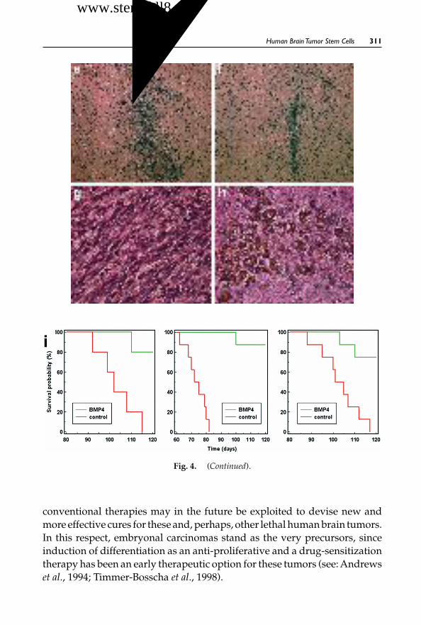
October 30, 2007 12:29 WSPC/SPI-B559: Autologous & Cancer Stem Cell Gene Therapy ch11 FA
Human BrainTumor Stem Cells 311
Fig. 4. (Continued).
conventional therapies may in the future be exploited to devise new andmore effective cures for these and, perhaps, other lethal human brain tumors.In this respect, embryonal carcinomas stand as the very precursors, sinceinduction of differentiation as an anti-proliferative and a drug-sensitizationtherapy has been an early therapeutic option for these tumors (see: Andrewset al., 1994; Timmer-Bosscha et al., 1998).
干细胞之家www.stemcell8.cn ←点击进入

October 30, 2007 12:29 WSPC/SPI-B559: Autologous & Cancer Stem Cell Gene Therapy ch11 FA
312 S. G. M. Piccirillo and A. L. Vescovi
Fig. 5. Ex vivo pre-treatment of GBM-derived cells with BMP4 inhibits ventricular inva-sion following orthotopic injection. (a) 5 weeks after transplantation into the adult brain ofimmunodeficient mice, tumors from control GBM cells have invaded the ventricular systemincluding the fourth ventricle (b). Cells from cultures pretreated with BMP4 for 48 h showedno signs of ventricular invasion. Magnification: 5×. Based on supplementary Fig. 8 of:Piccirillo et al., Nature, 444, 761–765 (2006) with permission from Nature PublishingGroup.
6. Conclusions
The discovery that brain tumors, or at least some of them, comprise cancerstem cells presents us with new opportunities at both the experimental andclinical level. To achieve an effective implementation of new therapeuticstrategies, scientists/clinicians will require new methods of determiningthe identity, nature, physiology and idiosyncratic properties of the cancerstem cells from a given patient’s tumor. It is certainly reasonable to arguethat tumors of the same family may also share common features at the levelof cancer stem cell populations, which would facilitate diagnosis and theapplication of new, standard treatments. By the same token, cancer stem cellsfrom different patients may bear distinctive, peculiar properties that mightwarrant the development of more specific, patient-selective treatments. Thispoint, however, needs to be verified and refined by further study.
Unquestionably, to move the cancer stem cell field forward, a vital issuewill be the discovery of unequivocal markers allowing for the reliable iden-tification of brain tumor stem cells prior to any experimental manipula-tion. Furthermore, it is essential to generate novel drugs or strategies thatcould overcome the issue of putative drug resistance in cancer stem cells,improve therapeutic efficacy, and make cancer treatment more successful
干细胞之家www.stemcell8.cn ←点击进入

October 30, 2007 12:29 WSPC/SPI-B559: Autologous & Cancer Stem Cell Gene Therapy ch11 FA
Human BrainTumor Stem Cells 313
Fig. 6. BMP4 inhibits the tumorigenicity of GBM-derived cells. 48-hour exposure of GBM-derived cells to BMP4 in vitro, prior to transplantation into the brain of immunodeficient mice,caused a significant reduction in the ability of the injected cells to form tumors (b,d,f,h,j,l)as compared to controls (a,c,e,g,i,k). (a–j) Hematoxylin and eosin staining showed typicalglioblastoma masses 5 weeks after injection of control cells (a,e,g), whereas BMP4-treatedGBM-derived cells developed very small grafts (b,f,h). Tumors established from control cellsdisplayed a much higher mitotic index, as revealed by immunohistochemistry for Ki67 (4.3± 0.3% control vs. 0.76 ± 0.5% BMP-treated, mean ± SE, n = 3, Student’s t-test, P < 0.05) [(c),high power in (i)], than BMP4-treated cells [(d), high power in (j)]. (k, l) Hematoxylin andeosin staining showing invasion of the lumen of the right lateral ventricle at bregma point[(k), arrow] 5 weeks post-injection following implantation of control cells. This phenomenonwas never observed with BMP4-pretreated GBM-derived cells [(l), arrow]. 3 months post-injection, all control animals died; on the contrary, all animals receiving BMP4-pretreatedGBM-derived cells survived well beyond 5 months. (m, n) Following a 48-hour incubationin control conditions (control), CD133+ cells were purified by FACS (purity 97.3%) and theability of 3 × 10⁵ of these cells to establish GBMs intracerebrally was compared to that of thesame number of CD133+ cells, purified from sister cultures (purity 98.7%) after incubation for48 hours in the presence of BMP4 (treated). Animals injected with CD133+ cells from controlcultures developed large neoplastic formations by 5–7 weeks post-transplant (m, n = 8),whereas none of the animals (n = 8) receiving CD133+ cells sorted from BMP4-treated cellsdeveloped any tumor (n). By 90 days post-injection, all of the control animals died whereasthose receiving CD133+ cells from BMP4-treated cultures were alive. This shows that thetumorigenic ability of the residual fraction of CD133+ cells found in BMP4-treated culturesis virtually abolished by BMP4. (o, p) When CD133+ cells, which were acutely isolatedfrom primary tumors established in mice injected with CD133+ acutely isolated controlcells, were retransplanted into the brain of secondary recipients, they established secondarytumors in less than 4 months [example in (o)]. These same cells could also be cultured andexpanded with mitogens. Neither culturing nor establishment of secondary tumors couldbe accomplished with cells extracted from mice, which were initially injected with BMP4-treated GBM-derived cells. Similar serial transplantation experiments yielded equivalentresults when the primary tumor was generated by transplantation of 3×10⁵ briefly culturedCD133+ cells [(p), example of secondary tumor established from CD133+ GBM-derivedcells]. Magnification: (a–h) and (k–n), 5×; (i, j) and (o, p), 10×. Based on supplementary Fig. 7of: Piccirillo et al., Nature, 444, 761–765 (2006) with permission from Nature Publishing Group.
干细胞之家www.stemcell8.cn ←点击进入

October 30, 2007 12:29 WSPC/SPI-B559: Autologous & Cancer Stem Cell Gene Therapy ch11 FA
314 S. G. M. Piccirillo and A. L. Vescovi
and perhaps even curative, using the cancer stem cell population as a specifictarget. The search for such anti-CSC agents/protocols harmless to normalstem cells should also broaden our understanding of normal stem cell biol-ogy as well as of the genetics/epigenetics of tumor progression, therebyunraveling the roots of the inherent resistance of CSCs to conventionaltherapies.
References
Al-Hajj M, Wicha MS, Benito-Hernandez A, et al. (2003) Prospective identificationof tumorigenic breast cancer cells. Proc Natl Acad Sci USA 100: 3983–88.
Andrews PW, Damjanov I, Berends J, et al. (1994) Inhibition of proliferation andinduction of differentiation of pluripotent human embryonal carcinoma cells byosteogenic protein-1 (or bone morphogenetic protein-7). Lab Invest 71: 243–51.
Bao S, Wu Q, McLendon RE, et al. (2006a) Glioma stem cells promote radioresistanceby preferential activation of the DNA damage response. Nature 444: 756–60.
Bao S, Wu Q, Sathornsumetee S, et al. (2006b) Stem cell-like glioma cells promotetumor angiogenesis through vascular endothelial growth factor. Cancer Res 66:7843–48.
Bonnet D, Dick JE. (1997) Human acute myeloid leukemia is organized as a hierar-chy that originates from a primitive hematopoietic cell. Nat Med 3: 730–37.
Capela A, Temple S. (2002) LeX/ssea-1 is expressed by adult mouse CNS stem cells,identifying them as nonependymal. Neuron 35: 865–75.
Collins AT, Berry PA, Hyde C, et al. (2005) Prospective identification of tumorigenicprostate cancer stem cells Cancer Res 65: 10946–51.
Dean M, Fojo T, Bates S. (2005) Tumour stem cells and drug resistance. Nat RevCancer. 5: 275–84.
Doetsch F, Caille I, Lim DA, et al. (1999) Subventricular zone astrocytes are neuralstem cells in the adult mammalian brain. Cell 97: 703–16.
Frank SA, Nowak MA. (2003) Cell biology: developmental predisposition to cancer.Nature 422: 494.
Galli R, Binda E, Orfanelli U, et al. (2004) Isolation and characterization of tumori-genic, stem-like neural precursors from human glioblastoma. Cancer Res 64: 7011–21.
Gritti A, Parati EA, Cova L, et al. (1996) Multipotential stem cells from the adultmouse brain proliferate and self-renew in response to basic fibroblast growthfactor. J Neurosci 16: 1091–100.
Hanahan D, Weinberg RA. (2000) The hallmarks of cancer. Cell 100: 57–70.Hemmati HD, Nakano I, Lazareff JA, et al. (2003) Cancerous stem cells can arise
from pediatric brain tumors. Proc Natl Acad Sci USA 100: 15178–83.Holland EC. (2000) Glioblastoma multiforme: the terminator. Proc Natl Acad Sci
USA 97: 6242–44.
干细胞之家www.stemcell8.cn ←点击进入

October 30, 2007 12:29 WSPC/SPI-B559: Autologous & Cancer Stem Cell Gene Therapy ch11 FA
Human BrainTumor Stem Cells 315
Holland EC, Celestino J, Dai C, et al. (2000) Combined activation of Ras and Akt inneural progenitors induces glioblastoma formation in mice. Nat Genet 25: 55–57.
Holland EC, Hively WP, Gallo V, Varmus HE. (1998) Modeling mutations in theG1 arrest pathway in human gliomas: overexpression of CDK4 but not loss ofINK4a-ARF induces hyperploidy in cultured mouse astrocytes. Genes Dev 12:3644–49.
Hope KJ, Jin L, Dick JE. (2004) Acute myeloid leukemia originates from a hierarchyof leukemic stem cell classes that differ in self-renewal capacity. Nat Immunol 5:738–43.
Ignatova TN, Kukekov VG, Laywell ED, et al. (2002) Human cortical glial tumorscontain neural stem-like cells expressing astroglial and neuronal markers in vitro.Glia 39: 193–206.
Kummermehr J, Trott KR. (1997). Tumour stem cells. In: Stem Cells, Potten CS (ed.),Academic Press, London, UK, pp. 363–99.
Lee J, Kotliarova S, Kotliarov Y, et al. (2006) Tumor stem cells derived from glioblas-tomas cultured in bFGF and EGF more closely mirror the phenotype andgenotype of primary tumors than do serum-cultured cell lines Cancer Cell 9:391–403.
Looijenga LH, Stoop H, de Leeuw HP, et al. (2003) POU5F1 (OCT3/4) identifies cellswith pluripotent potential in human germ cell tumors. Cancer Res 63: 2244–50.
Monzani E, Facchetti F, Galmozzi E, et al. (2007) Melanoma contains CD133 andABCG2 positive cells with enhanced tumourigenic potential. Eur J Cancer 43:935–46.
O’Brien CA, PollettA, Gallinger S, Dick JE. (2007)Ahuman colon cancer cell capableof initiating tumour growth in immunodeficient mice. Nature 445: 106–10.
Pearce DJ, Bonnet D. (2007) Leukemia stem cells: studying the root of leukemia.Gene Ther Regul 3: 65–90.
Piccirillo SGM, Reynolds BA, Zanetti N, et al. (2006) Bone morphogenetic proteinsinhibit the tumorigenic potential of human brain tumour-initiating cells Nature444: 761–65.
Reya T, Morrison SJ, Clarke MF, Weissman IL. (2001) Stem cells, cancer, and cancerstem cells. Nature 414: 105–11.
Reynolds BA, Rietze RL. (2005) Neural stem cells and neurospheres — re-evaluatingthe relationship. Nat Meth 2: 333–36.
Reynolds BA, Weiss S. (1992) Generation of neurons and astrocytes from isolatedcells of the adult mammalian central nervous system. Science 255: 1707–10.
Ricci-Vitiani L, Lombardi DG, Pilozzi E, et al. (2007) Identification and expansionof human colon-cancer-initiating cells. Nature 445: 111–15.
Rietze RL, Valcanis H, Brooker GF, et al. (2001) Purification of a pluripotent neuralstem cell from the adult mouse brain. Nature 412: 736–39.
Sanai N, Alvarez-Buylla A, Berger MS. (2005) Neural stem cells and the origin ofgliomas. N Engl J Med 353: 811–22.
干细胞之家www.stemcell8.cn ←点击进入
October 30, 2007 12:29 WSPC/SPI-B559: Autologous & Cancer Stem Cell Gene Therapy ch11 FA
316 S. G. M. Piccirillo and A. L. Vescovi
Sanai N, Tramontin AD, Quinones-Hinojosa A, et al. (2004) Unique astrocyte ribbonin adult human brain contains neural stem cells but lacks chain migration. Nature427: 740–44.
Singh SK, Clarke ID, Terasaki M, et al. (2003) Identification of a cancer stem cell inhuman brain tumors Cancer Res 63: 5821–28.
Singh SK, Hawkins C, Clarke ID, et al. (2004) Identification of human brain tumourinitiating cells. Nature 432: 396–401.
Szotek PP, Pieretti-Vanmarcke R, Masiakos PT, et al. (2006) Ovarian cancer side pop-ulation defines cells with stem cell-like characteristics and Mullerian inhibitingsubstance responsiveness. Proc Natl Acad Sci USA, 103: 11154–59.
Taylor MD, Poppleton H, Fuller C, et al. (2005) Radial glia cells are candidate stemcells of ependymoma. Cancer Cell 8: 323–35.
Timmer-Bosscha H, de Vries EG, Meijer C, et al. (1998) Differential effects of all-trans-retinoicacid, docosahexaenoic acid, and hexadecylphosphocholine on cisplatin-induced cytotoxicity and apoptosis in a cisplantin-sensitive and resistant humanembryonal carcinoma cell line. Cancer Chemother Pharmacol 41: 469–76.
Uchida N, Buck DW, He D, et al. (2000) Direct isolation of human central nervoussystem stem cells. Proc Natl Acad Sci USA 97: 14720–25.
Vescovi AL, Galli R, Reynolds BA. (2006) Brain tumour stem cells. Nat Rev Cancer6: 425–36.
Zhu Y, Guignard F, Zhao D, et al. (2005) Early inactivation of p53 tumor suppressorgene cooperating with NF1 loss induces malignant astrocytoma. Cancer Cell 8:119–30.
干细胞之家www.stemcell8.cn ←点击进入

October 30, 2007 12:29 WSPC/SPI-B559: Autologous & Cancer Stem Cell Gene Therapy ch12 FA
Chapter 12
CD44 as a Functional Cancer StemCell Marker and a Potential
TherapeuticTarget
Lubna Patrawala† and Dean G. Tang∗,†,‡
CD44 is a well-characterized adhesion molecule with multi-faceted biological func-tions. Earlier studies have implicated CD44 in the invasion and metastasis of multi-ple human cancers. Newly generated data suggest that tumor cells expressing CD44may be intrinsically different from the bulk CD44− cells and that its expression maymark a population of tumor-initiating cells called cancer stem cells (CSCs). In thisreview, we succinctly summarize the evidence of CD44 as a CSC marker and alsodiscuss the possibility of targeting CD44-expressing CSCs.
Keywords: CD44; tumor-initiating cells; cancer stem cells; tumor progenitors; metastasis;homing.
1. Introduction
In the past decade, there has been a major paradigm shift in our under-standing of cancer. Rather than being viewed as a disease driven by randommutations, cancer is now viewed as a product of abnormal stem cell devel-opment. In other words, a mutated stem cell or its immediate progeny, stillretaining the ability to both self-renew and partially differentiate, developsinto a disorganized tumor mass comprising a kaleidoscope of cells withdifferent phenotypic properties. Regardless of the origin, many cancers arenow found to contain small subsets of cells, which have the exclusive ability
∗Corresponding author. [email protected]†Department of Carcinogenesis, The University of Texas MD Anderson Cancer Center,Science Park–Research Division, Smithville, TX 78957, USA‡Program in Environmental & Molecular Carcinogenesis, Graduate School ofBiomedical Sciences (GSBS), Houston, TX 77030, USA
317
干细胞之家www.stemcell8.cn ←点击进入

October 30, 2007 12:29 WSPC/SPI-B559: Autologous & Cancer Stem Cell Gene Therapy ch12 FA
318 L. Patrawala & D. G.Tang
to reinitiate tumors and are therefore called tumor-initiating cells or cancerstem cells (CSCs) (Reya et al., 2001; Clarke et al., 2006; Dalerba et al., 2007).These CSCs, at the cellular level, resemble normal stem cells in that theypossess the ability to self-renew and differentiate. Identification and charac-terization of CSCs in every type of human cancer will not only facilitate ourunderstanding of the etiology of that specific tumor, but also, more impor-tantly, help develop novel therapeutics to eradicate the “roots” of the malig-nancy. Most putative CSCs so far reported have been identified using cellsurface markers, among which CD44 and CD133 have emerged as the mostcommon ones. In this review, we focus on CD44 as a tempting “universal”CSC marker and on its potential utility in the development of therapeuticregimens aimed specifically at CSCs.
2. The CD44 Family
The CD44 proteins are a family of transmembrane glycoproteins that areencoded by a highly conserved gene containing 20 exons (Screaton et al.,1992; reviewed in Ponta et al., 2003). Structural and functional heterogeneityof the proteins can be attributed to alternative splicing events and post-translational modifications leading to the generation of a number of iso-forms. CD44s or CD44H, the standard form encoded by nine exons, is themost widely expressed isoform and is found in developing and normal adulttissues.Alternative splicing of at least 10 exons leads to the insertion of aminoacids in the stem region of the CD44s protein, giving rise to the variant (v)isoforms. In contrast to the standard form, the v forms are more restricted intheir expression, appearing mainly in proliferating cells of some epithelialtissues and in transformed cells (Ponta et al., 2003).All isoforms of CD44 bindto hyaluronan, a glycosaminoglycan that is ubiquitously distributed in theextracellular matrix (ECM), although CD44 has also been shown to bind to anumber of other ligands including fibronectin, collagen, laminin and osteo-pontin (Weber et al., 1996; Ponta et al., 2003). Interaction between CD44 andhyaluronan activates an array of downstream signaling pathways that reg-ulates functions as diverse as proliferation, adhesion, apoptosis, migration,metastasis and hematopoiesis. The complex involvement of CD44 in multi-ple physiological and disease processes owes to its structural heterogeneityas well as to the diversity of its ligands (Stamenkovic et al., 1991; Ponta et al.,2003). Another level of CD44 regulation is post-transcriptional, i.e., its prote-olysis at both extracellular and transmembrane domains. For example, CD44is a substrate of the presenilin-dependent gamma-secretase, which inducesintramembranous cleavage of CD44 (Murakami et al., 2003). Similarly, two
干细胞之家www.stemcell8.cn ←点击进入

October 30, 2007 12:29 WSPC/SPI-B559: Autologous & Cancer Stem Cell Gene Therapy ch12 FA
CD44 as a Functional Cancer Stem Cell Marker 319
ADAM family metalloproteinases,ADAM10 andADAM17 can induce CD44ectodomain cleavage and consequently terminate the CD44-mediated cell–matrix adhesion (Nagano et al., 2004).
3. CD44 and Normal Stem Cells
Surprisingly, CD44-null mice are viable and, with the exception of mildhematopoietic abnormalities, do not show any overt developmental defi-ciencies (Schmits et al., 1997; Protin et al., 1999). It has been suggested that inthe early absence of CD44, compensatory mechanisms may take over result-ing in only a subtle developmental phenotype. On the other hand, transgenicmice expressing antisense CD44 cDNA driven by the K5 promoter display amore severe phenotype such as decrease in skin elasticity, impaired inflam-mation and tissue repair, and delayed hair growth (Kaya et al., 1997). Theseobservations suggest that there is a small window of opportunity in earlydevelopment for CD44 compensation to kick in, and reiterate the impor-tance of CD44 in normal physiology. Indeed, CD44 plays a critical role inthe homing and distribution of hematopoietic progenitor cells (Schmits et al.,1997; Lapidot et al., 2005). In normal hematopoietic stem cells (HSCs), theinteraction between CD44 and hyaluronic acid is essential for their homingto the bone marrow and spleen (Avigdor et al., 2004). Similarly, CD44 is animportant molecule expressed in mesenchymal stem cells (MSCs) involvedin cell migration in ECM (Zhu et al., 2006). CD44 has also been reportedto mark astrocyte precursor cells (Liu et al., 2004) and multipotent centralnervous system (CNS) stem cells (Oishi et al., 2006).
4. CD44 as a Marker for Diverse CSCs
Early studies have implicated CD44H or some of its splice variants (in par-ticular, CD44v6 and CD44v9) in cancer cell adhesion, migration, invasionand metastasis (Gunthert et al., 1991; Lamb et al., 1997; Masellis-Smith et al.,1997; Ponta et al., 2003). The CD44-involved tumor cell invasion and dis-semination are generally ascribed to its role as an adhesion and signalingmolecule (Ponta et al., 2003), and also to its ability to direct some proteasessuch as matrix metalloproteinases to the invading lamellipodia (Mori et al.,2002).
4.1. Leukemic Stem Cells
In 1992, CD44 was found to be expressed not only on CD34+ normalHSCs, but also on CD34+ leukemia cells (Reuss-Borst et al., 1992), providing
干细胞之家www.stemcell8.cn ←点击进入

October 30, 2007 12:29 WSPC/SPI-B559: Autologous & Cancer Stem Cell Gene Therapy ch12 FA
320 L. Patrawala & D. G.Tang
the first hint that CD44 might mark tumorigenic leukemia cells. In 1996,Sutherland et al. demonstrated a hierarchical organization of human acutemyeloid leukemia (AML) cells; and in 1997, Dick’s group showed that onlyleukemic cells bearing the CD34+CD38− phenotype could transfer the dis-ease to immunodeficient NOD/SCID mice (Bonnet and Dick, 1997). TheseAML leukemic stem cells (AML-LSCs) were later phenotypically refinedas CD34+CD38−CD71−CD90− (Blair et al., 1997; 1998) that also uniquelyexpress IL3Rα (Jordan et al., 2000). Importantly, CD44 is also expressed inLSCs. Although CD44 was not used in the initial identification of LSCs, thevariant isoforms are known to be elevated on AML cells and are associatedwith poor prognosis (Bendall et al., 2000; Legras et al., 1998). In fact, CD44seems to be crucial in homing LSCs to the right bone marrow microenvi-ronment (i.e., niche) as an activating monoclonal antibody to CD44 (H90)interferes with the transport of AML-LSCs to stem cell-supportive microen-vironmental niches, leading to markedly reduced leukemic repopulation (Jinet al., 2006). H90 effectively reduced the tumor burden as well as the engraft-ment of leukemic cells. The defect in engraftment was specific to AML cells,as human cord blood cells were unaffected by H90. Interestingly, the authorsobserved that LSCs were somewhat niche-dependent, demonstrating forthe first time the link between CSCs and a specialized microenvironment.Furthermore, treatment with the H90 antibody triggered maturation of theLSCs, implicating CD44 as a key regulator of the primitive state of LSCs (Jinet al., 2006).
CD44 expression is also increased in human chronic myeloid leukemia(CML) (Ghaffari et al., 1995) as well as in BCR-ABL–expressing progenitorsin the bone marrow of mice with CML-like leukemia (Krausse et al., 2006).Using CD44-null mice as donors in a retroviral transplantation model ofCML, Krausse et al. (2006) found that CD44 was required for bone marrowhoming of CML progenitors. As in AML, dependence on CD44 for homingwas greatly increased in LSCs compared with normal HSCs (Krausse et al.,2006). Unlike the AML studies, however, direct injection of CML progenitorstreated with a CD44 blocking antibody (IM7) into the bone marrow led to aleukemia burden similar to that in control mice, suggesting that CD44 wasrequired for homing but not maintenance of LSCs in the mouse model ofCML (Krausse et al., 2006). These studies point to the crucial importance ofCD44 in LSC homing, and also highlight the complexity of CD44 signalingand the need to tease apart specific variants that may be involved in differentcancers.
干细胞之家www.stemcell8.cn ←点击进入

October 30, 2007 12:29 WSPC/SPI-B559: Autologous & Cancer Stem Cell Gene Therapy ch12 FA
CD44 as a Functional Cancer Stem Cell Marker 321
4.2. Breast Cancer Stem Cells
Recent work suggests that CD44 expression may also identify tumor-initiating cells in solid tumors. Studies on breast cancer have generatedcontroversial data over the potential involvement and prognostic value ofCD44 (Gotte andYip, 2006). For instance, hyaluronan is upregulated in breastcancer cells and their surrounding stroma (Auvinen et al., 2000). CD44s isupregulated in invasive breast carcinomas (Auvinen et al., 2005) but, para-doxically, correlated with overall patient survival (Diaz et al., 2005), whilesome variant forms such as CD44v6 are upregulated in breast cancer but donot correlate with clinical outcome (Auvinen et al., 2005; Diaz et al., 2005).CD44, although promoting the invasion of a variety of cancer cells, some-how seems to attenuate metastatic invasion during breast cancer progression(Lopez et al., 2005). Most of these studies relied on correlative tissue analy-sis or overexpression experiments in long-term cultured cell lines. The keymissing experiment was to purify CD44-expressing breast cancer cells outto ask whether these cells are intrinsically different from the CD44− tumorcells.
Spurred by the findings that leukemia is driven by LSCs (Bonnet andDick, 1997),Al-Hajj et al. (2003) proceeded to investigate the tumor-initiatingcapacity of CD44-expressing cells in human primary breast cancer samples.They found that as few as 200 CD44+CD24−/loESA+ cells could generate atumor in immunodeficient mice; whereas the remaining cancer cells, com-prising 95–98% of the population, were much less tumorigenic. Moreover,the CD44+CD24−/lo population could regenerate the phenotypic hetero-geneity of the original tumor. These observations provided the first evidencefor a hierarchical organization of cells in a solid tumor. Importantly, theypaved the way for the characterization of tumorigenic and non-tumorigeniccells from a single tumor, facilitating the elucidation of the molecular path-ways that preferentially mediate resistance to cancer therapies in the tumori-genic population. The CD44+CD24−/lo breast CSCs can be cultured in vitrowhile maintaining their tumorigenic potential (Ponti et al., 2005). It wasrecently demonstrated that the Hedgehog signaling pathway, known to reg-ulate self-renewal in mammary stem cells, is aberrantly activated in theCD44+CD24−/lobreast CSCs (Liu et al., 2006).
Many groups are also investigating the correlation between breast CSCsand invasion and metastasis. In a study done with 50 patients in the earlystages of breast cancer, an average of 72% of the disseminated tumor cells
干细胞之家www.stemcell8.cn ←点击进入

October 30, 2007 12:29 WSPC/SPI-B559: Autologous & Cancer Stem Cell Gene Therapy ch12 FA
322 L. Patrawala & D. G.Tang
in the bone marrow were found to be of the CD44+CD24−/lo phenotype(Balic et al., 2006). This finding was striking because cells of the same phe-notype were present in <10% of primary tumors, leading to speculationthat the breast CSCs mediate metastasis. Another correlative study suggeststhat the prevalence of CD44+CD24−/lo tumor cells may favor distant metas-tasis (Abraham, 2005). More recently, Liu et al. (2007) compared the gene-expression profiles of breast CSCs with those of normal breast epitheliumand generated an “invasive gene signature” (IGS) based on the differentialexpression of 186 genes. The prognostic power of the criteria used by theNational Institutes of Health to determine breast cancer patient survivalwas significantly strengthened when combined with the IGS. Additionally,the gene signature was associated with prognosis in medulloblastoma, lungcancer, and prostate cancer (Liu et al., 2007). Another independent molecularprofiling of CD24+ and CD44+ breast cancer cells also revealed them to bemore differentiated and progenitor-like cells, respectively (Shipitsin et al.,2007). Importantly, the CD44+ breast cancer cells were found to preferen-tially express many known stem-cell markers, and the CD44+ cell-specificgene expression correlates with decreased patient survival (Shipitsin et al.,2007). Furthermore, the CD44+ breast CSCs are negative for estrogen recep-tor even in some estrogen receptor-positive tumors (Shipitsin et al., 2007).Finally, CD44+CD24−/locells have been identified in breast cancer cell linesand have been shown to be highly invasive (Sheridan et al., 2006) and radiore-sistant, increasing in number after bouts of radiation (Phillips et al., 2006).
The above discussions suggest that, when done properly, the CD44-expressing breast cancer cells in both long-term and short-term culturesas well as in tumors are intrinsically different from the bulk, CD44− breastcancer cells.
4.3. Prostate Cancer Stem Cells
Similar to the situation in breast cancer, most early studies on the role ofCD44 in prostate cancer (PCa) development and progression have gen-erated conflicting data. One study of 109 cases reported a complete lackof membranous expression of all CD44 isoforms in 93–98% of primaryPCa examined (Kallakury et al., 1996). However, another study of 74PCa lesions reported moderate to high levels of CD44 expression in 60%of primary tumors, with ∼14% of metastases expressing low levels ofCD44 (Nagabhushan et al., 1996). Yet another study reported significantlyreduced CD44 expression in all 94 primary neoplastic foci and 48 metastases(De Marzo et al., 1998). The relationship between CD44 expression and tumor
干细胞之家www.stemcell8.cn ←点击进入

October 30, 2007 12:29 WSPC/SPI-B559: Autologous & Cancer Stem Cell Gene Therapy ch12 FA
CD44 as a Functional Cancer Stem Cell Marker 323
grade is also uncertain — one study showed strong correlation betweenGleason grade of the tumor and loss of CD44 expression (De Marzo et al.,1998), whereas another reported no correlation (Paradis et al., 1998). Further-more, although CD44 expression was reported to be reduced in metastases(Nagabhushan et al., 1996; De Marzo et al., 1998), the CD44+ PCa cells werefound to predominate in two visceral metastases (Liu et al., 1999). Simi-lar to expression studies, the potential role of CD44 in PCa developmentand metastases is controversial — while some studies showed a tumor-suppressive function of CD44 in overexpression experiments (Gao et al.,1997 and 1998), many other studies implicated CD44 in PCa cell prolifera-tion, adhesion, migration and invasion in vitro as well as metastatic dissemi-nation in vivo (Paradis et al., 1998; Liu et al., 1999; Omara-Opyene et al., 2004;Draffin et al., 2004). Most studies mentioned above utilized either human tis-sues to carry out correlative immunohistochemistry or bulk-cultured PCacells to do overexpression experiments, and the key experiment of usingpurified CD44+ and CD44− cells from the same culture or tumor to com-pare their potentially different biological and tumorigenic properties hadnot been done. Therefore, it was not known whether the minor subset ofCD44+ PCa cells in both primary and metastatic lesions were intrinsicallydifferent from bulk CD44− PCa cells.
We addressed this critical issue by isolating human CD44+ PCa cellsfrom multiple cultured PCa cell lines and three xenograft tumors (i.e.,Du145, LAPC4, and LAPC9), and compared their in vitro biological andin vivo tumorigenic properties with the isogenic CD44− PCa cell popula-tions (Patrawala et al., 2006). We have observed that CD44+ PCa cells aremore clonogenic, proliferative, tumorigenic and metastatic than their corre-sponding negative counterparts (Patrawala et al., 2006). Noticably, CD44+
and CD44− cells were purified to near homogeneity from freshly dissociatedxenograft tumor cells (Fig. 1) and injected into NOD/SCID mice either sub-cutaneously or orthotopically. In both cases, CD44+ PCa cells were shownto possess higher tumoregenicity than their CD44− counterparts (Tables1 and 2).
Moreover, CD44+cells possess many intrinsic traits of stem/progenitorcells, such as preferential activation of genes implicated in self-renewaland maintenance of an undifferentiated state (e.g., Oct3/4, Bmi, β-catenin,Smoothened), and intermediate cell-cycle properties. Importantly, CD44+cellsare distinct from the fully differentiated androgen receptor (AR)-expressingcells and have the ability to recapitulate the original heterogeneity of thetumor, indicating that they are upstream in the tumor hierarchy (Patrawala
干细胞之家www.stemcell8.cn ←点击进入

October 30, 2007 12:29 WSPC/SPI-B559: Autologous & Cancer Stem Cell Gene Therapy ch12 FA
324 L. Patrawala & D. G.Tang
Fig. 1. Purity of the CD44+ and CD44− PCa cell populations. Tumor cells were acutelypurified from xenograft tumors (NOD/SCID mice) and the purity of sorted CD44+ andCD44− cell populations was analyzed as shown here (LAPC-4 tumor). Cells were stainedfor CD44 and counterstained by DAPI. Based on supplementary Fig. 2 of: Patrawala et al.,Oncogene, 25: 1696–1708 (2006) with permission from Nature Publishing Group.
et al., 2006; Tang et al., 2007). Recently, using a combinatorial sortingstrategy, we clearly demonstrated the hierarchical organization of PCa cellsin xenograft tumors. Specifically, we found that the CD44+α2β1+ and CD44+
cell populations are very similarly enriched in tumorigenic PCa cells andthe CD44−α2β1+ PCa cells show much reduced tumorigenicity whereas theCD44−α2β1− cells essentially lack tumorigenic potential (Patrawala et al.,2007), suggesting that prostate CSCs reside in the CD44+ cell populationand α2β1 marks prostate tumor progenitors. Since in all of our experimentswe routinely carried out lineage selection by removing Lin+ cells (includingblood cells, endothelial cells, fibroblasts and other stromal cells), the prostateCSCs we identified thus possess the phenotype of CD44+Lin−. Collins et al.(2005) utilized in vitro approaches to identify putative CSCs in primaryhuman PCa samples. The putative prostate CSCs had the same antigenicprofile as normal prostate stem cells, i.e., CD44+α2β1hiCD133+, and werethe only cells with the capability to self-renew in vitro and generate moredifferentiated cell types (Collins et al., 2005). Due to technical challenges,there have been no reports to date describing the isolation of tumorigenicprostate CSCs from primary tumor samples. In ∼20 PCa patient samples wehave worked with, we could detect CD44+ and CD133+ cells, and we arecurrently studying whether these cells have unique tumor-initiating abili-ties. Our studies on xenograft tumors clearly indicate that the CD44+ cellpopulation, although enriched in tumorigenic cells, is still heterogeneous.Work is underway to further enrich for more tumorigenic subsets, such asthe side population (SP) cells (Patrawala et al., 2005) in the CD44+ PCa cell
干细胞之家www.stemcell8.cn ←点击进入

October 30, 2007 12:29 WSPC/SPI-B559: Autologous & Cancer Stem Cell Gene Therapy ch12 FA
CD44 as a Functional Cancer Stem Cell Marker 325
Table 1 Tumorigenicity of Subcutaneously Injected CD44+ and CD44−PCa Cells
Cell Typea Cell Nos. Injected Tumor Incidenceb Latency (Days)c
Du145 unsorted 100 3/5 61–95 (71)1,000 4/6 36–71 (61)10,000 5/6 36–95 (36)100,000 4/4 36 (36)
Du145-CD44+ 100 5/8∗ 41–67 (41)1,000 5/8∗ 32–50 (40)
Du145-CD44− 100 1/8 1081,000 0/610,000 3/8 96–174 (96)
LAPC4 unsorted 100 10/12 45–85 (63)1,000 5/8 42–130 (58)10,000 6/6 31–116 (31)
LAPC4-CD44+ 100 4/6∗ 60–102 (67)1,000 5/6 41–123(66)10,000 3/3 31–78 (47)
LAPC4-CD44− 100 2/14 41–120 (80)1,000 13/14 41–137 (90)10,000 7/7 35–83 (71)
LAPC9 unsorted 100 3/6 96–124 (96)1,000 6/6 60–67 (64)
LAPC9-CD44+ 10 1/4 67100 10/10∗ 63–88 (67)
1,000 10/11 55–82 (64)10,000 4/4 53–55 (54)
LAPC9-CD44− 100 0/81,000 5/6 67–81 (74)10,000 4/4 60–67 (64)
aTumor cells were acutely purified from the xenograft tumors maintained in NOD/SCIDmice. Cells, either unsorted or sorted as CD44+ or CD44–, were injected in 40 µL ofmedium/Matrigel (1:1) subcutaneously into the NOD/SCID mice.bThe number of tumors/number of injections. ∗P < 0.01 compared with the correspond-ing CD44− injections (F-test).cFrom tumor cell injection to the appearance of a palpable tumor. Values in parenthesesare the medians.Based on Table 1 of: Patrawala et al., Oncogene, 25: 1696–1708 (2006) with permission fromNature Publishing Group.
population. Taken together, studies on breast and prostate CSCs highlightthe importance of carrying out prospective in vivo tumor experiments usingpurified populations of cells, and point to CD44 as a CSC marker for bothtumor types.
干细胞之家www.stemcell8.cn ←点击进入

October 30, 2007 12:29 WSPC/SPI-B559: Autologous & Cancer Stem Cell Gene Therapy ch12 FA
326 L. Patrawala & D. G.Tang
Table 2 Tumorigenicity of Orthotopically Implanted LAPC-9 Cells
Cells (Number)a Tumor Incidenceb Latency (Days)c Metastasisd
LAPC-9/unsorted100 0/31,000 0/910,000 4/8 (50%) 46–75 (53) 0/4100,000 6/9 (67%) 32–69 (44) 0/61,000,000 4/4 (100%) 48–69 (56) 0/4
LAPC-9/CD44+100 0/51,000 2/8 (25%) 45–61 (53) 1/210,000 4/9 (44%) 42–57 (50) 1/4
LAPC-9/CD44−100 0/51,000 0/1010,000 0/10100,000 3/5 (60%) 107 (107) 0/3500,000 4/5 (80%) 33–107 (60) 0/4
aCells at the indicated numbers were injected in 25 µL of Matrigel into NOD/SCIDmouse prostate.bThe number of animals that developed tumors/The number of animals injectedwith tumor cells. Values in parentheses represent the % tumor incidence.cThe time (in days) from tumor cell injection to when tumors were observed. Theranges are shown with the median tumor latencies shown in the parentheses.dAt necropsy, urogenital organs as well as multiple distant organs were examinedfor tumor dissemination.Based on Table 2 of: Patrawala et al., Oncogene, 25: 1696–1708 (2006) with permissionfrom Nature Publishing Group.
4.4. Pancreatic and Head/Neck Cancer Stem Cells
CD44 may also mark other CSCs. For example, CD44 has proven useful inisolating CSCs from pancreatic cancer, one of the most lethal cancers. Cellsof the phenotype CD44+CD24+ESA+constituted 0.2–0.8% of the pancreatictumor cells, but were enriched 100-fold in tumorigenic potential comparedwith the rest of the population and generated tumors that were histologicallyindistinguishable from the original tumors from which they were derived(Li et al., 2007). The putative pancreatic CSCs showed increased expressionof Sonic hedgehog (Shh), presumably leading to dysregulated self-renewal(Li et al., 2007). Similarly, CD44 was found to mark primitive basal-like cellsin head and neck squamous cell carcinoma that met all of the criteria toqualify as CSCs. In this case, the Shh pathway target Bmi was identified asthe aberrantly expressed self-renewal gene (Prince et al., 2007).
干细胞之家www.stemcell8.cn ←点击进入

October 30, 2007 12:29 WSPC/SPI-B559: Autologous & Cancer Stem Cell Gene Therapy ch12 FA
CD44 as a Functional Cancer Stem Cell Marker 327
4.5. Importance of CD44 and Potential Comarkers in CSC Biology
Considering that CD44 interaction with hyaluronan plays a critical role incolon cancer cell invasion (Kim et al., 2004) and that CD44 expression in awide variety of cancer cells (including prostate, breast, colon, and bladdercancer cells as well as glioma and melanoma cells) correlates with theirmalignancy (Patrawala et al., 2006), it stands to reason that CD44 must beinvolved in the biological activities of a wide variety of CSCs.
It should be noted that in all four solid tumors discussed above, i.e.,breast (Al-Hajj et al., 2003), prostate (Collins et al., 2005; Patrawala et al.,2006), pancreatic (Li et al., 2007) and head/neck cancers (Prince et al., 2007),putative CSCs are purified using mainly CD44 as the positive marker. Therole of other marker(s) such as CD24 remains unclear. Intriguingly, breastCSCs bear CD24−/low (Al-Hajj et al., 2003), whereas pancreatic CSCs havethe phenotype of CD24+ (Li et al., 2007). In addition, CD24+cells are foundto significantly increase in breast cancer metastasis (Shipitsin et al., 2007),and CD24 expression has been associated with tumor growth and metasta-sis (Baumann et al., 2005). Also, ESA(epithelial specific antigen) is expressedmainly on differentiated cells, so it is unclear why in some purification pro-tocols ESA is used (or needed). At this moment, it is also unclear whetherCD44 expression per se is required for the manifestation of all CSC proper-ties. The fact that the anti-CD44 antibodies could block the homing of AML(Jin et al., 2006) and CML (Krausse et al., 2006) LSCs suggests that CD44 itselfis biologically important. Another point worth mentioning is that CD44 andCD133 — another popular cell surface marker that has been used to identifya variety of normal stem/progenitor cells (Uchida et al., 2000; Kuci et al.,2003) as well as putative CSCs in glioma (Singh et al., 2004), colon cancer(O’Brien et al., 2007; Ricci-Vitiani et al., 2007) and hepatocellular carcinoma(Suetsugu et al., 2006; Yin et al., 2007) — may identify overlapping popula-tions of stem/progenitor cells. For example, putative prostate CSCs reportedby Collins et al. (2005) are CD44+CD133+. Similarly, the CD133+ glioma stemcells are also enriched in CD44 (Liu et al., 2006a).
5. CD44-expressing CSCs asTherapeuticTargets
Several types of CSCs have been shown to be more resistant to therapeu-tics. For example, LSCs are resistant to a wide spectrum of chemodrugspossibly due to overexpression of multi-drug resistance proteins such asABCG2 (reviewed in Ravandi and Estrov, 2006). The CD44+CD24−/lowbreastcancer cells are radioresistant possibly because they produce lower levels
干细胞之家www.stemcell8.cn ←点击进入

October 30, 2007 12:29 WSPC/SPI-B559: Autologous & Cancer Stem Cell Gene Therapy ch12 FA
328 L. Patrawala & D. G.Tang
of reactive oxygen species (Phillips et al., 2006). The CD133+ glioma stemcells are also radioresistant due to a preferential activation of DNA damageresponse in these cells (Bao et al., 2006). If we know the gene expression sig-natures and/or signal transduction pathways unique to CSCs, we might beable to design specific therapeutics to target them. For example, recent workshowed that LSCs seem to have heightened NF-kB prosurvival mechanisms.Parthenolide (PTL), a naturally occurring small molecule, via inhibiting NF-kB activity and inducing generation of reactive oxygen species, causes robustapoptosis in primary humanAMLcells and blast crisis CMLcells while spar-ing normal hematopoietic cells (Guzman et al., 2005). Similarly, the BMPsignaling pathway normally regulates the neural stem cell fate, and BMP4demonstrates a potent inhibitory effect on CD133+ cell-initiated gliomagene-sis (Piccirillo et al., 2006). The CD44+ breast cancer cells seem to have uniqueactivation of the TGFβ signaling pathway due to exclusive expression ofTGFβRII, and a TGFβR inhibitor causes their rapid differentiation (Ship-itsin et al., 2007). These discussions suggest that CSCs are probably moreresistant to therapeutics through many different mechanisms, that CSCswill have to be eradicated if we want to cure the patients of primary tumorburden and prevent recurrence and dissemination, and that it is possible tofind CSC-unique signaling pathways to specifically target them.
CD44 may represent a good CSC-specific target. It is widely expressedin various CSCs and seems to be required for LSC homing (Jin et al., 2006;Krausse et al., 2006). Since both breast and prostate cancers have a highpropensity to metastasize to the bone and CD44+ CSCs might be the cellsmediating breast (Abraham, 2005; Balic et al., 2006) and prostate (Patrawalaet al., 2006) cancer metastass, it is tempting to speculate that CD44 mightalso be involved in breast/prostate CSC homing to the bone marrow. Fur-thermore, disruption of CD44 functions induces apoptosis of many can-cer cells (Yu et al., 1997; Ohashi et al., 2007). Therefore, targeting CD44may help both reduce tumor burden and prevent bone metastasis of thesemalignancies. Conceivable approaches may include local or imaging-guidedadministration of anti-CD44 antibodies such as H90 and IM7 (Jin et al.,2006; Krausse et al., 2006), antibody-conjugated cytotoxic agents (or pro-drugs), CD44 promoter-driven expression of proapoptotic genes or cyto-toxins in retroviral or lentiviral vectors, or hyaluronan polymer-conjugateddrugs (such as taxol). Neural stem/progenitor cells as well as mesenchy-mal stem cells have tremendous migratory ability and high tropism fortumors/ischemic foci (Aboody et al., 2000; Studeny et al., 2002) and havebeen exploited to carry therapeutic agents to treat tumors, e.g., intracranialbrain tumors (e.g., Aboody et al., 2006). Theoretically, CD44+ CSCs (or their
干细胞之家www.stemcell8.cn ←点击进入

October 30, 2007 12:29 WSPC/SPI-B559: Autologous & Cancer Stem Cell Gene Therapy ch12 FA
CD44 as a Functional Cancer Stem Cell Marker 329
normal counterparts) might be utilized in the same way to carry cytotoxicagents to home to the primary tumor and metastatic sites in order to achievetherapeutic efficacy. We envision many exciting developments along theselines in the near future.
Acknowledgments
We thank the other Tang lab members for their assistance in actual experi-ments. Original work in the lab was supported in part by grants from the NIH(AG023374), American Cancer Society (RSG MGO-105961), Department ofDefense, Prostate Cancer Foundation, and Elsa Pardee Foundation. We alsoacknowledge the support of two Center Grants (CCSG-5 P30 CA166672 andES07784). L. Patrawala was supported in part by a pre-doctoral fellowshipfrom the Department of Defense (W81XWH-06-1-023). We apologize to thecolleagues whose work could not be cited due to space constraints.
References
Aboody KS, Brown A, Rainov NG, et al. (2000) Neural stem cells display extensivetropism for pathology in adult brain: evidence from intracranial gliomas. ProcNatl Acad Sci USA 97: 12846–51.
Aboody KS, Bush RA, Garcia E, et al. (2006) Development of a tumor-selectiveapproach to treat metastatic cancer. PLoS ONE 1: e23.
Abraham BK, Fritz P, McClellan M, et al. (2005) Prevalence of CD44+/CD24−/lowcells in breast cancer may not be associated with clinical outcome but may favordistant metastasis. Clin Cancer Res 11: 1154–59.
Al-Hajj M, Wicha MS, Benito-Hernandez A, et al. (2003) Prospective identificationof tumorigenic breast cancer cells. Proc Natl Acad Sci USA 100: 3983–88.
Auvinen P, Tammi R, Parkkinen J, et al. (2000) Hyaluronan in peritumoral stromaand malignant cells associates with breast cancer spreading and predicts sur-vival. Am J Pathol 156: 529–36.
Auvinen P, Tammi R, Tammi M, et al. (2005) Expression of CD44s, CD44v3 andCD44v6 in benign and malignant breast lesions: correlation and colocalizationwith hyaluronan. Histopathology 47: 420–28.
Avigdor A, Goichberg P, Shivtiel S, et al. (2004) CD44 and hyaluronic acid cooperatewith SDF-1 in the trafficking of human CD34+ stem/progenitor cells to bonemarrow. Blood 103: 2981–89.
Balic M, Lin H, Young L, et al. (2006) Most early disseminated cancer cells detectedin bone marrow of breast cancer patients have a putative breast cancer stem cellphenotype. Clin Cancer Res 12: 5615–21.
Bao S, Wu Q, McLendon RE, et al. (2006) Glioma stem cells promote radioresistanceby preferential activation of the DNA damage response. Nature 444: 756–60.
干细胞之家www.stemcell8.cn ←点击进入

October 30, 2007 12:29 WSPC/SPI-B559: Autologous & Cancer Stem Cell Gene Therapy ch12 FA
330 L. Patrawala & D. G.Tang
Baumann P, Cremers N, Kroese F, et al. (2005) CD24 expression causes the acquisitionof multiple cellular properties associated with tumor growth and metastasis.Cancer Res 65: 10783–93.
Bendall LJ, Bradstock KF, Gottlieb DJ. (2000) Expression of CD44 variant exons inacute myeloid leukemia is more common and more complex than that observedin normal blood, bone marrow or CD34+ cells. Leukemia 14: 1239–46.
Blair A, Hogge DE, Ailles LE, et al. (1997) Lack of expression of Thy-1 (CD90) onacute myeloid leukemia cells with long-term proliferative ability in vitro andin vivo. Blood 89: 3104–12.
Blair A, Hogge DE, Sutherland HJ. (1998) Most acute myeloid leukemia progenitorcells with long-term proliferative ability in vitro and in vivo have the phenotypeCD34(+)/CD71(−)/HLA-DR-. Blood 92: 4325–35.
Bonnet D, Dick JE. (1997) Human acute myeloid leukemia is organized as a hierar-chy that originates from a primitive hematopoietic cell. Nat Med 3: 730–37.
Clarke MF, Dick JE, Dirks PB, et al. (2006) Cancer stem cells — perspectives oncurrent status and future directions: AACR Workshop on Cancer Stem Cells.Cancer Res 66: 9339–44.
Collins AT, Berry PA, Hyde C, et al. (2005) Prospective identification of tumorigenicprostate cancer stem cells. Cancer Res 65: 10946–51.
Dalerba P, Cho RW, Clarke MF. (2007) Cancer stem cells: models and concepts. AnnRev Med 58: 267–84.
De Marzo AM, Bradshaw C, Sauvageot J, et al. (1998) CD44 and CD44v6 downreg-ulation in clinical prostatic carcinoma: relation to Gleason grade and cytoarchi-tecture. Prostate 34: 162–68.
Diaz LK, Zhou X, Wright ET, et al. (2005) CD44 expression is associated withincreased survival in node-negative invasive breast carcinoma. Clin Cancer Res11: 3309–14.
Draffin JE, McFarlane S, Hill A, et al. (2004) CD44 potentiates the adherence ofmetastatic prostate and breast cancer cells to bone marrow endothelial cells.Cancer Res 64: 5702–11.
GaoAC, Lou W, Sleeman JP, Isaacs JT. (1998) Metastasis suppression by the standardCD44 isoform does not require the binding of prostate cancer cells to hyaluronate.Cancer Res 58: 2350–52.
Gao AC, Lou W, Dong JT, Isaacs JT. (1997) CD44 is a metastasis suppressor gene forprostatic cancer located on human chromosome 11p13. Cancer Res 57: 846–49.
Ghaffari S, Dougherty GJ, Lansdorp PM, et al. (1995) Differentiation-associatedchanges in CD44 isoform expression during normal hematopoiesis and theiralteration in chronic myeloid leukemia. Blood 86: 2976–85.
Gotte M, Yip GW. (2006) Heparanase, hyaluronan, and CD44 in cancers: a breastcarcinoma perspective. Cancer Res 66: 10233–37.
Gunthert U, Hofmann M, Rudy W, et al. (1991) A new variant of glycoprotein CD44confers metastatic potential to rat carcinoma cells. Cell 65: 13–24.
干细胞之家www.stemcell8.cn ←点击进入

October 30, 2007 12:29 WSPC/SPI-B559: Autologous & Cancer Stem Cell Gene Therapy ch12 FA
CD44 as a Functional Cancer Stem Cell Marker 331
Guzman ML, Rossi RM, Karnischky L, et al. (2005) The sesquiterpene lactoneparthenolide induces apoptosis of human acute myelogenous leukemia stemand progenitor cells. Blood 105: 4163–69.
Jin L, Hope KJ, Zhai Q, et al. (2006) Targeting of CD44 eradicates human acutemyeloid leukemic stem cells. Nat Med 12: 1167–74.
Jordan CT, Upchurch D, Szilvassy SJ, et al. The interleukin-3 receptor alpha chainis a unique marker for human acute myelogenous leukemia stem cells. Leukemia14: 1777–84.
Kallakury BV, Yang F, Figge J, et al. (1996) Decreased levels of CD44 protein andmRNA in prostate carcinoma. Correlation with tumor grade and ploidy. Cancer78: 1461–69.
Kaya G, Rodriguez I, Jorcano JL, et al. (1997) Selective suppression of CD44 inkeratinocytes of mice bearing an antisense CD44 transgene driven by a tissue-specific promoter disrupts hyaluronate metabolism in the skin and impairskeratinocyte proliferation. Genes Dev 11: 1996–2007.
Kim HR, Wheeler MA, Wilson CM, et al. (2004) Hyaluronan facilitates invasion ofcolon carcinoma cells in vitro via interaction with CD44. Cancer Res 64: 4569–76.
Krause DS, Lazarides K, von Andrian UH, Van Etten RA. (2006) Requirement forCD44 in homing and engraftment of BCR-ABL-expressing leukemic stem cells.Nat Med 12: 1175–80.
Kuci S, Wessels JT, Buhring HJ, et al. (2003) Identification of a novel class of humanadherent CD34- stem cells that give rise to SCID-repopulating cells. Blood 101:869–76.
Lamb RF, Hennigan RF, Turnbull K, et al. (1997) AP-1-mediated invasion requiresincreased expression of the hyaluronan receptor CD44. Mol Cell Biol 17: 963–76.
Lapidot T, Dar A, Kollet O. (2005) How do stem cells find their way home? Blood106: 1901–10.
Legras S, Gunthert U, Stauder R, et al. (1998) A strong expression of CD44-6v cor-relates with shorter survival of patients with acute myeloid leukemia. Blood 91:3401–13.
Li C, Heidt DG, Dalerba P, et al. (2007) Identification of pancreatic cancer stem cells.Cancer Res 67: 1030–37.
Liu AY, True LD, LaTray L, et al. (1999) Analysis and sorting of prostate cancer celltypes by flow cytometry. Prostate 40: 192–99.
Liu G, Yuan X, Zeng Z, et al. (2006a) Analysis of gene expression and chemoresis-tance of CD133+ cancer stem cells in glioblastoma. Mol Cancer 5: 67.
Liu S, Dontu G, Mantle ID, et al. (2006b) Hedgehog signaling and Bmi-1 regulateself-renewal of normal and malignant human mammary stem cells. Cancer Res66: 6063–71.
Liu R, Wang X, Chen GY, et al. (2007) The prognostic role of a gene signature fromtumorigenic breast-cancer cells. N Engl J Med 356: 217–26.
Liu Y, Han SS, Wu Y, et al. (2004) CD44 expression identifies astrocyte-restrictedprecursor cells. Dev Biol 276: 31–46.
干细胞之家www.stemcell8.cn ←点击进入

October 30, 2007 12:29 WSPC/SPI-B559: Autologous & Cancer Stem Cell Gene Therapy ch12 FA
332 L. Patrawala & D. G.Tang
Lopez JI, Camenisch TD, Stevens MV, et al. (2005) CD44 attenuates metastatic inva-sion during breast cancer progression. Cancer Res 65: 6755–63.
Masellis-Smith A, Belch AR, Mant MJ, Pilarski LM. (1997) Adhesion of multiplemyeloma peripheral blood B cells to bone marrow fibroblasts: a requirement forCD44 and alpha4beta7. Cancer Res 57: 930–36.
Mori H, Tomari T, Koshikawa N, et al. (2002) CD44 directs membrane-type 1matrix metalloproteinase to lamellipodia by associating with its hemopexin-likedomain. EMBO J 21: 3949–59.
Murakami D, Okamoto I, Nagano O, et al. (2003) Presenilin-dependent gamma-secretase activity mediates the intramembranous cleavage of CD44. Oncogene22: 1511–16.
Nagabhushan M, Pretlow TG, Guo YJ, et al. (1996) Altered expression of CD44 inhuman prostate cancer during progression. Am J Clin Pathol 106: 647–51.
Nagano O, Murakami D, Hartmann D, et al. (2004) Cell-matrix interaction via CD44is independently regulated by different metalloproteinases activated in responseto extracellular Ca(2+) influx and PKC activation. J Cell Biol 165: 893–902.
O’Brien CA, PollettA, Gallinger S, Dick JE. (2007)Ahuman colon cancer cell capableof initiating tumour growth in immunodeficient mice. Nature 445: 106–10.
Ohashi R, Takahashi F, Cui R, et al. (2007) Interaction between CD44 and hyaluronateinduces chemoresistance in non-small cell lung cancer cell. Cancer Lett. Epubahead of print.
Oishi K, Ito-Dufros Y. (2006) Angiogenic potential of CD44+ CD90+ multipotentCNS stem cells in vitro. Biochem Biophys Res Commun 349: 1065–72.
Omara-Opyene AL, Qiu J, Shah GV, Iczkowski KA. (2004) Prostate cancer invasionis influenced more by expression of a CD44 isoform including variant 9 than byMuc18. Lab Invest 84: 894–907.
Paradis V, Eschwege P, Loric S, et al. (1998) De novo expression of CD44 in prostatecarcinoma is correlated with systemic dissemination of prostate cancer. J ClinPathol 51: 798–802.
Patrawala L, Calhoun T, Schneider-Broussard R, et al. (2005) Side population (SP) isenriched in tumorigenic, stem-like cancer cells whereas ABCG2+ and ABCG2−
cancer cells are similarly tumorigenic. Cancer Res 65: 6207–19.Patrawala L, Calhoun T, Schneider-Broussard R, et al. (2006) Highly purified CD44+
prostate cancer cells from xenograft human tumors are enriched in tumorigenicand metastatic progenitor cells. Oncogene 25: 1696–708.
Patrawala L, Calhoun-Davis T, Schneider-Broussard R, Tang DG. (2007) Hierarchi-cal organization of prostate cancer cells in xenograft tumors: the CD44+α2β1+
cell population is enriched in tumor-initiating cells. Cancer Res. In press.Phillips TM, McBride WH, Pajonk F. (2006) The response of CD24(−/low)/CD44+
breast cancer-initiating cells to radiation. J Natl Cancer Inst 98: 1777–85.Piccirillo SG, Reynolds BA, Zanetti N, et al. (2006) Bone morphogenetic proteins
inhibit the tumorigenic potential of human brain tumour-initiating cells. Nature444: 761–65.
干细胞之家www.stemcell8.cn ←点击进入

October 30, 2007 12:29 WSPC/SPI-B559: Autologous & Cancer Stem Cell Gene Therapy ch12 FA
CD44 as a Functional Cancer Stem Cell Marker 333
Ponta H, Sherman L, Herrlich PA. (2003) CD44: from adhesion molecules to sig-nalling regulators. Nat Rev Mol Cell Biol 4: 33–45.
Ponti D, Costa A, Zaffaroni N, et al. (2005) Isolation and in vitro propagation oftumorigenic breast cancer cells with stem/progenitor cell properties. Cancer Res65: 5506–11.
Prince ME, Sivanandan R, Kaczorowski A, et al. (2007) Identification of a subpop-ulation of cells with cancer stem cell properties in head and neck squamous cellcarcinoma. Proc Natl Acad Sci USA 104: 973–78.
Protin U, Schweighoffer T, Jochum W, Hilberg F. (1999) CD44-deficient mice developnormally with changes in subpopulations and recirculation of lymphocytesubsets. J Immunol 163: 4917–23.
Ravandi F, Estrov Z. (2006) Eradication of leukemia stem cells as a new goal oftherapy in leukemia. Clin Cancer Res 12: 340–44.
Reya T, Morrison SJ, Clarke MF, Weissman IL. (2001) Stem cells, cancer, and cancerstem cells. Nature 414: 105–11.
Reuss-Borst MA, Buhring HJ, Klein G, Muller CA. (1992) Adhesion molecules onCD34+ hematopoietic cells in normal human bone marrow and leukemia. AnnHematol 65:169–74.
Ricci-Vitiani L, Lombardi DG, Pilozzi E, et al. (2007) Identification and expansionof human colon-cancer-initiating cells. Nature 445: 111–15.
Schmits R, Filmus J, Gerwin N, et al. (1997) CD44 regulates hematopoietic progenitordistribution, granuloma formation, and tumorigenicity. Blood 90: 2217–33.
Screaton GR, Bell MV, Jackson DG, et al. (1992) Genomic structure of DNAencodingthe lymphocyte homing receptor CD44 reveals at least 12 alternatively splicedexons. Proc Natl Acad Sci USA 89: 12160–64.
Sheridan C, Kishimoto H, Fuchs RK, et al. (2006) CD44+/CD24− breast cancer cellsexhibit enhanced invasive properties: an early step necessary for metastasis.Breast Cancer Res 8: R59.
Shipitsin M, Campbell LL,Argani P, et al. (2007) Molecular definition of breast tumorheterogeneity. Cancer Cell 11: 259–73.
Singh SK, Hawkins C, Clarke ID, et al. (2004) Identification of human brain tumourinitiating cells. Nature 432: 396–401.
Stamenkovic I, Aruffo A, Amiot M, Seed B. (1991) The hematopoietic and epithelialforms of CD44 are distinct polypeptides with different adhesion potentials forhyaluronate-bearing cells. EMBO J 10: 343–48.
Studeny M, Marini FC, Champlin RE, et al. (2002) Bone marrow-derived mesenchy-mal stem cells as vehicles for interferon beta delivery into tumors. Cancer Res62: 3603–608.
Suetsugu A, Nagaki M, Aoki H, et al. (2006) Characterization of CD133+ hepa-tocellular carcinoma cells as cancer stem/progenitor cells. Biochem Biophys ResCommun 351: 820–24.
Sutherland HJ, Blair A, Zapf RW. (1996) Characterization of a hierarchy in humanacute myeloid leukemia progenitor cells. Blood 87: 4754–61.
干细胞之家www.stemcell8.cn ←点击进入

October 30, 2007 12:29 WSPC/SPI-B559: Autologous & Cancer Stem Cell Gene Therapy ch12 FA
334 L. Patrawala & D. G.Tang
Tang DG, Patrawala L, Calhoun T, et al. (2007) Prostate cancer stem/progenitor cells:identification, characterization, and implications. Mol Carcinogenesis 46: 1–14.
Uchida N, Buck DW, He D, et al. (2000) Direct isolation of human central nervoussystem stem cells. Proc Natl Acad Sci USA 97: 14720–25.
Weber GF, Ashkar S, Glimcher MJ, Cantor H. (1996) Receptor-ligand interactionbetween CD44 and osteopontin (Eta-1). Science 271: 509–12.
Yin S, Li J, Hu C, et al. (2007) CD133 positive hepatocellular carcinoma cells possesshigh capacity for tumorigenicity. Int J Cancer 120: 1436–42.
Yu Q, Toole BP, Stamenkovic I. (1997) Induction of apoptosis of metastatic mam-mary carcinoma cells in vivo by disruption of tumor cell surface CD44 function.J Exp Med 186: 1985–96.
Zhu H, Mitsuhashi N, Klein A, et al. (2006) The role of the hyaluronan receptorCD44 in mesenchymal stem cell migration in the extracellular matrix. Stem Cells24: 928–35.
干细胞之家www.stemcell8.cn ←点击进入

October 30, 2007 12:29 WSPC/SPI-B559: Autologous & Cancer Stem Cell Gene Therapy ch13 FA
Chapter 13
Neural Stem Cell-mediatedTherapy of Primary andMetastatic SolidTumors
Joseph Najbauer∗,†, Mary K. Danks§, Nils-Ole Schmidt¶,Seung U. Kim‖ and Karen S. Aboody∗,†,‡
The poor prognosis for patients with metastatic cancer and the toxic side effectsof currently available treatments necessitate the development of more effectivetumor-selective therapies. Dosage of systemically administered chemotherapeuticagents is limited by the toxicity to normal tissues, often precluding achievementof therapeutic indices of sufficient levels to affect complete cures. Malignant braintumors pose further limitations to available therapies due to the infiltrative natureof the tumor cells throughout the brain and the presence of the blood–brain barrier.Novel anti-cancer treatment approaches must be more tumor-localized and tumorcell-selective to improve effectiveness and clinical outcome.
Neural stem (and/or progenitor) cells (NSCs) display inherent tumor-tropicproperties that can be exploited for targeted delivery of anti-cancer agents to inva-sive and metastatic tumors. We and others have previously demonstrated that NSCscan deliver bioactive therapeutic agents to elicit a significant anti-tumor responsein animal models of intracranial glioma, medulloblastoma and melanoma brainmetastases. Recent studies demonstrate retention of tumor-tropic properties whenNSCs are injected into the peripheral vasculature, localizing to multiple tumor sitesin animal models of disseminated neuroblastoma and orthotopic breast carcinoma,with little accumulation in normal tissues. We postulate that this NSC-mediated,tumor-selective approach can maximize local concentrations of anti-cancer agents
∗Corresponding authors. [email protected], [email protected]†Division of Hematology/Hematopoietic Cell Transplantation and‡Division of Neurosciences, City of Hope National Medical Center and Beckman Research Institute,Duarte, CA, USA§Department of Molecular Pharmacology, St. Jude Children’s Research Hospital, Memphis, TN, USA¶Department of Neurosurgery, University Medical Center Hamburg-Eppendorf, Hamburg, Germany‖Division of Neurology, UBC Hospital, University of British Columbia, Vancouver, Canada; and Institutefor Regenerative Medicine, Gachon University, Inchon, Korea
335
干细胞之家www.stemcell8.cn ←点击进入

October 30, 2007 12:29 WSPC/SPI-B559: Autologous & Cancer Stem Cell Gene Therapy ch13 FA
336 J. Najbauer et al.
to tumor foci, while minimizing toxicity to normal tissues. This would potentiallyachieve therapeutic indices sufficient to eradicate invasive tumors that are other-wise lethal.
Several well-characterized immortalized murine and human NSC lines havebeen extensively studied for the determination of their therapeutic potential in ani-mal tumor models of glioblastoma, melanoma brain metastases, medulloblastoma,and disseminated neuroblastoma. In all invasive and metastatic solid tumor mod-els, 70–90% therapeutic efficacy was achieved as measured by increased long-termsurvival or decreased tumor burden. Other studies in the literature demonstratedthe achievement of similar results with nonimmortalized NSC pools modified withvarious therapeutic genes, further verifying the NSC tumor tropism phenomenon.However, these pools are more difficult to keep consistent over time and passage,adding more variability to pre-clinical trials and more difficulty in creating uniformmaster cell banks for clinical trials.
We suggest that using a stable, sustainable and easily expandable clonal NSCline will circumvent the problems associated with characterization, senescence,and replenishment sources of primary stem cell pools. It would also allow forcost-effective and accessible patient trials. At the very least, such NSC lines canserve in proof-of-concept pre-clinical studies, where tumor types and therapeuticregimens are being tested. Preliminary biodistribution studies indicate that onesuch immortalized human NSC line (HB1.F3) is safe, non-immunogenic and non-tumorigenic. Furthermore, these cells retain their tumor-tropic property when mod-ified to express therapeutic transgenes. Genomic stability and retention of tumor-tropic properties over time and passage has also been confirmed, making it a verypromising line for clinical trials.
Regardless of which NSCs move towards therapeutic cancer trials, safety andfeasibility would be best assessed first in patients with no alternative treatments,such as recurrent high-grade gliomas. These aggressive tumor cells infiltrate nor-mal brain, and are not readily treatable by resection, irradiation, chemotherapyor gene therapy. In rodent models of orthotopic human brain tumors, it has beendemonstrated that HB1.F3 NSCs can selectively target invasive tumor cells anddistant micro tumor foci, achieving therapeutic efficacy with an enzyme/prodrugstrategy. This serves as one example of many different anti-cancer agents that couldbe similarly delivered to tumor sites throughout the body.
Keywords: Neural stem cells; cancer therapy; gene therapy; targeted therapy; glioma; medul-loblastoma,melanoma,neuroblastoma,breast cancer,cytosine deaminase,carboxylesterase,5-fluorocytosine, 5-FC, 5-fluorouracil, 5-FU, irinotecan, CPT-11, SN-38; prodrug-activatingsystems.
1. Introduction
1.1. The Challenges of Effective CancerTreatment
The poor prognosis for patients with metastatic cancer and the toxic sideeffects of currently available treatments necessitate the development of moreeffective tumor-selective therapies. The molecular basis for treatment failureis undoubtedly multi-factorial, but depends partly on the intrinsic sensitivityof each tumor type to specific therapeutic approaches. Additionally, dosage
干细胞之家www.stemcell8.cn ←点击进入
October 30, 2007 12:29 WSPC/SPI-B559: Autologous & Cancer Stem Cell Gene Therapy ch13 FA
Neural Stem Cell-mediatedTherapy of Primary and Metastatic SolidTumors 337
of systemically administered treatments is limited by the toxicity to normaltissues, often precluding achievement of therapeutic indices of sufficient lev-els to affect complete cures. Novel anti-cancer treatment approaches requireimprovement in tumor cell selectivity to improve effectiveness and clinicaloutcome. Limiting toxicity to normal tissues would also provide improvedquality of life during treatment regimens.
Malignant brain tumors present additional treatment difficulties due totheir location and invasive nature. Treatment failure of gliomas is largelyattributable to the inability of current strategies to effectively address thediffuse and highly infiltrative nature of these tumors. Extensive surgicalremoval of the tumor can lead to permanent damage to nervous tissue anddoes not eliminate cancer cells that have migrated throughout the normalbrain parenchyma. Drug delivery has been ineffective due to the inabilityof many chemotherapeutic drugs to efficiently cross the blood–brain bar-rier and/or poor diffusion of drugs within the brain and within the tumor(Minchinton and Tannock, 2006). In addition, systemic toxicity of traditionalchemotherapeutic agents limits their usefulness. The inability to deliver ther-apeutic agents to where they are needed similarly decreases the effectivenessof gene therapy. Cell-mediated vector delivery is limited by the ability of thecarrier cell line to migrate with sufficient distribution throughout primaryand distant tumor sites. Immunotherapeutic strategies have also failed toshow significant impact at the clinical level, largely due to their inabilityto eradicate disseminated tumor foci throughout the brain (Ehtesham et al.,2005). Despite aggressive treatments, the recurrence rate for glioma is 98%,with 80% occurring within 2 cm of the resection cavity, and with a mediansurvival of 3–6 months (Hochberg and Pruitt, 1980; Stupp et al., 2005; Priceet al., 2007).
1.2. Stem Cell-mediated GeneTherapy:ATumor-selectiveTherapeutic Approach
Stem cells hold the promise of improved cancer treatment without thecurrent limitations of traditional surgical therapies, radio therapies andchemotherapies. Utilized as a delivery vehicle to target therapeutic geneproducts to tumor sites, stem/progenitor cells may overcome the major chal-lenges facing current gene therapy strategies, by allowing effective deliv-ery and distribution of a therapeutic agent throughout the tumor mass andselectively to metastatic tumor foci. Neural stem cells (NSCs), neural pro-genitor cells (NPCs) and mesenchymal stem cells (MSCs) from bone mar-row or adipose tissue migrate in vivo to sites of pathology, including tumors
干细胞之家www.stemcell8.cn ←点击进入

October 30, 2007 12:29 WSPC/SPI-B559: Autologous & Cancer Stem Cell Gene Therapy ch13 FA
338 J. Najbauer et al.
Fig. 1. NSCs target invading tumor cells and distant tumor sites. (A,B) Administereddirectly into the tumor in a preclinical orthotopic glioma model, NSCs (blue, blue arrows)distribute throughout the main tumor mass and co-localize with islands of brain tumor cells(red, red arrows). (C,D) NSC (blue) “tracking” single invasive tumor cell (red) infiltrating intonormal tissue away from primary tumor mass. (E–H) When injected into the contralateralhemisphere, NSCs (blue) migrate across white matter tracks [(E–G), red] to enter tumor [(H),red] on opposite side. Based on Figs. 2 and 3 from: Aboody et al., Proc Natl Acad Sci USA 97:12846–12851 (2000) with permission from the National Academy of Sciences (USA).
(Aboody et al., 2000; Brown et al., 2003; Kabos et al., 2003; Studeny et al.,2004; Aboody et al., 2006a and 2006b). NSCs in particular have demon-strated inherently robust migratory and tumor-tropic properties that canbe exploited for targeted delivery and distribution of anti-cancer agents toinvasive and metastatic tumors. An example in orthotopic glioma models isshown with the well-characterized C17.2 clonal, murine NSC line (Fig. 1).
The tumor tropism of these types of cells appears to be independentof immune response, as evident in studies using syngeneic and immun-odeficient animal tumor models. The molecular mechanism responsiblefor the tumor tropism of stem and progenitor cells is poorly understood,but it is unlikely that species specificity plays a dominant role since cellsof murine origin migrate to tumors derived from humans, rats or mice(Aboody et al., 2000; Brown et al., 2003;Aboody et al., 2006a). Further, NSCs ofhuman or murine origin migrate to numerous invasive tumor types includ-ing melanoma, glioblastoma, medulloblastoma, prostate and breast carci-noma, and neuroblastoma in pre-clinical tumor models (Aboody et al., 2000;Brown et al., 2003; Kabos et al., 2003; Studeny et al., 2004; Aboody et al., 2006aand 2006b; Kim et al., 2006a). These studies demonstrate NSC localization to
干细胞之家www.stemcell8.cn ←点击进入

October 30, 2007 12:29 WSPC/SPI-B559: Autologous & Cancer Stem Cell Gene Therapy ch13 FA
Neural Stem Cell-mediatedTherapy of Primary and Metastatic SolidTumors 339
metastases throughout the body, including the liver, ovaries, and bone mar-row, with little accumulation in nontumor tissues. Our laboratories havebeen investigating the possibility that the tumor tropism of NSCs mightbe exploited to deliver therapeutic cDNA products selectively to tumorfoci, to maximize therapeutic efficacy and minimize toxic side effects inmetastatic solid tumor models both inside and outside the brain. Specifi-cally, we have evaluated the potential use of v-myc–immortalized murineand human clonal NSC lines (C17.2 and HB1.F3, respectively) as deliv-ery vehicles for therapeutic cDNAs including interferon-β (Dickson et al.,2007), cytosine deaminase (Aboody et al., 2000 and 2006a; Kim et al., 2006a)and carboxylesterase (Aboody et al., 2006b; Danks et al., 2007). We mostrecently reported the long-term efficacy for the latter approach, document-ing unchanged plasma carboxylesterase activities and active drug (SN-38)levels within ranges tolerated by pediatric patients (Furman et al., 1999; Maet al., 2000; Furman et al., 2006) at doses of prodrug (CPT-11) that produced1-year disease-free survival in 90% of NSC-treated mice. Other laboratorieshave achieved significant results with NSCs modified to express alterna-tive therapeutic transgenes in various animal tumor models (summarizedin Table 1). These results are promising for extending NSC applications toother types of invasive solid tumors and eventual clinical use.
1.3. Use of Clonal, Immortalized NSC Lines forTumor-selectiveTherapy
1.3.1. Advantages of clonal NSC Lines
Immortalized NSC lines, generated by the introduction of oncogenes (e.g.,v-myc), have demonstrated advantageous characteristics for pre-clinical celltherapy and gene therapy studies (Snyder et al., 1992; Hoshimaru et al., 1996;Martinez-Serrano and Bjorklund, 1997; Martinez-Serrano et al., 2001; Yipet al., 2003; Muller et al., 2006; Kim, 2007). Advantages of well-characterized,clonal NSC lines over primary and autologous stem cell pools include:(1) stable NSC lines can be readily expanded to large numbers in culture,negating the need for a constant replenishment source of cells/tissue; (2)NSC lines are generated from a single clone and comprise an expandableline that can be specifically characterized for genomic stability and retentionof tumor tropism over time and passage, providing a constant variable acrosspre-clinical studies; (3) stable transgene expression can be readily achievedby retroviral (Aboody et al., 2000; Brown et al., 2003) or lentiviral transduc-tion (Aboody, unpublished data), and high transient transgene expression
干细胞之家www.stemcell8.cn ←点击进入

October
30,200712:29
WSPC
/SPI-B
559:Autologous
&C
ancerStem
CellG
eneT
herapych13
FA
340J.N
ajbaueretal.
Table 1 Genetically Modified NSCs Demonstrate Tumor Localization and/or Therapeutic Efficacy, as Measured by a SignificantDecrease in Tumor Burden and/or Increased Long-term Survival, in Pre-clinical Tumor Models.
NSC Type (Source) Route of Tumor Type Tumor Host Therapeutic Therapeutic ReferencesAdministration (Source) Location Transgene Efficacy
C17.2 (mouse) Intratumoral CNS-1 (rat glioma)Intracranial
Nu/nu mouse HSV-1 vector Not tested Herrlingeret al. (2000)
C57.npr.IL-4(mouse)
Intratumoral GL261 (mouseglioma) Intracranial
C57BL/6J mouse IL-4 Extendedsurvival
Benedettiet al. (2000)
ST14A.IL-4.3(rat)
Intratumoral C6 (rat glioma)Intracranial
Sprague — Dawleyrat
IL-4 Extendedsurvival
Benedettiet al. (2000)
C17.CD2 (mouse) Intracranial —C17.2 +CNS-1
CNS-1 (rat glioma)Intracranial
Nu/nu mouse CD (5-FC i.p.) ∼ 80%reduction intumor volume
Aboodyet al. (2000)
NSC-IL-12(mouse)
Intratumoral GL26 (mouse glioma)Intracranial
Sprague — Dawleyrat
IL-12 Extendedsurvival
Ehteshamet al. (2002a)
NSC-TRAIL(mouse)
Intracranial/Intratumoral
U343MG (humanglioma) Intracranial
Nu/nu mouse TRAIL Tumor sizereduction
Ehteshamet al. (2002b)
ST14A (rat) Intracranial —ST14A mixedwith C6 cells
C6 (rat glioma)Intracranial
Sprague–Dawleyrat
CD (5-FC i.p.) ∼ 50%reduction intumor growth
Barresi et al.(2003)
(Continued)
干细胞之家www.stemcell8.cn ←点击进入
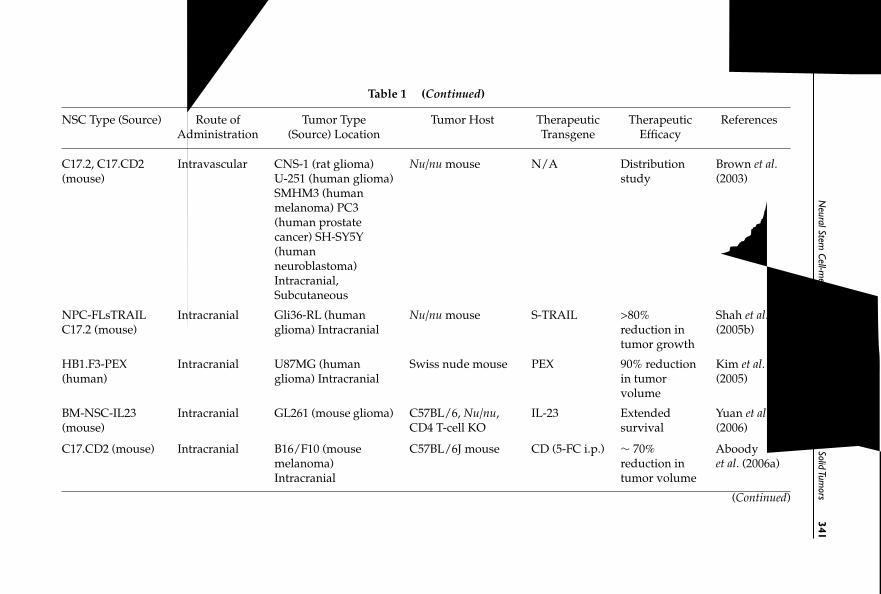
October
30,200712:29
WSPC
/SPI-B
559:Autologous
&C
ancerStem
CellG
eneT
herapych13
FA
NeuralStem
Cell-mediated
TherapyofPrim
aryand
Metastatic
SolidTum
ors341
Table 1 (Continued)
NSC Type (Source) Route of Tumor Type Tumor Host Therapeutic Therapeutic ReferencesAdministration (Source) Location Transgene Efficacy
C17.2, C17.CD2(mouse)
Intravascular CNS-1 (rat glioma)U-251 (human glioma)SMHM3 (humanmelanoma) PC3(human prostatecancer) SH-SY5Y(humanneuroblastoma)Intracranial,Subcutaneous
Nu/nu mouse N/A Distributionstudy
Brown et al.(2003)
NPC-FLsTRAILC17.2 (mouse)
Intracranial Gli36-RL (humanglioma) Intracranial
Nu/nu mouse S-TRAIL >80%reduction intumor growth
Shah et al.(2005b)
HB1.F3-PEX(human)
Intracranial U87MG (humanglioma) Intracranial
Swiss nude mouse PEX 90% reductionin tumorvolume
Kim et al.(2005)
BM-NSC-IL23(mouse)
Intracranial GL261 (mouse glioma) C57BL/6, Nu/nu,CD4 T-cell KO
IL-23 Extendedsurvival
Yuan et al.(2006)
C17.CD2 (mouse) Intracranial B16/F10 (mousemelanoma)Intracranial
C57BL/6J mouse CD (5-FC i.p.) ∼ 70%reduction intumor volume
Aboodyet al. (2006a)
(Continued)
干细胞之家www.stemcell8.cn ←点击进入

October
30,200712:29
WSPC
/SPI-B
559:Autologous
&C
ancerStem
CellG
eneT
herapych13
FA
342J.N
ajbaueretal.
Table 1 (Continued)
NSC Type (Source) Route of Tumor Type Tumor Host Therapeutic Therapeutic ReferencesAdministration (Source) Location Transgene Efficacy
HB1.F3.CD(human)
Intratumoral Daoy (humanmedulloblastoma)Intracranial
Nu/nu mouse CD (5-FC i.p.) 76% reduction intumor volume
Kim et al.(2006a)
HB1.F3.rCE(human)
Intravascular(tail vein)
NB-1691, NB-1643,SK-N-AS (humanneuroblastoma)Disseminated
Es1e SCID mice CE (CPT-11 i.v.) 100% tumor-freesurvival for >6months
Aboodyet al. (2006b)
HB1.F3.rCE(human)
Intravascular(tail vein)
NB-1691, NB-1643,SK-N-AS (humanneuroblastoma)Disseminated
Es1e SCID mice CE (CPT-11 i.v.) 90% tumor-freesurvival for 1year
Danks et al.(2007)
HB1.F3. IFN-β(human)
Intravascular(tail vein)
NB-1691 (humanneuroblastoma)Disseminated
Es1e SCID mice IFN-β (i.v.) Significant tumorreduction/Slowed diseaseprogression
Dicksonet al. (2007)
HB1.F3 (human) Intracranial U251, D566 (humanglioma) Intracranial
Nu/nu mouse N/A Distributionstudy
Lin et al.(2007)
Abbreviations: 5-FC, 5-fluorocytosine; 5-FU, 5-fluorouracil; BM-NSC, bone marrow-derived neural stem-like cells; CD, cytosine deaminase; i.p., intraperitoneal;i.v., intravenous; IFN-β, interferon-β; PEX, a naturally-occurring fragment of human metalloproteinase-2; TRAIL, tumor necrosis factor-related apoptosis-inducing ligand; S-TRAIL, secreted TRAIL; CPT-11, irinotecan; rCE, rabbit carboxylesterase (CPT-11–activating enzyme).
干细胞之家www.stemcell8.cn ←点击进入

October 30, 2007 12:29 WSPC/SPI-B559: Autologous & Cancer Stem Cell Gene Therapy ch13 FA
Neural Stem Cell-mediatedTherapy of Primary and Metastatic SolidTumors 343
by adenoviral transduction (Aboody et al., 2006b; Danks et al., 2007); (4) awell-characterized, tested parental line can be engineered to express a vari-ety of therapeutic gene products that can then be used sequentially or incombination to hit multiple tumor targets for enhanced treatment strate-gies; and (5) clonal expandability allows for the generation of a Master CellBank and the development of an off-the-shelf treatment, which would sig-nificantly decrease costs and increase availability for patient trials.
In recent studies, the c-myc gene (cellular homolog of v-myc) has beenshown to contribute to long-term maintenance of the stem cell phenotype(Cartwright et al., 2005; Takahashi and Yamanaka, 2006), suggesting that itserves as a stemness gene and that v-myc introduced into primary neuralstem/progenitor cells may similarly act as a stemness gene, leading to thelong-term maintenance of stem cell characteristics of immortalized NSCssuch as the human HB1.F3 cells (see below).
Of significance, immortalized NSCs have emerged as a highly effectivesource for ex vivo manipulation, and for transplantation into various focaland global CNS disease models for delivery of therapeutic gene productsand/or integration into damaged or developing host brain (Renfranz et al.,1991; Snyder et al., 1992; Martinez-Serrano and Bjorklund, 1997; Flax et al.,1998). The current literature demonstrates the use of v-myc–immortalizedNSCs both as a vehicle to target therapeutic agents to tumors (Aboody et al.,2000; Brown et al., 2003; Kim et al., 2005; Aboody et al., 2006b) and for regen-erative purposes in pre-clinical models of Huntington’s disease (Ryu et al.,2004; Goldman, 2005; Lee et al., 2005 and 2006; Kim, 2007), Parkinson’s dis-ease (Ourednik et al., 2002; Kim, 2004; Goldman, 2005; Ryu et al., 2005; Kimet al., 2006b), cerebral ischemia (Jeong et al., 2003; Chu et al., 2004; Imitolaet al., 2004; Kim, 2004; Muller et al., 2006) and spinal cord injury (Kim et al.,2002; Goldman, 2005; Sykova and Jendelova, 2005; Kim, 2007).
1.3.2. Disadvantages of primary (non-immortalized/clonal) NSC pools
Our own culture studies of NSCs/neurospheres derived from human fetalbrain tissue indicate that continuous passage over time results in decreasedproliferative capacity and increased terminal differentiation into neuronsor glial cells (S. Kim, unpublished data). In addition, primary pools areproblematic for clinical applications because: (1) they show drift of cell pop-ulation, proliferation rate and differentiation ability over time and acrossconditions, making them difficult to characterize and necessitating a con-stant replenishment source of cells/tissue and retesting; (2) individual cellcharacteristics are unstable over passage and time, with cell population drift
干细胞之家www.stemcell8.cn ←点击进入

October 30, 2007 12:29 WSPC/SPI-B559: Autologous & Cancer Stem Cell Gene Therapy ch13 FA
344 J. Najbauer et al.
adding undesired variability to pre-clinical studies; and (3) instability of cellpools and limited expansion make them impractical for generating a MasterCell Bank.
1.3.3. Disadvantages of autologous stem cells
Like primary NSCs, the use of autologous stem cells has a number of disad-vantages, including: (1) they have questionable availability and time courseto expand autologous cultures; (2) identification and testing of autologousstem cell sources that will efficiently target tumors has not been done; (3)these cells have limited expansion capabilities in cultures; (4) autologouscells may contain the genetic defect that originally facilitated tumor devel-opment; and (5) this type of therapy encompasses considerably higher costsper treatment, and would be less widely available.
1.3.4. HB1.F3 NSCs as potential off-the-shelf human tumor-homing cell line
We suggest that using a stable, sustainable and easily expandable clonal NSCline will circumvent the problems associated with characterization, senes-cence and replenishment sources of primary stem cell pools. It would alsoallow for cost-effective and accessible patient trials. At the very least, theymay serve as standard NSC lines in proof-of-concept pre-clinical studies,where tumor types and therapeutic regimens are being tested.
One such immortalized human NSC line (HB1.F3), derived by v-mycimmortalization of human fetal telencephalon cells, has been extensivelystudied and characterized. With various genetic modifications, these NSCshave demonstrated selective tumor localization (to invasive tumor cells anddistant tumor microfoci) and/or therapeutic efficacy in pre-clinical mod-els of glioma (Kim et al., 2005), medulloblastoma (Kim et al., 2006a), dis-seminated neuroblastoma (Aboody et al., 2006b; Danks et al., 2007), andbreast carcinoma (Aboody et al., unpublished data). In all invasive andmetastatic solid tumor models, 70–90% therapeutic efficacy was achievedas measured by increased long-term survival or decreased tumor burden.HB1.F3 NSCs retain their tumor-tropic property when modified to expressvarious transgenes. Because of their highly specific tumor-targeting abil-ity, HB1.F3 and other clonal NSC lines hold great promise for successfullocalized therapy for glioma and other malignant cancer patients. Withinthe framework of carefully designed and monitored clinical trials, the likelybenefits appear much greater than the potential risk factors. This has yet to beshown.
干细胞之家www.stemcell8.cn ←点击进入

October 30, 2007 12:29 WSPC/SPI-B559: Autologous & Cancer Stem Cell Gene Therapy ch13 FA
Neural Stem Cell-mediatedTherapy of Primary and Metastatic SolidTumors 345
2. Literature Summary
We and others have used NSCs that have been modified to express a varietyof therapeutic transgenes to target tumors in numerous pre-clinical models.These experimental trials are summarized in Table 1.
3. Generation of Immortalized Human HB1.F3 NeuralStem Cells
Human fetal telencephalon (15-week gestation) brain cells were dissoci-ated into single cells through trypsin treatment and grown in serum-freemedium consisting of Dulbecco’s modified Eagle medium (DMEM) sup-plemented with insulin, transferrin, selenium, hydrocortisone, triiodothy-otrophin, antioxidants and basic fibroblast growth factor (Ryu et al., 2003;Kim et al., 2004). Neurospheres formed within three days in culture, com-prised of small aggregates of cells including neurons, astrocytes and neuralstem/progenitor cells. Within seven days in culture, many neurospheresadhered to the surface of the plastic culture dish, displaying extensiveoutgrowth of neuronal or glial processes. Neurons and neural stem cellsmigrated from the neurosphere core and grew in a monolayer on top ofcellular sheets of astrocytes. An amphotropic replication-incompetent retro-viral vector encoding v-myc, pLSN.v-myc, was used to infect human telen-cephalon cells [Fig. 2(A)]. This method was previously used to generatev-myc-immortalized, clonal murine NSC lines (Snyder et al., 1997 and 1992;Ryder et al., 1990). Following G418 selection, large clusters of cells wereindividually isolated and grown in poly-L-lysine coated dishes [Fig. 2(B)].Further clonal selection by limited dilution yielded several propagated NSClines, including HB1.F3 and HB1.F5, used in our studies. The HB1.F3 cloneexpressed v-myc, as detected by RT-PCR and Western blotting [Fig. 2(C)],and displayed a normal 46XX karyotype [Fig. 2(D)]. Gene expression analy-sis of HB1.F3 human NSCs, carried out by RT-PCR, showed mRNAs forABCG2 and nestin, type-specific markers of NSCs. In addition, HB1.F3cells expressed specific receptors involved in cellular migration includingCXCR4, VEGFR-1, VEGFR-2, c-Kit and EGFR (Lee et al., 2005).
HB1.F3 cells appear to replicate for a maximum of∼48 h following in vivoadministration, as assessed by immunostaining for Ki-67, PCNA and BrdU.Downregulation of the NSC propagating gene product v-myc occurs con-stitutively and spontaneously, becoming undetectable 48 h following in vivoengraftment (Flax et al., 1998). These data suggest that v-myc is regulatedby the normal developmental mechanisms that downregulate endogenous
干细胞之家www.stemcell8.cn ←点击进入

October 30, 2007 12:29 WSPC/SPI-B559: Autologous & Cancer Stem Cell Gene Therapy ch13 FA
346 J. Najbauer et al.
Fig. 2. Isolation of human NSC line HB1.F3. (A) HB1.F3 cells were immortalized usinga retrovirus vector (pLSN.v-myc, modified from pLCNX, which was purchased fromClonetech) carrying the v-myc gene. Cells were selected for neomycin resistance. The pro-ducer line was PA317. (B) HB1.F3 cells shown under phase contrast microscopy. (C) v-mycexpression was confirmed by RT-PCR and Western blotting. (D) The karyotype displayednormal 46 XX chromosomes. Reproduced from: Kim, Neuropathology, 24: 159–171 (2004) withpermission of the Japanese Society of Neuropathology and Blackwell Publishing.
cellular myc in CNS precursors during mitotic arrest and/or differentiation.The loss of v-myc expression from murine and human NSC lines follow-ing intraparenchymal or intravascular administration is consistent with theabsence of tumors in short- and long-term studies in rodents (Danks et al.,2007; Aboody et al., 2006a and 2006; Kim et al., 2006a and 2005; Lee et al.,2005; Jeong et al., 2004; Chu et al., 2004; Snyder et al., 1992) and primates(Redmond et al., 2007).
4. Introduction ofTherapeutic Genes into Neural Stem Cells
NSCs can be engineered to express various anti-tumor gene products,including prodrug-activating enzymes, apoptosis-inducing or cell differ-entiating agents, cell cycle modulators, anti-angiogenesis factors, immune-enhancing agents and oncolytic factors. Additionally, these engraftable
干细胞之家www.stemcell8.cn ←点击进入

October 30, 2007 12:29 WSPC/SPI-B559: Autologous & Cancer Stem Cell Gene Therapy ch13 FA
Neural Stem Cell-mediatedTherapy of Primary and Metastatic SolidTumors 347
migratory NSCs may also be engineered to serve as viral vector producercells, allowing extended delivery of lethal virus-mediated genes to tumorcells. When administered into the tumor-bearing animal with any of thesestrategies, NSCs can target their therapeutic “payload” selectively to inva-sive tumor sites, thereby increasing localization of therapy and minimizingexposure to normal tissues.
4.1. Cytosine Deaminase
The principal advantage of a directed enzyme/prodrug approach com-pared to conventional chemotherapy is its potential to achieve tumor cell-selective cytotoxicity. One example of this therapeutic strategy uses NSCsmodified to express the prodrug-activating enzyme E. coli cytosine deam-inase (HB1.F3.CD). This CD-expressing NSC line was generated from thev-myc-immortalized HB1.F3 parental line, using an amphotropic replication-incompetent retroviral vector encoding E. coli cytosine deaminase (CD; EC3.5.4.1), transcribed from the retrovirus LTR plus puromycinR transcribedfrom an internal SV40 early promoter. Following puromycin selection andcloning by limited dilution, selected CD-expressing NSC lines were estab-lished and expanded for further characterization. The HB1.F3.CD21 line hasbeen used in most of our studies. This line stably expresses the functionalCD enzyme, which converts 5-fluorocytosine (5-FC) into 5-fluorouracil (5-FU), a pyrimidine analog that acts as an anti-neoplastic anti-metaboliteby interfering with DNA synthesis, blocking the thymidylate synthetaseconversion of deoxyuridylic acid to thymidylic acid (Fig. 3). Expression ofCD in HB1.F3.CD cells was confirmed by FACS and immunofluorescencemicroscopy, and the karyotype was also tested for chromosomal stability(Aboody et al., unpublished data). The HB1.F3.CD NSCs retain their inher-ent tumor-tropic properties, thereby localizing the prodrug-activating CDenzyme to primary and metastatic tumor sites (Fig. 4). Systemically admin-istered 5- FC crosses the blood–brain barrier, is converted to the activecytotoxic 5-FU by CD expressed in the NSCs, and readily diffuses out toselectively kill surrounding, dividing tumor cells (Fig. 4). This CD/5-FCprodrug strategy is known to have a large bystander effect (Huber et al.,1994). In effect, the NSCs serve as a platform for the production of tumor-localized chemotherapy. This allows the achievement of concentrated druglevels selectively at tumor sites, minimizing toxicity associated with sys-temic admistration. In regards to brain tumors, this strategy circumventsthe inability of 5-FU and other therapeutic agents to cross the blood–brainbarrier (see Sec. 6 for in vivo results).
干细胞之家www.stemcell8.cn ←点击进入

October 30, 2007 12:29 WSPC/SPI-B559: Autologous & Cancer Stem Cell Gene Therapy ch13 FA
348 J. Najbauer et al.
Fig. 3. Conversion of 5-fluorocytosine prodrug to 5-fluorouracil anti-cancer drug by cyto-sine deaminase. The Km value of the E. coli CD enzyme for 5-FC is 17.9 mM (Springer andNiculescu-Duvaz, 2000; Ireton et al., 2002).
Fig. 4. Production of 5-FU chemotherapeutic agent localized to immediate vicinity of stemcells. NSCs expressing the enzyme E. coli cytosine deaminase (HB1.F3.CD) migrate to tumorsites. Systemically administered prodrug 5-FC crosses the blood–brain barrier and is con-verted to 5-FU by the CD in the NSCs. 5-FU and its toxic metabolites readily diffuse out of thecell to selectively kill dividing tumor cells. This CD enzyme/5-FC prodrug strategy is knownto have a large bystander effect, predicting that one NSC can kill off many surrounding tumorcells.
4.2. Carboxylesterase
Another enzyme/prodrug strategy, useful in neuroblastoma and perhapsother metastatic solid tumor models, uses intravascularly administeredNSCs engineered to express the activating enzyme rabbit carboxylesterase(rCE), which efficiently converts the prodrug CPT-11 (irinotecan) to theactive metabolite SN-38, a potent topoisomerase I inhibitor (Fig. 5). Usinga disseminated neuroblastoma mouse model, we postulated that rCE-secreting NSCs (HB1.F3.AdCMVrCE) would localize selectively to dissem-inated tumor foci throughout the body. The secreted rCE enzyme at tumor
干细胞之家www.stemcell8.cn ←点击进入

October 30, 2007 12:29 WSPC/SPI-B559: Autologous & Cancer Stem Cell Gene Therapy ch13 FA
Neural Stem Cell-mediatedTherapy of Primary and Metastatic SolidTumors 349
Fig. 5. Metabolic activation of CPT-11 to SN-38 (a potent topoisomerase inhibitor) byesterases (Guichard et al., 1998; Pommier, 2006). Rabbit carboxylesterase (rCE) and its Kmfor CPT-11 are shown.
Fig. 6. HB1.F3.AdCMVrCE neural stem cells migrate to tumor sites and secrete rabbitcarboxylesterase (rCE). The secreted rCE is a prodrug-activating enzyme produced locallyin the tumor by stem cells which have accumulated in the tumor. The CPT-11 prodrug isthen administered systemically, and becomes converted to the active cytotoxic drug SN-38by rCE locally in the tumor tissue. SN-38 is a potent topoisomerase inhibitor that selectivelykills surrounding dividing tumor cells.
sites activates systemically administered CPT-11 to the highly chemotoxicagent SN-38, leading to selective killing of dividing cancer cells (Fig. 6) (seeSec. 6b for in vivo results).
Replication-deficient adenovirus expressing a secreted form of rabbitcarboxylesterase (rCE; EC 3.1.1.1) under the control of the cytomegalovirus(CMV) promoter (AdCMVrCE) was constructed using standard methods, asdescribed previously (Danks et al., 1998; Potter et al., 1998; Danks et al., 1999).HB1.F3 cells were co-cultured with AdCMVrCE for 24 h prior to intravenousinjection. The enzyme activity assay used to quantitate the level of expres-sion of rCE by HB1.F3 cells transduced with adenovirus, and the enzymaticmechanisms of CPT-11 prodrug activation (Fig. 5) have been reported pre-viously (Guichard et al., 1998; Danks et al., 1999; Pommier, 2006).
干细胞之家www.stemcell8.cn ←点击进入

October 30, 2007 12:29 WSPC/SPI-B559: Autologous & Cancer Stem Cell Gene Therapy ch13 FA
350 J. Najbauer et al.
4.3. OtherTherapeutic Agents
4.3.1. Interleukins
The genes of various cytokines have been introduced into NSCs totreat experimental brain tumors in mice and rats. SV40 large T antigen-immortalized primary mouse NSCs were transduced with the gene forinterleukin 4 (IL-4) (Benedetti et al., 2000), a cytokine that promotesT-cell–mediated immune response against tumor cells (Faber et al., 2000).In another study, C57BL6J mouse primary NSCs were retrovirally trans-duced to express the IL-4 gene and these NSCs were used to treat micewith established syngeneic GL261 brain glioblastomas (Benedetti et al.,2000). This treatment led to a 90-day survival of > 80% of mice whentumor cells were co-injected with IL-4–secreting NSCs, and 70% of micesurvived when IL-4–secreting NSCs were injected into mice with alreadyestablished tumors (all tumor-bearing, untreated mice died by day 30). Sim-ilar results were obtained by implanting immortalized NSCs derived fromSprague–Dawley rats into C6 glioblastomas (Benedetti et al., 2000). IL-12–secreting NSCs that had been derived from the fronto-parietal region ofembryonic day 15 mice and transduced with adenovirus vector carrying anIL-12 expression cassette were used for treatment of glioblastoma-bearingmice. The treatment with NSC–IL-12 cells led to significantly prolongedsurvival and resulted in long-term anti-tumor immunity, as compared toanimals treated with nonmodified NSCs (Jean et al., 1998; Ehtesham et al.,2002a). Bone marrow-derived neural stem-like cells (BM-NSCs) were used toexpress and deliver interleukin-23 (IL-23), which showed protective effectson intracranial tumor-bearing C57BL/6 mice (Yuan et al., 2006). Depletionof CD8+ T cells showed that this subpopulation of lymphocytes was criticalfor the anti-tumor immunity elicited by IL-23–expressing BM-NSCs, andthat CD4+ T cells and natural killer cells also participated in the anti-tumoractivity. Importantly, the IL-23–expressing BM-NSC–treated survivors wereresistant to tumor re-challenge associated with enhanced IFN-gamma, butnot IL-17, expression in the brain tissue. These data suggest that IL-23–expressing BM-NSCs can induce anti-tumor immunity against intracranialgliomas.
4.3.2. TRAIL
TRAIL (TNF-α-related apoptosis-inducing ligand, or APO-2L) is a memberof the TNF ligand family (Pitti et al., 1996). TRAIL is trimeric and binds to
干细胞之家www.stemcell8.cn ←点击进入
October 30, 2007 12:29 WSPC/SPI-B559: Autologous & Cancer Stem Cell Gene Therapy ch13 FA
Neural Stem Cell-mediatedTherapy of Primary and Metastatic SolidTumors 351
DR4 or DR5 death receptors, and induces their trimerization which leads torecruitment of the death-inducing signaling complex (DISC). Formation ofDISC induces activation of caspases and downstream intrinsic death signal-ing pathways (Walczak and Krammer, 2000). TRAIL can induce apoptosisin numerous tumor cells, including gliomas (Pollack et al., 2001; Shah et al.,2003), while being relatively nontoxic to most normal cells (Ashkenazi et al.,1999); thus, it has attracted great interest as a cancer therapeutic agent. NSCsderived from embryonic day 15 mice were transduced with an adenovi-ral expression vector for human TRAIL, and were used in experiments onintracranial glioma (Ehtesham et al., 2002b). Inoculation of human glioblas-toma xenografts with TRAIL-secreting NSCs resulted in dramatic inductionof apoptosis in treated tumors and tumor satellites, and was associated withsignificant inhibition of tumor growth (Ehtesham et al., 2002b). For more effi-cient protein delivery, a secretable version of TRAIL(S-TRAIL) was designedand introduced into NSCs to deliver S-TRAIL directly to invasive tumorcells (Shah et al., 2004). For monitoring of highly malignant gliomas andmigration of NSCs, cell lines were also engineered with luminescent andfluorescent transgenes for dual bioluminescence imaging (Shah et al., 2005aand 2005b). A highly malignant human glioma model expressing Renillaluciferase was used. Intracranially implanted NSCs expressing both fireflyluciferase and S-TRAIL were shown to migrate into the gliomas and havesignificant anti-tumor effects (>80% reduction in tumor growth) (Shah et al.,2005a). More recent results show that simultaneous triggering of the TRAIL-mediated death receptor pathway and downregulation of Bcl-2 by shRNAleads to highly enhanced eradication of gliomas in experimental animals(Kock et al., 2007).
4.3.3. PEX
The human metalloproteinase-2 fragment, PEX, acts as an inhibitor of gliomaand endothelial cell proliferation, migration and angiogenesis (Brooks et al.,1998; Bello et al., 2001). The anti-tumor activity of PEX-producing humanNSCs against malignant glioma was investigated using HB1.F3 cells inmouse models of human glioma. HB1.F3-PEX cells labeled with CM-DiImigrated to the tumor boundary and caused 90% reduction of tumor volume(Kim et al., 2005). The reduction in tumor volume in HB1.F3-PEX–treated ani-mals was associated with a decrease in angiogenesis and cell proliferation,supporting the use of NSCs as a delivery vehicle for targeting therapeuticgenes to human gliomas.
干细胞之家www.stemcell8.cn ←点击进入

October 30, 2007 12:29 WSPC/SPI-B559: Autologous & Cancer Stem Cell Gene Therapy ch13 FA
352 J. Najbauer et al.
4.3.4. Interferon-β
NSC-mediated interferon-β therapy has been investigated in aminal modelsof human neuroblastoma (Dickson et al., 2007). Disseminated neuroblastomawas established in SCID mice by tail vein injection of tumor cells. Mice weretreated by intravenous injection of human HB1.F3 NSCs which had beentransduced with a replication-deficient adenoviral vector to express hIFN-β(HB1.F3–IFN-β). Control mice received NSCs transduced with empty vectoradenovirus. Progression of disease was monitored using bioluminescenceimaging, which demonstrated a significant decrease in disease progressionin mice receiving HB1.F3–IFN-β cells. Necropsy of control mice revealedbulky tumor in multiple organs, while the organs of mice receiving HB1.F3–IFN-β therapy appeared normal with the exception of small residual tumornodules within the kidneys. Importantly, systemic levels of hIFN-β in thetreated mice remained below detectable levels, suggesting that the ther-apeutic effect was due to hIFN-β secreted by NSCs at tumor sites. ThisNSC-mediated IFN-β therapy may circumvent limitations associated withsystemic toxicity of IFN-β.
5. Tropism of Neural Stem Cells toTumors In vitro
5.1. HB1.F3 NSCs Migrate toTumor-conditioned Medium
The migratory capabilities of the immortalized human HB1.F3 line wasexamined in response to conditioned medium from human glioma (U251,U251.eGFP, U251.ffluc), breast carcinoma (MCF7, MDA) and neuroblastoma(SK-N-AS) cell lines. Modified Boyden chamber assays were used to assessthe in vitro tropism of HB1.F3 cell lines to tumor cell-conditioned media or tocontrol media, using standard methods. Briefly, HB1.F3 cells were detachedby trypsinization, washed and resuspended in DMEM containing 5% BSA.NSCs (3 × 10⁴ cells/100 µL) were placed in upper chambers and tumor cell-conditioned medium in lower chambers (10% FCS was used as positivecontrol for chemoattraction). Tumor cell-conditioned medium in both theupper and lower chambers comprised the chemokinesis control without achemoattractant gradient. Cells were allowed to migrate for 4 h in a cell cul-ture incubator at 37˚C and 6% CO2. The number of migrated HB1.F3 cellsin the lower chamber of each well was quantitated using CyQuant GR flu-orescent dye (Chemicon) and a fluorescence microplate reader (MolecularDevices, Sunnyvale, CA). Assays were performed in triplicate.
Tumor-conditioned media from all tumor cell lines investigated (U251,SK-N-AS, MCF7, MDA) significantly stimulated the directional migration of
干细胞之家www.stemcell8.cn ←点击进入

October 30, 2007 12:29 WSPC/SPI-B559: Autologous & Cancer Stem Cell Gene Therapy ch13 FA
Neural Stem Cell-mediatedTherapy of Primary and Metastatic SolidTumors 353
Fig. 7. NSC migration to tumor-conditioned media. HB1.F3 cell migration in response toconditioned media from human tumor lines, including U251 glioma, U251.eGFP, U251.ffluc,SK-N-AS neuroblastoma, MCF7 and MDA breast carcinoma. Note the lack of migration ofcontrol 3T3 fibroblasts (Aboody et al., unpublished data).
the NSCs compared to 3T3 cells (Fig. 7). In control chemokinesis assay, min-imal cell migration was detected, confirming NSC migration due to chemo-taxis (data not shown).
5.2. Neural Stem CellsTransduced withTherapeutic or ReporterGenes RetainTheirTumor-tropic Properties
Insertion of the E. coli cytosine deaminase gene into the HB1.F3 parental linewas successfully achieved by retroviral transduction. Directed migration inresponse to conditioned media from human tumor cell lines (UPN029, U251,U87 and U87ffLuc) was assessed in 96-well Boyden chamber assays (Fig. 8).The results demonstrated that insertion of the CD transgene into NSCs hasno significant effect on migratory and tumor-tropic properties, supportingtheir use for delivery of CD directly to tumor sites.
Furthermore, HB1.F3 cells transduced with adenovirus carrying the rCEprodrug activating enzyme gene (MOI of 0, 5, 10 or 20 AdCMVrCE), orlentivirus carrying the eGFP reporter gene, also showed retention of tumor
干细胞之家www.stemcell8.cn ←点击进入

October 30, 2007 12:29 WSPC/SPI-B559: Autologous & Cancer Stem Cell Gene Therapy ch13 FA
354 J. Najbauer et al.
Fig. 8. NSCs retain their tropism for tumor-conditioned media (Boyden chamber assays).(A) HB1.F3 at passage 12 (dark red) and HB1.F3.CD cells (green; retroviral transduction)at p20 retained their migratory properties to media conditioned by a variety of glioma celllines (UPN029, U251 and U87+/− ffLuc). (B) Migratory properties of HB1.F3 cells (red) wereretained following lentiviral transduction with a reporter gene (HB2.F3.eGFP; green) or ade-noviral transduction with a therapeutic gene encoding the prodrug activating enzyme rCE(HB1.F3.AdCMVrCE; olive). Control 3T3 fibroblasts (blue) showed little or no migration.
tropism in Boyden chamber migration assays to U251 glioma and MDAbreast carcinoma cell-conditioned media [Fig. 8(B)].
6. NSC-Mediated CancerTherapy: Pre-ClinicalTumor Models
6.1. BrainTumor Models
6.1.1. Glioma
Treatment failure of gliomas is largely attributable to the inability of cur-rent strategies to address the diffuse and highly infiltrative nature ofthese tumors. Due to extensive dissemination of malignant glial cells awayfrom the primary site, there is no clear border between normal brain andtumor edge, with invasive cells and distant microfoci proving exceptionallydifficult to treat with surgical and radiotherapies. Drug delivery has beenineffective due to the inability of many drugs to cross the blood–brain bar-rier and/or poor diffusion of drugs within the brain and within the tumor.In addition, systemic toxicity of traditional chemotherapeutic agents lim-its their usefulness. An inability to deliver therapeutic agents with sufficientdistribution throughout primary and distant tumor sites limits the effective-ness of gene therapy. Immunotherapeutic strategies have also failed to showsignificant impact at the clinical level, largely due to their inability to erad-icate disseminated tumor foci throughout the brain (Ehtesham et al., 2005).Despite aggressive treatments, the recurrence rate for glioma is 98%, with
干细胞之家www.stemcell8.cn ←点击进入

October 30, 2007 12:29 WSPC/SPI-B559: Autologous & Cancer Stem Cell Gene Therapy ch13 FA
Neural Stem Cell-mediatedTherapy of Primary and Metastatic SolidTumors 355
80% occurring within 2 cm of the resection cavity and with a median sur-vival of 3–6 months. To improve the clinical outcome, novel therapies mustaddress the invasive tumor cells that escape currently available treatments,while limiting toxicity to normal tissues.
In vitro tumor cell killing by NSCs expressing the prodrug activating enzyme CD.We determined the sensitivity of the U87 human glioma cell line to the 5-FU anti-cancer drug and 5-FC prodrug in vitro. The half-maximal killingeffect was detected at ∼ 5 µg/mL 5-FU whereas 500 µg/mL of 5-FC wasnecessary to achieve the same effect, showing the high potency of 5-FUcompared to its precursor prodrug 5-FC (MTT assay). When U87 gliomacells were co-cultured in vitro at increasing ratios of murine NSCs express-ing the active CD enzyme (C17.2.lacZ.CD), increased tumor cell killing wasobserved in the presence of 5-FC prodrug, but not in its absence. How-ever, only 5% of C17.2.lacZ.CD stem cells were necessary to induce a tumorcell growth inhibition of ∼50%, indicating a strong bystander effect of CD-expressing NSCs (Schmidt et al., unpublished data). We observed a similartumor cell killing effect by HB1.F3.CD human NSCs when co-cultured withU251 human glioma cells in the presence of 5-FC prodrug (Aboody et al.,unpublished data).
Therapeutic effectiveness of tumor-homing NSCs expressing the CD transgene.We then investigated the in vivo therapeutic effectiveness of glioma-targetingNSCs expressing CD to convert systemically administered prodrug 5-FC to5-FU. 5-FU and its toxic metabolites diffused out of the NSCs to selectivelykill dividing tumor cells in their vicinity (Fig. 4). Murine C17.2.lacZ.CDNSCs (or C17.2.lacZ as a control) were stereotactically implanted distant toestablished orthotopic U87 glioma xenografts in adult nude mice. Systemictreatment with 5-FC (500 mg/kg/day i.p. ×2 weeks) was initiated three daysafter implantation of NSCs (Fig. 9).
Intracerebral implantation of C17.2.lacZ.CD cells caudal-lateral to thetumor site, followed by systemic administration of 5-FC, significantly inhib-ited tumor growth (>65%) relative to controls (CD-expressing NSCs only,control NSCs +5-FC, tumor only) as assessed by magnetic resource imaging(MRI) 14 days after the beginning of treatment (Fig. 10). The anti-tumor effectof NSC-mediated, tumor-localized CD enzyme/5-FC prodrug chemother-apy resulted in a significant increase of survival of treated glioma-bearingmice (Schmidt et al., unpublished data). These results support the use ofNSCs as a novel tumor-selective delivery vehicle to target therapeutic geneproducts to invasive glioma foci.
干细胞之家www.stemcell8.cn ←点击进入

October 30, 2007 12:29 WSPC/SPI-B559: Autologous & Cancer Stem Cell Gene Therapy ch13 FA
356 J. Najbauer et al.
Fig. 9. Therapeutic homing and anti-glioma activity of CD-NSCs. Murine C17.2.lacZ.CD(expressing CD) or control C17.2.lacZ (no CD) NSCs were stereotactically implanted distantto established orthotopic U87 human glioma xenografts in adult nude mice. Systemic treat-ment with 5-FC at 500 mg/kg/day started three days after NSC injection, and continued for14 days. (Schmidt et al., unpublished data).
Fig. 10. Tumor growth assessed by T1-weighted, gadolinium-enhanced magnetic reso-nance imaging. Note the significantly reduced hypodense signal (indicating reduced tumorsize) in representative mouse treated with C17.2.lacZ.CD NSCs and 5-FC prodrug (A), whencompared to brains of control mice (B–D) (Schmidt et al., unpublished data).
Visualization and modeling of spatial distribution of neural stem cells withinintracranial glioma. We used an orthotopic mouse model of human glioma,high-resolution fluorescent confocal microscopy, and three-dimensional(3D) modeling to develop an algorithm that allows the following
干细胞之家www.stemcell8.cn ←点击进入

October 30, 2007 12:29 WSPC/SPI-B559: Autologous & Cancer Stem Cell Gene Therapy ch13 FA
Neural Stem Cell-mediatedTherapy of Primary and Metastatic SolidTumors 357
analyses: (1) estimation of primary tumor boundary and centroid; (2) iden-tification of cancer cells and NSCs in the tumor; (3) determination of coordi-nates and 3D distribution of NSCs and (4) estimation of tumor volume thatwould be eradicated by NSCs (using a cell-killing radius) with a definedintratumoral and surrounding microfoci distribution. U251.eGFP humanglioma was established in the brain of athymic mice (for 1 or 2 weeks),followed by sterotactic injection of CM-DiI-labeled HB1.F3 NSCs posterior-lateral to the tumor site. This novel algorithm allowed 3D visualization oftumor cells and NSCs in and around the tumor site (Fig. 11). Furthermore,this model predicted that a single administration of NSCs would affect the
Fig. 11. 3D reconstructions show extent of NSC distribution in primary glioma mass. (a) 3DAMIRA model of aligned 2.5× serial images at 100 µm intervals. The NSC injection site (red)and tumor site (green) (inset) can be traced downward through the image series. Tumors wereestablished via orthotopic injection of U251.eGFP glioma cells, followed by posterior-lateralstereotactic injection of CM-DiI-labeled NSCs 7 days [Mouse A; (b–f)] or 14 days (MouseB) after implantation of U251.eGFP cells. (b) AMIRA surface reconstruction of tumor bulkin Mouse A showing NSCs (red), tumor cells (green) and nuclei (blue). (c) AMIRA voxelprojection of tumor in Mouse A showing NSCs (red) and tumor (green). (d–f) MATLABplots of tumor boundary and NSC locations in Animal A shown in various orientations;NSCs (red) and tumor mass (green). Based on Fig. 4 from: Lin et al., Neuroimage, Epub aheadof print (2007) with permission of Elsevier.
干细胞之家www.stemcell8.cn ←点击进入

October 30, 2007 12:29 WSPC/SPI-B559: Autologous & Cancer Stem Cell Gene Therapy ch13 FA
358 J. Najbauer et al.
vast majority (70–90%) of tumor cells within the primary mass and sur-rounding infiltrative tumor foci. This method may provide a quantitativeframework for projecting the therapeutic potential of a given NSC-expressedtransgene in glioma or other solid tumor treatment.
6.1.2. Medulloblastoma
Medulloblastoma is a malignant pediatric brain tumor that is incurable inabout 30% of patients despite multimodal therapies. The main cause ofdeath is recurrence associated with tumor dissemination, at which pointcurrent therapeutic options have little efficacy (Patrice et al., 1995; Grahamet al., 1997). Consequently, there is substantial need for novel, effective, low-toxicity therapies for children with medulloblastoma. Based on the inherenttropism of NSCs toward malignant gliomas (Aboody et al., 2000; Yip et al.,2003) and the secretion of growth factors and chemokines common to gliomaand medulloblastoma, we hypothesized that NSCs could also target thera-peutic gene products to medulloblastoma.
We investigated the migratory ability of HB1.F3.CD NSCs to medul-loblastoma both in vitro and in vivo. The migratory capacity of HB1.F3 cellswas confirmed by an in vitro cell migration assay, and then corroboratedin vivo by injecting CM-DiI–labeled HB1.F3 cells into the hemisphere con-tralateral to established medulloblastoma tumors in nude mice (Kim et al.,2006a). In vitro studies showed a tumor cell-killing effect of HB1.F3.CD NSCson Daoy medulloblastoma cells in co-culture experiments. For in vivo thera-peutic studies, animals bearing intracranial medulloblastoma were injectedipsilaterally with HB1.F3.CD cells followed by systemic 5-FC treatment. His-tologic analysis demonstrated HB1.F3.CD NSC migration to the tumor bedand its boundary and, following systemic 5-FC treatment, resulted in a 76%reduction of tumor volume compared to the control groups (P < 0.01) (Kimet al., 2006a). These studies showed for the first time the potential of humanNSCs as an effective delivery system to target and disseminate therapeuticagents to medulloblastoma.
6.1.3. Solid tumor metastases to brain: melanoma
The most common type of brain tumors are those that develop as distantmetastases from solid tumors in other organs, such as lung, prostate, breastor melanoma. Metastatic tumors to the brain occur 10 times more frequentlythan primary brain tumors (Hutter et al., 2003; Patchell, 2003). Malignantmelanoma is the third most common cause of intracranial metastases (Sahaet al., 1994). Each year, more than 100,000 patients in the United States are
干细胞之家www.stemcell8.cn ←点击进入

October 30, 2007 12:29 WSPC/SPI-B559: Autologous & Cancer Stem Cell Gene Therapy ch13 FA
Neural Stem Cell-mediatedTherapy of Primary and Metastatic SolidTumors 359
diagnosed with brain metastases; and although treatment for systemic dis-ease has been improving, drug delivery to brain metastases remains inef-fective. Although solitary metastasis can be surgically removed or treatedwith stereotactic radiosurgery, patients with multiple brain metastases havevery limited therapeutic options (Westphal et al., 2003). NSC-mediated ther-apy shows promise to overcome these current treatment limitations, dueto their inherent ability to migrate throughout the brain parenchyma andtarget tumor sites, including intracerebral solid tumor metastases.
Therapeutic paradigm for NSC-mediated CD enzyme/5-FC prodrug strategy ina syngeneic model of melanoma brain metastases. For our in vivo therapeu-tic paradigm, we established melanoma brain metastases by intracarotidinjection of B16/F10 melanoma cells in a syngeneic mouse model (Fig. 12).One week later, animals received rostral and caudal intracranial injectionsof murine C17.2.CD NSCs (Aboody et al., 2006a). On day 4 followingNSC injections, systemic 5-FC treatment was given for eight days. Resultsdemonstrated a significant reduction in resultant tumor burden (> 70%)in CD-expressing NSC treated animals, as compared to controls (non-CDexpressing NSCs +5-FC, tumor only) (Fig. 13). These data provide proofof principle for the use of NSCs for targeted delivery of therapeutic geneproducts to melanoma brain metastases.
Fig. 12. In vivo syngeneic experimental model of melanoma metastases to the brain. Thetrans-illuminated whole brain shows a wide range of sizes of pigmented melanoma metas-tases 19 days after injection of melanoma cells (arrows) into the carotid artery. Scale bar,2 mm. Based on Fig. 2 from: Aboody et al., Neuro-Oncol 8: 119–126 (2006) with permission ofthe Society for Neuro-Oncology.
干细胞之家www.stemcell8.cn ←点击进入

October 30, 2007 12:29 WSPC/SPI-B559: Autologous & Cancer Stem Cell Gene Therapy ch13 FA
360 J. Najbauer et al.
Fig. 13. In vivo schematic for therapeutic paradigm of C17.2.CD cells +5-FC for treatmentof brain metastases of melanoma. Tumor areas (outlined in red) in representative brains ofmice that (A) had received injections of melanoma cells only, and (B) had received injectionsof melanoma cells and were subsequently treated with CD-NSCs and 5-FC. Scale bar, 5 mm.(C) Quantitative analysis of tumor volume showing 71% and 69% less tumor burden in micethat had been treated with CD-NSCs and 5-FC when compared to control group 1 (tumoronly, no treatment) or control group 2 (tumor + NSCs + 5-FC), respectively (mean ± SD;Student’s t-test; n = 4 in each group). Tumor volumes were expressed as a percentage ofcontrol group 1 tumor (no treatment was taken as 100%). All mice were sacrificed 19 daysfollowing tumor injection. Based on Fig. 4 from: Aboody et al., Neuro-Oncol 8: 119–126 (2006)with permission of the Society for Neuro-Oncology.
6.2. Disseminated SolidTumor Models: Neuroblastoma
Neuroblastoma is the most common extracranial solid tumor in children.Typically, patients diagnosed with high-risk disease demonstrate a good ini-tial response to therapy, but as many as 80% of these patients relapse withmetastatic disease that is refractory to therapy. Like other solid tumors, whenneuroblastomas metastasize, they are very difficult to treat, and a major-ity of children with high-risk metastatic neuroblastoma die of their disease(Evans et al., 1994; Matthay et al., 1994; Brodeur, 2003). Current treatmentshave anti-tumor efficacy but also produce undesirable side effects to normaltissue, limiting the treatment dose that can be administered. Novel effective
干细胞之家www.stemcell8.cn ←点击进入

October 30, 2007 12:29 WSPC/SPI-B559: Autologous & Cancer Stem Cell Gene Therapy ch13 FA
Neural Stem Cell-mediatedTherapy of Primary and Metastatic SolidTumors 361
approaches for the treatment of neuroblastoma are needed. In the follow-ing studies, we exploited the tumor-tropic property of NSCs to selectivelylocalize to multiple solid tumor foci outside the brain, following intravas-cular administration. The specific goal of these studies was to determineif intravenous administration of HB1.F3 cells expressing a secreted formof the CPT-11 (irinotecan)-activating enzyme rCE (HB1.F3.AdCMVrCE; seeFig. 6) would significantly increase the anti-tumor effect of tolerated dosesof CPT-11 in mice bearing disseminated neuroblastoma tumors. Since noeffective treatments are available for most metastatic tumors, demonstratingthat stem or progenitor cells of fetal or adult origin can be used to improvethe prognosis of patients with fatal metastatic disease would be highly sig-nificant. The approach described might also be adapted to developing atreatment for patients with other types of metastatic solid tumors and/orresidual disease.
6.2.1. In vitro killing of neuroblastoma cells by NSCs secreting rabbit carboxylesterase
In order to determine whether the HB1.F3.AdCMVrCE cells are capableof killing neuroblastoma cells, we carried out in vitro cytotoxicity assays.HB1.F3.AdCMVrCE stem cells efficiently killed SK-N-AS.ffLuc neuroblas-toma cells in the presence of CPT-11 prodrug, with half-maximal cytototox-icity achieved at ∼ 0.1 µM CPT-11 (Fig. 14).
6.2.2. HB1.F3 NSCs target disseminated human neuroblastoma foci in liver,ovary and bone of SCID mice
To investigate whether HB1.F3 stem cells can target human neuroblastomatumors in vivo, we administered CM-DiI–labeled HB.F3 cells via tail veininjection to mice with established human NB1643 neuroblastoma tumors.Immunohistochemical analysis revealed significant homing of HB1.F3 cellsto multiple metastatic tumor sites, including micrometastases in the liver(Fig. 15), ovary and bone marrow. Note NSCs appearing to extravasateout of the blood vessels to infiltrate the surrounding tumor parenchyma[Fig. 15(E)]. No NSCs were found in non–tumor-bearing brain, kidney, heart,intestine, or skin, and were rarely found in non–tumor-bearing tissues (lung,liver and spleen) at 2–4 days post-injection (Aboody et al., 2006b).
6.2.3. Neural stem/progenitor cell-directed enzyme pro-drug therapy (NDEPT)
We postulated that HB1.F3.AdCMVrCE cells would preferentially local-ize to neuroblastoma tumor foci following intravascular injection. The
干细胞之家www.stemcell8.cn ←点击进入

October 30, 2007 12:29 WSPC/SPI-B559: Autologous & Cancer Stem Cell Gene Therapy ch13 FA
362 J. Najbauer et al.
Fig. 14. In vitro killing of neuroblastoma cells by NSCs secreting rabbit carboxylesterase.Neuroblastoma cells (SK-N-AS) were co-cultured with neural stem cells transduced withadenovirus to express the secreted form of rCE (HB1.F3.AdCMVrCE) or HB1.F3 parentalstem cell line at a 2:1 ratio of NSC-to-tumor cells in 96-well plated (100 µL/well). SKNAScultured without NSCs were included as controls. Following 24 h of culture, CPT-11 wasadded in 2× concentration (100 µL/well) to give a final of 1× concentration indicated abovein 200 µL/well total volume. Cells were incubated at 37◦C, 6% CO2 for an additional 48 to72 h. Sulforhodamine B (SRB) assay was performed to detect the total remaining cell mass.Optical density (OD) was measured at 570 mm.
rCE secreted into the extracellular milieu would then convert systemicallyadministered CPT-11 to its active form, producing high local extracellularconcentrations of SN-38 (up to 1000 times more toxic to tumor cells thanCPT-11 alone) (Fig. 16).
Therapeutic efficacy trials involved tail vein administration of rCEexpressing NSCs into neuroblastoma-bearing animals, followed by 5 daysof CPT-11 treatment and immediately followed by another round of NSCsand CPT-11. Following two weeks of treatment, the NSC/CPT-11 paradigmwas repeated and animals followed for long-term survival. Of great sig-nificance, animals receiving rCE-secreting NSCs +15 mg/kg/day CPT-11prodrug demonstrated 90% one-year survival compared to < 75 day sur-vival in animals with no treatment, and only 30% one-year survival ofanimals treated with the CPT-11 alone [Fig. 17(B)]. Importantly, in ourlong-term NSC-treated animals, we found no evidence of residual tumoror NSCs, suggesting a complete cure (Danks et al., 2007). Furthermore,plasma carboxylesterase activity and SN-38 levels in mice receiving bothrCE-expressing HB1.F3.AdCMVrCE cells and CPT-11 were comparable tothose in mice receiving CPT-11 only (Danks et al., 2007). These data sup-port the hypothesis that the anti-tumor effect of the described neural stemcell–directed enzyme prodrug therapy (NDEPT) is mediated by the produc-tion of high concentrations of active drug selectively at tumor sites, thereby
干细胞之家www.stemcell8.cn ←点击进入

October 30, 2007 12:29 WSPC/SPI-B559: Autologous & Cancer Stem Cell Gene Therapy ch13 FA
Neural Stem Cell-mediatedTherapy of Primary and Metastatic SolidTumors 363
Fig. 15. Intravascularly administered NSCs (HB1.F3) target human neuroblastoma livermetastases in SCID mice. (A) Dissected liver from a representative animal with hepaticmetastases. (B) Low- and (C) high-power magnification of a section of tumor-involved liver,stained with hematoxylin and eosin. Tumor cells appear dark purple (black arrows). (D)Liver section stained with an anti-human mitochondrial protein antibody and counterstainedwith hematoxylin. Tumor micrometastases stain dark brown (red arrows). (E) Immunoflu-orescence microscopy of liver tumor section. Tumor foci are identified by areas of denselypacked DAPI (blue)-stained tumor cell nuclei. The red arrows show extravasated CM-DiI(red)–labeled HB1.F3.cells proximal to a hepatic vein (v). Inset is high magnification of aCM-DiI–labeled HB1.F3 cell within the tumor. Scale bars: (A) 1 cm, (B) 2 mm, (C) 500 µm,(D,E) 100 µm and (E) inset 10 µm. Based on Fig. 3 from: Aboody et al., PLoS ONE 1: e23.(2006) with permission.
maximizing the anti-tumor an effect of CPT-11. NDEPT approaches meritfurther investigation as an effective, targeted therapy for metastatic tumors.We propose that the described approach may have the greatest use for erad-icating minimum residual disease.
6.2.4. NSC-mediated therapeutic efficacy in disseminated neuroblastoma model
With 1-year survival as the end point, the Kaplan–Meier plot shows aclear dose–response relationship for mice treated with 7.5 mg/kg CPT-11[Fig. 17(A)] compared with 15 mg/kg CPT-11 [Fig. 17(B)], both for micetreated with CPT-11 alone (10% survival versus 30%, respectively) and withCPT-11 as a component of NDEPT (50% versus 90%, respectively).
7. Factors Involved in NSCTumorTropism
Numerous cytokines and growth factors and their receptors have been iden-tified that affect NSC migration under normal and pathological conditions,
干细胞之家www.stemcell8.cn ←点击进入

October 30, 2007 12:29 WSPC/SPI-B559: Autologous & Cancer Stem Cell Gene Therapy ch13 FA
364 J. Najbauer et al.
Fig. 16. Schematic representation of neural stem/progenitor cell-directed enzyme pro-drug therapy (NDEPT). Mice bearing disseminated neuroblastoma receive i.v. injections ofHB1.F3 cells transduced to express a secreted form of rCE. These cells have been shown tomigrate selectively to tumors in vivo. The rCE at tumor sites systemically converts the givenCPT-11 to its active form, SN-38. High concentrations of SN-38 at tumor loci are cytotoxicto tumors, without additional toxic side effects. Of note, no detectable levels of SN-38 werefound in the blood. Based on Fig. 1 from: Danks et al., Cancer Res 67: 22–25 (2007) withpermission of the American Association for Cancer Research.
Fig. 17. Kaplan–Meier plots of tumor-bearing mice treated with HB1. F3.C1/AdCMVrCEand CPT-11. Mice (10 per group) were injected i.v. with 500,000 SK-N-AS cells to producedisseminated neuroblastoma (NB) tumors in 100% of mice. Two weeks later, mice weretreated with rCE/CPT-11 NDEPT by the following treatment protocol: week 1, 2 millionHB1.F3.C1 cells transduced with a MOI of 20 of AdCMVrCE followed by CPT-11 daily ×five i.v. at the indicated doses; week 2, repeat of week 1; weeks 3 and 4, no treatment; andweeks 5 and 6, same as weeks 1 and 2. The doses of CPT-11 given were (A) 7.5 mg/kg or (B)15 mg/kg. Based on Fig. 3 from: Danks et al., Cancer Res 67: 22–25 (2007) with permission ofthe American Association for Cancer Research.
干细胞之家www.stemcell8.cn ←点击进入

October 30, 2007 12:29 WSPC/SPI-B559: Autologous & Cancer Stem Cell Gene Therapy ch13 FA
Neural Stem Cell-mediatedTherapy of Primary and Metastatic SolidTumors 365
including cancer. Such agents include SDF-1/CXCR4 (Imitola et al., 2004;Kucia et al., 2004; Peng et al., 2004; Kucia et al., 2005), SCF/c-Kit (Erlandssonet al., 2004; Sun et al., 2004), HGF/c-Met (Heese et al., 2005), VEGF/VEGFR(Schmidt et al., 2005), MCP-1/CCR2 (Widera et al., 2004) and HMGB1/RAGE(Palumbo and Bianchi, 2004). Extracellular matrix proteins, especiallylaminin and tenascin have been shown to serve as strong modulators of NSCmigration (Ziu et al., 2006). Among transcription factors, hypoxia-induciblefactor-1 (HIF-1) plays a crucial role in the regulation/expression of manycytokines and their receptors in tumors (Semenza, 2003). Gliomas have beenextensively characterized with regard to the cytokines and growth factorsthey release, and stimulate the directed migration of exogenous and endoge-nous NSCs into the tumor microenvironment. There is evidence that cancercells and stem cells may share common molecular mechanisms used for celltrafficking (Kucia et al., 2005). It should be noted that in addition to gliomas,many cancers release cytokines that can serve as potent chemoattractantsfor stem cells, which suggests the feasibility of stem cell-mediated deliveryof therapeutic genes to tumors of various types. Clearly, the identificationand characterization of cancer-associated cytokines will be important for theoptimization of stem cell-mediated therapies.
8. NSC-mediatedTherapeutic Applications to Invasive andMetastaticTumors: Future Directions
The poor prognosis for patients with metastatic cancer and the toxic sideeffects of currently available treatments necessitates the development ofmore effective tumor-selective therapies. We postulate that the inherenttumor-tropic properties of NSCs can be harnessed to develop safe andeffective targeted therapy for invasive and metastastic cancers. This tumor-selective NSC-mediated delivery approach has the potential to maximizelocal concentrations of anti-cancer agents while minimizing toxicity to nor-mal tissues, making them a very attractive platform for localized chemother-apy. Specifically, NSCs can be engineered to deliver a wide range ofcDNAs encoding therapeutic transgenes specifically to tumor foci. Indeed,the prodrug-activating enzyme genetic strategy represents one of manypotential approaches to treating malignant solid tumors. Other candidatesinclude genes encoding proteins that induce differentiation of neoplas-tic cells and/or their signal-transduction mediators, cell cycle modulators,apoptosis-promoting agents, anti-angiogenesis factors, immune-enhancingagents, fusion agents, and oncolytic factors. Furthermore, NSC-mediatedtargeted tumor therapy is likely not limited to tumor types discussed.
干细胞之家www.stemcell8.cn ←点击进入

October 30, 2007 12:29 WSPC/SPI-B559: Autologous & Cancer Stem Cell Gene Therapy ch13 FA
366 J. Najbauer et al.
Preliminary data suggest that NSCs also display a tropism for malignantbreast carcinoma and lung cancer (Aboody et al., unpublished data). Cur-rent studies are in progress to elucidate the chemotactic factors and signalingpathways involved in this stem cell tumor tropism.
In order to move this novel therapeutic approach towards clinical trials,further long-term biodistribution data must be generated to evaluate thesafety and potential toxicity, tumorgenicity, immunogenecity, and cell fate.Optimization of tumor-specific therapeutic regimens must also be investi-gated. Additionally, allogeneic, autologous and clonal lines as well as pri-mary NSC pools must be compared for strength of tumor tropism, stabilityof expansion potential as well as feasibility and practicality.
Acknowledgments
This work was supported by the National Institutes of Health/NationalCancer Institute (CA113446, CA79763, CA76202 and CA21765), Stop CancerFoundation, The Rosalinde and Arthur Gilbert Foundation, Neidorf Fam-ily Foundation, H.L. Snyder Foundation, and American Lebanese SyrianAssociated Charities (ALSAC).
References
Aboody KS, Brown A, Rainov NG, et al. (2000) Neural stem cells display extensivetropism for pathology in adult brain: evidence from intracranial gliomas. ProcNatl Acad Sci USA 97: 12846–51.
Aboody KS, Najbauer J, Schmidt NO, et al. (2006a) Targeting of melanoma brainmetastases using engineered neural stem/progenitor cells. Neuro Oncol 8: 119–26.
Aboody KS, Bush RA, Garcia E, et al. (2006b) Development of a tumor-selectiveapproach to treat metastatic cancer. PLoS ONE 1: e23.
Ashkenazi A, Pai RC, Fong S, et al. (1999) Safety and antitumor activity of recombi-nant soluble Apo2 ligand. J Clin Invest 104: 155–62.
Barresi V, Belluardo N, Sipione S, et al. (2003) Transplantation of prodrug-convertingneural progenitor cells for brain tumor therapy. Cancer Gene Ther 10: 396–402.
Bello L, Lucini V, Carrabba G, et al. (2001) Simultaneous inhibition of glioma angio-genesis, cell proliferation, and invasion by a naturally occurring fragment ofhuman metalloproteinase-2. Cancer Res 61: 8730–36.
Benedetti S, Pirola B, Pollo B, et al. (2000) Gene therapy of experimental brain tumorsusing neural progenitor cells. Nat Med 6: 447–50.
Brodeur GM. (2003) Neuroblastoma: biological insights into a clinical enigma. NatRev Cancer 3: 203–16.
干细胞之家www.stemcell8.cn ←点击进入
October 30, 2007 12:29 WSPC/SPI-B559: Autologous & Cancer Stem Cell Gene Therapy ch13 FA
Neural Stem Cell-mediatedTherapy of Primary and Metastatic SolidTumors 367
Brooks PC, Silletti S, von Schalscha TL, et al. (1998) Disruption of angiogenesis byPEX, a noncatalytic metalloproteinase fragment with integrin binding activity.Cell 92: 391–400.
Brown AB, Yang W, Schmidt NO, et al. (2003) Intravascular delivery of neural stemcell lines to target intracranial and extracranial tumors of neural and non-neuralorigin. Hum Gene Ther 14: 1777–85.
Cartwright P, McLean C, Sheppard A, et al. (2005) LIF/STAT3 controls ES cell self-renewal and pluripotency by a Myc-dependent mechanism. Development 132:885–96.
Chu K, Kim M, Park KI, et al. (2004) Human neural stem cells improve sensorimotordeficits in the adult rat brain with experimental focal ischemia. Brain Res 1016:145–53.
Conti L, Cattaneo E. (2002) Gene therapy using neural stem cells. Meth Mol Biol 198:233–44.
Danks MK, Morton CL, Pawlik CA, Potter PM (1998) Overexpression of a rabbitliver carboxylesterase sensitizes human tumor cells to CPT-11. Cancer Res 58:20–22.
Danks MK, Morton CL, Krull EJ, et al. (1999) Comparison of activation of CPT-11 byrabbit and human carboxylesterases for use in enzyme/prodrug therapy. ClinCancer Res 5: 917–24.
Danks MK, Yoon KJ, Bush RA, et al. (2007) Tumor-targeted enzyme/prodrug ther-apy mediates long-term disease-free survival of mice bearing disseminatedneuroblastoma. Cancer Res 67: 22–25.
Dickson PV, Hamner JB, Burger RA, et al. (2007) Intravascular administration oftumor tropic neural progenitor cells permits targeted delivery of interferon-betaand restricts tumor growth in a murine model of disseminated neuroblastoma.J Pediatr Surg 42: 48–53.
Ehtesham M, Stevenson CB, Thompson RC. (2005) Stem cell therapies for malignantglioma. Neurosurg Focus 19: E5.
Ehtesham M, Kabos P, Kabosova A, et al. (2002a) The use of interleukin 12-secretingneural stem cells for the treatment of intracranial glioma. Cancer Res 62: 5657–63.
Ehtesham M, Kabos P, Gutierrez MA, et al. (2002b) Induction of glioblastoma apop-tosis using neural stem cell-mediated delivery of tumor necrosis factor-relatedapoptosis-inducing ligand. Cancer Res 62: 7170–74.
Erlandsson A, Larsson J, Forsberg-Nilsson K. (2004) Stem cell factor is a chemoat-tractant and a survival factor for CNS stem cells. Exp Cell Res 301: 201–10.
Evans AE, August CS, Kamani N, et al. (1994) Bone marrow transplantation for highrisk neuroblastoma at the Children’s Hospital of Philadelphia: an update. MedPediatr Oncol 23: 323–27.
Faber C, Terao E, Morga E, Heuschling P. (2000) Interleukin-4 enhances the in vitroprecursor cell recruitment for tumor-specific T lymphocytes in patients withglioblastoma. J Immunother (1997) 23: 11–16.
干细胞之家www.stemcell8.cn ←点击进入

October 30, 2007 12:29 WSPC/SPI-B559: Autologous & Cancer Stem Cell Gene Therapy ch13 FA
368 J. Najbauer et al.
Flax JD, Aurora S, Yang C, et al. (1998) Engraftable human neural stem cells respondto developmental cues, replace neurons, and express foreign genes. Nat Biotechnol16: 1033–39.
Furman WL, Stewart CF, Poquette CA, et al. (1999) Direct translation of a protractedirinotecan schedule from a xenograft model to a phase I trial in children. J ClinOncol 17: 1815–24.
Furman WL, Crews KR, Billups C, et al. (2006) Cefixime allows greater dose escala-tion of oral irinotecan: a phase I study in pediatric patients with refractory solidtumors. J Clin Oncol 24: 563–70.
Goldman S. (2005) Stem and progenitor cell-based therapy of the human centralnervous system. Nat Biotechnol 23: 862–71.
Graham ML, Herndon JE 2nd, Casey JR, et al. (1997) High-dose chemotherapy withautologous stem-cell rescue in patients with recurrent and high-risk pediatricbrain tumors. J Clin Oncol 15: 1814–23.
Guichard SM, Morton CL, Krull EJ, et al. (1998) Conversion of the CPT-11 metaboliteAPC to SN-38 by rabbit liver carboxylesterase. Clin Cancer Res 4: 3089–94.
Heese O, Disko A, Zirkel D, et al. (2005) Neural stem cell migration toward gliomasin vitro. Neuro Oncol 7: 476–84.
Herrlinger U, Woiciechowski C, Sena-Esteves M, et al. (2000) Neural precursor cellsfor delivery of replication-conditional HSV-1 vectors to intracerebral gliomas.Mol Ther 1: 347–57.
Hochberg FH, Pruitt A. (1980) Assumptions in the radiotherapy of glioblastoma.Neurology 30: 907–11.
Hoshimaru M, Ray J, Sah DW, Gage FH. (1996) Differentiation of the immortalizedadult neuronal progenitor cell line HC2S2 into neurons by regulatable suppres-sion of the v-myc oncogene. Proc Natl Acad Sci USA 93: 1518–23.
Huber BE, Austin EA, Richards CA, et al. (1994) Metabolism of 5-fluorocytosineto 5-fluorouracil in human colorectal tumor cells transduced with the cytosinedeaminase gene: significant antitumor effects when only a small percentage oftumor cells express cytosine deaminase. Proc Natl Acad Sci USA 91: 8302–306.
Hutter A, Schwetye KE, Bierhals AJ, McKinstry RC. (2003) Brain neoplasms: epi-demiology, diagnosis, and prospects for cost-effective imaging. NeuroimagingClin N Am 13: 237–50, x–xi.
Imitola J, Raddassi K, Park KI, et al. (2004) Directed migration of neural stem cellsto sites of CNS injury by the stromal cell-derived factor 1alpha/CXC chemokinereceptor 4 pathway. Proc Natl Acad Sci USA 101: 18117–22.
Ireton GC, McDermott G, Black ME, Stoddard BL. (2002) The structure of Escherichiacoli cytosine deaminase. J Mol Biol 315: 687–97.
Jean WC, Spellman SR, Wallenfriedman MA, et al. (1998) Interleukin-12-basedimmunotherapy against rat 9L glioma. Neurosurgery 42: 850–56; discussion856–57.
干细胞之家www.stemcell8.cn ←点击进入

October 30, 2007 12:29 WSPC/SPI-B559: Autologous & Cancer Stem Cell Gene Therapy ch13 FA
Neural Stem Cell-mediatedTherapy of Primary and Metastatic SolidTumors 369
Jeong SW, Chu K, Jung KH, et al. (2003) Human neural stem cell transplantation pro-motes functional recovery in rats with experimental intracerebral hemorrhage.Stroke 34: 2258–63.
Kabos P, Ehtesham M, Black KL, Yu JS. (2003) Neural stem cells as delivery vehicles.Expert Opin Biol Ther 3: 759–70.
Kim SK, Cargioli TG, Machluf M, et al. (2005) PEX-producing human neural stemcells inhibit tumor growth in a mouse glioma model. Clin Cancer Res 11: 5965–70.
Kim SK, Kim SU, Park IH, et al. (2006a) Human neural stem cells target experimentalintracranial medulloblastoma and deliver a therapeutic gene leading to tumorregression. Clin Cancer Res 12: 5550–56.
Kim SU. (2004) Human neural stem cells genetically modified for brain repair inneurological disorders. Neuropathology 24: 159–71.
Kim SU. (2007) Genetically engineered human neural stem cells for brain repair inneurological diseases. Brain Dev 29: 193–201.
Kim SU, Nakagawa E, Hatori K, et al. (2002) Production of immortalized humanneural crest stem cells. Meth Mol Biol 198: 55–65.
Kim SU, Park IH, Kim TH, et al. (2006b) Brain transplantation of human neural stemcells transduced with tyrosine hydroxylase and GTP cyclohydrolase 1 providesfunctional improvement in animal models of Parkinson disease. Neuropathology26: 129–40.
Kock N, Kasmieh R, Weissleder R, Shah K. (2007) Tumor therapy mediated bylentiviral expression of shBcl-2 and S-TRAIL. Neoplasia 9: 435–42.
Kucia M, Reca R, Miekus K, et al. (2005) Trafficking of normal stem cells and metas-tasis of cancer stem cells involve similar mechanisms: pivotal role of the SDF-1-CXCR4 Axis. Stem Cells.
Kucia M, Jankowski K, Reca R, et al. (2004) CXCR4-SDF-1 signalling, locomotion,chemotaxis and adhesion. J Mol Histol 35: 233–45.
Lee ST, Chu K, Park JE, et al. (2005) Intravenous administration of human neu-ral stem cells induces functional recovery in Huntington’s disease rat model.Neurosci Res 52: 243–49.
Lee ST, Park JE, Lee K, et al. (2006) Noninvasive method of immortalized neuralstem-like cell transplantation in an experimental model of Huntington’s disease.J Neurosci Meth 152: 250–54.
Lin D, Najbauer J, Salvaterra PM, et al. (2007) Novel method for visualizing andmodeling the spatial distribution of neural stem cells within intracranial glioma.Neuroimage.
Ma MK, Zamboni WC, Radomski KM, et al. (2000) Pharmacokinetics of irinotecanand its metabolites SN-38 and APC in children with recurrent solid tumors afterprotracted low-dose irinotecan. Clin Cancer Res 6: 813–19.
Martinez-Serrano A, Bjorklund A. (1997) Immortalized neural progenitor cells forCNS gene transfer and repair. Trends Neurosci 20: 530–38.
干细胞之家www.stemcell8.cn ←点击进入

October 30, 2007 12:29 WSPC/SPI-B559: Autologous & Cancer Stem Cell Gene Therapy ch13 FA
370 J. Najbauer et al.
Martinez-Serrano A, Rubio FJ, Navarro B, et al. (2001) Human neural stem andprogenitor cells: in vitro and in vivo properties, and potential for gene therapyand cell replacement in the CNS. Curr Gene Ther 1: 279–99.
Matthay KK, Seeger RC, Reynolds CP, et al. (1994) Allogeneic versus autologouspurged bone marrow transplantation for neuroblastoma: a report from the Chil-drens Cancer Group. J Clin Oncol 12: 2382–89.
MinchintonAI, Tannock IF. (2006) Drug penetration in solid tumours. Nat Rev Cancer6: 583–92.
Muller FJ, Snyder EY, Loring JF. (2006) Gene therapy: can neural stem cells deliver?Nat Rev Neurosci 7: 75–84.
Nakamizo A, Marini F, Amano T, et al. (2005) Human bone marrow-derived mes-enchymal stem cells in the treatment of gliomas. Cancer Res 65: 3307–18.
Ourednik J, Ourednik V, Lynch WP, et al. (2002) Neural stem cells display an inherentmechanism for rescuing dysfunctional neurons. Nat Biotechnol 20: 1103–10.
Ourednik V, Ourednik J, Park KI, et al. (2000) Neural stem cells are uniquely suitedfor cell replacement and gene therapy in the CNS. Novartis Found Symp 231:242–62; discussion 262–49, 302–246.
Palumbo R, Bianchi ME. (2004) High mobility group box 1 protein, a cue for stemcell recruitment. Biochem Pharmacol 68: 1165–70.
Patchell RA. (2003) The management of brain metastases. Cancer Treat Rev 29:533–40.
Patrice SJ, Tarbell NJ, Goumnerova LC, et al. (1995) Results of radiosurgery in themanagement of recurrent and residual medulloblastoma. Pediatr Neurosurg 22:197–203.
Peng H, Huang Y, Rose J, et al. (2004) Stromal cell-derived factor 1-mediated CXCR4signaling in rat and human cortical neural progenitor cells. J Neurosci Res 76:35–50.
Pitti RM, Marsters SA, Ruppert S, et al. (1996) Induction of apoptosis by Apo-2ligand, a new member of the tumor necrosis factor cytokine family. J Biol Chem271: 12687–90.
Pollack IF, Erff M, Ashkenazi A. (2001) Direct stimulation of apoptotic signaling bysoluble Apo2l/tumor necrosis factor-related apoptosis-inducing ligand leads toselective killing of glioma cells. Clin Cancer Res 7: 1362–69.
Pommier Y. (2006) Topoisomerase I inhibitors: camptothecins and beyond. Nat RevCancer 6: 789–802.
Potter PM, Pawlik CA, Morton CL, et al. (1998) Isolation and partial characterizationof a cDNA encoding a rabbit liver carboxylesterase that activates the prodrugirinotecan (CPT-11). Cancer Res 58: 2646–51.
Price SJ, Jena R, Burnet NG, et al. (2007) Predicting patterns of glioma recurrenceusing diffusion tensor imaging. Eur Radiol 17: 1675–84.
Redmond DE Jr, Bjugstad KB, Teng YD, et al. (2007) Behavioral improvement ina primate Parkinson’s model is associated with multiple homeostatic effects ofhuman neural stem cells. Proc Natl Acad Sci USA 104: 12175–80.
干细胞之家www.stemcell8.cn ←点击进入

October 30, 2007 12:29 WSPC/SPI-B559: Autologous & Cancer Stem Cell Gene Therapy ch13 FA
Neural Stem Cell-mediatedTherapy of Primary and Metastatic SolidTumors 371
Renfranz PJ, Cunningham MG, McKay RD. (1991) Region-specific differentiationof the hippocampal stem cell line HiB5 upon implantation into the developingmammalian brain. Cell 66: 713–29.
Ryder EF, Snyder EY, Cepko CL. (1990) Establishment and characterization of mul-tipotent neural cell lines using retrovirus vector-mediated oncogene transfer.J Neurobiol 21: 356–75.
Ryu JK, Kim J, Cho SJ, et al. (2004) Proactive transplantation of human neural stemcells prevents degeneration of striatal neurons in a rat model of Huntingtondisease. Neurobiol Dis 16: 68–77.
Ryu MY, Lee MA, Ahn YH, et al. (2005) Brain transplantation of neural stem cellscotransduced with tyrosine hydroxylase and GTP cyclohydrolase 1 in Parkinso-nian rats. Cell Transplant 14: 193–202.
Saha S, Meyer M, Krementz ET, et al. (1994) Prognostic evaluation of intracranialmetastasis in malignant melanoma. Ann Surg Oncol 1: 38–44.
Schmidt NO, Przylecki W, Yang W, et al. (2005) Brain tumor tropism of trans-planted human neural stem cells is induced by vascular endothelial growthfactor. Neoplasia 7: 623–29.
Semenza GL. (2003) Targeting HIF-1 for cancer therapy. Nat Rev Cancer 3: 721–32.Shah K, Tang Y, Breakefield X, Weissleder R. (2003) Real-time imaging of TRAIL-
induced apoptosis of glioma tumors in vivo. Oncogene 22: 6865–72.Shah K, Jacobs A, Breakefield XO, Weissleder R. (2004) Molecular imaging of gene
therapy for cancer. Gene Ther 11: 1175–87.Shah K, Tung CH, Breakefield XO, Weissleder R. (2005a) In vivo imaging of S-TRAIL-
mediated tumor regression and apoptosis. Mol Ther 11: 926–31.Shah K, Bureau E, Kim DE, et al. (2005b) Glioma therapy and real-time imaging of
neural precursor cell migration and tumor regression. Ann Neurol 57: 34–41.Snyder EY, Deitcher DL, Walsh C, et al. (1992) Multipotent neural cell lines can
engraft and participate in development of mouse cerebellum. Cell 68: 33–51.Snyder EY, Park KI, Flax JD, et al. (1997) Potential of neural “stem-like” cells for
gene therapy and repair of the degenerating central nervous system. Adv Neurol72: 121–32.
Springer CJ, Niculescu-Duvaz I. (2000) Prodrug-activating systems in suicide genetherapy. J Clin Invest 105: 1161–67.
Studeny M, Marini FC, Dembinski JL, et al. (2004) Mesenchymal stem cells: poten-tial precursors for tumor stroma and targeted-delivery vehicles for anticanceragents.J Natl Cancer Inst 96: 1593–1603.
Stupp R, Mason WP, van den Bent MJ, et al. (2005) Radiotherapy plus concomitantand adjuvant temozolomide for glioblastoma. N Engl J Med 352: 987–96.
Sun L, Lee J, Fine HA. (2004) Neuronally expressed stem cell factor induces neuralstem cell migration to areas of brain injury. J Clin Invest 113: 1364–74.
Sykova E, Jendelova P. (2005) Magnetic resonance tracking of implanted adult andembryonic stem cells in injured brain and spinal cord. Ann N Y Acad Sci 1049:146–60.
干细胞之家www.stemcell8.cn ←点击进入

October 30, 2007 12:29 WSPC/SPI-B559: Autologous & Cancer Stem Cell Gene Therapy ch13 FA
372 J. Najbauer et al.
Takahashi K, Yamanaka S. (2006) Induction of pluripotent stem cells from mouseembryonic and adult fibroblast cultures by defined factors. Cell 126: 663–76.
Uchida N, Buck DW, He D, et al. (2000) Direct isolation of human central nervoussystem stem cells. Proc Natl Acad Sci USA 97: 14720–25.
Walczak H, Krammer PH. (2000) The CD95 (APO-1/Fas) and the TRAIL (APO-2L)apoptosis systems. Exp Cell Res 256: 58–66.
Westphal M, Heese O, de Wit M. (2003) Intracranial metastases: therapeutic options.Ann Oncol 14 (Suppl 3): iii4–10.
Widera D, Holtkamp W, Entschladen F, et al. (2004) MCP-1 induces migration ofadult neural stem cells. Eur J Cell Biol 83: 381–87.
Yip S, Aboody KS, Burns M, et al. (2003) Neural stem cell biology may be well suitedfor improving brain tumor therapies. Cancer J 9: 189–204.
Yuan X, Hu J, Belladonna ML, et al. (2006) Interleukin-23-expressing bone marrow-derived neural stem-like cells exhibit antitumor activity against intracranialglioma. Cancer Res 66: 2630–38.
Zhou C, Wen ZX, Wang ZP, et al. (2003) Green fluorescent protein-labeled map-ping of neural stem cells migrating towards damaged areas in the adult centralnervous system. Cell Biol Int 27: 943–45.
Ziu M, Schmidt NO, Cargioli TG, et al. (2006) Glioma-produced extracellular matrixinfluences brain tumor tropism of human neural stem cells. J Neurooncol 79:125–33.
干细胞之家www.stemcell8.cn ←点击进入

October 30, 2007 12:30 WSPC/SPI-B559: Autologous & Cancer Stem Cell Gene Therapy ch14 FA
Chapter 14
Antitumor Activity of AdenovirallyTransduced CD34+ Cells Expressing
Membrane-boundTRAIL
Carmelo Carlo-Stella∗,†,§, Cristiana Lavazza†,§,Antonino Carbone‡ and Alessandro M. Gianni∗,†,§
Based on preclinical studies demonstrating that tumor necrosis factor-relatedapoptosis-inducing ligand (TRAIL) exerts a potent and cancer cell-specific pro-apoptotic activity, recombinant TRAIL as well as agonistic anti–TRAIL-R1 andanti–TRAIL-R2 antibodies recently entered clinical trials. Additionally, gene ther-apy approaches using TRAIL-encoding adenovirus (Ad-TRAIL) are currently beingdeveloped to overcome the limitations inherent to TRAIL receptor targeting, i.e.,pharmacokinetics of soluble TRAIL, pattern of receptor expression and tumor cellresistance. To optimize gene therapy approaches, CD34+ cells transduced with Ad-TRAIL (CD34-TRAIL+) have been investigated as tumor-homing cellular vehiclesfor TRAIL delivery. Transduced cells exhibit a potent tumor-killing activity on avariety of lympho-hematopoietic tumor cell types both in vitro and in vivo, andare also cytotoxic against tumor cells resistant to soluble TRAIL. Studies in tumor-bearing nonobese diabetic/severe combined immunodeficient (NOD/SCID) micesuggest that the antitumor effect of CD34-TRAIL+ cells is mediated by both directtumor cell killing due to apoptosis and indirect tumor cell killing due to vascular-disrupting mechanisms. The clinical translation of cell and gene therapy approachesrepresents a challenging strategy that might achieve systemic tumor targeting andefficient intratumor delivery of the therapeutic agent.
Keywords: TRAIL; TRAIL receptors; Ad-TRAIL; CD34+ cells; gene therapy; tumorhoming.
∗Corresponding authors. [email protected]; [email protected]†“Cristina Gandini” Medical Oncology Unit, Fondazione IRCCS Istituto Nazionale Tumori, Milano,Italy‡Department of Pathology, Fondazione IRCCS Istituto Nazionale Tumori, Milano, Italy§Medical Oncology, University of Milano, Milano, Italy
373
干细胞之家www.stemcell8.cn ←点击进入

October 30, 2007 12:30 WSPC/SPI-B559: Autologous & Cancer Stem Cell Gene Therapy ch14 FA
374 C. Carlo-Stella et al.
1. Introduction
Dysregulated apoptosis plays a key role in the pathogenesis and progressionof neoplastic disorders, allowing tumor cells to survive beyond their nor-mal life span and to eventually acquire chemo-radioresistance (Laconi et al.,2000; Pommier et al., 2004). Thus, apoptotic pathways represent attractivetherapeutic targets for restoring apoptosis sensitivity of malignant cells orfor activating agonists of apoptosis. Several strategies which target eitherthe mitochondria-dependent (intrinsic) or the death receptor-dependent(extrinsic) pathways of apoptosis can be envisaged to modulate apoptoticgenes and proteins (Waxman and Schwartz, 2003). Death receptor ligandsof the tumor necrosis factor α (TNFα) superfamily are attractive anti-canceragents due to their ability to induce cell death. These cytokines are type IItransmembrane proteins capable of cell targeting upon cell-cell contact orafter protease-mediated release to the extracellular space (Ashkenazi, 2002).Four members of this family — including Fas ligand (FasL), TNFα, TL1A,and tumor necrosis factor-related apoptosis-inducing ligand (TRAIL) —stand out because of their ability to induce cell death (Wiley et al., 1995;Wajant, 2003).
TRAIL, in its soluble form, is emerging as a relevant anti-cancer agentdue to its cancer cell specificity and potent antitumor activity. In vitro, sev-eral sets of evidence demonstrated that TRAIL selectively induces apoptosisin a variety of transformed cell lines (Pitti et al., 1996; Mariani et al., 1997;Almasan and Ashkenazi, 2003); in vivo administration of TRAIL to miceexerts a remarkable activity against tumor xenografts of various cancer types(Ashkenazi et al., 1999; Walczak et al., 1999; Mitsiades et al., 2001; Pollack et al.,2001; Fulda et al., 2002; LeBlanc et al., 2002). Unlike other apoptosis-inducingTNF family members, soluble TRAIL appears to be inactive against normalhealthy tissue (Ashkenazi et al., 1999), and reports in which TRAIL inducedapoptosis in normal cells could be attributed to the specific preparations ofTRAIL used in the experiments (Lawrence et al., 2001). The physiologicalfunctions of TRAIL are not yet fully understood, but mouse gene knockoutstudies indicated that this agent has an important role in antitumor surveil-lance by immune cells, mediates thymocyte apoptosis, and is involved inthe induction of autoimmune diseases (Cretney et al., 2002; Lamhamedi-Cherradi et al., 2003; Smyth et al., 2003). TRAIL signals by interacting withits receptors. So far, five receptors have been identified, including the twoagonistic receptors TRAIL-R1 (Pan et al., 1997b) and TRAIL-R2 (Walczaket al., 1997) as well as the three antagonistic receptors (Sheridan et al., 1997)
干细胞之家www.stemcell8.cn ←点击进入

October 30, 2007 12:30 WSPC/SPI-B559: Autologous & Cancer Stem Cell Gene Therapy ch14 FA
Antitumor Activity of Adenovirally Transduced CD34+ Cells Expressing Membrane-boundTRAIL 375
TRAIL-R3 (Pan et al., 1997a), TRAIL-R4 (Degli-Esposti et al., 1997) and osteo-protegerin (OPG) (Emery et al., 1998). Both TRAIL-R1 and TRAIL-R2 aretype I transmembrane proteins containing a cytoplasmic death domain (DD)motif that engage apoptotic machinery upon ligand binding (Almasan andAshkenazi, 2003), whereas the other three receptors either act as decoysor transduce antiapoptotic signals (Wang and El-Deiry, 2003). TRAIL-R3and TRAIL-R4 have close homology to the extracellular domains of ago-nistic receptors. TRAIL-R4 has a truncated, nonfunctional cytoplasmic DD,while TRAIL-R3 exists on the plasma membrane as a glycophospholipid-anchored protein lacking the cytosolic tail. The physiological relevance ofOPG as a soluble receptor for TRAIL is unclear, but a recent study suggeststhat cancer-derived OPG may be an important survival factor in hormone-resistant prostate cancer cells (Holen et al., 2002).
While apoptosis induction in response to most DNA-damaging drugsusually requires the function of the tumor suppressor p53, which engagesprimarily the mitochondria-dependent apoptotic pathway, activation of theagonistic TRAIL receptors represents a unique opportunity to exploit theextrinsic apoptotic pathway to destroy cancer cells, regardless of p53 sta-tus. Therefore, it might be a useful therapeutic strategy, particularly in cellsin which the p53-response pathway has been inactivated, thus helping tocircumvent resistance to chemotherapy and radiotherapy.
2. TRAIL-induced Apoptosis as aTherapeutic Window
2.1. TRAIL-induced Apoptosis Signaling
TRAIL forms homotrimers that bind three receptor molecules, each at theinterface between two of its subunits. A Zn atom bound to cysteine residuesin the trimeric ligand is essential for trimer stability and optimal biologicactivity. Binding of TRAIL to the extracellular domain of agonistic receptorsresults in trimerization of the receptors and clustering of the intracellularDDs, which leads to the recruitment of the adaptor molecule Fas-associatedprotein with death domain (FADD). Subsequently, FADD recruits initiatorcaspase-8 and -10, leading to the formation of the death-inducing signal-ing complex (DISC), where initiator caspases autoactivate by proteolysis.Once they become enzymatically active, caspase-8 and/or -10 are releasedfrom the DISC and signal through two different proteolytic pathwaysthat converge on caspase-3 and lead to cellular disassembly (Kaufmannand Steensma, 2005). In type I cells, activation of initiator caspases upondeath receptor ligation is sufficient to directly activate downstream effector
干细胞之家www.stemcell8.cn ←点击进入

October 30, 2007 12:30 WSPC/SPI-B559: Autologous & Cancer Stem Cell Gene Therapy ch14 FA
376 C. Carlo-Stella et al.
caspases, such as caspase-3 and/or -7 (Scaffidi et al., 1998). This extrinsicpathway is independent of the mitochondria and is not blocked by overex-pression of Bcl-2. In type II cells, the commitment of death receptor ligationto apoptosis is less direct (Scaffidi et al., 1998). The amount of initially cleavedcaspase-8 and/or -10 is not enough to directly trigger effector caspase acti-vation. Consequently, apoptotic signaling requires an amplification loop bymitochondrial pathway engagement through caspase 8-mediated cleavageof Bid (BH3 interacting death domain agonist), which in turn induces thecytosolic Bcl-2 family member Bax (Bcl-2–associated X protein) and/or theloosely bound mitochondrial homolog Bak (Bcl-2 antagonist/killer) to insertinto the mitochondrial membrane, where they contribute to the mitochon-drial release of cytochrome c (Lucken-Ardjomande and Martinou, 2005). Inthe cytosol, cytochrome c binds the adaptor protein Apaf-1 (apoptotic pro-tease activating factor 1) to form an apoptosome with recruitment and acti-vation of the apoptosis-initiating caspase-9, which proteolytically activatesadditional caspase-3. These events are further amplified by apoptogenic fac-tors released from the mitochondrial space, including Smac/DIABLO (sec-ond mitochondrial activator of caspases/direct IAP-binding protein withlow pI), which displaces the X-chromosome-linked inhibitor of apoptosisprotein (XIAP) from caspase-3, -7 and -9 (Verhagen and Vaux, 2002).
2.2. TRAIL Resistance
Tumor cells may have an impaired apoptotic response to TRAIL due toresistance mechanism(s) occurring at different points along the TRAIL sig-naling pathway (Zhang and Fang, 2005). Mutations and defects in eitherthe death receptors TRAIL-R1 or TRAIL-R2, as well as the adaptor proteinFADD and caspase-8, can lead to TRAILresistance, due to their essential rolein the DISC complex assembly (Degli-Esposti et al., 1997; Pan et al., 1997a;Pan et al., 1997b; Sheridan et al., 1997). Overexpression of cellular FADD-likeinterleukin-1beta–converting enzyme-inhibitory protein (cFLIP) correlateswith TRAIL resistance in several types of cancer. Overexpression of Bcl-2or Bcl-XL, loss of Bax or Bak function, high expression of inhibitor of apop-tosis proteins and reduced release of Smac/DIABLO from the mitochon-dria to the cytosol have all been reported to result in TRAIL resistance inmitochondria-dependent type II cancer cells (Fulda et al., 2002; LeBlanc et al.,2002). Finally, activation of different subunits of mitogen-activated proteinkinases (MAPKs) or nuclear factor-kappa B (NF-kB) can lead to developmentof TRAIL resistance in certain types of cancer cells (Sheridan et al., 1997).
干细胞之家www.stemcell8.cn ←点击进入

October 30, 2007 12:30 WSPC/SPI-B559: Autologous & Cancer Stem Cell Gene Therapy ch14 FA
Antitumor Activity of Adenovirally Transduced CD34+ Cells Expressing Membrane-boundTRAIL 377
The mechanism(s) to overcome TRAILresistance remain largely unclear.A prolonged exposure to the drug or very high doses of TRAIL mightallow overcoming resistance (Johnston et al., 2003; Mouzakiti and Pack-ham, 2003; Mathas et al., 2004; Hasegawa et al., 2005). However, becauseof the short half-life of TRAIL in plasma (Ashkenazi et al., 1999) and itsrapid elimination (Walczak et al., 1999), achieving prolonged exposure athigh concentrations is difficult. Although in vivo studies using a trimerized(Walczak et al., 1999) or a non-tagged (Ashkenazi et al., 1999; Hao et al., 2004)form of TRAIL have demonstrated a good toxicity profile of the moleculein mice and nonhuman primates, organ toxicity might occur when usinghigh doses of soluble TRAIL. As shown in several experimental anticancermodels, co-administration of TRAILand DNA-damaging chemotherapeuticdrugs might increase TRAIL-induced apoptosis and overcome TRAIL resis-tance due to up-regulation of TRAIL-R1 and/or TRAIL-R2 (Wen et al., 2000;LeBlanc and Ashkenazi, 2003). Additionally, irradiation appears to specifi-cally upregulate TRAIL-R2 receptor expression, and combining irradiationwith TRAIL treatment has an additive or synergistic effect (Chinnaiyan et al.,2000). Alternatively, upregulation of TRAIL-R1 or TRAIL-R2 using smallmolecules, such as the proteasome inhibitor bortezomib (Johnson et al., 2003)or inhibitors of histone deacetylase (Nakata et al., 2004); might overcomeTRAIL resistance.
2.3. Pharmacological Approaches forTargetingTRAILAgonistic Receptors
Based on promising preclinical observations, recombinant TRAIL as wellas agonistic anti–TRAIL-R1 and anti–TRAIL-R2 antibodies recently enteredclinical trials (Gajewski, 2007). Recombinant TRAIL is currently being co-developed by Genentech (San Francisco, CA, USA) and Amgen (ThousandOaks, CA, USA). Phase I studies using recombinant TRAIL as a targetedtherapy for solid tumors and hematological malignancies have been initi-ated, but results are not yet available (Buchsbaum et al., 2006). HGS-ETR1(Mapatumumab; Human Genome Sciences, Rockville, MD, USA) is a fullyhuman agonistic monoclonal antibody that targets TRAIL-R1 (Tolcher et al.,2007). This antibody is currently in phase II clinical development as a sin-gle agent in patients with non–small-cell lung cancer and colorectal cancer(reviewed in Rowinsky, 2005). Clinical activity of HGS-ETR1 is suggested bythree partial responses observed in a recently reported multicenter phase IItrial in relapsed non-Hodgkin lymphoma patients receiving either 3 mg/kg
干细胞之家www.stemcell8.cn ←点击进入

October 30, 2007 12:30 WSPC/SPI-B559: Autologous & Cancer Stem Cell Gene Therapy ch14 FA
378 C. Carlo-Stella et al.
or 10 mg/kg HGS-ETR1 every 3 weeks for six cycles or until disease progres-sion (Rowinsky, 2005). Additional phase Ib trials with HGS-ETR1 in com-bination with carboplatin/paclitaxel and cisplatin/gemcitabine have beeninitiated in patients with advanced solid malignancies (Rowinsky, 2005).Fully human antibodies to TRAIL-R2 (HGS-ETR2 and HGS-TR2J; HumanGenome Sciences) have also entered the clinic and are currently in phase Iclinical development (Rowinsky, 2005; Marini, 2006).
While recombinant TRAIL interacts with both TRAIL-R1 and TRAIL-R2 as well as the decoy receptors, agonist monoclonal antibodies selec-tively bind and activate TRAIL-R1 (HGS-ETR1) or TRAIL-R2 (HGS-ETR2and HGS-TR2J). Thus, monoclonal antibodies restrict the therapeutic targetto tumors with a distinct receptor expression profile. Soluble TRAIL mayeither have a wider therapeutic spectrum or a narrower and more unpre-dictable therapeutic window compared with the highly specific antibodies.The biologic relevance of the decoy receptors, their ability to inhibit TRAILsignaling, and the expression profile of the decoy receptors have not yetbeen fully investigated.
2.4. GeneTherapy Approaches forTargetingTRAILAgonistic Receptors
Several gene therapy approaches are currently being developed to enhancetumor cell targeting and overcome the limitations inherent to death recep-tor targeting, i.e., pharmacokinetics, pattern of receptor expression andtumor cell resistance. Recently, a TRAIL-expressing adenoviral vector (Ad-TRAIL) has been shown to cause direct tumor cell killing as well as a potentbystander effect through the presentation of TRAIL by transduced normalcells (Lee et al., 2002). Thus, using Ad-TRAIL might be an alternative tosystemic delivery of soluble TRAIL, possibly resulting in better tumor celltargeting and increased tumoricidal activity (Griffith et al., 2000; Griffithand Broghammer, 2001; Kagawa et al., 2001; Lee et al., 2002; Armeanu et al.,2003). However, systemic Ad-TRAIL–based gene therapy requires efficientinfection of target tumor cells as well as avoidance of immune clearance,and is limited by several safety and toxicity issues related to intravenousadenovector administration (Harrington et al., 2002). Intratumoral injectionof TRAIL-encoding adenovectors has been successfully explored in a num-ber of experimental models; however, this approach results in local antitu-mor activity and has little, if any, value in the treatment of disseminatedtumors.
干细胞之家www.stemcell8.cn ←点击进入

October 30, 2007 12:30 WSPC/SPI-B559: Autologous & Cancer Stem Cell Gene Therapy ch14 FA
Antitumor Activity of Adenovirally Transduced CD34+ Cells Expressing Membrane-boundTRAIL 379
3. CD34+ Cells asTumor-homingVectors for Membrane-boundTRAIL GeneTherapy
In order to optimize the use of TRAIL-encoding adenovectors for thetreatment of systemic tumors, we have recently investigated a cell-basedapproach using CD34+ cells transduced with a replication-deficient Ad-TRAIL (CD34-TRAIL+) (Carlo-Stella et al., 2006b). Several lines of evidencesupport the use of gene-modified CD34+ cells as optimal vehicles of anti-tumor molecules. CD34+ cells are already widely used in the clinical set-ting. Additionally, they can migrate from the bloodstream into tumor tis-sues due to the expression of adhesion receptors that specifically interactwith counter-receptors on endothelial cells in the tumor microenvironment(Verfaillie, 1998; Kaplan et al., 2005; Burger and Kipps, 2006). Moreover,upregulation of inflammatory chemo-attractants in the tumor microenvi-ronment provides a permissive milieu that potentially allows for homing ofsystemically delivered CD34-TRAIL+cells and efficient tumor targeting (Jinet al., 2006). Finally, upon adenovector transduction, CD34+ cells expresshigh levels of the transgene for only a few days (Bregni et al., 1998). Thistransient expression, partly related to dilution of the intracellular adenovi-ral episome through multiple cell divisions, enhances the safety of cell-basedgene therapy.
3.1. Ad-TRAILTransduction of CD34+ Cells
We used a replication-deficient Ad-TRAIL encoding a human, full-lengthmembrane-bound TRAIL (mTRAIL) under the control of the CMV pro-moter (University of Iowa Gene Transfer Vector Core, Iowa City, Iowa, USA)(Griffith et al., 2000). To optimize transduction efficiency, CD34+ cells wereexposed to Ad-TRAIL for 6 h, and then the BoosterExpress™(Gene Ther-apy System, San Diego, CA, USA) chemical cocktail was added. Usinga multiplicity of infection (MOI) of 500, this transduction protocol con-sistently resulted in a transduction efficiency higher than 80% (range70–96%), a high level expression of mTRAIL, and a cell viability ≥85%.Under these experimental conditions, flow cytometry analysis of CD34-TRAIL+ cells showed significant levels of transgene expression for atleast 96 h after transduction, and Western blot analysis revealed the pres-ence of 32- and 55-kDa proteins, which are the expected products forfull-length monomer and dimer TRAIL, respectively (Carlo-Stella et al.,2006b).
干细胞之家www.stemcell8.cn ←点击进入

October 30, 2007 12:30 WSPC/SPI-B559: Autologous & Cancer Stem Cell Gene Therapy ch14 FA
380 C. Carlo-Stella et al.
3.2. In Vitro Antitumor Activity of CD34-TRAIL+ Cells
The in vitro antitumor activity of mTRAIL-expressing cells was investigatedusing two human cell lines, including the multiple myeloma (MM) cell lineKMS-11 (Ronchetti et al., 2001) and the prolymphocytic leukemia (PLL) cellline JVM-2 (Carlo-Stella et al., 2006a), which were selected as being sensitive(KMS-11) or resistant (JVM-2) to apoptosis induced by soluble TRAIL. Trans-duced cells were co-cultured with tumor cells at a 1:1 effector:target cell ratiofor 24–48 h, and then cell death was evaluated by annexin-V/propidiumiodide (PI) double staining (Carlo-Stella et al., 2006b).
Co-culturing of CD34-TRAIL+ and KMS-11 cells resulted in a highlysignificant and time-dependent induction of apoptosis and necrosis, with anaverage 70% and 80% tumor cell death being detected following 24 and 48 hof co-culture [Fig. 1(A)]. Thus, the level of cell death induced by mTRAIL-armed cells was comparable to that induced by 100 ng/mL soluble TRAIL.In contrast, no tumor cell death could be detected by either co-culturingmock-transduced CD34+ cells and tumor cells or preventing cell-cell contactbetween effector cells and tumor cells using transwell culture dishes.
Exposure of JVM-2 cells to CD34-TRAIL+ cells resulted in significantlevels (up to 50%) of tumor cell death detected after a 48-hour co-culture; instriking contrast, no cell death could be detected by incubating JVM-2 cellswith 100 ng/mL soluble TRAIL or co-culturing mock-transduced CD34+
cells and tumor cells [Fig. 1(B)]. This cell death induction reflected a signif-icantly higher biological activity of membrane-bound TRAIL compared tosoluble TRAIL. The capacity of CD34-TRAIL+ cells to overcome resistanceto soluble TRAIL was further confirmed by analyzing mTRAIL-induced celldeath in a variety of TRAIL-resistant tumor cell lines, including lymphoma,multiple myeloma, as well as epithelial cancers (Carlo-Stella et al., 2006b). Inall instances, the membrane-bound form of the ligand was capable of trig-gering apoptosis and overcoming tumor resistance to the soluble ligand.This peculiar functional property of mTRAIL might be due to a differentialactivation of TRAIL-R1 and TRAIL-R2 by soluble and membrane TRAIL, assuggested by studies demonstrating that TRAIL-R1 signals apoptosis upontriggering by both the soluble and membrane-bound form of the ligand,whereas TRAIL-R2 becomes only activated by mTRAIL or soluble TRAILcross-linked by antibodies (Muhlenbeck et al., 2000; Wajant et al., 2001).
Co-culture of the TRAIL-sensitive KMS-11 cell line with CD34-TRAIL+
cells resulted in caspase-3, -8 and -9 activation and in PARP cleavage, i.e.,the same pattern of biochemical events achieved by incubating KMS-11
干细胞之家www.stemcell8.cn ←点击进入

October 30, 2007 12:30 WSPC/SPI-B559: Autologous & Cancer Stem Cell Gene Therapy ch14 FA
Antitumor Activity of Adenovirally Transduced CD34+ Cells Expressing Membrane-boundTRAIL 381
Fig. 1. CD34-TRAIL+ cells induce significant levels of cell death in both TRAIL-sensitiveand TRAIL-resistant cell lines. Annexin-V/PI double staining (A&B) was used to distin-guish between apoptotic (annexin-V+/PI−) and nonapoptotic, that is dead cells (annexin-V+/PI+ + annexin V−/PI+). (A) KMS-11 cell line exposed to soluble TRAIL (100 ng/mL) orco-cultured (effector:target ratio = 1:1) with CD34-mock or CD34-TRAIL+ cells for 24 h and48 h. To selectively detect KMS-11 cell death and rule out any contribution of CD34-TRAIL+cells to apoptosis and necrosis, co-cultures were additionally stained with an anti-CD45 mon-oclonal antibody that binds CD34+ cells but not tumor cells. Co-culturing CD34-TRAIL+ cellsand KMS-11 cells resulted in a marked, time-dependent cell death which was comparable tothat induced by 100 ng/mL soluble TRAIL. Negligible levels of cell death could be detectedby co-culturing mock-transduced CD34+ cells and tumor cells. (B) JVM-2 cell line exposedto soluble TRAIL (100 ng/mL) or co-cultured (effector:target ratio = 1:1) with CD34-mock orCD34-TRAIL+ cells for 24 h and 48 h. To selectively detect JVM-2 cell death and rule out anycontribution of CD34-TRAIL+ cells to apoptosis and necrosis, co-cultures were additionallystained with an anti-CD19 monoclonal antibody that binds tumor cells but not CD34+ cells.Co-culturing CD34-TRAIL+ and JVM-2 cells resulted in significant levels of tumor cell death,whereas no cell death could be detected by incubating JVM-2 cells with soluble TRAIL orco-culturing mock-transduced CD34+ cells and tumor cells. Based on Figs. 3(a) and 3(c) of:Carlo-Stella et al., Hum Gene Ther 17: 1225–1240, with permission from MA Liebert.
干细胞之家www.stemcell8.cn ←点击进入

October 30, 2007 12:30 WSPC/SPI-B559: Autologous & Cancer Stem Cell Gene Therapy ch14 FA
382 C. Carlo-Stella et al.
cells with soluble TRAIL (Debatin and Krammer, 2004). Even more interest-ingly, caspase activation and PARP cleavage were also observed by exposingthe TRAIL-resistant JVM-2 cells to mTRAIL-armed cells but not to solu-ble TRAIL. The specificity of tumor cell cytotoxicity induced by mTRAILwas demonstrated by the lack of caspase activation or PARP cleavage uponco-culture of KMS-11 or JVM-2 cells with mock-transduced CD34+ cells.Overall, these data demonstrate that the antitumor activity of CD34-TRAIL+
cells effectively depends on TRAIL receptor ligation by mTRAIL-expressingCD34+ cells (Carlo-Stella et al., 2006b).
3.3. In Vivo Antitumor Activity of CD34-TRAIL+ Cells
In addition to in vitro cell-killing effects, CD34-TRAIL+ cells showed potentin vivo tumoricidal activity in nonobese diabetic/severe combined immuno-deficient (NOD/SCID) mice xenografted with either the TRAIL-sensitiveKMS-11 or the TRAIL-resistant JVM-2 and SU-DHL-4V cell lines. Treatmentof NOD/SCID mice bearing an advanced-stage KMS-11 xenograft resultedin a significant increase of median survival over controls (83 vs 55 days,P ≤ 0.0001), with 28% of NOD/SCID mice being alive and disease-free atthe end of the 150-day observation period [Fig. 2(A)].Additionally, mTRAIL-armed cells induced a significantly better survival as compared to solubleTRAIL (83 vs 66 days, P ≤ 0.0001). CD34-TRAIL+ cells could also overcomeTRAIL resistance in vivo. In fact, four weekly injections of CD34-TRAIL+
cells resulted in a significant prolongation of the median survival times ofmice xenografted with JVM-2 (40 vs 31 days, P ≤ 0.0001) cells [Fig. 2(B)] andSU-DHL-4V (38 vs 30 days, P ≤ 0.0001) cells [Fig. 2(C)].
3.4. In VivoToxicity of CD34-TRAIL+ Cells
The use of soluble TRAIL in the treatment of tumors in vivo may resultin potential undesirable liver and brain toxicities (Jo et al., 2000; Leverkuset al., 2000; Nitsch et al., 2000). To investigate the potential liver toxic-ity of CD34-TRAIL+ cells, we monitored on a weekly basis liver enzymeactivity in mice receiving a single injection of Ad-TRAIL–transduced cells(5 × 10⁶ cells/mouse). Indeed, these mice failed to show any liver toxicity,as compared to controls (Carlo-Stella et al., 2006b). In contrast, injection ofNOD/SCID mice with the Ad-TRAIL vector (1 × 10⁹ pfu/mouse) resultedin an early appearing and long-lasting liver toxicity. The lack of liver tox-icity by CD34-TRAIL+ cells reflects tolerability differences when TRAIL isadministered as a membrane-bound ligand on the surface of CD34+ cells.
干细胞之家www.stemcell8.cn ←点击进入

October 30, 2007 12:30 WSPC/SPI-B559: Autologous & Cancer Stem Cell Gene Therapy ch14 FA
Antitumor Activity of Adenovirally Transduced CD34+ Cells Expressing Membrane-boundTRAIL 383
Fig. 2. In vivo antitumor activity of CD34-TRAIL+ cells. (A) Survival curves of NOD/SCIDmice xenografted with the TRAIL-sensitive KMS-11 cell line (0.5 × 10⁶ cells/mouse) andtreated for an advanced-stage disease. Mice received four weekly intravenous injections ofPBS (n = 20, black line), CD34-mock cells (n = 20, green line), CD34-TRAIL+cells (n = 20, redline), and soluble TRAIL (n = 10, blue line). Weekly administrations of mock- or Ad-TRAIL–transduced CD34+ cells (1 × 10⁶ cells/mouse) were started 14 days after tumor injection.Mice treated with recombinant soluble TRAIL received four weekly subcutaneous injections(50 µg/mouse/injection) starting 14 days after tumor inoculation. (B) Survival curves ofNOD/SCID mice xenografted with the TRAIL-resistant JVM-2 (1×10⁶ cells/mouse) cell line.(C) Survival curves of NOD/SCID mice xenografted with the TRAIL-resistant SU-DHL-4V(1 × 10⁶ cells/mouse) cell line. For experiments shown in panels (B) and (C), mice receivedfour weekly intravenous injections of PBS (n = 20, black line), CD34-mock cells (n = 10,green line) and CD34-TRAIL+ cells (n = 20, red line). Weekly administrations of mock- orAd-TRAIL–transduced CD34+ cells (1 × 10⁶ cells/mouse) were started on day 1 after tumorinoculation. Based on Figs. 5(b)–5(d) of: Carlo-Stella et al., Hum Gene Ther 17: 1225–1240 withpermission from MA Liebert.
Additional studies were carried out using an adenovector encoding murineTRAIL. The intravenous injection of 1 × 10⁹ pfu/mouse induced an averageAST and ALT increase of 20- and 65-fold, respectively (Carlo-Stella et al.,2006b). Conversely, mice receiving 5 × 10⁶ CD34+ cells transduced with thesame vector, and expressing high levels of the murine TRAIL, failed to showevidence of liver toxicity. These experiments, which closely mimic a clinicalscenario of somatic cell therapy, suggest that cell-mediated mTRAIL admin-istration does not result in liver toxicity, the greatest toxicity concern forsystemically administered TNF family members.
3.5. Mechanism(s) of Action of CD34-TRAIL+ Cells
To gain insight into the antitumor mechanism(s) of mTRAIL-armed cells, weinitially addressed the issue of tumor homing of transduced cells by inject-ing CD34-TRAIL+ cells in NOD/SCID mice bearing subcutaneous tumor
干细胞之家www.stemcell8.cn ←点击进入

October 30, 2007 12:30 WSPC/SPI-B559: Autologous & Cancer Stem Cell Gene Therapy ch14 FA
384 C. Carlo-Stella et al.
nodules. Following a single intravenous injection of CD34-TRAIL+ cells,nodules were excised and immunostained with the anti-human CD45 anti-body. Transduced cells were detected in the tumors as early as 30 min fol-lowing injection, peaked after 24 h and persisted up to 48 h post-treatment.At peak, the percentage of CD45+ cells per tumor section ranged from 0.2%to 0.4%, as evaluated using the NIH imaging software ImageJ (Carlo-Stellaet al., 2006b).
We then determined histological changes associated with administra-tion of CD34-TRAIL+ cells to mice bearing subcutaneous tumors. Consis-tent with the early tumor homing of transduced cells, hematoxylin andeosin staining revealed that injection of CD34-TRAIL+ cells, but not mock-transduced CD34+ cells, induced tumor hemorrhage that could be detectedas early as 0.5 h following treatment and necrosis which was detectable5 h following infusion (Fig. 3). Hemorrhagic and necrotic findings initiallydetected in the peripheral area of the tumor extended over time to the inner
Fig. 3. Injection of CD34-TRAIL+ cells induces tumor apoptosis, necrosis and endothelialcell damage. Hematoxylin and eosin (H&E) (magnification, ×20), TUNEL (magnification,×20) and anti-mouse VEGFR-3 (magnification, ×40) staining of sections from tumor nod-ules growing in mice injected subcutaneously with the KMS-11 cell line. Tumor nodules(approximately 10 mm in diameter) were excised and stained at different time points fol-lowing injections of mock-transduced CD34+ cells or CD34-TRAIL+ cells (5 × 10⁶/mouse).Injection of mTRAIL-expressing cells was associated with intratumor hemorrhage, apop-tosis, necrosis and vascular damage detected as early as 0.5 h following treatment. Basedon Fig. 7(c) of: Carlo-Stella et al., Hum Gene Ther 17: 1225–1240, with permission from MALiebert.
干细胞之家www.stemcell8.cn ←点击进入

October 30, 2007 12:30 WSPC/SPI-B559: Autologous & Cancer Stem Cell Gene Therapy ch14 FA
Antitumor Activity of Adenovirally Transduced CD34+ Cells Expressing Membrane-boundTRAIL 385
portion of the nodule, thus becoming a prominent finding in nodules ana-lyzed 120 h following treatment. TUNEL staining revealed the presence ofapoptotic cells, detectable as early as 0.5 h post-treatment and increasing ina time-dependent manner up to 120 h post-injection (Fig. 3). Interestingly,injection of CD34-TRAIL+ cells also induced significant levels of tumorapoptosis in mice bearing subcutaneous nodules of the TRAIL-resistantJVM-2 cell line (Carlo-Stella et al., 2006b).
Since the early appearing apoptotic and necrotic areas detected in micereceiving CD34-TRAIL+ cells were distributed along the tumor vasculature,we analyzed tumor blood vessels by staining endothelial cells with a mon-oclonal antibody directed against the vascular endothelial growth factorreceptor-3 (VEGFR3). As early as 0.5 h following injection of CD34-TRAIL+
cells, we observed signs of vascular damage leading to a progressive disinte-gration of the vascular bed, which co-localized with apoptotic and necroticphenomena (Fig. 3). Staining with an anti–TRAIL-R2 antibody showed thatthis receptor is widely expressed on tumor endothelial cells, although withvariable intensity, suggesting that tumor endothelial cells represent an earlytarget of CD34-TRAIL+ cells, and that binding of these cells to endothe-lial TRAIL-R2 might represent the first step of a complex cascade of eventsleading to tumor cell death (Carlo-Stella et al., 2006b).
4. Conclusions
4.1. CD34+ CellsTarget Disseminated Lympho-hematopoieticTumors
Genetically modified neural or mesenchymal stem cells have been demon-strated to exert an efficient antitumor activity following intratumor or sys-temic injection in xenograft models of glioma (Ehtesham et al., 2002a and2002b; Lee et al., 2003; Nakamizo et al., 2005), melanoma (Studeny et al. 2002)and breast cancer (Studeny et al., 2004). Our data demonstrated that target-ing of disseminated lympho-hematopoietic tumors can also be achieved byintravenous injection of CD34+ cells. These cells are already widely usedin the clinic (Carlo-Stella et al., 2006b) and might indeed easily become atherapeutically applicable approach.
CD34+ cells were selected as vehicles for mTRAIL delivery due to theirmigratory properties as well as then capacity of specifically interacting withendothelial cells through receptor-mediated mechanisms (Verfaillie, 1998;Lapidot et al., 2005). Due to inflammatory characteristics of tumor microen-vironment, systemically delivered CD34+ cells can efficiently home in the
干细胞之家www.stemcell8.cn ←点击进入

October 30, 2007 12:30 WSPC/SPI-B559: Autologous & Cancer Stem Cell Gene Therapy ch14 FA
386 C. Carlo-Stella et al.
tumor by rolling on tumor vasculature, adhering to it and finally extravasat-ing into the tumor (Jin et al., 2006). In our study, genetically modified CD34+
cells exhibited a tumor homing capacity similar to the bone marrow hom-ing capacity of CD34+ cells injected in non-irradiated recipients (De Palmaet al., 2003; Quesenberry et al., 2005). In vivo experiments with blocking anti-bodies (data not shown) demonstrated that tumor homing of CD34-TRAIL+
cells involves VLA-4/VCAM-1 and CXCR4/SDF-1 interactions (Burger andKipps, 2006; Jin et al., 2006).
4.2. Hemorrhagic Necrosis/Apoptosis of TRAIL-Sensitive/ResistantTumors
Tumor homing of CD34-TRAIL+ cells is followed by a direct tumor cell-killing effect due to apoptosis triggering. However, several lines of evidencesuggest that CD34-TRAIL+ cells also act through an indirect tumor cell-killing mechanism, most likely consisting in a potent vascular-disruptingactivity. In fact, tumor vascular endothelium expresses TRAIL-R2 at a highlevel. Moreover, as early as 30 min after a single intravenous injection ofCD34-TRAIL+ cells, tumor cell apoptosis and necrosis exhibited a charac-teristic perivascular distribution along the coronal vessels at the periphery ofthe tumor nodule. Thus, binding of CD34-TRAIL+ cells to vascular endothe-lial TRAIL-R2 might be the first step in a complex cascade involving intravas-cular tumor homing of mTRAIL-armed cells, followed by endothelial cellbinding, which in turn induces extensive vascular damage and hemorrhagicnecrosis of the tumor.
On reacting with either TRAIL-R1– and/or TRAIL-R2–expressing tumorcells, CD34-TRAIL+ cells exhibited high killing activity even at a low effec-tor/target ratio on a variety of tumor cell types. Target cell death occurredby apoptosis and did not correlate with the level of surface expression ofTRAILreceptors. Finally, and most important, CD34-armed cells were highlycytotoxic against tumor cells resistant to soluble TRAIL. CD34-TRAIL+ cellsalso showed potent in vivo tumoricidal efficacy. In vivo, repeated dosingwith mTRAIL-armed cells resulted in a significant survival prolongation oftumor-bearing mice. Interestingly, in mice with an advanced-stage KMS-11xenograft, the antitumor activity of mTRAIL-armed cells was higher thanthat induced by soluble TRAIL.Atherapeutic activity of mTRAIL-expressingcells could also be detected in mice xenografted with TRAIL-resistant celllines, suggesting that transduced cells might at least partially overcomeresistance to soluble TRAIL.
干细胞之家www.stemcell8.cn ←点击进入

October 30, 2007 12:30 WSPC/SPI-B559: Autologous & Cancer Stem Cell Gene Therapy ch14 FA
Antitumor Activity of Adenovirally Transduced CD34+ Cells Expressing Membrane-boundTRAIL 387
4.3. CD34-TRAIL+ Cells as a p53-Independent ApoptoticDeath Strategy
Since death receptor activation can instruct malignant cells to undergo apop-tosis independent of p53, targeting death receptors with TRAIL-targetingtherapeutics represents a rational therapeutic strategy against cancer.According to experimental data obtained thus far, TRAIL-targetingtherapeutics with the soluble ligand or monoclonal antibodies possess con-siderable and specific anti-tumor activity, both when used alone as well as incombination with nonspecific cytotoxic agents, radiation and other target-based therapeutics. Gene therapy approaches using adenoviral-transducedCD34+ cells for the delivery of membrane-bound TRAIL might result ina significant enhancement of the therapeutic potential of TRAIL. Theseapproaches represent a challenging strategy that might achieve systemictumor targeting and efficient intratumor delivery of the therapeutic agent.
Acknowledgments
This work was supported in part by grants from Ministero dell’Universitàe della Ricerca (MUR, Rome, Italy), Ministero della Salute (Rome, Italy) andMichelangelo Foundation for Advances in Cancer Research and Treatment(Milano, Italy).
References
Almasan A, Ashkenazi A. (2003) Apo2L/TRAIL: apoptosis signaling, biology, andpotential for cancer therapy. Cytokine Growth Factor Rev 14: 337–48.
Armeanu S, Lauer UM, Smirnow I, et al. (2003) Adenoviral gene transfer oftumor necrosis factor-related apoptosis-inducing ligand overcomes an impairedresponse of hepatoma cells but causes severe apoptosis in primary human hep-atocytes. Cancer Res 63: 2369–72.
Ashkenazi A. (2002) Targeting death and decoy receptors of the tumour-necrosisfactor superfamily. Nat Rev Cancer 2: 420–30.
Ashkenazi A, Pai RC, Fong S, et al. (1999) Safety and antitumor activity of recombi-nant soluble Apo2 ligand. J Clin Invest 104: 155–62.
Bregni M, Shammah S, Malaffo F, et al. (1998) Adenovirus vectors for gene trans-duction into mobilized blood CD34+ cells. Gene Ther 5: 465–72.
Buchsbaum DJ, Zhou T, Lobuglio AF. (2006) TRAIL receptor-targeted therapy.Future Oncol 2: 493–508.
Burger JA, Kipps TJ. (2006) CXCR4: a key receptor in the crosstalk between tumorcells and their microenvironment. Blood 107: 1761–67.
干细胞之家www.stemcell8.cn ←点击进入

October 30, 2007 12:30 WSPC/SPI-B559: Autologous & Cancer Stem Cell Gene Therapy ch14 FA
388 C. Carlo-Stella et al.
Carlo-Stella C, Di Nicola M, Turco MC, et al. (2006a) The anti-human leukocyteantigen-DR monoclonal antibody 1D09C3 activates the mitochondrial cell deathpathway and exerts a potent antitumor activity in lymphoma-bearing nonobesediabetic/severe combined immunodeficient mice. Cancer Res 66: 1799–808.
Carlo-Stella C, Lavazza C, Di Nicola M, et al. (2006b) Antitumor activity ofhuman CD34(+) cells expressing membrane-bound tumor necrosis factor-relatedapoptosis-inducing ligand. Hum Gene Ther 17: 1225–40.
ChinnaiyanAM, Prasad U, Shankar S, et al. (2000) Combined effect of tumor necrosisfactor-related apoptosis-inducing ligand and ionizing radiation in breast cancertherapy. Proc Natl Acad Sci USA 97: 1754–59.
Cretney E, Takeda K, Yagita H, et al. (2002) Increased susceptibility to tumor ini-tiation and metastasis in TNF-related apoptosis-inducing ligand-deficient mice.J Immunol 168: 1356–61.
De Palma M, Venneri MA, Roca C, Naldini L. (2003) Targeting exogenous genesto tumor angiogenesis by transplantation of genetically modified hematopoieticstem cells. Nat Med 9: 789–95.
Debatin KM, Krammer PH. (2004) Death receptors in chemotherapy and cancer.Oncogene 23: 2950–66.
Degli-Esposti MA, Dougall WC, Smolak PJ, et al. (1997) The novel receptor TRAIL-R4 induces NF-kappaB and protects against TRAIL-mediated apoptosis, yetretains an incomplete death domain. Immunity 7: 813–20.
Ehtesham M, Kabos P, Gutierrez MAR, et al. (2002a) Induction of glioblastomaapoptosis using neural stem cell-mediated delivery of tumor necrosis factor-related apoptosis-inducing ligand. Cancer Res 62: 7170–74.
Ehtesham M, Kabos P, Kabosova A, et al. (2002b) The use of interleukin 12-secretingneural stem cells for the treatment of intracranial glioma. Cancer Res 62: 5657–63.
Emery JG, Mcdonnell P, Burke MB, et al. (1998) Osteoprotegerin is a receptor for thecytotoxic ligand TRAIL. J Biol Chem 273: 14363–67.
Fulda S, Wick W, Weller M, Debatin K-M. (2002) Smac agonists sensitize forApo2L/TRAIL- or anticancer drug-induced apoptosis and induce regression ofmalignant glioma in vivo. Nat Med 8: 808–15.
Gajewski TF. (2007) On the TRAILtoward death receptor-based cancer therapeutics.J Clin Oncol 25: 1305–307.
Griffith TS, Anderson RD, Davidson BL, et al. (2000) Adenoviral-mediated transferof the TNF-related apoptosis-inducing ligand/Apo-2 ligand gene induces tumorcell apoptosis. J Immunol 165: 2886–94.
Griffith TS, Broghammer EL. (2001) Suppression of tumor growth following intrale-sional therapy with TRAIL recombinant adenovirus. Mol Ther 4: 257–66.
Hao C, Song JH, Hsi B, et al. (2004) TRAIL inhibits tumor growth but is nontoxic tohuman hepatocytes in chimeric mice. Cancer Res 64: 8502–506.
Harrington K, Alvarez-Vallina L, Crittenden M, et al. (2002) Cells as vehicles forcancer gene therapy: the missing link between targeted vectors and systemicdelivery? Hum Gene Ther 13: 1263–80.
干细胞之家www.stemcell8.cn ←点击进入

October 30, 2007 12:30 WSPC/SPI-B559: Autologous & Cancer Stem Cell Gene Therapy ch14 FA
Antitumor Activity of Adenovirally Transduced CD34+ Cells Expressing Membrane-boundTRAIL 389
Hasegawa H, Yamada Y, Harasawa H, et al. (2005) Sensitivity of adult T-cellleukaemia lymphoma cells to tumour necrosis factor-related apoptosis-inducingligand. Br J Haematol 128: 253–65.
Holen I, Croucher PI, Hamdy FC, Eaton CL. (2002) Osteoprotegerin (OPG) is asurvival factor for human prostate cancer cells. Cancer Res 62: 1619–23.
Jin H, Aiyer A, Su J, et al. (2006) A homing mechanism for bone marrow-derivedprogenitor cell recruitment to the neovasculature. J Clin Invest 116: 652–62.
Jo M, Kim TH, Seol DW, et al. (2000) Apoptosis induced in normal human hepa-tocytes by tumor necrosis factor-related apoptosis-inducing ligand. Nat Med 6:564–67.
Johnson TR, Stone K, Nikrad M, et al. (2003) The proteasome inhibitor PS-341 over-comes TRAIL resistance in Bax and caspase 9-negative or Bcl-xL overexpressingcells. Oncogene 22: 4953–63.
Johnston JB, Kabore AF, Strutinsky J, et al. (2003) Role of the TRAIL/APO2-L deathreceptors in chlorambucil- and fludarabine-induced apoptosis in chronic lym-phocytic leukemia. Oncogene 22: 8356–69.
Kagawa S, He C, Gu J, et al. (2001) Antitumor activity and bystander effects of thetumor necrosis factor-related apoptosis-inducing ligand (TRAIL) gene. CancerRes 61: 3330–38.
Kaplan RN, Riba RD, Zacharoulis S, et al. (2005) VEGFR1-positive haematopoieticbone marrow progenitors initiate the pre-metastatic niche. Nature 438: 820–27.
Kaufmann SH, Steensma DP. (2005) On the TRAIL of a new therapy for leukemia.Leukemia 19: 2195–202.
Laconi E, Pani P, Farber E. (2000) The resistance phenotype in the development andtreatment of cancer. Lancet Oncol 1: 235–41.
Lamhamedi-Cherradi SE, Zheng SJ, Maguschak KA, et al. (2003) Defective thy-mocyte apoptosis and accelerated autoimmune diseases in TRAIL−/− mice. NatImmunol 4: 255–60.
Lapidot T, Dar A, Kollet O. (2005) How do stem cells find their way home? Blood106: 1901–10.
Lawrence D, Shahrokh Z, Marsters S, et al. (2001) Differential hepatocyte toxicity ofrecombinant Apo2L/TRAIL versions. Nat Med 7: 383–85.
Leblanc H, Lawrence D, Varfolomeev E, et al. (2002) Tumor-cell resistance to deathreceptor–induced apoptosis through mutational inactivation of the proapoptoticBcl-2 homolog Bax. Nat Med 8: 274–81.
Leblanc HN, Ashkenazi A. (2003) Apo2L/TRAIL and its death and decoy receptors.Cell Death Differ 10: 66–75.
Lee J, Elkahloun AG, Messina SA, et al. (2003) Cellular and genetic characteriza-tion of human adult bone marrow-derived neural stem-like cells: a potentialantiglioma cellular vector. Cancer Res 63: 8877–89.
Lee J, Hampl M, Albert P, Fine HA. (2002) Antitumor activity and prolongedexpression from a TRAIL-expressing adenoviral vector. Neoplasia (New York, NY)4: 312–23.
干细胞之家www.stemcell8.cn ←点击进入

October 30, 2007 12:30 WSPC/SPI-B559: Autologous & Cancer Stem Cell Gene Therapy ch14 FA
390 C. Carlo-Stella et al.
Leverkus M, Neumann M, Mengling T, et al. (2000) Regulation of tumor necrosisfactor-related apoptosis-inducing ligand sensitivity in primary and transformedhuman keratinocytes. Cancer Res 60: 553–59.
Lucken-Ardjomande S, Martinou JC. (2005) Newcomers in the process of mitochon-drial permeabilization. J Cell Sci 118: 473–83.
Mariani SM, Matiba B, Armandola EA, Krammer PH. (1997) Interleukin 1 beta-converting enzyme related proteases/caspases are involved in TRAIL-inducedapoptosis of myeloma and leukemia cells. J Cell Biol 137: 221–29.
Marini P. (2006) Drug evaluation: lexatumumab, an intravenous human agonisticmAb targeting TRAIL receptor 2. Curr Opin Mol Ther 8: 539–46.
Mathas S, Lietz A, Anagnostopoulos I, et al. (2004) c-FLIP mediates resistance ofHodgkin/Reed-Sternberg cells to death receptor-induced apoptosis. J Exp Med199: 1041–52.
Mitsiades CS, Treon SP, Mitsiades N, et al. (2001) TRAIL/Apo2L ligand selectivelyinduces apoptosis and overcomes drug resistance in multiple myeloma: thera-peutic applications. Blood 98: 795–804.
Mouzakiti A, Packham G. (2003) Regulation of tumour necrosis factor-relatedapoptosis-inducing ligand (TRAIL)-induced apoptosis in Burkitt’s lymphomacell lines. Br J Haematol 122: 61–69.
Muhlenbeck F, Schneider P, Bodmer JL, et al. (2000) The tumor necrosis factor-relatedapoptosis-inducing ligand receptors TRAIL-R1 and TRAIL-R2 have distinctcross-linking requirements for initiation of apoptosis and are non-redundantin JNK activation. J Biol Chem 275: 32208–13.
Nakamizo A, Marini F, Amano T, et al. (2005) Human bone marrow-derived mes-enchymal stem cells in the treatment of gliomas. Cancer Res 65: 3307–18.
Nakata S, Yoshida T, Horinaka M, et al. (2004) Histone deacetylase inhibitorsupregulate death receptor 5/TRAIL-R2 and sensitize apoptosis induced byTRAIL/APO2-L in human malignant tumor cells. Oncogene 23: 6261–71.
Nitsch R, Bechmann I, Deisz RA, et al. (2000) Human brain-cell death induced bytumour-necrosis-factor-related apoptosis-inducing ligand (TRAIL). Lancet 356:827–28.
Pan G, Ni J, Wei YF, et al. (1997a) An antagonist decoy receptor and a death domain-containing receptor for TRAIL. Science 277: 815–18.
Pan G, O’rourke K, Chinnaiyan AM, et al. (1997b) The receptor for the cytotoxicligand TRAIL. Science 276: 111–13.
Pitti RM, Marsters SA, Ruppert S, et al. (1996) Induction of apoptosis by Apo-2ligand, a new member of the tumor necrosis factor cytokine family. J Biol Chem271: 12687–90.
Pollack IF, Erff M, Ashkenazi A. (2001) Direct stimulation of apoptotic sig-naling by soluble Apo2L/tumor necrosis factor-related apoptosis-inducingligand leads to selective killing of glioma cells. Clin Cancer Res 7:1362–69.
干细胞之家www.stemcell8.cn ←点击进入

October 30, 2007 12:30 WSPC/SPI-B559: Autologous & Cancer Stem Cell Gene Therapy ch14 FA
Antitumor Activity of Adenovirally Transduced CD34+ Cells Expressing Membrane-boundTRAIL 391
Pommier Y, Sordet O, Antony S, et al. (2004) Apoptosis defects and chemotherapyresistance: molecular interaction maps and networks. Oncogene 23: 2934–49.
Quesenberry PJ, Colvin G, Abedi M. (2005) Perspective: fundamental and clinicalconcepts on stem cell homing and engraftment: a journey to niches and beyond.Exp Hematol 33: 9–19.
Ronchetti D, Greco A, Compasso S, et al. (2001) Deregulated FGFR3 mutants inmultiple myeloma cell lines with t(4;14): comparative analysis of Y373C, K650Eand the novel G384D mutations. Oncogene 20: 3553–62.
Rowinsky EK. (2005) Targeted induction of apoptosis in cancer management: theemerging role of tumor necrosis factor-related apoptosis-inducing ligand recep-tor activating agents. J Clin Oncol 23: 9394–407.
Scaffidi C, Fulda S, Srinivasan A, et al. (1998) Two CD95 (APO-1/Fas) signalingpathways. EMBO J 17: 1675–87.
Sheridan JP, Marsters SA, Pitti RM, et al. (1997) Control of TRAIL-induced apoptosisby a family of signaling and decoy receptors. Science 277: 818–21.
Smyth MJ, Takeda K, Hayakawa Y, et al. (2003) Nature’s TRAIL — on a path tocancer immunotherapy. Immunity 18: 1–6.
Studeny M, Marini FC, Champlin RE, et al. (2002) Bone marrow-derived mesenchy-mal stem cells as vehicles for interferon-beta delivery into tumors. Cancer Res 62:3603–608.
Studeny M, Marini FC, Dembinski JL, et al. (2004) Mesenchymal stem cells: potentialprecursors for tumor stroma and targeted-delivery vehicles for anticancer agents.J Natl Cancer Inst 96: 1593–603.
Tolcher AW, Mita M, Meropol NJ, et al. (2007) Phase I pharmacokinetic and bio-logic correlative study of mapatumumab, a fully human monoclonal antibodywith agonist activity to tumor necrosis factor-related apoptosis-inducing ligandreceptor-1. J Clin Oncol 25: 1390–95.
Verfaillie CM. (1998)Adhesion receptors as regulators of the hematopoietic process.Blood 92: 2609–12.
Verhagen AM, Vaux DL. (2002) Cell death regulation by the mammalian IAP antag-onist Diablo/Smac. Apoptosis 7: 163–66.
Wajant H. (2003) Death receptors. Essays Biochem 39: 53–71.Wajant H, Moosmayer D, Wuest T, et al. (2001) Differential activation of TRAIL-
R1 and -2 by soluble and membrane TRAIL allows selective surface antigen-directed activation of TRAIL-R2 by a soluble TRAIL derivative. Oncogene 20:4101–106.
Walczak H, Degli-Esposti MA, Johnson RS, et al. (1997) TRAIL-R2: a novel apoptosis-mediating receptor for TRAIL. EMBO J 16: 5386–97.
Walczak H, Miller RE, Ariail K, et al. (1999) Tumoricidal activity of tumor necrosisfactor-related apoptosis-inducing ligand in vivo. Nat Med 5: 157–63.
Wang S, El-Deiry WS. (2003) TRAIL and apoptosis induction by TNF-family deathreceptors. Oncogene 22: 8628–33.
干细胞之家www.stemcell8.cn ←点击进入

October 30, 2007 12:30 WSPC/SPI-B559: Autologous & Cancer Stem Cell Gene Therapy ch14 FA
392 C. Carlo-Stella et al.
Waxman DJ, Schwartz PS. (2003) Harnessing apoptosis for improved anticancergene therapy. Cancer Res 63: 8563–72.
Wen J, Ramadevi N, Nguyen D, et al. (2000) Antileukemic drugs increase deathreceptor 5 levels and enhance Apo-2L-induced apoptosis of human acuteleukemia cells. Blood 96: 3900–906.
Wiley SR, Schooley K, Smolak PJ, et al. (1995) Identification and characterization ofa new member of the TNF family that induces apoptosis. Immunity 3: 673–82.
Zhang L, Fang B. (2005) Mechanisms of resistance to TRAIL-induced apoptosis incancer. Cancer Gene Ther 12: 228–37.
干细胞之家www.stemcell8.cn ←点击进入
October 30, 2007 12:30 WSPC/SPI-B559: Autologous & Cancer Stem Cell Gene Therapy ch15 FA
Chapter 15
Mesenchymal Stem Cells asVehiclesfor DeliveringTherapeutics and
OncolyticViruses
Larisa Pereboeva∗,†,‡ and Justin Roth†
Advanced understanding of adult stem cell biology as well as refined isolationand culturing techniques have led to an increased use of stem cells in clinicalapplications, particularly for tissue repair and regeneration. Stem cells have alsodemonstrated a great utility in disease settings as cell-based vehicles for deliveringtherapeutics. Among stem cell types, mesenchymal stem cells (MSCs) have greatappeal for use in tissue engineering and other therapeutic applications becausethey are easily isolated from several tissues, are usually multipotent and havean inherent tropism for injured/healing foci as well as tumor bed. This reviewhighlights the current state of MSC knowledge relevant to their application as cellvehicles, including their isolation, characterization and biodistribution. Examplesof MSC-based delivery in different experimental settings are described, includingtheir utility as vehicles for targeted delivery of anti-tumor agents such as toxic genesand oncolytic viruses.
Keywords: Mesenchymal stem cells; mesenchymal stromal cells; cell vehicles; oncolyticviruses; CRAds; tumor-targeted delivery.
1. Introduction: Cells asVectors forTherapeutic Delivery
Delivery of therapeutics is a vast field embracing different areas of biolog-ical and medical science. In general, therapeutic agents can be introduced
∗Corresponding author. [email protected]†Division of Human Gene Therapy, Departments of Medicine, Obstetrics and Gynecology, Pathology,and Surgery, and the Gene Therapy Center, University of Alabama at Birmingham, Birmingham, AL35294-2172, USA‡Division of Molecular and Cellular Pathology, Department of Pathology, University of Alabama atBirmingham, Birmingham, AL 35294, USA
393
干细胞之家www.stemcell8.cn ←点击进入

October 30, 2007 12:30 WSPC/SPI-B559: Autologous & Cancer Stem Cell Gene Therapy ch15 FA
394 L. Pereboeva and J. Roth
either directly or with the help of a vehicle that serves to enhance deliv-ery. Cell-based delivery of therapeutics has been proposed based on severalunique features of cell carriers: 1) the possibility to utilize autologous cellsto avoid an immune response to the carrier; 2) the possibility of reducing thetoxicity associated with systemic administration of biotherapeutics; 3) thepossibility for ex vivo manipulations (isolation and expansion of a specific celltype/subtype and/or genetic modifications); 4) the possibility to serve asintermediate carriers to preserve a therapeutic load (e.g., cytokines, viruses)from degradation by the body’s defense systems; and 5) the opportunityto utilize native cellular-specific homing properties for targeted deliveryof therapeutics including intrinsic secretions and oncolytic viruses, therebyreleasing the therapeutics locally for maximized activity.
The feasibility of utilizing various cell types as vehicles for gene deliv-ery has been widely demonstrated. However, several important parametershave to be considered in order to translate cell-based protocols from the lab-oratory to a clinical setting (Fig. 1). First, the cells should be accessible usingsimple and non-invasive procedures. Once isolated, the cells should havethe capacity for ex vivo expansion, without a loss of phenotype. Finally, thecells should be amenable to genetic manipulations, or efficiently loaded withthe therapeutic agent to be delivered. Despite discrepancies in the definition,isolation procedures, and characterization of true mesenchymal stem cells,which will be described below, there is common agreement that multipotentmesenchymal stromal or stem cells have several properties which advocatetheir growing use in cell and gene therapy applications. Mesenechymal stemcells (MSCs) are easy to isolate and culture, and usually have a great prolif-erative capacity. Thus, for the most part, MSCs meet the criteria establishedfor clinically relevant cell types used for cell-based therapies.
2. Mesenchymal Stem Cells (MSCs)
2.1. Definition: Mesenchymal Stem Cells or MultipotentMesenchymal Stromal Cells?
The nonhematopoietic cellular compartment of the bone marrow (BM) hasbeen the subject of intensive investigation over the past decade. These cellsare variously designated in different studies as mesenchymal stromal cells,marrow stromal cells, or mesenchymal stem cells, which are all abbreviatedas “MSCs.” The variety of names and definitions that describe this cell typereflects our limited understanding of MSC biology. MSC characteristics dif-fer among laboratories and species, which hampers data comparison. This
干细胞之家www.stemcell8.cn ←点击进入

October 30, 2007 12:30 WSPC/SPI-B559: Autologous & Cancer Stem Cell Gene Therapy ch15 FA
MSCs asVehicles for DeliveringTherapeutics and Oncolytic Viruses 395
Fig. 1. Cells as therapeutic delivery vehicles. Cells can be used as therapeutic deliveryvehicles. This strategy relies on the ability to isolate, expand and load a cell with a therapeuticagent. The loaded cells are then re-infused into the patient for localized delivery of thetherapeutic payload.
is often caused by variations in isolation and culturing conditions (Wagneret al., 2006). To date, there is no specific marker or combination of markersthat is uniformly applied to characterize or assess the presence of true MSCsin any given population. Several attempts have been undertaken to outlinethe minimal criteria to define human MSCs (Horwitz et al., 2005; Dominiciet al., 2006). The International Society for Cellular Therapy (ISCT) hasrecently clarified the nomenclature, to some extent, by designating the cellsas “multipotent mesenchymal stromal cells” (still MSCs), and recognizesthat not all MSCs are stem cells (Dominici et al., 2006). The minimum criteriafor defining multipotent mesenchymal stromal cells are: (1) plastic-adherentcells under standard culture conditions; (2) express, CD105, CD73 and CD90,and lack of expression of CD45, CD34, CD14 or CD11b, CD79alpha or CD19,and HLA-DR surface molecules; and (3) differentiation into osteoblasts,adipocytes and chondroblasts in vitro. The source tissue from which the
干细胞之家www.stemcell8.cn ←点击进入

October 30, 2007 12:30 WSPC/SPI-B559: Autologous & Cancer Stem Cell Gene Therapy ch15 FA
396 L. Pereboeva and J. Roth
MSCs were isolated should also be included in the terminology. Therefore,MSCs are currently defined by a combination of physical, morphologicaland functional properties. We favor the term “multipotent mesenchymalstromal cells” in accordance with the current ISCT recommendations, andwith acknowledgment of the fact that a majority of the published studieshave utilized this cell population. However, for the purpose of cohesivenessthroughout the review, we will broadly label both mesenchymal stromaland mesenchymal stem cells as “MSCs,” regardless of the name given in thereferenced study.
2.2. Methods of Isolation and Culturing
Several distinct protocols are used for MSC isolation and culturing, aimingat maximizing cell proliferation without losing multipotency. The proto-cols vary based on the techniques used for isolation, the selection of basicmedium, and the source and concentration of fetal bovine serum (FBS). Thegeneral growth medium is typically Dulbecco’s modified Eagle’s medium(DMEM) or α-minimum essential medium (α-MEM) with 10% to 20% fetalbovine serum (FBS) (Pittenger et al., 1999; Digirolamo et al., 1999). Mostisolation protocols involve density centrifugation of bone marrow aspi-rates, followed by plastic adherence-based selection of MSCs (Owen, 1988;Friedenstein et al., 1992; Caplan et al., 1998). However, several other celltypes, such as macrophages, endothelial cells, lymphocytes, and smoothmuscle cells, can also adhere to plastic and contaminate early MSC cul-tures (Clark and Keating, 1995; Deans and Moseley, 2000). Eliminationof such contaminants can be achieved by using well-established proto-cols (Digirolamo et al., 1999). In some cases, MSCs are further purifiedbased on the expression of the primitive MSC marker, STRO-1 (Gronthoset al., 2003). Variations in initial plating densities have also been reported,ranging from 1–1000 cells/cm2 (Prockop et al., 2001; Sekiya et al., 2002) to1–4 × 10⁶ cells/cm2 (Pittenger et al., 1999), which may affect the propertiesof the resulting culture. MSCs in culture are typically described as small,adherent, spindle-shaped cells. However, morphologically heterogeneouscultures containing polygonal and fibroblast-like cells are also commonlyobserved.
MSCs represent a very small fraction of BM cells, ranging from 0.001–0.01% of the total population of nucleated BM cells. However, cells can beexpanded from a few milliliters of bone aspirate up to millions after severalpassages, with little differentiation for at least 6–10 passages (Pittenger et al.,1999; Spees et al., 2004). MSCs can grow up to 24–40 population doublings
干细胞之家www.stemcell8.cn ←点击进入

October 30, 2007 12:30 WSPC/SPI-B559: Autologous & Cancer Stem Cell Gene Therapy ch15 FA
MSCs asVehicles for DeliveringTherapeutics and Oncolytic Viruses 397
in vitro under widely described culture conditions, but the proliferativecapacity is inversely proportional to the age of the donor (Stenderup et al.,2003; Sethe et al., 2006). Different MSC populations demonstrate varyingpropensities toward senescence. For instance, human MSCs senesce afterapproximately 40 population doublings, whereas senescence of rat MSCshas not been observed (Digirolamo et al., 1999; Javazon et al., 2001). Changesin MSC characteristics occur with increased time in culture, reducing theirtherapeutic potential. During culture expansion, MSCs undergo an agingprocess that involves a gradual loss of their early progenitor characteris-tics, such as proliferation, homing capability (Banfi et al., 2000; Romboutsand Ploemacher, 2003) and telomere length (Baxter et al., 2004; Bonab et al.,2006).
2.3. MSC Markers
Cultured MSCs have been extensively characterized in regard to surface andmolecular markers. MSCs are generally negative for hematopoietic surfacemarkers including CD34, CD45, CD14, CD31 and CD133 (Pittenger et al.,1999). The CD34 expression status in murine MSCs varies (Javazon et al.,2001; Peister et al., 2004), but human and rat MSCs have been shown to beCD34- negative. MSCs also express numerous surface adhesion moleculessuch as CD29, CD44, CD49e, CD58, CD62 and CD106 (VCAM-1), and aretypically positive for MHC I, CD90 (Thy-1) and Sca-1 (Majumdar et al.,2003; Deans and Moseley, 2000). However, due to heterogeneity betweenspecies and within cultures, variable expression of CD90 (Thy1.1), CD117(c-kit), SH2 (CD105 or endoglin), SH3, SH4 (CD73) and STRO-1 have beenobserved (Haynesworth et al., 1992a and 1992b; Pittenger et al., 1999). Anti-bodies against SH2, SH3, SH4 and STRO-1 are most specific for BM-derivedMSCs, and do not react with BM-derived hematopoietic cells, osteoblastsor osteocytes (Barry et al., 2001). Isolation of MSCs by specific antigensoffers the potential for more accurate isolation methods, compared to plasticadherence. However, there is currently no consensus over a single markeror a combination of markers that allows for a pure MSC population to beisolated, besides the minimal criteria proposed by ISCT mentioned above.Phenotypic criteria alone are not sufficient to specifically identify MSCsand, therefore, have to be assessed along with their functional proper-ties. The capacity for in vitro differentiation into several cell types, such asosteoblasts, adipocytes and chondrocytes (Pittenger et al., 1999) is the moststringent method for characterizing an MSC population. The typical defaultdifferentiation pathway for most MSCs in culture is osteogenesis (Jaiswal
干细胞之家www.stemcell8.cn ←点击进入

October 30, 2007 12:30 WSPC/SPI-B559: Autologous & Cancer Stem Cell Gene Therapy ch15 FA
398 L. Pereboeva and J. Roth
et al., 1997; Phinney et al., 1999a); however, depending on the culture condi-tions, they can also be induced to differentiate into non-mesoderm-type cells(Zhao et al., 2002; Minguell et al., 2005; Sanchez-Ramos et al., 2000). The mul-tipotentiality of MSCs can also be demonstrated in vivo by transplantingthe cells subcutaneously into immunodeficient mice and confirming theirability to form bone, adipocytes and hematopoietic BM-supporting stroma(Friedenstein et al., 1987; Haynesworth et al., 1992b).
2.4. Subtypes
Different isolation methods have been attempted in order to obtain morehomogeneous MSC populations. Gronthos et al. developed a monoclonalantibody, Stro-1, which has been used to isolate a population of cells withcharacteristics similar to MSCs (Gronthos et al., 1994). Rare multipotent adultprogenitor cells (MAPCs) can also be obtained from human BM (Jiang et al.,2002; Reyes and Verfaillie, 2001). MAPCs can be identified in cultured MSCsonly after the cells undergo approximately 30 or more population doublings.The MAPC population is characterized by rapid proliferation and by theirability to differentiate into multiple mesenchymal and hematopoietic celltypes, which demonstrates that they are a truly primitive stem cell popula-tion. MAPC cells are selected from BM mononuclear cells by depletion ofCD45 and glycophorin-A–expressing cells (Gly-A). MAPCs can be culturedfor more than 70 doublings and have long telomeres that do not shortenduring culture, in contrast to MSCs. To date, the relationship between thetwo cell types has not been determined and it is not clear whether MAPCsare a small rare subpopulation of MSCs or a new cell population developedduring its purification.
Finally, MIAMI (marrow-isolated adult multilineage inducible) cellswere isolated from whole BM cells by selective adhesion to fibronectin-coated plates in the presence of reduced serum conditions and underlow oxygen tension (D’Ippolito et al., 2006). These specific culture con-ditions were used to mimic the microenvironmental conditions in whichthe more primitive cells are expected to be found. The MIAMI cell pop-ulation is consistently obtained from donors ranging from 3 to 72 yearsold. In addition, MIAMI cells have been expanded in vitro for more than50 population doublings without detectable changes in their molecularprofile. Furthermore, this cell population has the capacity to differenti-ate into progeny cells with mesodermal, neuroectodermal and endodermalcharacteristics.
干细胞之家www.stemcell8.cn ←点击进入

October 30, 2007 12:30 WSPC/SPI-B559: Autologous & Cancer Stem Cell Gene Therapy ch15 FA
MSCs asVehicles for DeliveringTherapeutics and Oncolytic Viruses 399
2.5. Species Variations
Nonhuman mesenchymal stem cells are required for the development ofdisease models and for the evaluation of MSC-based therapies. Isolationof MSCs has been attempted with variable difficulty from several species,including mice (Short et al., 2001; Peister et al., 2004; Phinney et al., 1999a;Tropel et al., 2004), rats (Javazon et al., 2001), guinea pig (Friedenstein et al.,1970), humans (Pittenger et al., 1999 and 2000), cats (Martin et al., 2002),dogs (Kadiyala et al., 1997), rabbits (Park et al., 2007), pigs (Shake et al., 2002;Makkar et al., 2005), baboons (Bartholomew et al., 2001) and rhesus macaques(Izadpanah et al., 2005). MSCs derived from human, rat, dog and baboonBM are relatively easy to isolate, based on their adherence to plastic. Cellsfrom these species are easily expanded in culture and have been the mostextensively characterized for their capacity to differentiate into multiple cellphenotypes, including bone, fat, and cartilage. In contrast, murine MSCs arefar more difficult to isolate from bone marrow and to expand in culture, com-pared to human or rat MSCs (Peister et al., 2004; Phinney et al., 1999b). Unlikehuman or rat MSCs, murine MSC cultures are frequently contaminated byhematopoietic progenitors that overgrow the cultures. Even after immun-odepletion, the cultures expand poorly, possibly due to the accumulation oflarge amounts of extracellular matrix (Phinney et al., 1999b). This greatly lim-its the ability to test murine MSCs in vivo, which requires large cell numbers.To this end, several methods to improve murine MSC isolation and expan-sion have been reported (Short et al., 2001; Peister et al., 2004; Phinney et al.,1999a; Tropel et al., 2004; Schrepfer et al., 2007a). Despite the overall improve-ment in these protocols, isolation and expansion of murine MSCs remainsproblematic compared to other species. Variations in the growth rate andsurface marker profile of murine MSCs from different mouse strains havealso been noted (Phinney et al., 1999a; Baddoo et al., 2003). We have encoun-tered difficulties attempting to isolate hamster MSCs (unpublished data).These species-specific differences in MSC biology preclude our ability todevelop a standard isolation protocol. However, MSCs from a number ofspecies tested are amenable to conventional adherence-based isolation pro-cedures. This was shown in a study that described the MSC isolation fromeight different species: human, baboon, canine, sheep, goat, pig, rabbit andrat (Mosca et al., 2000).
Derivation of MSCs from bone marrow appears to be the most fre-quently published strategy. However, similar MSC populations have beenobtained from alternative sources such as lipoaspirates (Zuk et al., 2002),peripheral and cord blood (Kern et al., 2006; Bieback et al., 2004), and
干细胞之家www.stemcell8.cn ←点击进入

October 30, 2007 12:30 WSPC/SPI-B559: Autologous & Cancer Stem Cell Gene Therapy ch15 FA
400 L. Pereboeva and J. Roth
fetal tissues (Charbord et al., 2002). The in vitro characteristics of thesecell types are similar to those of BM MSCs, including plastic adherence,fibroblast-like morphology, colony-forming unit fibroblast (CFU-F) content,phenotypic characteristics and tridifferentiation potential. Newer studieshave also demonstrated the possibility of isolating MSC-like cells from the“stroma” of a number of organs including synovial membranes (De Bariet al., 2004) and deciduous teeth (Miura et al., 2003). It has not been estab-lished whether these cells are truly MSCs or whether the application ofsimilar isolation and culture techniques induce them to appear the samein vitro.
3. MSC as a Delivery Platform: Biodistribution, Homingand Engraftment
The fate of cells that are introduced into the body for therapeutic pur-poses will depend on their biodistribution, homing and survival rate. Theterms “biodistribution” and “homing” are not clearly defined, and are oftenused interchangably to describe the same process of cell destination afterintroduction into the living organism. Whereas biodistribution is mostly amechanical process of dispersing cell particles throughout the body homingis a rather active process of cell movement in response to certain stimuli orgradients. Cell therapy applications, in addition to these events, rely on thecapacity of MSCs to survive long-term by engrafting and proliferating in theappropriate target tissue(s). In such applications, MSCs potentially replen-ish or substitute local stem cell function, provided that they are deliveredto the appropriate niche in the desired tissue. Applied use of MSCs as vehi-cles for delivering gene therapy products may or may not require actualengraftment, but would still rely on specific biodistribution or homing todeliver the therapeutic payload. Regardless of the possible applications,MSC biodistribution and homing patterns are worthy topics for investi-gation. This information will be important for considering whether or notthese cells have utility for specific therapeutic applications. In this section,we will concentrate mostly on short-term MSC homing and biodistributionin different models to address the key question of cell-based therapeuticdelivery. Although the long-term engraftment and site-specific differenti-ation of MSCs are important topics, they will mostly remain out of scopeof this review and can be found elsewhere (Javazon et al., 2004; Minguellet al., 2001; Bianco et al., 2001; Bianco and Gehron Robey, 2000; Baksh et al.,2004).
干细胞之家www.stemcell8.cn ←点击进入

October 30, 2007 12:30 WSPC/SPI-B559: Autologous & Cancer Stem Cell Gene Therapy ch15 FA
MSCs asVehicles for DeliveringTherapeutics and Oncolytic Viruses 401
3.1. Detection and Quantification of Transplanted MSCs
Tracking implanted cells, especially after intravenous administration, is anessential component for studying cellular biodistribution. Most trackingexperiments are carried out after the cells are labeled with reporter genesex vivo (GFP, luciferase) (Devine et al., 2003; Barbash et al., 2003; Komarovaet al., 2006). Reverse transcription–polymerase chain reaction (RT-PCR)amplification of a transgene has also been used to quantify transgene-labeledMSC engraftment (Pereira et al., 1995). Quantitative PCR detection of allo-geneic DNAis also common in experiments, when allogeneic donor cells areinfused in recipients (Meyerrose et al., 2007; Brouard et al., 2000; Allers et al.,2004). Although each technique may have specific advantages, they eachhave specific limitations. PCR-based quantification of transduced MSCs islimited when the number of engrafted cells is low. In techniques that requirehistological analysis to quantify the donor cells in recipient tissues, the indexof homing or engraftment is calculated by simply counting the number oflabeled cells in a representative sample of high-powered fields. This method-ology is hindered by the necessity of counting multiple cell layers and, there-fore, is not exact. Moreover, a range of quantitative techniques, includingPCR, can only be performed as a single “snapshot” either postmortem or inlimited tissue obtained after biopsy. A technique for continuous and non-invasive monitoring of transplanted cell biodistribution, migration and fateis crucial for understanding the major events after cell infusion. The recentability to label MSCs with radiotracers has allowed homing and biodistri-bution to be assessed using radionuclide imaging in living organisms. Com-bining the different tracking strategies may compensate for the limitationsof each individual method. Dual labeling of MSCs may help to combinedifferent methods of cell detection.
A dual cell labeling strategy was applied in a study of Barbash et al.,(2003) to monitor the kinetics of MSC distribution in the body after intra-venous or intraventricular injection in a rat model of myocardial infarction.The cells were labeled with technetium 99m (99mTc) for gamma camera-basedin vivo imaging and with BrdU for immunohistochemical detection. In addi-tion, a gene encoding LacZ was delivered for histological detection. Imagingscans taken 4 h after intravenous injection showed a significant amount ofMSCs trapped in the lungs, with a lower amount present in the heart andother organs. Intraventricular administration significantly augmented car-diac engraftment and attenuated pulmonary entrapment. One week afterintravenous delivery of BM MSCs into infarcted rat myocardium, the labeled
干细胞之家www.stemcell8.cn ←点击进入

October 30, 2007 12:30 WSPC/SPI-B559: Autologous & Cancer Stem Cell Gene Therapy ch15 FA
402 L. Pereboeva and J. Roth
cells were identified either in the infarct or infarct border zone, but not inremote myocardium.
Magnetic resonance imaging (MRI) of iron-labeled cells is another tech-nique that has recently been used to track the distribution of MSCs afterintramyocardial injection into pigs (Kraitchman et al., 2003; Hill et al., 2003).The iron label can also be conjugated to a fluorescent particle for subsequenthistological detection of MSCs. Single photon emission computed tomogra-phy (SPECT) was used to track 111In oxine-labeled MSCs after intravenousadministration into a porcine model of myocardial infarction (Chin et al.,2003). 111In oxine is a commercially available radioactive tracer, and is clini-cally used to label leukocytes for monitoring inflammation and other typesof cells. High-quality SPECT images, obtained throughout 2 weeks of imag-ing, confirmed the feasibility of this approach for sequential monitoringof cell trafficking. Initial detection of MSCs demonstrated elevated local-ization in the lungs; however, there was no appreciable accumulation inthe myocardium. It is likely that the elevated signal detected in the lungsobscured the assessment of less prominent trafficking to the myocardium,which may be a limitation for this method of detection.
In this regard, an attempt at improved MSC detection was reported in astudy by Kraitchman et al. (2005). Dynamic imaging of allogeneic cell traf-ficking to the sites of myocardial infarction was performed using a caninemodel of acute myocardial infarction. This study used a radiotracer (111Inoxine) and an MRI contrasting agent (ferumoxides–poly-L-lysine) to labelMSCs, which were then systemically injected and detected using a single-photon emission CT (SPECT/CT) and MRI, respectively. Importantly, focaland diffuse uptake of MSCs in the infarcted myocardium was already visibleas a cardiac “hot spot” in SPECT/CT images in the first 24 h after injection,and persisted 6 additional days after injection. This was further validatedby assessing the radioactivity counts in the tissues. In contrast, MRI wasunable to detect MSC cardiac localization in part because of the lower sen-sitivity of MRI. Thus, this study not only dynamically tracked the overallcell biodistribution, but also demonstrated the feasibility to non-invasivelydetect specific homing of infused cells to the target organ, which in previousstudies were assessed primarily by postmortem histology. Monitoring stemcell trafficking by radiolabeling may prove to be a more sensitive strategycompared to immunofluorescent or PCR-based quantitative techniques.
In general, different methods of MSC detection help to analyze differentaspects of cell homing and biodistribution, such as the efficiency of thera-peutics delivery versus the efficiency of cell engraftment.
干细胞之家www.stemcell8.cn ←点击进入

October 30, 2007 12:30 WSPC/SPI-B559: Autologous & Cancer Stem Cell Gene Therapy ch15 FA
MSCs asVehicles for DeliveringTherapeutics and Oncolytic Viruses 403
3.2. Mode of Cell Introduction: Local Versus Systemic Injectionand LungTrapping
Different routes of MSC infusion have been tested. Gao et al. (2001) reportedone of the first studies aimed at investigating the kinetics of cell biodis-tribution by whole-body scanning and real-time tracing of labeled MSCs.Rat bone marrow-derived MSCs were labeled with111In oxine and infusedinto syngeneic rats by three routes: intra-arterially (ia), intravenously (iv) orintraperitoneally (ip). The MSC distribution was monitored immediately or48 h post-infusion, after which the radioactivity in excised organs was mea-sured. After both ia and iv infusion, most MSC-associated radioactivity wasdetected in the lungs and a small percentage of the radioactivity accumu-lated in the liver, spleen and other organs. To maximize systemic cell distri-bution, a vasodilating agent — sodium nitroprusside — was administeredprior to the ia and iv cell infusions. In these conditions, more labeled MSCscleared the lungs, resulting in a larger proportion of radioactivity detectedin the liver. The radioactivity in the lungs and liver after iv and ia infusions,calculated as a percentage of the total infused radioactivity related to theorgan mass, comprised about 50% of the infused radioactivity. The use ofa vasodilator increased the count in the liver by about 10% and decreasedthat in the lungs by 15%. These results indicate that lungs represent a sig-nificant obstacle for MSC distribution; however, the secondary distributionof MSCs to different organs suggests the possibility that at least a fractionof the cells circulate in the blood or lymphatic flow and eventually home todifferent organs. Moreover, cells that were allowed to escape the lungs, viavasodilators, demonstrated increased homing to the bone marrow.
It is not surprising that MSCs are initially trapped in the lung vas-culature. The lung represents a first-pass mechanical barrier to differentcell types after they are injected systemically. However, immune cells canbe cleared from the lungs in as little as 2–8 h (Kuppen et al., 1992), whilethe majority of MSCs are still detected in the lungs as long as 48 h afterinjection (Gao et al., 2001). The extent of mechanical cell trapping in thelungs is largely dependent on the size of the cells injected, relative to thesize of the lung capillaries. Schrepfer et al. (2007b) reported the mean sizeof a suspended mouse MSC to be 15 µm to 19 µm, which is larger thanthe diameter of murine pulmonary capillaries (10–15 µm). Because of theirlarge size, most iv-injected MSCs are trapped within the pulmonary cap-illaries, limiting their access to other organs and causing pulmonary andhemodynamic alterations that are often detected in animals receiving intra-venous cell injections (Anjos-Afonso et al., 2004). The average size of rat
干细胞之家www.stemcell8.cn ←点击进入

October 30, 2007 12:30 WSPC/SPI-B559: Autologous & Cancer Stem Cell Gene Therapy ch15 FA
404 L. Pereboeva and J. Roth
MSCs at the first passage of culture was estimated to be between 20 µmand 24 µm in diameter, while the diameter of the rat lung capillaries rangefrom 10–15 µm. MSCs are naturally located within the bone marrow and,under certain stimuli, occasionally enter circulation. Therefore, their sizeis usually not a barrier to their physiological role. However, the unnatu-ral introduction of MSCs creates a mismatch between MSCs and the cap-illary diameters. Therefore, alternative strategies are needed to enhanceMSC escape from lung capillaries following iv administration of MSCsfor cell-based therapy. In this regard, vasodilating agents offer a betterchance for the cells to pass through the lung vasculature and to diminishthe symptoms of pulmonary embolism. Although it has been shown thatthe mechanical barrier of the lung can be overcome to some extent withvasodilation (Gao et al., 2001; Schrepfer et al., 2007b), the kinetics of cellclearance in this context was not addressed and would be of considerableinterest.
Another issue that may influence the degree of MSCs trapped in the lungis the attachment potential of BM-derived MSCs. MSCs normally attach tothe plastic surfaces in a fibronectin-facilitated manner in vitro (Dennis et al.,2002) and, therefore, may specifically attach to fibronectin-rich endothelialcells in a receptor-dependent manner in vivo. It is also likely that cultur-ing MSCs on plastic surfaces for extended periods may change the level ofexpression of adhesive factors they express, and thereby influence their hom-ing pathway. In accordance with this hypothesis, Rombouts and Ploemacher(2003) have recently demonstrated that MSC engraftment in the bone mar-row was severely diminished in response to short periods (24 h) of plasticadherence, and was completely lost when the cultures were extended to 48 hon plastic. Therefore, the level of MSC lodging in the lungs could also resultfrom endothelial attachment and depend on the differentiated state of theinfused MSCs.
The extent to which MSCs become entrapped in the lungs after sys-temic injection has prompted the evaluation of alternative, localized routesof MSC introduction. Barbash et al. (2003) compared the beneficial effectsof local versus systemic MSC delivery in a rat myocardial infarction (MI)model. Radioactively labeled MSCs were transfused several days after MIeither intravenously or locally into the left ventricular cavity. Cell deliv-ery into the left ventricular cavity resulted in drastic reduction in the levelof lung uptake and better cell uptake in the heart, specifically in infarctedcompared with sham-MI hearts. The authors concluded that direct infusioninto the left ventricular cavity enhances migration and colonization of the
干细胞之家www.stemcell8.cn ←点击进入

October 30, 2007 12:30 WSPC/SPI-B559: Autologous & Cancer Stem Cell Gene Therapy ch15 FA
MSCs asVehicles for DeliveringTherapeutics and Oncolytic Viruses 405
cells preferentially to the ischemic myocardium. Toma et al. showed thathuman MSCs injected directly into the myocardium of mice can expresssome cardiac markers possibly through differentiation (Toma et al., 2002)or fusion with endogenous cardiomyocytes (Alvarez-Dolado et al., 2003). Inanother study, MSCs were injected into the borders of scar tissue in the leftventricular wall 4 weeks after infarction (Olivares et al., 2004). In support ofpreviously reported studies, locally delivered cells appeared to have benefi-cial effects and improved cardiac performance even after the healing processhad occurred.
In another study, a GFP-labeled MSC line immortalized with the humantelomerase catalytic subunit (hTERT) was injected into immunodeficientmice to evaluate three important parameters: 1) cell biodistribution follow-ing different routes of administration (intracardiac and intravenous), 2) MSCadherence to the endothelium of the organ in question, and 3) transmigrationover an intact endothelial lining to engraft (Bentzon et al., 2005). The patternof organ distribution suggested that infused cells were efficiently arrested inmicrovasculature during the first pass, particularly in the lungs after iv injec-tion. Aconsiderable number of cells were still lodging in the vasculature oneweek after infusion. Only a fraction of cells were capable of vascular emigra-tion and tissue engraftment. These observations indicated that the MSC linewas subjected to endothelial adhesion, but it did not enhance the efficiencyof their emigration from the microvasculature.
Although lung trapping of infused cells has been documented and hashampered most studies aimed at investigating MSC homing or biodistri-bution after systemic injection, long-term engraftment studies are demon-strating the possibility of MSC extravasation (Devine et al., 2001 and 2003).The high incidence of MSCs found in tissues such as bone marrow, spleen,cartilage, and muscle after extended post-infusion periods (4–13 months)suggests that mesenchymal progenitors do escape, expand and migrate intodistinct mesenchymal tissues (Pereira et al., 1995). This activity is crucialfor targeting sites of pathology, especially in intravascular applications. Theability of MSCs to transmigrate over the endothelial barrier was investi-gated by Schmidt et al. (2006b). The authors used electron microscopy toanalyze direct interactions of gold-labeled MSCs with endothelial mono-layer in co-culture and in perfusions of isolated heart. MSCs developedtight cell-cell contacts and became integrated into the endothelial wall ofthe capillary vessel. The ability of the MSCs to fully pass the endothelialbarrier was also estimated. The authors showed that the vast majority ofMSCs migrated through the endothelial barrier within 30 min. To explore
干细胞之家www.stemcell8.cn ←点击进入

October 30, 2007 12:30 WSPC/SPI-B559: Autologous & Cancer Stem Cell Gene Therapy ch15 FA
406 L. Pereboeva and J. Roth
the initial steps by which transplanted MSCs interact with the vessel wallin the course of extravasation, Ruster et al. studied the level of human MSCbinding to endothelial cells (Ruster et al., 2006). MSCs displayed coordi-nated rolling and adhesion behavior on endothelial cells similar to periph-eral blood mononuclear cells (PBMCs) or CD34+ hematopoietic progenitors.The authors suggested that circulating MSCs are capable of extravasationat levels potentially comparable with those of PBMCs. These observationsstill have to be confirmed using more stringent in vivo conditions, since MSCextravasation abilities can be very important in clinical settings.
In summary, both cell and gene therapy applications favor systemic infu-sion as a less invasive and more accessible route of delivery. A majority ofthe studies evaluating the distribution of systemically infused MSCs foundtemporary or permanent entrapment of MSCs in the lungs of mice, rats andprimates. In order to drive or enhance homing of the transplanted MSCs,strategies that prevent their retention in the lung or enhance their specificrecruitment may be required.
Different models, cell modifications and the number of cells injectedcan likely influence the extent of cell entrapment in the lung the ability ofcells to evacuate the lung and their ability to utilize true homing signals toengraft other tissues. It is also possible that direct systemic infusion mightnot be the best delivery route for this type of stem cell. To this end, differentways to locally introduce MSCs have been tested for treatment of cardioor brain injuries, and have demonstrated improved therapeutic endpoints(Chopp and Li, 2002; Chopp et al., 2000). MSCs injected directly into theorgan (i.e., intracardially) are able to migrate from the site of injection andseek the site of pathology (infarction). MSCs implanted in different parts ofmouse or rat brains were able to migrate along well-defined neural migra-tion pathways and, possibly, to differentiate through microenvironmentalstimuli (Azizi et al., 1998; Kopen et al., 1999) or cell fusion (Alvarez-Doladoet al., 2003). Therefore, localized cell infusion, when feasible, may enhancethe efficacy of MSC-based therapy. Although compartmentalized infusionrestricts cellular biodistribution to the area of the injected organ or com-partment, it avoids lung entrapment where a significant fraction of cellsget destroyed. Moreover, the homing activities of MSCs have been demon-strated after localized injection. MSC migration to intraperitoneally dissemi-nated tumors was observed when the MSCs were injected ip in mice bearingovarian tumor xenografts (Komarova et al., 2006). Such motility greatly con-trasts the levels observed for fibroblasts, when used as controls. Fibroblastsdemonstrate very modest motility and generally remain at the site of injec-tion (Nakamizo et al., 2005). Several pathways in addition to local injections
干细胞之家www.stemcell8.cn ←点击进入

October 30, 2007 12:30 WSPC/SPI-B559: Autologous & Cancer Stem Cell Gene Therapy ch15 FA
MSCs asVehicles for DeliveringTherapeutics and Oncolytic Viruses 407
could be manipulated to improve cell delivery to specific sites. First, lungentrapment can be bypassed by administration to the artery supplying theorgan of interest. Second, retention in normal tissues could be blocked byreducing cell adhesion to vessels. Finally, organ-specific or vascular-specificcapture could be enhanced by inducing the chemokine molecules that nor-mally recruit MSCs to sites of damage.
3.3. MSC Biodistribution in Unconditioned Versus ConditionedInjury/Cancer Models
Several groups have tested the consequences of using xenogeneic or syn-geneic MSCs in both unconditioned and conditioned recipients. Significantvariations in the initial biodistribution, as well as the fate of infused MSCs,were commonly reported and were greatly dependent on the type of modelused (Fig. 2).
3.3.1. Unconditioned models
Studies using unconditioned recipients have thus far demonstrated MSCengraftment in various tissues; however, this was generally at only low lev-els. In one such study, Allers et al. investigated the biodistribution of humanBM-derived MSCs that were intravenously infused into unconditioned adultnude mice (Allers et al., 2004). The cells were labeled with Tc99m and trackedusing gamma camera imaging, PCR, and in situ hybridization. Whole-bodyimaging of scattered radioactivity was obtained at different time points post-cell infusion, indicative of the initial accumulation of signal in the lungs andliver and in several additional organs at longer time points. MSCs that aresystemically infused into non-conditioned hosts have been shown to home
Fig. 2. MSC homing in unconditioned and conditioned animals. MSCs have diffuse andnonspecific homing and engraftment patterns in unconditioned animals, whereas condition-ing (injury/tumor) induces cytokine signals that attract and support MSC survival.
干细胞之家www.stemcell8.cn ←点击进入

October 30, 2007 12:30 WSPC/SPI-B559: Autologous & Cancer Stem Cell Gene Therapy ch15 FA
408 L. Pereboeva and J. Roth
to hematopoietic tissues (Erices et al., 2003; Brouard et al., 2000). Apparently,the natural capacity of MSCs to adhere to matrix components favors theirpreferential homing to bone, lung and cartilage. Murine stem cell engraft-ment into different organs has also been tested in response to a sublethal doseof total body irradiation (Anjos-Afonso et al., 2004). In this study, the donorMSCs were transduced with a lentiviral vector expressing GFP and sys-temically injected into sublethally irradiated (minimally injured) syngeneicrecipients. Rare donor-derived hepatocytes, lung epithelial cells, myofibrob-lasts, myofibers and renal tubular cells were found in some of the recipientmice. The authors concluded that even in the absence of substantial injury,murine mesenchymal stem cells could contribute to different tissue cell typesin vivo and acquire tissue-specific morphology. However, extensive levels ofMSCs trapped in the lung were also noted, which caused adverse effects inthe majority of the experimental animals transplanted. Therefore, in murinerecipients, injection of both syngeneic and xenogeneic MSCs results in pre-dominant lung retention, which likely limits the number of MSCs availablefor homing to other organs (Gao et al., 2001; Barbash et al., 2003). The low levelof MSC engraftment observed in unconditioned models may question thereality of the engraftment phenomenon due to possible artifacts in detec-tion, especially when a single method is used to detect the transplantedMSCs. A recent study by Meyerrose et al. employed a range of detectiontechniques, models and routes of cell introduction, and demonstrated thereality of MSC engraftment more clearly (Meyerrose et al., 2007). This studyinvestigated the distribution of human adipose-derived MSCs (AMSCs) fol-lowing xenotransplantation into three sublethally irradiated immunodefi-cient mouse models: nonobese diabetic/severe combined immunodeficient(NOD/SCID), nude/NOD/SCID, and NOD/SCID/MPSVII mice. The cellswere labeled via transduction with a GFP-expressing retrovirus and weretransplanted by iv, ip or subcutaneous injection. Different tissues were eval-uated for AMSC engraftment, and persistence of transgene expression wasassessed by quantitative duplex PCR and fluorescence at various time pointsthat extended up to 75 days post-transplant. Donor-derived cells were con-sistently observed in multiple tissues, regardless of the route of admin-istration, and persisted up to the last (75-day) time point. Therefore, thehuman AMSCs infused were able to migrate to a wide range of tissuesand persisted for the duration of the study. The widespread distributionof human cells in mouse tissues suggests that they have the ability to navi-gate in vivo. However, the broad distribution of cells observed in this studymay have been influenced by the total body irradiation used for all three
干细胞之家www.stemcell8.cn ←点击进入

October 30, 2007 12:30 WSPC/SPI-B559: Autologous & Cancer Stem Cell Gene Therapy ch15 FA
MSCs asVehicles for DeliveringTherapeutics and Oncolytic Viruses 409
models. Irradiation may induce a global inflammatory response affectingcell migration.
In summary, systemically infused MSCs are able to circulate and engraftin specific tissues of unconditioned recipients. This phenomenon representsan interesting observation because a similar mechanism is not known tooccur physiologically with other cell types. It should be emphasized thatmost of the studies described were aimed at investigating long-term engraft-ment. However, the organs or tissues permitting long-term engraftment maydiffer from the organs or tissues MSCs are localizing to in short-term biodis-tribution assays. This is due to the fact that long-term engraftment will relyon initial biodistribution, which is influenced by several factors, as well assecondary survival and proliferation signals, which may or may not be pro-vided by the tissue or organ of initial distribution.
3.3.2. Injury models
One of the most interesting characteristics of MSCs is their ability to sense tis-sue damage or inflammation. It has been noted that various agents or meth-ods of inducing injury promote MSC homing and engraftment. Althoughthe factors responsible for inducing injury-specific MSC migration have yetto be fully elucidated, it is postulated that the MSCs migrate in order to con-tribute in tissue repair. This remarkable ability of MSCs has been demon-strated in models mimicking widespread or localized damage. Widespreaddamage is typically modeled using total body irradiation, whereas localizedinjury is modeled using organ-specific injury such as bone fracture (Moscaet al., 2000; Devine et al., 2002), cerebral ischemia (Chen et al., 2001) and heartinfarction (Saito et al., 2002; Price et al., 2006). Apparently, signals from thedamaged site are required for recruiting MSCs to engraft and proliferatein the niche. Thus, the ability to home and deliver therapeutics to diseasesites represents an additional attractive feature of these cells favorable forcell-based strategies.
The effect of total body irradiation (TBI) on MSC homing has been eval-uated (Francois et al., 2006). In this study, human MSCs were infused intoNOD/SCID mice exposed to TBI. The impact of localized irradiation (ALI)to the abdomen or leg was superimposed with TBI, as a model of accidentalirradiation. Quantitative and spatial hMSC distribution was studied 15 daysafter irradiation by quantitative PCR-based detection of human DNA. ThehMSCs homed to specific tissues (lung, bone marrow and muscle) at a verylow level, and no significant engraftment was found in other organs. TBIincreased hMSC engraftment levels in the brain, heart, bone marrow and
干细胞之家www.stemcell8.cn ←点击进入
October 30, 2007 12:30 WSPC/SPI-B559: Autologous & Cancer Stem Cell Gene Therapy ch15 FA
410 L. Pereboeva and J. Roth
muscles. Local irradiation increased hMSC engraftment specifically in theareas exposed; abdominal irradiation resulted in gut, liver and spleen local-ization, whereas MSCs localized in the skin, quadriceps and muscles inresponse to hind leg irradiation. However, increased levels of hMSC engraft-ment in organs outside the fields of ALI were also observed. The authorsconcluded that irradiation damage promotes homing of MSCs to injuredtissues, and that local irradiation not only induces homing of human MSCsat exposed sites but also promotes their widespread engraftment to multipleorgans.
Cardio-injury. Stem cell transplantation for myocardial infarction has cap-tured much attention as a new approach to repair injured myocardium orto boost the limited regenerative capacity of the heart. This has promptedadditional studies aimed at evaluating MSC biodistribution following sys-temic or localized injections. It has also stimulated the application of in vivomonitoring techniques, as discussed above. Saito et al. were the first todemonstrate that intravenously administered MSCs engraft within regionsof myocardial infarction (Saito et al., 2002). This study utilized a xeno-geneic transplantation model in which murine MSCs transduced with aLacZ reporter gene were injected intravenously into rats (in the absenceof immunosuppression). In healthy (noninjured) animals, the MSCs pref-erentially engrafted in the bone marrow cavity and were detected forat least 13 weeks after transplantation. However, in rats subjected toischemia/reperfusion, a significant number of the labeled MSCs could beidentified in circulation and, subsequently, in the infarcted region of theheart. Another observation of MSC persistence following implantation intoacute myocardial infarct models was reported in a subsequent study by thesame group (MacDonald et al., 2005). This study confirmed the observa-tion that xenogeneic MSCs can survive and differentiate after immediateimplantation into the peri-infarct area of the left ventricle, and this attenu-ates deterioration of left ventricular function. Functionally, all parameterstested for ventricular function were significantly higher in the animal cohortthat received MSCs. Importantly, no improved functions were detected ingroups transplanted with lacZ-labeled mouse skin fibroblasts. None of thelabeled fibroblasts survived after 4 days. These studies showed that xeno-geneic bone marrow stromal cells delivered by both systemic and localinjection could engraft into the acutely ischemic myocardium. Cells wereimmunologically tolerated, survived and differentiated, resulting in a car-diac chimera with improved left ventricular function. The remarkable abilityof systemically injected MSCs to home to regions of injury, with essentially
干细胞之家www.stemcell8.cn ←点击进入

October 30, 2007 12:30 WSPC/SPI-B559: Autologous & Cancer Stem Cell Gene Therapy ch15 FA
MSCs asVehicles for DeliveringTherapeutics and Oncolytic Viruses 411
no engraftment in normal tissue, was illustrated by Pittenger and Martin(2004). To determine the specificity of homing to regions of infarction, ratMSCs labeled with fluorescent dyes were injected iv into rats, followed byan induced infarction. Histological detection of the fluorescently labeledMSCs were clearly identified and restricted to an infarct zone. Essentially,no cells were identified in regions of viable myocardium or noncardiactissues.
Interestingly, the time window for MSC homing to an infarcted heartappeared to be limited. MSC homing to the injured heart occurred withindays following myocardial infarction, and its limited duration may explainthe incomplete healing of infarcted myocardium observed in other stud-ies (Bittira et al., 2003). Enhanced homing has also been demonstrated in acase of acute infarction, but not when the cells were administered 2 weeksafter infarction (Pittenger and Martin, 2004). These studies suggest that thereare molecular pathways which are upregulated immediately following amyocardial infarction that cause stem cells to home to the heart. The tran-sient nature of this phenomenon may be similar to the transient infiltrationof injured tissues with macrophages and neutrophils in response to inflam-matory mediators. The degree to which species differences account for thesevarying results requires further investigation.
Although the engrafted cells can be detected, it should be noted thatthe overall level of MSC engraftment in the heart was only modest ineach of these studies. Some experiments have indicated that <3% of MSCsadministered by direct injection persist after 2 weeks. Administration ofhigher numbers of cells results in only modest augmentation of long-termengraftment.
Lung injury. Enhanced engraftment of MSCs has also been described ininjured lungs. Ortiz et al. purified MSCs from male bleomycin (BLM)-resistant BALB/c mice and transplanted them into female BLM-sensitiveC57BL/6 recipients (Ortiz et al., 2003). Engraftment levels were quantifiedin recipient lungs by real-time PCR in the presence or absence of bleomycin-induced damage. Engraftment of male DNA in the lungs was 23-fold higherin animals with lung injury than in animals without injury. Fluorescencein situ hybridization revealed that male cells which engrafted were localizedto areas of BLM-induced injury and exhibited an epithelium-like morphol-ogy. Rojas et al. also demonstrated participation of bone marrow-derivedmesenchymal stem cells in repair of the bleomycin-induced lung injury.MSCs localized to the injured lung and assumed lung cell phenotypes (Rojaset al., 2005).
干细胞之家www.stemcell8.cn ←点击进入

October 30, 2007 12:30 WSPC/SPI-B559: Autologous & Cancer Stem Cell Gene Therapy ch15 FA
412 L. Pereboeva and J. Roth
Brain injury (stroke, traumatic injury). There is increasing evidence that stemcells transplanted into the CNS extensively migrate into damaged tissue,and have the potential to improve neurological function and possibly to dif-ferentiate into neural cells (Deng et al., 2006; Urdzikova et al., 2006). Mostof the studies investigating MSCs for treatment of traumatic or ischemicbrain injury have detected MSC migration and accumulation at the sites ofbrain injury followed by enhanced therapeutic effects (Satake et al., 2004).Chen et al. investigated the possibility of using intravenously administeredautologous MSCs as a strategy for stroke treatment (Satake et al., 2004; Chenet al., 2003). The donor rat MSCs were found to survive and preferentiallylocalized to the ipsilateral ischemic hemisphere of rats with induced cerebralischemia. Significant behavioral recovery was found in MSC recipient ratscompared to control animals. In another study by Mahmood et al., MSCswere evaluated as a therapy to improve the functional outcome of trau-matic brain injury (TBIn) (Mahmood et al., 2004b). Systemically injected cellssuccessfully migrated into the rat brain following TBIn, selectively concen-trated around the injury sites, and partially expressed the immature neu-ronal marker, Tuj1, and an astrocytic marker, GFAP. Functional improve-ment was evident at 7 days post-infusion and increased with time. Theresearchers concluded that the functional recovery may have been the resultof neurotrophic growth factor production, rather than the replacement ofdamaged neurons. A later study continued the investigation to determinethe mechanism of functional recovery observed in the TBIn rat model afterintravenous administration of rat BM MSCs (Mahmood et al., 2004a). Inaddition to functional outcomes, the level of nerve growth factor (NGF),brain-derived neurotrophic factor (BDNF) and basic fibroblast growth fac-tor (bFGF) were measured, since they are thought to play a part in functionalrecovery. Immunohistochemical analysis implied that the transplanted BMMSCs likely produced the trophic factors and induced their production inthe surrounding glial tissue.
Urdzikova et al. investigated the potential of MSCs for therapeuticimprovement of spinal cord compression lesions in a rat model (Urdzikovaet al., 2006). Seven days after induced injury, rats were intravenously injectedwith iron-oxide–labeled MSCs. Five weeks post-injection, MR images ofthe spinal cords were taken ex vivo. Engrafted MSCs were evident in thelesions, as confirmed histologically by a large number of iron-containingand PKH 26-positive cells in the lesion site. In attempts at identifying poten-tial candidates for the treatment of CNS injuries, the migration and fate
干细胞之家www.stemcell8.cn ←点击进入

October 30, 2007 12:30 WSPC/SPI-B559: Autologous & Cancer Stem Cell Gene Therapy ch15 FA
MSCs asVehicles for DeliveringTherapeutics and Oncolytic Viruses 413
of several stem cell types were compared by Sykova et al. (2006). Mouseembryonic stem cells, rat and human mesenchymal stem cells labeled withiron oxide nanoparticles, and human CD34+ stem cells labeled with mag-netic MicroBeads were injected into rats containing cortical or spinal cordlesions. Labeled cells were grafted intracerebrally or contralaterally to acortical photochemical lesion, or were injected intravenously. The authorsconfirmed that transplanted cells migrated to the cortical lesion by sev-eral detection methods. This was visible on MR images as early as 2 hafter induction and persisted for more than 30 days. These data corre-lated with electron microscopy and histological cell detection of other celllabels.
There is significant evidence that systemically or locally administeredMSCs migrate to sites of pathology. The question that has yet to beanswered is what signals direct and promote the migration of MSCs tothe injured tissue. Chemokines are likely to be the chief determinantsof MSC migration. SDF-1 was identified as one of the myocardial hom-ing factors (Askari et al., 2003). In another study, monocyte chemotac-tic protein-3 (MCP3) was identified as a potential myocardial mesenchy-mal stem cell homing factor (Schenk et al., 2007). There is growing inter-est in the spectrum of biologically active substances that MSC normallysecret, as well as the substances that attract MSCs to sites of pathology.Marrow-derived stromal cells secrete a broad spectrum of cytokines andother biologically important molecules including interleukins 6, 7, 8, 11,12, 14 and 15, M-CSF, Flt-3 ligand, SCF, LIF, bFGF, VEGF, PlGF and MCP-1 (Kinnaird et al., 2004; Potapova et al., 2007; Haynesworth et al., 1996).Croitoru-Lamoury et al. assessed the functionality of the chemokines andchemokine receptor expression in human MSCs (Croitoru-Lamoury et al.,2007). Using chemotaxis assays, they showed that SDF-l, alpha, fractalkine,and interferon-gamma (IFN-γ)–inducible protein (IP-10) lead to significantlevels of MSC migration. Additionally, tumor necrosis factor-alpha (TNF-α)and IFN-γ were determined to be major regulators of chemokine and/orcytokine receptor expression in MSCs. In another study, MSC chemotaxistowards bFGF, erythropoietin, interleukin-6, stromal cell-derived factor-beta and vascular endothelial growth factor was examined using a Boy-den chamber assay (Schmidt et al., 2006a). The addition of bFGF wasfound to enhance the migratory activity of MSCs through activation ofthe Akt/protein kinase B (PKB) pathway. The most comprehensive anal-ysis of chemotactic factors active on MSCs was recently described by Ozaki
干细胞之家www.stemcell8.cn ←点击进入

October 30, 2007 12:30 WSPC/SPI-B559: Autologous & Cancer Stem Cell Gene Therapy ch15 FA
414 L. Pereboeva and J. Roth
Y et al. (2007). The effects of 26 growth factors/cytokines were evaluatedto determine which growth factors or cytokines affect the migration ofMSCs to injured tissues. The migration activity of rabbit and human MSCswere analyzed using a microchemotaxis chamber. Among them, platelet-derived growth factor (PDGF)-BB, as well as various combinations of growthfactors/cytokines, showed the greatest effect on migration. Several stud-ies indicated that the Wnt signaling pathway is critically involved in theregulation of the migration/invasion capacity of hMSCs as well as cellproliferation (Neth et al., 2006). Despite great interest in this issue, thedata from different studies are still contradictory and have likely onlyscratched the surface of the complex signaling pathways involved in thisprocess.
A growing number of studies are aimed at using MSC transplantationfor cell and gene therapy applications. This has promoted further investi-gation into the homing properties and homing mechanisms of transplantedMSCs. A deeper understanding into the mechanisms associated with stemcell trafficking to injured tissues is needed. This includes the identifica-tion of the factors involved, the time course in which these factors areactive, the specificity of each factor in relation to the different stem celllineages, and the co-factors that may be needed in order to optimize cellengraftment. Elucidation of the homing mechanisms will allow attemptsat enhancing gene-modified MSC homing to locations of clinical inter-est. Despite all of the issues that need further investigation, the endoge-nous homing ability of MSCs is intriguing and enhances their potential ascell vehicles.
3.3.3. Cancer models
As shown above, MSCs contribute to tissue regeneration and wound heal-ing at the sites of injury. The formation of tumor stroma closely resem-bles wound healing and is so tightly associated to cancer pathologicalgrowth that tumors are seen as “never-healing wounds” (Dvorak, 1985).Increased turnover and proliferation of connective stromal cells in tumorsappear thus to rely, at least in part, on MSC regenerative potential, therebypaving the way for MSC-mediated targeted cancer therapy (Studeny et al.,2002). The propensity of MSCs to home into the tumor bed is the verydrive of an emerging MSC-based strategy for targeted delivery of ther-apeutics and oncolytic viruses to primary and metastatic tumors, and isdiscussed below.
干细胞之家www.stemcell8.cn ←点击进入

October 30, 2007 12:30 WSPC/SPI-B559: Autologous & Cancer Stem Cell Gene Therapy ch15 FA
MSCs asVehicles for DeliveringTherapeutics and Oncolytic Viruses 415
4. MSC-based Delivery of Anti-CancerTherapeutics
Genetically engineered MSCs are being evaluated as vehicles for deliveringchemotherapeutic payloads to tumors. These studies aim to capitalize onthe inherent tumor-homing activity of MSCs. A pioneering study by Stu-deny et al. demonstrated that cultured MSCs can serve as a platform for thedelivery of biological agents to tumors (Studeny et al., 2002). MSCs wereengineered to express IFN-beta in order to generate a localized anticancerresponse at tumor sites. These MSCs were injected systemically to evaluatethe effect on the growth of pulmonary metastases and murine survival, com-pared to the effect of IFN-beta delivered by intravenous injection. IFN-β–expressing MSCs prolonged mouse survival compared with untreated mice,and the mice intravenously infused with recombinant IFN-β. The authorsalso documented that intravenous injection of the IFN-β–expressing MSCsled to incorporation of MSCs in the tumor architecture, and that the tumormicroenvironment preferentially promoted the engraftment of MSCs, com-pared to engraftment levels in other tissues. Systemically delivered IFN-βand IFN-β produced by MSCs at a site distant from the tumors, both failedto suppress tumor growth. These data suggested that tumor inhibition wasthe result of IFN-β expressed by MSCs which were detected in the tumormicroenvironment.
Although this study was the first to demonstrate the fact that MSCsengraft and proliferate in tumors, subsequent concern was raised over theuse of a xenogeneic model (Wolf et al., 2005). The concern was over the factthat human MSCs may preferentially target human cells when evaluated inmurine models, and not necessarily due to the fact that the human cells were“tumor” in origin (i.e., a species-specific rather than an MSC tumor-specificattraction). To address this issue, several studies included fibroblast controlsalong with MSCs and showed that only the injected MSCs targeted tumors(Nakamizo et al., 2005; Xin et al., 2007). Importantly, in the study of Xin et al.,the tumor homing properties of MSCs were documented in a syngeneicsystem where mouse MSCs were used to target mouse tumors. These dataconfirmed that, although most of the cell homing phenomena are describedusing xenograft models, MSCs do in fact demonstrate tumor-specific hom-ing activity. Another study by Hung et al. attempted to assess the efficacyof human MSC engraftment in tumors (Hung et al., 2005). MSCs express-ing “tracer genes” HSV1-TK and eGFP were transplanted systemically intoimmunodeficient mice bearing sc xenografts of human colon cancer. Theresulting tumors were examined for the specificity and magnitude of HSV1-TK+, eGFP+ stem cell engraftment as well as for their proliferation in tumor
干细胞之家www.stemcell8.cn ←点击进入

October 30, 2007 12:30 WSPC/SPI-B559: Autologous & Cancer Stem Cell Gene Therapy ch15 FA
416 L. Pereboeva and J. Roth
stroma by in vivo positron emission tomography (PET). The authors con-firmed the notion that hMSCs can target microscopic tumors, where theysubsequently proliferate and differentiate, to contribute to the formationof a significant portion of tumor stroma. GFP-positive cells were detectedby PCR and flow cytometry only in the stromal cell fraction, but were notdetected in the loose tumor cell fraction. To quantitate the number of MSCsthat reached sc tumors, partially digested and minced tumors were fraction-ated and the cell fractions were subjected to flow cytometry. Donor-derivedGFP-positive cells were found to represent 11.5% of total cells in the stromalcell fraction.
The tumor-targeting property of MSCs has been exploited in severalstudies to deliver different therapeutics to tumors. MSC-based deliveryof the immunostimulatory chemokines IFN-β (Nakamizo et al., 2005) andCX3CL1 (Xin et al., 2007) were tested in tumor models. Intravenous injec-tion of CX3CL1-expressing MSCs into mice bearing C26 or B16F10 lungmetastasis inhibited the development of tumors and prolonged the survivalof treated animals via enhancement of both innate and adaptive immunity.
Astudy by Stagg et al. evaluated MSC-mediated tumor delivery of IL-2 inorder to enhance immune responses against a poorly immunogenic murinemelanoma model (Stagg et al., 2004). A similar study by Elzaouk et al. uti-lized MSCs to deliver IL-12 to murine melanomas (Elzaouk et al., 2006). MSCsexpressing IL-12 strongly reduced the formation of B16F10 lung metastases,compared to control MSCs, and this effect was dependent on the presence ofnatural killer (NK) cells. Furthermore, when injected into established sub-cutaneous melanomas, the IL-12–expressing MSCs elicited a pronouncedtumor growth delay and led to prolonged survival. MSC-mediated tumorsuppression has also been evaluated in a murine model of Burkitt’s lym-phoma (Kyriakou et al., 2006). MSCs stably expressing tsFlk-1 were injectedto evaluate the effects on subcutaneous tumor growth in immunodeficientmice. Tumors in these mice were established as an injection of premixedRaji Burkitt’s lymphoma cells and MSCs. Tumor growth was significantlyimpaired in the cohort of mice transplanted with MSCs expressing tsFlk-1,compared to controls with either unmanipulated MSCs or GFP-expressingMSCs.
MSCs have also been tested for engraftment into brain tumors.Nakamizo et al. tested human bone marrow-derived MSCs for brain tumortropism and potential therapy for gliomas (Nakamizo et al., 2005). Fluores-cently labeled MSCs injected into the carotid artery of mice bearing humanglioma xenografts were detected exclusively within the brain tumors. In
干细胞之家www.stemcell8.cn ←点击进入

October 30, 2007 12:30 WSPC/SPI-B559: Autologous & Cancer Stem Cell Gene Therapy ch15 FA
MSCs asVehicles for DeliveringTherapeutics and Oncolytic Viruses 417
contrast, intracarotid injections of fibroblasts or U87 glioma cells resulted ina widespread distribution of cells without tumor specificity. Furthermore,hMSCs injected directly into the cerebral hemisphere opposite to an estab-lished human glioma were capable of migrating into the xenograft. Severalfactors were evaluated for their influence on hMSC migration in this study.MSC migration was enhanced by glioma-conditioned media and by severalgrowth factors that were tested separately, but not by media derived fromfibroblasts or astrocytes. MSCs expressing IFN-β were also given to micecontaining human U87 intracranial glioma xenografts. Compared to con-trol MSCs, mice injected with the IFN-β–MSCs had a survival advantage.A similar approach was tested by Nakamura et al. using a syngeneic gliomamodel in rats (Nakamura et al., 2004). Primary rat MSCs were shown tomigrate towards rat glioma cells in vitro and in vivo. MSCs injected into thecontralateral hemisphere migrated towards glioma cells through the corpuscallosum, and MSCs implanted directly into the tumor localized mainly atthe border between the tumor cells and normal brain parenchyma. Intratu-moral injection of MSCs caused significant inhibition of tumor growth andincreased the survival of the glioma-bearing rats. MSCs transduced with anadenoviral vector encoding human interleukin-2 (IL-2) augmented the anti-tumor effect and further prolonged the survival of tumor-bearing rats. Thus,gene therapy employing MSCs as a targeting vehicle would be promisingas a new therapeutic approach for refractory brain tumors.
Little is known about the factors involved in regulating MSC motil-ity. Solid tumor tissues express and secrete growth factors, cytokines andchemokines that are similar to those secreted in response to injury and are,therefore, likely involved in promoting MSC migration. Some investigatorsare beginning to study this issue, although the conclusions drawn from thedifferent studies can be contradictory. For example, in a study by Schichoret al., vascular endothelial growth factorA(VEGF-A) was found to contributeto glioma-induced migration of human MSCs (Schichor et al., 2006). In thisstudy, human MSC migration and invasiveness in response to VEGF-A, orglioma-conditioned medium, was analyzed in vitro using a modified Boydenchamber assay. Glioma-conditioned medium significantly increased direc-tional migration and invasion of MSCs. VEGF-A induced similar effects,but to a lesser extent than glioma-conditioned medium. This is in contrastto the study of Nakamizo et al. where VEGF did not show any in vitroeffect on MSC migration. Sordi et al. showed that MSCs chemotacticallymigrate in response to CX3CL1 and CXCL12 (Sordi et al., 2005). Since injuredor inflammatory tissues upregulate CXCL12 and CX3CL1, the migration
干细胞之家www.stemcell8.cn ←点击进入

October 30, 2007 12:30 WSPC/SPI-B559: Autologous & Cancer Stem Cell Gene Therapy ch15 FA
418 L. Pereboeva and J. Roth
of MSCs to the injured tissues as well as to tumors may be regulated byCXCR4/CXCL12 and CX3CR1/CX3CL1 interactions. MSCs were recentlydemonstrated to express functional c-met and exhibit chemotactic migrationtowards hepatocyte growth factor (HGF). CXCL12 was found to enhanceHGF-mediated MSC migration activity in vitro (Son et al., 2006). Althoughthe precise molecular mechanisms by which MSCs migrate to tumor tissuesare still unknown, the mechanisms of migration of MSCs to injured organsare beginning to be understood, thereby opening the way to cross-checkingexperiments in tumor settings.
In summary, the utility of MSCs for treatment of cancer has been demon-strated using models that exploit the tumor-homing activity of these cells todeliver therapeutic products. However, the question of MSC homing specif-ically to tumors still requires further investigation. New approaches thatincrease MSC homing to specific sites, especially tumors, will be needed torealize the full therapeutic potential of the transplanted cells.
5. Cells as Carriers of OncolyticViruses: Examplesof Strategies
5.1. Problems Related to Direct Virus Introduction
Virotherapy describes the use of viruses to treat illness. The use of virusesto treat malignancies dates back to the 1950s, when wild-type adenoviruswas used to treat cervical cancer (Huebner et al., 1956). Since that time,our knowledge of viral biology has led to tremendous advances in thisfield and has resulted in viral vectors that are specifically intended foruse in therapy, bringing a new spin to the old idea of virotherapy. Manyviruses have now been engineered or selected as oncolytic agents thatcapitalize on unique features of viral or tumor biology to selectively killtumor cells. Oncolytic viruses entering the clinic include herpes simplexvirus, adenovirus, Newcastle disease virus, measles virus, reovirus, vesic-ular stomatitis virus and others. These viruses selectively replicate in can-cer cells, albeit by different mechanisms (Roberts et al., 2006). Additionaltumor cell selectivity can be engineered by placing an essential viral geneunder the control of a tumor-specific promoter (Nettelbeck, 2003). The moststraightforward approach for tumor-related virotherapy involves the directinjection of purified virions, either locally or systemically. Indeed, directintratumoral administration of many oncolytic viruses has been shownto be effective in a variety of tumor models when the tumors are easilyaccessible (Liu and Deisseroth, 2006; Taki et al., 2005). However, systemic
干细胞之家www.stemcell8.cn ←点击进入

October 30, 2007 12:30 WSPC/SPI-B559: Autologous & Cancer Stem Cell Gene Therapy ch15 FA
MSCs asVehicles for DeliveringTherapeutics and Oncolytic Viruses 419
Fig. 3. Problems associated with systemically-injected virions.Avirus injected directly intoblood circulation is susceptible to inactivation by immunological effector molecules, such asneutralizing antibodies. The virus is also lost due to nonspecific adsorption onto blood cellsor sequestration in the liver.
injection of viruses for treatment of disseminated or inaccessible tumorshas resulted in difficulties. Different arms of the host immune system —including neutralizing antibodies (Chen et al., 2000; Tsai et al., 2004), inac-tivation by serum proteins or blood cells (Baker et al., 2007; Seymour, 2006;Lyons et al., 2006), or uptake by the reticuloendothelial system (Worgallet al., 1997a and 1997b; Ye et al., 2000) — have limited the effectivenessof systemically delivered viruses (Fig. 3). In addition, infection of non-target cells results in only a small fraction of virus remaining to reachthe tumor. Therefore, even though the viruses are engineered with selec-tive tumor-restricted replication mechanisms, their systemic delivery is stillcompromised. Thus, alternative strategies for viral introduction will haveto be developed in order for virotherapy to be utilized as an effectivetreatment.
5.2. Requirements for Cell-based Virus Delivery
The idea of hiding viruses from the immune system appears to be obvious,and several strategies have been implemented that utilize this approach.One approach that has demonstrated some efficacy involves the use ofpolymers to coat virions. This strategy has been applied to capsid- andmembrane-bound viruses, and helps to conceal the virus from immune clear-ance and prevent uptake in non-target cells. Moreover, targeting moieties
干细胞之家www.stemcell8.cn ←点击进入

October 30, 2007 12:30 WSPC/SPI-B559: Autologous & Cancer Stem Cell Gene Therapy ch15 FA
420 L. Pereboeva and J. Roth
can be introduced in polymer coating to redirect the virus to tumor cells(Stevenson et al., 2007).
Alternatively, the idea of intermediate “stealthy” virus carriers hasrecently become an attractive option for viral delivery. To this end, differentcell types offer the potential to serve as a sort of “Trojan horse” for deliv-ering viral particles to tumors. This strategy appears reasonable, since cellsoften serve as natural hosts for viruses and in some cases promote viraldissemination (Digel et al., 2006; Morrow et al., 2007). Cells are normallyoverlooked by the immune system, especially by the humoral arm, until theonset of antigen expression at the later stages of infection. Therefore, viruseshidden in the cells may get a chance to survive longer in circulation. Cellcarriers offer additional advantages in that the viral load can be amplifiedvia replication as the cell travels to its destination. Furthermore, the cells canbe manipulated to confer or enhance endogenous tumor-homing activities.This approach involves infection of the cell carriers in vitro, after which theyare systemically injected to reach the tumor sites.
In order for cells to be utilized as cell carriers, they must themselves besusceptible to virus infection and maintain de novo production and release ofprogeny virions. Therefore, the search for an appropriate cell vehicle requirespreliminary investigation of the viral life cycle in each particular cell type.Although these requirements may seem obvious, some primary cell typespossess natural mechanisms of viral defense that might either block viralentry or alter further steps of viral replication and release. Ideally, the virusmust efficiently infect the cells, but the viral life cycle should be sufficientlyslow or delayed enough to allow the cell carriers time to reach the tumorsites before the virus is released. An even better solution would allow viralreplication or release to be controlled by exogenous factors that are trig-gered when cells reach the tumors. Several strategies have been utilizedto control virus replication, and are typically based on the nature of thevirus being delivered. For example, Herrlinger et al. temporarily arrestedHSV1 vector replication in cell carriers by treating the cells with mimosine,reversibly arresting cell cycle progression of the cell carriers (Herrlinger et al.,2000). This prevented rapid HSV-mediated cell killing and allowed time forcell migration to achieve localized virus delivery. Controlled replication ofretroviruses has also been achieved in T-cells (Crittenden et al., 2003; Yotndaet al., 2004). The cytotoxicity of adenoviruses for cell carriers can also be regu-lated by the introduction of regulatory elements that enable viral replicationonly under specific conditions. The most straightforward regulatory mech-anism to control adenoviral replication would be to use the Tet-off system
干细胞之家www.stemcell8.cn ←点击进入
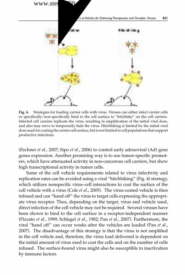
October 30, 2007 12:30 WSPC/SPI-B559: Autologous & Cancer Stem Cell Gene Therapy ch15 FA
MSCs asVehicles for DeliveringTherapeutics and Oncolytic Viruses 421
Fig. 4. Strategies for loading carrier cells with virus. Viruses can either infect carrier cellsor specifically/non-specifically bind to the cell surface to “hitchhike” on the cell carriers.Infected cell carriers replicate the virus, resulting in amplification of the initial viral dose,and also may serve to temporarily hide the virus. Hitchhiking is limited by the initial viraldose used for coating the carrier cell surface, but is not limited to cell populations that supportproductive infections.
(Fechner et al., 2007; Sipo et al., 2006) to control early adenoviral (Ad) genegenes expression. Another promising way is to use tumor-specific promot-ers, which have attenuated activity in non-cancerous cell carriers, but showhigh transcriptional activity in tumor cells.
Some of the cell vehicle requirements related to virus infectivity andreplication rates can be avoided using a viral “hitchhiking” (Fig. 4) strategy,which utilizes nonspecific virus–cell interactions to coat the surface of thecell vehicle with a virus (Cole et al., 2005). The virus-coated vehicle is theninfused and can “hand off” the virus to target cells expressing the appropri-ate virus receptor. Thus, depending on the target, virus and vehicle used,direct infection of the cell vehicle may not be required. Several viruses havebeen shown to bind to the cell surface in a receptor-independent manner(Pizzato et al., 1999; Schlegel et al., 1982; Pan et al., 2007). Furthermore, theviral “hand off” can occur weeks after the vehicles are loaded (Pan et al.,2007). The disadvantage of this strategy is that the virus is not amplifiedin the cell vehicle and, therefore, the virus load delivered is dependent onthe initial amount of virus used to coat the cells and on the number of cellsinfused. The surface-bound virus might also be susceptible to inactivationby immune factors.
干细胞之家www.stemcell8.cn ←点击进入

October 30, 2007 12:30 WSPC/SPI-B559: Autologous & Cancer Stem Cell Gene Therapy ch15 FA
422 L. Pereboeva and J. Roth
5.3. CellTypeTested
Different cell types have been evaluated as vehicles for delivering oncolyticviruses to tumors. The inherent tumor-homing and tumor-fighting activi-ties of the cell will depend on the specific cell type used. The importanceof choosing an appropriate carrier cell type for therapeutic delivery wasdemonstrated in cell carrier studies for glioma treatment (Li et al., 2007;Namba et al., 1998 and 2000). These studies demonstrated that, among thecarriers tested, the best therapeutic efficacy was achieved with neural pro-genitor cells (Li et al., 2005 and 2007).
The infectability of the vehicle and the level of viral replication withinthat vehicle will also vary, depending on the cell type used. Although thebenefits of cells as viral carriers have been recognized, only initial steps havebeen undertaken towards a practical implementation of this approach.
5.3.1. Immune cells (primaryT lymphocytes, CIK cells)
Immune cells are logical candidates as carriers for systemic virus delivery,since they naturally circulate and have the capacity to infiltrate tumors. Inaddition, many immune cell types have natural tumor-killing activities thathave been widely exploited for cancer immunotherapy. Therefore, these cellsmay exhibit synergistic tumor cell-killing activities when combined withoncolytic viruses. T lymphocytes were utilized as carriers of conditionallyreplicative retroviruses in a study by Chester et al. (2002). In this investiga-tion, the T lymphocytes were equipped with a chimeric receptor against thecolorectal tumor-associated antigen, carcinoma embryonic antigen (CEA),and were then used to produce and deliver retroviruses to CEA-positivetumor xenografts. The key aspect of this strategy lies in the controlled viralproduction, which was specifically triggered in response to the recombinantT-cell receptor binding to the tumor antigens. Multiple levels of targetingcontrol were incorporated in this delivery system at the stages of surface tar-geting, viral production and gene expression (Chester et al., 2002; Crittendenet al., 2003).
A method of controlled viral release in response to specific bindinghas also been proposed in a study by Yotnda et al. (2004). This study uti-lized cytotoxic T-lymphocytes (CTLs) directed against Epstein–Barr virus(EBV) antigens. These CTLs were derived from mice transgenic for the ade-noviral E1 gene under the control of the cell activation-dependent CD40ligand (CD40L) promoter. Following transduction with E1-deficient aden-oviral vectors, the CTLs produced infectious virus only when exposed to
干细胞之家www.stemcell8.cn ←点击进入

October 30, 2007 12:30 WSPC/SPI-B559: Autologous & Cancer Stem Cell Gene Therapy ch15 FA
MSCs asVehicles for DeliveringTherapeutics and Oncolytic Viruses 423
HLA-matched EBV-expressing targets, but not when exposed to major his-tocompatibility complex (MHC)-mismatched or otherwise irrelevant cells.
Ong et al. utilized T-cells as vehicles for delivering oncolytic measlesviruses to tumors (Ong et al., 2007). In this study, the authors initially exam-ined freshly isolated human peripheral blood lymphocytes (PBLs), but theyfound that T-cells activated with interleukin-2 and phytohemagglutininwere the primary population susceptible to viral infection. Interestingly, theviral replication cycle appeared to be abortive even in these pre-activatedT cells. Nevertheless, infected T cells were still able to transfer the measlesvirus to tumor cells via a cell-cell fusion mechanism. This “heterofusion” pro-cess led to successful delivery of measles viruses and tumor infection whenthe infected T cells were administered to mice by intravenous injection.
Since T-cells are typically refractory to viral infection, Cole et al. devel-oped the viral hitchhiking strategy to coat tumor-specific T-cells with aretrovirus vector expressing the immuno-stimulatory molecule IL-12 or thesuicide gene HSV-TK (Cole et al., 2005). These cells were then evaluated fortreatment of a murine lung metastasis model. Although the T cells were ableto mediate complete tumor regression alone, a protective memory responsewas only generated in mice infused with T-cells that were coated with theIL-12–expressing virus. T-cells coated with HSV-TK expression vectors werealso shown to protect animals challenged with a greater tumor burden, fol-lowing ganciclovir treatment. These findings indicate that arming tumoreffector cells with an oncolytic virus serves to combine and complement theindividual properties of cellular and viral therapeutics.
Cytokine-induced killer (CIK) cells are another potential virus carrierwith effector properties. CIK cells are obtained by in vitro treatment of humanperipheral blood lymphocytes or murine splenocytes with interferon-γ ,interleukin-2 and an antibody that cross-links the T-cell receptor (Bakeret al., 2001). The resultant cells bear phenotypic markers of natural killer(NK) and T cells, mediate NKG2D-dependent, non-major histocompatibil-ity complex-restricted lysis of a variety of transformed cell types and trafficto tumors in vivo (Verneris et al., 2004; Negrin et al., 2002). Thorne et al.used CIK cells to deliver an oncolytic strain of the Vaccinia virus. Thesecells supported Vaccinia virus infection and, more importantly, were ableto produce viral titers comparable to those generated by transformed cells(Thorne et al., 2006). In addition, viral release was delayed by several days.This rate of replication supports the cell-based virus delivery strategy, as itallows a time window for the infected CIK cells to traffic to tumors beforethe viral payload is released. In this study, the systemically administered
干细胞之家www.stemcell8.cn ←点击进入

October 30, 2007 12:30 WSPC/SPI-B559: Autologous & Cancer Stem Cell Gene Therapy ch15 FA
424 L. Pereboeva and J. Roth
CIK cells took up to 72 h to traffic to tumors, which corresponded to thepeak timing for Vaccinia virus production in CIK cells. Non-invasive imag-ing techniques were used to demonstrate that the Vaccinia-infected CIK cellswere present in tumors following intravenous administration in mice, andthis strategy resulted in powerful antitumor efficacy in several murine can-cer models. These findings demonstrate that some immunological killer cellscan be loaded with oncolytic viruses to achieve potentially synergistic gainsin antitumor activity.
5.3.2. Tumor cell lines
Although the introduction of tumor cell lines in patients would raise signif-icant safety concerns, the ease of handling these cells and the fact that theycan be readily infected with most oncolytic viruses make them attractivecandidates as cell-based viral delivery vehicles. One of the first examples ofa strategy utilizing tumor cells for delivering viruses used a carcinoma cellline (PA-1) as a vehicle for delivering a replication-competent attenuatedherpes simplex virus (HSV-1716) to treat human epithelial ovarian cancerin vivo (Coukos et al., 1999). In a separate study by Garcia-Castro et al. (2005),irradiated human breast carcinoma (MDA-MB-231) cells were used in anattempt to deliver adenovirus to lung metastases in a murine model. Bothstudies utilized immuno-compromised murine xenograft models to evalu-ate their cell-based delivery strategies. The use of immunocompetent modelsrepresents a more stringent system for evaluating cell-based delivery strate-gies, as the immune system is responsible for clearing virus-infected cells.A variety of tumor cell lines have been tested in immunocompetent mousemodels for their ability to deliver oncolytic parvovirus (Raykov et al., 2004),measles virus (Iankov et al., 2007) and vesicular stomatitis virus (Power et al.,2007). Tumor cell lines support high levels of viral replication and, therefore,greatly increase the initial viral load that can be delivered to the tumors.However, tumor cells do not necessarily have tumor-homing activity andrepresent an additional neoplastic threat when introduced into the body.In the clinical context, it will be critical to ensure that uninfected cells can-not establish de novo metastatic growth following systemic administration.This would require a supplemental mechanism by which the transferredcells are killed after fulfilling their transient function. This could be accom-plished by irradiating the tumor cells before administration, which ablatestumorigenicity but preserves the metabolic activity required for virus repli-cation. Alternatively, suicide genes could be used that could be activated
干细胞之家www.stemcell8.cn ←点击进入

October 30, 2007 12:30 WSPC/SPI-B559: Autologous & Cancer Stem Cell Gene Therapy ch15 FA
MSCs asVehicles for DeliveringTherapeutics and Oncolytic Viruses 425
after the time whereby the virus would have been released to ensure elim-ination of residual cells. Tumor cells may offer potential as cell carriers incancer settings; however, translation to clinical practice might present moreproblems than benefits.
5.3.3. Progenitor/stem cells
Progress in the fields of developmental and cellular biology has led to asignificant number of progenitor or stem cell types available as potentialtherapeutic vehicles. So far, neural, mesenchymal and endothelial precursorcells have been evaluated for use in cell-based approaches to cancer treat-ment and, more specifically, in approaches using these cells as viral deliv-ery vehicles. While immune cell vehicles offer the potential for an inherenttumor-killing effect, stem cell populations offer alternative properties thatcould also be advantageous in tumor settings.
Neural progenitor/stem cells (NSCs). The native tumor-tropic and migratorycapacities of neural stem cells (NSCs) were recently published by severalgroups (Aboody et al., 2000; Yip et al., 2003). These capacities have beenexploited lately to deliver therapeutics to the brain. Cell-mediated vectordelivery to brain tumors has been explored previously using retrovirus pack-aging cell lines, as well as other cell types (Culver et al., 1992; Ram et al., 1997).However, the tumor-homing ability of these cell populations was minimal.In this regard, the NSCs ideally filled the missing homing requirement forefficient cell vehicle-mediated delivery of viral vectors. Herrlinger et al. usedNSCs infected with an HSV-1–based virus to target pre-established intrac-erebral CNS-1 gliomas in nude mice (Herrlinger et al., 2000). The cells wereinjected directly into the tumors, but migrated extensively throughout thetumor and into the surrounding parenchyma over the following 3 days.Afterintratumoral injection, HSV-1 replication was also detected in the tumor andthe surrounding parenchyma. This study demonstrated the potential of cell-based delivery of HSV-1 vectors to tumor cells in the brain.
Endothelial progenitor/stem cells (EPCs). It is widely accepted that tumorgrowth is dependent on the formation of supportive blood vessels. Theexistence of bone marrow-derived endothelial progenitor cells (EPCs) ledto the suggestion that bone marrow-derived stem cells might be a sys-temic source of endothelial cells supporting neovascularization (Asaharaet al., 1997). Recently, studies have convincingly demonstrated that circu-lating EPCs are recruited to the tumor neovasculature and that genetically
干细胞之家www.stemcell8.cn ←点击进入

October 30, 2007 12:30 WSPC/SPI-B559: Autologous & Cancer Stem Cell Gene Therapy ch15 FA
426 L. Pereboeva and J. Roth
modified EPCs can serve as angiogenesis-targeting vectors to deliver anti-cancer genes (Lyden et al., 2001; Davidoff et al., 2001). The potential of EPCs asviral carriers has been investigated; notably, these cells supported infectionwith non-replicating retroviral and adenoviral vectors, and were also ableto deliver retrovirus to tumors upon systemic administration (Jevremovicet al., 2004).
Mesenchymal progenitor/stem cells (MSCs). MSCs have recently gainedincreased attention in tumor-related applications and have been tested ascarriers of replication-competent adenoviruses, as will be discussed in moredetail below.
5.4. Carrier Cell Immune Evasion
Although one of the main advantages proposed for the use of cells as viraldelivery vehicles was immune evasion, only a few recent studies haveaddressed this therapeutic strategy in the context of host immunity. Thisis largely due to the fact that most oncolytic viruses are based on humanviruses. These viruses usually demonstrate host (human)–restricted repli-cation and oncolysis (adenovirus or measles virus) or may only have weakinfectivity in murine tissues (adenovirus). These issues have limited the useof animal models for evaluating active virus production in the setting ofa functional immune system. Nevertheless, initial attempts to use cells asviral carriers to evade anti-viral immunity have been undertaken for severaloncolytic viruses.
Iankov et al. have shown that an immortalized monocyte cell line candeliver measles virus to tumors despite the presence of high titers of humanantibodies passively transferred to mice (Iankov et al., 2007). This modelwas used to recreate the high anti-measles antibody titers situation foundin patients. In contrast to direct ip injection of naked virions, infected cellcarriers transferred the virus to tumor cells by heterofusion, which was moreresistant to antibody neutralization. Repeated injection of monocytic cellcarriers significantly improved survival of mice with human ovarian cancerxenografts.
The negative impact the humoral immune response has on the systemicdelivery of oncolytic vesicular stomatitis virus (VSV) was demonstrated in astudy of Power et al. (2007). As a better alternative to direct virus administra-tion, the authors tested different tumor cell lines as cell carriers. This studyconfirmed that a wide variety of established, nonautologous cell lines, par-ticularly those of hematological origin, could serve as vehicles for delivery of
干细胞之家www.stemcell8.cn ←点击进入

October 30, 2007 12:30 WSPC/SPI-B559: Autologous & Cancer Stem Cell Gene Therapy ch15 FA
MSCs asVehicles for DeliveringTherapeutics and Oncolytic Viruses 427
oncolytic viruses. It has been convincingly demonstrated that cellular vehi-cles can shield oncolytic virus from neutralizing antibodies during delivery,providing a simple and effective means to improve therapeutic outcomes inthe context of antiviral immunity.
CIK-mediated delivery of Vaccinia virus was tested in both immun-odeficient and immunocompetent mouse models with established tumors(Thorne et al., 2006). Although a direct assessment of an immune responsewas not carried out, a survival benefit was seen in groups with cell-basedviral delivery compared to groups treated with cells or virus alone, sug-gesting that infected cell carriers indeed protect viral agents from beinginactivated by the immune system.
An immunocompetent rat model was also used to investigate cell-baseddelivery of an oncolytic parvovirus to tumor metastases (Raykov et al., 2004).The Morris hepatoma (MH3924A) tumor cell line was used as a parvovirusvehicle to target and suppress metastases formed by the same cells. Thequestion of a neutralizing antibody response was also addressed in regardto carrier cell-mediated or direct administration of virus. The presence ofneutralizing antiviral antibodies was quantified by measuring the ability ofrat serum to protect indicator cells from the killing effect of the parvovirus.The sera of rats injected with either carrier cells or free virus had a clearneutralizing activity in vitro. However, it was shown that the infected cellcarriers elicited less of a neutralizing antibody response, compared to sys-temically administered virions.
In contrast, a study by Ong et al. demonstrated that both infected cellvehicles and viruses can be inactivated by anti-viral antibodies in passivelyimmunized mice (Ong et al., 2007). In this study, T-cells were evaluated fordelivering oncolytic measles virus (MV) to multiple myeloma xenografts inthe presence of anti-MV antibodies. Although the feasibility of viral deliv-ery was established using the T-cell vehicles, neither delivery strategy wasable to overcome high titers of anti-viral antibodies. However, the cell-baseddelivery was effective over a broader range of antibody titers that mitigatedinfection with cell-free virus. The authors further demonstrated that theextent of tumor infection was dependent on the titer of neutralizing anti-bodies present, whether the therapeutic was administered as naked virionsor within infected T cells. Based on imaging analysis, this study determinedthat 1–2% of the total T-cells injected reached tumor nodules.
Although these studies provide a novel strategy for systemic deliveryof oncolytic viruses, further work is now required to determine how theimmune response will affect the vehicles carrying the virus. Additional
干细胞之家www.stemcell8.cn ←点击进入

October 30, 2007 12:30 WSPC/SPI-B559: Autologous & Cancer Stem Cell Gene Therapy ch15 FA
428 L. Pereboeva and J. Roth
investigations are also needed to evaluate the extent to which consecutiveviral vehicle administrations will elicit an immune response, and whethermulti-dose treatment regimens are feasible in a clinical setting.
6. Mesenchymal Stromal Cells as Carriers for OncolyticAdenoviruses
The inherent properties of MSCs that favor their use as clinically relevantcell carriers has led to practical attempts at using these cells for deliveringanti-tumor therapeutics such as interleukins and toxic genes (see above).However, it is questionable whether or not cell vehicles expressing cytotoxicgenes as tumor payloads will provide an effective tumor-killing bystanderresponse. Although the tumor-targeting aspect has been added via cell vehi-cles, the requirement of a bystander effect limits the overall efficacy of thestrategy.
The ability of oncolytic viruses to replicate offers the potential forexponential amplification of the killing effect. This demonstrates a clearadvantage in using these agents, compared to toxic gene therapy strate-gies. Conditionally replicative adenoviruses (CRAds) have been developedfor a broad range of tumors and are rapidly transitioning to clinical settings.However, it is apparent that direct introduction of replication competentAds would face the same problems as non-replicative Ad vectors, whichhave previously been tested in a range of gene therapy applications. To thisend, viral delivery could also make use of vehicles with endogenous tumor-targeting activity, allowing viral replication in the vicinity or directly insidethe tumor. In this regard, MSCs represent a promising cell population for useas a viral delivery platform. We first investigated the properties of MSCs ascell vehicles for delivering anti-cancer therapeutics (Pereboeva et al., 2003)and subsequently hypothesized that MSCs would serve as efficient CRAddelivery vehicles. Therefore, we established the key feasibilities determin-ing whether MSC-based CRAd delivery was a viable strategy (Komarovaet al., 2006). First, we investigated the ability of MSCs to home to growingtumors. An ovarian cancer xenograft model and several detection methodswere used to demonstrate that ip-injected MSCs preferentially localize inthe vicinity or within the growing ovarian tumors [Fig. 5(A)]. Only a short-term MSC homing effect to ovarian or breast cancer xenografts was inves-tigated, since cell-based delivery of Ad does not rely on long-term survivaland proliferation of cell carriers. Imaging signals were used in the ovar-ian model to demonstrate that the ip-injected cells were initially distributednonspecifically in the peritoneal cavity (15 min, 2 h). Two days later, a faint
干细胞之家www.stemcell8.cn ←点击进入

October 30, 2007 12:30 WSPC/SPI-B559: Autologous & Cancer Stem Cell Gene Therapy ch15 FA
MSCs asVehicles for DeliveringTherapeutics and Oncolytic Viruses 429
Fig. 5. MSCs as intermediate carriers of oncolytic adenoviruses. (A) MSCs home to tumors.MSCs labeled with fluorescent dye CFDA-SE and injected i.p. into mice bearing ovariantumor xenografts were located in tumor parenchyma or attached to the tumor nodules.Microphotographs represent cryosections of tumor samples. (B) MSCs are loaded with ade-novirus via infection and produce de novo viral particles with delayed kinetics. MSCs andHela cells (Ad susceptible cell line) were infected with Ad5 at MOI 1 pfu/cell for 2 h. De novoviral production in infected cells was estimated at 1, 3, 5 and 7 days by titration of cell lysateson 293 cells. Virus concentration (vp/mL) was calculated as recommended in the manualfor AdEasy protocol (TCID50 assay for virus titration).
overall luminescent signal became patchy, indicating that several hours aresufficient for MSCs to travel within the peritoneal cavity to accumulate inspecific sites. Imaging of dissected ovarian tumors and other organs of theperitoneal cavity at the time of sacrifice confirmed that the signal was almostexclusively within the tumors.
Next, the ability of MSCs to maintain an adenoviral infection, includ-ing viral DNA replication and formation of new infectious viral particles,was examined. Compared to cancer cell lines that are efficiently infectedand rapidly produce adenovirus, the ability of different primary cells tosupport Ad infection was not widely tested. We utilized several methodsto demonstrate that MSCs were able to support Ad5 replication, althoughwith slower production kinetics, compared to the permissive HeLa cells[Fig. 5(B)]. In addition, we showed that some of the tropism-modified aden-oviruses were more efficiently produced in the MSC vehicles. For instance,the level of Ad5/3 amplification in MSCs almost reached the levels gener-ated in Hela cells. Since adenovirus spreads via lysis of infected cells, theviral life cycle should be slow enough in the cell vehicle to allow sufficient
干细胞之家www.stemcell8.cn ←点击进入

October 30, 2007 12:30 WSPC/SPI-B559: Autologous & Cancer Stem Cell Gene Therapy ch15 FA
430 L. Pereboeva and J. Roth
time for the vehicle to reach and establish contact with tumor cells beforebeing destroyed. A panel of replication-competent Ads was tested for anMSC-based cell vehicle approach, which led to the identification of Ad5/3as the best virus for our experiments. This virus is derived from the wild-typehuman Ad5 virus, but contains a fiber modification for targeting purposes(Krasnykh et al., 1996). As a viral payload, we used replication-competentAd5/3 (Komarova et al., 2006) and a conditionally replicative version of thisvirus with the E1 gene driven by the CXCR4 promoter (Ad5/3CXCR4) (Stoff-Khalili et al., 2007). Both viruses were tested in vitro and were demonstratedto have delayed growth kinetics in the vehicle, compared to tumor cells.
The therapeutic efficacy of MSC-based Ad delivery was investigated inhuman xenograft models using two modes of cell vehicle injection. InfectedMSCs were administered locally for an ovarian tumor xenograft model, andsystemically for a model of breast cancer metastases of the lung. We wantedto test whether administration of virus-loaded MSCs would improve thetherapeutic effect over virus alone. In the ovarian model, the tumor growthwas more delayed in animals that received MSC-Ad5/3. The average tumorburden, as measured by total weight of tumor nodules or the intensity ofimaging signal at day 21, was significantly less than in tumors collectedfrom control groups treated with PBS, MSCs and Ad5/3. The rate of tumordevelopment correlated with the tumor mass detected by image intensity(Fig. 6). Treatment with MSC-based virus vehicles also resulted in the longestanimal survival times. The median survival time was extended to 69 daysin groups treated with MSCs-Ad5/3, compared to the control PBS-treatedgroup (34 days). However only modest differences in average survival rateswere noted, compared to the virus-onlyAd5/3-treatment group (59 days). Inthe model of pre-established pulmonary metastases derived from MDA-MB-231 cells, MSCs loaded with Ad5/3.CXCR4 also improved the survival ofmice with lung tumors, compared to mice treated with either Ad5/3.CXCR4or hMSCs alone (Stoff-Khalili et al., 2007).
In summary, MSCs loaded with replication-competent Ad were ableto exert an oncolytic effect on tumor cells in vitro and in vivo in bothmodels tested. Delivery of virus by cell carriers with endogenous tumor-targeting properties resulted in a therapeutic benefit, compared to viral injec-tions alone. Although complete tumor regression was not achieved, the cellvehicle-based delivery was superior in terms of delayed tumor develop-ment. This effect may result from the amplification of viral load in cell car-riers and/or the targeted virus delivery and multifocal release of the virusin the vicinity of tumor cells. Regardless of the mechanism, the rationale for
干细胞之家www.stemcell8.cn ←点击进入

October 30, 2007 12:30 WSPC/SPI-B559: Autologous & Cancer Stem Cell Gene Therapy ch15 FA
MSCs asVehicles for DeliveringTherapeutics and Oncolytic Viruses 431
Fig. 6. Effect of oncolytic Ad5/3 delivered directly or via MSC cell carriers on humanovarian xenograft growth. (A) Ovarian tumor xenografts were established ip in CB17 SCIDmice by injection of SKOV3luc cells. Animals received three injections of PBS only (T), directinjections of Ad5/3 (T + Ad) at 1 × 10¹⁰ vp/injection, or MSCs preloaded with Ad5/3(T + MSC/Ad) at 1 × 10⁶ cells/injection. Live image of mice taken at day 28 demonstratetumor development in three experimental groups. The bioluminescence intensity of theimage corresponds to the luc-expressing tumor burden (black, least intense; and red, mostintense). One mouse in the tumor-only group did not develop tumor and served as a negativecontrol. (B) Potential attributes of MSC-Ad delivery: amplification of initial viral load andlocal release of viral load in vicinity of tumors. (A) Adapted from Fig. 6(A) of: Komarovaet al., Mol Cancer Ther 5: 755–766, 2006 with permission from the American Association forCancer Research.
using MSCs as adenoviral vehicles is valid, and calls for further investigationand optimization.
Acknowledgments
This work was supported by the NIH grant 1 R21 CA115568 (Dr. Larisa Pere-boeva). We thank Svetlana Komarova for excellent assistance with experi-mental studies and contribution to the manuscript.
References
Aboody KS, Brown A, Rainov NG, et al. (2000) Neural stem cells display extensivetropism for pathology in adult brain: evidence from intracranial gliomas. ProcNatl Acad Sci USA 97: 12846–51.
干细胞之家www.stemcell8.cn ←点击进入

October 30, 2007 12:30 WSPC/SPI-B559: Autologous & Cancer Stem Cell Gene Therapy ch15 FA
432 L. Pereboeva and J. Roth
Allers C, Sierralta WD, Neubauer S, et al. (2004) Dynamic of distribution of humanbone marrow-derived mesenchymal stem cells after transplantation into adultunconditioned mice. Transplantation 78: 503–508.
Alvarez-Dolado M, Pardal R, Garcia-Verdugo JM, et al. (2003) Fusion of bone-marrow-derived cells with Purkinje neurons, cardiomyocytes and hepatocytes.Nature 425: 968–73.
Anjos-Afonso F, Siapati EK, Bonnet D. (2004) In vivo contribution of murine mes-enchymal stem cells into multiple cell-types under minimal damage conditions.J Cell Sci 117: 5655–64.
Asahara T, Murohara T, Sullivan A, et al. (1997) Isolation of putative progenitorendothelial cells for angiogenesis. Science 275: 964–67.
Askari AT, Unzek S, Popovic ZB, et al. (2003) Lancet 362: 697–703.Azizi SA, Stokes D, Augelli BJ, et al. (1998) Engraftment and migration of human
bone marrow stromal cells implanted in the brains of albino rats–similarities toastrocyte grafts. Proc Natl Acad Sci USA 95: 3908–13.
Baddoo M, Hill K, Wilkinson R, et al. (2003) Characterization of mesenchymal stemcells isolated from murine bone marrow by negative selection. J Cell Biochem 89:1235–49.
Baker AH, McVey JH, Waddington SN, et al. (2007) The influence of blood on in vivoadenovirus bio-distribution and transduction. Mol Ther 15: 1410–16.
Baker J, Verneris MR, Ito M, et al. (2001) Expansion of cytolytic CD8(+) naturalkiller T cells with limited capacity for graft-versus-host disease induction due tointerferon gamma production. Blood 97: 2923–31.
Baksh D, Song L, Tuan RS. (2004) Adult mesenchymal stem cells: characteriza-tion, differentiation, and application in cell and gene therapy. J Cell Mol Med 8:301–16.
Banfi A, Muraglia A, Dozin B, et al. (2000) Proliferation kinetics and differentiationpotential of ex vivo expanded human bone marrow stromal cells: implicationsfor their use in cell therapy. Exp Hematol 28: 707–15.
Barbash IM, Chouraqui P, Baron J, et al. (2003) Systemic delivery of bone marrow-derived mesenchymal stem cells to the infarcted myocardium: feasibility, cellmigration, and body distribution. Circulation 108: 863–68.
Barry F, Boynton R, Murphy M, et al. (2001) The SH-3 and SH-4 antibodies recognizedistinct epitopes on CD73 from human mesenchymal stem cells. Biochem BiophysRes Commun 289: 519–24.
Bartholomew A, Patil S, Mackay A, et al. (2001) Baboon mesenchymal stem cells canbe genetically modified to secrete human erythropoietin in vivo. Hum Gene Ther12: 1527–41.
Baxter MA, Wynn RF, Jowitt SN, et al. (2004) Study of telomere length reveals rapidaging of human marrow stromal cells following in vitro expansion. Stem Cells 22:675–82.
干细胞之家www.stemcell8.cn ←点击进入

October 30, 2007 12:30 WSPC/SPI-B559: Autologous & Cancer Stem Cell Gene Therapy ch15 FA
MSCs asVehicles for DeliveringTherapeutics and Oncolytic Viruses 433
Bentzon JF, Stenderup K, Hansen FD, et al. (2005) Tissue distribution and engraft-ment of human mesenchymal stem cells immortalized by human telomerasereverse transcriptase gene. Biochem Biophys Res Commun 330: 633–40.
Bianco P, Gehron Robey P. (2000) Marrow stromal stem cells. J Clin Invest 105:1663–68.
Bianco P, Riminucci M, Gronthos S, Robey PG. (2001) Bone marrow stromal stemcells: nature, biology, and potential applications. Stem Cells 19: 180–92.
Bieback K, Kern S, Kluter H, Eichler H. (2004) Critical parameters for the isolationof mesenchymal stem cells from umbilical cord blood. Stem Cells 22: 625–34.
Bittira B, Shum-Tim D, Al-Khaldi A, Chiu RC. (2003) Mobilization and homing ofbone marrow stromal cells in myocardial infarction. Eur J Cardiothorac Surg 24:393–98.
Bonab MM, Alimoghaddam K, Talebian F, et al. (2006) Aging of mesenchymal stemcell in vitro. BMC Cell Biol 7: 14.
Brouard N, Chapel A, Thierry D, et al. (2000) Transplantation of gene-modified human bone marrow stromal cells into mouse-human bone chimeras.J Hematother Stem Cell Res 9: 175–81.
Caplan AI, Reuben D, Haynesworth SE. (1998) Cell-based tissue engineering ther-apies: the influence of whole body physiology. Adv Drug Deliv Rev 33: 3–14.
Charbord P, Oostendorp R, Pang W, et al. (2002) Comparative study of stromal celllines derived from embryonic, fetal, and postnatal mouse blood-forming tissues.Exp Hematol 30: 1202–10.
Chen J, Li Y, Katakowski M, et al. (2003) Intravenous bone marrow stromal cell ther-apy reduces apoptosis and promotes endogenous cell proliferation after strokein female rat. J Neurosci Res 73: 778–86.
Chen J, Li Y, Wang L, et al. (2001) Therapeutic benefit of intravenous administrationof bone marrow stromal cells after cerebral ischemia in rats. Stroke 32: 1005–11.
ChenY,Yu DC, Charlton D, Henderson DR. (2000) Pre-existent adenovirus antibodyinhibits systemic toxicity and antitumor activity of CN706 in the nude mouseLNCaP xenograft model: implications and proposals for human therapy. HumGene Ther 11: 1553–67.
Chester J, Ruchatz A, Gough M, et al. (2002) Tumor antigen-specific induction oftranscriptionally targeted retroviral vectors from chimeric immune receptor-modified T cells. Nat Biotechnol 20: 256–63.
Chin BB, Nakamoto Y, Bulte JW, et al. (2003) 111In oxine labelled mesenchymal stemcell SPECT after intravenous administration in myocardial infarction. Nucl MedCommun 24: 1149–54.
Chopp M, Li Y. (2002) Treatment of neural injury with marrow stromal cells. LancetNeurol 1: 92–100.
Chopp M, Zhang XH, Li Y, et al. (2000) Spinal cord injury in rat: treatment withbone marrow stromal cell transplantation. Neuroreport 11: 3001–3005.
干细胞之家www.stemcell8.cn ←点击进入

October 30, 2007 12:30 WSPC/SPI-B559: Autologous & Cancer Stem Cell Gene Therapy ch15 FA
434 L. Pereboeva and J. Roth
Clark BR, Keating A. (1995) Biology of bone marrow stroma. Ann N Y Acad Sci 770:70–78.
Cole C, Qiao J, Kottke T, et al. (2005) Tumor-targeted, systemic delivery of ther-apeutic viral vectors using hitchhiking on antigen-specific T cells. Nat Med 11:1073–81.
Coukos G, Makrigiannakis A, Kang EH, et al. (1999) Use of carrier cells to deliver areplication-selective herpes simplex virus-1 mutant for the intraperitoneal ther-apy of epithelial ovarian cancer. Clin Cancer Res 5: 1523–37.
Crittenden M, Gough M, Chester J, et al. (2003) Pharmacologically regulated pro-duction of targeted retrovirus from T cells for systemic antitumor gene therapy.Cancer Res 63: 3173–80.
Croitoru-Lamoury J, Lamoury FM, Zaunders JJ, et al. (2007) Human mesenchymalstem cells constitutively express chemokines and chemokine receptors that canbe upregulated by cytokines, IFN-beta, and copaxone. J Interferon Cytokine Res27: 53–64.
Culver KW, Ram Z, Wallbridge S, et al. (1992) In vivo gene transfer with retroviralvector-producer cells for treatment of experimental brain tumors. Science 256:1550–52.
Davidoff AM, Ng CY, Brown P, et al. (2001) Bone marrow-derived cells contribute totumor neovasculature and, when modified to express an angiogenesis inhibitor,can restrict tumor growth in mice. Clin Cancer Res 7: 2870–79.
De Bari C, Dell’Accio F, Luyten FP. (2004) Failure of in vitro differentiated mesenchy-mal stem cells from the synovial membrane to form ectopic stable cartilage in vivo.Arthritis Rheum 50: 142–50.
Deans RJ, MoseleyAB. (2000) Mesenchymal stem cells: biology and potential clinicaluses. Exp Hematol 28: 875–84.
Deng YB, Liu XG, Liu ZG, et al. (2006) Implantation of BM mesenchymal stem cellsinto injured spinal cord elicits de novo neurogenesis and functional recovery:evidence from a study in rhesus monkeys. Cytotherapy 8: 210–14.
Dennis JE, Carbillet JP, Caplan AI, Charbord P. (2002) The STRO-1+ marrow cellpopulation is multipotential. Cells Tissues Organs 170: 73–82.
Devine MJ, Mierisch CM, Jang E, et al. (2002) Transplanted bone marrow cells local-ize to fracture callus in a mouse model. J Orthop Res 20: 1232–39.
Devine SM, Bartholomew AM, Mahmud N, et al. (2001) Mesenchymal stem cellsare capable of homing to the bone marrow of non-human primates followingsystemic infusion. Exp Hematol 29: 244–55.
Devine SM, Cobbs C, Jennings M, et al. (2003) Mesenchymal stem cells distributeto a wide range of tissues following systemic infusion into nonhuman primates.Blood 101: 2999–3001.
Digel M, Sampaio KL, Jahn G, Sinzger C. (2006) Evidence for direct transfer of cyto-plasmic material from infected to uninfected cells during cell-associated spreadof human cytomegalovirus. J Clin Virol 37: 10–20.
干细胞之家www.stemcell8.cn ←点击进入

October 30, 2007 12:30 WSPC/SPI-B559: Autologous & Cancer Stem Cell Gene Therapy ch15 FA
MSCs asVehicles for DeliveringTherapeutics and Oncolytic Viruses 435
Digirolamo CM, Stokes D, Colter D, et al. (1999) Propagation and senescence ofhuman marrow stromal cells in culture: a simple colony-forming assay identifiessamples with the greatest potential to propagate and differentiate. Br J Haematol107: 275–81.
D’Ippolito G, Howard GA, Roos BA, Schiller PC. (2006) Isolation and characteriza-tion of marrow-isolated adult multilineage inducible (MIAMI) cells. Exp Hematol34: 1608–10.
Dominici M, Le Blanc K, Mueller I, et al. (2006) Minimal criteria for defining multipo-tent mesenchymal stromal cells. The International Society for Cellular Therapyposition statement. Cytotherapy 8: 315–17.
Dvorak HF. (1986) Tumors: wounds that do not heal: similarities between tumorstroma generation and wound healing. N Engl J Med 315: 1650–59.
Elzaouk L, Moelling K, Pavlovic J. (2006) Anti-tumor activity of mesenchymal stemcells producing IL-12 in a mouse melanoma model. Exp Dermatol 15: 865–74.
Erices AA, Allers CI, Conget PA, et al. (2003) Human cord blood-derived mesenchy-mal stem cells home and survive in the marrow of immunodeficient mice aftersystemic infusion. Cell Transplant 12: 555–61.
Fechner H, Wang X, Pico AH, et al. (2007) A bidirectional Tet-dependent promotorconstruct regulating the expression of E1A for tight control of oncolytic aden-ovirus replication. J Biotechnol 127: 560–74.
Francois S, Bensidhoum M, Mouiseddine M, et al. (2006) Local irradiation not onlyinduces homing of human mesenchymal stem cells at exposed sites but promotestheir widespread engraftment to multiple organs: a study of their quantitativedistribution after irradiation damage. Stem Cells 24: 1020–29.
FriedensteinAJ, Chailakhjan RK, Lalykina KS. (1970) The development of fibroblastcolonies in monolayer cultures of guinea-pig bone marrow and spleen cells. CellTissue Kinet 3: 393–403.
Friedenstein AJ, Chailakhyan RK, Gerasimov UV. (1987) Bone marrow osteogenicstem cells: in vitro cultivation and transplantation in diffusion chambers. CellTissue Kinet 20: 263–72.
Friedenstein AJ, Latzinik NV, Gorskaya Yu F, et al. (1992) Bone marrow stromalcolony formation requires stimulation by haemopoietic cells. Bone Miner 18:199–213.
Gao J, Dennis JE, Muzic RF, et al. (2001) The dynamic in vivo distribution of bonemarrow-derived mesenchymal stem cells after infusion. Cells Tissues Organs 169:12–20.
Garcia-Castro J, Martinez-Palacio J, Lillo R, et al. (2005) Tumor cells as cellularvehicles to deliver gene therapies to metastatic tumors. Cancer Gene Ther 12:341–49.
Gronthos S, Graves SE, Ohta S, Simmons PJ. (1994) The STRO-1+ fractionof adult human bone marrow contains the osteogenic precursors. Blood 84:4164–73.
干细胞之家www.stemcell8.cn ←点击进入

October 30, 2007 12:30 WSPC/SPI-B559: Autologous & Cancer Stem Cell Gene Therapy ch15 FA
436 L. Pereboeva and J. Roth
Gronthos S, Zannettino AC, Hay SJ, et al. (2003) Molecular and cellular characteri-sation of highly purified stromal stem cells derived from human bone marrow.J Cell Sci 116: 1827–35.
Haynesworth SE, Baber MA, Caplan AI. (1992a) Cell surface antigens on humanmarrow-derived mesenchymal cells are detected by monoclonal antibodies. Bone13: 69–80.
Haynesworth SE, Baber MA, Caplan AI. (1996) Cytokine expression by humanmarrow-derived mesenchymal progenitor cells in vitro: effects of dexamethasoneand IL-1 alpha. J Cell Physiol 166: 585–92.
Haynesworth SE, Goshima J, Goldberg VM, Caplan AI. (1992b) Characterization ofcells with osteogenic potential from human marrow. Bone 13: 81–88.
Herrlinger U, Woiciechowski C, Sena-Esteves M, et al. (2000) Neural precursor cellsfor delivery of replication-conditional HSV-1 vectors to intracerebral gliomas.Mol Ther 1: 347–57.
Hill JM, Dick AJ, Raman VK, et al. (2003) Serial cardiac magnetic resonance imagingof injected mesenchymal stem cells. Circulation 108: 1009–14.
Horwitz EM, Le Blanc K, Dominici M, et al. (2005) Clarification of the nomencla-ture for MSC: The International Society for Cellular Therapy position statement.Cytotherapy 7: 393–95.
Huebner RJ, Rowe WP, Schatten WE, et al. (1956) Studies on the use of viruses inthe treatment of carcinoma of the cervix. Cancer 9:1211–18.
Hung SC, Deng WP, Yang WK, et al. (2005) Mesenchymal stem cell targeting ofmicroscopic tumors and tumor stroma development monitored by noninvasivein vivo positron emission tomography imaging. Clin Cancer Res 11: 7749–56.
Iankov ID, Blechacz B, Liu C, et al. (2007) Infected cell carriers: a new strategy forsystemic delivery of oncolytic measles viruses in cancer virotherapy. Mol Ther15: 114–22.
Izadpanah R, Joswig T, Tsien F, et al. (2005) Characterization of multipotent mes-enchymal stem cells from the bone marrow of rhesus macaques. Stem Cells Dev14: 440–51.
Jaiswal N, Haynesworth SE, Caplan AI, Bruder SP. (1997) Osteogenic differentia-tion of purified, culture-expanded human mesenchymal stem cells in vitro. J CellBiochem 64: 295–312.
Javazon EH, Beggs KJ, Flake AW. (2004) Mesenchymal stem cells: paradoxes ofpassaging. Exp Hematol 32: 414–25.
Javazon EH, Colter DC, Schwarz EJ, Prockop DJ. (2001) Rat marrow stromal cellsare more sensitive to plating density and expand more rapidly from single-cell-derived colonies than human marrow stromal cells. Stem Cells 19: 219–25.
Jevremovic D, Gulati R, Hennig I, et al. (2004) Use of blood outgrowth endothelialcells as virus-producing vectors for gene delivery to tumors. Am J Physiol HeartCirc Physiol 287: H494–H500.
干细胞之家www.stemcell8.cn ←点击进入

October 30, 2007 12:30 WSPC/SPI-B559: Autologous & Cancer Stem Cell Gene Therapy ch15 FA
MSCs asVehicles for DeliveringTherapeutics and Oncolytic Viruses 437
Jiang Y, Vaessen B, Lenvik T, et al. (2002) Multipotent progenitor cells can be isolatedfrom postnatal murine bone marrow, muscle, and brain. Exp Hematol 30: 896–904.
Kadiyala S, Young RG, Thiede MA, Bruder SP. (1997) Culture expanded caninemesenchymal stem cells possess osteochondrogenic potential in vivo and in vitro.Cell Transplant 6: 125–34.
Kern S, Eichler H, Stoeve J, et al. (2006) Comparative analysis of mesenchymal stemcells from bone marrow, umbilical cord blood, or adipose tissue. Stem Cells 24:1294–301.
Kinnaird T, Stabile E, Burnett MS, et al. (2004) Local delivery of marrow-derivedstromal cells augments collateral perfusion through paracrine mechanisms. Cir-culation 109: 1543–49.
Komarova S, Kawakami Y, Stoff-Khalili MA, et al. (2006) Mesenchymal progenitorcells as cellular vehicles for delivery of oncolytic adenoviruses. Mol Cancer Ther5: 755–66.
Kopen GC, Prockop DJ, Phinney DG. (1999) Marrow stromal cells migrate through-out forebrain and cerebellum, and they differentiate into astrocytes after injectioninto neonatal mouse brains. Proc Natl Acad Sci USA 96: 10711–16.
Kraitchman DL, Heldman AW, Atalar E, et al. (2003) In vivo magnetic resonanceimaging of mesenchymal stem cells in myocardial infarction. Circulation 107:2290–93.
Kraitchman DL, Tatsumi M, Gilson WD, et al. (2005) Dynamic imaging of allo-geneic mesenchymal stem cells trafficking to myocardial infarction. Circulation112: 1451–61.
Krasnykh VN, Mikheeva GV, Douglas JT, Curiel DT. (1996) Generation of recom-binant adenovirus vectors with modified fibers for altering viral tropism. J Virol70: 6839–46.
Kuppen PJ, Marinelli A, Camps JA, et al. (1992) Biodistribution of lymphokine-activated killer (LAK) cells in Wag rats after hepatic-artery or jugular-veininfusion. Int J Cancer 52: 266–70.
Kyriakou CA, Yong KL, Benjamin R, et al. (2006) Human mesenchymal stemcells (hMSCs) expressing truncated soluble vascular endothelial growth factorreceptor (tsFlk-1) following lentiviral-mediated gene transfer inhibit growth ofBurkitt’s lymphoma in a murine model. J Gene Med 8: 253–64.
Li S, Tokuyama T, Yamamoto J, et al. (2005) Bystander effect-mediated gene therapyof gliomas using genetically engineered neural stem cells. Cancer Gene Ther 12:600–607.
Li S, Gao Y, Tokuyama T, et al. (2007) Genetically engineered neural stem cellsmigrate and suppress glioma cell growth at distant intracranial sites. Cancer Lett251: 220–27.
Liu Y, Deisseroth A. (2006) Oncolytic adenoviral vector carrying the cytosine deam-inase gene for melanoma gene therapy. Cancer Gene Ther 13: 845–55.
干细胞之家www.stemcell8.cn ←点击进入

October 30, 2007 12:30 WSPC/SPI-B559: Autologous & Cancer Stem Cell Gene Therapy ch15 FA
438 L. Pereboeva and J. Roth
Lyden D, Hattori K, Dias S, et al. (2001) Impaired recruitment of bone-marrow-derived endothelial and hematopoietic precursor cells blocks tumor angiogene-sis and growth. Nat Med 7: 1194–201.
Lyons M, Onion D, Green NK, et al. (2006) Adenovirus type 5 interactions withhuman blood cells may compromise systemic delivery. Mol Ther 14: 118–28.
MacDonald DJ, Luo J, Saito T, et al. (2005) Persistence of marrow stromal cellsimplanted into acutely infarcted myocardium: observations in a xenotransplantmodel. J Thorac Cardiovasc Surg 130: 1114–21.
Mahmood A, Lu D, Chopp M. (2004a) Intravenous administration of marrow stro-mal cells (MSCs) increases the expression of growth factors in rat brain aftertraumatic brain injury. J Neurotrauma 21: 33–39.
Mahmood A, Lu D, Chopp M. (2004b) Marrow stromal cell transplantation aftertraumatic brain injury promotes cellular proliferation within the brain. Neuro-surgery 55: 1185–93.
Majumdar MK, Keane-Moore M, Buyaner D, et al. (2003) Characterization and func-tionality of cell surface molecules on human mesenchymal stem cells. J BiomedSci 10: 228–41.
Makkar RR, Price MJ, Lill M, et al. (2005) Intramyocardial injection of allo-genic bone marrow-derived mesenchymal stem cells without immunosuppres-sion preserves cardiac function in a porcine model of myocardial infarction.J Cardiovasc Pharmacol Ther 10: 225–33.
Martin DR, Cox NR, Hathcock TL, et al. (2002) Isolation and characterization ofmultipotential mesenchymal stem cells from feline bone marrow. Exp Hematol30: 879–86.
Meyerrose TE, De Ugarte DA, HoflingAA, et al. (2007) In vivo distribution of humanadipose-derived mesenchymal stem cells in novel xenotransplantation models.Stem Cells 25: 220–27.
Minguell JJ, Erices A, Conget P. (2001) Mesenchymal stem cells. Exp Biol Med (May-wood) 226: 507–20.
Minguell JJ, Fierro FA, Epunan MJ, et al. (2005) Nonstimulated human uncommit-ted mesenchymal stem cells express cell markers of mesenchymal and neurallineages. Stem Cells Dev 14: 408–14.
Miura M, Gronthos S, Zhao M, et al. (2003) SHED: stem cells from human exfoliateddeciduous teeth. Proc Natl Acad Sci USA 100: 5807–12.
Morrow G, Vachot L, Vagenas P, Robbiani M. (2007) Current concepts of HIV trans-mission. Curr HIV/AIDS Rep 4: 29–35.
Mosca JD, Hendricks JK, Buyaner D, et al. (2000) Mesenchymal stem cells as vehiclesfor gene delivery. Clin Orthop Relat Res: S71–S90.
Nakamizo A, Marini F, Amano T, et al. (2005) Human bone marrow-derived mes-enchymal stem cells in the treatment of gliomas. Cancer Res 65: 3307–18.
Nakamura K, ItoY, KawanoY, et al. (2004)Antitumor effect of genetically engineeredmesenchymal stem cells in a rat glioma model. Gene Ther 11: 1155–64.
干细胞之家www.stemcell8.cn ←点击进入

October 30, 2007 12:30 WSPC/SPI-B559: Autologous & Cancer Stem Cell Gene Therapy ch15 FA
MSCs asVehicles for DeliveringTherapeutics and Oncolytic Viruses 439
Namba H, Tagawa M, Iwadate Y, et al. (1998) Bystander effect-mediated therapy ofexperimental brain tumor by genetically engineered tumor cells. Hum Gene Ther9: 5–11.
Namba H, Tagawa M, Miyagawa T, et al. (2000) Treatment of rat experimental braintumors by herpes simplex virus thymidine kinase gene-transduced allogeneictumor cells and ganciclovir. Cancer Gene Ther 7: 947–53.
Negrin RS, Edinger M, Verneris M, et al. (2002) Visualization of tumor growth andresponse to NK-T cell based immunotherapy using bioluminescence. Ann Hema-tol 81 (Suppl 2): S44–S45.
Neth P, Ciccarella M, Egea V, et al. (2006) Wnt signaling regulates the invasioncapacity of human mesenchymal stem cells. Stem Cells 24: 1892–903.
Nettelbeck DM. (2003) Virotherapeutics: conditionally replicative adenoviruses forviral oncolysis. Anticancer Drugs 14: 577–84.
Olivares EL, Ribeiro VP, Werneck de Castro JP, et al. (2004) Bone marrow stromalcells improve cardiac performance in healed infarcted rat hearts. Am J PhysiolHeart Circ Physiol 287: H464–70.
Ong HT, Hasegawa K, Dietz AB, et al. (2007) Evaluation of T cells as carriers forsystemic measles virotherapy in the presence of antiviral antibodies. Gene Ther14: 324–33.
Ortiz LA, Gambelli F, McBride C, et al. (2003) Mesenchymal stem cell engraftment inlung is enhanced in response to bleomycin exposure and ameliorates its fibroticeffects. Proc Natl Acad Sci USA 100: 8407–11.
Owen M. (1988) Marrow stromal stem cells. J Cell Sci Suppl 10: 63–76.OzakiY, Nishimura M, Sekiya K, et al. (2007) Comprehensive analysis of chemotactic
factors for bone marrow mesenchymal stem cells. Stem Cells Dev 16: 119–29.Pan YW, Scarlett JM, Luoh TT, Kurre P. (2007) Prolonged adherence of human
immunodeficiency virus-derived vector particles to hematopoietic target cellsleads to secondary transduction in vitro and in vivo. J Virol 81: 639–49.
Park H, Temenoff JS, Tabata Y, et al. (2007) Injectable biodegradable hydrogel com-posites for rabbit marrow mesenchymal stem cell and growth factor delivery forcartilage tissue engineering. Biomaterials 28: 3217–27.
Peister A, Mellad JA, Larson BL, et al. (2004) Adult stem cells from bone marrow(MSCs) isolated from different strains of inbred mice vary in surface epitopes,rates of proliferation, and differentiation potential. Blood 103: 1662–68.
Pereboeva L, Komarova S, Mikheeva G, et al. (2003) Approaches to utilize mes-enchymal progenitor cells as cellular vehicles. Stem Cells 21: 389–404.
Pereira RF, Halford KW, O’Hara MD, et al. (1995) Cultured adherent cells frommarrow can serve as long-lasting precursor cells for bone, cartilage, and lung inirradiated mice. Proc Natl Acad Sci USA 92: 4857–61.
Phinney DG, Kopen G, Isaacson RL, Prockop DJ. (1999a) Plastic adherent stromalcells from the bone marrow of commonly used strains of inbred mice: variationsin yield, growth, and differentiation. J Cell Biochem 72: 570–85.
干细胞之家www.stemcell8.cn ←点击进入

October 30, 2007 12:30 WSPC/SPI-B559: Autologous & Cancer Stem Cell Gene Therapy ch15 FA
440 L. Pereboeva and J. Roth
Phinney DG, Kopen G, Righter W, et al. (1999b) Donor variation in the growthproperties and osteogenic potential of human marrow stromal cells. J Cell Biochem75: 424–36.
Pittenger MF, Mackay AM, Beck SC, et al. (1999) Multilineage potential of adulthuman mesenchymal stem cells. Science 284: 143–47.
Pittenger MF, Martin BJ. (2004) Mesenchymal stem cells and their potential as car-diac therapeutics. Circ Res 95: 9–20.
Pittenger MF, Mosca JD, McIntosh KR. (2000) Human mesenchymal stem cells:progenitor cells for cartilage, bone, fat and stroma. Curr Top Microbiol Immunol251: 3–11.
Pizzato M, Marlow SA, Blair ED, Takeuchi Y. (1999) Initial binding of murineleukemia virus particles to cells does not require specific Env-receptor inter-action. J Virol 73: 8599–611.
Potapova IA, Gaudette GR, Brink PR, et al. (2007) Mesenchymal stem cells supportmigration, extracellular matrix invasion, proliferation, and survival of endothe-lial cells in vitro. Stem Cells 25: 1761–68.
Power AT, Wang J, Falls TJ, et al. (2007) Carrier cell-based delivery of an oncolyticvirus circumvents antiviral immunity. Mol Ther 15: 123–30.
Price MJ, Chou CC, Frantzen M, et al. (2006) Intravenous mesenchymal stem celltherapy early after reperfused acute myocardial infarction improves left ventric-ular function and alters electrophysiologic properties. Int J Cardiol 111: 231–39.
Prockop DJ, Sekiya I, Colter DC. (2001) Isolation and characterization of rapidly self-renewing stem cells from cultures of human marrow stromal cells. Cytotherapy3: 393–96.
Ram Z, Culver KW, Oshiro EM, et al. (1997) Therapy of malignant brain tumorsby intratumoral implantation of retroviral vector-producing cells. Nat Med 3:1354–61.
Raykov Z, Balboni G, Aprahamian M, Rommelaere J. (2004) Carrier cell-mediateddelivery of oncolytic parvoviruses for targeting metastases. Int J Cancer 109:742–49.
Reyes M, Verfaillie CM. (2001) Characterization of multipotent adult progenitorcells, a subpopulation of mesenchymal stem cells. Ann N Y Acad Sci 938: 231–33;discussion 233–35.
Roberts MS, Lorence RM, Groene WS, Bamat MK. (2006) Naturally oncolyticviruses. Curr Opin Mol Ther 8: 314–21.
Rojas M, Xu J, Woods CR, et al. (2005) Bone marrow-derived mesenchymal stemcells in repair of the injured lung. Am J Respir Cell Mol Biol 33: 145–52.
Rombouts WJ, Ploemacher RE. (2003) Primary murine MSC show highly efficienthoming to the bone marrow but lose homing ability following culture. Leukemia17: 160–70.
Ruster B, Gottig S, Ludwig RJ, et al. (2006) Mesenchymal stem cells display coordi-nated rolling and adhesion behavior on endothelial cells. Blood 108: 3938–44.
干细胞之家www.stemcell8.cn ←点击进入

October 30, 2007 12:30 WSPC/SPI-B559: Autologous & Cancer Stem Cell Gene Therapy ch15 FA
MSCs asVehicles for DeliveringTherapeutics and Oncolytic Viruses 441
Saito T, Kuang JQ, Bittira B, et al. (2002) Xenotransplant cardiac chimera: immunetolerance of adult stem cells. Ann Thorac Surg 74: 19–24; discussion 24.
Sanchez-Ramos J, Song S, Cardozo-Pelaez F, et al. (2000)Adult bone marrow stromalcells differentiate into neural cells in vitro. Exp Neurol 164: 247–56.
Satake K, Lou J, Lenke LG. (2004) Migration of mesenchymal stem cells throughcerebrospinal fluid into injured spinal cord tissue. Spine 29: 1971–79.
Schenk S, Mal N, Finan A, et al. (2007) Monocyte chemotactic protein-3 is a myocar-dial mesenchymal stem cell homing factor. Stem Cells 25: 245–51.
Schichor C, Birnbaum T, Etminan N, et al. (2006) Vascular endothelial growth fac-tor A contributes to glioma-induced migration of human marrow stromal cells(hMSC) Exp Neurol 199: 301–10.
Schlegel R, Willingham MC, Pastan IH. (1982) Saturable binding sites for vesicularstomatitis virus on the surface of Vero cells. J Virol 43: 871–75.
Schmidt A, Ladage D, Schinkothe T, et al. (2006a) Basic fibroblast growth factorcontrols migration in human mesenchymal stem cells. Stem Cells 24: 1750–58.
Schmidt A, Ladage D, Steingen C, et al. (2006b) Mesenchymal stem cells transmi-grate over the endothelial barrier. Eur J Cell Biol 85: 1179–88.
Schrepfer S, Deuse T, Lange C, et al. (2007a) Simplified protocol to isolate, purify,and culture expand mesenchymal stem cells. Stem Cells Dev 16: 105–107.
Schrepfer S, Deuse T, Reichenspurner H, et al. (2007b) Stem cell transplantation: thelung barrier. Transplant Proc 39: 573–76.
Sekiya I, Larson BL, Smith JR, et al. (2002) Expansion of human adult stem cells frombone marrow stroma: conditions that maximize the yields of early progenitorsand evaluate their quality. Stem Cells 20: 530–41.
Sethe S, Scutt A, Stolzing A. (2006) Aging of mesenchymal stem cells. Ageing ResRev 5: 91–116.
Seymour JF. (2006) Extra-pulmonary aspects of acquired pulmonary alveolar pro-teinosis as predicted by granulocyte-macrophage colony-stimulating factor-deficient mice. Respirology 11(Suppl): S16–S22.
Shake JG, Gruber PJ, Baumgartner WA, et al. (2002) Mesenchymal stem cell implan-tation in a swine myocardial infarct model: engraftment and functional effects.Ann Thorac Surg 73: 1919–25; discussion 1926.
Short B, Brouard N, Driessen R, Simmons PJ. (2001) Prospective isolation of stromalprogenitor cells from mouse BM. Cytotherapy 3: 407–408.
Sipo I, Hurtado Pico A, Wang X, et al. (2006) An improved Tet-On regulat-able FasL-adenovirus vector system for lung cancer therapy. J Mol Med 84:215–25.
Son BR, Marquez-Curtis LA, Kucia M, et al. (2006) Migration of bone marrow andcord blood mesenchymal stem cells in vitro is regulated by stromal-derivedfactor-1-CXCR4 and hepatocyte growth factor-c-met axes and involves matrixmetalloproteinases. Stem Cells 24: 1254–64.
干细胞之家www.stemcell8.cn ←点击进入

October 30, 2007 12:30 WSPC/SPI-B559: Autologous & Cancer Stem Cell Gene Therapy ch15 FA
442 L. Pereboeva and J. Roth
Sordi V, Malosio ML, Marchesi F, et al. (2005) Bone marrow mesenchymal stemcells express a restricted set of functionally active chemokine receptors capableof promoting migration to pancreatic islets. Blood 106: 419–27.
Spees JL, Gregory CA, Singh H, et al. (2004) Internalized antigens must be removedto prepare hypoimmunogenic mesenchymal stem cells for cell and gene therapy.Mol Ther 9: 747–56.
Stagg J, Lejeune L, PaquinA, Galipeau J. (2004) Marrow stromal cells for interleukin-2 delivery in cancer immunotherapy. Hum Gene Ther 15: 597–608.
Stenderup K, Justesen J, Clausen C, Kassem M. (2003) Aging is associated withdecreased maximal life span and accelerated senescence of bone marrow stromalcells. Bone 33: 919–26.
Stevenson M, Hale AB, Hale SJ, et al. (2007) Incorporation of a laminin-derivedpeptide (SIKVAV) on polymer-modified adenovirus permits tumor-specific tar-geting via alpha6-integrins. Cancer Gene Ther 14: 335–45.
Stoff-Khalili MA, Rivera AA, Mathis JM, et al. (2007) Mesenchymal stem cells as avehicle for targeted delivery of CRAds to lung metastases of breast carcinoma.Breast Cancer Res Treat, Epub ahead of print.
Studeny M, Marini FC, Champlin RE, et al. (2002) Bone marrow-derived mesenchy-mal stem cells as vehicles for interferon-beta delivery into tumors. Cancer Res 62:3603–608.
Sykova E, Jendelova P, Urdzikova L, et al. (2006) Bone marrow stem cells and poly-mer hydrogels — two strategies for spinal cord injury repair. Cell Mol Neurobiol26: 1113–29.
Taki M, Kagawa S, Nishizaki M, et al. (2005) Enhanced oncolysis by a tropism-modified telomerase-specific replication-selective adenoviral agent OBP-405(’Telomelysin-RGD’) Oncogene 24: 3130–40.
Thorne SH, Negrin RS, Contag CH. (2006) Synergistic antitumor effects of immunecell-viral biotherapy. Science 311: 1780–84.
Toma C, Pittenger MF, Cahill KS, et al. (2002) Human mesenchymal stem cells dif-ferentiate to a cardiomyocyte phenotype in the adult murine heart. Circulation105: 93–98.
Tropel P, Noel D, Platet N, et al. (2004) Isolation and characterisation of mesenchymalstem cells from adult mouse bone marrow. Exp Cell Res 295: 395–406.
Tsai V, Johnson DE, RahmanA, et al. (2004) Impact of human neutralizing antibodieson antitumor efficacy of an oncolytic adenovirus in a murine model. Clin CancerRes 10: 7199–206.
Urdzikova L, Jendelova P, Glogarova K, et al. (2006) Transplantation of bone mar-row stem cells as well as mobilization by granulocyte-colony stimulating factorpromotes recovery after spinal cord injury in rats. J Neurotrauma 23: 1379–91.
Verneris MR, Karami M, Baker J, et al. (2004) Role of NKG2D signaling in the cyto-toxicity of activated and expanded CD8+ T cells. Blood 103: 3065–72.
干细胞之家www.stemcell8.cn ←点击进入

October 30, 2007 12:30 WSPC/SPI-B559: Autologous & Cancer Stem Cell Gene Therapy ch15 FA
MSCs asVehicles for DeliveringTherapeutics and Oncolytic Viruses 443
Wagner W, Feldmann RE Jr, Seckinger A, et al. (2006) The heterogeneity of humanmesenchymal stem cell preparations — evidence from simultaneous analysis ofproteomes and transcriptomes. Exp Hematol 34: 536–48.
Wolf D, Rumpold H, Koeck R, Gunsilius E. (2005) Re: Mesenchymal stem cells:potential precursors for tumor stroma and targeted-delivery vehicles for anti-cancer agents. J Natl Cancer Inst 97: 540–41; author reply 541–42.
Worgall S, Leopold PL, Wolff G, et al. (1997a) Role of alveolar macrophages in rapidelimination of adenovirus vectors administered to the epithelial surface of therespiratory tract. Hum Gene Ther 8: 1675–84.
Worgall S, Wolff G, Falck-Pedersen E, Crystal RG. (1997b) Innate immune mecha-nisms dominate elimination of adenoviral vectors following in vivo administra-tion. Hum Gene Ther 8: 37–44.
Xin H, Kanehira M, Mizuguchi H, et al. (2007) Targeted delivery of CX3CL1 tomultiple lung tumors by mesenchymal stem cells. Stem Cells 25: 1618–26.
Ye X, Jerebtsova M, Ray PE. (2000) Liver bypass significantly increases the trans-duction efficiency of recombinant adenoviral vectors in the lung, intestine, andkidney. Hum Gene Ther 11: 621–27.
Yip S, Aboody KS, Burns M, et al. (2003) Neural stem cell biology may be well suitedfor improving brain tumor therapies. Cancer J 9: 189–204.
Yotnda P, Savoldo B, Charlet-Berguerand N, et al. (2004) Targeted delivery of ade-noviral vectors by cytotoxic T cells. Blood 104: 2272–80.
Zhao LR, Duan WM, Reyes M, et al. (2002) Exp Neurol 174: 11–20.Zuk PA, Zhu M, Ashjian P, et al. (2002) Human adipose tissue is a source of multi-
potent stem cells. Mol Biol Cell 13: 4279–95.
干细胞之家www.stemcell8.cn ←点击进入

October 30, 2007 12:30 WSPC/SPI-B559: Autologous & Cancer Stem Cell Gene Therapy ch15 FA
This page intentionally left blankThis page intentionally left blank
干细胞之家www.stemcell8.cn ←点击进入

November 3, 2007 15:6 WSPC/SPI-B559: Autologous & Cancer Stem Cell Gene Therapy index FA
INDEX
in vitro, 115, 118–123, 127, 1285-FC, 340–342, 347, 348, 355, 356, 358–3605-FU, 342, 347, 348, 3555-fluorocytosine, 342, 347, 3485-fluorouracil, 342, 347, 348
AAVS1 locus, 19, 21, 24, 26, 27, 34Ad-TRAIL, 373, 378, 379, 382, 383adeno-associated virus, 19, 21, 47, 48adenosine deaminase, 1, 2autologous pluripotent stem cells, xxix,
xxxiv, 89, 98, 104
bone morphogenetic proteins, 299, 307brain tumors, 299, 301–307, 309, 311, 312breast cancer, 262, 264, 280–286
cancer stem cells (CSCs), xxii, xxvi–xxviii,203, 213, 285, 286, 317, 318, 321, 322, 326
cancer therapy, 351, 354carboxylesterase, 339, 342, 348, 349, 361, 362CD24, 274–276, 280, 285, 286CD29/β1-integrin, 274CD34+ cells, 373, 379–387CD44, 317–328CD49f/α6-integrin, 274cell vehicles, 393, 414, 425, 427, 428clinical trials, 2–5, 8, 9CPT-11, 339, 342, 348, 349, 361–364CRAds, 428custom site-specific endonucleases, 169,
180, 183cybrid stem cells, xxix, xxxii, 100–102, 104cytosine deaminase, 339, 342, 347, 348, 353
DNA recognition, 135, 140, 145, 146, 150,151, 156, 158–160
emulsion, 119–121, 123, 125, 128endonuclease-boosted gene targeting, xxi,
xxii, xxviii, xxx, xxxi, 81, 84, 86, 90, 103estrogen receptor, 275, 284expression, 115, 116, 118–123, 125–128
gene correction, 135, 139, 140gene repair, 169, 172, 179, 182gene repair/alteration, xxi, xxix, xxxi, 84,
86, 96, 104gene therapy, 1, 2, 4–6, 8, 10–13, 136, 139,
169, 170, 178–180, 183, 187–191, 195, 197,336, 337, 339, 354, 373, 378, 379, 387
genome engineering, 136–140, 145,150, 161
glioma, 335–338, 340–342, 344,350–358, 365
helper-dependent adenoviral vectors, 169,173, 174
hematopoietic stem cells (HSC), 1, 2,204–209, 211, 212, 215, 217–220, 222, 225,226, 232, 234–238, 240–242
Herpes simplex virus type 1, 47, 48homing, 319, 320, 327, 328homing endonuclease, 135, 140–145, 147,
149–161homologous recombination, 169–171HSV/AAV hybrid vector, 47, 49, 54, 60–66,
68, 69
irinotecan, 342, 348, 361
LAGLIDADG, 135, 141, 142, 144–149, 151,153, 160
leukemic stem cells (LSCs), 203, 204, 214,215, 229, 233
long-term and transient gene therapy, xxi,xxii, xxiv, xxvii, xxix, xxxii, xxxiii
445
干细胞之家www.stemcell8.cn ←点击进入

November 3, 2007 15:6 WSPC/SPI-B559: Autologous & Cancer Stem Cell Gene Therapy index FA
446 Index
long-term gene expression, 48, 59, 60long-term gene therapy, 81, 103, 105
mammary stem cell, 261, 262, 267–269, 271,273–276, 278–281, 284, 286
medulloblastoma, 335, 336, 338, 342, 344,358
melanoma, 335, 336, 338, 341, 358–360mesenchymal stem cells, 393, 394, 396, 399,
408, 411, 413, 426mesenchymal stromal cells, 394–396, 428metastasis, 317–319, 321–323, 326–328methyltransferase, 115, 124, 126, 127MMTV-wnt-1, 286multipotency, 187myelopreparative conditioning, 1, 2, 5, 8
neu, 283neural stem cells, 335, 337, 345, 346, 349,
352, 353, 356, 361, 362, 364neuroblastoma, 335, 336, 338, 341, 342, 344,
348, 352, 353, 360–364neurosphere assay, 302, 303, 306normal and cancer stem cells, 300, 305, 314
oncolytic viruses, 393, 394, 414, 418,422–424, 426–428
plasticity, 192prodrug-activating enzyme, 346, 347, 353,
355, 365protein engineering, 144, 151, 152
recombination, 135, 137–140, 142, 143, 149,153, 154, 157, 158, 161
Rep protein, 19, 22–30, 32, 34–37
selection, 115, 117–122, 124–128self-renewal, 204, 206–208, 213, 214,
216–224, 237–239, 242severe combined immunodeficiency, 1–3, 8site-specific integration, 47, 59–63, 66–69Site-specific transgene integration, 19SN-38, 339, 348, 349, 362, 364stem cells, 169, 180–183, 187–190, 192, 193,
195, 196
targeted therapy, 363, 365targeted transgene integration, xxxi, 81, 94tissue- and tumor-homing, xxivTRAIL, 373–387TRAIL receptors, 373–376, 382, 386transient regenerative gene therapy, 81,
102, 104tumor homing, 373, 379, 383, 384, 386tumor progenitors, 324tumor-initiating cells, 317, 318, 321tumor-targeted delivery, 393tumorigenicity, 305, 310, 313
wnt-1, 280, 284
xenotransplantation models, 207
zinc finger, 115–117, 122, 124, 127
干细胞之家www.stemcell8.cn ←点击进入