Orchid Biotechnology (276 pages
277
-
Upload
independent -
Category
Documents
-
view
1 -
download
0
Transcript of Orchid Biotechnology (276 pages


ORCHIDBIOTECHNOLOGY

This page intentionally left blankThis page intentionally left blank

BIOTECHNOLOGYORCHID
edited by
Wen-Huei ChenNational Inoversity of Kaohsiung, Taiwan
Hong-Hwa ChenNational Cheng Kung University, Taiwan
World ScientificN E W J E R S E Y • L O N D O N • S I N G A P O R E • B E I J I N G • S H A N G H A I • H O N G K O N G • TA I P E I • C H E N N A I

Library of Congress Cataloging-in-Publication DataOrchid biotechnology / editors, Hong-Hwa Chen, W.H. Chen.
p. cm.Includes bibliographical references and index.ISBN-13: 978-981-270-619-5 (hardcover : alk. paper)ISBN-10: 981-270-619-4 (hardcover : alk. paper)
1. Orchids--Biotechnology. I. Chen, Hong-Hwa. II. Chen, W. H. (Wen Huei)QK495.O64O55 2007635.9'344--dc22
2007016772
British Library Cataloguing-in-Publication DataA catalogue record for this book is available from the British Library.
For photocopying of material in this volume, please pay a copying fee through the CopyrightClearance Center, Inc., 222 Rosewood Drive, Danvers, MA 01923, USA. In this case permission tophotocopy is not required from the publisher.
Typeset by Stallion PressEmail: [email protected]
All rights reserved. This book, or parts thereof, may not be reproduced in any form or by any means,electronic or mechanical, including photocopying, recording or any information storage and retrievalsystem now known or to be invented, without written permission from the Publisher.
Copyright © 2007 by World Scientific Publishing Co. Pte. Ltd.
Published by
World Scientific Publishing Co. Pte. Ltd.
5 Toh Tuck Link, Singapore 596224
USA office: 27 Warren Street, Suite 401-402, Hackensack, NJ 07601
UK office: 57 Shelton Street, Covent Garden, London WC2H 9HE
Printed in Singapore.
Orchid Biotechnology.pmd 8/29/2007, 3:41 PM1

Foreword
The Phalaenopsis is the national flower of Taiwan, first found and col-lected by the Japanese on Lanyu (Orchid Island) in 1897. After winningback-to-back championships in the International Orchid Exhibition inCalifornia in 1952 and 1953, Taiwan’s native Phalaenopsis has gainedworldwide admiration and the pride of the Taiwanese people.
With an optimal climate for growing the Phalaenopsis, Taiwan iswell situated for commercialization of the flower, being in the northern-most area of Phalaenopsis’ natural germplasm. Today, the Phalaenopsisindustry in Taiwan is well developed and has advanced to green housebreeding and systematic production. Moreover, Taiwanese productvarieties now account for more than 50% of the global Phalaenopsismarket share. It is no surprise that the Phalaenopsis industry isviewed as an example of the most advanced knowledge-based agriculturein Taiwan.
The future of the Phalaenopsis is exceptionally bright. On August24, 2004, the New York Times reported that there is a $2 billion globalmarket for orchids, with Phalaenopsis holding the leading share withinthat market. Phalaenopsis is also recognized as one of the most excitingand elegant indoor flowers by the American Orchid Society (AOS). Andrecently, Mr. Ed Matsui, owner of Matsui Nursery, the largestPhalaenopsis producer in the US, has estimated there will be a five-foldincrease in the Phalaenopsis market within the next ten years.
Furthermore, according to the December 2004 issue of Flora-Culture International, the most influential floral magazine, thePhalaenopsis, a newly developing flower with 20% growth each year forthe past five years, is the top seller among all pot flowers in TheNetherlands and Japan. Moreover, with the advent of mass retail as anew distribution channel, demand has increased for mini-type, low-priced product, and color variants, boosting flower sales in recent years.
All these factors have led to the Phalaenopsis being selected as oneof the top four most important export products for Taiwan by theAgriculture Product Competition Module (APCM), a group developed
v
FAb494_FM.qxd 8/29/2007 1:48 PM Page v

by A-Turn Biotech Company, an advisor to the government for agricul-ture, to analyze and evaluate potential floral products for export.
However, the value created by the Phalaenopsis is greater than thatof just the plant itself; strategic alliances with other industries will pro-vide the opportunity for extracting further value and greater marginfrom the flower. Phalaenopsis is recognized as a symbol of eleganceamongst flowers. Properly managed, this rare property enables theattraction and development of many complementary products andindustries such as gifts, arts, and home decoration.
As Taiwan moves into the future, the establishment of acclimationand overseas sales points is one of the important steps in broadeningthe market of Phalaenopsis globally. Taiwan has a complete range oftechnologies from seedling acclimation to product vernalization.Through international strategic alliances enabling joint ventures andtechnology transfer, Taiwan will create higher profits through wideningmarkets internationally for all parties involved.
Currently, due to shipping costs and importation regulations, cer-tain finished Phalaenopsis products are not able to reach the US mar-kets. Thus, establishing a local acclimation facility for consumers is nowthe most cost effective way to market and distribute Phalaenopsis.Fortunately, the US government has recently begun accepting theimportation of Phalaenopsis with moss as a supporting medium, whichwill provide a new international trading opportunity for Taiwan.
There are many factors that contribute to Taiwan’s unique capabil-ity to take the greatest advantage of the Phalaenopsis phenomenon. Asmentioned above, the subtropical climate makes Taiwan a near perfectenvironment for the production of Phalaenopsis. In addition, Taiwan isrich in orchid species, holding a worldwide leading position in newproduct development fueled by hundreds of professional and amateurbreeders who have won gold medals in world competitions. Taiwan hasmaintained and will continue to maintain its competitive edge onresearch and development in the Phalaenopsis business.
From a technical perspective, Taiwan’s Phalaenopsis industry hasthe strength to compete with any of the major floral countries. It isstrongly supported by The National Science and Technology Programfor Agricultural Biotechnology (NSTP.AB), a joint program of the NationalScience Council, Council of Agriculture and Academia Sinica. TheNSTP.AB supports universities and research institutions in advancingthe technology in genetic transforming, tissue culture, and production.
vi ✦ Foreword
FAb494_FM.qxd 8/29/2007 1:48 PM Page vi

Most of the chapters in this book have come about as a result of thisNational Project and I am honored to have served as the team leadersince the initiation of the project in 1998.
For many years, significant advances in the biotechnologicalresearch of Phalaenopsis have been made in the areas of thermo-toler-ance, pathogen resistance, flowering control, flower color and virusdiagnosis. This strength of research ability and experience shouldattract international cooperation on technological applications.
Taiwan’s Phalaenopsis production system has evolved to a com-mercialized scale by through involvement of many breeders and com-panies. In addition, it has developed into a complete high tech system,i.e. from product selection, healthy seedling propagation, quarantinesystems, and production automation, which form a well-set package inagriculture developments. As such, it is well situated for forming strate-gic alliances worldwide as a turn key project.
In order to vertically integrate the orchid industry, the Council forEconomic Planning and Development, has established and funded theTaiwan Orchid Plantation (TOP) in Tainan County in 2003. TOP wasdesigned and developed by myself together with the management teamof A-Turn Biotech. We believe that TOP will be the platform tostrengthen the orchid industry in R&D, mass production, exhibition,trading and so forth, which will make Taiwan the largest orchid sup-plier in the world. In addition, the “TOP” name will provide a strongdifferentiating brand for Taiwan’s Phalaenopsis.
Since there are many highly skilled breeders and technology develop-ers on the island, TOP will also be acting as a platform for trading orchidvarieties and technologies internationally. Taiwan has the capability tocustom-make specific products for every specific market in the shortestpossible period because of its rich breeding materials and skillful breeders.This will enhance its position as a global leader of Phalaenopsis suppliers.
Technological development is the foundation of the industry; ittakes tremendous time and investment to build up, but is absolutelyessential. I am glad to see this book published and believe that the bookwill become the most useful and valuable technological resource for allorchid lovers worldwide.
Dr. Irwin Y.E. ChuFounder
A-Turn Biotech Co.
Foreword ✦ vii
FAb494_FM.qxd 8/29/2007 1:48 PM Page vii

FAb494_FM.qxd 8/29/2007 1:48 PM Page viii
This page intentionally left blankThis page intentionally left blank

Preface
The appreciation of orchid beauty has a long history in both westernand eastern cultures. Over many years of development, the orchid hasevolved such that it embraces not just the hobbyists’ market but ahighly commercial market, thanks to advances in techniques such asbreeding, micropropagation, industrial cultivation, etc. Today, orchidcut-flowers of Cymbidium, Dendrobium and Oncidium, and pottedplants of Phalaenopsis are marketed globally. It is envisaged that grow-ing tropical orchids for cut-flower production and potted plants willbenefit from the recent advances in the crop science technology.However, for the orchid industry, producing an improved orchidthrough biotechnology is only the beginning.
Taiwan has been the main driving force of the world’s Phalaenopsisbreeding and plant production. The orchid research program was firstlysupported 10 years ago by the Taiwan Sugar Corporation for the firstthree years, and currently has been one of the National Science-TechProgram for Agriculture Biotechnology (NSTP.AB) for more than sixyears. The budgets of the NSTP.AB are founded by National ScienceCouncil, Council of Agriculture and Academic Sinica, Taiwan. The con-tributors to the book include researchers from the Institute of Plantand Microbial Biology, Academia Sinica, National Taiwan University,National Tsing Hua University, National Cheng Kung University,National University of Kaohsiung, and National Pingtung University ofScience & Technology. We collaborate with the growers of TaiwanOrchid Plantation, a government sponsored entity, in terms of researchand training, in order to bring the Taiwan orchid industry to a new levelof sophistication and profitability.
This book is the first volume devoted exclusively to orchid biotech-nology. It is extremely informative as it addresses many aspects oforchid biotechnology, including modern breeding (Chapters 1 and 2),in vitro morphogenesis (Chapter 3), somaclonal variation (Chapter 4),application of orchid mycorrhized fungi (Chapter 5), analysis of orchidgenomes (Chapters 6–8) and functional genomics (Chapters 9–12), and
ix
FAb494_FM.qxd 8/29/2007 1:48 PM Page ix

genetic transformation (Chapter 13). It will be a valuable guide forreaders such as research workers, graduate students, people interestedin orchid biology and floriculturists. Its publication will be a milestonesets the foundation for the next level of orchid research.
Wen-Huei Chen and Hong-Hwa ChenThe Editors
x ✦ Preface
FAb494_FM.qxd 8/29/2007 1:48 PM Page x

Contents
Foreword v
Preface ix
List of Contributors xiii
Chapter 1 Breeding and Development of New Varieties 1in PhalaenopsisChing-Yan Tang and Wen-Huei Chen
Chapter 2 Embryo Development of Orchids 23Yung-I Lee, Edward C Yeung andMei-Chu Chung
Chapter 3 In vitro Morphogenesis and Micro-Propagation 45of OrchidsWei-Chin Chang
Chapter 4 Somaclonal Variation in Orchids 65Fure-Chyi Chen and Wen-Huei Chen
Chapter 5 The Screening of Orchid Mycorrhizal Fungi 77(OMF) and their ApplicationsDoris C. N. Chang
Chapter 6 Analysis of the Orchid Genome Size Using 99Flow CytometryTsai-Yun Lin and Hsiao-Ching Lee
xi
FAb494_FM.qxd 8/29/2007 1:48 PM Page xi

Chapter 7 The Cytogenetics of Phalaenopsis Orchids 115Yen-Yu Kao, Chih-Chung Lin, Chien-Hao Huangand Yi-Hsueh Li
Chapter 8 Analysis of the Chloroplast Genome of 129Phalaenopsis aphroditeChing-Chun Chang, Hsien-Chia Lin andWun-Hong Zeng
Chapter 9 Analysis of Expression of Phalaenopsis 145Floral ESTsWen-Chieh Tsai, Yu-Yun Hsiao, Zhao-Jun Panand Hong-Hwa Chen
Chapter 10 Orchid MADS-Box Genes Controlling Floral 163MorphogenesisWen-Chieh Tsai, Chin-Wei Lin,Chang-Sheng Kuoh and Hong-Hwa Chen
Chapter 11 Pseudobulb-Specific Gene Expression of Oncidium 185Orchid at the Stage of Inflorescence InitiationJun Tan, Heng-Long Wang and Kai-Wun Yeh
Chapter 12 Application of Virus-induced Gene Silencing 211Technology in Gene Functional Validationof OrchidsHsiang-Chia Lu, Hong-Hwa Chen andHsin-Hung Yeh
Chapter 13 Genetic Transformation as a Tool for 225Improvement of OrchidsSanjaya and Ming-Tsair Chan
Index 255
xii ✦ Contents
FAb494_FM.qxd 8/29/2007 1:48 PM Page xii

xiii
FA
List of Contributors
Ming-Tsair ChanAgricultural Biotechnology Research CenterAcademia SinicaTaipeiTaiwan
Ching-Chun ChangInstitute of BiotechnologyNational Cheng Kung UniversityTainanTaiwan
Doris C. N. ChangDepartment of HorticultureNational Taiwan UniversityTaipeiTaiwan
Wei-Chin ChangInstitute of Plant and Microbial BiologyAcademia SinicaTaipeiTaiwan
Fure-Chyi Chen Department of Plant Industry and Graduate Institute
of BiotechnologyNational Pingtung University of Science & TechnologyPingtungTaiwan
b494_FM.qxd 8/29/2007 1:48 PM Page xiii

Hong-Hwa Chen Department of Life SciencesNational Cheng Kung UniversityTainanTaiwan
Wen-Huei Chen Department of Life SciencesNational University of KaohsiungKaohsiungTaiwan
Mei-Chu Chung Institute of Plant and Microbial BiologyAcademia Sinica115, TaipeiTaiwan, ROC
Yu-Yun Hsiao Department of Life SciencesNational Cheng Kung UniversityTainanTaiwan
Chien-Hao Huang Department of BotanyNational Taiwan UniversityTaipeiTaiwan
Yen-Yu Kao Department of BotanyNational Taiwan UniversityTaipeiTaiwan
Institute of Molecular and Cellular BiologyNational Taiwan UniversityTaipeiTaiwan
xiv ✦ List of Contributors
FAb494_FM.qxd 8/29/2007 1:48 PM Page xiv

Chang-Sheng Kuoh Department of Life SciencesNational Cheng Kung UniversityTainanTaiwan
Hsiao-Ching Lee Institute of Bioinformatics and Structural Biology and
Department of Life ScienceNational Tsing Hua UniversityHsinchuTaiwan
Yung-I Lee Institute of Plant and Microbial BiologyAcademia Sinica115, TaipeiTaiwan, ROC
Botany DepartmentNational Museum of Natural ScienceTaichungTaiwan
Yi-Hsueh Li Department of BotanyNational Taiwan UniversityTaipeiTaiwan
Chih-Chung Lin Department of BotanyNational Taiwan UniversityTaipeiTaiwan
Chin-Wei Lin Department of Life SciencesNational Cheng Kung UniversityTainanTaiwan
List of Contributors ✦ xv
FAb494_FM.qxd 8/29/2007 1:48 PM Page xv

Hsien-Chia Lin Institute of BiotechnologyNational Cheng Kung UniversityTainanTaiwan
Tsai-Yun Lin Institute of Bioinformatics and Structural Biology
and Department of Life ScienceNational Tsing Hua UniversityHsinchuTaiwan
Hsiang-Chia Lu Department of Plant Pathology and MicrobiologyNational Taiwan UniversityTaipeiTaiwan
Zhao-Jun Pan Department of Life SciencesNational Cheng Kung UniversityTainanTaiwan
SanjayaAgricultural Biotechnology Research CenterAcademia SinicaTaipeiTaiwan
Jun Tan College of BioinformationChongqing University of Post and TelecomChongqingChina
xvi ✦ List of Contributors
FAb494_FM.qxd 8/29/2007 1:48 PM Page xvi

Ching-Yan Tang Department of Life SciencesNational University of KaohsiungKaohsiungTaiwan
Wen-Chieh Tsai Department of Biological Science and TechnologyChung Hwa University of Medical TechnologyTainan CountyTaiwan
Heng-Long Wang Department of Life ScienceNational Kaohsiung UniversityKaohsiungTaiwan
Edward C. Yeung Department of Biological SciencesUniversity of CalgaryCalgaryAlberta T2N 1N4Canada
Hsin-Hung YehDepartment of Plant Pathology and MicrobiologyNational Taiwan UniversityTaipeiTaiwan
Kai-Wun Yeh Institute of Plant BiologyCollege of Life ScienceNational Taiwan UniversityTaipeiTaiwan
List of Contributors ✦ xvii
FAb494_FM.qxd 8/29/2007 1:48 PM Page xvii

Wun-Hong ZengInstitute of BiotechnologyNational Cheng Kung UniversityTainanTaiwan
xviii ✦ List of Contributors
FAb494_FM.qxd 8/29/2007 1:48 PM Page xviii

Chapter 1
Breeding and Development ofNew Varieties in Phalaenopsis
Ching-Yan Tang† and Wen-Huei Chen*,†
One of the most important strategies to keep Taiwan as the leading pro-ducer of Phalaenopsis in the world, is breeding and development of newvarieties. Pedigree analysis of the 12 most popular white hybrids ofPhalaenopsis indicated that the tetraploids of Phal. amabilis and thehybrid, Phal. Doris were used frequently as parents of these hybrids.Besides the standard big flower Phalaenopsis, development of noveltyvarieties, such as the Harlequins and the multi-floral types constitutethe new trends in the Phalaenopsis breeding programs and markets inthe last decade. The somaclonal mutants of Phal. Golden Peoker and thewild species, Phal. equestris played an important role in the developmentof these novelty varieties. Breeding for new varieties of Phalaenopsis islengthy and time consuming. New techniques are needed to increase thebreeding efficiency of crops having long life cycles. The recent develop-ment of molecular markers, such as restricted fragment length poly-morphism (RFLP), random amplified polymorphic DNA (RAPD) andDNA amplification fingerprinting (DAF) and their applications inPhalaenopsis breeding are discussed and evaluated in this chapter.
1.1 Introduction
The 1980s was the decade that has divided the orchid business ofTaiwan into two distinct phases. Before 1980, the cultivation of orchid
1
FA
*Corresponding author.†Department of Life Sciences, National University of Kaohsiung, Kaohsiung, Taiwan.
b494_Ch-01.qxd 8/29/2007 1:26 PM Page 1

was considered a hobby. Most hobbyists were raising orchids in smallscale with simple green-house facilities. With the rapid growth ofeconomy, Phalaenopsis orchids became one of the most importantcommodity in the domestic as well as the international markets.Since 1988, Taiwan Sugar Corporation has started a comprehensiveprogram to modernize Phalaenopsis production through intensiveresearch effort, while more modern and well-equipped greenhouses1
were built to meet the demands of an expanding market which couldnot be met through the activities of orchid hobbyists. Moreover,Phalaenopsis breeding became more professional and was usually welldesigned as compared with the trial and error approach of the tradi-tional breeding programs.
Another change during this period was the product type in theexport markets. Instead of cut flowers, the medium- and large-sizedseedlings of selected hybrids became the major items for export.Consequently, Phalaenopsis growers in Taiwan had to equip them-selves with modern greenhouses as well as facilities for mass produc-tion of hybrid seedlings which are normally derived from crossesof two high quality parental varieties. Seeds from the mature cap-sules are sown by in vitro method. Young seedlings developed in thetest tube are transplanted into pots which are divided into fourgroups: small-, medium-, and large-sized seedlings and floweringplants, according to market demands.2 Progeny test is used to evalu-ate and select the potential hybrids at different stages of develop-ment. To save time, parental plants used for hybridization to producehybrids are propagated by the mericlone method, while evaluation ofthe new hybrids is in progress. Therefore, at the final stage of selec-tion of the hybrids, there will be enough parental stocks available formaking crosses to produce a large amount of hybrid seedlings for themarket.3
The Phalaenopsis varieties used for breeding are usually dividedinto two groups — the standard big flower group and the novelty group.The standard big flower group includes the white, pink as well as thevarieties with stripes, being derivatives of the white Phal. amabilis andthe pink Phal. schilleriana. The varieties of the novelty group are usu-ally small flowers with special coloration; some have special fragrances,i.e. if Phal. violacea is involved in the pedigree. Other parental varietiesin this group are Phal. amboinensis, Phal. venosa, etc. In recent years,
2 ✦ C.-Y. Tang and W.-H. Chen
FAb494_Ch-01.qxd 8/29/2007 1:26 PM Page 2
the pot varieties which have small but plentiful flowers have become anew market trend. Phal. equestris and Phal. stuartiana are the commonparental varieties of this group.
In general, the breeding programs are designed to improve the sizeand color of the flowers as well as other characteristics such as,longevity, stalk length, leaf shape, ease of cultivation, disease resistanceand the number of viable seeds through the selection of parents forhybridization and so on. Through tremendous efforts in breeding, vari-ous types of Phalaenopsis varieties with attractive color and gracefulappearance (Fig. 1.1) have been developed and the success of the devel-opment has made Taiwan one of the most important producers ofPhalaenopsis in the world.
The growing cycles of Phalaenopsis orchids are long, a cycle being2–3 years. Using the traditional hybridization to transmit useful traitsinto the commercial varieties is a long process which takes years toachieve. In addition, some species of orchids are cross-incompatible,thereby limiting the work of variety improvement. Hence, newapproaches and techniques are needed in order to produce superiorPhalaenopsis varieties for the fast growing and highly competitive mar-kets. This chapter discusses the recent developments in the breedingwork of Phalaenopsis.
Breeding and Development of New Varieties in Phalaenopsis ✦ 3
FA
Fig. 1.1. Phalaenopsis varieties showing various attractive colors and grace-ful appearance.
b494_Ch-01.qxd 8/29/2007 1:26 PM Page 3

1.2 Development of Phalaenopsis Varietiesby Hybridization
1.2.1 White Phalaenopsis varieties
The standard big white flower is the most important group ofPhalaenopsis in the market (Fig. 1.2). Besides the large size, the breed-ing objectives of the white Phalaenopsis include long flower stalk, well-shaped flowers with long life span, etc. Taiwan is located at the northernborder of the natural growth habitat of Phalaenopsis. A white floweredspecies, Phal. amabilis var. formosa was found native in Heng-ChungPeninsula, Taitung County and the Orchid Island off the coast of south-ern Taiwan.4 This native species had won several awards in differentinternational orchid conferences as early as in the 1950s for the beautyof their multi-flowers. By using the technique of polyploidization, supe-rior tetraploid varieties with short flower stalk, round-shaped petalsand good quality flowers were developed. These varieties were wellaccepted by the Japanese market.
The modern superior large white hybrids were developed throughthe hybridization of breeding stocks from different sources, includingthose from the local Phalaenopsis farms and many from foreign coun-tries, such as Japan, the Netherlands and the United States. Based onthese materials, large, well-shaped white-flowered Phalaenopsis
4 ✦ C.-Y. Tang and W.-H. Chen
FA
Fig. 1.2. The appearance the standard big white variety “Phal. Taisuco Brinasu.”
b494_Ch-01.qxd 8/29/2007 1:26 PM Page 4

hybrids with uniform morphology were developed. Through analysis ofthe pedigree of the 12 most popular white Taisuco Phalaenopsis hybridsin 1997/98, it was found that all of them were the offspring of Phal.amabilis, Phal. rimestadiana, Phal. aphrodite, Phal. schilleriana, Phal.stuartiana and Phal. sanderiana with the exception of the Phal. Taisucowhite which was not related to Phal. stuartiana and Phal. sanderiana(Table 1.1). Among these wild species, Phal. amabilis, Phal. rimestadianaand Phal. aphrodite were the most important ancestors for the modernwhite commercial hybrids.5 The proportion of the genetic constitutioncontributed by Phal. amabilis, Phal. rimestadiana and Phal. aphroditewere 40.34%, 38.56%, and 16.41%, respectively. Based on this information,one can note that these hybrids were closely related in their geneticmake-up. This narrow genetic background in Phalaenopsis white hybridswas difficult to avoid due to the demand for high uniformity of thehybrid seedlings by the market. That means genetic homogeneity of theparental stocks was required in order to produce uniform hybrid seedlings.However, one has to be aware that genetic depression may occur duringthe process of improvement.
From the same analysis, it was found that these 12 Taisuco hybridswere originated from 17 ancestral hybrids (Table 1.2). However, the
Breeding and Development of New Varieties in Phalaenopsis ✦ 5
FA
Table 1.1. Genetic Contribution of the Wild Species for the 12 MostPopular Commercial Hybridsa of the White Taisuco Phalaenopsis
Wild Species Used Percentage of Mean ofin the Pedigree Genetic Contribution Percentage C.V.b
Phal. amabilis 39.11–42.19 40.34 2.8Phal. rimestadiana 37.64–39.22 38.56 1.2Phal. aphrodite 15.36–17.74 16.41 4.3Phal. schilleriana 2.64–4.49 3.48 19.8Phal. stuartiana 0–1.17 0.51 70.6Phal. sanderianac 0–0.78 0.47 51.1
aThe name of the 12 hybrids are: Phal. Taisuco Kochdian, Phal. Taisuco Kaaladian,Phal. Taisuco Windian, Phal. Taisuco Bright, Phal. Taisuco Bridian, Phal. TaisucoAdian, Phal. Taisuco White, Phal. Taisuco Brinasu, Phal. Taisuco Silver, Phal. TaisucoCrane, Phal. Taisuco Swan, Phal. Taisuco Nasubula.bC.V. = coefficient of variation.cThe commercial hybrid, Phal. Taisuco White, does not have the genetic contribution ofPhal. stuartiana and Phal. sanderiana.
b494_Ch-01.qxd 8/29/2007 1:26 PM Page 5

average genetic contribution for Phal. Doris to the current large whitePhalaenopsis hybrids was about 50.47%, which was equally importantto the direct parental hybrids. Through the genetic flow from the ances-tors to the modern white Taisuco hybrids, it is observed that the supe-rior clones of the large white Taisuco Phalaenopsis were developedfirstly through the improvement of the genetic characters for Phal.Doris by chromosome doubling, resulting in a tetraploid with a largergenomic capacity to accumulate more additive alleles. Then it was
6 ✦ C.-Y. Tang and W.-H. Chen
FA
Table 1.2. Genetic Contribution of the Important Parental Hybridsin the Pedigree of the 12 Most Popular Commercial Hybridsa of theWhite Taisuco Phalaenopsis
Name of Genetic No. ofParental Hybrid Contribution (%) Commercial Hybrids
Group APhal. Elisabethae 40.23 12Phal. Gilles Gratiot 30.69 12Phal. Katherine Siegwart 31.56 12
Group BPhal. Doris 50.47 12Phal. Doreen 17.36 9Phal. La Canada 12.18 9Phal. Winged Victory 19.37 12
Group CPhal. Grace Palm 23.57 12Phal. Thomas Tucker 12.26 9
Group DPhal. Elinor Shaffer 18.75 10Phal. Long Life 14.84 8Phal. Opaline 14.84 8Phal. Vallehigh 22.27 8
Group EPhal. Kochs Schneestern 27.78 9Phal. Meridian 27.78 9Phal. Mount Kaala 26.70 11Phal. Schone Von Unna 14.84 8
aThe names of the 12 hybrids are the same as in Table 1.1.
b494_Ch-01.qxd 8/29/2007 1:26 PM Page 6

followed by backcrossing and hybridizing with its relatives to recom-bine and to accumulate desirable additive alleles for the flower size andother favorable traits. By this selection scheme, more than 30 TaisucoPhalaenopsis white hybrids were obtained and they won many awardsthroughout the world, including eight from the American Orchid Society.
1.2.2 Harlequin (novelty) varieties
Development of the Harlequin varieties is a new trend in Phalaenopsisbreeding which was developed in Taiwan in the last 12 years. The mostdistinguished characteristic of this new group of Phalaenopsis is theappearance of large blotches of coalesced spots with intense coloragainst the light creamy white or other colors. The blotching appears tobe unstable. It may vary in size, shape and location from flower toflower. It was also found that temperature may influence the expressionof the blotches.6 With cooler temperatures, the color intensity of theHarlequin spot will increase.
The breeding of the Harlequins began in Taiwan when the famoushybrid Phal. Golden Peoker “Brother” (Phal. Misty Green × Phal. LiuTuen Shen, Reg. Brothers’s Orchid, 1983) was mericloned in the 1990s.The special feature of this variety is its creamy white flower withintense wine-colored spots. From the pedigree analysis, Phal. GoldenPeoker was developed from 12 wild species through 11 generations.7
The genetic contribution in generating wine-colored spots is 25, 18.75,12.5 and 6.25% from Phal. gigantea, Phal. leuddemanniana, Phal.amboinensis and Phal. faciata, respectively (Table 1.3). Besides the con-tribution of nicely spotted flowers in the particular group, these speciesalso have a tendency to add other characters, such as leather-like tex-ture, and flattened and round appearance of the flowers of theHarlequins. In the process of mericloning the Phal. Golden Peoker“Brother,” somaclonal mutants with different, remarkably, fused spotsof Harlequin pattern on the sepals and petals have been found in dif-ferent orchid farms in Taiwan. Among these mutants, three of them,namely “Ever-spring,” “Nan Cho” and “S.J.” were most famous andreceived AOS recognition and awards. The mericlones of these mutantsalso produced flowers that were dominated with this Harlequin pattern(Fig. 1.3). From the emergence of these clones, intensive breedingfor the Harlequins began. They were used to hybridize high quality
Breeding and Development of New Varieties in Phalaenopsis ✦ 7
FAb494_Ch-01.qxd 8/29/2007 1:26 PM Page 7

8 ✦ C.-Y. Tang and W.-H. Chen
FA
Table 1.3. Genetic Contribution of the Wild Species to Phal. GoldenPeoker
% of GeneticWild Species 1 2 3 4 5 6 7 8 9 10 11 Contribution
P. gigantean 1 25.00P. luddemanniana 1 1 18.75P. rimestadiana 4 19 18 9 2 15.04P. amboinensis 1 12.50P. amabilis 3 13 12 4 10.16P. aphrodite 1 9 13 5 2 7.42P. faciata 1 6.25P. sumarana 1 1.56P. schilleriana 2 2 1.27P. stuartiana 1 1 1.07P. equestris 1 0.78P. sanderiana 1 0.20
Data from RHS98 (1998).
No. of Times of Each SpeciesIntroduced into Each Generation
Fig. 1.3. Coalescence of red-brownish blotches on the flowers is the charac-teristic of Harlequin Phalaenopsis (Phal. Golden Peoker “A87–100”).
parental varieties possessing different flower colors and to create novelcultivars of Harlequin flowers.
Phal. Golden Peoker is an excellent parent. From 1992 to 2003, 423hybrids developed from Phal. Golden Peoker were registered in Sander’s
b494_Ch-01.qxd 8/29/2007 1:26 PM Page 8

list of orchid hybrids. Among them, 149 were the Harlequins. In addi-tion to Phal. Golden Peoker, there were six important related varietieswhich were widely used in the breeding programs to create novel culti-vars of Harlequins (Table 1.4). From 1994 to 2002, 58 Harlequin flow-ers won awards from the AOS. Among these varieties, Phal. Ever SpringFairy “Tokai Silky Star” and Dtps. Chain Xen Diamond “Celebration”were so commanding that they received 90-points and won the muchsought after FCC/AOS. Another variety, “Dtps. Ever Spring Prince”received seven AOS awards in 2001 and 2002, including two AMs andfive HCCs.
Good progress has been made to breed for Harlequin varieties sincethe development of Phal. Golden Peoker “ES,” a somaclonal variant.Due to the fascinating and unpredictable pattern of the Harlequin flow-ers, there remains a lot of room for the improvement of this group ofnovelty variety in the future.
Breeding and Development of New Varieties in Phalaenopsis ✦ 9
FA
Table 1.4. Number of Registered Hybrids Derived from HarlequinParents in the Breeding Programa
No. of Harlequin HybridsVariety Parent Generation Registered
Phal. Golden Phal. Misty Green × 0 96Peokerb Phal. Liu Tuen-Shen
Phal. Ever-spring Phal. Chih Shang’s Stripes × 1 37King Phal. Golden Peoker
Phal. Ever-spring Phal. Ever-spring Star × 1 9Light Phal. Golden Peoker
Phal. Ever-spring Phal. Golden Peoker × 1 8Prince Dtps. Taisuco Beauty
Phal. Ching Her Phal. Ever-spring King × 2 6Prince Phal. Golden Peoker
Phal. Haur Jin Phal. Golden Peoker × 1 5Diamond Phal. Ching her Buddha
Phal. Ho’s Phal. Ever-spring King × 2 5Fantastic Splash Phal. Ho’s French Fantasia
aData from Wildcatt Orchids Database (2003) and RHS98 (1998).bNo. of crosses including all clones of Phal. Golden Peoker which have “Brother”, “Ever-spring”, “Nan-Cho”, “S.J.” and “BL”, etc.
b494_Ch-01.qxd 8/29/2007 1:26 PM Page 9

1.2.3 Potted varieties
Before the 1980s, cut-flowers dominated the Phalaenopsis market.However, demands for potted Phalaenopsis varieties have increasedtremendously in the last decade. Breeding for the potted-plant marketis different from breeding for the cut-flower market. While the cut-flower market emphasized floral traits, for the potted-plant market,vegetative traits are equally important. These traits include smallplants with considerable number of flowers, shortened and multiplebranching of the inflorescence, easy growing and flowering, etc. At thebeginning, potted varieties available for the market were usuallysmaller version of the standard Phalaenopsis. Selections were made formore compact growth and flowering. As the demand of potted varietiesincreased, special effort was made to develop hybrids for this sector ofthe market. For this purpose, Phal. equestris is being used as the mostimportant parent in producing hybrids for potted varieties.8 Phal.equestris is a native species in the Philippines and is one of the twoindigenous Phalaenopsis species in Taiwan. Bearing either white orpink flowers are two common forms of Phal. equestris. Sequential flow-ering having flowers all around the inflorescence and short flowerstalks are special characteristics of this species. In addition to Phal.equestris, Phal. stuartiana and Phal. schilleraiana are also importantspecies used for breeding of potted varieties having heavily branchingflowers. The first important hybrid in this line of breeding is Phal.Cassandra (Phal. equestris × Phal. stuartiana). Though it was made bySeden and was registered by Veitch in 1899,9 it was not heavily used inPhalaenopsis breeding until the 1960s. Numerous hybrids with multi-branching which is expected as a major characteristic of multi-floralvarieties, were developed by using Phal. Cassandra as one of the par-ents in their pedigree. More than 150 first generation hybrids were reg-istered using Cassandra as one of the parents. Recently, Cassandrahybrid was remade by crossing the tetraploid form of Phal. equestris“Riverbend” and Phal. stuartiana. The resulting hybrid was a triploidhybrid which was sterile. The tetraploid form of the other parental vari-eties is needed in order to make use of the advantages of tetraploidbreeding. Breeding for potted Phalaenopsis varieties is a new trend inthe markets. One can expect to see more and better varieties in the nearfuture.
10 ✦ C.-Y. Tang and W.-H. Chen
FAb494_Ch-01.qxd 8/29/2007 1:26 PM Page 10

1.3 Breeding Behavior and Inheritance
1.3.1 Inheritance of floral color
Taiwan is one of the native habitats of Phal. amabilis and Phal. equestriswhich are used extensively in the development of Phalaenopsis hybrids.10
Phal. equestris which appeared naturally with pink or white flowers,produces branched inflorescences with a short juvenile period, and isnaturally dwarf. It is an important parent for breeding the miniaturetype of plants which produce a large number of small flowers and aremuch easier to pack and to transport. It was also used to produce hybridsthat had white petals and sepals with a red lip (semi-alba). Plants ofPhal. equestris are highly variable in terms of morphology as well as infloral color. It can be divided into the following forms11:
(1) Phal. equestris var. alba — a pure white form; no yellow pigmentson the callus.
(2) Phal. equestris var. aurea — white flowers with solid yellow lip.(3) Phal. equestris var. leucotante — flowers with white lips and yellow
callus.(4) Phal. equestris var. rosea — flowers with even red petals and sepals;
color of the mid-lobe of the lip varies from deep red to light red.(5) Phal. equestris var. leucaspis — small flowers with white edges on
pink petals and sepals; mid-lobe of the lip is purple or orange incolor with white or yellow callus.
It was reported that several independent genes control the colors ofthe lip of Phal. equestris through the expression of both anthocyaninsand carotenoids.12 By crossing between the white and red forms of Phal.equestris, Fu et al.11 reported that the pink floral color was controlled bya single dominant gene. This gene acts on the coloration of petals,sepals, the mid-lobe and the apex area of the side-lobe of the lip. Also,it is expressed as a brownish color through a pleiotrophic effect on thecoloration of the floral stalk and the spot of the callus. Compared withthe petals and sepals, the color inheritance of the lip is more compli-cated. Two dominant duplicated genes which are independent of the abovementioned red gene, control the yellow color of the base of the mid-lobeand side-lobe as well as the callus of the lip.
Breeding and Development of New Varieties in Phalaenopsis ✦ 11
FAb494_Ch-01.qxd 8/29/2007 1:26 PM Page 11

Since Phal. equestris is frequently used to cross with other hybrids,it has been found that the expression of these color-genes variesaccording to the different genetic backgrounds. For example, when apink-flowered Phal. equestris was used to cross with various commer-cial hybrids of pink, yellow with magenta spots or semi-alba floral col-ors, the colors of the flower of the progenies were pink, orange withpink blush, lavender or white with pink splash, respectively. Retentionof the pink color in the flowers of the progenies from crosses with dif-ferent genetic backgrounds suggested that the inheritance of pink flo-ral colors of Phal. equestris might be controlled by a dominant gene13
which was the same red gene as in the previous study. However, whenthe same Phal. equestris was used to cross with commercial hybrids ofthe white, orange or yellow varieties, flowers with pink lips and variousdegrees of pink blush were observed in the progenies. These results sug-gested the presence of two complementary genes, C and R which con-trolled the pink color in the flowers of Phal. equestris, similar to thosein Cattleya.14
1.3.2 Influence of Phalaenopsis equestrisparents on fertility
To study the relationship between fertility and pollen or pod parentsused in the crosses, four varieties (including two white and two pinkforms) of Phal. equestris were used to cross with the commercialhybrids. Each form was used as either pollen or pod parents and viceversa for the commercial hybrids. A total of 147 crosses were made andthe fertility of each cross was determined by measuring the viable seedsproduced from each cross. The results showed that 50–57% of thecrosses (Table 1.5) produced viable seeds if the white or pink forms ofPhal. equestris were used as pollen parents to cross with the commer-cial hybrids. However, no viable seed was produced if Phal. equestriswas used as pod parents, regardless of the floral color. The varieties ofPhal. equestris used in this study were diploids while the majority of theother parents were tetraploid hybrids. That means failure of seed pro-duction was found when the tetraploid plants were used as pollen par-ents to cross with the diploid varieties. Therefore, in order to enhancethe breeding efficiency, a breeder has to use the diploid varieties aspollen parents if the counterparts are tetraploid plants.
12 ✦ C.-Y. Tang and W.-H. Chen
FAb494_Ch-01.qxd 8/29/2007 1:26 PM Page 12

1.4 Application of Molecular Markers inPhalaenopsis Breeding
1.4.1 Screening for red floral gene by RAPD markers
A single dominant gene was found to control the red floral color (asagainst white color) in Phal. equestris.11 Due to the long life cycle ofPhalaenopsis orchids, it is a time consuming procedure to identify theprogenies carrying this gene after hybridization. Therefore, a rapidtechnique is needed for early detection of the presence of this gene inorder to increase the efficiency of the breeding procedure. By using astepwise screening method, Chen et al.15 identified a RAPD (randomamplified polymorphic DNA) marker linked to the red floral gene inPhal. equestris. In this experiment, the leaf tissue of plants from thewhite and red parents, F1 and F2 progenies were subjected to RAPDanalysis. In the first step, 920 primers were screened by RAPD analysisusing the leaf-tissue from the white and red parents and a single F1plant. One hundred and fifty (16.3%) of them were found to produce dis-tinct DNA polymorphic bands in the red floral parent and F1 progeny.These were absent in the white floral parent (Table 1.6). In the secondstep, three F1 plants were used for the detection of polymorphic andhomozygous bands that could distinguish the red floral parent and F1progenies from the white floral parent. Homozygous trait could be con-firmed if polymorphic bands were present in all three F1 plants. Amongthe 150 primers selected from the first step, 34 showed distinct poly-morphic and homozygous DNA bands that could distinguish the red
Breeding and Development of New Varieties in Phalaenopsis ✦ 13
FA
Table 1.5. Effect of Phalaenopsis equestris Parents on the Fertility
Phal. equestrisFloral Color of Used as Pod or Total No. No. of % ofPhal. equestris Pollen Parents of Crosses Crosses Crosses
White Pollen parent 36 18 50White Pod parent 10 0 0Pink Pollen parent 91 52 57Pink Pod parent 10 0 0
aThe crosses with viable seeds were considered as fertility > 0.
Fertility > 0a
b494_Ch-01.qxd 8/29/2007 1:26 PM Page 13

flowered parent and the three F1 progenies from the white floweredparent. The third screening was made by using 34 primers for 106 indi-vidual plants (84 from red, 22 from white) randomly selected from theF2 population. The results showed that the primer “OPQ-10” (5′-TGT-GCC-CGA-A-3′) generated a 380 bp DNA band (OPQ 10–380) that waslinked to the red floral gene. Chi-square analysis indicated that theOPQ 10–380 marker and the red flower gene were two closely linkedgenes with a distance of 30.8 centiMorgan (cM) apart. With the use ofthis OPQ 10–380 marker, one can identify the presence of the red-flower gene in the progenies of hybridization at any stage of the plantdevelopment. In association with the in situ hybridization technique,the molecular marker may potentially be used for the identification andgene mapping of the chromosome where the red flower gene is located.
1.4.2 Investigation of the parental and phylogeneticrelationship by RFLP markers in chloroplast DNA
Traditionally, morphological characteristics and cytological analysiswere used for the classification of plant species as well as for the phylo-genetic study. Recently, due to the fast development of biotechnology atthe molecular level, molecular markers using restriction fragmentlength polymorphism (RELP) or random amplified polymorphic DNA(RAPD) were commonly used for various areas of plant sciences.16,17
Because of the specificity, consistency and precision of the performanceof these molecular markers, these techniques became widely used tostudy the phylogenetic relationship of plant species or the parental rela-tionship in the plant breeding programs.
14 ✦ C.-Y. Tang and W.-H. Chen
FA
Table 1.6. Numbers and Probabilities of Polymorphic PrimersDetected by RAPD Analysis and PCR Reactions Required in the F1and F2 Progenies of Phalaenopsis equestris from 920 Primers
F1 Plants F2 Plants
First Second ThirdScreening Screening Screening
No. of polymorphic primers 150 34 1Probability of polymorphism (%) 16.3 3.7 0.1
No. of PCR reactions 2,760 750 3,604
b494_Ch-01.qxd 8/29/2007 1:26 PM Page 14

In one of the studies, RFLP was used to analyze the mode of inheri-tance of chloroplasts in both interspecific hybrids of Phalaenopsis (Phal.amabilis × Phal. amboinensis; Phal. mannii × Phal. stuartiana) andintergeneric hybrids of Phalaenopsis equestris and Doritis pulcherrima.18
Chloroplast DNA digested with Dra I followed by hybridization with anrbcL probe revealed that Phal. amabilis, Phal. aphrodite and Phal. stu-artiana had the same size 2.0-kb fragment while the Phal. mannii andPhal. amboinensis had a 2.3-kb fragment. The size of the fragment inDoritis pulcherrima was 3.5-kb. In the analysis of the interspecific recip-rocal crosses between two Phalenopsis species or the intergeneric recip-rocal crosses between Phal. equestris and Doritis pulcherrima, similarresults were found, i.e. the sizes of the fragments shown in the F1 proge-nies were the same as that in the maternal parents (Table 1.7). Therefore,maternal inheritance of the cpDNA as revealed by the RFLP markers wasclearly demonstrated in the reciprocal crosses between the interspecifichybrids and intergeneric hybrids. These results suggested that cpDNAcan be used as a marker for the identification of the parentage and forphylogenetic studies of taxonomy.
1.4.3 Use of RAPD markers for phylogenetic studyand variety identification
By using the morphological characteristics of the petal and sepal,Sweet19 classified Phalaenopsis orchids into 45 species and 9 sections.All the species of Phalaenopsis have the same chromosome number(2n = 38) with chromosome sizes ranging from 1.5 to 3.5 µm.20 Theycan be divided into large, medium and small chromosome groupsaccording to their chromosome size.21 By using flow-cytometry, Lin et al.22
Breeding and Development of New Varieties in Phalaenopsis ✦ 15
FA
Table 1.7. Polymorphism as Shown by the RFLP Markers in theParental Lines and the F1 Progenies of the Interspecific ReciprocalCrosses of Phalaenopsis
Parents F1 Progenies
Fragment Size (kb) Aa B A × B B × A
2.3 +b − + −2.0 − + − +
aA and B represent Phal. amboinensis and Phal. amabilis, respectively.b+ and – represent presence or absence of polymorphism as shown by RFLP markers.
b494_Ch-01.qxd 8/29/2007 1:26 PM Page 15

studied the nuclear DNA content of 18 species of Phalaenopsis. Thequantities of the nuclear DNA content ranged from 2.74–16.61 pg/2c.They were classified into eight groups according to the nuclear DNAcontent. This information is useful in terms of orchid classificationas well as the phylogenetic relationship among species. In additionto the various approaches mentioned, RAPD analysis was also usedfor these purposes. Fu et al.23 studied the relationship of 16 wildspecies of Phalaenopsis using RAPD markers. They found that thesimilarity coefficient and the relative order were stabilized when 20primers were used to generate 381 DNA bands for analysis. By usingthe results of this analysis, 16 wild species of Phalaenopsis couldbe classified into five groups (Table 1.8) according to the similarity
16 ✦ C.-Y. Tang and W.-H. Chen
FA
Table 1.8. Comparison of the Classifications of 16 Wild Species ofPhalaenopsis According to the Dendrogram Generated by RAPD,23
Morphological Characteristics19 and Chromosome Sizes20
Group Section GroupAccording According According toto RAPD to Sweet, Chromosome
Species Dataa 1980 Size
Phal. micholitzii E Amboinensis NAb
Phal. intermedia E Phalaenopsis NAPhal. mannii D Polychilos LargePhal. lueddemanniana D Zebrinae MediumPhal. mariae D Zebrinae SmallPhal. pulchra D Zebrinae NAPhal. sumatrana C Zebrinae NAPhal. venosa C Amboinensis MediumPhal. violacea C Zebrinae LargePhal. gigantea B Amboinensis MediumPhal. amboinensis B Amboinensis MediumPhal. schilleriana A Phalaenopsis SmallPhal. stuartiana A Phalaenopsis SmallPhal. amabilis A Phalaenopsis SmallPhal. aphrodite A Phalaenopsis SmallPhal. equestris A Stauroglottis Small
aGrouping according to Fu et al.23
bNA = not available.
b494_Ch-01.qxd 8/29/2007 1:26 PM Page 16

coefficient and the relative order as shown by the dendrogram. Theauthors claimed that 11 out of 16 species studied were matchedbetween the grouping methods based on morphological characteris-tics and use of molecular markers. Furthermore, Phal. amabilis andPhal. equestris were the most closely related species according to theRAPD data, but they were classified into two far-related sectionsbased on morphology. Similarly, Phal. mannii and Phal. lueddeman-niana were considered to be closely related according to the RAPDdata which was different from the traditional taxonomic classifica-tion. If the cytogenetic evidence comes into the picture, one can findthat the chromosome size of Phal. amabilis and Phal. equestris fallsinto to the small group, while those of the Phal. mannii and Phal.lueddemanniana falls into to the large group. It is more reasonableto put the varieties having similar chromosome size into the samegroup as shown by the RAPD data, instead of into different groups asin the traditional classification based morphological characteristics.In addition, based on comparison between the dendrogram generatedby the RAPD analysis and chromosome sizes as shown by the studyof the karyotype,20,21,24 it is noted that the tendency is for thePhalaenopsis chromosome size to probably evolve from large tosmall, and its origin seems to be polyphyletic.
Cross-incompatibility is one of the problems needed to be solved inthe Phalaenopsis breeding programs. Compatibility is usually corre-lated to the closeness of their phylogenetic relationship which is, on theother hand, related to the status of the chromosome (i.e. chromosomenumber and size) and the homology of the nuclear DNA. On the otherhand, RAPD analysis as shown by the previous study provides a rapidmethod to understand the phylogenetic relationship of different species.If this relationship is correlated with compatibility among species, itbecomes an useful reference for the choice of parents in the work ofhybridization by the breeders.
The morphological characteristics, and cytological and isozymeanalysis were generally used in the identification of new species andcultivars. However, these methods are limited by the environmentaleffects and the diagnostic resolution. Recently, DNA amplificationfingerprinting (DAF) has been shown to be an effective method indetecting polymorphism and thus is a powerful tool for species orcultivar identification.25 In a study, 20 random primers were used to
Breeding and Development of New Varieties in Phalaenopsis ✦ 17
FAb494_Ch-01.qxd 8/29/2007 1:26 PM Page 17

analyze the DAF patterns among five genera, five species in the genusof Phalaenopsis and five clones in a species, Phalaenopsis equestris.Polymorphism was observed among them when a suitable primer wasused in the PCR reaction. In this study, it was shown that 9, 8 and 3primers produced considerable polymorphism which could distinguishamong five genera, five species and five clones, respectively (Table 1.9).Distinguishable bands of DAF patterns among the clones with similargenetic background were obtained when a suitable primer was used.Therefore, DAF is a powerful and useful tool to generate a group ofmolecular markers which represent the identity of a new variety. It isone of the means by which one can use to protect the patent rights ofthe new varieties in Phalaenopsis as well as in other species.
1.5 Conclusion and Prospective
The standard white Phalaenopsis is a successful “research and devel-opment” product from both the horticultural and industrial points of
18 ✦ C.-Y. Tang and W.-H. Chen
FA
Table 1.9. DAF Patterns Generated by 20 Random Primers which couldDistinguish among 5 Genera,a 5 Species in Phalaenopsis and 5 Clones inPhal. equestris
Primer Genera Species Clones Primer Genera Species Clones
OPF-1 −b + + OPF-11 − − −OPF-2 + + − OPF-12 + − −OPF-3 − − − OPF-13 − − −OPF-4 − + + OPF-14 − − −OPF-5 + − − OPF-15 + + −OPF-6 + − − OPF-16 − + −OPF-7 + − − OPF-17 − + −OPF-8 + − − OPF-18 − + −OPF-9 + − − OPF-19 − − −OPF-10 + + − OPF-20 − − +
a5 genera were: Phalaenopsis, Doritis, Cattleya, Dendrobium, Cymbidium; the 5 species ofPhalaenopsis were: Phal. amabilis, Phal. amboinensis, Phal. mannii, Phal. violacea, Phal.equestris; 5 clones of Phal. equestris.b+ and – represent presence or absence of polymorphism as shown by RAPD markers.
b494_Ch-01.qxd 8/29/2007 1:26 PM Page 18

view. Because of the development of the superior white Phalaenopsisvarieties, not only did it lead to the opening of an international marketfor the Phalaenopsis business, but it also stimulated the modernizationof the production facilities, technique and management for the orchidindustry in Taiwan in the last two decades. In this review, one can findthat the success of the standard white Phalaenopsis varieties was basedon the discovery of the tetraploid from Phal. amabilis and the hybridPhal. Doris. From the analysis of the 12 white TAISUCO varieties,these two tetrapoloids were involved in their pedigree one way or theother. This means the development of white Phalaenopsis is no longerat the diploid level; it is a kind of tetraploid breeding. Although thereare only a few of tetraploid parental stocks, yet new and superior vari-eties of standard white Phalaenopsis varieties have been developed yearafter year. This indicates that the genetic heterogeneity of thetetraploid parents is broad enough to maintain the genetic variabilityfor continuous selection. However, one cannot overlook the potentialproblem of genetic depression due to the narrow genetic background inthese stocks. Exploration and development of new tetraploid breedingstocks with diverse genetic background is an urgent need in order todevelop better white Phalaenopsis varieties as well as other types ofmoth orchid.
Development of the novelty varieties, including Harlequin and mul-tifloral Phalaenopsis is a new trend in the orchid business since thelast decade. It opens the door to the exploitation of the use of thegenetic diversity in various wild species of Phalaenopsis to create newtypes of varieties besides the standard moth orchids. This approach inPhalaenopsis breeding including various kinds of interspecific andintergeneric crosses will form more diverse and unusual types ofPhalaenopsis that may be important in the future market. Because ofthe creation of novelty varieties, demands for Phalaenopsis orchidsshould continue to grow in the future.
Phalaenopsis breeding is a lengthy and time consuming processdue to the long life cycle. DNA markers associated with useful genessuch as the red floral gene of Phal. equestris as reviewed in this chap-ter, will increase the breeding efficiency through identification of thedesired offspring at the seedling stage. Use of the RFLP and RAPDmarkers to study the phylogenetic relationship of wild species or theparentage of breeding stocks will provide good information on the
Breeding and Development of New Varieties in Phalaenopsis ✦ 19
FAb494_Ch-01.qxd 8/29/2007 1:26 PM Page 19

genetic relationship among different species and cultivars. This kind ofinformation is helpful for breeders to choose the parents for hybridiza-tion with more precision. The technique of DNA amplification finger-printing (DAF) is useful to identify different varieties developed in abreeding program. With this method, a breeder can protect the patentrights of the Phalaenopsis hybrids produced.
Looking into the future, there will be unlimited opportunities forthe expansion of Phalaenopsis orchid in the international markets.However, in order to maintain the competitiveness of Taiwan in thePhalaenopsis business, development of new and superior varieties is thekey to success. Besides the traditional hybridization technique, efforton the exploration of new sources of genetic diversity as well as thedevelopment in the biotechnology of Phalaenopsis orchids to increasethe breeding efficiency and accelerate the development of novelty vari-eties should be emphasized, so as to maintain the leading role of Taiwanin the international orchid business.
References
1. Huang TJ, Pan CL, Jean MH, Chen CF. (1996) Design and development ofa greenhouse automatic environmental control system. Report of theTaiwan Sugar Research Institute 153:53–74.
2. Chen WH, Wang YT. (1996) Phalaenopsis orchid culture. Taiwan Sugar43:11–16.
3. Chen WH, Chyou MS, Wu CC, et al. (1998) Breeding Phalaenopsis. Experi-mental Report of the Taiwan Sugar Research Institute 1997/1998:94–102.
4. Lin TP. (1977) Native Orchids in Taiwan. Vol. 2. Chong Tao Company,Chiayi, Taiwan.
5. Chen WH, Chen YH, Chyou MS, et al. (1999) Development of white TaisucoPhalaenopsis. In: Clark J, Elliott WM, Tingley G, Biro J (eds.), Proceedingsof the 16th World Orchid Conference. Vancouver, Canada, pp. 272–278.
6. Fighetti C. (2004) Passing the torch. Phalaenopsis-J Int Phalaenopsis All,Winter 2004:20–31.
7. Chen WH, Chen TC, Wu WL. (2004) The influence of Phalaenopsis GoldenPeoker “Brother” on Harlequins. Phalaenopsis-J Int Phalaenopsis All,Summer 2004:14–16.
8. Harper T. (1991). Mutliflora Phalaenopsis: The contribution of Phal.equestris in breeding multifloras. Amer Orch Soci Bull 60:106–114.
20 ✦ C.-Y. Tang and W.-H. Chen
FAb494_Ch-01.qxd 8/29/2007 1:26 PM Page 20

9. Griesbach RJ. (2002) Development of Phalaenopsis orchids for the mass-market. In: Janick J, Whipkey A (eds.), Trends in New Crops and NewUses. ASHS Press, Alexandra, VA.
10. Stubbings J. (2006) Development of white with colored lip Phalaenopsis. In:Hwang JH (ed.), Proceedings of Taiwan International Orchid Symposium,Taiwan Orchid Growers Association, Tainan, Taiwan, pp. 38–51.
11. Fu YM, Chen WH, Tsai WT et al. (1996) Studies on floral color heredity ofPhalaenopsis equestris. Report of the Taiwan Sugar Research Institute152:35–49. (In Chinese with English abstract).
12. Christense EA. (2001) Phalaenopsis: A Monograph. Timber Press, Inc.,Portland, Oregon.
13. Chen WH, Tsai WT, Chyou MS, et al. (2000) The breeding behavior ofPhalaenopsis equestris (Schauer) Rchb.f. Taiwan Sugar 47(1):11–14.
14. Lenz LW, Wimber DE. (1959) Hybridization and inheritance in orchids.In: Wither CL (ed.), The Orchids: A Scientific Survey. Ronald Press,New York, pp. 261–314.
15. Chen WH, Fu YM, Lin YS, Chen YH. (2001) Identification of RAPD mark-ers linked to the red floral gene in Phalaenopsis equestris by a stepwisescreening method. Taiwan Sugar 48(4):23–29.
16. Williams JGK, Kubelik AR, Livake KJ, et al. (1990) DNA polymorphismsamplified by arbitrary primers are useful as genetic markers. Nucl AcidsRes 18:6532–6535.
17. Paran I, Kesseli R, Michelmore R. (1991) Identification of restriction frag-ment length polymorphism and random amplification polymorphic DNAmarkers linked to downy mildew resistance genes in lettuce using near-isogenic line. Genome 34:1021–1027.
18. Chang SB, Chen WH, Chen HH, et al. (2000) RFLP and inheritance pat-terns of chloroplast DNA in intergeneric hybrids of Phalaenopsis andDoritis. Bot Bull Acad Sin 41:219–223.
19. Sweet HR. (1980) The Genus Phalaenopsis. The Orchid Digest, Inc. USA.20. Arends JC. (1970) Cytological observation on genome homology in eight
interspecific hybrids of Phalaenopsis. Genetica 41:88–100.21. Shindo K, Kamemoto H. (1963) Karyotype analysis of some species of
Phalaenopsis. Cytologia 28:390–398.22. Lin S, Lee HC, Chen WH, et al. (2001) Nuclear DNA contents of
Phalaenopsis species and Doritis pulcherrima. J Amer Soc Hort Sci126(2):195–199.
23. Fu YM, Chen WH, Tsai WT, et al. (1997) Phylogentic studies of taxonomyand evolution among wild species of Phalaenopsis by random amplifiedpolymorphic DNA markers. Report of the Taiwan Sugar ResearchInstitute 157:27–42. (In Chinese with English abstract).
Breeding and Development of New Varieties in Phalaenopsis ✦ 21
FAb494_Ch-01.qxd 8/29/2007 1:26 PM Page 21

24. Sagawa Y. (1962) Cytological studies of the genus Phalaenopsis. AmerOrchid Bul 31:459–465.
25. Chen WH, Fu YM, Hsieh RM, et al. (1995) Application of DNA amplifica-tion fingerprinting in the breeding of Phalaenopsis orchid. In: Terzi M,et al. (eds.), Current Issues in Plant Molecular and Cellular Biology. KluwerAcademic, Netherlands, pp. 341–346.
22 ✦ C.-Y. Tang and W.-H. Chen
FAb494_Ch-01.qxd 8/29/2007 1:26 PM Page 22

Chapter 2
Embryo Development of Orchids
Yung-I Lee*,†,‡, Edward C Yeung§ and Mei-Chu Chung†
The pattern of orchid embryo development is unique among floweringplants. The minute size of the embryos, lack of cotyledon, absence of anendosperm, the varied suspensor morphology, and the simple seed coatstructure are some of the unique features of orchid seeds. This chaptersummarizes the recent observations on the structural and physiologicalaspects of orchid embryo development using a few case histories, i.e.Cypripedium formosanum, Calanthe tricarinata and Phalaenopsisamabilis var. formosa. The unique features of orchid embryo develop-ment such as: 1) the lack of a clear histodifferentiation pattern; 2) pres-ence of a cuticle over the embryo proper; 3) absence of a cuticle in thesuspensor cell wall; 4) a suspensor having a transfer cell morphology;and 5) accumulation of high levels of ABA in mature seeds of some ter-restrial species are discussed. A comprehensive understanding of thestructural and physiological changes during the orchid embryo devel-opment will facilitate successful micropropagation of orchids.
2.1 Introduction
Compared to a majority of flowering plants, the process leading to andthe pattern of seed development in orchids are unique. In orchids,ovules are not present or poorly developed at the time of anthesis.
23
FA
*Corresponding author.†Institute of Plant and Microbial Biology, Academia Sinica, 115, Taipei, Taiwan, ROC.‡Botany Department, National Museum of Natural Science, Taichung, Taiwan.§Department of Biological Sciences, University of Calgary, Calgary, Alberta T2N 1N4,Canada.
b494_Ch-02.qxd 8/29/2007 1:28 PM Page 23

A successful pollination event triggers ovule development within anovary. Numerous pollen tubes penetrate into the ovary chamber. Oncethe ovules mature, double fertilization results in the formation of azygote and a polar chalazal complex within the endosperm cavity ofeach fertilized ovule.1,2 The triggering of ovule development after a suc-cessful pollination event may ensure the survival of the parent plantsince little energy is channeled to the unpollinated ovaries. The polar-chalazal complex fails to develop into an endosperm. Hence, anendosperm is absent from mature orchid seeds. Numerous seeds areproduced within a single capsule (Fig. 2.1A). The seeds, which are verysmall, contain a simple embryo with no distinct tissue differentiation(Fig. 2.1B).1,3–5 Seed morphology varies within the orchid family.3 Theinteguments develop into a thin seed coat with varied surface fea-tures.3,4 Under natural conditions, germination of the mature orchidseeds, especially those from the terrestrial orchids, is dependant uponthe association of a mycorrhizal fungus. Since Knudson’s discovery,6
24 ✦ Y.-I. Lee, E.C. Yeung and M.-C. Chung
FA
Fig. 2.1. The mature capsule and seed of Calanthe tricarinata.
(A) Light micrograph showing a mature capsule of Calanthe tricarinata. There arenumerous tiny seeds within a capsule. Scale bar = 15 mm.(B) Light micrograph showing a mature seed of Calanthe tricarinata. SC = seed coat; E =embryo. Scale bar = 150 µm.
b494_Ch-02.qxd 8/29/2007 1:28 PM Page 24

orchid seeds can also germinate successfully in a culture medium in theabsence of a mycorrhizal fungus. This method is known as asymbioticgermination, and is a useful propagation technique for most orchids.Substantive information on orchid embryo development can be found ina number of reviews.3–5,7–9 In this chapter, we summarize recent obser-vations on the structural and physiological aspects of orchid embryodevelopment using several case histories as examples. Some of theunique characteristics of embryo development are discussed.
2.2 Embryo Development
2.2.1 Case histories of embryo development
2.2.1.1 Cypripedium formosanum
Cypripedium formosanum is a terrestrial, native orchid species inTaiwan. At 60 days after pollination, fertilization has just taken place,and an elongated zygote (Fig. 2.2A) can readily be detected within theembryo sac in orchids grown in Mei-Fong farm (2100 m above sealevel). Compared with other orchids (Table 2.1), the size of the embryosac of Cypripedium formosanum is large (approximate 62 × 154 µm).In this species, the endosperm fails to develop. The polar-chalazalcomplex degenerates early in the endosperm cavity. The growingembryo gradually expands and fills the endosperm cavity. Judgingfrom the location of the mitotic apparatus, the first division of thezygote is asymmetrical (Fig. 2.2A), giving rise to two daughter cells ofdifferent sizes and fates (Figs. 2.2B). The smaller terminal cell formsthe embryo proper and the larger basal cell gives rise to a short-livedembryonic organ known as the suspensor. The suspensor of thisspecies consists of a highly vacuolated, single cell. The two-celledembryo divides further, resulting in the formation of a four-celledembryo proper at 75 days after pollination (Fig. 2.2C). During globu-lar embryo formation (90–105 days after pollination), cell divisionsoccur in the outermost as well as the inner layers of the embryoproper, resulting in an increase of embryo volume (Fig. 2.2D and E).Throughout proper embryo development, no further change occurs inthe suspensor as cell division or swelling of the suspensor cannot beobserved. The suspensor finally degenerates at the late globular stage(Fig. 2.2F). In contrast to the highly vacuolated nature of the suspensor
Embryo Development of Orchids ✦ 25
FAb494_Ch-02.qxd 8/29/2007 1:28 PM Page 25

26 ✦ Y.-I. Lee, E.C. Yeung and M.-C. Chung
FA
Fig. 2.2. The embryo development of Cypripedium formosanum.
(A) Light micrograph showing a longitudinal section of a zygote at 60 days after pollina-tion. Before the first transverse division, the condensed chromosomes at metaphase(arrowhead) are located towards the chalazal end, while a large vacuole is located at themicropylar end of the cell. SG, starch grains. Scale bar = 30 µm.(B) Light micrograph showing a two-celled embryo. The first cell division of the zygote isunequal, resulting in the formation of a smaller terminal cell and a larger basal cell. Scalebar = 30 µm.(C) Light micrograph showing a five-celled embryo at 75 days after pollination. Scalebar = 30 µm.(D) Light micrograph showing a longitudinal section through an early globular embryowith a single-celled suspensor (S) at 90 days after pollination. The occurrence of pericli-nal division (arrowhead) within the embryo proper results in the formation of the innertier of cells. Scale bar = 25 µm.(E) At 105 days after pollination, the globular embryo continues to develop by cell divi-sion (arrowhead) in the inner layer cell of the embryo proper. The suspensor cell (S) ishighly vacuolated. Scale bar = 25 µm.(F) As the seed approaches maturity (150 days after pollination), large vacuoles begin tobe replaced by small ones. Small protein bodies begin to appear within the cells of theembryo proper. The cells of both inner (IL) and outer (OL) layers of the seed coat becomedehydrated and are gradually compressed into a thin layer. Scale bar = 45 µm.
b494_Ch-02.qxd 8/29/2007 1:28 PM Page 26

cell, the cytoplasm of the embryo proper cell is densely stained withamido black 10B — a protein stain. As the seed approaches maturity(150 days after pollination), the vacuoles begin to break down, andsome small protein bodies begin to appear within the cells of theembryo proper (Fig. 2.2F).
In this species, the seed coat is derived from the outer integument,which consists of two cell layers. The outer integumentary cells are highlyvacuolated and contain chloroplasts with starch granules (Fig. 2.2A).A secondary wall is added to the seed coat cells as the seeds mature. Theinner integument is composed of two layers of compact and vacuolatedparenchyma cells (Fig. 2.2F). As the seed approaches maturity, the seedcoat layers begin to desiccate and are compressed into a thin layer. Thedehydrated inner integument is known as “carapace,” which is found insome terrestrial orchid species, such as Cephalanthera, Cypripediumand Epipactis. The carapace tightly envelops the embryo at seed matu-rity. It has been suggested that the carapace can play a role in seed dor-mancy.10,11 It is worthwhile to note that both the seed coat and carapacestained blue green with the TBO stain and react positively to the Nilered stain. The accumulation of cuticular material may increase thehydrophobic characteristic of the seed coat that impedes the uptake ofwater and nutrients during seed germination.12,13
Embryo Development of Orchids ✦ 27
FA
Table 2.1. The Characteristics of the Embryos of Three OrchidSpecies in the Case Histories
PhalaenopsisCypripedium Calanthe amabilis var.formosanum tricarinata formosa
Habitat Terrestrial Terrestrial EpiphyticEmbryo sac size 62 ± 7.3/154 ± 10.2 31 ± 3.2/54 ± 4.6 18 ± 1.8/30 ± 2.4
(X/Y-axis, µm)a
Embryo size 125 ± 11.4/ 132 ± 8.5/ 92 ± 10.1/(X/Y-axis, µm)b 228 ± 16.2 184 ± 14.7 250 ± 22.2
Suspensor 1-celled, not 1-celled, enlarged 8-celled, tubularenlarged
A gradient of cell No No Yessize withinembryo
a, b X, Y ± standard deviation. At least 20 individual embryo sacs and mature embryos aremeasured.
b494_Ch-02.qxd 8/29/2007 1:28 PM Page 27

During the course of embryo development, the ABA level is low atthe proembryo stage, then increases quickly through the late globularstages (120–150 days after pollination), and maintains a maximal level(approximate 13.8 ng · mg−1 of fresh weight) at seed maturity.14
2.2.1.2 Calanthe tricarinata
In Calanthe tricarinata, the elongated zygote appears as a highly polarizedcell at 70 days after pollination (Fig. 2.3A). Under the light microscope, thenucleus and most cytoplasm of the zygote are located toward the chalazalend, and a large vacuole is found near the micropylar end of the cell. Thenucleolus is distinct at this stage. The endosperm of this species degener-ates in the early stages of embryo development. After fertilization, thepolar nuclei and one of the male nuclei forms the endosperm complex, andthese nuclei eventually disintegrate (Fig. 2.3A and B). The first zygotedivision is unequal, producing a larger basal cell and a smaller terminalcell (Fig. 2.3B). Derivatives of the basal cell give rise to the suspensor, andthe terminal cell gives rise to the embryo proper. Later, the terminal cellundergoes further anticlinal and periclinal divisions, resulting in the for-mation of a globular-shaped embryo proper (Fig. 2.3C and D). At 150 daysafter pollination, the protoderm has differentiated and the mitotic activityof embryo proper cells has ceased (Fig. 2.3E). During embryo develop-ment, the cytoplasm of the embryo proper cells remains dense. Starchgranules are the first storage product to appear, and they tend to congre-gate around the nucleus of the cell. As the seed approaches maturity, theembryo proper is filled with protein and lipid bodies. At seed maturity, anembryo measures about six cells long and five to six cells wide (Fig. 2.3F).
Since Calanthe and Phaius are relative genus, the pattern of sus-pensor development of Calanthe tricarinata is similar to that of Phaiustankervilliae as described by Ye et al.15 In Calanthe tricarinata, the sus-pensor consists of a large, single cell (Fig. 2.3E). During the course ofthe suspensor development, the suspensor cell begins to enlarge first,then elongates quickly by the process of vacuolation (Fig. 2.3C and D).Finally, the suspensor protrudes beyond the inner integument to makedirect and contact with the seed coat cells. At seed maturity, the sus-pensor eventually degenerates and are compressed (Fig. 2.3F).
The seed coat of this species is derived from the outer integumentwhich has only two cell layers (Fig. 2.3F). The cell wall of the seedcoat cells gives a greenish blue color when stained with the TBO stain,indicating the presence of polyphenols and lignin (Fig. 2.4A). The inner
28 ✦ Y.-I. Lee, E.C. Yeung and M.-C. Chung
FAb494_Ch-02.qxd 8/29/2007 1:28 PM Page 28

Embryo Development of Orchids ✦ 29
FA
Fig. 2.3. The embryo development of Calanthe tricarinata.
(A) Light micrograph showing a longitudinal section of a zygote, which is highly polarizedwith a chalazally located nucleus and a prominent vacuole occupying at the micropylarend. Scale bar = 8 µm.(B) Light micrograph showing a two-celled embryo. The first cell division of the zygote isunequal, resulting in the formation of a smaller terminal cell and a larger basal cell. Inthis species the endosperm fails to develop. The polar-chalazal complex (arrowhead) includesthe chalazal nuclei, polar nuclei and one of the male nuclei. Scale bar = 10 µm.(C) Light micrograph showing a six-celled embryo. The cells of the embryo proper have con-densed cytoplasm, while the suspensor cell (S) remains highly vacuolated. Scale bar = 8 µm.(D) Light micrograph showing an early globular embryo. The suspensor cell (S) begins toenlarge by vacuolation. Scale bar = 15 µm.(E) Light micrograph showing a globular embryo. At this stage, the mitotic activity ofembryo proper cell has ceased. There are starch grains (arrowhead) scattered within theembryo proper cells. The cells are also with an abundant deposition of protein and lipidbodies. The suspensor (S) has enlarged and elongated toward the micropylar end of theseed. Both inner (IL) and outer (OL) layer of the seed coat become dehydrated and grad-ually are compressed into a thin layer. Scale bar = 80 µm.(F) A longitudinal section through a mature seed. As the seed approached maturity, thereis a concurrent diminishing of starch granules within the cells. The lipid bodies (arrow-head) and protein bodies (arrow) of various sizes can be found within the cells of theembryo proper. The suspensor has degenerated at this stage. II, inner layer of the seedcoat; OI, outer layer of the seed coat. Scale bar = 60 µm.
b494_Ch-02.qxd 8/29/2007 1:28 PM Page 29

30 ✦ Y.-I. Lee, E.C. Yeung and M.-C. Chung
FA
Fig. 2.4. Nile red staining fluorescence micrograph of the embryos of Calanthetricarinata.
(A) Light micrograph showing a globular embryo with an enlarged and elongated sus-pensor (S) toward the micropylar end of the seed. IL, inner layer of the seed coat; OL,outer layer of the seed coat. Scale bar = 70 µm.(B) Nile red staining fluorescence pattern of a developing seed at the stage similar to thatin A. The outermost layer of outer layer of the seed coat (OL) and the surface wall (SW)of the embryo proper react positively to the stain, while the inner layer of the seed coat(IL) does not fluoresce brightly. There is a thin layer cuticular substance covering theexposed portion of the suspensor (S). Scale bar = 50 µm.(C) Light micrograph showing a mature seed. The suspensor has degenerated at thisstage, and the mature embryo is enveloped by the shriveled seed coat. IL, inner layer ofthe seed coat; OL, outer layer of the seed coat. Scale bar = 60 µm.(D) Nile red staining fluorescence pattern of a developing seed at the stage similar to thatin C. The inner layer of the seed coat (IL) and the outermost layer of the outer layer ofthe seed coat (OL) reacted positively to the stain. Moreover, the surface wall (SW) of theembryo proper also fluoresced brightly. Scale bar = 50 µm.
b494_Ch-02.qxd 8/29/2007 1:28 PM Page 30

integument is composed of two layers of vacuolated parenchyma cells(Fig. 2.3B). The cell wall of the inner integument gives a purple colorwhen stained with the TBO stain, suggesting a lack of a secondary wall.Nile red staining indicates the accumulation of cuticular substances inthe outer layer of the seed coat, and the absence of cuticular substance inthe inner layer of the seed coat (Fig. 2.4B and D). As the seed matures, theseed coat layers dehydrate and are compressed into thin layers (Fig. 2.4C).
From determination of the endogenous ABA during the course ofembryo development, it has been shown that relatively lower levels ofABA (approximate 2 ng · mg−1 of fresh weight) are found during earlyembryo development.13 Later, the ABA level continues to increase, andis then maintained at a maximal level (approximate 11.6 ng · mg−1 offresh weight) at seed maturity.
2.2.1.3 Phalaenopsis amabilis var. formosa
Phalaenopsis amabilis var. formosa is an epiphytic, native species ofTaiwan. At 60 days after pollination, the zygote takes on an ovoid shapewithin the small embryo sac (Fig. 2.5A). The nucleus is located towardthe chalazal end, and numerous tiny vacuoles are present in the cyto-plasm of the zygote. Similar to the majority of orchids, the endospermfails to develop in this species. Within the endosperm cavity, the endospermcomplex eventually degenerates and is absorbed by the developing embryo(Fig. 2.5A and B). The first zygote division is asymmetrical, producinga larger basal cell and a smaller terminal cell (Fig. 2.5B). The basal cellof the two-celled embryo divides further, resulting in the formation ofa three-celled embryo (Fig. 2.5C). The upper two cells of the three-celledembryo give rise to the embryo proper, and the larger basal cell becomesthe suspensor initial cell. During the early stages of embryo properformation, oblique cell divisions are commonly observed (Fig. 2.5F).Furthermore, the cells towards the chalazal end divide more frequentlythan the cells towards the micropylar end, forming two different sizesof cells within an ellipsoidal embryo: smaller cells in the anteriorregion and larger cells in the posterior end near the suspensor cells(Fig. 2.6B and C). During the early stages of embryo development, nodistinct storage products are observed within the cytoplasm of theembryo proper (Fig. 2.6A). After the cells have ceased to divide, starchgrains are the first storage product to appear within the embryo proper;the starch grains tend to congregate around the nucleus of the cells(Fig. 2.6B). As the seed approaches maturity, large vacuoles have broken
Embryo Development of Orchids ✦ 31
FAb494_Ch-02.qxd 8/29/2007 1:28 PM Page 31

32 ✦ Y.-I. Lee, E.C. Yeung and M.-C. Chung
FA
Fig. 2.5. The early embryo development of Phalaenopsis amabilis var. formosa.
(A) Light micrograph of a zygote (arrowhead) after fertilization. The zygote has a densecytoplasm and a prominent nucleus. Scale bar = 20 µm.(B) The first cell division of the zygote is unequal, resulting in the formation of a smallerterminal cell and a larger basal cell. Scale bar = 20 µm.(C) Light micrograph showing a three-celled embryo. The basal cell towards the micropy-lar end is larger with a prominent nucleus. Scale bar = 20 µm.(D) Light micrograph showing a four-celled embryo. The two cells towards the chalazalend have a dense cytoplasm, whereas the other two cells towards the micropylar end con-tinue to enlarge and elongate. Scale bar = 20 µm.(E) The basal cells toward the micropylar end continue to divide (arrowhead), and thedaughter cells then differentiate into suspensor cells. In these dividing cells, vacuoles alsobecome more prominent in the cytoplasm. Scale bar = 20 µm.(F) The oblique cell division occurs in the embryo proper (arrowhead), which signals theformation of the globular shaped embryo. At the same time, the suspensor cells (S) enlargeand elongate rapidly within the endosperm cavity towards both the micropylar and cha-lazal ends. Scale bar = 20 µm.
b494_Ch-02.qxd 8/29/2007 1:28 PM Page 32

down and the starch grains have vanished. At the same time, lipid bodiesand protein bodies start to accumulate within the cytoplasm. At seedmaturity, protein and lipid bodies are the major storage products withinthe embryo proper (Fig. 2.6C).
Embryo Development of Orchids ✦ 33
FA
Fig. 2.6. The later stages of embryo development of Phalaenopsis amabilisvar formosa.
(A) Light micrograph showing an early globular embryo. The suspensor cells (S) has elon-gated and surround the embryonic mass. In the Spurr’s resin section, it is worthwhile tonote that several lipid droplets (arrowhead) appeared in the suspensor cells after TBOstaining. Scale bar = 25 µm.(B) Light micrograph showing a globular embryo, which consists of two cell types: smallercells in the anterior region and larger cells in the posterior end near the suspensor. Thesuspensor cells (S) begin to degenerate at this stage. In the embryo proper cells, starchgrains (arrowhead) are abundant in the cytoplasm, and tend to congregate around thenucleus. Scale bar = 50 µm.(C) Light micrograph showing a longitudinal section through a mature seed. Severalminute protein bodies (arrowhead) can be observed within the cells of embryo proper. Inthe historesin section, lipid is not preserved; the spaces between the protein bodies areoccupied by storage lipid bodies. At maturity, the embryo is enclosed by the shriveled seedcoat (SC). Scale bar = 50 µm.
b494_Ch-02.qxd 8/29/2007 1:28 PM Page 33

The embryo development of Phalaenopsis amabilis var. formosa ischaracterized by a vertical division in the basal cell of a three-celledembryo, resulting in the formation of a ⊥-shaped four-celled embryo(Fig. 2.5D). The two basal cells of the four-celled embryo dividetwo times and eventually give rise to a plate of eight suspensor cells(Fig. 2.5E). Soon after, the suspensor cells elongate rapidly by vacuola-tion, and some lipid droplets appear in the cytoplasm of the suspensorcell (Fig. 2.5F). Throughout embryo development, the tubular suspen-sor cells extend towards the micropylar and the chalazal ends, and sur-round the embryonic mass (Fig. 2.6A). As the seed approaches maturity,the suspensor cells shrivel, and the remnant of the suspensor could beobserved in the mature seed (Fig. 2.6A and B). This pattern of embryodevelopment occurs commonly in the genera of the Vandoid group,such as Phalaenopsis, Rhynchostylis and Vanda.16 The oblique divisionof the proembryo is characteristic of the Cymbidium type, suggestingthat it is a programmed process that results in the formation of the sus-pensor structure17; in Phalaenopsis, the formation of a ⊥-shaped four-celled embryo may be the trait of the Vandoid group that has theeight-celled filamentous suspensor.
The mature seed coat is derived from the outer integument, whichis two-three cells thick. The cells of the inner integument gradually col-lapse at the proembryo stage (Fig. 2.5F). The thickened radial walls of theoutermost layer of the seed coat stain greenish blue with the TBO stain,indicating the presence of secondary walls (Fig. 2.7A). Autofluorescenceof the seed coat indicates that lignin is present. Staining using Nile redindicates that cuticular materials are also present in the secondarywalls (Fig. 2.7B). At maturity, the cells of seed coat become dehydratedand are compressed into a thin layer (Fig. 2.6C).
2.3 Integuments and the Seed Coat
At the time of seed dispersal, the mature embryo of orchids is protectedby a thin seed coat. The seed coat is derived from the maternal tissues —the integuments of the ovules. In Cymbidium sinense,17 the seed coatis derived from both the inner and outer integuments. The innerintegument of some species degenerates in the early stages of embryodevelopment, and the seed coat is derived from the outer integument,such as Calypso bulbosa18 and Phalaenopsis.19 Generally, the inner layers
34 ✦ Y.-I. Lee, E.C. Yeung and M.-C. Chung
FAb494_Ch-02.qxd 8/29/2007 1:28 PM Page 34

of the seed coat is composed of two layers of vacuolated parenchymacells (Figs. 2.2A and 2.3B). In Calypso bulbosa18 and Epidendrumibaguense (Fig. 2.8), the inner layers of the seed coat that originatedfrom the inner integument gives a strong positive protein staining dur-ing the ovule and early embryo developmental stages, suggesting thatthese cell layers may play a role as the nutrient supplier during thesestages of development. In the outer layers of the seed coat, the cells areusually vacuolated. In many species, such as Cypripedium formosanum,starch grains can be found in the cells of the outer layers of the seedcoat at the early stage of embryo development (Fig. 2.2A), and thestarch grains disappear as the seed approaches maturity (Fig. 2.2F). Asthe seed matured, the seed coat cells dehydrate and are compressed intoa thin layer, which envelopes the embryo.
Although the seed coat of an orchid seed looks simple, the develop-mental pattern and the addition of secondary walls are diverse amongthe species. During seed development, cuticular materials, phenolicsubstances, and lignin may accumulate differently in the seeds coat.In Calanthe tricarinata (Fig. 2.4), Cymbidium sinense17 and Cypripediumformosanum,12 cuticular material as well as phenolic compounds are
Embryo Development of Orchids ✦ 35
FA
Fig. 2.7. Nile red staining fluorescence micrograph of the developing embryoof Phalaenopsis amabilis var. formosa.
(Α) Light micrograph showing a cross section of a globular embryo of Phalaenopsis ama-bilis var. formosa. Secondary wall was present in the radial walls of the outer layer of theseed coat. E, embryo proper; OL, outer layer of the seed coat. Scale bar = 50 µm.(B) Nile red staining fluorescence micrograph of a developing seed at the stage similar tothat in A. The radial walls of outer layer of the seed coat (OL) and the surface wall of theembryo proper react positively to the stain. E, embryo proper; OL, outer layer of the seedcoat. Scale bar = 50 µm.
b494_Ch-02.qxd 8/29/2007 1:28 PM Page 35

present in the outermost layer of the seed coat, and persists throughseed maturation. In Paphiopedilum delenatii, the cuticular material ispresent over the innermost walls of the inner layer of the seed coat thatencloses the endosperm cavity at the early globular stage, while theouter layers lack the cuticular material.20 From previous reports,mature embryos of some terrestrial species are covered by a uniquestructure which is known as the “inner seed coat” or the “carapace.”21,22
The embryo with thick and complete “carapace” is hard to germinate,such as Cephalanthera,11,22 Cypripedium21 and Epipactis.22 Based on aseries histological observation of developing seeds, the carapace isderived from the inner integument that envelops the embryo tightly asthe seed matures.12 In Phalaenopsis amabilis var. formosa, an easy-to-germinate epiphytic species, the presence of lignin and cuticular mate-rial are discontinuous, accumulating mainly in the radial walls of theoutermost layer of the seed coat. This feature may allow water andnutrients access to the embryo for germination (Fig. 2.7A and B). InCypripedium formosanum, Nile red staining indicates the accumulation
36 ✦ Y.-I. Lee, E.C. Yeung and M.-C. Chung
FA
Fig. 2.8. Light micrograph showing a two-celled embryo of Epidendrumibaguense. The inner integument cells gives a strong positive protein staining,Amido black 10B. Scale bar = 20 µm.
b494_Ch-02.qxd 8/29/2007 1:28 PM Page 36

of cuticular substance of the carapace.12 Ultrastructural observationalso confirms the presence of an electron dense layer in the surface wallof the carapace (unpublished data). Seed germination of the terrestrialspecies is usually difficult. Treating mature seeds by calcium hypochlo-rite, sodium hypochlorite, or ultrasound may scarify and break the firmcovering of the embryo, and thus improve seed germination.13,23–25
2.4 A General Discussion on Features ofOrchid Embryo Development
2.4.1 The lack of a clearly histodifferentiation pattern
One of the most striking features of embryo development is the lack ofa clear histodifferentiation pattern (Figs. 2.2F and 2.3F). As illustratedin the above case histories, the embryo proper lacks defined tissues suchas the apical meristems, cotyledon and primary tissue system. Althougha protoderm is present, its organization is not as well defined as in otherflowering plants such Brassica napus.17 In an easier and faster germi-nating species, Phalaenopsis amabilis var. formosa, a gradient of cellsize is observed, with smaller cells occupying the future shoot pole(Fig. 2.6C). This difference in cell size may represent different develop-mental potential and may be a reflection of biological differentiation ofcells within the embryo proper. Is the absence of a distinct histodiffer-entiation pattern due to the absent of the nutritive tissue, the endosperm?In flowering plants, the endosperm is an integral part of seed andembryo development. Abnormalities in endosperm development oftenresult in embryo abortion.26 The failure of endosperm development maycause abnormal differentiation and starvation of the developingembryos.27,28 The endosperm is the food storage site for the developingembryo in most flowering plants, and the lack of an endosperm inorchids may have prevented further histodifferentiation of the globularembryo. Moreover, the absence of an endosperm results in an “empty”cavity. Since the seed coat of orchids is thin and without the nutritiveendosperm tissue serving as buffer, the developing embryo may be sub-ject to water stress. In Cypripedium formosanum14 and Calanthe tricar-inata,13 the speedy increase of the ABA content is found as the seedsapproach maturity. The rapid increase in the ABA content may be anindication of the unusually “dry” embryonic environment. It is well
Embryo Development of Orchids ✦ 37
FAb494_Ch-02.qxd 8/29/2007 1:28 PM Page 37

known that a majority of the developmental process cannot tolerate thelowering of water content and water stress. Mitotic divisions and DNAsynthesis are both sensitive to water stress and increasing ABA lev-els.29,30 In maize, water deficit decreases the rate of endosperm cell divi-sion and inhibits DNA synthesis.31 If this is indeed the case, the mitoticactivity becomes arrested early during the orchid embryo development.Hence, further histodifferentiation simply cannot occur.
2.4.2 Presence of a cuticle over the embryo proper
All exposed plant surfaces have a cuticle. Rodkiewicz et al.32,33 clearlyestablish that the embryo proper in flowering plants has a distinct cuti-cle covering its surface, while a cuticle is absent from the suspensor.In orchid embryos, as shown in Section 2.2.2, a clearly defined cuticleis present over the entire embryo proper surface (Fig. 2.4B and D).In Cymbidium sinense,17 Paphiopedilum delenatii,20 and Phalaenopsisamabilis var. formosa,19 a cuticle can be detected as early as the globu-lar stage of development. The formation of a cuticle at such as an earlystage supports the notion that the embryonic environment may be“dry.” In plants, a cuticle functions to prevent water loss from the sur-face of the plant body. The cuticle covering the orchid embryo propermost likely serves to protect it from premature desiccation. The nega-tive side of having a cuticle is that it may retard or impede nutrientabsorption directly from the surface of the embryo proper.
2.4.3 The absence of a cuticle in the suspensor cell wall
Histochemical studies using Nile red staining reveal that cuticular sub-stances are absent from the suspensor cell wall.17,20 This observationclearly indicates structural and functional differences between the twoparts of the embryo. In orchids as well as in a majority of floweringplants, the suspensor is usually embedded within the integumentarytissues and is not “exposed” (Figs. 2.3E and 2.6B). This may be the pri-mary reason why a cuticle is absent from the suspensor. The conse-quence of not having a cuticle is important in facilitating nutrientuptake from the maternal tissues as the embryo is attached to the seedcoat via the suspensor.
The suspensor is a short-lived embryonic organ. Because of its preco-cious development during early embryogeny, together with its structural
38 ✦ Y.-I. Lee, E.C. Yeung and M.-C. Chung
FAb494_Ch-02.qxd 8/29/2007 1:28 PM Page 38

and physiological characteristics, the suspensor is believed to be essen-tial to early embryo development.34 One of the important functions ofthe suspensor is to serve as a channel of nutrient uptake for the devel-oping embryo as it connects the embryo proper to the maternal tis-sues.34 In the study of plant cell biology, the staining characteristics ofa cell wall indirectly indicate the structure and function of the cell ofinterest. When suspensors are stained with toluidine blue O, the cellwalls give a purple color (Fig. 2.4A). The walls stain negatively towardslipid stains such as Nile red and negatively toward the phloroglucinol-HCl stain. These stains clearly indicate the cell wall is “primary” innature and the absence of a cuticle and lignin in the wall (Fig. 2.4B).Since the primary cell wall allows for apoplastic movement of water anddissolved solutes, the suspensor cell wall can form an apoplastic contin-uum with cells of the maternal tissues, i.e. the seed coat. Albeit not nec-essary an efficient translocation process, the apoplastic continuumensures nutrient flow directly from the maternal tissue to the embryoproper via the suspensor.
2.4.4 A suspensor having a “transfer cell morphology”
It is well established that orchid suspensors have a varied morphology(Table 2.1); their nutritive function is usually inferred from their pat-tern of growth. At the light microscope level, orchid suspensor cells areusually highly vacuolated with no obvious structural specializationdetected. The highly vacuolated cells together with the absence of struc-tural specializations such as wall ingrowths suggest that the orchid sus-pensor may not have specialized functions similar to that reported inother flowering plants, such as the Phaseolus species.35,36 Ultrastructuralstudies concerning orchid suspensors are rare. As indicated above, inour recent ultrastructural studies, we noted that the Paphiopedilum dele-natii suspensor indeed takes on a “transfer cell” morphology, indicatingthat the suspensor can play an important role in embryo development.20
The transfer cells function as the short-distance transporter forsolutes across cells through wall ingrowths. In developing Vicia fabaseeds, genes involved in plasma membrane sucrose transport (H+/sucrosesymporter gene, sucrose binding protein gene, and H+-ATPase gene)are found to express in the wall ingrowths.37 Furthermore, H+/aminoacid co-transporter genes are found to express predominantly in thetransfer cells of pea seeds.38 These observations provide strong evidence
Embryo Development of Orchids ✦ 39
FAb494_Ch-02.qxd 8/29/2007 1:28 PM Page 39

that transfer cells play an important role in the uptake of the solutes.Our recent ultrastructural study clearly indicates that the suspensorof Paphiopedilum delenatii takes on a “transfer cell” morphology20;(Fig. 2.9A and B). Although there is no experimental data indicating thatthe orchid suspensor cells have similar biochemical functions as indicatedabove, having a transfer cell morphology strongly suggests that similarbiochemical properties and functions are possible. In Paphiopedilumdelenatii, wall ingrowths begin to appear when the embryo reaches thesix-cell stage.20 The wall ingrowths are strategically located on the sidewalls abutting the maternal seed coat. The increase in the surface areaof the suspensor cell supports the idea of enhancing nutrient uptake atthis location. The early formation of wall ingrowths suggests that theingrowths are important and have unique functions to play during theearly stages of orchid embryo development. Additional ultrastructural
40 ✦ Y.-I. Lee, E.C. Yeung and M.-C. Chung
FA
Fig. 2.9. Electron micrographs of the suspensor development of Paphiopedilumdelenatii embryo.
(A) Electron micrograph of the micropylar end of the suspensor at the proembryo stage,showing the formation of the wall ingrowth (WI) along the basal wall. The cytoplasm isfilled with the tubular SER system. D = dictyosomes; L = lipid body; M = mitochondrion;V = vacuole. Scale bar = 1 µm.(B) Electron micrograph showing the micropylar end of the fully developed suspensor atthe globular stage. At this stage, the wall ingrowths (WI) are well developed; the roughER (RER) begins to appear, which is in the form of long strands. The plastids (P), dic-tyosomes (D) and mitochondria (M) are plentiful in the cytoplasm. Scale bar = 1 µm.
b494_Ch-02.qxd 8/29/2007 1:28 PM Page 40

studies are needed to determine whether structural specializations arepresent in other orchid suspensor systems.
2.4.5 The accumulation of high levels of ABA inmature seeds of some terrestrial species
ABA is essential in regulating seed embryonic maturation, dormancyand germination.39 In orchids, there are only a few reports on theendogenous ABA concentrations in developing seeds. In Epipactis helle-borine, it has been shown that the endogenous ABA level in matureseeds is 14-fold greater than that in immature seeds.40 In recent studieson Calanthe tricarinata13 and Cypripedium formosanum,14 as the seedsapproach maturity, there is a rapid increase in the ABA concentration(Fig. 2.4). In cereals, the ABA concentration is low in early developingseeds, reaches the highest concentration during mid-development,and then declines as the seed matures.41–43 In the developing seeds ofCalanthe tricarinata and Cypripedium formosanum, the ABA concen-tration does not decrease, but continues to increase as the seeds approachmaturity. Furthermore, in vitro germination decreases sharply as theseeds approach maturity; and this coincides with increasing ABA levels,and decreasing water content. These results suggest that ABA may reg-ulate the processes of storage protein synthesis, acquisition of desicca-tion tolerance, and prevention of germination in orchid seeds.
2.5 Conclusion and Perspectives
In Taiwan, a number of orchids, such as Cymbidium, Paphiopedilum,Phalaenopsis and Oncidium have become important floriculturalcrops. In addition, there are numerous native species with ornamentalvalue. Asymbiotic germination is a useful and popular technique inobtaining seedlings for nursery culture, and to conserve our naturalresources. Although most orchids can germinate successfully on adefined medium, asymbiotic seed germination is often intricate forsome terrestrial orchids. A detailed knowledge of embryo developmentand its structure will facilitate our success in asymbiotic seed germi-nation by selecting the proper age of developing seeds for in vitro ger-mination. Our knowledge concerning the timing of the cessation ofmitotic activity, appearance of the cuticle in the embryo, and presence
Embryo Development of Orchids ✦ 41
FAb494_Ch-02.qxd 8/29/2007 1:28 PM Page 41

or absence of specialization in cells of the embryo and surroundingmaternal tissues will enable us to determine the most opportune timefor the asymbiotic germination of immature seeds. The structure of theseed coat, thickness of the cuticle surrounding the embryo proper, cellnumber and presence or absence of a distinct cell size gradient alsoallow us to estimate the ease of symbiotic and asymbiotic germinationof mature seeds.
References
1. Arditti J. (1992) Fundamentals of Orchid Biology. John Wiley and Sons,Inc, New York.
2. Ye, XL, Yeung EC, Zee SY. (2002) Sperm movement during double fertil-ization of a flowering plant, Phaius tankervilliae. Planta 215:60–66.
3. Molvray M, Chase MW. (1999). Seed morphology. In: Pridgeon AM, Cribb PJ,Chase MW, Rasmussen FN (eds.), Genera Orchidacearum, Vol. 1. OxfordUniversity Press, Oxford, UK, pp. 59–66.
4. Yam TW, Yeung EC, Ye XL, et al. (2002) Orchid embryos. In: Kull T, Arditti J(eds.), Orchid Biology: Reviews and Perspectives, 8th edn. Kluwer, Dordrecht,pp. 287–385.
5. Yam TW, Nair H, Hew CS, Arditti J. (2002) Orchid seeds and their germi-nation: an historical account. In: Kull T, Arditti J (eds.). Orchid Biology:Reviews and Perspectives, 8th edn. Kluwer, Dordrecht, pp. 387–504.
6. Knudson L. (1922) Nonsymbiotic germination of orchid seeds. Bot Gaz73:1–25.
7. Clements MA. (1999) Embryology. In: Pridgeon AM, Cribb PJ, Chase MW,Rasmussen FN (eds.), Genera Orchidacearum, Vol. 1. Oxford UniversityPress, Oxford, UK, pp. 38–66.
8. Batygina TB, Bragina EA, Vasilyeva VE. (2003) The reproductive systemand germination in orchids. Acta Biol Cracov Series Botanica 45:21–34.
9. Andronova EV. (2006) Embryogenesis in Orchidaceae. In: Batygina TB(ed.), Embryology of Flowering Plants, Vol. 2 Seed. Science Publishers,New Hampshire, pp. 355–363.
10. Rasmussen HN. (1995) Terrestrial Orchids from Seed to MycotrophicPlants. Cambridge University Press, Cambridge.
11. Yamazaki J, Miyoshi K. (2006) In vitro asymbiotic germination of immatureseed and formation of protocorm by Cephalanthera falcata (Orchidaceae).Ann Bot 98:1197–1206.
42 ✦ Y.-I. Lee, E.C. Yeung and M.-C. Chung
FAb494_Ch-02.qxd 8/29/2007 1:28 PM Page 42

12. Lee YI, Lee N, Yeung EC, Chung MC. (2005) Embryo development ofCypripedium formosanum in relation to seed germination in vitro. J AmerSoc Hort Sci 130:747–753.
13. Lee YI, Lu CF, Chung MC, et al. (2007) Developmental changes inendogenous abscisic acid concentrations and asymbiotic seed germinationof a terrestrial orchid, Calanthe tricarinata Lindl. J Amer Soc Hort Sci132:246–252.
14. Lee YI. (2003) Growth periodicity, changes of endogenous abscisic acid dur-ing embryogenesis, and in vitro propagation of Cypripedium formosanumHay, Ph.D dissertation. National Taiwan University, Taipei, Taiwan, ROC.
15. Ye XL, Zee SY, Yeung EC. (1997) Suspensor development in the nun orchid,Phaius tankervilliae. Intern J Plant Sci 158:704–712.
16. Swamy BGL. (1949) Embryological studies in the Orchidaceae. II.Embryology. Am Midland Naturalist 41:202–232.
17. Yeung EC, Zee SY, Ye XL. (1996) Embryology of Cymbidium sinense:Embryo development. Ann Bot 78:105–110.
18. Yeung EC, Law SK. (1992) Embryology of Calypso bulbosa. II. Embryodevelopment. Can J Bot 70:461–468.
19. Lee YI, Yeung EC, Lee N, Chung MC. (2007) Embryology of Phalaenopsisamabilis var. formosa: Embryo development. Botanical Study (submitted).
20. Lee YI, Yeung EC, Lee N, Chung MC. (2006) Embryo development in thelady’s slipper orchid, Paphiopedilum delenatii with emphases on the ultra-structure of the suspensor. Ann Bot 98:1311–1319.
21. Carlson MC. (1940) Formation of the seed of Cypripedium parviflorum.Bot Gaz 102:295–301.
22. Veyret Y. (1969) La structure des semences des orchidaceae et leur aptitudea la germination in vitro en cultures pures. Musee d’Histoire Naturelle deParis, Travaux du Laboratoire La Jaysinia 3:89–98.
23. Van Waes JM, Debergh PC. (1986) Adaption of the tetrazolium method fortesting the seed viability and scanning electron microscopy of someWestern European orchids. Physiol Plant 66:435–442.
24. Van Waes JM, Debergh PC. (1986) In vitro germination of some WesternEuropean orchids. Physiol Plant 67:253–261.
25. Miyoshi K, Mii M. (1988) Ultrasonic treatment for enhancing seed germi-nation of terrestrial orchid, Calanthe discolor, in asymbiotic culture.Scientia Hort 35:127–130.
26. White DWR, Williams E. (1976) Early seed development after crossing ofTrifolium semipilosum and T. repens. NZ J Bot 74:161–168.
27. Lester RN, Kang JH. (1988) Embryo and endosperm function and failurein Solanum species and hybrids. Ann Bot 82:445–453.
Embryo Development of Orchids ✦ 43
FAb494_Ch-02.qxd 8/29/2007 1:28 PM Page 43
28. Niroula RK, Bimb HP, Sah BP. (2006) Interspecific hybrids of buckwheat(Fagopyrum spp.) regenerated through embryo rescue. Scientific World4:74–77.
29. Levi M, Brusa P, Chiatante D, Sparvoli E. (1993) Cell cycle reactivation incultured pea embryo axes: Effect of abscisic acid. In Vitro Cell Dev BiolPlant 29P:47–50.
30. Liu Y, Bergervoet JHW, Ric De Vos CH, et al. (1994) Nuclear replicationactivities during imbibition of abscisic acid- and gibberellin-deficienttomato (Lycopersicon esculentum Mill.) seeds. Planta 194:368–373.
31. Setter TL, Flannigan BA. (2001) Water deficit inhibits cell division andexpression of transcripts involved in cell proliferation and endoreduplica-tion in maize endosperm. J Expt Bot 52:1401–1408.
32. Rodkiewicz B, Fyk B, Szczuka E. (1994) Chlorophyll and cutin in earlyembryogenesis in Capsella, Arabidopsis, and Stellaria investigated by flu-orescence microscopy. Sex Plant Reprod 7:287–289.
33. Rodkiewicz B, Szczuka E. (2006) Cuticle of developing embryo. In:Batygina TB (ed.), Embryology of Flowering Plants, Vol. 2, Seed. SciencePublishers, New Hampshire, pp. 373–374.
34. Yeung EC, Meink DW. (1993) Embryogenesis in angiosperms: Developmentof the suspensor. Plant Cell 5:1371–1381.
35. Yeung EC, Clutter ME. (1978) Embryogeny of Phaseolus coccineus: Growthand microanatomy. Protoplasma 94:19–40.
36. Yeung EC, Clutter ME. (1979) Embryology of Phaseolus coccineus: Theultrastructure and development of the suspensor. Can J Bot 57:120–136.
37. Harrington GN, Franceschi VR, Offler CE, et al. (1997) Cell specific expres-sion of three genes involved in plasma membrane sucrose transport indeveloping Vicia faba seed. Protoplasma 197:160–173.
38. Tegeder M, Offler CE, Frommer WB, Patrick JW. (2000) Amino acid trans-porters are localized to transfer cells of developing pea seeds. Plant Physiol122:319–325.
39. Bewley JD, Black M. (1985) Seeds: Physiology of the Development andGermination. Plenum Press, New York, USA.
40. Van der Kinderen G. (1987) Abscisic acid in terrestrial orchid seeds: A pos-sible impact on their germination. Lindleyana 2:84–87.
41. Goldbach H, Michael G. (1976) Abscisic acid content of barley grains dur-ing ripening as affected by temperature and variety. Crop Sci 16:797–799.
42. Kawakami N, Miyake Y, Noda K. (1997) ABA insensitivity and low ABAlevels during seed development of non-dormant wheat mutants. J Expt Bot48:1415–1421.
43. King RW. (1976) Abscisic acid in developing wheat grains and its relation-ship to grain growth and maturation. Planta 132:43–51.
44 ✦ Y.-I. Lee, E.C. Yeung and M.-C. Chung
FAb494_Ch-02.qxd 8/29/2007 1:28 PM Page 44

Chapter 3
In vitro Morphogenesis andMicro-Propagation of Orchids
Wei-Chin Chang*
Plant biotechnology, especially in vitro regeneration and flowering,cell biology, DNA manipulation, and biochemical engineering, isreshaping orchid research in four major areas: 1) benefiting micro-propagation and transgenic research with findings on the totipotencyand regeneration ability through shoot-bud formation and somaticembryogenesis from callus, direct somatic embryogenesis from explantsand thin-section cultures of leaves and roots, and even shoot-bud for-mation of suspension cells of several major commercial orchids;2) active research into the dissection of genes responsible for control-ling growth, meristem functioning, and flowering of orchids; 3) suc-cessful application of molecular genetics and plant transformationunder laboratory conditions for protecting commercial orchids againstbiotic stress; 4) production of specialty biochemicals and pharmaceu-ticals. These are all good starts, but more devotion and support areneeded for further research into both basic and practical aspects. Thischapter will include findings on: 1) direct somatic embryogenesis of leafexplants of Oncidium, Phalaenopsis, and Dendrobium; 2) direct shoot-bud formation from leaf explants and regeneration from suspensioncells of Paphiopedilum; 3) in vitro flowering of callus-derived somaticembryos and plantlets of Cymbidium, Dendrobium, and Phalaenopsis;and 4) disease-resistant Dendrobium and Oncidium through genetictransformation. For the challenges ahead in orchid biotechnology andits practical application, I strongly encourage integrating modern
45
FA
*Institute of Plant and Microbial Biology, Academia Sinica, Taipei, Taiwan.
b494_Ch-03.qxd 8/29/2007 1:29 PM Page 45

technologies with classical breeding for the future success of the com-mercial orchid industry.
3.1 Introduction
This chapter is meant to be a personal view on the current advancesin the micro-propagation of and transgenic approach to commercialorchids, not a review of all the current and past literature on thesubject. I knew little about orchids, especially about their in vitroculture, 10 years ago. However, during the last 10 years, orchidshave consumed my interest and my desire to learn more about exoticplants.
When I finished my training at the University of California,Riverside, in 1972 and returned to Taipei to build a laboratory forworking on in vitro morphogenesis at Academia Sinica, orchids werenot the plants we chose as our early experimental material. Many col-leagues in the academic community and the industry had worked onorchids for years and produced many publications on orchid tissue cul-ture. Working on a variety of plants, we eventually focused on ginseng(Panax ginseng C. A Meyer), a popular herb among the Chinese com-munity. We learned a lot about somatic embryogenesis and plant regen-eration on ginseng callus culture1 and found a way to reduce itsjuvenility by inducing the callus-derived embryo, which flowered in afew months under a definite condition.2 Those experiences helped a lotin our later attempts at somatic embryogenesis and in vitro floweringin cultures of bamboo3,4 and orchids,5,6 the latter of which I will focuson in this chapter.
3.2 Cymbidium
Cymbidium was the first orchid genus we learned to work on in vitroin the mid-1990s. Chen Chang, a Ph.D. student in my lab, tried toinduce callus from various tissue explants of Cymbidium ensifoliumvar. misericors. The explants included roots, leaves, and pseudo-bulbsof 7- to 8-cm seedlings derived from seed-derived rhizomes and therhizomes themselves. Chang eventually found that a medium of 1/2
46 ✦ W.-C. Chang
FAb494_Ch-03.qxd 8/29/2007 1:29 PM Page 46

Murashige and Skoog (MS) plus (2,4-dichlorophoxyacetic acid (2,4-D)(10 mg/l) and thioridazine TDZ (0.1 mg/l) was effective for callusingfrom explants of pseudo-bulbs and rhizomes. However, callusing atthat time was a slow process: it took 12- to 18-months to obtain reli-able callus for subculture. Those calli, sub-cultured on a 1/2 MS plusa smaller amount of 2,4-D (3.3 mg/l) and the same amount of TDZ(0.3 mg/l) demonstrated their totipotency through three differentmorphogenetic routes, including granular embryoid-like structures.Plants were regenerated from these calli through two morphogeneticroutes: 1) rhizomes produced on basal medium plus 5 mg/l benzy-aminopurine (BA), from which plantlets proliferated on basal mediumplus 2 mg/l BA; and 2) embryoids formed on basal medium plus 5 mg/lBA or 1 mg/l TDZ, from which rhizomes developed on 1/10 basalmedium plus coconut milk (150 ml/l), with lateral buds proliferatingand eventually developing into plantlets on basal medium plus 2 mg/lBA (Fig. 3.1).5
We learned a lot about in vitro orchid culture from the early work onCymbidium, especially the important role of TDZ in callusing and embryo-genesis. Were those protocols useful for propagation of Cymbidium ona large scale? We demonstrated that one piece of rhizome produced sevenshoot buds in 45 days cultured in agitated 1/2 MS liquid medium plusappropriate amounts of 2iP, TDZ, BA and naphthaleneacetic acid(NAA).7 Plantlets were then transferred onto with the same Gelrite-gelled basal medium supplemented with banana pulp for five monthsbefore being put into pots in the greenhouse. The scale was satisfac-tory for large-scale propagation, but still time consuming.
Similar work on callusing from protocorm-like bodies (PLBs) ofCymbidium Twilight Moon “Day Light” was reported by Huan et al.8
The authors obtained embryogenic calli from longitudinally bisectedsegments of PLBs on modified Vacin and Went medium plus NAA or2,4-D alone or in combination with TDZ within one month. Themedium containing the combination of 0.1 mg/l NAA and 0.01 mg/l TDZwas optimal for callus formation and also good for callus subculture.Embryos/PLBs formed when callus was transferred to the basalmedium devoid of hormonal additives. These callus-derived PLBs con-verted into normal plants with well-developed shoots and roots on thebasal medium without hormones after about four months. Properacclimatization in a green house gave with 100% survival rate and
In vitro Morphogenesis and Micro-Propagation of Orchids ✦ 47
FAb494_Ch-03.qxd 8/29/2007 1:29 PM Page 47

demonstrated the efficiency of the method for micro-propagation ofCymbidium.
We even found a repeatable way to encourage in vitro flowering ofthe sub-cultured callus-derived embryos and plantlets. More intensive
48 ✦ W.-C. Chang
FA
Fig. 3.1. In vitro morphogenesis of Cymbidium ensifolium var. misericors.
3.1A Callus from lateral buds of rhizome explant (a); callus from a pseudobulb explant(b); callus form a root explant (c), callus from rhizome, after 3 subcultures (d).3.1B Morphogenesis and structure of granular embryoids: longitudinal section of clus-ter of white granular embryoids (a); cross-section of white granular embryoids (b); sec-ondary granular embryoids (arrows) proliferating from the surface of a granule (c).3.1C Morphogenesis of subcultured rhizome calli: rhizomes (a); shoot buds (b), whitegranular embryoids (c).3.1D Plantlet regeneration: granular embryoids developing into rhizomes (a); early ger-mination stage of granular embryoids (b); rhizome-derived shoot buds (c); pottedplantlets derived from granular embryoids (d).( From Chang and Chang (1998). Plant Cell Rep 17:251–255.)
b494_Ch-03.qxd 8/29/2007 1:29 PM Page 48

studies on in vitro flowering led to a report on the role of cytokininsin flowering (Fig. 3.2).6 Among the eight cytokinins tested, only TDZ,2iP, and BA induced de novo flowering of rhizomes. Callus-derivedrhizomes produced flowers precociously within 100 days on 1/2 MScontaining TDZ (33.3–10 µM) or 2iP (10–33 µM) combined with 1.5 µMNAA. The flowers we made bloomed for two weeks in vitro andwere physically normal, with high pollen viability (Fig. 3.2). Thiswas a promising result on the attempt to donate off-season pollenfor other parents in terrestrial Cymbidium breeding programs.
In vitro Morphogenesis and Micro-Propagation of Orchids ✦ 49
FA
Fig. 3.2. In vitro flowering of Cymbidium ensifolium var. misericors.
3.2A Morphogenesis of callus-derived rhizomes in vitro: rhizome explant for in vitroflowering study (a); proliferated rhizomes on basal medium after 100 days of culture (b);shoot formed on basal medium supplemented with TDZ and NAA for 100 days (c); inflo-rescences formed on basal medium supplemented with BA and NAA after 100 days (d).3.2B In vitro floral morphology of Cymbidium ensifolium var. misericors: undersizedinflorescences derived from the apex and lateral buds of the rhizome (a); apex of younginflorescence that contains several floret primordia (b); erect raceme inflorescences (c);gravitropic panicle inflorescences (d), inflorescence clumps (e); normal flower with threesepals, 3 petals, and column (f); column with anther and stigma (g); longitudinal sectionof a column showing anther stigma and pollen (h); germinated pollen (I). ( From Chang and Chang (2003). Plant Growth Reg 39:217–221.)
b494_Ch-03.qxd 8/29/2007 1:29 PM Page 49

We admired a fine piece of work on in vitro flowering of Cymbidiumpublished in 1999 by Kstenyuk et al.9 The authors used a combinedtreatment of cytokinins (BA), restricted nitrogen supply with phospho-rus enrichment, and root excision to induce the shoots of 90-day-oldrhizome-derived plantlets to flower. A few plants even produce fruitwhen self-pollinated.
Chang et al.10 also checked the early morpghogenesis of seed germi-nation of Cymbidium dayanum reichb.
3.3 Oncidium
Oncidium were usually clonally propagated from the dormant buds onflower stalks. The method is quite effective and may not need otherprotocols to replace it on an industrial scale. Since 1999, we havereleased a series of reports on direct somatic embryogenesis for leavesand calli derived from a variety of vegetative and reproductive tissues.Segments taken from young leaves of Oncidium “Grower Ramsey”produced clusters of somatic embryos directly from epidermal and mes-ophyll cells of leaf tips and wound surfaces without any interveningcallus within one month on culture with Gelrite-gelled 1/2 MS supple-mented with low dosage (0.3–1 mg/l) TDZ (Fig. 3.3).11 Sub-cultures ofthese embryo clusters produced more embryos and subsequent plant-let formation on the same medium. Similar data were found withOncidium “Sweet Sugar,”12 but requirements for cytokinin andmedium composition for embryo formation were not exactly the sameas in “Gower Ramsey.” The effects of different cytokinins and auxinsand their combinations have been tested.13 Cytokinins played a promo-tive role on direct embryo formation, while auxins in general retardedthe process. Embryo formation was significantly affected by explantposition.14 Leaf tip segments had a significantly higher embryogenicresponse than other segments of leaves. Adaxial-side-up orientationsignificantly promoted embryogenesis as compared to abaxial-side-uporientation. The best response on direct embryo formation wasobtained on modified 1/2 MS medium supplemented with 10–20 g/lsucrose, 170 mg/l NaH2PO4, and 0.5 g/l peptone. We also found thatthree ethylene synthesis inhibitors, 1-aminocyclopropane-1-carbocylicacid (ACC), silver nitrate, and cobalt chloride enhanced embryo
50 ✦ W.-C. Chang
FAb494_Ch-03.qxd 8/29/2007 1:29 PM Page 50

formation.15 Gibberellic acid (GA3), cycocel, and paclobutrazol, in gen-eral, retarded embryo formation, while ancymidol in low concentrationhad a promotive effect.16 Also, 2,3,4-triiodobenzoic (TIBA) had a pro-motive effect on embryo formation.17
We also reported that calli derived from Oncidium roots, leaves,and stems all showed their embryogenic ability for plant regeneration
In vitro Morphogenesis and Micro-Propagation of Orchids ✦ 51
FA
Fig. 3.3. Direct somatic embryogenesis of Oncidium.
3.3A In vitro morphogenesis of leaf explants on 1/2 MS basal medium plus TDZ: embyo-genic nodular mass protruding from the wound surfaces (a); embryogenic nodular masson epidermal cell layers of leaf explant (b); cluster of embryos on tip of leaf explant (c);small globular embryo formed form the mesophyll cells under the leaf surface (d); smallPLBs eventually formed from embryos (e); cluster of 4 young PLBs from embryos (f );PLBs developed on the surface of a piece of leaf explant (g); tip of a leaf explant bearinga cluster of embryos.3.3B Morphogenesis of embryos and PLBs: cross section of a leaf explant showingembryonic cell of epidermal layer with densely stained cytoplasm and nuclei (a); a youngembryo with approximately 10 cells protruding from the epidermal layer of a leaf explant(b); a developing embryo with more embryonic cells (c); well-developed embryo or youngPLB protruding into the adjacent surface (d).( From Chen et al. (1999) Plant Cell Rep 19:143–149.)
b494_Ch-03.qxd 8/29/2007 1:29 PM Page 51

via embryo formation,18 and auxins and cytokinins played a role inembryogenesis similar to direct embryogenesis.
Such embryogenesis from a variety of tissue explants of Oncidiumis easy to induce in large quantity. The usefulness of direct embryogen-esis for genetic transformation was clearly demonstrated in a recentreport on transformation attempt in Oncidium. Seed-derived PLBswere bombarded with psSPFLP containing genes encoding a sweet pep-per ferredoxin-like protein (pflp), hygromycin phosphotransferase, andb-glucuronidase (GUS) driven by the cauliflower mosaicvirus (CaMV)35S promoter. PCR analysis confirmed the success of the transformation(Li et al.19).
3.4 Phalaenopsis
Ten years ago, the use of flower stalks20 and cytokinin-induced nodes21
became popular for mass propagation of Phalaenopsis. In addition, PLBproliferation in liquid medium for mass propagation has been reported.22
A paper on Phalaenopsis regeneration from callus culture via somaticembryogenesis has generated much attention.23 The authors believedthat their histological observations of the early young stage of PLBsregenerated from the primary callus are somatic embryos, with no vari-ation in flowering plants regenerated through somatic embryogenesis.However, lacking were data and descriptions on the subculture of theprimary callus culture. Chen et al.24 reported a reliable protocol forplant regeneration from subcultured callus of Phalaenopsis nebula. Theauthors still used the term “PLB” for the early structure of the regen-eration (Fig. 3.4).
Successive mass culture of PLBs in suspension culture for masspropagation is promising. Doritaenopsis callus cells can be subcul-tured in suspension culture, and the suspension cells can be cryopre-served.25 Young et al.26 demonstrated mass multiplication of PLBs ina bioreactor system and subsequent plant regeneration. Tokuharaand Mii27 obtained embryogenic callus and cell suspensions fromshoot tips of flower stalk buds and later found that carbohydratesources played a crucial role in somatic embryogenesis in cell suspen-sion cultures.28
The thin-cell layer method has been an efficient means for plant regen-eration of orchids. A thin-section culture system for rapid regeneration
52 ✦ W.-C. Chang
FAb494_Ch-03.qxd 8/29/2007 1:29 PM Page 52

In vitro Morphogenesis and Micro-Propagation of Orchids ✦ 53
FA
Fig. 3.4. Plant regeneration for callus culture of Phalaenopsis Nebula: proto-corm-like body (PLB)-derived sub-culturable callus (a); embryo-like structureson the surface of callus (b); longitudinal section of callus showing a pro-embryo-like structure formed on the surface of the callus (c); PLBs formed on subcul-tured green callus (d); cluster of PLBs derived from green calli (e); regeneratedplantlets in vitro (f ); potted regenerants grown in sphagnum moss in the green-house (g); 76 chromosomes of a metaphase cell form a rot-tip squash of a regen-erant (h); root-tip squash showing chromosomes of the donor plant.( From Chen et al. (2000) In Vitro/Plant 46:420–423.)
b494_Ch-03.qxd 8/29/2007 1:29 PM Page 53

of the monopodal orchid hybrid Aranda has been developed byLakshmanan et al.29 Le et al.30 obtained high-frequency shoot regen-eration from Rhynchostylis gigantean using thin cell layers of leaves.Park et al.31 developed an efficient and rapid method of PLB regenera-tion of Doritaenopsis that utilizes thin sections obtained from leaves asexplants. Root tips also produced PLBs on a defined medium supple-mented with TDZ.32
Direct somatic embryogenesis occurs in a culture of Phalaenopsis.A simple protocol for regenerating a Phalaenopsis cultivar throughdirect somatic embryogenesis on leaf explants in a defined medium plusTDZ (Fig. 3.5) was established.33,34 Repetitive production of embryosinvolving secondary embryogenesis could be obtained by culturing seg-ments of embryogenic mass on TDZ-containing medium, and plant con-version from embryos was successfully achieved on regulator-free growthmedium. Basically, the plantlet obtained through direct somatic embryo-genesis originates from a single cell origin. Therefore, the method is suit-able for regenerating transgenic plants and could avoid the formation ofchimera.
3.5 Dendrobium
Highlights among the Dendrobium literature can be categorized intothree groups: shoot-bud proliferation, regeneration from callus, anddirect somatic embryogenesis. Nayak35 developed a TDZ-inducedhigh-frequency shoot proliferation of two Dendrobium spp. High-fre-quency proliferation from thin cross-sections of PLBs of Dendrobiumnobile was achieved in a defined medium.36 Plant regeneration ofD. fimbriatum from shoot-tip-derived callus has been documented.37
Encapsulated PLBs has been attempted for long-term storage of theseed-derived PLBs.38 Tricontanol (TRIA), a long 30-carbon primaryalcohol, also a naturally occurring plant growth promoter, has beenused for promoting shoot proliferation from cultured shoot tips ofD. nobile.39
Several medicinally important orchids have been intensively stud-ied in Taiwan over the last decade. Asymbiotic germination of imma-ture seeds, plant development, and ex vitro establishment of plants ofShi-hu (D. tosaense) has been studied in detail by Tsay’s group.40 Nodal
54 ✦ W.-C. Chang
FAb494_Ch-03.qxd 8/29/2007 1:29 PM Page 54

segments have been used for mass propagation of D. candidum, aChinese medicinal plant, through lateral bud proliferation.41
Direct organogenesis and direct somatic embryogenesis in Dendro-bium spp. in vitro have been documented recently. Cytokinins werefound causing direct somatic embryo formation on leaf explants ofDendrobium chiengmai Pink (Fig. 3.6).42,43 Direct shoot formation from
In vitro Morphogenesis and Micro-Propagation of Orchids ✦ 55
FA
Fig. 3.5. Direct somatic embryogenesis for leaf explants of Phalaenopsis“Little Steve” and subsequent plant regeneration.
3.5A Somatic embryos formed directly from the leaf surface after 30 d of culture (a);embryos formed directly from the adaxial surface and the wounding of a leaf explant (b);Scanning electron microscopy (SEM) photograph of somatic pre-embryos formed from theleaf surface (c); SEM photograph of leaf-derived embryos (d); cluster of leaf-derivedembryos forming shoots (e); regenerated plantlets (f ).3.5B Histology of direct somatic embryogenesis from leaf explants. Embryogenic cellsoriginating from the leaf epidermal cell layer (a); a globular embryo contains denselystained and smaller embryonic cells than leaf cells (b); a developing embryo (c); a matureembryo, so-called protocorm, consisting of a shoot apical meristem and root meristem,and meristematic cells, formed from the basal region, were the origin of secondaryembryogenesis (d).( From Kuo et al. (2005). In Vitro/Plant 41:453–456.)
b494_Ch-03.qxd 8/29/2007 1:29 PM Page 55

56 ✦ W.-C. Chang
FA
Fig. 3.6. Direct somatic embryogenesis from leaf explants of Dendrobium“Chiengmai Pink.” Somatic embryos directly formed from leaf tip after 30 d ofculture (a); embryo formed on the cut end (b); leaf-derived embryos formedshoots (c); regenerants (d); scanning electron microscopy (SEM) photograph ofembryos were originating from leaf epidermal cells (e); an embryo in globularshape (f ); secondary somatic embryos formed from the sheath leaf of a primarysomatic embryos (g); secondary embryos in globular shape (h). ( From Chung et al. (2005). In Vitro/Plant 41:765–769.)
b494_Ch-03.qxd 8/29/2007 1:29 PM Page 56

foliar explants and PLBs has been developed for in vitro propagation oftwo Dendrobium hybrids.
Lin’s group44 demonstrated a successive transformation on a den-drobium to resist cymbidium mosaic virus.
3.6 Paphiopedilum and Cypripedium
Not much research has been documented in the past 10 years onlady’s slippers. Efficient protocols for seed germination of slippersfor propagation are still urgently needed. Regeneration from cul-tured explants is still considered difficult. Totipotent calli of aPaphiopedilum hybrid were induced from seed-derived protocormson a defined medium containing 2,4-D and TDZ (Fig. 3.7).45 Huanget al.46 released a mericloning protocol enabling shoot multiplicationand rooting of Paphiopedilum in a single medium. Nodal explantswere useful for mass shoot bud proliferation on two Paphiopedilumhybrids (Fig. 3.7).47 Direct multiple shoot bud proliferation for leafexplants of two Paphiopedilum hybrids clearly demonstrated thata practical mass propagation protocol can be developed from themorphogenesis.48
In the case of Cypripedium, five papers on improving seedgermination have been published in the last decade. Cold tempera-ture pre-treatment in seeds at 4°C is crucial for seed germinationof C. candidum.49 A fungus isolated from the roots of C. macranthosvar rebunense induced symbiotic germination. Cold treatment ofseeds at 4°C prior to fungal inoculation was required for symbioticgermination. Changing the timing of inoculation of the fungusto the seeds greatly improved germination frequency.50 Totipotentcallus of C. formosanum, an endangered slipper orchid species, wasinduced for seed-derived protocorm segments.51 Protocorms wereregenerated from the callus on a medium supplemented with BAonly and, eventually, developed into plants. The age of seed capsuleis crucial for good germination in C. formosanum.52 Yan et al.53
recently demonstrated an efficient way to mass-propagate C. flavumthrough multiple shoots of seedlings derived from mature seeds.The authors demonstrated that the efficiency in plantlets from themultiple shoots was much higher than that through PLBs derivedfrom seeds.
In vitro Morphogenesis and Micro-Propagation of Orchids ✦ 57
FAb494_Ch-03.qxd 8/29/2007 1:29 PM Page 57

58 ✦ W.-C. Chang
FA
Fig. 3.7. Plant regeneration of Paphiopedilum in vitro.
3.7A Plant regeneration via shoot-bud formation from seed-derived subculturable callus( From Lin et al. (2000). Plant Cell Tiss Org Cult 62:21–25.)3.7B Plant regeneration via direct shoot-bud formation from nodal tissue of dwarf stem(Chen et al. (2002). In Vitro/Plant 38:595–597.)3.7C Plant regeneration through direct shoot-bud formation from leaf explants( From Chen et al. (2004). Plant Cell Tiss Org Cult 76:11–15.)
b494_Ch-03.qxd 8/29/2007 1:29 PM Page 58

3.7 Epidendrum
Sympodium orchid Epidendrum radicans was successively propagatedvia multiple-shoot proliferation from stem explants.54
3.8 Outlook: How Basic Research canHelp the Orchid Industry
Any orchid biotechnology research should be relevant to the needs oforchid growers in any country and the benefits of the research trans-mitted to consumers at an affordable price. For the challenges ahead inorchid biotechnology and its practical application, I strongly encourageintegrating modern technologies with classical breeding and physiolog-ical studies for the future success of the orchid industry.
The physiological basics of crop yield have been dealt with ingreat detail for most agricultural crops. Of course, orchids are noexception. In starting an orchid business or research, an importantconsideration is to ensure a steady supply of plant materials. Obtain-ing plant materials through conventional vegetative propagationmethods is slow and costly. Today, the supply of uniform clonal plant-ing materials comes, mainly from in vitro culture. The demand formicropropagated orchids also explains the recent, rapid increase inthe number of commercial orchid laboratories and companies operat-ing in Asia.
Clonal propagation of orchids, involving batch tissue culture, hasbeen the mainstay throughout the world since 1960. Many problemsare associated with the in vitro method and the batch tissue cultureapproach, as you can see in the presentations today. For example,batch culture is essentially a closed system, and in vitro conditionschange with time and may not be optimal for cell growth. To optimizecell growth, all the factors must be maintained at optimal conditions.However, in batch culture, this condition is only possible by frequentsubculturing. Subculturing involves considerable time and effort andwill certainly increase production costs. Thus, improved culturalmethodology is essentially based on a better understanding of basicphysiology.
In vitro Morphogenesis and Micro-Propagation of Orchids ✦ 59
FAb494_Ch-03.qxd 8/29/2007 1:29 PM Page 59

Furthermore, orchid seedlings grown in flasks are first transferredto a community pot, then to thumb pots, and then to a larger typicalcommercial pot. The duration of each transfer is about 3- to 6-months.Surprisingly, few scientific studies have investigated the growth andsurvival rate of plantlets during and after transfer from culture flasksto community pots in the greenhouse. In fact, high plantlet mortalityrates have often been showed with some orchids. The hardening oracclimatization of plants in flasks and community pots certainlydeserves more research.
More than two years are required for the orchid plantlets to reachthe flowering stage. Orchids, particularly those with an epiphytic ori-gin, are notoriously slow-growing plants. In their natural habitat, epi-phytes usually meet with a greater degree of environmental stress, suchas the supply of water and minerals. An understanding of how theseorchids cope physiologically with environmental stress is important inorder to improve their cultivation.
Flowering production is a major concern of any orchid industry.Flowering production depends on the genetic make-up of the orchidhybrids and how well they are grown. To achieve maximal flower yield,proper agronomic practices must be understood. Equally important isthe control of flowering to meet market demand. The ability to controlflowering in tropical orchids by physiological tools is indeed crucial.
The importance of proper post-harvest handling of cut-flowers hasoften been overlooked in the orchid cut-flower industry. The lack ofproper post-harvest management in the industry is attributed to thepaucity of information on the post-harvest physiology of orchid flowers.
New findings on the formation of direct embryos and shoot-buds forvarious tissue explants and in vitro flowering of regenerated embryosand plantlets on several orchids may address only a few aspects of thebasic studies on orchids. The new knowledge may someday help to sup-port the research I have mentioned previously.
In the Chinese literature, Lan (which means orchid in Chinese), isoften personified as a man of virtue who strives for self-discipline,champions principles, and does not succumb to poverty and distress.Confucius wrote some 2500 years ago the following: “Lan that growsin deep forests never withholds its fragrances even when no oneappreciates it.” As a biotechnology researcher of orchids, I would liketo see more insightful researches on such unique plants in the yearsto come.
60 ✦ W.-C. Chang
FAb494_Ch-03.qxd 8/29/2007 1:29 PM Page 60

References
1. Chang WC, Hsing YE. (1980) In vitro flowering of embryoids derived frommature root callus of ginseng (Panax ginseng). Nature 284:341–342.
2. Chang WC, Hsing YE. (1980) Plant regeneration through somatic embryo-genesis in root-derived callus of ginseng (Panax ginseng C. A. Meyer).Theor Appl Genet 57:133–135.
3. Lin CS, Chen CT, Lin CC, Chang WC. (2003) A method for inflorescenceproliferation. Plant Cell Rep 21:838–843.
4. Lin CS, Lin CC, Chang WC. (2004) Effect of thidiazuron on vegetative tis-sue-derived somatic embryogenesis and flowering of Bambusa edulis.Plant Cell Tiss Org Cult 76:75–82.
5. Chang C, Chang WC. (1998) Plant regeneration from callus culture ofCymbidium ensifolium var. misericors. Plant Cell Rep 17:251–255.
6. Chang C, Chang WC. (2003) Cytokinins promotion of flowering inCymbidium ensifolium var. misericors in vitro. Plant Growth Reg39:217–221.
7. Chang C, Chang WC. (2000) Effect of thidiazuron on bud development ofCymbidium sinense Willd in vitro. Plant Growth Regul 30:171–175.
8. Huan LVT, Takamura T, Tanaka M. (2004) Callus formation and plantregeneration from callus through somatic embryo structures inCymbidium orchid. Plant Sci 166:1443–1449.
9. Kstenyuk I, So IS. (1999) Induction of early flowering in Cymbidiumniveo-narginatum (Mark) in vitro. Plant Cell Rep 19:1–5.
10. Chang C, Chen YC, Yen HF. (2005) Protocorm or rhizome? The morphologyof seed germination in Cymbidium dayanum reichb. Bot Bull AcademiaSinica 46:71–74.
11. Chen JT, Chang C, Chang WC. (1999) Direct somatic embryogenesis onleaf explants on Oncidium Grower Ramsey and subsequent plant regener-ation. Plant Cell Rep 19:144–149.
12. Chen JT, Chang WC. (2000). Plant regeneration via embryo and shootbude formation from flower-stalk explants of Oncidium Sweet Sugar.Plant Cell Tiss Org Cult 62:95–100.
13. Chen JT, Chang WC. (2001) Effects of auxins and cytokinins on directsomatic embryogenesis on leaf explants of Oncidium Grower Ramsey.Plant Growth Reg 34:229–232.
14. Chen JT, Chang WC. (2002) Effects of tissue culture conditions andexplant characteristics on direct somatic embryogenesis on OncidiumGrower Ramsey. Plant Cell Tiss Org Cult 69:41–44.
15. Chen JT, Chang WC. (2003) 1-aminocyclopropane-1-carboxylic ancidenhanced direct somatic embryogenesis from Oncidium leaf cultures. BiolPlant 46:455–458.
In vitro Morphogenesis and Micro-Propagation of Orchids ✦ 61
FAb494_Ch-03.qxd 8/29/2007 1:29 PM Page 61

16. Chen JT, Chang WC. (2003) Effects of GA3, ancymidol, cycocel and paclobu-trazol on direct somatic embryogenesis of Oncidium in vitro. Plant CellTiss Org Cult 72:105–108.
17. Chen JT, Chang WC. (2004) TIBA affects the induction of direct somaticembryogenesis from leaf explants of Oncidium. Plant Cell Tiss Org Cult79:315–320.
18. Chen JT, Chang WC. (2000) Efficient plant regeneration through somaticembryogenesis form callus cultures of Oncidium (Orchidaceae). Plant Sci160:87–93.
19. Li SH, Kuoh CS, Chen YH, et al. (2005) Osmotic sucrose enhancement ofsingle-cell embryogenesis and transformation efficiency in Oncidium.Plant Cell Tiss Org Cult 81:183–192.
20. Chen YQ, Piluek C. (1995) Effects of thidiazuron and N-6-beney-ladaminopurine on shoot regeneration of Phalaenopsis. Plant Growth Reg16:99–101.
21. Duan DX, Chen H, Yazawa S. (1996) In vitro propagation of Phalaenopsisvia culture of cytokinin-induced nodes. J Plant Growth Reg 15:133–137.
22. Park YS, Kakuta S, Kano A, Okabe M. (1996) Efficient propagation of pro-tocorm-like bodies of Phalaenopsis in liquid medium. Plant Cell Tiss OrgCult 45:79–85.
23. Ishii Y, Takamura T, Goi M, Tanaka M. (1998) Callus induction andsomatic embryogenesis of Phalaenopsis. Plant Cell Rep 17:446–450.
24. Chen YC, Chang C, Chang WC. (2000) A reliable protocol for plant regen-eration form callus culture of Phalaenopsis. In Vitro/Plant 36:420–423.
25. Tukazaki H, Mii M, Tokuuhara K, Ishikawa K. (2000) Cryopreservation ofdiritaenopsis suspension culture by vitrification. Plant Cell Rep19:160–164.
26. Young PS, Murthy HN, Yoeup PK. (2000) Mass multiplication of proto-corm-like bodies using bioreactor system and subsequent plant regenera-tion in Phalaenopsis. Plant Cell Tiss Org Cult 63:67–72.
27. Tokuara K, Mii M. (2001) Induction of embryogenic callus and cell sus-pension culture from shoot tips excised from flower stalk buds ofPhalaenopsis (Orchidaceae). In Vitro/Plant 37:457–461.
28. Tokuara K, Mii M. (2003) Highly-efficient somatic embryogenesis from cellsuspension cultures of Phalaenopsis orchids by adjusting carbohydratesources. In Vitro/Plant 39:635–639.
29. Lakshmanan P, Loh CS, Goh CJ. (1995) An in vitro method for rapid regen-eration of manopodial orchid hybrid Aranda Deborah using thin sectionculture. Plant Cell Rep 14:510–514.
30. Le BV, Phuong NTH, Hong LTA, et al. (1999) High frequency shoot regen-eration from Rhynchostylis gigantean (Orchidaceae) using thin cell layers.Plant Growth Reg 28:178–185.
62 ✦ W.-C. Chang
FAb494_Ch-03.qxd 8/29/2007 1:29 PM Page 62

31. Park SY, Yeung EC, Chakrabarty D, Paek KY. (2002) An efficient directinduction of protocorm-like bodies form leaf subepidermal cells ofDoritaenpsis hybrid using thin-section culture. Plant Cell Rep 21:46–51.
32. Park SY, Murthy HN, Peak KY. (2003) Protocorm-like body induction andsubsequent plant regeneration from root tip cultures of Doritaenopsis.Plant Sci 164:919–923.
33. Ku HL, Chen JT, Chang WC. (2005) Efficient plant regeneration throughdirect somatic embryogenesis from leaf explants of Phalaenopsis “littleSteve.” In Vitro/Plant 41:453–456.
34. Chen JT, Chang WC. (2006) Direct somatic embryogenesis and plantregeneration from leaf explants of Phalaenopsis anabilis. Biol Plant50:169–173.
35. Nayak NR, Rath SP, Patnaik S. (1997) In vitro propagation of three epiphyticorchids Cymbidium aloifolium (L.) Sw., Dendrobium apyllum (Roxb.) fishch.and Dendrobium moschatum (Buch-Ham) Sw. Through thidiazuron-induced high frequency shoot proliferation. Sci Hort 71:243–250.
36. Nayak NR, Sahoo S, Patnaik S, Rath SP. (2002) Establishment of thin crosssection (TCS) culture method for rapid micropropagation of Cymbidiumaloifolium (L.) Sw. and Dendrobium nobile Lindl. (Orchidaceae). Sci Hort94:107–116.
37. Roy J, Banerjee N. (2003) Induction of callus and plant regeneration formshoot-tip explants of Dendrobium fimbriatum Lindl. Var. oculatum HK.f.Sci Hort 97:333–340.
38. Saiprasad GVS, Polisetty R. (2003) Propagation of three orchid generausing encapsulated protocorm-like bodies. In Vitro/Plant 39:42–48.
39. Malabadi RB, Mulgund GS, Kallappa N. (2005) Micropropagation ofDendribium nobile form shot tip sections. J Plant Physiol 162:473–478.
40. Lo SF, Nalawade SM, Kuo CL, et al. (2004) Asymbiotic germination ofimmature seeds, plantlet development and ex vitro establishment of plantsof Dendrobium tosaense makino — A medicinally important orchid.In Vitro/Plant 40:528–535.
41. Shiau YH, Malawade SM, Hsia CN, et al. (2005) In vitro propagation of theChinese medicinal plant, Dendrobium candidum Wall, ex Lindl., fromaxenic nodal segments. In Vitro/Plant 41:666–670.
42. Chung HH, Chen JT, Chang WC. (2005) Cytokinins induce direct somaticembryogenesis of Dendrobiu chiengmai Pink and subsequent plant regen-eration. In Vitro/Plant 45:765–769.
43. Chung HH, Chen JT, Chang WC. Cytokinins induce direct somatic embryoge-nesis from leaf explants of Dendrobiu chiengmai Pink. Biol Plant (in press).
44. Chang C, Chen YC, Hsu YH, et al. (2005) Transgenic resistance to Cymbidiummosaic virus in Dendrobium expressing the viral capsid protein gene.Transgenic Res 14:41–46.
In vitro Morphogenesis and Micro-Propagation of Orchids ✦ 63
FAb494_Ch-03.qxd 8/29/2007 1:29 PM Page 63

45. Li YH, Chang C, Chang WC. (2000) Regeneration from callus culture ofPaphiopedilum hybrid. Plant Cell Tiss Org Cult 62:21–25.
46. Huang LC, Lin CJ, Kuo CI, et al. (2001) Paphiopedilum cloning in vitro.Sci Horti 91:111–121.
47. Chen TY, Chen JT, Chang WC. (2002) Multiple shoot formation and plantregeneration from stem nodal explants of Paphiopedilum orchids. In Vitro/Plant 38:595–597.
48. Chen TY, Chen JT, Chang WC. (2004) Plant regeneration through directshoot bud formation from leaf cultures of Paphiopedilum orchids. PlantCell Tiss Org Cult 76:11–15.
49. Shimura H, Koda Y. (2004) Micropropagation of Cypripedium macranthosvar. rebunense through protocorm-like bodies derived from mature seeds.Plant Cell Tiss Org Cult 78:273–276.
50. Shimura H, Koda Y. (2005) Enhanced symbiotic seed germination ofCypripedium macranthos var. rebunense following inoculation after coldtreatment. Physiol Plant 123:281–287.
51. Lee YI, Lee N. (2003) Plant regeneration from protocorm-derived callus ofCypripedium formosanum. In Vitro/Plant 39:475–479.
52. Lee YI, Lee N, Yeung EC, Chung MC. (2005) Embryo development ofCypripedium formosanum in relation to seed germination in vitro. J AmerSoc Horti Sci 130:747–753.
53. Yan N, Hu H, Huang Jl, et al. (2006) Micropropagation of Cypripediumflavum through multiple shoots of seedlings derived from mature seeds.Plant Cell Tiss Org Cult 84:113–117.
54. Chen LR, Chen JT, Chang WC. (2002) Efficient production of protocorm-like bodies and plant regeneration from flower stalk explants of the sym-podial orchid Epidendrum Radicans. In Vitro/Plant 38:441–445.
64 ✦ W.-C. Chang
FAb494_Ch-03.qxd 8/29/2007 1:29 PM Page 64

Chapter 4
Somaclonal Variation in Orchids
Fure-Chyi Chen*,† and Wen-Huei Chen‡
Mass production of orchids is achieved through micropropagation ofaxillary buds of flower stalks or shoot meristem culture. Somaclonalvariation occurs during proliferation of both shoots and protocorm-like bodies (PLBs) and leads to morphological or physiological changesin the finished potted plants. We observed both vegetative and repro-ductive variants, depending on the cultivar or genetic background,among tissue-cultured orchid plants. The use of molecular approachessuch as random amplified polymorphic DNA (RAPD), cDNA-RAPDand cDNA suppression subtractive hybridization has resulted in thesuccessful identification of expressed sequence tags (ESTs) from wild-type and peloric flower buds of several Phalaenopsis and Doritaenopsishybrids. Several candidate genes were cloned and transcript levelscompared in these plants. The results suggested that some genes, suchas a retroelement, could have been abnormally activated in the peloricmutants. We discuss other investigations, such as DNA methylationstatus, which might also play a role in somaclonal variations in orchids.
4.1 Introduction
Somaclonal variation refers to the variation seen in plants regen-erated from tissue culture of any plant tissue. Earlier work on
65
FA
*Corresponding author.†Department of Plant Industry and Graduate Institute of Biotechnology, NationalPingtung University of Science & Technology, Pingtung, Taiwan.‡Department of Life Sciences, National University of Kaohsiung, Kaohsiung, Taiwan.
b494_Ch-04.qxd 8/29/2007 1:30 PM Page 65

somaclones aimed at selecting for breeding valuable genotypessuch as variant protoclones inheriting new phenotypes1,2 or withaltered ploidy levels.3,4 For other crops, interest was in selectingsomaclonal variants with disease resistance, low-temperature toler-ance and other traits;5–7 (for detailed reviews of somaclonal variation,see Refs. 1, 7, 8).
Orchids are grown worldwide for their commercial value.9 InTaiwan, Phalaenopsis, Oncidium and several other orchid generahave been developed quickly in response to market demand. A novelorchid variety selected from a seedling population results from cross-hybridization of two parents with desirable traits. The selected plantis then propagated from either axillary buds of flower stalks or shootmeristem culture in vitro. Such mass propagation can result inseveral hundred thousands of plantlets with uniform growth andflowering time.
Some micropropagated plantlets of certain hybrids may appearabnormal during the vegetative or reproductive stages because ofsomaclonal variation, which is a concern for orchid growers because thevariants may affect pot plant quality. The mechanism of these varia-tions during tissue culture is still largely unknown. Regardingsomaclonal variation of other crop plants, the past two decades haveseen investigations into DNA methylation status, activation of trans-posable elements or retrotransposons, and histone modifications.10–16
This chapter covers the methods of tissue culture, somaclonalvariation, and molecular approaches to understanding the mecha-nisms of somaclonal epigenetic changes in exploring novel strategiesfor lowering the somaclonal variation rate during tissue culture oforchids.
4.2 Orchid Tissue Culture and Plantlet Production
To evaluate horticultural traits or performance of a hybrid of two desir-able cultivars, plants of a small population of the same clones derivedfrom tissue culture must be grown in potting medium. For tissue cul-ture, first an elite orchid plant from the hybrid combination is selected,usually during the flowering stages, then an axillary bud is excised from apseudobulb axil or flower-stalk node for use as starting explant materials.The materials are surface sterilized with diluted sodium hypochloride
66 ✦ F.-C. Chen and W.-H. Chen
FAb494_Ch-04.qxd 8/29/2007 1:30 PM Page 66

containing surfactant and washed with sterile water several times. Theexplants are then cultured on a suitable nutrient medium, such asHyponex-based medium supplemented with cytokinin and auxin,17 andplantlets are potted.
Oncidium and Phalaenopsis orchids have been successfully propa-gated by culture of flower-stalk nodes.17,18 Generally, two methods ofmicropropagation are adopted by the industry, one through multiplica-tion of adventitious buds and the other through the induction andsubsequent proliferation of protocorm-like bodies (PLBs). PLBs havebeen successfully induced from in vitro-grown etiolated leaves ofPhalaenopsis aphrodite.19
4.3 Somaclonal Variation in Orchids
Orchid mutants can be derived from variations during tissue culture orfrom seedling mutations. Since these days mericloned plants are pre-ferred for the flower market,20 one can look for orchid plants withabnormal morphology during visits to orchid nurseries. We grew suchplants in the greenhouse for at least two flowering seasons to furtherobserve their stability. From our observations, the mutations could bedivided into two categories: those affecting vegetative growth and thoserendering reproductive abnormalities, such as peloric or semipeloricflowers on which the lateral petals are completely or partially convertedinto labellum (lip)-like structures.21
We obtained several vegetative mutations, including overabun-dant leaves, multiple axillary shoots, distorted sickle-shaped leaves,and polyploidy (Fig. 4.1). In reproductive mutants, peloric and semi-peloric flowers have been observed in mericloned plants of manyPhalaenopsis and/or Doritaenopsis hybrids, with semipeloric flowersbeing more common (Fig. 4.2). Since Doritaenopsis hybrids are derivedfrom cross-hybridization of Phalaenopsis and Doritis genotypes,their progenies tend to be more unstable genetically than thoseof Phalaenopsis hybrids because of variation in chromosome con-figuration or genome size.21,22 Mutations affecting internode lengthwere reported in tissue culture-derived plants of Phalaenopsis andDoritaenopsis orchids.23
Peloric flower mutations vary depending on the degree of severeconversion of petals into lip-like protrusions or narrowed petals. In some
Somaclonal Variation in Orchids ✦ 67
FAb494_Ch-04.qxd 8/29/2007 1:30 PM Page 67

68 ✦ F.-C. Chen and W.-H. Chen
FA
A CB
D E F
Fig. 4.1. Vegetative mutants in orchids. (A) Carotenoid accumulation in leavesand inflorescence of a Phalaenopsis aphrodite; (B) Sickle-shaped leaves in aP. Hwafeng Redjewel mutant plant; (C), A Doritaenopsis Purple Gem mutantwith an overabundance of leaves; (D) A Dtps. mutant with flower-like struc-tures on the leaves; (E), A hybrid of Dtps. Sogo Pride mutant with multiple axil-lary shoots; (F) Normal plants of Phalaenopsis mericlones.
A CB
D E F
Fig. 4.2. Reproductive mutants in orchids. (A) Mutation with petaloid col-umn; (B) A semi-peloric flower with labellum-like lateral petals; (C) Sepalmutation with protrusion on the upper epidermal tissues of lateral sepals; (D)Mutation possessing anther-like structures in the distal part of lateral petals;(E) Yellow peloric flowers; (F) Anthocyanin pattern mutation in a Doritaenopsishybrid, wild-type on the left and color-stripe mutant on the right.
b494_Ch-04.qxd 8/29/2007 1:30 PM Page 68

severe cases, the lateral petals are changed completely into lip struc-tures, and the plants lack pollinia.
Another class of mutations involves serrated petals and labellum.The remaining plant tissues are not affected. The serrated petals are vis-ible only during the flowering stages. The cause of this mutation is notclear. A serrated-petal mutation in Arabidopsis was caused by alteredsterol composition and ecotopic endoreduplication in petal tips.24 Methodssuccessful in Arabidopsis study could be used to investigate whetherendoreduplication caused the serrated petals of our Phalaenopsis mutantsand to clone the candidate gene for sterol biosynthesis.
4.4 Strategy for Studying Somaclonal Variation
Since the generation time for orchids is relatively long, conventionalcross-hybridization or selfing to learn the inheritance of somaclonalvariation is difficult. Unconventional molecular approaches to studyepigenetic mutations in other organisms have involved suppressionsubtractive hybridization (SSH);25,26 differential display;27,28 molecularmarkers such as random amplified polymorphic DNA (RAPD);29–33
amplified fragment-length polymorphism (AFLP);34,35 methylation-sensitive restriction FLP (RFLP);13 and candidate gene cloning by use ofdegenerate primers and PCR based on conserved amino acid sequencesof target genes. Flow cytometry has been used to study endopolyploidyof embryos or PLBs in Vanda and Doritaenopsis orchids during in vitrogermination, with ploidy levels enhanced by treatment with the auxinsN-acetyl aspartate or 2,4-dichlorophenoxyacetic acid.36,37
4.4.1 Molecular markers
Somaclones derived from in vitro culture of flower-stalk nodes of thePhalaenopsis cultivar True Lady were studied by use of RAPD markersand isozyme electrophoretic patterns. With the use of 38 selected ran-dom primers for PCR amplification of genomic DNAs, polymorphismwas revealed in somaclones with deformed flower shapes,30 and withtwo isozymes, viz., aspartate aminotransferase and phosphoglucomu-tase, wild types were differentiated from variants.30 cDNA-RAPD analy-sis revealed differences in mRNA expression in both the wild type andpeloric and semipeloric variants in P. Little Mary.26
Somaclonal Variation in Orchids ✦ 69
FAb494_Ch-04.qxd 8/29/2007 1:30 PM Page 69

4.4.2 cDNA suppression subtractive hybridization
Differentially expressed genes have been effectively studied by use ofcDNA SSH25 and modified SSH methods such as SSH followed by neg-ative subtraction chain (NSC.)38 Both SSH or NSC techniques canenrich target sequences at different efficiency rates, but backgroundsignals may be a concern. We adopted SSH to compare transcriptsenriched in young flower buds of both the wild-type and peloricPhalaenopsis and Doritaenopsis hybrids.26 One problem encounteredwith SSH of Phalaenopsis orchids is that often the plants were infectedby viruses such as cymbidium mosaic virus (CymMV) and odontoglos-sum ringspot virus (ORSV), which may interfere with the effectivenessof the SSH approach. In one case, approximately 28% of the ESTs fromthe peloric mutants were due to CymMV sequences.26 A similar resulthas been reported for P. Hsiang Fei with use of cDNA-AFLP.39
To confirm the expression level of selected ESTs resulting from ourSSH analysis, we used real-time RT-PCR to compare the transcripts inboth the wild-type and peloric and semipeloric flower buds. CymMV RNA-dependent RNA polymerase, a retroelement, and an auxin-regulateddual-specificity cytosolic kinase were preferentially upregulated in thepeloric flower buds (Fig. 4.3) and to a lesser extent in semipeloric buds.26
A TCP protein, reported to be responsible for the symmetric developmentof plant organs,40 was slightly up-regulated in peloric flower buds.26
Obtaining virus-free orchid plants will help in better adopting SSH forfuture analysis of transcript profiling in the somaclonal mutants.
4.4.3 Methylation-specific restriction PCR assays
DNA hypomethylation, which may lead to abnormal activation of cer-tain genes, has been related to epigenetic changes in diseased animaltissues.41 Methylation-sensitive restriction enzymes and quantitativePCR have been widely used to investigate methylation status in the pro-moter regions of target genes.42,43 Mutation in histone H3 methylationmay also contribute to DNA methylation changes.44,45 McrBC, a restric-tion enzyme that preferentially digests tracts of DNA flanked bymethylcytosine residues, has been used to reveal genome-wide reduc-tion in DNA methylation in the mantled phenotypes of regenerated oilpalm.16 The use of these techniques in investigating somaclonal varia-tion in orchids may be profitable.
70 ✦ F.-C. Chen and W.-H. Chen
FAb494_Ch-04.qxd 8/29/2007 1:30 PM Page 70

4.4.4 Cloning of candidate genes
Peloric flowers show a change from bilateral to radial symmetry; however,an attempt to clone the TCP homolog of cycloidea,46 based on conservedamino acid sequences by RT-PCR, was fruitless (Chen FC, unpublishedresult). Thus, the conserved amino acid sequences of Phalaenopsis orchidsmay differ from that of other higher plants. A candidate gene of theknotted1-like homeobox gene family,47 which may have roles in the pat-terning of plant body plans, has been cloned by the design of degenerateprimers following alignment of several protein sequences. The knotted1-like homeobox genes may play a role in the formation of the lobe in the
Somaclonal Variation in Orchids ✦ 71
FA
Fig. 4.3. Transcript profiles in wild-type, peloric and semi-peloric flower budsof Phalaenopsis Little Mary by real-time RT-PCR analyses. (A) a TGA1a-likeprotein; (B) a protein kinase; (C) CymMV RNA-dependent RNA polymerase;(D) a proline iminopeptidase; (E) a retroelement; (F) an unknown protein;(G) auxin-regulated dual specificity cytosolic kinase; (H) cyclophilin-like protein;(I) a transcriptional factor PCF6 or TCP family protein. (Chen et al., 2005;Courtesy of Cell Research.)
b494_Ch-04.qxd 8/29/2007 1:30 PM Page 71

labellum of Phalaenopsis flowers. Four B-class Phalaenopsis DEF-likeMADS-box genes were detected in floral organs; the expression ofPeMADS4 was in the lips and column of the wild type, and it extendedto the lip-like petals in the peloric mutant.48
4.5 Conclusions
We have obtained many somaclonal variants of Phalaenopsis andOncidium orchids. Molecular and morphological studies have revealedabnormalities in the vegetative and reproductive tissues. Orchid virusesexist widely in orchid plants and may hinder some molecular analyses.Several candidate genes were preferentially expressed in the peloricmutants. The wide spectrum of somaclonal variation may contribute toour further understanding of the molecular basis of epigenetic changesat the DNA or chromatin levels. Recently, we cloned DNA methyltrans-ferase from Phalaenopsis. Our preliminary study indicated its down-regulation in peloric mutants. Further characterization of this geneshould offer an opportunity to elucidate its function in the DNA methy-lation status of orchid genomes.
4.6 Perspectives
Because epigenetic changes often occur in orchid tissue culture, thequestion is whether the phenomenon actually disturbs the develop-mental pattern of an orchid flower. Several approaches to investigationare available and await confirmation. Genetic transformation of candi-date genes to reveal their function in orchids requires efficient tissueculture and regeneration, such as induction of PLBs from cultures ofetiolated leaf segments.19 The candidate genes can also be used to trans-form corresponding mutants of Arabidposis or rice to complement theirdefect. One can also use RNA interference to block the candidate genesin planta to abolish their morphological competency.
Acknowledgments
The work described in this chapter was funded by the Council of Agricul-ture, Taiwan, awarded to the first author. We thank the graduate studentsand research assistants in both authors’ labs for assistance and support.
72 ✦ F.-C. Chen and W.-H. Chen
FAb494_Ch-04.qxd 8/29/2007 1:30 PM Page 72

References
1. Bidney DL, Shepard JF. (2004) Phenotypic variation in plants regeneratedfrom protoplasts: the potato system. Biotechnol Bioeng 23:2691–2701.
2. Ramulu KS, Dijkhuis P, Roest S. (1984) Genetic instability in protoclonesof potato (Solanum tuberosum L. cv. “Bintje”): new types of variation aftervegetative propagation. Theor Appl Genet 68:515–519.
3. Nelson RS, Karp A, Bright SWJ. (1986) Ploidy variation in Solanum brev-idens plants regenerated from protoplasts using an improved culture sys-tem. J Exp Bot 37:253–261.
4. Ramulu KS, Dijkhuis P, Roest S. (1983) Phenotypic variation and ploidylevel of plants regenerated from protoplasts of tetraploid potato (Solanumtuberosum L. cv. “Bintje”). Theor Appl Genet 65:329–338.
5. Heath-Pagliuso S, Pullman J, Rappaport L. (1988) Somaclonal variation incelery: screening for resistance to Fusarium oxysporum f. sp. apii. TheorAppl Genet 75:446–451.
6. Huitema JBM, Preil W, Gussenhoven GC, Schneidereit M. (1989) Methodsfor the selection of low-temperature tolerant mutants of Chrysanthemummorifolium Ramat. by using irradiated cell suspension cultures. I. Selectionof regenerants in vivo under suboptimal temperature conditions. PlantBreed 102:140–147.
7. Maliga P. (1984) Isolation and characterization of mutants in plant cell cul-ture. Ann Rev Plant Physiol 35:519–542.
8. Mohan Jain S. (2001) Tissue culture-derived variation in crop improve-ment. Euphytica 118:153–166.
9. Griesbach RJ. (2002) Development of Phalaenopsis orchids for the mass-market. In: Janick J, Whipkey A (ed.), Trends in New Crops and New Uses.ASHS Press, Alexandria, VA, pp. 458–465.
10. Cheng C, Daigen M, Hirochika H. (2006) Epigenetic regulation of the riceretrotransposon Tos17. Mol Genet Genomics 276:378–390.
11. Hirochika H, Sugimoto K, Otsuki Y, et al. (1996) Retrotransposons of riceinvolved in mutations induced by tissue culture. Proc Natl Acad Sci USA93:7783–7788.
12. Jaligot E, Rival A, Beule T, et al. (2000) Somaclonal variation in oil palm(Elaeis guineensis Jacq.): the DNA methylation hypothesis. Plant Cell Rep7:684–690.
13. Jaligot E, Beule T, Rival A. (2002) Methylation-sensitive RFLPs: Character-isation of two oil palm markers showing somaclonal variation-associatedpolymorphism. Theor Appl Genet 104:1263–1269.
14. Jaligot E, Beule T, Baurens FC, et al. (2004) Search for methylation-sensitiveamplification polymorphisms associated with the mantled variant phenotypein oil palm (Elaeis guineensis Jacq). Genome 47:224–228.
Somaclonal Variation in Orchids ✦ 73
FAb494_Ch-04.qxd 8/29/2007 1:30 PM Page 73

15. Koukalova B, Fojtova M, Lim KY, et al. (2005) Dedifferentiation of tobaccocells is associated with ribosomal RNA gene hypomethylation, increasedtranscription, and chromatin alterations. Plant Physiol 139:275–286.
16. Kubis SE, Castilho AM, Vershinin AV, Heslop-Harrison JS. (2003)Retroelements, transposons and methylation status in the genome of oilpalm (Elaeis guineensis) and the relationship to somaclonal variation.Plant Mol Biol 52:69–79.
17. Chen FC, Hsu CC. (2002) Techniques of orchid tissue culture. In: Proceedingsof the Current Situation of Floricultural Industry and Future PerspectivesTaiwan Floricultural Association, Taipei, pp. 26–36.
18. Chen FC, Chen TC. (1998) Effect of salt strength and organic additives onthe in vitro growth of protocorm-like-bodies and plantlets of OncidiumGrower Ramsey. J Chin Soc Hortic Sci 44:403–412.
19. Hsu CC, Chen FC. (2003) Plant regeneration from protocorm-like bodiesinduced in etiolated leaves of Phalaenopsis aphrodite Rchb. f. J ChineseSoc Hort Sci 49:335–342.
20. Shen T-M, Hsu ST, Chen JJ, Chen FC. (2006) Research and development ofPhalaenopsis industry in Taiwan. In: Proceedings of the 2006 Taiwan IntlOrchid Symp, Taiwan Orchid Growers Association, Tainan, Taiwan, pp. 7–13.
21. Chen FC, Chen YH, Lee WL, et al. (2004) Orchid mutants from tissue cul-ture — implication of gene expression to orchid development. In: Proceedingsof the 8th Asia Pacific Orchid Conference, Taiwan Orchid Growers Association,Tainan, Taiwan, pp. 314–328.
22. Lin S, Lee HC, Chen WH, et al. (2001) Nuclear DNA contents of Phalaenopsissp. and Doritis pulcherrima. J Amer Soc Hort Sci 126:195–199.
23. Tokuhara K, Mii M. (1998) Somaclonal variations in flower and inflores-cence axis in micropropagated plants through flower stalk bud culture ofPhalaenopsis and Doritaenopsis. Plant Biotech (Japan) 15:23–28.
24. Hase Y, Fujioka S, Yoshida S, et al. (2005) Ectopic endoreduplicationcaused by sterol alteration results in serrated petals in Arabidopsis. J ExpBot 56:1263–1268.
25. Diatchenko L, Lau YF, Campbell AP, Chenchik A. (1996) Suppression sub-tractive hybridization: a method for generating differentially regulatedor tissue-specific cDNA probes and libraries. Proc Natl Acad Sci USA93:6025–6030.
26. Chen, YH, YJ Tsai, JZ Huang, FC Chen. (2005) Transcription analysis ofpeloric mutants of Phalaenopsis orchids derived from tissue culture. CellRes 15:639–657.
27. Liang P, Pardee AB. (1992) Differential display of eukaryotic messengerRNA by means of the polymerase chain reaction. Science 257:967–971.
28. Tregear JW, Morcillo F, Richaud F, et al. (2002) Characterization of adefensin gene expressed in oil palm inflorescences: induction during tissue
74 ✦ F.-C. Chen and W.-H. Chen
FAb494_Ch-04.qxd 8/29/2007 1:30 PM Page 74

culture and possible association with epigenetic somaclonal variationevents. J Exp Bot 53:1387–1396.
29. Purwantoro A, Supaibulwatana K, Mii M, Koba T. (1999) Cytological andRAPD (random amplified polymorphic DNA) analysis of somaclonal varia-tion in Easter lily (Lilium longiflorum Thinb). Plant Biotechnol (Jpn)16:247–250.
30. Chen WH, Chen TM, Fu YM, et al. (1998) Studies on somaclonal variationin Phalaenopsis. Plant Cell Rep 18:7–13.
31. Martins M, Sarmento D, Oliveira MM. (2004) Genetic stability of micro-propagated almond plantlets, as assessed by RAPD and ISSR markers.Plant Cell Rep 23:492–496.
32. Palombi MA, Lombardo B, Caboni E. (2007) In vitro regeneration of wildpear (Pyrus pyraster Burgsd) clones tolerant to Fe-chlorosis and somaclonalvariation analysis by RAPD markers. Plant Cell Rep 26:489–496. [Epubahead of print] 10.1007/s00299-006-0256-9
33. Rival A, Bertrand L, Beulé T, et al. (1998) Suitability of RAPD analysis forthe detection of somaclonal variants in oil palm (Elaeis guineensis Jacq.).Plant Breed 117:73–76.
34. Chen J, Henny RJ, Devanand PS, Chao CT. (2006) AFLP analysis of neph-thytis (Syngonium podophyllum Schott) selected from somaclonal vari-ants. Plant Cell Rep 24:743–749.
35. Matthes M, Singh R, Cheah S-C, Karp A. (2001) Variation in oil palm(Elaeis guineensis Jacq.) tissue culture-derived regenerants revealed byAFLPs with methylation-sensitive enzymes. Theor Appl Genet 102:971–979.
36. Lim WL, Loh CS. (2003) Endopolyploidy in Vanda Miss Joaquim(Orchidaceae). New Phytol 159:279–287.
37. Mishiba K-I, Okamoto T, Mii M. (2001) Increasing ploidy level in cellsuspension cultures of Doritaenopsis by exogenous application of2,4-dichlorophenoxyacetic acid. Physiol Plant 112:142–148.
38. Li L, Techel D, Gretz N, Hildebrandt A. (2005) A novel transcriptome sub-traction method for the detection of differentially expressed genes inhighly complex eukaryotes. Nucleic Acids Res 33:e136.
39. Chen H-H, Tsai W-C, Lin S, Chen W-H. (2001) cDNA-AFLP analysis ofdifferential gene expression in the flower buds of Phalaenopsis HSINGFEI cv. H. F. and its somaclonal variant. Plant and Animal Genome IXConference, p. 323.
40. Cubas P, Lauter N, Doebley J, Coen E. (1999b) The TCP domain: a motif foundin proteins regulating plant growth and development. Plant J 18:215–222.
41. Ortmann CA, Burchert A, Hölzle K, et al. (2005) Down-regulation ofinterferon regulatory factor 4 gene expression in leukemic cells due tohypermethylation of CpG motifs in the promoter region. Nucleic Acids Res33:6895–6905.
Somaclonal Variation in Orchids ✦ 75
FAb494_Ch-04.qxd 8/29/2007 1:30 PM Page 75

42. Germann S, Juul-Jensen T, Letarnec B, Gaudin V. (2006) DamID, a newtool for studying plant chromatin profiling in vivo, and its use to identifyputative LHP1 target loci. Plant J 48:153–163.
43. Ott T, Nelsen-Salz B, Doring HP. (1992) PCR-aided genomic sequencing of5’ subterminal sequences of the maize transposable element Activator (Ac)in transgenic tobacco plants. Plant J 2:705–711.
44. Jackson JP, Lindroth AM, Cao X, Jacobsen SE. (2002) Control of CpNpGDNA methylation by the KRYPTONITE histone H3 methyltransferase.Nature 416:556–560.
45. Johnson L, Cao X, Jacobsen S. (2002) Interplay between two epigeneticmarks. DNA methylation and histone H3 lysine 9 methylation. Curr Biol12:1360–1367.
46. Cubas P, Vincent C, Coen E. (1999a) An epigenetic mutation responsiblefor natural variation in floral symmetry. Nature 401:157–161.
47. Serikawa KA, Martinez-Laborda A, Zambryski P. (1996) Three knotted1-like homeobox genes in Arabidopsis. Plant Mol Biol 32:673–683.
48. Tsai WH, Kuoh CS, Chuang MH, et al. (2004) Four DEF-like MADS boxgenes displayed distinct floral morphogenic roles in Phalaenopsis orchid.Plant Cell Physiol 45:831–844.
76 ✦ F.-C. Chen and W.-H. Chen
FAb494_Ch-04.qxd 8/29/2007 1:30 PM Page 76

Chapter 5
The Screening of OrchidMycorrhizal Fungi (OMF)
and their Applications
Doris C. N. Chang*
Wild-grown and greenhouse-cultivated orchid roots and the associatedfungi were isolated on 1/6 PDA. The fungi isolated from various Taiwanorchid roots were Acremonium spp.; Alternaria spp.; Cylindrocarponspp.; Fusarium spp.; mycelia sterile; Penecillum spp.; Rhizoctoniaspp.; and Trichoderma spp. Those isolates that showed no patho-genecity to the major crops of Taiwan: mungbean, cucumber, radishand rice seedlings were used for inoculation of orchid mycorrhizal fungi.Only those proving beneficial in the growth of orchids were consideredas orchid mycorrhizal fungi (OMF). Trichoderma and Rhizoctoniawere the most frequent fungi isolates from the Taiwan wild-grownorchid roots. Based on 10 years of inoculation tests, R01, R02, and R04are shown to be the three isolates with OMF of commercial value.Among them, R01 and R02 are binucleate, while R04 is a multinucle-ate Rhizoctonia sp. Up till now the identification of R01 and R02 hasbeen uncertain. However, it is known that R04 is Rhizoctonia solani,in the AG6, which has no pathogenic effects on orchids. Seed germi-nations were enhanced by R02 for Anoectochilus formosanus Hayataand R01 for Haemaria discolor var. dawsoniana. Growth of seedlingswas also highly enhanced by the inoculation of Rhizoctonia OMF iso-lates for three medicinal uses of orchids, such as A. formosanus (R02,R04), H. discolor (R01), and Dendrobium spp. Higher acid and alka-line phosphatases and superoxide dismutase activities, together with
77
FA
*Department of Horticulture, National Taiwan University, Taipei, Taiwan.
b494_Ch-05.qxd 8/29/2007 1:30 PM Page 77

flavonoids, phenolic compounds, polysaccharides, ascorbic acids, andphosphate contents were all higher in mycorrhizal tissues than thenon-mycorrhizal control of A. formosanus. Vegetative and reproduc-tive growth was stimulated by OMF for ornamental orchids, such asDoritaenopsis spp. (R02, R01, R04), Phalaenopsis spp. (R01, R02,R04), and Phaphiopedium delenatii (R02, R04). Oat meal agar shouldreplace MS or Kyoto agar medium if OMF is inoculated in vitro. Thephysiological effects of OMF and their potential applications on com-mercially cultivated orchids are presented. It was highly recom-mended that the OMF be applied as follows: 1) to increase the survivalrates of micropropagated plantlets or seedlings ex vitro; 2) to enhanceboth vegetative and reproductive growth of orchid plants; 3) to resultin earlier flowering and enhanced flower quality; and 4) to reducedisease infection rates.
5.1 Introductions
It is well-established that orchids have fungi growing in their roots.Cells of the root cortex are occupied, to a greater or lesser extent,by clusters of fungal hyphae, and these occur under both naturaland horticultural conditions. The orchid-fungus relationship is oneof many known forms of mycorrhiza (the term means fungus-root),a common form of symbiosis between fungi and plants.1 Many orchidmycorrhizal fungi belong to the genera Rhizoctonia, Epulorrhiza, orCeratorhiza.2
Bernard3 and Burgeff 4–7 studied seed germination of orchids andrecognized that, in general, and particularly under natural condi-tions, successful germination was virtually impossible in the absenceof a fungus. This implies that germination is not successful unless itis succeeded by growth. In the absence of infection, the growth rateis often negligible regardless of the availability of nutrients.Consequently, a symbiotic method for seed germination was devel-oped, which involved culture tubes inoculated with both seeds andfungi.7–13 This procedure was difficult for the commercial grower. Itwas also characterized by a low success rate.1,3 Hadley1 wrote a chap-ter called “Orchid mycorrhiza” in the book Orchid Biology Reviewsand Perspectives, II,14 an excellent volume on orchid mycorrhizalresearches from 1960- to the 1980s. In contrast to symbiotic germina-tion, the non-symbiotic method of seed germination offered obvious
78 ✦ D.C.N. Chang
FAb494_Ch-05.qxd 8/29/2007 1:30 PM Page 78

attractions in orchid horticulture.1 The success of orchid seed germi-nation was achieved by Knudson15,16 on a medium containing onlysugars and mineral salts, and then the non-symbiotic methods aresuccessfully used by many growers.1 Since then, the non-symbioticseed germination methods have been very well established in theorchid industry. Now, the non-symbiotic method is applied to orchidseed germination and growth of orchid plants by almost all of thecommercial orchid growers. Because the orchid industry became veryimportant in Taiwan in the late 20th century, the author decided tostudy vesicular arbuscular mycorrhiza since many years ago.Therefore, orchid mycorrhiza is included in my research programs.In Taiwan, it was reported by three orchid mycorrhizal researchgroups that Rhizoctonia spp. could enhance the growth of orchids,including Bletilla formosana (Hayata) Schltr. by Su,17 Oncidium spp.by Chu18 and Dendrobium spp. by Lin.11 This review covers mostof the orchid mycorrhizal researches conducted in my laboratory atthe National Taiwan University which were aimed at the following:1) To find out whether the orchid plants grown in green houses areassociated with fungi; 2) To test the growth effects of inoculationwith orchid mycorrhizal fungi (OMF) in the roots of greenhousegrown orchids; 3) To determine if those inoculated i.e. the mycor-rhizal orchid plants were healthier than the non-mycorrhizal controlplants; 4) To observe the flowering responses of the mycorrhizaland non-mycorrhizal orchid plants. 5) To understand the growthand developmental response of Anoectochilus formosanus Hayata,Dendrobium spp., Haemaria discolor var. dawsoniana, Phalaenopsisspp. Dendrobium spp., and slipper orchids (Paphiopedium spp.) tothe inoculation of OMF. Hopefully, the importance of OMF could berecognized and reconsidered by the commercial and amateur orchidgrowers.
5.2 The Isolation, Purification, and Culturing ofOrchid Mycorrhizal Fungi (OMF)
5.2.1 Isolation, purification, and culturing of OMF
Wild-grown and greenhouse-cultivated orchid roots are collected and pas-teurized using sodium hypochloride solution, then cultured on a 1/6 PDA
The Screening of Orchid Mycorrhizal Fungi (OMF) and their Applications ✦ 79
FAb494_Ch-05.qxd 8/29/2007 1:30 PM Page 79

(potato dextrose agar) medium at 20–30°C (Fig. 5.1). Single hypha isisolated for the pure culture of single fungus. The fungi isolated fromthe various Taiwan orchid roots are Acremonium spp.; Alternaria spp.;Cylindrocarpon spp.; Fusarium spp.; mycelia sterile; Penecillum spp.;Rhizoctonia spp.; and Trichoderma spp. (Table 5.1). Trichoderma andRhizoctonia are most frequently isolated from the wild-grown orchidroots. The results showed that greenhouse grown orchids could becomemycorrhizal without artificial inoculation.
It was reported that the majority of the fungi associated withorchids are regarded as members of the form-genus Rhizoctonia.1
Thus, the Rhizoctonia spp. was isolated (Fig. 5.2). Note the presence ofsepta in the hyphae, the angle of the branching hyphae (90° or 45°),and the monilioid cells (Fig. 5.2). The isolation results showed that theorchid roots usually harbors more than one fungus. The unifying fea-ture of most isolates of fungi from orchid roots remains their vegeta-tive form, i.e. the features that enables them to be recognized as theRhizoctonia species. At present, the classification of Rhizoctonia spp. isbased on cytomorphology of hyphae, morphology of cultures, morphol-ogy of teleomorphs, affinities for hyphal anastomosis (rejoining ofhyphae), the number of nuclei in the cells, and more recently, DNA
80 ✦ D.C.N. Chang
FA
A B
C D
Fig. 5.1. Orchid mycorrhizal fungi were isolated from wild grown (A), orgreenhouse cultivated (B), orchid roots (C) on 1/6 PDA (D).
b494_Ch-05.qxd 8/29/2007 1:31 PM Page 80

base sequence homology. Rhizoctonia is divided into species with binu-cleate vegetative hyphal cells and those with multinucleate hyphalcells. Several effective staining methods have been developed, such asAniline blue or Trypan blue, HCl-Geimsa, Orcein, DAPI, and Acridineorange or Safranin O.19
The Screening of Orchid Mycorrhizal Fungi (OMF) and their Applications ✦ 81
FA
c
s
C
S
Fig. 5.2. The hyphae with septum (s) and constriction (c) at the base ofbranching (left), and there were monilioid cells appearing in later stages (right).
Table 5.1. Fungi Isolated from Various Kinds of Healthy OrchidRoots in Taiwan
Fungus Name Plant Host
Acremonium spp. Bletilla, Cleisostoma & Dendrobium spp.Alternaria spp. Dendrobium spp.Cylindrocarpon spp. Dendrobium spp.Fusarium spp. Anoectochilus, Cleisostoma, Dendrobium &
Phalaenopsis spp.mycelia sterile Bulbophyllum, Dendrobium & Paphiopedilum spp.Penicillium spp. Anoectochilus, Bletilla, Bulbophyllum, Cleisostoma,
Dendrobium, Haemaria & Phalaenopsis spp.Rhizoctonia spp. Anoectochilus, Bletilla, Dendrobium, Haemaria,
Spiranthes, Paphiopedilum & Phalaenopsis spp.Trichoderma spp. Anoectochilus, Paphiopedilum & Phalaenopsis spp.
b494_Ch-05.qxd 8/29/2007 1:31 PM Page 81

5.2.2 Pathogenecity analysis of the isolated fungi
The isolates of purified fungi are cultured on PDA; about 1 mm2 of hyphaeare injected under the roots of each seedling, such as cabbage, cucumber,mungbean and rice, which are the major crops in Taiwan (Fig. 5.3). Onlythose isolates with no harmful symptoms on all the four crop seedlings arechosen as the potential orchid mycorrhizal fungi (POMF). Those isolatesof Rhizoctonia spp. which show beneficial effects for the growth of orchidsare screened as OMF inocula and are named R01–R09. Based on 10 yearsof inoculation tests, R01, R02, and R04 are shown to be the three isolateswith OMF of commercial value. Among them, R01 and R02 are binucleate,while R04 is a multinucleate Rhizoctonia spp. Up until now, the identifi-cation of R01 and R02 has been uncertain. However, it is known that R04is Rhizoctonia solani, in the AG6,20 which could enhance seed germinationof orchids and was non-pathogenic to the major crops, such as mungbean,cucumber, cabbage, and rice seedlings.
82 ✦ D.C.N. Chang
FA
R04
R02 R01 NM
NM
NM
R01
R01
R02
R02
R04
R04
R04
Fig. 5.3. The inoculation of isolated orchid root fungi for pathogenisity teston cucumber, mungbean and rice, respectively.
b494_Ch-05.qxd 8/29/2007 1:31 PM Page 82

5.2.3 The application of orchid mycorrhiza
5.2.3.1. Anoectochilus formosanus Hayata andHaemaria discolor var. Dawsoniana
(A) Seed germination of Anoectochilus formosanus Hayata(Fig. 5.4)
For in vitro germination, Hyponex # 3 agar medium (containing 3 g ofHyponex # 3, 3 g tryptone, 30 g sucrose and 8 g agar (modified fromKano, 1968) was used as a non-mycorrhizal (NM) control and oat mealagar (2.5 g oat meal in 1 L of water, with 8 g of agar, pH 5.7) was usedfor both mycorrhizal seedlings. For non-symbiotic germination ofH. discolor, WM agar medium (1 L containing NaNO3, 0.3 g, KH2PO4,0.2 g, MgSO4.7H2O, 0.1 g, MgSO4.7H2O, 0.1 g, KCl, 0.1 g, malt extract,0.1 g, agar, 11.5 g) was used. Results showed that the germination rateswere about the same for both treatments.9,25 However, the growth ofseedlings was faster for the symbiotic germination than for the non-symbiotic one (Fig. 5.5).
The Screening of Orchid Mycorrhizal Fungi (OMF) and their Applications ✦ 83
FA
Anoectochilus formosanusHayata.
Haemaria discolor var.dawsoniana
Jewel’s orchids
Fig. 5.4. Anoectochilus formosanus was called as “The King of Medicine” byaborigines in Taiwan, used mainly for lung and liver protection, and cancer pre-vention. H. discolor was used for lung protection.
b494_Ch-05.qxd 8/29/2007 1:31 PM Page 83

(B) Seedling growth of both A. formosanus andH. discolor in vivo and in vitro
The growth of both seedlings of A. formosanus9,20–24 and H. discolor 8
(Fig. 5.6; Refs. 8, 9, 21, 25) was promoted by inoculation of OMF.In most cases, better combinations of plant-OMF are as follows:
A. formosanus — R02 or R04;B. H. discolor — R01 or R02.
These results were useful for the production of orchids for both medic-inal and ornamental uses.
(C) Component changes of A. formosanus
The activities of acid and alkaline phosphatases and SOD (superoxide dis-mutase), contents of polysaccharides, polyphenols, flavonoids, ascorbic
84 ✦ D.C.N. Chang
FA
A B
C D
A B
NM +M
NM
+M
+M
Fig. 5.5. In vitro (A, C & D) and in vivo germinations (B) of Anoectochilusformosanas Hayata (A, B) and Haemaria discolor var. dawsoniana (C & D)as affected by orchid mycorrhizal fungi (A, +M; B & D) (Chang, 2006).
NM: non-mycorrhiza; +M: mycorrhiza.
b494_Ch-05.qxd 8/29/2007 1:31 PM Page 84

acid, and phosphate were markedly higher in the mycorrhizal tissuesof A. formosanus than the non-mycorrhizal controls (NMC) (Ref. 9;Table 1). Thus, the mycorrhizal A. formosanus should be more effectivein their medicinal function.
Currently, wild-grown A. formosanus could sell for about four timesthe price compared with greenhouse-cultivated plants. Actually, mostof the wild-grown A. formosanus plants were mycorrhizal. The growthof mycorrhizal H. discolor var. dawsoniana seedlings was almost dou-bled that of the NMC (Fig. 5.6).
5.2.3.2. The application of orchid mycorrhizal fungi: Phalaenopsis spp.
(A) Vegetative growth
Seed germination of many orchids could be greatly enhanced by the pres-ence of OMF.1,6,12,25–27 Plants of Phalaenopsis spp. of various sizes weretested (Fig. 5.7). The growth of most of the Phalaenopsis spp. andDoritaenopsis spp. cultivars could be enhanced after their transplantationout of glass containers and inoculation with Rhizoctonia spp. of OMF.28
The Screening of Orchid Mycorrhizal Fungi (OMF) and their Applications ✦ 85
FA
A B
C D
NM R02 R04 NM R02 R04
NM R02
Fig. 5.6. In vitro and ex vitro seedling growth of Anoectochilus formosanasHayata (A & B) and Haemaria discolor var. dawsoniana (C & D) in oat mealagar (A & C) and commercial growth media in pots (B & D) (Chang, 2006).
NM: non-mycorrhiza; R02, R04: mycorrhiza.
b494_Ch-05.qxd 8/29/2007 1:31 PM Page 85

The vegetative growth of Phalaenopsis spp. could be enhanced bythe inoculation of Rhizoctonia spp. of OMF (Fig. 5.8); thus, leaf num-bers, leaf span, leaf length, and leaf width were enhanced.21 The chloro-phyll contents in some Phalaenopsis spp. were greatly increased by thepresence of OMF (Fig. 5.9). However, the increase of chlorophyll con-tents was not consistent for all of the orchids.
(B) Reproductive growth
The reproductive growth (Fig. 5.10) of many Phalaenopsis spp. culti-vars was enhanced by the presence of OMF, including earlier flowering,
86 ✦ D.C.N. Chang
FA
Young plants Middle-sized plantsAdult plants
Fig. 5.7. In Taiwan, commercially available small, medium and large plantsof Phalaenopsis orchids are grown in 2.5-, 3.5- and 4.5-inch pots, respectively.
NM R02 R04 NM R01 R02 R04
Fig. 5.8. Growth of Phalaenopsis amabilis and Doritaenopsis sp. ex vitro afterthe inoculation of orchid mycorrhizal fungi for 4 months (Chang, 2006). Goodcombinations of host and orchid mycorrhizal fungi are as follows:
Phalaenopsis amabilis: R01, R04.Doritaenopsis sp.: R02, R04.
b494_Ch-05.qxd 8/29/2007 1:31 PM Page 86

The Screening of Orchid Mycorrhizal Fungi (OMF) and their Applications ✦ 87
FA
CK R01 R02 R04 FL01 G040
33
34
35
36
37
38
39
40
41
42
Ch
loro
ph
yll c
on
ten
t
Fig. 5.9. Chlorophyll contents in the leaf of Dtps. Luchia Davis × Dtps.Taisuco Firebird as influenced by the inoculation of orchid mycorrhizal fungi.
CK: non-mycorrhizal control;R01, R02, R04, FL01, G04: mycorrhizal plants.
Phal. White hybrid
Phalaenopsis sp.
Phal. amabilis Dtps. Sogo Manager
Dtps . Ho s Happy Auckland Song
Fig. 5.10. Flowers of tested Phalaenopsis spp.
b494_Ch-05.qxd 8/29/2007 1:31 PM Page 87

increase in flower number, bigger flower diameter, etc.21 Lan28 reportedbetter plant growth and a greater percentage of orchid bloomingthrough inoculation with OMF. OMF also resulted in brighter orchidflowers (Fig. 5.11). Thus, it was concluded that both the quantity andquality of orchid flowers were enhanced by OMF.
If, after inoculation with OMF, GA3 was applied to the Phalaenopsisamabilis, a much higher flowering rate under greenhouse conditionscould result, without need for the temperature to be lowered.29
However, if after inoculation with OMF, GA3 or BA (benzyl adenine), isapplied, a greater flower stalk emergence rate could result.21 Thus, allthe results indicated that inoculation with OMF could significantly pro-mote quality and quantity of Phalaenopsis spp.
(C) Disease infection
Much less infection was found in the greenhouses of mycorrhizalorchids. In our greenhouse, only about 1% of infection was found for allthe mycorrhizal and non-mycorrhizal orchids. In contrast, in the green-houses of commercial orchid growers, the infection rate was 3% orgreater.
88 ✦ D.C.N. Chang
FA
R02NMFig. 5.11. Flower color of Doritaenopsis Minho Princess was brighter, asinfluenced by the inoculation of orchid mycorrhizal fungi (R02), than the non-mycorrhizal control (NM).
b494_Ch-05.qxd 8/29/2007 1:31 PM Page 88

One commercial orchid grower in southern Taiwan observed thatinoculation of Phalaenopsis spp. with Rhizoctonia spp. OMF resulted invirus-free orchid plants in a greenhouse, while the non-mycorrhizalorchid plants were all infected with CymMV. Cultivation of virus-freeorchid plants is an important goal for most orchid growers. Therefore,research on the ability of OMF to enhance orchid plants to resist infec-tion by CymMV is being conducted and is in progress in our laboratory.
(D) Seasonal infection of OMF
In Taiwan, the percentage of infection of OMF was 100% within onemonth of inoculation with the three Rhizoctonia spp. isolated in Taiwan(R01, R02 and R04), i.e. during spring, summer, and fall. Only in win-ter season was the percentage of the infection of OMF slightly lower. Itwas found that the rates of infection of KC 1111 (white flower ofPhalaenopsis spp.) by the various Rhizoctonia spp. varied; some werefast (such as R02, R04, and R19) and some were slow (R01 and R15).
5.2.3.3. Application of orchid mycorrhizal fungi:Dendrobium spp. and Slipper orchids
The vegetative growth of Dendrobium spp. of orchids — for medicinaluses — could be promoted by inoculation with OMF, such as Rhizoctoniaspp. and Cylindricarpon spp. (Fig. 5.12).30,31 The growth of slipperorchids could also be promoted by Rhizoctonia spp. of OMF.32,33 Afterinoculation of OMF (R02 and R04) for six months, leaf length, freshweight, and survival rates were increased.33
5.3 Observations of Orchid Mycorrhizae byVarious Microscopies
The orchid mycorrhizae were observed using light microscopy, SEMmicroscopy, or confocal microscopy. Aniline blue (0.05%) in lactoglycerinwas the stain used in light microscopy.34 SEM samples were prepared byfixing with 2% glutaraldehyde, dehydrating in acetone series, then crit-ical-point drying with liquid carbon dioxide, and finally coated with gold(modified from Ref. 35. Fresh roots of Jewels orchid (Haemaria sp.)inoculated with OMF were sectioned using Lancer vibratome series1000 to obtain sections 30–50 µm in thickness; these were stained with
The Screening of Orchid Mycorrhizal Fungi (OMF) and their Applications ✦ 89
FAb494_Ch-05.qxd 8/29/2007 1:31 PM Page 89

0.05% aniline blue in lactoglycerin for 3 to 4 h (modified from Ref. 34) andwere mounted on slides. The stained sections were observed via LeicaTCS SP2 laser scanning confocal microscope under 488 and 543 nm, andphotos were taken using a computer. The results of light microscopyshowed that the hyphae of OMF usually infected root hairs or root epi-dermal cells in orchids (Fig. 5.13) and to form peletons in the cortex of theroots. In germinating seeds, the fungal hyphae penetrate via the base endof the seed. The hyphae enter the cells and coil into structures called pele-tons. Germination of the seeds into a portocorm follows. The cells even-tually digest the peletons.2 SEM revealed the three-dimensional structureof the peletons in the cortex cells (Fig. 5.13). Confocal microscopy showedthat in the hyphae infection processes,9 the hyphae passed mainlythrough intercellular spaces of the root epidermal cells and the few lay-ers underneath, then entered the cortex cells intracellularly to form pele-tons. According to Burgeff 6 and Hadley,1 there were two types of OMFinfection, i.e. a) Tolypophagy: showing one layer of host cells adjacentto the epidermis and two layers of digestion cells; and b) Ptyophagy:showing two layers of passage cells and the phagocyte layer with hyphae
90 ✦ D.C.N. Chang
FA
A
CB
NM +M
Fig. 5.12. Growth of Dendrobium sp. (A), Oncidium sp. (B), (Chu, 2000) andPhaphiopedium sp. (C) as affected by orchid mycorrhizal fungi.
(A) NM: non-mycorrhiza; +M: mycorrhiza(B) CK: non-mycorrhiza; DC, GD, BF2: mycorrhiza(C) NM: non-mycorrhiza; TR3, TR2, TR1, R04: mycorrhiza
b494_Ch-05.qxd 8/29/2007 1:31 PM Page 90

liberating the cytoplasm, which is absorbed into spherical ptysomes. Forthe infection of three Rhizoctonia spp. in several orchid roots (such asAnoectochilus sp., Haemaria sp., Dendrobium sp., Phalaenopsis spp., andPaphiophilium sp.), only tolypophagy type was observed.
5.4 Identification of OMF by Hyphal AnastomosisGroup or RAPD and SSR Markers
Hyphal fusion (anastomosis): Isolates of Rhizoctonia are assigned to theanastomosis groups by pairing the isolates with “tester” strains andobserving the hyphae for fusion. There are three types of hyphal fusion:perfect fusion, imperfect fusion, and contact fusion.36 Carling et al.37
reported that of 23 selected mycorrhizal isolates of Rhizoctonia solanifrom an orchid, 20 were members of AG-12 and the rest were membersof AG-6.
It is known that our R04 is Rhizoctonia solani, which is in theAG6;20 AG12 also could enhance seed germination of orchids.38 Thepathogenic Rhizoctonia solani belong to AG 1, 2, 3, 4, 5, 7, and 8.38–41
Rhizoctonia spp. of OMF with multinucleate, were first identifiedas Rhizoctonia solani. Thus, five R. solani isolates (R04–7, and R09)
The Screening of Orchid Mycorrhizal Fungi (OMF) and their Applications ✦ 91
FA
Fig. 5.13. Hyphae (H) of orchid mycorrhizal fungi (OMF) could infect root hair(RH) or orchid root epidermal cells (A) and formed peletons (P) in cortex cells(B) as stained by aniline blue and revealed by light microscopy. Also, SEM obser-vation of non-mycorrhizal (C) and mycorrhizal (D) orchid roots showed numer-ous peletons (P) formed in the mycorrhizal cortex cells, which was a tolypophagytype of infection.
b494_Ch-05.qxd 8/29/2007 1:31 PM Page 91

from orchids were compared with a total of 19 ATCC test isolates forclassifying the anastomosis groups. They were then compared withATCC 76129 and 76130, two AG-6 isolates, for further grouping.
RAPDs with nine primers (OPD-01, 05–08, 10–12 and 14) andfour primers (Y 17, 18, 19 and 27) were used for SSR analysis of OMF.Amplification of ribosomal DNA, sequencing and data analysis werecarried out by a commercial company in Taiwan.20 PCR reactionswere based on the analysis techniques recommended by Rafalaskiand Tingey.42 The results of nine OMF calculated from RAPD andSSR were given by Lee.20 Multinucleate Rhizoctonia solani (R04,R05, R06, R07 and R09) all belong to AG-6, which has not shown anypathogenic effects on various kinds of orchids since they were iso-lated in 1997.
5.5 Specificity of OMF and Hosts
The orchid mycorrhizal fungi (OMF) showed some degree of specificityin its effect on seed germination of orchids (Fig. 5.5). Both binucleateand multi-nucleate Rhizoctonia spp. could promote seed germinationand growth of orchids. However, usually for seed germination, the bin-ucleate species is more effective. That is why a better combination ofplant-OMF should be tested for practical use.
5.6 Important Features for the SuccessfulApplication of OMF In Vitro
Inoculation of the roots of orchids with Rhizoctonia spp. showed thatthe growth of many kinds of orchids could be thus enhanced (Fig. 5.5,5.6, 5.8, 5.12 and Table 5.3). Rhizoctonia spp. produced a higher germi-nation rate than the multi-nucleated ones.9 It is very important that,in vitro, the growth media for the inoculation be changed to oat meal agar(OMA). If an MS medium or a Hyponex medium for the micropropaga-tion of orchid seedlings or plantlets is used, the growth of OMF wouldbe very fast and is thus harmful to the plantlets or seedlings. Usuallythese seedlings or plantlets will be covered by the hyphae of OMF andwill eventually die (Fig. 5.14). Also the in vitro inoculation of OMF wasoften contaminated by other microorganisms. Therefore, inoculation
92 ✦ D.C.N. Chang
FAb494_Ch-05.qxd 8/29/2007 1:31 PM Page 92

with OMF in vitro is currently not recommended for mass productionof mycorrhizal orchid plants in commercial tissue cultural companies.
There was some degree of plant-OMF specificity for the usefulinfection of orchid plants by OMF. For example, some Dendrobium sp.showed better growth enhancement by infection of R01, while someother Dendrobium sp. needed R04 for growth.30 Therefore, proper com-bination tests for orchid-OMF should be conducted before mass inocu-lation of the orchid plants.
The Screening of Orchid Mycorrhizal Fungi (OMF) and their Applications ✦ 93
FA
Table 5.2. Mycorrhizal A. formosana Contained Significantly HigherEnzyme Activities or Component Contents than Non-mycorrhizalControl (modified from Refs. 9 and 21)
Plant Part Enzyme Activity Component Contents
Leaf superoxide ascorbic acid, flavonoid, phosphate,dismutase polyphenol and polysaccharide
Stem — polyphenol and polysaccharideRoot acid & alkaline ascorbic acid, polyphenol, and
phosphatases polysaccharide
Fig. 5.14. Mass hyphae of orchid mycorrhizal fungus covered and resulted inthe death of seedling in MS medium.
b494_Ch-05.qxd 8/29/2007 1:31 PM Page 93

5.7 Physiological Effects of Orchid MycorrhizalFungi and Their Possible Applications
(a) To enhance symbiotic seed germination of orchids in vitro and in vivo.(b) To increase the survival rates of the micropropagated plantlets or
seedlings in vivo. Specificity of OMF-hosts should be considered.(c) To stimulate nutrient absorption and translocation.(d) To enhance both vegetative and reproductive growth of orchid
plants.(e) To result in earlier flowering and increase in flower quality.(f ) May help to supply plant hormones (GAs, CK).(g) To increase chlorophyll contents (photosynthesis might be related).(h) To increase enzymatic activities (acid and alkaline phosphatases,
SOD), and to promote antioxidant abilities by increasing amountsof ascorbic acids, flavonoids, polyphenols, and polysaccharides inAnoctochilus formosana Hayata.
(i) To reduce the infection rate and hence less agrichemicals arerequired.
94 ✦ D.C.N. Chang
FA
Table 5.3. Combinations of Orchid Hosts and Rhizoctonia spp. whichcould result in significant differences in growth of orchids (Chang, 2006)
Orchid Mycorrhizal Vegetative ReproductiveHost Cultivar Fungi (OMF) Growth Growth
Anoectochilus R02, R04 +formosanas
Haemaria discolor R01, R02 +Dendrobium spp. R01, R04 +Doritaenopsis sp.
Ho’s Happy R02 +Auckland ‘Song’
Ho’s Happy R02, R01, R04 +Auckland ‘Song’
Paphiopedium R02, R04 +delenatii
Phalaenopsis spp.P. amabilis R02 + +P. Sogo Manager R01, R02, R04 +P. Sogo Manager R04, R02 +
b494_Ch-05.qxd 8/29/2007 1:31 PM Page 94
( j) To combine the use of proper plant growth substances and cool tem-perature to enhance the production of more flowers as well as toincrease flower quality and quantity.
(k) To promote the growth and flowering ability of plants after bare-root shippings.
(l) May be helpful for the restoration of orchids in the wilderness.
Among them, items (b), (d), (e), (i), and ( j) were more readilyachievable. Better combinations of orchid cultivars and OMF, whichcould result in significant differences in growth of orchids, are listed inTable 5.2.
It is concluded that various orchid hosts had some degree of specificityfor orchid mycorrhizal fungi. Therefore, before large scale applications,tests for proper combinations of orchid-OMF should be conducted. Theenhancement of disease resistance of OMF might be the most unique fea-ture that could not be easily replaced by other cultural practices.
References
1. Hadley G. (1982) Orchid Mycorrhiza. In: Arditti J (ed.), Orchid Biology:Reviews and Perspective II. Cornell University Press, Ithaca, New York,pp. 83–118.
2. Ian St, G. (2006) Orchids and fungi. New Zealand Native Orchid Group.Orchid Journal 66.
3. Benard N. (1909) L’évolution dans la symbiose. Ann Sci Nat Bot 9:1–196.4. Burgeff H. (1936) Samenkeimung der Orchideen. Fischer, Jena.5. Burgeff H. (1943) Problematik der Mycorrhiza. Nautrwiss 31:558–567.6. Burgeff H. (1959) Mycorrhiza of orchids. In: Withner CL (ed.), The
Orchids: A Scientific Survey. Ronald Press, New York, pp. 361–395.7. Arditti J. (1967) Factors affecting the germination of orchid seeds. Bot Rev
31:1–97.8. Chou LC. (1997) Mass propagation of Haemaria discolor var. dowsoniana
and the use of Rhizoctonia spp., Master’s thesis. Department ofHorticulture, National Taiwan University.
9. Chou LC. (2004) The mycorrhizal physiology and cultivation of Anoectochilusformosanus Hayata, Haemaria discolor var. dawsoniana and their F1hybrids, Ph.D. dissertation. Institute of Horticulture, National TaiwanUniversity.
10. Deutsch G. (2001) In vitro-propagation of Nigritella (Orchidaceae-Orchideae)from seeds with the help of mycorrhizal fungi. Phyton 41:111–128.
The Screening of Orchid Mycorrhizal Fungi (OMF) and their Applications ✦ 95
FAb494_Ch-05.qxd 8/29/2007 1:31 PM Page 95

11. Lin CF. (2002) Effects of orchid mycorrhizal fungi on seed germination andthe seedling growth of Dendrobium, Master’s thesis. Department ofHorticulture, National Chung Hsing University.
12. Sharma J, Zettler LW, Sambeek JWV, et al. (2003) Symbiotic seed germi-nation and mycorrhizae of federally threatened Platanthera praeclara(Orchidaceae). Am Midl Nat 149:104–120.
13. Takahashi K, Ogiwara I, Hakoda N. (2001) Inoculation of Habenaria(Pecteilis) radiata with two isolates of orchid mycorrhizal fungi: Germinationand growth response to separate, simultaneous, and consecutive inocula-tion. Lindleyana 16:102–108.
14. Arditti J. (1982) Orchid Biology Reviews and Perspectives, II. Cornell Univ.Press.
15. Knudson L. (1922) Nonsymbiotic germination of orchid seeds. Bot Gaz73:1–25.
16. Knudson L. (1924) Further observations on nonsymbiotic germination oforchid seeds. Bot Gaz 77:212–219.
17. Su MC. (1995) Preliminary study of the symbiosis Bletilla formosanain Taiwan, Master’s thesis. Department of Forestry, National TaiwanUniversity.
18. Chu JN. (2000) Isolation and inoculation of orchid mycorrhizal fungi and theireffects on the seedling growth of Oncidium Goldiana × Onc. Guiena Gold,Master’s thesis. Institute of Tropical Agriculture & International Cooperation.
19. Sneh B, Burpee L, Ogoshli A. (1991) Identification of Rhizoctonia species.APS Press, Minnesota, USA.
20. Lee GC. (2001) Identification and improvement of production techniquesfor Anoectochilus formosanus Hayata and orchid mycorrhizal fungi, Ph.D.dissertation. Department of Horticulture, National Taiwan University.
21. Chang DCN. (2006) Research and application of orchid mycorrhizal fungiin Taiwan. Internatl Hortl Cong (IHC) 2006, Aug. 13–19, Seoul, Korea.
22. Li HMC. (2001) Physiology of orchid mycorrhizal fungi (Rhizoctonia spp.),inoculum production and their effects on the growth of Anoectochilus for-mosanus Hayata, Master’s thesis. Department of Horticulture, NationalTaiwan University.
23. Sheu CL. (1994) The effect of mycorrhizae on the growth of Carrot plant-let derived from somatic embryo and Anoectochilus formosanus Hayatatissue culture plantlet, Master’s thesis. Department of Horticulture,National Taiwan University.
24. Tsai CY. (1997) Effects of Rhizoctonia spp, and temperature on the growthof Anoectochilus formosanus Hayata plantlets, Master’s thesis. Departmentof Horticulture, National Taiwan University.
25. Chang DCN, Chou LC. (2001) Seed germination of Haemaria discolor var.dawsoniana and the use of mycorrhizae. Symbiosis 30:29–40.
96 ✦ D.C.N. Chang
FAb494_Ch-05.qxd 8/29/2007 1:31 PM Page 96

26. Uetake Y, Farquhar ML, Peterson RL. (1997) Changes in microtubules arraysin symbiotic orchid protocorms during fungal colonization and senescence.New Phytol 135:701–709.
27. Zettler LW, Hofer CJ. (1998) Propagation of the little club-spur orchid(Platanthera clavellata) by symbiotic seed germination and its ecologicalimplications. Environ Exp Bot 39:189–195.
28. Lan IC. (2001) Effects of orchid mycorrhizal fungi on the growth anddevelopment of Phalaenopsis and Paphiopedium spp., Master’s thesis.Department of Horticulture, National Taiwan University.
29. Wang YF. (2005) Effects of orchid mycorrhizal fungi and plant growth sub-stances on the growth and flowering of Phalaenopsis spp., Master’s thesis.Department of Horticulture, National Taiwan University.
30. Kang CW. (2004) Effects of orchid mycorrhizal fungi and plant growth sub-stances on the growth and development of medical Dendrobium spp.,Master’s thesis. Department of Horticulture, National Taiwan University.
31. Tseng CJ. (2001) Isolation, identification and culture of Dendribium myc-orrhizal fungi and their effects on the growth of Dendrobium spp., Master’sthesis. Department of Horticulture, National Taiwan University.
32. Tsai LC. (2003) Effects of orchid mycorrhizal fungi and plant growth sub-stances on the growth and development of Paphiopedilum spp., Master’sthesis. Department of Horticulture, National Taiwan University.
33. Chen CC. (2005) Effects of orchid mycorrhizal fungi with plant growthsubstances on the growth and development of Paphiopedilum spp.,Master’s thesis. Department of Horticulture, National Taiwan University.
34. Koske RE, Gemma JN. (1989) A modified procedure staining roots todetect VA mycorrhizas. Mycol Res 92:486–488.
35. Porter KR, Kelly D, Andrews PM. (1972) The preparation of cultured cellsand soft tissues for scanning electron microscopy. Proc Fifth AnnStereoscan Colloq (1972). Kent Cambridge Corp., 19 pp.
36. Matsumoto T, Yamamoto W, Hirane S. (1932) Physiology and parasitologyof the fungi generally referred to as Hypochnus sasakii Shirai. I.Differentiation of the strains by means of hyphal fusion and culture in dif-ferential media. J Soc Trop Agric 4:370–388.
37. Carling DE, Pope EJ, Brainard KA, Carter DA. (1999) Characterizationof mycorrhizal isolates of Rhizoctonia solani from an orchid, includingAG-12, a new anastomosis group. Phytopathology 89:942–946.
38. Pope J, Carter DA. (2006) Phylogenetic placement and host specificity ofmycorrhizal isolates belonging to AG-6 and AG-12 in the Rhizoctoniasolani species complex. Mycologia 93:712–719.
39. Harikrishnan R, Yang XB. (2002) Effects of herbicides on root rot anddamping-off caused by Rhizoctonia solani in glyphosate-tolerant soybean.Plant Dis 86:1369–1373.
The Screening of Orchid Mycorrhizal Fungi (OMF) and their Applications ✦ 97
FAb494_Ch-05.qxd 8/29/2007 1:31 PM Page 97

40. Priyatmojo A, Escopalao VE, Tangonan NG, et al. (2001) Characterization ofa new subgroup of Rhizoctonia solani anastomosis group 1 (AG-1-ID), causalagent of a necrotic leaf spot on coffee. Phytopathology 91:1054–1061.
41. Stepniewska-Jarosz S, Manka M, Asiegbu FO. (2006) Studies on anasto-mosis group of isolates causing disease in two forest nurseries in Poland.Pathology 36:97–109.
42. Rafalski JA, Tingey SV. (1993) Genetic diagnostics in plant bredding:RAPDs, microsatellites and machines. TIG 9:275–280.
98 ✦ D.C.N. Chang
FAb494_Ch-05.qxd 8/29/2007 1:31 PM Page 98

Chapter 6
Analysis of the Orchid GenomeSize Using Flow Cytometry
Tsai-Yun Lin*,† and Hsiao-Ching Lee†
Flow cytometry is an efficient and reliable method for the estimation ofnuclear DNA contents. Nuclei suspension from orchid plant is often con-taminated with a high level of crystalline calcium oxalate that blocks thefluidics system of the flow cytometer. A simple and highly efficient proto-col enables isolation of intact nuclei from recalcitrant plant tissues con-taining high levels of polysaccharides, calcium oxalate crystals, and othermetabolites. This method was applied to determine the genome sizes ofPhalaenopsis sp. using Pisum sativum Minerva Maple as the standard.The occurrence of endoreduplication in orchids during organ developmentand the influences of environmental perturbations are also discussed.
6.1 Introduction
Interspecific hybridization and chromosome doubling are the tech-niques often applied to produce new cultivars of orchids.1 A betterunderstanding of the karyotypes and DNA contents of orchid will aid inthe development of new cultivars of high quality. Intraspecific varia-tions in genome size occur due to environmental influences, commonchromosome polymorphism, and spontaneous aberration. However, theDNA content per genome is fairly constant, both between cells of an
99
FA
*Corresponding author.†Institute of Bioinformatics and Structural Biology and Department of Life Science,National Tsing Hua University, Hsinchu, Taiwan.
b494_Ch-06.qxd 8/29/2007 1:31 PM Page 99

individual and between different individuals of the same species.2,3
An accurate determination of genome size provides basic information forbreeders and molecular geneticists. Interspecific comparison of nuclearDNA amounts is also useful in cytotaxonomic and evolutionary studies.The classical methods for the estimation of genome sizes are based onthe determination of the phosphate content in the DNA backbone oftotal DNA isolated from a defined number of cells or on re-associationkinetics of high molecular weight genomic DNA. More recent techniquesemploy DNA-specific fluorescent dyes in flow cytometry analysis.
Cell polyploidy is often due to endoreduplication, when one orseveral rounds of DNA synthesis occur in the absence of mitosis orcytokinesis, resulting in an increase in the genomic DNA content.4
Endoreduplication is an important component of organ developmentand is often related to plant cell size. The number of endoredupli-cation cycles is tissue-specific and is characteristic of the stage ofdevelopment.5
6.2 Flow Cytometric Methods
6.2.1 Flow cytometers
Flow cytometry has proven to be an efficient and reliable method foranalyzing plant genomes. The principle of flow cytometry is that singleparticles suspended within a stream of liquid are interrogated individu-ally in a very short time as they pass through a light source focused ata very small region. The optical signals generated are mostly spectralbands of light in the visible spectrum, which represent the detection ofvarious chemical or biological components, mostly fluorescence.6
6.2.2 Applications of flow cytometry
DNA flow cytometry has become a popular method for ploidy screening,detection of mixoploidy and aneuploidy, cell cycle analysis, assessmentof the degree of polysomaty, determination of reproductive pathways,and estimation of absolute DNA amount or genome size.7 Plant scien-tists have used flow cytometry to determine the nuclear DNA contentof a wide variety of species, such as Arabidopsis8; cucumber9; tomato10;orchids11,12; several succulents13; and endosperm in maize.14 In addition,
100 ✦ T.-Y. Lin and H.-C. Lee
FAb494_Ch-06.qxd 8/29/2007 1:31 PM Page 100

flow cytometry has been used to analyze the cell cycle, sort protoplastsand chromosomes, and examine various cellular parameters.15
6.3 Estimation of Orchid Genome Size
6.3.1 Preparation of orchid nuclei suspensions
There is no general procedure that works with all plants, although sev-eral protocols have been described for nuclei isolation.16–18 Most proto-cols use young tissues to avoid the high concentration of starch,polysaccharides, calcium oxalate crystals, and other metabolites foundin old tissues. Orchid plant nuclei suspension often contains a high levelof crystalline calcium oxalate that blocks the fluidics system of the flowcytometer. To avoid this problem, a cotton column was designed andpolyvinyl pyrrolidone-40 (PVP-40) was added to the buffer to removephenolic impurities and cytoplasmic compounds from plant nuclei,making the suspension suitable for flow cytometry. This simple andhighly efficient protocol enables the isolation of intact nuclei from planttissues containing high levels of polysaccharides, calcium oxalate crys-tals, and other metabolites.19
6.3.1.1 Solution and reagents
• MgSO4 buffer: 10 mM MgSO4.7H2O, 50 mM KCl, 5 mM HEPES
[N-2-hydroxyethylpip erazine-N′-2-ethanesulfonic acid], pH 8.0.• Extraction buffer A: MgSO4 buffer with 1% polyvinyl pyrrolidone-
40, 6.5 mM dithiothreitol, 0.25% Triton X-100; stored at 4°C.• Extraction buffer B: MgSO4 buffer with 6.5 mM dithiothreitol,
0.25% Triton X-100, 0.2 mg ml−1 propidium iodide (Calbiochem, LaJolla, Calif.), 1.25 µg ml−1 RNase (DNase-free); prepared on ice justprior to use.
6.3.1.2 Column for nuclei filtration
To make the column for nuclei filtration, cosmetic cotton made of 100%cotton is cut into 5 × 1-cm pieces moistened with MgSO4 buffer andloosely rolled into a cylindrical shape. The cotton ball is gently insertedinto the middle of a 5 ml pipette tip (Fig. 6.1). The cotton ball filters out
Analysis of the Orchid Genome Size Using Flow Cytometry ✦ 101
FAb494_Ch-06.qxd 8/29/2007 1:31 PM Page 101

the particles on the basis of differences in physical structure of the par-ticles, and the position of the cotton ball affects the efficiency of filtra-tion and yield of nuclei. Users may shift the position of the cotton ballup or down to find the optimal pore size for different plant materials.Prior to use, the column must be rinsed with 10 ml or more of MgSO4
buffer solution to remove floating cotton fibers.
6.3.1.3 Nuclei isolation
While on ice, 50 to 300 mg of fresh tissue is sliced into thin strips of lessthan 0.5 mm with a sharp razor blade in a glass petri dish containing 1 mlof ice-cold extraction buffer A. Two ml of ice-cold extraction buffer A isadded to the chopped tissue and gently shaken for 1 min on a shaker orby hand. The nuclei extract is filtered into a 15-ml Falcon tube throughthe cotton column. The homogenate is transferred to the cotton columnand nuclei are eluted with a 7 ml ice-cold extraction buffer A. Thehomogenate is centrifuged at 120 g for 10 min at 4°C. The supernatantis then carefully decanted and the pellet gently suspended in 400 µlof extraction buffer B, which is then incubated in the dark at 37°C for15 min before flow cytometry analysis.
6.3.1.4 Flow cytometry analysis
Flow cytometry analysis lacks the intrinsic ability to deal with cellaggregates or other large particles, thus requiring the isolation of intactnuclei. Isolating intact nuclei from plant tissues is not easy, especiallywhen young tissues are unavailable. Fully developed, mature organs areusually heavily loaded with polysaccharides, calcium oxalate crystals, and
102 ✦ T.-Y. Lin and H.-C. Lee
FA
Fig. 6.1. The cotton column for filtration of plant nuclei. Cosmetic cotton iscut into 5 × 1-cm pieces, wetted with MgSO4 buffer and rolled into a cylindricalshape loosely, then inserted into a 5 ml pipette tip to form a filtration column.19
(Photo from Lee et al., courtesy of the ISPMB abd PMBR.)
b494_Ch-06.qxd 8/29/2007 1:31 PM Page 102
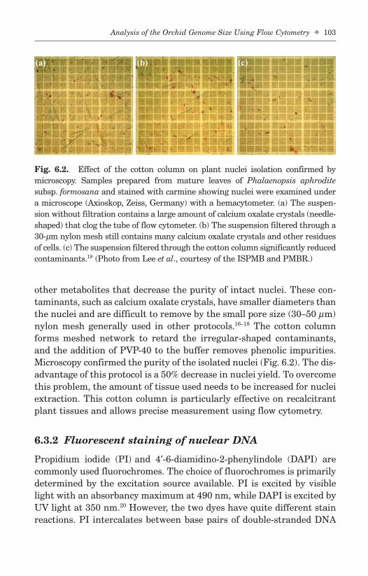
other metabolites that decrease the purity of intact nuclei. These con-taminants, such as calcium oxalate crystals, have smaller diameters thanthe nuclei and are difficult to remove by the small pore size (30–50 µm)nylon mesh generally used in other protocols.16–18 The cotton columnforms meshed network to retard the irregular-shaped contaminants,and the addition of PVP-40 to the buffer removes phenolic impurities.Microscopy confirmed the purity of the isolated nuclei (Fig. 6.2). The dis-advantage of this protocol is a 50% decrease in nuclei yield. To overcomethis problem, the amount of tissue used needs to be increased for nucleiextraction. This cotton column is particularly effective on recalcitrantplant tissues and allows precise measurement using flow cytometry.
6.3.2 Fluorescent staining of nuclear DNA
Propidium iodide (PI) and 4′-6-diamidino-2-phenylindole (DAPI) arecommonly used fluorochromes. The choice of fluorochromes is primarilydetermined by the excitation source available. PI is excited by visiblelight with an absorbancy maximum at 490 nm, while DAPI is excited byUV light at 350 nm.20 However, the two dyes have quite different stainreactions. PI intercalates between base pairs of double-stranded DNA
Analysis of the Orchid Genome Size Using Flow Cytometry ✦ 103
FA
Fig. 6.2. Effect of the cotton column on plant nuclei isolation confirmed bymicroscopy. Samples prepared from mature leaves of Phalaenopsis aphroditesubsp. formosana and stained with carmine showing nuclei were examined undera microscope (Axioskop, Zeiss, Germany) with a hemacytometer. (a) The suspen-sion without filtration contains a large amount of calcium oxalate crystals (needle-shaped) that clog the tube of flow cytometer. (b) The suspension filtered through a30-µm nylon mesh still contains many calcium oxalate crystals and other residuesof cells. (c) The suspension filtered through the cotton column significantly reducedcontaminants.19 (Photo from Lee et al., courtesy of the ISPMB and PMBR.)
b494_Ch-06.qxd 8/29/2007 1:31 PM Page 103

and RNA with little or no base specification,21 while DAPI is a nonin-tercalating stain that binds preferentially and in a complex manner toA-T base regions.22 Highly significant differences were detected in plantnuclear DNA contents obtained using DAPI and PI and this cast doubton the reliability of base preference fluorochromes.23 PI-based flowcytometry produced results consistent with those based on Feulgenmicrospectrophotometry; DAPI was suggested not to be used as the solebasis for DNA content comparisons.20 PI was recommended as the fluo-rochrome of choice for flow cytometric determination of plant DNA con-tent. DAPI should be used only if the estimated DNA value iscorroborated by using a second stain that has no bias for AT- or GC-richsequences within genomes.20
6.3.3 DNA reference standards
Using proper reference standards for flow cytometric determination ofDNA contents is important. Most of the published DNA values forplants have been calibrated against chicken erythrocytes. However, thismay result in significant error in estimated DNA contents, as there isconsiderable reported variations in DNA content among chicken lines.24
Price et al.25 suggested that for technical reasons a plant standard is bet-ter for estimating the DNA content of plants. Five species are recom-mended by Johnston et al.20 as an initial set of international standardsfor plant DNA content determinations: Sorghum bicolor cv. Pioneer8695 (2C = 1.74 pg); P. sativum cv. Minerva Maple (2C = 9.56 pg);Hordeum vulgare cv. Sultan (2C = 11.12 pg); Vicia faba (2C = 26.66 pg);and Allium cepa cv. Ailsa Craig (2C = 33.55 pg). It is recommended thatone of the reference standards be chosen for determination of DNA con-tent of plant genome.
6.4 Nuclei DNA Contents of Orchids
6.4.1 Nuclei DNA contents of Phalaenopsis sp.
Chromosome numbers for the orchids are usually examined andstained by basic fuchsin and acetic orcein. There is a 6.07-fold varia-tion in genome size within 18 Phalaenopsis species, ranging from2.74 pg/2C for P. sanderiana to 16.61 pg/2C for P. parishii (Table 6.1).11
104 ✦ T.-Y. Lin and H.-C. Lee
FAb494_Ch-06.qxd 8/29/2007 1:31 PM Page 104

Analysis of the Orchid Genome Size Using Flow Cytometry ✦ 105
FA
Table 6.1. Nuclear DNA Contents of Phalaenopsis sp. Determined byFlow Cytometry of Propidium Iodide Attained Nuclei using P. sativumMinerva Maple as the Standard11
MeanGenus Sectiona Speciesa TSRI Cloneb (pg/2C)
Phalaenopsis Phalaenopsis P. aphrodite Rchb.f.c W01–38 2.80P. sanderiana Rchb.f. W36–01 2.74P. stuartiana Rchb.f. W40–04 3.13
Stauroglottis P. equestris (Schauer) W09–51 3.37Rchb.f.
Amboinenses P. amboinensis W02–07 14.36J. J. Smith
P. amboinensis W02–08 14.50J. J. Smith
P. gigantea S82–314 5.28J. J. Smith
P. micholitzii Rolfe W27–05 6.49P. venosa Shim & W42–01 9.52
Fowl.Zebrinae P. fasciata Rchb.f. W10–01 6.56
P. lueddemanniana W23–02 6.49Rchb.f.
P. modesta J. J. Smith W28–06 5.15P. mariae Burb. W26 6.48
ex Warn. & Wms.P. pulchra (Rchb. f.) W33–04 6.37
SweetP. sumatrana W41–03 6.62
Korth. & Rchb.f.P. bellina (Rchb. f.) S82–429 15.03
Cristensond
Polychilos P. cornu-cervi (Breda) W08–01 6.44Bl & Rchb.f.
P. mannii Rchb.f. W25–06 13.50P. mannii Rchb.f. W25–07 13.61
Parishianae P. parishii Rchb.f. W32 16.61
aClassification according to Sweet (1980).bSource of plant material: Taiwan Sugar Research Institute (TSRI).cChromosome number are 2n = 2x = 38.dNomenclature according to Christenson and Whitten (1995).
b494_Ch-06.qxd 8/29/2007 1:31 PM Page 105

Chromosome numbers of different TSRI clones of P. aphrodite wereconfirmed to be 2n = 2x = 38 for W01-38; 2n = 3x = 57 for W01-41; and2n = 4x = 76 for W01-22. The nuclear DNA contents of diploidy,triploidy and tetraploidy of P. aphrodite had a ratio near 2:3:4, cor-responding to their somatic chromosome numbers, 38, 57 and 76(Fig. 6.3). The linear relationship between nuclear DNA contents andchromosome numbers of these three clones indicated the accuracy offlow cytometry.
6.4.2 Intraspecific variations in genome size
No variation in DNA content was found between the two differentP. amboinesis clone, W02–07 (14.36 pg/2C) and W02–08 (14.50 pg/2C)or between the two different P. mannii clones, W25–07 (13.50 pg/2C)and W25–06 (13.61 pg/2C) (Table 6.1). Our DNA 2C-values of 3.37pg/2C for P. equestris and 6.49 pg/2C for P. lueddemanniana are smallerthan that previously reported (5.53 pg/2C for P. equestris and 8.65 pg/2Cfor P. lueddemanniana).26 These differences could have resulted fromintraspecific variations.11
106 ✦ T.-Y. Lin and H.-C. Lee
FA
0
2
4
6
8
Diploidy Triploidy Tetraploidy
Mea
n D
NA
con
tent
(pg
/2C
)
Fig. 6.3. Nuclear DNA contents of diploidy, triploidy, and tetraploidy ofP. aphrodite determined by flow cytometry of propidium iodide stained nuclei.The DNA contents (mean ± SE) of diploidy, triploidy, and tetraploidy ofP. aphrodite are 2.8 ± 0.06, 4.52 ± 0.16 and 5.89 ± 0.10 pg/2C, respectively.11
Nuclei extracted from P. sativum Minerva Maple were used as the standard.
b494_Ch-06.qxd 8/29/2007 1:31 PM Page 106

6.5 Endoreduplication Occurrence in Orchid
6.5.1 Phenomenon of endoreduplication
Cell polyploidy is often due to endoreduplication, when one or severalrounds of DNA synthesis occur in the absence of mitosis or cytokinesis,resulting in an increase in the genomic DNA content.4,27–29 Endoredupli-cation is common in animal and plant cells, especially in those under-going differentiation and expansion.30–32 Endoreduplication in theendosperm and cotyledons of developing seeds is well documented, but italso occurs in many tissues throughout the plant. An increase in ploidymay be part of the differentiation of a single cell, as it is in Arabidopsistrichomes,33–35 and may also be related to cell size.36 Cell size in yeastincreases with increasing ploidy.37 In plants, increases in ploidy level arerelated to increases in nuclear volume and cell size. Cells that have under-gone endoreduplication are larger than comparable cells that have not.5,38
Endoreduplication is widespread in orchid plants. Sturdier flowersand better forms may accompany an increase in ploidy in orchids.Although chromosome doubling is a technique for breeders to obtainnew cultivars, little is known regarding the mechanism of endoredupli-cation in orchids. Use of flow cytometry in determining polyploidy isfast and makes determining polyploidy easier.
6.5.2 Occurrence of endoreduplication duringorgan development
The frequency of nuclei at different ploidy levels differs in variousorgans and is correlated with development. Older tissues often possessnuclei of higher ploidy levels than younger ones within the same plant.For example, in Cucumis sativus, ploidy levels were described as a con-tinuous process during the developmental stages from seed to floweringplants.9 The distribution of nuclei at different ploidy levels also changeswith the developmental stages as determined by flow cytometry. Thenumber of endoreduplication cycles appears to be controlled by devel-opmental signals.27 Nevertheless, direct evidence for the link betweendevelopment and the ploidy level in various organs and tissues of plantstill remains to be determined.4
In P. aphrodite and P. equestris, endoreduplication stopped once theflower was fully expanded,12,39 and ploidy levels stabilized, similar to
Analysis of the Orchid Genome Size Using Flow Cytometry ✦ 107
FAb494_Ch-06.qxd 8/29/2007 1:31 PM Page 107

leaves in Arabidopsis,8 cucumber,9 and several succulents.13 Floral budsand opened flowers in an inflorescence had different frequencies of endo-reduplication. The distribution of endopolyploidy also varied among thedifferent parts of a flower (Fig. 6.4). The proportion of endoploidy incells increased with the increasing size of a floral bud. A similar patternof polysomaty is found in the fully opened flowers. Our results showedthat the younger floral buds have higher proportions of 2C cells. Todescribe the relationship between endoreduplication and cell growth, amodel was designed combining a logistic growth model with an endo-reduplication model of Schweizer et al.14 Our results indicate that cellswith higher C values had lower transition rates and less potential forfurther endoreduplication, and when cessation of endoreduplicationoccurred, the flower fresh weight also stopped increasing. The averagecell fresh weight was positively correlated to the average C value, sug-gesting that endoreduplication in Phalaenopsis is a contributing factorto cell growth.39
The Arabidopsis plant-specific cyclin-dependent kinase CDKB1;1and transcription factor E2Fa-DPa control the balance between mitoticdivision and endoreduplication.40 Plants which overexpressed a dominantnegative allele of CDKB1;1 were found to undergo enhanced endoredu-plication, demonstrating that the CDKB1;1 activity is required toinhibit the endocycle. In E2Fa-DPa–over expressing plants, DNA repli-cation is strongly activated. Plant organ cell number and cell size mightbe controlled by modulation of genes involved in cyclin D-CDK inhibitorinteraction. Plant CDK inhibitors, ICK1/KRP1 can move between cellsand can inhibit entry into mitosis.41
6.5.3 Endoreduplication influenced byenvironmental perturbations
Environmental perturbations can influence endoreduplication. For exam-ple, both DNA endoreduplication and mitosis in maize endosperm cellswere significantly correlated with decreased fresh weight and nucleinumber after exposure to 35°C for 4 to 6 days.42 In Arabidopsis, reduc-tions in light intensity and soil water content reduces the extent ofendoreduplication.42 The hypocotyls cells go through several cycles ofendoreduplication, and the process is differentially regulated in thelight and in the dark.44 Activated phytochrome specifically inhibits athird endoreduplication cycle in developing hypocotyls cells. The 16C
108 ✦ T.-Y. Lin and H.-C. Lee
FAb494_Ch-06.qxd 8/29/2007 1:31 PM Page 108

Analysis of the Orchid Genome Size Using Flow Cytometry ✦ 109
FA
TS97u, 2n=3x TS97v, 2n=4x
0
20
40
60
80
F1 F2 B1 B2 B3 B4 B5 B60
20
40
60
80
F1 F2 B1 B2 B3 B4 B5 B6
0
20
40
60
80
F1 F2 B1 B2 B3 B4 B5 B6
0
20
40
60
80
F1 F2 B1 B2 B3 B4 B5 B6
0
20
40
60
80
F1 F2 B1 B2 B3 B4 B5 B6
0
20
40
60
80
F1 F2 B1 B2 B3 B4 B5 B6
0
20
40
60
80
F1 F2 B1 B2 B3 B4 B5 B6
0
20
40
60
80
F1 F2 B1 B2 B3 B4 B5 B6
0
20
40
60
80
F1 F2 B1 B2 B3 B4 B5 B6
0
20
40
60
80
F1 F2 B1 B2 B3 B4 B5 B6
lateral petals & sepals lateral petals & sepals
lower labellum lower labellum
upper labellum
gynostemium
upper labellum
gynostemium
ovary ovary
Fig. 6.4. Frequencies of cells with various C-values in different parts offlower. The flowers of TS97u (2n = 3x) and TS97v (2n = 4x) in a florescence werenumbered from the bottom to the top, with the oldest flower as number one.The ploidy distribution of two inflorescences was recorded. The fully openedflowers had a similar pattern of polysomaty (�: 2C, �: 4C, ▲: 8C, �: 16C).
b494_Ch-06.qxd 8/29/2007 1:31 PM Page 109

nuclei were present in the hypocotyls of phytochrome ( phy) A mutants,but not in that of wild type Arabidopsis plants under continuous far redlight.45 The salt treatment (150 mM NaCl) induced chromosome endore-duplication during the differentiation of cells in the root cortex ofSorghum bicolor cv. 610 plants, but not in the cells of the root vascularcylinder or the leaves. In another S. bicolor genotype DK 34-Alabama,noncompetent for salt adaptation, the same salt treatment did notinduce chromosome endoreduplication in the root cortex cells.Endopolyploidy may be considered as a part of the adaptive response ofS. bicolor competent genotypes to salinity.46 The number of endoredu-plication cycles appears to be under the control of developmental andenvironmental signals.
Acknowledgments
We thank AH Marhkart for comments on the manuscript and theNational Science Council, Republic of China for grant NSC 92-2317-B-007-001 and Council of Agriculture, Republic of China for grants 94AS-5.2.1-ST-a1 and 95AS-6.2.1-ST-a1.
References
1. Arditti J. (1997) Fundamentals of Orchid Biology. Wiley, New York.2. Greilhuber J. (1998) Intraspecific variation in genome size: A critical
reassessment. Ann Bot 82(Supp. A):27–35.3. Greilhuber J, Obermayer R. (1997) Genome size and maturity group in
Glycine max (soybean). Heredity 78:547–551.4. Joubès J, Chevalier C. (2000) Endoreduplication in higher plants. Plant
Mol Biol 43:735–745.5. Kudo N, Kimura Y. (2002) Nuclear DNA endoreduplication during petal
development in cabbage: Relationship between ploidy levels and cell size.J Exp Bot 53:1017–1023.
6. Gary W, Gary LB. (2004) Encyclopedia of Biomaterials and BiomedicalEngineering. Marcel Dekker, New York.
7. Dolezel J, Bartos J. (2005) Plant DNA flow cytometry and estimation ofnuclear genome size. Ann Bot 95:99–110.
8. Galbraith DW, Harkins KR, Knapp S. (1991) Systemic endopolyploidy inArabidopsis thaliana. Plant Physiol 96:985–989.
110 ✦ T.-Y. Lin and H.-C. Lee
FAb494_Ch-06.qxd 8/29/2007 1:31 PM Page 110

9. Gilissen LJW, van Staveren MJ, Creemers-Molenaar J, Verhoeven HA.(1993) Development of polysomaty in seedlings and plants of Cucumissativus L. Plant Sci 91:171–179.
10. Smulders MJM, Rus-Kortekaas W, Gilissen LJW. (1994) Development ofpolysomaty during differentiation in diploid and tetraploid tomato(Lycopersicon esculentum) plants. Plant Sci 97:53–60.
11. Lin S, Lee HC, Chen WH, et al. (2001) Nuclear DNA contents of Phalaenopsissp. and Doritis pulcherrima. J Amer Soc Hort Sci 126:195–199.
12. Yang M, Loh CS. (2004) Systemic endopolyploidy in Spathoglottis plicata(Orchidaceae) development. BMC Cell Biol 5:33.
13. de Rocher EJ, Harkins KR, Galbraith DW, Bohnert HJ. (1990)Developmentally regulated systemic endopolyploidy in succulents withsmall genomes. Science 250:99–101.
14. Schweizer L, Yerk-Davis GL, Phillips RL, et al. (1995) Dynamics of maizeendosperm development and DNA endoreduplication. Proc Natl Acad SciUSA 92:7070–7074.
15. Brown SC, Bergounioux C. (1989) Plant flow cytometry. In: Yen A (ed.),Flow Cytometry: Advanced Research and Clinical Applications, Vol. 2.CRC Press, Boca Raton, Florida, pp. 195–220.
16. Galbraith DW, Harkins KR, Maddox JM, et al. (1983) Rapid flow cytopho-tometric analysis of the cell cycle in intact plant tissues. Science220:1049–1051.
17. Arumuganathan K, Earle ED. (1991) Estimation of nuclear DNA contentof plants by flow cytometry. Plant Mol Bio Rep 9:229–233.
18. Ulrich I, Ulrich W. (1991) High-resolution flow cytometry of nuclear DNAin higher plants. Protoplasma 165:212–215.
19. Lee HC, Lin TY. (2005) Isolation of plant nuclei suitable for flow cytome-try from recalcitrant tissue using a filtration column. Plant Mol Biol Rep23:53–58.
20. Johnston JS, Bennett MD, Rayburn AL, et al. (1999) Reference standardsfor determination of DNA content of plant nuclei. Amer J Bot 86:609–613.
21. Properi E, Giangare MC, Bottiroli G. (1991) Nuclease induced DNA struc-tural changes assessed by flow cytometry with the intercalating dye pro-pidium iodide. Cytometry 12:323–329.
22. Godelle B, Cartier D, Marie D, et al. (1993) Heterochromatin study demon-strating the non-linearity of fluorometry useful for calculating genomicbase composition. Cytometry 14:618–626.
23. Dolezel J, Sgorbati S, Lucretti S. (1992) Comparison of three DNA fluo-rochromes for flow cytometric estimation of nuclear DNA content inplants. Physiol Plantarum 85:625–631.
24. Bennett MD, Leitch I. (1995) Nuclear DNA amounts in angiosperms. AnnBot 76:113–176.
Analysis of the Orchid Genome Size Using Flow Cytometry ✦ 111
FAb494_Ch-06.qxd 8/29/2007 1:31 PM Page 111

25. Price HJ, Bachmann K, Chambers KL, Riggs J. (1980) Detection ofintraspecific variation in nuclear DNA content in Microseris douglasii. BotGaz 141:195–198.
26. Jones WE, Kuehnle AR, Arumuganathan K. (1998) Nuclear DNA contentof 26 orchid (Orchidaceae) genera with emphasis on Dendrobium. Ann Bot82:189–194.
27. Traas J, Hülskamp M, Gendreau E, Höfte H. (1998) Endoreduplicationand development: Rule without dividing? Curr Opin Plant Biol 1:498–503.
28. Grafi G, Larkins BA. (1995) Endoreduplication in maize endosperm:Involvement of M phase-promoting factor inhibition and induction of Sphase-related kinases. Science 269:1262–1264.
29. Grafi G. (1998) Cell cycle regulation of DNA replication: The endoredupli-cation perspective. Exp Cell Res 244:372–378.
30. Nagl W. (1976) DNA endoreduplication and polyteny understood as evolu-tionary strategies. Nature 261:614–615.
31. Brodsky WY, Uryvaeva LV. (1977) Cell polyploidy: Its relation to tissuegrowth and function. Int Rev Cytol 50:275–332.
32. Larkins BA, Dilkes BP, Dante RA, et al. (2001) Investigating the hows andwhys of DNA endoreduplication. J Exp Bot 52:183–192.
33. Hülskamp M, Schnittger A, Folkers U. (1999) Pattern formation and celldifferentiation: Trichomes in Arabidopsis as a genetic model system. IntRev Cytol 186:147–178.
34. Szymanski DB, Lloyd AM, Marks MD. (2000) Progress in the moleculargenetic analysis of trichome initiation and morphogenesis in Arabidopsis.Trends Plant Sci 5:214–219.
35. Folker U, Berger J, Hülskamp M. (1997) Cell morphogenesis of trichomesin Arabidopsis: Differential control of primary and secondary branching bybranch initiation regulators and cell growth. Development 124:3779–3786.
36. Kondorosi E, Roudier F, Gendreau E. (2000) Plant cell-size control: Growingby ploidy? Curr Opin Plant Biol 3:488–492.
37. Galitski T, Saldanha AJ, Styles CA, et al. (1999) Ploidy regulation of geneexpression. Science 285:251–254.
38. Mizukami Y. (2001) A matter of size: Development control of organ size inplants. Curr Opin Plant Biol 4:533–539.
39. Lee HC, Chiou DW, Chen WH, et al. (2004) Dynamics of cell growth andendoreduplication during orchid flower development. Plant Sci166:659–667.
40. Boudolf V, Vlieghe K, Beemster GTS, et al. (2004) The plant-specific cyclin-dependent kinase CDKB1;1 and transcription factor E2Fa-DPa control thebalance of mitotically dividing and endoreduplicating cells in Arabidopsis.Plant Cell 16:2683–2692.
112 ✦ T.-Y. Lin and H.-C. Lee
FAb494_Ch-06.qxd 8/29/2007 1:31 PM Page 112

41. Weinl C, Marquardt S, Kuijt SJ, et al. (2005) Novel functions of plantcyclin-dependent kinase inhibitors, ICK1/KRP1, can act non-cell-autonomously and inhibit entry into mitosis. Plant Cell 17:1704–1722.
42. Engelen-Eigles G, Jones RJ, Phillips RL. (2000) DNA endoreduplication inmaize endosperm cells: The effect of exposure to short-term high temper-ature. Plant Cell Environ 23:657–663.
43. Cookson SJ, Radziejwoski A, Granier C. (2006) Cell and leaf size plasticityin Arabidopsis: What is the role of endoreduplication? Plant Cell Environ29:1273–1283.
44. Gendreau E, Traas J, Desnos T, et al. (1997) Cellular basis of hypocotylgrowth in Arabidopsis thaliana. Plant Physiol 114:295–305.
45. Gendreau E, Hofte H, Grandjean O, et al. (1998) Phytochrome controls thenumber of endoreduplication cycles in the Arabidopsis thaliana hypocotyl.Plant J 13:221–230.
46. Ceccarelli M, Santantonio E, Marmottini F, et al. (2006) Chromosomeendoreduplication as a factor of salt adaptation in Sorghum bicolor. Proto-plasma 227:113–118.
Analysis of the Orchid Genome Size Using Flow Cytometry ✦ 113
FAb494_Ch-06.qxd 8/29/2007 1:31 PM Page 113

FAb494_Ch-06.qxd 8/29/2007 1:31 PM Page 114
This page intentionally left blankThis page intentionally left blank

Chapter 7
The Cytogenetics ofPhalaenopsis Orchids
Yen-Yu Kao*,†,‡, Chih-Chung Lin†, Chien-Hao Huang†
and Yi-Hsueh Li†
All Phalaenopsis species have the same chromosome number(2n = 2x = 38), but their karyotypes and genome sizes vary markedly.The variation is positively correlated with the amount of constitutiveheterochromatin in the genome. Genomic in situ and Southernhybridizations indicate that species with large genomes contain morerepetitive sequences; however, species with small genomes also havetheir own specific repetitive sequences. One family of tandem repeatsnamed Pvr I consisting of 7-bp repeat units was isolated and charac-terized. Chromosomal locations of these repeats coincide with hete-rochromatic blocks.
7.1 Introduction
The genus Phalaenopsis (Orchidaceae) represents an important sourcefor floricultural commodity. Many species in this genus and interspecificand intergeneric hybrids are of high value due to their graceful andlong-lasting flowers. Phalaenopsis plants are slow-growing perennials
115
FA
*Corresponding author.†Department of Botany, National Taiwan University, Taipei 106, Taiwan.‡Institute of Molecular and Cellular Biology, National Taiwan University, Taipei 106,Taiwan.
b494_Ch-07.qxd 8/29/2007 1:32 PM Page 115

that take two to three years to reach maturity. In addition, each plantproduces very few roots and flowers, making cytogenetic analysis diffi-cult. According to Sweet,1 the genus Phalaenopsis is composed ofapproximately 47 species that are grouped into nine sections, whileChristenson2 recognizes 63 species in the genus and divides them intofive subgenera and eight sections. These species are widely distributed,ranging from the Himalayas of northern India, through Southeast Asiato northern Australia.1 Previous studies showed that all Phalaenopsisspecies possess the same chromosome number (2n = 2x = 38),3,4 but theirkaryotypes differ markedly.5,6 Arends7 studied meiotic chromosomeassociations at metaphase I of eight interspecific hybrids. His resultsindicated that hybrids between species both with large chromosomes(e.g. P. amboinensis × P. mannii) and between species both with smallchromosomes (e.g. P. amabilis × P. stuartiana) had complete bivalentformation, whereas hybrids between one species with large and theother species with small chromosomes (e.g. P. mannii × P. equestris)showed low frequencies of bivalents.
Two molecular techniques, fluorescence in situ hybridization(FISH) and genomic in situ hybridization (GISH), have been devel-oped and shown as effective tools for genome research. FISH is usefulfor localization of specific DNA sequences on chromosomes,8 whileGISH, in which total genomic DNA from one species is the probe, candiscriminate parental chromosomes in interspecific or intergenerichybrids.9,10,11 In addition, genomic Southern hybridization reveals theamount and distribution of repetitive DNA sequences in genomes, andis therefore also a useful tool to achieve an understanding of genomedifferentiation between species.12,13 The karyotypes, genome organiza-tion and localization of tandemly repetitive sequences on chromo-somes of some representative Phalaenopsis species were studied by usin the past few years. The results from these studies are summarizedbelow.
7.2 Karyotypes
Karyotypes of nine Phalaenopsis species were analyzed based onFeulgen-stained somatic metaphase chromosomes prepared from roottips.14 Consistent with previous studies,4,6 all the species are diploidswith a chromosome number of 2n = 38, but their chromosomes vary
116 ✦ Y.-Y. Kao et al.
FAb494_Ch-07.qxd 8/29/2007 1:32 PM Page 116

in size and centromere position. The chromosomes of five species,P. aphrodite, P. stuartiana, P. equestris, P. cornu-cervi and P. lueddeman-niana, are small and uniform in size (1–2.5 µm) and are all metacentricor submetacentric (Fig. 7.1A–B; Table 7.1). However, two species,P. venosa and P. amboinensis have strongly asymmetrical and bimodalkaryotypes. They possess small, medium and large chromosomes, mostof them being subtelocentric or acrocentric (Fig. 7.1C). These two speciesdiffer in the number of small chromosomes: P. venosa has 12 small chro-mosomes whereas P. amboinensis has 8 (Table 7.1). The karyotype of
The Cytogenetics of Phalaenopsis Orchids ✦ 117
FA
Fig. 7.1. Feulgen-stained somatic metaphase chromosomes. (A) Phalaenopsisstuartiana; (B) P. cornu-cervi; (C) P. venosa; (D) P. violacea; (E) P. mannii. Scalebar = 10 µm.
b494_Ch-07.qxd 8/29/2007 1:32 PM Page 117

118✦
Y.-Y. Kao et al.
FA
Table 7.1. Somatic Metaphase Chromosomes, Total Chromosome Volume (TCV), Nuclear DNA Content andConstitutive Heterochromatin (CH) of Nine Phalaenopsis Species
TSRIb Nuclear DNA CHc
Accession Contentd (% NuclearTaxona Number 2n <2.0 2.0–2.5 >2.5 TCVc (µm3) (pg 2C−1) Area)
Phalaenopsis
Sect. PhalaenopsisP. aphrodite Rchb.f. W1 38 38 0 0 ND 2.80 ± 0.06 1.87 ± 0.24P. stuartiana Rchb.f. W40 38 38 0 0 23.70 ± 4.51 3.13 ± 0.07 1.77 ± 0.42
Sect. StauroglottisP. equestris (schauer) Rchb.f. W9 38 38 0 0 22.29 ± 4.04 3.37 ± 0.05 2.74 ± 0.33
Sect. PolychilosP. cornu-cervi Rchb.f. W8 38 34 4 0 35.91 ± 5.75 6.44 ± 0.16 2.44 ± 0.47P. mannii Rchb.f. W25 38 0 6 32 86.06 ± 19.06 13.50 ± 0.12 11.32 ± 1.08
Sect. AmboinensesP. amboinensis J. J. Smith W2 38 8 4 26 ND 14.36 ± 0.19 14.17 ± 1.08P. venosa Shim & Fowl W42 38 12 4 22 79.76 ± 24.98 9.52 ± 0.27 13.25 ± 0.88
Sect. ZebrinaeP. lueddemanniana Rchb.f. W23 38 28 10 0 44.58 ± 3.48 6.49 ± 0.22 5.51 ± 0.46P. violacea Witte (Bornean W43 38 6 6 26 87.84 ± 8.32 15.03 ± 0.21 27.17 ± 2.03
form)
aClassification of Sweet.1bTaiwan Sugar Research Institute.cKao et al.14
dLin et al.17
ND: not determined.
ChromosomeSize (µm)c
b494_Ch-07.qxd 8/29/2007 1:32 PM Page 118

P. violacea is also bimodal, with12 chromosomes being small or mediumand 26 chromosomes, large (Table 7.1). The 16 largest chromosomes aremetacentric or submetacentric, while the remaining 10 large chromo-somes are subtelocentric or acrocentric (Fig. 7.1D). P. mannii has com-paratively more symmetrical karyotype, with 6 chromosomes beingmedium and 32, large (Table 7.1). Most of its chromosomes are meta-centric or submetacentric (Fig. 7.1E).
The variation in genome size between related species is attributedto the difference in the amount of heterochromatin.15,16 In order tounderstand the causes of karyotype variation in Phalaenopsis species,the amount of constitutive heterochromatin (CH) in the cell cycle frominterphase was estimated and compared (Fig. 7.2; Table 7.1). The value,expressed as a percentage of the total area of CH in the interphase
The Cytogenetics of Phalaenopsis Orchids ✦ 119
FA
Fig. 7.2. Feulgen-stained interphase nuclei. (A) Phalaenopsis aphrodite;(B) P. lueddemanniana; (C) P. amboinensis; (D) P. violacea. Scale bar = 10 µm.
b494_Ch-07.qxd 8/29/2007 1:32 PM Page 119

nucleus, showed a range of a 15-fold variation, ranging from 1.77%in P. stuartiana to 27.17% in P. violacea (Table 7.1). The amount ofCH is positively correlated with the nuclear DNA content as reportedby Lin et al.17 (Table 7.1). Therefore, we suggested that differentialaccumulation of CH may be a major cause for karyotype variation inPhalaenopsis orchids.
The distribution of CH at late prophase/early metaphase chromo-somes indicates that large chromosomes generally contain more CHthan small ones, and this is true for chromosomes both within andbetween species (Fig. 7.3). In P. aphrodite, P. stuartiana, P. equestris,
120 ✦ Y.-Y. Kao et al.
FA
Fig. 7.3. Feulgen-stained late prophase/early metaphase chromosomes.(A) Phalaenopsis aphrodite; (B) P. cornu-cervi; (C) P. venosa. (D) P. violacea.Scale bar = 10 µm.
b494_Ch-07.qxd 8/29/2007 1:32 PM Page 120

P. cornu-cervi and P. lueddemanniana, all species containing smallchromosomes, CH mainly clusters around centromeres (Fig. 7.3A–B).In P. venosa, P. amboinensis and P. violacea, both ends of large meta-centric/submetacentric chromosomes and the ends of the long arms ofsubtelocentric/acrocentric chromosomes possess large blocks of CH,whereas small chromosomes have little or no CH (Fig. 7.3C–D). Theheterochromatin blocks in P. mannii are smaller in size comparedwith those in the species with bimodal karyotypes, and the distribu-tion of CH is not restricted to chromosome ends. To understand thebase composition of CH, root-tip cells were stained with the fluo-rochrome 4′-6-diamidino-2-phenyl-indole (DAPI). The result showedthat DAPI-positive bands coincide with CH regions as revealed byFeulgen staining. Since DAPI binds preferentially to AT-rich regions,18
the result suggests that the DNA of CH in Phalaenopsis orchids is richin AT base pairs.
7.3 Genome Organization and RelationshipsRevealed by GISH
Meiotic chromosome associations in interspecific hybrids have long beenused to indicate genome homology between plant species,19 but the degreeof chromosome pairing is not the only criterion for evaluating species rela-tionships. Additionally, analysis of chromosome pairing in Phalaenopsis isdifficult because: 1) each plant produces very few flowers, hindering col-lection of sufficient microsporocytes at the right stages for analysis; 2)microsporocytes are enclosed in a thick callose wall, which hampers stainpenetration; and 3) meiotic chromosomes cannot be spread well due toclumping and stickiness. GISH has been shown to be a powerful tool foridentifying genome constitution,20,21 and therefore, may be a method com-plementing analysis of meiotic chromosome pairing. In order to under-stand the relationships of Phalaenopsis species with various sizes ofgenomes, we examined seven interspecific hybrids of Phalaenopsis byGISH.22 The results are listed in Table 7.2. In hybrid P. aphrodite ×P. sanderiana, in which both parents contained small genomes, theparental chromosome sets could not be distinguished even in the presenceof 100-fold blocking DNA (Fig. 7.4A–B). A similar result was observed inhybrid P. mannii × P. violacea, in which both parents possessed largegenomes (Fig. 7.4C–D). Normal chromosome pairing in hybrids with
The Cytogenetics of Phalaenopsis Orchids ✦ 121
FAb494_Ch-07.qxd 8/29/2007 1:32 PM Page 121
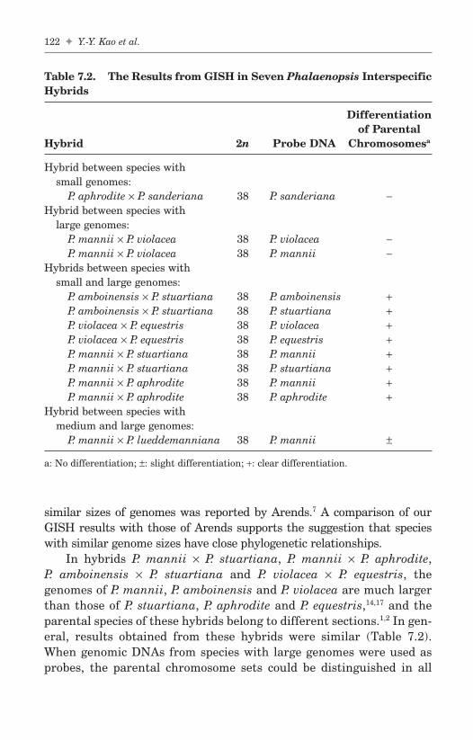
similar sizes of genomes was reported by Arends.7 A comparison of ourGISH results with those of Arends supports the suggestion that specieswith similar genome sizes have close phylogenetic relationships.
In hybrids P. mannii × P. stuartiana, P. mannii × P. aphrodite,P. amboinensis × P. stuartiana and P. violacea × P. equestris, thegenomes of P. mannii, P. amboinensis and P. violacea are much largerthan those of P. stuartiana, P. aphrodite and P. equestris,14,17 and theparental species of these hybrids belong to different sections.1,2 In gen-eral, results obtained from these hybrids were similar (Table 7.2).When genomic DNAs from species with large genomes were used asprobes, the parental chromosome sets could be distinguished in all
122 ✦ Y.-Y. Kao et al.
FA
Table 7.2. The Results from GISH in Seven Phalaenopsis InterspecificHybrids
Differentiationof Parental
Hybrid 2n Probe DNA Chromosomesa
Hybrid between species withsmall genomes:
P. aphrodite × P. sanderiana 38 P. sanderiana −Hybrid between species with
large genomes:P. mannii × P. violacea 38 P. violacea −P. mannii × P. violacea 38 P. mannii −
Hybrids between species withsmall and large genomes:
P. amboinensis × P. stuartiana 38 P. amboinensis +P. amboinensis × P. stuartiana 38 P. stuartiana +P. violacea × P. equestris 38 P. violacea +P. violacea × P. equestris 38 P. equestris +P. mannii × P. stuartiana 38 P. mannii +P. mannii × P. stuartiana 38 P. stuartiana +P. mannii × P. aphrodite 38 P. mannii +P. mannii × P. aphrodite 38 P. aphrodite +
Hybrid between species withmedium and large genomes:
P. mannii × P. lueddemanniana 38 P. mannii ±
a: No differentiation; ±: slight differentiation; +: clear differentiation.
b494_Ch-07.qxd 8/29/2007 1:32 PM Page 122

The Cytogenetics of Phalaenopsis Orchids ✦ 123
FA
Fig. 7.4. Genomic in situ hybridization in Phalaenopsis interspecific hybrids.Somatic metaphase chromosomes prepared from the hybrids were probed withFITC-labeled genomic DNA from one parent in the presence or absence ofunlabeled (blocking) DNA from the other parent. Chromosomes were counter-stained with propidium iodide. (A–B) Hybrid P. aphrodite × P. sanderiana chro-mosomes probed with P. sanderiana DNA in the absence (A) and presence of100× blocking DNA (B); (C–D), Hybrid P. mannii × P. violacea chromosomesprobed with P. mannii DNA in the absence (C) and presence of 100× blockingDNA (D); (E), Hybrid P. mannii × P. stuartiana chromosomes probed withP. mannii DNA in the absence of blocking DNA; (F–G) Hybrid P. mannii ×P. stuartiana chromosomes probed with P. stuartiana DNA in the absence (F)and presence of 50× blocking DNA (G); (H–I), Hybrid P. mannii × P. luedde-manniana chromosomes probed with P. mannii DNA in the absence (H) andpresence of 50× blocking DNA (I). Scale bar = 10 µm.
b494_Ch-07.qxd 8/29/2007 1:33 PM Page 123

hybrids without the application of blocking DNA (Fig. 7.4E). Thestrength and distribution of hybridization signals in these hybridsindicate that species with large genomes contain more repetitivesequences than species with small genomes and confirm distant rela-tionships between the two parental species based on morphologicalcharacters.1,2 The results are consistent with Arend’s findings thathybrids between species with dissimilar sizes of genomes showed poorchromosome pairing.7 When total DNA from species with smallgenomes was used as the probe in the absence of blocking DNA,strong signals dispersed on chromosomes of the species with smallgenomes (Fig. 7.4F). After addition of 50× unlabeled blocking DNAfrom species with large genomes, hybridization signals were restrictedto centromeric regions of small chromosomes (Fig. 7.4G). Theseresults suggest that species with small genomes have their own spe-cific sequences at or around centromeres.
In hybrid P. mannii × P. lueddemanniana, nuclear DNA contentsof these two parents differ approximately twofold; addition of 50-foldblocking DNA was necessary to distinguish the parental chromo-somes (Fig. 7.4H–I). This observation indicates that P. mannii andP. lueddemanniana share a high degree of sequence homology and,therefore, have a close phylogenetic relationship. This is consistentwith the cytological work of Arends,7 who found complete bivalentformation at the first meiotic division in hybrid P. lueddemanniana ×P. mannii.
7.4 Repetitive Sequences
7.4.1 (GA)n microsatellites
Microsatellites, also called simple sequence repeats (SSRs), are tandemrepeated arrays of short core sequence. One microsatellite, (GA)11, hasbeen mapped physically to seven Phalaenopsis species and Doritis pul-cherrima.23 (GA)11 signals are dispersed on the chromosomes ofP. amboinensis and P. violacea, but clustered in the centromeric regionsof P. stuartiana, P. equestris, P. aphrodite, P. mannii, P. lueddemannianaand D. pulcherrima chromosomes (Fig. 7.5A–B). Signals were muchstronger in P. stuartiana and P. aphrodite than in other species.In GISH experiment of hybrids between species with small and large
124 ✦ Y.-Y. Kao et al.
FAb494_Ch-07.qxd 8/29/2007 1:33 PM Page 124

genomes mentioned above, when total DNA from species with smallgenomes was used as the probe in the presence of 50× blocking DNA,hybridization signals were restricted to the centromeric regions ofthe small chromosomes (Fig. 7.4G). From these, we suspect that GISHsignals in the centromeric regions of P. stuartiana, P. aphrodite, andP. equestris chromosomes were from hybridization of (GA)n microsatel-lite sequences.
The Cytogenetics of Phalaenopsis Orchids ✦ 125
FA
Fig. 7.5. Fluorescence in situ hybridization of (GA)11 microsatellite (A–B)and Pvr-93 sequences (C–D) on chromosomes of Phalaenopsis species. The probewas labeled with digoxigenin and detected with anti-digoxigenin-fluorescein.Chromosomes were counterstained with propidium iodide. (A) Phalaenopsisstuartiana; (B) P. equestris; (C) P. venosa; (D) P. mannii. Scale bar = 10 µm.
b494_Ch-07.qxd 8/29/2007 1:33 PM Page 125

7.4.2 Pvr I tandem repeats
Genomic Southern hybridization was used to investigate the amountand distribution of repetitive DNA sequences in Phalaenopsis.24 Thehybridization patterns in P. violacea and P. mannii were similar whenthe total genomic DNA from P. violacea was the probe. This result sug-gests that the kind and amount of repetitive sequences in these twospecies are similar. This may explain the difficulty of discriminating thechromosomes of these two species by GISH (Fig. 7.4C–D; Table 7.2).In addition, the probe from P. violacea hybridized strongly to DNAsfrom species with large genomes, but weakly to DNAs from species withsmall genomes. However, when total genomic DNA from P. aphroditewas used as a probe, an opposite result was obtained, i.e. species withsmall genomes showed stronger signals than did species with largegenomes. These results are consistent with the observations of GISHthat species with large genomes contain more repetitive sequences andspecies with small genomes have their own specific sequences.
One family of tandemly repeated sequences, Pvr I, was isolated fromP. violacea.24 Twelve clones of this family were sequenced. Sequences ofthese clones are not found in the EMBL/Genbank database. All sequencesare AT-rich at the 5′- and 3′-ends and consist of 7-bp repeat units organ-ized in tandems, ranging from 13 to 22 copies. The consensus sequence ofthese repeats is GTAAGCC. Physical mapping of one sequence of thisfamily, Pvr-93, was preformed by FISH on mitotic metaphase chromo-somes prepared from the root tips of Phalaenopsis species and the relatedspecies Doritis pulcherrima. The results revealed that in species withlarge genomes, including P. violacea, P. mannii, P. amboinensis, P. venosaand D. pulcherrima, Pvr-93 sequences were located in the constitutiveheterochromatin regions of large chromosomes corresponding to DAPI-positive bands (Fig. 7.5C–D). No hybridization signals were observed onthe small chromosomes of P. aphrodite and P. equestris. These resultsindicate that Pvr I sequences are species-specific and reside in AT-richheterochromatic blocks of large chromosomes.
7.5 Conclusion
The nine representative Phalaenopsis species studied by us have thesame chromosome number (2n = 2x = 38), but their karyotypes differ
126 ✦ Y.-Y. Kao et al.
FAb494_Ch-07.qxd 8/29/2007 1:33 PM Page 126

markedly. We suggest that differential accumulation of CH is a majorcause for karyotype variation. Our results from FISH, GISH and genomicSouthern hybridization demonstrate that the Phalaenopsis species pos-sess different kinds and amounts of repetitive sequences. A more detailedunderstanding of genome organization and relationships in Phalaenopsiswill result from comparative studies of chromosome pairing at meiosis,isolation and characterization of species-specific sequences.
Acknowledgment
We are grateful to Dr. Chi-Chang Chen for his critical reading of thismanuscript.
References
1. Sweet HR. (1980) The Genus Phalaenopsis. Day Printing Corporation,California.
2. Christenson EA. (2001) Phalaenopsis: A Monograph. Timber Press,Portland, Oregon.
3. Sagawa Y. (1962) Cytological studies of the genus Phalaenopsis. AmerOrchid Soc Bull 31:459–465.
4. Sagawa Y, Shoji T. (1968) Chromosome numbers in Phalaenopsis andDoritis. Bul Pacific Orchid Soc 26:9–13.
5. Woodard JW. (1951) Some chromosome numbers in Phalaenopsis. AmerOrchid Soc Bull 20:356–358.
6. Shindo K, Kamemoto H. (1963) Karyotype analysis of some species ofPhalaenopsis. Cytologia 28:390–398.
7. Arends JC. (1970) Cytological observations on genome homology in eightinterspecific hybrids of Phalaenopsis. Genetica 41:88–100.
8. Jiang J, Gill BS. (1994) Nonisotopic in situ hybridization and plant genomemapping: the first 10 years. Genome 37:717–725.
9. Schwarzacher T, Leitch AR, Bennett MD, Heslop-Harrison JS. (1989) In situlocalization of parental genomes in a wide hybrid. Ann Bot 64:315–324.
10. Ørgaard M, Heslop-Harrison JS. (1994) Investigations of genome relation-ships between Leymus, Psathyrostachys, and Hordeum inferred bygenomic DNA: DNA in situ hybridization. Ann Bot 73:195–203.
11. D’Hont A, Paget-Goy A, Escoute J, Carreel F. (2000) The interspecificgenome structure of cultivated banana, Musa spp. revealed by genomicDNA in situ hybridization. Theor Appl Genet 100:177–183.
The Cytogenetics of Phalaenopsis Orchids ✦ 127
FAb494_Ch-07.qxd 8/29/2007 1:33 PM Page 127

12. Ørgaard M, Heslop-Harrison JS. (1994) Relationships between species ofLeymus, Psathyrostachys, and Hordeum (Poaceae, Triticeae) inferred fromSouthern hybridization of genomic and cloned DNA probes. Plant SystEvol 189:217–223.
13. Aggarwal RK, Brar DS, Khush GS. (1997) Two new genomes in the Oryzacomplex identified on the basis of molecular divergence analysis usingtotal genomic DNA hybridization. Mol Gen Genet 254:1–12.
14. Kao YY, Chang SB, Lin TY, et al. (2001) Differential accumulation of het-erochromatin as a cause for karyotype variation in Phalaenopsis orchids.Ann Bot 87:387–395.
15. Flavell R. (1982) Sequence amplification, deletion and rearrangement:Major sources of variation during species divergence. In: Dover FA, FlavellRB (eds.), Genome Evolution. Academic Press, London, pp. 301–323.
16. Kubis S, Schmidt T, Heslop-Harrison JS. (1998) Repetitive DNA elementsas a major component of plant genomes. Ann Bot 82 (Suppl. A):45–55.
17. Lin S, Lee HC, Chen WH, et al. (2001) Nuclear DNA contents of Phalaenopsisspecies and Doritis pulcherrima. J Amer Soc Hort Sci 126:195–199.
18. Sumner AT. (1990) Chromosome Banding. Unwin Hyman, London.19. Singh RJ. (2003) Plant Cytogenetics. 2nd Edition. CRC Press, Boca Raton,
Florida, pp. 277–306.20. Bennett ST, Kenton AY, Bennett MD. (1992) Genomic in situ hybridization
reveals the allopolyploid nature of Milium montianum (Gramineae).Chromosoma 101:420–424.
21. Fukui K, Shishido R, Kinoshita T. (1997) Identification of the rice D-genomechromosomes by genomic in situ hybridization. Theor Appl Genet 95:1239–1245.
22. Lin CC, Chen YH, Chen WH, et al. (2005) Genome organization and rela-tionships of Phalaenopsis orchids inferred from genomic in situ hybridiza-tion. Bot Bull Acad Sin 46:339–345.
23. Li YH. (2000) Investigation on the physical distribution of microsatellitesin Phalaenopsis orchids by fluorescence in situ hybridization. Master the-sis, Department of Botany, National Taiwan University, Taipei, Taiwan.
24. Huang CH. (1999) Isolation and characterization of two repetitive DNAsequences in Phalaenopsis orchids. Master thesis, Department of Botany,National Taiwan University, Taipei, Taiwan.
128 ✦ Y.-Y. Kao et al.
FAb494_Ch-07.qxd 8/29/2007 1:33 PM Page 128
Chapter 8
Analysis of theChloroplast Genome ofPhalaenopsis aphrodite
Ching-Chun Chang*,†, Hsien-Chia Lin† and Wun-Hong Zeng†
The complete nucleotide sequence of the chloroplast genome of theTaiwan moth orchid (Phalaenopsis aphrodite subsp. formosana) wasdetermined. The circular, double-stranded DNA of 148,964 bp comprisesa pair of inverted repeats of 25,732 bp, which are separated by a smallsingle copy (SSC) and a large single copy (LSC) region of 11,543 and85,957 bp, respectively. The genome contains 76 protein coding genes,four ribosomal RNA genes, 30 tRNA genes, and 24 putative open readingframes (ORFs). Seventeen genes are intron-containing, including 6 tRNAand 11 protein coding genes. Unlike other chloroplast genomes of photo-synthetic angiosperms, which have a complete set of genes in the 11 sub-units of NADH dehydrogenase, the chloroplast genome of Phalaenopsiscompletely lacks the ndhA, ndhH, and ndhF genes. The other eight ndhgenes have various degrees of nucleotide insertion/deletion, and they areall frameshifted. This loss results in the SSC region in Phalaenopsis beingthe shortest among known photosynthetic angiosperms.
8.1 Introduction
Chloroplasts, in which photosynthesis takes place, have distinct functionalgenomes. They were once free-living cyanobacteria and even now containpreserved remnants of eubacterial genomes,1 but with some of their genes
129
FA
*Corresponding author.†Institute of Biotechnology, National Cheng Kung University, Tainan, Taiwan.
b494_Ch-08.qxd 8/29/2007 1:40 PM Page 129

having been transferred to the nucleus of the cell during the course of evo-lution.2,3 Since the complete plastid genome sequences of tobacco andliverwort were reported in 1986,4,5 over 30 complete sequences repre-senting the major lineages of vascular plants have been made availablein the genomic database of NCBI (http://www.ncbi.nlm.nih.gov/genomes/ORGANELLES/plastids_tax.html). The DNA molecules in the chloroplastgenome of photosynthetic plants are circular and range in size from 116 to204 kb. With few exceptions, such as in conifers6 and Medicago (accessionnumber NC_003119, unpublished), most of the genomes that have beenexamined have a large (5–76 kb) inverted repeat (IR) segment, whichaccounts for the variation in length of the genomes.2,7,6 Two segments ofthe IRs are divided by a large single copy (LSC) and a small single copy(SSC) region.2,7 Among vascular plants, chloroplast genome contains110–130 genes, and most of these genes code for proteins mostly involvedin photosynthesis or gene expression, with the remainder being transferRNA or ribosomal RNA genes.8 Furthermore, the polycistronic transcrip-tion units of the known chloroplast genomes are also highly conserved.7
Although the plastid genomes of vascular plants are conserved inthe overall structure,7 comparative genomic studies have recentlyrevealed many evolutionary hotspots and single nucleotide polymor-phism that are useful for phylogenetic studies below the family and sub-species level.9–12 Furthermore, genome-wide analysis of transcripts inthe chloroplast has revealed that plants have undergone dramaticchanges in both the levels and patterns of editing, from hornworts(1.2%) and ferns (0.38%) to seed plants (less than 0.05%).13,14 In additionto phylogenetic studies, complete genome sequences provide usefulinformation for designing expression cassettes for chloroplast geneticmanipulation and for elucidating functional genomics.15–17
With approximately 30,000 species, orchids are one of the largest andmost diverse families of flowering plants.18 The mode of chloroplasts DNAtransmission in orchids is uniparentally inherited from female ances-tors.19,20 Based on molecular markers from plastid genes, the phylogeneticrelationship has been investigated at the family and subfamily levelwithin Orchidaceae.21,22 As a part of a larger effort to preserve the nativespecies and to utilize its chloroplast genomes for various biotechnologicalpurposes, we sequenced the complete chloroplast genome of Phalaenopsisaphrodite subsp. formosana. It is anticipated that knowledge of thechloroplast genome of this species will be useful for commercial breedingand for phylogenetic classification within Orchidaceae as well as forgenome-based analysis in angiosperm evolution.
130 ✦ C.-C. Chang, H.-C. Lin and W.-H. Zeng
FAb494_Ch-08.qxd 8/29/2007 1:40 PM Page 130

8.2 Phalaenopsis Chloroplast DNA Extraction,Genome Sequencing, and Gene Annotation
Leaves of Phalaenopsis aphrodite subsp. formosana were obtained fromseedlings at the four-leaf stage of development. Intact chloroplasts werefractionated with step percoll (40–80%) gradient.23 The chloroplast DNAwas further isolated according to a CTAB-based protocol.24 ChloroplastDNA was randomly fragmented into 2 to 3 kb pieces through hydros-hear with GeneMachine (Genomic Solutions Inc., Michigan, USA), andthen cloned into the pBluescriptSK vector. After transformation, posi-tive shotgun clones were propagated on microtiter plates. PlasmidDNAs were isolated and used as templates for sequence analysis.
The sequencing reaction was performed using the BigDye terminatorcycle sequencing kit (Applied Biosystems, USA), according to the proto-col recommended by the manufacturer. The DNA sequencer used was anApplied Biosystems ABI 3700. The sequence data from both ends of eachshotgun clone were accumulated, trimmed, aligned, and assembled usingPhred-Phrap programs (Phil Green, University of Washington, Seattle,USA). The sequence data were accumulated to 1,728,000 nucleotides,which is 11.6X coverage for the chloroplast genome of P. aphrodite subsp.formosana. Database searches were conducted with the BLAST algo-rithm provided by the National Center for Biotechnology Information(NCBI). The tRNAscan program was used to assign the tRNA genes.
8.3 Phalaenopsis Chloroplast Genome Properties
8.3.1 Overall genome structure
The complete circular chloroplast DNA of Phalaenopsis aphroditesubsp. formosana (the Taiwan moth orchid) is 148,964 bp (accessionnumber AY916449) long and can be separated into two regions by a pairof inverted repeat regions (IR) of 25,732 bp (Fig. 8.1). The large singlecopy region (LSC) and the small single copy region (SSC) are 85,957 bpand 11,543 bp long, respectively. The overall A+T content of the chloro-plast genome of P. aphrodite subsp. formosana is 63.4%, which is closeto the A+T percentages of rice (61%), maize (61.5%), wheat (61.7%),wild rice (61%), and sugarcane (61.6%). The A+T contents in the LSCand SSC region are higher, being 66.1% and 71.9%, respectively, than inthe IR regions (56.8%). The gene order in the Phalaenopsis chloroplast
Analysis of the Chloroplast Genome of Phalaenopsis aphrodite ✦ 131
FAb494_Ch-08.qxd 8/29/2007 1:40 PM Page 131

132 ✦ C.-C. Chang, H.-C. Lin and W.-H. Zeng
FA
Fig. 8.1. Gene map of Phalaenopsis aphrodite subsp. formosana chloroplastgenome.30 Genes shown inside circle are transcribed clockwise; those outside aretranscribed counterclockwise. Genes belonging to different functional groupsare color coded. Asterisks indicate genes containing introns.
b494_Ch-08.qxd 8/29/2007 1:40 PM Page 132

genome is more similar to that of the known dicots than to the grasses,as in the latter, gene rearrangements caused by inversion are extensivein the LSC region.9,25–28
8.3.2 Gene content, intron, and codon usage
Table 1 presents the content of genes in the Phalaenopsis chloroplastDNA. A total of 110 genes (not including genes duplicated in the IR) wereidentified as being scattered around the chloroplast genome. They include76 protein coding genes, four ribosomal RNA genes, and 30 transfer RNAgenes. Among them, 17 are intron-containing genes, including 6 tRNA and11 protein coding genes (Table 2). Three of the latter genes, i.e. clpP, rps12,and ycf3, contain two introns. In the five grasses, there are 16 intron-containing genes, and the clpP and rpoC1 genes have lost introns.9,25–28
The rps12 gene is a divided gene whose 5′-end exon is located in the LSCregion, but whose second and third exons are in the IR regions. It appearsthat a trans-splicing mechanism is required between exon I and exon II toyield mature rps12 transcripts. The intron of the trnL-UAA gene containsminisatellite repeat elements in the marsh orchid, Orchis palustris,29 butin P. aphrodite subsp. formosana it does not.
In addition, 24 ORFs were identified with a threshold of 225 bp. AnORF, orf91, located in the complementary strand of the rrn23 gene, wasreported for the first time.30 Its amino acid sequence is 81% identical toa hypothetical protein (NCBI accession no. ZP_00203428) encoded inthe genome of Anabaena variabilis. We also observed the orf91 tran-scripts in the total RNAs of P. aphrodite and tobacco by RT-PCR assayand RNA filter hybridization,30 which demonstrated that orf91 wasactually transcribed in the chloroplasts. However, further studies arerequired to determine if the orf91 transcripts are translated and tocharacterize the function of the translated protein.
The codon usage in Phalaenopsis chloroplast genome and the anti-codons of 30 tRNA species are summarized in Table 3. The codon usagedetermined from 67 protein coding genes, is strongly biased towardA or T due to the high A-T content at the third codon positions. For exam-ple, degenerated codons at the third position exhibit usage frequenciesof 65% to 81% for both A or T. The frequencies of the stop codon usagefor UAA, UAG, and UGA are 48%, 27%, and 25%, respectively. Theseare also strongly biased toward A or T at both the second and third posi-tions. The strong AT bias of codon usage in Phalaenopsis is consistent
Analysis of the Chloroplast Genome of Phalaenopsis aphrodite ✦ 133
FAb494_Ch-08.qxd 8/29/2007 1:40 PM Page 133

134✦
C.-C
. Ch
ang, H
.-C. L
in an
d W.-H
. Zen
g
FA
Table 8.1. List of Gene Contents in Phalaenopsis Chloroplast Genome (Total 110 Genes)
Group of Genes
RNA genesrRNA genes rrn4.5,b rrn5,b rrn16,b rrn23b
tRNA genes trnA (ugc),a,b trnC (gca), trnD (guc), trnE (uuc), trnF (gaa), trnG (gcc),trnG (ucc),a trnH (gug),b trnI (cau), trnI (gau),a,b trnK (uuu),a trnL (caa),b
trnL (uag), trnL (uaa),a trnM (cau),b trnfM (cau), trnN (guu),b trnP (ugg),trnQ (uug), trnR (ucu), trnR (acg),b trnS (gcu), trnS (uga), trnS (gga),trnT (ggu), trnT (ugu), trnV (gac),b trnV (uac),a trnW (cca), trnY (gua)
Protein coding genesRibosomal proteins
Small subunit rps2, rps3, rps4, rps7,b rps8, rps11, rps12,a,b rps14, rps15, rps16,a rps18, rps19b
Large subunit rpl2,a,b,c rpl14, rpl16,a rpl20, rpl22, rpl23,b rpl32, rpl33, rpl36Transcription/translation apparatus
DNA dependent RNA polymerase rpoA, rpoB, rpoC1,a rpoC2Translation initiation factor infA
PhotosynthesisSubunits of Photosystem I psaA, psaB, psaC, psaI, psaJ, ycf3,a ycf4Subunits of Photosystem II psbA, psbB, psbC, psbD, psbE, psbF, psbH, psbI, psbJ, psbK, psbL, psbM, psbN,
psbT, psbZSubunits of Cytochrome b6/f complex petA, petB,a petD,a petG, petL, petNSubunits of ATP synthase atpA, atpB, atpE, atpF,a atpH, atpISubunits of NADH-dehydrogenase ψ -ndhB,a,b ψ -ndhC, ψ -ndhD, ψ -ndhE, ψ -ndhG, ψ -ndhI, ψ -ndhJ, ψ -ndhKLarge subunit of RubisCO rbcL
(Continued )
b494_Ch-08.qxd 8/29/2007 1:40 PM Page 134

An
alysis of the C
hloroplast G
enom
e of Ph
alaenopsis aph
rodite✦
135
FA
Table 8.1. (Continued )
Group of Genes
Miscellaneous proteins accD, clpP,a matK, cemA, ccsAConserved proteins ycf1, ycf 2b
Open reading frames ORF114 (175d) ORF80 (1754d) ORF98 (22677d) ORF87B (23929d)ORF103 (51817d) ORF88 (61993e) ORF87C (64106d) ORF85 (69399e)ORF79 (70125d) ORF99 (70354e) ORF81C (74208e) ORF86B (86687d)
b
ORF90 (93146d)b ORF81B (97939d)
b ORF79 (99557d)b ORF77A (100484d)
b
ORF81A (102460d)b ORF115 (102606d)
b ORF86A (104928d)b ORF170 (105185d)
b
ORF91 (109119d)b ORF131 (110120d)
b ORF77B (112628e) ORF87A (122213d)
aIntron containing gene.bTwo copies due to inverted repeat.cInitiation codon created by RNA editing.dORF translated clockwise.eORF translated counterclockwise.The position of start codon of putative ORF is given in parenthesis.
b494_Ch-08.qxd 8/29/2007 1:40 PM Page 135

136 ✦ C.-C. Chang, H.-C. Lin and W.-H. Zeng
FA
Table 8.2. The Splitting Genes on Phalaenopsis Chloroplast Genome
Gene Location Exon I Intron I Exon II Intron II Exon III
atpF LSC 145 986 410clpP LSC 71 981 292 689 252ndhB IR 733 532 763petB LSC 6 726 642petD LSC 8 865 484rpl2 IR 391 664 431rpl16 LSC 9 1251 399rpoC1 LSC 453 758 1608rps12* IR 114 — 232 547 26rps16 LSC 245 914 40trnA-ugc IR 38 802 35trnG-ucc LSC 23 687 48trnI-gau IR 37 944 35trnK-uuu LSC 37 2896 35trnL-uaa LSC 35 717 50trnV-uac LSC 39 582 35ycf3 LSC 124 731 230 750 153
*The rps12 gene is divided. The 5′-rps12 locates on the LSC region, while the 3′-rps12locates on the IR region. The number indicates the length of the intron or exon in thebase pairs.
with the results published elsewhere concerning the chloroplastgenome of many higher plants.5,11,25,27
8.3.3 Loss of ndh genes
Ndh genes that encode the subunits of the NADH dehydrogenase com-plex are involved in the cyclic electron flow of photosystem I andchlororespiration in tobacco.15 All 11 subunits of ndh genes are present inthe chloroplast genomes of photosynthetic angiosperms so far sequenced.In contrast, in the chloroplast genomes of black pine, the ndhA, ndhG,ndhF, and ndhJ genes were completely absent, and sequences of theother seven ndh genes were truncated as obvious pseudogenes.31 InPhalaenopsis, the ndhA, ndhF, and ndhH genes are completely absentfrom the chloroplast genome, and the other remaining eight ndh genesare frameshifted. The ndhB, ndhC, and ndhJ genes have small region ofnucleotide insertion/deletion, and the ndhD, ndhE, ndhG, ndhI, and
b494_Ch-08.qxd 8/29/2007 1:40 PM Page 136

An
alysis of the C
hloroplast G
enom
e of Ph
alaenopsis aph
rodite✦
137
FA
Table 8.3. Codon Usage of the Phalaenopsis Chloroplast Genome
UUU F 612 trnF-GAA UCU S 398 trnS-GGA UAU Y 475 trnY-GUA UGU C 147 trnC-GCAUUC F 331 UCC S 215 UAC Y 116 UGC C 48UUA L 557 trnL-UAA UCA S 254 trnS-UGA UAA — 32 UGA — 17 trnW-CCAUUG L 386 trnL-CAA UCG S 100 UAG — 18 UGG W 308
CUU L 363 trnL-UAG CCU P 287 trnP-UGG CAU H 355 trnH-GUG CGU R 272 trnR-ACGCUC L 113 CCC P 167 CAC H 95 CGC R 60CUA L 226 CCA P 200 CAA Q 501 trnQ-UUG CGA R 250CUG L 119 CCG P 68 CAG Q 158 CGG R 64
AUU I 727 trnI-GAU ACU T 379 trnT-GGU AAU N 612 trnN-GUU AGU S 289 trnS-CCUAUC I 294 ACC T 152 AAC N 165 AGC S 62AUA I 423 trnI-CAU ACA T 263 trnT-UGU AAA K 663 trnK-UUU AGA R 331 trnR-UCUAUG M 395 trnM-CAU ACG T 98 AAG K 243 AGG R 119
trnfM-CAU
GUU V 369 trnV-GAC GCU A 473 trnA-UGC GAU D 570 trnD-GUC GGU G 433 trnG-GCCGUC V 129 GCC A 138 GAC D 124 GGC G 122GUA V 357 trnV-UAC GCA A 312 GAA E 701 trnE-UUC GGA G 490 trnG-UCCGUG V 155 GCG A 90 GAG E 228 GGG G 207
b494_Ch-08.qxd 8/29/2007 1:40 PM Page 137

ndhK genes are truncated with large deletion of 26%, 60%, 45%, 46%, and32% of the sequences, respectively, as compared with that of tobacco.Probably, all the resident ndh genes were pseudogenes. The loss of func-tional ndh genes explains well why P. aphrodite subsp. formosana has thesmallest SSC region among the sequenced monocots. Although themajority of the ndh genes have been lost from the chloroplast genome ofPinus and Phalaenopsis, the ndhF gene was present in all vascular plantdivisions,32 suggesting that the lost ndhF and other ndh genes in the plas-tid genome of Phalaenopsis might have been transferred to the nucleargenome. Indeed, we were able to detect the presence of ndhA, ndhF, andndhH gene fragments by RT-PCR (unpublished data). Further studieswill unravel the details of the lost ndh genes in the plastid of P. aphrodite.
8.3.4 RNA editing
RNA editing plays an important role in the regulation of gene expressionin chloroplasts. The plastid rpl2 gene of Phalaenopsis has an ACG codonrather than an ATG codon at the translation initiation site (positions88242–88244) as was observed in maize.33 The ACG rather than an ATGwas also found at the initiation codon of the ndhD genes (positions115315–115317), as in tobacco and spinach.34 Furthermore, frameshiftswere observed in the ndh genes, suggesting RNA editing is required torestore their gene function. Our RT-PCR assays indicated that RNA edit-ing occurs in the orchid because a C to U conversion was verified in theinitiation codon of rpl2 transcripts.30 However, a C to U conversion was notapparent in the ndhD transcripts, which further indicates that ndhD is apseudogene. Furthermore, we did not detect RNA editing in the ndhC,ndhD, ndhJ, and ndhK transcripts for repairing the internal stop codon,30
further implying that the eight variously truncated and frameshifted ndhgenes are probably pseudogenes. Currently, we have been analyzing thetranscripts of 76 protein coding genes from P. aphrodite subsp. formosanato extensively study the pattern of RNA editing in chloroplast.
8.3.5 IR contraction and expansion
The border between the inverted repeat region and the single copy regionusually shifts among the various plastome species.11,26,35 Using tobaccoas a representative of dicots, a comparison of the exact IR-border posi-tions and their adjacent genes among tobacco, orchid, maize, rice,and wheat is shown in Fig. 8.2. This comparison further demonstrates
138 ✦ C.-C. Chang, H.-C. Lin and W.-H. Zeng
FAb494_Ch-08.qxd 8/29/2007 1:40 PM Page 138

that the border positions vary among the chloroplast genomes ofangiosperms. As illustrated in Fig. 8.2, the IRb/LSC border in P.aphrodite subsp. formosana is located within the coding region of rpl22,while in maize, rice, and wheat it is located up-stream of the noncoding
Analysis of the Chloroplast Genome of Phalaenopsis aphrodite ✦ 139
FA
Fig. 8.2. Comparison of border positions of LSC/IR and SSC/IR regions andgene order within two LSC/IR regions among five chloroplast genomes.30
Various lengths of ycf1 or ndhH pseudogenes are created in tobacco, riceand wheat at the IRb/SSC border. 29 bp of ndhF pseudogene is created in maizeat the SSC/IRa border. 31 bp of rpl22 pseudogene is created in Phalaenopsisaphrodite subsp. formosana at the IRa/LSC border. Both rps19 and trnH genesare located within IR region in P. aphrodite subsp. formosana, maize, rice,wheat; whereas in tobacco they are located in LSC. The rps15 gene is located inSSC in P. aphrodite subsp. formosana and tobacco, whereas it is present in theIR regions in the chloroplast genomes of five grasses. The ycf1 and ycf2 genesare lost in five grasses, while ndhA, ndhF, ndhH genes are lost in P. aphroditesubsp. formosana. Black triangles indicate loss of genes.
b494_Ch-08.qxd 8/29/2007 1:40 PM Page 139

region of rpl22. The IRa/LSC borders in P. aphrodite subsp. formosana,maize, rice, and wheat are located on the downstream of the non-codingregion of psbA. The IRa/LSC and IRb/LSC borders of tobacco, however,are located downstream of the noncoding region of trnH-GUG andupstream of the non-coding region of rps19, respectively. The IRb/LSCborders in many other dicot plants, such as Panax,11 Spinacia,36
Oenothera,37 Arabidopsis,38 Lotus,39 and Atropa,40 are located within thecoding region of rps19, and as a result, give rise to the rps19 pseudo-genes in IRa. In monocots, the rps19 and trnH genes appear to havebeen completely shifted into the IR region, which may represent amajor structural difference between the chloroplast genomes of mono-cots and dicots.
In P. aphrodite subsp. formosana, the IRa/SSC border (Fig. 8.2) islocated upstream of the noncoding region of ycf1, while in tobacco theborder is in the coding region of ycf1, with the result that the 996 bpof ycf1 pseudogene in IRb is created. In grasses, however, the IRa/SSCborder is either within the initiation codon of the ndhH gene as in maizeor shifted into the central region of the ndhH gene as in rice and wheat,resulting in the creation of the 163 bp and 207 bp of the ndhH pseudogeneat the IRb/SSC borders in rice and wheat, respectively. The IRb/SSCborder resides downstream of the noncoding region of rpl32 in P. aphroditesubsp. formosana and downstream of the coding region of the ndhFgene in tobacco, rice and wheat, but in maize it is shifted into the ndhFcoding region, thereby creating the 29 bp of ndhF pseudogene in IRa.The intramolecular recombination of short direct repeat sequencesprobably is responsible for the shift of the IR/SSC or IR/LSC bordersduring evolution.11,26
The expansions and contractions of the IR region contribute to thevariation in length of the chloroplast genome among plant lineages.11
The IR regions of the grass family (including maize, rice, wild rice,wheat and sugarcane) are shorter than those in P. aphrodite subsp. for-mosana and tobacco, mainly because their ycf2 were lost. The smallersizes in the SSC regions of the six known monocots are due to loss ofthe ycf1 genes in the grass family and the deletion of several ndh genesfrom P. aphrodite subsp. formosana. In P. aphrodite subsp. formosanaand in dicots, the rps15 genes are located in the SSC regions, whilein the grass family the gene is in the IR regions. Likely, a duplication ofthe rps15 gene followed by an inversion occurred in the grass lineageduring evolution.
140 ✦ C.-C. Chang, H.-C. Lin and W.-H. Zeng
FAb494_Ch-08.qxd 8/29/2007 1:40 PM Page 140

8.4 Conclusion and Prospective
The complete nucleotide sequence of the chloroplast genome of theTaiwan moth orchid (Phalaenopsis aphrodite subsp. formosana) wasdetermined. The genome contains 76 protein coding genes, four riboso-mal RNA genes, 30 tRNA genes, and 24 open reading frames (ORFs). Theavailability of complete chloroplast genomic sequence of Phalaenopsisaphrodite subsp. formosana will be very useful in designing molecularmarkers both for commercial breeding and for phylogenetic classificationwithin Orchidaceae. Furthermore, the plastid genomic information pro-vides the fundamentals to study gene expression and regulation in thechloroplasts of Phalaenopsis orchid. In addition, since the lack of com-plete chloroplast genome sequences is still one of the major limitations forextending chloroplast genetic engineering technology to useful crops, acomplete genomic sequence of Phalaenopsis chloroplast will providevaluable information. This information can be used for the constructionof chloroplast expression vector for site-specific integration of foreigngenes through homologous recombination in orchid plastid transforma-tion in the near future.
Acknowledgments
This work was financially supported by grants (NSC92-2317-B006-004)to C.-C. Chang from the National Science Council of Taiwan.
References
1. Martin W, Stoebe B, Goremykin V, et al. (1998) Gene transfer to thenucleus and the evolution of chloroplasts. Nature 393:162–165.
2. Palmer JD. (1991) Plastid chromosome: Structure and evolution. In: Vasil IK,Bogorad L (eds.), Cell Culture and Somatic Cell Genetics in Plants, Vol. 7A,The Molecular Biology of Plastids. Academic Press, San Diego, pp. 5–53.
3. Martin W, Rujan T, Richly E, et al. (2002) Evolutionary analysis ofArabidopsis, cyanobacterial, and chloroplast genomes reveals plastid phy-logeny and thousands of cyanobacterial genes in the nucleus. Proc NatlAcad Sci USA 99:12246–12251.
4. Ohyama K, Fukuzawa H, Kohchi T, et al. (1986) Chloroplast gene organi-zation deduced from complete sequence of liverwort Marchantia polymor-pha chloroplast DNA. Nature 322:572–574.
Analysis of the Chloroplast Genome of Phalaenopsis aphrodite ✦ 141
FAb494_Ch-08.qxd 8/29/2007 1:40 PM Page 141

5. Shinozaki K, Ohme M, Tanaka M, et al. (1986) The complete nucleotidesequence of tobacco chloroplast genome: Its gene organization and expres-sion. EMBO J 5:2043–2049.
6. Raubeson LA, Jansen RK. (2005) Chloroplast genomes of plants. In:Herry RJ (ed.), Plant Diversity and Evolution: Genotypic and PhenotypicVariation in Higher Plants. CABI Publishing, Cambridge, pp. 45–68.
7. Sugiura M. (1995) The chloroplast genome. Essays Biochem 30:49–57.8. Jansen RK, Raubeson LA, et al. (2005). Methods for obtaining and analyz-
ing whole chloroplast genome sequences. Methods Enzymol 395:348–384.9. Asano T, Tsudzuki T, Takahashi S, et al. (2004) Complete nucleotide
sequence of the sugarcane (Saccharum officinarum) chloroplast genome: Acomparative analysis of four monocot chloroplast genomes. DNA Res11:93–99.
10. Calsa T, Carraro DM, Benatti MR, et al. (2004) Structural features andtranscript-editing analysis of sugarcane (Saccharum officinarum L.)chloroplast genome. Curr Genet 46:366–373.
11. Kim KJ, Lee HL. (2004) Complete chloroplast genome sequences fromKorean ginseng (Panax schinseng Nees) and comparative analysis ofsequence evolution among 17 vascular plants. DNA Res 11:247–261.
12. Tang J, Xia H, Cao M, et al. (2004) A comparison of rice chloroplastgenomes. Plant Physiology 135:412–420.
13. Kugita M, Yamamoto Y, Fujikawa T, et al. (2003) RNA editing in hornwortchloroplasts makes more than half the genes functional. Nucleic Acids Res31:2417–2423.
14. Wolf PG, Rowe CA, Hasebe M. (2004) High levels of RNA editing in a vas-cular plant chloroplast genome: Analysis of transcripts from the fernAdiantum capillus-veneris. Gene 339:89–97.
15. Burrows PA, Sazanov LA, Svab Z, et al. (1998) Identification of afunctional respiratory complex in chloroplasts through analysis oftobacco mutants containing disrupted plastid ndh genes. EMBO J17:868–876.
16. Drescher A, Ruf S, Calsa T, et al. (2000) The two largest chloroplastgenome-encoded open reading frames of higher plants are essential genes.Plant J 22:97–104.
17. Daniell H, Khan MS, Allison L. (2002) Milestones in chloroplast geneticengineering: An environmentally friendly era in biotechnology. TrendsPlant Sci 7:84–91.
18. Atwood JT. (1986) The size of the Orchidaceae and the systematic distri-bution of epiphytic orchids. Selbyana 9:171–186.
19. Chang SB, Chen WH, Chen HH, et al. (2000) RFLP and inheritance pat-terns of chloroplast DNA in intergeneric hybrids of Phalaenopsis andDoritis. Botany Bulletin of Academia Sinica 41:219–223.
142 ✦ C.-C. Chang, H.-C. Lin and W.-H. Zeng
FAb494_Ch-08.qxd 8/29/2007 1:40 PM Page 142

20. Cafasso D, Widmer A, et al. (2005) Chloroplast DNA inheritance in theorchid Anacamptis palustris using single-seed polymerase chain reaction.J Hered 96:66–70.
21. Neyland R, Urbatsch LE. (1996) The ndhF chloroplast gene detected in allvascular plant divisions. Planta 200:273–277.
22. Cameron KM. (2004) Utility of plastid psaB gene sequences for investi-gating intrafamilial relationships within Orchidaceae. Mol PhylogenetEvol 31:1157–1180.
23. Robinson SP, Downton WJ. (1984) Potassium, sodium, and chloride con-tent of isolated intact chloroplasts in relation to ionic compartmentationin leaves. Arch Biochem Biophys 228:197–206.
24. Stewart CN Jr, Via LE. (1993) A rapid CTAB DNA isolation technique use-ful for RAPD fingerprinting and other PCR applications. Biotechniques14:748–750.
25. Hiratsuka J, Shimada H, Whittier R, et al. (1989) The complete sequenceof the rice (Oryza sativa) chloroplast genome: Intermolecular recom-bination between distinct tRNA genes accounts for a major plastidDNA inversion during the evolution of the cereals. Mol Gen Genet217:185–194.
26. Maier RM, Neckermann K, Igloi GL, Kossel H. (1995) Complete sequence ofthe maize chloroplast genome: Gene content, hotspots of divergence and finetuning of genetic information by transcript editing. J Mol Biol 251:614–628.
27. Ogihara Y, Isono K, Kojima T, et al. (2002) Structural features of a wheatplastome as revealed by complete sequencing of chloroplast DNA. MolGenet Gen 266:740–746.
28. Shahid Masood M, Nishikawa T, Fukuoka S, et al. (2004) The completenucleotide sequence of wild rice (Oryza nivara) chloroplast genome: Firstgenome wide comparative sequence analysis of wild and cultivated rice.Gene 340:133–139.
29. Cafasso D, Pellegrino G, Musacchio A, et al. (2001) Characterization of aminisatellite repeat locus in the chloroplast genome of Orchis palustris(Orchidaceae). Curr Genet 39:394–398.
30. Chang CC, Lin HC, et al. (2006) The chloroplast genome of Phalaenopsisaphrodite (Orchidaceae): Comparative analysis of evolutionary ratewith that of grasses and its phylogenetic implications. Mol Biol Evol23:279–291.
31. Wakasugi T, Tsudzuki J, Ito S, et al. (1994) Loss of all ndh genes as deter-mined by sequencing the entire chloroplast genome of the black pine Pinusthunbergii. Proc Natl Acad Sci USA 91:9794–9798.
32. Neyland R, Urbatsch LE. (1996) Phylogeny of subfamily Epidendroideae(Orchidaceae) inferred from ndhF chloroplast gene sequences. Amn J Bot83:1195–1206.
Analysis of the Chloroplast Genome of Phalaenopsis aphrodite ✦ 143
FAb494_Ch-08.qxd 8/29/2007 1:40 PM Page 143

33. Hoch B, Maier RM, Appel K, et al. (1991) Editing of a chloroplast mRNAby creation of an initiation codon. Nature 353:178–180.
34. Neckermann K, Zeltz P, Igloi GL, et al. (1994) The role of RNA editing inconservation of start codons in chloroplast genomes. Gene 146:177–182.
35. Goulding SE, Olmstead RG, Morden CW, Wolfe KH. (1996) Ebb and flowof the chloroplast inverted repeat. Mol Gen Genet 252:195–206.
36. Schmitz-Linneweber C, Maier RM, Alcaraz JP, et al. (2001) The plastidchromosome of spinach (Spinacia oleracea): Complete nucleotide sequenceand gene organization. Plant Mol Biol 45:307–315.
37. Hupfer H, Swiatek M, Hornung S, et al. (2000) Complete nucleotidesequence of the Oenothera elata plastid chromosome, representingplastome I of the five distinguishable euoenothera plastomes. Mol GenGenet 263:581–585.
38. Sato S, Nakamura Y, Kaneko T, et al. (1999) Complete structure of thechloroplast genome of Arabidopsis thaliana. DNA Res 6:283–290.
39. Kato T, Kaneko T, Sato S, et al. (2000) Complete structure of the chloro-plast genome of a legume, Lotus Japonicus. DNA Res 7:323–330.
40. Schmitz-Linneweber C, Regel R, Du TG, et al. (2002) The plastid chromo-some of Atropa belladonna and its comparison with that of Nicotianatabacum: The role of RNA editing in generating divergence in the processof plant speciation. Mol Biol Evol 19:1602–1612.
144 ✦ C.-C. Chang, H.-C. Lin and W.-H. Zeng
FAb494_Ch-08.qxd 8/29/2007 1:40 PM Page 144
Chapter 9
Analysis of Expression ofPhalaenopsis Floral ESTs
Wen-Chieh Tsai†, Yu-Yun Hsiao‡, Zhao-Jun Pan‡
and Hong-Hwa Chen*,‡
Orchids have great diversity of specialized pollination and ecologicalstrategies and provide a rich setting for studying evolutionary rela-tionships and molecular biology. The sophisticated orchid flower mor-phology offers an opportunity to discover new variant genes anddifferent levels of complexity in the morphogenesis of flowers. Toobtain plentiful gene information from orchid reproductive organs, weconstructed a cDNA library of mature flower buds of Phalaenopsisequestris, a native diploid species of Phalaenopsis in Taiwan. A total of5,593 expressed sequence tags (ESTs) from randomly selected cloneswere identified and characterized. Cluster analysis enabled the identi-fication of a unigene set of 3,688 sequences. The abundance of tran-scripts with predicted cellular roles were functionally characterized byusing the BLASTX matches to known proteins. Comparison of the rel-ative EST frequencies based on functional categories among floral tis-sues of five species including P. equestris, Acorus Americanus,Asparagus officinalis, Oryza sativa, and Arabidopsis thaliana was per-formed. The most highly transcribed genes in Phalaenopsis floral budsare those coding for RNA-dependent RNA polymerase of Cymbidiummosaic virus, followed by heat shock protein genes. A total 217 puta-tive transcription factor related ESTs were identified. C3H and trihelix
145
FA
*Corresponding author.†Department of Biological Science and Technology, Chung Hwa University of MedicalTechnology, Tainan County, Taiwan.‡Department of Life Sciences, National Cheng Kung University, Tainan, Taiwan.
b494_Ch-09.qxd 8/29/2007 1:42 PM Page 145

families occupied 25% of the transcribed transcription factor genes,indicating that the profile of the transcription factors in orchid flowerbuds is polarized. The extensive analysis of the genes in floral organsadds to the growing repertoire of known plant genes and may alsoreveal unique features of the reproductive organs of orchids.
9.1 Introduction
9.1.1 Arabidopsis and rice structure genomics
Access to a complete, finished genome for any organism provides thebasis for large-scale exploration of biology. With respect to plants,Arabidopsis has secured the historical record of being the first plantgenome to be sequenced (Arabidopsis Genome Initiative, 2000),1 withrice (Oryza sativa) coming in second.2,3 The Arabidopsis genome isessentially complete with the exception of a few gaps, primarily at thecentromeres. The rice genome was completed in December 2004 by apublic effort of the International Rice Genome Sequencing Project(IRGSP; http://rgp.dna.affrc.go.jp/IRGSP/). The value of both organ-isms as model species for plant biology is further supported by theavailability of not one genome sequence, but multiple genome sequences.For Arabidopsis, the public consortium sequenced to the draft level theheavily utilized Columbia accession, while a private company, Cereon,sequenced the second most utilized accession, Landsberg erecta (Ler).4
For rice, there have been four genome sequencing efforts, three beingfocused on the Nipponbare cultivar from the temperate subspeciesjaponica2,5,6 and one on the 93–11 variety from the tropical subspeciesindica.3 In addition to the nuclear genomes, both the chloroplastand mitochondrial genomes of Arabidopsis and rice are publicly avail-able.7–9 Both Arabidopsis and rice genome sequencing was precededby expressed sequence tag (EST) sequencing, as this provides not onlyan inexpensive sampling method for the expressed fraction of agenome, but also a quantitative profile of the expression levels inspecific tissues. ESTs also have utilities, as the cDNA clones them-selves are valuable reagents for functional genomic studies. Currently,there are 326,202 Arabidopsis and 299,998 rice ESTs in thedbEST division of GenBank (http://www.ncbi.nlm.nih.gov/dbEST/dbESTsummary.html).
146 ✦ W.-C. Tsai et al.
FAb494_Ch-09.qxd 8/29/2007 1:42 PM Page 146

9.1.2 The significance of Orchidaceae for plant science
The family of Orchidaceae has an estimated 17,000 to 35,000 species,making it among the largest families of flowering plants.10 The speciesare known for their diversity of both specialized pollination andecological strategies and provide a rich subject for investigating evo-lutionary relationships and molecular biology. The versatility and spe-cialization in orchid floral morphology, structure, and physiologicalproperties have fascinated botanists and collectors for centuries.Their most astonishing evolution is seen in their reproductive biology.Like most angiosperm flowers, orchids have two whorls of perianthsegments. The outer whorl consists of three sepals. Three petals makeup the inner whorl, one of them (the labellum or lip) being highlyevolved with different size, form, color, and general appearance fromthe other two. In some genera, such as Paphiopedilum andPhragmipedium, the labellum has even evolved into a pouch. In allgenera, the male and female reproductive organs are fused to form agynostemium. Modifications of the perianth, androecium, and gynoe-cium can represent a basis for a variety of floral morphology. Thehighly sophisticated flower organization offers the opportunity to dis-cover new variant genes and different levels of complexity in the mor-phogenetic networks.11,12
9.2 Expressed Sequence Tags
9.2.1 Significance of EST in genome research
Single-run partial sequencing of randomly selected clones is a widelyused tool in genome research. Expressed sequence tags (ESTs) haveplayed significant roles in accelerating gene discovery, including genefamily expansion,13,14 large-scale expression analysis,15–17 and elucidat-ing phylogenetic relationships.17 Libraries of cDNAs are routinely pre-pared and contain tens of thousands of clones, representing a variety ofspecific tissue types and a snapshot of gene expression during defineddevelopmental stages and following specific biotic and abiotic chal-lenges. Recent developments in high volume biotechnology combinedwith advanced DNA sequencing technology have made it feasible to per-form large-scale EST sequencing projects.
Analysis of Expression of Phalaenopsis Floral ESTs ✦ 147
FAb494_Ch-09.qxd 8/29/2007 1:42 PM Page 147

The concept of using cDNAs as a route to expedite gene discoverywas first demonstrated in the early 1980s.19 In 1990, Sydney Brennerproposed that an obvious method for characterizing the “important”part of the human genome would involve looking at messengersfrom the expressed genes, thus advocating the application of high-throughput methods for transcriptome sampling.20 Mark Adams firstused the term EST in relation to gene discovery and the humangenome project in 1991.21 Currently there are near 25 million ESTsin the National Center for Biotechnology Information (NCBI) publiccollection-dbEST database (http://www.ncbi.nlm.nih.gov/dbEST/index.html), representing a wide taxonomic variety of fungi, plants,and animals. With many large-scale EST sequencing projects inprogress and new projects being initiated, the number of ESTs in thepublic domain will likely increase substantially, providing additionalopportunities for intra- and inter-specific expression comparisons ona genomics scale.
Expressed sequence tags are created by partially sequencing ran-domly isolated gene transcripts that have been converted into cDNA.EST sequencing initially favored the 5′-end of directionally clonedcDNAs because the 5′-sequences are likely to contain more proteincoding sequence than the 3′-ends, which often contain significantuntranslated regions (UTRs). Currently, the 3′-end of the cDNA cloneis often preferred because it is likely to offer more unique sequence(in many cases, the UTR) and can be used to distinguish betweengene paralogues. EST sequencing strategies in which both ends of thecDNA are sequenced are also becoming prevalent. In the absence ofcomplete genome sequences, the cDNA (and its EST) remains the onlylink back to the genome. However, cDNA sequencing remains one ofthe more accessible and widely used methods for sampling the activelytranscribed portion of the gene space. The preparation of cDNAlibraries depends on the underlying mRNA population of a cell, tissue,or organism. The genomic structure of the host plant is, therefore,largely inconsequential. EST data have been directly applied for genediscovery,22,23 evaluation of the genome-wide gene content and struc-ture,24 as well as in silico comparative expression analysis betweendifferent plant tissues.16,17 Moreover, ESTs can be a valuable resourcefor highthroughput expression analysis via the cDNA-array technol-ogy.25–28 Recent studies have revealed that mRNA transcripts can also
148 ✦ W.-C. Tsai et al.
FAb494_Ch-09.qxd 8/29/2007 1:42 PM Page 148
contain repeat motifs, and the abundance of ESTs available makesthis an attractive potential source of microsatellite markers.29 EST-derived SSRs have been developed for many plants, such as Triticumaestivum L.,30 Hordeum vulgare L.,31 Gossypium L.,32 and Medicagotruncatula.33
9.2.2 Application of full-length cDNAs (FL-cDNA) tofunctional genomics
Most of the EST projects are based on cDNA libraries in which most of theinserts are not full-length. ESTs are useful for making a catalogue ofexpressed genes, but not for further study of gene function. The onlyviable alternatives to EST sequencing that address the attributes ofincomplete sequence coverage and nucleotide quality are the full-lengthcDNA sequences. Consequently, genome-scale collections of the full-lengthcDNAs of expressed genes are both essential for gene identification andannotation of complex genomes and provide a valuable resource for exper-imentalists interested in gene function. In addition, full-length cDNAs areuseful resources for transgenic analysis, such as overexpression, antisensesuppression, double-stranded RNA interference (dsRNAi), and biochemi-cal analysis to study the function of the encoded proteins.34 Thus, therehas been a large effort to produce annotated EST and full-length cDNAcollections for mammals and plants.35–37
Full-length cDNA sequences are obtained by Cap-Trapper or othertechnologies that take advantage of the property of the cap structure,and sequencing clones for both 5′- and 3′-end.38–44 Such a strategy yieldsmany individual ESTs that can be assembled into a single contig andgreatly improves identification of the complete exon structures ofeukaryotic genes.45
9.2.3 Bioinformatics of plant EST collections
It has been shown previously that EST databases are valid and reliablesources of gene expression data.15–17 With the rapid expansion of avail-able EST data, opportunities for digital gene expression analysis willcontinue to expand. As a result of advances in computational molecularbiology and biostatistics, it is possible to mine and analyze large-scaleEST datasets efficiently and exhaustively.15–17
Analysis of Expression of Phalaenopsis Floral ESTs ✦ 149
FAb494_Ch-09.qxd 8/29/2007 1:42 PM Page 149
Bioinformatics-based sequence resources have been developed thataddress the quality, redundancy, and partial nature of EST sequences.The first crucial step in adding value to EST sequences involves clusteringand assembling the ESTs into a more manageably sized dataset of bet-ter quality clustered sequences. Sequences are aggressively trimmed ofvector and polylinker remnants before a fast clustering method placesthe ESTs into buckets of similar sequences. A final assembly step placesthe clustered sequences into logical contigs and singletons.46,47 The clus-tered sequences are typically longer than any individual EST and are ofa higher quality.
These cluster consensus and singleton sequences form the coresequence data within several plant specific EST derived databases.Most of these sequence databases have added further value to thesequences by attaching additional annotation to the sequences and byproviding methods to select specific sequences or groups of sequencesthat satisfy specific criteria. The most valuable annotations andmethods are those that assign a tentative function and allow retrievaland identification of sequences on the basis of tissue or challengespecificity.
9.2.4 Reasons for applying ESTs to studyPhalaenopsis equestris
Based on morphological characteristics, the genus Phalaenopsis com-prises approximately 45 species that are grouped into nine sections.10
They have broad geographical distribution as well as commercial valueas floricultural commodities. Two native species of Phalaenopsis, P.equestris and P. aphrodite var. formosa48 have been reported in Taiwan.Few molecular studies of Phalaenopsis orchids’ reproductive biologyexist because of their long life cycles and inefficient transformationsystems. With the use of flow cytometry, nuclear DNA contents of P.equestris were estimated to be 1.6 × 10⁹ bp (2n = 2x = 38), about four-fold that of the rice genome.49,50 The expansion of plant genomes hasmainly been the result of multiplications of retrotransposon repeatsequences.51 Thus, expressed sequence tags (ESTs) avoid the highlyrepetitive DNA that make up the bulk of most plant genomes andoffer both a reasonable cost and rapid generation of data that can beexploited for gene discovery and comparative genomics.
150 ✦ W.-C. Tsai et al.
FAb494_Ch-09.qxd 8/29/2007 1:42 PM Page 150

9.3 Characterization and Significance ofPhalaenopsis Floral EST
9.3.1 Significance of ESTs shared no significantsimilarity to any other protein sequences inpublic databases
To provide considerable sequence information from orchid, we char-acterized the 5,593 floral bud ESTs reading from the 5′-end. TheseESTs were assembled and generated by the PHRAP program. Amongthem, 2,637 ESTs were assembled into 732 contigs. These contigs and2,956 singletons represented up to 3,688 different genes. The resultsreflected 34.1% (1 − number of unigenes/number of ESTs) redundancyrate of the library. The unigene set was used for similarity searchesand annotation using BLAST algorithm. Putative functions of thecDNAs were assigned after applying a stringency of BLASTX scores>50. More than 75% of the database-matched unigenes were found tomatch proteins from plants (Table 9.1). By contrast, 8.32% of the uni-genes represented genes that had been previously found in non-plantorganisms, such as E. coli, Drosophila, fungi, mice, and humans.Furthermore, about 15% of the unigenes revealed no hits, indicatingthat these sequences may have specific roles in orchids. Although thedeposited sequences in public databases are increasing exponentiallyyear by year, recent EST analysis in non-model plants always showeda substantial portion of expressed genes that are species specific.52–54
Defining the functional identities of these unidentified genes mightadvance the understanding of the differences among plant species.
Analysis of Expression of Phalaenopsis Floral ESTs ✦ 151
FA
Table 9.1. Overview of the Unigenes BlastX Analysisa
Source No. of Unigenes Percentage of Total ESTs (%)
Plant 2,833 76.82Non plant 307 8.32No hits found 548 14.86
Total 3,688 100.000
a Adapted from Tsai et al., Plant Science 170 (2006): 426–432.
b494_Ch-09.qxd 8/29/2007 1:42 PM Page 151
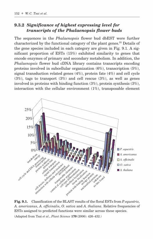
9.3.2 Significance of highest expressing level fortranscripts of the Phalaenopsis flower buds
The sequences in the Phalaenopsis flower bud dbEST were furthercharacterized by the functional category of the plant genes.55 Details ofthe gene species included in each category are given in Fig. 9.1. A sig-nificant proportion of ESTs (15%) exhibited similarity to genes thatencode enzymes of primary and secondary metabolism. In addition, thePhalaenopsis flower bud cDNA library contains transcripts encodingproteins involved in subcellular organization (6%), transcription (5%),signal transduction related genes (4%), protein fate (4%) and cell cycle(3%), tags to transport (3%) and cell rescue (3%), as well as genesinvolved in proteins with binding function (3%), protein synthesis (3%),interaction with the cellular environment (1%), transposable element
152 ✦ W.-C. Tsai et al.
FA
metaboli
sm
energ
y
cell cy
clean
d DNApro
cessi
ng
transc
riptio
n
protein sy
nthesi
s
protein fat
e
cellu
lartra
nsport
signa
l transd
uctio
n
cell res
cue,
deffen
sean
d virulen
ce
interac
tion with
cellu
laren
vironmen
t
develo
pment
transp
osab
leele
ments
contro
l ofce
llular
organ
izatio
n
cell typ
e differe
ntiati
on
subc
elluar
localiza
tion
tissu
e locali
zatio
n
protein with
binding
functio
n
storag
e protei
n
unclas
sified
proteins
unkno
wn
no hits fo
und
0%
5%
10%
15%
20%
25%
P. equestris
A. americanus
A. officinalis
O. sativa
A. thaliana
Fig. 9.1. Classification of the BLAST results of the floral ESTs from P. equestris,A. americanus, A. officinalis, O. sativa and A. thaliana. Relative frequencies ofESTs assigned to predicted functions were similar across these species.
(Adapted from Tsai et al., Plant Science 170 (2006): 426–432.)
b494_Ch-09.qxd 8/29/2007 1:42 PM Page 152

(1%), cellular organization (1%), energy production (1%), and develop-ment (1%). Another 30% showed similarity to sequences of unknownproteins (24%) and unclassified functions (6%).
These ESTs that have been obtained from a non-normalized cDNAlibrary could be used to reveal global gene expression patterns asdeduced from transcript abundance. The highly expressed transcriptsof the Phalaenopsis flower buds are listed in Table 9.2. The highestexpressing level for transcripts of the Phalaenopsis flower buds washomologous to RNA-dependent RNA polymerase (1.98%) of Cymbidiummosaic virus. Although no virus infection symptoms were observed,these transcripts existed significantly in the floral buds, indicating thatexperimental materials used here were infected with the virus. Correlatedwith the highly expressed virus RNA-dependent RNA polymerase tran-scripts, ESTs related to β-1,3-glucanase were also comparably detected(0.86%). Considerable evidence suggested that β-1,3-glucanase is induced
Analysis of Expression of Phalaenopsis Floral ESTs ✦ 153
FA
Table 9.2. Highly Abundant Transcripts Detected in the P. equestrisFlower Bud dbESTc
No. ofPutative Function Organism ESTsa Percentageb (%)
RNA-dependent RNA Cymbidium mosaic 111 1.98polymerase virus
heat shock cognate Dictyostelium 73 1.40protein Hsc70-1 discoideum
beta-1,3-glucanase Oryza sativa 48 0.8640S ribosomal protein S5 Oryza sativa 45 0.80lipid transfer protein Arabidopsis thaliana 44 0.79phospholipid transfer Aerides japonica 36 0.64
proteinmetallothionein-like Musa acuminata 30 0.54
proteinlipid transfer protein Nicotiana glauca 27 0.48mannose-binding Cymbidium hybrid 24 0.43
lectin precursorS-adenosyl-L-methionine Dendrobium 20 0.36
synthetase crumenatum
a Number of clones assigned to the related gene function.b Abundant genes/total number *100.c Adapted from Tsai et al., Plant Scinece 170 (2006): 426–432.
b494_Ch-09.qxd 8/29/2007 1:42 PM Page 153

during response of plants to viral, bacterial, and fungal pathogens.56 Inthe top 10 highly abundant ESTs, three different kinds of ESTs relatedto lipid transfer proteins occupied at least 1.91% of the total ESTs.These lipid transfer protein related genes, including lipid transferprotein per se, its precursor, and phospholipid transfer proteins playroles in defense and pollen tube adhesion in plants.57–59 ESTs homolo-gous to heat shock cognate protein Hsc70-1 genes also presented asignificant expression level (1.4%). Hsp70s function as molecular chap-erones. In addition to heat stress, these proteins can also be inducedby other environmental stresses, including cold, drought, or salinity,as well as during various developmental processes, such as embryogen-esis, germination, and fruit development.60–62 Dissection of these genefunctions might be a useful direction for further study of orchid floraldevelopment.
9.3.3 Significance of similar expression patternsin floral tissues of flowering plants
For comparison of floral transcriptome among different plant species,plentiful EST resources deposited at public databases were down-loaded to the local server for in silico comparative analysis. In partic-ular, ESTs of floral tissue were chosen from Arabidopsis thaliana, themodel plant of eudicots, Oryza sativa, the first monocot whose genomewas completely sequenced (International Rice Genome SequencingProject, 2005);63 Acorus americanus, the basal monocot which pro-vides support as a sister to all other monocots, and Asparagus offici-nalis, an economically important non-grain monocot with the highlyderived floral structures found in the grasses and grass relatives.64
The number of unigene sets were 2,707, 3,934, 4,548, and 4,267 fromassembled floral ESTs of Arabidopsis, rice, A. americanus, andA. officinalis, respectively, and functionally characterized and com-pared to that of P. equestris. The results showed that the relative fre-quencies of ESTs assigned to predicted functions were very similaracross these species (Fig. 9.1). Interestingly, transcripts related to celltype differentiation, tissue localization, and storage protein werebarely detected in the floral tissues of these five species (Fig. 9.1).These findings suggested that the basic floral transcriptomes wereparallel among the flowering plants from basal monocots to monocotsand to eudicots.
154 ✦ W.-C. Tsai et al.
FAb494_Ch-09.qxd 8/29/2007 1:42 PM Page 154

9.3.4 Significance of transcription factor expressionprofiles in the transcriptome of Phalaenopsisflower buds
Because transcription factors control the expression of a genome and playimportant roles in all aspects of a higher plant’s life cycle, we character-ized the transcription factor associated ESTs from the transcriptome ofP. equestris flower bud through the use of Arabidopsis transcription fac-tor sequences as queries.65 In the orchid flower bud dbEST, 217 ESTsencoding putative transcription factors were identified, occupying 4%(217/5593) of flower bud transcriptome. The most abundantly expressedtranscription factor gene families, C3H and trihelix, accounted for a full25% (54/217) of the overall transcription factor expression (Fig. 9.2).In addition, bHLH (9%), C2H2 (8%) and WRKY (6%) families were foundin turn (Fig. 9.2). These five families occupied approximately 50% of theexpressed transcription factors. However, BBR-BPC, CCAAT-DR1,CCAAT-HAP2, CPP, GRF, SBP, VOZ-9, and WHIRLY families were notdetected. The results suggested that expression of transcription factors inmature orchid flower buds is highly regulated.
The C3H family has been reported to be involved in Arabidopsis embryo-genesis66 and response to abscisic acid in Craterostigma plantagineum.67
To our knowledge, documents describing the relationship between C3H
Analysis of Expression of Phalaenopsis Floral ESTs ✦ 155
FA
9
12
5
10
19
7
23
21
2
17
27
10 0
12
0
4
13
12
0
9
2
10
78
53
6
0
11
27
20 0
14
0
5
10
15
20
25
30
ABI3VP1
Alfin-
like
AP2-EREBP
ARF
ARR-B
BBR-BPC
BHLHbZ
IPBZR
C2C2-
CO-like
C2C2-
Dof
C2C2-
Gata
C2C2-
YABBYC2H
2C3H
CAMTA
CCAAT-DR1
CCAAT-HAP2
CCAAT-HAP3
CCAAT-HAP5
CPP
E2F-D
PEIL
G2-lik
eGeB
P
GRASGRF
HBHSF
MADS
MYB-re
latedM
YBNAC
orph
anREM
SBPTCP
Triheli
xTUB
VOZ-9
WHIR
LY
WRKY
No.
of
clon
es
Fig. 9.2. Number of ESTs related to transcription factors appearing in eachtranscription factor family. A total of 1,583 putative Arabidopsis transcriptionfactors were searched against P. equestris flower bud dbEST, and then the tar-get ESTs were classified into corresponding transcription factor families. Eachbar represents the number of EST clones in one family.
(Adapted from Tsai et al., Plant Science 170 (2006): 426–432.)
b494_Ch-09.qxd 8/29/2007 1:42 PM Page 155

and floral development have not been proposed. In addition to involve-ment in light signaling, the trihelix family also regulates perianth archi-tecture in Arabidopsis flowers.68,69 Understanding these gene functionswill advance our understanding of orchid floral development. However,there are eight transcription factor families, including BBR-BPC, CCAAT-DR1, CCAAT-HAP2, CPP, GRF, SBP, VOZ-9, and WHIRLY, which werenot discovered in our floral bud dbEST. A CPP family gene, TSO1, mod-ulates cytokinesis and cell expansion in Arabidopsis flowers.70 The SBPfamily controls the flowering time, leaf organogenesis, and pollen sacdevelopment and is associated with floral development in silver birch.71–73
These two families of transcription factors might either be rarelyexpressed genes or might not appear in stage IV flower buds so that ESTsampling would not detect them. While no literature concerning the func-tions of the other six transcription factor families exists, the coincidenceof absence of expression of these six families of transcription factors inorchid flower dbEST and the lack of functions in floral development forthese transcription factors may explain the precise regulation of tran-scription factor expression in orchid flowers.
9.4 Conclusions
Large-scale EST sequencing provides a gateway into the genome oforganisms owing to the massive information buried in the genome-scaleexpression data. So far, less than 150 orchid protein sequences havebeen deposited in public databases (http://www.ncbi.nlm.nih.gov). Therandom and partial sequencing of orchid cDNA has been a highlyrewarding program, particularly because genomic information is stilllacking in orchid. The data presented here represents a significant con-tribution to the publicly accessible expressed sequences for theOrchidaceae family. Application of this knowledge through the commonlanguage of nucleotide sequences will provide many candidate genesinvolved in orchid floral development for further characterization.
References
1. Arabidopsis Genome Initiative. (2000) Analysis of the genome sequence ofthe flowering plant Arabidopsis thaliana. Nature 408:796–815.
2. Goff SA, Ricke D, Lan TH, et al. (2002) A draft sequence of the rice genome(Oryza sativa L. ssp. japonica). Science 296:92–100.
156 ✦ W.-C. Tsai et al.
FAb494_Ch-09.qxd 8/29/2007 1:42 PM Page 156

3. Yu J, Hu S, Wang J, et al. (2002) A draft sequence of the rice genome (Oryzasativa L. ssp. indica). Science 296:79–92.
4. Jander G, Norris SR, Rounsley SD, et al. (2002) Arabidopsis map-basedcloning in the post-genome era. Plant Physiol 129:440–450.
5. Sasaki T, Burr B. (2000) International Rice Genome Sequencing Project:The effort to completely sequence the rice genome. Curr Opin Plant Biol3:138–141.
6. Barry GF. (2001) The use of Monsanto draft rice genome sequence inresearch. Plant Physiol 125:1164–1165.
7. Unseld M, Marienfeld JR, Brandt P, Brenicke A. (1999) The mitochondriagenome of Arabidopsis thaliana contains 57 genes in 366,924 nucleolides.Nat Genet 15:59–61.
8. Sato S, Nakamura Y, Kaneko T, et al. (1999) Complete structure of thechloroplast genome of Arabidopsis thaliana. DNA Res 6:283–290.
9. Notsu Y, Masood S, Nishikawa T, et al. (2002) The complete sequence ofthe rice (Oryza sativa L.) mitochondrial genome: Frequent DNA sequenceacquisition and loss during the evolution of flowering plants. Mol GenetGenomics 268:434–445.
10. Dressler RL. (1993) Phylogeny and Classification of the Orchid Family.Dioscorides Press, Portland, Oregon.
11. Tsai WC, Kuoh CS, Chuang MH, et al. (2004) Four DEF-like MADS-boxgenes displayed distinct floral morphogenetic roles in Phalaenopsis orchid.Plant Cell Physiol 45:831–844.
12. Tsai WC, Lee PF, Chen HI, et al. (2005) PeMADS6, a GLOBOSA/PISTIL-LATA-like gene in Phalaenopsis equestris involved in petaloid formation,and correlated with flower longevity and ovary development. Plant CellPhysiol 46:1125–1139.
13. Bourdon V, Naef F, Rao PH, et al. (2002) Genomic and expression analysisof the 12p11–p12 amplicon using EST arrays identifies two novel amplifiedand overexpressed genes. Cancer Res 62:6218–6223.
14. Rogaev E, Sherrington R, Rogaeva E, et al. (1995) Familial Alzheimers-dis-ease in kindreds with missense mutations in a gene on chromosome-1related to the Alzheimers-disease type-3 gene. Nature 376:775–778.
15. Ewing RM, Kahla AB, Poirot O, et al. (1999) Large-scale statistical analy-ses of rice ESTs reveal correlated patterns of gene expression. Genome Res9:950–959.
16. Ogihara Y, Mochida K, Nemoto Y, et al. (2003) Correlated clusteringand virtual display of gene expression patterns in the wheat life cycleby large-scale statistical analyses of expressed sequence tags. Plant J33:1001–1011.
17. Ronning C, Stegalkina S, Ascenzi R, et al. (2003) Comparative analyses ofpotato expressed sequence tag libraries. Plant Physiol 131:419–429.
Analysis of Expression of Phalaenopsis Floral ESTs ✦ 157
FAb494_Ch-09.qxd 8/29/2007 1:42 PM Page 157

18. Nishiyama T, Fujita T, Shin IT et al. (2003) Comparative genomics ofPhyscomitrella patens gametophytic transcriptome and Arabidopsis thaliana:Implication for land plant evolution. Proc Natl Acad Sci USA 100:8007–8012.
19. Putney SD, Herlihy WC, Schimmel P. (1983) A new troponin T and cDNAclones for 13 different muscle proteins, found by shotgun sequencing.Nature 302:718–721.
20. Brenner S. (1990) The human genome: The nature of the enterprise. CIBAFound Symp 149:6–17.
21. Adams MD, Kelley JM, Gocayne JD, et al. (1991) Complementary DNAsequencing: Expressed sequence tags and human genome project. Science252:1651–1656.
22. Ohlrogge J, Benning C. (2000) Unraveling plant metabolism by EST analy-sis. Curr Opin Plant Biol 3:224–228.
23. Sterky F, Regan S, Karlsson J, et al. (1998) Gene discovery in the wood-forming tissues of poplar: Analysis of 5,692 expressed sequence tags. ProcNat Acad Sci USA 95:13330–13335.
24. van der Hoeven R, Ronning C, Giovannoni J, et al. (2002) Deductionsabout the number, organization, and evolution of genes in the tomatogenome based on analysis of a large expressed sequence tag collection andselective genomic sequencing. Plant Cell 14:1441–1456.
25. Potokina E, Sreenivasulu N, Altschmied L, et al. (2002) Differential geneexpression during seed germination in barley (Hordeum vulgare L.). FunctIntegr Genomics 2:28–39.
26. Schena M, Shalon D, Davis RW, Brown PO. (1995) Quantitative monitor-ing of gene expression patterns with a complementary DNA microarray.Science 270:467–470.
27. Sreenivasulu N, Altschmied L, Panitz R, et al. (2002) Identification ofgenes specifically expressed in maternal and filial tissues of barley cary-opses: A cDNA array analysis. Mol Genet Genomics 266:758–767.
28. Sreenivasulu N, Altschmied L, Radchuk V, et al. (2004) Transcript profilesand deduced changes of metabolic pathways in maternal and filial tissuesof developing barley grains. Plant J 37:539–553.
29. Kantety RV, Rota ML, Matthews DE, Sorrells ME. (2002) Data mining forsimple sequence repeats in expressed sequence tags from barely, maize,rice, sorghum and wheat. Plant Mol Biol 48:501–510.
30. Gao LF, Jing RL, Huo NX, et al. (2004) One hundred and one newmicrosatellite loci derived from ESTs (EST-SSRs) in bread wheat. TheorAppl Genet 108:1392–1400.
31. Thiel T, Michalek W, Varshney RK, Graner A. (2003) Exploiting ESTdatabases for the development and characterization of gene-derivedSSR-markers in barley (Hordeum vulgare L.). Theor Appl Genet 106:411–422.
158 ✦ W.-C. Tsai et al.
FAb494_Ch-09.qxd 8/29/2007 1:42 PM Page 158

32. Saha S, Karaca M, Jenkins JN, et al. (2003) Simple sequence repeatsas useful resources to study transcribed genes of cotton. Euphytica 130:355–364.
33. Eujayl I, Sledge MK, Wang L, et al. (2004) Medicago truncatula EST-SSRsreveal cross-species genetic markers for Medicago spp. Theor Appl Genet108:414–422.
34. Seki M, Satou M, Sakurai T, et al. (2004) RIKEN Arabidopsis full-length(RAFL) cDNA and its applications for expression profiling under abioticstress conditions. J Exp Bot 55:213–223.
35. Okazaki Y, Furuno M, Kasukawa T, et al. (2002) Analysis of the mousetranscriptome based on functional annotation of 60,770 full length cDNAs.Nature 420:563–573.
36. Nagata T, Iizumi S, Satoh K, et al. (2004) Comparative analysis of plantand animal calcium signal transduction element using plant full-lengthcDNA data. Mol Biol Evol 21:1855–1870.
37. Ota T, Suzuki Y, Nishikawa T, et al. (2004) Complete sequencing and char-acterization of 21,243 full-length human cDNAs. Nat Genet 36:40–45.
38. Carninci P, Kvam C, Kitamura A, et al. (1996) High-efficiency full-lengthcDNA cloning by biotinylated CAP trapper. Genomics 37:327–336.
39. Carninci P, Westover A, Nishiyama Y, et al. (1997) High efficiency selection offull-length cDNA by improved biotinylated cap trapper. DNA Res 4:61–66.
40. Carninci P, Nishiyama Y, Westover A, et al. (1998) Thermostabilization andthermoactivation of thermolabile enzymes by trehalose and its applicationfor the synthesis of full length cDNA. Proc Natl Acad Sci USA 95:520–524.
41. Suzuki Y, Yoshitomo-Nakagawa K, Maruyama K, et al. (1997) Constructionand characterization of a full length-enriched and a 5′-end-enriched cDNAlibrary. Gene 200:149–156.
42. Seki M, Carninci P, Nishiyama Y, et al. (1998) High-efficiency cloning ofArabidopsis full-length cDNA by biotinylated CAP trapper. Plant J15:707–720.
43. Carninci P, Hayashizaki Y. (1999) High-efficiency full-length cDNAcloning. Methods Enzymol 303:19–44.
44. Mizuno Y, Carninci P, Okazaki Y, et al. (1999) Increased specificity ofreverse transcription priming by trehalose and oligo-blockers allowshigh-efficiency window separation of mRNA display. Nucleic Acids Res27:1345–1349.
45. Castelli V, Aury JM, Jaillon O, et al. (2004) Whole genome sequencecomparisons and “full-length” cDNA sequences: A combined approachto evaluate and improve Arabidopsis genome annotation. Genome Res14:406–413.
46. Gordon D, Abajian C, Green P. (1998) Consed: A graphical tool forsequence finishing. Genome Res 8:195–202.
Analysis of Expression of Phalaenopsis Floral ESTs ✦ 159
FAb494_Ch-09.qxd 8/29/2007 1:42 PM Page 159

47. Huang X, Madan A. (1999) CAP3: A DNA sequence assemblyprogram.Genome Res 9:868–877.
48. Chen WH, Wang YT. (1996) Phalaenopsis orchid culture. Taiwan Sugar43:11–16.
49. Lin S, Lee HC, Chen WH, et al. (2001) Nuclear DNA contents of Phalaenopsisspecies and Doritis pulcherrima. J Amer Soc Horti Sci 126:195–199.
50. Kao YY, Chang SB, Lin TY, et al. (2001) Differential accumulation of het-erochromatin as a cause for karyotype variation in Phalaenopsis orchids.Ann Bot 87:387–395.
51. Rudd S. (2003) Expressed sequence tags: Alternative or complement towhole genome sequences? Trends Plant Sci 8:321–329.
52. Aziz N, Paiva NL, May GD, Dixon RA. (2005) Transcriptome analysis ofalfalfa glandular trichomes. Planta 221:28–38.
53. Chen L, Zhao LP, Gao QK. (2005) Generation and analysis of expressedtags from the tender shoots cDNA library of tea plant (Camellia sinensis).Plant Sci 168:359–363.
54. Sterky F, Bhalerao RR, Unneberg P, et al. (2004) A Populus EST resourcefor plant functional genomics. Proc Natl Acad Sci USA 101:13951–13956.
55. Frishman D, Albermann K, Hani J, et al. (2001) Functional and structuralgenomics using PEDANT. Bioinformatics 17:44–57.
56. Bucher GL, Tarina C, Heinlein M, et al. (2001) Local expression of enzy-matically active calss I β-1, 3-glucanase enhances symptoms of TMV infec-tion in tobacco. Plant J 28:361–369.
57. Buhot N, Douliez JP, Jacquemard A, et al. (2001) A lipid transfer proteinbinds to a receptor involved in the control of plant defense responses.FEBS Lett 509:27–30.
58. Park SY, Jauh GY, Mollet JC, et al. (2000) A lipid transfer-like protein isnecessary for lily pollen tube adhesion on in vitro stylar matrix. Plant Cell12:151–163.
59. Wang ST, Wu JH, Ng TB, et al. (2004b) A non-specific lipid transfer pro-tein with antifungal and antibacterial activities from the mung bean.Peptides 25:1235–1242.
60. Almoguera C, Prieto-Dapena P, Jordano J. (1998) Dual regulation of a heatshock promoter during embryogenesis: Stage-dependent role of heat shockelements. Plant J 13:437–446.
61. Sun W, van Montagu M, Verbruggen N. (2002) Small heat shock proteinsand stress tolerance in plants. Biochim Biophys Acta 19:1–9.
62. Wehmeyer N, Hernandez LD, Finkelstein RR, Vierling E. (1996) Synthesisof small heat-shock proteins is part of the developmental program of lateseed maturation. Plant Physiol 112:747–757.
63 International Rice Genome Sequencing Project. (2005) The map-basedsequence of the rice genome. Nature 436:793–800.
160 ✦ W.-C. Tsai et al.
FAb494_Ch-09.qxd 8/29/2007 1:42 PM Page 160
64 Albert VA, Soltis DE, Carlson JE, et al. (2005) Floral gene resources frombasal angiosperms for comparative genomics research. BMC Plant Biol 5:5.
65. Riechmann JL, Heard J, Martin G, et al. (2002) Arabidopsis transcriptionfactors: Genome-wide comparative analysis among eukaryotes. Science290:2105–2110.
66. Li Z, Thomas TL. (1998) PEI1, an embryo-specific zinc finger protein generequired for heart-stage embryo formation in Arabidopsis. Plant Cell10:383–398.
67. Hilbricht T, Salamini F, Bartels D. (2002) CpR18, a novel SAP-domainplant transcription factor, binds to a promoter region necessary for ABAmediated expression of the CDeT27-45 gene from the resurrection plantCraterostigma plantagineum Hochst. Plant J 31:293–303.
68. Brewer PB, Howles PA, Dorian K, et al. (2004) PETAL LOSS, a trihelixtranscription factor gene, regulates perianth architecture in theArabidopsis flower. Development 131:4035–4045.
69. Wang R, Hong G, Han B. (2004a) Transcript abundance of rml1, encodinga putative GT1-like factor in rice, is up-regulated by Magnaporthe griseaand down-regulated by light. Gene 324:105–115.
70. Hauser BA, He JQ, Park SO, Gasser CS. (2000) TSO1 is a novel proteinthat modulates cytokinesis and cell expansion in Arabidopsis. Development127:2219–2226.
71. Cardon G, Hohmann S, Klein J, et al. (1999) Molecular characterization ofthe Arabidopsis SBP-box genes. Gene 237:91–104.
72. Lannenpaa M, Janonen I, Holtta-Vuori M, et al. (2004) A new SBP-boxgene BpSPL1 in silver birch (Betula pendula). Physiol Plant 120:491–500.
73. Unte US, Sorensen AM, Pesaresi P, et al. (2003) SPL8, an SBP-box genethat affects pollen sac development in Arabidopsis. Plant Cell 15:1009–1019.
Analysis of Expression of Phalaenopsis Floral ESTs ✦ 161
FAb494_Ch-09.qxd 8/29/2007 1:42 PM Page 161

FAb494_Ch-09.qxd 8/29/2007 1:42 PM Page 162
This page intentionally left blankThis page intentionally left blank

Chapter 10
Orchid MADS-Box GenesControlling Floral Morphogenesis
Wen-Chieh Tsai†, Chin-Wei Lin‡, Chang-Sheng Kuoh‡
and Hong-Hwa Chen*,‡
Orchids are known for both their floral diversity and ecological strate-gies. The versatility and specialization in orchid floral morphology,structure, and physiological properties have fascinated botanists for cen-turies. In floral studies, MADS-box genes contributing to the now-famous“ABCDE model” of floral organ identity control have dominated concep-tual thinking. The sophisticated orchid floral organization offers anopportunity to discover new variant genes and levels of complexitydifferent from the ABCDE model. Recently, several remarkable researchesinvolving orchid MADS-box genes have revealed the important roles ofthese genes in orchid floral development. Knowledge about MADS-box genes encoding ABCDE functions in orchids will provide insightsinto the highly evolved floral morphogenetic networks of orchids.
10.1 Introduction
With more than 270,000 known species, the angiosperms are by far themost diverse and widespread group of plants. The ancestry of angiospermsis still uncertain. The fossil records showed that they appeared at the
163
FA
*Corresponding author.†Department of Biological Science and Technology, Chung Hwa University of MedicalTechnology, Tainan County, Taiwan.‡Department of Life Sciences, National Cheng Kung University, Tainan, Taiwan.
b494_Ch-10.qxd 8/29/2007 1:42 PM Page 163

early Cretaceous period, about 130 million years ago. By the end ofthe Cretaceous period, 65 million years ago, the angiosperms had radi-ated and become the dominant plants on earth, as they are today. Theorigin and diversification of angiosperms, what Charles Darwin charac-terized as “an abominable mystery,” has been the subject of much spec-ulation over the last 100 years.1–3 The rapid explosion in diversity thatfollowed their origin in the early Cretaceous period may be linkedto modularity within a new structure, the flower.4 The flower is thedefining reproductive adaptation of angiosperms and is the predomi-nant source of characters for angiosperm taxonomy and phylogenyreconstruction.5
Over the past decades, the codification of rigorous methods of phy-logenetic analysis, the emergence of molecular techniques, and arenewed interest in developmental pathways during the growth of plantorgans have improved the understanding of the relations amongangiosperms.6–9 The angiosperms consist of some small relic basalclades (basal angiosperms), magnollids, and two main clades, monocotsand eudicots (Fig. 10.1). The basal angiosperms and magnollids sharesome primitive traits, such as a typical spiral rather than whorled
164 ✦ W.-C. Tsai et al.
FA
Am
bore
lla
Wat
er li
lies
Star
ani
se
and
rela
tives
Mag
nolli
ds
Mon
ocot
s
Eud
icot
s
Fig. 10.1. Phylogeny of angiosperms.
b494_Ch-10.qxd 8/29/2007 1:42 PM Page 164

arrangement of flower organs.10,11 The monocots show extreme varia-tion in floral form, including bilaterally symmetric (zygomorphic) flow-ers with elaborately modified perianth parts. The organization of flowerparts is slightly less variable in the core eudicots.
The MADS-box-containing transcriptional regulators have been thefocus of floral organ specification, development, and evolutionary stud-ies in plants.12–14 In the well-known “ABC model,” the organ identity ineach whorl is determined by a unique combination of three activities byfloral organ identity genes, A, B, and C.12 In any one of the four flowerwhorls, the expression of A alone specifies sepal formation. The combi-nation of AB determines the development of petals, and the combina-tion of BC offers stamen formation. The expression of the C functionalone determines the development of carpels. The functions of A and Care mutually repressive.15
The ABC genes were cloned from a wide range of species, and themodel has been used to explain floral organ development in plants.15–21
From studies of Petunia, the ABC model was later extended to includeD-class genes, which specify ovules.22 An important recent discoverywas that another set of MADS-box genes, SEPALLATA1, 2, and 3, func-tion redundantly to specify petals, stamens, and carpels as well as floraldeterminacy.23 Recently, SEP4 has been defined. sep1 sep2 sep3 sep4quadruple mutants develop vegetative leaves rather than sepals, petals,stamens, or carpels.24 SEPALLATA function or E function has led to arevision of the ABC model into the “ABCDE model.”25,26 The diversifi-cation of MADS-box genes during evolution has been proposed to be amajor driving force for floral diversity in land plant architecture.15,27
Containing more than 20,000 species, the Orchidaceae, of classLiliopsida and order Asparagales, is one of the largest angiosperm fam-ilies. Associated with this enormous size is extraordinary floral diver-sity. Orchids are extremely rich in species, and speciation rates arepresumed to be exceptionally high.28 Although this spectacular diversi-fication is often thought to be linked to the intimate and sometimesbizarre interaction of many species with their pollinators,29 we still facethe challenge of explaining how these mechanisms work and why theyhave evolved.
According to the classic view, the orchid flower is composed of fivewhorls of three segments, each including two perianth whorls, two sta-minal whorls, and one carpel whorl (Fig. 10.2A).30 This formation con-forms to the general flower structure of many other monocotyledonous
Orchid MADS-Box Genes Controlling Floral Morphogenesis ✦ 165
FAb494_Ch-10.qxd 8/29/2007 1:42 PM Page 165

families. Orchidaceae represents an unusually coherent group amongmonocots, possessing several reliable floral morphological synapomor-phies, including the presence of a gynostemium, or column, fused by thestyle and at least part of the androecium, a highly evolved petal calledthe labellum, and resupination caused by 180o torsion of the pedicel.31
Within the monocots, only well-known crop species, such as rice andmaize have been studied thoroughly, but the highly reduced flowersmake these plants unsuitable for general floral development studies. Allexpected whorls in the flowers are present in orchids, and the highlysophisticated flower organization offers an opportunity to discover newvariant genes and different levels of complexity within morphogeneticnetworks. Therefore, the Orchidaceae can be used to validate the ABCmodel in the monocots and study how MADS-box genes are involved indefining the different, highly specialized structures in orchid flowers.
10.2 Phalaenopsis Orchid Floral Ontogeny
Floral ontogenetic studies provide an essential tool for research intosystematics and phylogenetics as well as developmental regulation.Kurzweil and Kocyan32 reviewed research on species that have beenstudied for orchid floral ontogeny and included data on the total num-ber of genera and species in Dressler’s subfamilies of Orchidaceae andthe number of species whose floral ontogeny was studied, for a total of128 genera and 211 species in five subfamilies examined. The authors
166 ✦ W.-C. Tsai et al.
FA
(A) (B)
Fig. 10.2. Flowers from wild type (A) and peloric mutant (B) of Phalaenopsisequestris. Bar = 1 cm.
b494_Ch-10.qxd 8/29/2007 1:42 PM Page 166

concluded that the sequence of floral organ initiation on the floral apexand the timing are identical in all monandrous species studied, includ-ing Phalaenopsis.32
Our recent studies of floral ontogeny in Phalaenopsis equestris, anative species in Taiwan, could strengthen the evidence for monan-drous orchid floral ontogeny (Fig. 10.3). The floral primordium of thisspecies emerges on the flanks of the inflorescence meristem and thenenlarges (Fig. 10.3A and 3B). The first organs initiated are the lateralsepals located at the adaxial side (Fig. 10.3C). The primordium of thelip soon follows (Fig. 10.3D). Then the lateral petal primodia emerge(Fig. 10.3E). The last observed tepal is the dorsal sepal located at theabaxial side (Fig. 10.3F). Subsequently, the first organ of the gynostemium
Orchid MADS-Box Genes Controlling Floral Morphogenesis ✦ 167
FA
A B C
D
G
E F
IH
IM
FP
IM
FP
LS
LSDSL LS
LSL
LP LP
LS L
LP LP
LS
DS
FS
LS LS
DS
L
LP LPACPO PO
R
Fig. 10.3. Transverse sections of developing P. equestris flowers showing flo-ral ontogeny. (A) and (B) Initiation of floral primodium. (C)–(F) Differentiationof floral perianth. (G) Initiation of gynostemium. (H) and (I) Late developmen-tal stage of gynostemium. FP: floral primodium, IM: inflorescence meristem,LS: lateral sepal, L: lip, LP: lateral petal, DS: dorsal sepal, FS: fertile stamen,AC: anther cap, PO: pollinium, R: rostellum.
b494_Ch-10.qxd 8/29/2007 1:42 PM Page 167

to be initiated is the fertile stamen (Fig. 10.3G). Then, the carpel apicesbecome visible in the central part of the receptacle. The anther capand the rostellum derived from the median stigma lobe develop later(Fig. 10.3H and 3I). This process of floral development in P. equestris isnearly parallel to that of the other monandrous orchids. The detailedknowledge gained from orchid floral ontogeny will provide a solid foun-dation for the study of morphogenetic functions of orchid floral organidentity genes.
10.3 A-Class Genes in Orchids
To date, almost all cloned orchid MADS-box genes involved in floraldevelopment were from Epidendroideae, the largest orchid subfamilywith many more genera and species than all of the other four subfam-ilies together (Table 10.1). So far, four A-class genes were identifiedfrom Dendrobium. One of them, DOMADS2, was isolated fromDendrobium grex Madame Thong-In.33 The other three genes werecloned from Dendrobium thyrsiflorum and named DthyrFL1,DthyrFL2, and DthyrFL3.34 The APETALA1/FRUITFULL (AP1/FUL)MADS-box genes lineage within the eudicots was recognized as twoclades (the euAP1 and euFUL clades), while the non-core eudicots andmonocots have sequences similar only to euFUL genes.35 Sequenceanalysis showed that these genes contain the C-terminal FUL-likemotif LPPWML of monocot FUL-like proteins, but this motif is notpresent in the sequence of DthyrFL3.34 Phylogenetic analysis showedthat DthyrFL1 and DOMADS2 are orthologous genes (Fig. 10.4A). Theexistence of DthyrFL2 and DthyrFL3 represents a recent duplicationevent in D. thyrsiflorum (Fig. 10.4A).34 DOMADS2 is expressed in boththe shoot apical meristem and the emerging floral primordiumthroughout the process of floral transition and later in the column ofmature flowers.33 The expression pattern of DOMADS2, from theshoot apical meristem and increasing in later stages of floral develop-ment, suggests that DOMADS2 is one of the earliest regulatory genesduring the transition of flowering. DthyrFL genes are expressed dur-ing inflorescence development and also in developing ovules.34 TheseA-class genes in orchid may be involved in floral meristem identity andcolumn and ovule development. Unlike its homolog AP1 in Arabidopsis,DOMADS2 may not be associated with the development of the first
168 ✦ W.-C. Tsai et al.
FAb494_Ch-10.qxd 8/29/2007 1:42 PM Page 168

Orch
id MA
DS
-Box G
enes C
ontrollin
g Floral M
orphogen
esis✦
169
FA
Table 10.1. Floral MADS-box Genes and their Expression in Orchidaceae
Expression Location
Pedicel/Class Gene Species Sepal Petal Labellum Column Ovary Root Leaf Shoot Ovule Analysis
A DOMADS2 Dendrobium grex −a − − + b − − − + NDc Id, IIe
Madame Thong-InDthyrFL1 Dendrobium ND ND ND ND ND − − ND + IIIf
thyrsiflorumDthyrFL2 Dendrobium ND ND ND ND ND − − ND + III
thyrsiflorumDthyrFL3 Dendrobium ND ND ND ND ND − − ND + III
thyrsiflorum
B OMADS3 Oncidium Gower + + + + ND ND + ND ND IVg
(paleoAP3) RamseyPeMADS2 Phalaenopsis + + − − − − − − ND I
equestrisPeMADS3 Phalaenopsis − + + − − − − − ND I
equestrisPeMADS4 Phalaenopsis − − + + − − − − ND I
equestrisPeMADS5 Phalaenopsis + + + − − − − − ND I
equestrisDcOAP3A Dendrobium + + + + + ND + ND ND II, IV
crumenatumDcOAP3B Dendrobium − + + + + ND − ND ND IV
crumenatum
(Continued )
b494_Ch-10.qxd 8/29/2007 1:42 PM Page 169

170✦
W.-C
. Tsai et al.
FA
Table 10.1. (Continued)
Expression Location
Pedicel/Class Gene Species Sepal Petal Labellum Column Ovary Root Leaf Shoot Ovule Analysis
B PeMADS6 Phalaenopsis + + + + + − − − ND I, II(PI) equestris
DcOPI Dendrobium + + + + + ND − ND ND II, IVcrumenatum
ORCPI Orchis italica ND ND ND ND ND ND ND ND ND
C PeMADS1 Phalaenopsis − − − + + − − − ND I, IVequestris
PhalAG1 Phalaenopsis − − − + + ND ND ND + II, IVHatsuyuki
DthyrAG1 Dendrobium − − − + ND ND ND ND + II, IIIthyrsiflorum
DcOAG1 Dendrobium + + + + + ND − ND ND II, IVcrumenatum
D PhalAG2 Phalaenopsis − − − + + ND ND ND + II, IVHatsuyuki
DthyrAG2 Dendrobium − − − + ND ND ND ND + II, IIIthyrsiflorum
DcOAG2 Dendrobium − − − + + ND − ND ND II, IVcrumenatum
(Continued )
b494_Ch-10.qxd 8/29/2007 1:42 PM Page 170

Orch
id MA
DS
-Box G
enes C
ontrollin
g Floral M
orphogen
esis✦
171
FA
Table 10.1. (Continued)
Expression Location
Pedicel/Class Gene Species Sepal Petal Labellum Column Ovary Root Leaf Shoot Ovule Analysis
E OM1 Aranda Deborah + + + − ND ND ND ND ND IDOMADS1 Dendrobium grex + + + + + − − − ND I, II
Madame Thong-InDOMADS3 Dendrobium grex − − − − + − − − − I, II
Madame Thong-InDcOSEP1 Dendrobium + + + + + ND − ND ND IV
crumenatum
aTranscripts of gene not detected.bTranscripts of gene detected.cDetection of transcripts has not been performed.dTranscripts detected by northern blotting.eTranscripts detected by in situ hybridization.fTranscripts are detected by real-time RT-PCR.gTranscripts detected by RT-PCR.
b494_Ch-10.qxd 8/29/2007 1:42 PM Page 171

172 ✦ W.-C. Tsai et al.
FA
E class
AP3
PI
B class
A class
Fig. 10.4. Phylogenetic relationship of MADS-box genes of ABCDE class. (A)Phylogenetic analysis of A-, B- and E-class genes. (B) Phylogenetic analysis ofC- and D-class genes. Orchid MADS-box genes are highlighted by open boxes.
(A)
b494_Ch-10.qxd 8/29/2007 1:42 PM Page 172

Orchid MADS-Box Genes Controlling Floral Morphogenesis ✦ 173
FA
C class
D class
Fig. 10.4. (Continued)
(B)
b494_Ch-10.qxd 8/29/2007 1:42 PM Page 173
two whorls. However, whether the DthyrFL genes associate with peri-anth formation is not clear. In addition, a MADS-box gene OMADS1 inOncidium is also involved in floral initiation and formation andbelongs to the AGL6 subfamily rather than the A-class genes.36
Transgenic Arabidopsis and tobacco overexpressing OMADS1 showedsignificantly reduced plant size, flowering extremely early, and losinginflorescence indeterminacy.36
10.4 B-Class Genes in Orchids
Both the developmental and the biochemical aspects of B-class genesrequired to specify the identity of petals in whorl 2 and stamens inwhorl 3 appear to be conserved in many core eudicots.15,16 The B-classgenes in the monocots rice and maize are similar in function to the coreeudicots B-class genes.19,20,37,38 The orchid flowers have a petaloid peri-anth arrangement that could be explained by a “modified ABC model”in that the expression of the B-class genes has expanded to whorl 1.39 Inaddition, the orchid flowers display an elaborated labellum that is ahighly modified petal. Because the function of A-class genes is poorlydefined in angiosperms, studies of petal development and evolutionhave generally focused on B-class genes.40 The extraordinary floraldiversity in orchids may be associated with the evolution of B-classgenes.
The molecular functions of B-class genes have been studied in moredetail than that of any other floral homeotic genes in orchids. So far,various numbers of APETALA3 (AP3)-like and PISTILLATA (PI)-likegenes have been isolated from several orchids. These include 1 AP3-likeOMADS3 from Oncidium Gower Ramsey; 4 AP3- and 1 PI-like genesfrom P. equestris; and 2 AP3- and 1 PI-like genes from Dendrobium cru-menatum.41–43 All these AP3-like genes are members of the paleoAP3lineage (Fig. 10.4A).
The paleoAP3 genes identified from orchids were subdivided intotwo subclades. One subclade contains OMADS3, PeMADS5, DcOAP3A,and PeMADS2 and the other OcOAP3B, PeMADS3, and PeMADS4(Fig. 10.4A). This division suggests that the ancestor of Orchidaceae mighthave two paleoAP3-like genes, and further gene duplication has at leasttaken place in the AP3 clade in the monocots. Interestingly, bothOMADS3 and PeMADS5 do not show a paleoAP3 motif, which suggests
174 ✦ W.-C. Tsai et al.
FAb494_Ch-10.qxd 8/29/2007 1:42 PM Page 174

that they are orthologous genes. Although they share similar expressionpatterns in orchid floral organs, PeMADS5 is not expressed in vegeta-tive tissues, but OMADS3 can be detected in leaves.41,42 Phylogeneticanalysis also showed that DcOAP3A and PeMADS2 are orthologousgenes (Fig. 10.4A). However, they possess different expression patterns.Similar to OMADS3, DcOAP3A is ubiquitously expressed in all floralorgans and leaves, while PeMADS2 is predominantly expressed insepals and petals.41–43 Recently, we discovered at least three paleoAP3genes displaying distinct expression patterns in Oncidium floral organs(our unpublished data). In addition, we also noticed that the expressionprofile of OMADS3 examined by Hsu and Yang41 was indeed composedof the expression patterns from 2 paleoAP3 genes of Oncidium (ourunpublished data). A specialized paleoAP3 gene, PeMADS4, discoveredin P. equestris, is specifically expressed in the labellum and column,which suggests that its function is associated with the development ofthese organs. Gene duplication is important for generating new genesduring evolution44 and, therefore, may lead to the generation of neworgans. In orchids, duplication of paleoAP3-like genes, followed bydiversification and specialization of PeMADS4-like genes probably isconcomitant with the appearance of a new floral organ, the labellum.
Overexpression of paleoAP3 genes from Oncidium, Dendrobium,and Phalaenopsis under the control of the cauliflower mosaic virus35S promoter has been examined in Arabidopsis41,43 (our unpublisheddata). Consistently, these results showed that the flower morphologyof transgenic Arabidopsis plants overexpressing the orchid paleoAP3genes is indistinguishable from that of wild-type plants. Dominant-negative mutation strategy was further used to investigate the func-tions of OMADS3 and DcOAP3A.43 By doing this, the OMADS3was shown to have a function similar to that of the A functionalgene in regulating flower formation as well as floral initiation, whileDcOAP3A had a putative B function.41,43 However, these resultscould not reflect the real roles the genes may play during orchidfloral development.
Peloric flowers that are actinomorphic mutants with lip-like petalsare widely found in natural populations of species from Veronicaceae,Gesneriaceae, Labiatae, and Orchidaceae.45 With the high frequency oforchid peloric mutants derived from micropropagation (Fig. 10.2B), wewere able to infer the individual roles the diversified paleoAP3 genesplay in orchids by comparing the expression patterns of the 4 paleoAP3
Orchid MADS-Box Genes Controlling Floral Morphogenesis ✦ 175
FAb494_Ch-10.qxd 8/29/2007 1:42 PM Page 175

genes (PeMADS2, PeMADS3, PeMADS4 and PeMADS5) in wild-typePhalaenopsis floral organs and peloric mutants.42 First, both PeMADS2and PeMADS5 were expressed in sepals of wild-type plants, but onlyPeMADS2 transcript was detected in the sepals of peloric mutantswhose morphology is not affected. This result suggests that PeMADS5is dispensable, while PeMADS2 is crucial for sepal development. Second,the expression of all four PeMADS genes, except PeMADS4, wasdetected in wild-type petals. However, the expression of PeMADS5 wasnot noted in the lip-like petals of peloric mutant. This result suggeststhat PeMADS5 is associated with petal development. Third, the expres-sion of PeMADS4 was concentrated in the lips and columns in wild-typeplants and is extended to the lip-like petals in the peloric mutant.Because PeMADS4 was detected as well in the lip-like petal of thepeloric mutant, it might be required for labellum identity. Fourth,PeMADS3 showed similar expression patterns in the wild-type plantand the peloric mutant, which implies its important function in innerperianth whorl morphogenesis.
So far, only one PI-like gene has been found in D. crumenatum andP. equestris: DcOPI and PeMADS6, respectively.43,46 Southern blothybridization results supported that the Phalaenopsis orchid genomecontains only one copy of the PI-like gene.46 Both genes are expressedin all the floral organs, except that PeMADS6 is not detected inthe pollinia of P. equestris.43,46 In addition, PeMADS6 is expressed inthe undeveloped ovary.46 Tsai et al.46 suggested that the expressionof PeMADS6 in the ovary has an inhibitory effect on the developmentof the ovary, and auxin acts as the candidate signal to regulatethe repression of PeMADS6 expression in the ovary. Furthermore,PeMADS6 is not differentially expressed in wild-type and peloric flo-ral organs, which suggests that PeMADS6 is not responsible for thealtered phenotype of the peloric mutant. Overexpression of DcOPI orPeMADS6 in Arabidopsis demonstrated that both genes share theangiosperm PI function.43,46 Further evidence came from the comple-mentation of the pi-1 phenotype in Arabidopsis by overexpressingDcOPI and showed that DcOPI is able to substitute PI in Arabidopsis,46
while PeMADS6 could not complement the pi-4 mutant (our unpub-lished data).
In conclusion, the expression patterns of B-class genes in orchid floralorgans nicely fits the “modified ABC model” in that their expression hasexpanded to whorl 1 in plants possessing nearly identical morphology
176 ✦ W.-C. Tsai et al.
FAb494_Ch-10.qxd 8/29/2007 1:42 PM Page 176

of sepals and petals.39 paleoAP3 genes are highly duplicated in theEpidendroideae genome. Diversification and fixation of both these genesequences and expression profiles might cause subfunctionalization andeven neofunctionalization. The driving force of the specialized labellumand diversified orchid flowers may be linked to the fast evolution rateof paleoAP3 genes. Studies of the B-class genes from other orchid sub-families, such as Apostasioideae, Cypripedioideae, Spiranthoideae,Orchidoideae, and even more members of Epidendroideae,47 will provideprofound knowledge to resolve the mystery of orchid floral developmentand diversification.
10.5 C- and D-Class Genes in Orchids
A gynostemium or column, comprising stamen filaments adnate to asyncarpous style, is normally regarded as a structure peculiar to orchids.47
The development of the column, which involves whorls 3 and 4, wouldbe one of the most interesting subjects for elucidating the evolution ofC-class genes. In most orchid flowers, ovary and ovule development isprecisely and completely triggered by pollination and, therefore, orchidsoffer a unique opportunity to study D-class genes involved in ovuledevelopment. Recently, one C- and one D-class gene were isolated inde-pendently from three orchid species: Phalaenopsis Hatsuyuki (PhalAG1,PhalAG2),48 D. thyrsiflorum (DthyrAG1, DthyrAG2),49 and D. crumena-tum (DcOAG1, DcOAG2).43 PhalAG1, DthyrAG1, DcOAG1 were classi-fied in the C lineage of AG-like genes, with PhalAG2, DthyrAG2,DcOAG2 classified in the D lineage (Fig. 10.4B). PhalAG1, PhalAG2,DthyrAG1, and DthyrAG2 share similar spatial expression patterns inthe column, ovary and developing ovules, despite the fact that these fourgenes belong to different lineages.48,49 One possible explanation is thatC- and D-class genes in orchid would act redundantly with each other infloral and ovule development. Although PhalAG1 is expressed in all thefloral organs at their initiation, its expression quickly decreases and isdetected only in the column and ovary of mature flowers, which is alsotrue for DthyrAG1.48,49 We also identified a C-class gene, PeMADS1, fromP. equestris, whose expression patterns were consistent with those ofPhalAG1 and DthyrAG1 (our unpublished data). However, DcOAG1 isexpressed in all mature floral organs, and DcOAG2 is expressed in onlythe anther cap and column of D. crumenatum.43 The unusual expression
Orchid MADS-Box Genes Controlling Floral Morphogenesis ✦ 177
FAb494_Ch-10.qxd 8/29/2007 1:42 PM Page 177

patterns of DcOAG1 in monocots evoke that its regulatory mechanism isindependently evolved in D. crumenatum as in some basal angiosperms,such as Illicium and Persea,50 but the function of DcOAG1 and ArabidopsisAG is conserved, as seen by the phenotypic similarity between transgenicArabidopsis expressing either 35S::DcOAG1 or 35S::AG.43 The molecularmechanism of morphogenesis of orchid gynostemium is still enigmatic,but mutation of C-class genes in orchids could possibly provide the oppor-tunity to shed light on the mystery.
10.6 E-Class Genes in Orchids
E-class genes are required for floral organ identity in all four orchid flo-ral organs as well as for floral determinacy.24,51 They have been shownto form ternary complexes with A- and B-class proteins, by a yeastthree-hybrid system, and can mediate the interactions between B- andC-class proteins in higher-order complexes.52 The first E-class genefound, OM1, was isolated from the supposed bigeneric hybrid ArandaDeborah.53 The other three E-class genes, DOMADS1, DOMADS3,DcOSEP1, were identified from Dendrobium. Two were cloned fromD. grex Madame Thong-In and the third from D. crumenatum.33,43
Phylogenetic analysis showed that OM1 was clustered with DOMADS1and DcOSEP1, with DOMADS3 separated at a distance from the otherthree E-class genes (Fig. 10.4A). DOMADS1 RNA is uniformly expressedin both the inflorescence meristem and floral primordium and laterexists in all of the floral organs.33 The expression pattern of DOMADS1in mature flowers coincides with that of its counterpart DcOSEP1 in D.crumenatum and their orthologs in Arabidopsis.23,33,43 However, OM1 isexpressed in mature flowers and not in young developing inflorescenceor young floral buds. In mature flowers, it is expressed only in petalsand weakly in sepals, but not in the column.53 Spatiotemporal expres-sion differences imply that functional diversification among these genesclosely relates to phylogeny. The onset of DOMADS3 transcriptionoccurs in the early shoot apical meristem at the stage before the differ-entiation of the first flower primordium and later can only be detectedin the pedicels.33 DOMADS3 may function as a regulatory factor notonly in early floral transition, but also in the development of the pedicel.
The fact that the expression of E-class genes overlaps with that ofABC genes in orchids suggests that the higher-order MADS complexesare involved in orchid floral development. Recently, one line of evidence
178 ✦ W.-C. Tsai et al.
FAb494_Ch-10.qxd 8/29/2007 1:42 PM Page 178

that MADS proteins form higher-order complexes comes from the for-mation of DcOAP3A-DcOPI-DcOSEP1 and DcOAP3B-DcOPI-DcOSEP1detected by yeast three-hybrid experiments.43
10.7 Perspective
Owing to the large genome size, the long life cycle and the inefficienttransformation system of orchids, few studies of orchid biology exist.Recently, the genetic architecture of the sophisticated floral organiza-tion of orchids has begun to be investigated. Given that orchids repre-sent one of the most successful and diverse plant families worldwide, thedevelopment of genomic resources is imperative.54 Thanks to advancedprogress in genomics and bioinformatics, plentiful gene informationand integrated bioinformatics tools are ready to be used for studyingorchid biology.55–57 The effort of many scientists will promise to lead toa better understanding of the molecular and genetic mechanisms oforchid floral control in the years to come.
Acknowledgments
We thank Wen-Huei Chen (Department of Life Sciences, NationalScience Council, National University of Kaohsiung, Kaohsiung, Taiwan)for leading us into the field of orchids; Chang-Sheng Kuoh (Departmentof Life Sciences, National Cheng Kung University, Tainan, Taiwan) forsharing his knowledge of orchid floral ontogeny; and Tung-Hua DavidHo (Institute of Plant and Microbial Biology, Academia Sinica, Taipei,Taiwan) and Michel Delseny (Laboratory of Plant Genome andDevelopment, University of Perpignan, France) for helpful discussions.Grant funding from both the National Science Council and Council ofAgriculture in Taiwan supports the current research in our labs.
References
1. Cronquist A. (1988) The Evolution and Classification of Flowering Plants,2nd ed. New York Botanical Garden, Bronx, NY.
2. Darwin C. (1903) The Different Forms of Flowers on Plants of the SameSpecies. D. Appleton, New York.
Orchid MADS-Box Genes Controlling Floral Morphogenesis ✦ 179
FAb494_Ch-10.qxd 8/29/2007 1:42 PM Page 179

3. Doyle JA. (1994a) Origin of the angiosperm flower: A phylogenetic per-spective. Plant Sys Evol Supple 8:7–29.
4. Carrol SB. (2001) Chance and necessity: The evolution of morphologicalcomplexity and diversity. Nature 409:1102–1109.
5. Doyle JJ. (1994b) Evolution of a plant homeotic multigene family: Towardsconnecting molecular systematics and molecular developmental genetics.Syst Biol 43:307–328.
6. Crane PR, Friis EM, Pedersen KR. (1995) The origin and early diversifi-cation of angiosperm. Nature 353:31–37.
7. Soltis PS, Solti DE, Savolainen V, et al. (2002) Rate heterogeneity amonglineages of tracheophytes: Integration of molecular and fossil data and evi-dence for molecular living fossils. Proc Natl Acad Sci USA 99:4430–4435.
8. Chaw SM, Chang CC, Chen HL, Li WH. (2004) Dating the monocot-dicotdivergence and the origin of core eudicots using whole chloroplastgenomes. J Mol Evol 58:424–441.
9. Davies TJ, Barraclough TG, Chase MW, et al. (2004) Darwin’s abominablemystery: Insights from a supertree of the angiosperms. Proc Natl Acad SciUSA 101:1904–1909.
10. Albert VA, Gustafsson MHG, DiLaurenzio L. (1998) Ontogenetic system-atics, molecular developmental genetics, and the angiosperm petal. In:Soltis DE, Soltis PS, Doyle JJ (eds.), Molecular Systematics of Plants II:DNA Sequencing. Kluwer Academic Publishers, Boston, pp. 349–374.
11. Endress PK. (2001) Origins of flower morphology. J Exp Zool 291:105–115.12. Weigel D, Meyerowitz EM. (1994) The ABCs of floral homeotic genes. Cell
78:203–209.13. Purugganan MD, Rounsley SD, Schmidt RJ, Yanofsky MF. (1995)
Molecular evolution of flower development: Diversification of the plantMADS-box regulatory gene family. Genetics 140:345–356.
14. Münster T, Pahnke J, Di Rosa A, et al. (1997) Floral homeotic geneswere recruited from homologous MADS-box genes preexisting in thecommon ancestor of ferns and seed plants. Proc Natl Acad Sci USA94:2415–2420.
15. Theissen G, Becker A, Rosa AD, et al. (2000) A short history of MADS-boxgenes in plants. Plant Mol Biol 42:115–149.
16. Irish VF, Kramer EM. (1998) Genetic and molecular analysis of angiospermflower development. Adv Bot Res 28:197–230.
17. Kang HG, Jeon JS, Lee S, An G. (1998) Identification of class B and class Cfloral organ identity genes from rice plants. Plant Mol Biol 38:1021–1029.
18. Kramer EM, Dorit RL, Irish VF. (1998) Molecular evolution of genescontrolling petal and stamen development: Duplication and divergencewithin the APETALA3 and PISTILLATA MADS-box gene lineage.Genetics 149:765–783.
180 ✦ W.-C. Tsai et al.
FAb494_Ch-10.qxd 8/29/2007 1:42 PM Page 180

19. Ambrose BA, Lerner DR, Ciceri P, et al. (2000) Molecular and geneticanalyses of the silky1 gene reveal conservation in floral organ specificationbetween eudicots and monocots. Mol Cell 5:569–579.
20. Whipple CJ, Ciceri P, Padilla CM, et al. (2004) Conservation of B-class flo-ral homeotic gene function between maize and Arabidopsis. Development131:6083–6091.
21. Yamaguchi T, Lee DY, Miyao A, et al. (2006) Functional diversification ofthe two C-class MADS-box genes OSMADS3 and OSMADS58 in Oryzasativa. Plant Cell 18:15–28.
22. Angenent GC, Colombo L. (1996) Molecular control of ovule development.Trends Plant Sci 1:228–232.
23. Pelaz S, Ditta GS, Baumann E, et al. (2000) B and C floral organ identityfunctions require SEPALLATA MADS-box genes. Nature 405:200–203.
24. Ditta G, Pinyopich A, Robles P, et al. (2004) The SEP4 gene of Arabidopsisthaliana functions in floral organ and meristem identity. Curr Biol14:1935–1940.
25. Theissen G, Saedler H. (2001) Floral quartets. Nature 409:469–471.26. Zahn LM, Leebens-Mack J, DePamphilis CW, et al. (2005) To B or Not to B
a flower: The role of DEFICIENS and GLOBOSA orthologs in the evolu-tion of the angiosperms. J Hered 96:225–240.
27. Irish VF, Litt A. (2005) Flower development and evolution: Gene duplica-tion, diversification and redeployment. Curr Opin Genet Dev 15:454–460.
28. Gill DE. (1989) Fruiting failure, pollinator inefficiency, and speciationin orchids. In: Otte D, Endler JA (eds.), Speciation and its Consequences.Sunderland, Sinauer Associates, pp. 458–481.
29. Darwin C. (1885) On the Various Contrivances by which Orchids areFertilized by Insects, 2nd edn. John Murray, London.
30. Brown R. (1810) Prodromus Florae Novae Hollandiae et Insula Van-Diemen.J. Johnson and Co., London.
31. Rudall PJ, Bateman RM. (2002) Roles of synorganisation, zygomorphy andheterotopy in floral evolution: The gynostemium and labellum of orchidsand other lilioid monocots. Biol Rev 77:403–441.
32. Kurzweil H, Kocyan A. (2002) Ontogeny of orchid flowers. In: Kull T,Arditti J (eds.), Orchid Biology: Reviews and Perspectives, VIII. Kluwer,Dordrecht, pp. 83–138.
33. Yu H, Goh CJ. (2000) Identification and characterization of three orchidMADS-box genes of the AP1/AGL9 subfamily during floral transition.Plant Physiol 123:1325–1336.
34. Skipper M, Pedersen KB, Johansen LB, et al. (2005) Identification andquantification of expression levels of three FRUITFULL-like MADS-boxgenes from the orchid Dendrobium thyrsiflorum (Reichb. f.). Plant Sci169:579–586.
Orchid MADS-Box Genes Controlling Floral Morphogenesis ✦ 181
FAb494_Ch-10.qxd 8/29/2007 1:42 PM Page 181

35. Litt A, Irish VF. (2003) Duplication and diversification in the APETALA1/FRUITFULL floral homeotic gene lineage: Implications for the evolutionof floral development. Genetics 165:821–833.
36. Hsu HF, Huang CH, Chou LT, Yang CH. (2003) Ectopic expression of an orchid(Oncidium Gower Ramsey) AGL6-like gene promotes flowering by activatingflowering time genes in Arabidopsis thaliana. Plant Cell Physiol 44:783–794.
37. Lee S, Jeon JS, An K, et al. (2003) Alteration of floral organ identity in ricethrough ectopic expression of OsMADS16. Planta 217:904–911.
38. Nagasawa N, Miyoshi M, Sano Y, et al. (2003) SUPERWOMAN1 andDROOPING LEAF genes control floral organ identity in rice. Development130:705–718.
39. van Tunen AJ, Eikelboom W, Angenent GC. (1993) Floral organogenesis inTulipa. Flowering Newsl 16:33–37.
40. Baum DA, Whitlock BA. (1999) Plant development: Genetic clues to petalevolution. Curr Biol 9:R525–R527.
41. Hsu HF, Yang CH. (2002) An orchid (Oncidium Gower Ramsey) AP3-likeMADS gene regulates floral formation and initiation. Plant Cell Physiol43:1198–1209.
42. Tsai WC, Kuoh CS, Chuang MH, et al. (2004) Four DEF-like MADS boxgenes displayed distinct floral morphogenetic roles in Phalaenopsis orchid.Plant Cell Physiol 45:831–844.
43. Xu Y, Teo LL, Zhou J, et al. (2006) Floral organ identity genes in the orchidDendrobium crumenatum. Plant J 46:54–68.
44. Ohno S. (1970) Evolution by Gene Duplication. Springer, Berlin Heidelberg,New York.
45. Cubas P. (2004) Floral zygomorphy, the recurring evolution of a successfultrait. BioEssays 26:1175–1184.
46. Tsai WC, Lee PF, Chen HI, et al. (2005) PeMADS6, a GLOBOSA/PISTILLATA-like gene in Phalaenopsis equestris involved in petaloid for-mation, and correlated with flower longevity and ovary development.Plant Cell Physiol 46:1125–1139.
47. Dressler RL. (1993) Phylogeny and Classification of the Orchid Family.Cambridge University Press, Cambridge.
48. Song I-J, Nakamura T, Fukuda T, et al. (2006) Spatiotemporal expression ofduplicate AGAMOUS orthologues during floral development in Phalaenopsis.Dev Genes Evol 4:1–13.
49. Skipper M, Johansen LB, Pederson KB, et al. (2006) Cloning and tran-scription analysis of an AGAMOUS- and SEEDSTICK ortholog in theorchid Dendrobium thyrsiflorum (Reichb. f.). Gene 366:266–274.
50. Kim S, Koh J, Yoo MJ, et al. (2005) Expression of floral MADS-box genesin basal angiosperms: Implications for the evolution of floral regulators.Plant J 43:724–744.
182 ✦ W.-C. Tsai et al.
FAb494_Ch-10.qxd 8/29/2007 1:42 PM Page 182

51. Kaufmann K, Melzer R, Theissen G. (2005) MIKC-type MADS-domainproteins: Structure modularity, protein interactions and network evolutionin land plants. Gene 347:183–198.
52. Honma T, Goto K. (2001) Complexes of MADS-box proteins are sufficientto convert leaves into floral organs. Nature 409: 525–529.
53. Lu ZX, Wu M, Loh CS, et al. (1993) Nucleotide sequence of a flower-spe-cific MADS box cDNA clone from orchid. Plant Mol Biol 23:901–904.
54. Cozzolino S, Widmer A. (2005) Orchid diversity: An evolutionary conse-quence of deception? Trends Ecol Evol 20:487–494.
55. Wang HC, Kuo HC, Chen HH, et al. (2005) KSPF: Using gene sequencepatterns and data mining for biological knowledge management. ExpertSyst Appl 28:537–545.
56. Hsiao YY, Tsai WC, Kuoh CS, et al. (2006) Comparison of transcripts inPhalaenopsis bellina and Phalaenopsis equestris (Orchidaceae) flowers todeduce monoterpene biosynthesis pathway. BMC Plant Biol 6:14.
57. Tsai WC, Hsiao YY, Lee SH, et al. (2006) Expression analysis of the ESTsderived from the flower buds of Phalaenopsis equestris. Plant Sci170:426–432.
Orchid MADS-Box Genes Controlling Floral Morphogenesis ✦ 183
FAb494_Ch-10.qxd 8/29/2007 1:42 PM Page 183

FAb494_Ch-10.qxd 8/29/2007 1:42 PM Page 184
This page intentionally left blankThis page intentionally left blank

Chapter 11
Pseudobulb-Specific GeneExpression of Oncidium Orchid
at the Stage ofInflorescence Initiation
Jun Tan†, Heng-Long Wang‡ and Kai-Wun Yeh*,§
Oncidium pseudobulb is a critical organ in the developmental stage. It isa sink for nutrition, water, and mineral storage organ during the vege-tative growth stage. The growth and development determine Oncidiumplant growth cycle transforming from the vegetative into the reproduc-tive stage. Therefore, the genes activating in the pseudobulb of before-inflorescence stage attract much research interest. In this chapter, wepresent a subtractive EST data bank of EST genes, which are activelyexpressing in the pseudobulb tissues before inflorescence initiation. Intotal, 74.8% of 636 unique gene parts were annotated on the database ofthe NCBI GenBank. Largely, all EST was classified into carbohydratemetabolism involved in mannan, pectin, and starch bosynthesis, trans-portation, stress-related, and regulatory function. Most of the genes thatwere differentially expressed are involved in early flowering develop-ment, carbohydrate metabolism, and stress-response physiology. Theefficient pseudobulb-specific EST-library represented an explicit tran-scriptome profile before the flowering stage. It is a valuable data bankfor molecular biology study in orchid.
185
FA
*Corresponding author.†College of Bioinformation, Chongqing University of Post and Telecom, Chongqing, China.‡Department of Life Science, National Kaohsiung University, Kaohsiung, Taiwan.§Institute of Plant Biology, College of Life Science, National Taiwan University, Taipei,Taiwan.
b494_Ch-11.qxd 8/29/2007 1:43 PM Page 185

11.1 Introduction
Orchids (Orchidaceae, L.) are the largest family of plants and are pre-dominantly found in the tropics and subtropics, especially in the moun-tains of the tropical Americas and Southeast Asia. The number oforchid species may exceed 30,000 and constitute almost 30% of mono-cots or 10% of flowering plants (Royal Botanic Gardens, Kew, 2003).The genus Oncidium has become more and more commercially impor-tant in the market for cut flowers and houseplants.1 Consequently,efforts to improve the economic traits of this ornamental plant areimmense at present. After the method of efficient plant regenerationthrough somatic embryogenesis from Oncidium orchid callus,2 a rou-tine procedure of transformation with Agrobacterium tumefaciens wasalso established.1 Sweet pepper ferredoxin-like protein ( pflp) was usedas a novel selection marker for orchid transformation.3 At the sametime the selection marker was demonstrated to confer resistanceagainst soft rot disease in Oncidium orchid.4 Other results showed thatthe function of its AP3-like MADS gene in regulating floral formationand initiation.5 Recently, a sucrose-phosphate synthase gene highlyexpressed in Oncidium flowers and leaves was cloned.6
At the base of the second upper leaf, Oncidium orchid has anenlarged bulb-like stem, termed a pseudobulb, which is important forthe storage and maintenance of moisture, mineral nutrition, and car-bohydrates during both auxiliary bud and flower development.7 It isclear that current and former pseudobulbs are connected,8 and the car-bohydrate pool in the current pseudobulb varied strikingly during inflo-rescence development.9 From initiation to the end of inflorescencedevelopment, galactonic acid 1,4-lactone, mannan, and hexoses includ-ing glucose, fructose, and galactose gradually decreased in the currentpseudobulb, but sucrose and mannose contents almost always remainedlow. During the period before flowering, there was dramatic accumula-tion followed by degradation of starch.10
Here, subtractive ESTs were generated by subtracting Rsa I-digestedcDNAs of the upper leaf tissue from those of the pseudobulb. Thehighly expressed and tissue-specific genes, such as peroxidase, sodium/dicarboxylate cotransporter, mannose binding lectin, senescence, orresistance associated proteins and other genes were thought of as hav-ing specific functions in the pseudobulb. Redundancies of these ESTsalso revealed some interesting information on their expressed genefamily members. The sequence dataset was not on a large scale, but did
186 ✦ J. Tan, H.-L. Wang and K.-W. Yeh
FAb494_Ch-11.qxd 8/29/2007 1:43 PM Page 186

cover most of the genes involved in the metabolisms of Oncidiumpseudobulb, mainly carbohydrates, transportation, stress-related, cellcycle, and regulatory function. All of them were related to the water,nutrition, and energy support of the pseudobulb during inflorescencedevelopment. Our research on pseudobulb- specific gene expression isbeneficial for the further functional determination of these genes andto identify the physiological linkage with floral time and qualities.
11.2 Overall Distribution of Subtractive ESTs fromPseudobulb of Oncidium Gower Ramsy
In total, 1248 clones were sequenced. 1088 clones were readable andhad an average length of 1031 bp. The distribution of their length isshown in Fig. 11.1. After the pGEM-T easy vector sequence wasremoved by cross match from them, 1080 inserts with a length morethan 100 bp were accepted as subtractive ESTs for further study. Theirlength was 430 bp on average and in the range of 100 to 1200 bp, mainlyfrom 200 to 800 bp (Fig. 11.1A).
After phrap, 1080 subtractive ESTs were assembled into 149 con-tigs and 543 singletons. Furthermore, they were aligned into 51 clustersand 585 singles by blastn. There were 69 contigs in the 585 singles, so516 real singles had only one subtractive EST, 48.3% of the total ESTs.The number of ESTs in clusters, also called cluster size, reflected theabundance of mRNAs. Including singles with one EST, the distributionof cluster size was indicated in Fig. 11.1B, and clusters were divided
Pseudobulb-Specific Gene Expression of Oncidium Orchid ✦ 187
FA
Fig. 11.1. Alignment and annotation of subtractive ESTs. (A) Length distri-bution of readable sequences and subtractive ESTs. (B) Prevalence distributionof subtractive EST cluster size.
(From Tan et al., Biotech Letts (2005) 27: 1517–1528. The authors wish to thank Biotech-nology Letters for providing the figures and tables cited in the text.)
b494_Ch-11.qxd 8/29/2007 1:43 PM Page 187

into six classes. There were 95 clusters containing 2–4 ESTs, constitut-ing 22.3% of the total accepted ESTs (241 of 1080 subtractive ESTs) and79.1% of the total clusters (95 of 120 clusters).
There were 12 clusters containing 5–7 ESTs, constituting 6.5% of thetotal accepted ESTs (70 of 1080 subtractive ESTs) and 10.0% of the totalclusters (12 of 120 clusters). These clusters represented lipid transfer pro-tein, DP-E2F-related protein, senescence-associated protein, mannose-binding lectin, sucrose synthase, peroxidase, GDSL-motif lipase/hydrolase, pollen allergen, and two unknown protein genes. One clusterhad no significant similarity to any protein sequence in the GenBank nrdatabase.
There were five clusters containing 8–10 ESTs, constituting 4.1% ofthe total accepted ESTs (44 of 1080 subtractive ESTs) and 4.1% of thetotal clusters (5 of 120 clusters). These clusters represented mannose-binding lectin, BURP domain protein, peroxidase, chitinase, and 19genes. One cluster had no significant similarity to any protein sequencein the GenBank nr database. There were another five clusters contain-ing 11–13 ESTs constituting 5.5% of the total accepted ESTs, 59 of 1080subtractive ESTs. These clusters represent short-chain dehydrogenase/reductase and two unkonwn genes (Arabidopsis T23E23.17, T18N14.110).Two clusters had no significant similarity to any protein sequence inthe GenBank nr database.
Three clusters were regarded as the most abundant transcripts.They constituted only 2.5% of the total clusters (3 of 120 clusters), but12.7% of the total subtractive ESTs (137 of 1080 subtractive ESTs).Two of them were peroxidase genes, and one was a sodium/dicarboxy-late cotransporter gene. More details and the possible functions of thesegenes are shown and discussed in Table 11.1. Comparatively, mannosebinding lectin genes were expressed predominately in normal pseudo-bulb (data not shown).
11.3 Annotation and Classification of theSubtractive ESTs
A total of 636 clusters and singles including 1080 subtractive ESTs weresubmitted to blastX for homologous searching with the nr database inGenBank. 439 clusters and singles composed of 808 ESTs (484 contigs andsingletons) were identified. A total of 309 of them comprising 614 ESTs
188 ✦ J. Tan, H.-L. Wang and K.-W. Yeh
FAb494_Ch-11.qxd 8/29/2007 1:43 PM Page 188

Pseudobulb-Specific Gene Expression of Oncidium Orchid ✦ 189
FA
Table 11.1. Assembled Clusters that Contain more than 4 ESTs
Reference GIGene Annotation Organism Number E Value ESTs
peroxidase (EC 1.11.1.7) Gossypium 7433087 2.10E-47 92irsutum
sodium/dicarboxylate Arabidopsis 15238130 5.10E-43 24cotransporter thaliana
peroxidase Glycine max 7433098 6.10E-26 21(EC 1.11.1.7) 2,cationic
T23E23.17 Arabidopsis 9369404 7.00E-11 12thaliana
similar to Arabidopsis Oryza sativa 13486662 7.10E-29 12thaliana T18N14.110
short-chain Arabidopsis 15224306 1.10E-76 11dehydrogenase/ thalianareductase
No hits found 11mannose-binding lectin Cymbidium 2144226 2.10E-51 10
hybridBURP domain protein Vigna 7106540 6.00E-12 9
unguiculataNo hits found 9peroxidase (EC 1.11.1.7) Gossypium 7433087 2.00E-17 8
irsutumglycosyl hydrolase Arabidopsis 15228911 1.10E-37 8
family 19 (chitinase) thalianalipid transfer protein Vitis vinifera 28194086 3.00E-16 7
isoform 4unknown Arabidopsis 21553375 1.10E-22 7
thalianaDP-E2F-related Arabidopsis 22331664 1.10E-48 7
protein 1 thalianaunknown protein Arabidopsis 28393189 7.10E-70 6
thalianasenescence-associated Pisum sativum 13359451 3.10E-42 6
proteinmannose-binding lectin Cymbidium 2144226 2.10E-21 6
hybrid
(Continued)
b494_Ch-11.qxd 8/29/2007 1:43 PM Page 189

were annotated with a gene name and could be analyzed further. FromFig. 11.1A, it is obvious the length of subtractive ESTs had a linear rela-tionship with the number of ESTs that could be annotated. The longer thesequence, the greater the chance of being annotated and vice versa.
Using the GO classification system, only 155 ESTs (25.2% of 614annotated ESTs) were matched, protein sequences with specialGI numbers which had been previously classified on three differentGO trees (cell component, molecular function, and cellular process)(Fig. 11.2). This represented three different points of view of classi-fied genes. There were 104 classified ESTs belonging to cellular com-ponents, 29 ESTs belonging to extracellular components, and 19ESTs to unlocalized components. According to the molecular functiontree, the majority (85 ESTs) were classified as enzymes, 44 ESTs asbinding proteins, and 20 as transporters. A total of 130 ESTs wereinvolved in physiological processes, and 42 were involved in cellularprocesses.
11.4 Northern Blot Check of the SubtractiveEfficiency of the Subtractive ESTs Dataset
From the ESTs clones, 16 sequences, including eight abundant transcripts,were selected as probes for Northern blot (Fig. 11.3). Sodium/dicarboxylate
190 ✦ J. Tan, H.-L. Wang and K.-W. Yeh
FA
Table 11.1. (Continued)
Reference GIGene Annotation Organism Number E Value ESTs
sucrose synthase Oncidium 22347630 1.10E-95 6peroxidase Glycine max 7433098 1.10E-90 5
(EC 1.11.1.7) 2,cationic
GDSL-motif lipase/ Arabidopsis 15228189 3.10E-90 5hydrolase protein thaliana
pollen allergen-like Arabidopsis 21593946 6.10E-20 5protein thaliana
No hits found 5peroxidase Glycine max 5002234 7.00E-18 5
(From Tan et al., Biotech Letts (2005) 27: 1517–1528. The authors wish to thank Biotech-nology Letters for providing the figures and tables cited in the text.)
b494_Ch-11.qxd 8/29/2007 1:43 PM Page 190
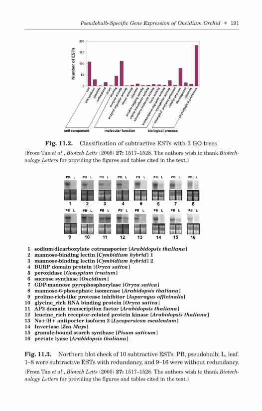
Pseudobulb-Specific Gene Expression of Oncidium Orchid ✦ 191
FA
Fig. 11.2. Classification of subtractive ESTs with 3 GO trees.
(From Tan et al., Biotech Letts (2005) 27: 1517–1528. The authors wish to thank Biotech-nology Letters for providing the figures and tables cited in the text.)
1 sodium/dicarboxylate cotransporter [Arabidopsis thaliana]2 mannose-binding lectin [Cymbidium hybrid] 13 mannose-binding lectin [Cymbidium hybrid] 24 BURP domain protein [Oryza sativa]5 peroxidase [Gossypium irsutum]6 sucrose synthase [Oncidium]7 GDP-mannose pyrophosphorylase [Oryza sativa]8 mannose-6-phosephate isomerase [Arabidopsis thaliana]9 proline-rich-like protease inhibitor [Asparagus officinalis]
10 glycine_rich RNA binding protein [Oryza sativa]11 AP2 domain transcription factor [Arabidopsis thaliana]12 leucine_rich receptor-related protein kinase [Arabidopsis thaliana]13 Na+/H+ antiporter isoform 2 [Lycopersicon esculentum]14 Invertase [Zea Mays]15 granule-bound starch synthase [Pisum sativum]16 pectate lyase [Arabidopsis thaliana]
Fig. 11.3. Northern blot check of 10 subtractive ESTs. PB, pseudobulb; L, leaf.1–8 were subtractive ESTs with redundancy, and 9–16 were without redundancy.
(From Tan et al., Biotech Letts (2005) 27: 1517–1528. The authors wish to thank Biotech-nology Letters for providing the figures and tables cited in the text.)
b494_Ch-11.qxd 8/29/2007 1:43 PM Page 191

cotransporter, BURP domain protein (dehydration-responsive proteinRD22), peroxidase, mannose-6-phosephate isomerase, proline-rich-likeprotease inhibitor, Na+/H+ antiporter isoform 2, invertase, and pectatelyase were highly expressed in the pseudobulbs at the initiation of inflo-rescence, but they exhibited almost no expression in its upper leaf. Theexpression patterns of mannose binding lectin, sucrose synthase, GDP-mannose pyrophosphorylase, and granule-bound starch synthase weresimilar. Glycine-rich RNA binding protein and leucine-rich receptor-related protein kinase genes were highly expressed in the pseudobulband slightly in leaves at the same time. Only AP2 domain transcriptionfactor gene displayed the opposite quality, the expression level inpseudobulbs was high, but that in leaves seemed higher. Obviously,abundant ESTs had more specific expression profiles. Significantly, theresults indicated that subtractive ESTs dataset showed pseudobulb-spe-cific genes expressed at the initiation of the inflorescence. Thus, theseNorthern data demonstrated that the EST subtraction was very preciseand reliable.
11.5 Alignment of Subtractive ESTs with KnownSucrose Synthase and Peroxidase Genes
Sucrose synthase was the only known Oncidium gene as a referencesequence for ESTs annotation. Using blastn, three EST contigs andthree singletons were aligned with 2520 bp, a complete CDS. They werealigned on four different regions of the gene and resulted in three clus-ters and one single (Table 11.3). This is because RsaI digested a com-plete cDNA into several pieces. It also hinted that perhaps two genefamily members were expressed at that time. Obviously, this examplehelped explain why several clusters or singles had the same annotation(Fig. 11.4A).
Another explanation may lie in the large gene family. All ESTsannotated by the peroxidase gene were aligned with a reference CDS ofGossypium irsutum. There were nine overlapped sequences, includingeight contigs and one singleton, in the middle of the genes (Fig. 11.4B).Their homologous sequences were 205 pb. Based on their single nucleotidepolymorphism, a phylogenetic tree was constructed with clustal W(Fig. 11.5). It showed three main groups on the tree. In Table 11.1, fiveannotations refer to peroxidase genes. Two annotations were sequence
192 ✦ J. Tan, H.-L. Wang and K.-W. Yeh
FAb494_Ch-11.qxd 8/29/2007 1:43 PM Page 192

consensuses belonging to the front and rear part of the gene, respec-tively. The other three annotations came from sequences aligned in themiddle of the gene. That is to say, the peroxidase gene family wasexpressed in the pseudobulb, and it could be divided into three primarysubfamilies.
Pseudobulb-Specific Gene Expression of Oncidium Orchid ✦ 193
FA
Fig. 11.4. Statement of subtractive ESTs with same annotations. (A) SubtractiveESTs were aligned with a known sucrose gene of Oncidium. (B) SubtractiveESTs were aligned with a known peroxidase gene of Gossypium irsutum.
(From Tan et al., Biotech Letts (2005) 27: 1517–1528. The authors wish to thank Biotech-nology Letters for providing the figures and tables cited in the text.)
Fig. 11.5. Phylogenetic analysis of peroxidase genes expressed in pseudobulbaccording to sequences of overlapped 120 bp of subtractive EST contigs.
(From Tan et al., Biotech Letts (2005) 27: 1517–1528. The authors wish to thank Biotech-nology Letters for providing the figures and tables cited in the text.)
b494_Ch-11.qxd 8/29/2007 1:43 PM Page 193

11.6 Subtractive ESTs Relevant to Inflorescence,Carbohydrate Metabolisms, Transportation,Stress, Cell Cycle, and Regulation
According to the annotations and references about their functions, thesubtractive ESTs were gathered and analyzed manually.
One cluster and seven singles (10 ESTs in total) were related to specificflower genes (Table 11.2). Abnormal inflorescence meristem 1 (AIM1)could affect inflorescence and floral development in Arabidopsis.11 MADSbox protein genes were expressed in different organs and mainly duringfloral development. DOMADS2 was expressed throughout the processof flower development and responded to osmotic changes and darkness.12
Shaggy-like kinase was flower-specific and responsible for osmoticchanges and darkness13 inflorescence development dependent genes. Theothers were related to four kinds of genes within the synthesis pathwayof flower pigment. Chalcone synthase14 and chalcone-flavanone iso-merase15 were at the upper stream of the pathway. More chalcone-flavanone isomerase gene seemed to be expressed in the pseudobulb at
194 ✦ J. Tan, H.-L. Wang and K.-W. Yeh
FA
Table 11.2. Selected Examples of ESTs for Genes with Known orPutative Functions Related to Inflorescence
Reference GIGene Annotation Organism Number E Value ESTs
abnormal inflorescence Arabidopsis 15235527 2.00E-36 1meristem 1 (AIM1) thaliana
chalcone sythase C2 Zea mays 116380 6.00E-15 1chalcone-flavanone Arabidopsis 18414838 3.00E-29 3
isomerase thalianacytochrom P450 Arabidopsis 21595357 2.00E-34 1
thalianacytochrome P450 71D2 Catharanthus 28261339 2.00E-30 1
roseusdihydroflavonol Arabidopsis 18390863 1.00E-16 1
reductase thalianaMADS box protein Dendrobium 6467974 5.00E-24 1
(DOMADS2) grexshaggy-like kinase Ricinus 1877397 8.00E-99 1
communis
(From Tan et al., Biotech Letts (2005) 27: 1517–1528. The authors wish to thank Biotech-nology Letters for providing the figures and tables cited in the text.)
b494_Ch-11.qxd 8/29/2007 1:43 PM Page 194
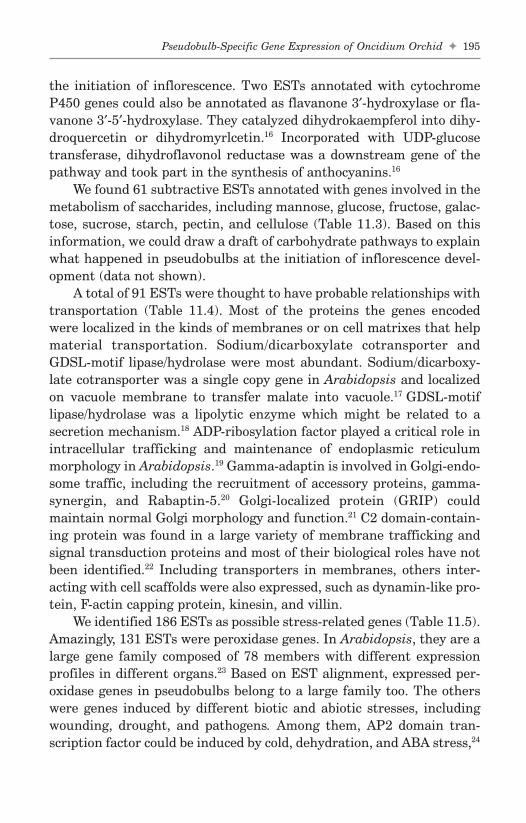
the initiation of inflorescence. Two ESTs annotated with cytochromeP450 genes could also be annotated as flavanone 3′-hydroxylase or fla-vanone 3′-5′-hydroxylase. They catalyzed dihydrokaempferol into dihy-droquercetin or dihydromyrlcetin.16 Incorporated with UDP-glucosetransferase, dihydroflavonol reductase was a downstream gene of thepathway and took part in the synthesis of anthocyanins.16
We found 61 subtractive ESTs annotated with genes involved in themetabolism of saccharides, including mannose, glucose, fructose, galac-tose, sucrose, starch, pectin, and cellulose (Table 11.3). Based on thisinformation, we could draw a draft of carbohydrate pathways to explainwhat happened in pseudobulbs at the initiation of inflorescence devel-opment (data not shown).
A total of 91 ESTs were thought to have probable relationships withtransportation (Table 11.4). Most of the proteins the genes encodedwere localized in the kinds of membranes or on cell matrixes that helpmaterial transportation. Sodium/dicarboxylate cotransporter andGDSL-motif lipase/hydrolase were most abundant. Sodium/dicarboxy-late cotransporter was a single copy gene in Arabidopsis and localizedon vacuole membrane to transfer malate into vacuole.17 GDSL-motiflipase/hydrolase was a lipolytic enzyme which might be related to asecretion mechanism.18 ADP-ribosylation factor played a critical role inintracellular trafficking and maintenance of endoplasmic reticulummorphology in Arabidopsis.19 Gamma-adaptin is involved in Golgi-endo-some traffic, including the recruitment of accessory proteins, gamma-synergin, and Rabaptin-5.20 Golgi-localized protein (GRIP) couldmaintain normal Golgi morphology and function.21 C2 domain-contain-ing protein was found in a large variety of membrane trafficking andsignal transduction proteins and most of their biological roles have notbeen identified.22 Including transporters in membranes, others inter-acting with cell scaffolds were also expressed, such as dynamin-like pro-tein, F-actin capping protein, kinesin, and villin.
We identified 186 ESTs as possible stress-related genes (Table 11.5).Amazingly, 131 ESTs were peroxidase genes. In Arabidopsis, they are alarge gene family composed of 78 members with different expressionprofiles in different organs.23 Based on EST alignment, expressed per-oxidase genes in pseudobulbs belong to a large family too. The otherswere genes induced by different biotic and abiotic stresses, includingwounding, drought, and pathogens. Among them, AP2 domain tran-scription factor could be induced by cold, dehydration, and ABA stress,24
Pseudobulb-Specific Gene Expression of Oncidium Orchid ✦ 195
FAb494_Ch-11.qxd 8/29/2007 1:43 PM Page 195

196 ✦ J. Tan, H.-L. Wang and K.-W. Yeh
FA
Table 11.3. Selected Examples of ESTs for Genes Related toCarbohydrate Metabolisms
Reference GIGene Annotation Organism Number E Value ESTs
ADP-glucose Arabidopsis 30699056 4.00E-66 2pyrophosphorylase thaliana
aldose 1-epimerase Arabidopsis 15242099 3.00E-65 2thaliana
alpha 1,4-glucan Oryza sativa 13195430 1.00E-43 1phosphorylase Lisozyme
alpha-galactosidase Arabidopsis 11264291 3.00E-33 1thaliana
beta-1,3 glucanase Oryza sativa 20161490 7.00E-39 1beta-fructofuranosidase 1 Zea mays 1352468 2.00E-31 1beta-galactosidase Oryza sativa 20514290 7.00E-63 1beta-galactosidase Oryza sativa 18461259 2.00E-49 1beta-mannosidase Lycopersicon 17226270 1.00E-103 1
esculentumcinnamyl alcohol Populus 9998899 3.00E-38 1
dehydrogenase balsamiferadTDP-glucose Arabidopsis 21594350 1.00E-16 1
4-6-dehydratase thalianaepimerase/dehydratase Oryza sativa 20042976 1.00E-56 3glucosyltransferase Arabidopsis 25408401 2.00E-26 1
thalianaglycogenin Oryza sativa 5441877 5.00E-84 1
glucosyltransferase(EC 2.4.1.186)
glycogenin Oryza sativa 5441877 7.00E-93 1glucosyltransferase(EC 2.4.1.186)
glycosyl hydrolase 1 Arabidopsis 15220627 6.00E-36 1thaliana
granule-bound starch Pisum sativum 15626365 4.00E-86 1synthase
mannose-6-phosphate Arabidopsis 15232927 8.00E-50 1isomerase thaliana
mannose-6-phosphate Oryza sativa 11275529 6.00E-45 1isomerase
(Continued )
b494_Ch-11.qxd 8/29/2007 1:43 PM Page 196

and was involved in the regulation of low-temperature responsive genesin barley.25
A total of 23 ESTs were found to have functions in the cell cycle(Table 11.6). That is to say, the cells in pseudobulbs kept growingactively and differentiating at this stage. DP-E2F-related protein 1 and
Pseudobulb-Specific Gene Expression of Oncidium Orchid ✦ 197
FA
Table 11.3. (Continued )
Reference GIGene Annotation Organism Number E Value ESTs
mannosyltransferase Arabidopsis 22326970 4.00E-15 1thaliana
NAD-dependent Arabidopsis 15231926 2.00E-63 1epimerase/dehydratase thaliana
nucleoside-diphosphate- Oryza sativa 29893646 6.00E-68 4sugar pyrophosphorylase
N-acetylglucosamine- Arabidopsis 30686654 8.00E-38 1phosphate mutase thaliana
pectate lyase Arabidopsis 10177179 4.00E-23 1thaliana
pectin esterase Oryza sativa 20161185 1.00E-12 2pectinesterase 1 Lycopersicon 6174913 1.00E-13 4
esculentumphosphoglucomutase, Solanum 12585316 2.00E-84 1
cytoplasmic tuberosumphosphoglucose isomerase Dioscorea 2351056 2.00E-80 1
septemlobaphosphomannomutase Arabidopsis 15225896 2.00E-16 2
thalianapolygalacturonase Pisum sativum 13958032 3.00E-57 3polygalacturonase Arabidopsis 18412253 2.00E-22 1
thalianaripening-related protein Vitis vinifera 7406669 4.00E-79 3starch phosphorylase Ipomoea batatas 12658431 2.00E-31 1sucrose synthase Oncidium 22347630 1.00E-96 6sucrose synthase Oncidium 22347630 2.00E-27 2sucrose synthase Oncidium 22347630 1.00E-38 2sucrose synthase Oncidium 22347630 4.00E-30 1triosephosphate isomerase, Petunia x 1351279 4.00E-27 1
cytosolic (TIM) hybrida
(From Tan et al., Biotech Letts (2005) 27: 1517–1528. The authors wish to thank Biotech-nology Letters for providing the figures and tables cited in the text.)
b494_Ch-11.qxd 8/29/2007 1:43 PM Page 197

198 ✦ J. Tan, H.-L. Wang and K.-W. Yeh
FA
Table 11.4. Selected Examples of ESTs for Genes Related toTransportation
Reference GIGene Annotation Organism Number E Value ESTs
Acyl-CoA-binding protein Panax ginseng 19352190 3.00E-19 2ADP-ribosylation factor Glycine max 4324967 2.00E-52 2ADP-ribosylation factor Oryza sativa 18844784 2.00E-25 1C2 domain-containing Arabidopsis 15239959 2.00E-36 1
protein thalianaC2 domain-containing Arabidopsis 15223764 5.00E-15 1
protein thalianaC2 domain-containing Arabidopsis 15217968 5.00E-37 1
protein thalianadynamin like protein 2a Arabidopsis 19032337 2.00E-44 1
thalianaF-actin capping protein, Oryza sativa 23617186 2.00E-12 1
alpha subunitgamma-adaptin 1 Oryza sativa 19386749 1.00E-30 1GDSL-like lipase/ Oryza sativa 29837765 1.00E-26 4
acylhydrolaseGDSL-like lipase/ Oryza sativa 29837765 9.00E-34 2
acylhydrolaseGDSL-motif lipase/ Arabidopsis 15228189 3.00E-91 5
hydrolase thalianaGDSL-motif lipase/ Arabidopsis 21593518 2.00E-25 3
hydrolase thalianaGDSL-motif lipase/ Arabidopsis 15221260 6.00E-54 2
hydrolase thalianaGDSL-motif lipase/ Arabidopsis 18416824 2.00E-25 1
hydrolase thalianaGDSL-motif lipase/ Arabidopsis 15224201 4.00E-12 1
hydrolase thalianagolgi-localized protein Oryza sativa 22093862 2.00E-18 1
(GRIP)high mobility group Arabidopsis 15231065 7.00E-31 1
protein 2 thalianakinesin Daucus carota 15186760 3.00E-30 2kinesin-related protein Arabidopsis 22327641 3.00E-54 3
thalianalipid transfer protein Vitis vinifera 28194086 3.00E-16 7
isoform 4
(Continued )
b494_Ch-11.qxd 8/29/2007 1:43 PM Page 198

homobox genes were most abundant among this group. The E2F/DPprotein family controls cell cycle progression by acting predominantlyas an activator or repressor of transcription.26 Arabidopsis had morethan 180 potential E2F target genes with various functions: cell cycle,transcription, stress and defense, or signaling.27 Homeobox 20 had a
Pseudobulb-Specific Gene Expression of Oncidium Orchid ✦ 199
FA
Table 11.4. (Continued )
Reference GIGene Annotation Organism Number E Value ESTs
membrane bound O-acyl Arabidopsis 22329514 9.00E-24 1transferase (MBOAT) thaliana
membrane bound O-acyl Arabidopsis 22329514 1.00E-75 1transferase (MBOAT) thaliana
mitochondrial carrier Arabidopsis 15240756 5.00E-39 1protein thaliana
myosin heavy chain Arabidopsis 18402909 5.00E-17 1thaliana
permease Oryza sativa 27545049 2.00E-17 3peroxisomal targeting Arabidopsis 15241175 3.00E-15 1
signal type 1 receptor thalianaPEX14 protein Arabidopsis 30697742 3.00E-15 1
thalianaplasma membrane Oryza sativa 22831004 2.00E-44 4
intrinsic proteinRer1A protein (AtRer1A) Oryza sativa 10945247 2.00E-37 2Sec31p Oryza sativa 22831279 4.00E-63 1secretory carrier Arabidopsis 15222550 1.00E-30 1
membrane protein thalianasignal peptidase Arabidopsis 15240934 7.00E-56 1
thalianasodium/dicarboxylate Arabidopsis 15238130 5.00E-44 24
cotransporter thalianasodium/dicarboxylate Arabidopsis 15238130 2.00E-39 3
cotransporter thalianasodium-dicarboxylate Arabidopsis 21536650 2.00E-35 1
cotransporter thalianavesicle transport Oryza sativa 19571103 8.00E-55 1
v-SNARE proteinvillin 1 (VLN1) Arabidopsis 26451417 2.00E-22 1
thaliana
(From Tan et al., Biotech Letts (2005) 27: 1517–1528. The authors wish to thank Biotech-nology Letters for providing the figures and tables cited in the text.)
b494_Ch-11.qxd 8/29/2007 1:43 PM Page 199
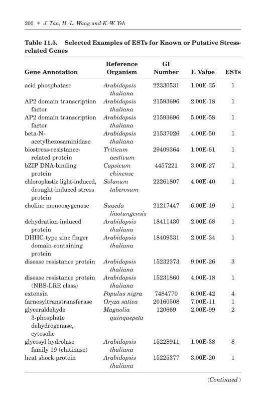
200 ✦ J. Tan, H.-L. Wang and K.-W. Yeh
FA
Table 11.5. Selected Examples of ESTs for Known or Putative Stress-related Genes
Reference GIGene Annotation Organism Number E Value ESTs
acid phosphatase Arabidopsis 22330531 1.00E-35 1thaliana
AP2 domain transcription Arabidopsis 21593696 2.00E-18 1factor thaliana
AP2 domain transcription Arabidopsis 21593696 5.00E-58 1factor thaliana
beta-N- Arabidopsis 21537026 4.00E-50 1acetylhexosaminidase thaliana
biostress-resistance- Triticum 29409364 1.00E-61 1related protein aestivum
bZIP DNA-binding Capsicum 4457221 3.00E-27 1protein chinense
chloroplastic light-induced, Solanum 22261807 4.00E-40 1drought-induced stress tuberosumprotein
choline monooxygenase Suaeda 21217447 6.00E-19 1liaotungensis
dehydration-induced Arabidopsis 18411430 2.00E-68 1protein thaliana
DHHC-type zinc finger Arabidopsis 18409331 2.00E-34 1domain-containing thalianaprotein
disease resistance protein Arabidopsis 15232373 9.00E-26 3thaliana
disease resistance protein Arabidopsis 15231860 4.00E-18 1(NBS-LRR class) thaliana
extensin Populus nigra 7484770 6.00E-42 4farnesyltranstransferase Oryza sativa 20160508 7.00E-11 1glyceraldehyde Magnolia 120669 2.00E-99 2
3-phosphate quinquepetadehydrogenase,cytosolic
glycosyl hydrolase Arabidopsis 15228911 1.00E-38 8family 19 (chitinase) thaliana
heat shock protein Arabidopsis 15225377 3.00E-20 1thaliana
(Continued )
b494_Ch-11.qxd 8/29/2007 1:43 PM Page 200

Pseudobulb-Specific Gene Expression of Oncidium Orchid ✦ 201
FA
Table 11.5. (Continued )
Reference GIGene Annotation Organism Number E Value ESTs
heat shock protein Arabidopsis 15225377 1.00E-40 1thaliana
heat shock protein Oryza sativa 29124135 2.00E-42 1cognate 70
heat shock protein Arabidopsis 15232682 7.00E-29 1hsc70-3 (hsc70.3) thaliana
late embryogenesis Arabidopsis 15224810 2.00E-16 1abundant protein thaliana
leucine rich repeat Arabidopsis 30686169 1.00E-37 1protein thaliana
major intrinsic protein Arabidopsis 15236485 1.00E-81 2(MIP) thaliana
Na+/H+ antiporter 2 Lycopersicon 15982206 4.00E-17 1esculentum
nodulin Oryza sativa 11072005 9.00E-31 1PDR-like ABC transporter Oryza sativa 27368827 4.00E-35 1peroxidase Glycine max 5002234 7.00E-18 5peroxidase (EC 1.11.1.7) Gossypium 7433087 2.00E-48 92
irsutumperoxidase (EC 1.11.1.7) Gossypium 7433087 2.00E-17 8
irsutumperoxidase (EC 1.11.1.7) 2, Glycine max 7433098 6.00E-27 21
cationicperoxidase (EC 1.11.1.7) 2, Glycine max 7433098 1.00E-91 5
cationicphosphoethanolamine Oryza sativa 22535531 8.00E-13 1
methyltransferaseplastid-lipid associated Arabidopsis 18403751 2.00E-35 1
protein PAP/fibrillin thalianaproline rich protein 3 Cicer arietinum 21615411 5.00E-75 1proline-rich protein APG Cicer arietinum 10638955 4.00E-16 1
isologproline-rich-like protein Asparagus 1531756 2.00E-29 1
officinalissenescence-associated Pisum sativum 13359451 3.00E-43 6
proteinsenescence-associated Arabidopsis 18398417 2.00E-20 1
protein thalianawound-induced Arabidopsis 15234987 2.00E-15 3
protein thaliana
(From Tan et al., Biotech Letts (2005) 27: 1517–1528. The authors wish to thank Biotech-nology Letters for providing the figures and tables cited in the text.)
b494_Ch-11.qxd 8/29/2007 1:43 PM Page 201

202 ✦ J. Tan, H.-L. Wang and K.-W. Yeh
FA
Table 11.6. Selected Examples of ESTs for Genes Related to Cell Cycle
ReferenceGene Annotation Organism GI Number E Value ESTs
26S proteasome Oryza sativa 20978545 8.00E-79 1non-ATPase, regulatorysubunit 6
3-hydroxy-3- Solanum 11133016 5.00E-32 1methylglutaryl- tuberosumcoenzyme A reductase 3(HMG3.3)
AUX1-like permease Arabidopsis 5881784 2.00E-24 1thaliana
auxin efflux carrier Arabidopsis 15239215 3.00E-50 1protein thaliana
biotin carboxyl carrier Glycine max 12006165 2.00E-29 1protein subunit
cyclic nucleotide-regulated Arabidopsis 15234769 8.00E-34 1ion channel (CNGC9) thaliana
cysteine proteinase AALP Arabidopsis 23397070 6.00E-30 1thaliana
cysteine proteinase mir3 Zea mays 7435806 5.00E-47 1(EC 3.4.22.)
DP-E2F-related protein 1 Arabidopsis 22331664 1.00E-49 7thaliana
histone deacetylase 2 Zea mays 7716948 6.00E-19 1isoform b
homeobox 20 Nicotiana 4589882 1.00E-49 1tabacum
homeobox protein Malus x 6016217 8.00E-46 3knotted-1 2 (KNAP2) domestica
homeobox-leucine zipper Arabidopsis 15222452 9.00E-38 1protein ATHB-13 thaliana
Homeotic protein Lycopersicon 3023974 9.00E-16 1knotted-1 (TKN1) esculentum
nucleolysin Oryza sativa 4680340 1.00E-15 1
(From Tan et al., Biotech Letts (2005) 27: 1517–1528. The authors wish to thank Biotech-nology Letters for providing the figures and tables cited in the text.)
common motif and took part in xylem cell differentiation.28 Homeobox-leucine zipper protein ATHB-13 was a transcription factor. It couldspecify the cell fate and body plan in early embryogenesis.29
A total of 59 ESTs were annotated with known or putative regula-tory functions (Table 11.7). It seemed that the regulation of the genes
b494_Ch-11.qxd 8/29/2007 1:43 PM Page 202

Pseudobulb-Specific Gene Expression of Oncidium Orchid ✦ 203
FA
Table 11.7. Selected Examples of ESTs for Known or PutativeRegulatory Functions
Reference GIGene Annotation Organism Number E Value ESTs
adapter protein SPIKE1 Oryza sativa 24899400 4.00E-56 1adenine phosphoribosyl- Oryza sativa 29826070 8.00E-75 1
transferase form 2amidase Arabidopsis 8163875 7.00E-31 1
thalianaamidase Oryza sativa 18542894 1.00E-12 1BURP domain protein Vigna 7106540 6.00E-12 9
unguiculatac-myc binding protein Arabidopsis 22325671 2.00E-12 1
thalianacupin domain-containing Arabidopsis 15226403 3.00E-29 2
protein thalianaDEAD/DEAH box helicase Arabidopsis 15222526 5.00E-40 1
thalianaDEAD/DEAH box helicase Arabidopsis 15219185 1.00E-26 1
thalianaDnaJ protein Salix gilgiana 11277163 1.00E-106 2DnaJ protein homolog 2 Allium porrum 1169382 4.00E-26 1DNAJ-like protein Oryza sativa 29367357 8.00E-33 1elongation factor 1-alpha Elaeis oleifera 18419676 8.00E-40 1GAMYB-binding protein Hordeum 27948448 7.00E-51 1
vulgareGF14 protein Fritillaria 2921512 2.00E-66 1
agrestisglycine-rich RNA-binding Arabidopsis 21553602 7.00E-21 1
protein thalianaHD-Zip transcription Arabidopsis 15226808 1.00E-96 1
factor Athb-14 thalianahelicase Arabidopsis 18395518 4.00E-30 1
thalianahomeobox-leucine zipper Arabidopsis 15222452 9.00E-38 1
protein ATHB-13 thalianaMuDR mudrA-like protein Oryza sativa 5441874 4.00E-31 1phosphoprotein Arabidopsis 25513447 2.00E-94 1
phosphatase thaliana(EC 3.1.3.16)
(Continued )
b494_Ch-11.qxd 8/29/2007 1:43 PM Page 203

204 ✦ J. Tan, H.-L. Wang and K.-W. Yeh
FA
Table 11.7. (Continued )
Reference GIGene Annotation Organism Number E Value ESTs
probable protein Nicotiana 7489183 1.00E-88 1disulfide-isomerase tabacum
Ras-related protein Nicotiana 3024503 5.00E-31 1Rab11C tabacum
receptor-like kinase Glycine max 21239384 2.00E-17 1RHG4
receptor-like protein Arabidopsis 7487253 6.00E-41 1kinase thaliana
receptor-like protein Oryza sativa 7434420 3.00E-18 1kinase (EC 2.7.1)
receptor-related protein Arabidopsis 15240720 6.00E-45 1kinase thaliana
RNA recognition motif Arabidopsis 22328805 8.00E-18 1(RRM)-containing thalianaprotein
RNA-binding protein Oryza sativa 18087662 2.00E-37 2RNA-binding protein Mesembryanthe- 1076251 4.00E-26 1
mum crys-tallinum
serine/threonine kinase Arabidopsis 25387051 2.00E-51 1thaliana
serine/threonine protein Nicotiana 3811293 3.00E-19 1kinase tabacum
serine/threonine protein Avena sativa 7489361 5.00E-37 1kinase (EC 2.7.1)
serine/threonine-specific Arabidopsis 25751318 2.00E-18 1protein kinase thaliana
SNF2 domain/helicase Arabidopsis 15226870 4.00E-42 1domain-containing thalianaprotein
sphingosine kinase Oryza sativa 13786462 3.00E-71 1transcription factor LIM Nicotiana 18565124 5.00E-46 2
tabacumtranscription factor X1 Oryza sativa 6650526 2.00E-28 1transducin/WD-40 repeat Arabidopsis 30682603 3.00E-21 1
protein thalianatransfactor Arabidopsis 6223653 2.00E-34 1
thaliana
(Continued )
b494_Ch-11.qxd 8/29/2007 1:43 PM Page 204

involved in the active material and energy metabolism in the pseudo-bulb was very complex.
11.7 Discussion
In Oncidium plant, the pseudobulb is a critical organ regulating plantgrowth. In general, when the pseudobulb grows healthily and accu-mulates abundant nutrition, the embedded young bud develops intoinflorescence, a so-called reproductive state. However, the bud mightdevelop into a young adventitious shoot and begin a new vegetativestate. Therefore, the pseudobulb plays a functional role in regulatingOncidium’s switch between the reproductive state and the vegetativestate. In the previous study, we analyzed the carbohydrate compositionand amino acid pool in the pseudobulb prior to and during inflorescencedevelopment.10 Alternation of nutrition in the pseudobulb during floraldevelopment was also investigated.10 To our knowledge, mannan is theprincipal storage polysaccharide synthesized in the pseudobulb beforeinflorescence initiation. It is degraded into mannose and de novo syn-thesized into starch during inflorescence development. Eventually,starch is utilized while the flower is forming.10 Therefore, active metab-olism related to carbohydrate and other types of metabolism may affect
Pseudobulb-Specific Gene Expression of Oncidium Orchid ✦ 205
FA
Table 11.7. (Continued )
Reference GIGene Annotation Organism Number E Value ESTs
translational activator Arabidopsis 25404492 2.00E-18 1thaliana
translational activator Arabidopsis 15217742 5.00E-42 1thaliana
WD-40 repeat protein Arabidopsis 30685408 5.00E-46 1thaliana
zinc finger (C3HC4-type Arabidopsis 15233298 9.00E-28 1RING finger) protein thaliana
zinc finger protein Pisum sativum 11288368 3.00E-77 1zinc finger protein 5 Arabidopsis 21592423 5.00E-12 1
(ZFP5) thalianazinc-finger protein Lsd1 Arabidopsis 30685085 1.00E-22 1
thaliana
(From Tan et al., Biotech Letts (2005) 27: 1517–1528. The authors wish to thank Biotech-nology Letters for providing the figures and tables cited in the text.)
b494_Ch-11.qxd 8/29/2007 1:43 PM Page 205

floral development. However, the genes involved in these active meta-bolic steps are still unknown.
Indubitably, subtractive EST data have more redundancy than ESTdata. Including the inevitable consequence of the nonuniform abun-dance of mRNA from different genes with dramatically various num-bers in the cDNA pool, the bias must be further enhanced byhybridization, PCR amplification twice, ligation, and transformation.To this point, this is also the main problem in EST sequencing based onthe cDNA library. It could perhaps be resolved by subtracting newlyfound redundant sequences from templates during stepwise sequenc-ing. EST sequencing only from the 5′-ends of cDNAs is a rapid, eco-nomical, and amenable strategy.6 However, for subtractive ESTsequencing, not only is Rsa I digestion infeasible, it also causes shorterpieces of ESTs. Furthermore, it seemed the middle parts of genes havemore chances to be sequenced than the ends (Fig. 11.4).
Thus, a large-scale analysis of gene expression related to physio-logical responses of the Oncidium pseudobulb, particularly duringthe early floral stage, has not been reported. Therefore, the expressedgene catalogue presented here will provide the basic information toinvestigate the molecular genetics basis of the Oncidium pseudobulb’searly flowering stage by transcriptome profiling. In this small-scalesubtractive EST, a conclusive picture of the cellular processes ofstress-response (Table 11.5), carbohydrate metabolism (Table 11.3),and transportation (Table 11.4) was obtained. Also, the RNA gel-blotexpression data showed some evidence that this EST-set is indeedenriched with such genes (Fig. 11.3), indicating the high efficiency ofthe cDNA subtraction strategy.
Based on the types of genes represented, the mechanism of inflo-rescence initiation in the early flowering stage of the Oncidiumpseudobulb relies on precise integration of developmental processeswith the carbohydrate metabolism-related response. The pseudobulbat the stage of inflorescence initiation seems like a ripening fruit forthe coming flower development. First, it is in growing, including cellenlargement and differentiation. Especially, several genes involvedin pectin degradation are expressing actively (Table 11.3). Thismay be a physiological marker for the pseudobulb, which is going tomature.30 From the Northern results of genes related to mechanismsof saccharides at different stages of inflorescence, the ripening of
206 ✦ J. Tan, H.-L. Wang and K.-W. Yeh
FAb494_Ch-11.qxd 8/29/2007 1:43 PM Page 206

pseudobulb is just in the initiation of inflorescence development(data not shown). At the same time, transportation is very active, andstorage seems to be in preparation. There are two main directions oftransportation: extracellular and intracellular, in which vacuoles playa predominant role. For extracellular transportation, saccharides andproteins are transferred to cell wall for synthesis. Intracellular trans-portation involves mainly the transfer of low molecular saccharidesinto vacuoles to maintain osmosis and to prepare for future storagefunctions.
Pseudobulb mannan is very rich and in degradation at the initiationof inflorescence. Plant mannose binding lectin is believed to play a rolein the recognition of high-mannose type glycans of foreign microorgan-isms or plant predators.31 However, mannose is grasped with mannosebinding lectin in the pseudobulb, for it is harmful to plant cells.32 Thenthe mannose can be further catalyzed into fructose and glucose. Starchis also synthesized at this stage, but almost no starch is stored in cellscompared with the starch granules seen before blossoming (data notshown). Starch is perhaps also degraded into sucrose and other mono-hexoses for cell growth and differentiation.
At the bottom side of the pseudobulb, the little flower bud is also indifferentiation and growing at the same time. Usually the bud theredevelops into inflorescence, but it can sometimes turn into a vegetativereproductive bud. This is a hint that the pseudobulb offers signals lead-ing to inflorescence development unless some accident occurs. Thegenes involved in this function are perhaps flower-specific along withsome transcriptional factors. It is no wonder so many genes are relatedto regulation because the pseudobulb is a very active organ leading toinflorescence at this stage. Making their functions clear is importantand will require further research.
In summary, the subtractive EST approach is an efficient tool toprovide an overview of gene expression profiles in the metabolicallyactive tissue of the Oncidium pseudobulb. The EST data provide uswith an insight into a wide range of genes. These genes represent thephysiological status in the pseudobulb during the early inflorescencedevelopment. The abundant genes, e.g. peroxidase and sodium/dicar-boxylate cotransporter, shown in the profile revealed some especiallyunexpected facts. These will someday make it possible to exploit flow-ering-related mechanisms for the benefit of mankind.
Pseudobulb-Specific Gene Expression of Oncidium Orchid ✦ 207
FAb494_Ch-11.qxd 8/29/2007 1:43 PM Page 207

Acknowledgment
This work was financially supported by National Science Council, ROCunder Grant NSC 91-2317-B-002-041 to Professor K. W. Yeh.
References
1. Liau CH, You SJ, Prasad V, et al. (2003a) Agrobacterium tumefaciens-mediated transformation of an Oncidium orchid. Plant Cell Rep 21:993–998.
2. Chen J, Chang W. (2000) Efficient plant regeneration through somaticembryogenesis from callus cultures of Oncidium (Orchidaceae). Plant Sci160:87–93.
3. You SJ, Liau CH, Huang HE, et al. (2003) Sweet pepper ferredoxin-likeprotein (pflp) gene as a novel selection marker for orchid transformation.Planta 217:60–65.
4. Liau CH, Lu JC, Prasad V, et al. (2003b) The sweet pepper ferredoxin-likeprotein (pflp) conferred resistance against soft rot disease in Oncidiumorchid. Transgenic Res 12:329–336.
5. Hsu HF, Yang CH. (2002) An orchid (Oncidium Gower Ramsey) AP3-likeMADS gene regulates floral formation and initiation. Plant Cell Physiol43:1198–1209.
6. Li CR, Zhang XB, Hew CS. (2003) Cloning of a sucrose-phosphate synthasegene highly expressed in flowers from the tropical epiphytic orchid OncidiumGoldiana. J Exp Bot 21:89–91.
7. Hew CS, Yang JWH. (1994) Growth and photosynthesis of OncidiumGoldiana. J Hort Sci 69:809–819.
8. Wadasinghe G, Hew CS. (1995) The importance of back shoots as a sourceof photo-assimilates for growth and flower production in Dendrodium cv.Jashika pink (Orchidaceae). J Hort Sci 70:207–214.
9. Hew CS, Ng CKY. (1996) Changes in mineral and carbohydrate content inpseudobulbs of the C3 epiphytic orchid hybrid Oncidium Goldiana at dif-ferent growth stages. Lindleyana 11:125–134.
10. Wang HL, Chung JD, Yeh KW. (2003) Changes of carbohydrate and freeamino acid pools in current pseudobulbs of Oncidium Gower Ramseyduring inflorescence development. J Agric Associ China 4:476–488.
11. Richmond TA, Bleecker AB. (1999) A defect in beta-oxidation causes abnor-mal inflorescence development in Arabidopsis. Plant Cell 11:1911–1924.
12. Yu H, Goh CJ. (2000) Identification and characterization of three orchidMADS-box genes of the AP1/AGL9 subfamily during floral transition.Plant Physiol 123:1325–1336.
208 ✦ J. Tan, H.-L. Wang and K.-W. Yeh
FAb494_Ch-11.qxd 8/29/2007 1:43 PM Page 208

13. Charrier B, Champion A, Henry Y, Kreis M. (2002) Expression profiling of thewhole Arabidopsis shaggy-like kinase multigene family by real-time reversetranscriptase-polymerase chain reaction. Plant Physiol 130:577–590.
14. Wade HK, Bibikova TN, Valentine WJ, Jenkins GI. (2001) Interactions withina network of phytochrome, cryptochrome and UV-B phototransductionpathways regulate chalcone synthase gene expression in Arabidopsis leaftissue. Plant J 25:675–685.
15. van Tunen AJ, Koes RE, Spelt CE, et al. (1988) Cloning of the two chalconeflavanone isomerase genes from Petunia hybrida: Coordinate, light-regu-lated and differential expression of flavonoid genes. EMBO J 7:1257–1263.
16. de Vetten N, ter Horst J, van Schaik HP, et al. (1999) A cytochrome b5 isrequired for full activity of flavonoid 3′, 5′-hydroxylase, a cytochrome P450involved in the formation of blue flower colors. Proc Natl Acad Sci USA96:778–783.
17. Emmerlich V, Linka N, Reinhold T, et al. (2003) The plant homolog to thehuman sodium/dicarboxylic cotransporter is the vacuolar malate carrier.Proc Natl Acad Sci USA 100:11122–11126.
18. Wilhelm S, Tommassen J, Jaeger KE. (1999) A novel lipolytic enzymelocated in the outer membrane of Pseudomonas aeruginosa. J Bacteriol181:6977–6986.
19. Lee MH, Min MK, Lee YJ, et al. (2002) ADP-ribosylation factor 1 ofArabidopsis plays a critical role in intracellular trafficking and mainte-nance of endoplasmic reticulum morphology in Arabidopsis. Plant Physiol129:1507–1520.
20. Nogi T, Shiba Y, Kawasaki M, et al. (2002) Structural basis for the acces-sory protein recruitment by the gamma-adaptin ear domain. Nat StructBiol 9:527–531.
21. Ungar D, Oka T, Brittle EE, et al. (2002) Characterization of a mammalianGolgi-localized protein complex, COG, that is required for normal Golgimorphology and function. J Cell Biol 157:405–415.
22. Ochoa WF, Corbalan-Garcia S, Eritja R, et al. (2002) Additional binding sitesfor anionic phospholipids and calcium ions in the crystal structures of com-plexes of the C2 domain of protein kinase calpha. J Mol Biol 320:277–291.
23. Tognolli M, Penel C, Greppin H, Simon P. (2002) Analysis and expressionof the class III peroxidase large gene family in Arabidopsis thaliana. Gene288:129–138.
24. Shen YG, Zhang WK, He SJ, et al. (2003) An EREBP/AP2-type protein inTriticum aestivum was a DRE-binding transcription factor induced bycold, dehydration and ABA stress. Theor Appl Genet 106:923–930.
25. Xue GP. (2003) The DNA-binding activity of an AP2 transcriptional acti-vator HvCBF2 involved in regulation of low-temperature responsive genesin barley is modulated by temperature. Plant J 33:373–383.
Pseudobulb-Specific Gene Expression of Oncidium Orchid ✦ 209
FAb494_Ch-11.qxd 8/29/2007 1:43 PM Page 209

26. Cayirlioglu P, Ward WO, Silver Key SC, Duronio RJ. (2003) Transcriptionalrepressor functions of Drosophila E2F1 and E2F2 cooperate to inhibitgenomic DNA synthesis in ovarian follicle cells. Mol Cell Biol 23:2123–2134.
27. Ramirez-Parra E, Frundt C, Gutierrez C. (2003) A genome-wide identifi-cation of E2F-regulated genes in Arabidopsis. Plant J 33:801–811.
28. Hertzberg M, Olsson O. (1998) Molecular characterisation of a novel planthomeobox gene expressed in the maturing xylem zone of Populus tremulax tremuloides. Plant J 16:285–295.
29. Yang JY, Chung MC, Tu CY, Leu WM. (2002) OSTF1: A HD-GL2 familyhomeobox gene is developmentally regulated during early embryogenesisin rice. Plant Cell Physiol 43:628–638.
30. Medina-Escobar N, Cardenas J, Moyano E, et al. (1997) Cloning, molecu-lar characterization and expression pattern of a strawberry ripening-spe-cific cDNA with sequence homology to pectate lyase from higher plants.Plant Mol Biol 34:867–877.
31. Chai Y, Pang Y, Liao Z, et al. (2003) Molecular cloning and characterizationof a mannose-binding lectin gene from Crinum asiaticum. J Plant Physiol160:913–920.
32. Stein JC, Hansen G. (1999) Mannose induces an endonuclease responsiblefor DNA laddening in plant cells. Plant Physiol 121:71–80.
33. Tan J, Wang HL, Yeh KW. (2005) Analysis of organ-specific, expressed genesin Oncidium orchid by substractive expressed sequence tags library. BiotechLett 27:1517–1528.
210 ✦ J. Tan, H.-L. Wang and K.-W. Yeh
FAb494_Ch-11.qxd 8/29/2007 1:43 PM Page 210

Chapter 12
Application of Virus-inducedGene Silencing Technology
in Gene FunctionalValidation of Orchids
Hsiang-Chia Lu†, Hong-Hwa Chen‡ and Hsin-Hung Yeh*,†
The largest family of angiosperms, Orchidaceae, has diverse, spe-cialized pollination and ecological strategies and provides a rich sub-ject for investigating evolutionary relationships and developmentalbiology. However, the study of these non-model organisms may behindered by challenges, such as their large genome size, low trans-formation efficiency, long regeneration time, and long life-cycle. Toovercome these obstacles, we first developed vectors with the use ofa symptomless Cymbidium mosaic virus, which infects most orchids,then combined simple physiological controls and virus-induced genesilencing (VIGS) for validation of gene function in orchids. The suc-cess of our strategies was verified by our functional validation of flo-ral identity gene(s) in the tetraploid Phalaenopsis orchids, whichhave an unusually long life-cycle (two years from sowing to flow-ering). We could knock down the RNA level of either a specificPhalaenopsis floral identity gene or a family of genes congruentlyin two months. Functional analysis of orchid genes could becomeeasier and profit from the VIGS approach.
211
FA
*Corresponding author.†Department of Plant Pathology and Microbiology, National Taiwan University, Taipei,Taiwan.‡Department of Life Sciences, National Cheng Kung University, Tainan, Taiwan.
b494_Ch-12.qxd 8/29/2007 1:43 PM Page 211

12.1 Introduction
Orchids, plants with elegant and beautiful flowers, have attractedhumans for centuries. Known for their diversity, they form the largestfamily of angiosperms, Orchidaceae.1 Orchids have evolved specializedand astonishing pollination and ecological strategies for survival.Therefore, they are a rich source for investigating evolutionary rela-tionships and developmental biology. Although evolutionary develop-mental biology (Evo-Devo) has become an important topic in every fieldof biology today,2–4 the study of orchids has been hindered by severalchallenges, including their large genome size, long life-cycle, and lowtransformation efficiency. In fact, orchids are the opposite of modelplants in every trait studied by geneticists and molecular biologists. Forexample, the most widely grown orchid, Phalaenopsis spp., has a largegenome size, ranging from 1 × 10⁹ to 6 × 10⁹ bp/1C,5,6 and several impor-tant commercial cultivars are multiploid. The life-cycle of Phalaenopsisspp. naturally takes about 2 to 3 years from the vegetation to the repro-ductive phases. Therefore, applications of commonly used genetic andmolecular approaches to the study of orchids is difficult. However, thedevelopment of several new research techniques has led to increasedinvestigation of model plants and numerous exciting findings. Fromthese developments, we can select suitable tools to solve the problemsfaced in the study of orchids.
12.2 Approaches Used for GeneFunctional Analysis
Forward and reverse genetics is commonly used in the elucidation ofthe function of a gene or genes. Forward genetics tries to identify thegenetic source of a trait of interest, beginning with random mutagen-sis of the whole genome to follow the selection of the trait and subse-quently identifying the mutated gene or genes related to the trait.Reverse genetics, however, aims to disrupt the expression of genes tosearch for a phenotype and usually begins with cloned DNA sequences,with or without prior knowledge about the DNA. In plants, disruptingthe expression of a gene of interest is difficult owing to low homolo-gous recombination frequencies. Alternatively, plants transformedwith antisense and/or sense genes that can produce dsRNA from an
212 ✦ H.-C. Lu, H.-H. Chen, and H.-H. Yeh
FAb494_Ch-12.qxd 8/29/2007 1:43 PM Page 212

inverted-repeat of a target gene may disrupt the RNA or block thetranslation of the gene.7
Forward genetics is not practical for functional analysis of plants,such as orchids because of their long life-cycle and lack of high-resolu-tion genetic maps. Reverse genetics to find genes is problematic as wellbecause of difficulties in plant transformation and long regenerationtime, especially for genes involved in reproductive stages, the last stagesof the life-cycle and so important to orchid study.
In addition to genetic transformation, a recently developedapproach to functional analysis of plant genes involves loss-of-functionmediated by virus-induced gene silencing (VIGS). Since the early suc-cess of VIGS with Tobacco mosaic virus (TMV)- and Potato virusX (PVX)-based vectors in silencing the phytoene desaturase gene (PDS)in Nicotiana benthamiana,8,9 the use of virus vectors to induce plantgene silencing has been efficient and reliable in many systems, such astomato (Lycopersicum esculentum), Arabidopsis spp., and N. ben-thamiana, some Solanum species; a legume species, Pisum sativum;soybean (Glycin spp.); and Papaver somniferum.10–18 Among the mono-cots, only a Barley strip mosaic virus-derived vector has been devel-oped for VIGS.19,20
VIGS has seen success in diverse fields of plant science. Forexample, Tobacco rattle virus (TRV)-based VIGS vectors have beenapplied to the study of floral development,21 plant resistance againstpathogens,22–24 and fruit ripening.25 A PVX-based vector has beenapplied in high throughput analysis to screen 4,992 cDNAs derivedfrom N. benthamiana. Discovered were genes involved in signaltransduction pathways of Pto-mediated resistance to a pathogenicbacteria, Pseudomomas syringae.26
VIGS has advantages over other approaches, such as transformingplants with antisense and/or sense genes that can produce dsRNA oftarget genes. The technique can be induced within a month after inoc-ulation in every plant tested so far. In addition, it can be applied at dif-ferent stages of plant development, as long as viruses can infect theplant tissues. Therefore, it prevents suppression of some essentialgenes required for seed maturation, a common problem with mutage-nesis screening. For the detailed mechanism and advantages of VIGS,see Refs. 11, 12, 27 and 28. Therefore, VIGS offers an opportunity forfunctional analysis of genes in plants with long life-cycle, such asorchids.
Application of Virus-Induced Gene Silencing Technology ✦ 213
FAb494_Ch-12.qxd 8/29/2007 1:43 PM Page 213

12.3 Viral Vectors Suitable for Gene FunctionalAnalysis of Orchids
Among the currently constructed viral vectors, only TRV can infectorchids.29 However, despite the successful application of the TRV vectorin the systems described above, its application in orchids has not beenreported.10–18 Since viral isolates may differ in their biological nature interms of symptoms and host range, we do not know if the currentlyavailable TRV vectors are suitable for orchid gene functional analysis.For application of VIGS, the desired viral vectors must not induce symp-toms in plants, so as not to complicate the functional interpretation ofthe gene of interest. The currently available TRV vectors cause no visi-ble symptoms in tested plants. However, whether the TRV vectors caninfect orchids and induce symptoms or not remains to be resolved.Another important issue is biological safety of the vectors. In countries,such as Taiwan, infection with TRV has not been reported in any plants.Therefore, application of TRV vectors must not violate local biosafetylegislation and/or potential release of the viruses outside the testedplants.
Alternatively, orchid viruses belonging to the same genus of the cur-rently successful VIGS vector are potential resources for developingVIGS vectors. Examples are the successful PVX vector and Cymbidiummosaic virus (CymMV), belonging to Potexvirus, and TMV and Odonto-glossum ringspot tobamovirus (ORSV), belonging to Tobamovirus.
Recently, we screened and selected a symptomless CymMV, one ofthe most prevalent orchid viruses, to develop a VIGS vector (Fig. 12.1).30
The vector could induce gene silencing by knocking down the RNA levelof PDS in leaves and that of a specific floral identity gene or, congru-ently, floral identity family genes in Phalaenopsis in less than twomonths. The following describes our novel use of VIGS in functionalanalysis of floral identity genes in Phalaenopsis.
12.4 Applicability of Vigs in the Study of GeneFunction in Leaves of Orchids
To monitor VIGS efficacy, we inserted a Phalaenopsis amabilis var. for-mosa PDS fragment into our CymMV vector (pCymmV-pro60) and inoc-ulated P. amabilis var. formosa with the vector. Real-time RT-PCR was
214 ✦ H.-C. Lu, H.-H. Chen, and H.-H. Yeh
FAb494_Ch-12.qxd 8/29/2007 1:43 PM Page 214

used to monitor the knockdown of PDS in leaves beginning at threeweeks after inoculation (approximately three weeks are needed for theCymMV to establish systemic infection). The RNA level of PDS wasgradually reduced to 54% in plants at eight weeks postinoculation, thelevel increasing thereafter; thus, gene-silencing efficacy was reducedafter eight weeks. Our results showed that the CymMV vector caninduce gene silencing in leaves of orchids and are consistent with a pre-vious report indicating that VIGS efficacy is progressively reduced overtime.21 However, the result also revealed the limitation of the applica-tion of VIGS in leaves of Phalaenopsis orchids. PDS is a visual markercommonly used for VIGS because the bleaching phenotype is easily
Application of Virus-Induced Gene Silencing Technology ✦ 215
FA
Fig. 12.1. Schematic representation of CymMV cDNA infectious cloneand derivative vectors. Schematic representation of CymMV infectious clone(A), and derivative vectors (B–D). Rectangles represent ORFs encoded byCymMV genomic RNA. RNA-dependent RNA polymerase (RDRP), triple geneblock 1, 2, and 3, capsid protein (CP) are indicated. The T3 promoter immedi-ately adjacent to the most 5′-end and a poly(A) tail (25 adenosine) at the most3′-end are indicated. The SpeI restriction enzyme digestion site is indicated bya red arrow. The numbers below the red lines indicate the selected region of CPsubgenomic promoter, and plus or minus corresponds to the upstream anddownstream CP translation start codon, respectively. The light green line indi-cates the duplicated CP subgenomic promoter. Head to head arrows on (C) indi-cate that the selected 150 bp of PeMADS6 was cloned as an inverted repeat. Thedirect arrow on (D) indicates that the PeMADS6 conserved region was cloneddirectly.
b494_Ch-12.qxd 8/29/2007 1:43 PM Page 215

observed,8,9,21 but only on newly emerging leaves, not fully expandedleaves. Because Phalaenopsis is a Crassulacean acid metabolism (CAM)plant with very slow growth rate (more than six months to generate anew leaf ), no new leaves were able to generate before the VIGS efficacywas reduced. Therefore, no visible phenotype was observed on theplants silenced with PDS. Although the CymMV vector could inducegene silencing in the leaves of orchids, a visible phenotype could not beeasily observed on those leaves. However, since gene silencing wasinduced in leaves, the transcriptome was changed, and an invisiblephenotype might be measured or analyzed by biochemical or molecularbiology approaches.
12.5 Strategies for Functional Validation of GenesInvolved in the Reproductive Stage of Orchids
For functional validation of orchid genes, the most intriguing andchallenging studies are those analyzing genes involved in the repro-ductive stages, intriguing because of the diversity of orchid flowersand challenging because in orchids, the transition from vegetative toreproductive stages usually takes about 2 to 3 years. In addition, orchidsusually bloom only once a year, which limits the time for conductingexperiments.
To overcome such obstacles in the study of orchids, we first learnedfrom commercial orchid growers. Commercial orchid growers usually keeptheir plants under low temperature controls (25°C/day and 20°C/night)with appropriate humidity and fertilization to induce stalks for flower-ing.31 With this process, orchid growers have flowers to sell year-roundand we have more flowers for study.
To test whether the CymMV vector (pCymMV-pro60) could inducegene silencing in all floral organs, we selected a B-class MADS-boxfamily gene, PeMADS6, which is transcribed in all flower organs.32
Since MADS-box family genes are less conserved in their 3′-terminus,we selected a stretch of 150 nucleotides (nts) in this region and insertedit to the CymMV vector. We directly inoculated the CymMV vectorcontaining the 150 nts of PeMADS6 [pCymMV-pro60-PeMADS6IR(Fig. 12.1B)] into emerging stalks with six nodes (about 8 cm) ofP. amabilis var. formosa. Approximately six weeks later, the flowers blos-somed. Compared with mock-inoculated plants, the plants inoculated
216 ✦ H.-C. Lu, H.-H. Chen, and H.-H. Yeh
FAb494_Ch-12.qxd 8/29/2007 1:43 PM Page 216

with pCymMV-pro60-PeMADS6IR showed reduced PeMADS6 RNAlevels in sepals, petals, lips, and columns to 63 ± 2%, 33 ± 3%, 23 ± 5%and 33 ± 2%, respectively, as measured by real-time RT-PCR.30
However, the plants inoculated with the pCymMV-pro60 vector showeda PeMADS6 RNA level similar to that of mock-inoculated plants. Wealso analyzed the RNA level of PeMADS1 and PeMADS3, which belongto C- and B-class-like MADS-box genes, in the plants inoculated withpCymMV-pro60-PeMADS6IR. No obvious transcriptional changesin PeMADS1 and PeMADS3 were detected among the plants inocu-lated with the buffer, pCymMV-pro60, or pCymMV-pro60-PeMADS6IR.Thus, only PeMADS6 was silenced in pCymMV-pro60-PeMADS6IR-inoculated plants.
To confirm that the reduced expression of PeMADS6 was due to thepresence of RNAi mediated by VIGS, we purified small-molecular-weightRNA from inoculated plants and performed Northern blot hybridiza-tion, with the CymMV CP gene or PeMADS6 used as probes. CymMVcould induce 21-nt siRNA, because the CymMV CP probe could detect21-nt siRNA in both CymMV-pro60- and pCymMV-pro60-PeMADS6IR-inoculated plants. However, the PeMADS6 21-nt siRNA was detectedonly in plants inoculated with pCymMV-pro60-PeMADS6IR (thePeMADS6 region used for probes is different from that inserted inpCymMV-pro60-PeMADS6). No siRNA was detected in the mock-inocu-lated plants using of both probes. Thus, the generation of PeMADS6siRNA is specific, and the reduction of the PeMADS6 RNA level isthrough the gene-silencing mechanism.30
However, we observed no visible morphologic changes in PeMADS6-silenced plants, perhaps because the knockdown level induced by VIGSwas enough to induce flower morphologic change. The expression ofMADS-box genes is dose-dependent, and such genes may require com-plete silencing to produce a phenotype.33–35
12.6 Simultaneous Knockdown of MADS-BoxFamily Genes
Family genes with redundant functions are not easily targeted bygenetic knockout assay. In addition, a vector that can easily induce avisible phenotype in orchids during VIGS will be desirable for furtherresearch. Therefore, we inserted a 500-bp DNA fragment of a PeMADS6
Application of Virus-Induced Gene Silencing Technology ✦ 217
FAb494_Ch-12.qxd 8/29/2007 1:43 PM Page 217

conserved region of the MADS-box family of genes into pCymMV-pro60[pCymMV-pro60-PeMADS6 (Fig. 12.1C)]. We expected that severalMADS-box family genes would be affected, with consequent prominentmorphologic changes. In plants inoculated with pCymMV-pro60-PeMADS6, the flower blossomed, but streaks or patches of greenish tis-sue appeared in the sepals, petals, and lips (Fig. 12.2). Interestingly,some of the inoculated plants initially produced flower buds on thelower stalks, but the buds could not blossom (Fig. 12.2A). The flowerbuds on the upper stalk could blossom to some extent, but streaks orpatches of greenish tissue were observed in the sepals, petals, and lips.We dissected some initial flower buds that turned yellow (an indicationthat these buds would eventually abort) and found fully formed sepals,petals, lips, and columns within the buds and morphology similar to thatof green buds of healthy plants. These results suggest that the reducedtranscript level of MADS-box family genes still allowed the flower todevelop normally at first, but not enough for the flower to furtherdevelop and blossom. Real-time RT-PCR revealed that PeMADS6 andtwo randomly selected C- and B-class-like MADS-box genes, PeMADS1and PeMADS3, were all silenced in plants inoculated with pCymMV-pro60-PeMADS6. Especially, for the three analyzed genes, more than85% of the transcript level was reduced in the initial flower buds.30
In addition, the transcript level of all the analyzed genes was similar,and no phenotype described above was observed in either the mock- orpCymMV-pro60-inoculated plants.
The MADS-box family gene-silenced plants were unable to blossomor showed a greenish structure in the sepals and lips. This result wasconsistent with the prediction of the well-known ABC model, whichsuggests that A-, B-, and C-class-like MADS-box genes act cooperativelyto control floral identity.36,37
We performed experiments in two different orchid varieties, P. ama-bilis and P. Sogo Musadium and produced similar results. Both varietiesare tetraploid and, in general, loss-of-function assays are not easy toperform in plants with a multiploid genome or when the genes of inter-est have multiple copies. With loss-of-function assays, such as T-DNAinsertion or transposon tagging, simultaneously targeting all geneswith functional redundancy is difficult. In contrast, VIGS can knockdown the RNA level after RNA transcription, regardless of the RNAstranscribed from genes in different genome locations and, thus cansilence all genes simultaneously.
218 ✦ H.-C. Lu, H.-H. Chen, and H.-H. Yeh
FAb494_Ch-12.qxd 8/29/2007 1:43 PM Page 218

Application of Virus-Induced Gene Silencing Technology ✦ 219
FA
Fig. 12.2. Phenotype on MADS-box gene-silenced plants P. Sogo Musadiuminfected with pCymMV-pro60 (B, D) and pCymMV-CP60- PeMADS6 (A, C, E).The arrow on (A) indicates an aborted flower bud. Below this node, flower budswere all aborted, and flower buds produced beyond this node blossomed nor-mally. The arrows on (C) indicate the greenish streaks in the sepal and petals.The arrows on (E) indicate the greenish patches in the lip.
b494_Ch-12.qxd 8/29/2007 1:43 PM Page 219

12.7 Perspective
We demonstrated that our newly constructed CymMV-based vector issuitable for analyzing genes involved in floral morphogenesis ofPhalaenopsis spp. As CymMV has a wide host range among speciesbelonging to Orchidaceae, including Phalaenopsis, Cymbidium,Cattleya, Dendrobium, Epidendrum, Laelia, Laeliocattleya, Oncidium,Zygopetalum, Vanilla, and Vinda,38,39 the developed vectors will con-tribute well to functional genomic studies of orchids. With the advent ofgenomics and bioinformatics, the past few years have seen great stridesin the amount of gene information available and development of toolsfor their analysis of orchids.40–42 Here, we have demonstrated that VIGScan be used for the functional validation of genes in orchids, difficultplants to study by the traditional genetic approaches because of theirlarge genome size, low transformation efficiency, long regenerationtime, and long life-cycle. The study of orchids will flourish and profitfrom approaches such as VIGS.
Acknowledgments
We thank Dr. Wen-Hui Chen (Department of Life Sciences, NationalScience Council, National University of Kaohsiung, Kaohsiung,Taiwan) for orchid variety consulting and Dr. Hong-Ji Su for helping tocollect virus isolates. We also thank the National Science Council andCouncil of Agriculture in Taiwan for supporting the research grants inHHY’s laboratory.
References
1. Dressler RL. (1993) Phylogeny and Classification of the Orchid Family.Dioscorides Press, Portland, Oregon.
2. Pennisi E. (2004) RNAi takes Evo-devo world by storm. Science 304:384.3. Babbitt C, Giorgianni M, Price A. (2002) Evo-devo comes into focus.
BioEssay 24:677–679.4. Kramer EM, Hall JC. (2005) Evolutionary dynamics of genes controlling
floral development. Curr Opin Plant Bio 8:13–18.5. Chen WH, Kao YY, Lin TY, et al. (2001) Genomic study of Phalaenopsis
orchid. Proceedings of APOC7, Nagoya, Japan.
220 ✦ H.-C. Lu, H.-H. Chen, and H.-H. Yeh
FAb494_Ch-12.qxd 8/29/2007 1:43 PM Page 220

6. Lin S, Lee HC, Chen WH, et al. (2001) Nuclear DNA contents of Phalaenopsissp. and Doritis pulcherrima. J Am Soc Hortic Sci 126:195–199.
7. Kusaba M. (2004) RNA interference in crop plants. Curr Opin Biotechnol15:139–143.
8. Kumagai MH, Donson J, della-Cioppa G, et al. (1995) Cytoplasmic inhibi-tion of carotenoid biosynthesis with virus-derived RNA. Proc Natl AcadSci USA 92:1679–1683.
9. Ruiz MT, Voinnet O, Baulcombe DC. (1998) Initiation and maintenance ofvirus-induced gene silencing. Plant Cell 10:937–946.
10. Wang MB, Waterhouse PM. (2002) Application of gene silencing in plants.Curr Opin Plant Biol 5:146–150.
11. Benedito VA, Visser PB, Angenent GC, Krens FA. (2004) The potential ofvirus-induced gene silencing for speeding up functional characterization ofplant genes. Genet Mol Res 3:323–341.
12. Burch-Smith TM, Anderson JC, Martin GB, Dinesh-Kumar SP. (2004)Applications and advantages of virus-induced gene silencing for gene func-tion studies in plants. Plant J 39:734–746.
13. Robertson D. (2004) VIGS vectors for gene silencing: Many targets, manytools. Annu Rev Plant Biol 55:495–519.
14. Brigneti G, Martin-Hernandez AM, Jin H, et al. (2004) Virus-induced genesilencing in Solanum species. Plant J 39:264–272.
15. Constantin GD, Krath BN, MacFarlane SA, et al. (2004) Virus-inducedgene silencing as a tool for functional genomics in a legume species. Plant J40:622–631.
16. Fofana IB, Sangare A, Collier R, et al. (2004) A geminivirus-induced genesilencing system for gene function validation in cassava. Plant Mol Biol56:613–624.
17. Hileman LC, Drea S, Martino G, et al. (2005) Virus-induced gene silencingis an effective tool for assaying gene function in the basal eudicot speciesPapaver somniferum (opium poppy). Plant J 44:334–341.
18. Zhang C, Ghabrial SA. (2006) Development of Bean pod mottle virus-basedvectors for stable protein expression and sequence-specific virus-inducedgene silencing in soybean. Virology 344:401–411.
19. Holzberg S, Brosio P, Gross C, Pogue GP. (2002) Barley stripe mosaic virus-induced gene silencing in a monocot plant. Plant J 30:315–327.
20. Scofield SR, Huang L, Brandt AS, Gill BS. (2005) Development of a virus-induced gene-silencing system for hexaploid wheat and its use in func-tional analysis of the Lr21-mediated leaf rust resistance pathway. PlantPhysiol 138:2165–2173.
21. Ratcliff F, Martin-Hernandez AM, Baulcombe DC. (2001) TechnicalAdvance. Tobacco rattle virus as a vector for analysis of gene function bysilencing. Plant J 25:237–245.
Application of Virus-Induced Gene Silencing Technology ✦ 221
FAb494_Ch-12.qxd 8/29/2007 1:43 PM Page 221

22. Peart JR, Cook G, Feys BJ, et al. (2002) An EDS1 orthologue is required forN-mediated resistance against tobacco mosaic virus. Plant J 29:569–579.
23. Liu Y, Schiff M, Marathe R, Dinesh-Kumar SP. (2002) Tobacco Rar1, EDS1and NPR1/NIM1 like genes are required for N-mediated resistance totobacco mosaic virus. Plant J 30:415–429.
24. Sharma PC, Ito A, Shimizu T, et al. (2003) Virus-induced silencing ofWIPK and SIPK genes reduces resistance to a bacterial pathogen, but hasno effect on the INF1-induced hypersensitive response (HR) in Nicotianabenthamiana. Mol Gen Genomics 269:583–591.
25. Fu DQ, Zhu BZ, Zhu HL, et al. (2005) Virus-induced gene silencing intomato fruit. Plant J 43:299–308.
26. Lu R, Malcuit I, Moffett P, et al. (2003) High throughput virus-inducedgene silencing implicates heat shock protein 90 in plant disease resistance.The EMBO J 22:5690–5699.
27. Marathe R, Anandalakshmi R, Smith TH, et al. (2000) RNA viruses asinducers, suppressors and targets of post-transcriptional gene silencing.Plant Mol Biol 43:295–306.
28. Burch-Smith TM, Schiff M, Liu Y, Dinesh-Kumar SP. (2006) Efficient virusinduced gene silencing in Arabidopsis thaliana. Plant Physiol 142:21–27.
29. Korotieieva HV, Polishchuk VP. (2004) Viruses of orchids of the naturalflora in Ukraine. Mikrobiol Z 66:74–80.
30. Lu HC, Chen HH, Tsai WC, et al. (2006) Strategies for functional valida-tion of genes involved in reproductive stages of orchids. (Accepted)
31. Gorden B. (1998) Phalaenopsis flower induction (or, how to make thembloom). Am Orchid Soc Bull 9:908–910.
32. Tsai WC, Lee PF, Chen HI, et al. (2005) PeMADS6, a GLOBOSA/PISTIL-LATA-like gene in Phalaenopsis equestris involved in petaloid formation,and correlated with flower longevity and ovary development. Plant CellPhysiol 46:1125–1139.
33. Scortecci KC, Michaels SD, Amasino RM. (2001) Identification of a MADS-box gene, FLOWERING LOCUS M, that represses flowering. Plant J26:229–236.
34. Yu H, Xu Y, Tan EL, Kumar PP. (2002) AGAMOUS-LIKE 24, a dosage-dependent mediator of the flowering signals. Proc Natl Acad Sci USA99:16336–16341.
35. Zachgo S, Silva Ede A, Motte P, et al. (1995) Functional analysis of theAntirrhinum floral homeotic DEFICIENS gene in vivo and in vitro byusing a temperature-sensitive mutant. Development 121:2861–2875.
36. Bowman JL, Smyth DR, Meyerowitz EM. (1991) Genetic interactionsamong floral homeotic genes of Arabidopsis. Development 112:1–20.
37. Coen ES, Meyerowitz EM. (1991) The war of the whorls: Genetic interac-tions controlling flower development. Nature 353:31–37.
222 ✦ H.-C. Lu, H.-H. Chen, and H.-H. Yeh
FAb494_Ch-12.qxd 8/29/2007 1:43 PM Page 222

38. Zettler FW, KO NJ, Wisler GC, et al. (1990) Viruses of orchids and theircontrol. Plant Dis 74:621–626.
39. Hu SJ, Ferreira S, Wang M, Xu MQ. (1993) Detection of Cymbidiummosaic virus, Odontoglossum ringspot virus, Tomato spotted wilt virus,and Potyviruses infecting Orchids in Hawaii. Plant Dis 77:464–468.
40. Tsai WC, Hsiao YY, Lee SH, et al. (2006) Expression analysis of the ESTsderived from the flower buds of Phalaenopsis equestris. Plant Sci 170:426–432.
41. Wang HC, Kuo HC, Chen HH, et al. (2005) KSPF: Using gene sequencepatterns and data mining for biological knowledge management. ExpertSyst Appl 28:537–545.
42. Hsiao YY, Tsai WC, Kuoh CS, et al. (2006) Comparison of transcripts inPhalaenopsis bellina and Phalaenopsis equestris (Orchidaceae) flowers todeduce monoterpene biosynthesis pathway. BMC Plant Biol. (Accepted)
Application of Virus-Induced Gene Silencing Technology ✦ 223
FAb494_Ch-12.qxd 8/29/2007 1:43 PM Page 223

FAb494_Ch-12.qxd 8/29/2007 1:43 PM Page 224
This page intentionally left blankThis page intentionally left blank

Chapter 13
Genetic Transformation asa Tool for Improvement
of Orchids
Sanjaya† and Ming-Tsair Chan*,†
Orchids are primarily grown for their large, long-lasting, and fas-cinating flowers; thus, the improvement of quality attributes suchas flower color, longevity, shape, architecture, biotic and abioticstress tolerance, and creation of novel variations are important eco-nomic goals for floriculturists across the world. Recent advances ingenetic engineering and molecular biology techniques augmentedwith gene transformation could help growers to meet the demand ofthe orchid industry in the new century. Presented is an overview ofAgrobacterium-mediated and particle bombardment or direct genetransformation in orchids and the essential factors involved in thetechnology systems. Available methods for the transfer of genescould greatly simplify traditional breeding procedures and overcomesome of the inherent genetic problems, which otherwise would notbe achievable through conventional methods. Indeed, more recently,orchids have been the subject of new areas of research, includingfunctional genomics, proteomics, and metabolomics. The successfulapplication of these new approaches to improve traits requires a reli-able and reproducible transformation technique. The developmentand remarkable achievements of biotechnology in orchids during thepast decade are reviewed, as are potential areas of research for theimprovement of orchids.
225
FA
*Corresponding author.†Agricultural Biotechnology Research Center, Academia Sinica, Taipei, Taiwan.
b494_Ch-13.qxd 8/29/2007 1:44 PM Page 225

13.1 Introduction
Orchids are the largest group of flowering plants and comprise approx-imately 750 genera, more than 25,000 identified species, and 120,000hybrids. They represent the upper class in floriculture as cut flowersand potted plants worldwide.1,2 According to the 2005 floriculture cropssurvey of the United States Department of Agriculture, orchid salesincreased by approximately 12% over the previous year and currentlyaccount for the second highest sales in potted flowering plants (behindpoinsettias), with a wholesale value of nearly US$144 million. Thedemand for orchids is increasing tremendously; in 2005, more than18 million orchids were sold in the United States alone (http://www.orchidweb.org).
In response to ever-increasing consumer demand, the creation ofnew varieties with unique flower patterns, colors, shapes, fragrance,and resistance against pests and diseases is highly desirable.1 Recently,Taiwan has emerged as a leader in the breeding and export of orchids.According to statistics from the Bureau of Foreign Trade in Taiwan,sales in 2003 accounted for approximately US$20 million. As a token ofencouragement, the United States gave Taiwan permission to exportPhalaenopsis grown in approved media to US soil in 2004. Currently,approximately 200 hectares of land in Taiwan are devoted to cultivatingorchids, with most orchid farms being located in Yunlin, Chiayi, andTainan counties of southern Taiwan.2 To boost the floriculture industryin Taiwan, the government has invested nearly US$25 million in infra-structure development across the country.3
Traditionally, classical breeding has been used to introduce newtraits for creating new hybrids in many orchid species. However, thetechnique is tedious and selected offspring are maintained throughoutvegetative propagation to ensure genetic identity. Genetic improvementin orchids by sexual hybridization is hampered because of long juvenileperiods and reproductive cycles. In addition, the available gene pool fornew traits in orchids is limited because of the genetic background ofparents. Nevertheless, rapid and dynamic progress in molecular biologyand biotechnology has allowed for producing new orchid varieties withnovel characteristics, such as flower pigmentation as well as disease andpest resistance. Sustaining both supply and demand of orchids in thefuture will depend more heavily on the development and deployment ofnew technologies, including genetic engineering.2,4,5
226 ✦ Sanjaya and M.-T. Chan
FAb494_Ch-13.qxd 8/29/2007 1:44 PM Page 226

Gene transformation is an essential tool, both for the experimentalinvestigation of gene function and the improvement of orchids, eitherby enhancing existing traits or introducing new genes.5 Introducing andexpressing foreign genes stably into the orchid genome by differentmeans (bacteria/viruses), with Agrobacterium-mediation or micropro-jectile bombardment, is now possible, and many aspects of plant physi-ology and biochemistry that cannot be addressed easily by any otherexperimental means can be investigated by the analysis of gene func-tion and regulation in transgenic orchids.
13.2 Foreign DNA Transfer Methods
In the last century, traditional plant breeding techniques have beenused to increase many agronomic traits and the commercial value oforchids. The main drawback of traditional plant breeding is that itrelies on the use of germplasm of the same or closely related species,which is sometimes a serious limiting factor.5 In addition, progress istime-consuming and relies on the extensive use of natural resources.Gene transformation is an important tool in biotechnology and isbased on the introduction of DNA into totipotent plant cells, followedby the regeneration of such cells into whole fertile plants. Earlydevelopments arising from transgenic techniques in plants in themid-1970s produced tomatoes, corn, soybean, and canola with modi-fied traits. Since the 1980s, these techniques have matured, becomemore target-specific, and generated transgenic plants in most majoragricultural crops.
Particularly, the last two decades have seen significant develop-ments in plant transformation technologies for the insertion of foreignDNA into plant genomes.6 Among available techniques for gene trans-formation, the most popular systems are Agrobacterium tumefaciens-mediated transformation and microparticle bombardment (biolistics),with which transgenic plants have been successfully established in avariety of plant species such as maize, rice, wheat, and barley. Comparedwith the growing number of reports of genetically engineered crops inthe literature, very few studies have demonstrated engineered plantsfor orchids. The optimization of efficient DNA delivery, successfulregeneration, and suitable selection system are, therefore, prerequisitesfor most orchid transformation systems.
Genetic Transformation as a Tool for Improvement of Orchids ✦ 227
FAb494_Ch-13.qxd 8/29/2007 1:44 PM Page 227

The three major plant transformation systems routinely employedin orchid transformation — Agrobacterium tumefaciens-mediatedtransformation, microprojectile bombardment, and direct gene transferinto protoplasts — have been used in the transfer of orchids, but onlyin a few species compared with agronomic plants.7–11 So, research intoimproving existing or establishing new transformation techniques fororchids is essential.
13.3 Prerequisites for DNA Transfer into Orchids
13.3.1 Explant type and regeneration capacity
A reliable gene transformation system depends on the regenerationcapacity of putatively transformed tissues so that various potentialgenes and their functions can be explored. In most orchids, protocorm-like bodies (PLBs) serve as target tissue for gene transformation:as the origin of these PLBs are single somatic cells, they are easy toroot, presumed to be genetically uniform, and can be induced effi-ciently from various somatic tissues including young leaves, stemsegments, or flower stalks, regardless of cultivar. Routine gene trans-formation systems have been developed in Oncidium, Dendrobium,and Phalaenopsis, with PLBs used as starting material.10,12–15 However,Belarmino and Mii7 used cell clumps/aggregates in Phalaenopsis trans-formation. Yu et al.13 regenerated Dendrobium transgenic plantsby using thin-section explants from PLBs, whereas calli or PLBs wereused as target explants in Dendrobium phalaenopsis and D. nobiletransformation.10
Several media compositions have been standardized for orchid cul-tivars for successful regeneration of PLBs and subsequent regenerationof putatively transformed plants. However, the nutritional requirementin orchid transformation depends on the species or variety. For exam-ple, in Oncidium transformation, a G10 medium supplemented with4.3 g/L MS salts, 1 g/L tryptone, 20 g/L sucrose, 1 g/L charcoal, 65 g/Lpotato tubers, and 3 g/L phytagel (pH 5.4) is routinely used.12 T2 medium(3.5 g Hyponex No. 1 [N:P:K 6:6.5:19; Hyponex Co. Ltd., Japan], 1 gtryptone, 0.1 g citric acid, 20 g sucrose, 1 g charcoal, 20 g sweet potato,25 g banana and 3 g phytagel, pH 5.5) was successful in Phalaenopsistransformation.1,14,15 Growth and morphogenesis in vitro are said to be
228 ✦ Sanjaya and M.-T. Chan
FAb494_Ch-13.qxd 8/29/2007 1:44 PM Page 228

controlled by the interaction and balance between growth regulators inthe medium and growth substances produced endogenously by culturedcells.16 The use of auxin and cytokinin concentration is unresolved inorchid transformation.
13.3.2 Selection marker gene
The proportion of totipotent cells that become transformed is low com-pared with untransformed cells. Most foreign genes introduced into theorchid genome do not confer a phenotype that can be conveniently usedfor selective propagation of transformed cells. For this reason, a selec-table marker gene is introduced at the same time as the nonselectableforeign DNA to enable the survival of transformed cells in the presenceof a particular chemical, the selective agent, which is toxic to nontrans-formed cells.
Currently, more than 50 selection systems have been reported, butonly a few are frequently used in plant transformation.17,18 Examplesare those that used a gene encoding neomycin phosphotransferase II(NPT II), hygromycin phosphotransferase II (HPT II), and phos-phinothricin acetyltransferase (bar) to develop the first generation oftransgenic crops, regardless of tissue systems,17,19 in order to conferantibiotic resistance with the gene of interest. The most commonly usedantibiotics are hygromycin and kanamycin. Resistance to these antibi-otics is conferred by insertion and expression of a gene encodinghygromycin phosphotransferase II (HPT II) under the control ofCauliflower mosaic virus (CaMV) 35S promoter. In direct DNA transfermethods, the selectable marker and nonselected transgene(s) may belinked on the same cointegrate vector or may be introduced on separatevectors (cotransformation). Both strategies are suitable because exoge-nous DNA, whether homogeneous or a mixture of different plasmids,predominantly integrates at a single locus.
Experiments in our laboratory involving the pCAMBIA (Center forthe Application of Molecular Biology of International Agriculture, BlackMountain, Australia) binary vector series showed the most consistentresults in Oncidium and Phalaenopsis transformation.8,14,15 Screenablemarkers or reporter genes are widely used, particularly when trans-formation procedures are being optimized. The most commonly usedgene in higher plants is gusA, which encodes β-glucuronidase (GUS)(Tables 13.1 and 13.2). Expression and activity of this enzyme are easily
Genetic Transformation as a Tool for Improvement of Orchids ✦ 229
FAb494_Ch-13.qxd 8/29/2007 1:44 PM Page 229

230✦
San
jaya and M
.-T. Ch
an
FA
Table 13.1. Agrobacterium-Mediated Transformation in Orchids
Agrobacterium Selection Reporter/ Transformation Efficiency ReferenceSpecies/Variety Target Tissue Strain Marker Gene Foreign Gene and Remarks No.
Dendrobium PLBs LBA4301 vir Presence of coniferyl alcohol 22(UH800, UH44 and in PLBs acts as vir geneThai hybrid M61) inducer
Phalaenopsis Transversely EHA105 NPT II Intron-GUS 100% GUS gene expression in 23(True Lady A76-13, bisected (pMT1) Asian Elegance B79-11 andBrother Mirage PLBs Taisuco Kaaladian F80-13,A79-69, Asian 50–80% in True LadyElegance B79-11 A76-13 and lowest inand Taisuco Brother Mirage A79-69Kaaladian F80-13)
Phalaenopsis Cell clumps LBA4404 NPT II and Intron-Gus More than 100 hygromycin- 7(Doritaenopsis (pTOK233) HPT II resistant plantlets producedCoral Fantasy X and EHA101 and transformation wasPhalaenopsis) (pIG121Hm) confirmed by histochemical(Baby Hat X GUS assay, PCR andAnn Jessica) Southern hybridization
analysisDendrobium PLBs LBA4404 NPT II Orchid DOH1 Expression of DOH1 antisense 8
(Dendrobium antisense gene caused abnormal shootSomasak X gene development and presenceDendrobium Suzie of transgene in transgenicWong) plants confirmed by
genomic PCR and Southernhybridization
(Continued )
b494_Ch-13.qxd 8/29/2007 1:44 PM Page 230

Gen
etic Tran
sformation
as a Tool for Im
provemen
t of Orch
ids✦
231
FA
Table 13.1. (Continued)
Agrobacterium Selection Reporter/ Transformation Efficiency ReferenceSpecies/Variety Target Tissue Strain Marker Gene Foreign Gene and Remarks No.
Phalaenopsis Intact and LBA4404 NPT II Intron-GUS Transgenic plants derived 24(T0, T5, T10 and transversely from T0 and Hikaru showedHikaru) bisected 70% GUS expression and
PLBs 10% of transformationefficiency was observed inHikaru PLBs
Dendrobium nobile PLBs AGL1 and HPT II Intron-GUS 18% transformation efficiency 9EHA105 obtained with 73 stably
transformed lines andtransgenes confirmed bySouthern blot and GUShistochemical assay
Oncidium PLBs EHA105 and HPT II Intron-GUS Among 1000 inoculated PLBs, 12(Sherry Baby LBA4404 108 putatively transformedcultivar OM8) PLBs were proliferated on
hygromycin selectionmedium (10%). A total of28 independent transgenicorchid plants were obtained,from which six transgeniclines were confirmed bySouthern, Northern, andWestern blot analyses
(Continued )
b494_Ch-13.qxd 8/29/2007 1:44 PM Page 231

232✦
San
jaya and M
.-T. Ch
an
FA
Table 13.1. (Continued)
Agrobacterium Selection Reporter/ Transformation Efficiency ReferenceSpecies/Variety Target Tissue Strain Marker Gene Foreign Gene and Remarks No.
Oncidium PLBs EHA105 HPT II pflp, GFP and Demonstrated pflp as selection 25(Sherry Baby Intron GUS marker gene and Erwiniacultivar OM8) carotovora as selection agent.
Out of 32 independenttransgenic lines, 9 wererandomly selected andconfirmed by Southern andNorthern blot analyses
Oncidium PLBs EHA105 HPT II pflp, GFP and Among 17 independent 26(Sherry Baby Intron GUS transgenic orchid lines,cultivar OM8) 6 (GUS) positive were
randomly selected andconfirmed by Southern,Northern, and Western blotanalyses. Transgenic plantsshowed enhanced resistanceto E. carotovora, even whenthe entire plant waschallenged with thepathogen
(Continued )
b494_Ch-13.qxd 8/29/2007 1:44 PM Page 232
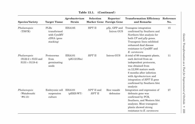
Gen
etic Tran
sformation
as a Tool for Im
provemen
t of Orch
ids✦
233
FA
Table 13.1. (Continued)
Agrobacterium Selection Reporter/ Transformation Efficiency ReferenceSpecies/Variety Target Tissue Strain Marker Gene Foreign Gene and Remarks No.
Phalaenopsis PLBs EHA105 HPT II pflp, GFP and Transgene integration 15(TS97K) transformed Intron GUS confirmed by Southern and
with CymMV Northern blot analysis forcDNA (gene both CP and pflp genes.stacking) Transgenic lines exhibited
enhanced dual diseaseresistance to CymMV andE. carotovora
Phalaenopsis Protocorms EHA101 HPT II Intron-GUS A total of 88 transgenic plants, 11(S122-2 × S153 and from (pIG121Hm) each derived from anS153 × S119-4) germinating independent protocorm,
seeds was obtained fromca.12,500 mature seeds6 months after infectionwith Agrobacterium andintegration of HPT II geneconfirmed by Southern blotanalysis
Phalaenopsis Embryonic cell EHA101 NPT II and Rice wasabi Integration and expression of 27(Wataboushi suspension (pEKH-WT) HPT II defensine defensin gene was‘#6.13) culture confirmed by PCR,
Southern, and Western blotanalyses. Most transgenicplants showed strongresistance to E. carotovora
b494_Ch-13.qxd 8/29/2007 1:44 PM Page 233

234✦
San
jaya and M
.-T. Ch
an
FA
Table 13.2. Particle Bombardment/Direct Gene Transformation in Orchids
Selection Reporter/ Transformation Efficiency and ReferenceSpecies/Variety Target Tissue Marker Gene Foreign Gene Remarks No.
Dendrobium Protocoms NPT II Papaya ringspot Obtained 13 kanamycin resistant plants 37(Dendrobium × virus (PRV) and confirmed foreign gene (NPT II)Jaquelyn Thomas coat protein by genomic PCR and Southern blothybrids) (CP) gene analyses
Dendrobium Calli cultured in NPT II Luciferase Transformed tissues expressing luciferase 29(White angel) liquid medium were detected, allowed to regenerated
into complete plant on selectionmedium and integration of transgeneconfirmed by Southern and Northernblot analyses
Phalaenopsis PLBs NPT II and Intron-GUS About 7 bialophos-resistant plantlets 39(Danse × Happy bar were obtained from 622 bombardedValentine) PLBs and only one showed GUS
expressionDendrobium Protocorms HPT II Intron-GUS 15 hygromycin-resistant lines recovered 13
(hybrid ‘MiHua’) on selection medium and integration offoreign genes was confirmed by GUShistochemical assay and Southern blotanalysis
Cymbidium PLBs NPT II Intreon-GUS About 85% of GUS positive shoots were 40obtained and further transgenic natureconfirmed by genomic PCR andSouthern blot analyses of the PCRproduct
(Continued)
b494_Ch-13.qxd 8/29/2007 1:44 PM Page 234

Gen
etic Tran
sformation
as a Tool for Im
provemen
t of Orch
ids✦
235
FA
Table 13.2. (Continued)
Selection Reporter/ Transformation Efficiency and ReferenceSpecies/Variety Target Tissue Marker Gene Foreign Gene Remarks No.
Brassia, Cattleya and PLBs bar Selection of putative transformants was 20Doritaenopsis accomplished by using bialaphos. The
presence of bar gene was confirmedby PCR, Southern, and Northern blotanalyses
Dendrobium Calli HPT II Intron-GUS About 12% (D. Phalaenopsis) and 2% 10phalaenopsis (D. nobile) transformation efficiencyBanyan Pink was achieved and integration of foreignand D. nobile gene confirmed by Southern and
Northern blot analysesPhalaenopsis TS444 Petels — Flavonoid-3′,5′- Transient transformation achieved by 43
[(New Eagle × hydroxylase particle bombardment and thePinlong gene from transgenic petals changed from pink toCinderella) × Dtps. Phalaenopsis magenta, demonstrated the role ofTaisuco Red] flavonoid-3′,5′-hydroxylase gene in
anthocyanin pigment synthesisPhalaenopsis PLBs HPT II CymMV coat Among 13 transgenic plants confirmed 14
“TS340” protein (CP) by Southern blot analysis, most(P. Taisuco cDNA transgenic lines showed CymMVKochdiam × protection. Nuclear run-on and smallP. Taisuco interfering RNA (siRNAs) analysesKaaladian) showed that CymMV resistance was RNA-
mediated through a posttranscriptionalgene silencing mechanism (PTGS) in thesilenced transgenic orchid plants.
(Continued )
b494_Ch-13.qxd 8/29/2007 1:44 PM Page 235

236✦
San
jaya and M
.-T. Ch
an
FA
Table 13.2. (Continued)
Selection Reporter/ Transformation Efficiency and ReferenceSpecies/Variety Target Tissue Marker Gene Foreign Gene Remarks No.
Dendrobium Protocorms HPT II CymMV coat Presence of the transgene confirmed by 42(Hickam Deb) protein (CP) PCR, Southern, Northern, and Western
cDNA blot analyses. Transgenic plantsharboring the CymMV CP geneexpressed a very low level of virusaccumulation 4 months postinoculationwith CymMV
Phalaenopsis PLBs HPT II CymMV coat PLBs transformed with CymMV coat 14(TS97K) protein (CP) protein cDNA (CP) were then
cDNA retransformed with sweet pepperferredoxin-like protein cDNA ( pflp) byAgrobacterium tumefaciens, to enableexpression of dual (viral and bacterial)disease resistant traits. Doubletransformants confirmed by molecularanalysis and exhibited enhanced dualdisease resistance
Oncidium PLBs HPT II pflp Sucrose pretreatment enhanced the 41(Sharry Baby (pretreated with regeneration of PLBs, single-cell“OM8”) sucrose) embryogenesis, and transformation
efficiency
b494_Ch-13.qxd 8/29/2007 1:44 PM Page 236

detectable by use of both histochemical and fluorigenic substrates. Thisreporter gene was successfully used in the transformation of differentorchids.10,11 In some cases, the herbicide resistance (bar) gene has alsobeen useful in the selection of transgenic orchids.20
13.4 General Protocol for Agrobacterium-MediatedTransformation in Orchids
The landmark discovery of A. tumefaciens21 provided a natural genetransfer mechanism to introduce and express DNA stably in differentplant species, regardless of their origin. Several hybrid orchids, whichare difficult for A. tumefaciens infection, have been transformed bydirect gene gun or microparticle bombardment. However, gene transfertechniques have been established only in a few orchids.7,10,11,13 In gen-eral, Agrobacterium-mediated transformation in orchids was achievedby cocultivating a virulent A. tumefaciens strain containing a recombi-nant Ti plasmid with PLBs/cell clumps, from which complete plantswere regenerated.
Although Agrobacterium-mediated transformation needs to be opti-mized for different species, the Agrobacterium infection can be pro-moted under conditions that induce virulence, such as use ofacetosyringone (AS) or a-hydroxyacetosyringone, acidic pH, and appro-priate incubation temperature. After coculture for 2 to 3 days, PLBs aretransferred to a medium containing selective agents to eliminate non-transformed plant cells, antibiotics to kill the Agrobacterium, and hor-mones to induce shoot and root growth. After a few months, shootsdevelop from transformed PLBs. These can be removed and transferredto a rooting medium, and then shifted to soil. Most current protocols forthe Agrobacterium-mediated transformation of orchids involve varia-tions in the starting material. In Phalaenopsis, using PLBs as target tis-sue, we have developed a reliable and reproducible transformationprotocol (Fig. 13.1).
13.4.1 Recent advances in Agrobacterium-mediatedtransformation
Several plant phenolic compounds, including AS, coumaryl alcohol, andsinapyl alcohol, are known as vir gene inducers; but in orchids, their
Genetic Transformation as a Tool for Improvement of Orchids ✦ 237
FAb494_Ch-13.qxd 8/29/2007 1:44 PM Page 237

238 ✦ Sanjaya and M.-T. Chan
FA
Regenerate of PLBs from leaf segments
Transfer PLBs (45-day-old) to fresh T2 mediumsupplemented with 200 µM AS and continue in dark for 1 h
Infect with Agrobacterium for 1 h in dark
Cocultivate blot-dried PLBs on T2 solid medium supplemented with 200 µM AS and 5% glucose at 26˚C in dark for 3 days
Wash infected PLBs in MS liquid mediumsupplemented with 200 mg/L timentin
Transfer blot-dried PLBs on to T2 medium supplemented with 200 mg/Ltimetin, incubate at 26˚C with 16 h light/8 h dark photoperiod for 4 weeks at a light intensity of 100 to 200 µE/m2/s.
Separate newly differentiated PLBs from the original explant, subculture on T2 medium supplemented with 200 mg/L timentin and the optimal concentration of antibiotics for selection of putative transformants
Transfer actively growing PLBs on T2 medium supplemented with the optimalconcentration of antibiotics for 2nd selection to establish transgenic lines
Transfer well-rooted plants for hardening and carry out molecular analysis
Transfer to pots containing sphagnum moss andacclimatize under greenhouse conditions
After 6–8 weeks, transfer survival and actively growing putatively transformed PLBs on fresh T2 medium for shoot elongation
Fig. 13.1. Different stages of A. tumefaciens-based transformation inPhalaenopsis.
b494_Ch-13.qxd 8/29/2007 1:44 PM Page 238

presence and characteristics were not well understood until Nan et al.22
identified these compounds in high amounts in light-grown DendrobiumPLBs. The authors demonstrated activation of the Agrobacterium virgene in the presence of these phenolic compounds, and revealed thepossibility of Agrobacterium-mediated transformation in orchids. Theseobservations come as a great boon to orchid biologists to initiate genetransformation studies in various commercial varieties.
The feasibility of using Agrobacterium-mediated transformationsystem in orchid transformation was not explored until Hsieh et al.23
first reported the transformation of different varieties of Phalaenopsiswith NPT II and GUS genes using PLBs as target tissue (Table 13.1).Subsequently, other authors reported success with Agrobacterium-mediated transformation in orchids. Belarmino and Mii7 reportedPhalaenopsis transformation with a GUS gene and confirmedthe transgenic nature of developed plants by histochemical GUSassay, PCR analysis, and Southern hybridization. Use of this trans-formation system successfully produced more than 100 hygromycin-resistant plantlets within 7 months following infection of cell aggregates.Dendrobium was transformed with DOH1 by use of thin-section explantsfrom PLBs.13
Chai et al.24 evaluated two types of explants, intact and transverselybisected PLBs, in Phalaenopsis transformation, and observed thattransversely bisected PLBs responded better than intact ones in termsof efficiency of multiplication and transformation. A high efficiency oftransformation (18%) in Dendrobium was reported, with 73 stablytransformed lines accomplished in the presence of 100 µ M AS andhygromycin as the selection marker.10 Liau et al.12 first reported a trans-formation method suitable for improving Oncidium by using PLBsderived from protocorms as target tissue. Among 1000 inoculated PLBs,108 putatively transformed PLBs were proliferated on hygromycinselection medium. A total of 28 independent transgenic orchid plantswere obtained, from which 6 transgenic GUS-positive lines were ran-domly selected and confirmed by Southern, Northern, and Western blotanalyses. This successfully demonstrated the integration and expres-sion of foreign DNA in the Oncidium genome. Soonafter, the samegroup transformed sweet pepper ferredoxin-like protein (pflp) cDNA,with a HPT II and GUS coding sequence, into PLBs of Oncidium, anddemonstrated the applicability of the pflp gene as an antibiotic-freeselection marker in Oncidium25; intriguingly, transgenic plants showed
Genetic Transformation as a Tool for Improvement of Orchids ✦ 239
FAb494_Ch-13.qxd 8/29/2007 1:44 PM Page 239

enhanced resistance to Erwinia carotovora, which causes soft rot dis-ease, even when the entire plant was challenged with the pathogen.26
Recently, Chan et al.15 used “gene stacking” in Phalaenopsis by dou-ble transformation events. Phalaenopsis PLBs originally transformedwith CymMV coat protein cDNA (CP) were then transformed with pflpcDNA by A. tumefaciens infection to enable the expression of dual (viraland bacterial) disease-resistant traits. The immature protocorms devel-oped from Phalaenopsis seeds were also demonstrated to be a potentialstarting source for Agrobacterium-mediated transformation.11 However,this protocol may not have broader application because most of the com-mercial hybrids are sterile in nature. Interestingly, 5 day-old embryo-genic suspension cells of Phalaenopsis were used as starting materialin rice wasabi defensin gene transformation, and the authors claimedthat most of the transgenic plants exhibited enhanced resistance toE. carotovora.27
13.5 General Conditions for A. Tumefaciens-Mediated Transformation
13.5.1 Effect of feeder cells/acetosyringone ontransformation efficiency
Acetosyringone (AS), a phenolic compound produced in metabolicallyactive plant cells or released by wounded cells, induces the expressionof the vir genes on the Ti plasmid of A. tumefaciens, which in turnfacilitates T-DNA transfer into plant cells.28 Although orchid cells arecapable of producing certain phenolic compounds at a low level, suchproduction is not sufficient for high and consistent transformationevents.22
The feasibility of AS in orchid transformation was demonstratedby Hsieh et al.23 in Phalaenopsis transformation; the authors added100 µM AS and regeneration medium to the Agrobacterium cultureprior to infection to enhance transformation events. In concordance withthe above observations, Chia et al.29 and Mishiba et al.11 used 100 µMAS during the infection and cocultivation stages in Phalaenopsistransformation. In Dendrobium, Men et al.10 observed higher GUS geneexpression when PLBs were cocultivated with 100 µM AS-activatedA. tumefaciens for 2 to 3 days. Interestingly, Chan et al.15 reported the
240 ✦ Sanjaya and M.-T. Chan
FAb494_Ch-13.qxd 8/29/2007 1:44 PM Page 240

use of 200 µM AS and 5% glucose during cocultivation and foundenhanced transformation efficiency. Belarmino and Mii7 infectedexplants with 200 µM AS-treated Agrobacterium and included 500 µMAS in cocultivation medium. In Oncidium transformation, the use of1 mL of tightly packed 3-day-old freshly subcultured tobacco suspen-sion cells on G10 medium supplemented with 200 µM AS as a cocul-tivation medium and dark incubation for 3 days at 26°C significantlytriggered the A. tumefaciens vir genes, which in turn resulted in hightransformation efficiency. Furthermore, these results were consis-tent and reproducible.12 However, in the absence of AS, only a fewtransgenic plants were obtained. The use of feeder cells from speciessuch as tobacco petunia, and tomato as a phenolic source to enhancethe transformation rate still needs to be optimized for various com-mercial orchids.
13.5.2 Agrobacterium strain and density
The choice of Agrobacterium strain greatly influences the transforma-tion success in orchids. A wide range of A. tumefaciens strains usedinclude LBA4301,22 EHA105 (pMT1),23 LBA4404 (pTOK233),8,24
EHA101 (pIG121Hm),11 and EHA101 (pEKH-WT).27 However, othershave advocated that the success of transformation efficiency is notinfluenced by the Agrobacterium strain alone.7,30 In Dendrobium, Menet al.10 examined two Agrobacterium strains, AGL1 and EHA105, andobserved a high level of GUS staining in AGL1-infected PLBs.Simultaneously, Liau et al.26 using EHA105 and LBA4404, found no dif-ference in transformation efficiency. However, the shoot formation rateof EHA105-transformed PLBs was higher than that of LBA4404-treated PLBs. In addition, after hygromycin selection, the antibioticsused (timentin/cefatoxime) did not always control the overgrowth ofLBA4404 and resulted in a low rate of shoot formation.
Therefore, in general, the tissue culture conditions and choiceof bacteriostatic substances employed to suppress the overgrowthof Agrobacterium are important factors in orchid transformation.Although the bacterial density was not thoroughly examined inmost of the transformation studies, most authors used diluted Agro-bacterium at an A600 of 0.2–1.0 for improved transformation effi-ciency. Thus, Agrobacterium density is also a key factor for successfultransformation.11,23–26
Genetic Transformation as a Tool for Improvement of Orchids ✦ 241
FAb494_Ch-13.qxd 8/29/2007 1:44 PM Page 241

13.5.3 Effect of selection agents
In general, Agrobacterium-mediated transformation in orchids is pre-ceded by infection, 2 to 3 days of cocultivation, and selection of putativetransformants on selection medium supplemented with antibiotics andbacteriostatic substances to cull untransformed tissues and A. tumefa-ciens. The ideal antibiotic for inhibiting Agrobacterium growth should behighly effective, inexpensive, not have a negative effect on plant growthand regeneration, and be stable in culture.31 The selective effects ofdifferent antibiotics in plant transformation have been reported forseveral species.32 In orchid transformation, most authors used differentconcentrations of cefotaxime/carbenicillin as antibiotics for suppressingAgrobacterium growth after cocultivation.7–9,23,24,27 Mishiba et al.11 usedmeropenem (β-lactam antibiotics) to remove excess Agrobacteriumfrom PLBs.
In our lab, we standardized a two-step transformation procedure inOncidium: after infection, PLBs were cocultivated for 3 days; then,infected PLBs were transferred to a medium supplemented with lowconcentrations of timentin/cefatoxime to suppress A. tumefaciensgrowth and incubated at 26°C for a 16-h photoperiod at a light intensityof 100–200 µEm−2s−1 for a month. No selective antibiotic had been incor-porated in the medium to select putatively transformed PLBs becausethe long-term transformation process might enhance transfer of foreignDNA from A. tumefaciens into actively growing PLBs. Subsequently,A. tumefaciens was completely avoided with high concentrations oftimentin/cefatoxime and a selective agent was used to eliminate untrans-formed PLBs. We achieved approximately 10% transformation effi-ciency, with 108 antibiotic-resistant independent PLBs proliferatedfrom 1000 infected PLBs.12
13.6 Microprojectile/Particle Bombardment orDirect Gene Transformation
A novel method for plant transformation was introduced by JohnSanford and colleagues in 1987. The authors showed that projectilebombardment, with several modifications, was an efficient methodfor stable integration of foreign genes into the plant genome.33
Subsequently, a gunpowder-based device was refined for a system based
242 ✦ Sanjaya and M.-T. Chan
FAb494_Ch-13.qxd 8/29/2007 1:44 PM Page 242

on high-pressure blasts of helium;34 this is the only commercially avail-able particle gun, which is marketed by Bio-Rad. Particle bombardmentis a simple technique: a plasmid DNA is prepared by standard methodsand precipitated onto tungsten or gold particles with use of CaCl2, sper-midine, and PEG to protect the DNA during precipitation; and then theparticles are washed and suspended in ethanol before being firedagainst a retaining screen that allows the microprojectiles through tostrike the target tissue.
Particle bombardment is widely used because it circumvents twomajor limitations of the Agrobacterium system. First, any hybrid orchidspecies and cell type can be transformed by this method because DNAdelivery is controlled entirely by physical rather than biological param-eters. The range of species transformable by particle bombardment is,therefore, restricted only by the competence of cells for regeneration.The technique is independent of genotype and, consequently, useful forthe transformation of elite cultivars. However, careful optimization isrequired to tailor the method for different varieties or cell types and toachieve the highest efficiency transformation with the least cell dam-age. Important parameters include acceleration method, particle veloc-ity (controlled by the discharge voltage and/or gas pressure), particlesize, and use of different materials (tungsten, gold).35 Second, particlebombardment allows the stable and heritable introduction of many dif-ferent genes at once with the use of different plasmids, as these tend toconcatemerize to form one DNA cluster that integrates at a single locus.Remarkably, the transgene silencing, instability, and rearrangementsare more evident with particle bombardment than the Agrobacteriummethod of gene transformation.36
13.6.1 Recent advances in particle bombardmentin orchids
Pioneering gene transformation experiments augmented with particlebombardment were successfully used to produce transgenic orchidswith a marker/reporter gene in Dendrobium,10,13,28,37 Phalaenopsis,39
Cymbidium,40 Brassia, Cattleya, and Doritaenopsis20 (Table 13.2).Osmotic pretreatment of Oncidium PLBs on high sucrose mediumbefore bombardment and transformation have enhanced single-cellembryogenesis and transformation efficiency.41 This situation can alsobe achieved by partial drying or the addition of osmoticum (mannitol
Genetic Transformation as a Tool for Improvement of Orchids ✦ 243
FAb494_Ch-13.qxd 8/29/2007 1:44 PM Page 243

and/or sorbitol) to the culture medium. In general, stable transforma-tion by any direct DNA transfer method occurs at a much lower fre-quency than transient transformation.
The direct gene transformation has also been successfully used indeveloping transgenic orchids resistant to Cymbidium mosaic virus inPhalaenopsis14,15 and Dendrobium.42 Interestingly, Chan et al.15 demon-strated the usefulness of direct gene transformation in introducingmultiple genes into the Phalaenopsis genome. Su and Hsu43 reportedtransient transformation of Phalaenopsis petals with the putativecytochrome P450 gene. In addition, other direct gene transformationmethods, including seed imbibition and pollen tube mediation, havebeen reported in Dendrobium and Phalaenopsis38,44 (Table 13.3).However, these methods are not in frequent use because of low trans-formation efficiency.
13.7 Selection of Putative Transformants andMolecular Analysis
Selection of the transformed cells and subsequent regeneration ofplants is the most important requirement in orchid transformation.The most commonly used antibiotics for selection of transformed orchidPLBs are hygromycin and kanamycin. Resistance to these antibiotics isconferred by insertion and expression of HPT II or NPT II under thecontrol of a CaMV35S promoter.7,12,13,23,24,37 Knapp et al.20 demonstratedthe usefulness of bar as a selection marker in Brassia, Cattleya, andDoritaenopsis transformation. Our laboratory experiments involving
244 ✦ Sanjaya and M.-T. Chan
FA
Table 13.3. Other Direct Gene Transformation Methods in Orchids
TransformationSpecies/Variety Target Tissue Method Reference No.
Dendrobium Seeds Imbibition in solution 44containing foreignDNA
Dendrobium Ovary Pollen-tube pathway 44Dendrobium Protocorms Electro injection 44Phalaenopsis Ovary Pollen-tube pathway 38
b494_Ch-13.qxd 8/29/2007 1:44 PM Page 244

pCAMBIA binary vector series showed the most consistent results inPhalaenopsis and Oncidium transformation. The screenable markersor reporter genes are widely used, particularly when transformationprocedures are being optimized. The most commonly used reportergene in higher plants is GUS; the expression and activity of this enzymeare easily detectable by the use of both histochemical and fluorigenicsubstrates. This reporter gene was used successfully in the transforma-tion of other orchids, including Cymbidium,37 Dendrobium,10 andPhalaenopsis.11
Furthermore, the available molecular tools such as PCR, Northern,Southern, and Western blot analyses have allowed for confirming trans-gene integration and expression and have clarified the outcome of suchtransformation experiments. Because orchids have a long floweringstage, the molecular analysis of T1 progeny is time-consuming.Alternatively, T0 transgenic lines can be thoroughly diagnosed for for-eign gene integration by Southern, Northern, and Western blot analy-ses.12 Southern and Northern blot analysis was used to confirm thetransformation, copy number, and transcriptional activity of transgenesin putatively transformed orchids.
13.8 Expression of Genes with PotentialCommercial Application
The main drawback of traditional plant breeding is that it relies on theuse of germplasm of the same or closely related species, and theprogress is time-consuming. The 21st century will likely pose a new setof challenges for orchid growers, as increased demand for cut flowersand potted plants will require quick production practices. Sustainingboth supply and demand in the future will depend more heavily on thedevelopment and deployment of a range of new technologies, includingbiotechnology.
13.8.1 Marker-free transgenic orchids
To obtain maximum putatively transformed plants in each transfor-mation event, effective selectable markers, antibiotics, or herbicide-resistant genes have been used to simplify detection procedures. Theexisting selection systems can be divided into two groups: conventional
Genetic Transformation as a Tool for Improvement of Orchids ✦ 245
FAb494_Ch-13.qxd 8/29/2007 1:44 PM Page 245

selection systems, the largest group, which rely on an antibiotic or aherbicide as the selective agent detoxified by a selective gene;45 andpositive selection systems, in which the selective agent is convertedinto a simple compound by the selective gene product and transformedcells have metabolic or developmental advantages.46,47 In most of theorchid transformation protocols, the available methods for screeningtransgenic plants are based on antibiotics or herbicide-resistantmarker genes such as NPT II, HPT II, and bar on media containing theselective agent.7,8,20 However, the use of these selection marker genes isa matter of concern because of their possible toxicity or allergenicity tohumans and other organisms. In addition, the presence of such resist-ance genes and their proteins is undesirable in transgenics.Furthermore, these selection systems are time-consuming, expensive,and labor-intensive, therefore, the development of alternative or newselection marker genes and improved selection procedures is essentialfor the orchid industry.
Sweet pepper ferredoxin-like protein (pflp), isolated from pepper,was successfully cloned and reported to have antimicrobial activityagainst Pseudomonas syringae by delaying the hypersensitive responsethrough the release of a hairpin proteinaceous elicitor.48 We developed anovel selection procedure for orchid transformation by introducing pflpas a selection marker into the Oncidium genome via A. tumefaciensinfection and particle bombardment, and putatively transformed plantswere screened by the use of the natural bacterial pathogen E. carotovoraas a selection agent (Fig. 13.2). We were able to obtain approximately 32independent transgenic lines without the use of an antibiotic selectionagent.25 Although the selection efficiency with pflp or hygromycin doesnot differ much, the time required for selecting transgenic plants by useof pflp is shorter. Therefore, to minimize the chances for somaclonalvariation caused by long-term tissue culture selection procedures and toenhance the rooting ability of transgenic plants, always a serious prob-lem in the antibiotic selection system, the use of pflp is preferable.Furthermore, the pflp system can be used to introduce economicallyimportant genes into any other orchid. Recently, Chan et al.15 success-fully demonstrated the practicality of the pflp system as a nonantibioticselection system in Phalaenopsis transformation. Plants that are notnatural hosts of E. carotovora, used as transformation materials, do notconform to the pflp selection system. However, the deployment of thissystem may substantially decrease concerns about the negative effect on
246 ✦ Sanjaya and M.-T. Chan
FAb494_Ch-13.qxd 8/29/2007 1:44 PM Page 246

biodiversity with the use of antibiotics or herbicide-resistance markergenes and their presence in transgenic plants.
13.8.2 Disease-resistant genes
The efficient control of phytopathogens affecting orchid crops repre-sents one of the major challenges in the floriculture industry worldwide.Several orchid species are highly susceptible to different pathogens,among them the devastating disease soft rot caused by E. carotovora,and this genus is considered to be the natural host. Advances in tech-nology have allowed for introducing a novel gene(s) singly or in combi-nation into the orchid genome for the dual purposes of transformationand disease resistance. For example, the overexpression of pflp in riceconfers resistance against Xanthomonas oryzae.49 We introduced an
Genetic Transformation as a Tool for Improvement of Orchids ✦ 247
FA
Fig. 13.2. Schematic representation of the development of marker-free trans-genic orchids with pflp used as a marker gene and Erwinia carotovora (ECC) asa selection agent.
b494_Ch-13.qxd 8/29/2007 1:44 PM Page 247

antimicrobial gene (pflp) into the Oncidium genome via A. tumefaciensinfection. Application of bacteria on the leaves of transgenic and wild-type plants showed characteristic symptoms of water-soak regions fol-lowed by maceration, more evident in wild-type plants than intransgenic plants; resistance scoring at the whole plant level alsorevealed that pflp confers enhanced resistance to E. carotovora.26
Viral diseases in orchids, especially those caused by Odontoglossumringspot tobamovirus (ORSV) and Cymbidium mosaic virus (CymMV), area major concern among breeders because of their catastrophic effects onplant yield and market value. Liao et al.14 successfully created transgenicPhalaenopsis orchid plants transformed with CymMV coatprotein cDNA (CP), which conferred protection against CymMV infec-tion. To enhance the resistance of orchids to both viral and bacterialphytopathogens, Chan et al.15 used the “gene-stacking” phenomena inPhalaenopsis by double transformation events: Phalaenopsis PLBs, origi-nally transformed with CP, were then transformed with pflp cDNA by theuse of A. tumefaciens to enable the expression of dual (viral and bacterial)disease-resistant traits. Disease-resistant assays revealed transgenicplants with enhanced resistance to CymMV and E. carotovora infection.Recently, Chang et al.39 created transgenic Dendrobium plants harboringthe CymMV CP gene, which expressed a very low level of virus accumula-tion 4 months after inoculation with CymMV.
Large numbers of toxic compounds exist in higher plants to ensurevarious physical and biochemical defense barriers against pests andpathogens. Among these, defensin proteins form a well-defined group oflow-molecular-weight cystein-rich polypeptides whose toxic effect onfungi and bacteria have been studied in model systems and are knownto be expressed either by pathogen attack or elicitors. Recently, Sjahrilet al.27 successfully introduced the wasabi defensin gene intoPhalaenopsis and recorded enhanced tolerance to E. carotovora. Similarstrategies can be extended to other commercially important orchids forsingle- or multiple-disease resistance.
13.9 Conclusions and Future Prospects
As more genes are being isolated and understood in model systems andcrop plants, more options are becoming available for the application ofgenetic modification in orchid improvement. However, such advanceshave not been fully realized in orchids, probably because most hybrid
248 ✦ Sanjaya and M.-T. Chan
FAb494_Ch-13.qxd 8/29/2007 1:44 PM Page 248

orchids are recalcitrant to tissue culture and transformation. So,research to increase transformation efficiency, develop new markergenes, introduce new genes alone or in combination by multigene trans-formation (gene stacking), and improve cryopreservation techniqueswould be extremely useful for orchid improvement.
Recent studies of the entire chloroplast genome sequencing (cpDNA)of Phalaenopsis are novel examples of the successful utilization of cut-ting-edge technology in orchids, and the information generated can besuccessfully utilized in chloroplast transformation in Phalaenopsis andother orchids.50 Functional genomics studies of Oncidium and Dendrobiumflower formation and initiation are promising and useful in terms ofindustrial as well as academic interests.51–53 In addition, the informationgenerated by Wu et al.54 on Dendrobium MYB genes during flower devel-opment could be useful in exploring new genes involved in early/lateflower development, subsequent functional analysis, and innovation ofnovel flower-specific promoters that will certainly benefit orchid improve-ment. Pathway engineering, involving the simultaneous introduction andconcerted expression of multiple transgenes, will be featured prominentlyin future strategies for orchid transformation. Finally, the outputs fromcurrent genomics, proteomics, and metabolomics programs can be fullyexploited with sophisticated methods to introduce large pieces of DNAinto the orchid genome.
Acknowledgments
Updates are not intended to be comprehensive reviews, and the authorsapologize to those colleagues whose works could not be cited because ofthe restricted number of references. The authors thank S.J. Yu, I.C.Pan, and C.H. Liau for graphics. This work was supported by grantsfrom Academia Sinica as well as the National Science and TechnologyProgram for Agricultural Biotechnology of the Republic of China.
References
1. Chan MT, Chan YL, Sanjaya. (2006) Orchids (Cymbidium spp., Oncidiumand Phalaenopsis). In: Kang Wang (ed.), Methods in Molecular Biology,Vol. 344: Agrobacterium Protocols. 2/e, Vol. II, Humana Press Inc., Totowa,USA, pp. 331–337.
Genetic Transformation as a Tool for Improvement of Orchids ✦ 249
FAb494_Ch-13.qxd 8/29/2007 1:44 PM Page 249

2. Chan MT, Sanjaya, Teixeira da Silva JA. (2006) Oncidium tissue culture,transgenics and biotechnology. In: Teixeira da Silva JA (ed.), Floriculture,Ornamental and Plant Biotechnology: Advances and Topical Issues, Vol. II,Global Science Books, Ltd., London, UK, pp. 193–198.
3. Chan MT, Sanjaya. (2006) Current trends in Taiwan’s orchid biotechnol-ogy. Global Plant Lett I:2–3.
4. Teixeira da Silva AJ. (2004) Ornamental chrysanthemums: Improvementby biotechnology. Plant Cell Tissue Organ Cult 79:1–18.
5. Tanaka Y, Katsumoto Y, Bruglier F, Mason J. (2005) Genetic engineeringin floriculture. Plant Cell Tissue Organ Cult 80:1–24.
6. Vain P. (2006) Global trends in plant transgenic science and technology(1973–2003). Trends Biotechnol 24:206–211.
7. Belarmino MM, Mii M. (2000) Agrobacterium-mediated genetic transfor-mation of a Phalaenopsis orchid. Plant Cell Rep 19:435–442.
8. Yu H, Yang SH, Goh CJ. (2001) Agrobacterium-mediated transformation ofa Dendrobium orchid with the class 1 knox gene DOH1. Plant Cell Rep20:301–305.
9. Men S, Ming X, Liu R, et al. (2003) Agrobacterium-mediated genetictransformation of a Dendrobium orchid. Plant Cell Tissue Organ Cult75:63–71.
10. Men S, Ming X, Wang Y, et al. (2003) Genetic transformation of two speciesof orchid by biolistic bombardment. Plant Cell Rep 21:592–598.
11. Mishiba KI, Chin DP, Mii M. (2005) Agrobacterium-mediated transforma-tion of Phalaenopsis by targeting protocorms at an early stage after ger-mination. Plant Cell Rep 24:297–303.
12. Liau CH, You SJ, Prasad V, et al. (2003) Agrobacterium tumefaciens-mediatedtransformation of an Oncidium orchid. Plant Cell Rep 21:993–998.
13. Yu Z, Chen M, Nie L, et al. (1999) Recovery of transgenic orchid plantswith hygromycin selection by particle bombardment to protocorms. PlantCell Tissue Organ Cult 58:87–92.
14. Liao LJ, Pan IC, Chan YI, et al. (2004) Transgene silencing in Phalaenopsisexpressing the coat protein of Cymbidium mosaic virus is a manifestationof RNA-mediated resistance. Mol Breed 13:229–242.
15. Chan YL, Lin KH, Sanjaya, et al. (2005) Gene stacking in Phalaenopsisorchid enhances dual tolerance to pathogen attack. Transgenic Res14:279–288.
16. George EF, Sherrington PD. (1984) Plant Propagation by Tissue Culture —Handbook and Dictionary of Commercial Laboratories. Exgetics Ltd.,England.
17. Miki B, McHugh S. (2004) Selectable marker genes in transgenic plants:Applications, alternatives and biosafety. J Biotechnol 107:193–232.
250 ✦ Sanjaya and M.-T. Chan
FAb494_Ch-13.qxd 8/29/2007 1:44 PM Page 250
18. Tian L. (2006) Markers gene removal from transgenic plants. In: Teixeira daSilva JA (ed.), Ornamental and Plant Biotechnology, Vol. II, Global ScienceBooks, London, pp. 26–29.
19. Haldrup A, Petersen S, Okkels F. (1998) Plant xylose isomerase gene fromThermoanaerobacterium thermosulfurogenes allows effective selection oftransgenic plant cells using D-xylose as the selection agent. Plant Mol Biol37:287–296.
20. Knapp JE, Kausch AP, Chandlee JM. (2000) Transformation of three generaof orchid using the bar gene as a selectable marker. Plant Cell Rep 19:893–898.
21. Chilton MD, Drummond MH, Merlo DJ, et al. (1977) Stable incorporationof plasmid DNA into higher plant cells: The molecular basis of crown galltumorigenesis. Cell 11:263–271.
22. Nan GL, Tang CS, Kuehnle AR, Kado CI. (1997) Dendrobium orchids con-tain an inducer of Agrobacterium virulence genes. Physiol Mol PlantPathol 51:391–399.
23. Hsieh RM, Chen WH, Lin YS, et al. (1997) Agrobacterium tumefaciens-mediated transformation of Phalaenopsis orchid. Rept Taiwan Sugar ResInst 155:41–54.
24. Chai ML, Xu CJ, Senthil KK, et al. (2002) Stable transformation of proto-corm-like bodies in Phalaenopsis orchid mediated by Agrobacterium tume-faciens. Sci Hortic 96:213–224.
25. You SJ, Liau CH, Hauang HE, et al. (2003) Sweet pepper ferredoxin-likeprotein (pflp) gene as a novel selection marker for orchid transformation.Planta 217:60–65.
26. Liau CH, Lu JC, Prasad V, et al. (2003) The sweet pepper ferredoxin-likeprotein (pflp) conferred resistance against soft rot disease in Oncidiumorchid. Transgenic Res 12:329–336.
27. Sjahril R, Chin DP, Khan RS, et al. (2006) Transgenic Phalaenopsisplants with resistance to Erwinia carotovora produced by introducingwasabi defensin gene using Agrobacterium method. Plant Biotechnol23:191–194.
28. Bolton GW, Nestor EW, Gordon MP. (1986) Plant phenolic compoundsinduce expression of the Agrobacterium tumefaciens loci needed for viru-lence. Science 232:983–985.
29. Chia TF, Chan YS, Chua NH. (1994) The firefly luciferase gene as a non-invasive reporter for Dendrobium transformation. Plant J 6:441–446.
30. Hiei Y, Komari T, Kuba T. (1997) Transformation of rice mediated byAgrobacterium tumefaciens. Plant Mol Biol 35:205–218.
31. Cheng ZM, Schnurr JA, Kapaun JA. (1998). Timentin as an alternativeantibiotic for suppression of Agrobacterium tumefaciens in genetic trans-formation. Plant Cell Rep 17:646–649.
Genetic Transformation as a Tool for Improvement of Orchids ✦ 251
FAb494_Ch-13.qxd 8/29/2007 1:44 PM Page 251

32. Hammerschlag FA, Zimmerman RH, Yadava et al. (1997) Effect of antibioticsand exposure to an acidified medium on the elimination of Agrobacteriumtumefaciens from apple leaf explants and on shoot regeneration. J AmHortic Soc 112:758–763.
33. Klein TM, Fromm M, Weissinger A, et al. (1988) Transfer of foreign genesinto intact maize cells with high velocity microprojectiles. Proc Natl AcadSci USA 85:4305–4309.
34. Ye G-N, Daniell H, Stanford JC. (1990) Optimization of delivery of foreignDNA into higher plant chloroplasts. Plant Mol Biol 15:809–819.
35. Christou P. (1994) Particle gun mediated transformation. Curr OpinBiotechnol 4:135–141.
36. Birch RG. (1997) Plant transformation: Problems and strategies for prac-tical application. Annu Rev Plant Physiol Plant Mol Biol 48:297–326.
37. Kuehnle AR, Sugii N. (1992) Transformation of Dendrobium orchid usingparticle bombardment of protocorms. Plant Cell Rep 11:484–488.
38. Hsieh YH, Huang PL. (1995) Studies on genetic transformation ofPhalaenopsis via pollen tube pathway. J Chin Soc Hortic Sci 41:309–324.
39. Anzai H, Ishii Y, Shichinohe M, et al. (1996) Transformation of Phalaenopsisby particle bombardment. Plant Tissue Cult Lett 3:265–271.
40. Yang J, Lee H, Shin DH, et al. (1999) Genetic transformation of Cymbidiumorchid by particle bombardment. Plant Cell Rep 18:978–984.
41. Li SH, Kuoh CS, Chen YH, et al. (2005) Osmotic sucrose enhancementof single-cell embryogenesis and transformation efficiency in Oncidium.Plant Cell Tissue Organ Cult 81:183–192.
42. Chang C, Chen YC, Hsu YH, et al. (2005) Transgenic resistance toCymbidium mosaic virus in Dendrobium expressing the viral capsid pro-tein gene. Transgenic Res 14:41–46.
43. Su V, Hsu BD. (2003) Cloning and expression of a putative cytochromeP450 gene that influences the colour of Phalaenopsis flowers. BiotechnolLett 25:1933–1939.
44. Nan GL, Kuehnle AR. (1995) Genetic transformation in Dendrobium(orchid). In: Bajaj YPS (ed.), Plant Protoplasts and Genetic Engineering VI,Biotechnology in Agriculture and Forestry, Vol. 34, Springer-Verlag, Berlin,pp. 145–155.
45. Joerbo M. (2001) Advances in the selection of transgenic plants using non-antibiotic marker genes. Physiologic Planatarum 111:269–272.
46. Erikson O, Hertzherg M, Nasholm T. (2004) A conditional marker geneallowing both positive and negative selection in plants. Nat Biotechnol22:455–458.
47. Wenck A, Hansen G. (2005) Positive selection. Methods Mol Biol 286:227–236.
252 ✦ Sanjaya and M.-T. Chan
FAb494_Ch-13.qxd 8/29/2007 1:44 PM Page 252

48. Lin HJ, Cheng HY, Chen CH, et al. (1997) Plant amphipathic proteinsdelay the hypersensitive response caused by harpin (Pss) and Pseudomonassyringae pv. Syringae. Physiol Mol Plant Pathol 51:367–376.
49. Tang K, Sun X, Hu Q, et al. (2001) Transgenic rice plants expressing theferredoxin-like protein (AP1) from sweet pepper show enhanced resistanceto Xanthomonas oryzae pv. oryzae. Plant Sci 160:1035–1042.
50. Chang CC, Lin HC, Lin IP, et al. (2006) The chloroplast genome ofPhalaenopsis aphrodite (Orchidaceae): Comparative analysis of evolution-ary rate with that of grasses and its phylogenetic implications. Mol BiolEvol 23:279–291.
51. Hsu HF, Yang CH. (2002) An orchid (Oncidium Gower Ramsey) AP3-likeMADS gene regulates floral formation and initiation. Plant Cell Physiol43:1198–1209.
52. Hsu HF, Huang CH, Chou LT, Yang CH. (2003) Ectopic expression of anorchid (Oncidium Gower Ramsey) AGL6-like gene promotes flowering byactivation flowering time genes in Arabidopsis thaliana. Plant Cell Physiol44:783–794.
53. Xu Y, Teo LL, Zhou J, et al. (2006) Floral organ identity genes in the orchidDendrobium crumenatum. Plant J 46:54–68.
54. Wu XM, Lim SH, Yang WC. (2003) Characterization, expression and phy-logenetic study of R2R3-MYB genes in orchid. Plant Mol Biol 51:959–972.
Genetic Transformation as a Tool for Improvement of Orchids ✦ 253
FAb494_Ch-13.qxd 8/29/2007 1:44 PM Page 253

FAb494_Ch-13.qxd 8/29/2007 1:44 PM Page 254
This page intentionally left blankThis page intentionally left blank

Index
255
FA
ABCDE model 163, 165acetosyringone 237, 240A-class genes 168, 174AFLP 69, 70agrobacterium-mediated
transformation 230, 237,239, 240, 242
Anoectochilus 77, 79, 81, 83–85,91, 94
B-class genes 174, 176, 177bioinformatics 149, 150, 179, 220biotechnology 14, 20, 45, 59, 60,
131, 147, 148, 225–227, 245, 249breeding 1–4, 7, 9–14, 17, 19, 20, 46,
49, 59, 66, 130, 141, 225–227, 245
carbohydrate metabolism 185, 194,196, 206
C-class genes 177, 178cDNA 65, 69, 70, 145–149,
151–153, 156, 186, 192, 206, 213,215, 233, 235, 236, 239, 240, 248
cDNA library 145, 152, 153, 206cDNA suppression subtractive
hybridization 65, 70cDNA-RAPD 65, 69centromere 117, 121, 124, 146chloroplast 14, 15, 27, 129–134,
136–141, 146, 200, 249chloroplast DNA 14, 15, 131, 133chloroplast DNA extraction 131chloroplast genome structure
129–134, 136–141, 249
coat protein (CP) 234, 240, 248codon usage 133, 137constitutive heterochromatin 115,
118, 119, 126cross-incompatibility 17cuticular material 27, 34, 35, 36Cymbidium 18, 34, 35, 38, 41,
45–50, 57, 70, 145, 153, 189, 191,211, 214, 220, 234, 243–245, 248
Cymbidium mosaic virus 57, 70,145, 153, 211, 214, 244, 248
DAF 1, 17, 18, 20D-class genes 165, 172, 177Dendrobium 18, 45, 54–57, 77, 79,
81, 89–91, 93, 94, 153, 168–171,174, 175, 178, 194, 220, 228, 230,231, 234–236, 239–241, 243–245,248, 249
developmental biology 211, 212disease resistance 3, 66, 95, 200,
233, 236, 247, 248DNA methylation 65, 66, 70, 72Doritaenopsis 52, 54, 65, 67–70, 78,
85, 86, 88, 94, 230, 235, 243, 244dsRNA 149, 212, 213
E-class genes 172, 178embryo proper 23, 25–33, 35,
37–39, 42embryology 25, 34, 40endoreduplication 69, 99, 100, 107,
108, 110Epidendroideae 168, 177
b494_Index.qxd 8/29/2007 1:48 PM Page 255

Erwinia carotovora 240, 247expressed sequence tag (EST) 65,
70, 145–156, 185–207
fertility 12, 13Feulgen staining 121floral color 11–13floral morphogenetic networks 163floral morphology 49, 147, 163floral ontogeny 166–168, 179floral organ identity genes 165, 168floral-identity gene 211, 214flow cytometry 15, 69, 99–107, 150flowering 2, 10, 23, 37–39, 45, 46,
48–50, 52, 60, 66, 67, 69, 78, 79,86, 88, 94, 95, 107, 130, 147, 154,156, 168, 174, 185, 186, 206, 207,211, 216, 226, 245
fluorescence in situ hybridization116, 125
full-length EST database 148, 149functional genomics 130, 149, 225,
249
gene annotation 131, 189, 190,194, 196–205
gene contents 134gene functional analysis 212, 214gene map 14, 132gene silencing 211, 213–217, 235,
243gene stacking 240, 248, 249genetic engineering 141, 225, 226genome organization 116, 121, 127genome relationship 121, 122,
124–127genome size 67, 99, 100, 101, 104,
106, 115, 119, 122, 179, 211, 212,220
genomic in situ hybridization 116,123
genomic Southern hybridization116, 126, 127
genomics 130, 146, 148–150, 179,220, 225, 249
growth enhancement 93gynostemium 109, 147, 166, 167,
177, 178
Haemaria 77, 79, 81, 83–85, 89, 91,94
Harlequin 7–9, 19
in vitro flowering 45, 46, 48–50, 60inflorescence 10, 11, 49, 68, 108,
109, 167, 168, 174, 178, 185–187,192, 194, 195, 205–207
integument tissues 24, 27, 28, 31,34–36, 38
interspecific hybrid 15, 99, 116,121–123
inverted repeat region (IR) 130,131, 133, 136, 138, 139, 140, 146,155, 156, 216, 217
inverted-repeat 213IR contraction 138, 140IR expansion 138, 140
karyotypes 99, 115, 116, 117, 121,126
labellum 67–69, 72, 109, 147, 166,169–171, 174–177
large single copy region (LSC)129–131, 133, 136, 139, 140
MADS-box genes 72, 163, 165, 166,168, 169, 172, 217, 218
mannan 185, 186, 205, 207mannose-binding lectin 186, 188,
189, 191, 192, 207microprojectile/particle
bombardment 225, 227, 228,234, 235, 237, 242, 243, 246
micropropagation 23, 45, 65, 67,92, 175
256 ✦ Index
FAb494_Index.qxd 8/29/2007 1:48 PM Page 256

microsatellite 124, 125, 149molecular markers 13, 14, 17, 18,
69, 130, 141
NCBI GenBank 126, 146, 185, 188,190
ndh genes 129, 136, 138, 140nonantibiotic selection marker
186, 229, 239, 244, 246novelty varieties 7, 9, 19, 20nuclear DNA content 16, 99, 100,
104, 105, 106, 118, 120, 124, 150
Oncidium 41, 45, 50–52, 66, 67, 72,79, 90, 169, 174, 175, 185–187,190–193, 197, 205–207, 220, 228,229, 231, 232, 236, 239, 241–243,245, 246, 248, 249
orchid 2–4, 7, 9, 13, 15, 16, 19, 20,23–25, 27, 31, 34, 35, 37–41, 45–47,52, 54, 57, 59, 60, 65–72, 77–95,99–101, 104, 107, 115, 120, 121,129–131, 133, 138, 141, 145–147,150, 151, 154–156, 163, 165–169,172, 174–179, 185, 186, 211–218,220, 225–232, 234, 235, 237
Orchidaceae 115, 130, 141, 147,156, 165, 166, 169, 174, 175, 186,211, 212, 220
organogenesis 55, 156organ-specific 165ornamental crops 41, 78, 84, 186
Paphiopedilum 36, 38–41, 45, 57,58, 81, 147
PDS 213–216pectin, 185, 195, 197, 206peroxidase 186, 188–193, 195, 201,
207peloric mutants 65, 70, 72, 166,
175, 176pepper ferredoxin-like protein ( pflp)
52, 186, 232, 233, 236, 239, 240,246–248
Phal. amabilis 2, 4, 5, 11, 15, 16,17, 18, 19, 87
Phal. Cassandra 10Phal. Doris 6, 19Phal. equestris 3, 10–13, 15–19Phal. Golden Peoker 7–9Phalaenopsis 2–8, 10–20, 23, 27,
31–38, 41, 45, 52–55, 65–72, 78,79, 81, 85–89, 91, 94, 99, 103–105,108, 115–127, 129–134, 136–139,141, 145, 150–153, 155, 166, 167,169, 170, 175–177, 211, 212,214–216, 220, 226, 228–231,233–240, 243–246, 248, 249
Phalaenopsis amboinensis J. J.Smith 105, 118
Phalaenopsis aphrodite rchb. f. 105Phalaenopsis aphrodite subsp.
Formosana 103, 129–133,138–141
Phalaenopsis bellina (Rchb. f.)Cristenson 105
Phalaenopsis cornu-cervi (Breda)Bl & Rchb. f. 105
Phalaenopsis equestris 12–15, 18,145, 150, 166, 167
Phalaenopsis equestris (Schauer)Rchb. f. 105, 118
Phalaenopsis fasciata Rchb. f. 105Phalaenopsis gigantea J. J. Smith
105Phalaenopsis lueddemanniana
Rchb. f. 105, 118Phalaenopsis mannii Rchb. f. 105,
118Phalaenopsis mariae Burb.
ex Warn. & Wms. 105Phalaenopsis micholitzii Rolfe 105Phalaenopsis modesta J. J. Smith
105Phalaenopsis parishii Rchb. f. 105Phalaenopsis pulchra (Rchb. f.)
Sweet 105
Index ✦ 257
FAb494_Index.qxd 8/29/2007 1:48 PM Page 257

Phalaenopsis sanderiana Rchb. f.105
Phalaenopsis stuartiana Rchb. f.105, 118
Phalaenopsis sumatrana Korth. &Rchb. f. 105
Phalaenopsis venosa Shim & Fowl105, 118
phylogenetic 14–17, 19, 122, 124,130, 141, 147, 164, 166, 168, 172,175, 178, 192, 193
plastid/chloroplast transformation141, 249
ploidy 66, 67, 69, 100, 106–110postharvest improvement 60potted varieties 10propidium iodide 4′-6-diamidino-2-
phenylindole 103protocorm-like bodies (PLBs) 47,
51–54, 57, 65, 67, 69, 72, 228,230–244, 248
pseudobulb 48, 66, 185–188,191–195, 197, 205–207
RAPD 1, 13–19, 65, 69, 91, 92Real-time RT-PCR 70, 71, 171, 214,
217, 218reproductive growth 78, 86, 94reproductive stages 66, 213, 216RFLP 1, 14, 15, 19, 69Rhizoctonia 77–82, 85, 86, 89, 91,
92, 94rhizome 46–50RNA editing 135, 138RNAi 149, 217RT-PCR 70, 71, 133, 138, 171, 214,
217, 218
seed coat 23, 24, 26–31, 33–38, 40,42
selection marker 186, 229, 239,244, 246
shoot-bud formation 45, 58single copy region 138singlet 150, 151, 187, 192small single copy region (SSC)
129–131, 138–140soft rot disease 186, 240somatic embryogenesis 45, 46,
50–52, 54–56, 186storage products 31, 33subtractive cDNA library 206suspensor 23, 25–34, 38–41
Taiwan moth orchid 129, 131, 141tandem repeats 115, 126tetraploid 4, 6, 10, 12, 19, 106, 211,
218thioridazine 47tissue culture 46, 59, 65–67, 72,
241, 246, 249total chromosome volume 118transcription factor 108, 145, 146,
155, 156, 191, 192, 202, 204transcription profile 71, 146, 155,
185, 206transcriptome 148, 154, 155, 185,
206, 216transformation efficiency 211, 212,
220, 230–236, 240–244, 249transplanting 2, 85Trichoderma 77, 80, 81
unigene 145, 151, 154
vegetative growth 67, 85, 86, 89,185
viral vectors 214
zygote 24, 25, 26, 28, 29, 31, 32
258 ✦ Index
FAb494_Index.qxd 8/29/2007 1:48 PM Page 258