Autologous Pancreatic Islet Transplantation in Human Bone Marrow
9
Autologous Pancreatic Islet Transplantation in Human Bone Marrow Paola Maffi, 1,2 Gianpaolo Balzano, 3 Maurilio Ponzoni, 4 Rita Nano, 2,5 Valeria Sordi, 2,5 Raffaella Melzi, 2,5 Alessia Mercalli, 2,5 Marina Scavini, 6 Antonio Esposito, 7 Jacopo Peccatori, 8 Elisa Cantarelli, 2,5 Carlo Messina, 8 Massimo Bernardi, 8 Alessandro Del Maschio, 7,9 Carlo Staudacher, 3,9 Claudio Doglioni, 4,9 Fabio Ciceri, 8 Antonio Secchi, 9,10 and Lorenzo Piemonti 2,5 The liver is the current site of choice for pancreatic islet transplantation, even though it is far from being ideal. We recently have shown in mice that the bone marrow (BM) may be a valid alternative to the liver, and here we report a pilot study to test feasibility and safety of BM as a site for islet transplantation in humans. Four patients who developed diabetes after total pancreatectomy were candidates for the autologous transplantation of pancreatic islet. Because the patients had contraindications for intraportal infusion, islets were infused in the BM. In all recipients, islets engrafted successfully as shown by measurable posttransplantation C-peptide levels and histo- pathological evidence of insulin-producing cells or molecular markers of endocrine tissue in BM biopsy samples analyzed during follow-up. Thus far, we have recorded no adverse events related to the infusion procedure or the presence of islets in the BM. Islet function was sustained for the maximum follow-up of 944 days. The encouraging results of this pilot study provide new perspectives in identifying alternative sites for islet infusion in patients with type 1 diabetes. Moreover, this is the first un- equivocal example of successful engraftment of endocrine tissue in the BM in humans. Diabetes 62:3523–3531, 2013 I slet transplantation represents an important thera- peutic option for adults with unstable type 1 diabetes (T1D) who, despite their best efforts, have wide and unpredictable fluctuations of glucose levels or who are no longer able to sense hypoglycemia with an increased risk of acute and chronic complications of diabetes and a significant worsening of quality of life (1). The liver is the current site of choice for pancreatic islet transplantation, even though it is far from being ideal because of immu- nologic (2–4), anatomic (5), and metabolic (6–8) factors leading to significant early graft loss. Along with preex- isting and transplant-induced autospecific and allospecific immune responses (9), a nonspecific response, predom- inantly mediated by innate inflammatory processes related to mechanics and site, plays a major role in the loss of islets and islet function after transplantation in the liver (4,10–13). As reported by many studies, an estimated 60–80% of the transplanted islet mass is lost within hours or days after intrahepatic islet infusion (12,14,15), mainly because of immediate blood-mediated inflammatory reaction (16), thrombosis (11,17), and hepatic tissue ischemia (18,19) with release of liver enzymes (20,21). Furthermore, from a clinical point of view, the process of islet infusion in the liver is associated with an increase of portal pressure proportional to the islet mass (22), thus limiting the total islet mass to be transplanted (23). Recognizing these prob- lems has increased the interest in the search for alter- native sites for islet transplantation to avoid liver-specific problems (24). Despite the success of experimental islet transplantation in mouse models using different sites, the results of only a few of those studies were applied in large animal models and none was applied in human models. Bone marrow (BM) may be an alternative site for pan- creatic islet transplantation because it offers a protected and extravascular, although well-vascularized, microenvi- ronment (25). Because of BM broad distribution and easy access, islet infusion in the BM may overcome technical limitations and reduce complications of islet infusion in the liver through the portal vein (24). In a recent pre- clinical study, we tested whether syngeneic pancreatic islets could engraft in the BM of diabetic mice by com- paring survival, function, and morphology of syngeneic islets infused in the BM or in the liver (26). Islets engrafted efficiently in the BM of diabetic mice and for .1 year posttransplantation, the glucose metabolism of those ani- mals was similar to that of nondiabetic mice. Furthermore, mice with islets infused in the BM were more likely to reach euglycemia than mice with islets infused in liver. Islets in the BM showed a compact morphology with a preserved ratio between a-cells and b-cells, with only marginal effects on bone structure. Moreover, the pres- ence of islets in the BM did not affect hematopoietic ac- tivity, even when this function was strongly upregulated in response to virus-induced BM aplasia. Based on these results, we were granted approval to use this approach in humans, and we performed a pilot study in which patients with diabetes and hepatic contraindications for liver islet From the 1 Islet Transplantation Unit, Diabetes Research Institute, Ospedale San Raffaele, Milan, Italy; the 2 Division of Immunology, Transplantation, and Infectious Diseases, San Raffaele Scientific Institute, Milan, Italy; the 3 De- partment of Surgery, San Raffaele Scientific Institute, Milan, Italy; the 4 De- partment of Pathology, San Raffaele Scientific Institute, Milan, Italy; the 5 Beta Cell Biology Unit, Diabetes Research Institute, Ospedale San Raffaele, Milan, Italy; the 6 Epidemiology and Data Management Unit, Diabetes Re- search Institute, Ospedale San Raffaele, Milan, Italy; the 7 Department of Radiology, San Raffaele Scientific Institute, Milan, Italy; the 8 Hematology Unit, San Raffaele Scientific Institute, Milan, Italy; the 9 Vita-Salute San Raffaele University, Milan, Italy; and the 10 Clinical Transplant Unit, Division of Immunology, Transplantation and Infectious Diseases, San Raffaele Sci- entific Institute, Milan, Italy. Corresponding author: Lorenzo Piemonti, [email protected], or Antonio Secchi, [email protected]. Received 25 March 2013 and accepted 23 May 2013. DOI: 10.2337/db13-0465 This article contains Supplementary Data online at http://diabetes .diabetesjournals.org/lookup/suppl/doi:10.2337/db13-0465/-/DC1. P.M. and G.B. contributed equally to this work. Ó 2013 by the American Diabetes Association. Readers may use this article as long as the work is properly cited, the use is educational and not for profit, and the work is not altered. See http://creativecommons.org/licenses/by -nc-nd/3.0/ for details. See accompanying commentary, p. 3333. diabetes.diabetesjournals.org DIABETES, VOL. 62, OCTOBER 2013 3523 ORIGINAL ARTICLE
-
Upload
independent -
Category
Documents
-
view
4 -
download
0
Transcript of Autologous Pancreatic Islet Transplantation in Human Bone Marrow

Autologous Pancreatic Islet Transplantation in HumanBone MarrowPaola Maffi,
1,2Gianpaolo Balzano,
3Maurilio Ponzoni,
4Rita Nano,
2,5Valeria Sordi,
2,5
Raffaella Melzi,2,5
Alessia Mercalli,2,5
Marina Scavini,6Antonio Esposito,
7Jacopo Peccatori,
8
Elisa Cantarelli,2,5
Carlo Messina,8Massimo Bernardi,
8Alessandro Del Maschio,
7,9
Carlo Staudacher,3,9
Claudio Doglioni,4,9
Fabio Ciceri,8Antonio Secchi,
9,10and Lorenzo Piemonti
2,5
The liver is the current site of choice for pancreatic islettransplantation, even though it is far from being ideal. Werecently have shown in mice that the bone marrow (BM) maybe a valid alternative to the liver, and here we report a pilotstudy to test feasibility and safety of BM as a site for islettransplantation in humans. Four patients who developed diabetesafter total pancreatectomy were candidates for the autologoustransplantation of pancreatic islet. Because the patients hadcontraindications for intraportal infusion, islets were infused inthe BM. In all recipients, islets engrafted successfully as shownby measurable posttransplantation C-peptide levels and histo-pathological evidence of insulin-producing cells or molecularmarkers of endocrine tissue in BM biopsy samples analyzedduring follow-up. Thus far, we have recorded no adverse eventsrelated to the infusion procedure or the presence of islets in theBM. Islet function was sustained for the maximum follow-up of944 days. The encouraging results of this pilot study provide newperspectives in identifying alternative sites for islet infusion inpatients with type 1 diabetes. Moreover, this is the first un-equivocal example of successful engraftment of endocrine tissuein the BM in humans. Diabetes 62:3523–3531, 2013
Islet transplantation represents an important thera-peutic option for adults with unstable type 1 diabetes(T1D) who, despite their best efforts, have wide andunpredictable fluctuations of glucose levels or who
are no longer able to sense hypoglycemia with an increasedrisk of acute and chronic complications of diabetes anda significant worsening of quality of life (1). The liver is thecurrent site of choice for pancreatic islet transplantation,
even though it is far from being ideal because of immu-nologic (2–4), anatomic (5), and metabolic (6–8) factorsleading to significant early graft loss. Along with preex-isting and transplant-induced autospecific and allospecificimmune responses (9), a nonspecific response, predom-inantly mediated by innate inflammatory processes relatedto mechanics and site, plays a major role in the loss of isletsand islet function after transplantation in the liver (4,10–13).As reported by many studies, an estimated 60–80% of thetransplanted islet mass is lost within hours or days afterintrahepatic islet infusion (12,14,15), mainly because ofimmediate blood-mediated inflammatory reaction (16),thrombosis (11,17), and hepatic tissue ischemia (18,19)with release of liver enzymes (20,21). Furthermore, froma clinical point of view, the process of islet infusion in theliver is associated with an increase of portal pressureproportional to the islet mass (22), thus limiting the totalislet mass to be transplanted (23). Recognizing these prob-lems has increased the interest in the search for alter-native sites for islet transplantation to avoid liver-specificproblems (24). Despite the success of experimental islettransplantation in mouse models using different sites,the results of only a few of those studies were appliedin large animal models and none was applied in humanmodels.
Bone marrow (BM) may be an alternative site for pan-creatic islet transplantation because it offers a protectedand extravascular, although well-vascularized, microenvi-ronment (25). Because of BM broad distribution and easyaccess, islet infusion in the BM may overcome technicallimitations and reduce complications of islet infusion inthe liver through the portal vein (24). In a recent pre-clinical study, we tested whether syngeneic pancreaticislets could engraft in the BM of diabetic mice by com-paring survival, function, and morphology of syngeneicislets infused in the BM or in the liver (26). Islets engraftedefficiently in the BM of diabetic mice and for .1 yearposttransplantation, the glucose metabolism of those ani-mals was similar to that of nondiabetic mice. Furthermore,mice with islets infused in the BM were more likely toreach euglycemia than mice with islets infused in liver.Islets in the BM showed a compact morphology with apreserved ratio between a-cells and b-cells, with onlymarginal effects on bone structure. Moreover, the pres-ence of islets in the BM did not affect hematopoietic ac-tivity, even when this function was strongly upregulated inresponse to virus-induced BM aplasia. Based on theseresults, we were granted approval to use this approach inhumans, and we performed a pilot study in which patientswith diabetes and hepatic contraindications for liver islet
From the 1Islet Transplantation Unit, Diabetes Research Institute, OspedaleSan Raffaele, Milan, Italy; the 2Division of Immunology, Transplantation, andInfectious Diseases, San Raffaele Scientific Institute, Milan, Italy; the 3De-partment of Surgery, San Raffaele Scientific Institute, Milan, Italy; the 4De-partment of Pathology, San Raffaele Scientific Institute, Milan, Italy; the5Beta Cell Biology Unit, Diabetes Research Institute, Ospedale San Raffaele,Milan, Italy; the 6Epidemiology and Data Management Unit, Diabetes Re-search Institute, Ospedale San Raffaele, Milan, Italy; the 7Department ofRadiology, San Raffaele Scientific Institute, Milan, Italy; the 8HematologyUnit, San Raffaele Scientific Institute, Milan, Italy; the 9Vita-Salute SanRaffaele University, Milan, Italy; and the 10Clinical Transplant Unit, Divisionof Immunology, Transplantation and Infectious Diseases, San Raffaele Sci-entific Institute, Milan, Italy.
Corresponding author: Lorenzo Piemonti, [email protected], or AntonioSecchi, [email protected].
Received 25 March 2013 and accepted 23 May 2013.DOI: 10.2337/db13-0465This article contains Supplementary Data online at http://diabetes
.diabetesjournals.org/lookup/suppl/doi:10.2337/db13-0465/-/DC1.P.M. and G.B. contributed equally to this work.� 2013 by the American Diabetes Association. Readers may use this article as
long as the work is properly cited, the use is educational and not for profit,and the work is not altered. See http://creativecommons.org/licenses/by-nc-nd/3.0/ for details.
See accompanying commentary, p. 3333.
diabetes.diabetesjournals.org DIABETES, VOL. 62, OCTOBER 2013 3523
ORIGINAL ARTICLE

autotransplantation (IAT) received a single intra-BM isletinfusion in the iliac crest.
RESEARCH DESIGN AND METHODS
Pilot study. A pilot study to test feasibility and safety of BM as a site for IAT inhumans was approved by the Italian Transplant Regulatory Agency (CentroNazionale Trapianti) and by the Institutional Review Board of the Ospedale SanRaffaele in August 2009 (NCT01346098). We were granted permission toperform islet infusion in the BM of the iliac crest in patients with contra-indications for intraportal infusion (“second choice”). The Institutional ReviewBoard asked us to follow-up for indications for intra-BM infusion in the sameprocedures already approved for intraportal infusion and islet isolation, and toperform posttransplantation clinical follow-up. At our center (Pancreatic Unitof the Department of Surgery of the San Raffaele Scientific Institute, Milan,Italy), IAT is indicated in the following patients: those undergoing pancrea-tectomy for painful chronic pancreatitis; those with severe complications afterpancreatic surgery; those who, during a pancreaticoduodenectomy, have theirprocedure changed to total pancreatectomy because the pancreatic anasto-mosis is deemed to be at high risk for leakage (NCT01346098); and thoseundergoing extensive distal pancreatectomy for benign or borderline neo-plasms of the pancreatic body/neck. From August 2009 to April 2011 at ourcenter, we identified 17 IAT candidates. Intra-BM islet infusion was performedin four patients after total pancreatectomy. All patients signed informedconsent before enrollment in this study.
Intraoperative collection of the pancreas for islet isolation and
purification. Open surgery was performed under general anesthesia. Sur-gery included total pancreatectomy or complete pancreatectomy. If a tumorwas the reason for pancreatic resection, then 1 cm of the pancreatic remnant inthe proximity of the pancreatic margin was resected and sent to the pathologistto confirm that margins were not infiltrated. Pancreas remainders were im-mediately flushedwith cold preservation solution (University ofWisconsin) andbrought to the islet isolation facility. Islets were isolated and purified accordingto the automated method described by Ricordi (27), with local modifications.Briefly, the pancreatic duct was catheterized and distended by intraductalinfusion of a cold collagenase solution. After digestion at 37°C in a modifiedRicordi chamber, islets were purified on a Cobe 2991 using continuous Hanks’balanced salt solution–Ficoll (Biochrom, Berlin, Germany) gradient. Purifiedislet fractions were pooled in final wash (Mediatech Cellgro, Manassas, VA)plus 1% penicillin–streptomycin and 1% glutamine (Lonza, Basel, Switzerland),counted, and their numbers were expressed as number of islets normalized toa 150-mm diameter (islet equivalents [IEQ]).Islet transplantation. Islets were transferred back to the operating roomwithout time in culture (n = 2) or were infused after being in culture fora maximum of 48 h (n = 2). The intra-BM infusion was performed after thesame procedures used for the BM administration of cord blood cells inpatients with acute leukemia (28). Briefly, a needle for BM aspiration (14gauge) was inserted into the left superior-posterior iliac crest under localanesthesia and the islet suspension (1:2.5 ratio of tissue to Ringer’s lactatesolution) was infused. The entire injection procedure lasted 8–15 min. All islet
TABLE 1Patient and transplant characteristics
Patient 1 Patient 2 Patient 3 Patient 4
Patient characteristicsAge/sex 45/M 51/F 78/M 56/MWeight 70 48 70 68BMI 21.6 21.05 25.4 20.99
Creatinine
7.83(ESRD
on hemodialysis) 0.69 1.06 0.54HbA1c before Tx, % 5 5.9 5 5.9
Primary diagnosis Chronic pancreatitisPancreatic
ductal adenocarcinoma
Pancreaticductal
adenocarcinomaNeuroendocrine
carcinomaTNM — pT3pN1pM0 pT3pN0pM0 pT3pN1pM1R — 1 0 2Grading — 3 3 3Stage, UICC-TNM — IIB IIA IV
Islet isolation and transplantationPancreas weight, g 88 15 82 54IEQ/g of pancreas 2,020 2,133 4,080 3,541Islet infused, IEQ 177,800 32,000 334,600 191,225Islet infused, IEQ/kg 2,540 666 4,780 2,812Islet purification, % 5 25 80 50Tissue volume, mL 8 1 1.5 1.2Culture time, h 12 21 0 0
In-hospital AEs*Hemorrhage/bleeding 5† — — —
Lung infection — — — 2Wound infection — — — 1Delayed gastric emptying — 3 2 —
Gastrointestinal ulcer — 2 — —
Lymphatic fistula — — — 2Hypocalcemia 2 1 2 2Hyponatremia 3 1 — 1Hypokaliemia 1 — — —
Hyperkaliemia — — — 1ALT/AST increase 2 — 1 —
Anemia — 1 1 2
ALT, alanine aminotransferase; AST, aspartate aminotransferase; ESRD, end-stage renal disease; F, female; M, male; TNM, TNM Classificationof Malignant Tumors (tumor, nodes, metastasis); R, UICC-R classification: R0 radical, R1 microscopic residue, R2 macroscopic residue; Tx,transplantation; UICC, Union for International Cancer Control. *Data represent grades according to terminology criteria for AEs. †Fatalbleeding from the gastroduodenal artery judged to be unrelated to the intra-BM islet infusion by an Independent Data Monitoring Committee.
PANCREATIC ISLET TRANSPLANTATION
3524 DIABETES, VOL. 62, OCTOBER 2013 diabetes.diabetesjournals.org

preparations used had negative Gram stain results immediately pretrans-plantation and negative microbial culture results at the time of infusion.Perioperative monitoring and follow-up. A target glucose level of ;100mg/dL using intensive capillary glucose monitoring and continuous regularintravenous infusion of insulin plus intermittent insulin injections was main-tained in all patients for at least 5 days. Adverse events (AEs) related to theprocedure were recorded and classified according to the terminology criteriafor AEs in Trials of Adult Pancreatic Islet Transplantation version 4.1 (16 July2008) (http://www.isletstudy.org/CITDocs/CIT-TCAE%20V4.pdf). Serious AEswere reviewed by an Independent Data Safety Monitoring Committee. Follow-up outpatient visits were scheduled at 1, 3, 6, and 12 months and every yearafter hospital discharge. We updated medical history and performed a physicalexamination during each visit. In case of malignancy, adjuvant chemotherapyor radiotherapy was administered when indicated, and computed tomographyscan was performed and blood neoplastic markers were measured every 3 or 6months, according to the risk of recurrence.b-Cell function. b-Cell function was assessed by measuring fasting C-peptide,HbA1c, glycemia, average daily insulin requirement, glucose, C-peptide, andinsulin levels during (basal fasting and 10–120 min) an arginine test ora mixed-meal tolerance test. Laboratory and stimulation tests were performedas previously described (29).BMaspiration and biopsy.We planned to perform BM aspiration and biopsyof both iliac crests (the one infused with islets and the contralateral one) at 1, 3,and 12 months after islet infusion. Histology and fluorescence-activated cellsorter analyses of BM leukocyte populations were performed as previouslyreported (30). Histopathological analysis was performed on Bouin solution–fixed, paraffin-embedded BM biopsy specimens. Basic stains included hematoxylin-eosin, Giemsa, and silver impregnation. For immunohistochemistry, the followingantibodies were used: cytokeratin 8–18 (5D3 [1:200]; Novocastra, Newcastle-upon-Tyne, U.K.); chromogranin A (LK2H10 [1:600]; Biogenex, San Ramon, CA);CD34 (QBEND/10 [1:100]; Novocastra); insulin (2D11-H5 [1:100]; Novocastra);glucagon (polyclonal Rb [1:50]; Novocastra); somatostatin (polyclonal Rb[1:600]; Novocastra); and pancreatic polypeptide (polyclonal Rb [1:500];Novocastra). For all antigens the retrieval procedure was performed with ER2solution (pH 9). Reactions were developed using an automated immunostainer(i6000; Biogenex, San Ramon, CA). Sections were counterstained with he-matoxylin. Also, 1–2 mm of the biopsy sample was cut before fixation and
preserved in RNAlater (Qiagen, Hilden, Germany). Tissue was then disruptedwith a homogenizer (Tissue Ruptor; Qiagen) and total RNA was extractedusing mirVana Isolation Kit (Applied Biosystem, Foster City, CA). We obtainedgood-quality RNA from all the samples, with a mean yield of 16.2 6 11.3 mg oftotal RNA. Reverse-transcription was performed using 5 mg total RNA withSuperScriptIII (Roche, Basel, Switzerland). Real-time quantitative RT-PCR wasperformed to study the expression of selected genes with TaqMan Gene Ex-pression Assays (Applied Biosystems). Results were analyzed with RT2 Pro-filer PCR Array software (Qiagen).Bone structure. Patients underwent magnetic resonance imaging (MRI) be-fore and 30, 90, and 365 days after transplantation using a 1.5-T clinical magnet(Achieva Nova; Philips Medical Systems, Best, the Netherlands). MRI studiesincluded short T1 inversion recovery sequences in the coronal and axial planesto detect possible areas of BM edema, T2-wighted and T1-weighted fast spinecho sequences in the axial and coronal planes, respectively, and a T1-weightedcontrast-enhanced dynamic study performed with ultrafast fat-suppressedgradient echo sequences (THRIVE) acquired in the axial plane during in-travenous injection of a bolus of gadolinium. Computed tomography wasperformed 365 days after transplantation.
RESULTS
Patients and surgery. Four patients with contra-indications for liver IAT received an intra-BM islet infusionin the iliac crest. Patient characteristics are summarizedin Table 1. Patient 1 underwent a complete pancreatec-tomy 34 days after pancreaticoduodenectomy because ofuncontrolled bleeding from the gastroduodenal artery causedby a grade C pancreatic fistula (31). Liver IAT was contra-indicated because of portal vein thrombosis. Patients 2, 3,and 4 were initially scheduled for pancreaticoduodenectomy,but the procedure was changed to total pancreatectomyat the time of surgery because the pancreatic anastomosiswas deemed to be at high risk for leakage. Liver IAT wascontraindicated because of the high risk of complications
FIG. 1. Metabolic control and graft function after IAT. Insulin requirement and fasting C-peptide after IAT. Data are shown for each individualpatient. Left axis: Exogenous insulin requirement expressed as IU/kg/day. Right axis: Fasting C-peptide expressed as ng/mL. The gray arearepresents the adjuvant therapy treatment for primary cancer during follow-up (chemotherapy or radiotherapy or both). Dotted line representsmedian fasting C-peptide (interquartile range represented by the yellow area) of 13 consecutive patients with total pancreatectomy receivingintraportal autologous islet infusion (median, 1,939 IEQ/kg; 25–75th percentiles, 1,659–2,151 IEQ/kg; min-max, 628–3,154 IEQ/kg) at our centerduring the same period. □, insulin requirement; ●, C-peptide.
P. MAFFI AND ASSOCIATES
diabetes.diabetesjournals.org DIABETES, VOL. 62, OCTOBER 2013 3525

during percutaneous cannulation of the portal vein inpatients 2 and 3, whereas patient 4 had diffuse small livermetastases.Follow-up: AEs related to BM islet infusion andpatient survival. AEs that occurred during hospitaliza-tion are reported in Table 1. A total of 21 AEs wererecorded; 18 were mild to moderate grade 1–2 AEs, 2 wereserious grade 3 AEs, and 1 was grade 5 (death). None of theAEs was related to the islet infusion procedure. A detaileddescription of the grade 5 AE is provided. Patient 1 was onhemodialysis and underwent pancreaticoduodenectomybecause of a mass of the pancreatic head. Massive bleed-ing from the gastroduodenal artery occurred on day 12after surgery because of leakage of the pancreatic anas-tomosis. The bleeding was treated with selective endo-vascular embolization. The complete pancreatectomyand subsequent IAT were performed 18 days after embo-lization because of massive bleeding (second instance ofbleeding) from the gastroduodenal artery. The patient diedof bleeding (third instance of bleeding) on day 4 after IAT.The postmortem examination documented the rupture ofthe gastroduodenal artery and the event was consideredunrelated to the intra-BM islet infusion by the IndependentData Safety Monitoring Committee. Patient 2 (pancreaticductal carcinoma) received adjuvant chemotherapy andradiotherapy (cisplatin, epirubicin, capecitabine, andgemcitabine from day 52 to day 227 after IAT) and wasalive and disease-free at the latest follow-up (944 daysafter IAT). Patient 3 (pancreatic ductal carcinoma) re-ceived adjuvant radiotherapy (from day 97 to day 145 afterIAT) and a postirradiation bowel obstruction developed,requiring surgical intervention (day 599 after IAT). Patient 3was alive and disease-free at the latest follow-up (843 dayafter IAT). Patient 4 already had liver metastases at the timeof diagnosis (neuroendocrine carcinoma of the Vater am-pulla), received adjuvant chemotherapy (cisplatin, epirubicin,
capecitabine, and gemcitabine) and radiotherapy (from day33 to day 238 after IAT, then from day 279), and died ofliver disease progression at day 302 without radiologicevidence of tumor localization in the BM, where islets wereinfused.Follow-up: primary graft function, glycemic control,and graft survival. In all recipients, islets engrafted suc-cessfully as shown by circulating C-peptide levels afterislet transplantation (Fig. 1). Patient 3 gained insulin in-dependence for 1 week after islet infusion but it wassubsequently lost. Patients 2, 3, and 4 maintained goodmetabolic control and had sustained insulin production,although all required exogenous insulin injections duringfollow-up. At the last metabolic follow-up (mean follow-up,545 6 369 days), they had stable HbA1c (7.0 6 0.7% [53 68 mmol/mol]) while using exogenous insulin treatment(0.37 6 0.2 IU/kg/day) and showed sustained endogenousinsulin secretion (fasting C-peptide, 0.127 6 0.097 nmol/L;stimulated C-peptide, 0.196 6 0.057 nmol/L). Moreover,after intravenous arginine stimulation we observed aninsulin secretory response, which documented that reg-ulated insulin secretion was restored with islet trans-plantation in the BM (Supplementary Fig. 1). Because allpatients had undergone total pancreatectomy, C-peptidedetection unequivocally proves the successful engraft-ment of pancreatic islets in the BM.Follow-up: effect of islet infusion on peripheral bloodleukocyte and BM. White blood cell, erythrocyte (redblood cells), and platelet counts were not affected bythe presence of islet in the BM. Recovery of peripheralblood cells after adjuvant cytotoxic chemotherapy wasefficient and did not differ from what was expected inthese conditions. At the last follow-up, white blood cellcounts (7.4 6 1.99 109/L), leukocyte formula (neutrophils,60 6 14%; lymphocytes, 30 6 8%; monocytes, 8.3 6 0.5%),and platelet counts (230 6 12 109/L) were within the
FIG. 2. BM morphology at day 4 posttransplantation. Photomicrographs of BM tissue obtained from the postmortem examination of patient 1.A: Histological appearance (left panel, magnification 1003; right panel, inset of left panel, magnification 4003) of transplanted tissue. Hema-toxylin and eosin staining. Str, stromal reaction; Is, pancreatic islet; Epit, epithelial pancreatic cells. B: Representative immunohistochemicalstainings (magnification 2003) with anti-insulin, antiglucagon, anticytokeratin 8–18 (cytokeratin 8–18), and anti-CD34 antibodies.
PANCREATIC ISLET TRANSPLANTATION
3526 DIABETES, VOL. 62, OCTOBER 2013 diabetes.diabetesjournals.org

normal range in all patients. Expected mild anemia asso-ciated with exocrine pancreatic insufficiency was present(red blood cells, 4.1 6 0.2 1012/L).
BM biopsies (both graft-bearing and contralateral iliaccrests) and aspirates (contralateral iliac crest only) wereplanned at 1, 3, and 12 months after IAT. A total of eightbiopsies were performed in patients 2, 3, and 4. Moreover,postmortem BM tissue at day 4 after IAT was obtainedfrom patient 1 (Fig. 2). Myeloerythroid ratios, hematopoi-etic series maturation, lamellar bone, and reticulin fibercontent (with the exception of the areas in which pan-creatic islet were detected) were within physiologicalrange in all patients at all time points. Overall cellularityshowed changes during follow-up (Table 2), with a trendtoward slightly hypocellular marrow. This was expectedconsidering that patients 2 and 4 displayed slight matura-tion defects involving both erythroid and myeloid lineages.Reactive lymphoid aggregates were observed in patient 3.None of these findings was specific of the graft-bearingBM. The morphologic and flow cytometry analyses of theavailable BM aspirates did not show specific unexpectedchanges during follow-up (Supplementary Table 1).
The presence of cytokeratin or chromogranin A–positivecell aggregates within the graft-bearing BM was detectedby immunohistochemistry in four out of eight biopsiesperformed (Table 3). Cytokeratin-positive and chromo-granin A–positive components were generally surroundedby a stromal reaction that was focal and close to the bonelamellae (Figs. 2 and 3). All four islet cell types, e.g., in-sulin, glucagon, somatostatin, and pancreatic polypeptidecells, were present in the BM 1 year after islet infusion(Fig. 3). Moreover, the presence of CD34-positive endo-thelial cells inside and around the islets was suggestive ofislet neovascularization.
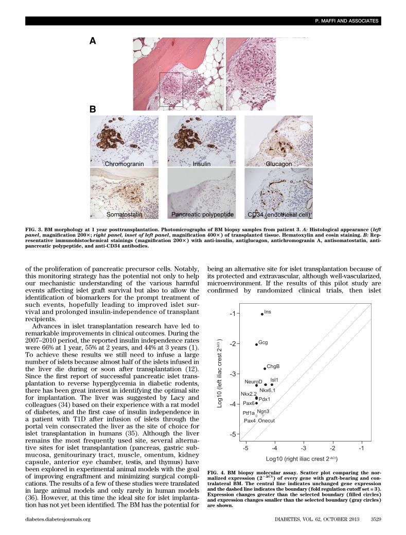
Molecular analysis revealed increased expression ofgenes related to endocrine pancreatic cells in six out of
eight biopsy samples (Table 3). In these six cases, the graft-bearing BM showed 8,077-fold, 21,318-fold, and 41-foldincreases in insulin, glucagon, and chromogranin A mRNAlevels compared with the contralateral BM in the samepatient. Moreover, in addition to these markers of endo-crine fully differentiated pancreatic cells, we analyzed theexpression of transcription factors involved in normalpancreatic development and differentiation. As shown inFig. 4, the expression of transcription factors of endocrine-committed progenitor cells and mature b-cells, such asPdx1, Nkx2.2 Nkx6.1, NeuroD/beta2, and Pax6, was higherin the graft-bearing iliac crest BM than in contralateral BM,whereas the expression of transcription factors expressedin early pancreatic precursors and late-stage pancreaticbud precursor cells, such as Ptf1alpha, Onecut, and Ngn3,was not different.Follow-up: BM imaging. On MRI T2-weighted images, thesite of islet infusion at the posterior-superior iliac spineappeared as a small hypointense area inside the normalhyperintense signal of the iliac BM (Fig. 5). This hypo-intense area did not significantly change over time. Oneyear after islet infusion, bone structure was unaffected bythe presence of the infused islets. Moreover, a gadolinium-enhanced MRI perfusion study did not reveal areas ofanomalous enhancement surrounding the site of islet in-fusion. Computed tomography scans showed the presenceof small calcified spots at the site of islet infusion.
DISCUSSION
To the best of our knowledge, our article represents thefirst unequivocal example of successful engraftment ofendocrine tissue in BM. Our recent preclinical studies inmice (26) showed that the amount of success and thetiming of reverse hyperglycemia were superior after isletinfusion in the BM than in the liver. Therefore, we
TABLE 2BM biopsy morphology
Patient 2 Patient 3 Patient 4
Site1
month3
months12
months1
month3
months12
months1
month3
months
Cellularity (ratio ofcells to fat)
L 2:3 1:3 1:3 1:1 1:3 1:5 2:3 1:6R 2:3 1:3 1:3 1:1 1:3 1:4 2:3 1:6
Myeloid/erythroid ratio L 3:1 1:1 3:1 3:1 4:1 3:1 3:1 3:1R 3:1 1:1 3:1 3:1 4:1 3:1 3:1 3:1
Myeloid component L Red Red Red Inc Inc Red Red RedR Red Red Red Inc Inc Nor Red Red
Myeloid series maturation L Nor Mild right shift Mild left shift Nor Nor Nor Mild left shift NorR Nor Mild right shift Mild left shift Nor Nor Nor Mild left shift Nor
Erythroid component L Red Red Red Inc Nor Red Red RedR Red Red Red Inc Nor Nor Red Red
Erythroid series maturation L Nor Mild right shift Mild left shift Nor Nor Nor Mild left shift NorR Nor Mild right shift Mild left shift Nor Nor Nor Mild left shift Nor
Lymphoid component L Nor Nor Nor Reactive lymphoid aggregateswith a predominance ofB cells (CD20+)
Nor NorR Nor Nor Nor Nor Nor
Lymphoid series maturation L Nor Nor Nor Nor NorR Nor Nor Nor Nor Nor
Megakaryocyte numbers L Nor Red Nor Nor Nor Red Nor RedR Nor Red Nor Nor Nor Red Nor Red
Megakaryocyte morphology L Nor Hypolobate Nor Hypolobate Nor Nor Naked nucleus NorR Nor Hypolobate Nor Hypolobate Nor Nor Naked nucleus Nor
BoneL Nor Nor Nor Nor Nor Nor Nor NorR Nor Nor Nor Nor Nor Nor Nor Nor
Inc, increased; L, graft-bearing iliac crest; Nor, normal; R, not graft-bearing iliac crest; Red, reduced.
P. MAFFI AND ASSOCIATES
diabetes.diabetesjournals.org DIABETES, VOL. 62, OCTOBER 2013 3527

translated our preclinical findings to a proof-of-conceptpilot phase 1 study in which four patients with pan-creatogenic diabetes and hepatic contraindications forreceiving islet transplant in the liver received a single intra-BM islet infusion at the iliac crest. This study has limi-tations, intrinsic to all phase 1 studies, such as the limitednumber of patients enrolled, the nonrandomized design,and the absence of a control group. Furthermore, becauseof the heterogeneity of the pancreatic disease of thepatients enrolled in the study, the results cannot be di-rectly compared with those observed with autologousislets transplanted to the liver.
Although conducted with a small number of patients,this pilot experience has generated some important data.First, we were able to document the feasibility and thesafety of this approach for islet infusion. The direct isletinfusion in the BM was performed according to the sameprocedure used in our institution for the administration ofcord blood cells in patients with acute leukemia (28). Theprocedure was easy and reproducible and, thus far, wehave recorded no AEs related to the islet infusion in theiliac crest. Islets in the BM did not affect hematopoieticactivity, even when it was strongly upregulated in re-sponse to adjuvant chemotherapy. Moreover, bone struc-ture and trabecular compartments were not significantlyaffected by the presence of the infused islets.
Second, and equally important, we demonstrated thepresence of insulin-producing cells in BM biopsy speci-mens, and this presence was associated with detectablelevels of fasting and stimulated circulating C-peptide. Thisimplies that the BM microenvironment is able to supportislet revascularization and function, providing an appro-priate oxygen tension, a suitable pH, clearance of toxicmetabolites, and access to nutrients. Our study unequiv-ocally proves that islets can successfully engraft in the BM,and it provides the rationale for testing the BM as a site forislet infusion in patients with T1D selected to receiveallotransplantation of pancreatic islets. A phase 2 trial inwhich patients with T1D will be randomized to receiveislets either in the liver or in the BM is currently ongoing atour institution (NCT01722682). This trial will allow us toassess whether islet infusion in the BM may improve theoutcome of an islet transplant infused in the liver, asmeasured by glycemic control.
Third, we have shown that islet sampling in the BM ishighly feasible. Because BM is an easily accessed and well-confined site ideal for serial multiple biopsies, we have theunique opportunity to monitor, over time, different mark-ers of engraftment or survival of islets directly at the site ofislet infusion. Although sequential biopsies are often usedto monitor acute or chronic events in solid organ trans-plants, no study in humans has ever attempted to harvestliver biopsy samples and monitor the fate of islets infusedvia the portal vein. This is because islets are rapidly andrandomly scattered throughout the liver after intraportalinfusion, and subsequent liver needle biopsies would havelimited value because of the low yield of islets in biopsysamples (32). In contrast, this study has shown that isletsampling in BM is feasible and allows the histological andimmunohistochemical analyses of the transplanted tissueand surrounding BM and the real-time quantitative PCRanalyses of messenger RNAs. Molecular analysis allowedus to detect the presence of endocrine-specific proteinswith higher sensitivity than with immunohistochemicalanalysis, and to search for the expression of transcriptionfactors of pancreas development (33) that may be markersT
ABLE
3Evide
nceof
panc
reatic
tissue
inBM
Patient
2Patient
3Patient
4
Site
1mon
th3
mon
ths
12mon
ths
1mon
th3
mon
ths
12mon
ths
1mon
th3
mon
ths
BM
biop
syim
mun
ohistoch
emistry
Cytok
eratin
8–18
L2
22
+2
++
+R
22
22
22
22
Chrom
ograninA
L2
22
+2
++
+R
22
22
22
22
Insulin
L2
22
+2
++
2R
22
22
22
22
BM
biop
symolec
ular
assay*
Insulin
3.68
(7.51E
-04)
491(2.2E-05)
12,590
(3E-06)
668,23
6(1E-06)
1.28
(5.7E-05)
3,22
3,06
0(3E-06)
1.2(9.43E
-04)
3,56
5(3.3E-05)
Gluca
gon
4.77
(4.97E
-05)
344.89
(5E-06)
29,328
(1E-07)
13,307
(1E-06)
2.22
(1E-06)
614,90
3(1E-07)
3.27
(3E-05)
32,995
(1E-06)
Chrom
ograninA
24.85(1.7E-05)
24.76(5E-06)
78.79(7E-06)
26.91(1E-06)
0.11
(3.88E
-04)
714.11
(4.5E-05)
0.08
(1E-05)
55.72(3.6E-05)
L,graft-be
aringiliac
crest;R,n
otgraft-be
aringiliac
crest.+,p
resenc
eof
positive
cells;2
,absen
ceof
positive
cells.*Dataareex
pressedas
fold
increa
seof
leftco
mpa
redwithrigh
tiliac
crest(2
2DCtrigh
tiliac
crest).
PANCREATIC ISLET TRANSPLANTATION
3528 DIABETES, VOL. 62, OCTOBER 2013 diabetes.diabetesjournals.org
of the proliferation of pancreatic precursor cells. Notably,this monitoring strategy has the potential not only to helpour mechanistic understanding of the various harmfulevents affecting islet graft survival but also to allow theidentification of biomarkers for the prompt treatment ofsuch events, hopefully leading to improved islet sur-vival and prolonged insulin-independence of transplantrecipients.
Advances in islet transplantation research have led toremarkable improvements in clinical outcomes. During the2007–2010 period, the reported insulin independence rateswere 66% at 1 year, 55% at 2 years, and 44% at 3 years (1).To achieve these results we still need to infuse a largenumber of islets because almost half of the islets infused inthe liver die during or soon after transplantation (12).Since the first report of successful pancreatic islet trans-plantation to reverse hyperglycemia in diabetic rodents,there has been great interest in identifying the optimal sitefor implantation. The liver was suggested by Lacy andcolleagues (34) based on their experience with a rat modelof diabetes, and the first case of insulin independence ina patient with T1D after infusion of islets through theportal vein consecrated the liver as the site of choice forislet transplantation in humans (35). Although the liverremains the most frequently used site, several alterna-tive sites for islet transplantation (pancreas, gastric sub-mucosa, genitourinary tract, muscle, omentum, kidneycapsule, anterior eye chamber, testis, and thymus) havebeen explored in experimental animal models with the goalof improving engraftment and minimizing surgical compli-cations. The results of a few of these studies were translatedin large animal models and only rarely in human models(36). However, at this time the ideal site for islet implanta-tion has not yet been identified. The BM has the potential for
being an alternative site for islet transplantation because ofits protected and extravascular, although well-vascularized,microenvironment. If the results of this pilot study areconfirmed by randomized clinical trials, then islet
FIG. 3. BM morphology at 1 year posttransplantation. Photomicrographs of BM biopsy samples from patient 3. A: Histological appearance (leftpanel, magnification 2003; right panel, inset of left panel, magnification 4003) of transplanted tissue. Hematoxylin and eosin staining. B: Rep-resentative immunohistochemical stainings (magnification 2003) with anti-insulin, antiglucagon, antichromogranin A, antisomatostatin, anti-pancreatic polypeptide, and anti-CD34 antibodies.
FIG. 4. BM biopsy molecular assay. Scatter plot comparing the nor-malized expression (2
2DCt) of every gene with graft-bearing and con-
tralateral BM. The central line indicates unchanged gene expressionand the dashed line indicates the boundary (fold regulation cutoff set = 3).Expression changes greater than the selected boundary (filled circles)and expression changes smaller than the selected boundary (gray circles)are shown.
P. MAFFI AND ASSOCIATES
diabetes.diabetesjournals.org DIABETES, VOL. 62, OCTOBER 2013 3529

infusion in BM may become an ambulatory procedure oflimited invasiveness, well-suited for repeated infusions,with the possibility of performing repeated graft biopsieswith a low-risk and simple procedure. Moreover, BM alsomay be an appropriate site to test, in future trials, theimpact of coinjecting islets with cells of putative immu-nomodulatory capacity, such as T-regulatory cells (37) ormesenchymal stem cells (38), that could help preventor minimize detrimental autoimmune and alloimmuneresponses. T-regulatory cells or mesenchymal stem cellswould benefit from the close proximity of islet antigens,the target of their tolerogenic function, and from thefavorable microenvironment of the BM. The demon-stration that pancreatic islets can efficiently engraft inBM holds the potential to revolutionize the field of islettransplantation, thus allowing new lines of research withsignificant clinical impact on the treatment of diabetesand, more generally, on cell therapy.
ACKNOWLEDGMENTS
This study was supported by the Italian Minister of Health(Ricerca Finalizzata RF-2009-1469691) and by the Euro-pean Union (HEALTH-F5-2009-241883-BetaCellTherapy).
No potential conflicts of interest relevant to this articlewere reported.
P.M. and G.B. managed patients. M.P. performed thehistopathological analysis of the bone marrow. R.N.performed islet isolations. V.S. performed the molecularanalysis of the bone marrow biopsy samples. R.M. andA.M. performed islet isolations. M.S. reviewed and editedthe manuscript and contributed to discussion. A.E. per-formed magnetic resonance imaging studies. J.P. developed
the intra-bone marrow islet infusion method and performedthe transplantations. E.C. conceived the intra-bone marrowstrategy. C.M. and M.B. developed the intra-bone marrowislet infusion method and performed the transplantations.A.D.M. performed magnetic resonance imaging studies. C.S.reviewed and edited the manuscript and contributed to thediscussion. C.D. performed the histopathological analysis ofthe bone marrow. F.C. developed the intra-bone marrowislet infusion method and performed the transplantations.A.S. reviewed and edited the manuscript and researcheddata. L.P. conceived the intra-bone marrow strategy, de-veloped the concept, designed the experiments, wrote themanuscript, promoted the study, and researched data. L.P.is the guarantor of this work and, as such, had full access toall the data in the study and takes responsibility for theintegrity of the data and the accuracy of the data analysis.
REFERENCES
1. Barton FB, Rickels MR, Alejandro R, et al. Improvement in outcomes ofclinical islet transplantation: 1999-2010. Diabetes Care 2012;35:1436–1445
2. Toyofuku A, Yasunami Y, Nabeyama K, et al. Natural killer T-cells par-ticipate in rejection of islet allografts in the liver of mice. Diabetes 2006;55:34–39
3. Yasunami Y, Kojo S, Kitamura H, et al. Valpha14 NK T cell-triggered IFN-gamma production by Gr-1+CD11b+ cells mediates early graft loss ofsyngeneic transplanted islets. J Exp Med 2005;202:913–918
4. Citro A, Cantarelli E, Maffi P, et al. CXCR1/2 inhibition enhances pancre-atic islet survival after transplantation. J Clin Invest 2012;122:3647–3651
5. Korsgren O, Lundgren T, Felldin M, et al. Optimising islet engraftment iscritical for successful clinical islet transplantation. Diabetologia 2008;51:227–232
6. Desai NM, Goss JA, Deng S, et al. Elevated portal vein drug levels of si-rolimus and tacrolimus in islet transplant recipients: local immunosup-pression or islet toxicity? Transplantation 2003;76:1623–1625
7. Shapiro AM, Gallant HL, Hao EG, et al. The portal immunosuppressivestorm: relevance to islet transplantation? Ther Drug Monit 2005;27:35–37
8. Bhargava R, Senior PA, Ackerman TE, et al. Prevalence of hepatic stea-tosis after islet transplantation and its relation to graft function. Diabetes2004;53:1311–1317
9. Piemonti L, Everly MJ, Maffi P, et al. Alloantibody and autoantibodymonitoring predicts islet transplantation outcome in human type 1 diabetes.Diabetes 2013;62:1656–1664
10. Piemonti L, Guidotti LG, Battaglia M. Modulation of early inflammatoryreactions to promote engraftment and function of transplanted pancreaticislets in autoimmune diabetes. Adv Exp Med Biol 2010;654:725–747
11. Moberg L, Johansson H, Lukinius A, et al. Production of tissue factor bypancreatic islet cells as a trigger of detrimental thrombotic reactions inclinical islet transplantation. Lancet 2002;360:2039–2045
12. Eich T, Eriksson O, Lundgren T; Nordic Network for Clinical IsletTransplantation. Visualization of early engraftment in clinical islettransplantation by positron-emission tomography. N Engl J Med 2007;356:2754–2755
13. Barshes NR, Wyllie S, Goss JA. Inflammation-mediated dysfunction andapoptosis in pancreatic islet transplantation: implications for intrahepaticgrafts. J Leukoc Biol 2005;77:587–597
14. Eriksson O, Eich T, Sundin A, et al. Positron emission tomography inclinical islet transplantation. Am J Transplant 2009;9:2816–2824
15. Crowe LA, Ris F, Nielles-Vallespin S, et al. A novel method for quantitativemonitoring of transplanted islets of langerhans by positive contrast mag-netic resonance imaging. Am J Transplant 2011;11:1158–1168
16. Bennet W, Sundberg B, Groth CG, et al. Incompatibility between humanblood and isolated islets of Langerhans: a finding with implications forclinical intraportal islet transplantation? Diabetes 1999;48:1907–1914
17. Johansson H, Lukinius A, Moberg L, et al. Tissue factor produced by theendocrine cells of the islets of Langerhans is associated with a negativeoutcome of clinical islet transplantation. Diabetes 2005;54:1755–1762
18. Sakata N, Hayes P, Tan A, et al. MRI assessment of ischemic liver afterintraportal islet transplantation. Transplantation 2009;87:825–830
19. Yin D, Ding JW, Shen J, Ma L, Hara M, Chong AS. Liver ischemia con-tributes to early islet failure following intraportal transplantation: benefitsof liver ischemic-preconditioning. Am J Transplant 2006;6:60–68
20. Barshes NR, Lee TC, Goodpastor SE, et al. Transaminitis after pancreaticislet transplantation. J Am Coll Surg 2005;200:353–361
FIG. 5. MRI and computed tomography scan of iliac BM. MRI T2-weighted images acquired 30 and 90 days and 1 year after islet trans-plantation in patient 3 (left). A small hypointense area (red circle)inside the normal hyperintense signal was evident at the site of the isletinfusion at the level of the posterior-superior iliac spine. The gadolinium-enhanced MR perfusion study did not show areas of anomalous en-hancement surrounding the site of the islets infusion (right). A computedtomography scan showed the presence of a small calcified spot at the siteof islet infusion (lower right).
PANCREATIC ISLET TRANSPLANTATION
3530 DIABETES, VOL. 62, OCTOBER 2013 diabetes.diabetesjournals.org

21. Rafael E, Ryan EA, Paty BW, et al. Changes in liver enzymes after clinicalislet transplantation. Transplantation 2003;76:1280–1284
22. Casey JJ, Lakey JR, Ryan EA, et al. Portal venous pressure changesafter sequential clinical islet transplantation. Transplantation 2002;74:913–915
23. Morrison CP, Wemyss-Holden SA, Dennison AR, Maddern GJ. Islet yieldremains a problem in islet autotransplantation. Arch Surg 2002;137:80–83
24. Cantarelli E, Piemonti L. Alternative transplantation sites for pancreaticislet grafts. Curr Diab Rep 2011;11:364–374
25. Ciceri F, Piemonti L. Bone marrow and pancreatic islets: an old story withnew perspectives. Cell Transplant 2010;19:1511–1522
26. Cantarelli E, Melzi R, Mercalli A, et al. Bone marrow as an alternative sitefor islet transplantation. Blood 2009;114:4566–4574
27. Melzi R, Mercalli A, Sordi V, et al. Role of CCL2/MCP-1 in islet trans-plantation. Cell Transplant 2010;19:1031–1046
28. Frassoni F, Gualandi F, Podestà M, et al. Direct intrabone transplant ofunrelated cord-blood cells in acute leukaemia: a phase I/II study. LancetOncol 2008;9:831–839
29. Caumo A, Maffi P, Nano R, et al. Comparative evaluation of simple in-dices of graft function after islet transplantation. Transplantation 2011;92:815–821
30. Mercalli A, Sordi V, Ponzoni M, et al. Rapamycin induces a caspase-independent cell death in human monocytes. Am J Transplant 2006;6:1331–1341
31. Bassi C, Dervenis C, Butturini G, et al.; International Study Group onPancreatic Fistula Definition. Postoperative pancreatic fistula: an in-ternational study group (ISGPF) definition. Surgery 2005;138:8–13
32. Toso C, Isse K, Demetris AJ, et al. Histologic graft assessment after clinicalislet transplantation. Transplantation 2009;88:1286–1293
33. Servitja JM, Ferrer J. Transcriptional networks controlling pancreatic de-velopment and beta cell function. Diabetologia 2004;47:597–613
34. Kemp CB, Knight MJ, Scharp DW, Ballinger WF, Lacy PE. Effect oftransplantation site on the results of pancreatic islet isografts in diabeticrats. Diabetologia 1973;9:486–491
35. Scharp DW, Lacy PE, Santiago JV, et al. Insulin independence after islettransplantation into type I diabetic patient. Diabetes 1990;39:515–518
36. Rafael E, Tibell A, Rydén M, et al. Intramuscular autotransplantation ofpancreatic islets in a 7-year-old child: a 2-year follow-up. Am J Transplant2008;8:458–462
37. Battaglia M, Stabilini A, Migliavacca B, Horejs-Hoeck J, Kaupper T,Roncarolo MG. Rapamycin promotes expansion of functional CD4+CD25+FOXP3+ regulatory T cells of both healthy subjects and type 1 diabeticpatients. J Immunol 2006;177:8338–8347
38. Tan J, Wu W, Xu X, et al. Induction therapy with autologous mesenchymalstem cells in living-related kidney transplants: a randomized controlledtrial. JAMA 2012;307:1169–1177
39. Piatti PM, Pontiroli AE, Caumo A, et al. Hyperinsulinemia decreases second-phase but not first-phase arginine-induced insulin release in humans.Diabetes 1994;43:1157–1163
P. MAFFI AND ASSOCIATES
diabetes.diabetesjournals.org DIABETES, VOL. 62, OCTOBER 2013 3531