impact of myocardial infarction on patient and partner quality ...
Upload
independentCategory
view
0download
0
Cell Transplantation, Vol. 18, pp. 1–100, 2009 0963-6897/09 $90.00 + .00Printed in the USA. All rights reserved. E-ISSN 1555-3892Copyright 2009 Cognizant Comm. Corp. www.cognizantcommunication.com
Autologous Bone-Marrow Mononuclear Cell Transplantation After AcuteMyocardial Infarction: Comparison of Two Delivery Techniques
Suzana A. Silva,*1 Andre L. S. Sousa,†1 Andrea F. Haddad,* Jader C. Azevedo,†Vinicio E. Soares,‡ Cıntia M. Peixoto,* Ana J. S. Soares,‡ Aurora F. C. Issa,‡
Luis Renato V. Felipe,† Rodrigo V. C. Branco,† Joao A. Addad,† Rodrigo C. Moreira,*Fabio A. A. Tuche,* Claudio T. Mesquita,† Cristina C. O. Drumond,† Amarino O. Junior,†
Carlos E. Rochitte,† Jose H. M. Luz,† Arnaldo Rabischoffisky,† Fernanda B. Nogueira,†Rosana B. C. Vieira,§ Hamilton S. Junior,¶ Radovan Borojevic,§ Hans F. R. Dohmann,*
*Teaching and Research Center of Pro-Cardıaco/PROCEP, Rio de Janeiro, Brazil†Pro-Cardıaco Hospital, Rio de Janeiro, Brazil
‡Miguel Couto Municipal Hospital, Rio de Janeiro, Brazil§Federal University of Rio de Janeiro, Rio de Janeiro, Brazil
¶Excellion Servicos Biomedicos S/A, Petropolis, Brazil
The objective of this study was to investigate safety and feasibility of autologous bone marrow mononuclearcells (BMMNC) transplantation in ST elevation myocardial infarction (STEMI), comparing anterograde in-tracoronary artery (ICA) delivery with retrograde intracoronary vein (ICV) approach. An open labeled, ran-domized controlled trial of 30 patients admitted with STEMI was used. Patients were enrolled if they 1)were successfully reperfused within 24 h from symptoms onset and 2) had infarct size larger than 10% ofthe left ventricle (LV). One hundred million BMMNC were injected in the infarct-related artery (intra-arterial group) or vein (intravenous group), 1% of which was labeled with Tc99m-hexamethylpropylenami-neoxime. Cell distribution was evaluated 4 and 24 h after injection. Baseline MRI was performed in orderto evaluate microbstruction pattern. Baseline radionuclide ventriculography was performed before cell trans-fer and after 3 and 6 months. All the treated patients were submitted to repeat coronary angiography after 3months. Thirty patients (57 ± 11 years, 70% males) were randomly assigned to ICA (n = 14), ICV (n = 10),or control (n = 6) groups. No serious adverse events related to the procedure were observed. Early and lateretention of radiolabeled cells was higher in the ICA than in the ICV group, independently of microcircula-tion obstruction. An increase of EF was observed in the ICA group (p = 0.02) compared to baseline. Injec-tion procedures through anterograde and retrograde approaches seem to be feasible and safe. BMMNCretention by damaged heart tissue was apparently higher when the anterograde approach was used. Furtherstudies are required to confirm these initial data.
Key words: Bone marrow cell transplantation; Angiogenesis; Stem cells; Myocardial infarction;Myocardial ischemia
INTRODUCTION
Growing evidence suggests that cell-based therapiescan repair the injured myocardium after an acute infarc-tion. However, comparative data analyses of this newfield have been hampered by the great variety of cellphenotypes and protocol design in different clinical trials.Although the largest experience comes from bone mar-row-derived cell studies, which mostly point to myocar-dial regeneration and improvement of ventricular func-tion, the mechanisms of their actions are still poorly
1Drs. Silva and Sousa are co-principal investigators.Address correspondence to Hans Dohmann, Hospital Pro-Cardıaco, Rua Dona Mariana 219, Botafogo, Rio de Janeiro. Brasil. CEP: 22280-000.Tel: (55)21-21311584; Fax: (55)21-21311523; E-mail: [email protected]
1
understood (15). From the very beginning of these stud-ies, one of the most frequently asked questions has beenwhich is the best way to deliver cells into the heart.Animal studies give us important data but they are notreliably applicable to humans (19).We and others havepreviously reported the use of transendocardial injec-tions for bone marrow mononuclear cells (BMMNC)delivery in ischemic cardiomiopathy (13,20). Other ap-proaches were described for different clinical settingssuch as transepicardial and intracoronary injections (20).The latter approach was used in several clinical trials

2 SILVA ET AL.
demonstrating safety of BMMNC transplantation in theacute phase of ST elevation myocardial infarction(STEMI). The evidence for efficacy of the therapy hasbeen growing, but the ideal technique for delivery ofcells into the myocardium in humans has not been exten-sively investigated.
There is no clearly established relationship amongdelivery techniques, cell engraftment, and clinical ef-fects, and ambiguous results regarding infarct-relatedcoronary artery infusion of BMMNC after STEMI havebeen observed (5). Despite some controversies about theefficacy of BMMNC, the largest reported trial showed asignificant improvement for patients who received cellsafter the fifth day of large infarcts. On the other hand,microvascular obstruction may play an important role inthis case, as proposed by Janssen et al. (10), who re-ported that microvascular obstruction could impair cells’uptake by heart tissue and consequently heart functionimprovement. We hypothesized that coronary intrave-nous approach may overcame this issue, because diape-desis of circulating cells into the adjacent cardiac tissueoccurs in the venous side of microcirculation (11). Littleis known regarding retrograde percutaneous approachthrough the coronary vein, even though this route hasbeen shown as an effective way to deliver cells in ani-mal and human models (6,9,18).
The primary objective of this study is to comparetwo different delivery techniques for the infusion ofBMMNC in STEMI: the anterograde intracoronary ar-tery (ICA) delivery with the retrograde intracoronaryvein (ICV) approach. Furthermore, we used radiolabeledcells in order to evaluate their distribution pattern in theheart once injected and their relationship with left ven-tricle function improvement.
MATERIALS AND METHODS
Patients
Between January 3, 2005 and January 6, 2006, pa-tients admitted in Pro-Cardıaco Hospital or MiguelCouto Municipal Hospital, Rio de Janeiro, were selectedto be included into the study if they were between 18and 80 years old and had a STEMI. Patients selected atMiguel Couto Hospital were transferred to Pro-CardıacoHospital where all procedures related to the study wereperformed. Patients were enrolled if they had: 1) aSTEMI successfully reperfused with either thrombolytictherapy or primary angioplasty up to 24 h after thesymptoms onset, and 2) fixed perfusion defect largerthan 10% of the LV mass after 72 h on technetium 99mmethoxy-isobutyl-isonitrile (99mTc-MIBI) single-photonemission computed tomography (SPECT) with sublin-gual nitrate. Exclusion criteria were: 1) indication to un-dergo coronary artery bypass graft; 2) creatinine level
>2.0 mg/dl or hemodialysis; 3) infarct-related coronaryartery with thrombolysis in myocardial infarction (TIMI)flow <3 by the time of cells injection; 4) sepsis; 5) per-sistent cardiogenic shock after 72 h; 6) significant valvardisease; 7) mechanical complications of STEMI; 8) liverfailure; 9) severe pulmonary disease; 10) left bundlebranch block; 11) implanted pacemaker; 12) hematolog-ical illnesses; 13) neoplasia; 14) other disorders of he-mostasis and other pathologies that could have impacton life expectancy.
The Ethics Review Board of Pro-Cardıaco Hospital,Rio de Janeiro, and the Brazilian National Ethics Coun-cil for Human Research (CONEP, Brasilia) approved theprotocol. Written informed consent was obtained fromall patients.
Study Design and Randomization
This is an open-labeled, randomized controlled trialof a consecutive patient series. Between the third andsixth day after successful reperfusion of the infarct-related artery, included patients were randomly assignedinto one of the three groups: ICA approach, retrogradeICV approach, and control. Random allocation was stra-tified according to infarct size ( ≥ 25% or <25%) in threeblocks of different size (7, 5, and 3, respectively) foreach stratum, with the use of sealed envelopes.
Bone Marrow Cells Harvesting and Isolationof Mononuclear Cells
In subjects assigned to either of the two treatmentarms of this study, about 80 ml of autologous bone mar-row was aspirated under local anesthesia from the poste-rior iliac crest in the morning of the procedure, whichwas anticipated to occur 6 h later. BMMNC were iso-lated by density gradient centrifugation on Ficoll-PaquePlus (Amersham Biosciences, Sao Paulo, Brazil) andmanipulated under aseptic conditions. They were washedwith saline containing 5% human serum albumin. Thecells were resuspended in saline with 5% human serumalbumin and filtered for injection through 100-µm nylonmesh to remove cell aggregates. A small fraction of thecell suspension was used for cell counting and viabilitycontrol using the trypan blue exclusion assay. Cell via-bility was >90% in all subjects (93.26 ± 2.9%). Charac-terization of leukocyte differentiation markers by flowcytometry and functional assays were done on anotherfraction of cells following study intervention. The clono-genic capacity of hematopoietic progenitors was evalu-ated by colony-forming assays [granulocyte-macrophagecolony-forming units (CFU-GM)], as previously described(4). Fibroblast colony-forming assay (CFU-F) was doneto estimate the presence of putative progenitors of themesenchymal cell lineage, as previously described (3).
TWO DELIVERY TECHNIQUES FOR STEM CELLS 3
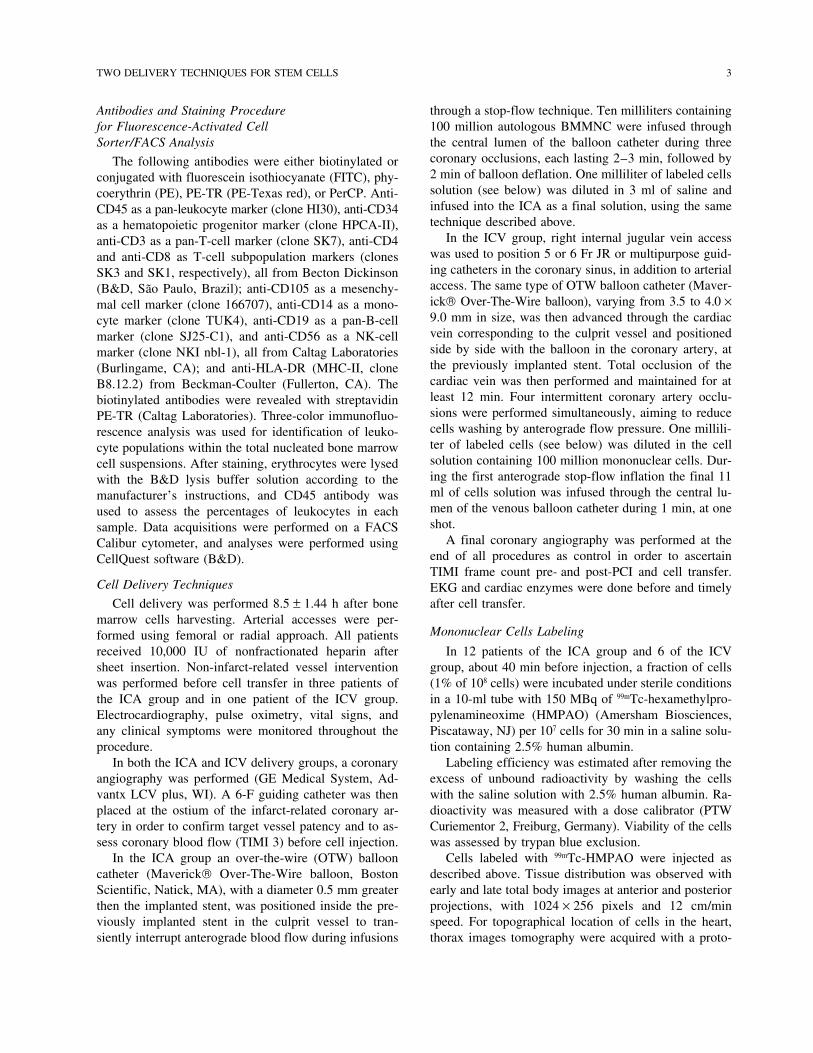
Antibodies and Staining Procedurefor Fluorescence-Activated CellSorter/FACS Analysis
The following antibodies were either biotinylated orconjugated with fluorescein isothiocyanate (FITC), phy-coerythrin (PE), PE-TR (PE-Texas red), or PerCP. Anti-CD45 as a pan-leukocyte marker (clone HI30), anti-CD34as a hematopoietic progenitor marker (clone HPCA-II),anti-CD3 as a pan-T-cell marker (clone SK7), anti-CD4and anti-CD8 as T-cell subpopulation markers (clonesSK3 and SK1, respectively), all from Becton Dickinson(B&D, Sao Paulo, Brazil); anti-CD105 as a mesenchy-mal cell marker (clone 166707), anti-CD14 as a mono-cyte marker (clone TUK4), anti-CD19 as a pan-B-cellmarker (clone SJ25-C1), and anti-CD56 as a NK-cellmarker (clone NKI nbl-1), all from Caltag Laboratories(Burlingame, CA); and anti-HLA-DR (MHC-II, cloneB8.12.2) from Beckman-Coulter (Fullerton, CA). Thebiotinylated antibodies were revealed with streptavidinPE-TR (Caltag Laboratories). Three-color immunofluo-rescence analysis was used for identification of leuko-cyte populations within the total nucleated bone marrowcell suspensions. After staining, erythrocytes were lysedwith the B&D lysis buffer solution according to themanufacturer’s instructions, and CD45 antibody wasused to assess the percentages of leukocytes in eachsample. Data acquisitions were performed on a FACSCalibur cytometer, and analyses were performed usingCellQuest software (B&D).
Cell Delivery Techniques
Cell delivery was performed 8.5 ± 1.44 h after bonemarrow cells harvesting. Arterial accesses were per-formed using femoral or radial approach. All patientsreceived 10,000 IU of nonfractionated heparin aftersheet insertion. Non-infarct-related vessel interventionwas performed before cell transfer in three patients ofthe ICA group and in one patient of the ICV group.Electrocardiography, pulse oximetry, vital signs, andany clinical symptoms were monitored throughout theprocedure.
In both the ICA and ICV delivery groups, a coronaryangiography was performed (GE Medical System, Ad-vantx LCV plus, WI). A 6-F guiding catheter was thenplaced at the ostium of the infarct-related coronary ar-tery in order to confirm target vessel patency and to as-sess coronary blood flow (TIMI 3) before cell injection.
In the ICA group an over-the-wire (OTW) ballooncatheter (Maverick Over-The-Wire balloon, BostonScientific, Natick, MA), with a diameter 0.5 mm greaterthen the implanted stent, was positioned inside the pre-viously implanted stent in the culprit vessel to tran-siently interrupt anterograde blood flow during infusions
through a stop-flow technique. Ten milliliters containing100 million autologous BMMNC were infused throughthe central lumen of the balloon catheter during threecoronary occlusions, each lasting 2–3 min, followed by2 min of balloon deflation. One milliliter of labeled cellssolution (see below) was diluted in 3 ml of saline andinfused into the ICA as a final solution, using the sametechnique described above.
In the ICV group, right internal jugular vein accesswas used to position 5 or 6 Fr JR or multipurpose guid-ing catheters in the coronary sinus, in addition to arterialaccess. The same type of OTW balloon catheter (Maver-ick Over-The-Wire balloon), varying from 3.5 to 4.0 ×9.0 mm in size, was then advanced through the cardiacvein corresponding to the culprit vessel and positionedside by side with the balloon in the coronary artery, atthe previously implanted stent. Total occlusion of thecardiac vein was then performed and maintained for atleast 12 min. Four intermittent coronary artery occlu-sions were performed simultaneously, aiming to reducecells washing by anterograde flow pressure. One millili-ter of labeled cells (see below) was diluted in the cellsolution containing 100 million mononuclear cells. Dur-ing the first anterograde stop-flow inflation the final 11ml of cells solution was infused through the central lu-men of the venous balloon catheter during 1 min, at oneshot.
A final coronary angiography was performed at theend of all procedures as control in order to ascertainTIMI frame count pre- and post-PCI and cell transfer.EKG and cardiac enzymes were done before and timelyafter cell transfer.
Mononuclear Cells Labeling
In 12 patients of the ICA group and 6 of the ICVgroup, about 40 min before injection, a fraction of cells(1% of 108 cells) were incubated under sterile conditionsin a 10-ml tube with 150 MBq of 99mTc-hexamethylpro-pylenamineoxime (HMPAO) (Amersham Biosciences,Piscataway, NJ) per 107 cells for 30 min in a saline solu-tion containing 2.5% human albumin.
Labeling efficiency was estimated after removing theexcess of unbound radioactivity by washing the cellswith the saline solution with 2.5% human albumin. Ra-dioactivity was measured with a dose calibrator (PTWCuriementor 2, Freiburg, Germany). Viability of the cellswas assessed by trypan blue exclusion.
Cells labeled with 99mTc-HMPAO were injected asdescribed above. Tissue distribution was observed withearly and late total body images at anterior and posteriorprojections, with 1024 × 256 pixels and 12 cm/minspeed. For topographical location of cells in the heart,thorax images tomography were acquired with a proto-

4 SILVA ET AL.
col similar to that used for myocardial perfusion images,64 projections of 20 s each, with resolution of 64 × 64pixels. Images were reconstructed with Butterworth fil-ter using the software e-Soft 3.0 n (Cedars. Sinai, QGSand Emory Cardiac Toolbox) and compared to the ac-quired perfusion images tomography. All images wereacquired in a dual head gamma camera (Ecam-Duet,Siemens Medical Systems Inc, IL).
Planar and SPECT images were obtained at 4 h (3.67 ±1.03 h) and 24 h (22.8 ± 4.46 h) after injection in allpatients. Retention was defined as the percentage of car-diac-originated number of counts in the anterior andposterior projections (cardiac counts), compared to thetotal number of body counts of radiolabeled cells (bodycounts) in both early and late images. Washout rate wascalculated by using the equation: [early (cardiac counts/body counts) − late (cardiac counts/body counts)] × 100/early (cardiac counts/body counts). Washout rate per hourwas calculated by dividing washout rate per the intervalbetween the early and late images retention.
Radionuclide ventriculography (MUGA) was per-formed 3–5 days after reperfusion and at least 24 h after99mTc-MIBI SPECT. Injected 99mTc-pertechnetate activitywas 925 MBq (25 mCi), after injection of 5 mg of pyro-phosphate. Conventional planar gated blood pool (GBP)imaging was performed in the appropriate left anterioroblique (LAO) projection angle. Data were acquiredwith low-energy, high-resolution (LEHR) collimators,by use of a dual-detector gamma camera with the detec-tors set at 90° relative to one another. No caudal tiltwas used in setting up patients for optimal planar GBPimaging. Data were acquired as 64 × 64 matrices, withLEHR collimators for 24 frames per R-R interval for10 min. Processing was performed with commerciallyavailable software, involving two-dimensional spatialsmoothing and one-dimensional time filtering of LAOdata sets. Software supplied by the manufacturer gener-ated automated outlines for LV identification and back-ground correction regions, but the observer was free toalter these as necessary to conform to the visual impres-sion of true LV boundaries and appropriate backgroundlocations. Observer drew LV outlines for end-diastolicframes, primarily guided by the visual impression of theLV shape and aided by Fourier amplitude and phasemaps. Counts within end-diastolic and end-systolic re-gions were corrected for average background counts de-rived from a region drawn beside the LV region. Back-ground-corrected counts were used to compute LVEF.From the time–activity curve we were able to obtain theLVEF as a systolic parameter and the diastolic parame-ter named time to peak filling (TPF) rate. TPF rate ismeasured from end-systole to the time of the peak LVfilling rate. It is calculated from the time–activity curveand its first derivative. The maximal slope of this deriva-
tive of the filling portion, normalized for the number ofcounts at end-diastole, gives rise to the peak filling rate.
Cardiac magnetic resonance imaging (MRI) was per-formed 3–5 days after reperfusion. All studies were donewith commercially available cardiac MRI software (GEHealthcare Milwaukee, WI, USA) as previously described(16). Briefly, microvascular obstruction (MO, or MVO)was defined on late enhanced images taken early (within2–5 min) after injection of 0.20 mmol/kg of gadopentet-ate dimeglumine (Gd-DTPA), in the LV short and longaxis at the same locations used for cine-MRI as a dark,subendocardial zone within the infarct area. We definedinfarct area as the zone of bright signal on late-enhancedimages (10–20 min after contrast injection) by inver-sion-recovery gradient-echo technique. All MRI studieswere analyzed on an off-line workstation.
Follow-up
All patients were standard treated. Follow-up to as-sess clinical status, review of current medication, and toreevaluate cardiac function through MUGA occurred at3 and 6 months after the procedure. In addition, at 3month follow-up all patients were scheduled to repeatcoronary angiography.
Statistical Analysis
All image exams were independently analyzed by anexperienced observer unaware of patients’ group alloca-tion throughout the study.
Continuous variables are presented as mean ± SE (un-less stated otherwise). Categorical variables were com-pared with chi-square or Fisher’s exact test, as appro-priate. Continuous variables were compared with t-testor ANOVA, as appropriate. Comparisons of the changesfrom baseline to 3 and 6 months among the groups weremade with repeated-measures ANOVA. Kendall’s taucorrelation coefficient was used to correlate continuousdata.
Statistical significance was assumed at a value of p <0.05. All reported p-values are two-sided. All statisticalanalysis was performed with SPSS (Version 13.0, SPSSInc.).
RESULTS
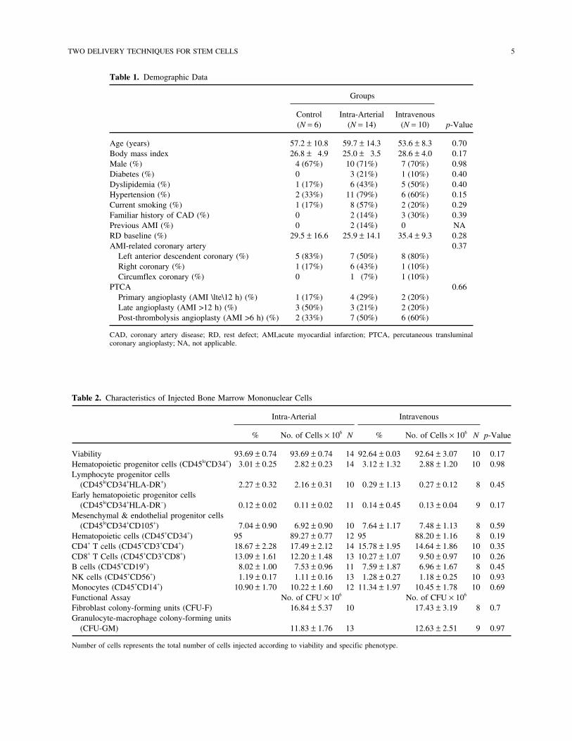
Thirty patients were included in the study, 14 in theICA group, 10 in the ICV group, and 6 in control group.There was no significant difference in demographic pa-rameters among the studied groups (Table 1), includinginfarct size and cell phenotypes and functionality (Ta-ble 2).
The time period between AMI and cell injection was5.5 ± 1.3 days and 6.1 ± 1.4 days in the ICA and ICVgroups (p = 0.14), respectively.
ICA and ICV injections were successfully performed
TWO DELIVERY TECHNIQUES FOR STEM CELLS 5
Table 1. Demographic Data
Groups
Control Intra-Arterial Intravenous(N = 6) (N = 14) (N = 10) p-Value
Age (years) 57.2 ± 10.8 59.7 ± 14.3 53.6 ± 8.3 0.70Body mass index 26.8 ± 4.9 25.0 ± 3.5 28.6 ± 4.0 0.17Male (%) 4 (67%) 10 (71%) 7 (70%) 0.98Diabetes (%) 0 3 (21%) 1 (10%) 0.40Dyslipidemia (%) 1 (17%) 6 (43%) 5 (50%) 0.40Hypertension (%) 2 (33%) 11 (79%) 6 (60%) 0.15Current smoking (%) 1 (17%) 8 (57%) 2 (20%) 0.29Familiar history of CAD (%) 0 2 (14%) 3 (30%) 0.39Previous AMI (%) 0 2 (14%) 0 NARD baseline (%) 29.5 ± 16.6 25.9 ± 14.1 35.4 ± 9.3 0.28AMI-related coronary artery 0.37
Left anterior descendent coronary (%) 5 (83%) 7 (50%) 8 (80%)Right coronary (%) 1 (17%) 6 (43%) 1 (10%)Circumflex coronary (%) 0 1 (7%) 1 (10%)
PTCA 0.66Primary angioplasty (AMI \lte\12 h) (%) 1 (17%) 4 (29%) 2 (20%)Late angioplasty (AMI >12 h) (%) 3 (50%) 3 (21%) 2 (20%)Post-thrombolysis angioplasty (AMI >6 h) (%) 2 (33%) 7 (50%) 6 (60%)
CAD, coronary artery disease; RD, rest defect; AMI,acute myocardial infarction; PTCA, percutaneous transluminalcoronary angioplasty; NA, not applicable.
Table 2. Characteristics of Injected Bone Marrow Mononuclear Cells
Intra-Arterial Intravenous
% No. of Cells × 106 N % No. of Cells × 106 N p-Value
Viability 93.69 ± 0.74 93.69 ± 0.74 14 92.64 ± 0.03 92.64 ± 3.07 10 0.17Hematopoietic progenitor cells (CD45loCD34+) 3.01 ± 0.25 2.82 ± 0.23 14 3.12 ± 1.32 2.88 ± 1.20 10 0.98Lymphocyte progenitor cells
(CD45loCD34+HLA-DR+) 2.27 ± 0.32 2.16 ± 0.31 10 0.29 ± 1.13 0.27 ± 0.12 8 0.45Early hematopoietic progenitor cells
(CD45loCD34+HLA-DR−) 0.12 ± 0.02 0.11 ± 0.02 11 0.14 ± 0.45 0.13 ± 0.04 9 0.17Mesenchymal & endothelial progenitor cells
(CD45loCD34+CD105+) 7.04 ± 0.90 6.92 ± 0.90 10 7.64 ± 1.17 7.48 ± 1.13 8 0.59Hematopoietic cells (CD45+CD34+) 95 89.27 ± 0.77 12 95 88.20 ± 1.16 8 0.19CD4+ T cells (CD45+CD3+CD4+) 18.67 ± 2.28 17.49 ± 2.12 14 15.78 ± 1.95 14.64 ± 1.86 10 0.35CD8+ T Cells (CD45+CD3+CD8+) 13.09 ± 1.61 12.20 ± 1.48 13 10.27 ± 1.07 9.50 ± 0.97 10 0.26B cells (CD45+CD19+) 8.02 ± 1.00 7.53 ± 0.96 11 7.59 ± 1.87 6.96 ± 1.67 8 0.45NK cells (CD45+CD56+) 1.19 ± 0.17 1.11 ± 0.16 13 1.28 ± 0.27 1.18 ± 0.25 10 0.93Monocytes (CD45+CD14+) 10.90 ± 1.70 10.22 ± 1.60 12 11.34 ± 1.97 10.45 ± 1.78 10 0.69Functional Assay No. of CFU × 106 No. of CFU × 106
Fibroblast colony-forming units (CFU-F) 16.84 ± 5.37 10 17.43 ± 3.19 8 0.7Granulocyte-macrophage colony-forming units
(CFU-GM) 11.83 ± 1.76 13 12.63 ± 2.51 9 0.97
Number of cells represents the total number of cells injected according to viability and specific phenotype.
6 SILVA ET AL.
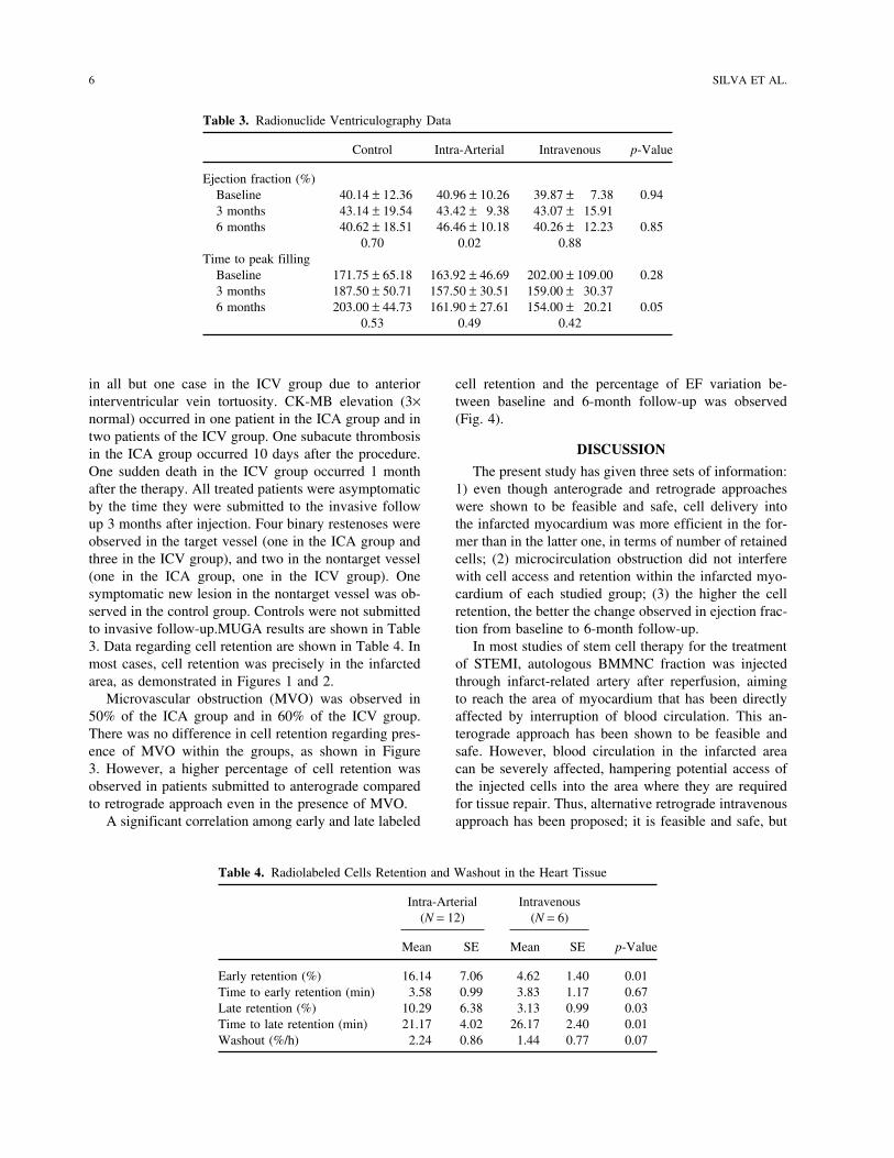
Table 3. Radionuclide Ventriculography Data
Control Intra-Arterial Intravenous p-Value
Ejection fraction (%)Baseline 40.14 ± 12.36 40.96 ± 10.26 39.87 ± 7.38 0.943 months 43.14 ± 19.54 43.42 ± 9.38 43.07 ± 15.916 months 40.62 ± 18.51 46.46 ± 10.18 40.26 ± 12.23 0.85
0.70 0.02 0.88Time to peak filling
Baseline 171.75 ± 65.18 163.92 ± 46.69 202.00 ± 109.00 0.283 months 187.50 ± 50.71 157.50 ± 30.51 159.00 ± 30.376 months 203.00 ± 44.73 161.90 ± 27.61 154.00 ± 20.21 0.05
0.53 0.49 0.42
in all but one case in the ICV group due to anteriorinterventricular vein tortuosity. CK-MB elevation (3×normal) occurred in one patient in the ICA group and intwo patients of the ICV group. One subacute thrombosisin the ICA group occurred 10 days after the procedure.One sudden death in the ICV group occurred 1 monthafter the therapy. All treated patients were asymptomaticby the time they were submitted to the invasive followup 3 months after injection. Four binary restenoses wereobserved in the target vessel (one in the ICA group andthree in the ICV group), and two in the nontarget vessel(one in the ICA group, one in the ICV group). Onesymptomatic new lesion in the nontarget vessel was ob-served in the control group. Controls were not submittedto invasive follow-up.MUGA results are shown in Table3. Data regarding cell retention are shown in Table 4. Inmost cases, cell retention was precisely in the infarctedarea, as demonstrated in Figures 1 and 2.
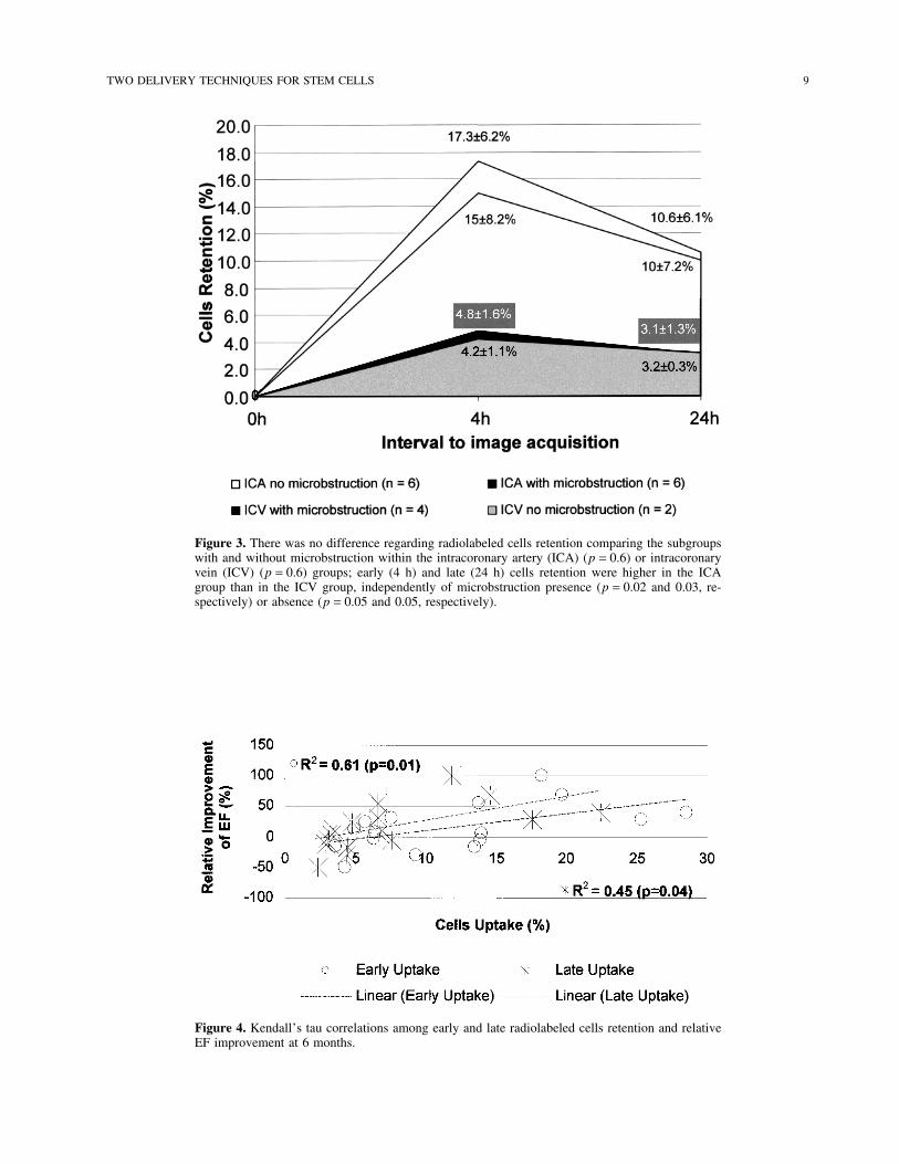
Microvascular obstruction (MVO) was observed in50% of the ICA group and in 60% of the ICV group.There was no difference in cell retention regarding pres-ence of MVO within the groups, as shown in Figure3. However, a higher percentage of cell retention wasobserved in patients submitted to anterograde comparedto retrograde approach even in the presence of MVO.
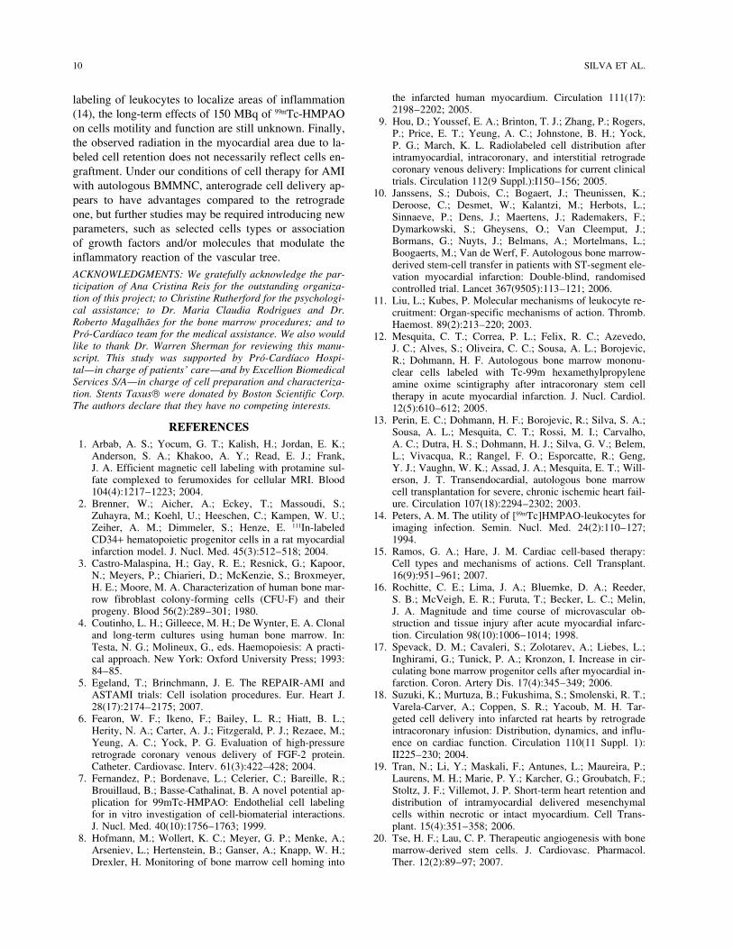
A significant correlation among early and late labeled
Table 4. Radiolabeled Cells Retention and Washout in the Heart Tissue
Intra-Arterial Intravenous(N = 12) (N = 6)
Mean SE Mean SE p-Value
Early retention (%) 16.14 7.06 4.62 1.40 0.01Time to early retention (min) 3.58 0.99 3.83 1.17 0.67Late retention (%) 10.29 6.38 3.13 0.99 0.03Time to late retention (min) 21.17 4.02 26.17 2.40 0.01Washout (%/h) 2.24 0.86 1.44 0.77 0.07
cell retention and the percentage of EF variation be-tween baseline and 6-month follow-up was observed(Fig. 4).
DISCUSSION
The present study has given three sets of information:1) even though anterograde and retrograde approacheswere shown to be feasible and safe, cell delivery intothe infarcted myocardium was more efficient in the for-mer than in the latter one, in terms of number of retainedcells; (2) microcirculation obstruction did not interferewith cell access and retention within the infarcted myo-cardium of each studied group; (3) the higher the cellretention, the better the change observed in ejection frac-tion from baseline to 6-month follow-up.
In most studies of stem cell therapy for the treatmentof STEMI, autologous BMMNC fraction was injectedthrough infarct-related artery after reperfusion, aimingto reach the area of myocardium that has been directlyaffected by interruption of blood circulation. This an-terograde approach has been shown to be feasible andsafe. However, blood circulation in the infarcted areacan be severely affected, hampering potential access ofthe injected cells into the area where they are requiredfor tissue repair. Thus, alternative retrograde intravenousapproach has been proposed; it is feasible and safe, but
TWO DELIVERY TECHNIQUES FOR STEM CELLS 7
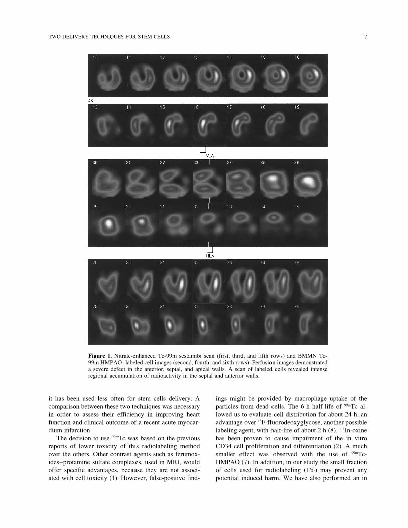
Figure 1. Nitrate-enhanced Tc-99m sestamibi scan (first, third, and fifth rows) and BMMN Tc-99m HMPAO–labeled cell images (second, fourth, and sixth rows). Perfusion images demonstrateda severe defect in the anterior, septal, and apical walls. A scan of labeled cells revealed intenseregional accumulation of radioactivity in the septal and anterior walls.
it has been used less often for stem cells delivery. Acomparison between these two techniques was necessaryin order to assess their efficiency in improving heartfunction and clinical outcome of a recent acute myocar-dium infarction.
The decision to use 99mTc was based on the previousreports of lower toxicity of this radiolabeling methodover the others. Other contrast agents such as ferumox-ides–protamine sulfate complexes, used in MRI, wouldoffer specific advantages, because they are not associ-ated with cell toxicity (1). However, false-positive find-
ings might be provided by macrophage uptake of theparticles from dead cells. The 6-h half-life of 99mTc al-lowed us to evaluate cell distribution for about 24 h, anadvantage over 18F-fluorodeoxyglycose, another possiblelabeling agent, with half-life of about 2 h (8). 111In-oxinehas been proven to cause impairment of the in vitroCD34 cell proliferation and differentiation (2). A muchsmaller effect was observed with the use of 99mTc-HMPAO (7). In addition, in our study the small fractionof cells used for radiolabeling (1%) may prevent anypotential induced harm. We have also performed an in

8 SILVA ET AL.
Figure 2. Nitrate-enhanced Tc-99m sestamibi short-axis slices (first row) and BMMN Tc-99m HMPAO–labeled cell slices (secondrow). An absence of radioactivity in the lateral and inferior walls and intense accumulation of labeled cells in the anterior andseptal walls in the fused images (third row), revealing the precise localization of the BMMN Tc-99m HMPAO–labeled cells in theinfarcted area.
vitro evaluation that showed no impairment of 150 MBq99mTc-HMPAO on human mononuclear cells viability.Based on the above, we consider the toxicity of our ra-diolabeling technique acceptable.
We have published a description of the first patientof this series (12). Our data revealed that the level ofBM progenitors homing to the irreversibly ischemicmyocardial area through ICA and ICV approaches wereabout 10% and 3% of the infused activity, which corre-sponded to 10 × 106 and 3 × 10⁶ autologous BMMNC,respectively. Our study provided clear evidence that ad-hesion and retention of BMMNC via ICA injection toinfarcted myocardium is feasible and safe and corrobo-rates previous findings of stem cell mobilization andhoming signals from injured tissue in the period of acuteischemic injury (17). It also showed that ICA deliverywas more efficient in terms of cell retention in the myo-cardium than the ICV approach. Such information is val-uable because a premise for this therapy benefit is theengraftment of the cells in the damaged tissue. Accord-ingly, the improvement of the heart function reflectedby the EF was consistent with the higher cell retentionin the infarcted area after anterograde injection.
This study was undertaken to detect significant dif-ference among the groups regarding LV global function,but some differences were observed in MUGA. EF im-proved in the arterial group at 3 and 6 months after cellsinjection. An improvement in the TPF rate was also ob-served in treated patients compared to controls, probablymeaning an effect on diastolic function. These data pointtowards a causal relationship between the total numberof cells that participate in infarct repair and the finalenhancement of cardiac function. Up to now, there is nohuman evidence supporting this hypothesis. New im-provements of cell therapies should consider possiblemeans to deliver more cells into the region attained byischemia.
This study was planned to answer the questions ofdelivery technique and the consequent distribution ofcells. Its small sample size and open label characteristicprevent us from conclusions regarding efficacy of thetreatment. We decided to label only a small fraction ofcells (1%) in order to avoid unpredicted negative effectson cells due to higher total radiation. This small fractionmay not reflect the global cell distribution. Although99mTc-HMPAO has been widely accepted for safe radio-
TWO DELIVERY TECHNIQUES FOR STEM CELLS 9
Figure 3. There was no difference regarding radiolabeled cells retention comparing the subgroupswith and without microbstruction within the intracoronary artery (ICA) (p = 0.6) or intracoronaryvein (ICV) (p = 0.6) groups; early (4 h) and late (24 h) cells retention were higher in the ICAgroup than in the ICV group, independently of microbstruction presence (p = 0.02 and 0.03, re-spectively) or absence (p = 0.05 and 0.05, respectively).
Figure 4. Kendall’s tau correlations among early and late radiolabeled cells retention and relativeEF improvement at 6 months.
10 SILVA ET AL.
labeling of leukocytes to localize areas of inflammation(14), the long-term effects of 150 MBq of 99mTc-HMPAOon cells motility and function are still unknown. Finally,the observed radiation in the myocardial area due to la-beled cell retention does not necessarily reflect cells en-graftment. Under our conditions of cell therapy for AMIwith autologous BMMNC, anterograde cell delivery ap-pears to have advantages compared to the retrogradeone, but further studies may be required introducing newparameters, such as selected cells types or associationof growth factors and/or molecules that modulate theinflammatory reaction of the vascular tree.ACKNOWLEDGMENTS: We gratefully acknowledge the par-ticipation of Ana Cristina Reis for the outstanding organiza-tion of this project; to Christine Rutherford for the psychologi-cal assistance; to Dr. Maria Claudia Rodrigues and Dr.Roberto Magalhaes for the bone marrow procedures; and toPro-Cardıaco team for the medical assistance. We also wouldlike to thank Dr. Warren Sherman for reviewing this manu-script. This study was supported by Pro-Cardıaco Hospi-tal—in charge of patients’ care—and by Excellion BiomedicalServices S/A—in charge of cell preparation and characteriza-tion. Stents Taxus were donated by Boston Scientific Corp.The authors declare that they have no competing interests.
REFERENCES1. Arbab, A. S.; Yocum, G. T.; Kalish, H.; Jordan, E. K.;
Anderson, S. A.; Khakoo, A. Y.; Read, E. J.; Frank,J. A. Efficient magnetic cell labeling with protamine sul-fate complexed to ferumoxides for cellular MRI. Blood104(4):1217–1223; 2004.
2. Brenner, W.; Aicher, A.; Eckey, T.; Massoudi, S.;Zuhayra, M.; Koehl, U.; Heeschen, C.; Kampen, W. U.;Zeiher, A. M.; Dimmeler, S.; Henze, E. 111In-labeledCD34+ hematopoietic progenitor cells in a rat myocardialinfarction model. J. Nucl. Med. 45(3):512–518; 2004.
3. Castro-Malaspina, H.; Gay, R. E.; Resnick, G.; Kapoor,N.; Meyers, P.; Chiarieri, D.; McKenzie, S.; Broxmeyer,H. E.; Moore, M. A. Characterization of human bone mar-row fibroblast colony-forming cells (CFU-F) and theirprogeny. Blood 56(2):289–301; 1980.
4. Coutinho, L. H.; Gilleece, M. H.; De Wynter, E. A. Clonaland long-term cultures using human bone marrow. In:Testa, N. G.; Molineux, G., eds. Haemopoiesis: A practi-cal approach. New York: Oxford University Press; 1993:84–85.
5. Egeland, T.; Brinchmann, J. E. The REPAIR-AMI andASTAMI trials: Cell isolation procedures. Eur. Heart J.28(17):2174–2175; 2007.
6. Fearon, W. F.; Ikeno, F.; Bailey, L. R.; Hiatt, B. L.;Herity, N. A.; Carter, A. J.; Fitzgerald, P. J.; Rezaee, M.;Yeung, A. C.; Yock, P. G. Evaluation of high-pressureretrograde coronary venous delivery of FGF-2 protein.Catheter. Cardiovasc. Interv. 61(3):422–428; 2004.
7. Fernandez, P.; Bordenave, L.; Celerier, C.; Bareille, R.;Brouillaud, B.; Basse-Cathalinat, B. A novel potential ap-plication for 99mTc-HMPAO: Endothelial cell labelingfor in vitro investigation of cell-biomaterial interactions.J. Nucl. Med. 40(10):1756–1763; 1999.
8. Hofmann, M.; Wollert, K. C.; Meyer, G. P.; Menke, A.;Arseniev, L.; Hertenstein, B.; Ganser, A.; Knapp, W. H.;Drexler, H. Monitoring of bone marrow cell homing into
the infarcted human myocardium. Circulation 111(17):2198–2202; 2005.
9. Hou, D.; Youssef, E. A.; Brinton, T. J.; Zhang, P.; Rogers,P.; Price, E. T.; Yeung, A. C.; Johnstone, B. H.; Yock,P. G.; March, K. L. Radiolabeled cell distribution afterintramyocardial, intracoronary, and interstitial retrogradecoronary venous delivery: Implications for current clinicaltrials. Circulation 112(9 Suppl.):I150–156; 2005.
10. Janssens, S.; Dubois, C.; Bogaert, J.; Theunissen, K.;Deroose, C.; Desmet, W.; Kalantzi, M.; Herbots, L.;Sinnaeve, P.; Dens, J.; Maertens, J.; Rademakers, F.;Dymarkowski, S.; Gheysens, O.; Van Cleemput, J.;Bormans, G.; Nuyts, J.; Belmans, A.; Mortelmans, L.;Boogaerts, M.; Van de Werf, F. Autologous bone marrow-derived stem-cell transfer in patients with ST-segment ele-vation myocardial infarction: Double-blind, randomisedcontrolled trial. Lancet 367(9505):113–121; 2006.
11. Liu, L.; Kubes, P. Molecular mechanisms of leukocyte re-cruitment: Organ-specific mechanisms of action. Thromb.Haemost. 89(2):213–220; 2003.
12. Mesquita, C. T.; Correa, P. L.; Felix, R. C.; Azevedo,J. C.; Alves, S.; Oliveira, C. C.; Sousa, A. L.; Borojevic,R.; Dohmann, H. F. Autologous bone marrow mononu-clear cells labeled with Tc-99m hexamethylpropyleneamine oxime scintigraphy after intracoronary stem celltherapy in acute myocardial infarction. J. Nucl. Cardiol.12(5):610–612; 2005.
13. Perin, E. C.; Dohmann, H. F.; Borojevic, R.; Silva, S. A.;Sousa, A. L.; Mesquita, C. T.; Rossi, M. I.; Carvalho,A. C.; Dutra, H. S.; Dohmann, H. J.; Silva, G. V.; Belem,L.; Vivacqua, R.; Rangel, F. O.; Esporcatte, R.; Geng,Y. J.; Vaughn, W. K.; Assad, J. A.; Mesquita, E. T.; Will-erson, J. T. Transendocardial, autologous bone marrowcell transplantation for severe, chronic ischemic heart fail-ure. Circulation 107(18):2294–2302; 2003.
14. Peters, A. M. The utility of [99mTc]HMPAO-leukocytes forimaging infection. Semin. Nucl. Med. 24(2):110–127;1994.
15. Ramos, G. A.; Hare, J. M. Cardiac cell-based therapy:Cell types and mechanisms of actions. Cell Transplant.16(9):951–961; 2007.
16. Rochitte, C. E.; Lima, J. A.; Bluemke, D. A.; Reeder,S. B.; McVeigh, E. R.; Furuta, T.; Becker, L. C.; Melin,J. A. Magnitude and time course of microvascular ob-struction and tissue injury after acute myocardial infarc-tion. Circulation 98(10):1006–1014; 1998.
17. Spevack, D. M.; Cavaleri, S.; Zolotarev, A.; Liebes, L.;Inghirami, G.; Tunick, P. A.; Kronzon, I. Increase in cir-culating bone marrow progenitor cells after myocardial in-farction. Coron. Artery Dis. 17(4):345–349; 2006.
18. Suzuki, K.; Murtuza, B.; Fukushima, S.; Smolenski, R. T.;Varela-Carver, A.; Coppen, S. R.; Yacoub, M. H. Tar-geted cell delivery into infarcted rat hearts by retrogradeintracoronary infusion: Distribution, dynamics, and influ-ence on cardiac function. Circulation 110(11 Suppl. 1):II225–230; 2004.
19. Tran, N.; Li, Y.; Maskali, F.; Antunes, L.; Maureira, P.;Laurens, M. H.; Marie, P. Y.; Karcher, G.; Groubatch, F.;Stoltz, J. F.; Villemot, J. P. Short-term heart retention anddistribution of intramyocardial delivered mesenchymalcells within necrotic or intact myocardium. Cell Trans-plant. 15(4):351–358; 2006.
20. Tse, H. F.; Lau, C. P. Therapeutic angiogenesis with bonemarrow-derived stem cells. J. Cardiovasc. Pharmacol.Ther. 12(2):89–97; 2007.
Copyright © 2022 FDOKUMEN