Absence of peripheral blood mononuclear cells priming in hemodialysis patients
7
Braz J Med Biol Res 36(2) 2003 Brazilian Journal of Medical and Biological Research (2003) 36: 219-225 ISSN 0100-879X Absence of peripheral blood mononuclear cells priming in hemodialysis patients 1 Departamento de Imunologia, Instituto de Ciências Biomédicas, Universidade de São Paulo, São Paulo, SP, Brasil 2 Laboratório de Imunogenética, Instituto Butantan, São Paulo, SP, Brasil 3 Disciplina de Nefrologia, Departamento de Medicina, Escola Paulista de Medicina, Universidade Federal de São Paulo, São Paulo, SP, Brasil B.C. Santos 3 , N. Starobinas 2 , J.A.M. Barbuto 1 , M. Russo 1 and N. Schor 3 Abstract As a consequence of the proinflammatory environment occurring in dialytic patients, cytokine overproduction has been implicated in hemodialysis co-morbidity. However, there are discrepancies among the various studies that have analyzed TNF- synthesis and the presence of peripheral blood mononuclear cell (PBMC) priming in this clinical setting. We measured bioactive cytokine by the L929 cell bioassay, and evaluated PBMC TNF- production by 32 hemodialysis patients (HP) and 51 controls. No difference in TNF- secretion was observed between controls and HP (859 ± 141 vs 697 ± 130 U/10 6 cells). Lipopolysaccharide (5 µg/ml) did not induce any further TNF- release, showing no PBMC priming. Paraformaldehyde-fixed HP PBMC were not cytotoxic to L929 cells, suggesting the absence of membrane-anchored TNF-. Cycloheximide inhibited PBMC cyto- toxicity in HP and controls, indicating lack of a PBMC TNF- pool, and dependence on de novo cytokine synthesis. Actinomycin D re- duced TNF- production in HP, but had no effect on controls. Therefore, our data imply that TNF- production is an intrinsic activity of normal PBMC and is not altered in HP. Moreover, TNF- is a product of de novo synthesis by PBMC and is not constitutively expressed on HP cell membranes. The effect of actinomycin D sug- gests a putative tighter control of TNF- mRNA turnover in HP. This increased dependence on TNF- RNA transcription in HP may reflect an adaptive response to hemodialysis stimuli. Correspondence B.C. Santos Rua Botucatu, 720 04023-900 São Paulo, SP Brasil Fax: +55-11-5573-9652 E-mail: [email protected] Publication supported by FAPESP. Received May 8, 2002 Accepted October 30, 2002 Key words Cytokine Stress response Translational control Transcriptional blockage Priming Membrane-anchored tumor necrosis factor Introduction During hemodialysis, blood contact with the dialysis membrane and other foreign sur- faces promotes a range of complex and inter- connected events, leading to an acute inflam- matory response. Specifically, mononuclear cells and complement activation induce the secretion of a variety of inflammatory me- diators including cytokines, reactive oxygen species, and nitric oxide. Therefore, hemo- dialysis affects several homeostatic systems and generates a complex of acute and chronic side effects also known as “bio-incompat- ibility phenomena” (1,2). Secretion of cytokines by peripheral blood mononuclear cells (PBMC) has been impli- cated in the pathogenesis of dialysis-related
-
Upload
independent -
Category
Documents
-
view
3 -
download
0
Transcript of Absence of peripheral blood mononuclear cells priming in hemodialysis patients
219
Braz J Med Biol Res 36(2) 2003
PMBC priming in hemodialysisBrazilian Journal of Medical and Biological Research (2003) 36: 219-225ISSN 0100-879X
Absence of peripheral bloodmononuclear cells priming inhemodialysis patients
1Departamento de Imunologia, Instituto de Ciências Biomédicas,Universidade de São Paulo, São Paulo, SP, Brasil2Laboratório de Imunogenética, Instituto Butantan, São Paulo, SP, Brasil3Disciplina de Nefrologia, Departamento de Medicina, Escola Paulista de Medicina,Universidade Federal de São Paulo, São Paulo, SP, Brasil
B.C. Santos3,N. Starobinas2,
J.A.M. Barbuto1,M. Russo1 and
N. Schor3
Abstract
As a consequence of the proinflammatory environment occurring in
dialytic patients, cytokine overproduction has been implicated in
hemodialysis co-morbidity. However, there are discrepancies among
the various studies that have analyzed TNF-� synthesis and the
presence of peripheral blood mononuclear cell (PBMC) priming in
this clinical setting. We measured bioactive cytokine by the L929 cell
bioassay, and evaluated PBMC TNF-� production by 32 hemodialysis
patients (HP) and 51 controls. No difference in TNF-� secretion was
observed between controls and HP (859 ± 141 vs 697 ± 130 U/106
cells). Lipopolysaccharide (5 µg/ml) did not induce any further TNF-
� release, showing no PBMC priming. Paraformaldehyde-fixed HP
PBMC were not cytotoxic to L929 cells, suggesting the absence of
membrane-anchored TNF-�. Cycloheximide inhibited PBMC cyto-
toxicity in HP and controls, indicating lack of a PBMC TNF-� pool,
and dependence on de novo cytokine synthesis. Actinomycin D re-
duced TNF-� production in HP, but had no effect on controls.
Therefore, our data imply that TNF-� production is an intrinsic
activity of normal PBMC and is not altered in HP. Moreover, TNF-�
is a product of de novo synthesis by PBMC and is not constitutively
expressed on HP cell membranes. The effect of actinomycin D sug-
gests a putative tighter control of TNF-� mRNA turnover in HP. This
increased dependence on TNF-� RNA transcription in HP may reflect
an adaptive response to hemodialysis stimuli.
CorrespondenceB.C. Santos
Rua Botucatu, 720
04023-900 São Paulo, SP
Brasil
Fax: +55-11-5573-9652
E-mail: [email protected]
Publication supported by FAPESP.
Received May 8, 2002
Accepted October 30, 2002
Key words� Cytokine� Stress response� Translational control� Transcriptional blockage� Priming� Membrane-anchored
tumor necrosis factor
Introduction
During hemodialysis, blood contact with
the dialysis membrane and other foreign sur-
faces promotes a range of complex and inter-
connected events, leading to an acute inflam-
matory response. Specifically, mononuclear
cells and complement activation induce the
secretion of a variety of inflammatory me-
diators including cytokines, reactive oxygen
species, and nitric oxide. Therefore, hemo-
dialysis affects several homeostatic systems
and generates a complex of acute and chronic
side effects also known as “bio-incompat-
ibility phenomena” (1,2).
Secretion of cytokines by peripheral blood
mononuclear cells (PBMC) has been impli-
cated in the pathogenesis of dialysis-related
220
Braz J Med Biol Res 36(2) 2003
B.C. Santos et al.
morbidity (3). In particular, tumor necrosis
factor � (TNF-�) is a potent proinflamma-
tory cytokine produced largely by macro-
phages in response to stimuli such as lipo-
polysaccharide (LPS), and binds to recep-
tors present on virtually all cells (4,5). The
physiological TNF-� range of action is broad
and this cytokine can be studied as a model
of cytokine response in hemodialysis pa-
tients. Acute secretion of TNF-� induced by
hemodialysis is not well established, but its
persistent low-level production may contri-
bute to the chronic inflammatory response
observed in end-stage renal disease patients
(2,6). Bone reabsorption (3,7), anemia (8,9)
and wasting (10,11) may all, in some meas-
ure, be attributable to TNF-�.
In hemodialysis, cytokine generation is
presumed to take place in two steps: induc-
tion of mRNA transcription for cytokines by
C5a and direct membrane contact, followed
by LPS-induced translation of mRNA (prim-
ing/second signal theory; 12). However, the
in vitro conditions on which this theory was
based differed markedly from clinical dialy-
sis. To test this postulate for routine hemodi-
alysis, we evaluated whether a proinflamma-
tory environment, as we see in hemodialysis
patients, induces a priming of PBMC, with
consequent higher production of TNF-� in
response to a secondary stimulus (13). This
immunological phenomenon was analyzed
by cytotoxicity bioassay on L929 tumori-
genic fibroblasts, a cell line specifically sensi-
tive to TNF-� (14). In addition, we character-
ized and contrasted some aspects of TNF-�
synthesis control in healthy individuals and
hemodialysis patients.
Patients and Methods
Blood samples were collected from 51
healthy controls from the blood donor center
of Hospital São Paulo and 32 patients (36 ±
3 years old) in the chronic hemodialysis
program. All patients were in the hemodialy-
sis program of the Renal Division, Escola
Paulista de Medicina, Universidade Federal
de São Paulo (UNIFESP). The average time
of dialytic treatment was 37 ± 7 months,
using a regenerated cellulose membrane
(cuprophane). Samples from hemodialysis
patients were collected 48 h after the hemo-
dialysis procedure. In both groups 5 ml of
blood was drawn into a tube with heparin
and kept on ice and the bioassay was per-
formed 2 h later.
Isolation of human peripheral bloodmononuclear cells
PBMC were harvested as previously de-
scribed (15). Briefly, each 5-ml blood sample
was diluted with 5 ml of pyrogen-free nor-
mal saline and underlayered with 10 ml of
Ficoll-Hypaque. The tube was spun at 450 g
for 45 min at room temperature and the
PBMC layer was harvested, washed in saline
and centrifuged at 400 g for 10 min. Then,
cells were washed in saline two additional
times and resuspended in filtered tissue cul-
ture medium (RPMI 1640, pH 7.4; Sigma,
St. Louis, MO, USA), containing 1 mM folic
acid, 23 mM L-asparagine, 0.2 mM gluta-
mine, 0.1 mM pyruvic acid, 100 U/ml peni-
cillin, 200 µg/ml streptomycin, 10 mM
HEPES, and 5% fetal bovine serum (Sigma).
PBMC were counted using a standard hemo-
cytometer and a suspension of 6 x 10⁶ PBMC/
ml was prepared in RPMI.
In vitro production of TNF-� by peripheralblood mononuclear cells
A modified TNF-� bioassay was used
(16). L929 tumorigenic murine cells (ATCC)
specifically sensitive to TNF-� (14) were
cultivated in RPMI 1640, pH 7.4, containing
1 mM folic acid, 23 mM L-asparagine, 0.2
mM glutamine, 0.1 mM pyruvic acid, 100 U/
ml penicillin, 200 µg/ml streptomycin, 10
mM HEPES, and 5% fetal bovine serum. For
the cytotoxicity assay, L929 cells were seeded
into flat bottom 96-well plates at a density of
221
Braz J Med Biol Res 36(2) 2003
PMBC priming in hemodialysis
5.5 x 10⁴ cells per well and incubated over-
night at 37ºC in a 5% CO2 atmosphere. After
incubation, spent medium was removed and
100 µl of serial dilutions of PBMC suspen-
sions (1.0 x 10³ to 3.2 x 10⁴ PBMC/well) was
added to each well. After 4 h, 10 µl of medium
containing actinomycin D was added to each
well, yielding a final concentration of 5 µg/
ml. In a special set of experiments actinomy-
cin D was added immediately after PBMC
seeding over L929 cells. Plates were simi-
larly re-incubated for 20 h and viable L929
cells were stained with 20 µl/well of 0.75%
crystal violet in 30% acetic acid for 15 min,
rinsed and dried. Methanol was added to
solubilize the stained cells, and the absorb-
ance of each well was read at 630 nm with a
Vmax-Kinetic Microplate Reader (Molecu-
lar Devices, Sunnyvale, CA, USA). Percent
cytotoxicity was calculated by reference to
control monolayers incubated in medium
only. One cytotoxic unit was defined as the
number of cells that killed 50% of the L929
cells. Results are reported as cytotoxic units
per 106 PBMC plated.
Fixation of peripheral blood mononuclearcells with paraformaldehyde
PBMC were incubated with 1% parafor-
maldehyde for 5 min at room temperature.
Thereafter, PBMC were washed three times
in RPMI 1640, counted and resuspended in
RPMI 1640. Fixed PBMC were then used in
the TNF-� cytotoxicity assay.
Treatment with cycloheximide
In the cytotoxicity assay, L929 cells were
seeded and incubated overnight at 37ºC in a
5% CO2 atmosphere. After incubation, spent
medium was removed and 100 µl of serial
dilutions of PBMC suspensions was added
to each well. Immediately after, 10 µl of
medium containing cycloheximide was added
to each well, yielding a final concentration
of 10 µg/ml. After 4 h, 10 µl of medium
containing actinomycin D was added to
each well, yielding a final concentration of 5
µg/ml. Of note, as positive control recombi-
nant TNF-� was added to a subset of wells.
Statistical analysis
Statistical analysis was performed using
the t-test and the Bonferroni multiple com-
parisons test when appropriate. All statisti-
cal analyses were performed using the Stat-
View Software (Abacus Concepts, Inc., Ber-
keley, CA, USA, 1996). Data are reported as
mean ± SEM and the level of significance
was set at P<0.05.
Results
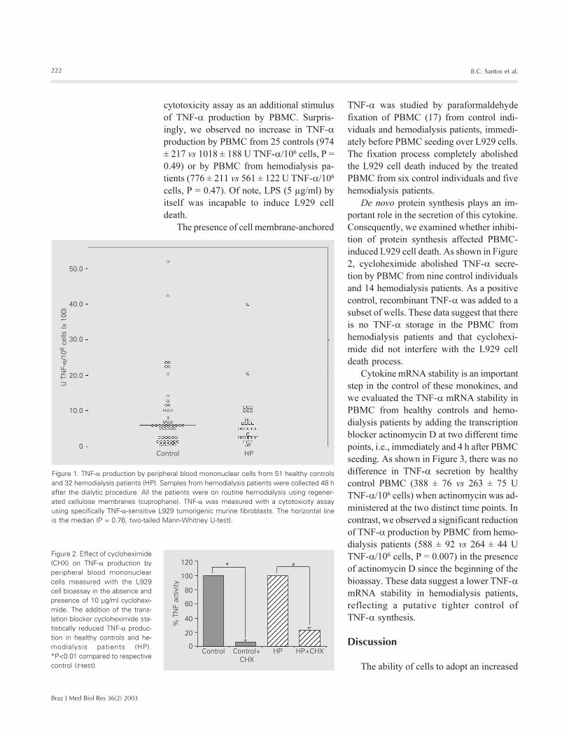
For measurement of cytokine PBMC pro-
duction we used an in vitro cell cytotoxicity
assay for TNF-� which is simple and sensi-
tive. In contrast to previous studies that used
ELISA, the evaluation of bioactive TNF-�
production by PBMC of healthy control in-
dividuals and patients on chronic hemodi-
alysis did not show any difference between
the two groups (859 ± 141 vs 697 ± 130 U
TNF-�/106 cells, P = 0.76) (Figure 1). Of
note, none of the individuals enrolled in this
study presented any pathology at the time
of blood sample drawing or were taking
any medication that could markedly affect
TNF-� secretion or activity. Concomitantly
to the cell cytotoxicity assay, a neutralizing
assay with a monoclonal anti-TNF-� anti-
body (Innogenetics S.A., Ghent, Belgium)
was performed. This experiment showed a
strong inhibition of L929 cell cytotoxicity
induced by PBMC from control individuals
or hemodialysis patients, suggesting that the
observed biological phenomenon was medi-
ated by TNF-� secretion in the cell culture
medium.
The bacterial-derived LPS is a strong
stimulus for TNF-� production and secre-
tion. Thus, we added 5 µg/ml of LPS to the
cell culture medium used in the L929 cell
222
Braz J Med Biol Res 36(2) 2003
B.C. Santos et al.
TNF-� was studied by paraformaldehyde
fixation of PBMC (17) from control indi-
viduals and hemodialysis patients, immedi-
ately before PBMC seeding over L929 cells.
The fixation process completely abolished
the L929 cell death induced by the treated
PBMC from six control individuals and five
hemodialysis patients.
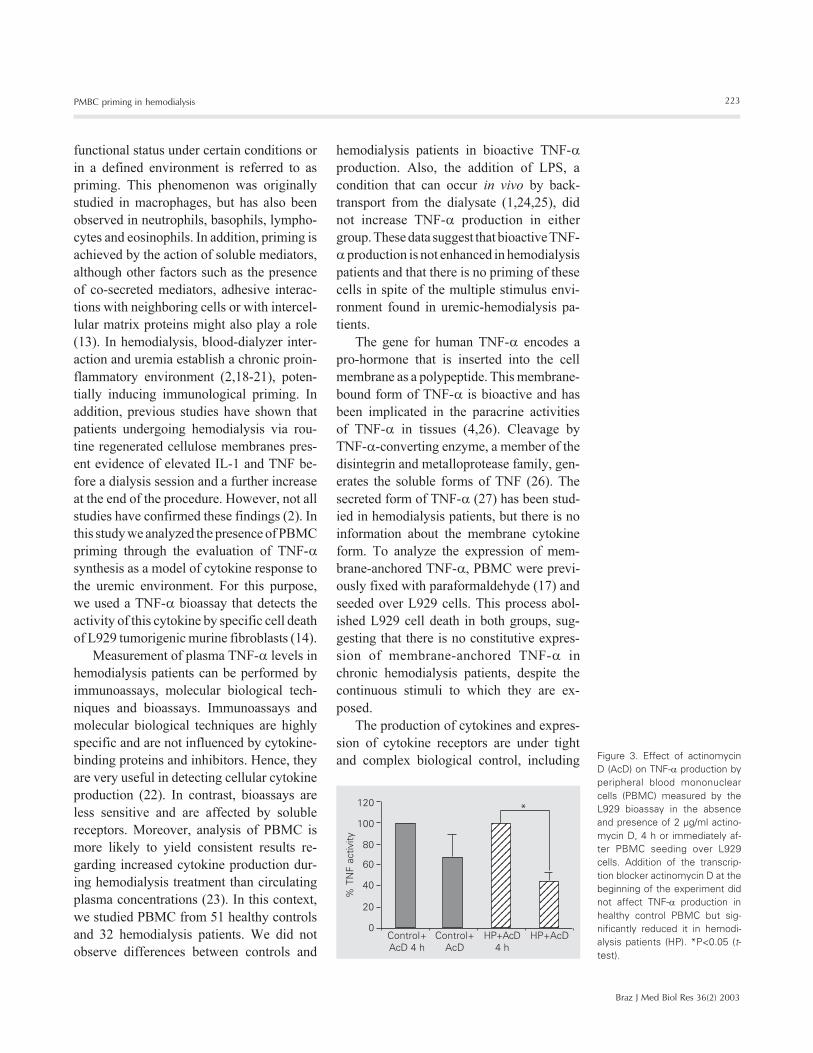
De novo protein synthesis plays an im-
portant role in the secretion of this cytokine.
Consequently, we examined whether inhibi-
tion of protein synthesis affected PBMC-
induced L929 cell death. As shown in Figure
2, cycloheximide abolished TNF-� secre-
tion by PBMC from nine control individuals
and 14 hemodialysis patients. As a positive
control, recombinant TNF-� was added to a
subset of wells. These data suggest that there
is no TNF-� storage in the PBMC from
hemodialysis patients and that cyclohexi-
mide did not interfere with the L929 cell
death process.
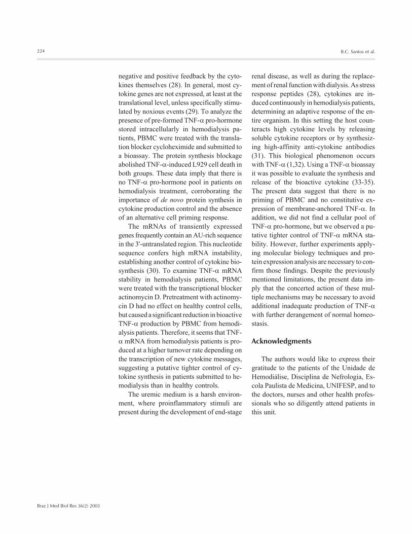
Cytokine mRNA stability is an important
step in the control of these monokines, and
we evaluated the TNF-��mRNA stability in
PBMC from healthy controls and hemo-
dialysis patients by adding the transcription
blocker actinomycin D at two different time
points, i.e., immediately and 4 h after PBMC
seeding. As shown in Figure 3, there was no
difference in TNF-� secretion by healthy
control PBMC (388 ± 76 vs 263 ± 75 U
TNF-�/106 cells) when actinomycin was ad-
ministered at the two distinct time points. In
contrast, we observed a significant reduction
of TNF-� production by PBMC from hemo-
dialysis patients (588 ± 92 vs 264 ± 44 U
TNF-�/106 cells, P = 0.007) in the presence
of actinomycin D since the beginning of the
bioassay. These data suggest a lower TNF-�
mRNA stability in hemodialysis patients,
reflecting a putative tighter control of
TNF-� synthesis.
Discussion
The ability of cells to adopt an increased
123456123456123456123456
123451234512345123451234512345123451234512345123451234512345123451234512345
**
Control Control+CHX
HP HP+CHX
% T
NF
activ
ity
120
100
80
60
40
20
0
Figure 2. Effect of cycloheximide(CHX) on TNF-� production byperipheral blood mononuclearcells measured with the L929cell bioassay in the absence andpresence of 10 µg/ml cyclohexi-mide. The addition of the trans-lation blocker cycloheximide sta-tistically reduced TNF-� produc-tion in healthy controls and he-modialysis patients (HP).*P<0.01 compared to respectivecontrol (t-test).
Figure 1. TNF-� production by peripheral blood mononuclear cells from 51 healthy controlsand 32 hemodialysis patients (HP). Samples from hemodialysis patients were collected 48 hafter the dialytic procedure. All the patients were on routine hemodialysis using regener-ated cellulose membranes (cuprophane). TNF-� was measured with a cytotoxicity assayusing specifically TNF-�-sensitive L929 tumorigenic murine fibroblasts. The horizontal lineis the median (P = 0.76, two-tailed Mann-Whitney U-test).
U T
NF-�
/106
cel
ls (
x 10
0)
50.0
40.0
30.0
20.0
10.0
0Control HP
cytotoxicity assay as an additional stimulus
of TNF-� production by PBMC. Surpris-
ingly, we observed no increase in TNF-�
production by PBMC from 25 controls (974
± 217 vs 1018 ± 188 U TNF-�/106 cells, P =
0.49) or by PBMC from hemodialysis pa-
tients (776 ± 211 vs 561 ± 122 U TNF-�/106
cells, P = 0.47). Of note, LPS (5 µg/ml) by
itself was incapable to induce L929 cell
death.
The presence of cell membrane-anchored
223
Braz J Med Biol Res 36(2) 2003
PMBC priming in hemodialysis
functional status under certain conditions or
in a defined environment is referred to as
priming. This phenomenon was originally
studied in macrophages, but has also been
observed in neutrophils, basophils, lympho-
cytes and eosinophils. In addition, priming is
achieved by the action of soluble mediators,
although other factors such as the presence
of co-secreted mediators, adhesive interac-
tions with neighboring cells or with intercel-
lular matrix proteins might also play a role
(13). In hemodialysis, blood-dialyzer inter-
action and uremia establish a chronic proin-
flammatory environment (2,18-21), poten-
tially inducing immunological priming. In
addition, previous studies have shown that
patients undergoing hemodialysis via rou-
tine regenerated cellulose membranes pres-
ent evidence of elevated IL-1 and TNF be-
fore a dialysis session and a further increase
at the end of the procedure. However, not all
studies have confirmed these findings (2). In
this study we analyzed the presence of PBMC
priming through the evaluation of TNF-�
synthesis as a model of cytokine response to
the uremic environment. For this purpose,
we used a TNF-� bioassay that detects the
activity of this cytokine by specific cell death
of L929 tumorigenic murine fibroblasts (14).
Measurement of plasma TNF-� levels in
hemodialysis patients can be performed by
immunoassays, molecular biological tech-
niques and bioassays. Immunoassays and
molecular biological techniques are highly
specific and are not influenced by cytokine-
binding proteins and inhibitors. Hence, they
are very useful in detecting cellular cytokine
production (22). In contrast, bioassays are
less sensitive and are affected by soluble
receptors. Moreover, analysis of PBMC is
more likely to yield consistent results re-
garding increased cytokine production dur-
ing hemodialysis treatment than circulating
plasma concentrations (23). In this context,
we studied PBMC from 51 healthy controls
and 32 hemodialysis patients. We did not
observe differences between controls and
hemodialysis patients in bioactive TNF-�
production. Also, the addition of LPS, a
condition that can occur in vivo by back-
transport from the dialysate (1,24,25), did
not increase TNF-� production in either
group. These data suggest that bioactive TNF-
� production is not enhanced in hemodialysis
patients and that there is no priming of these
cells in spite of the multiple stimulus envi-
ronment found in uremic-hemodialysis pa-
tients.
The gene for human TNF-� encodes a
pro-hormone that is inserted into the cell
membrane as a polypeptide. This membrane-
bound form of TNF-� is bioactive and has
been implicated in the paracrine activities
of TNF-� in tissues (4,26). Cleavage by
TNF-�-converting enzyme, a member of the
disintegrin and metalloprotease family, gen-
erates the soluble forms of TNF (26). The
secreted form of TNF-� (27) has been stud-
ied in hemodialysis patients, but there is no
information about the membrane cytokine
form. To analyze the expression of mem-
brane-anchored TNF-�, PBMC were previ-
ously fixed with paraformaldehyde (17) and
seeded over L929 cells. This process abol-
ished L929 cell death in both groups, sug-
gesting that there is no constitutive expres-
sion of membrane-anchored TNF-� in
chronic hemodialysis patients, despite the
continuous stimuli to which they are ex-
posed.
The production of cytokines and expres-
sion of cytokine receptors are under tight
and complex biological control, including Figure 3. Effect of actinomycinD (AcD) on TNF-� production byperipheral blood mononuclearcells (PBMC) measured by theL929 bioassay in the absenceand presence of 2 µg/ml actino-mycin D, 4 h or immediately af-ter PBMC seeding over L929cells. Addition of the transcrip-tion blocker actinomycin D at thebeginning of the experiment didnot affect TNF-� production inhealthy control PBMC but sig-nificantly reduced it in hemodi-alysis patients (HP). *P<0.05 (t-test).
12341234123412341234123412341234
1234123412341234123412341234123412341234123412341234123412341234
*
% T
NF
activ
ity
120
100
80
60
40
20
0Control+AcD 4 h
Control+AcD
HP+AcD4 h
HP+AcD
224
Braz J Med Biol Res 36(2) 2003
B.C. Santos et al.
negative and positive feedback by the cyto-
kines themselves (28). In general, most cy-
tokine genes are not expressed, at least at the
translational level, unless specifically stimu-
lated by noxious events (29). To analyze the
presence of pre-formed TNF-� pro-hormone
stored intracellularly in hemodialysis pa-
tients, PBMC were treated with the transla-
tion blocker cycloheximide and submitted to
a bioassay. The protein synthesis blockage
abolished TNF-�-induced L929 cell death in
both groups. These data imply that there is
no TNF-� pro-hormone pool in patients on
hemodialysis treatment, corroborating the
importance of de novo protein synthesis in
cytokine production control and the absence
of an alternative cell priming response.
The mRNAs of transiently expressed
genes frequently contain an AU-rich sequence
in the 3'-untranslated region. This nucleotide
sequence confers high mRNA instability,
establishing another control of cytokine bio-
synthesis (30). To examine TNF-� mRNA
stability in hemodialysis patients, PBMC
were treated with the transcriptional blocker
actinomycin D. Pretreatment with actinomy-
cin D had no effect on healthy control cells,
but caused a significant reduction in bioactive
TNF-� production by PBMC from hemodi-
alysis patients. Therefore, it seems that TNF-
� mRNA from hemodialysis patients is pro-
duced at a higher turnover rate depending on
the transcription of new cytokine messages,
suggesting a putative tighter control of cy-
tokine synthesis in patients submitted to he-
modialysis than in healthy controls.
The uremic medium is a harsh environ-
ment, where proinflammatory stimuli are
present during the development of end-stage
renal disease, as well as during the replace-
ment of renal function with dialysis. As stress
response peptides (28), cytokines are in-
duced continuously in hemodialysis patients,
determining an adaptive response of the en-
tire organism. In this setting the host coun-
teracts high cytokine levels by releasing
soluble cytokine receptors or by synthesiz-
ing high-affinity anti-cytokine antibodies
(31). This biological phenomenon occurs
with TNF-� (1,32). Using a TNF-� bioassay
it was possible to evaluate the synthesis and
release of the bioactive cytokine (33-35).
The present data suggest that there is no
priming of PBMC and no constitutive ex-
pression of membrane-anchored TNF-�. In
addition, we did not find a cellular pool of
TNF-� pro-hormone, but we observed a pu-
tative tighter control of TNF-� mRNA sta-
bility. However, further experiments apply-
ing molecular biology techniques and pro-
tein expression analysis are necessary to con-
firm those findings. Despite the previously
mentioned limitations, the present data im-
ply that the concerted action of these mul-
tiple mechanisms may be necessary to avoid
additional inadequate production of TNF-�
with further derangement of normal homeo-
stasis.
Acknowledgments
The authors would like to express their
gratitude to the patients of the Unidade de
Hemodiálise, Disciplina de Nefrologia, Es-
cola Paulista de Medicina, UNIFESP, and to
the doctors, nurses and other health profes-
sionals who so diligently attend patients in
this unit.
225
Braz J Med Biol Res 36(2) 2003
PMBC priming in hemodialysis
References
1. Pertosa G, Grandaliano G, Gesualdo L & Schena FP (2000). Clinicalrelevance of cytokine production in hemodialysis. Kidney Interna-tional, 58 (Suppl 76): S104-S111.
2. Horl WH (2002). Hemodialysis membranes: interleukins, biocompat-ibility, and middle molecules. Journal of the American Society ofNephrology, 13 (Suppl 1): S62-S71.
3. Bazzoni F & Beutler B (1996). The tumor necrosis factor ligand andreceptor families. New England Journal of Medicine, 334: 1717-1725.
4. Tracey KJ & Cerami A (1994). Tumor necrosis factor: a pleiotropiccytokine and therapeutic target. Annual Review of Medicine, 45:491-503.
5. Liu ZG & Han J (2001). Cellular responses to tumor necrosis factor.Current Issues in Molecular Biology, 3: 79-90.
6. Herbelin A, Nguyen AT, Zingraff J, Urena P & Descamps-Latscha B(1990). Influence of uremia and hemodialysis on circulating interleu-kin-1 and tumor necrosis factor alpha. Kidney International, 37: 116-125.
7. Hock JM, Krishnan V, Onyia JE, Bidwell JP, Milas J & Stanislaus D(2001). Osteoblast apoptosis and bone turnover. Journal of Boneand Mineral Research, 16: 975-984.
8. Jongen-Lavrencic M, Peeters HR, Wognum A, Vreugdenhil G,Breedveld FC & Swaak AJ (1997). Elevated levels of inflammatorycytokines in bone marrow of patients with rheumatoid arthritis andanemia of chronic disease. Journal of Rheumatology, 24: 1504-1509.
9. Papadaki HA, Kritikos HD, Valatas V, Boumpas DT & Eliopoulos GD(2002). Anemia of chronic disease in rheumatoid arthritis is associ-ated with increased apoptosis of bone marrow erythroid cells: im-provement following anti-tumor necrosis factor-alpha antibody thera-py. Blood, 100: 474-482.
10. Beutler B (1993). Cytokines and cancer cachexia. Hospital Practice,28: 45-52.
11. Reid MB & Li YP (2001). Tumor necrosis factor-alpha and musclewasting: a cellular perspective. Respiratory Research, 2: 269-272.
12. Schindler R, Lonnemann G, Shaldon S, Koch KM & Dinarello CA(1990). Transcription, not synthesis, of interleukin-1 and tumor ne-crosis factor by complement. Kidney International, 37: 85-93.
13. Kroegel C, Foerster M, Hafner D, Grahmann PR, Warner JA & BraunR (2000). Putting priming into perspective - from cellular heteroge-neity to cellular plasticity. Immunology Today, 21: 218-222.
14. Flick DA & Gifford GE (1984). Comparison of in vitro cell cytotoxicassays for tumor necrosis factor. Journal of Immunological Meth-ods, 68: 167-175.
15. Ali FMK (1986). Separation of Human Blood and Bone Marrow Cells.Wright, Bristol.
16. Taverne J, Treagust JD & Playfair JH (1986). Macrophage cytotoxic-ity in lethal and non-lethal murine malaria and the effect of vaccina-tion. Clinical and Experimental Immunology, 66: 44-51.
17. Bailly S, Ferrua B, Fay M & Gougerot-Pocidalo MA (1990). Parafor-maldehyde fixation of LPS-stimulated human monocytes: technicalparameters permitting the study of membrane IL-1 activity. Euro-pean Cytokine Network, 1: 47-51.
18. Memoli B, Minutolo R, Bisesti V, Postiglione L, Conti A, Marzano L,Capuano A, Andreucci M, Balletta MM, Guida B & Tetta C (2002).Changes of serum albumin and C-reactive protein are related tochanges of interleukin-6 release by peripheral blood mononuclear
cells in hemodialysis patients treated with different membranes.American Journal of Kidney Diseases, 39: 266-273.
19. Jaber BL, Lau J, Schmid CH, Karsou SA, Levey AS & Pereira B(2002). Effect of biocompatibility of hemodialysis membranes onmortality in acute renal failure: a meta-analysis. Clinical Nephrology,57: 274-282.
20. Carracedo J, Ramirez R, Madueno JA, Soriano S, Rodriguez-BenotA, Rodriguez M, Martin-Malo A & Aljama P (2002). Cell apoptosisand hemodialysis-induced inflammation. Kidney International, 61(Suppl 80): 89-93.
21. Muller-Steinhardt M, Kock N, Hartel C, Kirchner H & Steinhoff J(2001). Production of monokines in patients under polysulphonehaemodiafiltration is influenced by the ultrafiltration flow rate. Ne-phrology, Dialysis, Transplantation, 16: 1830-1837.
22. Engelberts I, Moller A, Schoen GJ, van der Linden CJ & BuurmanWA (1991). Evaluation of measurement of human TNF in plasma byELISA. Lymphokine and Cytokine Research, 10: 69-76.
23. Pereira BJ & Dinarello CA (1994). Production of cytokines and cy-tokine inhibitory proteins in patients on dialysis. Nephrology, Dialy-sis, Transplantation, 9 (Suppl 2): 60-71.
24. Schindler R, Eichert F, Lepenies J & Frei U (2001). Blood compo-nents influence cytokine induction by bacterial substances. BloodPurification, 19: 380-387.
25. Baccheschi S, Sereni L, De Nitti C, Barbucci R & Tetta C (2001).Blood tubing and cytokine production: effect of sterilization. RenalFailure, 23: 411-418.
26. Black RA (2002). Tumor necrosis factor-alpha converting enzyme.International Journal of Biochemistry and Cell Biology, 34: 1-5.
27. Lin YF, Chang DM, Shaio MF, Lu KC, Chyr SH, Li BL & Sheih SD(1996). Cytokine production during hemodialysis: effects of dialyticmembrane and complement activation. American Journal of Ne-phrology, 16: 293-299.
28. Callard R, George AJ & Stark J (1999). Cytokines, chaos, and com-plexity. Immunity, 11: 507-513.
29. Dinarello CA (2000). Proinflammatory cytokines. Chest, 118: 503-508.
30. Shaw G & Kamen R (1986). A conserved AU sequence from the 3'untranslated region of GM-CSF mRNA mediates selective mRNAdegradation. Cell, 46: 659-667.
31. Slifka MK & Whitton JL (2000). Clinical implications of dysregulatedcytokine production. Journal of Molecular Medicine, 78: 74-80.
32. Brockhaus M, Bar-Khayim Y, Gurwicz S, Frensdorff A & Haran N(1992). Plasma tumor necrosis factor soluble receptors in chronicrenal failure. Kidney International, 42: 663-667.
33. Bergqvist A, Nejaty H, Froysa B, Bruse C, Carlberg M, Sjoblom P &Soder O (2000). Production of interleukins 1beta, 6 and 8 and tumornecrosis factor alpha in separated and cultured endometrial andendometriotic stromal and epithelial cells. Gynecologic and Obstet-ric Investigation, 50: 1-6.
34. Fukuzawa M, Satoh J, Ohta S, Takahashi K, Miyaguchi S, Qiang X,Sakata Y, Nakazawa T, Takizawa Y & Toyota T (2000). Modulation oftumor necrosis factor-alpha production with anti-hypertensive drugs.Immunopharmacology, 48: 65-74.
35. Fukuzawa M, Satoh J, Qiang X, Miyaguchi S, Sakata Y, Nakazawa T,Ikehata F, Ohta S & Toyota T (1999). Inhibition of tumor necrosisfactor-alpha with anti-diabetic agents. Diabetes Research and Clini-cal Practice, 43: 147-154.