Vector Autoregressive with Exogenous Variable Model and its ...
Upload
khangminh22Category
view
1download
0
RESEARCH ARTICLE Open Access
Influence of exogenous ascorbic acid andglutathione priming on mitochondrialstructural and functional systems toalleviate aging damage in oat seedsFangshan Xia1,2, Hang Cheng2, Lingling Chen2, Huisen Zhu1, Peisheng Mao2* and Mingya Wang2
Abstract
Background: Loss of vigor caused by seed aging adversely affects agricultural production under natural conditions.However, priming is an economical and effective method for improving the vigor of aged seeds. The objective ofthis study was to test the effectiveness of exogenous ascorbic acid (ASC) and glutathione (GSH) priming in therepairing of aged oat (Avena sativa) seeds, and to test the hypothesis that structural and functional systems inmitochondria were involved in this process.
Results: Oat seeds were artificially aged for 20 days at 45 °C, and were primed with solutions (1mmol L− 1) of ASC,GSH, or ASC + GSH at 20 °C for 0.5 h before or after their aging. Seed germination, antioxidant enzymes in the ASC-GSHcycle, cytochrome c oxidase (COX) and mitochondrial malate dehydrogenase (MDH) activities, and the mitochondrialultrastructures of the embryonic root cells were markedly improved in aged oat seeds through post-priming with ASC,GSH, or ASC + GSH, while their malondialdehyde and H2O2 contents decreased significantly (P < 0.05).
Conclusion: Our results suggested that priming with ASC, GSH, or ASC + GSH after aging could effectively alleviateaging damage in oat seeds, and that the role of ASC was more effective than GSH, but positive effects of post-primingwith ASC and GSH were not superior to post-priming with ASC in repairing aging damage of aged oat seeds. However,pre-priming with ASC, GSH, or ASC + GSH was not effective in oat seeds, suggesting that pre-priming with ASC, GSH,or ASC + GSH could not inhibit the occurrence of aging damage in oat seeds.
Keywords: Ascorbic acid-glutathione cycle, Mitochondria, Oat, Seed aging, Seed priming
BackgroundSeed aging, resulting in a loss of vigor and viability undernatural storage conditions, is a major problem for success-ful agricultural production and productivity. Moreover,seed aging also leads to ecological problems due to short-ages of genetic resources and soil seed bank systems dys-function, e.g., loss of plant biodiversity, grassland
degeneration, and the enhancement of desertification [1].Thus, loss of seed vigor is a major challenge for food andecological security under natural conditions [2]. As a re-sult, it is necessary to better understand the possiblemethods and mechanisms of maintaining seed vigor.Accumulation of reactive oxygen species (ROS) is a
primary cause of seed aging during storage, causing lipidperoxidation, impairment of RNA and protein synthesis,degradation of DNA, and loss of membrane integrity [3,4]. The levels of ROS are tightly controlled by enzymaticand non-enzymatic antioxidant systems during seed
© The Author(s). 2020 Open Access This article is licensed under a Creative Commons Attribution 4.0 International License,which permits use, sharing, adaptation, distribution and reproduction in any medium or format, as long as you giveappropriate credit to the original author(s) and the source, provide a link to the Creative Commons licence, and indicate ifchanges were made. The images or other third party material in this article are included in the article's Creative Commonslicence, unless indicated otherwise in a credit line to the material. If material is not included in the article's Creative Commonslicence and your intended use is not permitted by statutory regulation or exceeds the permitted use, you will need to obtainpermission directly from the copyright holder. To view a copy of this licence, visit http://creativecommons.org/licenses/by/4.0/.The Creative Commons Public Domain Dedication waiver (http://creativecommons.org/publicdomain/zero/1.0/) applies to thedata made available in this article, unless otherwise stated in a credit line to the data.
* Correspondence: [email protected] Seed Laboratory/Beijing Key Laboratory of Grassland Science, ChinaAgricultural University, No 2, Yuanmingyuan West Road, Haidian Distr, Beijing100193, ChinaFull list of author information is available at the end of the article
Xia et al. BMC Plant Biology (2020) 20:104 https://doi.org/10.1186/s12870-020-2321-x

aging [1]. Once ·O2− is generated by the electron trans-
port chain during seed aging, it is promptly dismutatedinto hydrogen peroxide (H2O2) by superoxide dismutase(SOD) in the matrix [5, 6]. H2O2 is then eliminated viathe various antioxidant pathways that are crucial for theproduction and scavenging of ROS [7]. In particular, thescavenging of ROS largely depends on the availability ofmolecular antioxidants such as ascorbic acid (ASC) andglutathione (GSH) in dry seeds [8]. ASC and GSH arenecessary to maintain a net reducing environment byreacting with ROS or participating in the ASC-GSHcycle [9, 10]. Importantly, the ASC-GSH cycle is a cru-cial detoxifying mechanism in both dry and imbibedseeds, and is principally located in embryos [11]. In theASC-GSH cycle, ascorbate peroxidase (APX) reduces thetoxicity of ROS by using ASC as substrate; ASC is thenrecycled in the matrix by NADH-dependent monodehy-droascorbate reductase (MDHAR) or GSH-dependentdehydroascorbate reductase (DHAR), while GSH can berecycled in the matrix by glutathione reductase (GR)using NADPH as an electron donor [12, 13]. A declinein cellular ASC indicates failing antioxidant capacityduring seed aging, hence contributing to a loss of seedviability [14–16]. Similarly, cellular GSH also declinesrapidly, with a concomitant rapid decrease in seed vigor,during aging [4, 17, 18]. At the cellular level, seed agingalso reduces the activities of several enzymes involved inthe ASC-GSH cycle [16, 19]. Therefore, maintaininghigh levels of ASC and GSH is important to guaranteevigor in aged seeds.Mitochondria customarily play a central role in plant me-
tabolism as a major source of adenosine triphosphate(ATP) [20, 21]. Seeds are largely dependent on mitochon-drial respiration to provide obligatory energy for their ger-mination [22]. However, the activity of almost all enzymesinvolved in the tricarboxylic acid cycle decreases signifi-cantly during seed aging, e.g., the aging of elm (Ulmuspumila) [23] and rice (Oryza sativa) [24] seeds. Mitochon-drial ultrastructure is impaired along with a significant de-crease in the activities of cytochrome c oxidase (COX) andmalate dehydrogenase (MDH), thereby creating an insuffi-cient supply of mitochondrial energy and a surge in ROS[24]. Consequently, mitochondria play a dominant role inthe generation of ROS in seeds [25], and ROS-related mito-chondrial dysfunctions play a pivotal role in seed aging[23]. Similar to cellular antioxidant systems, the ASC-GSHcycle is also one of the major antioxidant protection sys-tems in mitochondria under oxidative stresses [10, 26]. Adistinct reduction has been observed in the content of bothASC and GSH and the activities of ASC-GSH cycle en-zymes during seed aging [7, 25]. Nevertheless, there is stillonly a limited understanding of how to enhance mitochon-drial function by improving the antioxidant capacity of theASC-GSH cycle.
Seed priming can enhance the vigor of aged seeds byefficaciously repairing cellular and mitochondrial com-ponents [27, 28]. The mechanism of priming-enhancedseed longevity is likely associated with antioxidants [29,30]. Consequently, priming with exogenous antioxidants,e.g., ASC and GSH, may be an important method for ef-fectively improving the vigor of aged seeds. Studies haveshown that exogenous ASC clearly promotes the ger-mination of aged seeds in many plants, such as onion(Allium cepa) [31] and Elymus sibiricus [32, 33]. None-theless, previous studies rarely focused on the mechan-ism of exogenous ASC in promoting the germination ofaged seeds, particularly at the mitochondrial level. More-over, exogenous GSH pre-treatment can also improvethe germination and antioxidant ability of aged Elymussibiricus seeds [34], but there are still only a few studieson the role of exogenous GSH in improving the vigor ofaged seeds. Therefore, it is necessary to better under-stand the effect of exogenous ASC and GSH on mito-chondrial structures and functions of aged seeds inorder to more effectively maintain their vigor.Oat (Avena sativa), an eco-friendly crop, is widely culti-
vated throughout the word because it can adapt to a var-iety of environmental stresses. However, oat grains oftenhave a high lipid content that can easily lead to rancidityor deterioration, hence limits its extensive use as a seed orfood [35, 36]. Therefore, the objectives of this study wereto determine the changes in the repair of ultrastructuralstructures, the enhancement of antioxidant capacity, theremission of lipid peroxidation, the recovery of respiratoryfunctions in embryonic mitochondria of aged oat seeds,and to better understand the response of priming with ex-ogenous ASC and GSH on the mitochondria in agedseeds.
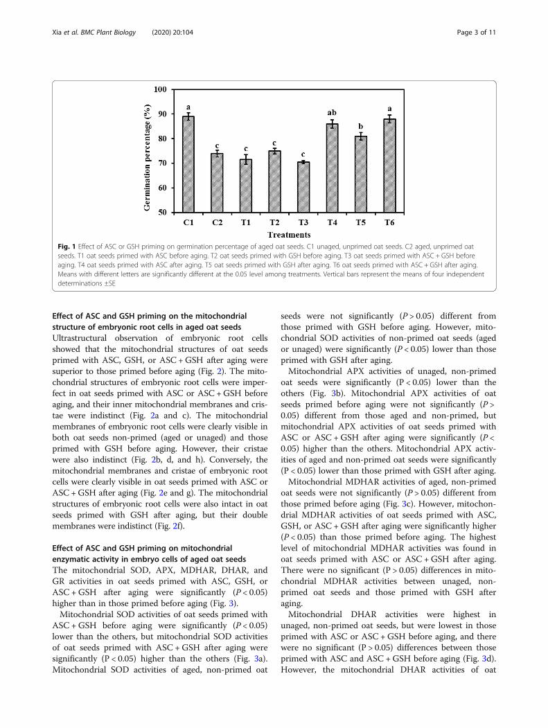
ResultsEffect of ASC and GSH priming on the germinationpercentage in aged oat seedsThe germination percentages of oat seeds primed withASC or GSH after aging were higher than those primedbefore aging (Fig. 1). There were no significant differ-ences (P > 0.05) in the germination percentages betweenoat seeds primed with ASC, GSH, or ASC +GSH beforeaging and those aged and non-primed. However, thegermination percentages of oat seeds primed with ASC,GSH, or ASC + GSH after aging were significantly (P <0.05) higher than those aged and non-primed. The ger-mination percentage of oat seeds primed with ASC +GSH after aging was significantly (P < 0.05) higher thanthose primed with GSH after aging, but the germinationpercentages of oat seeds primed with ASC or ASC +GSH after aging were not significantly (P > 0.05) differ-ent from those unaged and non-primed.
Xia et al. BMC Plant Biology (2020) 20:104 Page 2 of 11
Effect of ASC and GSH priming on the mitochondrialstructure of embryonic root cells in aged oat seedsUltrastructural observation of embryonic root cellsshowed that the mitochondrial structures of oat seedsprimed with ASC, GSH, or ASC +GSH after aging weresuperior to those primed before aging (Fig. 2). The mito-chondrial structures of embryonic root cells were imper-fect in oat seeds primed with ASC or ASC +GSH beforeaging, and their inner mitochondrial membranes and cris-tae were indistinct (Fig. 2a and c). The mitochondrialmembranes of embryonic root cells were clearly visible inboth oat seeds non-primed (aged or unaged) and thoseprimed with GSH before aging. However, their cristaewere also indistinct (Fig. 2b, d, and h). Conversely, themitochondrial membranes and cristae of embryonic rootcells were clearly visible in oat seeds primed with ASC orASC +GSH after aging (Fig. 2e and g). The mitochondrialstructures of embryonic root cells were also intact in oatseeds primed with GSH after aging, but their doublemembranes were indistinct (Fig. 2f).
Effect of ASC and GSH priming on mitochondrialenzymatic activity in embryo cells of aged oat seedsThe mitochondrial SOD, APX, MDHAR, DHAR, andGR activities in oat seeds primed with ASC, GSH, orASC +GSH after aging were significantly (P < 0.05)higher than in those primed before aging (Fig. 3).Mitochondrial SOD activities of oat seeds primed with
ASC +GSH before aging were significantly (P < 0.05)lower than the others, but mitochondrial SOD activitiesof oat seeds primed with ASC +GSH after aging weresignificantly (P < 0.05) higher than the others (Fig. 3a).Mitochondrial SOD activities of aged, non-primed oat
seeds were not significantly (P > 0.05) different fromthose primed with GSH before aging. However, mito-chondrial SOD activities of non-primed oat seeds (agedor unaged) were significantly (P < 0.05) lower than thoseprimed with GSH after aging.Mitochondrial APX activities of unaged, non-primed
oat seeds were significantly (P < 0.05) lower than theothers (Fig. 3b). Mitochondrial APX activities of oatseeds primed before aging were not significantly (P >0.05) different from those aged and non-primed, butmitochondrial APX activities of oat seeds primed withASC or ASC +GSH after aging were significantly (P <0.05) higher than the others. Mitochondrial APX activ-ities of aged and non-primed oat seeds were significantly(P < 0.05) lower than those primed with GSH after aging.Mitochondrial MDHAR activities of aged, non-primed
oat seeds were not significantly (P > 0.05) different fromthose primed before aging (Fig. 3c). However, mitochon-drial MDHAR activities of oat seeds primed with ASC,GSH, or ASC + GSH after aging were significantly higher(P < 0.05) than those primed before aging. The highestlevel of mitochondrial MDHAR activities was found inoat seeds primed with ASC or ASC +GSH after aging.There were no significant (P > 0.05) differences in mito-chondrial MDHAR activities between unaged, non-primed oat seeds and those primed with GSH afteraging.Mitochondrial DHAR activities were highest in
unaged, non-primed oat seeds, but were lowest in thoseprimed with ASC or ASC +GSH before aging, and therewere no significant (P > 0.05) differences between thoseprimed with ASC and ASC +GSH before aging (Fig. 3d).However, the mitochondrial DHAR activities of oat
Fig. 1 Effect of ASC or GSH priming on germination percentage of aged oat seeds. C1 unaged, unprimed oat seeds. C2 aged, unprimed oatseeds. T1 oat seeds primed with ASC before aging. T2 oat seeds primed with GSH before aging. T3 oat seeds primed with ASC + GSH beforeaging. T4 oat seeds primed with ASC after aging. T5 oat seeds primed with GSH after aging. T6 oat seeds primed with ASC + GSH after aging.Means with different letters are significantly different at the 0.05 level among treatments. Vertical bars represent the means of four independentdeterminations ±SE
Xia et al. BMC Plant Biology (2020) 20:104 Page 3 of 11

seeds primed after aging were significantly (P < 0.05)higher than those aged and non-primed. MitochondrialDHAR activities of oat seeds primed with ASC +GSHafter aging were significantly (P < 0.05) higher than otheraged oat seeds. Additionally, mitochondrial DHAR activ-ities were significantly (P < 0.05) higher in oat seedsprimed with GSH either before or after aging than inthose aged and non-primed.Mitochondrial GR activities were highest in unaged,
non-primed oat seeds, but were lowest in those primedwith ASC or ASC +GSH before aging (Fig. 3e). How-ever, mitochondrial GR activities of aged, non-primed
oat seeds were significantly (P < 0.05) lower than thoseprimed with GSH or ASC +GSH after aging.
Effect of ASC and GSH priming on mitochondrial H2O2
and MDA content in embryo cells of aged oat seedsMitochondrial H2O2 contents of unaged, non-primedoat seeds were significantly (P < 0.05) higher than thoseprimed with ASC, GSH, or ASC +GSH after aging, butthey were significantly (P < 0.05) lower than thoseprimed before aging and those aged and non-primed(Fig. 4a). Mitochondrial H2O2 contents were highest inoat seeds primed with ASC +GSH before aging, but they
Fig. 2 Effect of ASC or GSH priming on mitochondrial structures of embryonic root cells in aged oat seeds. a oat seeds primed with ASC beforeaging. b oat seeds primed with GSH before aging. c oat seeds primed with ASC + GSH before aging. d aged, unprimed oat seeds. e oat seedsprimed with ASC after aging. F oat seeds primed with GSH after aging. g oat seeds primed with ASC + GSH after aging. H unaged, unprimed oatseeds. Bars = 200 nm. C cristae. M mitochondria. MM mitochondrial membrane
Xia et al. BMC Plant Biology (2020) 20:104 Page 4 of 11

were significantly (P < 0.05) lower in oat seeds primedwith GSH before aging than those primed with ASC be-fore aging. Mitochondrial MDA contents of unaged,non-primed oat seeds were significantly (P < 0.05) lowerthan the others (Fig. 4b). Mitochondrial MDA contentsof oat seeds primed with GSH before aging were signifi-cantly (P < 0.05) lower than those aged and non-primed,but mitochondrial MDA contents of oat seeds primedwith ASC or ASC +GSH before aging were not differentfrom those aged and non-primed. Mitochondrial H2O2
and MDA contents in oat seeds primed with ASC, GSH,or ASC +GSH after aging were significantly (P < 0.05)lower than those aged and non-primed, and the lowest
level was observed in those primed with ASC or ASC +GSH after aging.
Effect of ASC and GSH priming on mitochondrial COX andMDH activities in embryo cells of aged oat seedsThe changes in mitochondrial COX and MDH activitieswere similar, and the activities of mitochondrial COXand MDH activities were significantly (P < 0.05) higherin oat seeds primed with ASC, GSH, or ASC +GSH afteraging than in those primed before aging (Fig. 5). Mito-chondrial COX and MDH activities were significantly(P < 0.05) lower in oat seeds primed with ASC or ASC +GSH before aging than in those aged and non-primed,
Fig. 3 Effect of ASC or GSH priming on mitochondrial antioxidant enzymes of embryo cells in aged oat seeds. a SOD activity. b APX activity. cMDHAR activity. d DHAR activity. e GR activity. C1 unaged, unprimed oat seeds. C2 aged, unprimed oat seeds. T1 oat seeds primed with ASCbefore aging. T2 oat seeds primed with GSH before aging. T3 oat seeds primed with ASC + GSH before aging. T4 oat seeds primed with ASC afteraging. T5 oat seeds primed with GSH after aging. T6 oat seeds primed with ASC + GSH after aging. Means with the different letters aresignificantly different at the 0.05 level among treatments. Vertical bars represent means of four independent determinations ±SE
Xia et al. BMC Plant Biology (2020) 20:104 Page 5 of 11

and the lowest level was found in those primed withASC +GSH before aging. However, mitochondrial COXand MDH activities were significantly (P < 0.05) higherin oat seeds primed with GSH before aging than in thoseaged and non-primed. Mitochondrial COX and MDHactivities maintained a high level in oat seeds primedwith ASC or ASC + GSH after aging and in those unagedand non-primed.
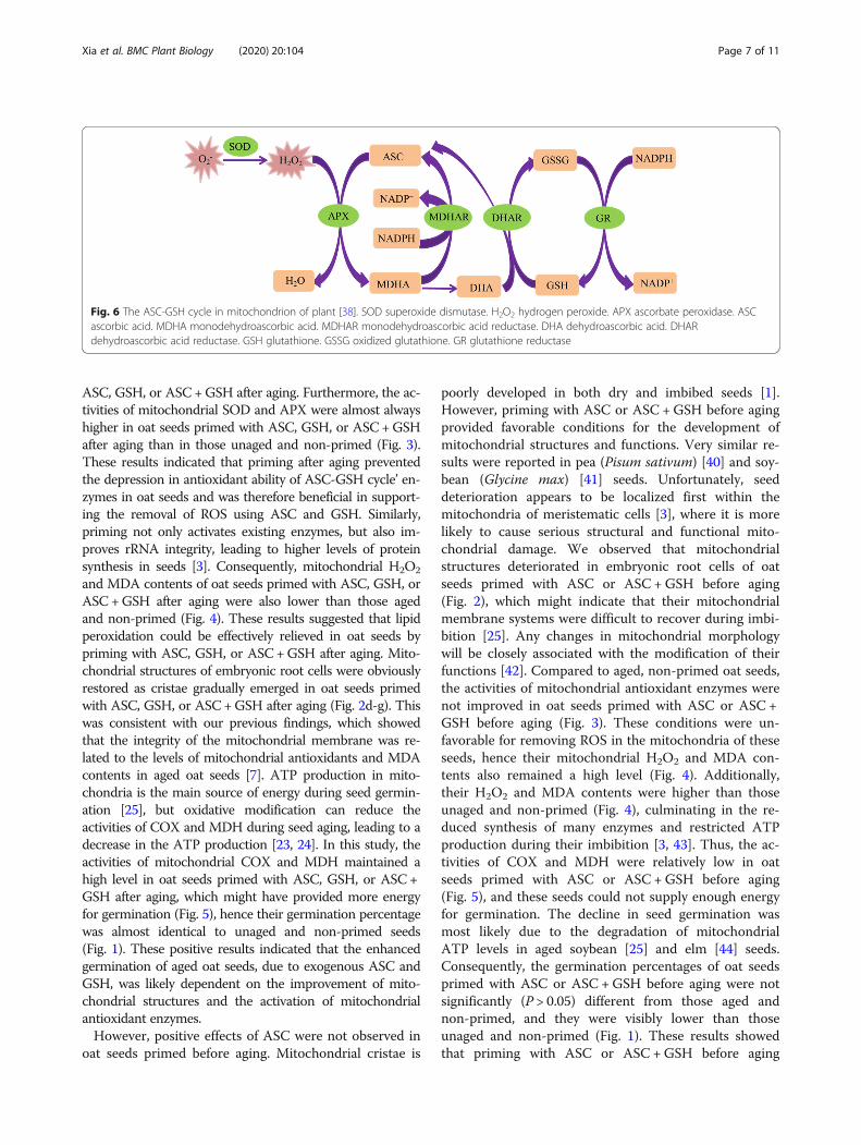
DiscussionFully functional mitochondria often have extensive cristaestructures and various biochemical activities [37], and theASC-GSH cycle is one of the primary antioxidant
protection systems in mitochondria under various oxidativestresses (Fig. 6) [39]. However, mitochondrial ultrastruc-tural damage in response to artificial aging negatively im-pacts mitochondrial function [25]. Previous research alsosuggests that a decrease in the activities of antioxidant en-zymes participating in the ASC-GSH cycle, accompaniedby mitochondrial ultrastructural damage, is the major causeof aging in oat seeds [7]. Our current results showed thatthe negative influence of aging in oat seeds could be effect-ively repaired by post-priming with ASC, GSH, or ASC +GSH (Fig. 3). Compared to aged and non-primed oat seeds,the mitochondrial SOD, APX, MDHAR, DHAR, and GRactivities were all markedly increased in those primed with
Fig. 4 Effect of ASC or GSH priming on mitochondrial H2O2 and MDA contents of embryo cells in aged oat seeds. a H2O2 content. b MDAcontent. C1 unaged, unprimed oat seeds. C2 aged, unprimed oat seeds. T1 oat seeds primed with ASC before aging. T2 oat seeds primed withGSH before aging. T3 oat seeds primed with ASC + GSH before aging. T4 oat seeds primed with ASC after aging. T5 oat seeds primed with GSHafter aging. T6 oat seeds primed with ASC + GSH after aging. Means with the different letters are significantly different at the 0.05 level amongtreatments. Vertical bars represent means of four independent determinations ±SE
Fig. 5 Effect of ASC or GSH priming on mitochondrial COX and MDH activity of embryo cells in aged oat seeds. a COX activity. b MDH activity.C1 unaged, unprimed oat seeds. C2 aged, unprimed oat seeds. T1 oat seeds primed with ASC before aging. T2 oat seeds primed with GSHbefore aging. T3 oat seeds primed with ASC + GSH before aging. T4 oat seeds primed with ASC after aging. T5 oat seeds primed with GSH afteraging. T6 oat seeds primed with ASC + GSH after aging. Means with the different letters are significantly different at the 0.05 level amongtreatments. Vertical bars represent means of four independent determinations ±SE
Xia et al. BMC Plant Biology (2020) 20:104 Page 6 of 11
ASC, GSH, or ASC +GSH after aging. Furthermore, the ac-tivities of mitochondrial SOD and APX were almost alwayshigher in oat seeds primed with ASC, GSH, or ASC +GSHafter aging than in those unaged and non-primed (Fig. 3).These results indicated that priming after aging preventedthe depression in antioxidant ability of ASC-GSH cycle’ en-zymes in oat seeds and was therefore beneficial in support-ing the removal of ROS using ASC and GSH. Similarly,priming not only activates existing enzymes, but also im-proves rRNA integrity, leading to higher levels of proteinsynthesis in seeds [3]. Consequently, mitochondrial H2O2
and MDA contents of oat seeds primed with ASC, GSH, orASC +GSH after aging were also lower than those agedand non-primed (Fig. 4). These results suggested that lipidperoxidation could be effectively relieved in oat seeds bypriming with ASC, GSH, or ASC +GSH after aging. Mito-chondrial structures of embryonic root cells were obviouslyrestored as cristae gradually emerged in oat seeds primedwith ASC, GSH, or ASC +GSH after aging (Fig. 2d-g). Thiswas consistent with our previous findings, which showedthat the integrity of the mitochondrial membrane was re-lated to the levels of mitochondrial antioxidants and MDAcontents in aged oat seeds [7]. ATP production in mito-chondria is the main source of energy during seed germin-ation [25], but oxidative modification can reduce theactivities of COX and MDH during seed aging, leading to adecrease in the ATP production [23, 24]. In this study, theactivities of mitochondrial COX and MDH maintained ahigh level in oat seeds primed with ASC, GSH, or ASC +GSH after aging, which might have provided more energyfor germination (Fig. 5), hence their germination percentagewas almost identical to unaged and non-primed seeds(Fig. 1). These positive results indicated that the enhancedgermination of aged oat seeds, due to exogenous ASC andGSH, was likely dependent on the improvement of mito-chondrial structures and the activation of mitochondrialantioxidant enzymes.However, positive effects of ASC were not observed in
oat seeds primed before aging. Mitochondrial cristae is
poorly developed in both dry and imbibed seeds [1].However, priming with ASC or ASC +GSH before agingprovided favorable conditions for the development ofmitochondrial structures and functions. Very similar re-sults were reported in pea (Pisum sativum) [40] and soy-bean (Glycine max) [41] seeds. Unfortunately, seeddeterioration appears to be localized first within themitochondria of meristematic cells [3], where it is morelikely to cause serious structural and functional mito-chondrial damage. We observed that mitochondrialstructures deteriorated in embryonic root cells of oatseeds primed with ASC or ASC + GSH before aging(Fig. 2), which might indicate that their mitochondrialmembrane systems were difficult to recover during imbi-bition [25]. Any changes in mitochondrial morphologywill be closely associated with the modification of theirfunctions [42]. Compared to aged, non-primed oat seeds,the activities of mitochondrial antioxidant enzymes werenot improved in oat seeds primed with ASC or ASC +GSH before aging (Fig. 3). These conditions were un-favorable for removing ROS in the mitochondria of theseseeds, hence their mitochondrial H2O2 and MDA con-tents also remained a high level (Fig. 4). Additionally,their H2O2 and MDA contents were higher than thoseunaged and non-primed (Fig. 4), culminating in the re-duced synthesis of many enzymes and restricted ATPproduction during their imbibition [3, 43]. Thus, the ac-tivities of COX and MDH were relatively low in oatseeds primed with ASC or ASC + GSH before aging(Fig. 5), and these seeds could not supply enough energyfor germination. The decline in seed germination wasmost likely due to the degradation of mitochondrialATP levels in aged soybean [25] and elm [44] seeds.Consequently, the germination percentages of oat seedsprimed with ASC or ASC +GSH before aging were notsignificantly (P > 0.05) different from those aged andnon-primed, and they were visibly lower than thoseunaged and non-primed (Fig. 1). These results showedthat priming with ASC or ASC +GSH before aging
Fig. 6 The ASC-GSH cycle in mitochondrion of plant [38]. SOD superoxide dismutase. H2O2 hydrogen peroxide. APX ascorbate peroxidase. ASCascorbic acid. MDHA monodehydroascorbic acid. MDHAR monodehydroascorbic acid reductase. DHA dehydroascorbic acid. DHARdehydroascorbic acid reductase. GSH glutathione. GSSG oxidized glutathione. GR glutathione reductase
Xia et al. BMC Plant Biology (2020) 20:104 Page 7 of 11

could not prevent the occurrence of seed aging, whichwas consistent with the outcomes of previous studies[45, 46]. However, compared with oat seeds primed withASC or ASC +GSH before aging, the activities of mito-chondrial SOD, APX, DHAR and GR were enhanced inthose primed with GSH before aging (Fig. 3). Addition-ally, their mitochondrial H2O2 and MDA contents werereduced (Fig. 4), hence the activities of COX and MDHin these seeds were also improved (Fig. 5). GSH is re-ported to recycle potential ASC via the ASC-GSH cycle(Fig. 6) [39, 47, 48], but this process might take a longtime to complete and its effect might also be relativelyweak. Mitochondrial structures and functions mighttherefore also be relatively slow to develop, which mightcause mild structural and functional damage for themitochondria during seed aging [3].In our study, mitochondrial DHAR and GR activities in
oat seeds primed with GSH or ASC +GSH after agingwere markedly higher than those primed with ASC afteraging (Fig. 3). This indicated that the ASC regenerationmaintained a relatively strong level in mitochondria bypost-priming with GSH or ASC +GSH, thereby regulatingthe balance of redox in the mitochondrial matrix viamaintaining ASC in a reduced state (Fig. 6) [39]. This wasin agreement with previous studies at the cellular level[38, 49]. However, mitochondrial SOD, APX, andMDHAR activities of oat seeds primed with GSH afteraging were markedly lower than those primed with ASCor ASC +GSH after aging (Fig. 3), and the mitochondrialH2O2 and MDA contents in these seeds were higher(Fig. 4). These results suggested that the antioxidant cap-acity of oat seeds primed with GSH after aging was lessthan those primed with ASC or ASC +GSH after aging,hence their lipid peroxidation was enhanced. Conse-quently, mitochondrial COX and MDH activities of oatseeds primed with GSH after aging were also markedlylower than those primed with ASC or ASC +GSH afteraging (Fig. 5), and their germination percentage was alsorelatively low (Fig. 1). Therefore, these results suggestedthat the effects of ASC priming were superior to GSHpriming for the recovery of aging damage in oat seeds.Previous studies at the cellular level had also demon-strated that the presence of ASC might be crucial for en-suring seed germination [12]. Unexpectedly, themitochondrial H2O2 and MDA contents showed no sig-nificant (P > 0.05) differences between oat seeds primedwith ASC and ASC +GSH after aging (Fig. 4), symbolizinga similar level of lipid peroxidation. Thereby, their mito-chondrial COX and MDH activities were also not signifi-cantly (P > 0.05) different (Fig. 5), showing that theyprovided similar level of energy for the germination ofaged oat seeds, and their germination ability was also notsignificantly (P > 0.05) different (Fig. 1). These resultsmight imply that positive effects of priming with ASC +
GSH after aging were not superior to post-priming withASC after aging on injured renovation of aged oat seeds.In other words, the regeneration of ASC was induced bypost-priming with GSH in oat seeds, but this effect wasnot obvious under sufficient ASC conditions.
ConclusionsOur results suggested that pre-priming with ASC, GSH, orASC +GSH could not inhibit the aging damage in oatseeds. Fortunately, post-priming with ASC, GSH, or ASC +GSH could effectively repair aging damage in oat seeds, andthe effect of ASC priming was greater than GSH priming.However, positive effects of priming with ASC +GSH afteraging were not greater than priming with ASC after agingon repairing aging damage of oat seeds.
MethodsSeed samplesOat seeds were collected by the Forage Seed Laboratoryof China Agricultural University in 2009 and stored inplastic bags at − 20 °C. The original materials wereimported from DYCK Forage & Grasses LTD of Canadaby Beijing Houderui Trading Co., LTD. At the start ofthe experiment in December 2014, the moisture contentof the seeds was 9.8% (on a fresh-weight basis), the seedgermination percentage was 89%, and the oil content ofthe seeds was 5.0%.
Seed aging and priming treatmentsSeed aging treatments: Primed or non-primed oat seedswith 10% moisture content on a fresh-weight basis (45%relative humidity, RH) were immediately sealed in analuminum foil bag (0.12 × 0.17 m2) and aged for 20 daysat 45 °C in a water bath.Seed priming treatments: Oat seeds with 10% moisture
content on a fresh-weight basis (45% RH) were soakedin a solution of ASC, GSH, or ASC +GSH at 20 °C for0.5 h (selected from 0, 0.5, 1, 3, 6, and 12 h) before orafter aging. The concentration of these solutions was 1mmol L− 1 (selected from 0, 0.25, 0.5, 1, 2, 4, and 8mmol L− 1). Thereafter, these seeds were rinsed twicewith de-ionized water and air-dried in the dark for 3 daysat 25 °C and 45% RH (moisture content reached approxi-mately 10% on a fresh-weight basis).Each treatment had four replicates, and 800 oat seeds
(approximately 20 g) were used for each replicate.Control 1 (C1) was comprised of unaged, non-primed
seeds (approximately 10% moisture content on a fresh-weight basis). Control 2 (C2) was comprised of aged,non-primed seeds (approximately 10% moisture contenton a fresh-weight basis).
Xia et al. BMC Plant Biology (2020) 20:104 Page 8 of 11

Germination testsGermination tests were conducted according to Inter-national Seed Testing Association rules [50]. Four repli-cates of 100 seeds from each sample were placed intoPetri dishes lined with three filter papers wetted with 10mL de-ionized water, and placed in a growth chamberunder a constant temperature of 20 °C. Germination waschecked daily for 10 days. On day 10, the final numberof normal seedlings was counted, and the length of seed-lings was measured.
Ultrastructural observations of mitochondria inembryonic root cellsSeed samples were selected randomly after differenttreatments. Embryonic roots, after imbibition for 4 h andwithout radicle protrusion, were removed and fixed in a4% glutaraldehyde solution for 24 h and then placed in arefrigerator at 4 °C. The samples of embryonic rootswere processed by rinsing with 50mmol L− 1 sodiumphosphate buffer, fixing with osmium tetroxide, rinsingwith buffer again, dehydrating with an alcohol gradient,and embedding in epoxy resin. After, ultrathin sliceswere obtained using an LKB8800 III ultramicrotome.Sections were placed on copper grids and stained with2% aqueous uranyl acetate for 20 min followed by a leadelectron staining solution (Katayama, Osaka) for 5 min.Twenty results per treatment were observed by thetransmission electron microscopy (Hitachi H-7500).
Isolation of mitochondriaMitochondrial extraction was carried out by the methodof Yin et al. [51]. The specific extract operation was per-formed as previously described [7]. Part of the enzymeactivity that was detected might be due to minimalmicrosomal contamination from this method.
Enzyme assaysSOD (EC 1.15.1.1) activity was determined according toRao and Sresty [42]. APX (EC 1.11.1.11) activity wasassayed according to the method of Nakano and Asada[52]. MDHAR (EC1.6.5.4) activity was measured accord-ing to Arrigoni et al. [53]. DHAR (EC 1.8.5.1) activity wasassayed according to Dalton et al. [54]. GR (EC 1.6.4.2) ac-tivity was assayed according to Madamanchi and Alscher[55]. The detailed determination method of SOD, APX,MDHAR, DHAR and GR was also performed as previ-ously described [7]. COX (EC 1.9.3.1) activity was assayedaccording to Neuburger et al. [56], and was determined asa decrease in absorbance at 550 nm (ε550 = 13.5mmol− 1
cm− 1) and 25 °C due to cytochrome c oxidation. MDH(EC 1.1.1.37) activity was measured by monitoring the in-crease in absorbance at 340 nm (ε340 = 6.2mmol− 1 cm− 1)and 25 °C due to NADH production according to themethods described by Glatthaar et al. [57]. Reaction
systems (1mL) were mixed with 10 μL mitochondria(20 μg mitochondrial protein) in all enzymes assays.
The H2O2 and malondialdehyde (MDA) contentsThe H2O2 content of isolated mitochondria was mea-sured by the method of Patterson et al. [58]. The deter-mination was depened on the changes in absorbancevalues of the titanium peroxide complex at 415 nm,which was calculated from a standard curve of knownH2O2 concentrations.MDA content of isolated mitochondria was measured
by the method of Bailly et al. [59], which was calculated bythe determining absorbance values at 532 and 600 nm.100 μL isolated mitochondria were mixed into 3mL reac-tion systems, which contained 20% (w/v) trichloroaceticacid and 0.5% (w/v) 2-thiobarbituric acid.
Statistical analysesComparisons of mean differences in different treatmentswere carried out through an analysis of variance(ANOVA), using SPSS for Windows version 13.0. Dun-can’s multiple range test (P = 0.05) was used to comparethe treatment means of physiological indicators for anti-oxidant systems and lipid peroxidation.
AbbreviationsAPX: Ascorbate peroxidase; ASC: Ascorbate acid; ATP: Adenosinetriphosphate; COX: Cytochrome c oxidase; DHAR: Dehydroascorbatereductase; GR: Glutathione reductase; GSH: Glutathione;MDA: Malondialdehyde; MDH: Malate dehydrogenase;MDHAR: Monodehydroascorbate reductase;; RH: Relative humidity;ROS: Reactive oxygen species; SOD: Superoxide dismutase.
AcknowledgementsWe thank all of our colleagues in our laboratory for providing usefuldiscussions and technical assistance. We are very grateful to the editor andreviewers for critically evaluating the manuscript and providing constructivecomments for its improvement. We are also very grateful to Radia LynaLahlou and Brendan Nuse for their careful improvement of the manuscriptlanguage.
Authors’ contributionsFX and PM conceived and designed the research. FX and HC conducted theexperiments. LC and HZ contributed new reagents and analytical tools. FXand MW analyzed the data. FX and PM wrote the manuscript. All authorsread and approved the manuscript.
FundingThis work was financially supported by the Natural Science Foundation ofChina (NSFC31702171, NSFC 31572454), the Natural Science Foundation ofShanxi (201701D221154), and the Science and Technology Innovation Fundof Shanxi Agricultural University (2016YJ15). The funding agencies providedonly the experimental costs and publication fee for this work. Funds wereused for the design of the study and collection, analysis, and interpretationof data and in writing the manuscript, as well as in the open accesspayment.
Availability of data and materialsThe datasets used and/or analysed during the current study available fromthe corresponding author on reasonable request.
Ethics approval and consent to participateThese plant materials are widely used all over the world, and no permits arerequired for the collection of plant samples. The plant materials were
Xia et al. BMC Plant Biology (2020) 20:104 Page 9 of 11
collected by the Forage Seed Laboratory of China Agricultural University,China. This article did not contain any studies with human participants oranimals, and did not involve any endangered or protected species.
Consent for publicationNot applicable.
Competing interestsThe authors declare that they have no competing interests.
Author details1College of Animal Science and Veterinary Medicine, Shanxi AgriculturalUniversity, Taigu 030801, China. 2Forage Seed Laboratory/Beijing KeyLaboratory of Grassland Science, China Agricultural University, No 2,Yuanmingyuan West Road, Haidian Distr, Beijing 100193, China.
Received: 10 May 2019 Accepted: 28 February 2020
References1. Bewley JD, Bradford KJ, Hilhorst HWM, Nonogaki H. Seeds: Physiology of
Development, Germination and Dormancy. 3rd ed. New York: SpringerPress; 2013. p. 363–7.
2. Gao J, Fu H, Zhou X, Chen Z, Luo L, Cui B, et al. Comparative proteomicanalysis of seed embryo proteins associated with seed storability in rice(Oryza sativa L) during natural aging. Plant Physiol Bioch. 2016;103:31–44.
3. McDonald MB. Seed deterioration: physiology, repair and assessment. SeedSci Technol. 1999;27:177–237.
4. Chen H, Osuna D, Colville L, Lorenzo O, Graeber K, Küster H, et al.Transcriptome-wide mapping of pea seed ageing reveals a pivotal role forgenes related to oxidative stress and programmed cell death. PLoS One.2013;8:1–15.
5. Iturbe-Ormaetxe I, Matamoros MA, Rubio MC, Dalton DA, Becana M. Theantioxidants of legume nodule mitochondria. Mol Plant Microbe In. 2001;14:1189–96.
6. Rubio MC, James EK, Clemente MR, Bucciarelli B, Fedorova M, Vance CP,et al. Localization of superoxide dismutases and hydrogen peroxide inlegume root nodules. Mol Plant Microbe In. 2004;17:1294–305.
7. Xia F, Wang X, Li M, Mao P. Mitochondrial structural and antioxidant systemresponses to aging in oat (Avena sativa L.) seeds with different moisturecontents. Plant Physiol Biochem. 2015;94:122–9.
8. Pukacka S, Ratajczak E. Age-related biochemical changes during storage ofbeech (Fagus sylvatica L.) seeds. Seed Sci Res. 2007;17:45–53.
9. Noctor G, Foyer CH. Ascorbate and glutathione: keeping active oxygenunder control. Annu Rev Plant Physiol Pant Mol Biol. 1998;49:249–79.
10. Gupta SD. Reactive oxygen species and antioxidants in higher plants. NewYork: CRC press; 2011. p. 50–4.
11. DePaula M, PerezOtaola M, Darder M, Torres M, Frutos G,MartinezHonduvilla CJ. Function of the ascorbate-glutathione cycle in agedsunflower seeds. Physiol Plant. 1996;96:543–50.
12. Tullio MCD, Arrigoni O. The ascorbic acid system in seeds: to protect and toserve. Seed Sci Res. 2003;13:249–60.
13. Wu JH, Wang WQ, Song SQ, Cheng HY. Reactive oxygen species scavengingenzymes and down-adjustment of metabolism level in mitochondriaassociated with desiccation-tolerance acquisition of maize embryo. J IntegrPlant Biol. 2009;51:638–45.
14. Tommasi F, Paciolla C, de Pinto MC, Gara LD. Effects of storage temperatureon viability, germination and antioxidant metabolism in Ginkgo biloba L.seeds. Plant Physiol Biochem. 2006;44:359–68.
15. Hu D, Ma G, Wang Q, Yao J, Wang Y, Pritchard HW, et al. Spatial andtemporal nature of reactive oxygen species production and programmedcell death in elm (Ulmus pumila L.) seeds during controlled deterioration.Plant Cell Environ. 2012;35:2045–59.
16. Xia F, Chen L, Sun Y, Mao P. Relationships between ultrastructure of embryocells and biochemical variation during ageing of oat (Avena sativa L.) seedswith different moisture content. Acta Physiol Plant. 2015;37:1–11.
17. Seal CE, Zammit R, Scott P, Flowers TJ, Kranner I. Glutathione half-cellreduction potential and α-tocopherol as viability markers during theprolonged storage of Suaeda maritime seeds. Seed Sci Res. 2010;20:47–53.
18. Long RL, Kranner I, Panetta FD, Birtic S, Adkins SW, Steadman KJ. Wet-drycycling extends seed persistence by re-instating antioxidant capacity. PlantSoil. 2011;338:511–9.
19. Yin G, Xin X, Song C, Chen X, Zhang J, Wu S, et al. Activity levels andexpression of antioxidant enzymes in the ascorbate-glutathione cycle inartificially aged rice seed. Plant Physiol Biochem. 2014;80:1–9.
20. Berkowitz O, De Clercq I, Breusegem FV, Whelan J. Interaction betweenhormonal and mitochondrial signaling during growth, development and inplant defence responses. Plant, Cell and Environment. 2016;39:1127–39.
21. Taylor NL. Editorial for special issue “plant mitochondria”. Int J Mol Sci. 2018;19:3849–54.
22. Sew YS, Strӧher E, Fenske R, Millar AH. Loss of mitochondrial malatedehydrogenase activity alters seed metabolism impairing seed maturationand post-germination growth in Arabidopsis. Plant Physiol. 2016;171:849–63.
23. Li Y, Wang Y, Xue H, Pritchard HW, Wang X. Changes in the mitochondrialprotein profile due to ROS eruption during ageing of elm (Ulmus pumila L.)seeds. Plant Physiol Biochem. 2017;114:72–87.
24. Yin G, Whelan J, Wu S, Zhou J, Chen B, Chen X, et al. Comprehensivemitochondrial metabolic shift during the critical node of seed ageing inrice. PLoS One. 2016;11:1–19.
25. Xin X, Tian Q, Yin G, Chen X, Zhang J, Ng S, et al. Reduced mitochondrialand ascorbated-glutathione activity after artificial ageing in soybean seed. JPlant Physiol. 2014;171:140–7.
26. Jacoby RP, Li L, Huang S, Lee CP, Millar AH, Taylor NL. Mitochondrialcomposition, function and stress response in plants. J Integr Plant Biol.2012;54:887–906.
27. Xia FS, Chen LL, Yan HF, Sun Y, Li ML, Mao PS. Antioxidant andultrastructural responses to priming with PEG in aged, ultra-dry oat seed.Seed Sci Technol. 2016;44:1–13.
28. Xia FS, Wang MY, Chen LL, Cheng H, Sun Y, Li ML, et al. Responses ofmitochondrial ultrastructure and physiological variations to PEG-priming onultra-dried oat (Avena sativa L.) seeds after ageing. Seed Sci Technol. 2017;45:622–37.
29. Bailly C, Benamar A, Corbineau F, Côme D. Free radical scavenging asaffected by accelerated ageing and subsequent priming in sunflower seeds.Physiol Plant. 1998;104:6467–652.
30. Yeh YM, Sung JM. Priming slows deterioration of artificially aged bittergourd seeds by enhancing antioxidative activities. Seed Sci Technol. 2008;36:350–9.
31. Kumari P, Sheoran IS, Tomar RPS. Onion seed longevity as affected byascorbic acid treatments during ambient storage. Haryana J Hortic Sci. 2004;33:257–60.
32. Yan HF, Xia FS, Sun Y, Mao PS. Protective effect of ascorbic acid on seedlinggrowth and embryo cellular ultrastructure in aged Elymus sibiricus seeds.Seed Sci Technol. 2015;43:1–13.
33. Yan HF, Mao PS, Sun Y, Li ML. Impacts of ascorbic acid on germination,antioxidant enzymes and ultrastructure of embryo cells of aged Elymussibiricus seeds with different moisture contents. Int J Agric Biol. 2016;18:176–83.
34. Yan HF, Mao CL, Zhu YQ, Cheng H, Mao PS. Exogenous glutathione pre-treatment improves germination and resistance of Elymus sibiricus seedssubjected to different ageing conditions. Seed Sci Technol. 2017;45:607–21.
35. Heini RL, Oksman-Caldentey KM, Lehtinen P, Lehtinen P, Poutanen K. Effectof drying treatment conditions on sensory profile of germinated oat. CerealChem. 2001;78:707–14.
36. Claudine C, Tom S, Susan RV, Stewart D. Comparison of two headspacesampling techniques for the analysis of off-flavour volatiles from oat basedproducts. Food Chem. 2012;134:1592–600.
37. Carrie C, Murcha MW, Giraud E, Ng S, Zhang MF, Narsai R, et al. How doplants make mitochondria? Planta. 2013;237:429–39.
38. Foyer CH, Noctor G. Ascorbate and glutathione: the heart of the redox hub.Plant Physiol. 2011;155:2–18.
39. Jiménez A, Hernández JA, del Río LA, Sevilla F. Evidence for the presence ofthe ascorbate-glutathione cycle in mitochondria and peroxisomes of pealeaves. Plant Physiol. 1997;114:275–84.
40. Benamar A, Tallon C, Macherel D. Membrane integrity and oxidativeproperties of mitochondria isolated from imbibing pea seeds after primingor accelerated ageing. Seed Sci Res. 2003;13:35–45.
41. Sun H, Li L, Wang X, Wu S, Wang X. Ascorbate-glutathione cycle ofmitochondria in osmoprimed soybean cotyledons in response toimbibitional chilling injury. J Plant Physiol. 2011;168:226–32.
Xia et al. BMC Plant Biology (2020) 20:104 Page 10 of 11
42. Rao KVM, Sresty TVS. Antioxidant parameters in the seedlings of pigeon pea(Cajanus cajan (L.) Millspaugh) in response to Zn and Ni stresses. Plant Sci.2000;157:113–28.
43. Daum B, Walter A, Horst A, Osiewacz HD, Kuhlbrandt W. Age-dependentdissociation of ATP synthase dimers and loss of inner-membrane cristae inmitochondria. Proc Natl Acad Science USA. 2013;110:15301–6.
44. Wang Y, Li Y, Xue H, Pritchard HW, Wang X. Reactive oxygen species-provoked mitochondrial-dependent cell death during ageing of elm (Ulmuspumila L.) seeds. Plant J. 2015;81:438–52.
45. Chhetri DR, Drrai AS, Bhattacharjee A. Chemical manipulation of seedlongevity of 4 crop species in an unfavorable storage environment. Seed SciTechnol. 1993;21:31–44.
46. Draganić I, Lekić S. Seed priming with antioxidants improves seedgermination and seedling growth under unfavorable germinationconditions. Turk J Agric For. 2012;36:421–8.
47. Foyer CH, Noctor G. Redox homeostasis and antioxidant signaling: ametabolic interface between stress perception and physiological responses.Plant Cell. 2005;17:1866–75.
48. Szalai G, Kellõs T, Galiba G, Kocsy G. Glutathione as an antioxidant andregulatory molecule in plants under abiotic stress conditions. J PlantGrowth Regul. 2009;28:66–80.
49. Gill SS, Tuteja N. Reactive oxygen species and antioxidant machinery inabiotic stress tolerance in crop plants. Plant Physiol Biochem. 2010;48:909–30.
50. ISTA. In: International Seed Testing Association, editor. International rules forseed testing. Bassersdorf; 2014.
51. Yin GK, Sun HM, Xin X, Qin GZ, Liang Z, Jing XM. Mitochondrial damage inthe soybean seed axis during imbibition at chilling temperatures. Plant CellPhysiol. 2009;50:1305–18.
52. Nakano Y, Asada K. Hydrogen peroxide scavenged by ascorbate-specificperoxidase in spinach chloroplast. Plant Cell Physiol. 1981;22:867–80.
53. Arrigoni O, Dipiero S, Borraccino G. Ascorbate free radical reductase: a keyenzyme of the ascorbic acid system. FEBS Lett. 1981;125:242–4.
54. Dalton DA, Baird LM, Langeberg L, Taugher CY, Anyan WR, Vance CP, et al.Subcellular localization of oxygen defense enzymes in soybean (Glycine max(L.) Merr.) root nodules. Plant Physiol. 1993;102:481–9.
55. Madamanchi NR, Alscher RG. Metabolic bases for differences in sensitivity oftwo pea cultivars to sulfur dioxide. Plant Physiol. 1991;97:88–93.
56. Neuburger M. Preparation of plant mitochondria, criteria for assessment ofmitochondrial integrity and purity, survival in vitro. In: Douce R, Day D,editors. Higher Plant Cell Respiration. Berlin: Springer-Verlag; 1985. p. 7–24.
57. Glatthaar BE, Barbarsah GR, Noyes BE, Banaszak LJ, Bradshaw RA. Thepreparation of the cytoplasmic and mitochondrial forms of malatedehydrogenase and aspartate aminotransferase from pig heart by a singleprocedure. Anal Biochem. 1974;57:432–51.
58. Patterson BD, Macrae EA, Ferguson IB. Estimation of hydrogen peroxide inplants extracts using titanium (IV). Anal Biochem. 1984;139:487–92.
59. Bailly C, Benamar A, Corbineau F, Côme D. Changes in malondialdehydecontent and superoxide dismutase, catalase and glutathione reductaseactivities in sunflower seeds as related to deterioration during acceleratedaging. Physiol Plant. 1996;97:104–10.
Publisher’s NoteSpringer Nature remains neutral with regard to jurisdictional claims inpublished maps and institutional affiliations.
Xia et al. BMC Plant Biology (2020) 20:104 Page 11 of 11
Copyright © 2022 FDOKUMEN