Association of serum lipid peroxidation and glutathione ...
11
Front. Biosci. (Landmark Ed) 2022; 27(2): 068 http://doi.org/10.31083/j.fbl2702068 Copyright: © 2022 The Author(s). Published by IMR Press. This is an open access article under the CC BY 4.0 license. Publisher’s Note: IMR Press stays neutral with regard to jurisdictional claims in published maps and institutional affiliations. Original Research Association of serum lipid peroxidation and glutathione peroxidase 4 levels with clinical outcomes and metabolic abnormalities among patients with gestational diabetes mellitus: a case-control study in the Chinese population Bao Zhang 1 , Tingting Zhang 1 , Siyu Hu 1 , Lei Sun 1, * 1 Department of Obstetrics and Gynecology, Shengjing Hospital of China Medical University, 110004 Shenyang, Liaoning, China *Correspondence: [email protected] (Lei Sun) Academic Editor: Graham Pawelec Submitted: 14 October 2021 Revised: 3 January 2022 Accepted: 6 January 2022 Published: 16 February 2022 Abstract Purpose: This study investigated the association of serum lipid peroxidation (LPO) and glutathione peroxidase 4 (GPx4) with gesta- tional diabetes mellitus (GDM) and metabolic abnormalities in Chinese pregnant women. Methods: The present case-control study was matched at a ratio of 1:1, and it recruited 132 pairs of participants at 24–28 gestational weeks. The serum LPO and GPx4 level were determined in each subject by enzyme-linked immunosorbent assay. The associations of LPO and GPx4 with metabolic parame- ters were analyzed. Thereafter, this study classified all subjects based on metabolic abnormality frequency (including body mass index, blood pressure, triglycerides, and fasting plasma glucose), and explored the association of the serum LPO and GPx4 levels in relation to metabolic abnormalities and clinical outcomes. Simultaneously, logistic regression analysis was used to estimate the odds radio (OR) and 95% confidence interval (CI) expressing the association between LPO/GPx4 and metabolic abnormalities. Results: Women with gestational diabetes mellitus (GDM) in second trimester displayed an increased LPO concentration, whereas the GPx4 concentration was decreased compared with normal subjects (174.58 ± 22.01 ng/mL vs. 119.54 ± 8.93 ng/mL, p < 0.001 and 27.31 ± 16.88 vs. 33.84 ± 19.55 ng/mL, p < 0.001, respectively). In addition, the GPx4 concentration was negatively associated with the plasma fasting 2 h plasma glucose level (2h-PG) and percentage glycated albumin (GA%) in the second trimester. Bivariate correlation analysis revealed that in GDM patients the serum GPx4 concentration displayed a significant linear correlation with glucose metabolism indexes, including fasting plasma glucose, glycated albumin, and 2h-PG (all p < 0.05). By contrast, there was no relationship between the serum LPO con- centration and glucose metabolism (p > 0.05) in GDM patients. Nevertheless, the LPO and GPx4 concentrations in the second trimester were not significantly related to the pregnancy/neonatal outcomes. Moreover, after the GDM subjects were grouped based on metabolic abnormality component, the metabolic abnormality risk was elevated with the increase in the LPO concentration (elevated diastolic blood pressure, OR = 1.04, p = 0.048; and high triglycerides, OR = 2.19, p < 0.001), together with a greater incidence of multiple metabolic abnormalities. Additionally, the serum LPO concentration increased with the increased metabolic abnormality frequency (OR = 1.93, 95% CI: 1.62–2.29, p < 0.001). Conclusions: In women with GDM, the serum GPx4 concentration was lower, which was strongly associated with second trimester glucose metabolism among the Chinese pregnant population. According to our findings, women with GDM had an increased LPO concentration, which was strongly associated with metabolic abnormalities among the pregnant women; this might be adopted as a predictor factor for metabolic abnormalities. The results of the present study suggest that a higher lipid oxidative stress and lower lipid antioxidant associated with an increased risk of GDM. Keywords: lipid peroxidation; glutathione peroxidase 4; lipid hydroperoxide; oxygen toxicity; gestational diabetes mellitus 1. Introduction Gestational diabetes mellitus (GDM) is an aberrant pathophysiological alteration in glucose metabolism among gestational women. Throughout the pregnancy, hyper- glycemia may cause oxidative stress (OS) in both the fetus and the mother, which has important effects on pregnancy as well as normal childbirth, and is associated with poor fe- tal outcome, including giant infant, fetal distress, and an in- creased incidence of congenital aplasia. Globally, the mor- bidity in GDM continues to increase, particularly in devel- oping countries, including India, China, and in Africa, and it thus receives considerable attention [1,2]. In a recent meta-analysis that included 79,064 preg- nant women, the GDM incidence was reported to be 14.8% in mainland China [3]. Currently, the pathophysiology of GDM remains strongly debated. GDM generally results from β-cell dysfunction [4] based on chronic insulin tol- erance [5] in pregnancy; in this regard, tissue insulin tol- erance and β-cell damage are important components in GDM pathophysiology [6]. As the pregnancy proceeds, the insulin response stimulated by nutrients progressively in- creases, even though glucose tolerance deteriorates mildly, and this conforms to the progressively increased insulin re- sistance. For pregnant women whose glucose tolerance is
-
Upload
khangminh22 -
Category
Documents
-
view
0 -
download
0
Transcript of Association of serum lipid peroxidation and glutathione ...
Front. Biosci. (Landmark Ed) 2022; 27(2): 068http://doi.org/10.31083/j.fbl2702068
Copyright: © 2022 The Author(s). Published by IMR Press.This is an open access article under the CC BY 4.0 license.
Publisher’s Note: IMR Press stays neutral with regard to jurisdictional claims in published maps and institutional affiliations.
Original Research
Association of serum lipid peroxidation and glutathione peroxidase 4levels with clinical outcomes and metabolic abnormalities amongpatients with gestational diabetes mellitus: a case-control study in theChinese populationBao Zhang1, Tingting Zhang1, Siyu Hu1, Lei Sun1,*1Department of Obstetrics and Gynecology, Shengjing Hospital of China Medical University, 110004 Shenyang, Liaoning, China*Correspondence: [email protected] (Lei Sun)Academic Editor: Graham PawelecSubmitted: 14 October 2021 Revised: 3 January 2022 Accepted: 6 January 2022 Published: 16 February 2022
Abstract
Purpose: This study investigated the association of serum lipid peroxidation (LPO) and glutathione peroxidase 4 (GPx4) with gesta-tional diabetes mellitus (GDM) and metabolic abnormalities in Chinese pregnant women. Methods: The present case-control studywas matched at a ratio of 1:1, and it recruited 132 pairs of participants at 24–28 gestational weeks. The serum LPO and GPx4 levelwere determined in each subject by enzyme-linked immunosorbent assay. The associations of LPO and GPx4 with metabolic parame-ters were analyzed. Thereafter, this study classified all subjects based on metabolic abnormality frequency (including body mass index,blood pressure, triglycerides, and fasting plasma glucose), and explored the association of the serum LPO and GPx4 levels in relationto metabolic abnormalities and clinical outcomes. Simultaneously, logistic regression analysis was used to estimate the odds radio (OR)and 95% confidence interval (CI) expressing the association between LPO/GPx4 and metabolic abnormalities. Results: Women withgestational diabetes mellitus (GDM) in second trimester displayed an increased LPO concentration, whereas the GPx4 concentration wasdecreased compared with normal subjects (174.58 ± 22.01 ng/mL vs. 119.54 ± 8.93 ng/mL, p < 0.001 and 27.31 ± 16.88 vs. 33.84± 19.55 ng/mL, p < 0.001, respectively). In addition, the GPx4 concentration was negatively associated with the plasma fasting 2 hplasma glucose level (2h-PG) and percentage glycated albumin (GA%) in the second trimester. Bivariate correlation analysis revealedthat in GDM patients the serum GPx4 concentration displayed a significant linear correlation with glucose metabolism indexes, includingfasting plasma glucose, glycated albumin, and 2h-PG (all p< 0.05). By contrast, there was no relationship between the serum LPO con-centration and glucose metabolism (p> 0.05) in GDM patients. Nevertheless, the LPO and GPx4 concentrations in the second trimesterwere not significantly related to the pregnancy/neonatal outcomes. Moreover, after the GDM subjects were grouped based on metabolicabnormality component, the metabolic abnormality risk was elevated with the increase in the LPO concentration (elevated diastolic bloodpressure, OR = 1.04, p = 0.048; and high triglycerides, OR = 2.19, p < 0.001), together with a greater incidence of multiple metabolicabnormalities. Additionally, the serum LPO concentration increased with the increased metabolic abnormality frequency (OR = 1.93,95% CI: 1.62–2.29, p < 0.001). Conclusions: In women with GDM, the serum GPx4 concentration was lower, which was stronglyassociated with second trimester glucose metabolism among the Chinese pregnant population. According to our findings, women withGDM had an increased LPO concentration, which was strongly associated with metabolic abnormalities among the pregnant women; thismight be adopted as a predictor factor for metabolic abnormalities. The results of the present study suggest that a higher lipid oxidativestress and lower lipid antioxidant associated with an increased risk of GDM.
Keywords: lipid peroxidation; glutathione peroxidase 4; lipid hydroperoxide; oxygen toxicity; gestational diabetes mellitus
1. Introduction
Gestational diabetes mellitus (GDM) is an aberrantpathophysiological alteration in glucosemetabolism amonggestational women. Throughout the pregnancy, hyper-glycemia may cause oxidative stress (OS) in both the fetusand the mother, which has important effects on pregnancyas well as normal childbirth, and is associated with poor fe-tal outcome, including giant infant, fetal distress, and an in-creased incidence of congenital aplasia. Globally, the mor-bidity in GDM continues to increase, particularly in devel-oping countries, including India, China, and in Africa, andit thus receives considerable attention [1,2].
In a recent meta-analysis that included 79,064 preg-nant women, the GDM incidence was reported to be 14.8%in mainland China [3]. Currently, the pathophysiology ofGDM remains strongly debated. GDM generally resultsfrom β-cell dysfunction [4] based on chronic insulin tol-erance [5] in pregnancy; in this regard, tissue insulin tol-erance and β-cell damage are important components inGDM pathophysiology [6]. As the pregnancy proceeds, theinsulin response stimulated by nutrients progressively in-creases, even though glucose tolerance deteriorates mildly,and this conforms to the progressively increased insulin re-sistance. For pregnant women whose glucose tolerance is
within the normal range, their insulin sensitivity is lower,while women with GDM have an even lower insulin sensi-tivity [7]. Such GDM-related metabolic defects occur be-fore type 2 diabetes mellitus (T2DM) onset. Therefore,GDM is recognized as a prediabetic state [8,9].
Oxidative stress frequently occurs during pregnancy;upon the increased OS level, antioxidant defenses may alsobe altered [10]. OS involves DNA oxidation/impairment,protein damage, and lipid peroxidation. Antioxidantsand oxidants are widely investigated in T2DM as wellas the related complications [11]. However, there arefew data on GDM, which shares similar pathophysiologywith T2DM.Some studies on GDM report increased lipidperoxidation products and reduced activities of antioxi-dant enzymes [12], even though there are no consistentfindings. Lipid peroxidase (LPO) are produced throughradical attack on the phospholipid polyunsaturated fattyacid (PUFA) residues, which later react with redox met-als and eventually produce the carcinogenic and mutagenic4-hydroxynonenal, malondialdehyde (MDA), together withadditional exocyclic DNA adducts (propano and/or ethenoadducts) [13,14]. With regard to selenoperoxidase, glu-tathione peroxidase 4 (GPx4) plays an important role in theanti-peroxidant defense. Combined results regarding theenzymological mechanisms of GPx4 and LPO suggest thatferrous iron-generated alkoxyl radicals from lipid hydroper-oxide derivatives initiate LPO, and their associated trace el-ements are normally seen in aerobic metabolism [15].
A recent study found increased oxidative stress (e.g.,LPO level) and decreased antioxidative defense (e.g., glu-tathione peroxidase [GPx] concentration) among womensuffering from late-onset GDM, which might have impor-tant clinical significance in the pregnancy course and/orpathogenesis among such women [16]. Currently, no con-sensus has been reached about the relationship of the serumlevels of GPx4 or LPO with GDM. In relation to priorwork concerning the association of LPO with GDM, thepresent study determined the second trimester serum GPx4and LPO concentrations. Considering the earlier detectiontime, it may be of greater significance in predicting and con-trolling GDM. The present study focused on exploring thepossible associations of GPx4 and LPO with metabolic ab-normalities apart from hyperglycemia during the progressof pregnancy.
2. Materials and methods2.1 Definitions and inclusion/exclusion criteria
At 24–48 gestational weeks, each participant receivedan oral glucose tolerance test (OGTT). In brief, after fastingfor 10 h ormore, the women received the 75-gOGTT.GDMwas classified according to the World Health Organization(WHO) [17] endorsed International Association of Diabetesand Pregnancy Study Groups [18] (fasting plasma glucose(FPG)≥5.1mmol/L or 1-h or 2-h plasma glucose PG≥10.0or 8.5 mmol/L, respectively). Diagnosis of metabolic ab-
normalities according to Chinese gestational metabolic syn-drome (GMS) diagnostic criteria, includes a confirmed di-agnosis of GDM, triglyceride (TG) ≥3.2 mmol/L, and ablood pressure higher than 140/90 mmHg [19]. Accord-ing to the number of metabolic abnormalities, patients wereassigned to a normal metabolic group, one metabolic ab-normality group, and two or more metabolic abnormalitiesgroup.
(a) The inclusion criteria were as follows:(1) aged 20–40 years;(2) planning to receive regular prenatal examinations
and deliver at Shengjing Hospital;(3) diagnosed with GDM at the 24–28 gestational
weeks.(b) Participants conforming to any one of the follow-
ing criteria were excluded:(1) no available data on the last menstrual period;(2) with fetal growth restriction, fetal malformation,
or stillbirth history, or those with ≥3 abortions;(3) receiving assisted reproductive technology;(4) with surgical or medical disorders;(5) hepatitis B virus, human immunodeficiency virus,
or syphilis carrier;(6) diagnosed with gestational complications in addi-
tion to GDM;(7) the use of insulin therapy or hypoglycemic drugs;(8) with autoimmune disease, thyroid disease, tumors
or hematopathy among others.
2.2 Normal glucose tolerance groupThe NGT gestational women (NGTGW) did not re-
ceive topical or systemic medication, and had no under-lying systemic disease. The NGTGW were recruited dur-ing their second trimester at the Out-patients Clinic, De-partment of Obstetrics and Gynecology, Shengjing Hos-pital of China Medical University between January 2019and July 2021. During this period, we identified 677 ges-tational women who received periodic systematic exami-nations at the outpatient department and were undergoingcomprehensive routine inpatient prenatal care, all of whomhad no abnormality in physical or OGTT examinations, andhad similar exclusion criteria (as the above-mentioned inGDM) matched with those in the GDM group. Informedwritten consent was obtained from all 649 NGTGW beforeparticipating in this study. Fig. 1 displays the participantscreening and recruitment procedure.
2.3 Selected study participants and physical measurementsThis study adopted the propensity score matching
(PSM) approach to reduce bias in case control selectionand to reduce the candidate clinical confounding factors atthe matching ratio of 1:1 [20]. Eventually, 132 eligibleNGT cases were collected based on PSM, and additionalanalyses were performed to match with the GDM group.Each participant and the corresponding clinical measure-
2
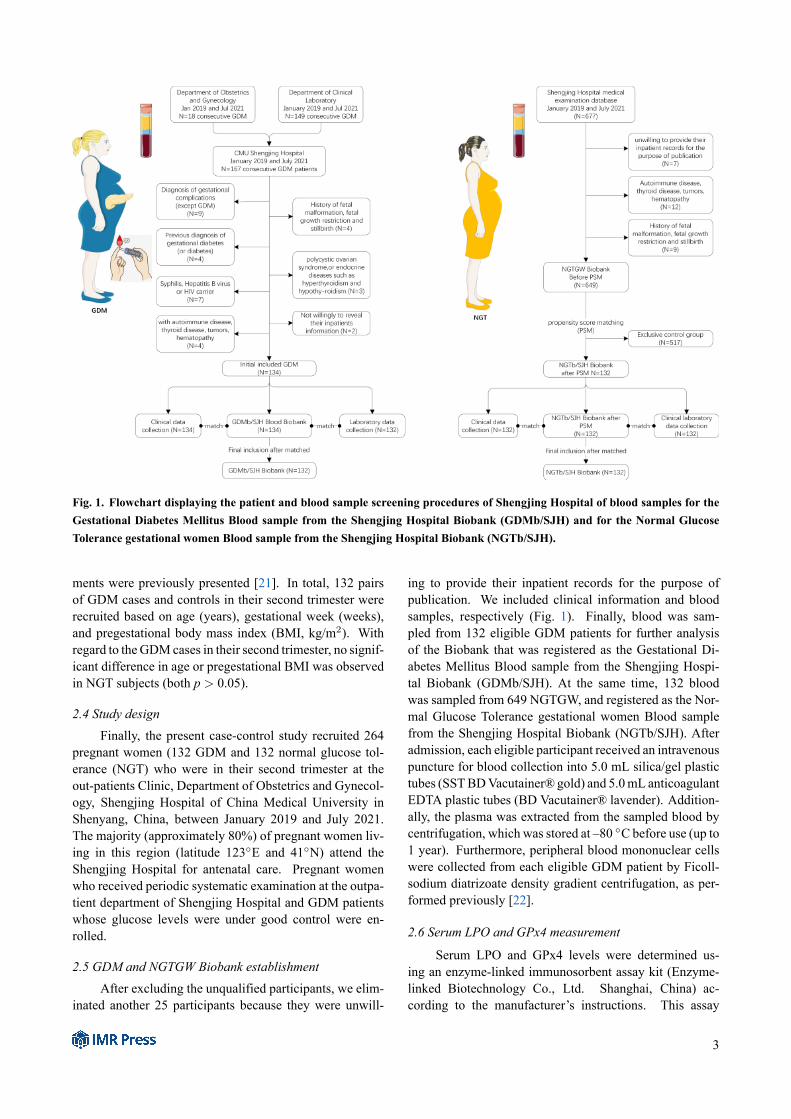
Fig. 1. Flowchart displaying the patient and blood sample screening procedures of Shengjing Hospital of blood samples for theGestational Diabetes Mellitus Blood sample from the Shengjing Hospital Biobank (GDMb/SJH) and for the Normal GlucoseTolerance gestational women Blood sample from the Shengjing Hospital Biobank (NGTb/SJH).
ments were previously presented [21]. In total, 132 pairsof GDM cases and controls in their second trimester wererecruited based on age (years), gestational week (weeks),and pregestational body mass index (BMI, kg/m2). Withregard to the GDMcases in their second trimester, no signif-icant difference in age or pregestational BMI was observedin NGT subjects (both p > 0.05).
2.4 Study designFinally, the present case-control study recruited 264
pregnant women (132 GDM and 132 normal glucose tol-erance (NGT) who were in their second trimester at theout-patients Clinic, Department of Obstetrics and Gynecol-ogy, Shengjing Hospital of China Medical University inShenyang, China, between January 2019 and July 2021.The majority (approximately 80%) of pregnant women liv-ing in this region (latitude 123E and 41N) attend theShengjing Hospital for antenatal care. Pregnant womenwho received periodic systematic examination at the outpa-tient department of Shengjing Hospital and GDM patientswhose glucose levels were under good control were en-rolled.
2.5 GDM and NGTGW Biobank establishmentAfter excluding the unqualified participants, we elim-
inated another 25 participants because they were unwill-
ing to provide their inpatient records for the purpose ofpublication. We included clinical information and bloodsamples, respectively (Fig. 1). Finally, blood was sam-pled from 132 eligible GDM patients for further analysisof the Biobank that was registered as the Gestational Di-abetes Mellitus Blood sample from the Shengjing Hospi-tal Biobank (GDMb/SJH). At the same time, 132 bloodwas sampled from 649 NGTGW, and registered as the Nor-mal Glucose Tolerance gestational women Blood samplefrom the Shengjing Hospital Biobank (NGTb/SJH). Afteradmission, each eligible participant received an intravenouspuncture for blood collection into 5.0 mL silica/gel plastictubes (SSTBDVacutainer® gold) and 5.0mL anticoagulantEDTA plastic tubes (BD Vacutainer® lavender). Addition-ally, the plasma was extracted from the sampled blood bycentrifugation, which was stored at –80 C before use (up to1 year). Furthermore, peripheral blood mononuclear cellswere collected from each eligible GDM patient by Ficoll-sodium diatrizoate density gradient centrifugation, as per-formed previously [22].
2.6 Serum LPO and GPx4 measurement
Serum LPO and GPx4 levels were determined us-ing an enzyme-linked immunosorbent assay kit (Enzyme-linked Biotechnology Co., Ltd. Shanghai, China) ac-cording to the manufacturer’s instructions. This assay
3
was highly sensitive to LPO (ml856631), and the rangedetectable concentration was 0.1–32.0 ng/mL for humanLPO. In addition, this assay was also highly sensitive forGPx4 (ml060706), and the detectable concentration rangewas 1.0–200.0 ng/mL for human GPx4. The coefficientsof variation (CV) in the inter-assay and intra-assay were<10% and 8%, respectively.
2.7 Clinical measurements and neonatal outcomes
All participates (132 GDMs and 132 NGTs) were intheir third trimester and planned to attend regular visits forprenatal examinations and delivery at the In-patients Clinic,Department of Obstetrics and Gynecology of ShengjingHospital. Their clinical measurements (In-patient Clinic)in the third trimester were collected, including blood rou-tine test, hepatic panel, lipid panel, blood coagulation func-tion, renal function, and serum electrolyte, and the selectivesolubilization approach are summarized in SupplementaryFile 1. Blood tests were completed by the Department ofLaboratory Medicine of Shengjing Hospital of China Med-ical University. After extracting the related clinical data,we constructed the related blood database, followed by im-plementation of laboratory tests. We also collected mater-nal clinical information, including delivery mode and ges-tational weeks at birth. In addition, we extracted neonataloutcomes from birth records, which included body length(cm) and birth weight (g).
2.8 Statistical analysis
We applied the PSM approach in this study to reducethe selection bias of case-controls and to reduce the pos-sible clinical confounding factors at the matching ratio of1:1. In addition, the model was evaluated by the Hosmer-Lemeshow degree of fitting for the C-statistic test and logis-tic regression. Prior to PSM, we compared continuous vari-ables in patient clinical features with controls by the Stu-dent’s t-test, and compared categorical variables using thechi-squared test. Following PSM, we inspected the variablenormality visually. The Mann-Whitney U-test was con-ducted for comparing continuous variables, whereas differ-ences among groups were compared by the Kruskal-WallisH test. The chi-squared test was conducted to compare cat-egorical variables. In addition, the associations of GPx4and LPO with clinical parameters were evaluated by mul-tiple linear regression and Spearman’s correlation analy-ses. This study adjusted confounders to conduct multipleregression analysis, including age, smoking status, bloodpressure (BP), and BMI before pregnancy. Related thresh-olds were determined by logistic models, receiver operat-ing characteristic (ROC) curves, and area under the ROCcurve. Subsequently, the lower and upper strata of theLPO/GPx4 concentrations were determined accordingly.This study compared the LPO/GPx4 concentrations and thepregnancy outcomes of both groups (e.g., lower strata ofLPO as the reference group and upper strata of LPO as
the case group). Thereafter, this study divided the subjectsbased on metabolic abnormality numbers (which includedblood lipids, PG, and BP) as the one metabolic abnormality,≥2 metabolic abnormalities, and normal metabolic groups.Finally, the odds ratios (ORs) together with 95% confidenceintervals (CIs) were estimated by multivariate logistic re-gression to predict the relationship of LPO with metabolicabnormalities among the gestational women. p < 0.05 wasconsidered to indicate statistical significance. SPSS version25.0 (IBM Inc., Chicago, IL, USA) and GraphPad Prismversion 8.0 (GraphPad Software Inc., La Jolla, CA, USA).
3. Results3.1 Comparison of baseline clinical characteristicsbetween the two groups
Table 1 displays the clinical features of the 649 NGTsubjects and 132 GDM cases. Among them, BMI, age, eth-nicity, smokers, and hypertension showed statistical signif-icance between the two groups prior to PSM to reduce theselection bias and possible clinical confounding factors. Wesubsequently compared the 132 pairs of GDM and controlsusing PSM by logistic regression. p = 0.524 and p = 0.781were obtained upon Hosmer-Lemeshow goodness of fit testand C-statistic test, respectively. Following PSM, we ex-cluded those heterogeneities in clinical features between thetwo groups. Clinical features of the GDM cases and NGTparticipants is elaborated in Table 1.
3.2 Comparison of LPO and GPX4 levels between theGDM and NGT groups
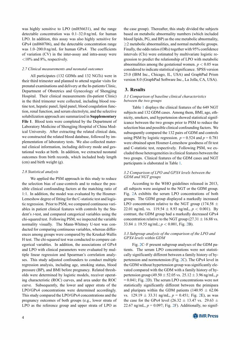
According to the WHO guidelines released in 2013,all subjects were assigned to the NGT or the GDM group.Fig. 2A exhibits the serum LPO concentrations of bothgroups. The GDM group displayed a markedly increasedLPO concentration relative to the NGT group (174.58 ±22.01 ng/mL vs. 119.54 ± 8.93 ng/mL, p < 0.001). Bycontrast, the GDM group had a markedly decreased GPx4concentration relative to the NGT group (27.31± 16.88 vs.33.84 ± 19.55 ng/mL; p < 0.001, Fig. 2B).
3.3 Subgroup analysis of the comparison of the LPO andGPX4 levels within GDM
Fig. 2C–F present subgroup analyses of the GDM pa-tients. The serum LPO concentrations were not statisti-cally significantly different between a family history of hy-pertension and normotension (Fig. 2C). The GPx4 level inthe GDMwithout hypertension group was significantly ele-vated compared with the GDM with a family history of hy-pertension group (49.50± 52.05 vs. 25.12± 3.96 ng/mL, p= 0.041; Fig. 2D). The serum LPO concentrations were notstatistically significantly different between the primiparaand pluripara within the GDM patients (140.95 ± 62.06vs. 129.19 ± 33.31 ng/mL, p = 0.451; Fig. 2E), as wasthe case for the GPx4 level (26.32 ± 13.47 vs. 29.63 ±22.67 ng/mL, p = 0.097; Fig. 2F). Additionally, no signif-
4
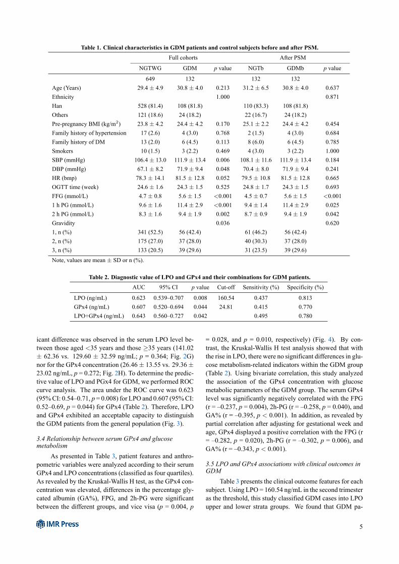
Table 1. Clinical characteristics in GDM patients and control subjects before and after PSM.Full cohorts After PSM
NGTWG GDM p value NGTb GDMb p value
649 132 132 132Age (Years) 29.4 ± 4.9 30.8 ± 4.0 0.213 31.2 ± 6.5 30.8 ± 4.0 0.637Ethnicity 1.000 0.871Han 528 (81.4) 108 (81.8) 110 (83.3) 108 (81.8)Others 121 (18.6) 24 (18.2) 22 (16.7) 24 (18.2)Pre-pregnancy BMI (kg/m2) 23.8 ± 4.2 24.4 ± 4.2 0.170 25.1 ± 2.2 24.4 ± 4.2 0.454Family history of hypertension 17 (2.6) 4 (3.0) 0.768 2 (1.5) 4 (3.0) 0.684Family history of DM 13 (2.0) 6 (4.5) 0.113 8 (6.0) 6 (4.5) 0.785Smokers 10 (1.5) 3 (2.2) 0.469 4 (3.0) 3 (2.2) 1.000SBP (mmHg) 106.4 ± 13.0 111.9 ± 13.4 0.006 108.1 ± 11.6 111.9 ± 13.4 0.184DBP (mmHg) 67.1 ± 8.2 71.9 ± 9.4 0.048 70.4 ± 8.0 71.9 ± 9.4 0.241HR (bmp) 78.3 ± 14.1 81.5 ± 12.8 0.052 79.5 ± 10.8 81.5 ± 12.8 0.665OGTT time (week) 24.6 ± 1.6 24.3 ± 1.5 0.525 24.8 ± 1.7 24.3 ± 1.5 0.693FFG (mmol/L) 4.7 ± 0.8 5.6 ± 1.5 <0.001 4.5 ± 0.7 5.6 ± 1.5 <0.0011 h PG (mmol/L) 9.6 ± 1.6 11.4 ± 2.9 <0.001 9.4 ± 1.4 11.4 ± 2.9 0.0252 h PG (mmol/L) 8.3 ± 1.6 9.4 ± 1.9 0.002 8.7 ± 0.9 9.4 ± 1.9 0.042Gravidity 0.036 0.6201, n (%) 341 (52.5) 56 (42.4) 61 (46.2) 56 (42.4)2, n (%) 175 (27.0) 37 (28.0) 40 (30.3) 37 (28.0)3, n (%) 133 (20.5) 39 (29.6) 31 (23.5) 39 (29.6)Note, values are mean ± SD or n (%).
Table 2. Diagnostic value of LPO and GPx4 and their combinations for GDM patients.AUC 95% CI p value Cut-off Sensitivity (%) Specificity (%)
LPO (ng/mL) 0.623 0.539–0.707 0.008 160.54 0.437 0.813GPx4 (ng/mL) 0.607 0.520–0.694 0.044 24.81 0.415 0.770LPO+GPx4 (ng/mL) 0.643 0.560–0.727 0.042 0.495 0.780
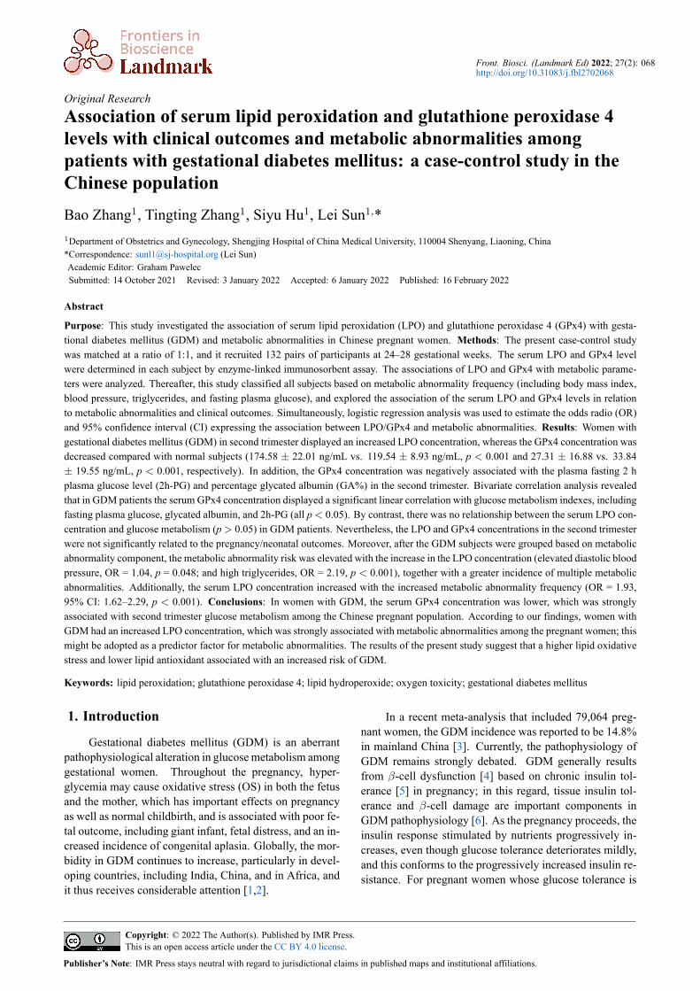
icant difference was observed in the serum LPO level be-tween those aged <35 years and those ≥35 years (141.02± 62.36 vs. 129.60 ± 32.59 ng/mL; p = 0.364; Fig. 2G)nor for the GPx4 concentration (26.46± 13.55 vs. 29.36±23.02 ng/mL, p = 0.272; Fig. 2H). To determine the predic-tive value of LPO and PGx4 for GDM, we performed ROCcurve analysis. The area under the ROC curve was 0.623(95%CI: 0.54–0.71, p = 0.008) for LPO and 0.607 (95%CI:0.52–0.69, p = 0.044) for GPx4 (Table 2). Therefore, LPOand GPx4 exhibited an acceptable capacity to distinguishthe GDM patients from the general population (Fig. 3).
3.4 Relationship between serum GPx4 and glucosemetabolism
As presented in Table 3, patient features and anthro-pometric variables were analyzed according to their serumGPx4 and LPO concentrations (classified as four quartiles).As revealed by the Kruskal-Wallis H test, as the GPx4 con-centration was elevated, differences in the percentage gly-cated albumin (GA%), FPG, and 2h-PG were significantbetween the different groups, and vice visa (p = 0.004, p
= 0.028, and p = 0.010, respectively) (Fig. 4). By con-trast, the Kruskal-Wallis H test analysis showed that withthe rise in LPO, there were no significant differences in glu-cose metabolism-related indicators within the GDM group(Table 2). Using bivariate correlation, this study analyzedthe association of the GPx4 concentration with glucosemetabolic parameters of the GDM group. The serum GPx4level was significantly negatively correlated with the FPG(r = –0.237, p = 0.004), 2h-PG (r = –0.258, p = 0.040), andGA% (r = –0.395, p < 0.001). In addition, as revealed bypartial correlation after adjusting for gestational week andage, GPx4 displayed a positive correlation with the FPG (r= –0.282, p = 0.020), 2h-PG (r = –0.302, p = 0.006), andGA% (r = –0.343, p < 0.001).
3.5 LPO and GPx4 associations with clinical outcomes inGDM
Table 3 presents the clinical outcome features for eachsubject. Using LPO = 160.54 ng/mL in the second trimesteras the threshold, this study classified GDM cases into LPOupper and lower strata groups. We found that GDM pa-
5
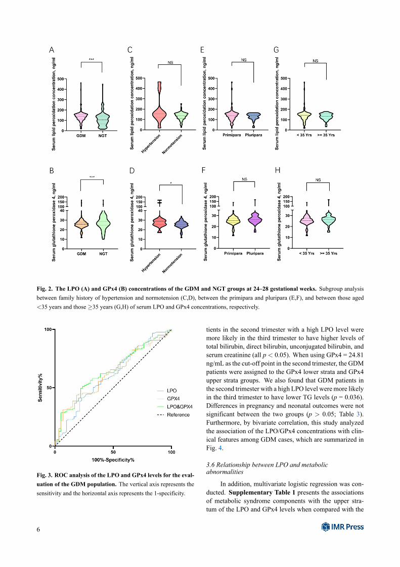
Fig. 2. The LPO (A) and GPx4 (B) concentrations of the GDM and NGT groups at 24–28 gestational weeks. Subgroup analysisbetween family history of hypertension and normotension (C,D), between the primipara and pluripara (E,F), and between those aged<35 years and those ≥35 years (G,H) of serum LPO and GPx4 concentrations, respectively.
Fig. 3. ROC analysis of the LPO and GPx4 levels for the eval-uation of the GDM population. The vertical axis represents thesensitivity and the horizontal axis represents the 1-specificity.
tients in the second trimester with a high LPO level weremore likely in the third trimester to have higher levels oftotal bilirubin, direct bilirubin, unconjugated bilirubin, andserum creatinine (all p < 0.05). When using GPx4 = 24.81ng/mL as the cut-off point in the second trimester, the GDMpatients were assigned to the GPx4 lower strata and GPx4upper strata groups. We also found that GDM patients inthe second trimester with a high LPO level were more likelyin the third trimester to have lower TG levels (p = 0.036).Differences in pregnancy and neonatal outcomes were notsignificant between the two groups (p > 0.05; Table 3).Furthermore, by bivariate correlation, this study analyzedthe association of the LPO/GPx4 concentrations with clin-ical features among GDM cases, which are summarized inFig. 4.
3.6 Relationship between LPO and metabolicabnormalities
In addition, multivariate logistic regression was con-ducted. Supplementary Table 1 presents the associationsof metabolic syndrome components with the upper stra-tum of the LPO and GPx4 levels when compared with the
6
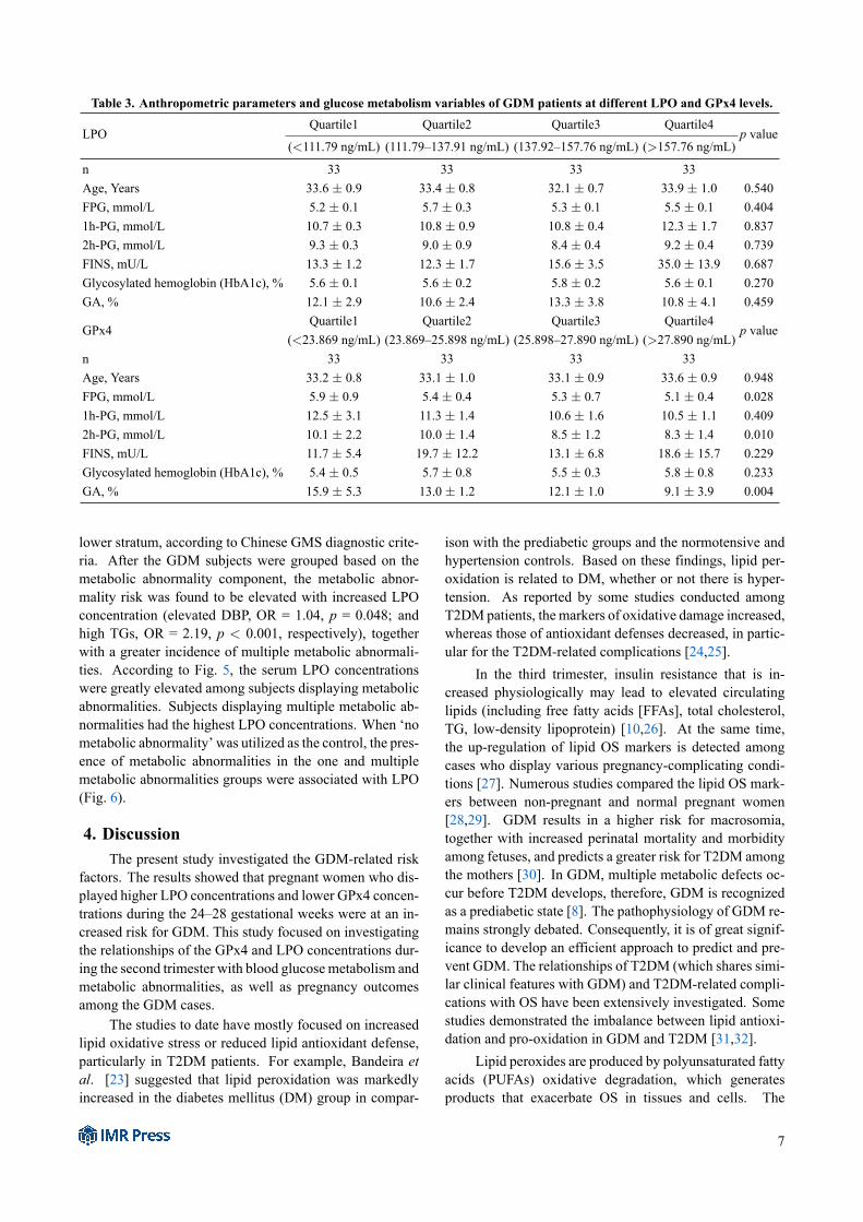
Table 3. Anthropometric parameters and glucose metabolism variables of GDM patients at different LPO and GPx4 levels.
LPOQuartile1 Quartile2 Quartile3 Quartile4
p value(<111.79 ng/mL) (111.79–137.91 ng/mL) (137.92–157.76 ng/mL) (>157.76 ng/mL)
n 33 33 33 33Age, Years 33.6 ± 0.9 33.4 ± 0.8 32.1 ± 0.7 33.9 ± 1.0 0.540FPG, mmol/L 5.2 ± 0.1 5.7 ± 0.3 5.3 ± 0.1 5.5 ± 0.1 0.4041h-PG, mmol/L 10.7 ± 0.3 10.8 ± 0.9 10.8 ± 0.4 12.3 ± 1.7 0.8372h-PG, mmol/L 9.3 ± 0.3 9.0 ± 0.9 8.4 ± 0.4 9.2 ± 0.4 0.739FINS, mU/L 13.3 ± 1.2 12.3 ± 1.7 15.6 ± 3.5 35.0 ± 13.9 0.687Glycosylated hemoglobin (HbA1c), % 5.6 ± 0.1 5.6 ± 0.2 5.8 ± 0.2 5.6 ± 0.1 0.270GA, % 12.1 ± 2.9 10.6 ± 2.4 13.3 ± 3.8 10.8 ± 4.1 0.459
GPx4Quartile1 Quartile2 Quartile3 Quartile4
p value(<23.869 ng/mL) (23.869–25.898 ng/mL) (25.898–27.890 ng/mL) (>27.890 ng/mL)
n 33 33 33 33Age, Years 33.2 ± 0.8 33.1 ± 1.0 33.1 ± 0.9 33.6 ± 0.9 0.948FPG, mmol/L 5.9 ± 0.9 5.4 ± 0.4 5.3 ± 0.7 5.1 ± 0.4 0.0281h-PG, mmol/L 12.5 ± 3.1 11.3 ± 1.4 10.6 ± 1.6 10.5 ± 1.1 0.4092h-PG, mmol/L 10.1 ± 2.2 10.0 ± 1.4 8.5 ± 1.2 8.3 ± 1.4 0.010FINS, mU/L 11.7 ± 5.4 19.7 ± 12.2 13.1 ± 6.8 18.6 ± 15.7 0.229Glycosylated hemoglobin (HbA1c), % 5.4 ± 0.5 5.7 ± 0.8 5.5 ± 0.3 5.8 ± 0.8 0.233GA, % 15.9 ± 5.3 13.0 ± 1.2 12.1 ± 1.0 9.1 ± 3.9 0.004
lower stratum, according to Chinese GMS diagnostic crite-ria. After the GDM subjects were grouped based on themetabolic abnormality component, the metabolic abnor-mality risk was found to be elevated with increased LPOconcentration (elevated DBP, OR = 1.04, p = 0.048; andhigh TGs, OR = 2.19, p < 0.001, respectively), togetherwith a greater incidence of multiple metabolic abnormali-ties. According to Fig. 5, the serum LPO concentrationswere greatly elevated among subjects displaying metabolicabnormalities. Subjects displaying multiple metabolic ab-normalities had the highest LPO concentrations. When ‘nometabolic abnormality’ was utilized as the control, the pres-ence of metabolic abnormalities in the one and multiplemetabolic abnormalities groups were associated with LPO(Fig. 6).
4. DiscussionThe present study investigated the GDM-related risk
factors. The results showed that pregnant women who dis-played higher LPO concentrations and lower GPx4 concen-trations during the 24–28 gestational weeks were at an in-creased risk for GDM. This study focused on investigatingthe relationships of the GPx4 and LPO concentrations dur-ing the second trimester with blood glucose metabolism andmetabolic abnormalities, as well as pregnancy outcomesamong the GDM cases.
The studies to date have mostly focused on increasedlipid oxidative stress or reduced lipid antioxidant defense,particularly in T2DM patients. For example, Bandeira etal. [23] suggested that lipid peroxidation was markedlyincreased in the diabetes mellitus (DM) group in compar-
ison with the prediabetic groups and the normotensive andhypertension controls. Based on these findings, lipid per-oxidation is related to DM, whether or not there is hyper-tension. As reported by some studies conducted amongT2DMpatients, the markers of oxidative damage increased,whereas those of antioxidant defenses decreased, in partic-ular for the T2DM-related complications [24,25].
In the third trimester, insulin resistance that is in-creased physiologically may lead to elevated circulatinglipids (including free fatty acids [FFAs], total cholesterol,TG, low-density lipoprotein) [10,26]. At the same time,the up-regulation of lipid OS markers is detected amongcases who display various pregnancy-complicating condi-tions [27]. Numerous studies compared the lipid OS mark-ers between non-pregnant and normal pregnant women[28,29]. GDM results in a higher risk for macrosomia,together with increased perinatal mortality and morbidityamong fetuses, and predicts a greater risk for T2DM amongthe mothers [30]. In GDM, multiple metabolic defects oc-cur before T2DM develops, therefore, GDM is recognizedas a prediabetic state [8]. The pathophysiology of GDM re-mains strongly debated. Consequently, it is of great signif-icance to develop an efficient approach to predict and pre-vent GDM. The relationships of T2DM (which shares simi-lar clinical features with GDM) and T2DM-related compli-cations with OS have been extensively investigated. Somestudies demonstrated the imbalance between lipid antioxi-dation and pro-oxidation in GDM and T2DM [31,32].
Lipid peroxides are produced by polyunsaturated fattyacids (PUFAs) oxidative degradation, which generatesproducts that exacerbate OS in tissues and cells. The
7
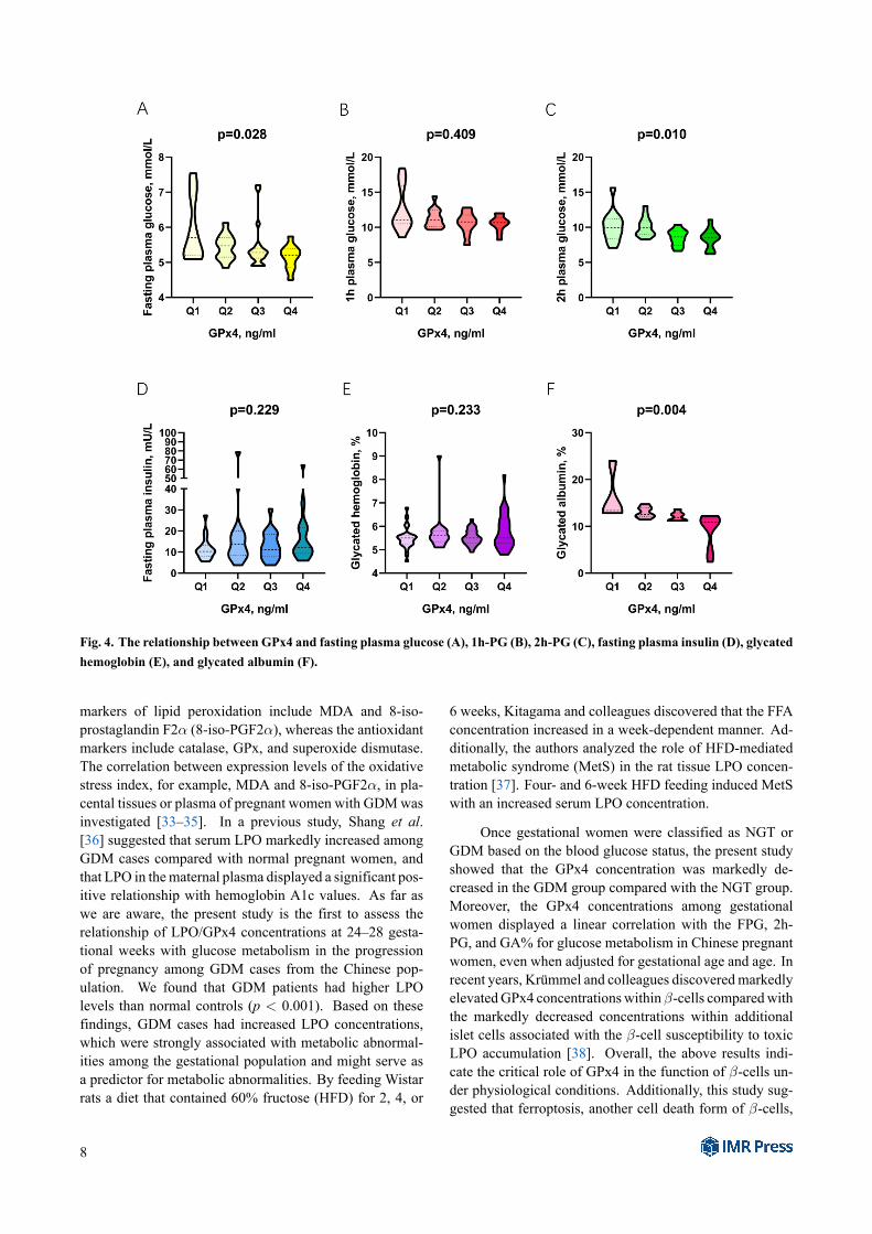
Fig. 4. The relationship between GPx4 and fasting plasma glucose (A), 1h-PG (B), 2h-PG (C), fasting plasma insulin (D), glycatedhemoglobin (E), and glycated albumin (F).
markers of lipid peroxidation include MDA and 8-iso-prostaglandin F2α (8-iso-PGF2α), whereas the antioxidantmarkers include catalase, GPx, and superoxide dismutase.The correlation between expression levels of the oxidativestress index, for example, MDA and 8-iso-PGF2α, in pla-cental tissues or plasma of pregnant women with GDMwasinvestigated [33–35]. In a previous study, Shang et al.[36] suggested that serum LPO markedly increased amongGDM cases compared with normal pregnant women, andthat LPO in thematernal plasma displayed a significant pos-itive relationship with hemoglobin A1c values. As far aswe are aware, the present study is the first to assess therelationship of LPO/GPx4 concentrations at 24–28 gesta-tional weeks with glucose metabolism in the progressionof pregnancy among GDM cases from the Chinese pop-ulation. We found that GDM patients had higher LPOlevels than normal controls (p < 0.001). Based on thesefindings, GDM cases had increased LPO concentrations,which were strongly associated with metabolic abnormal-ities among the gestational population and might serve asa predictor for metabolic abnormalities. By feeding Wistarrats a diet that contained 60% fructose (HFD) for 2, 4, or
6 weeks, Kitagama and colleagues discovered that the FFAconcentration increased in a week-dependent manner. Ad-ditionally, the authors analyzed the role of HFD-mediatedmetabolic syndrome (MetS) in the rat tissue LPO concen-tration [37]. Four- and 6-week HFD feeding induced MetSwith an increased serum LPO concentration.
Once gestational women were classified as NGT orGDM based on the blood glucose status, the present studyshowed that the GPx4 concentration was markedly de-creased in the GDM group compared with the NGT group.Moreover, the GPx4 concentrations among gestationalwomen displayed a linear correlation with the FPG, 2h-PG, and GA% for glucose metabolism in Chinese pregnantwomen, even when adjusted for gestational age and age. Inrecent years, Krümmel and colleagues discoveredmarkedlyelevatedGPx4 concentrationswithinβ-cells comparedwiththe markedly decreased concentrations within additionalislet cells associated with the β-cell susceptibility to toxicLPO accumulation [38]. Overall, the above results indi-cate the critical role of GPx4 in the function of β-cells un-der physiological conditions. Additionally, this study sug-gested that ferroptosis, another cell death form of β-cells,
8

Fig. 5. The correlations between the LPO and GPx4 levels and the clinical features of the blood routine (A), serum lipid profile(B), blood coagulation function (C), liver function (D), thyroid function (E), and serum electrolyte (F).
Fig. 6. The serum LPO level (A) and GPx4 level (B) in the no metabolic abnormality, one metabolic abnormality, and two ormore metabolic abnormalities groups.
did not involve the occurrence of a pro-inflammatory cy-tokine attack [38]. To date, no study has suggested that fer-roptosis is involved in the T1DM pathogenic mechanism asthe oxidative non-apoptotic cell death form. In addition, itremains unclear about the role of GPx4 as the main ferrop-tosis regulator and antioxidative enzyme.
The present study had some limitations. Firstly, therewas a small sample size, which had some impact on therelationship of serum LPO and GPx4 concentrations withGDM occurrence. Secondly, this study determined LPOand GPx4 contents only in the second trimester. We didnot perform dynamic monitoring of the of LPO or GPx4concentrations or determine their clinical values in the third
9

trimester. Thirdly, when collecting data, the time the LPOconcentration was determined were inconsistent with thatfor the biochemical examination in some subjects (differ-ences of up to 3weeks), thus, potentially resulting in numer-ical differences and compromising the model interpretabil-ity.
5. ConclusionsIn summary, the present study suggested that GPx4
played a vital role in glucose metabolism and that LPOwas a possible risk factor associated with the metabolic ab-normalities in the Chinese pregnant women. An increasedserum LPO concentration might predict the incidence anddegree of metabolic abnormalities among the Chinese ges-tational diabetes mellitus population in the future. To date,limited research has been conducted on the associations ofserum GPx4 and LPO concentrations with GDM, such thatmore evidence-based studies are warranted. The small sam-ple size and restricted geographical distribution were poten-tial limitations in the present study. Consequently, a multi-area and multi-center prospective study with a large sam-ple size should be conducted to illustrate the associationsof serum LPO/GPx4 with GDM.
AbbreviationsBMI, body mass index; SBP, systolic blood pressure;
DBP, diastolic blood pressure; TP, total protein; ALT, ala-nine aminotransferase; AST, aspartate aminotransferase;ALP, alkaline phosphatase; ALB, albumin; GGT, gamma-glutamyl transpeptidas; PA1, pre-albumin; BilT, totalbilirubin; CB, conjugated bilirubin; UCB, un-conjugatedbilirubin; TBA, total bile acid; FPG, fasting plasma glu-cose; TG, triglyceride TC, total cholesterol; HDL-C,high-density lipoprotein-cholesterol; LDL-C, low-densitylipoprotein-cholesterol; ApoA1, apolipoprotein A1; AopB,apolipoprotein B. WBC, white blood cells; RBC, red bloodcells; HGB, hemoglobin; PLT, platelet; BA, basophil;EO, eosinophil; LY, lymphocyte; MO, monocyte; NE,neutrophil; MCH, mean corpuscular hemoglobin; MCHC,mean corpuscular hemoglobin concentration; MCV, meancorpuscular Volume; MPV, mean platelet volume; HCT,hematocrit; PCT, platelet hematocrit; PDW, platelet dis-tribution width; RDW, red blood cell volume distributionwidth; APTT, activated partial thromboplastin time; PT,prothrombin time; PTA, prothrombin activity; INR, inter-national normalized ratio; CRP, C-reactive protein; PSM,propensity score matching.
Author contributionsBZ and LS was responsible for the study design, col-
lection of blood sample, experiment implementation, studyscreening, data analysis and interpretation, statistics anal-ysis, and drafting. LS, BZ, and TZ were in charge of re-search and drafting. SH, BZ, and LS were responsible for
data collection and analysis. All authorsmade contributionsto study design and interpretation, and further drafting. LSwas the guarantor.
Ethics approval and consent to participateThe Medical Ethics Committee of Shengjing Hospi-
tal of China Medical University approved our study pro-tocol (Ethics Approval No. 2017PS066K). This study wasperformed following the Declaration of Helsinki (revised inOctober 2013, Fortaleza, Brazil). Informed written consentwas obtained from each participant before participating inthis study.
AcknowledgmentWe thank International Science Editing
(http://www.internationalscienceediting.com) for edit-ing this manuscript.
FundingThis research received no external funding.
Conflict of interestThe authors declare no conflict of interest.
Supplementary materialSupplementary material associated with this article
can be found, in the online version, at https://www.imrpress.com/journal/FBL/27/2/10.31083/j.fbl2702068.
References[1] Macaulay S, Dunger DB, Norris SA. Gestational diabetes melli-
tus in Africa: a systematic review. PLoS ONE. 2014; 9: e97871.[2] Li KT, Naik S, Alexander M, Mathad JS. Screening and diag-
nosis of gestational diabetes in India: a systematic review andmeta-analysis. Acta Diabetologica. 2018; 55: 613–625.
[3] Gao C, Sun X, Lu L, Liu F, Yuan J. Prevalence of gestationaldiabetes mellitus in mainland China: a systematic review andmeta-analysis. Journal of Diabetes Investigation. 2019; 10: 154–162.
[4] Prentki M, Nolan CJ. Islet beta cell failure in type 2 diabetes.Journal of Clinical Investigation 2006; 116: 1802–1812.
[5] Catalano PM. Trying to understand gestational diabetes. Dia-betic Medicine. 2014; 31: 273–281.
[6] Retnakaran R, Hanley AJG, Raif N, Connelly PW, Sermer M,Zinman B. Reduced Adiponectin Concentration in Women withGestational Diabetes. Diabetes Care. 2004; 27: 799–800.
[7] Catalano PM, Huston L, Amini SB, Kalhan SC. Longitudi-nal changes in glucose metabolism during pregnancy in obesewomen with normal glucose tolerance and gestational diabetesmellitus. American Journal of Obstetrics and Gynecology. 1999;180: 903–916.
[8] Clark CM, Qiu C, Amerman B, Porter B, Fineberg N, AldasouqiS, et al. Gestational Diabetes: should it be Added to the Syn-drome of Insulin Resistance? Diabetes Care. 1997; 20: 867–871.
[9] Fan J, Zhang T, Yu Y, Zhang B. Is serum zinc status relatedto gestational diabetes mellitus? A meta‐analysis. Maternal &Child Nutrition. 2021; 17: e13239.
10
[10] Patrick TE, Hubel CA, Roberts JM. Evidence of increased ox-idative stress, unexplained by lipid changes, is present in nulli-parous black women from early gestation. Hypertension in Preg-nancy. 2004; 23: 91–100.
[11] Sekeroğlu MR, Sahin H, Dülger H, Algün E. The effect of di-etary treatment on erythrocyte lipid peroxidation, superoxidedismutase, glutathione peroxidase, and serum lipid peroxidationin patients with type 2 diabetes mellitus. Clinical Biochemistry.2000; 33: 669–674.
[12] Karacay O, Sepici-Dincel A, Karcaaltincaba D, Sahin D, YalvaçS, Akyol M, et al. A quantitative evaluation of total antioxidantstatus and oxidative stress markers in preeclampsia and gesta-tional diabetic patients in 24–36 weeks of gestation. DiabetesResearch and Clinical Practice. 2010; 89: 231–238.
[13] Valko M, Jomova K, Rhodes CJ, Kuča K, Musílek K. Redox-and non-redox-metal-induced formation of free radicals andtheir role in human disease. Archives of Toxicology. 2016; 90:1–37.
[14] Valko M, Morris H, Cronin M. Metals, Toxicity and OxidativeStress. Current Medicinal Chemistry. 2005; 12: 1161–1208.
[15] Ursini F, Maiorino M. Lipid peroxidation and ferroptosis: therole of GSH and GPx4. Free Radical Biology and Medicine.2020; 152: 175–185.
[16] López-Tinoco C, RocaM, García-Valero A,Murri M, TinahonesFJ, Segundo C, et al. Oxidative stress and antioxidant status inpatients with late-onset gestational diabetes mellitus. Acta Dia-betologica. 2013; 50: 201–208.
[17] Alberti KG, Zimmet PZ. Definition, diagnosis and classificationof diabetes mellitus and its complications. Part 1: diagnosis andclassification of diabetes mellitus provisional report of a whoconsultation. Diabetic Medicine. 1998; 15: 539–553.
[18] Metzger BE, Gabbe SG, Persson B, Buchanan TA, Catalano PA,Damm P, et al. International association of diabetes and preg-nancy study groups recommendations on the diagnosis and clas-sification of hyperglycemia in pregnancy. Diabetes Care. 2010;33: 676–682.
[19] Niu J, Lei Q, Lü L, Wen J, Lin X, Duan D, et al. Evaluationof the diagnostic criteria of gestational metabolic syndrome andanalysis of the risk factors. Zhonghua Fu Chan Ke Za Zhi. 2013;48: 92–97. (In Chinese)
[20] Bertolini G, D’Amico R, Nardi D, Tinazzi A, Apolone G. Onemodel, several results: the paradox of the Hosmer-Lemeshowgoodness-of-fit test for the logistic regression model. Journal ofEpidemiology and Biostatistics. 2000; 5: 251–253.
[21] Feng C, Jin Z, Chi X, Zhang B, Wang X, Sun L, et al. SHBGexpression is correlated with PI3K/AKT pathway activity in acellular model of human insulin resistance. Gynecological En-docrinology. 2018; 34: 567–573.
[22] Zhang B, Jin Z, Sun L, Zheng Y, Jiang J, Feng C, et al. Ex-pression and correlation of sex hormone-binding globulin andinsulin signal transduction and glucose transporter proteins ingestational diabetes mellitus placental tissue. Diabetes Researchand Clinical Practice. 2016; 119: 106–117.
[23] Bandeira SDM, Guedes GDS, da Fonseca LJS, Pires AS, GelainDP, Moreira JCF, et al. Characterization of blood oxidativestress in type 2 diabetes mellitus patients: increase in lipid per-oxidation and SOD activity. Oxidative Medicine and CellularLongevity. 2012; 2012: 819310.
[24] Ziegler D, Sohr CGH, Nourooz-Zadeh J. Oxidative stressand antioxidant defense in relation to the severity of diabeticpolyneuropathy and cardiovascular autonomic neuropathy. Dia-betes Care. 2004; 27: 2178–2183.
[25] Piwowar A, Knapik-Kordecka M, Warwas M. AOPP and its re-lations with selected markers of oxidative/antioxidative systemin type 2 diabetes mellitus. Diabetes Research and Clinical Prac-tice. 2007; 77: 188–192.
[26] Toescu V, Nuttall SL,Martin U, Kendall MJ, Dunne F. Oxidativestress and normal pregnancy. Clinical Endocrinology. 2002; 57:609–613.
[27] McKinney ET, Shouri R, Hunt RS, Ahokas RA, Sibai BM.Plasma, urinary, and salivary 8-epi-prostaglandin f2alpha levelsin normotensive and preeclamptic pregnancies. American Jour-nal of Obstetrics and Gynecology. 2000; 183: 874–877.
[28] Arikan S, Konukoğlu D, Arikan C, Akçay T, Davas I. Lipid per-oxidation and antioxidant status in maternal and cord blood. Gy-necologic and Obstetric Investigation. 2001; 51: 145–149.
[29] Orhan H, Onderoglu L, Yücel A, Sahin G. Circulating biomark-ers of oxidative stress in complicated pregnancies. Archives ofGynecology and Obstetrics. 2003; 267: 189–195.
[30] Xiang AH, Peters RK, Trigo E, Kjos SL, Lee WP, BuchananTA. Multiple metabolic defects during late pregnancy in womenat high risk for type 2 diabetes. Diabetes. 1999; 48: 848–854.
[31] Chen X, Scholl TO. Oxidative stress: changes in pregnancyandwith gestational diabetesmellitus. Current Diabetes Reports.2005; 5: 282–288.
[32] Lappas M, Mittion A, Permezel M. In response to oxidativestress, the expression of inflammatory cytokines and antioxi-dant enzymes are impaired in placenta, but not adipose tissue,of women with gestational diabetes. Journal of Endocrinology.2010; 204: 75–84.
[33] Li H, Dong A, Lv X. Advanced glycation end products andadipocytokines and oxidative stress in placental tissues of preg-nant women with gestational diabetes mellitus. Experimentaland Therapeutic Medicine. 2019; 18: 685–691.
[34] Shang M, Dong X, Hou L. Correlation of adipokines and mark-ers of oxidative stress in women with gestational diabetes melli-tus and their newborns. Journal of Obstetrics and GynaecologyResearch. 2018; 44: 637–646.
[35] Lappas M, Permezel M, Rice GE. Release of proinflamma-tory cytokines and 8-isoprostane from placenta, adipose tissue,and skeletal muscle from normal pregnant women and womenwith gestational diabetes mellitus. The Journal of Clinical En-docrinology and Metabolism. 2004; 89: 5627–5633.
[36] Shang M, Zhao J, Yang L, Lin L. Oxidative stress and antiox-idant status in women with gestational diabetes mellitus diag-nosed by IADPSG criteria. Diabetes Research and Clinical Prac-tice. 2015; 109: 404–410.
[37] Kitagawa A, Ohta Y, Ohashi K, Yashiro K, Fukuzawa K. Effectof High Fructose-Induced Metabolic Syndrome on Tissue Vita-min E and Lipid Peroxide Levels in Rats. Journal of NutritionalScience and Vitaminology. 2020; 66: 200–206.
[38] Krümmel B, Plötz T, Jörns A, Lenzen S, Mehmeti I. The centralrole of glutathione peroxidase 4 in the regulation of ferropto-sis and its implications for pro-inflammatory cytokine-mediatedbeta-cell death. Biochimica et Biophysica Acta (BBA) - Molec-ular Basis of Disease. 2021; 1867: 166114.
11