Assessment of Serum Copper Level among Sudanese ...
12
Page 73 Research Article Assessment of Serum Copper Level among Sudanese Patients with vitiligo Mehad Muawia 1 , Suad H H 2 , Gad Allah Modawe 3* 1. Department of Clinical chemistry, Faculty of Medical Laboratory Sciences, Al Neelain University, Omdurman, Sudan 2. Department of Dermatology, Faculty of Medicine and Health Sciences, Omdurman Islamic University, Omdurman, Sudan 3. Department of Biochemistry, Faculty of Medicine and Health Sciences, Omdurman Islamic University, Omdurman, Sudan Abstract Background: Vitiligo is a common skin disease of unknown etiology characterized clinically by depigmented patches, which can be localized or generalized; it usually runs a chronic course with an un predictable outcome and failure of complete cure in many affected individuals. Many communities consider it a contagious disease which leads to a great psychological and social stigma for patients; previous studies showed that copper might be associated with the pathogenesis of vitiligo. The aim of this study was to assess copper level in Sudanese vitiligo patients. Methods: This is a case-control study conducted in dermatology clinics in Khartoum state during the period from November 2018 to February 2019. Blood samples were obtained from 100 participants, 50 from vitiligo patients and 50 from non-vitiligo subjects representing a control group. Serum copper was measured by mind-ray (automation). Results: A highly significant increase (p = 0.000) in the copper level was seen in vitiligo patients compared with the control group. Of the total number of patients, 17 (34%) were females and 33 (66%) were males. According to the duration of the disease, the copper level was significantly increased in patient group with a disease duration of > one year compared to the patient group six months – one year and patient group < 6 months; we found no significance of the family history, 18% of the case group had a family history while 42 (82%) had no family history. Discussion: The relationship between the serum level of copper and vitiligo has been assessed by many studies. Copper is one of the trace elements that was found to be important for tyrosinase enzyme that catalyzes the first steps in melanin synthesis in the skin. Some studies showed that the disease was associated with low serum levels of copper and since vitiligo is a disease that is characterized clinically by white areas of skin with no melanin, these studies seem to be logical. However, in this study, the serum level of copper was found to be high in vitiligo patients which might be justified by the release of copper from the destroyed melanocytes. Another justification might be a defect in the carrier protein for copper. Conclusion: The study found that the serum copper was significantly increased in vitiligo patients compared to the control and it is recommended that serum copper level and copper profile should be assessed routinely in vitiligo patients. Sudan Journal of Medical Sciences Volume 15, Issue no. 1, DOI 10.18502/sjms.v15i1.6707 Production and Hosting by Knowledge E How to cite this article: Gad Allah Modawe (2019) “Assessment of Serum Copper Level among Sudanese Patients with vitiligo”, Sudan Journal of Medical Sciences, vol. 15, issue no. 1, pages 73–84. DOI 10.18502/sjms.v15i1.6707 Corresponding Author: [email protected] Received 02 January 2020 Accepted 17 March 2020 Published 31 March 2020 Production and Hosting by Knowledge E cc Gad Allah Modawe. This article is distributed under the terms of the Creative Commons Attribution License, which permits unrestricted use and redistribution provided that the original author and source are credited. Editor-in-Chief: Prof. Mohammad A. M. Ibnouf
-
Upload
khangminh22 -
Category
Documents
-
view
1 -
download
0
Transcript of Assessment of Serum Copper Level among Sudanese ...

Page 73
Research Article
Assessment of Serum Copper Level among Sudanese Patients with vitiligoMehad Muawia1, Suad H H2, Gad Allah Modawe3*
1. Department of Clinical chemistry, Faculty of Medical Laboratory Sciences, Al Neelain University, Omdurman, Sudan
2. Department of Dermatology, Faculty of Medicine and Health Sciences, Omdurman Islamic University, Omdurman, Sudan
3. Department of Biochemistry, Faculty of Medicine and Health Sciences, Omdurman Islamic University, Omdurman, Sudan
Abstract
Background: Vitiligo is a common skin disease of unknown etiology characterized clinically by depigmented patches, which can be localized or generalized; it usually runs a chronic course with an un predictable outcome and failure of complete cure in many affected individuals. Many communities consider it a contagious disease which leads to a great psychological and social stigma for patients; previous studies showed that copper might be associated with the pathogenesis of vitiligo. The aim of this study was to assess copper level in Sudanese vitiligo patients.Methods: This is a case-control study conducted in dermatology clinics in Khartoum state during the period from November 2018 to February 2019. Blood samples were obtained from 100 participants, 50 from vitiligo patients and 50 from non-vitiligo subjects representing a control group. Serum copper was measured by mind-ray (automation). Results: A highly significant increase (p = 0.000) in the copper level was seen in vitiligo patients compared with the control group. Of the total number of patients, 17 (34%) were females and 33 (66%) were males. According to the duration of the disease, the copper level was significantly increased in patient group with a disease duration of > one year compared to the patient group six months – one year and patient group < 6 months; we found no significance of the family history, 18% of the case group had a family history while 42 (82%) had no family history. Discussion: The relationship between the serum level of copper and vitiligo has been assessed by many studies. Copper is one of the trace elements that was found to be important for tyrosinase enzyme that catalyzes the first steps in melanin synthesis in the skin. Some studies showed that the disease was associated with low serum levels of copper and since vitiligo is a disease that is characterized clinically by white areas of skin with no melanin, these studies seem to be logical. However, in this study, the serum level of copper was found to be high in vitiligo patients which might be justified by the release of copper from the destroyed melanocytes. Another justification might be a defect in the carrier protein for copper.Conclusion: The study found that the serum copper was significantly increased in vitiligo patients compared to the control and it is recommended that serum copper level and copper profile should be assessed routinely in vitiligo patients.
Sudan Journal of Medical Sciences Volume 15, Issue no. 1, DOI 10.18502/sjms.v15i1.6707Production and Hosting by Knowledge E
How to cite this article: Gad Allah Modawe (2019) “Assessment of Serum Copper Level among Sudanese Patients with vitiligo”, Sudan Journal of Medical Sciences, vol. 15, issue no. 1, pages 73–84. DOI 10.18502/sjms.v15i1.6707
Sudan Journal of Medical SciencesVolume 14, Issue no. 4, DOI 10.18502/sjms.v14i4.5899Production and Hosting by Knowledge E
Research Article
Overview of the Course of UndergraduateMedical Education in the SudanTahra Al Sadig Al Mahdi
Medical Education, School of Medicine, Ahfad University for Women, Omdurman, Khartoum,Sudan
AbstractBackground: Sudan’s experience with Medical Education (ME) is one of the oldestregionally. It started with one school and has currently reached 66. This number isamong the highest and Sudan is one of the largest physicians-exporting countries.Thus, Sudanese ME has great regional influence.Objective: To review the history of Sudanese ME and determine factors contributingto its transformation.Methods: Internet and desk search was conducted, relevant articles and websiteswere accessed, hard documents were reviewed, and eminent Sudanese figures in thefield were consulted.Results: Sudanese ME is meagerly documented. The path of ME was described in fourphases including some of the significant local and global factors.Phase one (1924–1970) started by establishing the first medical school andcharacterized by steady growth and stability. Influences were the Flexner’s era andthe Sudanese independence atmosphere. During phase two (1978–1990), provincialpublic schools were opened in addition to the first private school. Influences were theSudan’s commitment to Al Ma Ata recommendations and the revolutionary changesfollowing constructivist views on learning. Phase three (1990–2005) was formed bythe Revolution in Higher Education leading to mushrooming of public and privateschools across the country and influenced by local sociopolitical turbulence. In phasefour (2006–2018), authorities launched formal ME regulatory efforts. It is still beingtransformed by contradicting local factors and strong international directions.Conclusion: Sudanese experience with ME is noteworthy; it offers important lessonsand gives the needed wisdom for dealing with ME challenges in Sudan and beyond.
1. Introduction
Sudan is the third largest country in Africa with a total population of around 40 millionpeople [1]. It borders seven countries and its capital is Khartoum. Sudan is a miniaturerepresentation of the diversity found in most African countries [2, 3].
The country is composed of 18 states; approximately 66% of the population lives inrural areas [4], and the percentage of poverty is around 46.5% [5]. The country suffersfrom a marked shortage in health workforce worsened by poor distribution over the
How to cite this article: Tahra Al Sadig Al Mahdi (2019) “Overview of the Course of Undergraduate Medical Education in the Sudan,” Sudan Journalof Medical Sciences, vol. 14, issue no. 4, pages 188–201. DOI 10.18502/sjms.v14i4.5899 Page 188
Corresponding Author:
Tahra Al Sadig Al Mahdi
Received 23 August 2019
Accepted 14 December 2019
Published 30 December 2019
Production and Hosting by
Knowledge E
Tahra Al Sadig Al
Mahdi. This article is
distributed under the terms of
the Creative Commons
Attribution License, which
permits unrestricted use and
redistribution provided that
the original author and
source are credited.
Editor-in-Chief:
Prof. Mohammad A. M. Ibnouf
Corresponding Author:
Received 02 January 2020
Accepted 17 March 2020
Published 31 March 2020
Production and Hosting by
Knowledge E
cc Gad Allah Modawe.
This article is distributed
under the terms of the
Creative Commons
Attribution License, which
permits unrestricted use and
redistribution provided that
the original author and source
are credited.
Editor-in-Chief:
Prof. Mohammad A. M. Ibnouf

Sudan Journal of Medical Sciences Mehad Muawia et al. et al.
DOI 10.18502/sjms.v15i1.6707 Page 74
Key words: vitiligo; depigmentation; copper, Sudanese
Introduction
Vitiligo is a chronic idiopathic skin disease, characterized by sharply marginated
depigmented patches [1]. Lesions often start on sun-exposed areas [2]. Both genetic and
environmental factors are believed to play a role in the pathogenesis of the disease [1, 2].
The distribution of the disease can be unilateral segmental or non-segmental [1]. Vitiligo
is estimated to account for 1% of the general population globally [3], however, in some
populations, the incidence has reached 2–3% [4]. Patients who are stigmatized for
the disease can experience depression and other psychological mood disorders [5].
The risk factors for developing vitiligo include family history and other autoimmune
diseases [2]. The disease is usually diagnosed clinically. The diagnosis is confirmed
by tissue biopsy and histopathological study [2]. Vitiligo should be differentiated from
other skin conditions in which there is decrease in or loss of pigment, for example, tinea
versicolor, piebaldism, idiopathic guttate hypomelanosis, etc. [6]. There is no cure for
vitiligo [1], but it can be treated with the many available options, including topical steroids,
sunscreens, phototherapy, etc. [1, 2]. Melanin is the pigment majorly responsible for skin
color; it is synthesized from amino acid tyrosine in the melanosomes that are found
within epidermal melanocytes. Tyrosinase enzyme catalyzes the first two reactions in
the pathway of melanogenesis [7]. It is found in animal and plant tissues responsible for
the production of melanin as well as other pigments in these tissues [8]. Melanin is an
effective absorbent of ultraviolet rays (UV) [10] and prevents the skin from its harmful
effects. Some studies have shown that there is a reduced incidence of some types
of skin cancers in individuals with dark skin compared to others, but the relationship
between photo protection and the incidence of skin cancer is not clearly understood [11].
Tyrosinase is a copper-containing enzyme encoded by the TYR gene in humans [9].
Copper is a trace element needed in small amount in the diet, the proximal small
intestine is recognized as the main site of dietary copper absorption in mammals. The
transport of copper from the intestinal lumen into the intestinal mucosa is a carrier-
mediated process involving a saturable transport component. The overall intestinal copper
uptake is influenced by amino acids, ascorbic acid, and other dietary factors [12]. Once in
mucosal cells, approximately 80% of the newly absorbed copper is in the cytosol, mainly
bound to metallothionein (MTs). These are low-molecular weight-inducible proteins with
many functions including homeostasis, storage, transport, and detoxification of metals.
The MTs bind to many metals, but in normal circumstances only Zn, Cu, and Cd binding is

Sudan Journal of Medical Sciences Mehad Muawia et al. et al.
DOI 10.18502/sjms.v15i1.6707 Page 75
significant [13]. After passing through the enterocytes, copper enters the portal circulation
where it is bound to carrier proteins (primarily albumin), peptides, and amino acids and is
transported to the liver, with lesser amounts entering the kidneys.
Recently, many studies have been conducted to show that copper plays an important
role in pigmentation, it accelerates the oxidation of dopa by skin extracts containing
dopa-oxidase. Furthermore, the action of copper in pigmentation was not clear until
Gorter [14], in 1935, conclusively demonstrated the fact that copper-free diets resulted
in depigmentation of the hair of rats, rabbits, and cats and that this depigmentation
disappeared following the administration of copper. On the other hand, no effect on
melanogenesis was obtained if other minerals or vitamins were added to the diet [14].
In an old study submitted in Philadelphia 1931, Cunningham [15] made an observation
relevant to this problem; he noted that the skin of black-coated animals contained
more copper than the white-coated ones and that copper was concentrated mainly
in the epidermis and “in vitro” experiments demonstrated the fact that copper
accelerated the oxidation of dopa by skin extracts containing dopa-oxidase [15]. In the
same year, Sarata [15] made the first definite attempt to correlate the copper content
of skin with its degree of pigmentation. A study carried out in mottled dogs and cats
showed that the copper content of pigmented hair was much higher than that of the non-
pigmented hair of the same animal. Moreover, generally speaking, copper was found to
be present in greater amounts in a skin covered by dark hair than the skin underlying
the colorless hairs [15].
In 1934, Schroeder, Gruenberg, and Schade, quoted by Cornbleet [16], demonstrated
that vitamin “C” inhibits the dopa reaction. Cornbleet [16] found that pigmentary
precipitates can occur in a solution of dopa under the action of UV light alone, Buthe was
able to accelerate this reaction considerably by adding copper to the solution. On the
other hand, the addition of vitamin C tended to slow down the reaction or, in other words,
to neutralize the catalytic effect of copper. Cornbleet concluded that the presence of
these two substances, having antagonistic actions, so far as the oxidation rate of dopa is
concerned, makes pigment formation susceptible to ready physiologic control [16].
Also, a study conducted at the Faculty of Medicine, Fayoum University showed that
serum Zn levels were lower in different studied groups but it was much lower in the
vitiligo group and that the serum Cu levels in the vitiligo group were insignificantly
higher compared to the control group. Hence, serum Zn and Cu may have an effect
on the vitiligo disease as Zn in combination with other micronutrients such as Cu,
cobalt, nickel, iron, manganese, and Ca++ plays an important role in the process of
melanogenesis [17].

Sudan Journal of Medical Sciences Mehad Muawia et al. et al.
DOI 10.18502/sjms.v15i1.6707 Page 76
In another study conducted at the Central South University, Changsha, China in 2014,
it was noted that that out of the 16 studies that detected serum Cu level, 6 reported no
statistically significant Cu level change in both the groups while 10 presented a significant
decrease in the Cu levels in the vitiligo group. Thereafter, a random effect model was
used for meta-analysis. In the pooled analysis, there was a significant decrease in the
Cu levels in the vitiligo group [16].
Copper is essential for human heath, it is crucial for growth and brain development
and helps fight dangerous infections, according to some reports in the literature review,
lower copper increases the susceptibility to vitiligo, vitiligo is one of the most common
skin diseases, it can persist for years and result in disfigurement and permanent
scarring, it can cause serious adverse effects on psychological development resulting
in emotional problems like withdrawing from society and depression. However, most of
the researches were directed to determine the pathological effect due to changes in
the level of copper, since there is a strong relationship between serum copper level and
vitiligo, hence this study was designed to highlight the relation between serum copper
level and Vitiligo in Sudanese patients. The aim of this study was to assess the serum
copper level in patients with vitiligo.
Materials and Methods
This study is a case-control hospital-based study carried out at Sudanese dermatology
clinics and the Khartoum Teaching Hospital for Dermatology, Khartoum city, Khartoum
state. The study was conducted during November 2018 to January 2019. The study
population was 100, with 50 vitiligo patients and 50 non-vertigo patients representing
the control group.
The study populations were matched in age and sex, the age ranged between 16 and
60 years, the number of males studied was 33 (66%) and females 17 (34%).
Sudanese patients with any type of vitiligo were included in the study. Whereas, all
subjects with leukoderma secondary to other causes, subjects with a history of other
obvious skin diseases, patients undergoing treatment with copper or any history of copper
intake for six weeks before this study, and subjects who suffered from any other systemic
diseases such as hepatic cirrhosis, viral hepatitis, neoplastic condition, myocardial
infarction, steatorrhea, renal failure, pregnancy, consumption of oral contraceptive pills,
and GIT troubles (like dyspepsia, diarrhea, etc.) were excluded from the study.

Sudan Journal of Medical Sciences Mehad Muawia et al. et al.
DOI 10.18502/sjms.v15i1.6707 Page 77
A blood sample of 3 ml was collected in plain containers from each volunteer under
optimum condition. The blood was centrifuged at 5000 r.p.m for 10 min which was
stored in small aliquots and kept in a deep freezer (–20ºC) until analyzed. Data were
collected from patients using questionnaire forms. After explaining the aim of the study,
verbal consent was taken from all participants before sample collection.
Serum copper level was analyzed through spectrophotometric method using Mindray.
The precision and accuracy of the methods used in this study were checked each time
using a control material.
This study was approved by the committee of department of clinical chemistry,
Faculty of Medical Laboratory Sciences, Al-Neelain University, Khartoum, Sudan.
The collected data were analyzed using the statistical package of social sciences
(SPSS) version 21.0. The results were expressed using (Mean ± SD) figures and tables.
Results
The study enrolled 50 vitiligo patients and 50 healthy subjects as a control group with
matched age and sex. It was conducted at different dermatology clinics in Khartoum
city, Khartoum state. The aim was to assess the serum copper level and to compare the
results of the case and control groups of the study populations.
Figure 1 shows the distribution of patients according to gender. The number of males
among the vitiligo patients was 33 (66%) and females 17 (34%).
Figure 1: Distribution of Patients According to the Gender.
Sudan Journal of Medical Sciences Mehad Muawia et al. et al.
DOI 10.18502/sjms.v15i1.6707 Page 78
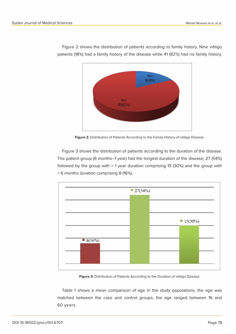
Figure 2 shows the distribution of patients according to family history. Nine vitiligo
patients (18%) had a family history of the disease while 41 (82%) had no family history.
Figure 2: Distribution of Patients According to the Family History of vitiligo Disease.
Figure 3 shows the distribution of patients according to the duration of the disease.
The patient group (6 months–1 year) had the longest duration of the disease, 27 (54%)
followed by the group with > 1 year duration comprising 15 (30%) and the group with
< 6 months duration comprising 8 (16%).
Figure 3: Distribution of Patients According to the Duration of vitiligo Disease.
Table 1 shows a mean comparison of age in the study populations, the age was
matched between the case and control groups, the age ranged between 16 and
60 years.

Sudan Journal of Medical Sciences Mehad Muawia et al. et al.
DOI 10.18502/sjms.v15i1.6707 Page 79
Table 1: Mean Comparison of Study Parameter Across Age.
Variable Group Min. Max. Mean ± SD
Age (Years) Case 16.00 60.00 32.52 ± 11.86
Control 16.00 60.00 32.72 ± 10.31
Table 2 shows the mean comparison of study parameter in case versus control
group. The copper level in the case and control groups was 21.69 ±5 .17 mμol/l and
18.05 ± 3.51 mμol/l, respectively). The serum copper was significantly increased in
vitiligo patients compared to the control group (p value = 0.000).
Table 2: Mean Comparison of Study Parameters in Case versus Control Groups.
Parameter Group Mean ± SD p value
Copper level mμol/l Case 21.69 ± 5.17 0.000
Control 18.05 ± 3.51
Table 3 shows mean comparison of study parameter across gender. There is no
difference between males and females.
Table 3: Mean Comparison of Study Parameter across Gender.
Parameter Gender Mean ± SD p value
Copper level mμol/l Male 22.18 ± 5.91 0.274
Female 20.74 ± 3.27
Table 4 shows mean comparison of study parameter across family history. The copper
levels in family history were 22.48 ± 5.53 mμol/l and 22.48 ± 5.53 mμol/l, respectively,
there were insignificant differences with p value = 0.612.
Table 4: Mean Comparison of Study Parameter across Family History.
Parameter Family History Mean ± SD p value
Copper level mμol/l Yes 22.48 ± 5.53 0.612
No 22.48 ± 5.53
Table 5 shows the mean comparison of study parameter across the duration of
vitiligo disease. There was no significant difference of serum copper level regarding
the duration of the disease.
Table 5: Mean Comparison of Study Parameter across the Duration of Disease.
Duration Mean ± SD p value
<6 Months 20.66 ± 4.05
6Months–1 Year 20.91 ± 4.52 0.905
>1 Year 23.63 ± 6.47 0.192

Sudan Journal of Medical Sciences Mehad Muawia et al. et al.
DOI 10.18502/sjms.v15i1.6707 Page 80
Figure 4 shows the correlations between the age and the serum copper level. There
was a negative correlation between age and serum copper level (r = 0.230, p = 0.108).
Figure 4: The Correlations Between Age and Serum Copper Level in the Study Group.
Discussion
Vitiligo is a chronic skin disease that is considered as a stigma to patients in certain
communities, it is wrongly believed to be contagious. The etiology of vitiligo is still not
known and the effect of all therapies till now doesn’t seem promising. Many studies
have been conducted on vitiligo pathogenesis but it is still idiopathic and no result
was conclusive. Some studies have demonstrated the role of trace elements such as
copper, zinc, iron, etc. in melanogenesis with different results and conclusions. The role
of copper in melanogenesis has been studied since 1931 when Keil and Nelson had
observed that the color of the hair of experimental rats had turned white when they
were fed with only milk, which is a copper-deficient diet while being studied for milk
anemia [1]. Cunningham in the same year found that the white-coated animals have
lower copper levels in their skin than the black-coated ones, he also found that copper
is concentrated mainly in the epidermis [2]. This observation is very important. The first
observation pointed to the possible role of copper in melanin synthesis and the second
is more supportive to this hypothesis since melanogenesis occurs in the epidermis,
but of course till that time no one had talked about the nature of melanocytes and
their location within epidermal keratinocytes. Since melanogenesis takes place in the
skin of animals and humans, researchers started to study the role of copper and other

Sudan Journal of Medical Sciences Mehad Muawia et al. et al.
DOI 10.18502/sjms.v15i1.6707 Page 81
trace elements in animals. These studies had raised the suspicion of the role of copper
in the pathogenesis of vitiligo in humans and since that time many other researches
had talked about the possible role of trace elements in the etiology of vitiligo with
the hope of finding promising treatment for this condition which is still idiopathic and
all the possible etiologies are stated as hypotheses. The results of this study pointed
to slightly increased incidence of vitiligo in male 22.18 ± 5.91 versus female 20.74 ±
3.27 with a P-value of 0.274, in agreement with Wang et al. [18] who found a higher
prevalence of the disease in male than in female. The male to female ratio was 1.6:1,
which is similar to that reported by McBurney [19] but in disagreement with Lu et al.
[20] who reported that vitiligo is distributed equally in men and women. The present
study was conducted on the age group ranging from 16–60 years with a mean age of
31 years, similar to Marwa Salem et al.’s study [21], in which she included 50 patients
with vitiligo of both sexes: 33 females (60%) and 17 males (34%). Fifty volunteers were
included as a control group. The age in vitiligo group ranged between 15 to 60 years
with a mean ± SD of 36.74 ± 14 years and in control group ranged from 15 to 60 years
with a mean ± SD of 31.28 ± 9.49 years, and disagreed with a study which reported
that the mean age of onset was 18.9 years, while another study found that the mean
age of onset was 23.7 years old. The present findings observed that there is a high
concentration of copper in vitiligo patients as compared to healthy controls, agreed
with Helmy et al. [22], who showed that Cu levels were significantly higher in active
vitiligo patients compared to the control. On the contrary, Wasan and Al-Rubayee [23],
Wu et al. [24], Kang et al. [27], Wang et al. [18], Shi et al. [25], and Li et al. [26] reported
no statistically significant Cu level change between the vitiligo patients and the control
group and all of them agreed with Marwa Salem et al. (26), who also reported that serum
Cu was insignificantly higher in vitiligo group compared to the control group. Of the 16
studies that detected the serum Cu level, six studies [24, 25, 28, 32, 37] reported no
statistically significant Cu level change between the vitiligo and the healthy groups. The
others presented significant decrease of Cu level in the vitiligo group (18). Melanin’s are
colloidal pigments and have a high affinity for metal ions; therefore, Cu is found in high
levels in pigmented tissues involved in melanin synthesis. As melanocytes degenerate
in vitiligo patients, less Cu is utilized for the melanin synthesis, which consequently
raise levels of Cu in serum in vitiligo patients (18). According to the results of the
study we reported that the disease has no association with family history, in the mean
comparison of study parameter across family history. The copper levels in family history
were (22.48 ± 5.53 μμol/l and 22.48 ± 5.53 μμol/l, respectively). The p value = 0.612

Sudan Journal of Medical Sciences Mehad Muawia et al. et al.
DOI 10.18502/sjms.v15i1.6707 Page 82
showed no significant differences, disagreeing with a Chinese study that reported 20%
of the patients with positive family history in first-degree relatives [18].
Conclusions
This study concluded that serum copper was highly and significantly increased in
Sudanese vitiligo patients studied compared to the control group.
Recommendation
In our study, we recommend encouragement of healthy diets containing copper and all
other trace elements important for enzymatic actions to prevent serious diseases, for
example, vitiligo, measurement of copper profile, routine investigations for copper, and
other trace elements for vitiligo patients, and further studies regarding the role of other
trace elements need to be performed in vitiligo patients because the etiopathogenesis
of the disease is still unclear and also the treatment of vitiligo still represents a great
challenge.
References
[1] Ezzedine K, Eleftheriadou V, Whitton M et al. (2015). Vitiligo. Lancet. 386(9988):
74–84.
[2] NIAMS (2014). Archived from the original on 21 August 2016. Retrieved 11 August
2016.
[3] Whitton M, Pinart M, Batchelor JM et al. (2016). Evidence-based management
of vitiligo: summary of a cochrane systematic review. The British Journal of
Dermatology. 174(5): 962–969.
[4] Krüger C, Schallreuter KU (2012). A review of the worldwide prevalence of vitiligo
in children/adolescents and adults. International Journal of Dermatology. 51(10):
1206–1212.
[5] Picardi A, Pasquini P, Cattaruzza MS et al. (2003). Stressful life events, social
support, attachment security and alexithymia in vitiligo. A case-control study.
Psychotherapy and Psychosomatics. 72(3): 150–158.
[6] Birlea SA, Spritz RA, Norris DA (2007). Vitiligo. In L. A. Goldsmith, S. I. Katz, B. A.
Gilchrest, A. S. Paller, D. J. Leffell, and K. Wolff (Eds.), Fitzpatrick’s Dermatology in
General Medicine (7th ed.). New York: McGraw-Hill Professional.

Sudan Journal of Medical Sciences Mehad Muawia et al. et al.
DOI 10.18502/sjms.v15i1.6707 Page 83
[7] Kumar CM, Sathisha UV, Dharmesh S et al. (2011). Interaction of sesamol
(3,4-methylenedioxyphenol) with tyrosinase and its effect on melanin synthesis.
Biochimie. 93(3): 562–569.
[8] Stevens LH, Davelaar E, Kolb RM et al. (1998). Tyrosine and cysteine are substrates
for blackspot synthesis in potato. Phytochemistry. 49(3): 703–707.
[9] Barton DE, Kwon BS, Francke U (1988). Human tyrosinase gene, mapped to
chromosome 11 (q14----q21), defines second region of homology with mouse
chromosome 7. Genomics. 3(1): 17–24.
[10] Meredith P, Riesz J (2004). Radiative relaxation quantum yields for synthetic
eumelanin. Photochemistry and Photobiology. 79(2): 211–216.
[11] Brenner M, Hearing VJ (2008)
[12] Fuentealba IC, Aburto EM (2003). Animal models of copper-associated liver
disease. Comparative Hepatology. 2(1): 5.
[13] Ligoxygakis P (2001). Copper transport meets development. Trends in Genetics.
17(8): 442.
[14] Becker SW (1933). Arch Derm Syph. 28: 497.
[15] Knin HL, Nelson VH (1931). Journal of Biological Chemistry. 93: 49; Cunningham IJ
(1931). Some biochemical and physiological aspects of copper in animal nutrition.
Biochemical Journal. 25(4): 1267–1294.
[16] Mabson P (1935). Bull. soc. franc. de dermat. et syph. (Reunion dermat.,
Stras-bourg). 42: 1112.
[17] Pillsbury DM, Kulchar GV (1933). Arch Derm Syph. 27: 36.
[18] Wang X, Du J, Wang T et al. (2013). Prevalence and clinical profile of vitiligo in
China: a community-based study in six cities. Acta Dermato-Venereologica. 93(1):
62–65.
[19] McBurney EI (1979). Vitiligo. Clinical picture and pathogenesis. Archives of Internal
Medicine. 139(11): 1295–1297.
[20] Lu T, Gao T, Wang A et al. (2007). Vitiligo prevalence study in Shaanxi Province,
China. International Journal of Dermatology. 46(1): 47–51.
[21] (2018). The Egyptian Journal of Hospital Medicine. 70(2).
[22] Salem M, Abd El-Raheen TA, Aboraia NM (2018). Serum Copper and zinc levels in
vitiligo patients. The Egyptian Journal of Hospital Medicine. 70(3): 364–370.
[23] Helmy MI, Gayyar EL, Hawas S et al. (2004). Role of oxidative stress in the
pathogenesis of vitiligo. Journal of Pan-Arab League of Dermatologists. 15(3):
97–105.

Sudan Journal of Medical Sciences Mehad Muawia et al. et al.
DOI 10.18502/sjms.v15i1.6707 Page 84
[24] Wasan TS, Al-Rubayee W (2011). Trace elements levels in serum and hair of patients
with vitiligo and alopecia areata. Karbala Journal of Medicine. 4(2): 1117–1121.
[25] Wu Y, He N, Li JS et al. (2010). The zinc and copper levels in serum of 70 vitiligo
patients from Guangxi Province. Chinese Journal of Dermatology Venereology.
24: 722.
[26] Shi DR, Pu XM, Ha LS (1993). A correlative study on serum copper and zinc in
patients with vitiligo. Journal of Clinical Dermatology. 5: 241–243.
[27] Li YG, Zhou JG, Shao ZH (1988). Determination of the levels of copper and zinc
in plasma of serum of some patients. Journal of Tianjin University of Commerce.
4: 24–30.
[28] Kang AJ, Su BS, Xu HQ (2002). Research on the melanocytes apoptosis in vitiligo
caused by oxygen free radicals and microelement. Journal of Chinese Clinical
Medicine. 3: 4–7.