Surface imprinted beads for the recognition of human serum albumin
Upload
independentCategory
view
0download
0
Journal of Structural Biology 157 (2007) 348–355
www.elsevier.com/locate/yjsbi
EVect of human serum albumin on drug metabolism: Structural evidence of esterase activity of human serum albumin
Feng Yang a,b, Chuanbing Bian a,b, Lili Zhu a,b, Gengxiang Zhao a,b, Zixiang Huang a, Mingdong Huang a,b,¤
a State Key Laboratory of Structural Chemistry, Fujian Institute of Research on the Structure of Matter, 155 Yang Qiao Xi Lu, Fuzhou, Fujian 350002, People’s Republic of China
b The Graduate School of Chinese Academy of Sciences, Chinese Academy of Sciences, People’s Republic of China
Received 7 July 2006; received in revised form 24 August 2006; accepted 25 August 2006Available online 9 September 2006
Abstract
Human serum albumin (HSA) is the most abundant plasma protein in the human body with a plasma concentration of 0.6 mM. HSAplays an important role in drug transport and metabolism. Enzymatic activity of HSA on diVerent substrates or drugs has been studiedand documented. The structural mechanism of this activity, however, is unknown. In this study, we have determined the crystal structuresof HSA–myristate in a complex of aspirin and of salicylic acid, respectively. The crystal structure of HSA–myristate–aspirin illustratesthat aspirin transfers acetyl group to Lys199 and is hydrolyzed into salicylic acid by HSA. The hydrolysis product, salicylic acid, remainsbound to HSA at a similar location, but it shows a very diVerent orientation when compared with the salicylic acid in the HSA–myris-tate–salicylic acid ternary complex. These results not only provide the structural evidence of esterase activity of HSA, and demonstratethe conformational plasticity of HSA on drug binding, but also may provide structural information for the modulation of HSA–druginteraction by computational approach based on HSA–drug structure.© 2006 Elsevier Inc. All rights reserved.
Keywords: Human serum albumin; Drug metabolism; Aspirin; Salicylic acid; Esterase activity; Structure
1. Introduction
Human serum albumin (HSA)1 is the most abundantsoluble protein in blood plasma. HSA provides a depot formany compounds, binds some ligands in a strained orienta-tion providing their metabolic modiWcation, renders poten-tial toxins harmless transporting them to disposal sites,accounts for most of the anti-oxidant capacity of humanserum, and acts as a NO-carrier (Fasano et al., 2005). Inaddition, HSA plays a central role in drugs pharmacokinet-ics. Among the four aspects of pharmacokinetics (absorp-tion, distribution, metabolism, and excretion), distribution
* Corresponding author. Fax: +591 83714946.E-mail address: [email protected] (M. Huang).
1 Abbreviations used: HSA, human serum albumin; Aly, acetyllysine;MYR, myristate; SA, salicylic acid; AIN, aspirin.
1047-8477/$ - see front matter © 2006 Elsevier Inc. All rights reserved.doi:10.1016/j.jsb.2006.08.015
is the one that this protein controls because most drugs thattravel in plasma bind to HSA. HSA consists of 585 aminoacids that form into three structurally similar �-helicaldomains. These domains are characterized by a commonmotif of 10 �-helices. Each domain can be divided into sub-domains A and B, which contain six and four �-helical,respectively (He and Carter, 1992; Sugio et al., 1999). Thedomain II and III of HSA contain two primary drug bind-ing sites, known as Sudlow’s site I and site II (Sudlow et al.,1975). Several additional sites were also observed (Bhat-tacharya et al., 2000a; Ghuman et al., 2005; Lejon et al.,2004; Petitpas et al., 2003). Crystallographic structuralanalysis of HSA-ligand complexes can reveal the moleculardetails of drug binding, clarify the interpretation of accu-mulated drug binding data, and provide a valuable struc-tural template to rationale interaction between drugs andHSA (Ghuman et al., 2005). Accumulated structural data
F. Yang et al. / Journal of Structural Biology 157 (2007) 348–355 349
can be used to develop quantitative structure–activity rela-tionships for albumin binding (Colmenarejo, 2003; Col-menarejo et al., 2001; Kratochwil et al., 2002).
In addition to its ligand binding capabilities, HSA pos-sesses interesting enzymic properties. The enzymatic activ-ity of serum albumin was Wrst noted in 1951. For decades,enzymatic activities of HSA have been extensively observed(Ahmed et al., 2005; Dubois-Presle et al., 1995; Kuronoet al., 1992; Sakurai et al., 2004; Salvi et al., 1997; Watanabeet al., 2000). HSA appears to have one markedly reactivesite (Kurono et al., 1992) or multiple nonspeciWc catalyticsites (Means and Bender, 1975) located at subdomain IIIAof HSA (site II). The active residues were proposed to be atyrosine residue (Tyr411) and a histidine residue (Yoshidaet al., 1985). Site-directed mutagenesis studies have shownthat indeed Arg410 and Tyr411 are indeed important forthe esterase activity of HSA (Watanabe et al., 2000). Subdo-main IIA of HSA (site I) also possesses esterase activity,which can convert aspirin (acetylsalicylic acid) to salicylicacid (Burch and Blazer-Yost, 1981; Gresner et al., 2006;Hawkins et al., 1969; Hawkins et al., 1968; Pinckard et al.,1968). By means of nuclear magnetic resonance (NMR)spectroscopy, Honma reported that the hydrolysis rates ofaspirin were greatly enhanced in the presence of HSA, thusconWrming the esterase-like activity of HSA (Honma andIshikawa, 1991). The HSA residues involved was proposedas Lys199 (Walker, 1976), but no further structural infor-mation is known.
Since its introduction in 1892 as a potent anti-inXam-matory and analgesic drug, aspirin has been extensivelystudied for decades, including its interaction with HSA.Aspirin was found to acetylate HSA under physiologicconditions in vivo (Burch and Blazer-Yost, 1981; Hawkinset al., 1969; Lighnter et al., 1988; Pinckard et al., 1968) andinduces the structural changes in HSA (Hawkins et al.,1969). The structure of the HSA complex with aspirin atlow resolution (4.0 ) was previously mentioned in a reportbut no details were given (He and Carter, 1992). There-fore, several questions remain unanswered. First, what isthe structural basis of the esterase activity of HSA? Doesthe hydrolysis product of aspirin remain bound to HSA?Is the Lys199 of HSA the only residue to be acetylated byaspirin and does the acetylation of HSA cause any confor-mational changes on HSA? Here, we report the crystalstructures of the HSA complex with aspirin and salicylicacid at a resolution of 2.7 and 2.9 , respectively. Ourresults elucidate the structural basis of esterase activity ofHSA and reveal that aspirin transfers its acetyl group toLys199. Our structures demonstrate that both aspirin andsalicylic acid bind to HSA at similar location (site I) butin very diVerent orientations. This diVerence in bindingorientation does not mean that the large site I pocket’sbinding to a drug is promiscuous; rather, it is due to theacetylation of Lys199 of HSA. These results may also pro-vide structural information for the modulation of HSA–drug interaction by computational approach based on theHSA–drug structures.
2. Materials and methods
2.1. Protein puriWcation and crystallization
Fatty acid free HSA was purchased from Sigma Inc.(catalogue number A3782) and further puriWed by remov-ing HSA dimers and multimers, according to the publishedprotocols (Curry et al., 1998). The protein was dissolved ina 20 mM potassium phosphate buVer (pH 7.5) to about100 mg/ml and stored in a ¡80 °C refrigerator before use.Sodium myristate, aspirin, and salicylic acid (SA) were pur-chased from Sigma Inc. Sodium myristate was dissolvedinto alcohol, and then it was diluted to 2.5 by 20 mM potas-sium phosphate (pH 7.5). HSA–myristate complexes wereprepared by mixing 100 �l of 1.5 mM HSA with 720 �l of2.5 mM myristate and then incubating the mixture for 2 h.Excessive undissolved myristate was removed by centrifu-gation at 12,000g for 4 min. Samples of HSA–myristatecomplexes were concentrated to 100 mg/ml with a Milliporespin Wlter (10,000 Da cutoV) and centrifugated at 12,000 gfor 4 min before cultivating crystals of HSA–myristate.
Crystallization was carried out by sitting drop vapordiVusion at room temperature. Equal volumes of the HSA–myristate complexes (2.0 �l) were mixed with the reservoirsolution, consisting of 31–34% (w/v) polyethylene glycol3350 and 50 mM potassium phosphate (pH 7.5). Crystalsgrew spontaneously as clusters of rods after three days. Atechnique of streak-seeding was used to improve crystalquality. In all cases, crystals were obtained by streak-seed-ing into drops that had been equilibrated for two days.Aspirin and salicylic acid were dissolved into alcohol anddiluted to 20 mM with the solution consisting of 32% (w/v)polyethylene glycol 3350, 50 mM potassium phosphate pH7.5. HSA–myristate complexes were rinsed by reservoirsolution, and then soaked directly into 20 mM drug solu-tion (50�l) for about 24 h.
2.2. Data collection and structure determination
X-ray diVraction data was collected at room tempera-ture with the Bruker ProteumR FR591 X-ray diVractome-ter with Cu K� radiation and were processed with Proscale.Crystals of the HSA–myristate–aspirin and HSA–myris-tate–salicylic acid complexes were isomorphous with theHSA–myristate complex (Bhattacharya et al., 2000b; Curryet al., 1998). The structures of HSA–myristate–aspirin andHSA–myristate–salicylic acid complexes were solved bymolecular replacement with the program AMORE using aHSA–myristate structure (PDB code 1BJ5) stripped of itsligands as the model. The model was initially reWned byusing a rigid body protocol in CNS and then subjected tocycles of positional and B-factor reWnement before the cal-culation of initial Fo¡Fc and 3Fo¡2Fc maps. These mapswere used to guide the positions of the fatty acid and drugligands and to make manual adjustments to the proteinprior to further cycles of reWnement. Details of the reWne-ment statistics were summarized in Table 1. Figures depict-

350 F. Yang et al. / Journal of Structural Biology 157 (2007) 348–355
ing the structure were prepared by PyMOL (DeLano,2004). The coordinates of HSA–myristate–aspirin andHSA–myristate–salicylic acid have been deposited in theProtein Data Bank under the accession codes 2I2Z and2I30, respectively.
3. Results
To study the interaction between HSA and drugs, wesoaked the drug molecules into the crystals of the HSA–myristate complex. The presence of myristate was necessaryto form well-behaviored crystals. In the absence of myris-tate, the HSA crystals either diVracted poorly or crackedupon soaking of drug molecules. Under physiological con-ditions, HSA normally binds with unesteriWed fatty acids ata molar ratio of up to 1:2, but under certain disease states itmay accommodate up to six moles.
Initial HSA–myristate crystals were grown usingPEG3350 as a precipitant. The crystallization behavior ofHSA turned out to be quite diYcult to control, usuallyleading to hollow HSA crystals that diVracted poorly or alarge number of overlapped and intertwined crystals thatwere hard to separate. To obtain the diVracting crystals,several rounds of streak-seeding procedures were needed.The crystals were then soaked in 20 mM aspirin or salicylicacid, respectively, for 24 h. The X-ray diVraction data setwas collected with Cu K� X-ray radiation at room temper-ature. HSA–myristate–aspirin crystals diVracted up to2.7 Å, and HSA–myristate–salicylic acid crystals diVractedto 2.9 Å. Both crystals were isomorphous with the crystalsof the HSA–myristate complex with cell parameters ofaD188.42 Å, bD 38.17 Å, cD95.38 Å, and a space group ofC2 (Table 1).
3.1. Crystal structure determination and model reliability
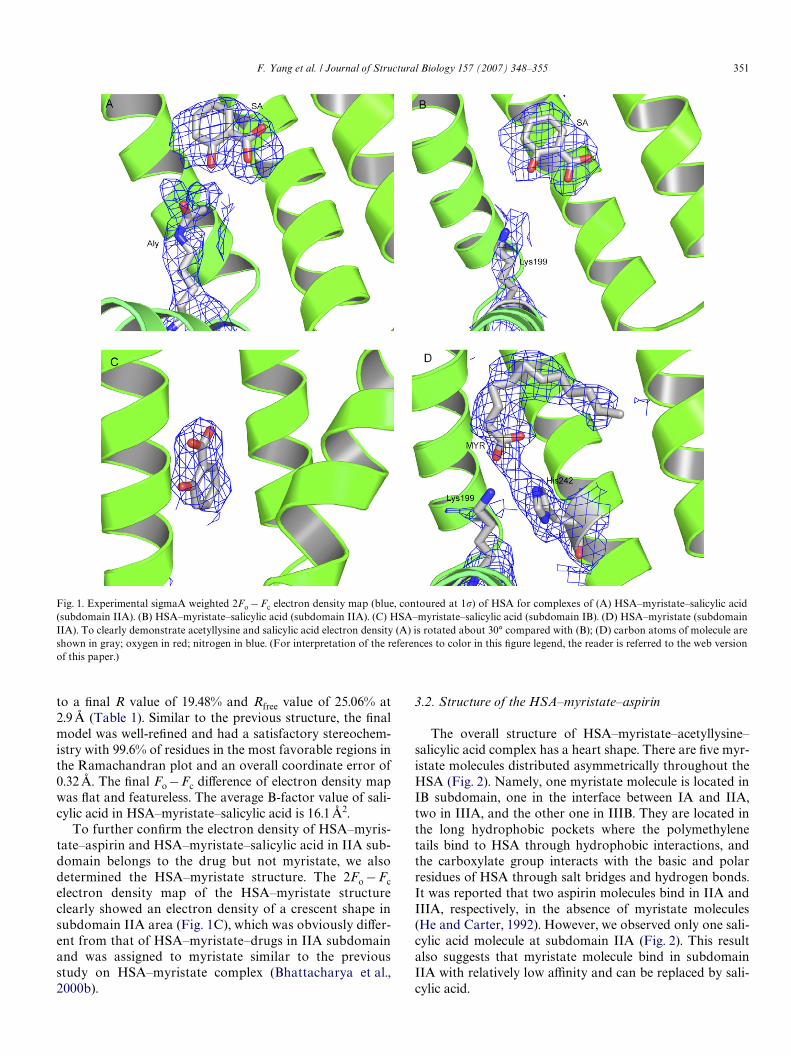
The 2Fo¡Fc sigmaa-weight map of the HSA–myristate–aspirin structure clearly showed the electron density for Wvemyristates and an extra density in subdomain IIA area.This extra electron density at subdomain IIA was not bigenough to accommodate aspirin molecule but Wt betterwith a salicylic acid molecule; Some extra electron densityaround Lys199 of HSA was also observed and was assignedas acetyllysine (Aly) residue (Fig. 1A). Although the resolu-tion of the current structure is limited (2.7 Å), the shape ofthese electron densities allows conWdent identiWcation ofsalicylic acid and acetyllysine residue. The correlationcoeYcient between the model and the map of acetyllysineand salicylic acid is 0.83 and 0.87, respectively. The averageB-factor of acetyllysine199 and salicylic acid is 18.1 and16.3 Å2, respectively, lower than the average B-factor ofoverall protein structure (47.1 Å2). The Wnal model of HSA–myristate–aspirin has satisfactory stereochemistry with99.5% residues in the most favorable regions in the Rama-chandran plot and an overall coordinate error of 0.34 Å.The Wnal Fo¡Fc electron density map was Xat and feature-less, which suggests that most densities have beenexplained. This structure was reWned to an R value of21.80% and Rfree value of 26.20% at 2.7 Å (Table 1).
For HSA–myristate–salicylic acid structure, a 2Fo¡Fcsigmaa-weight map calculated at 2.9 Å by using phasesfrom the reWned model of HSA–myristate complex revealsthe density for two well-separated binding sites. The sali-cylic acid located in IIA subdomain provided the best struc-ture to Wt the electron density (Fig. 1B). The correlationcoeYcient between model and the map of salicylic acid is0.934. HSA–myristate–salicylic acid structure was reWned
Table 1X-ray data collection and model reWnement statistics for the crystal structures of HSA–myristate–aspirin, HSA–myristate–salicylic acid, and HSA–myris-tate complexes
a Values for the outermost resolution shell are given in parentheses.b Rmerge D 100 £�j�j �Ihj ¡ Ih�/�j�j Ihj where Ih is the weighted mean intensity of the symmetry-related reXections Ihj.c Rmodel D 100 £ �hkl �Fobs ¡ Fcalc�/�hklFobs where Fobs and Fcalc are the observed and calculated structure factors, respectively.d Rfree is the Rmodel calculated using a randomly selected 5% sample of reXection data omitted from the reWnement.e R.m.s stands for root mean square.
HSA–myristate–drugs HSA–myristate–AIN HSA–myristate–SA HSA–myristate
Data collectionSpace group C2 C2 C2Cell parameters, a,b,c (Å) 188.41,38.94,96.02 187.64,38.87,95.56 188.42,38.17,95.38Cell parameters, �(°) 105.32 105.51 105.64Resolution range (Å) 99–2.7 99–2.9 99-2.8Data redundancy 3.5 3.4 3.4Completeness (%) 98% 96% 97%I/�I
a 9.34(2.2) 6.38(2.2) 6.96 (2.6)Rmerge (%)b 6.9%(38%) 6.5%(30%) 6.3 (32%)
Model reWnementRmodel (%)c 21.80% 19.43% 19.65%Rfree (%)d 26.20% 25.02% 25.19%R.m.s. deviation from ideal bond lengths (Å) 0.0071 0.0074 0.0073R.m.s. deviation from ideal angles (°) 1.65 1.57 1.57R.m.s. deviation in B-factors main/side chain (Å2)e 1.33/2.24 1.42/2.23 1.34/2.24Average B-factor (Å2) 47.1 56.7 60.3
F. Yang et al. / Journal of Structural Biology 157 (2007) 348–355 351
to a Wnal R value of 19.48% and Rfree value of 25.06% at2.9 Å (Table 1). Similar to the previous structure, the Wnalmodel was well-reWned and had a satisfactory stereochem-istry with 99.6% of residues in the most favorable regions inthe Ramachandran plot and an overall coordinate error of0.32 Å. The Wnal Fo¡Fc diVerence of electron density mapwas Xat and featureless. The average B-factor value of sali-cylic acid in HSA–myristate–salicylic acid is 16.1 Å2.
To further conWrm the electron density of HSA–myris-tate–aspirin and HSA–myristate–salicylic acid in IIA sub-domain belongs to the drug but not myristate, we alsodetermined the HSA–myristate structure. The 2Fo ¡Fcelectron density map of the HSA–myristate structureclearly showed an electron density of a crescent shape insubdomain IIA area (Fig. 1C), which was obviously diVer-ent from that of HSA–myristate–drugs in IIA subdomainand was assigned to myristate similar to the previousstudy on HSA–myristate complex (Bhattacharya et al.,2000b).
3.2. Structure of the HSA–myristate–aspirin
The overall structure of HSA–myristate–acetyllysine–salicylic acid complex has a heart shape. There are Wve myr-istate molecules distributed asymmetrically throughout theHSA (Fig. 2). Namely, one myristate molecule is located inIB subdomain, one in the interface between IA and IIA,two in IIIA, and the other one in IIIB. They are located inthe long hydrophobic pockets where the polymethylenetails bind to HSA through hydrophobic interactions, andthe carboxylate group interacts with the basic and polarresidues of HSA through salt bridges and hydrogen bonds.It was reported that two aspirin molecules bind in IIA andIIIA, respectively, in the absence of myristate molecules(He and Carter, 1992). However, we observed only one sali-cylic acid molecule at subdomain IIA (Fig. 2). This resultalso suggests that myristate molecule bind in subdomainIIA with relatively low aYnity and can be replaced by sali-cylic acid.
Fig. 1. Experimental sigmaA weighted 2Fo ¡ Fc electron density map (blue, contoured at 1�) of HSA for complexes of (A) HSA–myristate–salicylic acid(subdomain IIA). (B) HSA–myristate–salicylic acid (subdomain IIA). (C) HSA–myristate–salicylic acid (subdomain IB). (D) HSA–myristate (subdomainIIA). To clearly demonstrate acetyllysine and salicylic acid electron density (A) is rotated about 30° compared with (B); (D) carbon atoms of molecule areshown in gray; oxygen in red; nitrogen in blue. (For interpretation of the references to color in this Wgure legend, the reader is referred to the web versionof this paper.)

352 F. Yang et al. / Journal of Structural Biology 157 (2007) 348–355
The phenyl of salicylic acid is located in the bindingpocket that is formed by Aly199, Leu219, Arg222, Phe223,Leu238, Ile260, Ile264, Ile290, and Ala291 (Fig. 3A). Thephenyl group of salicylic acid makes mostly hydrophobiccontacts with the surrounding HSA side chains (Table 2).The closest distances between the phenyl group and sur-rounding HSA side chains range from 4.0 to 5.0 Å, indicat-ing weak hydrophobic interaction.
The carboxylate group of salicylic acid is surroundedby a hydrophobic side of the pocket which is delimited by
Fig. 2. Overall domain structure of HSA and the locations of myristate,salicylic acid and acetyllysine (Aly199). The domains are colour-coded asfollows: I, blue; II, green; III, red. The ligand molecules are shown in aspace-Wlling representation and are colored by atom type (carton, grey;oxygen, red; nitrogen, blue). (For interpretation of the references to colorin this Wgure legend, the reader is referred to the web version of thispaper.)
Leu238, Ile260, Ser287, and Ile290. The hydroxyl groupof salicylic acid is surrounded by Aly199, His242, andLeu238. The closest distances between hydroxyl group ofsalicylic acid and Aly199 and His242 are 4.12 and 4.53 Å,respectively, and no hydrogen bonds are found in theinterface.
Acetyllysine is close to Arg218, Trp214, Leu238, andHis242 in the pocket (Fig. 3A). The acetyl group of Acety-llysine is surrounded by Trp214 and Arg218 and forms ahydrogen bond with Arg218 (3.26 Å). Compared withLys199 in HSA–myristate, Lys199 side chain in HSA–myr-istate–aspirin stretches out to position the amine group ofLys199 toward the hydroxyl group of salicylic acid.Although there are some conformational changes in sidechains located in the aspirin binding site, the acetylation ofLys199 of HSA does not cause overall changes in the sec-ondary structures of HSA.
3.3. Structure of the HSA–myristate–salicylic Acid
Two salicylic acid molecules were identiWed in thiscomplex: one in subdomain IIA and another one in sub-domain IB where the bound salicylic acid contacts withmyristate. His146 is the primary ligand for the salicylicacid carboxylate group. The overall HSA structure andthe position of myristate are very similar to the HSA–myristate–aspirin complex. For example, the superposi-tion of C� atoms gives a root-mean-square-deviation of0.45 Å between complexes. The 2Fo¡ Fc electron densitymap clearly reveals the density for salicylic acid in subdo-main IIA. The phenyl group of salicylic acid is boundwithin binding pocket formed by Leu219, Phe223,Leu238, Arg257, Leu260, Ile264, Ser287, Ile290, andAla291 (Fig. 3B). The phenyl of salicylic acid made mostlyhydrophobic contacts with the surrounding side chains(Table 2). The carboxylate group of salicylic acid is
Fig. 3. Structural environments of (A) salicylic acid and acetyllysine (Aly) in the HSA–myristate–aspirin complex, and (B) salicylic acid in the HSA–myr-istate–salicylic acid complex. The blue dashed lines represent hydrogen bonds. The amino acid chains that are close to the drug molecules are shown assticks. Carbon, gray; nitrogen, blue; oxygen, red. Hydrogen bonds are shown in blue dash lines. (For interpretation of the references to color in this Wgurelegend, the reader is referred to the web version of this paper.)
F. Yang et al. / Journal of Structural Biology 157 (2007) 348–355 353
surrounded within a side of the pocket which is delimitedby Leu238, His242, Arg257, Ile260, and Ser287. The car-boxylate group of salicylic acid forms a hydrogen bondwith Arg257 (3.40 Å). However, the hydroxyl group of sal-icylic acid was surrounded completely by hydrophobicresidues (Leu219, Leu238, and Ala291).
3.4. Comparison of the binding mode of salicylic acid in HSA–myristate–acetyllysine–salicylic acid with aspirin analog binding in IIA
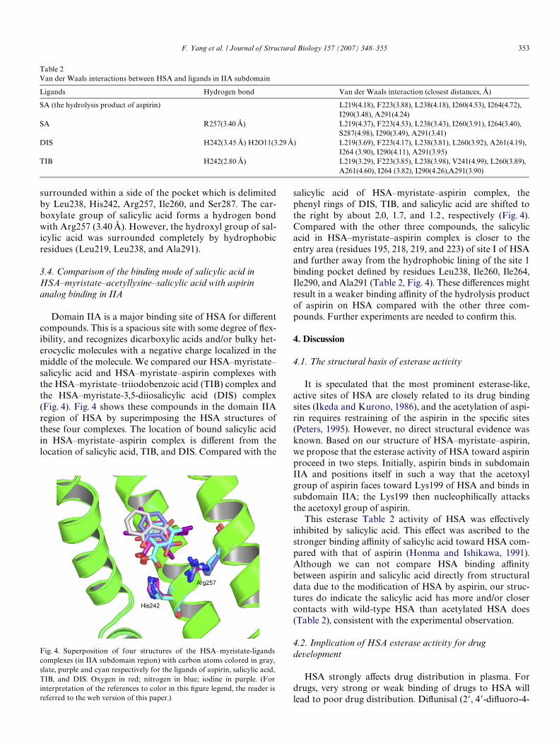
Domain IIA is a major binding site of HSA for diVerentcompounds. This is a spacious site with some degree of Xex-ibility, and recognizes dicarboxylic acids and/or bulky het-erocyclic molecules with a negative charge localized in themiddle of the molecule. We compared our HSA–myristate–salicylic acid and HSA–myristate–aspirin complexes withthe HSA–myristate–triiodobenzoic acid (TIB) complex andthe HSA–myristate-3,5-diiosalicylic acid (DIS) complex(Fig. 4). Fig. 4 shows these compounds in the domain IIAregion of HSA by superimposing the HSA structures ofthese four complexes. The location of bound salicylic acidin HSA–myristate–aspirin complex is diVerent from thelocation of salicylic acid, TIB, and DIS. Compared with the
Fig. 4. Superposition of four structures of the HSA–myristate-ligandscomplexes (in IIA subdomain region) with carbon atoms colored in gray,slate, purple and cyan respectively for the ligands of aspirin, salicylic acid,TIB, and DIS. Oxygen in red; nitrogen in blue; iodine in purple. (Forinterpretation of the references to color in this Wgure legend, the reader isreferred to the web version of this paper.)
salicylic acid of HSA–myristate–aspirin complex, thephenyl rings of DIS, TIB, and salicylic acid are shifted tothe right by about 2.0, 1.7, and 1.2 , respectively (Fig. 4).Compared with the other three compounds, the salicylicacid in HSA–myristate–aspirin complex is closer to theentry area (residues 195, 218, 219, and 223) of site I of HSAand further away from the hydrophobic lining of the site 1binding pocket deWned by residues Leu238, Ile260, Ile264,Ile290, and Ala291 (Table 2, Fig. 4). These diVerences mightresult in a weaker binding aYnity of the hydrolysis productof aspirin on HSA compared with the other three com-pounds. Further experiments are needed to conWrm this.
4. Discussion
4.1. The structural basis of esterase activity
It is speculated that the most prominent esterase-like,active sites of HSA are closely related to its drug bindingsites (Ikeda and Kurono, 1986), and the acetylation of aspi-rin requires restraining of the aspirin in the speciWc sites(Peters, 1995). However, no direct structural evidence wasknown. Based on our structure of HSA–myristate–aspirin,we propose that the esterase activity of HSA toward aspirinproceed in two steps. Initially, aspirin binds in subdomainIIA and positions itself in such a way that the acetoxylgroup of aspirin faces toward Lys199 of HSA and binds insubdomain IIA; the Lys199 then nucleophilically attacksthe acetoxyl group of aspirin.
This esterase Table 2 activity of HSA was eVectivelyinhibited by salicylic acid. This eVect was ascribed to thestronger binding aYnity of salicylic acid toward HSA com-pared with that of aspirin (Honma and Ishikawa, 1991).Although we can not compare HSA binding aYnitybetween aspirin and salicylic acid directly from structuraldata due to the modiWcation of HSA by aspirin, our struc-tures do indicate the salicylic acid has more and/or closercontacts with wild-type HSA than acetylated HSA does(Table 2), consistent with the experimental observation.
4.2. Implication of HSA esterase activity for drug development
HSA strongly aVects drug distribution in plasma. Fordrugs, very strong or weak binding of drugs to HSA willlead to poor drug distribution. DiXunisal (2�, 4�-diXuoro-4-
Table 2Van der Waals interactions between HSA and ligands in IIA subdomain
Ligands Hydrogen bond Van der Waals interaction (closest distances, Å)
SA (the hydrolysis product of aspirin) L219(4.18), F223(3.88), L238(4.18), I260(4.53), I264(4.72), I290(3.48), A291(4.24)
SA R257(3.40 Å) L219(4.37), F223(4.53), L238(3.43), I260(3.91), I264(3.40), S287(4.98), I290(3.49), A291(3.41)
DIS H242(3.45 Å) H2O11(3.29 Å) L219(3.69), F223(4.17), L238(3.81), L260(3.92), A261(4.19), I264 (3.90), I290(4.11), A291(3.95)
TIB H242(2.80 Å) L219(3.29), F223(3.85), L238(3.98), V241(4.99), L260(3.89), A261(4.60), I264 (3.82), I290(4.26),A291(3.90)
354 F. Yang et al. / Journal of Structural Biology 157 (2007) 348–355
hydroxybiphenyl-3-carboxylic acid) is an aspirin-like nons-teriodal anti-inXammatory drug and is superior to aspirinbecause of its higher potency, higher in vivo tolerance, andlonger duration of action (Hannah et al., 1978). However, itis 99% bound to plasma proteins, which makes it necessaryto have at least 250 mg twice a day for eYcacy. Such highdosage can lead to undesirable side eVects. Thus, bindingstrength to plasma proteins, especially HSA, is an impor-tant factor to be considered in drug development.
In consideration of the high concentration of HSA inhuman plasma (0.4 mM), the esterase activity of HSAshould also be taken into account during drug develop-ment. On the other hand, the esterase activity of HSA couldalso be used to convert an acetylated prodrug into a func-tional drug. For example, the crystallographic structure ofthe HSA-diXunisal complex shows that one diXunisal mole-cule binds in domain IIA of HSA and interacts with Tyr150(2.19 Å) and His242 (3.46 Å) of HSA (Ghuman et al., 2005).By utilizing acetylated diXunisal as a prodrug, it might bepossible to unmask this drug through HSA’s esterase activ-ity, alter its binding orientation, and thus to reduce its aYn-ity with HSA.
The biological consequences of acetylation of albuminin vivo are not fully understood. Previous Wndings sug-gested that this phenomenon may be of signiWcance(Minden and Farr, 1967). The syndrome of aspirin resis-tance characterized by asthma, rhinitis, and nasal polyps(Samter and Beers, 1967) is induced by aspirin but notother salicylates.
There is increasing evidence that aspirin diVers signiW-cantly from other salicylates in its eVects on hemostasis inhuman (Quike, 1966; Quike, 1996). Aspirin, but not salicy-lates, works by irreversibly acetylating COX-1 in platelets,preventing arachidonic acid from reaching the enzyme’sbinding and catalytic sites (Catella-Lawson et al., 2001).Aspirin’s cardio-protective eVects arise from its inhibitionof platelet thromboxane A2, which promotes vasoconstric-tion and platelet aggregation (Vane and Botting, 1992).Furthermore, aspirin has a strong anti-complementaryaction in vitro in normal therapeutic doses with respect to aguinea pig complement, while sodium salicylate has noaction at all (van et al., 1961). The acetyl group of aspirinappears to be implicated in most of these physiologic phe-nomena. The unique capacity of aspirin to form acetyl andsalicyloylamide derivatives of human proteins and otherbody constituents may well play a role in these reactions.These modiWcations by aspirin may alter HSA’s traditionalroles in drug pharmacokinetics. The study presented in thiswork provides a structural basis to understand HSA’s rolein drug metabolism.
Acknowledgments
This work is supported by the Frontier InterdisciplinaryInnovation Fund and State Key Laboratory of StructuralChemistry (IB021061) of Fujian Institute of Research onthe Structure of Matter.
References
Ahmed, N., Dobler, D., Dean, M., Thornalley, P.J., 2005. Peptide mappingidentiWes hotspot site of modiWcation in human serum albumin bymethylglyoxal involved in ligand binding and esterase activity. J. Biol.Chem. 280, 5724–5732.
Bhattacharya, A.A., Curry, S., Franks, N.P., 2000a. Binding of the generalanesthetics propofol and halothane to human serum albumin. Highresolution crystal structures. J. Biol. Chem. 275, 38731–38738.
Bhattacharya, A.A., Grune, T., Curry, S., 2000b. Crystallographic analysisreveals common modes of binding of medium and long-chain fattyacids to human serum albumin. J. Mol. Biol. 303, 721–732.
Burch, J.W., Blazer-Yost, B., 1981. Acetylation of albumin by low doses ofaspirin. Thromb. Res. 23, 447–452.
Catella-Lawson, F., Reilly, M.P., Kapoor, S.C., Cucchiara, A.J., DeMarco,S., Tournier, B., Vyas, S.N., FitzGerald, G.A., 2001. Cyclooxygenaseinhibitors and the antiplatelet eVects of aspirin. N. Engl. J. Med. 345,1809–1817.
Colmenarejo, G., 2003. In silico prediction of drug-binding strengths tohuman serum albumin. Med. Res. Rev. 23, 275–301.
Colmenarejo, G., Alvarez-Pedraglio, A., Lavandera, J.L., 2001. models topredict binding aYnities to human serum albumin. J. Med. Chem. 44,4370–4378.
Curry, S., Mandelkow, H., Brick, P., Franks, N., 1998. Crystal structure ofhuman serum albumin complexed with fatty acid reveals an asymmet-ric distribution of binding sites. Nat. Struct. Biol. 5, 827–835.
DeLano, W.L., 2004. The PyMol Molecular Graphics System. DeLanoScientiWc, San Carlos, CA.
Dubois-Presle, N., Lapicque, F., Maurice, M.H., Fournel-Gigleux, S., Mag-dalou, J., Abiteboul, M., Siest, G., Netter, P., 1995. Stereoselective ester-ase activity of human serum albumin toward ketoprofen glucuronide.Mol. Pharmacol. 47, 647–653.
Fasano, M., Curry, S., Terreno, E., Galliano, M., Fanali, G., Narciso, P.,Notari, S., Ascenzi, P., 2005. The extraordinary ligand binding proper-ties of human serum albumin. IUBMB Life 57, 787–796.
Ghuman, J., Zunszain, P.A., Petitpas, I., Bhattacharya, A.A., Otagiri, M.,Curry, S., 2005. Structural basis of the drug-binding speciWcity ofhuman serum albumin. J. Mol. Biol. 353, 38–52.
Gresner, P., Dolnik, M., Waczulikova, I., Bryszewska, M., Sikurova, L.,Watala, C., 2006. Increased blood plasma hydrolysis of acetylsalicylicacid in type 2 diabetic patients: a role of plasma esterases. Biochim.Biophys. Acta 1760, 207–215.
Hannah, J., Ruyle, W.V., Jones, H., Matzuk, A.R., Kelly, K.W., Witzel,B.E., Holtz, W.J., Houser, R.A., Shen, T.Y., Sarett, L.H., 1978. Novelanalgesic-antiinXammatory salicylates. J. Med. Chem. 21, 1093–1100.
Hawkins, D., Pinckard, R.N., Crawford, I.P., Farr, R.S., 1969. Structuralchanges in human serum albumin induced by ingestion of acetylsali-cylic acid. J. Clin. Invest. 48, 536–542.
Hawkins, D., Pinckard, R.N., Farr, R.S., 1968. Acetylation of humanserum albumin by acetylsalicylic acid. Science 160, 780–781.
He, X.M., Carter, D.C., 1992. Atomic structure and chemistry of humanserum albumin. Nature 358, 209–215.
Honma, K.N.M., Ishikawa, Y., 1991. Acetylsalicylate–human serum albu-min interaction as studied by NMR spectroscopy-antigenicity-produc-ing mechanism of acetylsalicylic acid. Mol. Immunol. 28, 107–113.
Ikeda, K., Kurono, Y., 1986. Enzymatic activity and drug binding activityof human serum albumin. Yakugaku Zasshi 106, 662–664.
Yoshida, Y.K., Mori, Y., Ikeda, K., 1985. Esterase-like activity of humanserum albumin: V. Reaction with 2,4-dinitrophenyl diethyl phosphate.Chem. Pharm. Bull. 33, 4995–5001.
Kratochwil, N.A., Huber, W., Muller, F., Kansy, M., Gerber, P.R., 2002.Predicting plasma protein binding of drugs: a new approach. Biochem.Pharmacol. 64, 1355–1374.
Kurono, Y., Kushida, I., Tanaka, H., Ikeda, K., 1992. Esterase-like activityof human serum albumin. VIII. Reaction with amino acid p-nitro-phenyl esters. Chem. Pharm. Bull. (Tokyo) 40, 2169–2172.
Lejon, S., Frick, I.M., Bjorck, L., Wikstrom, M., Svensson, S., 2004. Crystalstructure and biological implications of a bacterial albumin binding
F. Yang et al. / Journal of Structural Biology 157 (2007) 348–355 355
module in complex with human serum albumin. J. Biol. Chem. 279,42924–42928.
Lighnter, D.A., Wijekoon, W.M., Zhang, M.H., 1988. Understanding bili-rubin conformation and binding. Circular dichroism of human serumalbumin complexes with bilirubin and its esters. J. Biol. Chem. 263,16669–16676.
Means, G.E., Bender, M.L., 1975. Acetylation of human serum albumin byp-nitrophenyl acetate. Biochemistry 14, 4989–4994.
Minden, P., Farr, R.S., 1967. Human antibodies against acetylsalicylicacid-altered human serum albumin. Arthritis Rheum. 10, 299.
Peters, T., 1995. All About Albumin: Biochemistry, Genetics and MedicalApplications. Academic Press, San Diego.
Petitpas, I., Petersen, C.E., Ha, C.E., Bhattacharya, A.A., Zunszain, P.A.,Ghuman, J., Bhagavan, N.V., Curry, S., 2003. Structural basis of albu-min–thyroxine interactions and familial dysalbuminemic hyperthyroxi-nemia. Proc. Natl. Acad. Sci. USA 100, 6440–6445.
Pinckard, R.N., Hawkins, D., Farr, R.S., 1968. In vitro acetylation ofplasma proteins, enzymes and DNA by aspirin. Nature 219, 68–69.
Quike, A.J., 1966. Salicylates and bleeding: the aspirin tolerance test. Am.J. Med. Sci. 252, 265.
Quike, A.J., 1996. Aspirin and platelet stickness. Lancet, i 386.Sakurai, Y., Ma, S.F., Watanabe, H., Yamaotsu, N., Hirono, S., Kurono,
Y., Kragh-Hansen, U., Otagiri, M., 2004. Esterase-like activity of serum
albumin: characterization of its structural chemistry using p-nitro-phenyl esters as substrates. Pharm. Res. 21, 285–292.
Salvi, A., Carrupt, P.A., Mayer, J.M., Testa, B., 1997. Esterase-like activityof human serum albumin toward prodrug esters of nicotinic acid. DrugMetab. Dispos. 25, 395–398.
Samter, M., Beers, R.F., 1967. Concerning the nature of intolerance toaspirin. J. Allergy 40, 281.
Sudlow, G., Birkett, D.J., Wade, D.N., 1975. The characterization of twospeciWc drug binding sites on human serum albumin. Mol. Pharmacol.11, 824–832.
Sugio, S., Kashima, A., Mochizuki, S., Noda, M., Kobayashi, K., 1999.Crystal structure of human serum albumin at 2.5 A resolution. ProteinEng. 12, 439–446.
van, O.C., Friedmann, J.C., Fontaine, M., 1961. Anticomplementary actionof aspirin. Nature 189, 147.
Vane, J., Botting, R., 1992. In: Vane, J., Botting, R. (Eds.), Aspirin andOther Salicylates. Chapman & Hall, London.
Walker, J.E., 1976. Lysine residue 199 of human serum albumin is modi-Wed by acetylsalicyclic acid. FEBS Lett. 66, 173–175.
Watanabe, H., Tanase, S., Nakajou, K., Maruyama, T., Kragh-Hansen, U.,Otagiri, M., 2000. Role of arg-410 and tyr-411 in human serum albu-min for ligand binding and esterase-like activity. Biochem. J. 349 (Pt 3),813–819.
Copyright © 2022 FDOKUMEN


![Human Serum Albumin Binding of 2-[(Carboxymethyl)sulfanyl]-4-oxo-4-(4-tert-butylphenyl)butanoic Acid and its Mono-Me Ester](https://static.fdokumen.com/doc/165x107/6334ae932532592417002ca9/human-serum-albumin-binding-of-2-carboxymethylsulfanyl-4-oxo-4-4-tert-butylphenylbutanoic.jpg)