Development of a Marker of Estrogenic Exposure in Human Serum
Upload
independentCategory
view
5download
0
Research Paper
Recombinant Human Serum Albumin Dimer has High Blood CirculationActivity and Low Vascular Permeability in Comparisonwith Native Human Serum Albumin
Sadaharu Matsushita,1 Victor Tuan Giam Chuang,1,2 Masanori Kanazawa,1 Sumio Tanase,3
Keiichi Kawai,4 Toru Maruyama,1 Ayaka Suenaga,1 and Masaki Otagiri1,5
Received September 8, 2005; accepted January 12, 2006
Purpose. Human serum albumin (HSA) is used clinically as an important plasma expander. Albumin
infusion is not recommended for critically ill patients with hypovolemia, burns, or hypoalbuminemia
because of the increased leakage of albumin into the extravascular spaces, thereby worsening edema. In
the present study, we attempted to overcome this problem by producing a recombinant HSA (rHSA)
dimer with decreased vascular permeability and an increased half-life.
Methods. Two molecules of rHSA were genetically fused to produce a recombinant albumin dimer
molecule. The pharmacokinetics and biodistribution of the recombinant proteins were evaluated in
normal rats and carrageenin-induced paw edema mouse model.
Results. The conformational properties of this rHSA dimer were similar to those for the native HSA
(the HSA monomer), as evidenced by the Western blot and spectroscopic studies. The biological half-
life and area under the plasma concentrationYtime curve of the rHSA dimer were approximately 1.5
times greater than those of the monomer. Dimerization has also caused a significant decrease in the total
body clearance and distribution volume at the steady state of the native HSA. rHSA dimer accumulated
to a lesser extent in the liver, skin, muscle, and fat, as compared with the native HSA. Up to 96 h, the
vascular permeability of the rHSA dimer was less than that of the native HSA in paw edema mouse
models. A prolonged plasma half-life of the rHSA dimer was also observed in the edema model rats.
Conclusions. rHSA dimer has a high retention rate in circulating blood and a lower vascular
permeability than that of the native HSA.
KEY WORDS: biodistribution; human serum albumin; pharmacokinetic analysis; recombinant humanserum albumin dimer; vascular permeability.
INTRODUCTION
Human serum albumin (HSA), the most abundantprotein in plasma, is responsible for 80% of the colloidosmotic pressure of plasma (25Y33 mmHg). Because of this, it
is mainly used clinically to maintain colloid oncotic pressureand to increase the circulating plasma volume (1). HSAproduct was developed for the treatment of shock whereHSA Fraction V, USP, served as an attractive agent for thecorrection of hypovolemia, burns, and various diseases thatresult in albumin loss. In spite of the many theoreticalbenefits of albumin infusion in critically ill patients, therehas been little evidence to support its widespread clinical use.For many years, albumin infusion was believed to improvevital prognosis. Recently, a meta-analysis that included 24studies involving 1419 patients published by Cochran InjuriesGroup Albumin Reviewers suggested that the administrationof albumin-containing fluids resulted to a 1.68 times increasein the relative risk of death when compared to a crystalloidsolution (2). In contrast, no difference in mortality wasreported in a large-scale clinical trial in Australia and NewZealand, in which 4% albumin was compared to 0.9%sodium chloride for intravascular fluid resuscitation inpatients in intensive care units (ICUs) (3).
Reviews published on the parenteral use of albumin andthe consensus of several conferences recommended that theadministration of HSA can be justified for acute circulatory
0724-8741/06/0500-0882/0 # 2006 Springer Science + Business Media, Inc. 882
Pharmaceutical Research, Vol. 23, No. 5, May 2006 (# 2006)DOI: 10.1007/s11095-006-9933-1
1 Department of Biopharmaceutics, Graduate School of Pharmaceu-
tical Sciences, Kumamoto University, 5-1 Oe-honmachi, Kumamoto
862-0973, Japan.2 Department of Pharmacy, Faculty of Allied Health Sciences,
Universiti Kebangsaan Malaysia, Jalan Raja Muda Abdul Aziz,
50300 Kuala Lumpur Malaysia.3 Department of Analytical Biochemistry, School of Health Sciences,
Kumamoto University, 4-24-1 Kuhonji, Kumamoto 862-0976, Japan.4 School of Health Sciences, Faculty of Medicine, Kanazawa Univer-
sity, 5-11-80 Kodatsuno, Kanazawa 920-1192, Japan.5 To whom correspondence should be addressed. (e-mail: otagirim@
gpo.kumamoto-u.ac.jp
ABBREVIATIONS: AUC, area under the plasma concen-
trationYtime curve; CLtot, total body clearance; HSA, human serum
albumin; rHSA, recombinant HAS; Vdss, distribution volume of the
steady state; %ID, percentage of injected dose.

problems caused by hypovolemia, but it is seldom warrantedand is used merely to increase albumin concentrations incases of hypoalbuminemia (4). Only shock due to recentblood loss is an indication of the use of parenteral albumin,because the incidence of shock to acute-phase or hyper-allergic reactions is accompanied by increased albumin fluxto the extravascular compartment and adding to the albuminpool in this case would only expand this compartment andbring on edema (5Y7). The lung is particularly susceptible toalbumin overloading, and albumin administration for respi-ratory distress has been a matter of debate because of thepossibility that pulmonary edema may result (8,9). Cardiacfailure is another hazard of an overzealous increase incirculatory volume (10).
It is well known that capillary protein permeability isincreased in many pathological and physiological conditions.For instance, the rate of protein extravasation may increaseup to 100 times the normal rate in burn injury. Under suchconditions, the albumin molecules administered as a plasmaexpander are transported to organs and cause a furtheraccumulation of fluids, hence worsening the disease. Thefrequent administration of albumin infusion to maintain thedesired blood concentration, as well as the oncotic pressure,inevitably leads to the poor retention of albumin infusedduring the disease state. Although albumin receptors havebeen reported to mediate the transport of albumin across thecellular membrane, the mechanism of leakage during inflam-mation may involve a different route of extravasation sincecytokines are known to loosen tight junctions (11).
In other words, if a form of albumin that possesses goodblood retention and low extravasation properties could bedeveloped, the frequency of administration could be de-creased and further accumulation of fluids in tissues could beprevented. Hence, increasing the molecular size of albuminto stop leakage to extravascular spaces may be a potentialsolution in making albumin a superior candidate as a volumereplacement therapeutic agent. This type of albumin wouldnot only be superior for clinical use but would also be usefulpharmacoeconomically.
In addition, since albumin is typically harvested usingconventional techniques involving the fractionation of plas-ma obtained from blood donors, possible viral/prion con-taminants contained in the preparation impose risk to users.As a solution to this problem, the development of recombi-nant technology in producing therapeutic proteins such asHSA has been promoted. In this study, we genetically fusedtwo molecules of HSA and used Pichia pastoris to express therecombinant HSA (rHSA) dimer protein. The pharmacoki-netic and pharmacological properties of the rHSA dimerwere then evaluated.
MATERIALS AND METHODS
Materials
Native HSA was donated by the Chemo-Sera-TherapeuticResearch Institute (Kumamoto, Japan) and defatted bymeans of a charcoal treatment as described by Chen (12). Achimeric plasmid (pJDB-ADH-L10-HAS-A) having cDNAfor the mature form of HSA along with an L10 leader
sequence was a gift from Tonen Co. (Tokyo, Japan). Therestriction enzymes EcoRI, XhoI, AvaI, BamHI, and SalIand E. coli JM109 were obtained from Takara Shuzo Co.,Ltd. (Kyoto, Japan). KOD-Plus-DNA polymerase wasobtained from Toyobo Co., Ltd. (Osaka, Japan). Therestriction enzyme PciI was obtained from New EnglandBiolabs (Beverly, MA, USA). A DNA sequence kit wasobtained from Perkin-Elmer Applied Biosystems (FosterCity, CA, USA). The Pichia Expression Kit and therestriction enzyme NcoI were purchased from InvitrogenCorp. (Carlsbad, CA, USA). [125I] Iodine-125 was purchasedfrom Amersham Biosciences Corp. (Piscataway, NJ, USA).[111In] Indium chloride was supplied by Nihon Medi-Physics(Takarazuka, Japan). Trichloroacetic acid (TCA) waspurchased from Nakalai Tesque (Kyoto, Japan). 1-Carrageenin was purchased from Sigma (St. Louis, MO,USA). Other chemicals used were obtained from commercialsuppliers.
Synthesis, Purification, and Structure Analysisof rHSA Dimer
An rHSA dimer in which two HSA molecules are linkedwith an amino acid linker (GGGGS)2 was designed. TherHSA dimer expression vector was constructed through thefour-piece ligation of the following: (1) pPIC9K vector wasdigested with BamHI and EcoRI; (2) the fragment derivedfrom the digestion of pPIC9K with BamHI and XhoIrestriction sites was amplified by PCR; (3) the PCR producton the 50 side of the rHSA dimer, amplified using HSAcDNA as the template, was digested with XhoI and AvaI(HSAcDNA-1); (4) the PCR product on the 30 side of rHSAdimer, which was amplified using HSA cDNA as thetemplate, was digested with AvaI and EcoRI (HSAcDNA-2). These four DNA fragments were separated from thecleavage products by agarose gel electrophoresis and purified.
Construction of the expression vector of the rHSA dimerwas achieved by a four-piece ligation (Fig. 1). The resultingvector was transformed into E. coli JM109 and amplified.The sequence of the rHSA dimer in the resulting vector wasconfirmed by verifying the sequence of gel extract cDNAfragments of the resulting vector digested by PciI or NcoI.The resulting vector was introduced into the yeast P. pastoris(strain GS115), and the rHSA dimer was expressed andpurified. The purified rHSA dimer was confirmed by N-terminal amino acid sequences analysis, sodium dodecylsulfate polyacrylamide gel electrophoresis (SDS-PAGE),and Western blot analysis. The structure of the rHSA dimerwas estimated from fluorescence and circular dichroism (CD)spectral data (13).
Transformation, Expression, and Purificationof the rHSA Dimer
Construction of the expression vector of the rHSA dimer wasmade by a four-piece ligation (Fig. 1). The resulting vectorwas transformed into E. coli JM109 and amplified. Thesequence of the rHSA dimer in the resulting vector wasconfirmed on gel extracts of the cDNA fragments of theresulting vector digested by PciI or NcoI. The resultingvector was linearized with SalI and introduced into the yeast
883Biological Property of Recombinant HSA Dimer

P. pastoris (strain GS115) by electroporation using a GENE-PULSER II (Bio-Rad, Hercules, CA, USA) according to themanufacturer’s instruction from Invitrogen. His+ colonieswere subcultured in yeast protein dextrose (YPD) and platedon YPD agars containing 0.25Y3 mg/mL to select formulticopy recombinants. A 1-mL overnight preculture of thebest expression recombinant yeast was used to inoculate 1 L ofglycerol complex medium (1% yeast extract, 2% peptone,100 mM potassium phosphate buffer, pH 6.0, 1.34% yeastnitrogen base, 4 � 10–5% biotin, 3% glycerol) and incubatedfor 2 days at 30-C in three 3-L baffle flasks. The cells wereharvested by centrifugation (1500 � g, 10 min) and resus-pended in a buffered 250-mL methanol complex medium (1%yeast extract, 1% peptone, 100 mM potassium phosphatebuffer, pH 6.0, 1.34% yeast nitrogen base, 4 � 10–5% biotin,1% methanol) for 4 days at 30-C in twelve 1-L baffle flasks.An additional 1% methanol was added at 12-h intervals. Thesecreted rHSA dimer was purified on a Blue Sepharose CL-6Bcolumn (Amersham Biosciences). Isolated protein wasdefatted using the charcoal procedure described by Chen(12), deionized, freeze dried, and then stored at –20-C untiluse. N-terminal amino acid sequences of rHSA dimer weredetermined using a Perkin-Elmer ABI 477A proteinsequencer.
Fluorescence and CD Spectra Measurements
Intrinsic fluorescence spectra of the proteins were mea-sured using a Jasco FP-777 spectrofluorometer (Tokyo,
Japan) equipped with thermostatically controlled 1-cmquartz cells and 5-nm excitation and emission bandwidths.Native HSA and the rHSA dimer were excited at 295 nm,and the spectra were corrected for buffer baseline fluores-cence. CD spectra were obtained using a JASCO J-720spectropolarimeter (JASCO, Tokyo, Japan) at 25-C. Far-and near-UV CD spectra were recorded at protein concen-trations of 5 2M (native HSA) and 2.5 2M (rHSA dimer) andat 15 2M (native HSA) and 7.5 2M (rHSA dimer),respectively, in 67 mM phosphate buffer (pH 7.4).
SDS-PAGE and Western Blot Analysis
The rHSA dimer was analyzed via SDS-PAGE, using 8%polyacrylamide gel, and detected by staining with Coomassieblue. Western blotting was performed using an anti-HSAantibody raised in a rabbit as the primary antibody followedby an antirabbit secondary antibody conjugated to horserad-ish peroxidase. Proteins were detected using an enhancedchemiluminescence detection kit.
In Vivo Experiments: Animals
Male Wistar rats (5 weeks old, 150Y170 g) and male ddYmice (6 weeks old, 28Y30 g) were purchased from the KyudouCo. (Kumamoto, Japan) and Japan SLC, Inc. (Shizuoka,Japan), respectively. The animals were maintained under con-ventional housing conditions, with free access to water and
Fig. 1. Schematic of the pPIC9K expression vector containing the rHSA dimer gene (A) and the sequence of the
primers used in the construction of the rHSA dimer (B). The restriction site sequences are underlined. The linker
regions are in boldface.
884 Matsushita et al.

food throughout the experimental period, and allowed to ac-climatize to the laboratory environment for 1 week. After 1week (1 week before injection of labeled protein), rats injectedwith 111In-labeled protein were given water, which contains5 mM sodium iodide, until the end of the experiment. Allanimal experiments were undertaken according to theguideline principles and procedures of Kumamoto Universityfor the care and use of laboratory animals.
Radiolabeling with 125I
Native HSA and rHSA dimer were labeled with 125I usingthe Iodo-gen (1,3,4,6-tetrachoro-3a,6a-diphenylglycoluri)method (14). Nonincorporated 125I was removed using aPD-10 column (Amersham Biosciences). 125I-labeled proteinwas eluted from PD-10 column with phosphate-bufferedsaline (PBS). The derivative solution was concentrated andwashed with saline by ultrafiltration.
Radiolabeling with 111In
For the tissue distribution experiments, native HSA andrHSA dimer were radiolabeled with 111In using the bi-functional chelating agent diethylenetriaminepentaaceticacid (DTPA) anhydride according to the method ofHnatowich et al. (15). In a typical experiment, each sample(5 mg) was dissolved in 1 mL 0.1 M 4-(2-Hydroxyethyl)-1-piperazineethanesulfonic acid (HEPES) buffer, pH 7, andmixed with 15 mM DTPA anhydride in 10 2L dimethylsulfoxide. The mixture was stirred for 15 min at roomtemperature, and the radiolabeled product was purified bygel filtration using a Sephadex G-25 column (AmershamBiosciences) to remove unreacted DTPA. Fractionscontaining the sample were collected and concentrated byultrafiltration at 4-C. A 20-2L aliquot of an 111InCl3 solution(37 MBq/mL) was then added to 20 2L of 0.1 M citratebuffer, pH 5.5, and 40 2L of a DTPA-coupled derivativesolution was added to the mixture. After 15 min, the mixturewas applied to a PD-10 column and eluted with 0.1 M citratebuffer, pH 5.5. The derivative fractions were collected andconcentrated by ultrafiltration at 4-C (16).
Pharmacokinetic Analysis
Iodination proteins (1.0 � 108 cpm/kg, 0.1 mg/kg) in salinewere injected into the tail vein of male Wistar rats. Blood(100 2L) samples were periodically collected from the tailvein using a heparinized needle at 1, 5, 10, 20, 30, and 45 minand at 1, 2, 4, 8, 12, 18, 24, 36, 48, 60, 72, 84, and 96 h, andplasma was obtained by centrifugation. The radioactivity ofplasma (20 2L) samples was determined using a +-counter(ARC-380; Aloka, Tokyo, Japan); 1 mL of 1% bovine serumalbumin was then added to the plasma followed by 1 mL of40% TCA. The sample was incubated with stirring for 10min. After the incubation, the sample was centrifuged andthe radioactivity in the pellet was recounted on the +-counter.Urine and feces were collected at 24-h intervals, and bloodwas collected from other rats that were kept in a metaboliccage. The area under the plasma concentrationYtime curve
(AUC) was calculated by fitting an equation to the plasmaprecipitated by TCA concentrations of the derivatives usingMULTI, a nonlinear least-squares program (17).
Biodistribution Experiment
We injected 111In-protein into the tail vein of male ddYmice. At 1, 4, 12, and 24 h after injection of these radiolabeledproteins, the percentage of the injected dose (%ID) per gramor milliliter of several tissues was estimated (13). Male ddYmice received a 0.1-mg/kg dose (16.7 � 106 cpm/kg) of 111In-native HSA or 111In-rHSA dimer conjugate in saline byinjection into the tail vein. At 1, 4, 12, and 24 h after injectionof these radiolabeled proteins, blood was collected from thevena cava with the animal under ether anesthesia, andplasma was obtained by centrifugation. At each of thesetime points, an animal was killed for excision of kidney, liver,spleen skin, muscle, and fat. These organs were then rinsedwith saline and weighed, and the radioactivity contained bythe samples was determined. 111In radioactivity was countedusing a +-counter (ARC-5000; Aloka).
Carrageenin-Induced Paw Edema
Paw edema was induced by the subcutaneous injection of50 2L of a saline solution containing 0.4% carrageenin into theright paw of male ddY mice, and 50 2L of saline solution alonewas injected to the left paw as a control. The time ofcarrageenin injection was designated as time 0. After thecarrageenin injection, the mice were injected with a 0.1-mg/kgdose (16.7 � 106 cpm/kg) of 125I-native HSA or 125I-rHSAdimer into a tail vein at different time points. The mice wereexsanguinated to death 15 min after the injection ofradiolabeled protein. Both paws were removed, and theradioactivity in each paw was measured. Footpad thicknesswas measured twice immediately before the injection ofcarrageenin and the radiolabeled protein with a thickness
Fig. 2. Sodium dodecyl sulfate polyacrylamide gel electrophoresis
(A) and Western blot analysis (B) of native HSA and rHSA dimer.
Lane 1, native HSA; lane 2, rHSA dimer (3 2g per lane).
885Biological Property of Recombinant HSA Dimer

gage 7301 (Mitutoyo, Kanagawa, Japan). Paw edema wasexpressed in terms of the difference in footpad thicknessbetween the right and the left paw.
Carrageenin Air Pouch
Inflammation of the carrageenin air pouch was inducedaccording to the method of Tsurufuji (18). Male Wistar rats,6 weeks of age and weighing 180Y200 g, were subcutaneouslyinjected, under light ether anesthesia, with 8.0 mL of air onthe back to create an air pouch. After 24 h, 4.0 mL of a 2%(w/v) carrageenin solution in 0.9% NaCl was injected into theair pouch that had been produced. The carrageenin solutionwas autoclaved at 120-C for 20 min and injected, aftercooling, at a temperature of 40Y45-C. Immediately before theinjection, penicillin and streptomycin were added in aconcentration of 0.1 mg/mL each to the carrageenin solution.After 6 days of carrageenin injection, 1.0� 108 cpm/kg, 0.1 mg/kg 125I-native HSA or 125I-rHSA dimer was injected into atail vein. The day of radiolabel protein injection wasdesignated as day 0. Collection of blood, plasma, urine, andfeces and a pharmacokinetic analysis followed the abovemethod (Pharmacokinetic Analysis). The weight and radio-activity of the pouch fluid was measured after exsanguinationof the rats to death.
Statistical Analysis
All data are presented as mean T SD. Significant differencesbetween native HSA and rHSA dimer were tested byStudent’s t test.
RESULTS
Cloning and Expression of rHSA Dimer
The rHSA dimer was expressed using P. pastoris. TheHSA sequence contains 585 amino acids. The rHSA dimerwas designed to contain two sets of the 585 amino acids fusedwith a linker of 10 amino acid (GGGGS)2. The polypeptidelinker is flexible and highly hydrophilic and, hence, isresistant to proteases (19,20). Construction of the expressionvector of the rHSA dimer was made by a four-piece ligation(Fig. 1). The reason for the four-piece ligation was thatpPIC9K cannot be used directly due to the presence of theXhoI restriction site in the kanamycin resistance gene.Furthermore, the use of other restriction enzymes wouldresult in the addition of some other amino acids in theexpressed protein. Although there is another alternative forusing pPIC9, which does not contain a kanamycin resistancegene, pPIC9K is easier than pPIC9 in the selection of a high-expression strain. The N-terminal amino acid sequence of therHSA dimer expressed in the present study was found to beconsistent with that of native HSA.
Structural Aspects of rHSA Dimer
Sodium dodecyl sulfate polyacrylamide gel electrophoresisof the rHSA dimer showed a single band, and the dimerprotein was recognized by an anti-HSA antibody. As shownin Fig. 2, the molecular weight of rHSA dimer (�13 kDa) wasapproximately twice the molecular weight of native HSA inSDS-PAGE. The intrinsic fluorescence spectra of Trp-214,the only tryptophan residue of HSA, in the rHSA dimer wasslightly low compared with that of native HSA (data notshown). To obtain information on secondary and tertiaryprotein structures, CD measurements were performed in thefar-UV and near-UV regions (data not shown). A littledecrease in the molar ellipticity of the rHSA dimer wasobserved in comparison with that of native HSA in the far-UV CD spectrum. Although some spectral changes wereobserved, the near-UV CD spectra of the rHSA dimer showthe same minima and shape as that of native HSA. Theseresults suggest that the rHSA dimer structure is almostidentical to that of native HSA.
Pharmacokinetic Analysis of rHSA Dimer
Pharmacokinetics of the 125I-labeled native HSA andrHSA dimer was evaluated by residual TCA-precipitableplasma radioactivity. Figure 3 shows the time courses ofplasma concentration of radiolabeled rHSA dimer and nativeHSA, and the pharmacokinetic parameters for these twoproteins using a two-compartment model are listed in Table I.
Fig. 3. Plasma level of radiolabeled proteins after intravenous
administration of 125I-native HSA (filled symbol) or 125I-rHSA
dimer (open symbol) to normal rats. Each point represents the
mean T SD (n = 4). %ID, percentage of injected dose.
Table I. Pharmacokinetic Parameters After an Intravenous Administration of 125I-native HSA or 125I-rHSA Dimer to Normal Rats
AUC (min x %ID/mL) CLtot (2L/h) Vdss (mL) T1/2, " (h)
125I-native HAS 154.6 T 15.5 654.6 T 65.0 20.01 T 1.35 26.19 T 0.74125I-rHSA dimer 265.6 T 15.2. 380.1 T 25.6. 18.13 T 1.36 36.53 T 1.14*
All values are mean T SD (n = 4).*p < 0.005, . p < 0.001 vs. 125 I-HSA native.
886 Matsushita et al.

The AUC and half-life of the 125I-rHSA dimer were increasedby about 1.5 times, as compared with those of 125I-nativeHSA. On the other hand, the 125I-rHSA dimer showed a two-thirds decrease in total body clearance (CLtot), and a declinein the distribution volume of the steady state (Vdss) wasobserved. These results suggest that the 125I-rHSA dimer hasa better retention in the blood circulation, as compared with125I-native HSA.
Biodistribution of rHSA Dimer
The tissue distribution of 111In-labeled protein wasexamined in the mouse (Fig. 4). The retention of the 111In-rHSA dimer in the blood and plasma was confirmed whencompared with the 111In-native HSA. The accumulation ofthe 111In-rHSA dimer in the kidney was significantly loweronly after 12 and 24 h. The accumulation of the 111In-rHSAdimer in the liver was significantly higher at 12 h. Theaccumulation of the 111In-rHSA dimer in the skin, muscle,and fat was lower, with a significant decrease observed in theskin and muscle. In contrast, no significant difference wasobserved for the 111In-rHSA dimer in the urine and feces(Table II). The urine at 24 and 96 h from a mouseadministered with the 111In-rHSA dimer and 111In-nativeHSA, respectively, was ultrafiltrated. More than 98% of thefiltrate was composed of low molecular substances under30,000 in molecular weight, and the excretion of highmolecular weight protein was negligible. Ultrafiltration ofthe plasma containing the 111In-rHSA dimer and 111In-nativeHSA obtained at 1 h showed that more than 99% existed ashigh molecular weight protein.
Vascular Permeability of rHSA Dimer
The vascular permeability of the 125I-rHSA dimer wasevaluated using carrageenin-induced mouse paw edema as amodel system (Fig. 5). Carrageenin-induced mouse pawedema is known to exhibit a biphasic inflammatory response(21). In this study, the biphasic response was confirmed bymeasurements of the increase in the thickness of the foot.The vascular permeability of the 125I-rHSA dimer was lowbetween 0.5 and 120 h, especially at 0.5, 1, 4, 8, 24, 48, and96 h where a significantly low permeability was observedcompared with that of 125I-native HSA. These results suggestthat the escape of HSA to the extravascular space duringinflammation can be reduced by dimerization.
Pharmacokinetic Analysis of rHSA Dimerin Carrageenin-Air-Pouch Rats
Pharmacokinetics of 125I-labeled native HSA and therHSA dimer in carrageenin-air-pouch rats was evaluated bymeasuring the residual TCA-precipitable plasma radio-activity. Table III shows the results of an analysis of thepharmacokinetic parameters using a two-compartmentmodel. This model also showed an improvement in theretention of 125I-rHSA dimer in the blood. A significantdifference in half-life of the " phase could be observed; theAUC for the 125I-rHSA dimer was 1 1/2 times larger, whereasthe CLtot value was 2/3 times smaller, as compared with 125I-native HSA. The Vdss for the 125I-rHSA dimer showed asignificantly lower value than that of 125I-native HSA as well.Urine and feces were collected at 24-h intervals from rats
Fig. 4. Tissue distribution of 111In-native HSA (filled bar) and 111In-rHSA dimer (open bar) at 1, 4, 12, and 24 h after an intravenous
administration to normal mice. Lanes 1 and 5, 1 h; lanes 2 and 6, 4 h; lanes 3 and 7, 12 h; lanes 4 and 8, 24 h. Each bar represents the
mean T SD (n = 3). *p < 0.05, .p < 0.01, -p < 0.005, `p < 0.001 vs. 125I-native HSA.
Table II. Urinary and Fecal Excretion of Radioactivity After an Intravenous Administration of 111In-native HSA or 111In-rHSA Dimer
Urine (%ID) Feces (%ID)
12 h 24 h 12 h 24 h
111In-native HAS 1.65 T 0.02 3.37 T 1.08 0.82 T 0.15 1.43 T 0.06111In-rHSA dimer 1.74 T 0.37 2.67 T 0.36 0.68 T 0.20 1.32 T 0.18
All values are mean T SD (n = 3).
887Biological Property of Recombinant HSA Dimer

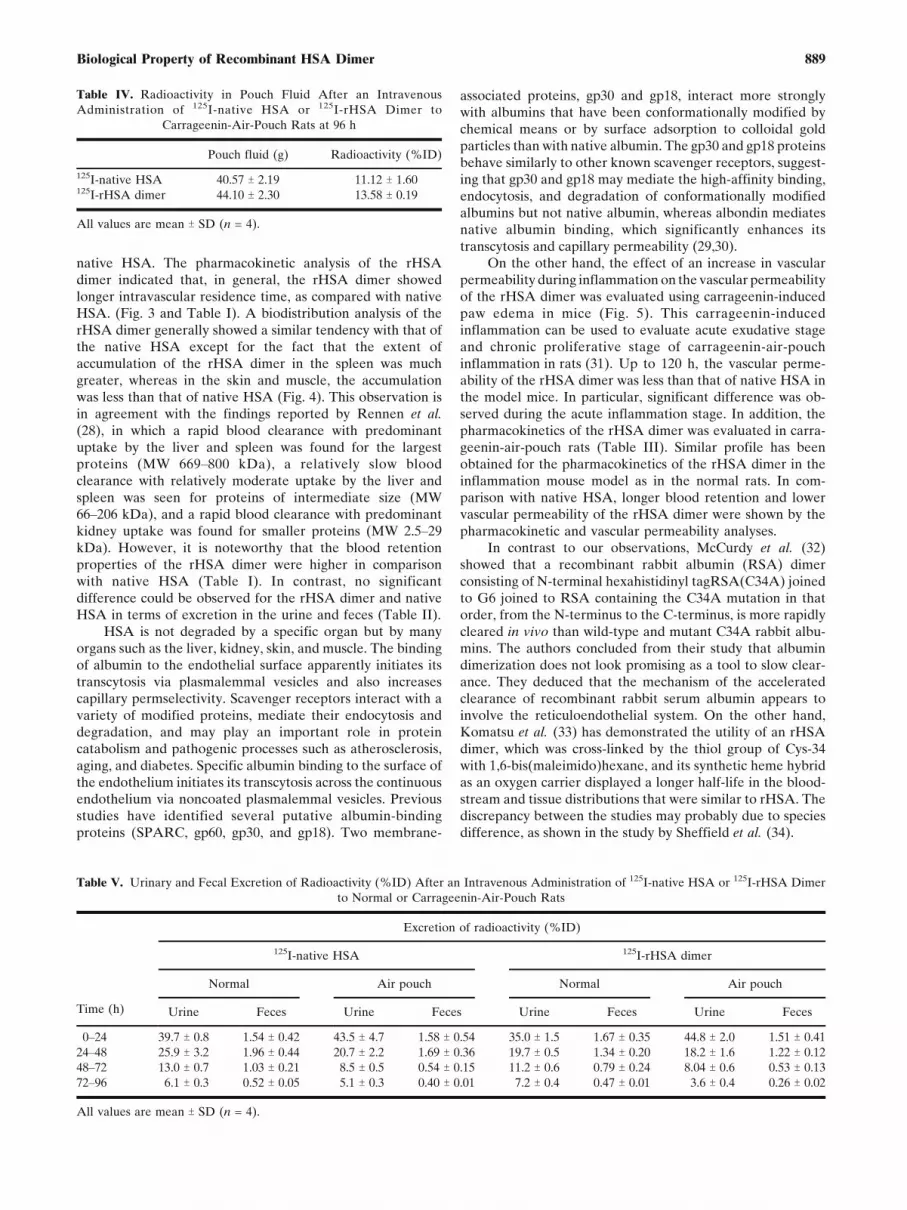
different from those used for blood collection kept inmetabolic cages (Table IV). According to the comparisonbetween rats administered with the same proteins, the air-pouch model rat showed a greater decrease in the earlyexcretion in both 125I-native HSA and the 125I-rHSA dimerthan normal rats. In addition, the 96-h alveolar activity of ratskept in the metabolic cage was determined. No significantdifference in the weight of alveolar fluid was found between125I-native HSA and the 125I-rHSA dimer (Table V).
DISCUSSION
Intravascular fluid therapy is a common critical careintervention. Many fluids have been studied for use inresuscitation, and these include isotonic sodium chloridesolution, lactated Ringer solution, hypertonic saline, albumin,purified protein fractions, freshly frozen plasma, hetastarch,pentastarch, and dextran 70. In spite of the many theoreticalbenefits of human albumin administration in critically illpatients, there has been little evidence to support itswidespread clinical use. Previous systematic reviews haveproduced conflicting results regarding the safety and efficacyof the use of albumin (2,22). The recently reported saline vs.albumin evaluation study has provided conclusive evidencethat 4% albumin is as safe as saline for resuscitation,although no overall benefit of albumin use was reported(3,23). Albumin solutions for resuscitation may still bewarranted in certain highly selected patient populations suchas liver transplant patients and burn patients.
Albumin plays an important role in the circulation bygenerating an inward oncotic force that counteracts theoutward capillary hydrostatic force. Without albumin, plasmavolume cannot be maintained and massive interstitial edemaresults. In burn shock, in which the effective circulation isreduced, albumin and extracellular fluid are rapidly shiftedfrom the plasma and sequestered in interstitial fluids. It iscommonly thought that the permeability of the capillarymembranes is pathologically increased in states of shock.Burn injury is also characterized by a dramatic increase incapillary protein permeability. When shock does not developin small burns, or is prevented by fluid therapy, the rate ofprotein extravasation may increase to up to 100 times thenormal rate. Mechanisms that regulate plasma albumincontent are the transcapillary escape rate of albumin(TERalb), lymphatic albumin return, and the albumin syn-thesis-to-degradation ratio. TERalb measures the whole bodyrate at which albumin leaves the vascular space. It iscontrolled by macrovascular and microvascular factors in-cluding plasma albumin concentration, plasma atrial natri-uretic peptide concentration, capillary filtration coefficient,capillary hydrostatic pressure, interstitial fluid hydrostaticpressure, interstitial fluid colloid osmotic pressure, andinterstitial fluid albumin concentration (24,25).
Albumin leaves the circulation via several mechanisms,which vary with the tissue involved. In organs havingsinusoids, i.e., liver and bone marrow, plasma can passthrough large gaps in the endothelium. Some other organshave fenestrated endothelia that allow unimpeded passage;the pancreas, small intestine, and adrenal gland areexamples of these. Collectively, these convection mecha-nisms account for about 50% of albumin transport from thecirculation. The transport may be described as taking placethrough two sets of parallel and cylindrical pores, one setwith diameters of 45Y60 A and another with a diameter of�200 A. It has been shown that albumin, with an estimatedradius of 35.5 A, and water do not share a commonpathway in crossing the endothelial monolayer, suggestingthe existence of a large pore pathway for albumin (26).Results reported by Sejrsen et al. (27) indicate that thepredominant transcapillary transport mechanism for 131I-albumin is compatible with transcapillary diffusion throughpores with an effective equivalent pore radius of 145 A.Hence, the extent of extravasation of albumin may bereduced by increasing its molecular size.
In this study, we attempted to express an rHSA dimer byfusing two HSA molecules, containing two sets of the proteinsequence of 585 amino acids, linked with an amino acidlinker (GGGGS)2. As can be inferred from the intrinsicfluorescence spectra and CD spectra, the structural charac-teristics of the rHSA dimer are almost identical to that of
Fig. 5. Vascular permeability of 125I-native HSA (filled bar) and 125I-
rHSA dimer (open bar) in mouse models of paw edema. A line graph
shows the increase in footpad thickness. The data are the average
values of five to seven experiments (TSD). *p < 0.05, .p < 0.01 vs.125I-native HSA.
Table III. Pharmacokinetic Parameters After an Intravenous Administration of 125I-native HSA or 125I-rHSA Dimer to Carrageenin-Air-
Pouch Rats
AUC (min x %ID/mL) CLtot (2L/h) Vdss (mL) T1/2, " (h)
125I-native HAS 103.1 T 5.3 974.6 T 53.2 22.38 T 1.01 24.85 T 0.55125I-rHSA dimer 164.0 T 10.1- 612.9 T 36.4- 19.47 T 1.09. 27.30 T 1.40*
All values are mean T SD (n = 5).*p < 0.05, . p < 0.01, - p < 0.001 vs. 125 I-native HSA.
888 Matsushita et al.
native HSA. The pharmacokinetic analysis of the rHSAdimer indicated that, in general, the rHSA dimer showedlonger intravascular residence time, as compared with nativeHSA. (Fig. 3 and Table I). A biodistribution analysis of therHSA dimer generally showed a similar tendency with that ofthe native HSA except for the fact that the extent ofaccumulation of the rHSA dimer in the spleen was muchgreater, whereas in the skin and muscle, the accumulationwas less than that of native HSA (Fig. 4). This observation isin agreement with the findings reported by Rennen et al.
(28), in which a rapid blood clearance with predominantuptake by the liver and spleen was found for the largestproteins (MW 669Y800 kDa), a relatively slow bloodclearance with relatively moderate uptake by the liver andspleen was seen for proteins of intermediate size (MW66Y206 kDa), and a rapid blood clearance with predominantkidney uptake was found for smaller proteins (MW 2.5Y29kDa). However, it is noteworthy that the blood retentionproperties of the rHSA dimer were higher in comparisonwith native HSA (Table I). In contrast, no significantdifference could be observed for the rHSA dimer and nativeHSA in terms of excretion in the urine and feces (Table II).
HSA is not degraded by a specific organ but by manyorgans such as the liver, kidney, skin, and muscle. The bindingof albumin to the endothelial surface apparently initiates itstranscytosis via plasmalemmal vesicles and also increasescapillary permselectivity. Scavenger receptors interact with avariety of modified proteins, mediate their endocytosis anddegradation, and may play an important role in proteincatabolism and pathogenic processes such as atherosclerosis,aging, and diabetes. Specific albumin binding to the surface ofthe endothelium initiates its transcytosis across the continuousendothelium via noncoated plasmalemmal vesicles. Previousstudies have identified several putative albumin-bindingproteins (SPARC, gp60, gp30, and gp18). Two membrane-
associated proteins, gp30 and gp18, interact more stronglywith albumins that have been conformationally modified bychemical means or by surface adsorption to colloidal goldparticles than with native albumin. The gp30 and gp18 proteinsbehave similarly to other known scavenger receptors, suggest-ing that gp30 and gp18 may mediate the high-affinity binding,endocytosis, and degradation of conformationally modifiedalbumins but not native albumin, whereas albondin mediatesnative albumin binding, which significantly enhances itstranscytosis and capillary permeability (29,30).
On the other hand, the effect of an increase in vascularpermeability during inflammation on the vascular permeabilityof the rHSA dimer was evaluated using carrageenin-inducedpaw edema in mice (Fig. 5). This carrageenin-inducedinflammation can be used to evaluate acute exudative stageand chronic proliferative stage of carrageenin-air-pouchinflammation in rats (31). Up to 120 h, the vascular perme-ability of the rHSA dimer was less than that of native HSA inthe model mice. In particular, significant difference was ob-served during the acute inflammation stage. In addition, thepharmacokinetics of the rHSA dimer was evaluated in carra-geenin-air-pouch rats (Table III). Similar profile has beenobtained for the pharmacokinetics of the rHSA dimer in theinflammation mouse model as in the normal rats. In com-parison with native HSA, longer blood retention and lowervascular permeability of the rHSA dimer were shown by thepharmacokinetic and vascular permeability analyses.
In contrast to our observations, McCurdy et al. (32)showed that a recombinant rabbit albumin (RSA) dimerconsisting of N-terminal hexahistidinyl tagRSA(C34A) joinedto G6 joined to RSA containing the C34A mutation in thatorder, from the N-terminus to the C-terminus, is more rapidlycleared in vivo than wild-type and mutant C34A rabbit albu-mins. The authors concluded from their study that albumindimerization does not look promising as a tool to slow clear-ance. They deduced that the mechanism of the acceleratedclearance of recombinant rabbit serum albumin appears toinvolve the reticuloendothelial system. On the other hand,Komatsu et al. (33) has demonstrated the utility of an rHSAdimer, which was cross-linked by the thiol group of Cys-34with 1,6-bis(maleimido)hexane, and its synthetic heme hybridas an oxygen carrier displayed a longer half-life in the blood-stream and tissue distributions that were similar to rHSA. Thediscrepancy between the studies may probably due to speciesdifference, as shown in the study by Sheffield et al. (34).
Table IV. Radioactivity in Pouch Fluid After an Intravenous
Administration of 125I-native HSA or 125I-rHSA Dimer to
Carrageenin-Air-Pouch Rats at 96 h
Pouch fluid (g) Radioactivity (%ID)
125I-native HSA 40.57 T 2.19 11.12 T 1.60125I-rHSA dimer 44.10 T 2.30 13.58 T 0.19
All values are mean T SD (n = 4).
Table V. Urinary and Fecal Excretion of Radioactivity (%ID) After an Intravenous Administration of 125I-native HSA or 125I-rHSA Dimer
to Normal or Carrageenin-Air-Pouch Rats
Time (h)
Excretion of radioactivity (%ID)
125I-native HSA 125I-rHSA dimer
Normal Air pouch Normal Air pouch
Urine Feces Urine Feces Urine Feces Urine Feces
0Y24 39.7 T 0.8 1.54 T 0.42 43.5 T 4.7 1.58 T 0.54 35.0 T 1.5 1.67 T 0.35 44.8 T 2.0 1.51 T 0.41
24Y48 25.9 T 3.2 1.96 T 0.44 20.7 T 2.2 1.69 T 0.36 19.7 T 0.5 1.34 T 0.20 18.2 T 1.6 1.22 T 0.12
48Y72 13.0 T 0.7 1.03 T 0.21 8.5 T 0.5 0.54 T 0.15 11.2 T 0.6 0.79 T 0.24 8.04 T 0.6 0.53 T 0.13
72Y96 6.1 T 0.3 0.52 T 0.05 5.1 T 0.3 0.40 T 0.01 7.2 T 0.4 0.47 T 0.01 3.6 T 0.4 0.26 T 0.02
All values are mean T SD (n = 4).
889Biological Property of Recombinant HSA Dimer

Indeed, the clearance of albumin depends on variousfactors including its degradation by a variety of organs, trans-capillary escape, filtration and reabsorption, and lymphaticdrainage, just to name a few. These pathways in turn are alsoinfluenced by the physicochemical state of the protein, such asits charge and conformation. In skin, the clearance for themost neutral modified albumins and cationic albumins wasfound to be 20 and 80% greater than that for native albumin,respectively. In skeletal muscle, the clearance for the mostneutral modified albumins and cationic albumins was found tobe 50 and 150% greater than that for native albumin, respec-tively. This clearly shows that charge affects the transvasculartransport of albumin (35,36). In this study, the fusion has beencarried out through the N- and C-terminals. Since no conju-gation of any of the amino acid residues has taken place, thecharge of the rHSA dimer should be the same as the nativeHSA (monomer albumin). In addition, environmental factorssuch as the presence of nitric oxide have also been reported toinfluence the extravasation of albumin (37).
Collectively, an increase in the molecular size of HSAprobably prevents or retards its convection movement throughpores with diameters of 45Y60 A, as observed by an increase inAUC and a decrease in the volume of distribution as well asthe clearance of the dimer. Meanwhile, since the dimerexhibits structural features similar to those of native albumin,we hypothesize that transvascular movement of the rHSAdimer takes place via the same route, i.e., gp60, as evidencedby the similarity in tissue distribution profile. However, fur-ther research must be carried out to validate such speculation.
On the other hand, since HSA is an endogenous protein,it has been developed for use as a drug carrier due to itsexcellent biological compatibility. A promising outcome hasalso been reported using recombinant albumin as a syntheticheme carrier protein in producing artificial blood (38,39). It isnoteworthy that albumin has recently been used as a carrier toprolong the blood retention properties of therapeutic proteinssuch as interferon and growth hormone. Through geneticfusion technology, the therapeutic protein is fused to albuminand, as a result, the plasma half-life of the protein becomesdependent on the half-life of albumin (40Y45). Fusion toalbumin offers several advantages. Albumin is the most abun-dant protein in mammalian plasma and one of the longestlived. It lacks posttranslational modifications, with the excep-tion of extensive disulfide bonding. If albumin can be fused in-frame with a therapeutic protein as a single-chain polypeptide,the novel protein may acquire the slow clearance profile ofalbumin, while retaining the activity important for clinical use.On the other hand, although conjugation of polyethyleneglycol with the protein may also produce similar tendency,such approach involves modification of the internal aminoacid side chain, which may change the charge distribution and,hence, affect the stability of the protein. Albuferon (albumin-interferon alpha) exhibits more antiviral activity at clinicallyachieved serum levels than standard interferon alpha or themodified interferons, Pegasys (peginterferon alpha-2a) andPeg-Intron (peginterferon alpha-2b) (43).
Clinical and preclinical results to date demonstrate thatAlbuferon is well tolerated, with adverse events that aretransient and mostly mild to moderate in severity, and exhibitsa robust antiviral activity, with a pharmacokinetic profile thatsupports dosing at intervals of 2 to 4 weeks. This prolongation
in half-life prevents the frequent administration of thetherapeutic protein. Hence, although albumin genetic fusiontechnology is very promising, if the blood retention propertiesof albumin can be improved further, it is expected that suchalbumin will be of great clinical use as a new drug deliverysystem material in addition to being a plasma expander.
ACKNOWLEDGMENTS
We wish to thank the members of the Gene TechnologyCenter in Kumamoto University for their important contri-butions to these experiments.
REFERENCES
1. T. Peters. All About Albumin: Biochemistry, Genetics, andMedical Applications, Academic, San Diego, 1996.
2. Cochrane Injuries Group. Human albumin administration incritically ill patients: systematic review of randomised controlledtrials. Cochrane Injuries Group Albumin Reviewers. BMJ 317:235Y240 (1998).
3. S. Finfer, R. Bellomo, N. Boyce, J. French, J. Myburgh, and R.Norton. A comparison of albumin and saline for fluid resusci-tation in the intensive care unit. N. Engl. J. Med. 350:2247Y2256(2004).
4. H. Rubin, S. Carlson, M. DeMeo, D. Ganger, and R. M. Craig.Randomized, double-blind study of intravenous human albuminin hypoalbuminemic patients receiving total parenteral nutri-tion. Crit. Care Med. 25:249Y252 (1997).
5. G. Akerstrom and B. Lisander. Tissue extravasation of albuminfrom intraabdominal trauma in rats. Acta Anaesthesiol. Scand.35:257Y261 (1991).
6. M. P. Margarson and N. C. Soni. Tissue extravasation ofalbumin from intraabdominal trauma in rats. Br. J. Anaesth.92:821Y826 (2004).
7. S. Berg, M. Golster, and B. Lisander. Albumin extravasationand tissue washout of hyaluronan after plasma volume expan-sion with crystalloid or hypooncotic colloid solutions. ActaAnaesthesiol. Scand. 46:166Y172 (2002).
8. M. Mathru, B. Blakeman, D. J. Dries, B. Kleinman, and P.Kumar. Permeability pulmonary edema following lung resec-tion. Chest 98:1216 Y1218 (1990).
9. K. Byrne, J. L. Tatum, D. A. Henry, J. I. Hirsch, M. Crossland,T. Barnes, J. A. Thompson, J. Young, and H. J. Sugerman.Increased morbidity with increased pulmonary albumin flux insepsis-related adult respiratory distress syndrome. Crit. CareMed. 20:28 Y34 (1992).
10. S. Galatius, L. Bent-Hansen, H. Wroblewski, V. B. Sorensen, T.Norgaard, and J. Kastrup. Plasma disappearance of albumin andimpact of capillary thickness in idiopathic dilated cardiomyop-athy and after heart transplantation. Circulation 102:319Y325(2000).
11. T. Oshima, F. S. Laroux, L. L. Coe, Z. Morise, S. Kawachi, P.Bauer, M. B. Grisham, R. D. Specian, P. Carter, S. Jennings, D.N. Granger, T. Joh, and J. S. Alexander. Interferon-gamma andinterleukin-10 reciprocally regulate endothelial junction integri-ty and barrier function. Microvasc. Res. 61:130 Y143 (2001).
12. R. F. Chen. Removal of fatty acids from serum albumin bycharcoal treatment. J. Biol. Chem. 242:173Y181 (1967).
13. S. Matsushita, Y. Isima, V. T. Chuang, H. Watanabe, S. Tanase,T. Maruyama, and M. Otagiri. Functional analysis of recombi-nant human serum albumin domains for pharmaceutical appli-cations. Pharm. Res. 21:1924 Y1932 (2004).
14. J. Bhatia, S. K. Sharma, K. A. Chester, R. B. Pedley, R. W.Boden, D. A. Read, G. M. Boxer, N. P. Michael, and R. H.Begent. Catalytic activity of an in vivo tumor targeted anti-CEAscFv::carboxypeptidase G2 fusion protein. Int. J. Cancer85:571Y577 (2000).
890 Matsushita et al.

15. D. J. Hnatowich, W. W. Layne, and R. L. Childs. Thepreparation and labeling of DTPA-coupled albumin. Int. J.Appl. Radiat. Isot. 33:327Y332 (1982).
16. H. Katsumi, M. Nishikawa, S. F. Ma, F. Yamashita, and M.Hashida. Physicochemical, tissue distribution, and vasodilationcharacteristics of nitrosated serum albumin: delivery of nitricoxide in vivo. J. Pharm. Sci. 93:2343 Y2352 (2004).
17. K. Yamaoka, Y. Tanigawara, T. Nakagawa, and T. A. Uno.Pharmacokinetic analysis program (multi) for microcomputer. J.Pharmacobio-dyn. 4:879Y885 (1981).
18. S. Tsurufuji, H. Sato, K. R. Min, and K. Ohuchi. Difference inthe anti-inflammatory effect of indomethacin between acute andchronic stages of carrageenin-induced inflammation. J. Pharma-cobio-dyn. 1:8 Y14 (1978).
19. D. Shan, O. W. Press, T. T. Tsu, M. S. Hayden, and J. A.Ledbetter. Characterization of scFv-Ig constructs generatedfrom the anti-CD20 mAb 1F5 using linker peptides of varyinglengths. J. Immunol. 162:6589Y6595 (1999).
20. M. Gustavsson, J. Lehtio, S. Denman, T. T. Teeri, K. Hult, andM. Martinelle. Stable linker peptides for a cellulose-bindingdomain-lipase fusion protein expressed in Pichia pastoris.Protein Eng. 14:711Y715 (2001).
21. I. Posadas, M. Bucci, F. Roviezzo, A. Rossi, L. Parente, L.Sautebin, and G. Cirino. Carrageenan-induced mouse pawoedema is biphasic, ageYweight dependent and displays differ-ential nitric oxide cyclooxygenase-2 expression. Br. J. Pharma-col. 142:331Y338 (2004).
22. M. M. Wilkes and R. J. Navickis. Patient survival after humanalbumin administration. A meta-analysis of randomized, con-trolled trials. Ann. Intern. Med. 135:149Y164 (2001).
23. E. Fan and T. E. Stewart. Albumin in critical care: SAFE, butworth its salt? Crit. Care 8:297Y299 (2004).
24. B. Rippe and B. Haraldsson. Transport of macromoleculesacross microvascular walls: the two-pore theory. Physiol. Rev.74:163Y219 (1994).
25. C. C. Michel and F. E. Curry. Microvascular permeability.Physiol. Rev. 79:703Y761 (1999).
26. R. O. Dull, H. Jo, H. Sill, T. M. Hollis, and J. M. Tarbell. Theeffect of varying albumin concentration and hydrostatic pressureon hydraulic conductivity and albumin permeability of culturedendothelial monolayers. Microvasc. Res. 41:390 Y 407 (1991).
27. P. Sejrsen, W. P. Paaske, and O. Henriksen. Capillary perme-ability of 131I-albumin in skeletal muscle. Microvasc. Res. 29:265Y281 (1985).
28. H. J. Rennen, J. Makarewicz, W. J. Oyen, P. Laverman, F. H.Corstens, and O. C. Boerman. The effect of molecular weight onnonspecific accumulation of (99m)T-labeled proteins in inflam-matory foci. Nucl. Med. Biol. 28:401Y 408 (2001).
29. J. E. Schnitzer, A. Sung, R. Horvat, and J. Bravo. Preferentialinteraction of albumin-binding proteins, gp30 and gp18, withconformationally modified albumins. Presence in many cells andtissues with a possible role in catabolism. J. Biol. Chem.267:24544 Y24553 (1992).
30. J. E. Schnitzer and P. Oh. Albondin-mediated capillary perme-ability to albumin. Differential role of receptors in endothelialtranscytosis and endocytosis of native and modified albumins. J.Biol. Chem. 269:6072Y 6082 (1994).
31. H. Sato, M. Hashimoto, K. Sugio, K. Ohuchi, and S. Tsurufuji.Comparative study between steroidal and nonsteroidal anti-inflammatory drugs on the mode of their actions on vascularpermeability in rat carrageenin-air-pouch inflammation. J.Pharmacobio-dyn. 3:345Y352 (1980).
32. T. R. McCurdy, S. Gataiance, L. J. Eltringham-Smith, and W. P.Sheffield. A covalently linked recombinant albumin dimer ismore rapidly cleared in vivo than are wild-type and mutantC34A albumin. J. Lab. Clin. Med. 143:115 Y124 (2004).
33. T. Komatsu, Y. Oguro, Y. Teramura, S. Takeoka, J. Okai, M.Anraku, M. Otagiri, and E. Tsuchida. Physicochemical charac-terization of cross-linked human serum albumin dimer and itssynthetic heme hybrid as an oxygen carrier. Biochim. Biophys.Acta 1675:21Y31 (2004).
34. W. P. Sheffield, A. Mamdani, G. Hortelano, S. Gataiance, L.Eltringham-Smith, M. E. Begbie, R. A. Leyva, P. S. Liaw, and F. A.Ofosu. Effects of genetic fusion of factor IX to albumin on in vivoclearance in mice and rabbits. Br. J. Haematol. 126:565Y573 (2004).
35. R. R. Gandhi and D. R. Bell. Importance of charge ontransvascular albumin transport in skin and skeletal muscle.Am. J. Physiol. 262:H999YH1008 (1992).
36. H. Vink and B. R. Duling. Capillary endothelial surface layerselectively reduces plasma solute distribution volume. Am. J.Physiol, Heart Circ. Physiol. 278:H285YH289 (2000).
37. L. W. Chen, J. S. Wang, B. Hwang, J. S. Chen, and C. M. Hsu.Reversal of the effect of albumin on gut barrier function in burnby the inhibition of inducible isoform of nitric oxide synthase.Arch. Surg. 138:1219Y1225 (2003).
38. E. Tsuchida, K. Ando, H. Maejima, N. Kawai, T. Komatsu, S.Takeoka, and H. Nishide. Properties of and oxygen binding byalbumin-tetraphenylporphyrinatoiron(II) derivative complexes.Bioconjug. Chem. 8:534 Y538 (1997).
39. T. Komatsu, H. Yamamoto, Y. Huang, H. Horinouchi, K.Kobayashi, and E. Tsuchida. Exchange transfusion with synthet-ic oxygen-carrying plasma protein Yalbumin-hemeY into an acuteanemia rat model after seventy-percent hemodilution. J.Biomed. Mater. Res., A. 71:644 Y 651 (2004).
40. S. Syed, P. Schuyler, M. Kulczycky, and W. P. Sheffield. Potentantithrombin activity and delayed clearance from the circulationcharacterize recombinant hirudin genetically fused to albumin.Blood 89:3243Y3252 (1997).
41. P. Yeh, D. Landais, M. Lemaitre, I. Maury, J.-Y. Crenne, J.Becquart, A. Murry-Brelier, F. Boucher, G. Montay, R. Fleer,P.-H. Hirel, J.-F. Mayaux, and D. Klatzmann. Design of yeast-secreted albumin derivatives for human therapy: biological andantiviral properties of a serum albuminYCD4 genetic conjugate.Proc. Natl. Acad. Sci. U. S. A. 89:1904 Y1908 (1992).
42. M. S. Dennis, M. Zhang, Y. G. Meng, M. Kadkhodayan, D.Kirchhofer, D. Combs, and L. A. Damico. Albumin binding as ageneral strategy for improving the pharmacokinetics of proteins.J. Biol. Chem. 277:35035Y35043 (2002).
43. B. L. Osborn, H. S. Olsen, B. Nardelli, J. H. Murray, J. X. Zhou,A. Garcia, G. Moody, L. S. Zaritskaya, and C. Sung. Pharma-cokinetic and pharmacodynamic studies of a human serumalbuminYinterferon-alpha fusion protein in cynomolgusmonkeys. J. Pharmacol. Exp. Ther. 303:540 Y548 (2002).
44. C. Sung, B. Nardelli, D. W. LaFleur, E. Blatter, M. Corcoran, H.S. Olsen, C. E. Birse, O. K. Pickeral, J. Zhang, D. Shah, G.Moody, S. Gentz, L. Beebe, and P. A. Moore. An IFN-betaYalbumin fusion protein that displays improved pharmaco-kinetic and pharmacodynamic properties in nonhuman primates.J. Interferon Cytokine Res. 23:25Y36 (2003).
45. B. L. Osborn, L. Sekut, M. Corcoran, C. Poortman, B. Sturm, G.Chen, D. Mather, H. L. Lin, and T. J. Parry. Albutropin: agrowth hormoneYalbumin fusion with improved pharmacokinet-ics and pharmacodynamics in rats and monkeys. Eur. J.Pharmacol. 456:149Y158 (2002).
891Biological Property of Recombinant HSA Dimer
Copyright © 2022 FDOKUMEN