Amyloid Fibril Formation and Other Aggregate Species Formed by Human Serum Albumin Association
18
Existence of Different Structural Intermediates on the Fibrillation Pathway of Human Serum Albumin Josue ´ Jua ´ rez, Pablo Taboada,* and Vı ´ctor Mosquera Grupo de Fı ´sica de Coloides y Polı ´meros, Departamento de Fı ´sica de la Materia Condensada, Facultad de Fı ´sica, Universidad de Santiago de Compostela, Santiago de Compostela, Spain ABSTRACT The fibrillation propensity of the multidomain protein human serum albumin (HSA) was analyzed under different solution conditions. The aggregation kinetics, protein conformational changes upon self-assembly, and structure of the different intermediates on the fibrillation pathway were determined by means of thioflavin T (ThT) fluorescence and Congo Red absor- bance; far- and near-ultraviolet circular dichroism; tryptophan fluorescence; Fourier transform infrared spectroscopy; x-ray diffraction; and transmission electron, scanning electron, atomic force, and microscopies. HSA fibrillation extends over several days of incubation without the presence of a lag phase, except for HSA samples incubated at acidic pH and room temperature in the absence of electrolyte. The absence of a lag phase occurs if the initial aggregation is a downhill process that does not require a highly organized and unstable nucleus. The fibrillation process is accompanied by a progressive increase in the b-sheet (up to 26%) and unordered conformation at the expense of a-helical conformation, as revealed by ThT fluorescence and circular dichroism and Fourier transform infrared spectroscopies, but changes in the secondary structure contents depend on solution conditions. These changes also involve the presence of different structural intermediates in the aggregation pathway, such as oligomeric clusters (globules), bead-like structures, and ring-shaped aggregates. We suggest that fibril formation may take place through the role of association-competent oligomeric intermediates, resulting in a kinetic pathway via clustering of these oligo- meric species to yield protofibrils and then fibrils. The resultant fibrils are elongated but curly, and differ in length depending on solution conditions. Under acidic conditions, circular fibrils are commonly observed if the fibrils are sufficiently flexible and long enough for the ends to find themselves regularly in close proximity to each other. These fibrils can be formed by an antiparallel arrangement of b-strands forming the b-sheet structure of the HSA fibrils as the most probable configuration. Very long incubation times lead to a more complex morphological variability of amyloid mature fibrils (i.e., long straight fibrils, flat-ribbon structures, laterally connected fibers, etc.). We also observed that mature straight fibrils can also grow by protein oligomers tending to align within the immediate vicinity of the fibers. This filament þ monomers/oligomers scenario is an alternative pathway to the other- wise dominant filament þ filament manner of the protein fibril’s lateral growth. Conformational preferences for a certain pathway to become active may exist, and the influence of environmental conditions such as pH, temperature, and salt must be considered. INTRODUCTION b-Sheet-based assemblies have attracted much interest from multidisciplinary researchers because of their association with a variety of diseases and their emerging potential in material science and biotechnology (1,2). Protein misfolding and self-assembly into highly ordered b-sheet-rich fibrillar assemblies known as amyloid fibrils are common features of a growing class of systemic and neurodegenerative diseases, including Alzheimer’s, Parkinson’s, and Hunting- ton’s diseases; senile systemic amyloidoses; type II diabetes (3,4); and many others. The ability to fibrillate is independent of the original native structure of the protein, whose amino acid sequence primarily appears to play a key role in terms of filament arrangement (5), fibrillation kinetics (6), and overall yield and stability of the fibrils (7,8). Fibrillation originates under conditions in which proteins are partially destabilized or completely unfolded (9), and the formation of the amyloid fibrils reflects an alternative to the native packing conformational struggle of a polypeptide chain to 1), reduce its surface-accessible area; 2), saturate hydrogen bonding; and 3), reach an alternative ‘‘nonnative’’ global free energy minimum (10). Therefore, investigators have emphasized understanding and inhibiting amyloid formation more so than amyloid dissociation and clearance. Although the stability of b-sheet-rich amyloid fibrils against proteases, acids, and chemical denaturants has been shown, increasing evidence from human (11) and in vitro studies indicates that a dynamic structure exists within amyloid fibrils and suggests that the process of amyloid formation is reversible (12). These findings, along with the fact that strategies aimed at stabilizing amyloid fibrils and/or accelerating their clear- ance seem to reverse the disease phenotype (13,14), suggest that a detailed understanding of the formation, stability, and dynamic behavior of amyloid fibrils is critically important to the development of therapeutic strategies for amyloid diseases. Thus, the reversible untangling of amyloid archi- tecture and intrafibrillar packing of the b-pleated sheets is a key issue to consider in designing inhibitors of fibrillar growth (15), and insights into the fibrillar assembly mecha- nisms may help elucidate the etiology of the ‘‘prion diseases’’, provided that the subtle structural differences underlying the puzzling phenomenon of ‘‘prion strains’’ can be understood (16). Submitted September 30, 2008, and accepted for publication December 1, 2008. *Correspondence: [email protected] Editor: Ruth Nussinov. Ó 2009 by the Biophysical Society 0006-3495/09/03/2353/18 $2.00 doi: 10.1016/j.bpj.2008.12.3901 Biophysical Journal Volume 96 March 2009 2353–2370 2353
-
Upload
independent -
Category
Documents
-
view
3 -
download
0
Transcript of Amyloid Fibril Formation and Other Aggregate Species Formed by Human Serum Albumin Association

Biophysical Journal Volume 96 March 2009 2353–2370 2353
Existence of Different Structural Intermediates on the Fibrillation Pathwayof Human Serum Albumin
Josue Juarez, Pablo Taboada,* and Vıctor MosqueraGrupo de Fısica de Coloides y Polımeros, Departamento de Fısica de la Materia Condensada, Facultad de Fısica, Universidad de Santiago deCompostela, Santiago de Compostela, Spain
ABSTRACT The fibrillation propensity of the multidomain protein human serum albumin (HSA) was analyzed under differentsolution conditions. The aggregation kinetics, protein conformational changes upon self-assembly, and structure of the differentintermediates on the fibrillation pathway were determined by means of thioflavin T (ThT) fluorescence and Congo Red absor-bance; far- and near-ultraviolet circular dichroism; tryptophan fluorescence; Fourier transform infrared spectroscopy; x-raydiffraction; and transmission electron, scanning electron, atomic force, and microscopies. HSA fibrillation extends over severaldays of incubation without the presence of a lag phase, except for HSA samples incubated at acidic pH and room temperature inthe absence of electrolyte. The absence of a lag phase occurs if the initial aggregation is a downhill process that does not requirea highly organized and unstable nucleus. The fibrillation process is accompanied by a progressive increase in the b-sheet (up to26%) and unordered conformation at the expense of a-helical conformation, as revealed by ThT fluorescence and circulardichroism and Fourier transform infrared spectroscopies, but changes in the secondary structure contents depend on solutionconditions. These changes also involve the presence of different structural intermediates in the aggregation pathway, such asoligomeric clusters (globules), bead-like structures, and ring-shaped aggregates. We suggest that fibril formation may take placethrough the role of association-competent oligomeric intermediates, resulting in a kinetic pathway via clustering of these oligo-meric species to yield protofibrils and then fibrils. The resultant fibrils are elongated but curly, and differ in length depending onsolution conditions. Under acidic conditions, circular fibrils are commonly observed if the fibrils are sufficiently flexible and longenough for the ends to find themselves regularly in close proximity to each other. These fibrils can be formed by an antiparallelarrangement of b-strands forming the b-sheet structure of the HSA fibrils as the most probable configuration. Very long incubationtimes lead to a more complex morphological variability of amyloid mature fibrils (i.e., long straight fibrils, flat-ribbon structures,laterally connected fibers, etc.). We also observed that mature straight fibrils can also grow by protein oligomers tending to alignwithin the immediate vicinity of the fibers. This filament þ monomers/oligomers scenario is an alternative pathway to the other-wise dominant filament þ filament manner of the protein fibril’s lateral growth. Conformational preferences for a certain pathwayto become active may exist, and the influence of environmental conditions such as pH, temperature, and salt must be considered.
INTRODUCTION
b-Sheet-based assemblies have attracted much interest from
multidisciplinary researchers because of their association
with a variety of diseases and their emerging potential in
material science and biotechnology (1,2). Protein misfolding
and self-assembly into highly ordered b-sheet-rich fibrillar
assemblies known as amyloid fibrils are common features
of a growing class of systemic and neurodegenerative
diseases, including Alzheimer’s, Parkinson’s, and Hunting-
ton’s diseases; senile systemic amyloidoses; type II diabetes
(3,4); and many others. The ability to fibrillate is independent
of the original native structure of the protein, whose amino
acid sequence primarily appears to play a key role in terms
of filament arrangement (5), fibrillation kinetics (6), and
overall yield and stability of the fibrils (7,8). Fibrillation
originates under conditions in which proteins are partially
destabilized or completely unfolded (9), and the formation
of the amyloid fibrils reflects an alternative to the native
packing conformational struggle of a polypeptide chain to
1), reduce its surface-accessible area; 2), saturate hydrogen
Submitted September30,2008, and accepted for publication December1,2008.
*Correspondence: [email protected]
Editor: Ruth Nussinov.
� 2009 by the Biophysical Society
0006-3495/09/03/2353/18 $2.00
bonding; and 3), reach an alternative ‘‘nonnative’’ global
free energy minimum (10). Therefore, investigators have
emphasized understanding and inhibiting amyloid formation
more so than amyloid dissociation and clearance. Although
the stability of b-sheet-rich amyloid fibrils against proteases,
acids, and chemical denaturants has been shown, increasing
evidence from human (11) and in vitro studies indicates that
a dynamic structure exists within amyloid fibrils and
suggests that the process of amyloid formation is reversible
(12). These findings, along with the fact that strategies aimed
at stabilizing amyloid fibrils and/or accelerating their clear-
ance seem to reverse the disease phenotype (13,14), suggest
that a detailed understanding of the formation, stability, and
dynamic behavior of amyloid fibrils is critically important to
the development of therapeutic strategies for amyloid
diseases. Thus, the reversible untangling of amyloid archi-
tecture and intrafibrillar packing of the b-pleated sheets is
a key issue to consider in designing inhibitors of fibrillar
growth (15), and insights into the fibrillar assembly mecha-
nisms may help elucidate the etiology of the ‘‘prion
diseases’’, provided that the subtle structural differences
underlying the puzzling phenomenon of ‘‘prion strains’’
can be understood (16).
doi: 10.1016/j.bpj.2008.12.3901

2354 Juarez et al.
Because of the physiological importance of human serum
albumin (HSA) as a carrier protein and blood pressure regu-
lator, and its propensity to easily aggregate in vitro, HSA has
become a good model for protein aggregation studies. More-
over, as the phenomenon of protein aggregation appears to
reflect certain generic ‘‘polymeric’’ features of proteins
(17), studying mechanisms of protein aggregation in model
systems is extremely useful for gaining a better under-
standing of the molecular mechanisms of disease-associated
amyloidogenesis.
Under physiological conditions, HSA consists of 585
amino acids in a single polypeptide chain, with a globular
structure composed of three main domains that are loosely
joined together through physical forces and six subdomains
that are wrapped by disulfide bonds. The protein contains
17 disulfide bridges and one free SH group, which facilitates
dimerization and also influences higher-order association.
Native HSA lacks any properties that suggest a predisposition
to form amyloid fibrils, since most of its sequence (>60%) is
arranged in an a-helix structure, with subsequent tightening
of its structure through intramolecular interactions such as
hydrogen bonds. Therefore, serum albumin aggregation is
promoted under conditions that favor partly destabilized
monomers and dimers, such as low pH, high temperature,
and the presence of chemical denaturants (18). In a recent
report (19), we showed that partially destabilized HSA mole-
cules form amyloid-like fibrils and other types of aggregates
under different solution conditions. These fibrils feature the
structural characteristics of amyloids: x-ray diffraction
(XRD) patterns, affinity to Congo Red (CR) and thioflavin
T (ThT), birefringence, and high stability. We now extend
that previous work to shed further light on the kinetics and
hierarchical assembly of HSA fibril formation, linking the
morphological structural transitions of aggregated protein
intermediates to conformational events on protein structure
as analyzed by means of different biophysical and spectro-
scopic methods. In this way, we present a systematic inves-
tigation of the relationship between protein conformation
and the amyloid-like self-assembly pathway for HSA under
different solution conditions. To this end, we incubated the
protein under different thermal and solvent conditions and
analyzed the protein conformation changes upon incubation.
We additionally imaged different structural intermediates on
the HSA fibrillation pathway depending on solution condi-
tions. In this way, we sought to uncover the structural
features underlying the formation of possibly cytotoxic
HSA assemblies.
MATERIALS AND METHODS
Materials
HSA (70024-90-7), CR, and ThT were obtained from Sigma (St. Louis,
MO) and used as received. All other chemicals were of the highest purity
available.
Biophysical Journal 96(6) 2353–2370
Preparation of HSA solutions
Protein was used after further purification by liquid chromatography using
a Superdex 75 column equilibrated with 0.01 M phosphate. Experiments
were carried out using double-distilled, deionized, and degassed water.
The buffer solutions used were glycine þ HCl (I ¼ 0.01 M) for pH 3.0,
and sodium monophosphate-sodium diphosphate for pH 7.4 (I ¼ 0.01 M),
respectively. HSA was dissolved in each buffer solution to a final concentra-
tion of typically 20 mg/mL and dialyzed extensively against the proper
buffer. Protein concentration was determined spectrophotometrically using
a molar absorption coefficient of 35,219 M�1 cm�1 at 280 nm (20). Before
incubation, the solution was filtered through a 0.2 mm filter into sterile test
tubes. Samples were incubated at a specified temperature in a refluxed
reactor. Samples were taken out at intervals and stored on ice before addition
of CR or ThT.
Seeding solutions
To test whether seeding with preformed aggregates increases the rate of
HSA aggregation under the different conditions in which fibrils are formed,
a protein solution was incubated for 24 h and an aliquot that corresponded to
10% (w/w) of the total protein concentration was then added to a fresh
protein solution.
CR binding
Changes in the absorbance of CR dye produced by binding onto HSA were
measured in an ultraviolet-visible spectrophotometer (DU series 640; Beck-
man Coulter, Fullerton, CA) operating at 190–1100 nm. All measurements
were made in the wavelength range of 220–500 nm in matched quartz
cuvettes. Protein solutions were diluted 20- to 200-fold into a buffer solution
with 5 mM of CR (Acros Organics, Geel, Belgium). Spectra in the presence
of the dye were compared with those of the buffer containing CR in the
absence of protein and also with those corresponding to the protein solution
without dye.
ThT spectroscopy
Protein and ThT were dissolved in the proper buffer at a final protein/dye
molar ratio of 50:1. Samples were continuously stirred during measurements.
Fluorescence was measured in a Cary Eclipse fluorescence spectrophotom-
eter equipped with a temperature control device and a multicell sample holder
(Varian Instruments, Palo Alto, CA). Excitation and emission wavelengths
were 450 and 482 nm, respectively. All intensities were background-cor-
rected for the ThT fluorescence in the respective solvent without the protein.
Protein fluorescence
To examine the conformational variations around the Tryp residue of HSA,
fluorescence emission spectra were recorded with a Cary Eclipse fluores-
cence spectrophotometer equipped with a temperature control device and
a multicell sample holder (Varian Instruments). HSA samples were excited
at 295 nm, which provides no excitation of tyrosine residues and, therefore
does not cause emission or energy transfer to the lone side chain. Slit widths
were typically 5 nm.
Circular dichroism
Far- and near-ultraviolet (UV) circular dichroism (CD) spectra were
obtained using a JASCO-715 automatic recording spectropolarimeter (Jasco,
Tokyo, Japan) with a JASCO PTC-343 Peltier-type thermostated cell holder.
Quartz cuvettes with 0.2 cm pathlength were used. CD spectra were obtained
from aliquots withdrawn from the aggregation mixtures at the indicated
conditions and recorded between 195 and 300 nm at 25�C. The mean residue
ellipticity q (deg cm2 dmol�1) was calculated from the formula:

Fibrillation Pathway of HSA 2355
q ¼ ðqobs=10ÞðMRM=lcÞ, where qobs is the observed ellipticity in deg, MRM
is the mean residue molecular mass, l is the optical pathlength
(in centimeters), and c is the protein concentration (in g mL�1). To calculate
the composition of the secondary structure of the protein, SELCON3, CON-
TIN, and DSST programs were used to analyze far-UV CD spectra. Final
results were assumed when data generated from all programs showed
convergence (21).
XRD
XRD experiments were carried out using a Siemens D5005 rotating anode
x-ray generator. Twin Gobel mirrors were used to produce a well-collimated
beam of CuKa radiation (l ¼ 1.5418 A). Samples were put into capillary
with a diameter of 0.5 mm. X-ray diffraction patterns were recorded with
an imaging plate detector (AXS F.Nr. J2-394).
Fourier transform infrared spectroscopy
Fourier transform infrared (FTIR) spectra of HSA in aqueous solutions were
determined by using an FTIR spectrometer (model IFS-66v; Bruker) equip-
ped with a horizontal ZnS ATR accessory. The spectra were obtained at
a resolution of 2 cm�1 and generally 200 scans were accumulated to obtain
a reasonable signal/noise ratio. Solvent spectra were also examined under
the same accessory and instrument conditions. Each different sample spec-
trum was obtained by digitally subtracting the solvent spectrum from the
corresponding sample spectrum. Each sample solution was repeated three
times to ensure reproducibility and averaged to produce a single spectrum.
Transmission electron microscopy
For transmission electron microscopy (TEM), suspensions of HSA were
applied to carbon-coated copper grids, blotted, washed, negatively stained
with 2% (w/v) of phosphotungstic acid, air dried, and then examined with
a Phillips CM-12 transmission electron microscope operating at an acceler-
ating voltage of 120 kV. Samples were diluted 20- to 200-fold when neces-
sary before deposition on the grids.
Scanning electron microscopy
Suspensions of HSA were applied to glass-coated stainless-steel grids,
blotted, washed, air dried, and then examined with an LEO-435VP scanning
electron microscope (Leica Microsystems GmbH, Wetlar, Germany) oper-
ating at an accelerating voltage of 30 kV. Samples were diluted 20- to
200-fold when necessary before deposition on the grids. Microanalysis of
the scanning electron microscopy (SEM) samples was also performed to
avoid the presence of impurities.
Atomic force microscopy
Atomic force microscopy (AFM) images were recorded in tapping mode
by using a multimode SPM microscope equipped with a Nanoscope IIIa
controller from Digital Instruments (Santa Barbara, CA). The microscope
was coupled to an AS-12 resp. E-scanner and an Extender Electronics Module
EX-II, which allows acquisition of phase images. The AFM probes were typi-
cally silicon SPM sensors (NCHR Nanosensors, Neuchatel, Switzerland).
Immediately after incubation, the protein samples were diluted 20–400 times
onto freshly cleaved muscovite mica (Sigma) attached to a magnetic steel disc
that served as the sample holder. The abrupt dilution of the samples immedi-
ately quenched the concentration-dependent aggregation process. The AFM
samples were dried on air or under nitrogen flow when required. Control
samples (freshly cleaved mica, and mica and buffer solution) were also inves-
tigated with AFM to exclude possible artifacts. Height and phase-shift data
were collected in the trace and the respective retrace direction of the raster.
The scan rate was tuned proportionally to the area scanned and was kept
within the 0.35–2 Hz range.
RESULTS
It is generally accepted that amyloid formation usually is
a result of misfolded and partially unfolded states acting in
competition with the normal folding pathways (22–24).
The HSA molecule is known to undergo several well-orga-
nized changes during its conformation, usually under non-
physiological conditions, as follows:
1. The N-F transition between pH 5.0 and 3.5 involves the
unfolding and separation of domain III without signifi-
cantly affecting the rest of the protein molecule
(25,26). The F form is characterized by a dramatic
increase in viscosity, lower solubility, and a significant
loss of helical content.
2. The F-E transition occurs between pH 3.5 and 1.2, and is
accompanied by a further protein expansion with a loss
of the intradomain helices of domain I. In addition,
the E form involves an increase in protein intrinsic
viscosity and a rise in the hydrodynamic axial ratio from
~4 to 9 (27).
3. The N-B transition occurs between pH 7.0 and 9.0, with
a slight reduction in helical content affecting the two interdo-
main helices and a small increase in sheet structure (28).
4. In the presence of denaturant agents, such as urea, HSA
shows a two-step, three-state transition with an interme-
diate (I) characterized by unfolding of the domain III and
partial but significant loss of native conformation of
domain I (29).
Therefore, an easy way to obtain at least partially denatu-
rated states is to induce a temperature-induced or solvent-
induced protein denaturation process through incubation.
Thus, HSA was subjected to conditions previously found to
be effective for protein aggregation, in particular those that
induce amyloid-like fibril formation (19,30). HSA solutions
were incubated at a concentration of 20 mg/mL in 0.01 M
sodium phosphate buffer, pH 7.4, or 0.01 M glycine buffer,
pH 3.0, in the presence of 0 or 50 mM of added NaCl at
25�C or 65�C for 15 days. We chose 65�C as the incubation
temperature because HSA temperature-induced denaturation
takes place through a two-state transition with a first melting
temperature, Tm, of ~56�C and a second Tm of ~62�C (31,32)
as a consequence of the sequential unfolding of the different
domains of the protein, in particular, the IIA and IIIA
subdomains. Moreover, as the pH becomes more acidic,
Tm becomes lower. We confirmed these melting temperatures
under our solution conditions by fluorescence spectroscopy,
and the results were in close agreement with previously
reported data (see Table S1 in the Supporting Material).
Therefore, it can be inferred that aggregation is unfavorable
below 65�C because protein folding competes with and
suppresses amyloid formation. It is known that hydrogen
bonding is weakened as temperature rises, but the hydro-
phobic interaction becomes strengthened (33). As the heating
process continues, some of the cooperative hydrogen bonds
Biophysical Journal 96(6) 2353–2370

2356 Juarez et al.
that stabilized helical structure begin to break and expose
hydrophobic groups to the solvent, partially unfolding the
protein structure, which favors aggregation.
Physiological conditions: kinetics and amyloidself-assembly of HSA
The propensity of protein solutions to form amyloid-like
aggregates under physiological conditions was assessed by
means of ThT fluorescence and CR absorbance measure-
ments. These two dyes specifically bind to ordered b-sheet
aggregates and, notably, to amyloid fibrils (34,35). Both
assays are necessary because positive ThT binding sometimes
does not occur with certain amyloid fibril systems (36).
Before incubation, native HSA does not display a capacity
to bind ThT. When incubated at physiological pH and room
temperature, the fluorescence emission intensity for the
protein at 482 nm is negligible, suggesting that HSA does
not form amyloid fibrils or other types of fluorescent aggre-
gates under these conditions. When the temperature is raised
to 65�C, a time-dependent increase in fluorescence is
observed (Fig. 1, a and b). The kinetics of HSA involved
the continuous rising of ThT fluorescence during the early
periods of the incubation procedure, and exhibited no discern-
ible lag phase until a quasi-plateau region was attained in the
FIGURE 1 Time evolution of ThT fluorescence in HSA solutions incu-
bated at 65�C at pH 7.4 in the (a) absence and (b) presence of 50 mM NaCl.
Biophysical Journal 96(6) 2353–2370
timescale analyzed. The addition of electrolyte favors a faster
formation of amyloid fibrils as a consequence of the screening
of electrostatic repulsions between protein molecules (37). In
fact, a gel phase can be observed after 4 days of incubation,
which denotes an enhanced development of fibril formation
because cross-links can be formed more easily, resulting in
a lower critical percolation concentration.
ThT fluorescence curves were fitted by means of nonlinear
square curve-fitting to a stretched exponential function
F ¼ FN þ DFexpð�½kspt�nÞ to obtain information on the
kinetics of amyloid formation. F, FN, and DF are the
observed fluorescence intensity at time t, the final fluores-
cence intensity, and the fluorescence amplitude, respectively,
and ksp is the rate of spontaneous fibril formation. Although
the interpretation of the parameters involved in this equation
is not straightforward, these are useful for empiric descrip-
tions of the complex reactions whose kinetics is not easily
modeled (38,39). Values of ksp, n, and DF determined in
this way under the different conditions are shown in Table 1.
Values of n < 1 indicated that the kinetics can be approxi-
mated to several exponential functions indicative of the
existence of multiple events in the amyloid formation.
Furthermore, larger ksp values in the presence of electrolyte
corroborate the more-efficient aggregation due to electro-
static screening under high ionic strength conditions.
CR absorption also corroborates the formation of amyloid
fibrils, displaying a progressive red shift from 495 to ~530 nm
of the differential absorption maximum at 65�C in both the
absence and presence of added electrolyte, a typical feature
of amyloid fibers (35). Since absorption of HSA alone after
incubation contributes to only ~15% of the increase in CR
absorption, the change in absorption is mainly caused by
the formation of CR-binding species (see Fig. S1 in the Sup-
porting Material).
Fibrillation is independent of seedingat physiological pH
Typical fibrillation processes involve a lag phase followed by
a relatively rapid elongation phase that stabilizes when all
TABLE 1 Kinetic parameters of the self-assembly process of
HSA solutions
FN DF n ksp (h�1)
pH 7.4
65�C0 mM NaCl 85 83 0.56 0.019
50 mM NaCl 85 83 0.82 0.064
pH 3.0
25�C0 mM NaCl 10 8 1.34 0.007
50 mM Na Cl 13 11 1.57 0.007
65�C0 mM NaCl 8/30 6/22 1.25/3.6 0.144/0.005
50 mM NaCl 59 57 0.96 0.010

Fibrillation Pathway of HSA 2357
monomers have been incorporated into fibrils (40–42). In our
case, the absence of a lag phase at pH 7.4 would suggest that
nuclei are either formed very rapidly or the aggregation
process we monitor is not a classical nucleation-based fibril-
lation. If nuclei consist of more than one molecule, their
formation will be reduced if the HSA concentration is low-
ered. Nevertheless, when the protein concentration was
decreased to 0.5 and 2 mg/mL at 65�C, we did not observe
the appearance of a lag phase (figure not shown). In addition,
another feature that confirms a continuous fibrillation process
without a nucleation step is the absence of any remarkable
effect on the fluorescence curves when protein seeds were
added to protein solutions followed by subsequent incubation
(see Fig. S2). The aggregation rate during the growth phase
was unchanged by the addition of preformed aggregates and
followed apparent first-order kinetics. These characteristics
suggest that aggregation does not require nucleation, i.e.,
each protein monomer association step is bimolecular and
effectively irreversible, and there is no energy barrier to aggre-
gate growth. As discussed in detail below, TEM pictures
showed the formation of spherical oligomers after very short
incubation times. This usually occurs by a mechanism of clas-
sical coagulation, or downhill polymerization (43), that does
not require a nucleation step.
Structural changes upon aggregationat physiological conditions: secondary structure
To gain insight into the structural protein modifications upon
formation of amyloid-like aggregates, we recorded CD, FTIR,
and tryptophan (Tryp) fluorescence spectra. As a supplement
to ThT and CR assays, which provide information only about
the formation of fibrillar protein assemblies, far-UV CD and
FTIR data reveal the overall protein secondary structural
composition and their evolution to form amyloid-like or amor-
phous aggregates. On the other hand, near-UV CD and Tryp
fluorescence data denote changes in protein tertiary structure
(for the latter technique, particularly in domain II of HSA).
Fig. 2, a and b, show far-UV CD spectra of HSA at pH 7.4
at 25�C and 65�C. The spectra at room temperature in the
absence of added salt show two minima—one at 208 and
other at 222 nm—characteristic of helical structure, which
remains unmodified upon incubation. When electrolyte is
present, a small decrease in ellipticity, [q], occurs as a conse-
quence of small changes in protein structure originating from
the formation of some amorphous aggregates in solution, as
shown in Fig. 2 a. In contrast, when the temperature is raised
to 65�C, the minimum at 222 nm progressively disappears
and [q] at 208 nm also strongly reduces (Fig. 2 b). This indi-
cates that high-temperature conditions spawn intermediates
that are clearly less helical than the starting conformations.
This change in CD spectra suggests the increment of either
b-sheet or loop structures, as discussed in detail further
below. The intensity loss of the 222 nm band indicates that
the increase in random coil/b-sheet conformations is accom-
panied by a reduction in the a-helical content of the protein
structure. At longer incubation times in the absence of added
salt in solution (~72 h), the CD spectrum resembles that
typical of proteins with high proportions of b-sheet structure,
which are characterized by a minimum at ~215–220 nm and
small [q] values as a consequence of the presence of
amyloid-like aggregates in solution. On the other hand, no
significant changes in far-UV CD spectra are observed
when electrolyte is added to solutions, except that the charac-
teristic features of b-sheet structure are present at earlier incu-
bation times at elevated temperature. A larger decrease in [q]
is also detected as a consequence of the increased number and
size of scattering objects in solution, which agrees with the
formation of a fibrillar gel upon longer incubation times.
Fig. 3 depicts the temporal evolution of the secondary
structure composition as revealed by CD analysis (21). At
pH 7.4 and room temperature, the initial a-helix content
is ~59%, the b-sheet conformation is ~5%, the turn content
FIGURE 2 (a) Far-UV spectra of HSA solutions at 25�C in the presence
of 50 mM NaCl at pH 7.4 at 1), 0 h; 2), 24 h; 3), 48 h; 4), 100 h; 5), 200 6),
and 7), 250 h of incubation. (b) Far-UV spectra of HSA solutions at 65�C in
the presence of 50 mM NaCl at 1), 0 h; 2), 12 h; 3), 24 h; and 4), 48 h of
incubation.
Biophysical Journal 96(6) 2353–2370

2358 Juarez et al.
FIGURE 3 Time evolution of secondary structure compo-
sitions of HSA solutions at pH 7.4 at 25�C (a and b) or 65�C(c and d) in the absence and presence of 50 mM NaCl, respec-
tively. (:) a-helix, (-) b-turn, (C) unordered, and (;)
b-sheet conformations.
is ~13%, and the remaining random coil content is ~23%, in
agreement with previous reports (25). In the presence of
added electrolyte, no significant alterations in the structure
compositions are observed upon incubation; the initial
a-helix content is slightly reduced to ~55%, in contrast to
a very small increase in turn and b-sheet conformations.
After ~150 h of incubation, an additional slight decrease in
a-helix is observed due to an enhancement of protein aggre-
gation, i.e., the formation of a certain amount of protein
aggregates is observed, which is a characteristic feature of
globular proteins under high ionic strength conditions.
On the other hand, protein conformational compositions at
65�C after incubation indicate an increase in b-sheet confor-
mation from ~5% to ~21% at the expense of the a-helix
content (which diminishes from ~59% to ~20%) in the
absence of electrolyte. This is also reflected by the decrease
in the CD signal and the shift of the spectral minimum from
208 nm to longer wavelengths, as noted above. Coil and turn
conformations also change through the incubation process,
with values of ~27–34% and ~17–21%, respectively. The
alteration in structure composition seems to be stronger
when salt is added, due to electrostatic screening, which
favors protein disruption and association by modifying the
balance of interactions, with final b-sheet and a-helix
contents of ~26% and ~19%, respectively (Fig. 3). More-
over, changes in secondary structure take place during the
first part of the incubation process in both the absence
(~90 h) and presence of electrolyte (~50 h), respectively,
and occur more rapidly under the latter condition. At very
long incubation times (>150 h) under high ionic strength
conditions, the increased scattering from the fibrillar aggre-
gates makes it difficult to estimate the secondary structure;
thus, these results are not shown in Fig. 3.
FTIR spectra were recorded at the beginning and end of
the incubation process by monitoring the observed changes
Biophysical Journal 96(6) 2353–2370
in the shape and frequency of the amide I and II bands, and
the results corroborated the CD data. Before incubation, two
major absorption peaks in the spectral region of interest were
observed: the amide I band at 1653 (1652) cm�1 and the amide
II band at 1542 (1544) cm�1 in both the original and second
derivative IR spectra, respectively. This indicates the predom-
inant structural contribution of major a-helix and minor
random coil structures, in agreement with CD data (44–46).
For the amide I band (Fig. 4 a), a shoulder at ~1630 cm�1
can be also observed in the second derivative spectra that is
related to intramolecular b-sheet structure. Additional peaks
at ~1689 and 1514 cm�1 would correspond to b-turn and tyro-
sine absorption, respectively (45). All of these peaks were also
observed in the presence of electrolyte at room temperature as
FIGURE 4 Second derivative of FTIR spectra at pH 7.4 of (a) native HSA
at 25�C before incubation, (b) HSA at 25�C in the presence of 50 mM NaCl,
and (c) HSA at 65�C in the absence of electrolyte.
Fibrillation Pathway of HSA 2359
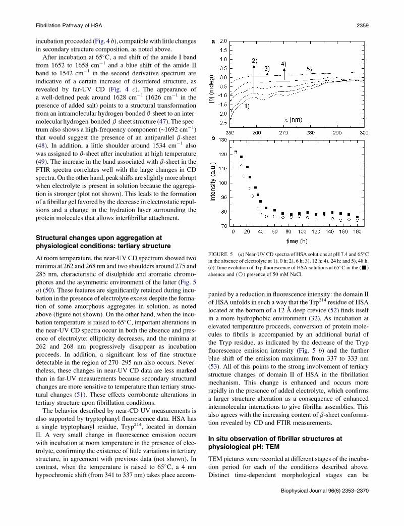
incubation proceeded (Fig. 4 b), compatible with little changes
in secondary structure composition, as noted above.
After incubation at 65�C, a red shift of the amide I band
from 1652 to 1658 cm�1 and a blue shift of the amide II
band to 1542 cm�1 in the second derivative spectrum are
indicative of a certain increase of disordered structure, as
revealed by far-UV CD (Fig. 4 c). The appearance of
a well-defined peak around 1628 cm�1 (1626 cm�1 in the
presence of added salt) points to a structural transformation
from an intramolecular hydrogen-bonded b-sheet to an inter-
molecular hydrogen-bonded-b-sheet structure (47). The spec-
trum also shows a high-frequency component (~1692 cm�1)
that would suggest the presence of an antiparallel b-sheet
(48). In addition, a little shoulder around 1534 cm�1 also
was assigned to b-sheet after incubation at high temperature
(49). The increase in the band associated with b-sheet in the
FTIR spectra correlates well with the large changes in CD
spectra. On the other hand, peak shifts are slightly more abrupt
when electrolyte is present in solution because the aggrega-
tion is stronger (plot not shown). This leads to the formation
of a fibrillar gel favored by the decrease in electrostatic repul-
sions and a change in the hydration layer surrounding the
protein molecules that allows interfibrillar attachment.
Structural changes upon aggregation atphysiological conditions: tertiary structure
At room temperature, the near-UV CD spectrum showed two
minima at 262 and 268 nm and two shoulders around 275 and
285 nm, characteristic of disulphide and aromatic chromo-
phores and the asymmetric environment of the latter (Fig. 5
a) (50). These features are significantly retained during incu-
bation in the presence of electrolyte excess despite the forma-
tion of some amorphous aggregates in solution, as noted
above (figure not shown). On the other hand, when the incu-
bation temperature is raised to 65�C, important alterations in
the near-UV CD spectra occur in both the absence and pres-
ence of electrolyte: ellipticity decreases, and the minima at
262 and 268 nm progressively disappear as incubation
proceeds. In addition, a significant loss of fine structure
detectable in the region of 270–295 nm also occurs. Never-
theless, these changes in near-UV CD data are less marked
than in far-UV measurements because secondary structural
changes are more sensitive to temperature than tertiary struc-
tural changes (51). These effects corroborate alterations in
tertiary structure upon fibrillation conditions.
The behavior described by near-CD UV measurements is
also supported by tryptophanyl fluorescence data. HSA has
a single tryptophanyl residue, Tryp214, located in domain
II. A very small change in fluorescence emission occurs
with incubation at room temperature in the presence of elec-
trolyte, confirming the existence of little variations in tertiary
structure, in agreement with previous data (not shown). In
contrast, when the temperature is raised to 65�C, a 4 nm
hypsochromic shift (from 341 to 337 nm) takes place accom-
panied by a reduction in fluorescence intensity: the domain II
of HSA unfolds in such a way that the Trp214 residue of HSA
located at the bottom of a 12 A deep crevice (52) finds itself
in a more hydrophobic environment (32). As incubation at
elevated temperature proceeds, conversion of protein mole-
cules to fibrils is accompanied by an additional burial of
the Tryp residue, as indicated by the decrease of the Tryp
fluorescence emission intensity (Fig. 5 b) and the further
blue shift of the emission maximum from 337 to 333 nm
(53). All of this points to the strong involvement of tertiary
structure changes of domain II of HSA in the fibrillation
mechanism. This change is enhanced and occurs more
rapidly in the presence of added electrolyte, which confirms
a larger structure alteration as a consequence of enhanced
intermolecular interactions to give fibrillar assemblies. This
also agrees with the increasing content of b-sheet conforma-
tion revealed by CD and FTIR measurements.
In situ observation of fibrillar structures atphysiological pH: TEM
TEM pictures were recorded at different stages of the incuba-
tion period for each of the conditions described above.
Distinct time-dependent morphological stages can be
FIGURE 5 (a) Near-UV CD spectra of HSA solutions at pH 7.4 and 65�Cin the absence of electrolyte at 1), 0 h; 2), 6 h; 3), 12 h; 4), 24 h; and 5), 48 h.
(b) Time evolution of Trp fluorescence of HSA solutions at 65�C in the (-)
absence and (B) presence of 50 mM NaCl.
Biophysical Journal 96(6) 2353–2370

2360 Juarez et al.
observed in these images. Thus, at room temperature neither
fibrils nor other types of aggregates are detected, except for
small amorphous protein clusters observed in the presence of
electrolyte after a long incubation period (150 h), which
possess a largely helical structure (Fig. 6 a). In contrast,
when the temperature is raised to 65�C, fibril formation is
observed. Electron microscopy indicates that aggregation
leads first to a globular species that subsequently converts
to fibrils with a curly morphology. The fibrillation pathway
in the presence of electrolyte is very similar to that observed
in its absence but it takes place in a shorter timescale, in
agreement with previous results. Fig. 6, b–j, show electron
micrographs of the sample heated at 65�C at different steps
of the incubation period in the presence of 50 mM of electro-
lyte. The number and length of the fibrils has increased in
relation to other structures, although several morphologies
can be observed throughout incubation.
Small spherical clusters of ~20 nm formed by protein olig-
omers are observed (Fig. 6 b) at short incubation times (5 h).
These aggregates present relatively few changes in their
tertiary and secondary structures, as shown by CD and fluo-
rescence data. With further incubation (Fig. 6 c, 15 h), a certain
elongation of these spherical aggregates can be observed. This
bead-like structure at short incubation times arises from what
appears to be attractive interactions between spherical
Biophysical Journal 96(6) 2353–2370
proteins aggregates, as shown in Fig. 6 d-1 (see also
Fig. S3), which may result in an increased exposure of hydro-
phobic residues, giving rise to more elongated structures. This
elongation involves a conformational conversion of protein
structure to consolidate the structure, and in all probability
it implies changes in the hydrogen-bonding status (Fig. 6
d-2). This is in agreement with a further development in
ThT fluorescence and decreases in both helical content and
Tryp fluorescence at this incubation point, as shown previ-
ously. On the other hand, we did not find evidence of
formation of elongated structures by longitudinal fusion of
oligomers, as recently reported (54).
Bead-like structures progressively become more elon-
gated upon incubation (35 h) due to mutual interactions
between these structures and subsequent annealing, and
convert into short protofibrils (Fig. 6 e), in agreement with
a decrease in helical structure as revealed by CD. Alterations
in the conformational structure of these oligomers and subse-
quent elongation via monomer addition may also be present;
however, the TEM resolution did not allow us to confirm
that. Several authors reported a tendency for these bead-
like structures to transform into fibrillar structures at elevated
temperatures caused by partial unfolding of the protein mole-
cules and giving rise to conditions conductive to fibril forma-
tion (55–57).
a b
c
d-1) d-2)
e f
g h
i j
k
FIGURE 6 TEM pictures of the different stages of the HSA fibrillation process at pH 7.4: (a) at 25�C in the presence of 50 mM NaCl after 150 h of incu-
bation, and at 65�C in the presence of 50 mM NaCl after (b) 5 h; (c) and (d) 15 h (part d shows the elongation of oligomers to give bead-like structures); (e) 35 h
(where short protofibrils are observed); (f) 45 h; (g) 50 h (where long curly fibrils are seen); and (h–k) after 72 h. Part i shows the addition of oligomers to mature
fibrils, j shows the association of mature fibrils in bundles, and k shows mature fibrils with ribbon-like structure.

Fibrillation Pathway of HSA 2361
Further incubation results in the presence of more fibrils,
which increase in length as incubation proceeds (Fig. 6,
f and g). After 2 days of incubation, numerous longer
curly-branched and interconnected fibrils are present
(Fig. 6 g), with lengths and widths characteristic of classical
amyloid fibrils (i.e., between 0.5 and several micrometers in
length and 9–10 nm in width (3,5,9)), as detected by TEM
and AFM (see Fig. 6 g and Fig. S4). The appearance of
this structure coincides well with the plateau region of ThT
binding, the far UV-CD profiles characterized by minima
around 215–220 nm, and the intermolecular b-sheet structure
observed by FTIR. Most fibrils appear curly and intercon-
nected, and some of them are even circular, as observed
previously for other proteins, such as b2 microglobulin
(58) and a-crystallin (56).
If the incubation time is extended further (3 days), one can
observe mature straight fibrils, which can be seen as a struc-
tural evolution of curly fibrils (Fig. 6 h). Mature fibrils are
thicker and stiffer than single fibrils and seem to be formed
by lateral (side-by-side) assembly of two or more individual
filaments. These mature fibrils are less numerous in the
absence of electrolyte because the electrostatic screening is
lower, avoiding direct contact between constitutive fibrils.
We have also observed that lateral interactions of single
particles collaborate in growing these straight mature fibrils
(57). Scrutiny of the protein aggregates indicates that HSA
particles and clusters tend to align within the immediate
vicinity of the fibers (Fig. 6 i), serving the single fiber as
a lateral template or scaffold for small protein molecules,
and would constitute a subcomponent in mature fibrils. It
is worth mentioning that some fibril solutions when analyzed
by SEM show the existence of very long fibers, exceeding
200 mm (see Fig. S5). We think that this effect may result
from the air-drying process favoring attractive interactions
between single fibers during the solvent removal.
In addition, a structural diversity of mature fibrils is noted:
flat ribbons are observed in solution, as well as long, straight
fibril ensembles (Fig. 6 j). This polymorphism may arise
from variations in the quaternary structure, the manner in
which protofilaments self-associate, or the protofilament
substructure (e.g., in the details of hydrogen-bonding
networks and side-chain packing) (59). Finally, one of the
characteristic traits of the mature amyloid fibrils is their
tendency to bend, twist, and agglomerate. Fig. 6 k shows
laterally connected fibers that split over a certain distance
or overlap each other. The extent and rate of this growth is
dependent on solution conditions and lateral interactions
between fibrils, which are responsible for the thickening of
the mature fibrils and the formation of suprafibrillar aggre-
gates. Thus, we conclude that pseudo-globular aggregates
rearrange slowly to form linear, curly fibrils. These may be
sufficient to produce a high-affinity template that is subse-
quently elongated by monomeric units or other fibrils, and
can lead to the formation of ordered, straight, or ribbon-
like fibrillar structures.
Fibril structure: XRD
The amyloid-like character of the fibrillar aggregates detected
by TEM was confirmed by XRD. The XRD image of the HSA
fibrils is shown in Fig. 7. Two strong reflections can be
observed: a dominant sharp and intense reflection occurs at
4.8 A, and one weaker, more diffuse, but still intense reflec-
tion is observed at ~11 A. The 4.8 A meridional reflection
arises from the spacing between hydrogen-bonded individual
strands in the b-sheet structure that lie perpendicular to the
fibril axis, and the 11 A equatorial reflection corresponds to
the intersheet spacing, with the b-sheets stacked face to face
to form the core structure of protofilaments (60,61). This indi-
cates that fibrils possess a cross-b structure, one of the diag-
nostic hallmarks of amyloid structures.
Acidic conditions: kinetics and amyloidself-assembly of HSA
We next subjected HSA to acidic pH and assessed its propen-
sity to form amyloid fibrils under such conditions in both the
presence and absence of 50 mM NaCl. Increases in ThT fluo-
rescence were observed at both 25�C and 65�C, although the
increase was quite small at 25�C. This indicates the formation
of a small amount of additional b-sheet conformation as
a consequence of the formation of oligomeric aggregates, as
will be shown below (Fig. 8). At 65�C, the increase in
ThT fluorescence in the absence of electrolyte is lower than
that obtained at physiological pH and extends over a larger
period of time. This can be a result, on the one hand, of a lower
capacity of fibrillation under these conditions (see below) or,
on the other, to the presence of a lag phase if compared with
solution to which electrolyte is added, as discussed further
below. After a small increase in ThT fluorescence at relatively
short incubation times, a plateau occurs at ~24–100 h, after
which the ThT fluorescence starts to increase again. Thus, it
FIGURE 7 XRD pattern of HSA fibrils.
Biophysical Journal 96(6) 2353–2370

2362 Juarez et al.
FIGURE 8 Time evolution of ThT fluorescence in HSA
solutions incubated at pH 3.0 at 25�C (a and b) or 65�C(c and d) in the absence and presence of 50 mM NaCl,
respectively.
is thought that under acidic conditions in the absence of elec-
trolyte, oligomeric structures (protein clusters) are formed in
a series of thermodynamically unfavorable assembly steps
followed by a growth phase in which clusters are elongated
by further addition of protein monomers and/or oligomers
upon mutual interaction.
Fitting of the time ThT fluorescence evolution shows us
that under acidic conditions, the self-assembly process
becomes more cooperative, as indicated by the values of
n> 1. This indicates the different nature of the self-assembly
pathway of HSA under acidic conditions with respect to
physiological pH, since interactions between protein mole-
cules are modulated by changes in both the pH and the initial
protein structure (62). In addition, the plot in the absence of
electrolyte is fitted in two steps: 1), the fast formation of
small clusters; and 2), the lag phase, which can originate
from the necessity to reach a critical concentration of clusters
for aggregation to continue. Probably, oligomeric species
formed in very early stages of the aggregation process
(whose existence is indicated by the slight increase of ThT
fluorescence at very short incubation times) are more soluble
under acidic conditions, and only after they achieve a critical
concentration are they able to grow to generate larger aggre-
gates (63). However, complete fibril formation can be
achieved only in the presence of electrolyte.
Nucleation-dependent growth mechanismin acidic medium
To corroborate the origin of the plateau region in the absence
of electrolyte at elevated temperature, we performed a seeding
fibril growth under the conditions previously specified. When
seeds are added to the protein solution, a continuous increase
in ThT fluorescence is observed. This is the typical behavior
observed for a nucleation-type growth mechanism and corrob-
orates the existence of this lag phase (see Fig. S6). On the other
hand, no changes are observed when protein seeds are added
Biophysical Journal 96(6) 2353–2370
to a solution containing 50 mM NaCl. The difference between
both solution conditions may arise from the greater hydropho-
bicity of oligomeric species formed during very earlier incuba-
tion stages in the presence of excess electrolyte. In its absence,
electrostatic repulsion between oligomeric species seems to
preclude for some time the formation of nuclei with a critical
size to overcome the energy barrier that impedes aggregation.
Structural changes at acidic pH: secondarystructure
At acidic pH and room temperature, both minima in [q] at 208
and 222 nm are still present before incubation proceeds, which
indicates an important retention of this type of structure, as
previously described (25,64). During incubation, a slight
decrease in [q] is observed in both the absence and presence
of added electrolyte, which is compatible with a small
decrease in a-helices and the development of a small amount
of b-sheet conformation due to the appearance of small amor-
phous aggregates in solution (figure not shown). In contrast,
a shift of the 208 nm minimum to lower wavelengths takes
place as incubation proceeds (0–200 h) at 65�C in the absence
of electrolyte (Fig. 9 a). An increase in random coil structure
(characterized by a single minimum below 200 nm) can
account for this shift. Upon further incubation, an additional
red shift occurs as a consequence of the increase of b-sheet
structure in the aggregates formed, as also indicated by the
increase in ThT fluorescence. The far-UV CD curves at
65�C in the presence of NaCl followed a trend similar to those
obtained at physiological pH, although the ellipticity decrease
is less severe at long incubation times as a consequence of
a lower amount of scattered light from fibril aggregates (see
Fig. 9 b). This confirms the lower fibrillar density under these
conditions, which also precludes the formation of a fibrillar
gel, in contrast to physiological conditions.
The secondary structure compositions in acidic solution at
room temperature at the beginning of the incubation process

Fibrillation Pathway of HSA 2363
indicate a reduction in a-helix conformation (~45%) and an
increase in b-sheet, b-upturn, and coil contents (7%, 20%,
and 28%, respectively), typical of the acid-expanded E-state
of HSA (25) (Fig. 10). No significant changes were detected
in structural composition during the incubation procedure at
room temperature in either the absence or presence of added
electrolyte up to 100 h incubation. At this stage, a slight
increase in b-sheet and unordered conformations is observed
for both solution conditions. In contrast, when the tempera-
ture is raised to 65�C, an important increase in b-sheets at
the expense of helical conformation occurs as also observed
at physiological pH. This is also accompanied by an increase
in unordered conformation at early incubation times (0–150
h). The change in secondary structure occurs during a longer
time interval (up to 9 days) in acidic solution. The final a-
helix and b-sheet compositions are respectively 24% and
16% in the absence of electrolyte (17% and 21% in the pres-
ence of 50 mM NaCl), compared to 20% and 21% at neutral
FIGURE 9 Far-UV spectra of HSA solutions at 65�C at pH 3.0 in (a) the
absence of electrolyte at 1), 0 h; 2), 24 h; 3), 125 h; 4), 175 h; 5), 200 h;
and 6), 250 h of incubation; and (b) the presence of 50 mM NaCl at 1),
0 h; 2), 15 h; 3), 48 h; 4), 100 h; 5), 150 h; and 6), 200 h of incubation.
pH. This indicates small compositional changes in the result-
ing amyloid aggregates. The turn conformation also shows
little changes throughout incubation.
FTIR experiments corroborate the structural changes
undergone by the protein molecules as incubation proceeds
at acidic conditions. Before incubation, the amide I band
at 1650 cm�1 and the amide II band at 1542 cm�1 confirm
that there is still a significant amount of a-helices. Incubation
at room temperature under acidic conditions leads to a certain
increase of the peak located at ~1627 nm, which corresponds
to intramolecular b-sheet structure, in agreement with
ThT fluorescence and CD data. On the other hand, after
incubation at 65�C, a red shift of the amide I band from
1650 to 1656 cm�1 and a blue shift of the amide II band to
1540 cm�1 are indicative of an increased amount of disor-
dered structure. In addition, the shift and further increase of
the 1627 cm�1 peak to 1624 cm�1 also points to a structural
transformation from an intramolecular hydrogen-bonded
b-sheet to an intermolecular hydrogen-bonded b-sheet struc-
ture (46), as seen for physiological pH. Spectra also show
a small high-frequency component (~1692 cm�1) that would
suggest the presence of antiparallel b-sheet (47) (see Fig. S7).
Structural changes at acidic pH: tertiary structure
When the pH is decreased, there is an increase in [q] between
260 and 280 nm, and a slight decrease between 285 and
300 nm, denoting loss of tertiary structure. Nevertheless, there
are still significant CD signals left, suggesting a remaining
tertiary structure, in agreement with previous reports (25,65).
These changes at acidic pH are related to structural rearrange-
ments of all HSA domains. In particular, some increase in ellip-
ticity below 295 nm takes place during incubation at room
temperature, which points to little further tertiary structural
changes as aggregation takes place; in particular, a loss of
fine structure is detectable in the 270–295 nm region (see
Fig. S8 a). At 65�C, changes in tertiary structure are more
important; in particular, an almost complete absence of the
minima is observed between 260 and 270 nm, indicating
a further loss of asymmetry around disulfide bridges and/or
aromatic residues as incubation proceeds. An additional loss
of fine structure in the range of 280–295 nm, similar to that
observed at physiological pH, is also detected (Fig. S8, b and c).
Upon incubation at acidic pH and room temperature,
a certain decrease in the tryptophanyl fluorescence and a slight
blue shift of the emission maximum occur between days 0 and
8 of incubation, which points to a certain internalization of
Tryp to the nonpolar environment of domain II as a result of
certain aggregation under these conditions (see Fig. S9).
Because changes in far- and near-UV CD spectra are relatively
small for this incubation period, this leads us to think that
structural changes in domain II of HSA are involved in this
aggregation process (66). At 65�C, the tryptophanyl residues
are in a more solvent-exposed environment during the incuba-
tion because the fluorescence intensity abruptly decreases
Biophysical Journal 96(6) 2353–2370

2364 Juarez et al.
FIGURE 10 Time evolution of secondary structure
compositions of HSA solutions at pH 3.0 at 25�C (a and
b) or 65�C (c and d) in the absence and the presence of
50 mM NaCl, respectively. (:) a-helix, (-) b-turn,
(C) unordered, and (;) b-sheet conformations.
during the first 4 days of incubation as a result of the sequential
unfolding of domains I and II of HSA. This period of time is
slightly shorter than at pH 7.4 (mainly in the presence of elec-
trolyte excess) because the expanded E state already involves
an important alteration in the tertiary structure.
In situ observation of fibrillar structures at acidicpH: TEM
HSA samples obtained at different incubation times were
also subjected to TEM analysis under acidic conditions. At
room temperature in the absence of salt, no aggregates
Biophysical Journal 96(6) 2353–2370
were observed during the first 2 days of the incubation
process. From the third day, the presence of small clusters
(globules) of aggregated protein could be observed. These
aggregates have a globular or spherical shape, not the regular
fibrillar appearance associated with amyloid structures (see
Fig. 11 a). In addition, less numerous, more elongated aggre-
gates (20–30 nm long, 3–4 nm wide) can be also observed,
and their population slightly increases as incubation
proceeds. The formation of these types of aggregates is char-
acterized by a decrease of helical structure accompanied by
a slight rise in b-sheet and unordered conformations from
CD measurements at long incubation times.
FIGURE 11 TEM pictures of the different stages of the
HSA fibrillation process at pH 3.0 in the presence of 50
mM NaCl (a) at 25�C after 150 h of incubation, and at
65�C after (b) 24 h, (c) 150 h, and (d) 250 h of incubation
in the presence of 50 mM NaCl.

Fibrillation Pathway of HSA 2365
When the temperature is raised to 65�C, fibril formation
takes place in several steps. First, formation of quasi-spher-
ical aggregates takes place under incubation times similar to
those at pH 7.4, and these seem to be fairly soluble. On the
other hand, we have found that a coexistence of spherical
aggregates with circular ring-shaped particles of larger size
(~100–300 nm) is observed at short incubation times (on
the order of a few hours) in the presence of excess electrolyte
(Fig. 11 b). These structures appear as a possible interme-
diate structural rearrangement of smaller protein aggregates
before fibril formation facilitated by electrostatic screening.
There is no evidence as to whether structural reorganization
to form fibrils takes place within this type of aggregates, or
whether dissociation of HSA molecules from these structures
occurs. Based on our CD data, which indicate that a progres-
sive gaining of b-sheet structure and unordered conformation
at the expense of a-helices takes place in the first part of the
incubation process, we suggest that only after a certain crit-
ical amount of b-sheet structure is reached inside these ring-
shaped particles will short fibrils be formed in solution from
decomposition of the former and become stable. Once suffi-
cient molecules are present within the oligomer, reorganiza-
tion steps become thermodynamically favorable as a result of
an increase in the number of hydrogen bonds and other stabi-
lizing interactions. Once fragments of highly ordered aggre-
gates are present, the free energy for addition of monomeric
molecules to a growing fibril will become more favorable.
Similar circular ring-shaped structures have also been
observed as a structural intermediate before the formation
of amyloid fibrils in the self-assembly process of insulin
(67), Ab17-42 peptide (68), Ab1-4 peptide, and HaPrP23-
144 prion protein (69). In this case, the circular structures
may incorporate into fibrils but also self-aggregate to form
large, amorphous structures (57). Given the large structural
differences between HSA insulin and Ab17-42 peptide, and
their distinct propensity to form amyloid fibrils, it seems
reasonable to think that these circular ring-shaped structures
can be a sort of common structural intermediate of amyloid
fibril formation under different solution conditions.
After longer incubation times (6 days) for HSA solutions in
the absence of electrolyte at 65�C, small, well-defined, short
protofibrils (~100 nm long and 3–4 nm wide) start to appear
(Fig. 11 c). The corresponding CD spectrum shows a progres-
sive increase in the proportion of b-structure at the expense of
the helical one during incubation. In contrast, the presence of
electrolyte involves an additional step: the formation of short
curly fibers occurs in a broader incubation timescale than at
physiological pH (Fig. 11 d); under further incubation, the
fibrils appear to increase slightly in number and length.
They range from ~100 nm to several hundred nanometers in
length and 8 to 11 nm in width; thus, they are shorter on
average than those obtained at physiological conditions but
have the same average width. Closed fibril loops with diame-
ters of ~100 nm were also frequently observed because the
formed filaments can remain short and thin to enable them
to bend and form closed rings, as also observed for b2 micro-
globulin (58), a-crystallin (56), equine lysozyme (70), insulin
(67), and a-synuclein (71). This fibrillar material appears
similar to protofilaments observed in the early stages of other
amyloidogenic systems, given the small length and the
absence of higher-order structures.
DISCUSSION
The ability to form amyloid fibrils is a generic property of
polypeptide chains, although the propensity of different
regions of proteins to form such structures varies substantially
(72). The properties of unfolded polypeptides, including their
relative propensities for a- and b-structure, their intrinsic
solubility, and the nature of the interactions within the resul-
tant fibrillar structures, are likely to be particularly important
determinants in their relative abilities to form fibrils. The
conformation of the partially folded state is not by itself a crit-
ical feature of fibril formation; rather, it is suggested that the
basis for amyloidogenesis is the presence of partially denatur-
ation conditions that destabilize the native fold of the protein
but do not preclude noncovalent interactions between the
various groups within the protein. In our case, it appears
necessary to destabilize HSA molecules and hence to generate
a partially folded intermediate that can aggregate to form
fibrils. Thus, one can readily atttain conditions in which
such aggregation-prone intermediate states are significantly
populated by lowering the pH or raising the temperature.
ThT fluorescence, CR staining, XRD, and TEM pictures
demonstrate the formation of amyloid-like structures under
these conditions. The aggregation process is governed by
the balance between attractive and repulsive interactions
between protein molecules. Conformational changes induced
by heat increase the number of hydrophobic residues exposed
to the aqueous solvent. The exposure of these groups results in
attractive hydrophobic interactions that play a dominant role
in the aggregation process. Repulsive forces are induced by
a surface charge that can be modulated by changes in pH,
which controls the net charge of the protein, and by the ionic
strength of the solvent, which controls the screening of elec-
trostatic interactions (73).
Absence of a lag phase in HSA fibrillation processin most solution conditions
A first characteristic of the HSA fibrillation process is the
absence of a lag phase, as previously observed for bovine
serum albumin (BSA) (74) and acyl phosphatase (75) under
all solutions conditions analyzed except for acidic pH at
room temperature. This occurs if the initial aggregation is
a downhill process that does not require a highly organized
and unstable nucleus. This is supported by the fact that
seeding of preformed aggregates does not accelerate the
fibrillation process. In this regard, it has been suggested
that large multidomain proteins like BSA are able to form
Biophysical Journal 96(6) 2353–2370

2366 Juarez et al.
propagation-competent nucleus-like structures (oligomeric
structures) (74). In our case, TEM pictures also show the
formation of spherical oligomers upon very short incubation
times, which occur by a means of classical coagulation mech-
anism. In contrast, the presence of a certain lag phase upon
incubation in acidic medium at 65�C in the absence of electro-
lyte may well indicate that oligomeric aggregates need more
time to develop and/or persist for longer times because of their
enhanced solubility, so they need to reach a certain number or
size to change the energy landscape of the system and promote
further aggregation.
Existence of different intermediates on the HSAfibrillation pathway
On a macroscopic scale, the different steps in the fibril forma-
tion pattern, as observed by TEM, consist of the formation of
nonfibrillar aggregates (oligomeric globules) and their subse-
quent elongation (bead-like structures and circular ring-
shaped structures), and the development of protofilaments
and their assembly in fibrils, which can rearrange in more
complex structures. On a molecular level, CD and FTIR results
show that HSA possesses native-like a-helical characteristics
with residual b-sheet content before acidic or heat treatment.
In very early periods of incubation, HSA forms small,
soluble, globular oligomers of mainly native-like molecules
in acidic, physiological, and/or high-temperature conditions,
with a progressive increase in b-sheet content and unordered
conformation upon further incubation, as revealed by ThT
fluorescence and far-UV CD. Thus, upon further incubation
(5–50 h) at elevated temperature, the spectroscopic character-
istics indicate losses of persistent tertiary structure along with
unfolding of certain secondary structure to different extents
depending on the solution conditions (acidic or physiological
pH, added salt). Different conditions may cause different
regions of the polypeptide chains that are relatively flexible
and not involved in strong intramolecular interactions
(76,77) to enhance the aggregation process, leading to an
evolution of the previous globules into additional intermediate
structures (bead-like structures and circular ring-shaped struc-
tures at physiological and acidic pH, respectively, with the
latter found only at high ionic strength). Bead-like structures
arise from what appears to be attractive interactions between
globular protein oligomeric clusters, which may result from
an increased exposure of hydrophobic residues, mainly in
the presence of electrolyte. We found no evidence of formation
of elongated structures by longitudinal fusion of oligomers.
These bead-like structures progressively become more elon-
gated upon incubation (35 h) due to mutual interactions
between them and convert into protofibrils, in agreement
with a decrease in helical structure as indicated by CD data.
The decrease in diameter accompanying elongation may be
explained on the basis of reorganization of structure, in partic-
ular of the b-strands. Several authors have also reported the
tendency of these bead-like structures to transform into fibrillar
Biophysical Journal 96(6) 2353–2370
structures at elevated temperatures caused by partial unfolding
of the protein molecules and giving rise to conditions condu-
cive to fibril formation (55–57), as shown in Fig. 6 d.
The combination of experimental observations described
here indicates that the formation of fibrils from soluble HSA
molecules proceeds in a series of stages, the first of which is
effectively the presence of oligomeric globules. Thus, the
role of association-competent oligomeric intermediates may
result in a kinetic pathway via clustering of these oligomeric
species, which can be rationalized in the light of colloid coag-
ulation theory, i.e., the formation of a critical oligomer or an
ensemble of critical oligomers and subsequent aggregation
into bead-like structures, and then protofibrillar structures
(78). The persistence of these spherical oligomers in solution
coinciding with fibril assembly also supports the view that
they may be ‘‘on-pathway’’ intermediates (see Fig. 6 and
Fig. S10). Spherical oligomeric structures have been
proposed to serve a key, on-pathway role in both the forma-
tion and elongation of amyloid fibrils of the Sup35 (79) and
Ure2p yeast prion proteins (80).
In contrast, we speculate that the circular ring-shaped struc-
tures found in acidic medium at high ionic strength, which
appear to be composed of two semicircular units, may come
from bending and association of early-formed, short, bead-
like structures due to a decrease in their solubility. This
decrease would stem from the electrolyte concentration
present in solution, which screens electrostatic interactions
between aggregates. Once sufficient molecules are present
within this type of intermediate structure, reorganization steps
become thermodynamically favorable as a result of an increase
in the number of hydrogen bonds and other stabilizing interac-
tions. Once critical fragments of ordered aggregates are
present (with a critical amount of b-sheet structure), the free
energy for addition of monomeric molecules to a growing
elongated structure becomes more favorable. In this way, short
protofibrils will be formed in solution upon dissolution of the
ring-shaped intermediate structures and will become stable.
These intermediates are not observed by TEM once protofi-
brils start to be observed, which suggests that they may act
as reservoirs of the initially very short protofibrils. However,
a deeper structural analysis of this structural intermediate
and its evolution to protofibrils is needed, and is currently
under way. On the other hand, the fact that some elongated
structures (but not fibrils) are formed in acidic pH at room
temperature, in contrast to physiological medium, for which
no aggregation is observed except for some amorphous aggre-
gates, suggests that the charge distribution on the protein influ-
ences the propensity to form amyloid fibrils. The neutralization
of negative charges at acidic pH may promote this elongation.
HSA fibrils are curly
The thermodynamic instability of prefibrillar aggregates
causes them to evolve into more stable assemblies upon addi-
tional incubation (>75 h), eventually leading to the appearance

Fibrillation Pathway of HSA 2367
of stable fibrils, mainly at physiological conditions at elevated
temperatures, where structural reorganization results in the
large majority of hydrophobic groups being concealed from
the solvent. This is corroborated by ThT fluorescence, far
UV-CD, and FTIR spectra, which demonstrate that the major
elements of ordered secondary structure are b-sheets, suggest-
ing that the a-helical regions of the native protein have under-
gone significant structural changes. TEM pictures corroborate
this picture, showing that the fibrils formed are mainly, narrow,
branched, and elongated but curly, in contrast to the straight
needle-like structures characteristic of bona fide fibrils. As
incubation proceeds, these worm-like structures increase in
length. These fibrils display the typical 4.8 A peak indicating
the typical interstrand distance of classical fibrils, and the
11 A equatorial reflection corresponds to the intersheet
spacing, with the b-sheets stacked face to face to form the
core structure of protofilaments (81). Based on the CD and
FTIR data, we speculate that these types of fibrils may be
formed by a seam of b-sheet structure decorated by relatively
disorganized a-helical structure, as previously observed for
RNase A (82) or yeast Ure2p (83). In addition, an antiparallel
arrangement of b-strands forming the b-sheet structure of the
HSA fibrils seems to be the most probable configuration, as
denoted by the band at ~1691–1693 cm�1 in the FTIR spectra.
Curly aggregates have been also seen in other proteins, such as
b2 microglobulin (58) and a-crystallin (56). In addition, intra-
molecular end-to-end association of short individual filaments
appears to be favorable, as TEM images reveal the presence of
a substantial number of closed loops appearing to form spon-
taneously, mainly in the absence of electrolyte at acidic condi-
tions. Qualitatively, the probability of a single fibril joining
end-to-end to form a closed loop will be high if the fibril is
sufficiently flexible and of appropriate length for the ends to
find themselves regularly in close proximity to each other
(84). Indeed, the results presented here exemplify the favorable
nature of loop formation when such fibril morphologies are
adopted. Loop formation was reported previously for other
amyloid-forming systems (85,86).
On the other hand, it is interesting to note that a similar
curly morphology for HSA can be achieved under different
solution conditions: the single filament, in the form of both
open flexible chains and closed loops, is observed at both
physiological and acidic pH in the presence of electrolyte.
Nevertheless, the rate and extent of aggregation depends
on the solution conditions: the amount of formed fibrils
and their length (larger at physiological conditions) favor
interactions between early aggregates to form fibrils. This
is revealed by the formation of a gel phase under suitable
solution conditions.
Curly fibers can evolve into a suprafibrillarstructure
Very long incubation times (>150 h) lead to a more complex
morphological variability among amyloid fibrils (e.g., long
straight fibrils, flat-ribbon structures, or laterally connected
fibers). These compact, mature fibrillar assemblies formed
at the endpoint of the aggregation process may result from
an effort to minimize the exposure of hydrophobic residues,
and are also likely to result in increased van der Waals inter-
actions, leading to greater stability, as previously noted for
b2-microglobulin (87), a-synuclein (5), and insulin (88). A
similar progression in structure from curly fibrils to mature
straight fibers was also observed for Ab-peptide (89) and
insulin (90). In contrast, BSA fibrillation was shown to be
halted at the early curly stage, despite the enormous struc-
tural similarities with HSA, since no further development
in fibrillar structure over long timescales was observed
(74). Mature fibrils are thicker and stiffer than single fibrils
and appear to be formed by lateral (side-by-side) assembly
of two or more individual filaments. Nevertheless, under
physiological conditions, we observed that mature straight
fibrils can also grow by swollen protein particles tending
to align within the immediate vicinity of the fibers, as shown
in Fig. 6 i, serving the single fiber as a lateral template or
scaffold for small protein molecules, and would constitute
a subcomponent in mature fibrils. Their population is depen-
dent on solution conditions and lateral interactions between
fibrils. This may be an additional pathway to the formation
of mature fibrils via association of protofibrils. Conforma-
tional preferences for a certain pathway to become active
may exist, and thus the influence of environmental condi-
tions such as pH, temperature, and salt must be considered.
Thus, it seems that a single filament may act as a ‘‘lateral
template’’ or scaffold for small protein particles, which
would constitute a neighboring subcord-like feature in the
fiber shown in Fig. 6 h. This filament þ monomers/oligo-
mers scenario is an alternative pathway to the otherwise
dominating filamentþ filament manner of the protein fibril’s
lateral growth, as has been also observed for insulin (57).
AFM data recently reported by Green et al. (91) suggest
that mature fibrils from human amylin are unlikely to be
assembled by the lateral association of protofibrils.
It appears, therefore, that even for a homogeneous HSA
sample undergoing uniform temperature or acidic treatment,
there is still more than one mode of assembly of filaments
(92). Such polymorphism may be caused by differences in
the number of filaments assembled in the mature fibrils;
however, it may also result from the incorporation in different
regions of the sequence of the polypeptide chain with various
types of fibrils. In addition, the differences between the pop-
ulations of fibers clearly involve not only the rate of the aggre-
gation process, but also the different quaternary folds.
Although the different distribution profiles of the fibrillar
features may still be explained in terms of kinetic effects
(e.g., temperature may differently affect the kinetics at various
stages of the assembly, effectively marginalizing certain
sequential processes), high temperatures may also have
a more direct effect on amyloidogenesis, for instance, by
increasing the thermal energy of the interacting molecules
Biophysical Journal 96(6) 2353–2370

2368 Juarez et al.
and hence causing the alignment of filaments to become less
accurate. Such unspecific effects may contribute to morpho-
logical differences in any protein’s amyloid samples induced
at high temperature. In this regard, recent reports based on
nuclear magnetic resonance showed that different fibril
morphologies have different underlying secondary structures,
and as such are likely produced by distinct independent
assembly pathways (93,94).
A summary of the various fibril morphologies observed in
this study is shown in Fig. 12 together with a schematic
representation of the assembly process. We propose that at
elevated temperature (except at pH 3.0 in the absence of elec-
trolyte), HSA forms rapidly globular oligomers that upon
mutual interaction evolve into more elongated structures
(bead-like) that grow to protofibrils either by subsequent
annealing of oligomers and/or protein monomers. Mature
fibrils can be formed by lateral association of protofibrils
or the addition of protein oligomers to the growing fibril,
both at the ends of the fibril and by lateral fusion. Ring-
like structures are present in acidic conditions at elevated
temperature in the presence of electrolyte as an additional
intermediate state formed by association of short bead-like
structures, which disappears when protofibrils are observed
in solution. Thus, we think that they may act as reservoirs
of initially very short protofibrils.
CONCLUSIONS
We observed the formation of protofibrils, curly fibers, and
mature fibrils by the protein HSA under different solution
conditions. We analyzed the fibrillation process and the
conformational changes associated with it by using different
spectroscopic techniques, and confirmed the necessary devel-
opment of b-sheet structure upon fibrillation. In addition, the
shapes of the different structural intermediates and final prod-
ucts in the fibrillation process were observed by TEM, SEM,
and AFM. The obtained fibrils show structural features typical
of classical amyloid fibers, as denoted by XRD, CD, and fluo-
rescence spectroscopies, and TEM. A model of fibril forma-
tion based on the elongation of protein oligomers through
mutual interactions and subsequent annealing and growth is
FIGURE 12 Mechanisms of fibril formation for HSA.
Biophysical Journal 96(6) 2353–2370
presented. Nevertheless, some differences in the fibrillation
mechanism occur depending on the solution conditions; for
example, ring-shaped structures are observed only as a struc-
tural intermediate under acidic conditions in the presence of
added electrolyte.
SUPPORTING MATERIAL
Ten figures and a table are available at http://www.biophysj.org/biophysj/
supplemental/S0006-3495(09)00322-1.
We thank Dr. Eugenio Vazquez for his assistance with the CD measure-
ments.
This study was supported by the Ministerio de Educacion y Ciencia (project
MAT-2007-61604).
REFERENCES
1. Reches, M., and E. Gazit. 2003. Casting metal nanowires within discreteself-assembled peptide nanotubes. Science. 300:625–627.
2. Rajagopal, K., and J. P. Schneider. 2004. Self-assembling peptides andproteins for nanotechnological applications. Curr. Opin. Struct. Biol.14:480–486.
3. Stefani, M., and C. M. Dobson. 2003. Protein aggregation and aggre-gate toxicity: new insights into protein folding, misfolding diseasesand biological evolution. J. Mol. Med. 81:678–699.
4. Soto, C., L. Estrada, and J. Castilla. 2006. Amyloids, prions and theinherent infectious nature of misfolded protein aggregates. TrendsBiochem. Sci. 31:150–155.
5. Khurana, R., C. Ionescu-Zanetti, M. Pope, J. Li, L. Nelson, et al. 2003.A general mode for amyloid fibril assembly based on morphologicalstudies using atomic force microscopy. Biophys. J. 85:1135–1144.
6. Chiti, F., M. Stefani, N. Taddei, G. Ramponi, and C. M. Dobson. 2003.Rationalization of the effects of mutations on peptide and protein aggre-gation rates. Nature. 424:805–808.
7. Williams, A. D., E. Portelius, I. Kheterpal, J. T. Guo, K. D. Cook, et al.2004. Mapping a b amyloid fibril secondary structure using scanningproline mutagenesis. J. Mol. Biol. 335:833–842.
8. Hortscansky, P., T. Christopeit, V. Schroeckh, and M. Fandrich. 2005.Thermodynamic analysis of the aggregation propensity of oxidizedAlzheimer’s b-amyloid variants. Protein Sci. 14:2915–2918.
9. Fandrich, M., V. Forge, K. Buder, M. Kittler, C. M. Dobson, et al. 2003.Myoglobin forms amyloid fibrils by association of unfolded peptidesegments. Proc. Natl. Acad. Sci. USA. 100:15463–15468.
10. Gazit, E. 2002. The ‘‘correctly folded’’ state of proteins: is it a meta-stable state? Angew. Chem. Int. Ed. Engl. 41:257–259.
11. Gillmore, J. D., A. J. Stangou, G. A. Tennet, D. R. Booth, J. O’Grady,et al. 2001. Clinical and biochemical outcome of hepatorenal transplan-tation for hereditary systemic amyloidosis associated with apolipopro-tein AI Gly26Arg. Transplantation. 71:986–992.
12. Carulla, N., G. L. Caddy, D. R. May, J. Zurdo, M. Gairi, et al. 2005.Molecular recycling within amyloid fibrils. Nature. 436:554–558.
13. Pepys, M. B., J. Herbert, W. L. Hutchison, G. A. Tennent, H. J. Lach-mann, et al. 2002. Targeted pharmacological depletion of serum amyloidP component for treatment of human amyloidosis. Nature. 417:254–259.
14. Janus, C., M. A. Chishti, and D. Westaway. 2000. Transgenic mousemodels of Alzheimer’s disease. Biochim. Biophys. Acta. 1502:63–75.
15. Cohen, F. E., and J. W. Kelly. 2003. Therapeutic approaches to protein-misfolding diseases. Nature. 426:905–909.
16. Tanaka, M., P. Chien, N. Narber, R. Cooke, and J. S. Weissman. 2004.Conformational variations in an infectious protein determine prionstrain differences. Nature. 428:323–328.

Fibrillation Pathway of HSA 2369
17. Fandrich, M., and C. M. Dobson. 2002. The behavior of polyaminoacids reveals an inverse side chain effect in amyloid structure formation.EMBO J. 21:5682–5690.
18. Gorinstein, S., A. Caspi, A. Rosen, I. Goshev, M. Zemser, et al. 2002.Structure characterization of human serum aproteins in solution and drystate. J. Pept. Res. 59:71–78.
19. Taboada, P., S. Barbosa, E. Castro, and V. Mosquera. 2006. Amyloidfibril formation and other aggregate species formed by human serumalbumin association. J. Phys. Chem. B. 110:20733–20736.
20. Pace, C. N., F. Vajdos, L. Fee, G. Grimsley, and T. Gray. 1995. How tomeasure and predict the molar absorption coefficient of a protein.Protein Sci. 4:2411–2423.
21. Sreerama, N., and R. W. Woody. 2000. Estimation of protein secondarystructure from circular dichroism spectra: comparison of CONTIN,SELCON, and CDSSTR methods with an expanded reference set.Anal. Biochem. 287:252–260.
22. Uversky, V. N., and A. L. Fink. 2004. Conformational constraints foramyloid fibrillation: the importance of being unfolded. Biochim.Biophys. Acta. 1698:131–153.
23. Calamai, M., F. Chiti, and C. M. Dobson. 2005. Amyloid fibril forma-tion can proceed from different conformations of a partially unfoldedprotein. Biophys. J. 89:4201–4210.
24. Morel, B., S. Casares, and F. A. Conejo-Lara. 2006. Single mutationinduces amyloid aggregation in the a-spectrin SH3 domain: analysisof the early stages of fibril formation. J. Mol. Biol. 356:453–468.
25. Dockal, M., D. C. Carter, and F. Ruker. 2000. Conformational transi-tions of the three recombinant domains of human serum albumindepending on pH. J. Biol. Chem. 275:3042–3050.
26. Peters, T. Jr. , 1996. All About Albumin: Biochemistry, Genetics andMedical Application. Academic Press, New York.
27. Geisow, M. J., and G. H. Beaven. 1977. Physical and binding propertiesof large fragments of human serum albumin. Biochem. J. 165:477–484.
28. Khan, M. Y. 1986. Direct evidence for the involvement of domain III inthe N-F transition of bovine serum albumin. Biochem. J. 236:307–310.
29. Ahmad, B., M. K. A. Khan, S. Haq, and R. H. Khan. 2004. Intermediateformation at lower urea concentration in ‘B’ isomer of human serumalbumin: a case study using domain specific ligands. Biochem. Biophys.Res. Commun. 314:166–173.
30. Taboada, P., S. Barbosa, E. Castro, M. Gutierrez-Pichel, and V. Mos-quera. 2007. Effect of solvation on the structure conformation of humanserum albumin in aqueous-alcohol mixed solvents. Chem. Phys.340:59–68.
31. Farruggia, B., F. Rodriguez, R. Rigatuso, G. Fidelio, and G. Pico. 2001.The participation of human serum albumin domains in chemical andthermal unfolding. J. Protein Chem. 20:81–89.
32. Flora, K., J. D. Brennan, G. A. Baker, M. A. Doody, and F. V. Bright.1998. Unfolding of acrylodan-labeled human serum albumin probedby steady-state and time resolved fluorescence methods. Biophys. J.75:1084–1096.
33. Rader, A. J., B. M. Hespenheide, K. A. Kuhn, and M. F. Thorpe. 2002.Protein unfolding: rigidity lost. Proc. Natl. Acad. Sci. USA. 99:3540–3545.
34. LeVine, H. 3rd. , 1993. Thioflavine T interaction with syntheticAlzheimer’s disease b-amyloid peptides: detection of amyloid aggrega-tion in solution. Protein Sci. 2:404–410.
35. Klunk, W. E., J. W. Pettegrew, and D. J. Abraham. 1989. Quantitativeevaluation of Congo Red binding to amyloid-like proteins witha b-pleated sheet conformation. J. Histochem. Cytochem. 37:1273–1281.
36. Khurana, R., C. Coleman, C. Ionescu-Zanetti, S. A. Carter, V. Krishna,et al. 2005. Mechanisms of thioflavin T binding to amyloid fibrils.J. Struct. Biol. 151:229–238.
37. Sagis, L. M. C., C. Veerman, and E. Van der Linden. 2004. Mesoscopicproperties of semiflexible amyloid fibrils. Langmuir. 20:924–927.
38. Morozova-Roche, L. A., J. A. Jones, W. Noppe, and C. M. Dobson.1999. Independent nucleation and heterogeneous assembly of structureduring unfolding of equine lysozime. J. Mol. Biol. 289:1055–1073.
39. Jund, P., R. Jullien, and I. Campbell. 2001. Random walks on fractalsand stretched exponential relaxation. Phys. Rev. E. 63:361311–361314.
40. Harper, J. D., and P. T. J. Landsbury. 1997. Models of amyloid seedingin Alzheimer’s disease and scrapie: mechanistic truths and physiolog-ical consequences of the time-dependent solubility of amyloid proteins.Annu. Rev. Biochem. 66:385–407.
41. Lomakin, A., D. S. Chung, G. B. Benedek, D. A. Kirschner, andD. B. Teplow. 1996. On the nucleation and growth of amyloid b-protein fibrils: detection of nuclei and quantitation of rate constants.Proc. Natl. Acad. Sci. USA. 93:1125–1129.
42. Lomakin, A., D. B. Teplow, D. A. Kirschner, and G. B. Benedek. 1997.Kinetic theory of fibrillogenesis of amyloid b-protein. Proc. Natl. Acad.Sci. USA. 94:7942–7947.
43. Ferrone, F. 1999. Analysis of protein aggregation kinetics. MethodsEnzymol. 309:256–274.
44. Susi, H., and D. M. Byler. 1983. Protein structure by Fourier transforminfrared spectroscopy: second derivative spectra. Biochem. Biophys.Res. Commun. 115:391–397.
45. Lin, S. -Y., Y. -S. Wei, M. -J. Li, and S. -L. Wang. 2004. Effect of ethanolor/and captopril on the secondary structure of human serum albuminbefore and after protein binding. Eur. J. Pharm. Biopharm. 57:457–464.
46. Jackson, M., and H. H. Mantsch. 1995. The use and misuse ofFTIR spectroscopy in the determination of protein structure. Crit.Rev. Biochem. Mol. Biol. 30:95–120.
47. Casal, H. L., U. Kohler, and H. H. Mantsch. 1988. Structural andconformational changes of b-lactoglobulin B: an infrared spectroscopystudy of the effect of pH and temperature. Biochim. Biophys. Acta.957:11–20.
48. Fabian, H., L. -P. Choo, G. I. Szendrei, M. Jackson, W. C. Halliday,et al. 1993. Infrared spectroscopic characterization of Alzheimerplaques. Appl. Spectrosc. 47:1513–1518.
49. Lin, S. -Y., Y. -S. Wei, M. -J. Li, and S. -L. Wang. 2004. Effect ofethanol or/and captopril on the secondary structure of human serumalbumin before and after protein binding. Spectrochim. Acta [A].60:3107–3111.
50. Uversky, V. N., N. V. Narizhneva, T. V. Ivanova, and A. Y. Tomashev-ski. 1997. Rigidity of human a-fetoprotein tertiary structure is underligand control. Biochemistry. 494:13638–13645.
51. Muzammil, S., Y. Kumar, and S. Tayyab. 1999. Molten globule-likestate of human serum albumin at low pH. FEBS Lett. 266:26–32.
52. Qiu, W., L. Zhang, O. Okobiah, Y. Yang, L. Wang, et al. 2006. Ultrafastsolvation dynamics of human serum albumin: correlations with confor-mational transitions and site-selected recognition. J. Phys. Chem. B.110:10540–10549.
53. Cowgill, R. W. 1968. Fluorescence and protein structure. XIV. Tyrosinefluorescence in helical muscle proteins. Biochim. Biophys. Acta.161:431–438.
54. Shahi, P., R. Sharma, S. Sanger, I. Kumar, and R. S. Jolly. 2007. Forma-tion of amyloid fibrils via longitudinal growth of oligomers. Biochem-istry. 46:7365–7373.
55. Burgio, M. R., P. M. Bwennet, and J. F. Koretz. 2001. Heat-inducedquaternary transitions in hetero- and homo-polymers of a-crystallin.Mol. Vis. 7:228–233.
56. Meehan, S., Y. Berry, B. Luisa, C. M. Dobson, J. A. Carver, et al. 2004.Amyloid fibril formation by lens crystallin proteins and its implicationsfor cataract formation. J. Biol. Chem. 279:3413–3419.
57. Jansen, R., W. Dzwolak, and R. Winter. 2005. Amyloidogenic self-assembly of insulin aggregates probed by high resolution atomic forcemicroscopy. Biophys. J. 88:1344–1353.
58. Radford, S. E., W. S. Gosal, and G. W. Platt. 2005. Towards an under-standing of the structural molecular mechanism of b2-microglobulinamyloid formation in vitro. Biochim. Biophys. Acta. 1753:51–63.
59. Kodali, R., and R. Wetzel. 2007. Polymosphism in the intermediatesand products of amyloid assembly. Curr. Opin. Struct. Biol. 17:48–57.
60. Sunde, M., and C. Blake. 1997. The structure of amyloid fibrils by elec-tron microscopy and X-ray diffraction. Adv. Protein Chem. 50:123–159.
Biophysical Journal 96(6) 2353–2370

2370 Juarez et al.
61. Bouchard, M., J. Zurdo, E. J. Nettleton, C. M. Dobson, and C. V. Rob-inson. 2000. Formation of insulin amyloid fibrils followed by FTIRsimultaneously with CD and electron microscopy. Protein Sci.9:1960–1967.
62. Vetri, V., F. Librizzi, M. Leone, and V. Militello. 2007. Thermal aggre-gation of bovine serum albumin at different pH: comparison withhuman serum albumin. Eur. Biophys. J. 36:717–725.
63. Bader, R., R. Bamford, J. Zurdo, B. F. Luisi, and C. M. Dobson. 2006.Probing the mechanism of amyloidogenesis through a tandem repeat ofthe PI3–SH3 domain suggests a generis model for protein aggregationand fibril formation. J. Mol. Biol. 356:189–208.
64. Shaw, A. K., and S. K. Pal. 2008. Spectroscopic studies on the effectof temperature on pH-induced folded states of human serum albumin.J. Photochem. Photobiol. B. 90:69–77.
65. Ahmad, B., S. Parveen, and R. H. Khan. 2006. Effect of albuminconformation on the binding of ciprofloxacin to human serum albumin:a novel approach directly assigning binding site. Biomacromolecules.7:1350–1356.
66. Eftink, M. R., and C. A. Ghiron. 1976. Exposure of tryptophyl residuesin proteins. Quantitative determination by fluorescence quenchingstudies. Biochemistry. 15:672–679.
67. Sibley, S. P., K. Sosinsky, L. E. Gulian, E. J. Gibbs, and R. F. Paster-nack. 2008. Probing the mechanism of insulin aggregation with addedmetalloporphyrins. Biochemistry. 47:2858–2865.
68. Zheng, J., H. Jang, B. Ma, and R. Nussinov. 2008. Annular structures asintermediates in fibril formation of Alzheimer Ab17–42. J. Phys. Chem.B. 112:6856–6865.
69. Moore, R. E., S. F. Hayes, E. R. Fischer, and S. A. Priola. 2007.Amyloid formation via supramolecular peptide assemblies. Biochem-istry. 46:7079–7087.
70. Malisauskas, M., V. Zamotin, J. Jass, W. Noppe, C. M. Dobson, et al.2003. Amyloid protofilaments from the calcium-binding protein equinelysozyme: formation of ring and linear structures depends on pH andmetal ion concentration. J. Mol. Biol. 330:879–890.
71. Lashuel, H. A., D. Hartley, B. M. Petre, T. Walz, and P. T. Lansbury Jr.2002. Neurodegenerative disease: amyloid pores from pathogenic muta-tions. Nature. 418:291.
72. Jarrett, J. T., and P. T. Lansbury Jr. 1992. Amyloid fibril formationrequires a chemically discriminating nucleation event—studies of anamyloidogenic sequence from the bacterial protein OSMB. Biochem-istry. 31:12345–12352.
73. Lefebvre, J., D. Renard, and A. C. Sanchez-Gimeno. 1998. Structureand rheology of heat-set gels of globular proteins. I. Bovine serumalbumin gels in isoelastic conditions. Rheol. Acta. 37:345–357.
74. Holm, N. K., S. K. Jespersen, L. V. Thomassen, T. Y. Wolff, P. Sehgal,et al. 2007. Aggregation and fibrillation of bovine serum albumin. Bio-chim. Biophys. Acta. 1774:1128–1138.
75. Chiti, F., P. Webster, N. Taddei, A. Clark, M. Stefani, et al. 1999.Designing conditions for in vitro, formation of amyloid protofilamentsand fibrils. Proc. Natl. Acad. Sci. USA. 96:3590–3594.
76. Krishnan, R., and S. L. Lindquist. 2005. Structural insights into a yeastprion illuminate nucleation and strain diversity. Nature. 435:765–772.
77. Moraitakis, G., and J. M. Goodfellow. 2003. Simulations of human lyso-zime: probing the conformations triggering amyloidosis. Biophys. J.84:2149–2158.
Biophysical Journal 96(6) 2353–2370
78. Modler, A. J., K. Gast, G. Lutsch, and G. Damaschun. 2003. Assemblyof amyloid protofibrils via critical oligomers. A novel pathway ofamyloid formation. J. Mol. Biol. 325:135–148.
79. Serio, T. R., A. G. Cashikar, A. S. Kowal, G. J. Sawicki, J. J. Moleshi,et al. 2000. Nucleated conformational conversion and the replication ofconformational information by a prion determinant. Science. 289:1317–1321.
80. Jiang, Y., H. Li, L. Zhu, J. M. Zhou, and S. Perrett. 2004. Amyloidnucleation and hierarchical assembly of Ure2p fibrils. Role of aspara-gines/glutamine repeat and nonrepeat regions of the prion domains.J. Biol. Chem. 279:3361–3369.
81. Makin, O. S., and L. C. Serpell. 2005. X-ray diffraction studies ofamyloid structure. Methods Mol. Biol. 299:67–80.
82. Sambashivan, S., M. R. Liu, M. R. Sawaya, M. Gingery, and D. Eisen-berg. 2005. Amyloid-like fibrils of ribonuclease A with three-dimensional domain-swapped and native-like structures. Nature.437:266–269.
83. Ranson, N., T. Stromer, L. Bousset, R. Melki, and R. C. Serpell. 2006.Insights into the architecture of the Ure2p yeats protein assemblies fromhelical twisted fibrils. Protein Sci. 15:2481–2487.
84. Hatters, D. M., C. A. MacRails, R. Daniels, W. S. Gosal, N. H. Thom-son, et al. 2003. The circularization of amyloid fibrils formed by apoli-poprotein C–II. Biophys. J. 85:3979–3990.
85. Lashuel, H. A., and P. T. Lansbury Jr. 2006. Are amyloid diseasescaused by protein aggregates that mimic bacterial pore-forming toxins?Q. Rev. Biophys. 39:167–201.
86. Meehan, S., T. P. J. Knowles, A. J. Baldwin, J. F. Smith, A. M. Squires,et al. 2007. Characterization of amyloid fibril formation by small heat-shock chaperone proteins human aA-, aB- and R120G aB-crystallins.J. Mol. Biol. 372:470–484.
87. Kad, N. M., S. L. Myers, D. P. Smith, S. E. Radford, and N. H. Thom-son. 2003. Hierarchical assembly of b2-microglobulin amyloid in vitrorevealed by atomic force microscopy. J. Mol. Biol. 330:785–797.
88. Khurana, R., J. L. Jimenez, E. J. Nettleton, M. Bouchard, C. V. Robin-son, et al. 2002. The protofilament structure of insulin amyloid fibrils.Proc. Natl. Acad. Sci. USA. 99:9196–9201.
89. Otzen, D. E., and M. Oliveberg. 2004. Transient formation of nanocry-talline structures during fibrillation of an Alzheimer-like peptide.Protein Sci. 13:1417–1421.
90. Grudzielanek, S., V. Smirnovas, and R. Winter. 2006. Solvation-assis-ted pressure tuning of insulin fibrillation: from novel aggregation path-ways to biotechnological applications. J. Mol. Biol. 356:497–506.
91. Green, J. D., C. Goldsbury, J. Kistler, G. K. Cooper, and U. Aebi. 2004.Human amylin oligomer growth and fibril elongation define two distinctphases in amyloid formation. J. Biol. Chem. 279:12206–12212.
92. Chamberlain, A. K., C. E. MacPhee, J. Zurdo, L. A. Morozova-Roche,H. A. O. Hill, et al. 2000. Ultrastructural organization of amyloid fibrilsby atomic force microscopy. Biophys. J. 79:3282–3293.
93. Goldsbury, C. S., S. Wirtz, S. A. Muller, S. Sunderji, P. Wicki, et al.2000. Studies on the in vitro assembly of Aba-40: implications forthe search for Ab fibril formation inhibitors. J. Struct. Biol. 130:217–231.
94. Petkova, A. T., R. D. Leapman, Z. Guo, W. M. Yau, M. P. Mattson,et al. 2005. Self-propagating, molecular-level polymorphism inAlzheimer’s b-amyloid fibrils. Science. 307:262–265.