Evidence that Chemical Chaperone 4- Phenylbutyric Acid Binds to Human Serum Albumin at Fatty Acid...
17
RESEARCH ARTICLE Evidence that Chemical Chaperone 4- Phenylbutyric Acid Binds to Human Serum Albumin at Fatty Acid Binding Sites Debasish Roy ☯ , Vinod Kumar ☯ , Joel James, Mohamed Sham Shihabudeen, Shweta Kulshrestha, Varun Goel, Kavitha Thirumurugan* 206, Structural Biology Lab, Center for Biomedical Research, Division of Biomedical Sciences, School of Bio Sciences and Technology, VIT University, Vellore-632014, Tamil Nadu, India ☯ These authors contributed equally to this work. * [email protected] Abstract Endoplasmic reticulum stress elicits unfolded protein response to counteract the accumulat- ing unfolded protein load inside a cell. The chemical chaperone, 4-Phenylbutyric acid (4- PBA) is a FDA approved drug that alleviates endoplasmic reticulum stress by assisting protein folding. It is found efficacious to augment pathological conditions like type 2 diabe- tes, obesity and neurodegeneration. This study explores the binding nature of 4-PBA with human serum albumin (HSA) through spectroscopic and molecular dynamics approaches, and the results show that 4-PBA has high binding specificity to Sudlow Site II (Fatty acid binding site 3, subdomain IIIA). Ligand displacement studies, RMSD stabilization profiles and MM-PBSA binding free energy calculation confirm the same. The binding constant as calculated from fluorescence spectroscopic studies was found to be k PBA = 2.69 x 10 5 M -1 . Like long chain fatty acids, 4-PBA induces conformational changes on HSA as shown by circular dichroism, and it elicits stable binding at Sudlow Site II (fatty acid binding site 3) by forming strong hydrogen bonding and a salt bridge between domain II and III of HSA. This minimizes the fluctuation of HSA backbone as shown by limited conformational space occu- pancy in the principal component analysis. The overall hydrophobicity of W214 pocket (located at subdomain IIA), increases upon occupancy of 4-PBA at any FA site. Descriptors of this pocket formed by residues from other subdomains largely play a role in compensat- ing the dynamic movement of W214. Introduction Endoplasmic reticulum (ER) is the chief cellular organelle involved in the biosynthesis of poly- peptides and lipids. After protein translation by ribosomal machinery on cytosolic surface of the ER, the unfolded polypeptide gets translocated in the ER lumen for folding, assisted by molecular chaperones and foldases. The ER maintains a rigid quality control and proteins that are well-folded only exits the ER for various cellular fates. This strict homeostasis is disrupted PLOS ONE | DOI:10.1371/journal.pone.0133012 July 16, 2015 1 / 17 OPEN ACCESS Citation: Roy D, Kumar V, James J, Shihabudeen MS, Kulshrestha S, Goel V, et al. (2015) Evidence that Chemical Chaperone 4-Phenylbutyric Acid Binds to Human Serum Albumin at Fatty Acid Binding Sites. PLoS ONE 10(7): e0133012. doi:10.1371/journal. pone.0133012 Editor: Jamshidkhan Chamani, Islamic Azad University-Mashhad Branch, Mashhad, ISLAMIC REPUBLIC OF IRAN Received: April 18, 2015 Accepted: June 22, 2015 Published: July 16, 2015 Copyright: © 2015 Roy et al. This is an open access article distributed under the terms of the Creative Commons Attribution License, which permits unrestricted use, distribution, and reproduction in any medium, provided the original author and source are credited. Data Availability Statement: All relevant data are within the paper. Funding: The authors have no support or funding to report. Competing Interests: The authors have declared that no competing interests exist.
Transcript of Evidence that Chemical Chaperone 4- Phenylbutyric Acid Binds to Human Serum Albumin at Fatty Acid...
RESEARCH ARTICLE
Evidence that Chemical Chaperone 4-Phenylbutyric Acid Binds to Human SerumAlbumin at Fatty Acid Binding SitesDebasish Roy☯, Vinod Kumar☯, Joel James, Mohamed Sham Shihabudeen,Shweta Kulshrestha, Varun Goel, Kavitha Thirumurugan*
206, Structural Biology Lab, Center for Biomedical Research, Division of Biomedical Sciences, School of BioSciences and Technology, VIT University, Vellore-632014, Tamil Nadu, India
☯ These authors contributed equally to this work.* [email protected]
AbstractEndoplasmic reticulum stress elicits unfolded protein response to counteract the accumulat-
ing unfolded protein load inside a cell. The chemical chaperone, 4-Phenylbutyric acid (4-
PBA) is a FDA approved drug that alleviates endoplasmic reticulum stress by assisting
protein folding. It is found efficacious to augment pathological conditions like type 2 diabe-
tes, obesity and neurodegeneration. This study explores the binding nature of 4-PBA with
human serum albumin (HSA) through spectroscopic and molecular dynamics approaches,
and the results show that 4-PBA has high binding specificity to Sudlow Site II (Fatty acid
binding site 3, subdomain IIIA). Ligand displacement studies, RMSD stabilization profiles
and MM-PBSA binding free energy calculation confirm the same. The binding constant as
calculated from fluorescence spectroscopic studies was found to be kPBA = 2.69 x 10⁵ M-1.
Like long chain fatty acids, 4-PBA induces conformational changes on HSA as shown by
circular dichroism, and it elicits stable binding at Sudlow Site II (fatty acid binding site 3) by
forming strong hydrogen bonding and a salt bridge between domain II and III of HSA. This
minimizes the fluctuation of HSA backbone as shown by limited conformational space occu-
pancy in the principal component analysis. The overall hydrophobicity of W214 pocket
(located at subdomain IIA), increases upon occupancy of 4-PBA at any FA site. Descriptors
of this pocket formed by residues from other subdomains largely play a role in compensat-
ing the dynamic movement of W214.
IntroductionEndoplasmic reticulum (ER) is the chief cellular organelle involved in the biosynthesis of poly-peptides and lipids. After protein translation by ribosomal machinery on cytosolic surface ofthe ER, the unfolded polypeptide gets translocated in the ER lumen for folding, assisted bymolecular chaperones and foldases. The ER maintains a rigid quality control and proteins thatare well-folded only exits the ER for various cellular fates. This strict homeostasis is disrupted
PLOSONE | DOI:10.1371/journal.pone.0133012 July 16, 2015 1 / 17
OPEN ACCESS
Citation: Roy D, Kumar V, James J, ShihabudeenMS, Kulshrestha S, Goel V, et al. (2015) Evidencethat Chemical Chaperone 4-Phenylbutyric Acid Bindsto Human Serum Albumin at Fatty Acid Binding Sites.PLoS ONE 10(7): e0133012. doi:10.1371/journal.pone.0133012
Editor: Jamshidkhan Chamani, Islamic AzadUniversity-Mashhad Branch, Mashhad, ISLAMICREPUBLIC OF IRAN
Received: April 18, 2015
Accepted: June 22, 2015
Published: July 16, 2015
Copyright: © 2015 Roy et al. This is an open accessarticle distributed under the terms of the CreativeCommons Attribution License, which permitsunrestricted use, distribution, and reproduction in anymedium, provided the original author and source arecredited.
Data Availability Statement: All relevant data arewithin the paper.
Funding: The authors have no support or funding toreport.
Competing Interests: The authors have declaredthat no competing interests exist.

in various disease states like diabetes, obesity, cancer, neurodegenerative disorders etc [1–3],where there is an accumulation of unfolded proteins owing to the availability of excess metabo-lites or due to an altered redox status. To counteract, the cell triggers an adaptive responseknown as the Unfolded Protein Response (UPR). UPR augments the ER to cope with themisfolded proteins by increasing the amount of chaperones, decreasing the entry of new pro-teins or by degrading irreversibly misfolded proteins in the ER lumen. Recently, several smallmolecules have been identified that act as chaperones. They have potential to reduce UPR andpromote protein folding by reducing the energy barrier of the intermittent transition statesthat occur as the proteins fold into their native conformation. Several studies have reportedthat 4-Phenylbutyric acid (PBA) (Fig 1A), a FDA approved drug for treating urea cycle disor-der [4, 5] has efficacy in assisting protein-folding in ER, thereby decreasing UPR levels both invitro and in vivo.
Drug bioavailability is majorly affected by its binding to Human serum albumin (HSA)[6];the most abundant protein in plasma. Apart from acting as a drug transporter, HSA transportsa large variety of endogenous and exogenous ligands like fatty acids, metabolites and hormones[7–9] to target organs. HSA has three homologous domains (I, II and III) and each domain isdivided into two subdomains (A and B) (Fig 1B). Drug binding to HSA takes place mostly atsubdomain IIA and IIIA which are commonly referred to as Sudlow site I and II respectively[9]. Molecular interactions at these sites are monitored using fluorescence properties of intrin-sic fluorescence probe, W214 (located in subdomain IIA). Some drug molecules which areknown subdomain IIIA binders can bind to subdomain IB to make it as a third major drug site[10]. Apart from drug binding, HSA also acts as a carrier for a number of physiological ligandslike heme, bilirubin, fatty acids etc [11]. Primary binding sites for fatty acids are located in sub-domain IA and IIIA of HSA. This versatile binding behaviour of HSA plays a major role indetermining the rate of delivery of ligands and hence has been extensively studied for its bind-ing with various ligands over the last three decades.
The binding nature of 4-PBA to HSA is still not known. Due to its promise in manypreclinical studies of diseases in which ER stress is a central mechanism of manifestation,4-PBA binding to HSA can provide important clues on its pharmacokinetics. With this view,binding studies of 4-PBA with HSA is performed.
Fig 1. A) Ball and Stick representation of 4PBA; Carbon = Grey, Hydrogen = Cyan, Oxygen = Red b)Cartoonmodel of HSA-MYR complex (PDB: 2BXP) showing different subdomains andmajor fatty acidbinding sites.
doi:10.1371/journal.pone.0133012.g001
Phenylbutyric Acid Binds to Serum Albumin at Fatty Acid Binding Sites
PLOS ONE | DOI:10.1371/journal.pone.0133012 July 16, 2015 2 / 17

Materials and Methods
MaterialsHuman serum albumin (fatty acid free), 4-Phenylbutyric Acid (4-PBA) and Palmitic acid werepurchased from Sigma Aldrich, India. Quercetin was purchased from SRL, India. For HSA,200μM stock solutions were prepared in Tris HCl (0.05 molL-1) and NaCl (0.1 molL-1), pH 7.4and stored at -20°C in aliquots. The same buffer was used for UV-Vis and Fluorescence assays.For CD analysis, Tris HCl (0.01 molL-1) and NaCl (0.1 molL-1), pH 7.4 was used. Stock solu-tions of PBA (2mM), Quercetin (2mM) and Palmitic acid (2mM) were prepared in absoluteethanol and stored at room temperature in experimental aliquots.
MethodsUV-Vis absorbance spectrophotometry. All UV-Vis spectroscopic measurements were
carried out using Thermo scientific Evolution 260 Bio double beam spectrophotometer (USA)with a 1 cm path length. Binding experiments were performed using a fixed concentration ofHSA (1 μM) and 4-PBA in the range 1–20 μM. The UV-Vis spectral scan was recorded from500–200 nm at 25 ± 0.2°C.
Fluorescence assays. Fluorescence measurements were carried out in a JASCO spectrofluo-rimeter FP 8600 (Tokyo, Japan), using 1 cm path length quartz cuvette. The emission spectrumwas studied with a fixed volume of HSA (1 μM) in the presence of increasing concentrations ofQuercetin (1–20 μM), 4-PBA (1–20 μM) and Palmitic acid (1–20 μM). For titrations, ligandswere successively added (5μl) to obtain the given concentration range from ethanol stock. Afterevery addition the solutions were thoroughly mixed and incubated for 5 minutes at 25 ± 0.2°Cbefore collecting spectra. Fluorescence spectra were recorded with an excitation wavelength of295 nm and an emission range of 300–500 nm. For fixed wavelength measurements, emissionwavelengths of 345 nm and 330 nmwere used. The effect of ethanol on the emission spectra wasevaluated and it was found to be negligible.
Ligand Displacement Studies. Two separate ligand displacement assays were conducted:(1) by adding varying concentrations of 4-PBA (2–20 μM) to a mixture of HSA (1 μM) +Quercetin (20 μM); (2) by adding varying concentrations of Palmitic acid & 4-PBA (1–8 μM)to HSA (1 μM) + Dansylglycine (1 μM) complex. HSA + ligand complexes were incubated at25°C for 30 mins prior data collection. The stability of the complex was evaluated by measuringfluorescence emission spectra with time (for 1 min) to ensure that it reached equilibrium beforetitration. Percentage initial fluorescence was calculated by: [Fluorescence in the presence ofadded ligand] / [Fluorescence in the absence of added ligand] � 100 [12].
Circular Dichroism. CD spectra of HSA and ligand complexes were recorded with aJASCO J-715 spectropolarimeter (Tokyo, Japan). For measurements in the far-UV region(178–280 nm), a quartz cell with a path length of 0.1 cm was kept in constant nitrogen flowatmosphere. HSA and 4-PBA was mixed in an equal stoichiometry. Accumulation of the scanwith the scan speed of 50 nm per min was performed and the data was collected from the 260–200 nm spectral range. All the CD measurements were carried out at 25 ± 0.2°C.
Molecular Dynamics Simulation. MD simulation was carried out using GROMACS 4.5.6[13] to understand the dynamic behaviour of HSA in a 4-PBA bound state. Amber 99sb forcefield [14] parameters were used for protein structure and GAFF parameters [15] were used forsmall molecules using ACPYPE tool [16]. The 4-PBA-HSA complexes were solvated in a cubicbox with SPCE water model under periodic boundary conditions. The system was neutralizedby adding 14 sodium ions. The neutralized protein–ligand complexes were energy minimizedby using steepest descent method. After energy minimization, the systems were equilibrated by
Phenylbutyric Acid Binds to Serum Albumin at Fatty Acid Binding Sites
PLOS ONE | DOI:10.1371/journal.pone.0133012 July 16, 2015 3 / 17
Position restrained molecular dynamics at constant temperature of 300 K and a constantpressure of 1 atm for about 100 ps. Finally, the equilibrated systems were subjected to MD for7,000 ps using NPT ensemble.
Principal Component Analysis (PCA). MD simulation trajectory file was used to identifythe motion of unliganded HSA and 4-PBA-HSA complex (4-PBA bound to different FA sites).PCA analysis in protein dynamics (analyzed using Gromacs tools: g_covar and g_anaeig) isused to identify possible collective motions of residues of a protein due to certain changesinflicted upon ligand binding [17]. A co-variance matrix for protein backbone was obtained,transformed in Cartesian co-ordinate space (before and after simulation) and diagonalized.The direction of motion was characterised by representing the vectors of every single compo-nents of motion. The eigenvalues represent the amplitude of fluctuation along their corre-sponding eigenvectors. Therefore, the contribution to the internal motion of HSA uponcomplexation is traced by PCA. The major combined movements arise along the directions ofeigenvectors associated to the largest eigenvalue. The first 10 eigenvectors were used to repre-sent the protein motion.
MM-PBSA binding free energy calculation. Binding free energy at each ligand bindingsite was computed by using GMXPBSA 2.1 [18] with trajectory files obtained after simulation.Polar solvation energy values was calculated using the implicit solvation Poisson–Boltzmannmodel (PB) and nonpolar solvation energy value was calculated based on the solvent accessiblesurface area (SASA); finally all these were combined to obtain final binding free energy value.
Binding free energy of 4-PBA at each fatty acid binding site was calculated by using [19]:
DGbind ¼ Gcomplex � GHSA � G4PBA ðEq 1Þ
G represents free energy of each state, which was computed by:
G ¼ EMM þ GPB þ GSA � TS ðEq 2Þ
EMM ¼ Evdw þ Eele þ Eint ðEq 3Þ
EMM is the molecular mechanical energy, GPB and GSA are the polar and nonpolar terms ofthe free energy and TS is the entropic contribution of solute. EMM was obtained by adding con-tributions from internal energies including bond, angle, and torsional angle energies (Eint),electrostatic energy (Eele), and van der Waals energy (Evdw).
Results
Interaction between 4-Phenylbutyric acid and HSAUV-Vis spectroscopy. UV-Vis and Fluorescence spectroscopy is used to understand
structural changes induced to a protein upon ligand binding and complex formation. Aromaticresidues like tryptophan are very sensitive to slight changes in the polar microenvironment.This rearrangement in the polar microenvironment of tryptophan is attributed to changes inthe protein secondary structure caused by ligand binding. Absorption spectrum of HSA isobtained by titrating with increasing concentration of 4-PBA (1–20 μM) which show twoabsorption peaks (Fig 2A). The major absorption peak is in the region of 200–230 nm attrib-uted to n-π� transition of the protein backbone and 278 nm peak is due to microenvironmentchanges of tryptophan (Fig 2A, insets). It can be seen that adding increasing concentrations of4-PBA causes a blue shift in the 280 nm region (Tryptophan absorption region) owing tochanges in tryptophan microenvironment upon complex formation (Fig 2A; inset). There is aslight increase in the absorption peak of HSA due to its interaction with 4-PBA, which inflicts
Phenylbutyric Acid Binds to Serum Albumin at Fatty Acid Binding Sites
PLOS ONE | DOI:10.1371/journal.pone.0133012 July 16, 2015 4 / 17
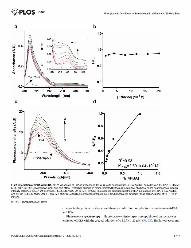
changes in the protein backbone, and thereby confirming complex formation between 4-PBAand HSA.
Fluorescence spectroscopy. Fluorescence emission spectroscopy showed an increase inemission of HSA with the gradual addition of 4-PBA (1–20 μM) (Fig 2B). Similar observations
Fig 2. Interaction of 4PBAwith HSA. a) UV-Vis spectra of HSA in presence of 4PBA. Cuvette concentration, cHSA: 1μM (a) and c4PBA (1,2,4,8,12,16,20 μM):b!h; pH 7.4 at 25°C. Inset shows slight blue shift at the Tryptophan absorption region indicated by the arrow. b) Effect of ethanol on the fluorescence emissionintensity of HSA. cHSA = 1μM, cEthanol =, 1,2,4,8,12,16,20 μM (pH 7.4, 25°C) c) Fluorescence emission spectra of HSA in presence of 4PBA. cHSA: 1μM (a)and c4PBA (2,4,8,12,16,20 μM): b!g; pH 7.4 at 25°C.Dotted line represents contribution of4PBA (20μM) at the emission range of HSA. d) Plot of 1/F-F0 vs 1/[4PBA]
doi:10.1371/journal.pone.0133012.g002
Phenylbutyric Acid Binds to Serum Albumin at Fatty Acid Binding Sites
PLOS ONE | DOI:10.1371/journal.pone.0133012 July 16, 2015 5 / 17

have been obtained upon binding of guaiacol [20] and betulinic acid [21] to HSA. The increasein the tryptophan fluorescence could be attributed to the shift of tryptophan towards a lesspolar microenvironment due to complex formation of 4-PBA with HSA.
The binding constant from the fluorescence experiments was determined by using the fol-lowing equation [22]:
1
DF¼ 1
DFmax
þ1
K½L�
� �
1DFmax
where ΔF = Fx-F0 and ΔFmax = F1-F0; F0, Fx and F1 are emission fluorescence intensities offree HSA, intermediate and saturation concentration of 4-PBA, respectively; K is the bindingconstant and [L] is the ligand concentration. Physiological ligands like fatty acids have Kd inthe range of 104−108 M-1 [23], due to their ability in binding to multiple sites. 4-PBA interactswith a binding constant of 2.69 x 10⁵ M-1 (Fig 2D). Drugs and small molecules like flavonoidswhich prefer binding to Sudlow Site I or II have binding constant in the range of 104−106 M-1
[24]. This is governed by a variation in the type of binding assisted by polar or non-polar inter-actions mostly within the van der Waal interacting region of W214 (i.e. ~4.0Å).
The effect of ethanol alone on the fluorescence spectra of HSA in Tris HCl-NaCl at pH7.4 was evaluated with an excitation at 295 nm. It can be seen that ethanol (used in dilutedamounts) did not cause any alterations to the HSA conformation (Fig 2B).
4-PBA does not displace Quercetin from Sudlow Site I at Subdomain IIA. Increasedemission fluorescence of HSA upon 4-PBA binding indicates increasing hydrophobicatmosphere of the lone tryptophan residue of HSA (Fig 3A). Ligands occupying Sudlow SiteI quench tryptophan fluorescence as repeatedly observed in previous studies [25–27]. Thisindicated the possibility of 4-PBA occupancy at other sites like the abundant fatty acid bindingsites (FA sites). To verify this selectivity, ligand displacement assay was performed with Dan-sylglycine which selectively binds to Sudlow Site II and Quercetin that selectively binds toSudlow Site I. In fixed fluorescence measurements, decrease in the percentage fluorescencewas observed when incremental concentration of Site I specific ligand Quercetin [28, 29](2–20 μM) was added to HSA-4-PBA complex (Fig 3B).This clearly suggests that in thepresence of 4-PBA, Quercetin specificity towards its binding at Sudlow Site I remains unal-tered. In the reverse experiment when HSA-Quercetin complex was titrated (Quercetin con-centration: 2–12 μM) with increasing concentration of 4-PBA (2–20 μM), a gradual increase in
Fig 3. Ligand displacement assay of HSAwith Quercetin, 4PBA. a) Percentage initial fluorescence of HSA (at 345 nm) upon addition of 4PBA, Palmiticacid and Quercetin (2–20 μM). b) Binding of 4PBA and Quercetin at different sites. Tryptophan fluorescence of HSA was monitored at 345 nm in thepresence of 4PBA. To HSA-PBA complex (of varying PBA concentration: 2–12 μM), Quercetin was added from 2–20 μM and c) To HSA-Quercetin complex(of varying Quercetin concentration: 2–12 μM), PBA was added from 2–12 μM. HSA fluorescence was normalized to 100% in the absence of added ligands.
doi:10.1371/journal.pone.0133012.g003
Phenylbutyric Acid Binds to Serum Albumin at Fatty Acid Binding Sites
PLOS ONE | DOI:10.1371/journal.pone.0133012 July 16, 2015 6 / 17
fluorescence was observed. This confirms the binding of 4-PBA to sites other than Sudlow SiteI. Note that the observed reduction in percentage initial fluorescence when incubated withQuercetin is due to its quenching effect on tryptophan.
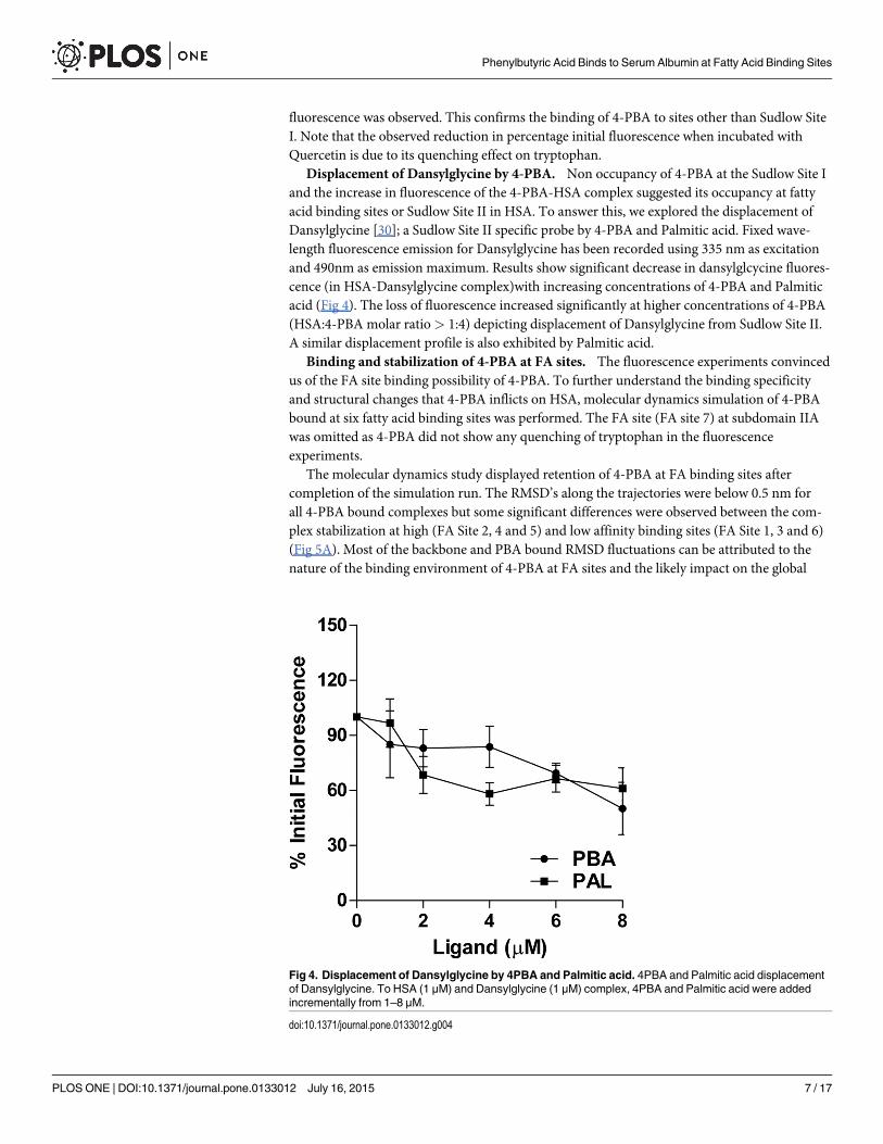
Displacement of Dansylglycine by 4-PBA. Non occupancy of 4-PBA at the Sudlow Site Iand the increase in fluorescence of the 4-PBA-HSA complex suggested its occupancy at fattyacid binding sites or Sudlow Site II in HSA. To answer this, we explored the displacement ofDansylglycine [30]; a Sudlow Site II specific probe by 4-PBA and Palmitic acid. Fixed wave-length fluorescence emission for Dansylglycine has been recorded using 335 nm as excitationand 490nm as emission maximum. Results show significant decrease in dansylglcycine fluores-cence (in HSA-Dansylglycine complex)with increasing concentrations of 4-PBA and Palmiticacid (Fig 4). The loss of fluorescence increased significantly at higher concentrations of 4-PBA(HSA:4-PBA molar ratio> 1:4) depicting displacement of Dansylglycine from Sudlow Site II.A similar displacement profile is also exhibited by Palmitic acid.
Binding and stabilization of 4-PBA at FA sites. The fluorescence experiments convincedus of the FA site binding possibility of 4-PBA. To further understand the binding specificityand structural changes that 4-PBA inflicts on HSA, molecular dynamics simulation of 4-PBAbound at six fatty acid binding sites was performed. The FA site (FA site 7) at subdomain IIAwas omitted as 4-PBA did not show any quenching of tryptophan in the fluorescenceexperiments.
The molecular dynamics study displayed retention of 4-PBA at FA binding sites aftercompletion of the simulation run. The RMSD’s along the trajectories were below 0.5 nm forall 4-PBA bound complexes but some significant differences were observed between the com-plex stabilization at high (FA Site 2, 4 and 5) and low affinity binding sites (FA Site 1, 3 and 6)(Fig 5A). Most of the backbone and PBA bound RMSD fluctuations can be attributed to thenature of the binding environment of 4-PBA at FA sites and the likely impact on the global
Fig 4. Displacement of Dansylglycine by 4PBA and Palmitic acid. 4PBA and Palmitic acid displacementof Dansylglycine. To HSA (1 μM) and Dansylglycine (1 μM) complex, 4PBA and Palmitic acid were addedincrementally from 1–8 μM.
doi:10.1371/journal.pone.0133012.g004
Phenylbutyric Acid Binds to Serum Albumin at Fatty Acid Binding Sites
PLOS ONE | DOI:10.1371/journal.pone.0133012 July 16, 2015 7 / 17

fluctuation of various subdomains. 4-PBA binding at FA site 3 and 6 shows considerablestability of the RMSD due to H-bonding mediated by terminal OH of the 4-PBA–COOHgroup (Fig 5B). 4-PBA at FA6 complex RMSD shows stabilization throughout the simulation,due to stable H-bonding with E354 of Subdomain IIB (which shows the least fluctuation duringthe course of simulation). However, FA6 complex backbone remains destabilized because ofthe outward motion of the protein during the simulation run. Among the others, both back-bone and PBA bound RMSD at FA3 site attains stability at about 1800 ps and shows better sta-bilization as compared to the starting structure. This stabilization is largely due to a bulk of H-bonded (Fig 5B) as well as non-covalent interactions (salt bridge) mediated by 4-PBA at FA3site. As the FA3 site is present in close proximity to the Subdomain IIIB (most flexible domainin HSA), binding of 4-PBA limits the flexible motion of this domain thereby contributing
Fig 5. Stability of 4PBA at FA binding sites of HSA. a) Time evolution of RMSD of the HSA backbone and PBA bound forms during 7ns MD simulation of4PBA bound to HSA at different FA binding sites. b) Interaction profile of 4PBA at all FA binding sites c) Salt bridge formation at FA3.
doi:10.1371/journal.pone.0133012.g005
Phenylbutyric Acid Binds to Serum Albumin at Fatty Acid Binding Sites
PLOS ONE | DOI:10.1371/journal.pone.0133012 July 16, 2015 8 / 17

greatly to its backbone stabilization. Much of the 4-PBA mediated internal motions of HSAoccur at the Subdomain IIIB and IA (which encloses FA sites 5 and 2), majorly contributed tothe RMSD fluctuations at both these sites in the entire simulation run. As the high-affinityfatty acid binding sites (FA sites 2, 4 and 5) have large hydrophobic cavities, 4-PBA with veryshort hydrophobic tail is preferably positioned at the origin of the cavity. Unlike long chainfatty acids, it shows lesser number of stabilizing hydrophobic interactions at these pockets thatcontribute majorly to the local fluctuation in FA sites 2, 4 and 5. Contrary to expectations, FAsite 5 backbone RMSD showed good stabilization throughout the simulation. Typically extrastabilization of 4PBA-HSA complex RMSD profiles (observed at FA1, FA3 and FA6) is attrib-uted to π-π, π-alkyl and H-bond interaction forces. Overall, 4-PBA binding at the low affinityFA binding sites showed better complex and backbone RMSD stabilization throughout thetime course of the simulation.
In FA site 1, 3 and 6 we observe a stabilization of RMSD majorly contributed by stacked π-π, π-alkyl and H-bond. The other sites, i.e. FA site 2, 4 and 5 we observe some slight fluctuationin the RMSD profile due to no/limited contribution from H-bond interaction. To sum up thefindings 4PBA prefers interacting majorly by stacked π-π (hydrophobic) and H-bond interac-tion (hydrophilic) in the low affinity FA binding sites and only by partially stacked π-π and π-alkyl interactions in the high affinity binding sites (S1 Table).
Binding of 4-PBA to fatty acid binding sites induces proteinconformational changes
Effect of binding of 4-PBA on the ellipticity changes in HAS. In order to analyse theeffect of HSA-4-PBA complex formation on the changes in secondary structure of HSA, theCD spectra of HSA in the presence and absence of 4-PBA was obtained. The changes in HSAellipticity is calculated using: MRE = θ/10rlC; where, MRE is mean residue ellipticity, θ isobserved ellipticity expressed in millidegree, r = 585 (number of amino acid residues of HSA),l is the path length (0.1cm) and C is the molar concentration of HSA. Circular Dichroism anal-ysis shows two major negative intensities at 208 and 222nm, characteristic of HSA alpha helixwhich shows significant decrease in negative ellipticity upon addition of 4-PBA (Fig 6D).This causes a slight increase in alpha helix percentage (38.8% in native HSA to 47.10% inHSA+4PBA) suggesting an increasing rigidity (stabilization) of the protein upon 4-PBA bind-ing (S2 Table). However, the CD spectra of HSA shows similar shape for both in presence andabsence of 4-PBA indicating predominant α helical content. This behaviour indicates overallmovement and stabilization of HSA on binding to 4-PBA. This is further explored by molecu-lar dynamics of HSA in an unliganded and different 4-PBA bound states.
Root Mean Square Fluctuations. The Root Mean Square Fluctuation of the Cα backbonewas computed for the Unliganded HSA and HSA-4-PBA complex from b-factors (RMSF =p3B/(8π2)), where B is the b-factor. The RMSF profiles of HSA show high fluctuations in the
C-terminal domain (IIIB) as compared to the N-terminal (IA) (Fig 6A); this is in agreementwith earlier studies [31]. Much of the HSA fluctuations are observed in the Sub domain IA andIIIB of all FA binding sites (Fig 6A). Binding of 4-PBA stabilizes the internal motion of thesetwo domains and contributes to increased stability. 4-PBA occupancy at nearly all FA sites sta-bilizes Subdomain IIA and IIB slightly (Fig 6A). Compared to these two subdomains, othersubdomains are mostly rigid and do not confer much to the 4-PBA induced conformationalchanges.
Principal Component Analysis. The simulation trajectory of 4-PBA-HSA complexeswere analysed as two principal components and the calculated eigenvectors were assembled inorder of decreasing eigenvalues. The relative contribution to protein fluctuation was obtained
Phenylbutyric Acid Binds to Serum Albumin at Fatty Acid Binding Sites
PLOS ONE | DOI:10.1371/journal.pone.0133012 July 16, 2015 9 / 17

from the first 10 modes which was responsible for more than 90% of the protein motion. PCAanalysis (Fig 6B) showed that 4-PBA occupancy at Sudlow Site II (FA3 and FA4; subdomainIIIA) and low affinity binding sites (FA1 and FA6) occupied less conformational space as com-pared to 4-PBA bound to high affinity binding sites like FA2 and FA5. Among the top teneigen values, the first two accounted largely for protein motion in each complex (Fig 6C). Longchain fatty acids binds linearly to the FA2 and FA5 sites (due to their large binding cavities),
Fig 6. Binding of 4PBA induces conformational changes on HSA. a) RMSF of Cα atoms of Unliganded HSA (discontinuous black lines) and 4PBA boundHSA at different FA binding sites. The demarcations show different Subdomains of HSA. Significant fluctuations can be seen at Subdomain IA and IIIB; themost mobile and hydrophobic fragments of HSA. b) 2D projection of first two principal components of different 4PBA-HSA bound models. c) Spectrum ofEigenvalues vs Eigenvector Index. d) CD absorption spectra of HSA-4PBA complex (HSA-1 μM; 4PBA-1μM).
doi:10.1371/journal.pone.0133012.g006
Phenylbutyric Acid Binds to Serum Albumin at Fatty Acid Binding Sites
PLOS ONE | DOI:10.1371/journal.pone.0133012 July 16, 2015 10 / 17
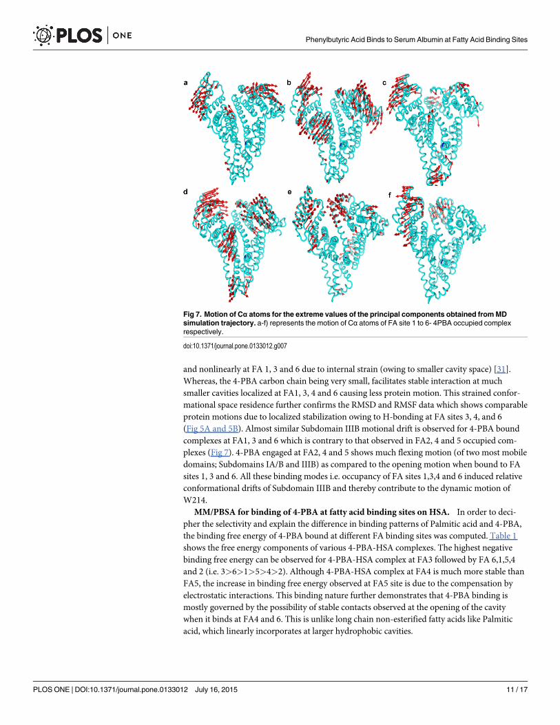
and nonlinearly at FA 1, 3 and 6 due to internal strain (owing to smaller cavity space) [31].Whereas, the 4-PBA carbon chain being very small, facilitates stable interaction at muchsmaller cavities localized at FA1, 3, 4 and 6 causing less protein motion. This strained confor-mational space residence further confirms the RMSD and RMSF data which shows comparableprotein motions due to localized stabilization owing to H-bonding at FA sites 3, 4, and 6(Fig 5A and 5B). Almost similar Subdomain IIIB motional drift is observed for 4-PBA boundcomplexes at FA1, 3 and 6 which is contrary to that observed in FA2, 4 and 5 occupied com-plexes (Fig 7). 4-PBA engaged at FA2, 4 and 5 shows much flexing motion (of two most mobiledomains; Subdomains IA/B and IIIB) as compared to the opening motion when bound to FAsites 1, 3 and 6. All these binding modes i.e. occupancy of FA sites 1,3,4 and 6 induced relativeconformational drifts of Subdomain IIIB and thereby contribute to the dynamic motion ofW214.
MM/PBSA for binding of 4-PBA at fatty acid binding sites on HSA. In order to deci-pher the selectivity and explain the difference in binding patterns of Palmitic acid and 4-PBA,the binding free energy of 4-PBA bound at different FA binding sites was computed. Table 1shows the free energy components of various 4-PBA-HSA complexes. The highest negativebinding free energy can be observed for 4-PBA-HSA complex at FA3 followed by FA 6,1,5,4and 2 (i.e. 3>6>1>5>4>2). Although 4-PBA-HSA complex at FA4 is much more stable thanFA5, the increase in binding free energy observed at FA5 site is due to the compensation byelectrostatic interactions. This binding nature further demonstrates that 4-PBA binding ismostly governed by the possibility of stable contacts observed at the opening of the cavitywhen it binds at FA4 and 6. This is unlike long chain non-esterified fatty acids like Palmiticacid, which linearly incorporates at larger hydrophobic cavities.
Fig 7. Motion of Cα atoms for the extreme values of the principal components obtained fromMDsimulation trajectory. a-f) represents the motion of Cα atoms of FA site 1 to 6- 4PBA occupied complexrespectively.
doi:10.1371/journal.pone.0133012.g007
Phenylbutyric Acid Binds to Serum Albumin at Fatty Acid Binding Sites
PLOS ONE | DOI:10.1371/journal.pone.0133012 July 16, 2015 11 / 17

Binding free energies as computed by the MMPBSA method varied from the spectroscopicbinding free energy. Similar discrepancies have been observed by Fujiwara et al [19] betweenexperimental and theoretical calculations. Perhaps, computationally expensive method likenormal mode analysis with very high simulation time>50ns will allow much conformationalfreedom of HSA to minimize this discrepancy.
Dynamic Movement of W214 and changes in hydrophobicity on 4-PBA occupancy atdifferent FA sites. AsW214 fluorescence shifts are affected by changes in its polar microen-vironment, the next step is to compare this with molecular dynamics studies. Movement andhydrophobicity changes of W214 and W214 pocket descriptor residues in (a) unliganded HSAand (b) HSA occupied by 4-PBA at individual FA binding sites by SASA analysis is compared(Table 2). The results show dynamic movement of W214 upon 4-PBA binding. The majorcontribution to W214 hydrophobicity is derived from its internal positional shift or movementof important descriptor residues from Subdomain IIA, IIB and IIIA which forms the W214pocket. Because these movements alter the solvent accessibility of W214, when 4-PBA occupiesvarious FA binding sites, the independent or co-ordinated motions of these Subdomains rec-ompenses the W214 hydrophobicity.
Table 2 shows W214 hydrophobicity and total hydrophobicity changes of HSA upon 4-PBAoccupancy at FA binding sites. Irrespective of its occupancy at any FA site, and difference inthe W214 hydrophobic SASA observed in each of these complexes, an overall increase in totalhydrophobicity of HSA upon binding of 4-PBA is observed. This can be understood from themovements of W214 and important residues of its W214 binding pocket (Fig 8). The net
Table 1. MM-PBSA Binding Free Energy components of HSA-4PBA complex.
Binding site ΔE coul* ΔE LJ* ΔG polar ΔG nonpolar ΔG binding
FA1 -63.417 ± 3.536 -114.161 ± 2.364 102.061 ± 3.514 -11.68 ± 0.142 -87.197 ± 3.481
FA2 -21.932 ± 2.326 -112.137 ± 1.843 84.129 ± 4.651 -12.088 ± 0.061 -62.028 ± 4.902
FA3 -226.546 ± 3.722 -102.734 ± 2.204 202.788 ± 3.920 -11.414 ± 0.052 -137.906 ± 4.208
FA4 -8.085 ± 3.780 -110.249 ± 1.194 54.044 ± 2.653 -11.976 ± 0.122 -76.266 ± 2.079
FA5 -60.194 ± 10.552 -109.691 ± 2.423 100.723 ± 8.327 -11.88 ± 0.090 -81.042 ± 3.809
FA6 -99.504 ± 3.568 -113.63 ± 1.632 112.627 ± 3.415 -11.647 ± 0.046 -112.153 ± 2.845
All energy values are expressed in KJM-1.
*LJ = Lennard Jones Potential
*Coul = Coulombic Charge
ΔG binding was calculated from Eq 1
doi:10.1371/journal.pone.0133012.t001
Table 2. Total and Residue Hydrophobicity (SASA analysis).
Binding site Hydrophobicity of W214* Total Hydrophobicity HSA-4PBA*
Unliganded HSA 0.77±0.122 136.556±0.25
FA1 0.74±0.125 139.159±0.220
FA2 0.81±0.111 138.709±0.220
FA3 0.72±0.114 140.541±0.212
FA4 0.91±0.140 144.522±0.215
FA5 0.91±0.147 139.153±0.226
FA6 0.67±0.116 143.883±0.214
*Values are expressed in nm2
doi:10.1371/journal.pone.0133012.t002
Phenylbutyric Acid Binds to Serum Albumin at Fatty Acid Binding Sites
PLOS ONE | DOI:10.1371/journal.pone.0133012 July 16, 2015 12 / 17

increase in hydrophobicity of the pocket is compensated either by the internal movement ofW214 or by important residues like L198, F206, A210, F211 (Subdomain IIA), V343, L347(Subdomain IIB) and L481 (Subdomain IIIA) (Fig 8B; Table 3). The residue W214 movesoutwards when 4-PBA complexes with HSA at FA 1, 4, 5 and 6 but inwardly at FA 2 and 3(Fig 8A). This discrepancy in the internal movement and hydrophobicity is resolved by themovement of the descriptor residues of the Subdomain IIA, IIB and IIIA.
So, an overall increase in hydrophobicity leads to the stabilization of HSA bound 4-PBA ascompared to unliganded HSA. The increased W214 hydrophobicity explains the rise in fluores-cence emission intensity when HSA is titrated with increasing concentrations of 4-PBA.
DiscussionHuman serum albumin is the most important multidrug transporter in our body and drugbinding to HSA is of important pharmacokinetic relevance. Binding nature of chemical
Fig 8. Dynamic Movement of W214 and descriptors of W214 pocket at HSA-4PBA complexes. a)Shifting of W214 residue in different HSA-4PBA complex. b) Movement of W214 pocket descriptors upon4PBA binding at different FA binding sites.
doi:10.1371/journal.pone.0133012.g008
Phenylbutyric Acid Binds to Serum Albumin at Fatty Acid Binding Sites
PLOS ONE | DOI:10.1371/journal.pone.0133012 July 16, 2015 13 / 17

chaperone 4-PBA to HSA is investigated by a combination of approaches. Absorption spectros-copy shows a stable complex formation between HSA and 4-PBA (Fig 2A), with a mild alter-ation to the protein backbone corroborated further by CD analysis. The absorption spectrashowed gradual broadening and a small blue shift in the tryptophan absorption region upongradual addition of 4-PBA, confirming alterations in the molecular environment of trypto-phan. Spectrofluorimetry assays show an increase in the tryptophan emission spectra on grad-ual addition in molar concentration of 4-PBA (Fig 2B). This is concurrent with the slightincrease in the quantum yield or decrease in the polarity of tryptophan. As tryptophan emis-sion is highly susceptible to changes in polarity of its ambient environment [32], this data sug-gests the possibility of tryptophan exposure to more hydrophobic environment.
Like non-esterified fatty acids, 4-PBA can bind to most of the major fatty acid binding sitesnon-covalently, but its principal binding sites vary. Fluorescence assays show an increase in rel-ative percentage fluorescence of HSA when titrated with increasing concentrations of Palmiticacid and 4-PBA (Fig 3A). Palmitic acid shows at least a fold increase in percentage fluorescencewhen compared with 4-PBA. Palmitic acid stabilizes the hydrophobic pockets at SubdomainIIIB and IA (where high affinity FA sites 5 and 2 are present) to a much greater extent whencompared to 4-PBA. In the ligand displacement assay, prior addition of Quercetin to HSA didnot prevent the increase in the fluorescence upon adding 4-PBA (Fig 3C). On the contrarywhen 4-PBA was pre-incubated with HSA, addition of Quercetin further quenched fluores-cence (Fig 3B). This clearly shows that 4-PBA has preferential binding to sites away fromSudlow Site I. (Fig 3B). Subdomain IIA binding of 4-PBA is excluded as it does not showW214 fluorescence quenching. 4-PBA binding at FA site 3 shows the highest negative bindingfree energy as shown by MMPBSA (Table 1) and least conformation space in its PCA analysis(Fig 6B). These findings are in concert and point out that 4-PBA prefers binding at SubdomainIIIA at FA sites 3 and 4 (Sudlow Site II). This is further supported by ligand displacement stud-ies using Site II probe Dansylglycine (Fig 4). 4-PBA at molar excess can displace dansylglycinefrom Site II showing its binding efficacy at this site.
Upon binding, 4-PBA stabilizes the overall conformation of HSA. This behaviour is similarto that of long chain non-esterified fatty acids like Palmitic acid and Myristic acid [32], [33].From earlier studies it is well known that binding of fatty acids to HSA produces dramaticconformational changes [34]. Domains I and III get displaced to the left thus opening up thecentral crevice. A similar behaviour is observed for 4-PBA binding (Fig 7) and moleculardynamics simulation with a partially and completely bound model of HSA-4-PBA complexcould reveal a better picture. FA2 site is at the interface of domain I and II and long chain fattyacid binding at this site stabilizes the movement of domain I. As 4-PBA binding takes place atthe origin of this cavity it is unable to inflict similar stabilization.
Table 3. Residue hydrophobicity of descriptors of W214 pocket upon different FA site complexes (SASA analysis).
Binding Site Descriptors of W214 binding pocket (values are expressed in nm2)
L481 L198 F206 A210 F211 V343 L347
Unliganded HSA 0.79±0.14 0.47±0.09 0.86±0.21 0.51±0.09 0.20±0.07 0.14±0.06 0.43±0.09
FA1 0.79±0.11 0.55±0.11 0.95±0.17 0.51±0.07 0.25±0.08 0.17±0.06 0.37±0.08
FA2 0.71±0.14 0.38±0.09 0.87±0.14 0.51±0.09 0.24±0.21 0.14±0.05 0.35±0.07
FA3 0.82±0.13 0.43±0.07 1.0±0.16 0.48±0.08 0.19±0.08 0.22±0.06 0.38±0.09
FA4 0.69±0.11 0.57±0.08 0.85±0.11 0.45±0.08 0.24±0.14 0.30±0.08 0.27±0.07
FA5 0.63±0.09 0.41±0.08 0.71±0.14 0.46±0.12 0.17±0.08 0.19±0.06 0.33±0.06
FA6 0.66±0.16 0.60±0.11 0.76±0.17 0.36±0.10 0.26±0.07 0.23±0.07 0.29±0.09
doi:10.1371/journal.pone.0133012.t003
Phenylbutyric Acid Binds to Serum Albumin at Fatty Acid Binding Sites
PLOS ONE | DOI:10.1371/journal.pone.0133012 July 16, 2015 14 / 17

In the MD simulation of 4-PBA bound to HSA at six FA binding sites, 4-PBA forms stableH-bonding at FA site 3, 4 and 6 (Fig 5B). In FA4 site, it forms stable hydrogen bonding withS489 at a distance of 2.8 Å. It forms a π-π stacking interaction with Y411 at 3.52 Å (Fig 5C).This binding is similar to the binding behaviour of nonharmane [35] and Bcl2 family antican-cer agent [36] as previously reported. In addition to this, the FA4 backbone stabilization roleof π-alkyl interactions with Subdomain IIIA residues V426 at 4.91Å and L460 at 5.17 Å isobserved. When considering 4-PBA binding at FA3, an overall stable complex formation isseen from the RMSD, RMSF and PCA. This particular stability can be attributed to linear occu-pation of 4-PBA and formation of three stable hydrogen bonds with R485, E450 and R348. Asalt bridge created between the domain III and II via supplanting of E450 (Subdomain IIIA) by4-PBA to R348 (Subdomain IIB) contributes greatly to the stabilization of the mobile subdo-main IIIB. As a result of this, a very stable RMSD is observed during the course of simulationwhen 4-PBA occupies FA3 site. Apart from this it is also stabilized by a large number of Cou-lombic interactions (Table 1). Moreover the high selectivity of 4-PBA to bind towards FA3 site(subdomain IIIA) can allow cooperative binding of physiological ligands like FA’s. In patholog-ical states like obesity, where there is an increase in circulating free fatty acids [37], 4-PBAbinding to HSA might be affected. Having obtained some hints on this phenomenon, it wouldbe interesting to find out the pharmacokinetics of 4-PBA in obese patients in clinical dosages.
ConclusionsThis work shows the binding nature of chemical chaperone 4-PBA with human serum albu-min. A cumulative analyses with different methods show that 4-PBA can bind to FA bindingsites in HSA with high specificity of binding at FA site 3 (subdomain IIIA). Like physiologicalFA’s it can inflict similar conformational changes upon interaction to HSA. Transport of4-PBA will largely vary due to increased concentrations of FA’s in the plasma in pathologicalstates like obesity. This study gives better perspective on future pharmacokinetic studies of4-PBA and so clinically relevant changes in dosage can be profiled for varying disease states.
Supporting InformationS1 Table. Interaction forces of 4PBA at different FA binding sites of HSA.(DOCX)
S2 Table. Secondary structure evaluation from far UV CD spectra.(DOCX)
AcknowledgmentsWe thank VIT University for supporting this work. We thank Bioinformatics Resources andApplications Facility (BRAF) supercomputing facility at C-DAC (Pune and Bangalore) for pro-viding grid facilities to carry out this work. We thank Dr. Bhaskar Mohan Murari and Prof. G.Jayaraman, VIT University for facilitating the fluorescence and CD studies.
Author ContributionsConceived and designed the experiments: DR VK KT. Performed the experiments: DR VK SKVG. Analyzed the data: DR VKMSS JJ. Contributed reagents/materials/analysis tools: MSS JJKT. Wrote the paper: DR KT.
Phenylbutyric Acid Binds to Serum Albumin at Fatty Acid Binding Sites
PLOS ONE | DOI:10.1371/journal.pone.0133012 July 16, 2015 15 / 17
References1. Yoshida H. ER stress and diseases. FEBS J. 2007; 274(3):630–58. Epub 2007/02/10. doi: EJB5639
[pii] doi: 10.1111/j.1742-4658.2007.05639.x PMID: 17288551.
2. Lindholm D, Wootz H, Korhonen L. ER stress and neurodegenerative diseases. Cell Death Differ.2006; 13(3):385–92. Epub 2006/01/07. doi: 4401778 [pii] doi: 10.1038/sj.cdd.4401778 PMID:16397584.
3. Back SH, Kaufman RJ. Endoplasmic reticulum stress and type 2 diabetes. Annu Rev Biochem. 2012;81:767–93. Epub 2012/03/27. doi: 10.1146/annurev-biochem-072909-095555 PMID: 22443930;PubMed Central PMCID: PMC3684428.
4. Dover GJ, Brusilow S, Charache S. Induction of fetal hemoglobin production in subjects with sickle cellanemia by oral sodium phenylbutyrate. Blood. 1994; 84(1):339–43. Epub 1994/07/01. PMID: 7517215.
5. Maestri NE, Brusilow SW, Clissold DB, Bassett SS. Long-term treatment of girls with ornithine transcar-bamylase deficiency. N Engl J Med. 1996; 335(12):855–9. Epub 1996/09/19. doi: 10.1056/NEJM199609193351204 PMID: 8778603.
6. Iranfar H, Rajabi O, Salari R, Chamani J. Probing the interaction of human serum albumin with cipro-floxacin in the presence of silver nanoparticles of three sizes: multispectroscopic and zeta potentialinvestigation. J Phys Chem B. 2012; 116(6):1951–64. Epub 2012/01/10. doi: 10.1021/jp210685qPMID: 22224861.
7. Fanali G, di Masi A, Trezza V, Marino M, Fasano M, Ascenzi P. Human serum albumin: from bench tobedside. Mol Aspects Med. 2012; 33(3):209–90. Epub 2012/01/11. doi: 10.1016/j.mam.2011.12.002S0098-2997(11)00089-6 [pii]. PMID: 22230555.
8. Kragh-Hansen U. Structure and ligand binding properties of human serum albumin. Dan Med Bull.1990; 37(1):57–84. Epub 1990/02/01. PMID: 2155760.
9. Carter DC, Ho JX. Structure of serum albumin. Adv Protein Chem. 1994; 45:153–203. Epub 1994/01/01. PMID: 8154369.
10. Zsila F. Subdomain IB is the third major drug binding region of human serum albumin: toward the three-sites model. Mol Pharm. 2013; 10(5):1668–82. Epub 2013/03/12. doi: 10.1021/mp400027q PMID:23473402.
11. Curry S, Mandelkow H, Brick P, Franks N. Crystal structure of human serum albumin complexed withfatty acid reveals an asymmetric distribution of binding sites. Nat Struct Biol. 1998; 5(9):827–35. Epub1998/09/10. doi: 10.1038/1869 PMID: 9731778.
12. Thumser AE, Buckland AG, Wilton DC. Monoacylglycerol binding to human serum albumin: evidencethat monooleoylglycerol binds at the dansylsarcosine site. J Lipid Res. 1998; 39(5):1033–8. Epub1998/06/04. PMID: 9610770.
13. Van Der Spoel D, Lindahl E, Hess B, Groenhof G, Mark AE, Berendsen HJ. GROMACS: fast, flexible,and free. J Comput Chem. 2005; 26(16):1701–18. Epub 2005/10/08. doi: 10.1002/jcc.20291 PMID:16211538.
14. Hornak V, Abel R, Okur A, Strockbine B, Roitberg A, Simmerling C. Comparison of multiple Amberforce fields and development of improved protein backbone parameters. Proteins. 2006; 65(3):712–25.Epub 2006/09/19. doi: 10.1002/prot.21123 PMID: 16981200.
15. Wang J, Wolf RM, Caldwell JW, Kollman PA, Case DA. Development and testing of a general amberforce field. J Comput Chem. 2004; 25(9):1157–74. Epub 2004/04/30. doi: 10.1002/jcc.20035 PMID:15116359.
16. Sousa da Silva AW, VrankenWF. ACPYPE—AnteChamber PYthon Parser interfacE. BMC Res Notes.2012; 5:367. Epub 2012/07/25. doi: 10.1186/1756-0500-5-367 1756-0500-5-367 [pii]. PMID:22824207; PubMed Central PMCID: PMC3461484.
17. Hess B. Similarities between principal components of protein dynamics and random diffusion. PhysicalReview E. 2000; 62(6):8438.
18. Paissoni C, Spiliotopoulos D, Musco G, Spitaleri A. GMXPBSA 2.1: A GROMACS tool to perform MM/PBSA and computational alanine scanning. Computer Physics Communications. 2015; 186:105–7.
19. Fujiwara S, Amisaki T. Identification of high affinity fatty acid binding sites on human serum albumin byMM-PBSAmethod. Biophys J. 2008; 94(1):95–103. Epub 2007/09/11. doi: biophysj.107.111377 [pii]doi: 10.1529/biophysj.107.111377 PMID: 17827235; PubMed Central PMCID: PMC2134860.
20. HeW, Li Y, Si H, Dong Y, Sheng F, Yao X, et al. Molecular modeling and spectroscopic studies on thebinding of guaiacol to human serum albumin. Journal of Photochemistry and Photobiology A: Chemis-try. 2006; 182(2):158–67.
21. Subramanyam R, Gollapudi A, Bonigala P, Chinnaboina M, Amooru DG. Betulinic acid binding tohuman serum albumin: a study of protein conformation and binding affinity. J Photochem Photobiol B.
Phenylbutyric Acid Binds to Serum Albumin at Fatty Acid Binding Sites
PLOS ONE | DOI:10.1371/journal.pone.0133012 July 16, 2015 16 / 17

2009; 94(1):8–12. Epub 2008/10/24. doi: 10.1016/j.jphotobiol.2008.09.002 S1011-1344(08)00190-5[pii]. PMID: 18945624.
22. Bhattacharyya J, Bhattacharyya M, Chakrabarty AS, Chaudhuri U, Poddar RK. Interaction of chlor-promazine with myoglobin and hemoglobin. A comparative study. Biochem Pharmacol. 1994; 47(11):2049–53. Epub 1994/06/01. PMID: 8010989.
23. He XM, Carter DC. Atomic structure and chemistry of human serum albumin. Nature. 1992; 358(6383):209–15. Epub 1992/07/16. doi: 10.1038/358209a0 PMID: 1630489.
24. Dufour C, Dangles O. Flavonoid-serum albumin complexation: determination of binding constants andbinding sites by fluorescence spectroscopy. Biochim Biophys Acta. 2005; 1721(1–3):164–73. Epub2005/01/18. doi: S0304-4165(04)00284-3 [pii] doi: 10.1016/j.bbagen.2004.10.013 PMID: 15652191.
25. Bhattacharya AA, Curry S, Franks NP. Binding of the general anesthetics propofol and halothane tohuman serum albumin. High resolution crystal structures. J Biol Chem. 2000; 275(49):38731–8. Epub2000/08/15. doi: 10.1074/jbc.M005460200 M005460200 [pii]. PMID: 10940303.
26. Fehske KJ, Schlafer U, Wollert U, Muller WE. Characterization of an important drug binding area onhuman serum albumin including the high-affinity binding sites of warfarin and azapropazone. Mol Phar-macol. 1982; 21(2):387–93. Epub 1982/03/01. PMID: 6808349.
27. Ghuman J, Zunszain PA, Petitpas I, Bhattacharya AA, Otagiri M, Curry S. Structural basis of the drug-binding specificity of human serum albumin. J Mol Biol. 2005; 353(1):38–52. Epub 2005/09/20. doi:S0022-2836(05)00885-5 [pii] doi: 10.1016/j.jmb.2005.07.075 PMID: 16169013.
28. Sengupta B, Sengupta PK. The interaction of quercetin with human serum albumin: a fluorescencespectroscopic study. Biochem Biophys Res Commun. 2002; 299(3):400–3. Epub 2002/11/26. doi:S0006291X02026670 [pii]. PMID: 12445814.
29. Sengupta B, Sengupta PK. Binding of quercetin with human serum albumin: a critical spectroscopicstudy. Biopolymers. 2003; 72(6):427–34. Epub 2003/10/31. doi: 10.1002/bip.10489 PMID: 14587065.
30. Graciani FS, Ximenes VF. Investigation of human albumin-induced circular dichroism in dansylglycine.PLoS One. 2013; 8(10):e76849. Epub 2013/10/23. doi: 10.1371/journal.pone.0076849 PONE-D-13-25398 [pii]. PMID: 24146932; PubMed Central PMCID: PMC3797758.
31. Guizado TC, Louro S, Anteneodo C. Dynamics of heme complexed with human serum albumin: a theo-retical approach. European Biophysics Journal. 2012; 41(12):1033–42. doi: 10.1007/s00249-012-0860-2 PMID: 23104623
32. Pongprayoon P, Gleeson MP. Probing the binding site characteristics of HSA: A combined moleculardynamics and cheminformatics investigation. J Mol Graph Model. 2014; 54:164–73. Epub 2014/12/03.doi: 10.1016/j.jmgm.2014.10.007 S1093-3263(14)00166-1 [pii]. PMID: 25459768.
33. Fujiwara S, Amisaki T. Molecular dynamics study of conformational changes in human serum albuminby binding of fatty acids. Proteins. 2006; 64(3):730–9. Epub 2006/06/20. doi: 10.1002/prot.21053PMID: 16783783.
34. Curry S, Brick P, Franks NP. Fatty acid binding to human serum albumin: new insights from crystallo-graphic studies. Biochim Biophys Acta. 1999; 1441(2–3):131–40. Epub 1999/11/26. doi: S1388-1981(99)00148-1 [pii]. PMID: 10570241.
35. Domonkos C, Fitos I, Visy J, Zsila F. Fatty acid modulated human serum albumin binding of the beta-carboline alkaloids norharmane and harmane. Mol Pharm. 2013; 10(12):4706–16. Epub 2013/11/01.doi: 10.1021/mp400531n PMID: 24171410.
36. Oltersdorf T, Elmore SW, Shoemaker AR, Armstrong RC, Augeri DJ, Belli BA, et al. An inhibitor of Bcl-2family proteins induces regression of solid tumours. Nature. 2005; 435(7042):677–81. Epub 2005/05/20. doi: nature03579 [pii] doi: 10.1038/nature03579 PMID: 15902208.
37. Boden G. Obesity and free fatty acids. Endocrinol Metab Clin North Am. 2008; 37(3):635–46, viii-ix.Epub 2008/09/09. doi: 10.1016/j.ecl.2008.06.007 S0889-8529(08)00033-9 [pii]. PMID: 18775356;PubMed Central PMCID: PMC2596919.
Phenylbutyric Acid Binds to Serum Albumin at Fatty Acid Binding Sites
PLOS ONE | DOI:10.1371/journal.pone.0133012 July 16, 2015 17 / 17