Oxidative stresses induced by glycoxidized human or bovine serum albumin on human monocytes
57
Oxidative stresses induced by glycoxidized human or bovine serum albumins on human monocytes Philippe Rondeau, Nihar Ranjan Singh, Henri Caillens, Frank Tallet, Emmanuel Bourdon PII: S0891-5849(08)00355-9 DOI: doi: 10.1016/j.freeradbiomed.2008.06.004 Reference: FRB 9438 To appear in: Free Radical Biology and Medicine Received date: 19 November 2007 Revised date: 28 May 2008 Accepted date: 3 June 2008 Please cite this article as: Philippe Rondeau, Nihar Ranjan Singh, Henri Caillens, Frank Tallet, Emmanuel Bourdon, Oxidative stresses induced by glycoxidized human or bovine serum albumins on human monocytes, Free Radical Biology and Medicine (2008), doi: 10.1016/j.freeradbiomed.2008.06.004 This is a PDF file of an unedited manuscript that has been accepted for publication. As a service to our customers we are providing this early version of the manuscript. The manuscript will undergo copyediting, typesetting, and review of the resulting proof before it is published in its final form. Please note that during the production process errors may be discovered which could affect the content, and all legal disclaimers that apply to the journal pertain.
-
Upload
ravenshawuniversity -
Category
Documents
-
view
0 -
download
0
Transcript of Oxidative stresses induced by glycoxidized human or bovine serum albumin on human monocytes

�������� ����� ��
Oxidative stresses induced by glycoxidized human or bovine serum albuminson human monocytes
Philippe Rondeau, Nihar Ranjan Singh, Henri Caillens, Frank Tallet,Emmanuel Bourdon
PII: S0891-5849(08)00355-9DOI: doi: 10.1016/j.freeradbiomed.2008.06.004Reference: FRB 9438
To appear in: Free Radical Biology and Medicine
Received date: 19 November 2007Revised date: 28 May 2008Accepted date: 3 June 2008
Please cite this article as: Philippe Rondeau, Nihar Ranjan Singh, Henri Caillens, FrankTallet, Emmanuel Bourdon, Oxidative stresses induced by glycoxidized human or bovineserum albumins on human monocytes, Free Radical Biology and Medicine (2008), doi:10.1016/j.freeradbiomed.2008.06.004
This is a PDF file of an unedited manuscript that has been accepted for publication.As a service to our customers we are providing this early version of the manuscript.The manuscript will undergo copyediting, typesetting, and review of the resulting proofbefore it is published in its final form. Please note that during the production processerrors may be discovered which could affect the content, and all legal disclaimers thatapply to the journal pertain.
ACC
EPTE
D M
ANU
SCR
IPT
ACCEPTED MANUSCRIPTRondeau
1
Original Research Communication
OXIDATIVE STRESSES INDUCED BY GLYCOXIDIZED HUMAN
OR BOVINE SERUM ALBUMINS ON HUMAN MONOCYTES
Philippe Rondeau1, Nihar Ranjan Singh
1, Henri Caillens
2, Frank Tallet
2,
Emmanuel Bourdon1*
1 Laboratoire de Biochimie et Génétique Moléculaire (LBGM), Université de La Réunion,
Saint Denis de La Réunion, France.
2 Unité fonctionnelle de recherche biochimie, Centre Hospitalier Félix Guyon, Saint Denis,
de La Réunion, France.
Acknowledgments
This work was supported by the Ministère de l’Enseignement Supérieur et de la Recherche et
de l’Outre Mer and the Université de La Réunion. NRS was supported by a fellowship from
the Conseil Régional de La Réunion. Helps from Drs. Christian Lefebvre d’Hellencourt,
Bernard Offmann and Serge Chesne for manuscript editing were greatly appreciated by the
authors.
* Corresponding author
LBGM - Université de la Réunion - 15, avenue René Cassin – BP 7151 – Cedex 09
97715 Saint Denis de La Réunion – France
Tel: (+262) 262 93 86 48 - Fax: (+262) 262 93 82 37
Running title: Effects of Glycoxidized Albumins on THP1

ACC
EPTE
D M
ANU
SCR
IPT
ACCEPTED MANUSCRIPTRondeau
2
OXIDATIVE STRESSES INDUCED BY GLYCOXIDIZED HUMAN
OR BOVINE SERUM ALBUMINS ON MONOCYTES
Abstract
Oxidative stress and protein modifications are frequently observed in numerous disease states.
Albumin, the major circulating protein in blood, can undergo increased glycoxidation in
diabetes. Proteins glycoxidation can lead to the formation of Advanced Glycoxidation
Endproducts, which induce various deleterious effects on cells. Herein, we report the effect of
glucose or methylglyoxal-induced oxidative modifications on BSA or HSA protein structures
and on THP1 monocyte physiology. Occurrence of oxidative modifications was found to be
enhanced in glycoxidized BSA and HSA, after determination of their free thiol group content,
relative electrophoretic migration, their carbonyl content and antioxidant activities. Cells
treated with glycoxidized albumins exhibited an over-generation of intracellular reactive
oxygen species, impairments in proteasomal activities, enhancements in RAGE expression
and an accumulation of carbonylated proteins. These novel observations made in the presence
of a range of modified BSA and HSA facilitate the comparison of the glycoxidation extent of
albumin with the oxidative stress induced in cultured monocytes. Finally, this study
reconfirms the influence of experimental conditions in which AGEs are generated and the
concentration levels in experiments designed to mimic pathological conditions.
Word count: 181
Keywords : Glycoxidation, Albumin, Monocytes, Oxidative stress, THP1, Glycation

ACC
EPTE
D M
ANU
SCR
IPT
ACCEPTED MANUSCRIPTRondeau
3
Introduction
Currently, in the United States alone, more than 17 million persons suffer from
diabetes and this number may double before 2030 [1,2]. Type 2 diabetes or Non-Insulin
Dependent Diabetes (NIDD) represents approximately 85% of all cases of diabetes. This
disease is a metabolic disorder characterized by hyperglycemia and is dramatically associated
with severe complications. NIDD could double the risk of developing cardiovascular disease,
which represents the leading cause of mortality in western countries [3].
Albumin is the most abundant serum protein with normal concentrations in the range of 35–
50 g/L. This protein possesses a wide range of biological properties [4], and there has been
ample evidence suggesting that it exerts significant antioxidant activity [5]. In fact, albumin
represents the major and predominant circulating antioxidant molecule in plasma known to be
exposed to continuous stress [6-8].
Oxidative modifications of proteins could occur in diabetes, which is one of the important
pathological conditions associated with the early occurrence of vascular complications,
together with functional alterations of albumin. In NIDD, albumin undergoes increased
glycation and glycoxidation [9]. The glycation phenomenon corresponds to the non-enzymatic
and non-oxidative covalent attachment of glucose molecule to protein [9]. Glycoxidation
refers to the radical-mediated oxidation reaction of both free and protein-bound sugars [2].
Amadori rearrangement of the above glycoxidized protein gives rise to deleterious, advanced
glycoxidation (also termed advanced glycation) end products (AGEs) [10,11]. The formation
and accumulation of AGEs constitute the characteristic features of tissues in NIDD patients.
The pathological conditions of diabetes has been associated with an enhanced oxidative stress
[12]. Oxidative stress in diabetes could originate from various processes, such as excessive
production of oxygen radical from the autoxidation of glucose, glycoxidized proteins and
glycoxidation of antioxidative proteins [13]. The compiled data from biochemical, animal and

ACC
EPTE
D M
ANU
SCR
IPT
ACCEPTED MANUSCRIPTRondeau
4
epidemiological studies strongly support the hypothesis that glycoxidative modification of
circulating proteins plays a pivotal and causative role in the pathogenesis of NIDD [14-17].
Interaction of AGEs with their receptors (RAGE) induces several cellular phenomena
potentially relating to diabetic complications. AGEs were demonstrated to augment
inflammatory responses in monocytes by up-regulating multiple signalling pathways via
RAGE leading to vascular cell dysfunction [18]. Conversely, the AGEs product inhibitor LR-
90 has been shown to exert anti-inflammatory effects in human monocytes [19]. AGEs have
been identified as one of the factors closely associated with the vascular complications in
NIDD. The implication of glycoxidized albumin in the generation of oxidative stress and
damages on inflammatory cells should be better specified. We previously reported the effects
of oxidative modifications induced by the BSA glycoxidation on its structure, antioxidant
properties and adipose cell physiology [20-22]. The majority of literature in this regard
reports the use of Bovine Serum Albumin. Hence it is of utmost importance to compare the
effects of glycoxidation on both the structures of Human Serum Albumin (HSA) and Bovine
Serum Albumin (BSA) and to investigate and compare the effects of such glycoxidized
albumins on monocyte cell physiology.
Here, we report the overgeneration of intracellular reactive oxygen species (ROS),
impairments in proteasomal activities, accumulation of carbonylated proteins and enhanced
expression of RAGE in glycoxidized-albumins-treated cells. These novel observations have
been made in the presence of various modified BSA and HSA, which allows the comparison
between the extent of albumin glycoxidation and the subsequent induced oxidative stress in
cultured monocytes.

ACC
EPTE
D M
ANU
SCR
IPT
ACCEPTED MANUSCRIPTRondeau
5
Materials and methods
Preparation of AGEs
For the preparation of glycoxidized albumins, non-recombinant bovine serum albumin (BSA)
(Sigma cat# A2153) and human serum albumin (HSA) were used. HSA were purified in our
laboratory from whole human plasma as follows: isolation of serum albumin from fresh
human plasma was based on an extensive dialysis against 50 mM-Tris/HCl, pH 7.4, followed
by an affinity chromatography using Cibacron Blue 3G linked to agarose (Amersham cat#17-
0948-01) as ligand for albumin. A, 1.5 M NaCl, pH 7.4 buffer was used for the desorption of
bound albumin from Cibacron Blue-Agarose following elution of other plasmatic proteins
with a 50 mM-Tris/HCl, pH 7.4 buffer. Each fraction of eluate was examined by gel
electrophoresis and the most purified and concentrated extracts were pooled. AGEs were
prepared as previously described [21] by incubating 0.5 mM protein (BSA or HSA) with
increasing concentrations of glucose (0 mM, 5 mM and 25 mM) and in PBS, pH 7.4 under
sterile conditions and nitrogen gas in capped vials at 37°C for three weeks or with
methylglyoxal (10 mM) for two days. The proteins were dialyzed against PBS, pH 7.4 and
sterile-filtered with 0.2 µm Millipore. Endotoxin content as assessed by in vitro toxicology
assay kit (E-TOXATE, Sigma) was bellow detectable level (0.03 Endotoxin unit/mL).
- Purification of albumin from diabetic and nondiabetic patients:
Purifications of albumin were conducted using pooled serum from three diabetic (% HbA1C =
11.9 ± 1.2) and three nondiabetic (% HbA1C = 4.83 ± 0.31) patients. Fasting blood glucose
concentrations were comprised between 0.73 and 1 g/L for the group of nondiabetic persons,
whereas it was higher (1.1 g/L) for diabetic patients. Isolation of serum albumin from human

ACC
EPTE
D M
ANU
SCR
IPT
ACCEPTED MANUSCRIPTRondeau
6
fresh plasma was performed as previously described and by using an affinity chromatography
(Cibacron Blue 3G - Amersham cat#17-0948-01).
Structural studies of AGEs
- Measurements of fluorescence and absorbance
AGE-related modifications were determined by assessing the production of fluorescent
compounds or glycophore [23] and by measuring the “browning color” resulting from the
nonenzymatic glycosylation of albumin [24]. The formation of glycophore associated with
glycoxidation process in albumin was monitored at excitation and emission wavelengths of
355 and 410 nm, respectively, by using a microplate fluorometer FLUOstar OPTIMA (BMG
Labtech, France). The brown color constitutes one of the qualitative properties of AGEs [25]
and was analyzed by measuring the absorbance at 420 nm using a UV-visible
spectrophotometer and expressed as absorbance per µg protein.
- Oxidative modification of SH
Thiol groups in native or modified albumin were measured by Ellman’s assay using 5, 5’-
dithiobis, 2-nitrobenzoic acid (DTNB) [26]. Briefly, 250 µL of albumin samples (in 0.1M
Tris-HCl, pH 8.0, EDTA 5 mM) were incubated with three volumes of 0.5 mM DTNB.
Considering the interference of browning state of modified albumin, the concentration of free
thiol was calculated with a regression model by using Partial Least Squares method (PLS)
[27]. The PLS model was optimized by using 22 standard absorbance spectra recorded,
between 250 and 500 nm, with a PowerWave™ Microplate Spectrophotometer (BioTek,
USA). These standards correspond to various concentrations of L-cysteine (10 to 100 nmoles)
mixed with a browning solution at different concentrations. Results were expressed as the
number of free –SH groups per mol of BSA.

ACC
EPTE
D M
ANU
SCR
IPT
ACCEPTED MANUSCRIPTRondeau
7
- REM study
Modifications of albumin (BSA or HSA) after glycoxidation were analyzed by native
Polyacrylamide Gel (12 % of acrylamide) and stained by Coomassie blue according to
Laemmli’s method [28]. Relative Electrophoretic Migration (REM) was determined for the
different AGEs by calculating the migration ratio of the modified albumin monomer with the
corresponding native albumin monomer (albumin which had not been previously incubated)
[21].
- Mass spectroscopic analyses
All mass spectroscopic measurements were performed at the Molecular Biophysics Unit
(MBU) of the Indian Institute of Science (Bangalore, India). Molecular weights of both
glycoxidized and non-glycoxidized samples were determined by electrospray ionisation mass
spectrometry (ESI/MS), while tryptic digestions were analyzed by MALDI-TOF-TOF.
ESI/MS was performed on Agilent 1100 LC coupled to Bruker Esquire3000plus operating in
positive ion mode. Complete ESI/MS settings were: high voltage capillary, 4000 V; high
voltage end plate offset, -500 V; nebulizer gas pressure, 28 psi; dry gas flow rate, 7.00 l/min;
dry gas temperature, 340°C and capillary exit, 196.0V; Trap Drive, 120.6. MALDI/MS
measurements were performed using UltraflexTOF/TOF from Bruker Daltomics (Germany).
The setting condition of the mass spectrometer was as follows: reflection positive ion mode,
accelerating voltage, 20Kev. Ions were generated by the emission of nitrogen laser at a
wavelength of 337 nm with 50 Hz repetition rate. Generally, an average of 100–150 laser
shots are required to record a mass spectrum. Sinapinic acid (50 mg/ml in 50% acetonitrile,
9% trifluoroacetic acid [TFA], in water) was used as the matrix. Target plate used was the
ground steel plate with 384 wells. Triplicate MALDI measurements were made for each
sample for the estimation of reproducibility. Before enzymatic digestion with trypsin, AGEs

ACC
EPTE
D M
ANU
SCR
IPT
ACCEPTED MANUSCRIPTRondeau
8
samples were reduced with 100 mM DTT at 95°C for 5 min. After alkylation with a solution
of 100 mM iodoacetamide for 20 min at room temperature, the samples were incubated with
trypsin (200 µl of a 0.1 µg/µl solution) for 3 h at 37°C followed with an additional incubation
overnight at 30°C. A total of 0.5 µL of digestion solutions were added to the same volume of
matrix solution and the resulting mixtures were completely dried before acquisition.
- Copper binding test
The capacity of albumin for copper ions binding can be measured spectrophotometrically by
the use of bathocuproinedisulfonic acid (BC, Fluka #11870) [29,30]. AGE preparations of
BSA and HSA (0.2 µM in NaCl 0.15 M) were incubated in triplicate for 2 h with 10 µM
CuSO4. Then an overnight dialysis against NaCl 0.15 M was performed and a BC solution
(1.2 mM) was added to each sample to a final concentration of 400 µM, followed by the
addition of 1 mM sodium ascorbate solution. Incubation for 5 min at room temperature
facilitated the complete reduction of Cu(II) to Cu(I) bound to albumin and resulted in the
formation of BC-Cu(I) complex, which was read at 480 nm with a UV-visible
spectrophotometer HP model 8453. The concentrations of copper bound to modified albumins
were calculated using a calibration standard curve by increasing the concentrations of CuSO4
up to 50 nM.
Hemolysis test of red blood cells
The antioxidant properties of AGEs were examined with the free radical-induced blood
hemolysis test [31]. Human blood samples were obtained from the Biochemistry department
of the local hospital center (CHD Felix Guyon) and were taken on EDTA substrate as
anticoagulant. Then plasma was removed and erythrocytes were washed with an isotonic
solution (NaCl 0.15 M). Each well of a 96 well-plate was filled with 100 µl (about 1.108

ACC
EPTE
D M
ANU
SCR
IPT
ACCEPTED MANUSCRIPTRondeau
9
erythrocytes, 400 000 cells/µL final concentration) of a diluted red blood cells solution (1/10
in 0.15 M NaCl). Different albumin samples (10 µM final concentration) were added in
triplicates. Hemolyses were started by adding 0.45 M of 2, 2’-azobis (2-amidinopropane)
(AAPH) in each well. Turbidimetry at 450 nm was recorded every 10 min using a 37°C-
thermostated microplate reader. Results were expressed as 50% of maximal hemolysis time
(HT50 in min) which represents the total defense against free radicals in human and animal
models submitted to oxidative stress [32].
Cell culture of THP1
Human monocytic leukemia cell line THP1 (ATCC TIB-202) was cultivated in RPMI 1640
medium with 10% SVF, L-glutamine (2 mM), penicillin (100 U/mL), streptomycin (100
µg/mL) and amphotericine B (0.5 µg/mL). Cells were grown in a 5% CO2 incubator at 37°C.
For all experiments, cells were plated in triplicate for each condition, at 104 cells/100 µL per
well in sterile 96-well plate (for cytotoxicity analyses), at 2.105 cells/ mL per well in sterile
24-well plate (for ROS assays, Western-blot and proteasome activity measurement
experiments). The culture plates were incubated for 24 h before adding different AGEs
preparations at final concentration of 10 µM. Thereafter, cells were maintained in the
humidified CO2 incubator for 16 h before further analyses. Before proteasomal activity
measurements and carbonyl Western blots, cells were washed thrice with PBS and were
treated at 4°C for 30 min with lysis buffer (25 mM Tris-HCl, pH 8.3, 10 mM KCl, 1 mM
DTT, 1 mM EDTA, 1% Triton X-100) without protease inhibitor. Cell lysates were then
centrifugated and protein concentrations were determined in the supernatant by BCA method
[33].

ACC
EPTE
D M
ANU
SCR
IPT
ACCEPTED MANUSCRIPTRondeau
10
Proliferation assay
The MTT-assay, using the standard 3-(4,5-dimethylthiazol-2-yl)-2,5-diphenyl tetrazolium
bromide (Sigma), was chosen to evaluate the effect of AGEs on THP1 cell proliferation [34].
This assay is based on the cleavage of yellow tetrazolium salt MTT into purple formazan by
metabolically active cells, which can be photometrically quantified. An increase in the
number of living cells results in an increase in total metabolic activity, which leads to a higher
colour formation. After overnight cell incubation in the presence of different reagents, 20 µL
of the MTT dye (5 mg/ml) was added into each well following by 4 h of incubation. After
discarding media, 150 µl of isopropanol was added into each well and plates were agitated in
the dark for 30 min to solubilize dark blue formazan crystals. The plates were read using a
microplate reader at a wavelength of 595 nm. The negative control well (medium alone with
no cell and no reagent) was used for zeroing absorbance. The control sample corresponds to
THP1 cells incubated in the medium containing no albumin. The results were expressed as the
percentage of viable cells with respect to the control.
Cytotoxicity: Lactate dehydrogenase (LDH) assay
To determine any possible toxic effect of AGEs on THP1 cells, the lactate dehydrogenase
assay was performed [35]. This assay is based on the measurement of LDH released by dead
cells in the culture medium. After overnight incubation of THP1 cells, mediums were
collected and centrifuged at 3000 g at 4°C for 5 min. The supernatants (medium) were
collected in fresh tubes. A total of 120 µL of LDH assay lysis solution was added to the cells
and tubes were put at 37°C for 45 min. Tubes were centrifuged at 250 g for 4 minutes and
supernatants (lysates) were transferred into a new tube. A total of 100 µl of supernatants
(medium and lysates) was used for the measurement of LDH activity according to the
manufacturer’s instructions (Sigma #TOX7). The reaction was performed in a new 96-well
plate and absorbance at 492 nm and 690 nm (control) were read using the microplate reader.

ACC
EPTE
D M
ANU
SCR
IPT
ACCEPTED MANUSCRIPTRondeau
11
Viable cell counts
FACScan flow cytometer (Becton-Dickinson) was used for the relative counting of viable
THP1 cells . Equal amounts of R-Phycoerythrin labelled beads (CaliBRITE PE-beads, 5 µm;
BD Biosciences) were added to each cell suspension just before the FACS-analysis. An
electronic gate was set to count the PE-labelled beads. All THP1 cell and PE-bead events
were recorded, and when the number of counted PE-labelled beads reached 2,000, the
counting process was stopped. A particular cell population was selected by gating and was
identified by its typical location in a FSC (forward scatter) v. SSC (side scatter) graph. Results
were expressed as the percentage of cells with respect to the control.
Measurements of proteasome activity
Chymotrypsin-like, trypsin-like, and caspase-like activities of proteasome were assayed using
fluorogenic peptides (from Sigma) Suc-Leu-Leu-Val-Tyr-7-amido-4-methylcoumarin
(LLVY-MCA at 25 µM), N-t-Boc-Leu-Ser-Thr-Arg-7-amido-4-methylcoumarin (LSTR-
MCA at 40 µM) and N-Cbz-Leu-Leu-Glu-β-naphthylamide (LLE-NA at 150 µM),
respectively [36]. Assays were carried out with approximately 50 µg of cell lysate in 25 mM
Tris–HCl (pH 7.5) and the appropriate substrate at 37°C for 0–30 min incubation. The
fluroscence of aminomethylcoumarin and β-naphthylamine products was determined at
excitation/emission wavelengths of 350/440 and 333/410 nm, respectively, using a microplate
spectrofluorometer reader (Fluostar - BMG France). Peptidase activities were measured in the
absence or in the presence (20 µM) of the proteasome inhibitor MG132 (N-Cbz-Leu-Leu-
leucinal) and the difference between the two values was attributed to proteasome activity.

ACC
EPTE
D M
ANU
SCR
IPT
ACCEPTED MANUSCRIPTRondeau
12
Western blots
Carbonylated proteins were analyzed using the Oxyblot kit (Oxyblot Detection, Chemicon
International Inc) [37]. Briefly, about 5 µg of proteins (1 µg/µL) was denaturated by 5 µL
12% SDS for 10 min at room temperature. Samples were then treated with 10 mM 2,4-
dinitrophenylhydrazine in 2 M HCl for 15 min at room temperature and then neutralized. The
derivatized proteins were separated by SDS- PAGE, transferred to a nitrocellulose membrane
(Biorad). The primary antibody used was directed against the 2,4-dinitrophenol moiety and
detection was performed using the ECL reagent (GE Healthcare). The monoclonal antibody
12G10 (anti α tubulin) was developed by Joseph Frankel and E.Marlo Nelsen. It was obtained
from the Developmental Studies Hybridoma Bank developed under the auspices of the
NICHD and maintained by The University of Iowa, Department of Biological Sciences, Iowa
City, IA 52242. The rabbit polyclonal antibody LMP2 was obtained from Santa Cruz (SC
28809). Rabbit anti-RAGE polyclonal antibodies were obtained from Sigma (R5278). Rabbit
anti-CML polyclonal antibodies were obtained from abcam (ab27684).
Signal quantifications from images were determined using the freeware ImageJ (version
1.32j), available from the internet website: http://rsb.info.nih.gov/ij/.
Determination of ROS production
A 20 mM solution of H2DCFDA was added to the cells at a final concentration of 10 µM, 30
min before the end of THP1 cells incubation with the different treatments. Then the cells were
washed with RPMI medium and the fluorescence of the oxidized H2DCFDA (DCF) was
measured by FACS with fluorescence excitation at 488 nm (blue laser) and an emission at 530
nm (FL1). THP1 cells were gated for analysis based on light-scattering properties and DCF
fluorescence intensity was assayed after counting 10 000 events. The results were expressed

ACC
EPTE
D M
ANU
SCR
IPT
ACCEPTED MANUSCRIPTRondeau
13
as the percentage of labelled cells with respect to the signal obtained for the positive control
(THP1 cells incubated with PMA).
Statistical analysis
Data were expressed as the means ± standard deviation (SD) from a minimum of three
experiments. Statistical significances were determined using one-way ANOVA (followed by
the Tukey test). Two-way ANOVA was used for multiple comparisons, with P value less than
0.05 required for significance level.

ACC
EPTE
D M
ANU
SCR
IPT
ACCEPTED MANUSCRIPTRondeau
14
Results
Characterization of AGEs preparation
Serum albumin (HSA) was purified from fresh human plasma by following different steps of
dialysis and affinity chromatography, and it was compared with commercial bovine serum
albumin by native PAGE electrophoresis. As shown in Fig. 1a, electrophoresis profiles of
commercial BSA and purified HSA gave striking differences. Contrary to the commercial
BSA, albumin purified from human plasma exhibited only one single band relative to
monomeric form which was not accompanied by dimeric or oligomeric forms. AGEs were
prepared by the treatment of these albumins with glucose (5 mM and 25 mM) and
methylglyoxal (10 mM), and their absorbance was measured at 420 nm. Figure 1 b features
the increasing level of fluorescence of BSA and HSA AGEs. This fluorescence is attributed to
the formation of glycophore and is significantly higher for BSAMGO (+ 1500 %, P < 0.001, vs.
BSAG0) and HSAMGO (+ 200 %, P < 0.001, vs. HSAG0). This increase in fluorescence was
associated with a parallel rise of absorbance at 420 nm, which is attributed to browning
reaction. Certainly, both pigmented and fluorescent products [24] were formed by
nonenzymatic glycosylation. As seen in Fig. 1c, only the AGEs solutions obtained by
incubations with methylglyoxal gave significant enhancement in absorbance for BSAMGO (+
68%, P < 0.001, vs. BSAG0) and for HSAMGO (+ 21 %, P < 0.05, vs. HSAG0). To assess
whether the degree of modification of HSA was pathophysiological, albumin from diabetic
and non diabetic patients had been isolated and used as the internal control. Similar increases
in browning absorbance at 420 nm were noted for those albumin purified from diabetic
patients or MGO-incubated HSA when compared with the respective controls (Fig. 1c)
To further characterize the oxidative modifications in our preparations, we examined whether
the content of free thiol groups in albumin were altered by glycoxidation process. The

ACC
EPTE
D M
ANU
SCR
IPT
ACCEPTED MANUSCRIPTRondeau
15
determination of free thiol groups was performed according to Ellman’s dosage and results
are shown in Fig. 1d. Regardless of the type of albumin, the results showed a significant
decrease in the number of free thiol groups following the increasing concentration of glucose
used for the incubations. In comparison with albumin incubated without glucose, this decrease
was more obvious for BSAMGO (- 90%, P< 0.001), but not for HSAMGO (-14%). The decreased
levels of reduced protein sulfhydryl groups was attributed to the oxidation of residue Cys34
and indicated the changes in protein conformation associated with glycoxidation process [38].
Surprisingly, average values of 0.55 (0.41) free -SH per mol of BSA (HSA) were obtained for
the untreated protein (no glucose) instead of one –SH group as expected for one mol of
albumin [4]. In healthy adults, approximately 70% of Cys34 in albumin contains a free
sulphydryl group, the rest forms a disulphide with several compounds like cysteine,
homocysteine or glutathione [39]. This suggest that commercially available BSA is already
oxidized at residue Cys34, and the incubation of BSA or HSA in PBS, pH 7.4, at 37 °C over a
period of 3 weeks is sufficient to induce a decrease in the number of reduced thiol groups
[21].
In a previous study [21], we showed that oxidation and glycation phenomena can affect the net
charge of a protein. Different AGEs preparations have been separated by native PAGE
electrophoresis. Under these conditions, the protein charge would be the main factor affecting
its migration. The native gel in Fig. 1e features the migration profiles for the different BSA
and HSA preparations. Relative Electrophoretic Migration (REM) was determined by
calculating the ratio of migrations of albumin monomer with nontreated albumin. Results are
shown in Table 1. Significant increases in REM were observed for both albumins. A slight
nonsignificant increase was noted for the HSA purified from diabetic patients in comparison
with that purified from control subjects. With a slight increase for albumin incubated with
glucose (+7.5% [+7%] for BSAG25 [HSAG25] respectively, P < 0.05 vs. BSAG0 [HSAG0]), a
ACC
EPTE
D M
ANU
SCR
IPT
ACCEPTED MANUSCRIPTRondeau
16
more pronounced rise was observed for BSAMGO (+23%, P < 0.001 vs. BSAG0) and HSAMGO
(+ 10.5%, P < 0.05 vs. HSAG0). In further Western analysis, we studied the appearance of
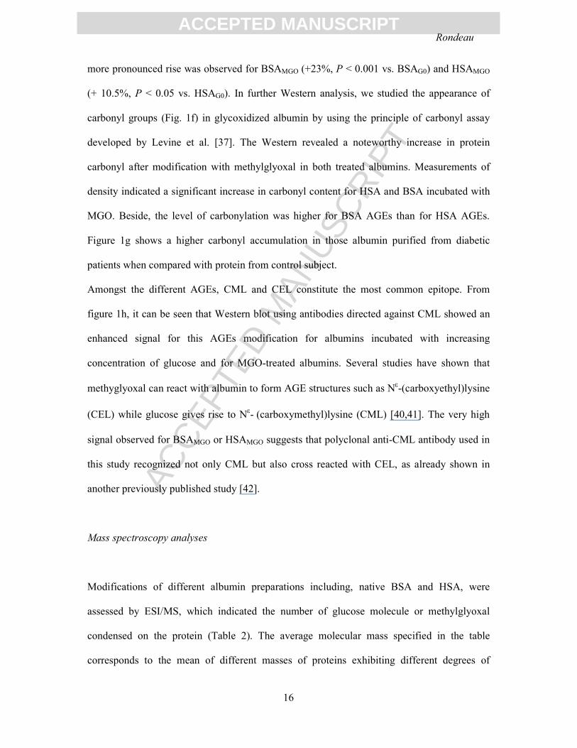
carbonyl groups (Fig. 1f) in glycoxidized albumin by using the principle of carbonyl assay
developed by Levine et al. [37]. The Western revealed a noteworthy increase in protein
carbonyl after modification with methylglyoxal in both treated albumins. Measurements of
density indicated a significant increase in carbonyl content for HSA and BSA incubated with
MGO. Beside, the level of carbonylation was higher for BSA AGEs than for HSA AGEs.
Figure 1g shows a higher carbonyl accumulation in those albumin purified from diabetic
patients when compared with protein from control subject.
Amongst the different AGEs, CML and CEL constitute the most common epitope. From
figure 1h, it can be seen that Western blot using antibodies directed against CML showed an
enhanced signal for this AGEs modification for albumins incubated with increasing
concentration of glucose and for MGO-treated albumins. Several studies have shown that
methyglyoxal can react with albumin to form AGE structures such as Nε-(carboxyethyl)lysine
(CEL) while glucose gives rise to Nε- (carboxymethyl)lysine (CML) [40,41]. The very high
signal observed for BSAMGO or HSAMGO suggests that polyclonal anti-CML antibody used in
this study recognized not only CML but also cross reacted with CEL, as already shown in
another previously published study [42].
Mass spectroscopy analyses
Modifications of different albumin preparations including, native BSA and HSA, were
assessed by ESI/MS, which indicated the number of glucose molecule or methylglyoxal
condensed on the protein (Table 2). The average molecular mass specified in the table
corresponds to the mean of different masses of proteins exhibiting different degrees of

ACC
EPTE
D M
ANU
SCR
IPT
ACCEPTED MANUSCRIPTRondeau
17
modification. The most abundant components are also featured in table 2. As expected,
nonenzymatic glycosylation of albumin with glucose or methyglyoxal induced an increase in
its average molecular weight, with variable extent, depending on the nature of glycoxidation.
Molecular masses of native BSA and HSA were approximately 66438 Da and 66567 Da,
respectively. The most abundant species in BSAG5 and HSAG5 samples were the non-modified
(native) albumin (no mass variation). The molecular mass of the most abundant species in
HSAG25 was increased to approximately 167 Da corresponding to the protein condensed with
one glucose unit. Definitely, the condensation of one glucose or methylglyoxal molecule to
the protein leads to a mass increase of 162 Da or 54 Da, respectively. Similarly, the molecular
mass of predominant species for HSAMGO was increased to 166 Da, which probably
correspond to three methylglyoxal units attached to the protein.
Different HSA and BSA samples were also digested by trypsin. MALDI spectrum for
the trypsic proteolyses of native, glycoxidized (with methylglyoxal) and diabetic HSA are
shown in Figure 2. The comparison of non-glycoxidized (HSAG0) and glycoxidized (HSAMGO
and HSAdiab) albumin clearly revealed several differences: first, the main obvious change was
the disappearance of most abundant ionic species (for non-glycoxidized albumin digest) at
m/z 1623.5 and second, the appearance of several predominant ionic species (for glycoxidized
albumin digest) at m/z 673.6, 875.4, 1149.5 and 1546.6. Some other peptides of HSAG0 digest
still remained at low abundance in glycoxidized digests (HSAMGO and HSAdiab), particularly
at m/z 927.5, 1342.5 and 1931.5. In conclusion, even if the tryptic patterns were not exactly
the same, an important similarity was evidenced between in vitro-modified albumins (HSAG25
and HSAMGO) and in vivo diabetic HSA digest profile.
In summary, main modifications in BSA and HSA in terms of oxidative and structural
parameters were observed in the presence of albumins incubated with methylglyoxal. Besides,

ACC
EPTE
D M
ANU
SCR
IPT
ACCEPTED MANUSCRIPTRondeau
18
comparison between in vitro-modified and in vivo-modified albumin (diabetic) showed
similar changes in the protein structural properties. This was more significant when structural
changes were compared between HSAdiab and HSAG25.
In the following experiments, antioxidant properties and effects of glycoxidized albumin on
THP1 cells were investigated.
Impairment of antioxidant properties of BSA and HSA after glycoxidation
To examine whether the glycoxidation treatment had any effect on the antioxidant properties
of albumin, different albumin preparations previously characterized were submitted to the free
radical-induced hemolysis test and their copper binding capacity was evaluated. The
antioxidant properties of albumin samples were investigated using the red blood hemolysis
test either in the absence or the presence of 10 µM protein. Typical hemolysis curves obtained
for control (PBS), HSAG0 and HSAMGO are illustrated in the insert of figure 3a. As expected,
the antioxidant property of nonmodified albumin was evidenced by delayed hemolysis in
comparison with control, and this was observed either with BSAGO (curves not shown) or
with HSAGO. The free radical scavenging properties of different albumin preparations,
symbolized by hemolysis half times (HT50), are shown in Fig. 3a. On the one hand, the
protective effect observed for BSAG0 (+ 16 %, P < 0.001 vs. PBS) and for HSAG0 (+ 26 %, P
< 0.001 vs. PBS) was progressively lost after the glycoxidation of protein (Fig. 3a). On the
other hand, a collapse in the antioxidant property of the protein was noticed for both BSAMGO
and HSAMGO. In comparison with HSAG0, albumin modification by methylglyoxal induced a
24 % reduction (P < 0.001) in the antioxidant activity of protein. A similar reduction was
observed for modified BSA with 26% (P < 0.001) reduction in the antioxidant activity.

ACC
EPTE
D M
ANU
SCR
IPT
ACCEPTED MANUSCRIPTRondeau
19
Remarkably, an increase in antioxidative properties was observed for in vivo diabetic HSA
compared with non diabetic albumin.
The antioxidant activity of albumin was also related with its capacity to bind metal such as
copper ions. By using bathocuproinedisulfonic acid to assay bound Cu(II), we determined
copper binding capacities (nM/µmol) of modified albumin (Fig. 3b). Surprisingly, the results
indicated a progressive rise in copper binding capacity of BSA with glycoxidation process.
This rise reached 260% for BSAMGO (P < 0.001, vs. BSAGO), while it was less for BSAG25
(112 %, P < 0.05 vs. BSAG0). Argirova and Ortwerth, in their study on glycoxidized BSA,
showed that highly glycoxidized (500 mM of glucose) albumin was less capable of competing
for copper ions in the presence of other ligands, suggesting that glycoxidized proteins might
have diminished stability constants of their copper chelates [29]. For HSA, no significant
variations in the copper-binding capacities of protein were observed after its modification by
glycoxidation with an exception for HSAG5, where a slight increase was noted.
Effects of AGEs on THP1 cells
Before further analysis on the impact of AGEs on monocytes physiology, the effect of
different AGEs preparations on the viability of THP1 cells were assessed. The cytotoxicity
effects were assayed by measuring cell proliferation and viability. In the subsequent
experiments, THP1 cells were incubated with mild concentrations of AGEs in the presence of
10 µM of native or glycoxidized albumins . First, the effect of characterized AGEs on the
metabolic activity of THP1 cells was assessed in vitro by the MTT assay. The incubation with
10 µM of albumin AGEs resulted in an increase in the viability of cells compared to PBS
control as shown in Fig. 4a. It increased up to 22% (P < 0.01) for BSAG25 and 20% (P < 0.05)
for HSAMGO compared with control. Regardless of the nature of albumin preparations
incubated in the presence of cells, no significant impairment in their viability was noted.

ACC
EPTE
D M
ANU
SCR
IPT
ACCEPTED MANUSCRIPTRondeau
20
In parallel, the membrane integrity of THP1 cells submitted to AGEs was monitored in terms
of cytoplasmic LDH activity of the medium. The results of LDH activity are shown in Fig. 4b
and no significant differences in LDH activity under each condition of cell incubation are
observed. Contrarily to the viability results obtained with MTT method, the direct cell count
by FACS showed a decrease in the number of THP1 cells when incubated in the presence of
albumin glycoxidized by 25 mM of glucose or 10 mM methylglyoxal. On the one hand, as
depicted in Fig. 4c, the decrease was approximately 70 % [20 %] (P<0.001 [0.001] vs. BSAG0
[HSAG0] for BSAG25 [HSAG25] and about 27% [36 %] (P<0.001 [0.001] vs. BSAG0 [HSAG0])
for BSAMGO [HSAMGO]. On the other hand, significant increase of viability was observed for
cells incubated in the presence of either glycoxidized or nonglycoxidized albumin (BSA and
HSA).
Effect on proteasomal proteolytic activities
The proteasome represents the major constituent of the proteolytic pathway for the
degradation of oxidized proteins and it includes a minimum of three distinct peptidase
activities: chymotrypsin-like, trypsin-like and caspase-like activities [43].
Figure 5a reports proteasomal activities measured in THP1 cells, which had been incubated in
the absence (PBS) or the presence of 10 µM native or glycoxidized BSA. Significant
increases in chymotrypsin-like activities (LLVY) were observed in glycoxidized-incubated
cells. No significant variation in the trypsin-like activities (LSTR) was noted when the cells
were incubated with native or glycoxidized bovine serum albumin. Regarding the caspase-like
activities (LLE), a slight but significant reduction was observed when the cells were incubated
in the presence of native BSA (BSAG0) than in the absence of albumin (PBS). LLE activities
were higher in glycoxidized BSA-incubated cells than in cells treated with native albumin
(BSAG0).

ACC
EPTE
D M
ANU
SCR
IPT
ACCEPTED MANUSCRIPTRondeau
21
Effects of native or glycoxidized HSA on the proteasomal activities of THP1 (Fig. 5b) were
quite different from those previously observed with glycoxidized and nontreated BSA.
Significant increases in chymotrypsin-like activities were observed when the cells had been
incubated with native or glycoxidized HSA than in the absence of the protein (PBS).
Concerning the trypsin-like activity of the proteasome and in contrast to what was previously
observed with BSA preparations-incubated cells, a significant reduction was evidenced in
cells incubated with HSAG25 (p<0.05 vs HSAG0). Caspase-like activity (LLE) was strongly
impaired when cells were incubated with HSAMGO (-30 %, p < 0.001 compared with HSAGO).
The different effects of HSA and BSA on proteasomal activity might be due to structural
difference between BSA and HSA.
The effect of glycoxidized BSA and HSA on carbonyl accumulation and the expressions of
RAGE and LMP2 by THP1
The investigation whether glycoxidized albumin might lead to an accumulation of
carbonylated proteins in the cells was highly important. Carbonyl assay was performed on
cells, which had been incubated for 16 hrs in the absence or the presence of 10 µM of
different albumin preparations (Fig. 6). A protective effect of native albumin was observed, as
the profiles for the carbonyl proteins were less intense when the cells were incubated with
native albumin (BSAG0 or HSAGO) compared to PBS control. As asserted by density
measurements, an increase in the signal for carbonylated cellular proteins was noticed in those
cells previously incubated in the presence of the following glycoxidized albumins: BSAG25,
BSAMGO and HSAMGO.
LMP2 constitutes a component of the eukaryotic multicatalytic proteinase complex, the
proteasome. As the glycoxidized albumin-induced changes in proteolytic activities could be
related to variations in the proteasome levels, Western blots were performed using antibodies

ACC
EPTE
D M
ANU
SCR
IPT
ACCEPTED MANUSCRIPTRondeau
22
directed against LMP2 subunit of the enzymatic complex (Fig. 6). The proteasome expression
was similar when cells were incubated with native or glycoxidized albumin. It is noteworthy
that a slight increase and little decrease in LMP2 were observed when cells were incubated
with BSAMGO and HSAMGO, respectively. This reduction in LMP2 expression in the cells
treated with HSAMGO might explain the significant alteration in the caspase like activity of the
proteasome in THP1 incubated with this modified albumin.
Concerning RAGE receptor expression, antibody used for the Western blot detected two
bands in the 45 kDa range representing the RAGE protein and a ~ 25 kDa protein that was
reported to be a nonspecific reaction. A stronger signal was observed in glycoxidized BSA
than in native BSA or in PBS control. No significant variations in RAGE expression was
evidenced in the cells incubated in the presence of the different preparations of HSA.
Nevertheless a higher RAGE expression was noted in cells incubated with HSAGO or
HSAMGO.
Reactive oxygen species
The oxidative stress in THP1 after 16 hrs incubation in the absence or presence (10 µM) of
various albumin preparations were examined by using the dichlorofluorescein diacetate
(DCF-DA) reagent [44]. After internalization by the cell, hydrolysis (removal of acetate
moiety) and oxidation by various oxidants, the nonfluorescent fluorescein derivatives
(dichlorofluorescin, DCFH) will become DCF and emit fluorescence. As THP1 monocytes
constitute nonadherent cells, DCF fluorescence was determined after cellular treatments by
using a FACS technique. The cellular counts of fluorescence in figures 7a and 7b reveal a
higher ROS formation in THP1-treated with MGO-modified albumins compared with
respective controls (BSAG0 or HSAGO). Results of ROS formation in THP1 incubated with the
different albumin preparations are shown in Fig. 7c. Protective effects of native albumins are

ACC
EPTE
D M
ANU
SCR
IPT
ACCEPTED MANUSCRIPTRondeau
23
again evidenced here. A decreased fluorescence signal was observed when the cells were
incubated with native albumin (BSAG0 or HSAGO), compared to PBS control. An increase in
the ROS formation was observed in those cells previously incubated in the presence of
glycoxidized albumins and especially with BSAMGO or HSAMGO.
The results of statistical analysis using two way analysis of variance (ANOVA) are
summarized in table 3. The two parameters analysed in this test were albumin origin (BSA or
HSA) and glycoxidation treatment (G0, G5, G25 and MGO). It was observed that the nature
of albumin chosen (BSA or HSA) in the different experiments significantly affected the
results at a structural or physiological level. Glycoxidation treatment significantly affected the
results of the structural studies, antioxidant capacity of albumin, caspase like activity of the
proteasome and ROS production in THP1 cells. Most interestingly, we addressed the question
of interactions between the two parameters: “Do both parameters exert any interactions?” In
most instances, for the majority of studies conducted, answer to this question is “yes” as
interactions between the two parameters were ascertained by the calculated P values inferior
to 0.05.

ACC
EPTE
D M
ANU
SCR
IPT
ACCEPTED MANUSCRIPTRondeau
24
Discussion
Elevated concentrations of circulating glycoxidized albumin demonstrate a typical feature of
blood in diabetic patients [9]. Glycoxidized proteins alter tissue function and disturb cellular
metabolism. Interaction of AGEs with several cell types has been shown to induce
intracellular stress and leads to an increased production of cytokines or nitric oxide (NO),
activate NFκB or heme oxygenase, produce lipid peroxidation products and crosslink proteins
[10,45]. Extensive studies have focused on the impact of AGEs on cell physiopathology.
Nonetheless, only few studies have reported the comparative effects of glycoxidation
phenomenon on the structure of BSA and HSA and on the induced-oxidative stress in cells. In
the present study, we compared (i) the consequences of glycoxidation induced by patho (25
mM) or physiological (5 mM) concentrations of glucose or methylglyoxal on the structure and
antioxidative properties of BSA or HSA, (ii) the cellular impact of glycoxidized albumins on
THP1 by the determination of cell viability, accumulation of carbonylated proteins,
expression of RAGE and the generation of oxidative stress in the cell.
Impact of glycoxidation on the structure of albumins
We previously reported some structural changes in albumin induced by the glycoxidation of
protein. But, in these studies, BSA was incubated with high concentrations of glucose (100
mM), or in the presence of copper, or submitted to elevated temperature. Also, cells were
incubated in the presence of high glycoxidized BSA concentrations (0.37 mM) [20,21,22,46].
Here, we tried to mimic either physiological or pathophysiological conditions by incubating
HSA or BSA with glucose, methylglyoxal and by treating monocytes with modified albumins.
Extensive care was taken for the incubation of albumin with sugar for three weeks, which

ACC
EPTE
D M
ANU
SCR
IPT
ACCEPTED MANUSCRIPTRondeau
25
corresponds to the half time of the protein in the plasma. The major structural change in
oxidized HSA from healthy human plasma occurred at the thiol group of Cys34 of reduced
HSA [47]. In our experimental conditions, purified HSA was less oxidized than commercial
BSA (Fig 1a), this is in accordance with another report detailing such structural differences in
albumin preparations [38]. Structural information indicated the formation of a molten globule-
like state of HSA after 21 days of incubation with 35 mM glucose [48]. Here, significant
oxidations of thiol in BSA and HSA were induced, when the proteins were glycoxidized by
25 mM of glucose. Cys34 could account for a large percentage (> 55%) of oxidized thiol in
human serum albumin obtained either from critically ill patients or from diverse commercial
preparations [38]. Similarly, as assessed by the carbonyl assay, an increase in the
carbonylation of the proteins was observed (figure 1f) and this is consistent with the
previously reported results, where a significant increase of carbonyl groups in HSA was
observed upon glycoxidation for 20 days with 50 mM of glucose [49]. Amongst carbonyl
modifications of plasmatic proteins, methylglyoxal (MGO), a very reactive dicarbonyl
compound, is increased in diabetes. Recent data suggest that MGO can strongly impair the
structure and antioxidants properties of albumin in vitro, leading to a modified protein similar
to that isolated from diabetic patients [50]. Here, utmost increases in glycophore
fluorescences, protein carbonylation and REM of albumins were observed in the MGO-
modified protein.
Very recently, a diabetic patient with poor glycemic control was found to exhibit more than
94% of his albumin presenting a glycative state [51]. Our group recently showed that the
incubation of BSA with 25 mM of glucose for seven weeks lead to 59% of glycoxidized
albumin [21]. Under the experimental conditions of this study, the protein structure of BSA
was more drastically affected by glycoxidation than that of HSA.

ACC
EPTE
D M
ANU
SCR
IPT
ACCEPTED MANUSCRIPTRondeau
26
The different impacts of glycoxidation on the antioxidant properties of BSA or HSA
Albumin, the most abundant protein in the plasma, could act as an important circulating
antioxidant [7,52], in addition to several properties such as the transport of fatty acids and
regulation of oncotic pressure [4]. Some previous studies showed that the integrity of albumin
was necessary for the protein to exert efficient antioxidant activity [20,30,46]. For the first
time, the effects of sole glycoxidation of BSA and HSA on the antioxidant activity of protein
were assessed by the determination of their copper-binding and free-radical trapping
capacities. Our present data are consistent with the previous observations made in 1999,
which showed that the sole glycoxidation of BSA with increasing concentration of glucose
(up to 500 mM of glucose) had no effect on the free radicals trapping capacities of albumin ,
as assessed by the AAPH-induced test [20]. Impaired BSA antioxidant activities were
observed only in copper-induced oxidized albumin previously glycoxidized [20]. A very
significant alteration in the free radical trapping capacity of BSA was observed (Fig 3a) when
the protein was modified by MGO. Faure et al recently showed that antioxidant capacity of
BSA determined by thiol measurement as well as in a cellular system (HeLa cells stressed by
H2O2) was significantly altered after modification by MGO [50]. In human albumin, its sole
glycoxidation with 25 mM of glucose was sufficient to significantly impair the free radical
trapping capacity of protein (Fig 3a). In addition, HSA modified by MGO did not exert any
antioxidant activity. This impairment in the free radical trapping capacity of HSA after
glycoxidation was not associated, in our experimental conditions, with any modification in the
copper binding capacity of the protein (Fig 3b). These results are in agreement with the
previous observation showing that Cys34 blockage in HSA (using N-ethyl maleimide, NEM,
reagent) was not associated with any reduction in the copper binding capacity of the protein
[30]. These results confirm that antioxidant activities exerted by albumin are somehow

ACC
EPTE
D M
ANU
SCR
IPT
ACCEPTED MANUSCRIPTRondeau
27
harboured by different localisations on the protein, therefore, these properties may differ even
under same glycoxidation treatment. Although structural modifications after glycoxidation
were more evident for BSA than for HSA, the impact of such treatment on the antioxidant
properties of albumin was significant mainly for the human form of the protein.
Induction of oxidative stress by glycoxidized BSA or HSA on THP1 cells
Interaction of advanced glycoxidation end products with AGE receptors (RAGE) induced
several cellular phenomena potentially relating to diabetic complications, including
atherosclerosis. Monocyte activation, adhesion, and migration are the key events in this
pathology. Conversely, AGEs inhibitors reduced the expression of RAGE and other
proinflammatory genes including monocyte chemoattractant protein-1 (MCP1), interferon γ-
inducible protein-10 (INFγIP10), and cyclooxygenase-2 (Cox2) in a dose-dependent manner
[19]. In addition, antioxidants inhibited the expression of the pro-inflammatory response in
THP1 cells induced by the ligation of the receptor for AGEs [53]. Grune and his group, in one
of their recent studies, showed that microglial cells were able to degrade glycoxidized
proteins. Furthermore, an inhibition of the cellular proteolytic systems was demonstrated in
the presence of AGEs in the culture medium [54]. Chronic exposure to glycoxidized serum in
J774.A macrophage cell line led to decreased expression of heat shock protein 70 (HSP 70)
which became undetectable over time; this was explained on the basis of direct involvement
of HSP 70 in the refolding of damaged proteins [55]. Furthermore, using the same cell type of
macrophages, Davies’s group showed that oxidized proteins altered the hydroperoxide
phosphatase activity of cellular protein and the cellular redox signalling pathway [56]. Our
team showed a pathophysiological effect of glycoxidized BSA on the primary cultures of
human adipose cells by inducing an accumulation of oxidatively modified proteins [21]. More

ACC
EPTE
D M
ANU
SCR
IPT
ACCEPTED MANUSCRIPTRondeau
28
recently, several proteins particularly prone to carbonylation in cells incubated with AGEs
were identified [22]. The heterogeneity of AGEs, experimental conditions under which they
are generated and concentrations used may lead to varied cellular responses. Valencia et al
showed that BSA incubated with 500 mM of glucose failed to bind RAGE and to induce any
cellular response when contaminating endotoxin was removed [57]. Our albumin preparations
were analyzed for endotoxin content, which appeared below detectable levels. It should be
noted that majority of studies dealing with the influence of AGEs on cell physiopathology
used either BSA or HSA modified by high nonphysiological concentrations of glucose or
MGO. Results reported here showed different and significant effects of low concentrations
(10 µM) of mild modified albumins on THP1 cells physiology. In most instances, drastic
influence of modified albumins was noted for HSAMGO and BSAMGO. Interestingly, both HSA
and BSA modified by physiological or pathological concentration of glucose induced an
enhanced cell proliferation as assayed by MTT measurement. Concerning cell counting,
perplexing results were obtained after various treatments with glycoxidized albumin, as a
reduced number of THP1 was counted when previously incubated with glycoxidized
albumins. The MTT system was used for measuring the activity of living cells via
mitochondrial dehydrogenases. One can hypothesize that the activity of this enzyme could be
enhanced in glycoxidized albumins-treated cells. Otherwise, in the FACS-mediated cell
countings, a window was defined to select and numerate only those cells presenting a
turgescent and round shape. So we assume that the treatment of THP1 with AGEs modified
the cell shape leading to their displacement from the counting window. We could conclude
that the presence of AGEs with mild concentration used under these experimental conditions
did not impair cell viability.
The oxidative modification of lipoproteins in the vascular wall leads to local production of
reactive carbonyl species that mediate the recruitment of macrophages, cellular activation and

ACC
EPTE
D M
ANU
SCR
IPT
ACCEPTED MANUSCRIPTRondeau
29
proliferation [58]. This multiplication of macrophages constitutes a typical feature of
atherosclerosis. Conversely, the impact of AGEs on human mesenchymal stem cells inhibited
the proliferation of cells, induced apoptosis, and prevented cognate differentiation into
adipose tissue, cartilage, and bone [59]. Recent results showed that “rejuvenation”
phenomenon in stem cells could be characterized by the decrease in AGEs and carbonylation
associated with enhanced proteasomal activities in blastocytes upon multiplication and
differentiation [60].
A reduction in carbonylation of cell proteins was observed in those THP1 incubated in the
presence of native albumins, showing the antioxidant protective effect of protein. Albumin
glycoxidized with MGO induced an enhanced carbonylation of proteins in the cells.
Glycoxidation of plasmatic proteins led to an altered cellular recognition and internalization
of these particles, which were bound by the specific AGEs receptors, named RAGE [61]. The
RAGE like receptor CD36 was overexpressed in aortic vascular smooth muscle cells treated
with AGEs [62]. Here, elevations in the expressions of RAGE were especially noted in those
cells incubated in the presence of glycoxidized BSA compared to native BSA. Surprisingly, a
higher level of RAGE expression was observed in HSAMGO and HSAGO-treated cells. We
hypothesize the differential effects of BSA and HSA on Human monocytes (THP1) RAGE
expression might have a link with the difference in the origin of species for both the albumin
and the cell used. Further investigations are necessary to detail the mechanisms of RAGE
expression.
The proteasome represents the main constituent of the proteolytic pathway for the degradation
of oxidized proteins [43]. Interestingly, enhanced chymotrypsin-like activity of the
proteasome was observed in the cells incubated with glycoxidized albumins. Significant
reduction in caspase-like activity was observed in HSAMGO-treated cells. Impairments in the
peptidylglutamyl-hydrolyzing activities of the proteasome were already reported when

ACC
EPTE
D M
ANU
SCR
IPT
ACCEPTED MANUSCRIPTRondeau
30
incubated with BSAG250 [54]. Definitely, it was reported that all three proteolytic activities
were significantly reduced following the effects of 20 µg of BSA-AGE on 1 µg of isolated
proteasome [54]. This concentration ratio between the oxidized compound (glycoxidized
albumin) and the proteolytic system (proteasome) was high compared to the mild conditions
used in our study. In addition, the concentrations of sugar used for BSA modifications were
higher than 250 mM. It was also reported by the same group, that exposure of proteasome to
oxidized protein leads to a biphasic response in the proteolytic activities. At moderate oxidant
concentrations, proteolytic susceptibility increases, whereas at higher oxidant concentrations,
a decrease in proteolytic susceptibility occurs’’ [63].
Antioxidant activity of native albumin in this cellular model of THP1 was further evidenced
by the significant decrease in DCF fluorescence when cells were incubated in the presence of
BSAGO or HSAGO as compared with the control (PBS). This protective action of albumin was
significantly impaired after glycoxidation of the protein. Noteworthy, compared to BSA, HSA
(glycoxidized or not) induced significant oxidative stress in THP1 cells, even though the
carbonyl content is lower in HSA. Enhanced oxidative stress assayed by DCF-DA in
macrophage-like RAW 264.7 cells exposed to various AGE-albumins was reported by
Subramaniam et al [64]. In addition, they observed that the mere addition of unmodified
albumin to cells was adequate to suppress the fluorescence of oxidized DCF. Here, the more
significant results were again analyzed in the presence of MGO-modified albumin.
Oxidative modifications of albumin were observed when the protein was glycoxidized
by pathophysiological concentrations of glucose. Such glycoxidized albumin induced an
accumulation of carbonyl proteins and enhanced the formation of ROS. Impairments in
proteosomal activities in human monocytes were also observed. We demonstrate herein the
important impact of the nature of albumin chosen in experiments dealing with
structural/function relationships after the modification of the protein by glycoxidation. AGEs

ACC
EPTE
D M
ANU
SCR
IPT
ACCEPTED MANUSCRIPTRondeau
31
originating from the glycoxidation of BSA or HSA have lead to differential cellular
responses. This study confirms and extends the determinant influence of the conditions in
which AGEs are generated and concentrations used in experiments designed to mimic
pathological conditions. Further on going projects try to better define the effects of AGEs on
cellular physiology and to observe whether any impairment of proinflammatory cytokine
expressions could be associated with the oxidative damages induced by glycoxidized albumin
in cells.

ACC
EPTE
D M
ANU
SCR
IPT
ACCEPTED MANUSCRIPTRondeau
32
References
[1] Resnick, H.E., Foster, G.L., Bardsley, J. and Ratner, R.E. (2006) Achievement of
American Diabetes Association clinical practice recommendations among U.S. adults
with diabetes, 1999-2002: the National Health and Nutrition Examination Survey.
Diabetes Care, 29, 531-7.
[2] Pennathur, S. and Heinecke, J.W. (2007) Mechanisms for oxidative stress in diabetic
cardiovascular disease. Antioxid Redox Signal, 9, 955-69.
[3] Shoelson, S.E., Lee, J. and Goldfine, A.B. (2006) Inflammation and insulin resistance.
J Clin Invest 116, 1793-1801.
[4] Peters, T.J. (1996), All about albumin. Academic Press, San Diego.
[5] Roche, M., Rondeau, P., Singh, N.R., Tarnus, E. and Bourdon, E. (2008) The
antioxidant properties of Serum Albumin FEBS Lett. , in press.
[6] Cha, M.K. and Kim, I.H. (1996) Glutathione-linked thiol peroxidase activity of human
serum albumin: a possible antioxidant role of serum albumin in blood plasma.
Biochem Biophys Res Commun, 222, 619-25.
[7] Soriani, M., Pietraforte, D. and Minetti, M. (1994) Antioxidant potential of anaerobic
human plasma: role of serum albumin and thiols as scavengers of carbon radicals.
Arch Biochem Biophys, 312, 180-8.
[8] Bourdon, E. and Blache, D. (2001) The importance of proteins in defense against
oxidation. Antioxid Redox Signal, 3, 293-311.
[9] Cohen, M.P. (2003) Intervention strategies to prevent pathogenetic effects of glycated
albumin. Arch Biochem Biophys, 419, 25-30.
[10] Cohen, M.P., Shea, E., Chen, S. and Shearman, C.W. (2003) Glycated albumin
increases oxidative stress, activates NF-kappa B and extracellular signal-regulated
kinase (ERK), and stimulates ERK-dependent transforming growth factor-beta 1
production in macrophage RAW cells. J Lab Clin Med, 141, 242-9.
[11] Huebschmann, A.G., Regensteiner, J.G., Vlassara, H. and Reusch, J.E. (2006)
Diabetes and advanced glycoxidation end products. Diabetes Care, 29, 1420-32.
[12] Houstis, N., Rosen, E.D. and Lander, E.S. (2006) Reactive oxygen species have a
causal role in multiple forms of insulin resistance. Nature, 440, 944-8.
[13] Brownlee, M. (2001) Biochemistry and molecular cell biology of diabetic
complications. Nature, 414, 813–820.
[14] Furukawa, S., Fujita, T., Shimabukuro, M., Iwaki, M., Yamada, Y., Nakajima, Y.,
Nakayama, O., Makishima, M., Matsuda, M. and Shimomura, I. (2004) Increased
oxidative stress in obesity and its impact on metabolic syndrome. J Clin Invest, 114,
1752-61.
[15] Droge, W. (2002) Free radicals in the physiological control of cell function. Physiol
Rev, 82 47-95.
[16] Vlassara, H. (2005) Advanced glycation in health and disease: role of the modern
environment. Ann N Y Acad Sci, 1043, 452-60.
[17] Devangelio, E., Santilli, F., Formoso, G., Ferroni, P., Bucciarelli, L., Michetti, N.,
Clissa, C., Ciabattoni, G., Consoli, A. and Davi, G. (2007) Soluble RAGE in type 2
diabetes: Association with oxidative stress. Free Radic Biol Med, In Press,
Uncorrected Proof.
[18] Shanmugam, N., Kim, Y.S., Lanting, L. and Natarajan, R. (2003) Regulation of
cyclooxygenase-2 expression in monocytes by ligation of the receptor for advanced
glycation end products. J Biol Chem, 278, 34834-44.

ACC
EPTE
D M
ANU
SCR
IPT
ACCEPTED MANUSCRIPTRondeau
33
[19] Figarola, J.L., Shanmugam, N., Natarajan, R. and Rahbar, S. (2007) Anti-
Inflammatory Effects of the Advanced Glycation End Product Inhibitor LR-90 in
Human Monocytes. Diabetes 56, 647-655.
[20] Bourdon, E., Loreau, N. and Blache, D. (1999) Glucose and free radicals impair the
antioxidant properties of serum albumin. Faseb J, 13, 233-44.
[21] Chesne, S., Rondeau, P., Armenta, S. and Bourdon, E. (2006) Effects of oxidative
modifications induced by the glycation of bovine serum albumin on its structure and
on cultured adipose cells. Biochimie, 88, 1467-77.
[22] Singh, N.R., Rondeau, P., Hoareau, L. and Bourdon, E. (2007) Identification of
preferential protein targets for carbonylation in human mature adipocytes treated with
native or glycated albumin. Free Radic Res, 41, 1078-88.
[23] Ogino, T. and Okada, S. (1995) Oxidative damage of bovine serum albumin and other
enzyme proteins by iron-chelate complexes. Biochim Biophys Acta. , 1245, 359-65.
[24] Brownlee, M., Vlassara, H. and Cerami, A. (1984) Nonenzymatic glycosylation and
the pathogenesis of diabetic complications. Ann Intern Med. , 101, 527-37. .
[25] Ulrich, P. and Cerami, A. (2001) Protein Glycation, Diabetes, and Aging. Recent Prog
Horm Res 56, 1-22.
[26] Ellman, G.L. (1959) Tissue sulfhydryl groups. Arch Biochem Biophys, 82, 70-7.
[27] Armenta, S., Garrigues, S., De la Guardia, M. and Rondeau, P. (2005) Attenuated
Total Reflection-Fourier transform infrared analysisof the fermentation process of
pineapple. Analytica Chimica Acta, 545, 99-106.
[28] Laemmli, U.K. (1970) Cleavage of structural proteins during the assembly of the head
of bacteriophage T4. Nature, 227, 680-5.
[29] Argirova, M.D. and Ortwerth, B.J. (2003) Activation of protein-bound copper ions
during early glycation: study on two proteins. Arch Biochem Biophys, 420, 176-84.
[30] Bourdon, E., Loreau, N., Lagrost, L. and Blache, D. (2005) Differential effects of
cysteine and methionine residues in the antioxidant activity of human serum albumin.
Free Radic Res, 39, 15-20.
[31] Prost, M. (1992) Process for the determination by means of free radicals of the
antioxidant properties of a living organism or a potentially agressive agents. US Patent
5 135 850 Aug 4.
[32] Girodon, F., Blache, D., Monget, A.L., Lombart, M., Brunet-Lecompte, P., Arnaud, J.,
Richard, M.J. and Galan, P. (1997) Effect of a two-year supplementation with low
doses of antioxidant vitamins and/or minerals in elderly subjects on levels of nutrients
and antioxidant defense parameters. J Am Coll Nutr. , 16, 357-65.
[33] Smith, P.K., Krohn, R.I., Hermanson, G.T., Mallia, A.K., Gartner, F.H., Provenzano,
M.D., Fujimoto, E.K., Goeke, N.M., Olson, B.J. and Klenk, D.C. (1985) Measurement
of protein using bicinchoninic acid. Anal Biochem, 150, 76-85.
[34] Mosmann, T. (1983) Rapid colorimetric assay for cellular growth and survival:
Application to proliferation and cytotoxicity assays. Journal of Immunological
Methods, 65, 55-63.
[35] Legrand, C., Bour, J.M., Jacob, C., Capiaumont, J., Martial, A., Marc, A., Wudtke,
M., Kretzmer, G., Demangel, C. and Duval, D. (1992) Lactate dehydrogenase (LDH)
activity of the cultured eukaryotic cells as marker of the number of dead cells in the
medium J Biotechnol., 25, 231-43. .
[36] Le Pecheur, M., Bourdon, E., Paly, E., Farout, L., Friguet, B. and London, J. (2005)
Oxidized SOD1 alters proteasome activities in vitro and in the cortex of SOD1
overexpressing mice. FEBS Lett, 579, 3613-8.
[37] Levine, R.L., Williams, J.A., Stadtman, E.R. and Shacter, E. (1994) Carbonyl assays
for determination of oxidatively modified proteins. Methods Enzymol, 233, 346-57.

ACC
EPTE
D M
ANU
SCR
IPT
ACCEPTED MANUSCRIPTRondeau
34
[38] Bar-Or, D., Bar-Or, R., Rael, L.T., Gardner, D.K., Slone, D.S. and Craun, M.L. (2005)
Heterogeneity and oxidation status of commercial human albumin preparations in
clinical use. Crit Care Med. , 33, 1638-41.
[39] Oettl, K. and Stauber, R.E. (2007) Physiological and pathological changes in the redox
state of human serum albumin critically influence its binding properties. Br J
Pharmacol, 151, 580-90.
[40] Nagai, R., Araki, T., Hayashi, C.M., Hayase, F. and Horiuchi, S. (2003) Identification
of N epsilon-(carboxyethyl)lysine, one of the methylglyoxal-derived AGE structures,
in glucose-modified protein: mechanism for protein modification by reactive
aldehydes. J Chromatogr B Analyt Technol Biomed Life Sci, 788, 75-84.
[41] Shibayama, R., Araki, N., Nagai, R. and Horiuchi, S. (1999) Autoantibody against
N(epsilon)-(carboxymethyl)lysine: an advanced glycation end product of the Maillard
reaction. Diabetes, 48, 1842-9.
[42] Horiuchi, S., Unno, Y., Usui, H., Shikata, K., Takaki, K., Koito, W., Sakamoto, Y.,
Nagai, R., Makino, K., Sasao, A., Wada, J. and Makino, H. (2005) Pathological roles
of advanced glycation end product receptors SR-A and CD36. Ann N Y Acad Sci,
1043, 671-5.
[43] Friguet, B. (2006) Oxidized protein degradation and repair in ageing and oxidative
stress. FEBS Lett., 580, 2910-6.
[44] Wang, H. and Joseph, J.A. (1999) Quantifying cellular oxidative stress by
dichlorofluorescein assay using microplate reader. Free Radic Biol Med, 27, 612-6.
[45] Ramasamy, R., Vannucci, S.J., Yan, S.S.D., Herold, K., Yan, S.F. and Schmidt, A.M.
(2005) Advanced glycation end products and RAGE: a common thread in aging,
diabetes, neurodegeneration, and inflammation. Glycobiology 15, 16R-28.
[46] Rondeau, P., Armenta, S., Caillens, H., Chesne, S. and Bourdon, E. (2007)
Assessment of temperature effects on beta-aggregation of native and glycated albumin
by FTIR spectroscopy and PAGE: Relations between structural changes and
antioxidant properties. Arch Biochem Biophys, 460, 141-150.
[47] Kawakami, A., Kubota, K., Yamada, N., Tagami, U., Takehana, K., Sonaka, I.,
Suzuki, E. and Hirayama, K. (2006) Identification and characterization of oxidized
human serum albumin. A slight structural change impairs its ligand-binding and
antioxidant functions. Febs J, 273, 3346-57.
[48] Sattarahmady, N., Moosavi-Movahedi, A.A., Ahmad, F., Hakimelahi, G.H., Habibi-
Rezaei, M., Saboury, A.A. and Sheibani, N. (2007) Formation of the molten globule-
like state during prolonged glycation of human serum albumin. Biochim Biophys Acta,
1770, 933-42.
[49] Khan, M.W., Rasheed, Z., Khan, W.A. and Ali, R. (2007) Biochemical, biophysical,
and thermodynamic analysis of in vitro glycated human serum albumin. Biochemistry
(Mosc), 72 146-52.
[50] Faure, P., Troncy, L., Lecomte, M., Wiernsperger, N., Lagarde, M., Ruggiero, D. and
Halimi, S. (2005) Albumin antioxidant capacity is modified by methylglyoxal.
Diabetes Metab, 31, 169-77.
[51] Kisugi, R., Kouzuma, T., Yamamoto, T., Akizuki, S., Miyamoto, H., Someya, Y.,
Yokoyama, J., Abe, I., Hirai, N. and Ohnishi, A. (2007) Structural and glycation site
changes of albumin in diabetic patient with very high glycated albumin. Clin Chim
Acta., In Press
[52] Azzi, A., Davies, K.J. and Kelly, F. (2004) Free radical biology - terminology and
critical thinking. FEBS Lett, 558, 3-6.

ACC
EPTE
D M
ANU
SCR
IPT
ACCEPTED MANUSCRIPTRondeau
35
[53] Huang, S.M., Wu, C.H. and Yen, G.C. (2006) Effects of flavonoids on the expression
of the pro-inflammatory response in human monocytes induced by ligation of the
receptor for AGEs. Mol Nutr Food Res, 50, 1129-39.
[54] Stolzing, A., Widmer, R., Jung, T., Voss, P. and Grune, T. (2006) Degradation of
glycated bovine serum albumin in microglial cells. Free Radic Biol Med, 40, 1017-
1027.
[55] Bassi, A.M., Ledda, S., De Pascale, M.C., Penco, S., Rossi , S., Odetti, P. and
Cottalasso, D. (2005) Antioxidant status in J774A.1 macrophage cell line during
chronic exposure to glycated serum. Biochem Cell Biol., 83, 176-87.
[56] Gracanin, M. and Davies, M.J. (2007) Inhibition of protein tyrosine phosphatases by
amino acid, peptide, and protein hydroperoxides: Potential modulation of cell
signaling by protein oxidation products. Free Radic Biol Med, 42, 1543-1551.
[57] Valencia, J.V., Mone, M., Koehne, C., Rediske, J. and Hughes, T.E. (2004) Binding of
receptor for advanced glycation end products (RAGE) ligands is not sufficient to
induce inflammatory signals: lack of activity of endotoxin-free albumin-derived
advanced glycation end products. Diabetologia, 47, 844-52.
[58] Baynes, J.W. and Thorpe, S.R. (2000) Glycoxidation and lipoxidation in
atherogenesis. Free Radic Biol Med, 28, 1708-16.
[59] Kume, S., Kato, S., Yamagishi, S., Inagaki, Y., Ueda, S., Arima, N., Okawa, T.,
Kojiro, M. and Nagata, K. (2005) Advanced glycation end-products attenuate human
mesenchymal stem cells and prevent cognate differentiation into adipose tissue,
cartilage, and bone. J Bone Miner Res, 20, 1647-58.
[60] Hernebring, M., Brolén, G., Aguilaniu, H., Semb, H. and Nyström, T. (2006)
Elimination of damaged proteins during differentiation of embryonic stem cells. Proc
Natl Acad Sci U S A., 103, 7700-5.
[61] Vlassara, H. (2001) The AGE-receptor in the pathogenesis of diabetic complications.
Diabetes Metab Res Rev, 17, 436-43.
[62] de Oliveira Silva, C., Delbosc, S., Arais, C., Monnier, L., Cristol, J.P. and Pares-
Herbute, N. (2006) Modulation of CD36 protein expression by AGEs and insulin in
aortic VSMCs from diabetic and non-diabetic rats. Nutr Metab Cardiovasc Dis.
[63] Grune, T., Merker, K., Sandig, G. and Davies, K.J. (2003) Selective degradation of
oxidatively modified protein substrates by the proteasome. Biochem Biophys Res
Commun, 305, 709-18.
[64] Subramaniam, R., Fan, X.J., Scivittaro, V., Yang, J., Ha, C.E., Petersen, C.E.,
Surewicz, W.K., Bhagavan, N.V., Weiss, M.F. and Monnier, V.M. (2002) Cellular
Oxidant Stress and Advanced Glycation Endproducts of Albumin: Caveats of the
Dichlorofluorescein Assay*. Arch Biochem Biophys, 400, 15-25.

ACC
EPTE
D M
ANU
SCR
IPT
ACCEPTED MANUSCRIPTRondeau
36
TABLES
BSAGO BSAG5 BSAG25 BSAMGO
REM 1.068 ± 0.008 1.122 ± 0.009* 1.149 ± 0.008
*** 1.317 ± 0.036
***
HSAGO HSAG5 HSAG25 HSAMGO
REM 1.015 ± 0.011 1.065 ± 0.005 1.086 ± 0.005* 1.120 ± 0.016
*
Table 1: Relative Electrophoretic Migration of glycoxidized Bovine and Human Serum
Albumin
Relative Electrophoretic Migration (REM) of the different preparations of BSA and HSA was
evaluated as described in Materials and Methods. Data of REM are represented as mean ± sd
(n=3) and statistical analyses were performed using one-way ANOVA. *
Effect of the
glycoxidation of BSA [HSA] (vs. BSAG0 [HSAGO]): ***
p<0.001, **
p<0.01, *p<0.05.

ACC
EPTE
D M
ANU
SCR
IPT
ACCEPTED MANUSCRIPT
Rondeau
37
Table 2: ESI Mass analysis of in vitro-modified BSA and HSA
Average molecular mass (Da) of the different preparations of BSA and HSA was obtained by ESI/MS. Abundance of the different components
was determined (%). Mass increase was calculated by comparison with native BSA (66438 Da) and HSA (66567 Da). Number of glucose (glc)
or methylglyoxal (mgo) unit attached to the protein was specified.
Major Molecular Abundance Mass Component Molecular Abundance Mass
Component Mass (%) Increase (Da) Mass (%) Increase (Da)
BSAG0 HSAG0
A 66438.47 100 A 66567,43 100
B 66552.06 44.52 +113.6 B 66746,18 59,01 + 178,7
Average Moledular Mass (Da) 66524 Average Moledular Mass (Da) 66613
BSAG5 HSAG5
A 66444.2 100 + 5,7 A 66567,18 100 - 0,2
B 66596.9 34.97 + 158,2 (+ 1 glc) B 66653,06 60,95 + 85,6
C 66730,66 56,06 163,2 (+1 glc)
Average Moledular Mass (Da) 66587 + 63 Average Moledular Mass (Da) 66795 + 182
BSAG25 HSAG25
A 66518.92 100 + 80.45 A 66734,78 100 + 167.3 (+1 glc)
B 66601.67 74.26 + 163.2 (+1 glc) B 66900,59 79,96 + 333.1 (+2 glc)
C 67091.31 65.8 + 652.8 (+4 glc) C 66564,44 73,97 - 2.9
Average Moledular Mass (Da) 66714 + 190 Average Moledular Mass (Da) 66925 + 312
BSAMGO HSAMGO
A 66681.65 100 + 243.1 A 66733,55 100 + 166.1 (+3 mgo)
B 66604.63 81 + 166.1 (+3 mgo) B 66570 91,97 + 2.5
C 66760.12 63.35 + 321.6 (+6 mgo) C 66625,72 93,18 + 58.2 (+1 mgo)
D 66444.84 66.06 + 6.3 D 66885,87 85 + 318.4 (+6 mgo)
Average Moledular Mass (Da) 67713 + 1189 Average Moledular Mass (Da) 66868 + 255

ACC
EPTE
D M
ANU
SCR
IPT
ACCEPTED MANUSCRIPT
38
Does albumin origin
(BSA or HSA) affect
the result?
Does glycoxidation
treatment (G0/G5/G25
or MGO) affect the
result?
Do both parameters
exert any interactions?
Structural studies
Thiol number *** *** P = 0.7284
Fluorescence *** *** ***
R.E.M. *** *** ***
Browning *** *** ***
Antioxidant properties
HT50 ** *** ***
Copper binding *** ** ***
Effects on THP1 cells
MTT measurements P = 0.2084 P = 0.0546 P = 0.5674
LDH measurements ** P = 0.2709 ***
Cell counting *** *** ***
Proteasome LLVY * P = 0.9285 **
Proteasome LSTR *** P = 0.0840 *
Proteasome LLE * *** **
Production of ROS ** *** P = 0.1198
Table3: Statistical analysis of data using two way analysis of variance
Data in this study was obtained from a minimum of three independent experiments and was
statistically analyzed using two-way analysis of variance (ANOVA). The major effects of the
albumin origin (BSA or HSA) and glycoxidation treatment (GO, G5, G25 or MGO) or their
interactions were evaluated by ANOVA. Proteasome LLVY, LSTR and LLE correspond to
the Chymotrypsin-like, trypsin-like, and caspase-like activities of the proteasome,
respectively. Significances were *** P < 0.001, ** P < 0.01 and * P < 0.05. P values higher
than 0.05 were specified.

ACC
EPTE
D M
ANU
SCR
IPT
ACCEPTED MANUSCRIPT
39
Figure legends
Fig. 1: Characterization of BSA and HSA AGEs
(a) Electrophoretic migration profile of commercial BSA and purified HSA (PAGE 12%). (b)
Fluorescence of AGEs at 355/410 nm expressed in arbitrary unit of fluorescence per mg
protein. (c) Absorbance at 420 nm for albumin incubated without glucose (G0) or with 10 mM
of methylglyoxal (MGO) and for albumin purified from diabetic and nondiabetic patients. (d)
Thiol groups contents in the different preparations of albumin evaluated with Ellman’s
method. (e) The comassie staining of the analysis of BSA and HSA preparations by native
PAGE electrophoresis (12%). (f) Oxidative modification of glycoxidized albumin as assessed
by the carbonyl assay with signal intensity (AU) of carbonyl spots. (g) Oxidative modification
of albumin purified from diabetic and nondiabetic patients as assessed by the carbonyl assay.
(h) AGEs content in albumin preparations as assessed by Western blotting with the
corresponding signal intensities (arbitrary unit). All data in (b) to (d) are expressed as mean ±
sd (n=4) and statistical analyses were performed using one-way ANOVA for multiple
comparisons. *
Effect of the glycoxidation of BSA [HSA] (vs. BSAG0 [HSAGO]): ***
p<0.001,
*p<0.05.
#Effect of diabetic pathology (vs. non diabetic):
# # # p<0.001,
#p<0.05.
Fig.2: Comparisons between peptidic profiles of glycoxidized and nonglycoxidized HSA
MALDI spectra of tryptic digest of (a) HSAG0, (b) HSAMGO and (c) HSA of diabetic patient.
The ionics species with underlined m/z correspond to peptides present in the three samples.

ACC
EPTE
D M
ANU
SCR
IPT
ACCEPTED MANUSCRIPT
40
Fig. 3: Effects of glycoxidation on the antioxidant activities of albumin
(a) Impairment in the antioxidant activities of BSA and HSA as assessed by the free radical-
mediated blood hemolysis test. Insert is an illustration of hemolysis curves obtained in the
absence ( control no HSA) or the presence of 10 µM HSA incubated with (�) or
without (�) methylglyoxal.
Histograms represent half time hemolysis (HT50 in min) expressed as means ± SD of three
independent experiments. The doted line facilitates comparison with control (no albumin).
Statistical analyses were performed using one-way ANOVA for multiple comparisons.
* Effect of BSA or HSA (vs. Control):
***p<0.001,
**p<0.01,
*p<0.05 .
# Effect of
glycoxidation (vs. BSAG0 or HSAG0): # # #
p<0.001, # #
p<0.01, #p<0.05.
+Effect of diabetic
pathology (vs. non diabetic): + + +
p<0.001.
(b) Effect of glycoxidation on copper binding capacities of BSA or HSA. Histograms
represent copper binding capacities of modified albumin (nM/µmol albumin). Data are
expressed as mean ± sd (n=3) and statistical analyses were performed using one-way ANOVA
for multiple comparisons. *Effect of glycoxidation (vs. BSAG0 (HSAG0)):
###p<0.001;
#p<0.05.
Fig. 4: Effects of AGEs on THP1 cells integrity
The relative viability (%) of THP1 cells incubated 16 hours with (a) 10 µM of AGEs and
determined by MTT assay. (b) Effect of 10 µM AGEs on the release of lactate dehydrogenase
in THP1 cells supernatant medium. Results are expressed as the percentage of LDH released
in the medium relative to the total LDH activity present (medium plus cell lysates). (c)
Viability of THP1 cells treated with 10 µM AGEs, expressed as the percentage of cells

ACC
EPTE
D M
ANU
SCR
IPT
ACCEPTED MANUSCRIPT
41
compared with PBS control and determined by a relative counting using FACSscan flow
cytometer. In all experiments, the viability was compared with control (cells incubated with
PBS) and positive control corresponds to cells incubated with hydrogen peroxyde (1.5 %).
Data are expressed as mean ± sd (n=3) and statistical analyses were performed using one-way
ANOVA for multiple comparisons. *Effect of BSA or HSA (vs. Control):
***p<0.001,
**p<0.01,
*p<0.05.
# Effect of glycoxidation (vs. BSAG0 or HSAG0):
# # #p<0.001,
# #p<0.01,
#p<0.05.
Fig. 5: Effect of AGEs on proteasomal activities.
THP1 cells were incubated in the presence or absence (PBS) of 10 µM AGEs generated either
from BSA (a) or HSA (b) for 16 hrs. Cells were washed and lysates were prepared as
described in the Materials and Methods section. The caspase-like, chymotrypsin-like and
trypsin-like activities of the proteasome were assayed using the fluorogenic peptides
substrates LLE-NA, LLVY-AMC and LSTR-AMC respectively, at 12.5 µM final
concentration. Assays were carried out with 50 µg of cell lysates in 25 mM Tris-HCl (pH 7.5)
and with the appropriate substrate at 37°C for 0–30 min incubation. The fluorescence of each
sample was evaluated using a multiplate spectrofluorimeter (BMG Labtech) at
excitation/emission wavelengths of 350/440 and 333/410 for aminomethylcoumarin and β-
naphthylamine products, respectively. Peptidase activities were measured in the absence or
the presence (10 µM) of the proteasome inhibitor MG132, and the difference between the two
values was attributed to proteasome activity. Bars represent mean ± S.D. (n=3 to 8), p values
were obtained using one-way ANOVA. *Effect of BSA or HSA (vs. Control):
***p<0.001,
*p<0.05.
# Effect of glycoxidation (vs. BSAG0 or HSAG0):
# #p<0.01,
#p<0.05.
ACC
EPTE
D M
ANU
SCR
IPT
ACCEPTED MANUSCRIPT
42
Fig. 6: Expression of LMP2, CD 36 and RAGE receptor by Western-blot
THP1 cells were incubated for 16 h in the absence or presence of 10 µM native or
glycoxidized albumins. Approximately 25 µg of total protein from lysates of treated cells
were separated by SDS-PAGE and transferred to nitrocellulose membranes. Antibodies (see
Materials and Methods) were used at a dilution of 1:1,000 and visualized using the
appropriate HRP-conjugated secondary antibody and the ECL system (GE Healthcare). Signal
quantification of carbonyl was determined by using the freeware ImageJ.
Fig. 7: Production of ROS in THP1 cells with AGEs
FACS analysis of DCF fluorescence of THP1 cells incubated overnight with (a) BSA and (b)
HSA AGEs. The ratio of fluorescence levels of cells treated with albumins with
methylglyoxal was compared with those without methylglyoxal or glucose. (c) Relative DCF
mean fluorescence intensity for cells incubated with BSA and HSA AGEs. All fluorescence
intensities are compared with positive control (cells incubated with 1 pg/ml PMA).
Data are expressed as mean ± sd (n=3) and statistical analyses were performed using one-way
ANOVA for multiple comparisons. *Effect of BSA or HSA (vs. PBS Control):
***p<0.001.
#
Effect of glycoxidation (vs. BSAG0 or HSAG0): # #
p<0.01, #p<0.05.

ACC
EPTE
D M
ANU
SCR
IPT
ACCEPTED MANUSCRIPT
43
List of abbreviations
AGEs, advanced glycation (or glycoxidation) endproducts; ANOVA, analysis of variance;
AU, arbitrary unit; BC, bathocuproinedisulfonic acid; BSA, bovine serum albumin; BSAGX,
BSA incubated with x mM of glucose; BSAG0, BSA incubated in the absence of glucose;
CEL, carboxy ethyl lysine; CML, carboxy-methyl-lysine; DCF, dichlorofluorescein; DCFH-
DA, dichloroflorescin diacetate; DCFH, dichloroflorescin; ECL, enhanced luminol
chemiluminescence; FACS, fluorescence-activated cell-sorting; HT50 , 50% hemolysis time;
LDH, lactate deshydrogenase; MALDI, matrix-assisted laser desorption ionisation; MGO,
methylglyoxal; NIDD, non-insulin dependent diabetes; PBS, phosphate-buffered buffer; PLS,
partial least squares; PMA, phorbol 12-myristate 13-acetate; PAGE, polyacrylamide gel
electrophoresis; RAGE, receptor for advanced glycation endproducts; ROS, reactive oxygen
species

ACC
EPTE
D M
ANU
SCR
IPT
ACCEPTED MANUSCRIPT
44

ACC
EPTE
D M
ANU
SCR
IPT
ACCEPTED MANUSCRIPT
45
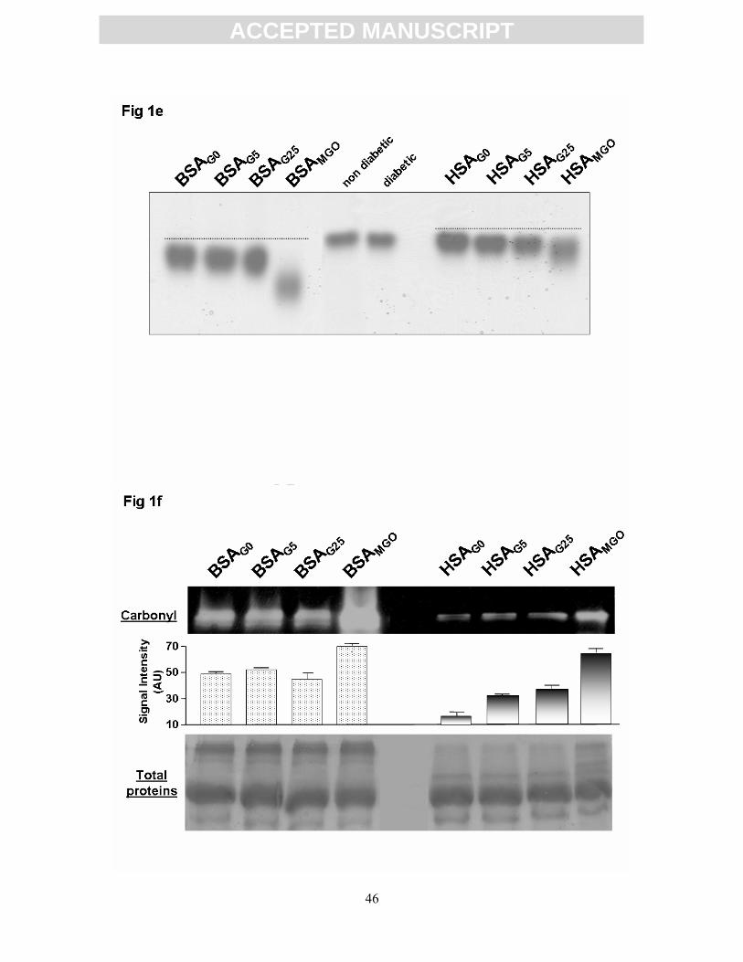
ACC
EPTE
D M
ANU
SCR
IPT
ACCEPTED MANUSCRIPT
46

ACC
EPTE
D M
ANU
SCR
IPT
ACCEPTED MANUSCRIPT
47

ACC
EPTE
D M
ANU
SCR
IPT
ACCEPTED MANUSCRIPT
48

ACC
EPTE
D M
ANU
SCR
IPT
ACCEPTED MANUSCRIPT
49

ACC
EPTE
D M
ANU
SCR
IPT
ACCEPTED MANUSCRIPT
50

ACC
EPTE
D M
ANU
SCR
IPT
ACCEPTED MANUSCRIPT
51

ACC
EPTE
D M
ANU
SCR
IPT
ACCEPTED MANUSCRIPT
52

ACC
EPTE
D M
ANU
SCR
IPT
ACCEPTED MANUSCRIPT
53

ACC
EPTE
D M
ANU
SCR
IPT
ACCEPTED MANUSCRIPT
54

ACC
EPTE
D M
ANU
SCR
IPT
ACCEPTED MANUSCRIPT
55

ACC
EPTE
D M
ANU
SCR
IPT
ACCEPTED MANUSCRIPT
56