Live Borrelia burgdorferi Spirochetes Elicit Inflammatory Mediators from Human Monocytes via the...
8
INFECTION AND IMMUNITY, Mar. 2009, p. 1238–1245 Vol. 77, No. 3 0019-9567/09/$08.000 doi:10.1128/IAI.01078-08 Copyright © 2009, American Society for Microbiology. All Rights Reserved. Live Borrelia burgdorferi Spirochetes Elicit Inflammatory Mediators from Human Monocytes via the Toll-Like Receptor Signaling Pathway Vida A. Dennis, 1 * Saurabh Dixit, 1 Shannon M. O’Brien, 1 Xavier Alvarez, 2 Bapi Pahar, 2 and Mario T. Philipp 1 * Divisions of Bacteriology and Parasitology 1 and Comparative Pathology, 2 Tulane National Primate Research Center, Tulane University Health Sciences Center, Covington, Louisiana Received 29 August 2008/Returned for modification 9 November 2008/Accepted 1 January 2009 We investigated the mechanisms that lead to the production of proinflammatory mediators by human monocytes when these cells are exposed in vitro to live Borrelia burgdorferi spirochetes. We first focused on myeloid differentiation primary response protein 88 (MyD88), an adapter molecule that is essential in the Toll-like receptor (TLR) pathway. Real-time PCR, flow cytometry, and confocal microscopy experiments revealed that MyD88 was maximally expressed in THP-1 cells after 24-h stimulation of these cells with live B. burgdorferi. Silencing of the MYD88 gene by using small interfering RNA resulted in 24%, 35%, and 84% down-modulation of the production of tumor necrosis factor alpha (TNF-), interleukin-8 (IL-8), and IL-6, respectively, in THP-1 cells stimulated with live B. burgdorferi. Specific silencing of the TLR1, TLR2, or TLR5 gene by RNA interference further revealed that silencing of the TLR1 and TLR2 genes alone or combined, but not the TLR5 gene, caused a downregulation of IL-6, IL-8, and TNF- in live B. burgdorferi-stimulated THP-1 cells. Overall, similar results were obtained for THP-1 cells stimulated with purified lipoproteins. Our results indicate that the TLR pathway mediates, at least in part, the release of inflammatory mediators in human monocytes stimulated with live B. burgdorferi spirochetes and furthermore suggest that the TLR-dependent interaction between these cells and live spirochetes is mediated by spirochetal lipoproteins but not by flagellin. Borrelia burgdorferi, the spirochete that causes Lyme disease, is spread to humans and other mammals through the bite of infected Ixodes ticks (8). Invasion of the mammalian host by B. burgdorferi results in the release of inflammatory mediators and the influx of inflammatory cells (23, 25, 26) in multiple organs such as the skin, heart, joints, and central and periph- eral nervous systems (15, 20, 46, 55). It is thought that inflam- mation, induced either by the spirochete or by the spirochetal antigens left in tissues after bacterial demise, plays a major role in disease pathogenesis. Recognition of microbial pathogens by cells of the innate immune system occurs in part via pattern recognition receptors such as those belonging to the germ line-encoded Toll-like receptor (TLR) family (31). To date, 13 mammalian TLRs (TLR1 to TLR13) have been identified (2, 33, 61). TLR2 recognizes bacterial lipopeptides and lipoproteins in a het- erodimeric complex with TLR1 or TLR6 (4, 30, 42, 52, 56, 57); TLR4 recognizes lipopolysaccharide (48); and TLR5 detects bacterial flagellin (27). TLR3, TLR7, and TLR8 are special- ized primarily for viral RNA detection (3, 18, 28, 41); TLR9 recognizes unmethylated CpG DNA (29, 59). TLR11 recog- nizes uropathogenic bacteria and apicomplexan profilins in mice (37, 47, 61), but it is nonfunctional in humans because of the presence of a stop codon in the gene (37, 47). The ligands for TLR10, TLR12, and TLR13 are as yet unknown (58). The signaling events downstream of TLRs proceed through at least four adapter proteins: myeloid differentiation factor 88 (MyD88), MyD88 adapter-like/Toll interleukin-1 receptor-as- sociated protein (MAL/TIRAP), TLR-associated activator of interferon (TRIF), and TLR-associated molecule (TRAM) (54). Notable among the TLR-mediated activation processes of innate immune cells is the induction of the transcription factor NF-B, which in turn triggers the expression of many proinflammatory mediators such as cytokines, chemokines, and costimulatory molecules (1, 2, 19, 22, 34, 36, 49). Perhaps the most important TLR-expressing cells are dendritic cells and macrophages. These are major effectors and mediators of innate and acquired immunity and, as such, are crucial players at the host-pathogen interface (31, 45). Studies addressing the mechanisms of TLR signaling by B. burgdorferi in innate immune cells have focused predominantly on spirochetal surface lipoproteins. These molecules have been shown to play an important role in the inflammatory response to and host defense against spirochetal infection. Lipoproteins of B. burgdorferi interact with TLR2/TLR1 het- erodimers (4, 56), resulting in the activation of NF-B and release of inflammatory mediators (30). Although purified mi- crobial motifs selectively activate TLRs, recent data indicate that the interaction of live organisms with TLR-bearing cells is more complex than initially anticipated, as different compo- nents of the same organism can activate several different TLRs, as well as other receptors, and can lead to the activation of multiple signaling cascades and different patterns of gene expression (1, 31). In this regard, it has been shown that B. burgdorferi binds to integrin 3 1 and that binding of the spi- rochete to this integrin results in the induction of inflammatory cytokines and chemokines in primary human chondrocytes (6). Elucidating the molecular basis of signaling events caused by * Corresponding author. Mailing address for Vida A. Dennis: Divi- sion of Bacteriology and Parasitology, Tulane National Primate Re- search Center, 18703 Three Rivers Rd., Covington, LA 70433. Phone: (985) 871-6221. Fax: (985) 871-6390. E-mail: [email protected]. E-mail for Mario T. Philipp: [email protected]. Published ahead of print on 12 January 2009. 1238 at NIH Library on March 3, 2010 iai.asm.org Downloaded from
-
Upload
independent -
Category
Documents
-
view
1 -
download
0
Transcript of Live Borrelia burgdorferi Spirochetes Elicit Inflammatory Mediators from Human Monocytes via the...

INFECTION AND IMMUNITY, Mar. 2009, p. 1238–1245 Vol. 77, No. 30019-9567/09/$08.00�0 doi:10.1128/IAI.01078-08Copyright © 2009, American Society for Microbiology. All Rights Reserved.
Live Borrelia burgdorferi Spirochetes Elicit Inflammatory Mediatorsfrom Human Monocytes via the Toll-Like Receptor Signaling Pathway�
Vida A. Dennis,1* Saurabh Dixit,1 Shannon M. O’Brien,1 Xavier Alvarez,2Bapi Pahar,2 and Mario T. Philipp1*
Divisions of Bacteriology and Parasitology1 and Comparative Pathology,2 Tulane National Primate Research Center,Tulane University Health Sciences Center, Covington, Louisiana
Received 29 August 2008/Returned for modification 9 November 2008/Accepted 1 January 2009
We investigated the mechanisms that lead to the production of proinflammatory mediators by humanmonocytes when these cells are exposed in vitro to live Borrelia burgdorferi spirochetes. We first focused onmyeloid differentiation primary response protein 88 (MyD88), an adapter molecule that is essential in theToll-like receptor (TLR) pathway. Real-time PCR, flow cytometry, and confocal microscopy experimentsrevealed that MyD88 was maximally expressed in THP-1 cells after 24-h stimulation of these cells with live B.burgdorferi. Silencing of the MYD88 gene by using small interfering RNA resulted in 24%, 35%, and 84%down-modulation of the production of tumor necrosis factor alpha (TNF-�), interleukin-8 (IL-8), and IL-6,respectively, in THP-1 cells stimulated with live B. burgdorferi. Specific silencing of the TLR1, TLR2, or TLR5gene by RNA interference further revealed that silencing of the TLR1 and TLR2 genes alone or combined, butnot the TLR5 gene, caused a downregulation of IL-6, IL-8, and TNF-� in live B. burgdorferi-stimulated THP-1cells. Overall, similar results were obtained for THP-1 cells stimulated with purified lipoproteins. Our resultsindicate that the TLR pathway mediates, at least in part, the release of inflammatory mediators in humanmonocytes stimulated with live B. burgdorferi spirochetes and furthermore suggest that the TLR-dependentinteraction between these cells and live spirochetes is mediated by spirochetal lipoproteins but not by flagellin.
Borrelia burgdorferi, the spirochete that causes Lyme disease,is spread to humans and other mammals through the bite ofinfected Ixodes ticks (8). Invasion of the mammalian host by B.burgdorferi results in the release of inflammatory mediatorsand the influx of inflammatory cells (23, 25, 26) in multipleorgans such as the skin, heart, joints, and central and periph-eral nervous systems (15, 20, 46, 55). It is thought that inflam-mation, induced either by the spirochete or by the spirochetalantigens left in tissues after bacterial demise, plays a major rolein disease pathogenesis.
Recognition of microbial pathogens by cells of the innateimmune system occurs in part via pattern recognition receptorssuch as those belonging to the germ line-encoded Toll-likereceptor (TLR) family (31). To date, 13 mammalian TLRs(TLR1 to TLR13) have been identified (2, 33, 61). TLR2recognizes bacterial lipopeptides and lipoproteins in a het-erodimeric complex with TLR1 or TLR6 (4, 30, 42, 52, 56, 57);TLR4 recognizes lipopolysaccharide (48); and TLR5 detectsbacterial flagellin (27). TLR3, TLR7, and TLR8 are special-ized primarily for viral RNA detection (3, 18, 28, 41); TLR9recognizes unmethylated CpG DNA (29, 59). TLR11 recog-nizes uropathogenic bacteria and apicomplexan profilins inmice (37, 47, 61), but it is nonfunctional in humans because ofthe presence of a stop codon in the gene (37, 47). The ligandsfor TLR10, TLR12, and TLR13 are as yet unknown (58).
The signaling events downstream of TLRs proceed through
at least four adapter proteins: myeloid differentiation factor 88(MyD88), MyD88 adapter-like/Toll interleukin-1 receptor-as-sociated protein (MAL/TIRAP), TLR-associated activator ofinterferon (TRIF), and TLR-associated molecule (TRAM)(54). Notable among the TLR-mediated activation processesof innate immune cells is the induction of the transcriptionfactor NF-�B, which in turn triggers the expression of manyproinflammatory mediators such as cytokines, chemokines,and costimulatory molecules (1, 2, 19, 22, 34, 36, 49). Perhapsthe most important TLR-expressing cells are dendritic cellsand macrophages. These are major effectors and mediators ofinnate and acquired immunity and, as such, are crucial playersat the host-pathogen interface (31, 45).
Studies addressing the mechanisms of TLR signaling by B.burgdorferi in innate immune cells have focused predominantlyon spirochetal surface lipoproteins. These molecules havebeen shown to play an important role in the inflammatoryresponse to and host defense against spirochetal infection.Lipoproteins of B. burgdorferi interact with TLR2/TLR1 het-erodimers (4, 56), resulting in the activation of NF-�B andrelease of inflammatory mediators (30). Although purified mi-crobial motifs selectively activate TLRs, recent data indicatethat the interaction of live organisms with TLR-bearing cells ismore complex than initially anticipated, as different compo-nents of the same organism can activate several differentTLRs, as well as other receptors, and can lead to the activationof multiple signaling cascades and different patterns of geneexpression (1, 31). In this regard, it has been shown that B.burgdorferi binds to integrin �3�1 and that binding of the spi-rochete to this integrin results in the induction of inflammatorycytokines and chemokines in primary human chondrocytes (6).
Elucidating the molecular basis of signaling events caused by
* Corresponding author. Mailing address for Vida A. Dennis: Divi-sion of Bacteriology and Parasitology, Tulane National Primate Re-search Center, 18703 Three Rivers Rd., Covington, LA 70433. Phone:(985) 871-6221. Fax: (985) 871-6390. E-mail: [email protected]. E-mailfor Mario T. Philipp: [email protected].
� Published ahead of print on 12 January 2009.
1238
at NIH
Library on March 3, 2010
iai.asm.org
Dow
nloaded from
live B. burgdorferi spirochetes in innate immune cells is crucialto understanding inflammation in Lyme disease. Thus, in thepresent study, we used human monocytic THP-1 cells and liveB. burgdorferi spirochetes as a model for monocyte-pathogeninteractions to mimic the initial phase of the immune responseduring the course of a spirochetal infection. We focused on thecandidates most likely to recognize live B. burgdorferi spiro-chetes, namely, TLR1, TLR2, and TLR5, and determinedwhether their involvement was dependent on the well-charac-terized downstream adapter protein MyD88 for inflammatorymediator production. First, we verified the ability of live B.burgdorferi to activate the expression of the MYD88 gene inTHP-1 cells and then correlated this expression level with theproduction of concomitantly elicited inflammatory mediators.We then used RNA interference (RNAi) to silence specificallythe MYD88 gene and evaluated the resulting effect on theproduction of inflammatory mediators induced by live B. burg-dorferi. Next, kinetics studies were conducted to determine theability of live B. burgdorferi to induce the expression of TLR1and TLR2 in THP-1 cells. The TLR1, TLR2, and TLR5 geneswere silenced to evaluate the contribution of these genes to theproduction of inflammatory mediators induced by live B. burg-dorferi. Finally, for the purpose of comparison, the effect ofsilencing MYD88, TLR1, and TLR2 expression on induction ofinflammatory mediators by purified recombinant lipidatedouter surface protein A (L-OspA) was quantified. The resultsof this study are presented herein.
MATERIALS AND METHODS
Bacteria and lipoprotein. B. burgdorferi spirochetes (strain B31, clone 5A19,with the complete plasmid content) were grown in vitro in Barbour-Stoenner-Kelly (BSK)-H medium, as previously described (16). Purified recombinant L-OspA protein was kindly provided by GlaxoSmithKline Biologicals (Rixensart,Belgium). The L-OspA preparation contained less than 0.25 endotoxin unit permg of protein, as assessed by Limulus amoebocyte assay (Associates of Cape CodInc., Woods Hole, MA).
Cell stimulation and culture conditions. The THP-1 monocytic cell line wasthe same as that described previously (43) and was obtained from the AmericanType Culture Collection (Manassas, VA). THP-1 cells were pretreated with 0.05�M 1�,25-dihydroxyvitamin D3 (Calbiochem-Nova Biochem International, LaJolla, CA) for 48 h prior to being used in this study. Cell culture mediumconsisted of RPMI 1640 medium (Gibco Invitrogen, Carlsbad, CA), 10% heat-inactivated fetal bovine serum (HyClone, Logan, UT), 1 mM HEPES (GibcoInvitrogen), 2 mM L-glutamine (Gibco Invitrogen), and 1 �g/ml antibiotic/anti-mycotic (complete medium). To determine the kinetics of cytokine productionand gene expression, THP-1 cells (3 � 106 cells/ml/well) were cultured in 24-wellplates (Costar, Cambridge, MA) with either L-OspA at 1 �g/ml or with live B.burgdorferi spirochetes at a multiplicity of infection (MOI) of 10:1 for 2, 4, 6, and24 h. Viable spirochetes were incubated with cells in antibiotic-free medium. Theorganisms remained viable for up to 24 h in culture, as assessed by dark-fieldmicroscopy and by their ability to grow successfully upon transfer to BSK-Hmedium. All stimulated and unstimulated cell cultures were subsequently cen-trifuged at 400 � g for 10 min at 4°C, and cell-free culture supernatants werecollected, aliquoted, and stored at �70°C until they were used. RNA was ex-tracted from the cell pellet using a Qiagen RNeasy kit (Qiagen Inc., Valencia,CA), which included a DNase I digestion step.
RNAi. A HiPerfect transfection reagent kit (Qiagen) containing positive andnontargeting control small interfering RNAs (siRNAs) was used in all studies.ON-TARGETplus SMARTpool siRNAs targeting human MyD88, TLR1,TLR2, or TLR5 transcripts were obtained from Dharmacon (Chicago, IL). Avolume of 100 �l of THP-1 cell suspension (containing 2 � 104 cells) in antibi-otic-free medium was seeded in each well of a 24-well plate. Transfection com-plexes (100 �l) were generated by vortexing and incubating 100 nM siRNA with3 �l of HiPerfect transfection reagent at room temperature for 10 min in serumand antibiotic-free medium. A volume of 100 �l of the complex was used totransfect each well containing 100 �l of cells. The plates were incubated at 37°C
for 6 h, after which 400 �l of fresh antibiotic-free medium was added to eachwell, and were incubated for an additional 18 h prior to stimulation. Transfectedcells were stimulated for 24 h with live B. burgdorferi (MOI of 10) or 1 �g/ml ofL-OspA to collect cell-free culture supernatants or RNA. RNA was isolated fromcell pellets using an Allprep RNA/protein kit (Qiagen). Supernatants and RNAsamples were stored at �70°C until used. All siRNA studies included negativeand positive control siRNAs. Transfection complexes were not cytotoxic toTHP-1 cells, as assessed by the trypan blue exclusion assay.
Cytokine ELISA. Cytokine enzyme-linked immunosorbent assay (ELISA)paired antibodies (BD-Pharmingen, San Jose, CA) were employed to detectinterleukin-6 (IL-6), IL-8, and tumor necrosis factor alpha (TNF-�) secretion incell-free culture supernatants of THP-1 cells. The ELISA protocol was describedpreviously (43).
qRT-PCR. Purified RNA (50 ng) was used as the template in the quantitativereal-time PCR (qRT-PCR) mixture according to the manufacturer’s standardprotocol for QuantiFast SYBR one-step RT-PCR (Qiagen). MAPK1, GAPDH,MyD88, TLR1, TLR2, and TLR5 QuantiTect primers were used (Qiagen), andquantifications using SYBR Green were performed using an ABI model 7700apparatus (Applied Biosystems, Foster City, CA). The specificity of the PCR wascontrolled by no-template controls. Specific cDNA was quantified by using stan-dard curves based on known amounts of product. Threshold values were nor-malized to the expression of GAPDH. qRT-PCR results are expressed as the foldincrease in induction (normalized copy number of stimulated cells/normalizedcopy number of unstimulated cells). The percentage of relative gene expressionwas calculated as the relative amount of MyD88, TLR1, TLR2, or TLR5 mRNAin cultures transfected with MyD88, TLR1, TLR2, or TLR5 siRNA (and stim-ulated with live B. burgdorferi) compared to that of cells transfected with thenontargeting control siRNA (and stimulated with live B. burgdorferi), which wasset to 100%.
Confocal microscopy. THP-1 cells (1.0 � 107), either unstimulated or stimu-lated with live B. burgdorferi, were washed and fixed in a microcentrifuge tubewith isotonic 2% paraformaldehyde. Cells were washed, resuspended in 100 mMglycine solution, washed again, and subsequently subjected to immunostainingwith rabbit anti-MyD88 primary antibody (Santa Cruz Biotechnology Inc., SantaCruz, CA) diluted to 1:50 in a 10% normal goat serum–phosphate-buffered–fishskin gelatin and Triton X-100 solution and a goat anti-rabbit secondary antibody(1:1,000) coupled to either Alexa-488 or Alexa-568 (Invitrogen-Molecularprobes). Nuclei were stained with ToPro-3 or BoPro-1 nuclear stain. For somestudies, THP-1 cells were stained with fluorescein isothiocyanate-labeled affinity-purified anti-B. burgdorferi antibody (Kirkegaard & Perry, Gaithersburg, MD) orwith human anti-CD14 antibody (produced from an ATCC hybridoma) coupledto Alexa-633. Protein expression was visualized by using a Leica True ConfocalLaser Scanning Microscope SP2 equipped with three lasers (Leica Microsystems,Exton, PA). MyD88 protein expression levels were semiquantified by measuringthe fluorescence intensity for the stained cells, using Image-Pro analysis software(Media Cybernetics, Silver Spring, MD).
Flow cytometry. The expression of MyD88 and the effect of silencing MYD88in THP-1 cells (0.5 � 106) after 24 h of stimulation with live B. burgdorferi wereexamined by flow cytometry using a rabbit anti-MyD88 primary antibody (SantaCruz Biotechnology), followed by a goat anti-rabbit secondary antibody (1:1,000)coupled to Alexa-488 (Invitrogen-Molecular probes). As MyD88 is known to berecruited to the cell surface for colocalization with TLRs (35), both surface andintracellular staining assays were performed using a Cytofix/Cytoperm kit (BD-Pharmingen). Stimulated cells were also stained with secondary antibody alone.Unstimulated control cell cultures (medium) were kept as background controls.All samples were fixed with 1% paraformaldehyde prior to data acquisition. Dataacquisition was performed using a FACScalibur flow cytometer and Cell Questsoftware (Becton Dickinson Immunocytometry Systems, Mountain View, CA).For each sample, a minimum of 1 � 105 cells were analyzed with FlowJo softwareversion 8.6.3 (Tree Star Inc., Ashland, OR) by gating the cluster of cells based onforward and side scatters. All gated cells were further analyzed for their expres-sion of MyD88, and the mean fluorescent intensities were determined.
Statistical analysis. All data were analyzed using GraphPad Prism software(GraphPad Software, Inc., La Jolla, CA), applying a one-way analysis of variancenonparametric test with Tukey’s multiple comparison test. Differences wereconsidered significant if the probability that they occurred by chance was lessthan 5%. (P � 0.05).
RESULTS
Live B. burgdorferi bacteria activate MyD88 expression inTHP-1 cells. To determine whether or not live B. burgdorferi
VOL. 77, 2009 TLR MEDIATES INFLAMMATORY RESPONSES TO B. BURGDORFERI 1239
at NIH
Library on March 3, 2010
iai.asm.org
Dow
nloaded from
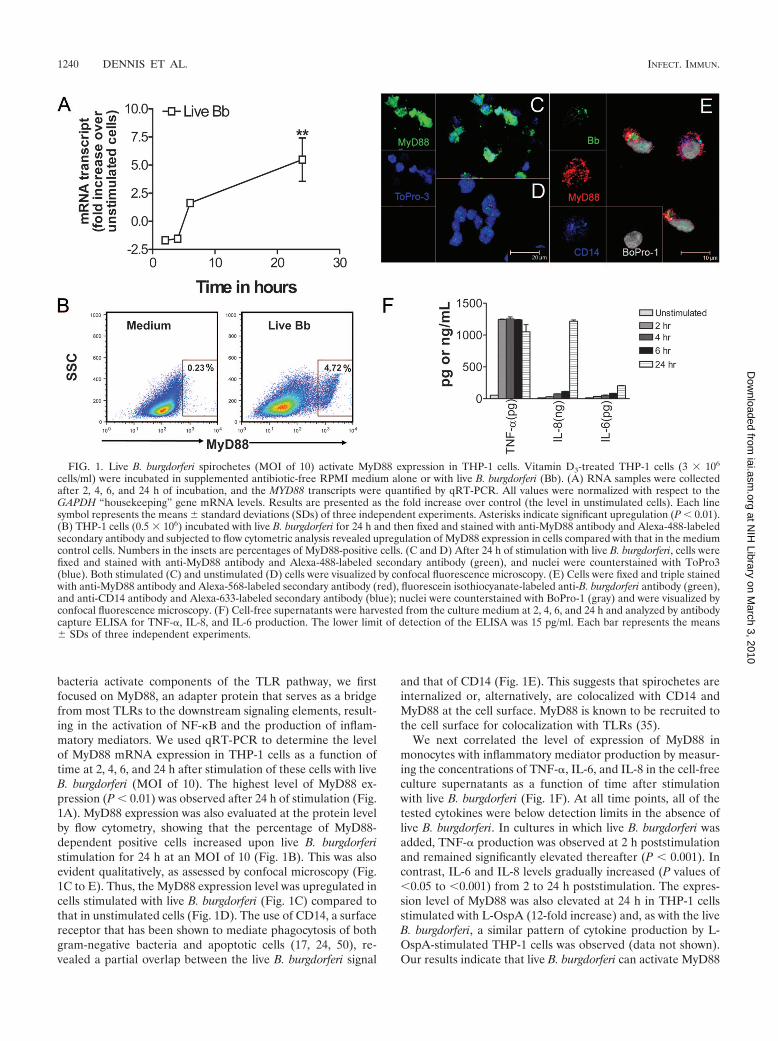
bacteria activate components of the TLR pathway, we firstfocused on MyD88, an adapter protein that serves as a bridgefrom most TLRs to the downstream signaling elements, result-ing in the activation of NF-�B and the production of inflam-matory mediators. We used qRT-PCR to determine the levelof MyD88 mRNA expression in THP-1 cells as a function oftime at 2, 4, 6, and 24 h after stimulation of these cells with liveB. burgdorferi (MOI of 10). The highest level of MyD88 ex-pression (P � 0.01) was observed after 24 h of stimulation (Fig.1A). MyD88 expression was also evaluated at the protein levelby flow cytometry, showing that the percentage of MyD88-dependent positive cells increased upon live B. burgdorferistimulation for 24 h at an MOI of 10 (Fig. 1B). This was alsoevident qualitatively, as assessed by confocal microscopy (Fig.1C to E). Thus, the MyD88 expression level was upregulated incells stimulated with live B. burgdorferi (Fig. 1C) compared tothat in unstimulated cells (Fig. 1D). The use of CD14, a surfacereceptor that has been shown to mediate phagocytosis of bothgram-negative bacteria and apoptotic cells (17, 24, 50), re-vealed a partial overlap between the live B. burgdorferi signal
and that of CD14 (Fig. 1E). This suggests that spirochetes areinternalized or, alternatively, are colocalized with CD14 andMyD88 at the cell surface. MyD88 is known to be recruited tothe cell surface for colocalization with TLRs (35).
We next correlated the level of expression of MyD88 inmonocytes with inflammatory mediator production by measur-ing the concentrations of TNF-�, IL-6, and IL-8 in the cell-freeculture supernatants as a function of time after stimulationwith live B. burgdorferi (Fig. 1F). At all time points, all of thetested cytokines were below detection limits in the absence oflive B. burgdorferi. In cultures in which live B. burgdorferi wasadded, TNF-� production was observed at 2 h poststimulationand remained significantly elevated thereafter (P � 0.001). Incontrast, IL-6 and IL-8 levels gradually increased (P values of�0.05 to �0.001) from 2 to 24 h poststimulation. The expres-sion level of MyD88 was also elevated at 24 h in THP-1 cellsstimulated with L-OspA (12-fold increase) and, as with the liveB. burgdorferi, a similar pattern of cytokine production by L-OspA-stimulated THP-1 cells was observed (data not shown).Our results indicate that live B. burgdorferi can activate MyD88
FIG. 1. Live B. burgdorferi spirochetes (MOI of 10) activate MyD88 expression in THP-1 cells. Vitamin D3-treated THP-1 cells (3 � 106
cells/ml) were incubated in supplemented antibiotic-free RPMI medium alone or with live B. burgdorferi (Bb). (A) RNA samples were collectedafter 2, 4, 6, and 24 h of incubation, and the MYD88 transcripts were quantified by qRT-PCR. All values were normalized with respect to theGAPDH “housekeeping” gene mRNA levels. Results are presented as the fold increase over control (the level in unstimulated cells). Each linesymbol represents the means standard deviations (SDs) of three independent experiments. Asterisks indicate significant upregulation (P � 0.01).(B) THP-1 cells (0.5 � 106) incubated with live B. burgdorferi for 24 h and then fixed and stained with anti-MyD88 antibody and Alexa-488-labeledsecondary antibody and subjected to flow cytometric analysis revealed upregulation of MyD88 expression in cells compared with that in the mediumcontrol cells. Numbers in the insets are percentages of MyD88-positive cells. (C and D) After 24 h of stimulation with live B. burgdorferi, cells werefixed and stained with anti-MyD88 antibody and Alexa-488-labeled secondary antibody (green), and nuclei were counterstained with ToPro3(blue). Both stimulated (C) and unstimulated (D) cells were visualized by confocal fluorescence microscopy. (E) Cells were fixed and triple stainedwith anti-MyD88 antibody and Alexa-568-labeled secondary antibody (red), fluorescein isothiocyanate-labeled anti-B. burgdorferi antibody (green),and anti-CD14 antibody and Alexa-633-labeled secondary antibody (blue); nuclei were counterstained with BoPro-1 (gray) and were visualized byconfocal fluorescence microscopy. (F) Cell-free supernatants were harvested from the culture medium at 2, 4, 6, and 24 h and analyzed by antibodycapture ELISA for TNF-�, IL-8, and IL-6 production. The lower limit of detection of the ELISA was 15 pg/ml. Each bar represents the means SDs of three independent experiments.
1240 DENNIS ET AL. INFECT. IMMUN.
at NIH
Library on March 3, 2010
iai.asm.org
Dow
nloaded from
expression in human monocytes. MyD88 expression as inducedby live B. burgdorferi or by L-OspA in THP-1 cells correlatedwith the enhanced production of TNF-�, IL-6, and IL-8 pro-teins.
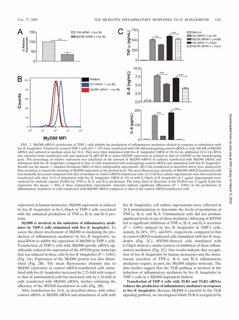
MyD88 is involved in the induction of inflammatory medi-ators by THP-1 cells stimulated with live B. burgdorferi. Toassess the direct involvement of MyD88 in mediating the pro-duction of inflammatory mediators by live B. burgdorferi, weused RNAi to inhibit the expression of MyD88 in THP-1 cells.Transfection of THP-1 cells with MyD88-specific siRNA sig-nificantly reduced the expression of the MYD88 gene transcriptthat was induced in these cells by live B. burgdorferi (P � 0.001)(Fig. 2A). Expression of the MyD88 protein was also dimin-ished (Fig. 2B). The mean fluorescence intensity due toMyD88 expression in control siRNA-transfected cells stimu-lated with live B. burgdorferi increased by 2.23-fold with respectto that of unstimulated cells but increased only by 1.10-fold incells transfected with MyD88 siRNA, further validating theefficiency of the MYD88 knockdown in cells (Fig. 2B).
After transfection for 24 h, as described above, with eithercontrol siRNA or MyD88 siRNA and stimulation of cells with
live B. burgdorferi, cell culture supernatants were collected at24 h poststimulation to determine the levels of production ofTNF-�, IL-6, and IL-8. Unstimulated cells did not producesignificant levels of any of these mediators. Silencing of MYD88led to significant inhibition of TNF-�, IL-8, and IL-6 secretion(P � 0.001) induced by live B. burgdorferi in THP-1 cells,namely, by 24%, 35%, and 84%, respectively, compared to thatin control siRNA-transfected cells stimulated with live B. burg-dorferi (Fig. 2C). MYD88-silenced cells stimulated withL-OspA showed a similar pattern of inhibition of these inflam-matory mediators (Fig. 2C). Our results indicate that recogni-tion of live B. burgdorferi by human monocytes and the down-stream secretion of TNF-�, IL-8, and IL-6 inflammatorymediators require, in part, the MyD88 adapter molecule. Thedata further suggest that the TLR pathway is involved in theinduction of inflammatory mediators by live B. burgdorferi inTHP-1 cells in a MyD88-dependent fashion.
Transfection of THP-1 cells with TLR1 and TLR2 siRNAsreduces the production of inflammatory mediators in responseto live B. burgdorferi. Because MyD88 is essential to the TLRsignaling pathway, we investigated which TLR is recognized by
FIG. 2. MyD88 siRNA transfection of THP-1 cells inhibits the production of inflammatory mediators elicited in response to stimulation withlive B. burgdorferi. Vitamin D3-treated THP-1 cells (0.5 � 106) were transfected with 100 nM nontargeting control siRNA or with 100 nM of MyD88siRNA and cultured in medium alone for 24 h. They were then stimulated with live B. burgdorferi (MOI of 10) for an additional 24 h. (A) RNAwas extracted from transfected cells and analyzed by qRT-PCR to assess MYD88 expression in relation to that of GAPDH as the housekeepinggene. The percentage of relative expression was calculated as the amount of MyD88 mRNA in cultures transfected with MyD88 siRNA andstimulated with live B. burgdorferi compared to that of cells transfected with nontargeting control siRNA and stimulated with live B. burgdorferi.Results are the means standard deviations (SDs) of three independent experiments. (B) Cells transfected as described above were analyzed byflow cytometry to assess the silencing of MyD88 expression at the protein level. The mean fluorescence intensity of MyD88 siRNA-transfected cellswas markedly decreased compared with that of medium or control siRNA-transfected cells. (C) Cell-free culture supernatants were harvested fromtransfected cells after 24 h of stimulation with live B. burgdorferi (MOI of 10) or with L-OspA of B. burgdorferi at 1 �g/ml. Supernatants wereanalyzed by antibody capture ELISA for TNF-�, IL-8, and IL-6 production. The lower limit of detection of the ELISA was 15 pg/ml. Each barrepresents the means SDs of three independent experiments. Asterisks indicate significant differences (P � 0.001) in the production ofinflammatory mediators in cells transfected with MyD88 siRNA compared to that of the control siRNA-transfected cells.
VOL. 77, 2009 TLR MEDIATES INFLAMMATORY RESPONSES TO B. BURGDORFERI 1241
at NIH
Library on March 3, 2010
iai.asm.org
Dow
nloaded from
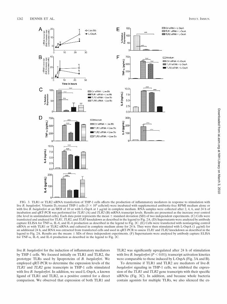
live B. burgdorferi for the induction of inflammatory mediatorsby THP-1 cells. We focused initially on TLR1 and TLR2, theprototype TLRs used by lipoproteins of B. burgdorferi. Weemployed qRT-PCR to determine the expression levels of theTLR1 and TLR2 gene transcripts in THP-1 cells stimulatedwith live B. burgdorferi. In addition, we used L-OspA, a knownligand of TLR1 and TLR2, as a positive control for a directcomparison. We observed that expression of both TLR1 and
TLR2 was significantly upregulated after 24 h of stimulationwith live B. burgdorferi (P � 0.01); transcript activation kineticswere comparable to those induced by L-OspA (Fig. 3A and B).
To determine if TLR1 and TLR2 are mediators of live-B.burgdorferi signaling in THP-1 cells, we inhibited the expres-sion of the TLR1 and TLR2 gene transcripts with their specificsiRNAs (Fig. 3C). In addition, and because whole bacteriacontain agonists for multiple TLRs, we also silenced the ex-
FIG. 3. TLR1 or TLR2 siRNA transfection of THP-1 cells affects the production of inflammatory mediators in response to stimulation withlive B. burgdorferi. Vitamin D3-treated THP-1 cells (3 � 106 cells/ml) were incubated with supplemented antibiotic-free RPMI medium alone orwith live B. burgdorferi at an MOI of 10 or with L-OspA at 1 �g/ml in complete medium. RNA samples were collected after 2, 4, 6, and 24 h ofincubation and qRT-PCR was performed for TLR1 (A) and TLR2 (B) mRNA transcript levels. Results are presented as the increase over control(the level in unstimulated cells). Each data point represents the mean standard deviation (SD) of two independent experiments. (C) Cells weretransfected and analyzed for TLR1, TLR2, and TLR5 knockdown as described in the legend to Fig. 2A. (D) Supernatants were analyzed by antibodycapture ELISA for TNF-�, IL-8, and IL-6 production as described in the legend to Fig. 2C. (E) Cells were transfected with nontargeting controlsiRNA or with TLR1 or TLR2 siRNA and cultured in complete medium alone for 24 h. They were then stimulated with L-OspA (1 �g/ml) foran additional 24 h, and RNA was extracted from transfected cells and used in qRT-PCR to assess TLR1 and TLR2 knockdown as described in thelegend to Fig. 2A. Results are the means SDs of three independent experiments. (F) Supernatants were analyzed by antibody capture ELISAfor TNF-�, IL-8, and IL-6 production as described in the legend to Fig. 2C.
1242 DENNIS ET AL. INFECT. IMMUN.
at NIH
Library on March 3, 2010
iai.asm.org
Dow
nloaded from
pression of TLR5 (Fig. 3C). The relative expression of thethree gene transcripts was significantly reduced by this proce-dure (P � 0.001). Silencing of TLR1 caused a significant down-regulation of TNF-� in cells stimulated with live B. burgdorfericompared to that of the control siRNA. Surprisingly, produc-tion of TNF-� was increased significantly (P � 0.0001) whenTLR2 was silenced (Fig. 3D). On the other hand, silencing ofeither TLR1 or TLR2 resulted in a significant downregulationof both IL-8 and IL-6 (P � 0.001) compared with their pro-duction levels induced in control cells (Fig. 3D). The TLR1TLR2 double knockdown yielded results similar to those ob-tained when these genes were knocked down individually (datanot shown).
The silencing of TLR5 had no effect on the production ofTNF-�, IL-8, or IL-6 by these cells upon stimulation with liveB. burgdorferi (Fig. 3D). Because lipoproteins of B. burgdorferiare known to signal through TLR1 TLR2 heterodimers, wealso individually silenced both receptors in experiments inwhich L-OspA was used as the stimulant (Fig. 3E). Silencing ofeither TLR1 or TLR2 in L-OspA-stimulated THP-1 cells sig-nificantly affected the protein expression levels of TNF-�, IL-8,and IL-6 (P � 0.001) (Fig. 3F). The downregulatory effect wasmore pronounced in TLR2-transfected cell cultures.
DISCUSSION
Substantial efforts have been made to understand the mo-lecular mechanism by which the lipoproteins of B. burgdorferielicit the production of inflammatory mediators in cells of thehuman innate immune system. In contrast, much has yet to belearned about how live B. burgdorferi participates in this pro-cess. Here, we investigated the molecular mechanisms thatlead to the production of proinflammatory mediators by hu-man monocytes when these cells are exposed in vitro to live B.burgdorferi. We showed in kinetics studies that live B. burgdor-feri upregulates the expression of components of the TLRpathway, such as MyD88, TLR1, and TLR2. We further dem-onstrated that MYD88, TLR1, and TLR2, but not TLR5, tran-script knockdown significantly reduced the production ofproinflammatory mediators by human monocytes in responseto live B. burgdorferi stimulation. THP-1 cells stimulated withlipoproteins yielded similar results but with some interestingdifferences that we discuss below.
Our results indicate that live B. burgdorferi activates MYD88transcription and translation in human monocytes. UsingRNAi, we showed that specific silencing of the MYD88 genesignificantly downmodulates the production of TNF-�, IL-6,and IL-8, suggesting that live B. burgdorferi stimulation ofmonocytes for the production of these inflammatory mediatorsis MyD88 dependent. MyD88 is an adapter molecule that playsa critical role in the downstream signaling of several TLRs ininnate immune cells. In vitro, studies using cells from MyD88-deficient mice have shown that MyD88 is an important medi-ator of inflammatory signaling in several bacterial diseases (38,40, 53). Studies conducted by Shin and coworkers (53) ele-gantly demonstrated that MyD88 was necessary for the inter-nalization and phagocytosis of live B. burgdorferi and for theproduction of several inflammatory mediators in mouse bonemarrow-derived macrophages. To our knowledge, our study isthe first to demonstrate a MyD88-mediated innate immune
signaling pathway activated by live B. burgdorferi in humanmonocytes. Our findings and those of Shin et al. (53) togetherascribe a critical role to MyD88 during the initial phase of B.burgdorferi infection.
Silencing of the TLR5 gene revealed that flagellin, the TLR5ligand, is not involved in the monocyte response to live B.burgdorferi, as measured by the production of TNF-�, IL-6, andIL-8. Our finding, however, contrasts with that of Shin et al.,(53), who observed that the knockdown of the TLR5 generesulted in diminished expression of cytokine (TNF-�, IL-6,and IL-�) but not chemokine (MCP-1 and CXCL-2) tran-scripts in murine bone marrow macrophages. Differences re-cently shown between TLR signaling of mouse and humanmacrophages (10, 44) may explain this discrepancy.
Evaluation of the roles of TLR1 and TLR2 as mediators ofinflammation in Lyme disease has been limited in most in-stances to examining the stimulation of innate immune cellswith B. burgdorferi lipoproteins or lysates (4, 9, 30, 32, 51, 53,60). Our study is one of the few to address the roles of bothTLR1 and TLR2 in inflammatory mediator production by hu-man monocytes stimulated with live organisms. Another study(21) focused only on TLR2 and showed that TLR2 was essen-tial in mediating the production of matrix metalloproteinasesfrom human monocytes in response to live spirochete stimu-lation.
While the inhibition of TLR1 and TLR2 transcription by thecorresponding siRNAs under conditions of live B. burgdorferistimulation was 94 and 79%, respectively (Fig. 3C), cytokine/chemokine production was less inhibited. Thus, IL-6 produc-tion was decreased by 63% upon TLR1 silencing and by 60%upon silencing of TLR2, whereas IL-8 production was reducedby only 21% (TLR1) and 28% (TLR2), respectively. TNF-�behaved differently, in that its production was 48% inhibited bythe silencing of TLR1 but was enhanced by 21% upon TLR2knockdown. These results indicate that alternative receptorsand pathways are involved in mediating inflammation as in-duced by live B. burgdorferi in human monocytes. In mice,Lyme disease inflammation can proceed via a TLR-indepen-dent pathway(s), since ablation of TLR or MyD88 signalingfailed to decrease the influx of inflammatory cells or inflam-mation in response to B. burgdorferi infection (6, 7, 39). To thisend, B. burgdorferi has been shown to bind to various integrinreceptors such as �IIb�3, �v�3, �3�1 (11, 12, 13, 14), and �3�1
(6) on host cells, suggesting the importance of these moleculesin the non-TLR-dependent pathogenesis of Lyme disease. Infact, a recent study uncovered the �3�1 integrin as a receptorutilized by live spirochetes, specifically the BBB07 protein of B.burgdorferi, for the induction of matrix metalloproteinases andseveral inflammatory mediators in human chondrocytes (5, 6).In experiments in vivo, those same investigators also showedsignificant production of several chemokines and cytokines inthe joints of MYD88 knockout mice, thus validating the occur-rence of inflammation in a MyD88-independent manner (6).
The probable utilization of receptors other than TLR1 andTLR2 by live B. burgdorferi in its interaction with human mono-cytes is further substantiated by the results obtained with L-OspA. For example, while the silencing of TLR2 led to thecomplete inhibition of the production of TNF-� by THP-1 cellsthat were stimulated with L-OspA (Fig. 3F), it resulted, asmentioned above, in an increase in the production of this
VOL. 77, 2009 TLR MEDIATES INFLAMMATORY RESPONSES TO B. BURGDORFERI 1243
at NIH
Library on March 3, 2010
iai.asm.org
Dow
nloaded from
cytokine upon stimulation with live B. burgdorferi. The increasein TNF-� production in TLR2-silenced cells stimulated withlive B. burgdorferi and not in those stimulated with L-OspAwould indicate the utilization of a TLR-independent pathwayby nonlipoproteins of B. burgdorferi for the induction ofTNF-�. Studies by Behera et al. (6) may help to corroborateour findings, as those investigators demonstrated via siRNAtechnology that the integrin �3 mediated the B. burgdorferi-induced expression of several inflammatory mediators, includ-ing TNF-� in human chondrocytes. Our present study providesadditional evidence that the induction of inflammation inLyme disease occurs via both TLR-dependent and -indepen-dent pathways (5, 6, 7, 39).
The other two mediators whose production we evaluated inthis study, IL-8 and IL-6, responded to L-OspA stimulationupon TLR1 and TLR2 silencing with a trend that was compa-rable to that shown in response to live B. burgdorferi. In addi-tion, silencing of MYD88 resulted in a similar response patternof THP-1 cells when these cells were stimulated with either liveB. burgdorferi or L-OspA. However, the inhibition of mediatorproduction upon TLR2 silencing was complete after stimula-tion with L-OspA, whereas it was partial when THP-1 cellswere stimulated with live B. burgdorferi. Thus, it is possible thatB. burgdorferi lipoproteins, as well as other surface proteinssuch as the recently identified BBB07 protein of B. burgdorferi(5), are involved in the interaction of human monocytes withlive spirochetes. In conclusion, our results indicate that theTLR pathway mediates, at least in part, the release of inflam-matory mediators in human monocytes stimulated with live B.burgdorferi spirochetes and further suggest that the TLR-de-pendent interaction between these cells and spirochetes is me-diated by spirochetal lipoproteins but not by flagellin.
ACKNOWLEDGMENTS
This work was supported by grant UO1-CI000153 from the Centersfor Disease Control and Prevention and by grants NS048952,RR00164, G20 R018397-01, and G20 R019607-01 from the NationalInstitutes of Health.
We thank Avery MacLean for excellent secretarial help. We alsothank Robin Rodriguez for help with confocal microscopy images.
REFERENCES
1. Akira, S., and K. Takeda. 2004. Toll-like receptor signalling. Nat. Rev.Immunol. 4:499–511.
2. Akira, S., K. Takeda, and T. Kaisho. 2001. Toll-like receptors: critical pro-teins linking innate and acquired immunity. Nat. Immunol. 2:675–680.
3. Alexopoulou, L., A. C. Holt, R. Medzhitov, and R. A. Flavell. 2001. Recog-nition of double-stranded RNA and activation of NF-�B by Toll-like recep-tor 3. Nature 413:732–738.
4. Alexopoulou, L., V. Thomas, M. Schnare, Y. Lobet, J. Anguita, R. T. Schoen,R. Medzhitov, E. Fikrig, and R. A. Flavell. 2002. Hyporesponsiveness tovaccination with Borrelia burgdorferi OspA in humans and in TLR1- andTLR2-deficient mice. Nat. Med. 8:878–884.
5. Behera, A. K., E. Durand, C. Cugini, S. Antonara, L. Bourassa, E. Hilde-brand, L. T. Hu, and J. Coburn. 2008. Borrelia burgdorferi BBB07 interactionwith integrin �3�1 stimulates production of pro-inflammatory mediators inprimary human chondrocytes. Cell Microbiol. 10:320–331.
6. Behera, A. K., E. Hildebrand, S. Uematsu, S. Akira, J. Coburn, and L. T. Hu.2006. Identification of a TLR-independent pathway for Borrelia burgdorferi-induced expression of matrix metalloproteinases and inflammatory media-tors through binding to integrin �3�1. J. Immunol. 177:657–664.
7. Bolz, D. D., R. S. Sundsbak, Y. Ma, S. Akira, C. J. Kirschning, J. F. Zachary,J. H. Weis, and J. J. Weis. 2004. MyD88 plays a unique role in host defensebut not arthritis development in Lyme disease. J. Immunol. 173:2003–2010.
8. Burgdorfer, W., A. G. Barbour, S. F. Hayes, J. L. Benach, E. Grunwaldt, andJ. P. Davis. 1982. Lyme disease—a tick-borne spirochetosis? Science 216:1317–1319.
9. Cassiani-Ingoni, R., E. S. Cabral, J. D. Lunemann, Z. Garza, T. Magnus, H.
Gelderblom, P. J. Munson, A. Marques, and R. Martin. 2006. Borreliaburgdorferi induces TLR1 and TLR2 in human microglia and peripheralblood monocytes but differentially regulates HLA-class II expression. J. Neu-ropathol. Exp. Neurol. 65:540–548.
10. Chang, S., A. Dolganiuc, and G. Szabo. 2007. Toll-like receptors 1 and 6 areinvolved in TLR2-mediated macrophage activation by hepatitis C virus coreand NS3 proteins. J. Leukoc. Biol. 82:479–487.
11. Coburn, J., S. W. Barthold, and J. M. Leong. 1994. Diverse Lyme diseasespirochetes bind integrin �IIb�3 on human platelets. Infect. Immun. 62:5559–5567.
12. Coburn, J., and C. Cugini. 2003. Targeted mutation of the outer membraneprotein P66 disrupts attachment of the Lyme disease agent, Borrelia burg-dorferi, to integrin �v�3. Proc. Natl. Acad. Sci. USA 100:7301–7306.
13. Coburn, J., J. M. Leong, and J. K. Erban. 1993. Integrin �IIb�3 mediatesbinding of the Lyme disease agent Borrelia burgdorferi to human platelets.Proc. Natl. Acad. Sci. USA 90:7059–7063.
14. Coburn, J., L. Magoun, S. C. Bodary, and J. M. Leong. 1998. Integrins �v�3and �5�1 mediate attachment of Lyme disease spirochetes to human cells.Infect. Immun. 66:1946–1952.
15. Coyle, P. K. 1993. Lyme disease. Mosby-Year Book, St. Louis, MO.16. Dennis, V. A., A. Jefferson, S. R. Singh, F. Ganapamo, and M. T. Philipp.
2006. Interleukin-10 anti-inflammatory response to Borrelia burgdorferi, theagent of Lyme disease: a possible role for suppressors of cytokine signaling1 and 3. Infect. Immun. 74:5780–5789.
17. Devitt, A., O. D. Moffatt, C. Raykundalia, J. D. Capra, D. L. Simmons, andC. D. Gregory. 1998. Human CD14 mediates recognition and phagocytosis ofapoptotic cells. Nature 392:505–509.
18. Diebold, S. S., T. Kaisho, H. Hemmi, S. Akira, and C. Reis e Sousa. 2004.Innate antiviral responses by means of TLR7-mediated recognition of single-stranded RNA. Science 303:1529–1531.
19. Dunne, A., and L. A. O’Neill. 2003. The interleukin-1 receptor/Toll-likereceptor superfamily: signal transduction during inflammation and host de-fense. Sci. STKE 2003:re3.
20. England, J. D., R. P. Bohm, Jr., E. D. Roberts, and M. T. Philipp. 1997.Mononeuropathy multiplex in rhesus monkeys with chronic Lyme disease.Ann. Neurol. 41:375–384.
21. Gebbia, J. A., J. L. Coleman, and J. L. Benach. 2004. Selective induction ofmatrix metalloproteinases by Borrelia burgdorferi via toll-like receptor 2 inmonocytes. J. Infect. Dis. 189:113–119.
22. Ghosh, S., M. J. May, and E. B. Kopp. 1998. NF-�B and Rel proteins:evolutionarily conserved mediators of immune responses. Annu. Rev. Im-munol. 16:225–260.
23. Glickstein, L., B. Moore, T. Bledsoe, N. Damle, V. Sikand, and A. C. Steere.2003. Inflammatory cytokine production predominates in early Lyme diseasein patients with erythema migrans. Infect. Immun. 71:6051–6053.
24. Grunwald, U., X. Fan, R. S. Jack, G. Workalemahu, A. Kallies, F. Stelter,and C. Schutt. 1996. Monocytes can phagocytose Gram-negative bacteria bya CD14-dependent mechanism. J. Immunol. 157:4119–4125.
25. Grygorczuk, S., S. Pancewicz, J. Zajkowska, M. Kondrusik, R. Rwierzbinska,and T. Hermanowska-Szpakowicz. 2004. Concentrations of macrophage in-flammatory proteins MIP-1� and MIP-1� and interleukin 8 (IL-8) in lymeborreliosis. Infection 32:350–355.
26. Guerau-de-Arellano, M., J. Alroy, and B. T. Huber. 2005. �2 Integrinscontrol the severity of murine Lyme carditis. Infect. Immun. 73:3242–3250.
27. Hayashi, F., K. D. Smith, A. Ozinsky, T. R. Hawn, E. C. Yi, D. R. Goodlett,J. K. Eng, S. Akira, D. M. Underhill, and A. Aderem. 2001. The innateimmune response to bacterial flagellin is mediated by Toll-like receptor 5.Nature 410:1099–1103.
28. Heil, F., H. Hemmi, H. Hochrein, F. Ampenberger, C. Kirschning, S. Akira,G. Lipford, H. Wagner, and S. Bauer. 2004. Species-specific recognition ofsingle-stranded RNA via toll-like receptor 7 and 8. Science 303:1526–1529.
29. Hemmi, H., O. Takeuchi, T. Kawai, T. Kaisho, S. Sato, H. Sanjo, M. Ma-tsumoto, K. Hoshino, H. Wagner, K. Takeda, and S. Akira. 2000. A Toll-likereceptor recognizes bacterial DNA. Nature 408:740–745.
30. Hirschfeld, M., C. J. Kirschning, R. Schwandner, H. Wesche, J. H. Weis,R. M. Wooten, and J. J. Weis. 1999. Cutting edge: inflammatory signaling byBorrelia burgdorferi lipoproteins is mediated by toll-like receptor 2. J. Immu-nol. 163:2382–2386.
31. Iwasaki, A., and R. Medzhitov. 2004. Toll-like receptor control of the adap-tive immune responses. Nat. Immunol. 5:987–995.
32. Izadi, H., A. T. Motameni, T. C. Bates, E. R. Olivera, V. Villar-Suarez, I.Joshi, R. Garg, B. A. Osborne, R. J. Davis, M. Rincon, and J. Anguita. 2007.c-Jun N-terminal kinase 1 is required for Toll-like receptor 1 gene expressionin macrophages. Infect. Immun. 75:5027–5034.
33. Jared, C. R., G. Gustavo, R. Lee, K. Amardeep, K. P. Maureen, D. S. Kelly,E. H. Leroy, and A. Alan. 2005. The evolution of vertebrate toll-like recep-tors. Proc. Natl. Acad. Sci. USA 102:9577–9582.
34. Jiang, Z., M. Zamanian-Daryoush, H. Nie, A. M. Silva, B. R. Williams, andX. Li. 2003. Poly(I-C)-induced Toll-like receptor 3 (TLR3)-mediated acti-vation of NF-�B and MAP kinase is through an interleukin-1 receptor-associated kinase (IRAK)-independent pathway employing the signaling
1244 DENNIS ET AL. INFECT. IMMUN.
at NIH
Library on March 3, 2010
iai.asm.org
Dow
nloaded from
components TLR3-TRAF6-TAK1-TAB2-PKR. J. Biol. Chem. 278:16713–16719.
35. Kagan, J. C., and R. Medzhitov. 2006. Phosphoinositide-mediated adaptorrecruitment controls Toll-like receptor signaling. Cell 125:943–955.
36. Karin, M., and Y. Ben-Neriah. 2000. Phosphorylation meets ubiquitination:the control of NF-�B activity. Annu. Rev. Immunol. 18:621–663.
37. Lauw, F. N., D. R. Caffrey, and D. T. Golenbock. 2005. Of mice and man:TLR11 (finally) finds profilin. Trends Immunol. 26:509–511.
38. Lehnardt, S., P. Henneke, E. Lien, D. L. Kasper, J. J. Volpe, I. Bechmann,R. Nitsch, J. R. Weber, D. T. Golenbock, and T. Vartanian. 2006. A mech-anism for neurodegeneration induced by group B streptococci through ac-tivation of the TLR2/MyD88 pathway in microglia. J. Immunol. 177:583–592.
39. Liu, N., R. R. Montgomery, S. W. Barthold, and L. K. Bockenstedt. 2004.Myeloid differentiation antigen 88 deficiency impairs pathogen clearance butdoes not alter inflammation in Borrelia burgdorferi-infected mice. Infect.Immun. 72:3195–3203.
40. Macedo, G. C., D. M. Magnani, N. B. Carvalho, O. Bruna-Romero, R. T.Gazzinelli, and S. C. Oliveira. 2008. Central role of MyD88-dependentdendritic cell maturation and proinflammatory cytokine production to con-trol Brucella abortus infection. J. Immunol. 180:1080–1087.
41. Mogensen, T. H., and S. R. Paludan. 2005. Reading the viral signature byToll-like receptors and other pattern recognition receptors. J. Mol. Med.83:180–192.
42. Moore, L. J., A. M. Gilbey, C. G. Dowson, A. C. Pridmore, S. K. Dower, andR. C. Read. 2007. Proinflammatory activation of Toll-like receptor-2 duringexposure of penicillin-resistant Streptococcus pneumoniae to beta-lactam an-tibiotics. J. Antimicrob. Chemother. 59:35–42.
43. Murthy, P. K., V. A. Dennis, B. L. Lasater, and M. T. Philipp. 2000. Inter-leukin-10 modulates proinflammatory cytokines in the human monocytic cellline THP-1 stimulated with Borrelia burgdorferi lipoproteins. Infect. Immun.68:6663–6669.
44. Nahori, M. A., E. Fournie-Amazouz, N. S. Que-Gewirth, V. Balloy, M. Chi-gnard, C. R. Raetz, I. Saint Girons, and C. Werts. 2005. Differential TLRrecognition of leptospiral lipid A and lipopolysaccharide in murine andhuman cells. J. Immunol. 175:6022–6031.
45. Pasare, C., and R. Medzhitov. 2003. Toll-like receptors: balancing hostresistance with immune tolerance. Curr. Opin. Immunol. 15:677–682.
46. Philipp, M. T., and B. J. Johnson. 1994. Animal models of Lyme disease:pathogenesis and immunoprophylaxis. Trends Microbiol. 2:431–437.
47. Plattner, F., F. Yarovinsky, S. Romero, D. Didry, M. F Carlier, A. Sher, andD. Soldati-Favre. 2008. Toxoplasma profilin is essential for host cell invasionand TLR11-dependent induction of an interleukin-12 response. Cell HostMicrobe 3:77–87.
48. Poltorak, A., X. He, I. Smirnova, M. Y. Liu, C. Van Huffel, X. Du, D.Birdwell, E. Alejos, M. Silva, C. Galanos, M. Freudenberg, P. Ricciardi-Castagnoli, B. Layton, and B. Beutler. 1998. Defective LPS signaling in
C3H/HeJ and C57BL/10ScCr mice: mutations in Tlr4 gene. Science 282:2085–2088.
49. Sato, S., M. Sugiyama, M. Yamamoto, Y. Watanabe, T. Kawai, K. Takeda,and S. Akira. 2003. Toll/IL-1 receptor domain-containing adaptor inducingIFN-� (TRIF) associates with TNF receptor-associated factor 6 and TANK-binding kinase 1, and activates two distinct transcription factors, NF-�B andIFN-regulatory factor-3, in the Toll-like receptor signaling. J. Immunol.171:4304–4310.
50. Schiff, D. E., L. Kline, K. Soldau, J. D. Lee, J. Pugin, P. S. Tobias, and R. J.Ulevitch. 1997. Phagocytosis of gram-negative bacteria by a unique CD14-dependent mechanism. J. Leukoc. Biol. 62:786–794.
51. Schroder, N. W., I. Diterich, A. Zinke, J. Eckert, C. Draing, V. von Baehr, D.Hassler, S. Priem, K. Hahn, K. S. Michelsen, T. Hartung, G. R. Burmester,U. B. Gobel, C. Hermann, and R. R. Schumann. 2005. HeterozygousArg753Gln polymorphism of human TLR-2 impairs immune activation byBorrelia burgdorferi and protects from late stage Lyme disease. J. Immunol.175:2534–2540.
52. Schwandner, R., R. Dziarski, H. Wesche, M. Rothe, and C. J. Kirschning.1999. Peptidoglycan- and lipoteichoic acid-induced cell activation is medi-ated by toll-like receptor 2. J. Biol. Chem. 274:17406–17409.
53. Shin, O. S., R. R. Isberg, S. Akira, S. Uematsu, A. K. Behara, and L. T. Hu.2008. Distinct roles for MyD88 and Toll-like receptors 2, 5, and 9 in phago-cytosis of Borrelia burgdorferi and cytokine induction. Infect. Immun. 76:2341–2351.
54. Singh, S. K., and H. J. Girschick. 2006. Toll-like receptors in Borrelia burg-dorferi-induced inflammation. Clin. Microbiol. Infect. 12:705–717.
55. Steere, A. C. 2001. Lyme disease. N. Engl. J. Med. 345:115–125.56. Takeda, K., T. Kaisho, and S. Akira. 2003. Toll-like receptors. Annu. Rev.
Immunol. 21:335–376.57. Underhill, D. M., A. Ozinsky, A. M. Hajjar, A. Stevens, C. B. Wilson, M.
Bassetti, and A. Aderem. 1999. The Toll-like receptor 2 is recruited tomacrophage phagosomes and discriminates between pathogens. Nature 401:811–815.
58. van Maren, W. W., J. F. Jacobs, I. J. de Vries, S. Nierkens, and G. J. Adema.2008. Toll-like receptor signalling on Tregs: to suppress or not to suppress?Immunology 124:445–452.
59. Wagner, H. 2004. The immunobiology of the TLR9 subfamily. Trends Im-munol. 25:381–386.
60. Wang, X., Y. Ma, A. Yoder, H. Crandall, J. F. Zachary, R. S. Fujinami, J. H.Weis, and J. J. Weis. 2008. T cell infiltration is associated with increasedLyme arthritis in TLR2�/� mice. FEMS Immunol. Med. Microbiol. 52:124–133.
61. Zhang, D., G. Zhang, M. S. Hayden, M. B. Greenblatt, C. Bussey, R. A.Flavell, and S. Ghosh. 2004. A toll-like receptor that prevents infection byuropathogenic bacteria. Science 303:1522–1526.
Editor: J. F. Urban, Jr.
VOL. 77, 2009 TLR MEDIATES INFLAMMATORY RESPONSES TO B. BURGDORFERI 1245
at NIH
Library on March 3, 2010
iai.asm.org
Dow
nloaded from