Common and Unique Contributions of Decorin-Binding Proteins A and B to the Overall Virulence of...
11
Common and Unique Contributions of Decorin-Binding Proteins A and B to the Overall Virulence of Borrelia burgdorferi Yanlin Shi, Qilong Xu, Sunita V. Seemanaplli, Kristy McShan, Fang Ting Liang* Department of Pathobiological Sciences, Louisiana State University, Baton Rouge, Louisiana, United States of America Abstract As an extracellular bacterium, the Lyme disease spirochete Borrelia burgdorferi resides primarily in the extracellular matrix and connective tissues and between host cells during mammalian infection, where decorin and glycosaminoglycans are abundantly found, so its interactions with these host ligands potentially affect various aspects of infection. Decorin-binding proteins (Dbps) A and B, encoded by a 2-gene operon, are outer surface lipoproteins with similar molecular weights and share approximately 40% identity, and both bind decorin and glycosaminoglycans. To investigate how DbpA and DbpB contribute differently to the overall virulence of B. burgdorferi,a dbpAB mutant was modified to overproduce the adhesins. Overproduction of either DbpA or DbpB resulted in restoration of the infectivity of the mutant to the control level, measured by 50% infectious dose (ID 50 ), indicating that the two virulence factors are interchangeable in this regard. Overproduction of DbpA also allowed the mutant to disseminate to some but not all distal tissues slightly slower than the control, but the mutant with DbpB overproduction showed severely impaired dissemination to all tissues that were analyzed. The mutant with DbpA overproduction colonized all tissues, albeit generating bacterial loads significantly lower than the control in heart and joint, while the mutant overproducing DbpB remained severely defective in heart colonization and registered bacterial loads substantially lower than the control in joint. Taken together, the study indicated that DbpA and DbpB play a similar role in contribution to infectivity as measured by ID 50 value but contribute differently to dissemination and tissue colonization. Citation: Shi Y, Xu Q, Seemanaplli SV, McShan K, Liang FT (2008) Common and Unique Contributions of Decorin-Binding Proteins A and B to the Overall Virulence of Borrelia burgdorferi. PLoS ONE 3(10): e3340. doi:10.1371/journal.pone.0003340 Editor: David M. Ojcius, University of California Merced, United States of America Received July 28, 2008; Accepted September 12, 2008; Published October 3, 2008 Copyright: ß 2008 Shi et al. This is an open-access article distributed under the terms of the Creative Commons Attribution License, which permits unrestricted use, distribution, and reproduction in any medium, provided the original author and source are credited. Funding: This research was financed by a career development award and a grant from NIH/NIAMS, an Arthritis Foundation Investigators award, and P20RR020159 from NIH/NCRR. Competing Interests: The authors have declared that no competing interests exist. * E-mail: [email protected] Introduction Borrelia burgdorferi, the Lyme disease spirochete transmitted by Ixodes ticks, causes the most common vector-borne illness in North America and Europe [1,2]. It is a slow growing bacterium but has a very low 50% infectious dose (ID 50 ) in the murine host [3,4], and can also cause persistent infection despite the development of strong immune responses [5]. Because B. burgdorferi is an extracellular pathogen, the interaction with host ligands mediated by spirochetal surface adhesins has been hypothesized to be critical for the pathogenic strategy since early 1990s [6]. B. burgdorferi resides primarily in the extracellular matrix (ECM) and connective tissues and between host cells during mammalian infection, where proteoglycans are abundantly found. Pioneering work by Guo et al. led to identification of two proteins with approximate molecular masses of 20 kDa on Western blot, which can bind decorin, an important component of proteoglycans; these adhesins were consequently named decorin-binding proteins (Dbps) A and B [6]. Their subsequent study demonstrated that the two adhesins are outer surface lipoproteins and are encoded by a 2-gene operon, in which dbpA is located downstream of dbpB [7,8]. At the same time, at least three groups in the field, including Hagman et al., Feng et al. and Hanson et al., started exploring the potential of DbpA as a vaccine [8–10]. However, the promise quickly diminished after Hagman et al. showed that the anti-DbpA response is unable to protect against a challenge via tick infestation, probably because DbpA is not upregulated to a level that can be effectively targeted by antibodies in the tick vector during feeding [11]. A few years later, Leong and colleagues showed that both DbpA and DbpB can also bind glycosamino- glycan, another major component of proteoglycans, albeit exhibiting distinct specificities [12], further strengthening the notion that the adhesins may play a critical role in the pathogenesis of B. burgdorferi. The contribution of DbpA and DbpB to the virulence of B. burgdorferi could only be indirectly explored before genetic tools had been available for the pathogen. Earlier studies indicated that mice deficient for decorin become less susceptible to murine Lyme disease and harbor fewer spirochetes during chronic infection [13,14]. The development in genetic manipulation of B. burgdorferi advanced investigation into the role of the dbpBA locus to a deeper level. Our recent studies via deletion of the 2-gene operon showed that both DbpA and DbpB are critical for the overall virulence of B. burgdorferi [4,15]. Most recently, Blevins et al. reported that at least DbpA is crucial for infectivity [16]. Furthermore, a study through modification of B. burgdorferi to increase DbpA expression indicated that higher DbpA expression dramatically enhances the interaction of the pathogen with decorin and, probably as a result, PLoS ONE | www.plosone.org 1 October 2008 | Volume 3 | Issue 10 | e3340
-
Upload
independent -
Category
Documents
-
view
0 -
download
0
Transcript of Common and Unique Contributions of Decorin-Binding Proteins A and B to the Overall Virulence of...
Common and Unique Contributions of Decorin-BindingProteins A and B to the Overall Virulence of BorreliaburgdorferiYanlin Shi, Qilong Xu, Sunita V. Seemanaplli, Kristy McShan, Fang Ting Liang*
Department of Pathobiological Sciences, Louisiana State University, Baton Rouge, Louisiana, United States of America
Abstract
As an extracellular bacterium, the Lyme disease spirochete Borrelia burgdorferi resides primarily in the extracellular matrixand connective tissues and between host cells during mammalian infection, where decorin and glycosaminoglycans areabundantly found, so its interactions with these host ligands potentially affect various aspects of infection. Decorin-bindingproteins (Dbps) A and B, encoded by a 2-gene operon, are outer surface lipoproteins with similar molecular weights andshare approximately 40% identity, and both bind decorin and glycosaminoglycans. To investigate how DbpA and DbpBcontribute differently to the overall virulence of B. burgdorferi, a dbpAB mutant was modified to overproduce the adhesins.Overproduction of either DbpA or DbpB resulted in restoration of the infectivity of the mutant to the control level,measured by 50% infectious dose (ID50), indicating that the two virulence factors are interchangeable in this regard.Overproduction of DbpA also allowed the mutant to disseminate to some but not all distal tissues slightly slower than thecontrol, but the mutant with DbpB overproduction showed severely impaired dissemination to all tissues that wereanalyzed. The mutant with DbpA overproduction colonized all tissues, albeit generating bacterial loads significantly lowerthan the control in heart and joint, while the mutant overproducing DbpB remained severely defective in heart colonizationand registered bacterial loads substantially lower than the control in joint. Taken together, the study indicated that DbpAand DbpB play a similar role in contribution to infectivity as measured by ID50 value but contribute differently todissemination and tissue colonization.
Citation: Shi Y, Xu Q, Seemanaplli SV, McShan K, Liang FT (2008) Common and Unique Contributions of Decorin-Binding Proteins A and B to the Overall Virulenceof Borrelia burgdorferi. PLoS ONE 3(10): e3340. doi:10.1371/journal.pone.0003340
Editor: David M. Ojcius, University of California Merced, United States of America
Received July 28, 2008; Accepted September 12, 2008; Published October 3, 2008
Copyright: � 2008 Shi et al. This is an open-access article distributed under the terms of the Creative Commons Attribution License, which permits unrestricteduse, distribution, and reproduction in any medium, provided the original author and source are credited.
Funding: This research was financed by a career development award and a grant from NIH/NIAMS, an Arthritis Foundation Investigators award, andP20RR020159 from NIH/NCRR.
Competing Interests: The authors have declared that no competing interests exist.
* E-mail: [email protected]
Introduction
Borrelia burgdorferi, the Lyme disease spirochete transmitted by
Ixodes ticks, causes the most common vector-borne illness in North
America and Europe [1,2]. It is a slow growing bacterium but has
a very low 50% infectious dose (ID50) in the murine host [3,4], and
can also cause persistent infection despite the development of
strong immune responses [5]. Because B. burgdorferi is an
extracellular pathogen, the interaction with host ligands mediated
by spirochetal surface adhesins has been hypothesized to be critical
for the pathogenic strategy since early 1990s [6].
B. burgdorferi resides primarily in the extracellular matrix (ECM)
and connective tissues and between host cells during mammalian
infection, where proteoglycans are abundantly found. Pioneering
work by Guo et al. led to identification of two proteins with
approximate molecular masses of 20 kDa on Western blot, which
can bind decorin, an important component of proteoglycans; these
adhesins were consequently named decorin-binding proteins
(Dbps) A and B [6]. Their subsequent study demonstrated that
the two adhesins are outer surface lipoproteins and are encoded by
a 2-gene operon, in which dbpA is located downstream of dbpB
[7,8]. At the same time, at least three groups in the field, including
Hagman et al., Feng et al. and Hanson et al., started exploring the
potential of DbpA as a vaccine [8–10]. However, the promise
quickly diminished after Hagman et al. showed that the anti-DbpA
response is unable to protect against a challenge via tick
infestation, probably because DbpA is not upregulated to a level
that can be effectively targeted by antibodies in the tick vector
during feeding [11]. A few years later, Leong and colleagues
showed that both DbpA and DbpB can also bind glycosamino-
glycan, another major component of proteoglycans, albeit
exhibiting distinct specificities [12], further strengthening the
notion that the adhesins may play a critical role in the
pathogenesis of B. burgdorferi.
The contribution of DbpA and DbpB to the virulence of B.
burgdorferi could only be indirectly explored before genetic tools
had been available for the pathogen. Earlier studies indicated that
mice deficient for decorin become less susceptible to murine Lyme
disease and harbor fewer spirochetes during chronic infection
[13,14]. The development in genetic manipulation of B. burgdorferi
advanced investigation into the role of the dbpBA locus to a deeper
level. Our recent studies via deletion of the 2-gene operon showed
that both DbpA and DbpB are critical for the overall virulence of
B. burgdorferi [4,15]. Most recently, Blevins et al. reported that at
least DbpA is crucial for infectivity [16]. Furthermore, a study
through modification of B. burgdorferi to increase DbpA expression
indicated that higher DbpA expression dramatically enhances the
interaction of the pathogen with decorin and, probably as a result,
PLoS ONE | www.plosone.org 1 October 2008 | Volume 3 | Issue 10 | e3340

significantly reduces the ID50 value and severely impairs
dissemination in a murine model [17].
DbpA and DbpB share approximately 40% identity with similar
molecular weights, and both bind decorin and glycosaminoglycan
and are critical for the overall virulence of B. burgdorferi [4,7–9,12].
In a recent study, we were able to override the essential role of
outer surface protein C (OspC) in mammalian infection by
modifying an ospC mutant to overproduce OspA, OspE, VlsE or
DbpA, indicating that OspC functions can be replaced, at least in
part, by other outer surface lipoproteins [18]. The dbpBA operon is
expressed at a moderate level during mammalian infection [17,19]
so their expression can be dramatically increased via genetic
manipulation. If increased synthesis of DbpA or DbpB is able to
compensate for the deficiency in the other, the study would
indicate they are interchangeable. In the current study, to
compare the contributions of DbpA and DbpB to the overall
virulence, a dbpBA mutant was modified to overproduce DbpA or
DbpB and examined for the influence on four aspects of the
overall virulence, including infectivity, measured by ID50 value,
pace of dissemination to distal tissues, tissue colonization,
measured by the frequency of tissue colonization as well as
bacterial load, and persistence in the murine model.
Results
Modification of the dbpAB mutant to overproduce DbpAor DbpB
The two constructs, pBBE22-dbpA’ and pME22-dbpB’, were
electroporated into DdbpAB, which was generated in our earlier
study [4]. pME22-dbpB’ was constructed from pME22 as
illustrated in Figure 1A, while pBBE22-dbpA’ was modified from
pBBE22 in our previous study [17]. The sole difference between
pBBE22 and pME22 was the location of the bbe22 copy; this
should not affect the expression activity of the inserted genes.
pBBE22-dbpA’ and pME22-dbpB’ carried promoterless dbpA and
dbpB, respectively, both of which were fused with a flaB promoter.
Because DdbpAB lacks lp25, the plasmid that carries bbe22 coding
for a nicotinamidase essential for survival of B. burgdorferi in the
mammalian environment [20], both constructs contain a copy of
bbe22. Fifteen and 23 transformants were obtained from
transformation with the constructs. Plasmid analyses led to
selection of two clones receiving each construct. These four
clones, namely, DdbpAB/dbpA’/1, DdbpAB/dbpA’/2, DdbpAB/
dbpB’/1 and DdbpAB/dbpB’/2, shared the same plasmid content
as DdbpBA, which lost cp9, lp5, lp21, lp28-4, lp25 and lp56 [4].
Overproduction of DbpA and DbpB during in vitro growth due to
the introduction of the constructs was confirmed by immunoblot
analysis (Figure 1B). The DbpA band appeared stronger than
DbpB shown for the parental clone 13A; however, this did not
necessarily reflect that the 13 spirochetes produced more DbpA
than DbpB, because the reactivity of the antisera used in the study
might result in different densities of immunoblot bands.
Either Dbp deficiency or overproduction does not affectspirochete growth in vitro
The influence of Dbp deficiency and overproduction on
spirochete growth was investigated. The DdbpAB, DdbpAB/
dbpAB/1, DdbpAB/dbpA’/1 and DdbpAB/dbpB’/1 spirochetes were
grown to late-logarithmic (log) phase (,108 cells/ml), diluted in
Barbour-Stoenner-Kelly H (BSK-H) complete medium to a
density of ,106 cells/ml and cultured at 33uC. The bacteria
were counted every 12 hours until reaching late-log phase. All the
four genotypes completed a generation approximately every
7 hours (data not shown), indicating that either Dbp deficiency
or overproduction does not influence in vitro bacterial growth.
The morphology and motility of the three genotypes were
indistinguishable when assessed under a darkfield microscope.
Overproduction of DbpA or DbpB dramatically increasesbinding of specific antibodies to intact spirochetes
Both lipidation and subsequent translocation are posttransla-
tional processes, which determine the cellular location of a
lipoprotein [21]. Our modification of B. burgdorferi to overproduce
DbpA and DbpB did not alter their sequences including signal
peptide sequences and thus should not change their cellular
locations. Nevertheless, indirect immunofluorescence was used to
assess whether the overproduced adhesins were surface exposed.
Figure 1. Modification of the dbpAB mutant to overproduceDbpA or DbpB. (A) Illustration of pME22-dbpB’ construction. (B)Confirmation of DbpA and DbpB overproduction by immunoblotting.The parental clone 13A, DdbpAB, and the clones DdbpAB/dbpA’/1,DdbpAB/dbpA’/2, DdbpAB/dbpB’/1 and DdbpAB/dbpB’/2 were subjectedto immunoblot analysis probed with a mixture of FlaB mAb and mouseanti-DbpA (top panel) or -DbpB sera (bottom panel). Lysates preparedfrom approximately 36106 organisms were applied to each lane.doi:10.1371/journal.pone.0003340.g001
DbpAB and Virulence
PLoS ONE | www.plosone.org 2 October 2008 | Volume 3 | Issue 10 | e3340
FlaB mAb could not bind its antigen in unfixed spirochetes but
resulted in strong fluorescence on all fixed bacteria (Figure S1),
indicating that our indirect immunofluoresence procedure did not
expose internal antigens of unfixed spirochetes. The DdbpAB
bacteria did not bind either anti-DbpA or -DbpB antibody, while
intact DdbpAB/dbpAB organisms showed poor binding. In contrast,
modification of the dbpAB mutant to overproduce DbpA or DbpB
dramatically increased binding of specific anibodies to intact
spirochetes, confirming the surface exposure nature of overpro-
duced DbpA and DbpB.
Overproduction of either DbpA or DbpB reduces the ID50
of the dbpAB mutant to the control levelOur previous study showed that the lack of either dbpA or dbpB
leads to nearly a 103-fold increase in the ID50 value [4]. To
investigate whether increasing DbpA or DbpB synthesis can
compensate for the deficiency in the other, groups of three BALB/
c mice each received one single inoculation of 101 to 103
spirochetes of the clone DdbpAB/dbpAB/1, DdbpAB/dbpAB/2,
DdbpAB/dbpA’/1, DdbpAB/dbpA’/2, DdbpAB/dbpB’/1 or DdbpAB/
dbpB’/2. The clones DdbpAB/dbpAB/1 and DdbpAB/dbpAB/2,
which were used as a control, were generated via introduction of a
pBBE22 derivative carrying the dbpAB operon in our previous
study [17], and thus expressed both dbpA and dbpB under control
of their native promoter. Animals were euthanized 1 month later;
heart, joint and skin specimens were subjected to spirochete
culture for ID50 determination. In two separate experiments, the
ID50 values for these six clones were determined within a range
from 3 to 32 organisms (Table 1). These data indicated that both
DbpA and DbpB contribute similarly to the infectivity of B.
burgdorferi. It also should be pointed out that the genotype DdbpAB/
dbpAB registered similar ID50 values as the parental clone 13A
carrying pBBE22, whose ID50 value was measured approximately
at 32 organisms [17].
DbpA is more important than DbpB in the colonizationof heart tissue
Our earlier study showed that the deficiency for DbpA alone
precludes B. burgdorferi from colonizing the heart and that the lack
of DbpB leads to a 57% decrease in frequency of heart
colonization, but we were not able to conclude whether DbpA is
more important than DbpB in the colonization of heart tissue [4].
As shown in Table 1, the dbpAB mutant modified with dbpA
overexpression was recovered from each heart specimen from all
of the 24 mice that had received a dose of 102 or 103 organisms,
albeit the five infected mice in the lowest dose group did not
produce a positive heart specimen. These data indicated that
overproducing DbpA alone fully restores the ability of the mutant
to colonize the heart, joint and skin tissues. In contrast, although
the genotype DdbpAB/dbpB’ was recovered from 25 joints and 27
skin specimens of the 27 infected mice, none of the infected mice
produced a positive heart culture (Table 1). These results highlight
the critical role of DbpA in the colonization of heart tissue.
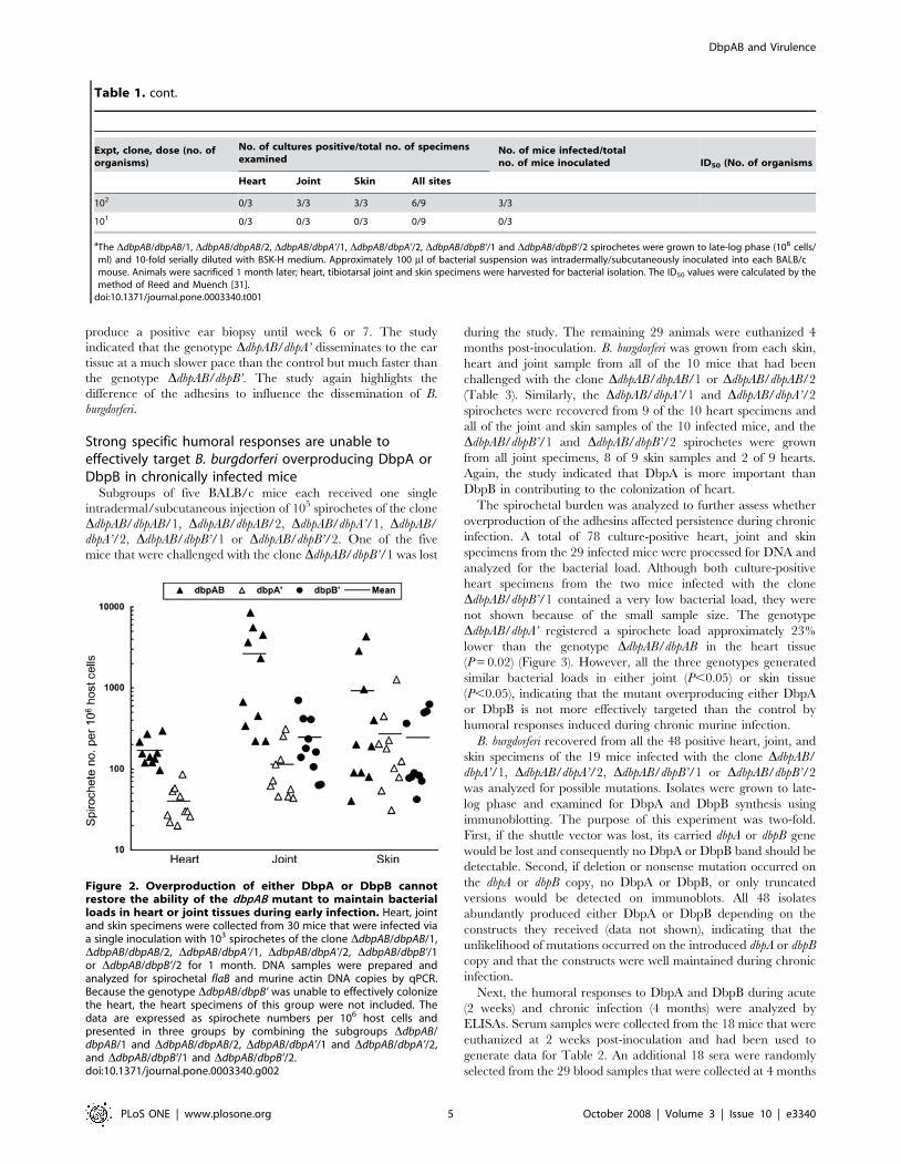
Overproduction of either DbpA or DbpB cannot restorethe ability of the dbpAB mutant to maintain the tissuebacterial load in heart or joint during early infection
The spirochete burden was analyzed to further assess how
DbpA and DbpB contribute differently to tissue colonization.
Although DbpA overproduction restored the frequency of
colonizing the heart tissue, the DdbpAB/dbpA’ spirochete load
was 4.3-fold lower than that of the genotype DdbpAB/dbpAB
(P = 1.661025) (Figure 2). The DdbpAB/dbpAB bacteria generated
loads 23-fold and 11-fold, respectively, higher than those of the
DdbpAB/dbpA’ (P = 0.01) and DdbpAB/dbpB’ spirochetes (P = 0.02)
in the joint. However, all the three genotypes produced similar
bacterial loads in skin (P,0.05). The two genotypes with adhesin
overproduction generated similar bacterial loads in the joint
(P,0.05). These results indicated that overproduction of either
DbpA or DbpB is unable to restore the ability of the dbpAB mutant
to maintain the bacterial load in either heart or joint tissue.
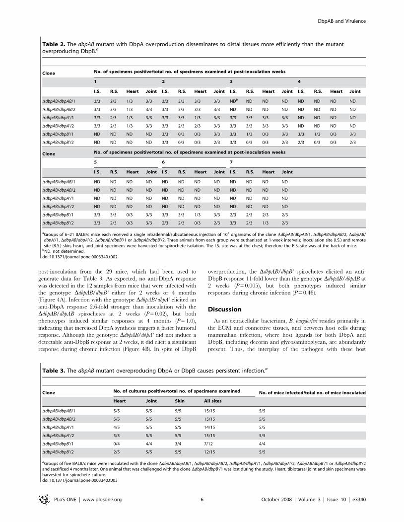
Overproduction of DbpA makes the dbpAB mutantdisseminate more efficiently to distal tissues thanincreased DbpB production
Dissemination was assessed as another aspect of the overall
virulence. Groups of six to 21 BALB/c mice each received a single
intradermal/subcutaneous inoculation of 105 spirochetes of the
clone DdbpAB/dbpAB/1, DdbpAB/dbpAB/2, DdbpAB/dbpA’/1,
DdbpAB/dbpA’/2, DdbpAB/dbpB’/1 or DdbpAB/dbpB’/2. This dose,
1000-fold higher than the ID50 value, was used to ensure that all
inoculated mice were infected. Three animals from each group
were euthanized at 1-week intervals; inoculation site and remote
site skin, heart and joint specimens were harvested for spirochete
isolation. Bacteria were injected into the dermis of the chest so skin
specimens from the back were harvested as remote sites. As a
positive control, the DdbpAB/dbpAB/1 and DdbpAB/dbpAB/2
bacteria were grown from 6 joint, 5 remote skin, and 2 heart
specimens of 6 examined mice at 1 week; all sites became culture-
positive at 2 weeks after inoculation (Table 2), consistent with our
previous study that showed the parental clone 13A carrying
pBBE22 colonized these tissues within 2 weeks [22]. The DdbpAB/
dbpA’/1 and DdbpAB/dbpA’/2 bacteria were grown from 6 joint, 4
remote skin and 2 heart specimens of 6 examined mice at 1 week,
and from 6 joint, 5 remote skin and 3 heart specimens of 6 mice at
2 weeks; all sites became culture-positive at 3 weeks. These results
indicated that DbpA overproduction allowed the mutant to
disseminate to the distal tissues that were analyzed slightly slower
than the mutant restored with dbpAB expression. In contrast, the
mutant overproducing DbpB disseminated extremely slowly; the
first positive remote skin and heart specimens were not detected
until 3 and 6 weeks, respectively. Only one out of the six remote
skin specimens produced a positive culture at 4 weeks. It should be
pointed out that the 105-dose inoculation apparently caused
delayed dissemination, as nearly all skin samples were culture-
positive when the dose of 102 to 103 organisms was used to
determine the ID50 value for the genotype DdbpAB/dbpB’ (Table 1).
The high-dose inoculation might induce a faster and stronger
immune response, which slowed the dissemination process, when
the strain had a severe defect in dissemination. This was also noted
in our previous study [17].
We previously showed that modification of B. burgdorferi to
overproduce DbpA results in severely impaired dissemination
based on an ear biopsy study [17]. Our subsequent studies showed
that ear is the last tissue to be colonized by B. burgdorferi during
murine infection [18]. To investigate how slowly the mutant
overproducing the adhesins disseminated to ear, subgroups of
three BALB/c mice each received one single inoculation of 105
spirochetes of the clone DdbpAB/dbpAB/1, DdbpAB/dbpAB/2,
DdbpAB/dbpA’/1, DdbpAB/dbpA’/2, DdbpAB/dbpB’/1 or DdbpAB/
dbpB’/2. Ear biopsies were taken for bacterial culture every week
up to 7 weeks post-inoculation. At 2 weeks post-inoculation, all of
the six mice inoculated with the genotype DdbpAB/dbpAB had a
positive biopsy (data not shown). In contrast, none of the mice
inoculated with the genotype DdbpAB/dbpA’ produced a positive
biopsy within the first 3 weeks but all of the six gave a positive
result at 4 weeks. The six mice receiving the DdbpAB/dbpB’ did not
DbpAB and Virulence
PLoS ONE | www.plosone.org 3 October 2008 | Volume 3 | Issue 10 | e3340
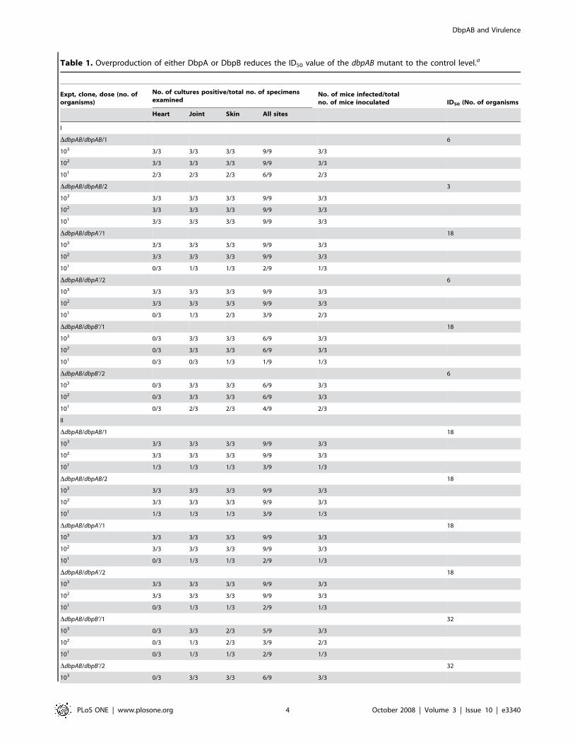
Table 1. Overproduction of either DbpA or DbpB reduces the ID50 value of the dbpAB mutant to the control level.a
Expt, clone, dose (no. oforganisms)
No. of cultures positive/total no. of specimensexamined
No. of mice infected/totalno. of mice inoculated ID50 (No. of organisms
Heart Joint Skin All sites
I
DdbpAB/dbpAB/1 6
103 3/3 3/3 3/3 9/9 3/3
102 3/3 3/3 3/3 9/9 3/3
101 2/3 2/3 2/3 6/9 2/3
DdbpAB/dbpAB/2 3
103 3/3 3/3 3/3 9/9 3/3
102 3/3 3/3 3/3 9/9 3/3
101 3/3 3/3 3/3 9/9 3/3
DdbpAB/dbpA’/1 18
103 3/3 3/3 3/3 9/9 3/3
102 3/3 3/3 3/3 9/9 3/3
101 0/3 1/3 1/3 2/9 1/3
DdbpAB/dbpA’/2 6
103 3/3 3/3 3/3 9/9 3/3
102 3/3 3/3 3/3 9/9 3/3
101 0/3 1/3 2/3 3/9 2/3
DdbpAB/dbpB’/1 18
103 0/3 3/3 3/3 6/9 3/3
102 0/3 3/3 3/3 6/9 3/3
101 0/3 0/3 1/3 1/9 1/3
DdbpAB/dbpB’/2 6
103 0/3 3/3 3/3 6/9 3/3
102 0/3 3/3 3/3 6/9 3/3
101 0/3 2/3 2/3 4/9 2/3
II
DdbpAB/dbpAB/1 18
103 3/3 3/3 3/3 9/9 3/3
102 3/3 3/3 3/3 9/9 3/3
101 1/3 1/3 1/3 3/9 1/3
DdbpAB/dbpAB/2 18
103 3/3 3/3 3/3 9/9 3/3
102 3/3 3/3 3/3 9/9 3/3
101 1/3 1/3 1/3 3/9 1/3
DdbpAB/dbpA’/1 18
103 3/3 3/3 3/3 9/9 3/3
102 3/3 3/3 3/3 9/9 3/3
101 0/3 1/3 1/3 2/9 1/3
DdbpAB/dbpA’/2 18
103 3/3 3/3 3/3 9/9 3/3
102 3/3 3/3 3/3 9/9 3/3
101 0/3 1/3 1/3 2/9 1/3
DdbpAB/dbpB’/1 32
103 0/3 3/3 2/3 5/9 3/3
102 0/3 1/3 2/3 3/9 2/3
101 0/3 1/3 1/3 2/9 1/3
DdbpAB/dbpB’/2 32
103 0/3 3/3 3/3 6/9 3/3
DbpAB and Virulence
PLoS ONE | www.plosone.org 4 October 2008 | Volume 3 | Issue 10 | e3340
produce a positive ear biopsy until week 6 or 7. The study
indicated that the genotype DdbpAB/dbpA’ disseminates to the ear
tissue at a much slower pace than the control but much faster than
the genotype DdbpAB/dbpB’. The study again highlights the
difference of the adhesins to influence the dissemination of B.
burgdorferi.
Strong specific humoral responses are unable toeffectively target B. burgdorferi overproducing DbpA orDbpB in chronically infected mice
Subgroups of five BALB/c mice each received one single
intradermal/subcutaneous injection of 105 spirochetes of the clone
DdbpAB/dbpAB/1, DdbpAB/dbpAB/2, DdbpAB/dbpA’/1, DdbpAB/
dbpA’/2, DdbpAB/dbpB’/1 or DdbpAB/dbpB’/2. One of the five
mice that were challenged with the clone DdbpAB/dbpB’/1 was lost
during the study. The remaining 29 animals were euthanized 4
months post-inoculation. B. burgdorferi was grown from each skin,
heart and joint sample from all of the 10 mice that had been
challenged with the clone DdbpAB/dbpAB/1 or DdbpAB/dbpAB/2
(Table 3). Similarly, the DdbpAB/dbpA’/1 and DdbpAB/dbpA’/2
spirochetes were recovered from 9 of the 10 heart specimens and
all of the joint and skin samples of the 10 infected mice, and the
DdbpAB/dbpB’/1 and DdbpAB/dbpB’/2 spirochetes were grown
from all joint specimens, 8 of 9 skin samples and 2 of 9 hearts.
Again, the study indicated that DbpA is more important than
DbpB in contributing to the colonization of heart.
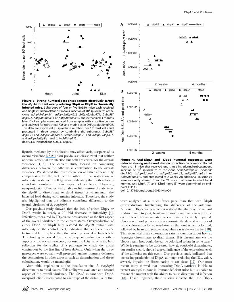
The spirochetal burden was analyzed to further assess whether
overproduction of the adhesins affected persistence during chronic
infection. A total of 78 culture-positive heart, joint and skin
specimens from the 29 infected mice were processed for DNA and
analyzed for the bacterial load. Although both culture-positive
heart specimens from the two mice infected with the clone
DdbpAB/dbpB’/1 contained a very low bacterial load, they were
not shown because of the small sample size. The genotype
DdbpAB/dbpA’ registered a spirochete load approximately 23%
lower than the genotype DdbpAB/dbpAB in the heart tissue
(P = 0.02) (Figure 3). However, all the three genotypes generated
similar bacterial loads in either joint (P,0.05) or skin tissue
(P,0.05), indicating that the mutant overproducing either DbpA
or DbpB is not more effectively targeted than the control by
humoral responses induced during chronic murine infection.
B. burgdorferi recovered from all the 48 positive heart, joint, and
skin specimens of the 19 mice infected with the clone DdbpAB/
dbpA’/1, DdbpAB/dbpA’/2, DdbpAB/dbpB’/1 or DdbpAB/dbpB’/2
was analyzed for possible mutations. Isolates were grown to late-
log phase and examined for DbpA and DbpB synthesis using
immunoblotting. The purpose of this experiment was two-fold.
First, if the shuttle vector was lost, its carried dbpA or dbpB gene
would be lost and consequently no DbpA or DbpB band should be
detectable. Second, if deletion or nonsense mutation occurred on
the dbpA or dbpB copy, no DbpA or DbpB, or only truncated
versions would be detected on immunoblots. All 48 isolates
abundantly produced either DbpA or DbpB depending on the
constructs they received (data not shown), indicating that the
unlikelihood of mutations occurred on the introduced dbpA or dbpB
copy and that the constructs were well maintained during chronic
infection.
Next, the humoral responses to DbpA and DbpB during acute
(2 weeks) and chronic infection (4 months) were analyzed by
ELISAs. Serum samples were collected from the 18 mice that were
euthanized at 2 weeks post-inoculation and had been used to
generate data for Table 2. An additional 18 sera were randomly
selected from the 29 blood samples that were collected at 4 months
Figure 2. Overproduction of either DbpA or DbpB cannotrestore the ability of the dbpAB mutant to maintain bacterialloads in heart or joint tissues during early infection. Heart, jointand skin specimens were collected from 30 mice that were infected viaa single inoculation with 103 spirochetes of the clone DdbpAB/dbpAB/1,DdbpAB/dbpAB/2, DdbpAB/dbpA’/1, DdbpAB/dbpA’/2, DdbpAB/dbpB’/1or DdbpAB/dbpB’/2 for 1 month. DNA samples were prepared andanalyzed for spirochetal flaB and murine actin DNA copies by qPCR.Because the genotype DdbpAB/dbpB’ was unable to effectively colonizethe heart, the heart specimens of this group were not included. Thedata are expressed as spirochete numbers per 106 host cells andpresented in three groups by combining the subgroups DdbpAB/dbpAB/1 and DdbpAB/dbpAB/2, DdbpAB/dbpA’/1 and DdbpAB/dbpA’/2,and DdbpAB/dbpB’/1 and DdbpAB/dbpB’/2.doi:10.1371/journal.pone.0003340.g002
Expt, clone, dose (no. oforganisms)
No. of cultures positive/total no. of specimensexamined
No. of mice infected/totalno. of mice inoculated ID50 (No. of organisms
Heart Joint Skin All sites
102 0/3 3/3 3/3 6/9 3/3
101 0/3 0/3 0/3 0/9 0/3
aThe DdbpAB/dbpAB/1, DdbpAB/dbpAB/2, DdbpAB/dbpA’/1, DdbpAB/dbpA’/2, DdbpAB/dbpB’/1 and DdbpAB/dbpB’/2 spirochetes were grown to late-log phase (108 cells/ml) and 10-fold serially diluted with BSK-H medium. Approximately 100 ml of bacterial suspension was intradermally/subcutaneously inoculated into each BALB/cmouse. Animals were sacrificed 1 month later; heart, tibiotarsal joint and skin specimens were harvested for bacterial isolation. The ID50 values were calculated by themethod of Reed and Muench [31].
doi:10.1371/journal.pone.0003340.t001
Table 1. cont.
DbpAB and Virulence
PLoS ONE | www.plosone.org 5 October 2008 | Volume 3 | Issue 10 | e3340
post-inoculation from the 29 mice, which had been used to
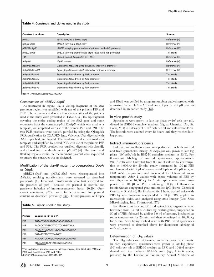
generate data for Table 3. As expected, no anti-DbpA response
was detected in the 12 samples from mice that were infected with
the genotype DdbpAB/dbpB’ either for 2 weeks or 4 months
(Figure 4A). Infection with the genotype DdbpAB/dbpA’ elicited an
anti-DbpA response 2.6-fold stronger than inoculation with the
DdbpAB/dbpAB spirochetes at 2 weeks (P = 0.02), but both
phenotypes induced similar responses at 4 months (P = 1.0),
indicating that increased DbpA synthesis triggers a faster humoral
response. Although the genotype DdbpAB/dbpA’ did not induce a
detectable anti-DbpB response at 2 weeks, it did elicit a significant
response during chronic infection (Figure 4B). In spite of DbpB
overproduction, the DdbpAB/dbpB’ spirochetes elicited an anti-
DbpB response 11-fold lower than the genotype DdbpAB/dbpAB at
2 weeks (P = 0.005), but both phenotypes induced similar
responses during chronic infection (P = 0.48).
Discussion
As an extracellular bacterium, B. burgdorferi resides primarily in
the ECM and connective tissues, and between host cells during
mammalian infection, where host ligands for both DbpA and
DbpB, including decorin and glycosaminoglycan, are abundantly
present. Thus, the interplay of the pathogen with these host
Table 3. The dbpAB mutant overeproducing DbpA or DbpB causes persistent infection.a
Clone No. of cultures positive/total no. of specimens examined No. of mice infected/total no. of mice inoculated
Heart Joint Skin All sites
DdbpAB/dbpAB/1 5/5 5/5 5/5 15/15 5/5
DdbpAB/dbpAB/2 5/5 5/5 5/5 15/15 5/5
DdbpAB/dbpA’/1 4/5 5/5 5/5 14/15 5/5
DdbpAB/dbpA’/2 5/5 5/5 5/5 15/15 5/5
DdbpAB/dbpB’/1 0/4 4/4 3/4 7/12 4/4
DdbpAB/dbpB’/2 2/5 5/5 5/5 12/15 5/5
aGroups of five BALB/c mice were inoculated with the clone DdbpAB/dbpAB/1, DdbpAB/dbpAB/2, DdbpAB/dbpA’/1, DdbpAB/dbpA’/2, DdbpAB/dbpB’/1 or DdbpAB/dbpB’/2and sacrificed 4 months later. One animal that was challenged with the clone DdbpAB/dbpB’/1 was lost during the study. Heart, tibiotarsal joint and skin specimens wereharvested for spirochete culture.doi:10.1371/journal.pone.0003340.t003
Table 2. The dbpAB mutant with DbpA overproduction disseminates to distal tissues more efficiently than the mutantoverproducing DbpB.a
Clone No. of specimens positive/total no. of specimens examined at post-inoculation weeks
1 2 3 4
I.S. R.S. Heart Joint I.S. R.S. Heart Joint I.S. R.S. Heart Joint I.S. R.S. Heart Joint
DdbpAB/dbpAB/1 3/3 2/3 1/3 3/3 3/3 3/3 3/3 3/3 NDb ND ND ND ND ND ND ND
DdbpAB/dbpAB/2 3/3 3/3 1/3 3/3 3/3 3/3 3/3 3/3 ND ND ND ND ND ND ND ND
DdbpAB/dbpA’/1 3/3 2/3 1/3 3/3 3/3 3/3 1/3 3/3 3/3 3/3 3/3 3/3 ND ND ND ND
DdbpAB/dbpA’/2 3/3 2/3 1/3 3/3 3/3 2/3 2/3 3/3 3/3 3/3 3/3 3/3 ND ND ND ND
DdbpAB/dbpB’/1 ND ND ND ND 3/3 0/3 0/3 3/3 3/3 1/3 0/3 3/3 3/3 1/3 0/3 3/3
DdbpAB/dbpB’/2 ND ND ND ND 3/3 0/3 0/3 2/3 3/3 0/3 0/3 2/3 2/3 0/3 0/3 2/3
Clone No. of specimens positive/total no. of specimens examined at post-inoculation weeks
5 6 7
I.S. R.S. Heart Joint I.S. R.S. Heart Joint I.S. R.S. Heart Joint
DdbpAB/dbpAB/1 ND ND ND ND ND ND ND ND ND ND ND ND
DdbpAB/dbpAB/2 ND ND ND ND ND ND ND ND ND ND ND ND
DdbpAB/dbpA’/1 ND ND ND ND ND ND ND ND ND ND ND ND
DdbpAB/dbpA’/2 ND ND ND ND ND ND ND ND ND ND ND ND
DdbpAB/dbpB’/1 3/3 3/3 0/3 3/3 3/3 3/3 1/3 3/3 2/3 2/3 2/3 2/3
DdbpAB/dbpB’/2 3/3 2/3 0/3 3/3 2/3 2/3 0/3 2/3 3/3 2/3 1/3 2/3
aGroups of 6–21 BALB/c mice each received a single intradermal/subcutaneous injection of 105 organisms of the clone DdbpAB/dbpAB/1, DdbpAB/dbpAB/2, DdbpAB/dbpA’/1, DdbpAB/dbpA’/2, DdbpAB/dbpB’/1 or DdbpAB/dbpB’/2. Three animals from each group were euthanized at 1-week intervals; inoculation site (I.S.) and remotesite (R.S.) skin, heart, and joint specimens were harvested for spirochete isolation. The I.S. site was at the chest; therefore the R.S. site was at the back of mice.
bND, not determined.doi:10.1371/journal.pone.0003340.t002
DbpAB and Virulence
PLoS ONE | www.plosone.org 6 October 2008 | Volume 3 | Issue 10 | e3340
ligands, mediated by the adhesins, may affect various aspects of its
overall virulence [23,24]. Our previous studies showed that neither
adhesin is essential for infection but both are critical for the overall
virulence [4,15]. The current study focused on comparing
differences between the adhesins in contribution to the overall
virulence. We showed that overproduction of either adhesin fully
compensates for the lack of the other in the restoration of
infectivity, as defined by ID50 value, indicating that both adhesins
contribute similarly to this aspect of virulence. However,
overproduction of either was unable to fully restore the ability of
the dbpAB to disseminate to distal tissues or to maintain the
bacterial load during early murine infection. Therefore, the study
also highlighted that the adhesins contribute differently to the
overall virulence of B. burgdorferi.
Our previous study showed that the lack of either DbpA or
DbpB results in nearly a 103-fold decrease in infectivity [4].
Infectivity, measured by ID50 value, was assessed as the first aspect
of the overall virulence in the current study. Overproduction of
either DbpA or DbpB fully restored the dbpAB mutant with
infectivity to the control level, indicating that either virulence
factor is able to replace the other when produced at high levels.
This finding is crucial for the subsequent evaluation of other
aspects of the overall virulence, because the ID50 value is the best
reflection for the ability of a pathogen to evade the initial
elimination by the first line of host defense. Only when the three
genotypes were equally well protected against immune defenses,
the comparison in other aspects, such as dissemination and tissue
colonization, would be meaningful.
After initial replication at the inoculation site, B. burgdorferi
disseminates to distal tissues. This ability was evaluated as a second
aspect of the overall virulence. The dbpAB mutant with DbpA
overproduction disseminated to each type of the distal tissues that
were analyzed at a much faster pace than that with DbpB
overproduction, highlighting the difference of the adhesins.
Although DbpA overproduction restored the ability of the mutant
to disseminate to joint, heart and remote skin tissues nearly to the
control level, its dissemination to ear remained severely impaired.
Our current and previous studies consistently showed an order of
tissue colonization by B. burgdorferi, as the joint is first colonized,
followed by heart and remote skin, while ear is always the last [18].
This sequential tissue colonization raises a question about how B.
burgdorferi disseminates to distal tissues. If it disseminates via the
bloodstream, how could the ear be colonized so late in some cases?
While it remains to be addressed how B. burgdorferi disseminates,
our studies clearly showed a great influence of the expression levels
of the adhesins on this event. Our previous study indicated that
increasing production of DbpA, although reducing the ID50 value,
severely impairs the dissemination to ear tissue [17]. Our most
recent study showed that increasing DbpA synthesis is able to
protect an ospC mutant in immunodeficient mice but is unable to
restore the mutant with the ability to cause disseminated infection
[18]. Taken together, these studies indicate that the DbpA
Figure 3. Strong humoral responses cannot effectively targetthe dbpAB mutant overproducing DbpA or DbpB in chronicallyinfected mice. Subgroups of four or five BALB/c mice each receivedone single intradermal/subcutaneous injection of 105 spirochetes of theclone DdbpAB/dbpAB/1, DdbpAB/dbpAB/2, DdbpAB/dbpA’/1, DdbpAB/dbpA’/2, DdbpAB/dbpB’/1 or DdbpAB/dbpB’/2, and euthanized 4 monthslater. DNA samples were prepared from samples with a positive cultureand analyzed for spirochetal flaB and murine actin DNA copies by qPCR.The data are expressed as spirochete numbers per 106 host cells andpresented in three groups by combining the subgroups DdbpAB/dbpAB/1 and DdbpAB/dbpAB/2, DdbpAB/dbpA’/1 and DdbpAB/dbpA’/2,and DdbpAB/dbpB’/1 and DdbpAB/dbpB’/2.doi:10.1371/journal.pone.0003340.g003
Figure 4. Anti-DbpA and -DbpB humoral responses wereinduced during acute and chronic infection. Sera were collectedfrom the 18 mice that received one single intradermal/subcutaneousinjection of 105 spirochetes of the clone DdbpAB/dbpAB/1, DdbpAB/dbpAB/2, DdbpAB/dbpA’/1, DdbpAB/dbpA’/2, DdbpAB/dbpB’/1 orDdbpAB/dbpB’/2, and euthanized at 2 weeks. An additional 18 sampleswere randomly chosen from the 29 mice that were infected for 4months. Anti-DbpA (A) and -DbpB titers (B) were determined by end-point ELISAs.doi:10.1371/journal.pone.0003340.g004
DbpAB and Virulence
PLoS ONE | www.plosone.org 7 October 2008 | Volume 3 | Issue 10 | e3340

production level significantly influences the dissemination of B.
burgdorferi and that this influence is also largely affected by the
presence of other surface lipoproteins.
The slow dissemination of the genotype DdbpAB/dbpB’ is also
indirectly reflected by a weak anti-DbpB response induced during
early infection. The severely impaired dissemination may limit a
quick expansion of the total bacterial load and thus affects the
immune response. This slow expansion could be a reason why
infection with the genotype DdbpAB/dbpB’ did not elicit a humoral
response cross-reactive with DbpA, although the DdbpAB/dbpA’
spirochetes stimulated a significant response cross-reactive with
DbpB during chronic infection. However, our study could not rule
out that DbpB may be less immunogenic than DbpA during
borrelial infection. Nevertheless, the antigenic cross-reactivity
between the adhesins had never been reported.
The ability to colonize tissues was assessed as a third aspect of
the overall virulence. DbpA overproduction restored this ability of
the dbpAB mutant to the control level, if measured by frequency of
tissue colonization. When the bacterial load was analyzed,
however, our data indicated that the overproduction far less
restored the ability to maintain the bacterial loads either in heart
or joint tissue during acute infection. In contrast, the mutant with
DbpB overproduction was not recovered from any of the heart
specimens within the first 5 weeks post-inoculation, but it was
indeed grown from some heart specimens between 6 weeks and 4
months of infection. Slower dissemination could be a contributing
factor for the negative heart culture, when bacterial isolation was
conducted within 1 month post-inoculation. However, the study
clearly showed that overproduction of DbpA, more effectively
than that of DbpB, restores the colonization of heart tissue. In joint
and skin tissues, overproduction of either adhesin had similar
effects on this aspect of virulence.
Persistence was examined as a fourth aspect of the overall
virulence. Overproduction of either adhesin did not reduce the
ability to cause chronic infection, as measured by bacterial load.
During early infection (1 month), the DdbpAB/dbpA’ load was 4.3-
fold lower than that of the genotype DdbpAB/dbpAB in heart; the
difference reduced to 1.7-fold after 4 months of infection. A more
dramatic change was noted in joint tissue, where the DdbpAB/
dbpAB load was 23-fold and 11-fold, respectively, higher than those
of the genotypes DdbpAB/dbpA’ and DdbpAB/dbpB’ at 1 month, but
the differences became indistinguishable among the three
genotypes after 4 months of infection. In spite of robust anti-
DbpA and -DbpB humoral responses, the mutant overproducing
either adhesin persisted as well as the control. This is unexpected
because our previous study showed that overproduction of DbpA
diminishes the ability of B. burgdorferi to persist in heart and joint
tissues of chronically infected immunocompetent mice [17],
although specific DbpA antibody is unable to reduce the bacterial
load of wild-type B. burgdorferi [25]. Two obvious differences can be
used to interpret these observations. First, in the previous study the
parental strain produces both DbpA and DbpB from the native
dbpBA locus, so further introduced DbpA overproduction may
dramatically increase accumulation of the adhesins on the
spirochete’s surface and, as a consequence, may improve the
effectiveness of specific antibodies to target B. burgdorferi in heart
and joint tissues. Second, because the genotypes DdbpAB/dbpA’
and DdbpAB/dbpB’ lack either DbpA or DbpB, the deficiency for
either may make specific antibodies to the other less effective in
killing the pathogen or in other words, the anti-DbpA and -DbpB
responses may have a synergetic effect in controlling B. burgdorferi.
A recent study by Blevins et al. reported that DbpA but not
DbpB is critical for infectivity measured by the ID50 value [16],
while our previous study showed that both adhesins are crucial to
this aspect of overall virulence [4]. The intrinsic difference of the
bacterial strains (297 used in their study vs. B31 13A in ours) could
cause the disparity. For instance, if the native promoter of the
dbpBA operon of the strain 297 drives more active expression than
that of B31, the dbpA gene alone controlled by the dbpBA promoter
may sufficiently restore the dbpAB mutant with full infectivity, as
the current study showed that both DbpA and DbpB are
interchangeable in contribution to infectivity as measured by
ID50 value when produced at a high level. They also reported that
their dbpAB mutant becomes less impaired following tick challenge.
In response to a fresh bloodmeal B. burgdorferi undergoes a
dramatic change, in terms of gene expression, a process that
prepares the pathogen for mammalian infection. This adaptation
process should certainly be considered as a contributing factor to
increase infectivity of their dbpAB mutant. The fact that ticks
acquired the dbpAB mutant organisms from infected animals must
also be considered. The genetic manipulation of B. burgdorferi is
time-consuming, and it often takes a long period of time to
generate, characterize and select a mutant. These processes may
attenuate the infectivity of generated mutants, as it has been
known for more than two decades that prolonged in vitro
cultivation reduces the infectivity of B. burgdorferi [26]. We noticed
that several of our genetically modified strains, including the dbpAB
mutant, showed improvement in infectivity when isolates that were
recovered from infected mice were used as an inoculum (Xu and
Liang, unpublished data).
Although both DbpA and DbpB are believed to be well
characterized outer surface lipoproteins [7–9], our immunofluo-
rescence showed limited surface exposure of the lipoproteins on
spirochetes grown in vitro. This observation is consistent with a
study by Radolf and colleagues, which showed limited surface
exposure of OspA, OspB and OspC on cultured B. burgdorferi by
immunofluorescence [27]. However, our finding that limited
exposure of DbpA and DbpB on the surface of cultured
spirochetes does not suggest that the adhesins can not have a
full access to their ligand decorin in mammalian tissues because
OspA and OspB, which are abundantly produced in spirochetes
grown in vitro but are downregulated to a baseline level during
mammalian infection [28], may reduce the accessibility of DbpA
and DbpB. Our modification of B. burgdorferi to overproduce DbpA
and DbpB led to a dramatically increased binding of the specific
antibodies to intact spirochetes as showed by immunofluorescence,
indicating that both adhesins are surface exposed.
Both DbpA and DbpB are surface lipoprotein adhesins with
similar molecular weights and encoded by the same operon, share
approximately 40% identity, and bind host decorin and
glycosaminoglycans [7–9,12]. The current study revealed that
the adhesins also share cross-antigenic reactivity. These similarities
can be used to explain why the two virulence factors can be
interchangeable in the protection of B. burgdorferi against initial
elimination by immune defenses when produced at an increased
level; however, they indeed contribute differently to dissemination
and tissue colonization.
Materials and Methods
Strains and constructs generated previously but used inthe current study
The B. burgdorferi B31 clone 13A, the dbpAB mutant (DdbpAB)
and the complemented clones DdbpAB/dbpAB/1 and DdbpAB/
dbpAB/2, the shuttle vector pME22, and the recombinant plasmids
pME22-dbpB and pBBE22-dbpA’ were generated in our previous
studies [4,17]. The features of these clones and constructs were
summarized in Table 4.
DbpAB and Virulence
PLoS ONE | www.plosone.org 8 October 2008 | Volume 3 | Issue 10 | e3340
Construction of pBBE22-dbpB’As illustrated in Figure 1A, a 258-bp fragment of the flaB
promoter region was amplified with use of the primers P1F and
P1R. The sequences and restriction enzyme sites of the primers
used in the study were presented in Table 5. A 1152-bp fragment
covering the entire coding region of the dbpB gene and some
sequences from the construct pME22-dbpB, which was used as a
template, was amplified with use of the primers P2F and P2R. The
two PCR products were pooled, purified by using the QIAquick
PCR purification kit (QIAGEN Inc., Valencia, CA), digested with
NdeI, repurified, and ligated. The resultant product was used as a
template and amplified by nested PCR with use of the primers P3F
and P3R. The PCR product was purified, digested with BamHI,
and cloned into the shuttle vector pME22 [4]. The insert and
flanking regions within the recombinant plasmid were sequenced
to ensure the construct was as designed.
Modification of the dbpAB mutant to overproduce DbpAor DbpB
pBBE22-dbpA’ and pME22-dbpB’ were electroporated into
DdbpAB; resulting transformants were screened as described
previously [4]. Identified transformants were first surveyed for
the presence of lp28-1 because this plasmid is essential for
persistent infection of immunocompetent hosts [20,29]. Only
clones containing lp28-1 were further analyzed for plasmid
content as described previously [30]. Overexpression of DbpA
and DbpB was verified by using immunoblot analysis probed with
a mixture of a FlaB mAb and anti-DbpA or -DbpB sera as
described in an earlier study [15].
In vitro growth studySpirochetes were grown to late-log phase (,108 cells per ml),
diluted in BSK-H complete medium (Sigma Chemical Co., St.
Louis, MO) to a density of ,106 cells per ml and cultured at 33uC.
The bacteria were counted every 12 hours until they reached late-
log phase.
Indirect immunofluorescenceIndirect immunofluorescence was performed on both unfixed
and fixed spirochetes. Briefly, B. burgdorferi was grown to late-log
phase (108 cells/ml) in BSK-H complete medium at 33uC. For
fluorescent labeling of unfixed spirochetes, approximately
26107 cells were harvested from 0.2 ml of culture by centrifuga-
tion at 4,0006g for 20 min, gently suspended in 100 ml PBS
supplemented with 2 ml of mouse anti-DbpA or -DbpB sera, or
FlaB mAb preparation, and incubated for 1 hour at room
temperature. After 2 washes with excess volumes of PBS by
centrifugation at 16,0006g for 5 min, spirochetes were resus-
pended in 100 ml of PBS containing 1.0 mg of fluorescein
isothiocyanate-conjugated goat anti-mouse IgG (Pierce Chemical
Company, Rockford, IL), incubated for 1 hour, washed twice with
PBS by centrifugation, resuspended in 50 ml PBS, applied to
microscopic slides, and analyzed using Axio Imager (Carl Zeiss
Microimaging, Inc., Thornwood, NY).
For fluorescent labeling of fixed spirochetes, organisms were
harvested from 0.2 ml of culture by centrifugation, suspended in
50 ml of PBS, followed by adding 1.0 ml of acetone, incubated at
room temperature for 20 min, and then centrifuged at 16,0006g
for 5 min. After being washed once with PBS, fixed spirochetes
were processed as described above for fluorescent labeling of
unfixed bacteria.
Determination of ID50 valuesThe ID50 values were determined in two separate experiments.
In each experiment, spirochetes were grown to late-log phase
(108 cells per ml) in BSK-H medium at 33uC and 10-fold serially
diluted with the medium. BALB/c mice (age, 4 to 6 weeks;
provided by the Division of Laboratory Animal Medicine at
Table 4. Constructs and clones used in the study.
Construct or clone Description Source
pME22 pBSV2 carrying a bbe22 copy Reference [4]
pME22-dbpB pME22 carrying a dbpB copy Reference [4]
pBBE22-dbpA’ pBBE22 carrying promoterless dbpA fused with flaB promoter Reference [17]
pME22-dbpB’ pME22 carrying promoterless dbpB fused with flaB promoter This study
13A Cloned from B. burgdorferi B31 A13 Reference [33]
DdbpAB dbpAB mutant Reference [4]
DdbpAB/dbpAB/1 Expressing dbpA and dbpB driven by their own promoter Reference [4]
DdbpAB/dbpAB/2 Expressing dbpA and dbpB driven by their own promoter Reference [4]
DdbpAB/dbpA’/1 Expressing dbpA driven by flaB promoter This study
DdbpAB/dbpA’/2 Expressing dbpA driven by flaB promoter This study
DdbpAB/dbpB’/1 Expressing dbpB driven by flaB promoter This study
DdbpAB/dbpB’/2 Expressing dbpB driven by flaB promoter This study
doi:10.1371/journal.pone.0003340.t004
Table 5. Primers used in the study.
Primer Sequence (59 to 39)a
P1F AGAAGTACGAAGATAGAGAGAGAAA
P1R AACACATATGTCATTCCTCCATGATAAA
P2F AACATATGAAAATTGGAAAGCTAAATTCA
P2R GGAAATCTTCCTTGAAGCT
P3F ATAGGATCCAAGATAGAGAGAGAAAAGT
P3R TTGGATCCTGATTATCGGGCGAAGAG
aThe underlined sequences are restriction enzyme sites: NdeI sites (P1R andP2F), and BamHI sites (P3F and P3R).doi:10.1371/journal.pone.0003340.t005
DbpAB and Virulence
PLoS ONE | www.plosone.org 9 October 2008 | Volume 3 | Issue 10 | e3340

Louisiana State University, Baton Rouge, LA) each received one
single intradermal/subcutaneous injection of 100 ml of spirochetal
suspension. Mice were euthanized 1 month post-inoculation;
heart, tibiotarsal joint and skin (not from inoculation site)
specimens were harvested for bacterial culture and DNA
preparation as described previously [30]. The ID50 value was
calculated as described by Reed and Muench [31]. DNA was
extracted from selective specimens and used for analysis of tissue
bacterial loads as described below. All animal procedures
described here and below were approved by the Institutional
Animal Care and Use Committee at Louisiana State University.
Quantification of tissue spirochetal loadDNA was extracted for heart, joint and skin specimens and
quantified for the copy numbers of flaB and murine actin genes by
quantitative PCR (qPCR) as previously described [30]. The tissue
spirochete burden was expressed as flaB DNA copies per 106 host
cells (26106 actin DNA copies).
Dissemination and chronic infectivity studiesBALB/c mice each received one single intradermal/subcuta-
neous injection of 105 spirochetes. In a dissemination study,
inoculated mice were euthanized at 1-week intervals for up to 7
weeks, starting at 1 week; inoculation site and remote skin, heart
and joint specimens were aseptically harvested for spirochete
isolation as previously described [30]. Because spirochetes were
injected into the chest skin, the back skin was harvested as a
remote site. In a second dissemination study, inoculated mice were
subjected to ear biopsy for up to 7 weeks as described previously
[17]. In a chronic study, inoculated animals were euthanized 4
months post-inoculation; heart, tibiotarsal joint and skin specimens
were aseptically collected for spirochete culture and DNA
extraction. DNA was prepared for analysis of tissue bacterial
loads as described above. Serum samples were collected and stored
in case an immune response analysis was needed.
Mutation analysisSpirochetes were isolated from chronically infected mice, grown
to late-log phase in BSK-H complete medium, harvested by
centrifugation and analyzed for DbpA and DbpB synthesis by
immunoblot analysis probed with a mixture of a FlaB mAb and
anti-DbpA or -DbpB sera as described above.
End-point ELISA titersSpecific DbpA and DbpB antibody end-point titers were
determined by ELISAs. Ninety-six-well plates (Fisher Scientific,
Pittsburgh, PA) were coated with 100 ml of 2.0 mg/ml recombi-
nant DbpA or DbpB per well. The recombinant proteins were
prepared as described in our earlier study [15]. Sera were two-fold
serially diluted, starting at 1:1000. Five samples drawn from naive
BALB/c mice were used as a control. The ELISA was performed
as previously described [32].
Statistical analysisA one-way analysis of variance (ANOVA) was used to analyze
data, followed by a two-tailed Student t test to calculate a P value
for each two groups. A P value#0.05 was considered to be
significant.
Supporting Information
Figure S1
Found at: doi:10.1371/journal.pone.0003340.s001 (13.57 MB
PDF)
Author Contributions
Conceived and designed the experiments: YS QX FTL. Performed the
experiments: YS QX SVS KM FTL. Analyzed the data: YS QX FTL.
Wrote the paper: FTL.
References
1. Burgdorfer W, Hayes SF, Benach JL (1988) Development of Borrelia burgdorferi in
ixodid tick vectors. Ann N Y Acad Sci 539: 172–179.
2. Steere AC (2001) Lyme disease. N Engl J Med 345: 115–125.
3. Barthold SW (1991) Infectivity of Borrelia burgdorferi relative to route of
inoculation and genotype in laboratory mice. J Infect Dis 163: 419–420.
4. Shi Y, Xu Q, McShan K, Liang FT (2008) Both decorin-binding proteins A and
B are critical for the overall virulence of Borrelia burgdorferi. Infect Immun 76:
1239–1246.
5. Seiler KP, Weis JJ (1996) Immunity to Lyme disease: protection, pathology and
persistence. Curr Opin Immunol 8: 503–509.
6. Guo BP, Norris SJ, Rosenberg LC, Hook M (1995) Adherence of Borrelia
burgdorferi to the proteoglycan decorin. Infect Immun 63: 3467–3472.
7. Guo BP, Brown EL, Dorward DW, Rosenberg LC, Hook M (1998) Decorin-
binding adhesins from Borrelia burgdorferi. Mol Microbiol 30: 711–723.
8. Hagman KE, Lahdenne P, Popova TG, Porcella SF, Akins DR, et al. (1998)
Decorin-binding protein of Borrelia burgdorferi is encoded within a two-gene
operon and is protective in the murine model of Lyme borreliosis. Infect Immun
66: 2674–2683.
9. Feng S, Hodzic E, Stevenson B, Barthold SW (1998) Humoral immunity to
Borrelia burgdorferi N40 decorin binding proteins during infection of laboratory
mice. Infect Immun 66: 2827–2835.
10. Hanson MS, Cassatt DR, Guo BP, Patel NK, McCarthy MP, et al. (1998) Active
and passive immunity against Borrelia burgdorferi decorin binding protein A
(DbpA) protects against infection. Infect Immun 66: 2143–2153.
11. Hagman KE, Yang X, Wikel SK, Schoeler GB, Caimano MJ, et al. (2000)
Decorin-binding protein A (DbpA) of Borrelia burgdorferi is not protective when
immunized mice are challenged via tick infestation and correlates with the lack
of DbpA expression by B. burgdorferi in ticks. Infect Immun 68: 4759–4764.
12. Fischer JR, Parveen N, Magoun L, Leong JM (2003) Decorin-binding proteins A
and B confer distinct mammalian cell type-specific attachment by Borrelia
burgdorferi, the Lyme disease spirochete. Proc Natl Acad Sci U S A 100:
7307–7312.
13. Liang FT, Brown EL, Wang T, Iozzo RV, Fikrig E (2004) Protective niche for
Borrelia burgdorferi to evade humoral immunity. Am J Pathol 165: 977–985.
14. Brown EL, Wooten RM, Johnson BJ, Iozzo RV, Smith A, et al. (2001)
Resistance to Lyme disease in decorin-deficient mice. J Clin Invest 107:
845–852.
15. Shi Y, Xu Q, Seemanapalli SV, McShan K, Liang FT (2006) The dbpBA locus of
Borrelia burgdorferi is not essential for infection of mice. Infect Immun 74:
6509–6512.
16. Blevins JS, Hagman KE, Norgard MV (2008) Assessment of decorin-binding
protein A to the infectivity of Borrelia burgdorferi in the murine models of needle
and tick infection. BMC Microbiol 8: 82.
17. Xu Q, Seemanaplli SV, McShan K, Liang FT (2007) Increasing the interaction
of Borrelia burgdorferi with decorin significantly reduces the 50 percent infectious
dose and severely impairs dissemination. Infect Immun 75: 4272–4281.
18. Xu Q, McShan K, Liang FT (2008) Essential protective role attributed to the
surface lipoproteins of Borrelia burgdorferi against innate defences. Mol Microbiol
69: 15–29.
19. Liang FT, Yan J, Mbow ML, Sviat SL, Gilmore RD, et al. (2004) Borrelia
burgdorferi changes its surface antigenic expression in response to host immune
responses. Infect Immun 72: 5759–5767.
20. Purser JE, Lawrenz MB, Caimano MJ, Howell JK, Radolf JD, et al. (2003) A
plasmid-encoded nicotinamidase (PncA) is essential for infectivity of Borrelia
burgdorferi in a mammalian host. Mol Microbiol 48: 753–764.
21. Schulze RJ, Zuckert WR (2006) Borrelia burgdorferi lipoproteins are secreted to the
outer surface by default. Mol Microbiol 59: 1473–1484.
22. Xu Q, McShan K, Liang FT (2008) Modification of Borrelia burgdorferi to
overproduce OspA or VlsE alters its infectious behavior. Microbiology in press.
23. Coburn J, Fischer JR, Leong JM (2005) Solving a sticky problem: new genetic
approaches to host cell adhesion by the Lyme disease spirochete. Mol Microbiol
57: 1182–1195.
24. Cabello FC, Godfrey HP, Newman SA (2007) Hidden in plain sight: Borrelia
burgdorferi and the extracellular matrix. Trends Microbiol 15: 350–354.
25. Barthold SW, Hodzic E, Tunev S, Feng S (2006) Antibody-mediated disease
remission in the mouse model of Lyme borreliosis. Infect Immun 74: 4817–4825.
26. Johnson RC, Marek N, Kodner C (1984) Infection of Syrian hamsters with
Lyme disease spirochetes. J Clin Microbiol 20: 1099–1101.
DbpAB and Virulence
PLoS ONE | www.plosone.org 10 October 2008 | Volume 3 | Issue 10 | e3340

27. Cox DL, Akins DR, Bourell KW, Lahdenne P, Norgard MV, et al. (1996)
Limited surface exposure of Borrelia burgdorferi outer surface lipoproteins. Proc
Natl Acad Sci U S A 93: 7973–7978.
28. Liang FT, Caimano MJ, Radolf JD, Fikrig E (2004) Borrelia burgdorferi outer
surface protein (osp) B expression independent of ospA. Microb Pathog 37: 35–40.
29. Labandeira-Rey M, Seshu J, Skare JT (2003) The absence of linear plasmid 25
or 28-1 of Borrelia burgdorferi dramatically alters the kinetics of experimental
infection via distinct mechanisms. Infect Immun 71: 4608–4613.
30. Xu Q, Seemanapalli SV, Lomax L, McShan K, Li X, et al. (2005) Association of
linear plasmid 28-1 with an arthritic phenotype of Borrelia burgdorferi. InfectImmun 73: 7208–7215.
31. Reed LJ, Muench H (1938) A simple method of estimating fifty percent
endpoint. Am J Hygiene 27: 493–497.32. Xu Q, Seemanapalli SV, McShan K, Liang FT (2006) Constitutive expression of
outer surface protein C diminishes the ability of Borrelia burgdorferi to evadespecific humoral immunity. Infect Immun 74: 5177–5184.
33. Xu Q, McShan K, Liang FT (2007) Identification of an ospC operator critical for
immune evasion of Borrelia burgdorferi. Mol Microbiol 64: 220–231.
DbpAB and Virulence
PLoS ONE | www.plosone.org 11 October 2008 | Volume 3 | Issue 10 | e3340