Effects of ectopic decorin in modulating intracranial glioma progression in vivo, in a rat syngeneic...
20
Effects of ectopic decorin in modulating intracranial glioma progression in vivo, in a rat syngeneic model Alireza Biglari 1,2 , Dominique Bataille 1,2 , Ulrike Naumann 3 , Michael Weller 3 , Jeffrey Zirger 1 , Maria G Castro 1,2,4 , and Pedro R Lowenstein 1,2,4 1 Gene Therapeutics Research Institute, Cedars-Sinai Medical Center, Research Pavilion, Suite 5090, 8700 Beverly Boulevard, Los Angeles, California 90048, USA 2 Molecular Medicine and Gene Therapy Unit, School of Medicine, University of Manchester, Manchester M13 9PT, UK 3 Department of Neurology, School of Medicine, University of Tubingen, Tubingen, Germany 4 Department of Medicine, and Department of Molecular Pharmacology, David Geffen School of Medicine, University of California at Los Angeles (UCLA), USA. Abstract Given the failure of conventional treatments for glioblastoma, gene therapy has gained interest considerable in recent years. Gliomas are associated with a state of immunosuppression, which appears to be partially mediated by an increase in secretion of transforming growth factor-β (TGF- β) from glioma cells. Decorin, a small proteoglycan which can bind to and inactivate TGF-β, has been successfully used as an antitumor strategy on stably transfected tumor cells and has been shown to cause growth suppression in neoplastic cells of various histological origins. In this paper, we investigated the use of gene therapy to deliver the decorin transgene in a site-specific manner in an experimental model of intracranial gliomas. Our aim was to inhibit the glioma-associated immunosuppressive state, and prolong the survival of tumor-bearing rats. We studied the effects of decorin gene transfer in the rat CNS-1 glioma model. To assess the effect of ectopic expression of decorin on glioma progression in vivo, stably transfected CNS-1 cells expressing decorin were implanted into the brain parenchyma of syngeneic Lewis rats. The rats implanted with CNS-1 cells expressing decorin survived significantly longer than those in the control groups which received CNS-1 cells that did not express decorin (P<.0001). We then investigated whether the survival observed with decorin expressing cells could be mimicked in vivo, using recombinant adenoviruses (RAds) expressing the decorin gene under the control of two different promoters: the human immediate-early cytomegalovirus (h-IE-CMV) and the glial fibrillary acidic protein (GFAP). In vivo results showed that administration of RAd expressing the human decorin under the control of h-IE-CMV promoter has a small, but significant effect in prolonging the survival of experimental tumor bearing rats (P<.0001). Our data indicate that ectopic decorin expression has the potential to slow glioma progression in vivo. Our results also indicate that expression of decorin has to be present in all cells which constitute the intracranial tumor mass for the inhibition of tumor growth and prolongation of the life expectancy of tumor-bearing rats to be effective. © 2004 Nature Publishing Group All rights reserved Address correspondence and reprint requests to: Dr Pedro R Lowenstein MD, PhD or Dr Maria G Castro PhD, Gene Therapeutics Research Institute, Cedars-Sinai Medical Center, Research Pavilion, Suite 5090, 8700 Beverly Boulevard, Los Angeles, California 90048, USA. [email protected]; [email protected]. NIH Public Access Author Manuscript Cancer Gene Ther. Author manuscript; available in PMC 2010 July 12. Published in final edited form as: Cancer Gene Ther. 2004 November ; 11(11): 721–732. doi:10.1038/sj.cgt.7700783. NIH-PA Author Manuscript NIH-PA Author Manuscript NIH-PA Author Manuscript
-
Upload
independent -
Category
Documents
-
view
0 -
download
0
Transcript of Effects of ectopic decorin in modulating intracranial glioma progression in vivo, in a rat syngeneic...

Effects of ectopic decorin in modulating intracranial gliomaprogression in vivo, in a rat syngeneic model
Alireza Biglari1,2, Dominique Bataille1,2, Ulrike Naumann3, Michael Weller3, JeffreyZirger1, Maria G Castro1,2,4, and Pedro R Lowenstein1,2,41Gene Therapeutics Research Institute, Cedars-Sinai Medical Center, Research Pavilion, Suite5090, 8700 Beverly Boulevard, Los Angeles, California 90048, USA2Molecular Medicine and Gene Therapy Unit, School of Medicine, University of Manchester,Manchester M13 9PT, UK3Department of Neurology, School of Medicine, University of Tubingen, Tubingen, Germany4Department of Medicine, and Department of Molecular Pharmacology, David Geffen School ofMedicine, University of California at Los Angeles (UCLA), USA.
AbstractGiven the failure of conventional treatments for glioblastoma, gene therapy has gained interestconsiderable in recent years. Gliomas are associated with a state of immunosuppression, whichappears to be partially mediated by an increase in secretion of transforming growth factor-β (TGF-β) from glioma cells. Decorin, a small proteoglycan which can bind to and inactivate TGF-β, hasbeen successfully used as an antitumor strategy on stably transfected tumor cells and has been shownto cause growth suppression in neoplastic cells of various histological origins. In this paper, weinvestigated the use of gene therapy to deliver the decorin transgene in a site-specific manner in anexperimental model of intracranial gliomas. Our aim was to inhibit the glioma-associatedimmunosuppressive state, and prolong the survival of tumor-bearing rats.
We studied the effects of decorin gene transfer in the rat CNS-1 glioma model. To assess the effectof ectopic expression of decorin on glioma progression in vivo, stably transfected CNS-1 cellsexpressing decorin were implanted into the brain parenchyma of syngeneic Lewis rats. The ratsimplanted with CNS-1 cells expressing decorin survived significantly longer than those in the controlgroups which received CNS-1 cells that did not express decorin (P<.0001). We then investigatedwhether the survival observed with decorin expressing cells could be mimicked in vivo, usingrecombinant adenoviruses (RAds) expressing the decorin gene under the control of two differentpromoters: the human immediate-early cytomegalovirus (h-IE-CMV) and the glial fibrillary acidicprotein (GFAP). In vivo results showed that administration of RAd expressing the human decorinunder the control of h-IE-CMV promoter has a small, but significant effect in prolonging the survivalof experimental tumor bearing rats (P<.0001). Our data indicate that ectopic decorin expression hasthe potential to slow glioma progression in vivo. Our results also indicate that expression of decorinhas to be present in all cells which constitute the intracranial tumor mass for the inhibition of tumorgrowth and prolongation of the life expectancy of tumor-bearing rats to be effective.
© 2004 Nature Publishing Group All rights reservedAddress correspondence and reprint requests to: Dr Pedro R Lowenstein MD, PhD or Dr Maria G Castro PhD, Gene Therapeutics ResearchInstitute, Cedars-Sinai Medical Center, Research Pavilion, Suite 5090, 8700 Beverly Boulevard, Los Angeles, California 90048, [email protected]; [email protected].
NIH Public AccessAuthor ManuscriptCancer Gene Ther. Author manuscript; available in PMC 2010 July 12.
Published in final edited form as:Cancer Gene Ther. 2004 November ; 11(11): 721–732. doi:10.1038/sj.cgt.7700783.
NIH
-PA Author Manuscript
NIH
-PA Author Manuscript
NIH
-PA Author Manuscript

Keywordsglioma; recombinant adenovirus; transfected glioma cells; CNS-1 cells
Decorin plays different roles in regulating matrix organization, growth factor activity,angiogenesis, and cell proliferation and differentiation.1–3 Overexpression of decorin inhibitsTGF-β-induced proliferation and slows growth of a Chinese hamster ovary cell line.4 In genetransfer studies of human colon cancer, cells stably transfected with decorin exhibited reducedgrowth rates in vitro and failed to generate tumors in severe combined immunodeficiency(SCID) mice.5 Decorin has been shown to arrest cells in the G1 phase of the cell cycle, andgrowth suppression could be restored by treatment with decorin antisense nucleotides.6–8 Themechanism of action of decorin-induced growth suppression appears to be mediated byinteraction with the epidermal growth factor receptor (EGFR),8–10 which leads to anupregulation of p21, a potent inhibitor of cyclin-dependent kinases.6,7 In agreement with this,it has been shown that adenovirus-mediated gene transfer of decorin attenuates EGFR activityand suppresses in vivo tumorigenesis.11 The function of decorin to inactivate the oncogenicErbB2 protein in breast carcinoma cells has also been reported.12 Additionally, it has beenindicated that decorin could adversely affect in vivo tumor growth by suppressing theendogenous tumor cell production of angiogenic stimulus.3 Finally, it has been shown thatectopic expression of decorin in C6 rat glioma cells resulted in the inhibition of tumor formationin vivo.13
These unique features make decorin a very good candidate gene for many experimental cancergene therapy applications. Malignant gliomas frequently secrete TGF-β which creates a stateof immunosuppression in the glioma micro-environment, and due to the invasive nature ofglioma, throughout the surrounding brain parenchyma.14–16 Decorin, by inhibiting TGF-β orEGFR activity could display antiglioma activity. We therefore proposed to test the hypothesisof using decorin as a therapeutic gene to inhibit intracranial glioma growth in a syngeneic ratmodel.
The development of an appropriate gene therapy approach requires not only a suitabletherapeutic gene, but an effective gene delivery system. Malignant brain tumors are attractivetargets for local gene therapy because of their exclusive location in the CNS. Adenoviral vectorsare highly efficient at in vivo transduction and can be concentrated to high titers, thus it ispossible to stereotactically inject high titers of viruses directly into the area of brain infiltratedby tumor cells.17–19 In addition to the high efficiency of transduction, adenoviruses mediatehigh-level transgene expression and pose little risk of insertional mutagenesis.
Unlike transient transgene expression observed following systemic administration ofadenoviral vectors, in the absence of prior systemic immune priming to adenovirus, first-generation adenoviral vectors injected into the brain parenchyma can sustain prolongedtransgene expression for at least 6–18 months.17,20–23 We have assessed this for up to 3 monthsafter CNS-1 tumor implantation in the survivors after gene therapy using RAd/HSV1-TK plusganciclovir. The reasons for this are two-fold. Firstly, although the tumors are fast growing,due to the fact that they are encased within the cranium, they have a limited space to grow.Second, due to the fact that the treated animals survive long term, either the tumor is completelyeliminated or the growth of the tumor is severely impaired. If the tumor is completelyeliminated, transgene expression is observed in the surrounding normal brain.17,18 Therefore,RAds are very good gene transfer vectors to test experimental therapies in animal models ofbrain pathologies, such as brain tumors and neurodegenerative diseases.
Biglari et al. Page 2
Cancer Gene Ther. Author manuscript; available in PMC 2010 July 12.
NIH
-PA Author Manuscript
NIH
-PA Author Manuscript
NIH
-PA Author Manuscript

Finally, a relevant animal model is critical for most gene therapy approaches, particularly incancer gene therapy. We chose the rat CNS-1 glioma17,24 model to conduct our studies sinceit is accepted as a highly reproducible syngeneic model with immunological and invasivecharacteristics similar to those of human gliomas.25 This constitutes one of the most stringentanimal models to study the effectiveness of novel therapies for glioblastoma multiforme.
ResultsGeneration of a CNS-1 glioma clonal cell subline engineered to stably express humandecorin
Human decorin cDNA was cloned into the BCMGS neo-expression plasmid26 and transfectedinto CNS-1 glioma cells by electroporation. The introduction of transgene into CNS-1 cell linewas confirmed by PCR amplification of the human decorin gene. A 946 bp human decorin-specific PCR product was detected in decorin-transfected cells, but not in untransfected or neo-transfected cells (Fig 1a,b). The capability of decorin-transfected CNS-1 cells to expressdecorin was assessed by immunocytochemical analysis (Fig 1b). The ability of secreted decorinto suppress TGF-β bioactivity was evaluated using a mink lung epithelial cell line stablytransfected with a plasminogen activator inhibitor-1 promoter-Luciferase construct (MLE/PAI/L cells). Exposure of MLE/PAI/L cells to TGF-β induces a dose-dependent increase inLuciferase activity27 (Fig 1c). The conditioned medium containing secreted decorin wascollected from decorin CNS-1 cell cultures and incubated with MLE/PAI/L cells. Wild-typeand neo-transfected CNS-1 cells were used as controls. Lysates from MLE/PAI/L cellsincubated with CNS-1 cell supernatant were tested for Luciferase activity. Followingincubation with supernatant from decorin-transfected cells a statistically significant (P<.005)reduction in Luciferase activity was seen (Fig 1d).
Assessment of the growth rate of decorin-transfected CNS-1 cells in vitroThe growth rate of decorin-transfected CNS-1 cells was assessed and compared with the growthrate of control (neo-) transfected CNS-1 cells and wild-type CNS-1 cells. Cells were seededinto 12-well plates at a density of 2500 cells/well and incubated at 37°C in 5% CO2 withconstant humidity. Cells were counted every 24 hours during 12 consecutive days. Decorin-transfected cells were the slowest growing cell population (Fig 2). In comparison with the neo-transfected cells or wild-type CNS-1 cells, the growth rate was inhibited by 25 and 40%,respectively (P<.0001) as analyzed using the Student’s t-test. The experiment was repeatedtwo times, giving identical results.
Effects of ectopic decorin expression on CNS-1 glioma growth in Lewis ratsTo assess the effect of ectopic expression of decorin on glioma tumor regression in a syngeneicglioma model, stably transfected CNS-1 cells expressing decorin were implanted into thestriatum of Lewis rats as described previously.17 Wild-type and neo-transfected CNS-1 cellswere used as experimental controls. Animals were monitored daily, and rats that demonstratedsigns of distress were perfused-fixed and their brains retained for histological analysis. Allcontrol animals implanted with wild-type CNS-1 cells or neo-transfected CNS-1 cells had tobe euthanized for progressive tumor growth within 30 days. Lewis rats implanted with CNS-1cells expressing decorin survived significantly longer than those in the control groups (Fig 3a).The surviving rats (60% of decorin CNS-1 group) were perfused-fixed 90 days post-tumorimplantation; upon histopathological analysis one of these animals was shown to still carryremaining tumor tissue indicating that decorin overexpression slows the growth of CNS-1glioma cells in vivo. In order to assess longer survival of Lewis rats implanted with decorin-transfected cells, the experiment was repeated and surviving animals were allowed to survivefor up to 180 days (Figs 3b and 4h). In the second long-term survival experiment (Fig 3b),most animals died at <180 days. Only one rat survived for 180 days and was shown to be tumor
Biglari et al. Page 3
Cancer Gene Ther. Author manuscript; available in PMC 2010 July 12.
NIH
-PA Author Manuscript
NIH
-PA Author Manuscript
NIH
-PA Author Manuscript

free (Fig 4h). Kaplan–Meier survival curves were devised for decorin-expressing CNS-1tumor-bearing animals and control groups, and significance was determined using the logranktest (P<.0001).
CNS-1 wild-type and CNS-1 neo-implanted animals evaluated at 14 days post-tumorimplantation showed tumor masses infiltrated with ED1, CD8α, CD8β and CD161 cells (Fig4d,e). These animals succumbed to the tumors between 2 and 4 weeks post-tumor implantation(Fig 3a,b), had large solid masses on the left cerebral hemisphere that filled the entire striatumand displaced the cerebral ventricles (Fig 4a). One asymptomatic rat from the CNS-1/decoringroup that was killed on day 90, had a small tumor in the striatum infiltrating the cerebral cortex(Fig 4b). Other asymptomatic animals from the same group that were killed on either day 90or 180 (Fig 4c,h) were tumor free; only a scar remained in the injection site of CNS-1 cells.The number of infiltrating immune cells was inversely correlated with the size of the remainingCNS tumor (Fig 4d–h). Activated macrophages and microglial cells that either infiltrate orsurround the tumor were identified using a mouse monoclonal anti-rat ED1. These cellsappeared with increased density in the larger tumors (Fig 4d,e). Using a mouse monoclonalanti-rat CD8β antibody, regions of densely staining CD8+ cells were detected within tumors.The density of CD8+ cells in CNS-1 neo-implanted rats that die between 26 and 31 days post-implantation was considerably higher than in wild-type CNS-1-implanted rats which die beforeday 15. Brain sections were also stained with mouse monoclonal anti-rat CD8β and CD161antibodies to detect T-lymphocytes and NK cells. Interestingly, the number of CD8β andCD161-positive cells in CNS-1/neo-implanted rats was much higher than in wild-type CNS-1-implanted rats (Fig 4d,e); this could indicate that neo is being recognized as a tumor neo-antigen. If this was the case, however, neo-antigenicity was not enough to lead to tumorrejection.
Assessment of bioactivity of RAds expressing decorin in vitroIn view of the in vivo effects of decorin expressed within stably transfected and implantedCNS-1 cells, we decided to implement a gene therapy approach to either eliminate or slowglioma growth in vivo using decorin gene transfer. To this end, two first-generation RAdvectors encoding the human decorin, RAd/GFAP/decorin and RAd/hCMV/decorin, wereconstructed. The presence of decorin in the viral genome was confirmed by Southern blottingHindIII-digested viral DNA with a decorin-specific probe (Fig 5, panels b and d, lane I). Theability of RAds encoding decorin to express biologically active protein and suppress TGF-βbioactivity was evaluated using MLE/PAI/L cells. Following incubation of MLE/PAI/L cellswith conditioned medium (containing 0.1 ng/ml recombinant TGF-β3) from HeLa cellsinfected with RAds encoding the human decorin at MOI of 100 and 300, luciferase activitywas measured (Fig 6). Supernatant from HeLa cells infected with a RAd encoding β-galactosidase, RAd/hCMV/β-gal, were used as controls. At MOI of 100, RAd/GFAP/decorindid not significantly suppress the biological activity of TGF-β. However, when a higher MOIof RAd/GFAP/decorin was used, a statistically significant inhibition of TGF-β occurred.Incubation of RAd/hCMV/decorin with MLE/PAI/L cells resulted in a statistically significantinhibition of TGF-β at MOI of 100 and 300 (Fig 6). HeLa cells were used to prepare conditionedmedia and as a source of decorin after infection with RAd/hCMV/Decorin or RAd/GFAP/Decorin, since they are easily infected by RAds and further, the RAds do not replicate withinthese cells. Final confirmation that the decorin-expressing RAds were biologically active wasperformed in the brain glioma model in Lewis rats in vivo.
Efficacy of RAd-mediated decorin expression to inhibit rat CNS-1 glioma growth in vivoHaving shown that RAd/GFAP/decorin and RAd/hCMV/decorin express biologically activedecorin and suppress TGF-β activity, we then investigated whether the survival observed withdecorin expressing CNS-1 cells could be replicated in vivo using RAds expressing the human
Biglari et al. Page 4
Cancer Gene Ther. Author manuscript; available in PMC 2010 July 12.
NIH
-PA Author Manuscript
NIH
-PA Author Manuscript
NIH
-PA Author Manuscript

decorin gene product to transduce established CNS-1 gliomas. We decided to test the efficiencyof two RAds expressing decorin, one driven by the pan-cellular hCMV promoter and the otherby the glial cell-type specific promoter, GFAP, with the aim of restricting expression of thetherapeutic transgene within cells of glial origin. CNS-1 cells are GFAP positive (results notshown). Lewis rats were injected intratumorally with 8 × 10⁷ infectious units (iu) of either ofthe vectors 3 days post-CNS-1 cell implantation. The survival rate of rats treated with RAd/hCMV/decorin was significantly better compared to the survival rate of rats treated with RAd/hCMV/β-gal (P<.005; Figure 7a). There was no statistically significant difference in survivalbetween the rats treated with RAd/GFAP/decorin or RAd/hCMV/β-gal (P<.12). The long-termefficiency of RAd/hCMV/decorin to inhibit glioma growth was also compared with theefficiency of a RAd/hCMV/HSV1-TK17 and ganciclovir (GCV). The survival of animalstreated with either RAd/hCMV/decorin (P<.0001) or RAd/hCMV/HSV1-TK (P<.0001) wasbetter than survival of animals treated with RAd/hCMV/β-gal (Fig 7b). Analysis of rat brainsshowed that the animals which survived for up to 12 months, were tumor free. Massiveenlargement of associated lateral ventricle was observed in the RAd/hCMV/decorin-treatedanimals that survived for 12 months, but not in RAd/hCMV/HSV1-TK-treated animals (Fig8a). This enlargement could be due to an ongoing inflammation process in the brains of theseanimals. All rats treated with RAd/hCMV/HSV1-TK and 20% of the rats treated with RAd/hCMV/decorin were completely tumor free at 12 months. These animals had infiltration ofED1-positive macrophages/microglia in the scar left at the injection site where both the tumorcells and the viruses were delivered, and throughout the striatum (Fig 8b). Rats which werecompletely tumor free at 12 months also had a small number of CD8β+ cells present along theinjection site (Fig 8b). These results indicate that following successful tumor treatment onlysmall amounts of activated macrophages (ED1) or CD8 T-cells (CD8β) remain in the tissue ofanimals injected with RAd/hCMV/decorin or RAd/hCMV/HSV1-TK when compared to salineor RAd/hCMV/β-gal-treated controls.
DiscussionThe purpose of this study was to examine whether intratumoral expression of decorin can resultin tumor regression in an intracranial tumor model and thereby prolong the survival of CNS-1tumor-bearing Lewis rats. To assess the effect of intratumoral expression of decorin on gliomatumor progression in vivo, we used stably transfected glioma cells expressing decorin. Invivo experiments with CNS-1 cells expressing decorin clearly indicated that sustainedexpression of decorin from stably transfected tumor cells is able to increase the survival oftumor-bearing Lewis rats, but it is not sufficient to prevent tumor growth in all animals. Theimproved survival in Lewis rats that received decorin-transfected CNS-1 cells, is similar toresults obtained in tumor xenografts and in the C6 glioma model using C6 cells expressingdecorin.11,13 However, in the C6 glioma model decorin expression resulted in tumor regressionin 100% of experimental Sprague–Dawley (SD) rats. One of the possible explanations for theincreased tumor regression is that because C6 cells are not syngeneic in SD rats, tumorregression by decorin could be enhanced by host versus graft rejection.25,28–30
Decorin has the ability to inhibit TGF-β by forming an inactive complex.4,31,32 Additionally,it has also been shown that decorin inhibits TGF-β mRNA transcription and TGF-β proteinsynthesis.13,33 However, these functions appear to be highly cell-type specific. In certaincellular systems, decorin blocks the activity of TGF-β, whereas in others its binding augmentsthe bioactivity of the cytokine.34 The capacity of CNS-1 cells to produce biologically activedecorin and inhibit the action of TGF-β was assessed in vitro using a highly sensitive andspecific assay.27 The specificity and sensitivity of the assay are the result of using a truncatedPAI-1 promoter which retains the two regions responsible for maximal response to TGF-β.These experiments proved that CNS-1 cells stably transfected with BCMGS/neo/decorin orinfected with RAds encoding decorin express and secrete active peptide which can significantly
Biglari et al. Page 5
Cancer Gene Ther. Author manuscript; available in PMC 2010 July 12.
NIH
-PA Author Manuscript
NIH
-PA Author Manuscript
NIH
-PA Author Manuscript

suppress TGF-β biological activity. RAd/GFAP/decorin only displayed a significant inhibitoryeffect that was detected at MOI of 300 (Fig 6). This could be explained by the weakertranscriptional activity of the GFAP promoter. Cell-type specific promoters are generallyweaker than the widely used strong promoters derived from viruses (e.g., CMV), which areable to drive high levels of transgene expression within many cell types. We have alsopreviously demonstrated that the GFAP promoter driving transgenes encoded withinrecombinant adenovirus vectors exerts a several fold lower translational activity whencompared to the hCMV promoter.35
To test the therapeutic effects of adenovirus-mediated decorin gene therapy in our brain tumormodel, 3 days after the implantation of tumor cells, adenovirus vectors bearing the therapeuticgene were stereotactically injected into the tumor. The intratumoral delivery of 4 × 10⁷ iu ofRAd/GFAP/decorin and RAd/hCMV/decorin resulted in no significant difference in thesurvival of CNS-1 tumor-bearing rats as compared with RAd/hCMV/β-gal-injected animals(data not shown). However, increasing the dose of inoculated virus two-fold to 8 × 10⁷ iuresulted in a statistically significant increase in the survival time of experimental rats treatedwith RAd/hCMV/decorin (Fig 7a). Work from our laboratory has demonstrated that the GFAPpromoter has restricted glial cell-type specificity; however, transgene expression levels arelower than the hCMV promoter, both in cell lines and in primary cultures.36 Lower expressionfrom the GFAP promoter could explain the failure of RAd/GFAP/decorin to improve thesurvival of tumor-bearing animals.
To evaluate the reproducibility, the experiment using RAd/hCMV/decorin was repeated ontwo different occasions (Fig 7b). In these experiments saline-, RAd/hCMV/β-gal- and RAd/hCMV/HSV1-TK-injected animals were evaluated as control groups. We observed significanttumor regression in rats treated with either RAd/hCMV/HSV1-TK or RAd/hCMV/decorincompared with RAd/hCMV/β-gal or saline injected. However, the survival of RAd/hCMV/HSV1-TK and GCV-treated tumor-bearing animals was better than the survival of RAd/hCMV/decorin-treated tumor-bearing animals. A possible explanation is that the rapid growthrate of CNS-1 cells may favor some gene therapy approaches, such as the HSV1-TK/GCVsystem, that rely on cell division to affect tumor cell killing, while it may be more difficult tosee high effectiveness for a strategy like decorin overexpression, that relies on the activationof the immune response and its effector mechanisms.37
Decorin is a secreted protein that has been suggested to act as both an autocrine and paracrineregulator of tumor growth. Decorin has been previously shown to bind the EGFR. Thisinteraction results in activation of the mitogen-activated protein (MAP) kinase pathway,induction of p21, a potent inhibitor of cyclin-dependent kinases, and ultimately cell cycle arrest.8 Recently, it has been determined that decorin causes a sustained down-regulation of the EGFRand the attenuation of EGFR-mediated mobilization of intracellular calcium resulting in adecrease in EGFR kinase activity and inhibition of tumor growth in vivo.10 Further, followingadenovirus-mediated transfer of decorin in a mouse lung carcinoma tumor model, tumorvolume was decreased and cell apoptosis was revealed in regions surrounding decorin-positivecells in these mice.38 In vivo studies using decorin have also provided evidence that decorinmay act through a novel mechanism, where it suppresses vascular endothelial growth factor(VEGF), resulting in inhibition of tumor growth.3 The differences in effectiveness seen in ourstudies using CNS-1 cells that were stably transfected with decorin, when compared to genetransfer of decorin using RAds, could be due to the fact that in the former, decorin is expressedfrom the time of tumor cell implantation and therefore its inhibiting effects are seen very earlyon, while in the latter model, decorin is being delivered using a RAd, which although efficient,does not infect 100% of the tumor mass. Also, delivery was performed 3 days post-tumorimplantation, once the tumor is already established, and this might impair the effectiveness ofdecorin in inhibiting tumor progression.
Biglari et al. Page 6
Cancer Gene Ther. Author manuscript; available in PMC 2010 July 12.
NIH
-PA Author Manuscript
NIH
-PA Author Manuscript
NIH
-PA Author Manuscript

Enlargements of the lateral ventricle could be seen in the vicinity of the RAd/hCMV/decorin-treated tumors, which is caused by tumor-induced destruction of striatal tissue. Similar effectscan be seen in CNS-1 decorin-implanted animals that were tumor free at day 180, but not inRAd/hCMV/HSV1-TK and GCV-treated tumors. This finding suggests that RAd/hCMV/HSV1-TK and GCV can kill tumors before they reach a size that causes striatal tissuedestruction.
In conclusion, our results indicate that although decorin has significant potential to slow CNS-1glioma growth in vivo; it will be necessary to increase its levels of expression, for example,using the murine CMV promoter,39 to enhance its effectiveness further. It will also be necessaryto express decorin very early on. Combination of decorin overexpression with conditionalcytotoxic strategies, and other antitumor agents, may provide added therapeutic advantages.
Materials and methodsRecombinant adenoviruses
The RAd vectors used are E1/E3-deleted first-generation adenovirus, in which the cassettecontaining a recombinant transgene and promoter is inserted in place of the E1 region. Fourdifferent vectors were used in this study: (I) RAd/GFAP/decorin; (II) RAd/hCMV/decorin;(III) RAd/hCMV/β-gal; (IV) RAd/hCMV/HSV1-TK. Construction of the RAd/hCMV/β-galand RAd/hCMV/HSV1-TK has been described in detail elsewhere.21,40 To create the shuttlevector pΔE1/GFAP/decorin, which was used to generate RAd/GFAP/decorin, a bluntedEcoRI human decorin fragment (1780 bp) was cloned into the blunted BamHI site of thepΔE1/GFAP.36 A blunted EcoRI human decorin fragment (1780 bp) was cloned into theblunted BamHI site of pAL119 (pMV35 in Shering et al40) to create the shuttle vector pAL119/decorin which was used to generate RAd/hCMV/decorin. Recombinant adenoviruses encodingthe decorin gene were then generated by homologous recombination in 293 cells followingcotransfection of the shuttle vector and pJM17 plasmid (Microbix biosystems, Inc.). Restrictionenzyme and Southern blot analysis were carried out to confirm the presence of the humandecorin sequences in the viral genome. Production of high-titer stocks, purification by double-cesium chloride density-gradient separation, and titration of viruses were carried out aspreviously described.41,42 Viral stocks were found to be free of replication-competentadenovirus using a supernatant rescue assay able to detect one replication-competent viruswithin 109 recombinant viruses.43 Adenovirus preparations were determined to be endotoxin(lipopolysaccharide) free, according to the criteria of Cotton et al44 using the E-TOXATE assay(Sigma-Aldrich, Dorset, UK).
Cell cultureThe kidney embryonic 293 cell line was obtained from Microbix biosystems (Ontario, Canada).The HeLa cell line was purchased from the European Collection of Animal Cell Cultures(Salisbury, UK). The mink lung epithelial cells transfected with a plasminogen activatorinhibitor-1 promoter-luciferase construct was a gift from Dr DB Rifkin (Department of CellBiology, New York University Medical Center, NY). The rat glioma CNS-1 cell line was agenerous gift from Professor. W Hickey (Department of Pathology, Dartmouth Medical Centre,Lebanon, NH). The 293 cells were grown in MEME containing 10% FCS and 2 mM glutamine.The HeLa cells, CNS-1 cells and mink lung epithelial cells were grown in DMEM containing10% FCS and 2 mM glutamine. All growing cells were incubated in a humidified incubatorcontaining 5% CO2 at 37°C.
Generation and evaluation of glioma CNS-1 clonal cell sublines stably expressing decorinHuman decorin cDNA was cloned into the BCMGS/neo plasmid.26 BCMGS/neo/decorin orBCMGS/neo without insert, as a control, was then transfected into CNS-1 glioma cells by
Biglari et al. Page 7
Cancer Gene Ther. Author manuscript; available in PMC 2010 July 12.
NIH
-PA Author Manuscript
NIH
-PA Author Manuscript
NIH
-PA Author Manuscript

electroporation.13 PCR was used to confirm the presence of human decorin sequences ingenomic DNA extracted from decorin stably transfected CNS-1 cells. A 944 bp decorin-specific PCR product was amplified using the primers decorin-fwd (5′-CCCAGAAGTTCCTGATGAC-3′) and decorin-rev (5′-CAGAGCGCACGTAGACAC-3′).Two control rat β-actin primers,17 β-actin-fwd (5′-CCAGCCATGTACGTAGCCATCC-3′)and β-actin-rev (5′-GCAGCTCATAGCTCTTCTCCAGG-3′), were used to produce a 340 bpβ-actin specific PCR product. In a 50-μl PCR reaction, 10 μl of genomic DNA was used in asolution containing 1 × PCR buffer (Promega), 200 μM dATP, 200 μM dTTP, 200 μM dCTP,200 μM dGTP, 2 mM MgCl2, 50 pM of each primer oligonucleotide, 2 U Taq polymerase(Promega). PCR conditions were as follows: 45 seconds denaturation, 1-minute annealing, and1-minute extension for 15 cycles, followed by another 7 minutes of extension. The annealingtemperature for all primers was 60°C. The PCR products were separated by 2% agarose gelelectrophoresis and were visualized on a UV transilluminator after being stained with ethidiumbromide.
Transgene expression was assessed by immunocytochemical analysis. CNS-1 cells were growndirectly on glass coverslips in six-well plates, washed twice with PBS and fixed in 4%paraformaldehyde/0.12 M sucrose for 20 minutes. Cells were then permeabilized with 0.5%Triton X-100 for 10 minutes and blocked with 10% normal horse serum in PBS for 30 minutes.A volume of 60 μl of the primary antibody (sheep anti-human decorin, United States Biological)diluted 1:1000 in PBS/1% horse serum, were placed onto coverslips and left at 4°C overnight.The next day, cells were washed three times with PBS and incubated with secondary FITC-labeled antibody (donkey anti sheep), diluted 1:1000 in PBS/1% horse serum, for 2 hours at4°C. Cells were washed 3 times with PBS, and incubated with 1 × DAPI solution (Sigma) for10 minutes. Unreacted DAPI was removed with three successive washes in PBS. The cell-coated coverslips were mounted in Mowiol mounting solution (Calbiochem Nottingham, UK)and left to dry at 4°C in dark. Images were acquired using Openlab software (Improvision,Coventry, UK) on an Olympus Corp. (Tokyo, Japan) Vanox microscope.
The ability of decorin-transfected CNS-1 cells to express biologically active protein andsuppress TGF-β bioactivity was evaluated using a mink lung epithelial cell line that was stablytransfected with a plasminogen activator inhibitor-1 promoter-Luciferase construct (MLE/PAI/L cells). Exposure of MLE/PAI/L cells to TGF-β induces a dose-dependent increase inLuciferase activity.27 The medium was aspirated from 25 cm2 flasks containing CNS-1 cellsat 40% confluency and replaced with 3 ml of DMEM supplemented with 0.1% bovine serumalbumin and 2 mM L-glutamine. The cells were then incubated in 5% CO2 at 37°C for 48 hours.After 2 days, MLE/PAI/L cells were seeded in a 24-well plate at a density of 8 × 10⁴ cells/welland incubated for 3 hours. After 3 hours the medium was aspirated and replaced with 500 μlsupernatant from CNS-1 cells in triplicate. After incubation in 5% CO2 at 37°C for 16 hours,the conditioned medium was removed, the cells washed twice with 500 μl PBS and lysed byaddition of 60 μl of cell lysis buffer (1.25 ml 1 M Tris-HCl pH 7.8, 100 μl 0.5 M EDTA pH 8,15% glycerol, 10 mM MgCl2 and 1% Triton X-100, made up to 50 ml with dH2O). The celldebris was pelleted by centrifugation at 13,000 rpm for 2 minutes and the supernatant wastransferred to a clean Eppendorf tube. Luciferase assay reagent (Promega) was preparedimmediately prior to use by diluting with an equal volume of distilled water. A volume of 20μl of cell lysates were added to 100 μl of Luciferase assay reagent (Promega) and Luciferaseactivity measured over 10 seconds in a luminometer. Luciferase activity was then standardizeddividing the RLU by the μg of protein in each sample.
Assessment of the growth rate of decorin-transfected CNS-1 cells in vitroThe growth rate of CNS-1 cells expressing decorin was assessed in vitro and compared withgrowth rate of wild-type CNS-1 and neomycin control CNS-1 cells. The cells were seeded into
Biglari et al. Page 8
Cancer Gene Ther. Author manuscript; available in PMC 2010 July 12.
NIH
-PA Author Manuscript
NIH
-PA Author Manuscript
NIH
-PA Author Manuscript
12-well plates at a density of 2500 cells/well in quadruplicates and incubated at 37°C in 5%CO2. During 12 days, every 24 hours, the cells were rinsed with 300 μl of Dulbecco’s PBS anddetached using 0.05% Trypsin-EDTA. Then 500 μl of growth medium was added, the cellswere dispersed by pipetting up and down, and the number of living cells was determined usingtrypan blue staining. An improved Neubauer haemocytometer was used to count the cells inquadruplicates.
Bioactivity of RAds encoding decorin in vitroThe medium was aspirated from 25 cm2 flasks containing HeLa cells at 80% confluency andreplaced with 3 ml of DMEM supplemented with 0.1% bovine serum albumin and 2 mMglutamine. The cells were then infected with RAds encoding decorin or β-galactosidase at MOIof 100 and 300. The cells were incubated at 37°C in 5% CO2 for 72 hours. After 72 hoursconditioned medium was removed from 25 cm2 flasks and TGF-β3 (Promega) was added intothe conditioned medium (0.10 ng/ml) and incubated at 37°C for 3 hours. MLE/PAI/L cellswere seeded in a 24-well plate at a density of 8 × 10⁴ cells/well and incubated for 3 hours.After incubation in 5% CO2 at 37°C for 16 hours, the conditioned medium was removed.Luciferase activity was determined as described previously.27
Experimental gliomasAll animal experiments were conducted in accordance with the guidelines of the UnitedKingdom Animal (Scientific Procedures) Act of 1986. The animals used were adult male Lewisrats with a body weight ranging from 250 to 300 g. All animals were housed at constanttemperature and humidity, and 12/12-hours light-dark cycle and had free access to food andwater. Following anesthesia with 4% halothane (AstraZeneca), the rats were placed in astereotaxic frame and connected to an anesthetic machine to deliver 1% halothane in 66%medical oxygen and 33% medical nitrous oxide. After making a hole in the skull, using aHamilton syringe with a 26 gauge needle, 5 × 10³ cells (in 3 μl PBS) were injected unilaterallyinto the right striatum at coordinates 1 mm forward and 3 mm lateral from the bregma and 4mm vertical from the dura. At 3 days post-tumor implantation, viruses were injected into thetumor site. RAd/hCMV/HSV1-TK-injected animals, received 25 mg/kg GCV (Rocheproducts, Welwyn Garden City, UK) intraperitoneally for 7 days. All animals were monitoreddaily for temperature and well-being. The tumor-implanted animals were monitored twice dailyfor any observable signs of distress or discomfort and killed when tumors prevented feedingor ambulation. Rats were monitored daily and unwell rats anesthetized, perfused, and fixed, asdescribed previously.21
Histological analysisHistological analysis of brain tissue and immunohistochemistry were carried out as previouslydescribed.45,46 The primary antibodies and the dilutions at which they were used were anti-decorin (1:1000; US Biological), anti-β-galactosidase (1:1000; Promega), anti-ED1 (1:1000;Serotec), anti-CD8 (1:500; Serotec), anti-CD8b (1:1000; Serotec), and anti-CD161 (1:1000;Serotec). All primary antibodies were mouse monoclonal anti-rat, except for anti-decorinwhich was sheep polyclonal anti-human. Reacted primary antibodies were labeled withbiotinylated anti-mouse or anti-sheep antibodies (Dako) at a dilution of 1:200.
Statistical analysisStudent’s t-test was used to analyze the in vitro experimental results. Survival data wereanalyzed by Kaplan–Meier estimator analysis, and compared using the logrank test. Statisticalanalysis was performed using the SPSS program software (version 10).
Biglari et al. Page 9
Cancer Gene Ther. Author manuscript; available in PMC 2010 July 12.
NIH
-PA Author Manuscript
NIH
-PA Author Manuscript
NIH
-PA Author Manuscript

AcknowledgmentsThe neurological gene therapy program in the GTRI is funded by NIH Grants 1 RO1 NS42893 (PRL), 1 RO1 NS44556(MGC), U54 4 NS04-5309 (PRL), R21 NS47298 (PRL), 1 RO3 TW006273-01A1 (MGC), and the Kane Fellowshipin Gene Therapy for Cancer Research. PRL holds the Bram and Elaine Goldsmith Chair in Gene Therapeutics. Wewould also like to thank the Board of Governors at Cedars-Sinai Medical Center for their vision and very generouscreation and support of the GTRI. During his PhD work, Dr Ali Biglari was very generously supported by a fellowshipfrom the Government of the Republic of Iran. We would like to thank Dr Shlomo Melmed for his support and academicleadership, Mr Richard Katzman for his first class administrative support, Mrs Semone Muslar for her excellentsecretarial skills, and Mr Nelson Jovel for the skillful and top quality editing and preparation of the figures andmanuscript for publication.
References1. Iozzo RV. Matrix proteoglycans: from molecular design to cellular function. Annu Rev Biochem
1998;67:609–652. [PubMed: 9759499]2. Kresse H, Schonherr E. Proteoglycans of the extracellular matrix and growth control. J Cell Physiol
2001;189:266–274. [PubMed: 11748584]3. Grant DS, Yenisey C, Rose RW, Tootell M, Santra M, Iozzo RV. Decorin suppresses tumor cell-
mediated angio-genesis. Oncogene 2002;21:4765–4777. [PubMed: 12101415]4. Yamaguchi Y, Ruoslahti E. Expression of human proteoglycan in Chinese hamster ovary cells inhibits
cell proliferation. Nature 1988;336:244–246. [PubMed: 3194009]5. Santra M, Skorski T, Calabretta B, Lattime EC, Iozzo RV. De novo decorin gene expression suppresses
the malignant phenotype in human colon cancer cells. Proc Natl Acad Sci USA 1995;92:7016–7020.[PubMed: 7624361]
6. De Luca A, Santra M, Baldi A, Giordano A, Iozzo RV. Decorin-induced growth suppression isassociated with up-regulation of p21, an inhibitor of cyclin-dependent kinases. J Biol Chem1996;271:18961–18965. [PubMed: 8702560]
7. Santra M, Mann DM, Mercer EW, Skorski T, Calabretta B, Iozzo RV. Ectopic expression of decorinprotein core causes a generalized growth suppression in neoplastic cells of various histogenetic originand requires endogenous p21, an inhibitor of cyclin-dependent kinases. J Clin Invest 1997;100:149–157. [PubMed: 9202067]
8. Moscatello DK, Santra M, Mann DM, McQuillan DJ, Wong AJ, Iozzo RV. Decorin suppresses tumorcell growth by activating the epidermal growth factor receptor. J Clin Invest 1998;101:406–412.[PubMed: 9435313]
9. Iozzo RV, Moscatello DK, McQuillan DJ, Eichstetter I. Decorin is a biological ligand for the epidermalgrowth factor receptor. J Biol Chem 1999;274:4489–4492. [PubMed: 9988678]
10. Csordas G, Santra M, Reed CC, et al. Sustained down-regulation of the epidermal growth factorreceptor by decorin. A mechanism for controlling tumor growth in vivo. J Biol Chem2000;275:32879–32887. [PubMed: 10913155]
11. Reed CC, Gauldie J, Iozzo RV. Suppression of tumorigenicity by adenovirus-mediated gene transferof decorin. Oncogene 2002;21:3688–3695. [PubMed: 12032837]
12. Santra M, Eichstetter I, Iozzo RV. An anti-oncogenic role for decorin. Down-regulation of ErbB2leads to growth suppression and cytodifferentiation of mammary carcinoma cells. J Biol Chem2000;275:35153–35161. [PubMed: 10942781]
13. Stander M, Naumann U, Dumitrescu L, et al. Decorin gene transfer-mediated suppression of TGF-beta synthesis abrogates experimental malignant glioma growth in vivo. Gene Therapy 1998;5:1187–1194. [PubMed: 9930319]
14. Kiefer R, Supler ML, Toyka KV, Streit WJ. In situ detection of transforming growth factor-betamRNA in experimental rat glioma and reactive glial cells. Neurosci Lett 1994;166:161–164.[PubMed: 8177493]
15. Sasaki A, Naganuma H, Satoh E, et al. Secretion of transforming growth factor-beta 1 and -beta 2 bymalignant glioma cells. Neurol Med Chir (Tokyo) 1995;35:423–430. [PubMed: 7477684]
16. Platten M, Wild-Bode C, Wick W, Leitlein J, Dichgans J, Weller M. N-[3,4-dimethoxycinnamoyl]-anthranilic acid (tranilast) inhibits transforming growth factor-beta relesase and reduces migration
Biglari et al. Page 10
Cancer Gene Ther. Author manuscript; available in PMC 2010 July 12.
NIH
-PA Author Manuscript
NIH
-PA Author Manuscript
NIH
-PA Author Manuscript
and invasiveness of human malignant glioma cells. Int J Cancer 2001;93:53–61. [PubMed:11391621]
17. Dewey RA, Morrissey G, Cowsill CM, et al. Chronic brain inflammation and persistent herpes simplexvirus 1 thymidine kinase expression in survivors of syngeneic glioma treated by adenovirus-mediatedgene therapy: implications for clinical trials. Nat Med 1999;5:1256–1263. [PubMed: 10545991]
18. Cowsill C, Southgate TD, Morrissey G, et al. Central nervous system toxicity of two adenoviralvectors encoding variants of the herpes simplex virus type 1 thymidine kinase: reduced cytotoxicityof a truncated HSV1-TK. Gene Therapy 2000;7:679–685. [PubMed: 10800091]
19. Lowenstein PR. Immunology of viral-vector-mediated gene transfer into the brain: an evolutionaryand developmental perspective. Trends Immunol 2002;23:23–30. [PubMed: 11801451]
20. Geddes BJ, Harding TC, Lightman SL, Uney JB. Long-term gene therapy in the CNS: reversal ofhypothalamic diabetes insipidus in the Brattleboro rat by using an adenovirus expressing argininevasopressin. Nat Med 1997;3:1402–1404. [PubMed: 9396613]
21. Navarro V, Millecamps S, Geoffroy MC, et al. Efficient gene transfer and long-term expression inneurons using a recombinant adenovirus with a neuron-specific promoter. Gene Therapy1999;6:1884–1892. [PubMed: 10602384]
22. Thomas CE, Schiedner G, Kochanek S, Castro MG, Lowenstein PR. Peripheral infection withadenovirus causes unexpected long-term brain inflammation in animals injected intracranially withfirst-generation, but not with high-capacity, adenovirus vectors: toward realistic long-termneurological gene therapy for chronic diseases. Proc Natl Acad Sci USA 2000;97:7482–7487.[PubMed: 10840055]
23. Thomas CE, Schiedner G, Kochanek S, Castro MG, Lowenstein PR. Preexisting antiadenoviralimmunity is not a barrier to efficient and stable transduction of the brain, mediated by novel high-capacity adenovirus vectors. Hum Gene Ther 2001;12:839–846. [PubMed: 11339900]
24. Kruse CA, Molleston MC, Parks EP, Schiltz PM, Kleinschmidt-DeMasters BK, Hickey WF. A ratglioma model, CNS-1, with invasive characteristics similar to those of human gliomas: a comparisonto 9L gliosarcoma. J Neurooncol 1994;22:191–200. [PubMed: 7760095]
25. Barth RF. Rat brain tumor models in experimental neuro-oncology: the 9L, C6, T9, F98, RG2 (D74),RT-2 and CNS-1 gliomas. J Neurooncol 1998;36:91–102. [PubMed: 9525831]
26. Karasuyama H, Kudo A, Melchers F. The proteins encoded by the VpreB and lambda 5 pre-B cell-specific genes can associate with each other and with mu heavy chain. J Exp Med 1990;172:969–972. [PubMed: 2117638]
27. Abe M, Harpel JG, Metz CN, Nunes I, Loskutoff DJ, Rifkin DB. An assay for transforming growthfactor-beta using cells transfected with a plasminogen activator inhibitor-1 promoter-luciferaseconstruct. Anal Biochem 1994;216:276–284. [PubMed: 8179182]
28. Trojan J, Johnson TR, Rudin SD, Ilan J, Tykocinski ML. Treatment and prevention of rat glioblastomaby immunogenic C6 cells expressing antisense insulin-like growth factor I RNA. Science1993;259:94–97. [PubMed: 8418502]
29. Beutler AS, Banck MS, Aguzzi A. Curing rat glioblastoma: immunotherapy or graft rejection? Science1997;276:20–21. [PubMed: 9122700]
30. Parsa AT, Chakrabarti I, Hurley PT, et al. Limitations of the C6/Wistar rat intracerebral glioma model:implications for evaluating immunotherapy. Neurosurgery 2000;47:993–999. discussion 999–1000.[PubMed: 11014444]
31. Yamaguchi Y, Mann DM, Ruoslahti E. Negative regulation of transforming growth factor-beta bythe proteoglycan decorin. Nature 1990;346:281–284. [PubMed: 2374594]
32. Border WA, Noble NA, Yamamoto T, et al. Natural inhibitor of transforming growth factor-betaprotects against scarring in experimental kidney disease. Nature 1992;360:361–364. [PubMed:1280332]
33. Isaka Y, Brees DK, Ikegaya K, et al. Gene therapy by skeletal muscle expression of decorin preventsfibrotic disease in rat kidney. Nat Med 1996;2:418–423. [PubMed: 8597951]
34. Takeuchi Y, Kodama Y, Matsumoto T. Bone matrix decorin binds transforming growth factor-betaand enhances its bioactivity. J Biol Chem 1994;269:32634–32638. [PubMed: 7798269]
35. Morelli AE, Larregina AT, Smith-Arica J, et al. Neuronal and glial cell type-specific promoters withinadenovirus recombinants restrict the expression of the apoptosis-inducing molecule Fas ligand to
Biglari et al. Page 11
Cancer Gene Ther. Author manuscript; available in PMC 2010 July 12.
NIH
-PA Author Manuscript
NIH
-PA Author Manuscript
NIH
-PA Author Manuscript

predetermined brain cell types, and abolish peripheral liver toxicity. J Gen Virol 1999;80(Part 3):571–583. [PubMed: 10091995]
36. Smith-Arica JR, Morelli AE, Larregina AT, et al. Cell-type-specific and regulatable transgenesis inthe adult brain: adenovirus-encoded combined transcriptional targeting and inducible transgeneexpression. Mol Ther 2000;2:579–587. [PubMed: 11124058]
37. Munz C, Naumann U, Grimmel C, Rammensee HG, Weller M. TGF-beta-independent induction ofimmunogenicity by decorin gene transfer in human malignant glioma cells. Eur J Immunol1999;29:1032–1040. [PubMed: 10092108]
38. Tralhao JG, Schaefer L, Micegova M, et al. In vivo selective and distant killing of cancer cells usingadenovirus-mediated decorin gene transfer. FASEB J 2003;17:464–466. [PubMed: 12631584]
39. Gerdes CA, Castro MG, Lowenstein PR. Strong promoters are the key to highly efficient,noninflammatory and noncytotoxic adenoviral-mediated transgene delivery into the brain in vivo.Mol Ther 2000;2:330–338. [PubMed: 11020348]
40. Shering AF, Bain D, Stewart K. Cell type-specific expression in brain cell cultures from a short humancytomegalovirus major immediate early promoter depends on whether it is inserted into herpesvirusor adenovirus vectors. J Gen Virol 1997;78(Part 2):445–459. [PubMed: 9018068]
41. Lowenstein, PR.; Shering, AF.; Bain, D.; Castro, MG.; Wilkinson, GWG. The use of adenovirusvectors to transfer genes to identified target brain cells in vitro. In: P.R.a.E.; Lowenstein, LW., editors.Protocols for Gene Transfer in Neuroscience. John Wiley and Sons; Chichester: 1996. p. 93-114.
42. Southgate, T.; Kingston, P.; Castro, MG. Gene transer into neural cells in vivo using adenoviralvectors. In: Gerfen, CR.; McKay, R.; Rogawski, MA.; Sibley, DR.; Skolnick, P., editors. CurrentProtocols in Neuroscience. John Wiley and Sons; New York, NY: 2000. p. 4.23.1-4.23.40.
43. Dion LD, Fang J, Garver RI Jr. Supernatant rescue assay vs. polymerase chain reaction for detectionof wild type adenovirus-contaminating recombinant adenovirus stocks. J Virol Methods 1996;56:99–107. [PubMed: 8690773]
44. Cotten M, Baker A, Saltik M, Wagner E, Buschle M. Lipopolysaccharide is a frequent contaminantof plasmid DNA preparations and can be toxic to primary human cells in the presence of adenovirus.Gene Therapy 1994;1:239–246. [PubMed: 7584087]
45. Thomas, CE.; Abordo-Adesida, E.; Maleniak, TC.; Stone, D.; Gerdes, G.; Lowenstein, PR. Genetransfer into rat brain using adenoviral vectors. In: Gerfen, JN.; McKay, R.; Rogawski, MA.; Sibley,DR.; Skolnick, P., editors. Current Protocols in Neuroscience. John Wiley and Sons; New York, NY:2000. p. 4.23.1-4.23.40.
46. Stone D, Xiong W, Williams JC, David A, Lowenstein PR, Castro MG. Adenovirus expression ofIL-1 and NF-kappaB inhibitors does not inhibit acute adenoviral-induced brain inflammation, butdelays immune system-mediated elimination of transgene expression. Mol Ther 2003;8:400–411.[PubMed: 12946313]
Biglari et al. Page 12
Cancer Gene Ther. Author manuscript; available in PMC 2010 July 12.
NIH
-PA Author Manuscript
NIH
-PA Author Manuscript
NIH
-PA Author Manuscript
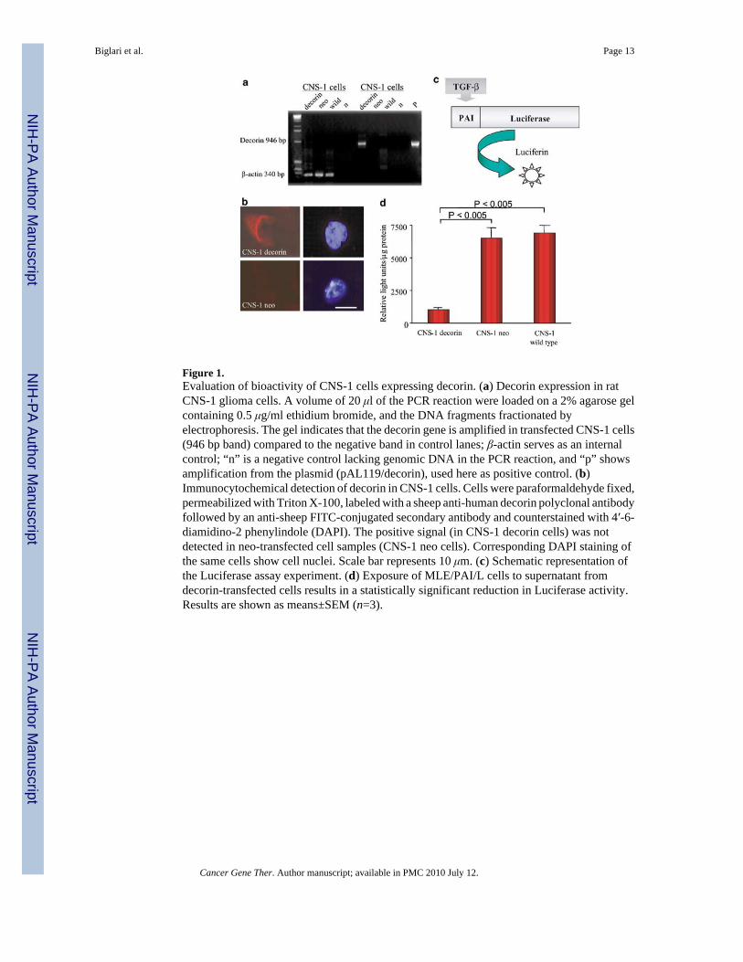
Figure 1.Evaluation of bioactivity of CNS-1 cells expressing decorin. (a) Decorin expression in ratCNS-1 glioma cells. A volume of 20 μl of the PCR reaction were loaded on a 2% agarose gelcontaining 0.5 μg/ml ethidium bromide, and the DNA fragments fractionated byelectrophoresis. The gel indicates that the decorin gene is amplified in transfected CNS-1 cells(946 bp band) compared to the negative band in control lanes; β-actin serves as an internalcontrol; “n” is a negative control lacking genomic DNA in the PCR reaction, and “p” showsamplification from the plasmid (pAL119/decorin), used here as positive control. (b)Immunocytochemical detection of decorin in CNS-1 cells. Cells were paraformaldehyde fixed,permeabilized with Triton X-100, labeled with a sheep anti-human decorin polyclonal antibodyfollowed by an anti-sheep FITC-conjugated secondary antibody and counterstained with 4′-6-diamidino-2 phenylindole (DAPI). The positive signal (in CNS-1 decorin cells) was notdetected in neo-transfected cell samples (CNS-1 neo cells). Corresponding DAPI staining ofthe same cells show cell nuclei. Scale bar represents 10 μm. (c) Schematic representation ofthe Luciferase assay experiment. (d) Exposure of MLE/PAI/L cells to supernatant fromdecorin-transfected cells results in a statistically significant reduction in Luciferase activity.Results are shown as means±SEM (n=3).
Biglari et al. Page 13
Cancer Gene Ther. Author manuscript; available in PMC 2010 July 12.
NIH
-PA Author Manuscript
NIH
-PA Author Manuscript
NIH
-PA Author Manuscript

Figure 2.In vitro proliferation of transfected CNS-1 cells. Cultured cells were subjected to daily countin quadruplicate. Growth curves were generated for each of the stably transfected and wildtype CNS-1 cells over a 12-day period. Data are represented as the mean of four values (SEMis less than 10%). There was significant difference in cell-doubling times among these celllines in vitro (P<.0001).
Biglari et al. Page 14
Cancer Gene Ther. Author manuscript; available in PMC 2010 July 12.
NIH
-PA Author Manuscript
NIH
-PA Author Manuscript
NIH
-PA Author Manuscript

Figure 3.Kaplan–Meier survival curves for Lewis rats implanted with decorin-transfected CNS-1 cells,observed for up to 90 (a) or 180 (b) days post cell implantation of transfected or wild-typeCNS-1 cells into the striatum of Lewis rats. Lewis rats implanted with CNS-1 cells expressingdecorin survive significantly longer than those in the control groups (n=5, logrank test P<.0001; in both a and b).
Biglari et al. Page 15
Cancer Gene Ther. Author manuscript; available in PMC 2010 July 12.
NIH
-PA Author Manuscript
NIH
-PA Author Manuscript
NIH
-PA Author Manuscript

Figure 4.Representative photomicrographs demonstrating brain histology and immunohistochemicalstaining for ED1, CD8, CD8β and CD161-positive cells in rats implanted with CNS-1 gliomas.(a) At death, all animals had large solid tumours at the site of cell implantation in the striatum.Surviving rats at 90 days post-tumor implantation had a small remaining tumor (b) or weretumor free (c). Scale bars represent 2 mm. (d–h) The distribution of activated ED1immunoreactive cells was much higher than the distribution of CD8 immunopositive cells inall rats. In the presence of a growing tumor (d–f), the infiltration of tumor and peritumoraltissue with inflammatory immune cells is higher in tumor-bearing animals (d–f) than in animalsthat eliminated the tumor (g,h). In animals that eliminated the tumor (g,h), only ED1+ cellsremained in the visible tissue scar. Infiltration of tumors was higher in animals implanted withCNS-1 neo cells when compared to CNS-1 cells; these animals died at the same time as thoseimplanted with control untransfected CNS-1 cells.
Biglari et al. Page 16
Cancer Gene Ther. Author manuscript; available in PMC 2010 July 12.
NIH
-PA Author Manuscript
NIH
-PA Author Manuscript
NIH
-PA Author Manuscript

Figure 5.(a) Schematic diagram of cotransfection of pJM17 and pAL119/hCMV/decorin to generatethe RAd/hCMV/decorin vector genome. (b) Southern blot analysis to verify the correctinsertion of the decorin expression cassette within the RAd vector. (c) Schematic diagram ofcotransfection of pJM17 and pΔE1/GFAP/decorin to generate the RAd/GFAP/decorin vectorgenome. (d) Southern blot analysis to confirm the correct insertion of the decorin expressioncassette within the RAd vector.
Biglari et al. Page 17
Cancer Gene Ther. Author manuscript; available in PMC 2010 July 12.
NIH
-PA Author Manuscript
NIH
-PA Author Manuscript
NIH
-PA Author Manuscript

Figure 6.Assessment of the biological activity of RAds encoding decorin. Luciferase activity in MLE/PAI/L cells following incubation with conditioned medium from HeLa cells infected withRAds encoding decorin or β-gal. Both RAd/hCMV/decorin and RAd/GFAP/decorin inhibitedTGF-β signaling; RAd/hCMV/decorin was more effective than RAd/GFAP/decorin.
Biglari et al. Page 18
Cancer Gene Ther. Author manuscript; available in PMC 2010 July 12.
NIH
-PA Author Manuscript
NIH
-PA Author Manuscript
NIH
-PA Author Manuscript
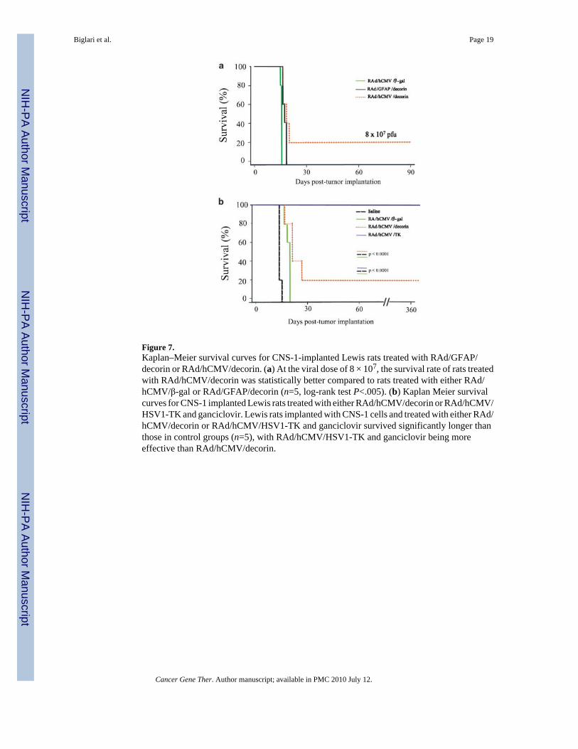
Figure 7.Kaplan–Meier survival curves for CNS-1-implanted Lewis rats treated with RAd/GFAP/decorin or RAd/hCMV/decorin. (a) At the viral dose of 8 × 10⁷, the survival rate of rats treatedwith RAd/hCMV/decorin was statistically better compared to rats treated with either RAd/hCMV/β-gal or RAd/GFAP/decorin (n=5, log-rank test P<.005). (b) Kaplan Meier survivalcurves for CNS-1 implanted Lewis rats treated with either RAd/hCMV/decorin or RAd/hCMV/HSV1-TK and ganciclovir. Lewis rats implanted with CNS-1 cells and treated with either RAd/hCMV/decorin or RAd/hCMV/HSV1-TK and ganciclovir survived significantly longer thanthose in control groups (n=5), with RAd/hCMV/HSV1-TK and ganciclovir being moreeffective than RAd/hCMV/decorin.
Biglari et al. Page 19
Cancer Gene Ther. Author manuscript; available in PMC 2010 July 12.
NIH
-PA Author Manuscript
NIH
-PA Author Manuscript
NIH
-PA Author Manuscript

Figure 8.Representative photomicrographs demonstrating brain histology and immunohistochemicalstaining for ED1 and CD8β-positive cells in rat CNS-1 gliomas treated with RAd/hCMV/decorin, RAd/hCMV/HSV1-TK and ganciclovir, RAd/hCMV/β-gal or saline. (a) Enlargementof the lateral ventricle observed in an animal treated with RAd/hCMV/decorin and that survivedfor 12 months; this was not seen in animals that had been treated with RAd/hCMV/HSV1-TKand ganciclovir which had also survived for 12 months. (b) Rats from control groups (salineand RAd/hCMV/β-gal) had high levels of ED1+ and CD8b+ cells throughout the tumor mass,with CD8β immunoreactive cells being higher in animals injected with RAd/hCMV/β-gal. Ratsthat were completely tumor free at 12 months post-tumor implantation and gene therapy hadinfiltration of ED1-positive monocytes/macrophages in the scar remaining at the injection site,and scattered ED1 staining throughout the striatum (RAd/hCMV/HSV1-TK and RAd/hCMV/decorin). Scale bar represent 200 μm.
Biglari et al. Page 20
Cancer Gene Ther. Author manuscript; available in PMC 2010 July 12.
NIH
-PA Author Manuscript
NIH
-PA Author Manuscript
NIH
-PA Author Manuscript