Identification of Lysine Residues in the Borrelia burgdorferi DbpA Adhesin Required for Murine...
17
Published Ahead of Print 19 May 2014. 2014, 82(8):3186. DOI: 10.1128/IAI.02036-14. Infect. Immun. Leong, Diana R. Tomchick and Jon S. Blevins Groshong, Brendan P. Moore, Kayla E. Hagman, John M. Danielle E. Fortune, Yi-Pin Lin, Ranjit K. Deka, Ashley M. Required for Murine Infection Borrelia burgdorferi DbpA Adhesin Identification of Lysine Residues in the http://iai.asm.org/content/82/8/3186 Updated information and services can be found at: These include: SUPPLEMENTAL MATERIAL Supplemental material REFERENCES http://iai.asm.org/content/82/8/3186#ref-list-1 at: This article cites 80 articles, 43 of which can be accessed free CONTENT ALERTS more» articles cite this article), Receive: RSS Feeds, eTOCs, free email alerts (when new http://journals.asm.org/site/misc/reprints.xhtml Information about commercial reprint orders: http://journals.asm.org/site/subscriptions/ To subscribe to to another ASM Journal go to: on August 26, 2014 by UNIV TEXAS SW MED CTR 904 http://iai.asm.org/ Downloaded from on August 26, 2014 by UNIV TEXAS SW MED CTR 904 http://iai.asm.org/ Downloaded from
-
Upload
utsouthwestern -
Category
Documents
-
view
1 -
download
0
Transcript of Identification of Lysine Residues in the Borrelia burgdorferi DbpA Adhesin Required for Murine...

Published Ahead of Print 19 May 2014. 2014, 82(8):3186. DOI: 10.1128/IAI.02036-14. Infect. Immun.
Leong, Diana R. Tomchick and Jon S. BlevinsGroshong, Brendan P. Moore, Kayla E. Hagman, John M. Danielle E. Fortune, Yi-Pin Lin, Ranjit K. Deka, Ashley M. Required for Murine InfectionBorrelia burgdorferi DbpA Adhesin Identification of Lysine Residues in the
http://iai.asm.org/content/82/8/3186Updated information and services can be found at:
These include:
SUPPLEMENTAL MATERIAL Supplemental material
REFERENCEShttp://iai.asm.org/content/82/8/3186#ref-list-1at:
This article cites 80 articles, 43 of which can be accessed free
CONTENT ALERTS more»articles cite this article),
Receive: RSS Feeds, eTOCs, free email alerts (when new
http://journals.asm.org/site/misc/reprints.xhtmlInformation about commercial reprint orders: http://journals.asm.org/site/subscriptions/To subscribe to to another ASM Journal go to:
on August 26, 2014 by U
NIV
TE
XA
S S
W M
ED
CT
R 904
http://iai.asm.org/
Dow
nloaded from
on August 26, 2014 by U
NIV
TE
XA
S S
W M
ED
CT
R 904
http://iai.asm.org/
Dow
nloaded from
Identification of Lysine Residues in the Borrelia burgdorferi DbpAAdhesin Required for Murine Infection
Danielle E. Fortune,a Yi-Pin Lin,b Ranjit K. Deka,c Ashley M. Groshong,a Brendan P. Moore,a Kayla E. Hagman,c* John M. Leong,b
Diana R. Tomchick,d Jon S. Blevinsa
Department of Microbiology and Immunology, University of Arkansas for Medical Sciences, Little Rock, Arkansas, USAa; Department of Molecular Biology andMicrobiology, Tufts University School of Medicine, Boston, Massachusetts, USAb; Department of Microbiology, University of Texas Southwestern Medical Center, Dallas,Texas, USAc; Department of Biophysics, University of Texas Southwestern Medical Center, Dallas, Texas, USAd
Decorin-binding protein A (DbpA) of Borrelia burgdorferi mediates bacterial adhesion to heparin and dermatan sulfate associatedwith decorin. Lysines K82, K163, and K170 of DbpA are known to be important for in vitro interaction with decorin, and theDbpA structure, initially solved by nuclear magnetic resonance (NMR) spectroscopy, suggests these lysine residues colocalize ina pocket near the C terminus of the protein. In the current study, we solved the structure of DbpA from B. burgdorferi strain 297using X-ray crystallography and confirmed the existing NMR structural data. In vitro binding experiments confirmed that re-combinant DbpA proteins with mutations in K82, K163, or K170 did not bind decorin, which was due to an inability to interactwith dermatan sulfate. Most importantly, we determined that the in vitro binding defect observed upon mutation of K82, K163,or K170 in DbpA also led to a defect during infection. The infectivity of B. burgdorferi expressing individual dbpA lysine pointmutants was assessed in mice challenged via needle inoculation. Murine infection studies showed that strains expressing dbpAwith mutations in K82, K163, and K170 were significantly attenuated and could not be cultured from any tissue. Proper expres-sion and cellular localization of the mutated DbpA proteins were examined, and NMR spectroscopy determined that the mutantDbpA proteins were structurally similar to wild-type DbpA. Taken together, these data showed that lysines K82, K163, and K170potentiate the binding of DbpA to dermatan sulfate and that an interaction(s) mediated by these lysines is essential for B. burg-dorferi murine infection.
Lyme disease, caused by spirochetes in the Borrelia burgdorferisensu lato complex, is the most prevalent vector-borne disease
in the United States and Europe (1, 2). The disease develops afterbacteria are introduced into a human via the bite of an infectedIxodes tick (3–5). Lyme disease initially presents as a localized skininfection at the bite site, which can develop into the pathogno-monic erythema migrans rash and is typically accompanied bynondescript flu-like symptoms. If the early infection is not treated,bacteria can disseminate from the primary bite site through thecirculatory and lymphatic systems and invade other tissues andorgans, thereby causing the severe secondary multisystemic ill-nesses associated with Lyme disease (e.g., carditis, arthritis, andneuroborreliosis) (6–8). The ability of bacteria to disseminate,colonize, and persist within an infected host is a complex andmultifactorial process (9). Surface-exposed bacterial proteinscalled adhesins promote interaction of the bacteria with a host cellor the extracellular matrix (ECM) and are recognized to be keymediators of bacterial colonization (10). A number of the host andbacterial factors that mediate B. burgdorferi attachment to mam-malian and tick tissues have been identified (5, 11, 12). Specifi-cally, B. burgdorferi adhesins bind host cell-associated integrins(13–16), as well as the mammalian ECM components fibronectin(17–23), type I collagen (23, 24), laminin (23, 25, 26), glycosami-noglycans (GAGs) (18, 27–29), and decorin (30–32). This capac-ity to interact with numerous host ligands is predicted to be re-sponsible for the spirochete’s ability to spread to and infect diversehost tissues, cause the various disease sequelae, and persist withinthe mammal and tick vector (5). Interestingly, strains of B. burg-dorferi appear to differ in their abilities to cause specific systemicsequelae, and these differences have been linked in part to theircapacities to bind/colonize certain host tissues (33–36).
Decorin, a small proteoglycan found in the ECM of numeroustissues (e.g., dermis and cartilage), is composed of a 36-kDa pro-tein core covalently linked to a 40-kDa GAG chain of chondroitinsulfate or dermatan sulfate (37–39). The lp54 linear plasmid of B.burgdorferi sensu stricto carries a two-gene operon that encodestwo surface lipoproteins, decorin-binding proteins A and B(DbpA and DbpB), which bind decorin (30–32). In vitro studieshave shown that both adhesins mediate interaction with the GAGsheparin and dermatan sulfate (31), but only DbpB binds chon-droitin sulfate (40). dbpA, and most likely dbpB, is upregulated byB. burgdorferi during tick feeding and expressed during mamma-lian infection (41–45). dbpA is presumed to remain highly ex-pressed throughout infection based on the presence of high levelsof reactive antibodies at 47 weeks postinoculation in macaquesexperimentally infected with B. burgdorferi (46). Considering thatdbpA is expressed during mammalian infection and decorin/der-matan sulfate can be found associated with almost all mammaliantissues, DbpA and the capacity to bind decorin have long been
Received 12 May 2014 Accepted 13 May 2014
Published ahead of print 19 May 2014
Editor: S. R. Blanke
Address correspondence to Jon S. Blevins, [email protected].
* Present address: Kayla E. Hagman, ThermoFisher Scientific, Molecular BiologyProducts Group, Lafayette, Colorado, USA.
Supplemental material for this article may be found at http://dx.doi.org/10.1128/IAI.02036-14.
Copyright © 2014, American Society for Microbiology. All Rights Reserved.
doi:10.1128/IAI.02036-14
3186 iai.asm.org Infection and Immunity p. 3186 –3198 August 2014 Volume 82 Number 8
on August 26, 2014 by U
NIV
TE
XA
S S
W M
ED
CT
R 904
http://iai.asm.org/
Dow
nloaded from
hypothesized to be key determinants in B. burgdorferi colonizationand dissemination within the host. In agreement with this, muta-tional studies have since confirmed that B. burgdorferi mutants inwhich dbpBA was disrupted have a diminished capacity for dis-semination and infection in mice challenged via needle inocula-tion (47–50), but dbpA and dbpB are not required for colonizationof Ixodes scapularis ticks (47). However, in contrast to the needlechallenge studies, the dbpBA mutant was capable of being trans-mitted to and infecting mice via tick bite (47). A recent study alsosuggests that the attenuation observed in dbpBA mutants is lim-ited to early stages of dissemination and that this defect is allevi-ated during chronic infection (51). Interestingly, this study alsofound that the dbpBA mutant was unable to migrate through thelymphatic system, suggesting that interaction of B. burgdorferiwith decorin/GAGs might be important when utilizing this routeof dissemination. A number of early biochemical and functionalstudies characterized the interaction between DbpA and decorin/GAGs (31, 52, 53). Comparison of DbpA sequences from 30 mem-bers of the B. burgdorferi sensu lato complex identified 11 lysineresidues that were highly conserved among the different strains;five lysine residues were conserved in all sequences, and six lysineresidues were found in �60% of the DbpA sequences (52). Anal-ysis of recombinant DbpA proteins carrying mutations in theseindividual lysines showed that mutation of K82, K163, or K170resulted in reduced decorin binding. A recent study also showedthat the highly basic C terminus of DbpA was required for bacte-rial interaction with decorin, dermatan sulfate, and 293 (humankidney epithelial) cells (35). Although the in vitro data clearly im-plicate these residues in DbpA-decorin/GAG interaction, they didnot address their contributions during mammalian infection.
Despite the abundance of functional and biochemical data avail-able regarding DbpA, it was not until recently that the solution struc-ture of DbpA from strain B31 was elucidated using nuclear magneticresonance (NMR) spectroscopy (54). In the present study, we soughtto complement the NMR studies by solving the structure of DbpAfrom B. burgdorferi strain 297 using X-ray crystallography, as wellas to assess whether lysines K82, K163, and K170 are requiredduring B. burgdorferi infection. Our crystal structure of a DbpAmonomer (determined at a resolution of 1.60 Å) confirmed theprior NMR structure, as well as the localization of the lysines crit-ical for the interaction of DbpA with decorin to a common basicpatch near the C terminus of the protein. To test the contributionsof lysines K82, K163, and K170 during mammalian infection,mice were challenged with B. burgdorferi dbpBA knockout strainsexpressing dbpA point mutants in which these lysine residues werechanged to alanine. The dbpBA mutants complemented with K82,K163, and K170 alanine point mutants were unable to infect mice,thus correlating the physiological contributions of these residuesto decorin/GAG binding and murine infection.
MATERIALS AND METHODSBacterial strains and culture conditions. All the strains and plasmidsused in this study are described in Table 1. Escherichia coli strain TOP10F=(Invitrogen, Carlsbad, CA) was used for cloning. E. coli strains XL1-Blue(Stratagene, La Jolla, CA) and B834(DE3) (Novagen, Madison, WI) wereused for protein expression. E. coli transformants were selected in Luria-Bertani (LB) medium supplemented with 100 �g/ml of ampicillin or 100�g/ml of spectinomycin. Infectious, low-passage-number B. burgdorferistrain 297 (Bb297) and the previously characterized B. burgdorferi dbpBAmutant (BbKH500) were used for these studies (47, 55). B. burgdorferi wasgrown in Barbour-Stoenner-Kelley II (BSK-II) medium at 37°C and pH
7.5 with 3 to 5% CO2 and 150 �g/ml of streptomycin or 150 �g/ml ofkanamycin (56).
Cloning wild-type, carboxyl-terminally truncated, and lysine mu-tant recombinant DbpA proteins. The first 25 amino acids of DbpA fromBb297 contain the putative signal peptide terminated by a potential con-sensus sequence for lipoprotein modification (31, 32). To facilitate ex-pression of the nonlipidated wild-type (WT) protein in E. coli, a DNAfragment encoding amino acids 26 to 187 of DbpA was amplified by PCRusing Bb297 genomic DNA as the template and the oligonucleotides 5=-DbpA(26) and 3=-DbpA(187) (Table 2); these primers contained BamHIand HindIII restriction sites, respectively. The 3=-DbpA(187) primer alsocontained tandem stop codons. A variant of DbpA with a C-terminaltruncation was also generated by PCR amplification with the oligonucle-otide pair 5=-DbpA(26) and 3=-DbpA(173) (Table 2); 3=-DbpA(173) in-troduces a HindIII site and two tandem stop codons on the 3= end of theamplified open reading frame (ORF). PCR amplification was performedusing Ex-Taq polymerase (TaKaRa, Madison, WI). The amplified frag-ments were then digested with BamHI and HindIII and ligated intopProEX-HTa digested with the same enzymes. The ligation was trans-formed into E. coli, and the resulting clones were verified by DNA se-quencing. Confirmed clones were designated pProEX-DbpAWT andpProEX-DbpA�C (Table 1).
The construct for the production of the His6-tagged RevA (recombinantRevA [rRevA]) control protein was previously described (20). Expressionconstructs for rDbpA variants carrying the lysine-to-alanine point mutationK51A, K82A, K124A, K163A, K170A, or K177A were generated by PCR am-plifying a DNA fragment corresponding to amino acids 26 to 187 of DbpAusing oligonucleotides 5=-DbpA(26) and 3=-DbpA(187). The pJD51 lysinepoint mutant shuttle vectors, the derivation of which is discussed below, wereused as the template in these PCRs. Cloning of the DbpA ORFs into pProEX-HTa was carried out as described above.
Expression and purification of rDbpA. Soluble His6-tagged rRevAwas purified as previously described (35). Expression of rDbpA frompProEX-HTa generates recombinant protein with an N-terminal His6 tagfollowed by a tobacco etch virus (TEV) protease cleavage site. For expression,cultures of XL1-Blue transformed with pProEX-DbpAWT or pProEX-DbpA�C were grown in LB medium containing ampicillin. When the celldensity reached an optical density at 600 nm (OD600) of 0.6, the culturewas induced for 3 h with 400 �M isopropyl-1-thio-�-D-galactopyrano-side (IPTG). Cells were collected by centrifugation; suspended in 20 mMHEPES (pH 7.5), 20 mM NaCl; and lysed by extrusion with an EmulsiFlex(Avestin, Ontario, Canada). To pellet cell debris, the lysate was centri-fuged at 27,000 � g for 15 min. rDbpA was purified from the supernatanton Ni2� affinity resin (Qiagen Inc., Valencia, CA) and eluted with 20 mMHEPES (pH 7.5), 300 mM NaCl, 500 mM imidazole. The elution bufferwas exchanged with 20 mM HEPES (pH 7.5), 50 mM NaCl, 15 mM�-mercaptoethanol (buffer A) using an Amicon Ultra-15 10,000-molec-ular-weight cutoff (MWCO) centrifugal-filtration unit (Millipore, Bil-lerica, MA). The His6 tag was cleaved from rDbpA by adding 1 mg ofHis6-TEV protease per 30 mg of recombinant protein. Proteolysis reac-tion mixtures were incubated overnight at room temperature and stoppedusing an Ni2� affinity column to remove the cleaved His6 tag and His6-TEV protease. The rDbpA protein was then concentrated to a final volumeof 1 ml and resolved on a HiLoad 16/60 Superdex 75 gel filtration columnusing an Äkta fast-performance liquid chromatography (FPLC) system(GE Healthcare Biosciences, Piscataway, NJ). The protein was eluted withbuffer A at a flow rate of 1 ml/min, and samples of the fractions containingprotein were analyzed by sodium dodecyl sulfate-polyacrylamide gel elec-trophoresis (SDS-PAGE) to assess purity. SDS-PAGE indicated that theprotein was pure to apparent homogeneity (i.e., �95%). Fractions con-taining purified DbpA were pooled and concentrated to 18 to 40 mg/ml inbuffer A for crystallization and binding experiments.
For the production of selenomethionine (SeMet)-labeled protein,pProEX-DbpAWT or pProEX-DbpA�C was transformed into the E. colimethionine auxotroph B834(DE3). Cells were cultured in 5% LB-95%
DbpA Lysines Critical for Murine Infection
August 2014 Volume 82 Number 8 iai.asm.org 3187
on August 26, 2014 by U
NIV
TE
XA
S S
W M
ED
CT
R 904
http://iai.asm.org/
Dow
nloaded from

M9 minimal medium containing 125 mg/liter each of adenine, uracil,thymine, and guanosine; 2.5 mg/liter thiamine; 4 mg/liter D-biotin; 20mM glucose; 2 mM MgSO4; 50 mg/liter each of 19 L-amino acids (with theexception of methionine); and 50 mg/liter L-selenomethionine. The cul-tures were induced, and the protein was purified as described above.
Crystallization, data collection, and structure determination. Crys-tals of a proteolytic fragment of wild-type rDbpA (rDbpAWT) were grownat 20°C using the vapor diffusion method in hanging-drop mode by mix-ing 2 �l protein (25 mg/ml) in 20 mM HEPES, pH 7.5, 50 mM NaCl, 15mM �-mercaptoethanol with 2 �l reservoir solution composed of 30 to35% (wt/vol) polyethylene glycol (PEG) 3000, 100 mM 2-(N-cyclohexyl-amino)ethanesulfonic acid (CHES), pH 9.5, 50 mM NaCl and equilibrat-ing against 1 ml of reservoir solution. The crystals exhibit the symmetry ofspace group P43 and were cryoprotected in reservoir solution supple-mented with 10% (vol/vol) ethylene glycol and then flash cooled in liquidnitrogen.
Crystals of selenomethionyl-derivatized rDbpA�C were grown at 20°Cusing the vapor diffusion method in hanging-drop mode by mixing 1 �lprotein (25 mg/ml) in 20 mM HEPES, pH 7.5, 50 mM NaCl, 15 mM�-mercaptoethanol with 1 �l reservoir solution, 30 to 35% (wt/vol) PEG3000, 0.1 M CHES, pH 9.5, 50 mM NaCl and equilibrating against 1 ml ofreservoir solution. The crystals exhibit the symmetry of space group
P212121 and were cryoprotected in reservoir solution supplemented with10% (vol/vol) ethylene glycol and then flash cooled in liquid nitrogen.
Phases obtained from a two-wavelength selenium (Se) anomalous-dispersion experiment were refined with the program MLPHARE (57),resulting in an overall figure of merit of 0.44 for data between 61.3 and2.40 Å. The phases were further improved by density modification and3-fold averaging with the program DM (58), resulting in a figure of meritof 0.63. An initial model containing 89% of all rDbpA residues was auto-matically generated by alternating cycles of the programs Resolve (59, 60)and ARP/wARP (61). Phases for the proteolytic fragment of rDbpA in theP43 cell were obtained via molecular replacement in the program PHASER(62), using the coordinates of DbpA from the orthorhombic cell as a searchmodel. Additional residues for rDbpA were manually modeled in the pro-gram O (63). Refinement was performed with the data collected on the pro-teolytic fragment of rDbpAWT to a resolution of 1.60 Å using the programPHENIX (64), with a random 5% of all data set aside for free refinement (R)factor (Rfree) calculation. Phasing and model refinement statistics are pro-vided in Table 3.
Site-directed mutagenesis of DbpA. Mutation of selected lysine resi-dues in DbpA was performed by overlap extension PCR. Primers 5=DbpAprom and 3= DbpA ORF represent the 5= and 3= outermost primers, re-spectively, used for all amplifications. The internal primers used to mutate
TABLE 1 Plasmids and strains used in this study
Plasmid or strain Descriptiona Source
PlasmidspGEM-T Easy TA cloning vector; Ampr PromegapProEX-HTa Expression construct; N-terminal and TEV-cleavable His6 tag; Ampr InvitrogenpProEX-DbpAWT pProEX HTa::DbpA (aa 26–187); Ampr This studypProEX-DbpA�C pProEX HTa::DbpA (aa 26–173); Ampr This studypProEX-DbpAK51A pProEX HTa::DbpAWT with K51A mutation; Ampr This studypProEX-DbpAK82A pProEX HTa::DbpAWT with K82A mutation; Ampr This studypProEX-DbpAK124A pProEX HTa::DbpAWT with K124A mutation; Ampr This studypProEX-DbpAK163A pProEX HTa::DbpAWT with K163A mutation; Ampr This studypProEX-DbpAK170A pProEX HTa::DbpAWT with K170A mutation; Ampr This studypProEX-DbpAK177A pProEX HTa::DbpAWT with K177A mutation; Ampr This studypRevA pQE30::RevA (aa 20–160); Ampr 20pJD51 B. burgdorferi and E. coli shuttle vector; Specr Strepr 47pKH2000 pJD51::PdbpBA-dbpA ORF complementation vector; Specr Strepr 47pDbpAK51A pKH2000 with DbpAK51A mutation; Specr Strepr This studypDbpAK82A pKH2000 with DbpAK82A mutation; Spec/Strepr This studypDbpAK124A pKH2000 with DbpAK124A mutation; Spec/Strepr This studypDbpAK163A pKH2000 with DbpAK163A mutation; Spec/Strepr This studypDbpAK170A pKH2000 with DbpAK170A mutation; Spec/Strepr This studypDbpAK177A pKH2000 with DbpAK177A mutation; Spec/Strepr This studypJSB568 pGEM-T Easy::flaB-actin qPCR standard; Ampr This study
StrainsE. coli
TOP10F= F= [lacIq Tn10 (Tetr)] mcrA �(mrr-hsdRMS-mcrBC) �80lacZ�M15 �lacX74 rec A1 ara�139�(ara-leu)7697 galU galK rpsL (Strr) endA1 nupG
Invitrogen
XL1-Blue recA1 endA1 gyrA96 thi-1 hsdR17 supE44 relA1 lac[F= proAB lacIq Z�M15::Tn10 (Tetr)] StrategeneB834(DE3) F ompT hsdSB(rB
mB) gal dcm met (DE3) Novagen
B. burgdorferiBb297 Strain 297; infectious, human spinal fluid isolate 55BbKH500 Strain 297; dbpBA::PflgB-Kan mutant; Kanr 47BbKH501 BbKH500 complemented with pKH2000; Kanr Strepr 47BbDbpAK51A BbKH500 complemented with pDbpAK51A; Kanr Strepr This studyBbDbpAK82A BbKH500 complemented with pDbpAK82A; Kanr Strepr This studyBbDbpAK124A BbKH500 complemented with pDbpAK124A; Kanr Strepr This studyBbDbpAK163A BbKH500 complemented with pDbpAK163A; Kanr Strepr This studyBbDbpAK170A BbKH500 complemented with pDbpAK170A; Kanr Strepr This studyBbDbpAK177A BbKH500 complemented with pDbpAK177A; Kanr Strepr This study
a aa, amino acids; Ampr, ampicillin resistant; Kanr, kanamycin resistant; Specr, spectinomycin resistant; Strepr, streptomycin resistant.
Fortune et al.
3188 iai.asm.org Infection and Immunity
on August 26, 2014 by U
NIV
TE
XA
S S
W M
ED
CT
R 904
http://iai.asm.org/
Dow
nloaded from

selected lysine amino acids to alanine (e.g., K51A, K82A, K124A, K163A,K170A, and K177A) are indicated with their intended mutation and de-noted as sense or antisense (Table 2). The numbering of the mutatedlysine residues is based on the full-length dbpA ORF from Bb297 (31).Oligonucleotides 5= DbpA prom and 3= DbpA ORF were paired with themutational antisense and sense oligonucleotides, respectively. DNA frag-ments were amplified with Ex-Taq polymerase using pKH2000 as thetemplate. pKH2000 (47), which encodes the native promoter for thedbpBA operon fused directly to the dbpA ORF, is a borrelial shuttle vectorused in prior studies for genetic complementation of dbpA expression inthe B. burgdorferi dbpBA mutant (BbKH500). Amplicons from the indi-vidual reactions were gel purified and combined to serve as the DNAtemplate in a second PCR using the outermost oligonucleotides, 5=DbpAprom and 3= DbpA ORF. The resulting amplicons for the point mutantswere cloned in pGEM-T Easy and confirmed by DNA sequencing withvector-specific primers and the 5=DbpA internal seq oligonucleotide. Theprimers 5= DbpA prom and 3= DbpA ORF incorporated BglII and SphIrestriction sites, respectively, to facilitate cloning of the mutated dbpAalleles into the borrelial shuttle vector pJD51 (Table 1).
Quantitative ELISA to measure decorin and dermatan sulfate bind-ing. Quantitative enzyme-linked immunosorbent assay (ELISA) for decorinand dermatan sulfate binding by rDbpA proteins was performed similarly tothat previously described (65). Briefly, wells of 96-well microtiter plates werecoated with 1 �g of decorin, dermatan sulfate, or bovine serum albumin(BSA); 100 �l of increasing concentrations (0.007, 0.015, 0.03125, 0.0625,0.125, 0.25, 0.5, and 1 �M for decorin binding; 0.03125, 0.0625, 0.125, 0.25,0.5, 1, and 2 �M for dermatan sulfate binding) of His6-tagged rRevA protein(20), rDbpAWT, or the lysine rDbpA mutant proteins was then added to thewells. To detect the binding of recombinant proteins, a 1:200 dilution of
mouse anti-His6 antibody (Sigma, St. Louis, MO) and a 1:1,000 dilution ofhorseradish peroxidase (HRP)-conjugated goat anti-mouse IgG (Invitrogen)were used as primary and secondary antibodies, respectively. The plates werewashed three times with PBST (0.05% Tween 20 in phosphate-buffered saline[PBS]), and 100 �l of tetramethylbenzidine (TMB) solution (KPL, Gaithers-burg, MD) was added to each well and incubated for 5 min. The reaction wasstopped by adding 100 �l of 0.5% hydrosulfuric acid to each well. The absor-bance was then read at 405 nm using an ELISA plate reader (SpectraMAX 250;Molecular Devices, Sunnyvale, CA). Statistical significance was determinedusing the Student t test; P values of �0.05 were considered significant. Todetermine the dissociation constant (Kd), the data were fitted by the followingequation using KaleidaGraph software (version 2.1.3; Abekbecj software,Reading, PA) where OD450(MAX) denotes the maximum absorbance atan optical density (OD) of 450 nm: OD450 OD450(MAX) � [DbpAprotein]/Kd � [DbpA protein].
SPR. Surface plasmon resonance (SPR) was measured using a Biacore3000 (GE Healthcare). Dermatan sulfate (EMD Millipore, Billerica, MA)and decorin (Sigma) were biotinylated using EZ-Link Biocytin Hydrazide(Pierce, Rockford, IL) and then dialyzed in PBS using a Slide-A-Lyzercassette (Pierce) with a 10,000-kDa-molecular-mass cutoff. Ten micro-grams of biotinylated dermatan sulfate or decorin was conjugated on astreptavidin SA sensor chip (GE Healthcare). A control flow cell was in-jected with PBS buffer lacking dermatan sulfate or decorin. For quantita-tive SPR experiments to measure decorin binding, 20 �l of increasingconcentrations (0, 0.03125, 0.0625, 0.125, 0.25, 0.5, and 1 �M) of rDb-pAWT or the rDbpA lysine mutants was injected into the control cell anddecorin-bound flow cells. To measure dermatan sulfate binding, 20 �l ofvarious concentrations (0, 0.09375, 0.1875, 0.375, 0.75, 1.5, and 3 �M) ofrecombinant protein was added to flow cells containing immobilized der-matan sulfate. The proteins were loaded into the cells at a flow rate of 10�l/min, and binding was carried out at 25°C. All sensogram data for thedermatan sulfate- and decorin-immobilized cells were subtracted fromthe negative-control flow cell. To obtain the kinetic parameters of theinteraction, the data from the sensograms were fitted by BIAevaluationsoftware version 3.0 using the one-step biomolecular association reactionmodel (1:1 Langmuir model), which resulted in optimum mathematicalfits using the lowest �2 values.
NMR spectroscopy. To express rDbpAWT for NMR characterization,bacteria were grown in M9 minimal medium supplemented with 1 g/liter15NH4Cl as the sole nitrogen source. The 15N-labeled recombinant pro-tein was purified, and the His6 tag was cleaved with TEV protease asdescribed above. The rDbpA proteins were then concentrated to a finalvolume of 2 ml and resolved on a HiLoad 16/60 Superdex 75 gel filtrationcolumn by FPLC. The protein was eluted with 50 mM sodium phosphate,50 mM NaCl (pH 6.5), and samples of the fractions containing proteinwere analyzed by SDS-PAGE to assess purity. Data were collected on 500�M protein samples in 50 mM sodium phosphate, 50 mM NaCl (pH 6.5)at 25°C. 1H, 15N heteronuclear single quantum coherence (HSQC) spec-tra of labeled protein were acquired by using a Varian Inova 500-MHzspectrometer and utilizing NMRPipe (66) and NMRView (67) for dataprocessing and analysis, respectively.
Transformation of B. burgdorferi. Electroporation of the B. burg-dorferi BbKH500 dbpBA mutant with the pJD51 shuttle vectors carryingthe dbpA lysine point mutants was carried out as previously described(68). Streptomycin-resistant (Strepr) clones were confirmed by DNA se-quence analysis of the transformed shuttle vector and PCR-based profil-ing to assess the endogenous borrelial plasmid content (47). Confirmedclones were designated according to the DbpA lysine mutation thateach transformant carried; BbDbpAK51A, BbDbpAK82A, BbDbpAK124A,BbDbpAK163A, BbDbpAK170A, or BbDbpAK177A.
Mouse infection experiments. The University of Arkansas for Medi-cal Sciences (UAMS) is accredited by the International Association forAssessment and Accreditation of Laboratory Animal Care (AAALAC),and all animal experiments were approved by the UAMS InstitutionalAnimal Care and Use Committee (IACUC). The infectivity of BbKH500
TABLE 2 Oligonucleotide primers used in this study
Primer designation Sequencea
5=-DbpA(26) ATATGGATCCGGGACTAACAGGAGCAACA3=-DbpA(187) ATATAAGCTTTTATTACGATTTAGCAGTGC
TGTCT3=-DbpA(173) CGATTTAGCAAGCTTCTACTAGTCTTGGTT
TTTCTTGTGA5= DbpA prom AGATCTTTGATTCAATTTGCAAAATAACCA3= DbpA ORF GCATGCCTTTGGGTTAATTGCTTTAAC5= DbpA internal seq GTAAGACCAAACAGCCCAACACK51A sense GATGCAATTAAAGCAAAGGCTGCK51A antisense GCAGCCTTTGCTTTAATTGCATCK82A sense CTTGAAGCAGCAGTGCGAGCTACK82A antisense GTAGCTCGCACTGCTGCTTCAAGK124A sense GAAGTCTCAGCACCATTACAAGK124A antisense CTTGTAATGGTGCTGAGACTTCK163A sense ATGAGAGAAGCATTACAAAGGGTTCK163A antisense GAACCCTTTGTAATGCTTCTCTCATK170A sense GTTCACAAGGCAAACCAAGACACCK170A antisense GGTGTCTTGGTTTGCCTTGTGAACK177A sense CCTTAAAGGCAAAAAATACCGAAGK177A antisense CTTCGGTATTTTTTGCCTTTAAGGflaBF-STD GGATCCTCACCAGCATCACTTTCAGGGTCTCflaBR-STD GGATCCACCTAAATTTGCCCTTTGATCACActF-STD GGATCCACCCACACTGTGCCCATCActR-STD GGATGCCACAGGATTCCATFlaB-ABI-F TTATGCAGCTAATGTTGCAAATCTTFlaB-ABI-R TTCCTGTTGAACACCCTCTTGAFlaB-ABI-Probe CTCAAACTGCTCAGGCTGCACCGGAct-ABI-F GACGGACTACCTCATGAAGATCCTAct-ABI-R CACGCACGATTACCCTCTCAAct-ABI-Probe ACCGAGCGTGGCTACAGCTTCATCAa Relevant restriction sites are italicized. Stop codons are underlined. Nucleotideschanged to generate mutant codons are in boldface.
DbpA Lysines Critical for Murine Infection
August 2014 Volume 82 Number 8 iai.asm.org 3189
on August 26, 2014 by U
NIV
TE
XA
S S
W M
ED
CT
R 904
http://iai.asm.org/
Dow
nloaded from

and dbpA-complemented strains was assessed using the murine needlechallenge model of Lyme borreliosis (32, 47). Clones were grown to themid-log phase with antibiotic selection, at which time the bacterial densityin each culture was enumerated using dark-field microscopy. Three- to4-week-old C3H/HeN (Harlan, Indianapolis, IN) mice were infected with1.5 � 105 bacteria via intradermal injection. At 2, 6, and 10 weeks postin-fection, ear punch, lymph node (brachial and inguinal), tarsal, and hearttissue samples were collected from the mice and placed in BSK-II mediumcontaining borrelia antibiotic cocktail (Monserate Biotechology Group,San Diego, CA). Skin, tibiotarsal, and heart samples were collected, storedin RNAlater Stabilization Solution (Life Technologies, Carlsbad, CA), andfrozen at 80°C until analyzed by quantitative PCR (qPCR). Dark-fieldmicroscopy was used to assess the growth of spirochetes in each of thesecultures 1 to 2 weeks postinoculation. Aliquots of growth-positive cul-tures were transferred to fresh medium with antibiotic selection for geno-typic confirmation of infectious clones (data not shown).
qPCR analysis of the spirochete tissue burden. To generate a copynumber standard for qPCR measurement of bacterial tissue burdens, afragment of the murine �-actin gene was amplified using primers ActF-STD and ActR-STD with C3H/HeN DNA (Table 2) (69). These primersintroduced a BamHI restriction site at one end. The amplicon was clonedinto pGEM-T Easy. A portion of flaB was then amplified using primersflaBF-STD and flaBR-STD and Bb297 genomic DNA (gDNA) (Table 2),which introduced BamHI restriction sites on both ends of the fragment.The flaB amplicon was ligated into pGEM-T Easy containing the actinfragment via the BamHI sites. The pGEM-T Easy::�-actin/flaB constructwas designated pJSB568.
For qPCR, DNA was extracted from skin, tibiotarsal, and heart sam-ples from infected mice as previously described by Maruskova et al. (69).
A High Pure PCR template preparation kit (Roche Applied Sciences, In-dianapolis, IN) was used to extract DNA according to the manufacturer’sinstructions, except that 200 �g of collagenase (Sigma) was added duringlysis. Purified DNA was analyzed via qPCR on a StepOnePlus Real TimePCR System using TaqMan Fast Advanced master mix (Life Technolo-gies). The primer set and probe used to detect flaB were FlaB-ABI-F,FlaB-ABI-R, and FlaB-ABI-Probe (Table 2). The primer set and probeused to detect the �-actin gene were Act-ABI-F, Act-ABI-R, and Actin-ABI-Probe (Table 2). The flaB and �-actin probes were labeled on the 5= endwith 6-carboxyfluorescein (FAM) and MAX Freedom dyes, respectively.Both probes were 3= labeled with Iowa Black FQ and internally quenched withZEN (Integrated DNA Technologies, Coralville, IA). A copy number stan-dard curve using the pJSB568 flaB/�-actin vector as the standard was used tomeasure the number of flaB copies and to quantify spirochetal burdens. Bac-terial burdens were reported as the number of flaB copies per 106 copies ofmouse �-actin. Analysis of variance (ANOVA) models were used to compareqPCR measurements between strains, and Tukey’s procedure was used toperform pairwise comparisons (Prism v6.0c; GraphPad software).
In vitro growth analyses. B. burgdorferi cultures were inoculated to astarting density of 103 spirochetes/ml and grown for 7 days at 37°C and pH7.5. Beginning at 3 days postinoculation, spirochetes were counted dailyusing dark-field microscopy to determine culture densities. For each cul-ture and time point, bacteria were enumerated in 20 individual micro-scope fields. To assess RpoS-dependent gene activation in B. burgdorferi,bacteria were grown in BSK-II at pH 6.8 until they reached the stationarygrowth phase (�108 bacteria/ml) (70). Bacteria were collected and pre-pared for SDS-PAGE and immunoblot analysis as described below.
Proteinase K accessibility assay. Borrelia cultures were grown to thelate exponential growth phase. Cells were collected by centrifugation,
TABLE 3 Data collection, phasing, and refinement statistics for DbpA structure
Statistic Valuea
Data collectionb (crystal [native/Se peak/Se inflection point])Space group P43/P21212/P21212Unit cell constants (Å) a 49.5, c 57.9/a 82.7, b 79.3, c 61.3/a 82.9, b 79.4, c 61.3Energy (eV) 12,681.9/12,686.5/12,684.5Resolution range (Å) 22.8–1.60 (1.64–1.60)/34.3–2.40 (2.46–2.40)/30.6–2.40 (2.46–2.40)Unique reflections (no.) 35,522 (1,861)/15,674 (1,071)/15,309 (1,057)Multiplicity 7.5 (3.3)/7.4 (7.7)/3.9 (4.0)Data completeness (%) 98.8 (87.9)/95.9 (99.9)/92.0 (97.1)Rmerge (%)c 5.0 (63.1)/8.9 (21.2)/7.9 (27.1)I/�(I) 33.9 (1.7)/34.1 (11.4)/21.9 (4.8)Wilson B value (Å2) 24.7/34.7/34.7
Phase determinationAnomalous scatterers Se, 15 out of 18 possible sitesFigure of merit (61.3–2.40 Å) 0.44
Refinement statisticsResolution range (Å) 22.77–1.60 (1.68–1.60)No. of reflections, Rwork/Rfree 18,318/937 (2,319/130)Data completeness (%) 98.9 (93.0)Atoms (non-H protein/solvent) 1,143/171Rwork (%) 16.9 (24.4)Rfree (%) 20.1 (27.3)RMSD bond length (Å) 0.010RMSD bond angle (°) 1.14Mean B value (Å2) (protein/solvent) 31.1/37.9Ramachandran plot (%) (favored/additional/disallowed)d 98.6/0.7/0.7Maximum-likelihood coordinate error 0.18Missing residues 65–71, 176–181
a Data for the outermost shell are given in parentheses.b For SE, Bijvoet pairs were kept separate for data processing.c Rmerge 100 h i|Ih,i �Ih�|/ h iIh,i, where the outer sum (h) is over the unique reflections and the inner sum (i) is over the set of independent observations of each uniquereflection.d As defined by the validation suite MolProbity (81).
Fortune et al.
3190 iai.asm.org Infection and Immunity
on August 26, 2014 by U
NIV
TE
XA
S S
W M
ED
CT
R 904
http://iai.asm.org/
Dow
nloaded from
washed twice with PBS, and suspended to a density of 2 � 109 cells/ml inPBS. Spirochetes (1 � 109) were treated with 200 �g of proteinase K (10mg/ml; Fisher Scientific, Pittsburgh, PA) or sham treated for 60 min atroom temperature. Proteinase K digestion was stopped by adding 15 �l ofphenylmethylsulfonyl fluoride (PMSF) (50 mg/ml in isopropanol;Sigma), and samples were prepared for SDS-PAGE and immunoblotting.
SDS-PAGE and immunoblot analysis. SDS-PAGE and immunoblot-ting were performed as previously described (47). A volume of cell lysateequivalent to 2 � 107 bacteria was loaded in each gel lane. The molecular-weight standard used for all immunoblots was All Blue Precision Plusstandard (Bio-Rad, Hercules, CA). For colorimetric detection of DbpA,
FlaB, and OspC, 4-chloro-1-naphthol was used as the substrate. Antibod-ies used to detect DbpA, FlaB, and OspC were described in prior studies(47, 71).
Protein structure accession number. The coordinates and structurefactors for DbpA have been deposited in the Research Collaboratory forStructural Bioinformatics Protein Data Bank (PDB) under the PDB ID4ONR.
RESULTS AND DISCUSSIONrDbpA adopts a four-helical-bundle fold. In the present study,we sought to expand our knowledge of DbpA by solving the struc-
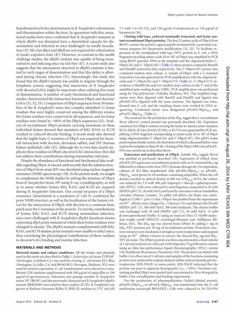
FIG 1 Crystal structure of rDbpA. (A) Helix 1 is colored green, helix 2 is blue, helix 3 is red, helix 4 is cyan, and helix 5 is magenta. The lysine residues knownto be involved in GAG binding (K82, K163, and K170) are shown as yellow spheres, and the N and C termini are labeled. (B) Refined atomic displacementparameters (ADP) from the X-ray structure mapped onto the structure of rDbpA. ADP values from low to high are colored from dark blue to red. The view onthe left is in the same orientation as in panel A, and the view on the right is rotated by 180°. (C) Electrostatic potential surface map of rDbpA revealing a positivelycharged cleft in the vicinity of the GAG binding lysines. This view has been rotated slightly from the view in panel A for clarity.
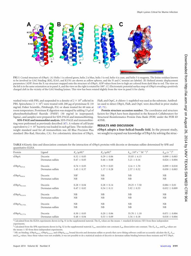
TABLE 4 Kinetic data and dissociation constants for the interaction of rDbpA proteins with decorin or dermatan sulfate determined by SPR andquantitative ELISA
Protein Ligand Kd (�M)a Kd (�M)b kon (104 s1 M1)b koff (s1)b
rDbpA Decorin 0.32 � 0.05 0.29 � 0.06 35.05 � 6.15 0.099 � 0.003Dermatan sulfate 0.47 � 0.05 0.46 � 0.08 5.21 � 0.16 0.024 � 0.004
rDbpAK51A Decorin 0.74 � 0.05 0.79 � 0.03 12.6 � 1.70 0.099 � 0.018Dermatan sulfate 1.45 � 0.37 1.17 � 0.28 2.57 � 0.32 0.030 � 0.003
rDbpAK82A Decorin NBc NB NB NBDermatan sulfate NB NB NB NB
rDbpAK124A Decorin 0.28 � 0.04 0.28 � 0.14 29.25 � 7.50 0.084 � 0.01Dermatan sulfate 0.47 � 0.02 0.54 � 0.11 5.92 � 0.53 0.032 � 0.009
rDbpAK163A Decorin NB NB NB NBDermatan sulfate NB NB NB NB
rDbpAK170A Decorin NB NB NB NBDermatan sulfate NB NB NB NB
rDbpAK177A Decorin 0.30 � 0.03 0.20 � 0.06 35.70 � 1.10 0.071 � 0.004Dermatan sulfate 0.48 � 0.04 0.53 � 0.09 3.34 � 0.18 0.018 � 0.004
a Calculated from the ELISA experiments shown in Fig. S1 in the supplemental material. The Kd values are the means � standard deviations (SD) from three independentexperiments.b Calculated from the SPR experiments shown in Fig. S2 in the supplemental material. kon, association rate constant; koff, dissociation rate constant. The Kd, kon, and koff values arethe means � SD from three independent experiments.c NB, no binding. rDbpAK82A, rDbpAK163A, and rDbpAK170A bound decorin and dermatan sulfate so poorly that curve-fitting software could not accurately calculate the Kd, kon,and koff values. Since these values were not available, it was not possible to do a statistical analysis of decorin or dermatan sulfate binding between these mutants and WT DbpA.
DbpA Lysines Critical for Murine Infection
August 2014 Volume 82 Number 8 iai.asm.org 3191
on August 26, 2014 by U
NIV
TE
XA
S S
W M
ED
CT
R 904
http://iai.asm.org/
Dow
nloaded from

ture of DbpA from B. burgdorferi strain 297 using X-ray crystal-lography. A nonlipidated form of Bb297 rDbpA was expressed inE. coli, and crystals of a proteolytic fragment of rDbpA were grownusing the vapor diffusion method in hanging-drop mode. Thestructure of the proteolytic fragment of rDbpA that encompassesresidues 26 to 175 was determined at a resolution of 1.60 Å via acombination of single-wavelength anomalous dispersion and mo-lecular replacement (Table 3). Analogous to the structure deter-mined via NMR methods (54), rDbpA is a monomer and containsan up-down four-alpha-helical bundle (Fig. 1A), with a fifth helixpacked against helices 2, 3, and 4. Residues 65 to 71 of the flexibleloop between helices 1 and 2 were disordered in the electron den-sity map and were not modeled. An alignment of the X-ray coor-dinates via DaliLite (72) with the deposited NMR coordinates(PDB ID 2LQU) yielded a root mean square deviation (RMSD) of2.2 Å for 143 equivalent C-alpha atoms and a Z-score of 15.4.Mapping of the refined atomic displacement parameters (B fac-tors) on the X-ray structure show that the largest values occur atthe ends of helices 1, 2, and 5 and along the distal surface of helix1 (Fig. 1B). This is in contrast to the published NMR study byWang (54), which indicated that helix 4 may undergo more inter-nal motion than the other helices. The larger values for atomicdisplacement parameters found in helix 1 do not appear to be anartifact of crystalline lattice packing, as the solvent content of thelattice is quite low (approximately 40%) and the largest solventchannel is in the vicinity of the disordered loop.
The three lysine residues (K82, K163, and K170) identified asessential for decorin binding in vitro interaction studies (52) aremapped to a common positively charged cleft near the disorderedhelix 1-helix 2 loop (Fig. 1C). Additional lysines mutated in thisstudy either are solvent exposed on the outer surface of the proteindistal to the disordered loop (e.g., K51 and K124) or were notlocated in the electron density at the C terminus of the moleculebut would be too distant from the positively charged cleft to con-tribute to substrate binding (K177). The proximity of the flexiblelysine side chains in the positively charged cleft provides a usefulmotif for interaction with the GAG of decorin, and the high mo-bility of the helix 1-helix 2 loop allows easy accessibility of thesubstrate to the cleft.
Contribution of conserved lysine residues in DbpA to der-matan sulfate binding. In vitro binding studies identified severallysine residues in DbpA that were involved in mediating adher-ence to decorin. Specifically, lysines K82, K163, and K170 are ab-solutely essential for decorin binding (52), and the structural data(Fig. 1) (54) confirmed that these critical lysines are located onproximal unique alpha helices that form a basic pocket in whichGAGs are speculated to bind. To confirm that these lysine muta-tions had the intended effect on decorin binding, we first gener-ated recombinant DbpA protein for wild-type DbpA and each ofthe lysine mutants. In addition to K82, K163, and K170, severalconserved lysine residues in DbpA that have not been implicatedin decorin binding were also targeted (52). Overlap extension PCRwas used to mutate individual lysine codons in the dbpA ORFto alanine, which were then expressed and purified as N-termi-nally His6-tagged recombinant proteins: rDbpAK51A, rDbpAK82A,rDbpAK124A, rDbpAK163A, rDbpAK170A, and rDbpAK177A. Decorin-binding activities of the rDbpA derivatives were quantitativelymeasured by both ELISA and quantitative SPR (Table 4; see Fig. S1and S2 in the supplemental material). As shown in Table 4,rDbpAK51A, rDbpAK124A, and rDbpAK177A bound decorin at
levels comparable to rDbpAWT. In contrast, rDbpAK82A,rDbpAK163A, and rDbpAK170A exhibited no decorin binding, con-firming that K82, K163, and K170 of DbpA are essential for inter-action with decorin. The reduction in binding with rDbpAK82A,rDbpAK163A, and rDbpAK170A at 0.25, 0.5, and 1 �M decorin wasstatistically significant relative to rDbpAWT (see Fig. S1 in the sup-plemental material). Since the GAGs associated with the proteincore mediate the interaction between decorin and DbpA, we alsowanted to assess the interaction of each of these mutants with
FIG 2 Superposition of 1H-15N HSQC spectra of rDbpAWT and rDbpA lysinepoint mutants. The NMR spectra of 15N-labeled rDbpAWT (black), rDbpAK82A
(red), rDbpAK163A (blue), and rDbpAK170A (green) were determined, and eachof the lysine mutant spectra was overlaid relative to rDbpAWT to assess changesin protein structure. The data were collected from a single analysis of eachrecombinant protein.
Fortune et al.
3192 iai.asm.org Infection and Immunity
on August 26, 2014 by U
NIV
TE
XA
S S
W M
ED
CT
R 904
http://iai.asm.org/
Dow
nloaded from
dermatan sulfate. In agreement with the decorin-binding data,rDbpAK51A, rDbpAK124A, and rDbpAK177A bound dermatan sul-fate at levels equivalent to rDbpAWT, while dermatan sulfate bind-ing was completely abolished with rDbpAK82A, rDbpAK163A, andrDbpAK170A. The reduced binding at 0.5, 1, and 2 �M dermatansulfate with rDbpAK82A, rDbpAK163A, and rDbpAK170A was statis-tically significant relative to rDbpAWT (see Fig. S1 in the supple-mental material). Taken together, these results agree with those ofprevious decorin-binding studies and indicate that K82, K163,and K170 of DbpA are essential for interaction with the dermatansulfate moiety of decorin (52).
While the previous data suggest that these three lysine residuescontribute to decorin and dermatan sulfate binding, it is possiblethat these substitutions could have altered binding activity by trig-gering structural changes that destabilized the DbpA protein. Toconfirm that the lysine mutations did not significantly alter thestructure of DbpA, rDbpAWT, rDbpAK82A, rDbpAK163A, andrDbpAK170A were expressed as 15N-labeled recombinant proteins,purified, and analyzed by NMR (Fig. 2). The 1H-15N HSQC spec-tra for rDbpAK82A, rDbpAK163A, and rDbpAK170A were then super-imposed on that of rDbpAWT to assess gross structural variationsbetween the individual lysine mutants and wild-type DbpA. Com-parisons revealed that DbpAK82A, DbpAK163A, and DbpAK170A
were correctly folded, suggesting that these lysine mutations hadlittle or no impact on the overall protein conformation and thatK82, K163, and K170 are directly involved in DbpA binding todermatan sulfate.
Contributions of conserved lysine residues in DbpA duringinfection. While the contributions of lysines K82, K163, and K170to the binding of decorin and dermatan sulfate have been demon-strated in vitro (52), it has not been assessed whether the impairedin vitro binding observed with these mutants correlates with adefect in B. burgdorferi infectivity. To test this, overlap extensionPCR was used to mutate individual lysine codons to alanine (e.g.,K51, K82, K124, K163, K170, and K177) in the dbpA ORF ofpKH2000, a borrelial shuttle vector that encodes the native pro-moter for the dbpBA operon fused directly to the dbpA ORF. Priorstudies confirmed that transformation of the B. burgdorferi dbpBAmutant, BbKH500, with pKH2000 is sufficient to restore dbpAexpression and infectivity to the mutant (47). BbKH500 wastransformed with each of the shuttle vectors carrying the lysinevariants to generate BbDbpAK51A, BbDbpAK82A, BbDbpAK124A,BbDbpAK163A, BbDbpAK170A, and BbDbpAK177A.
To assess the impacts of these DbpA lysine mutations on infec-
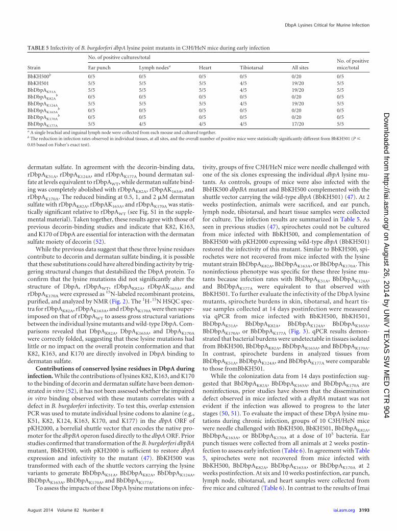
tivity, groups of five C3H/HeN mice were needle challenged withone of the six clones expressing the individual dbpA lysine mu-tants. As controls, groups of mice were also infected with theBbHK500 dbpBA mutant and BbKH500 complemented with theshuttle vector carrying the wild-type dbpA (BbKH501) (47). At 2weeks postinfection, animals were sacrificed, and ear punch,lymph node, tibiotarsal, and heart tissue samples were collectedfor culture. The infection results are summarized in Table 5. Asseen in previous studies (47), spirochetes could not be culturedfrom mice infected with BbKH500, and complementation ofBbKH500 with pKH2000 expressing wild-type dbpA (BbKH501)restored the infectivity of this mutant. Similar to BbKH500, spi-rochetes were not recovered from mice infected with the lysinemutant strain BbDbpAK82A, BbDbpAK163A, or BbDbpAK170A. Thisnoninfectious phenotype was specific for these three lysine mu-tants because infection rates with BbDbpAK51A, BbDbpAK124A,and BbDbpAK177A were equivalent to that observed withBbKH501. To further evaluate the infectivity of the DbpA lysinemutants, spirochete burdens in skin, tibotarsal, and heart tis-sue samples collected at 14 days postinfection were measuredvia qPCR from mice infected with BbKH500, BbKH501,BbDbpAK51A, BbDbpAK82A, BbDbpAK124A, BbDbpAK163A,BbDbpAK170A, or BbDbpAK177A (Fig. 3). qPCR results demon-strated that bacterial burdens were undetectable in tissues isolatedfrom BbKH500, BbDbpAK82A, BbDbpAK163A, and BbDbpAK170A.In contrast, spirochete burdens in analyzed tissues fromBbDbpAK51A, BbDbpAK124A, and BbDbpAK177A were comparableto those fromBbKH501.
While the colonization data from 14 days postinfection sug-gested that BbDbpAK82A, BbDbpAK163A, and BbDbpAK170A arenoninfectious, prior studies have shown that the disseminationdefect observed in mice infected with a dbpBA mutant was notevident if the infection was allowed to progress to the laterstages (50, 51). To evaluate the impact of these DbpA lysine mu-tations during chronic infection, groups of 10 C3H/HeN micewere needle challenged with BbKH500, BbKH501, BbDbpAK82A,BbDbpAK163A, or BbDbpAK170A at a dose of 105 bacteria. Earpunch tissues were collected from all animals at 2 weeks postin-fection to assess early infection (Table 6). In agreement with Table5, spirochetes were not recovered from mice infected withBbKH500, BbDbpAK82A, BbDbpAK163A, or BbDbpAK170A at 2weeks postinfection. At six and 10 weeks postinfection, ear punch,lymph node, tibiotarsal, and heart samples were collected fromfive mice and cultured (Table 6). In contrast to the results of Imai
TABLE 5 Infectivity of B. burgdorferi dbpA lysine point mutants in C3H/HeN mice during early infection
Strain
No. of positive cultures/totalNo. of positivemice/totalEar punch Lymph nodesa Heart Tibiotarsal All sites
BbKH500b 0/5 0/5 0/5 0/5 0/20 0/5BbKH501 5/5 5/5 5/5 4/5 19/20 5/5BbDbpAK51A 5/5 5/5 5/5 4/5 19/20 5/5BbDbpAK82A
b 0/5 0/5 0/5 0/5 0/20 0/5BbDbpAK124A 5/5 5/5 5/5 4/5 19/20 5/5BbDbpAK163A
b 0/5 0/5 0/5 0/5 0/20 0/5BbDbpAK170A
b 0/5 0/5 0/5 0/5 0/20 0/5BbDbpAK177A 5/5 4/5 4/5 4/5 17/20 5/5a A single brachial and inguinal lymph node were collected from each mouse and cultured together.b The reduction in infection rates observed in individual tissues, at all sites, and the overall number of positive mice were statistically significantly different from BbKH501 (P �
0.05 based on Fisher’s exact test).
DbpA Lysines Critical for Murine Infection
August 2014 Volume 82 Number 8 iai.asm.org 3193
on August 26, 2014 by U
NIV
TE
XA
S S
W M
ED
CT
R 904
http://iai.asm.org/
Dow
nloaded from

et al. (51), no spirochetes were recovered from mice infected withthe BbKH500 dbpBA mutant at either time point. Additionally, nospirochetes were recovered from mice infected with BbDbpAK82A,BbDbpAK163A, or BbDbpAK170A at 6 or 10 weeks postinfection.
Infectivity was restored in mice infected with BbKH501 at 2, 6, and10 weeks postinfection. Imai et al. also described a specific defectin the ability of the dbpBA mutant to disseminate through thelymphatic system (51). Our findings demonstrated a similar de-fect, since lymph nodes were taken from both a proximal (brachiallymph node) and a distal (inguinal lymph node) site in relation tothe site of inoculation, and no spirochetes were recovered in thesecultures. Taken together, these data suggest that the loss of bind-ing observed in vitro upon mutation of K82, K160, and K163 cor-relates with significant attenuation of the lysine mutants duringboth early and late stages of mammalian infection, and unlike theresults observed with the strain B31 dbpBA mutant, this defect wasnot overcome during chronic infection with the strain 297 dbpBAmutant.
B. burgdorferi strains in which dbpA has been inactivatedare unable to infect mice (47–50); therefore, to confirm thatloss of infectivity with strain BbDbpAK82A, BbDbpAK163A, orBbDbpAK170A was not due to aberrant expression or localizationof the mutated DbpA proteins, a number of in vitro studies wereperformed. In vitro growth curve analyses comparing Bb297 andBbKH501 to each of the attenuated lysine mutant clones deter-mined that their growth rates were similar; therefore, the loss ofinfectivity observed with BbDbpAK82A, BbDbpAK163A, orBbDbpAK170A was not due to a general growth defect in the pointmutant strains (see Fig. S3 in the supplemental material). Theattenuated phenotypes of BbDbpAK82A, BbDbpAK163A, andBbDbpAK170A could also be due to these mutated dbpA alleles notbeing properly expressed during infection. While it is difficult todirectly assess dbpA expression in vivo, it is possible to grow B.burgdorferi in vitro under conditions known to induce rpoS-de-pendent expression of dbpBA (e.g., elevated temperature and re-duced pH [41, 70, 73]). Cultures of BbDbpAK82A, BbDbpAK163A,or BbDbpAK170A were grown at pH 7.5 and 6.8, and induction ofDbpA production was evaluated using immunoblotting (Fig. 4).The production of OspC, an outer surface lipoprotein of B. burg-dorferi known to be activated by rpoS (70, 73), was assessed as apositive control. When cultured at pH 6.8, all of the clones carry-ing the DbpA lysine point mutants showed elevated OspC andDbpA at levels comparable to that observed with BbKH501. Thenoninfectious phenotype of BbDbpAK82A, BbDbpAK163A, orBbDbpAK170A could also be attributed to the mutated DbpA pro-teins not being properly localized to the bacterial surface, wherethey can interact with host GAGs. To assess surface localization,bacteria were subjected to proteinase K digestion and then immu-noblotted for DbpA. In all strains expressing dbpA, levels of DbpAdecreased significantly when the cells were treated with proteinaseK, suggesting that DbpA was appropriately localized on the cellsurface (Fig. 5). Immunoblot detection of FlaB was included as acontrol to confirm that the outer membranes of the cells remainedintact and only outer surface proteins were digested. Taken to-gether, these data support the conclusion that loss of infectivity inBbDbpAK82A, BbDbpAK163A, or BbDbpAK170A was due to the im-pact of the lysine mutations on decorin/dermatan sulfate bindingand was not due to an unknown secondary effect of these lysineresidues on overall dbpA expression or adhesin localization.
Summary and conclusions. There is a growing consensus thatNMR and crystallographic methods of structural determinationare complementary and should be pursued in tandem to fullyelucidate the structures of proteins of interest (74, 75). Therefore,we utilized X-ray crystallography to confirm the previously pub-
FIG 3 Spirochete burdens in tissues from mice infected with B. burgdorferiexpressing lysine point mutants. Tissues were collected at 2 weeks postinfec-tion from mice needle inoculated with BbKH500, BbKH501, BbDbpAK51A,BbDbpAK82A, BbDbpAK124A, BbDbpAK163A, BbDbpAK170A, or BbDbpAK177A
at a dose of 105 spirochetes. DNAs from tissue samples were analyzed by qPCRto measure spirochete burdens and are represented as copies of flaB/106 copiesof mouse �-actin. The results from skin (A), tibiotarsal (B), and heart (C)tissues are derived from a single infection experiment (n 5 mice for eachstrain). The results represent the mean values from two independent assays,with each sample measured in triplicate during each assay. The error bars representstandard errors of the mean (SEM). Statistical significance was determined byANOVA and Tukey’s procedure. The asterisks indicate a statistically signifi-cant difference relative to BbH501 within a given tissue set (P � 0.05).
Fortune et al.
3194 iai.asm.org Infection and Immunity
on August 26, 2014 by U
NIV
TE
XA
S S
W M
ED
CT
R 904
http://iai.asm.org/
Dow
nloaded from

lished NMR structure for DbpA (54). Although the flexible loopbetween helices 1 and 2 that contains a BXBB (where B is a basicamino acid) motif recently shown to contribute to GAG binding(76) could not be modeled in the X-ray structure, the two struc-tures were very similar and confirmed the clustering of lysine res-idues on helices 2 (e.g., K82) and 5 (e.g., K163 and K170) in anexposed binding cleft near the C terminus of the adhesin. In ad-dition, recombinant DbpA proteins containing mutations in K82,K163, or K170 demonstrated a complete loss of decorin and der-matan sulfate binding during in vitro binding experiments (52),and this binding defect correlated with significant attenuation ob-served in B. burgdorferi strains expressing dbpAK82A, dbpAK163A,and dbpAK170A during murine infection. Considering that a num-
ber of B. burgdorferi surface proteins have been shown to interactwith multiple host ligands (e.g., DbpA [35, 40, 76], OspC [77, 78],Bgp [18], and CRASP-1 [23, 79]), it remains possible that K82,K163, or K170 mediates interaction of DbpA with additional un-identified host ligands. However, these data support early studiesshowing the importance of decorin binding during mammalianinfection (80). The findings are also in agreement with studiesshowing that a B. burgdorferi strain expressing a dbpA allele lack-ing the 11 C-terminal amino acids, which is unable to binddecorin or dermatan sulfate (35), is also noninfectious in mice(J.M. Leong, unpublished results).
Similar to the results of Imai et al. (51), BbKH500,BbDbpAK82A, BbDbpAK163A, and BbDbpAK170A could not be re-covered from the brachial (proximal) or inguinal (distal) lymphnodes, which is supportive of their hypothesis that DbpA is im-
TABLE 6 Infectivity of B. burgdorferi dbpA lysine point mutants in C3H/HeN mice during chronic infection
StrainNo. of dayspostinfection
No. of positive cultures/totalNo. of positivemice/totalEar punch Lymph nodesa Heart Tibiotarsal All sites
BbKH500 14 0/10b NDc ND ND 0/10b 0/10b
42 0/5 0/5b 0/5b 0/5 0/20b 0/5b
70 0/5b 0/5b 0/5b 0/5b 0/20b 0/5b
BbKH501 14 10/10 ND ND ND 10/10b 10/10b
42 3/5 4/5 5/5 3/5 15/20b 5/5b
70 5/5 5/5 5/5 5/5 20/20b 5/5b
BbDbpAK82A 14 0/10b ND ND ND 0/10b 0/10b
42 0/5 0/5b 0/5b 0/5 0/20b 0/5b
70 0/5b 0/5b 0/5b 0/5b 0/20b 0/5b
BbDbpAK163A 14 0/10b ND ND ND 0/10b 0/10b
42 0/5 0/5b 0/5b 0/5 0/20b 0/5b
70 0/5b 0/5b 0/5b 0/5b 0/20b 0/5b
BbDbpAK170A 14 0/10b ND ND ND 0/10b 0/10b
42 0/5 0/5b 0/5b 0/5 0/20b 0/5b
70 0/5b 0/5b 0/5b 0/5b 0/20b 0/5b
a A single brachial and inguinal lymph node were collected from each mouse and cultured together.b The reductions in infection rates were statistically significantly different from BbKH501 (P � 0.05 based on Fisher’s exact test).c ND, not determined.
FIG 4 Assessment of DbpA expression in B. burgdorferi expressing lysinepoint mutants. Shown is immunoblot analysis of DbpA, OspC, and FlaB inBb297, BbKH500, BbKH501, BbDbpAK82A, BbDbpAK163A, and BbDbpAK170A.The strains were cultivated in vitro at pH 7.5 or 6.8 to induce production ofDbpA. Antibodies used to detect the respective proteins are indicated on theright. The values on the left denote relevant molecular masses (kDa) of thestandard (lane M). Samples from three independent biological replicates wereanalyzed and shown to yield similar results. Depicted is an immunoblot fromone of these biological replicates.
FIG 5 Confirmation of surface localization of DbpA in B. burgdorferi express-ing lysine point mutants. BbKH500, BbKH501, BbDbpAK82A, BbDbpAK163A,and BbDbpAK170A were grown in vitro at pH 7.5 to the late exponential growthphase. Whole-cell lysates from intact bacteria subjected to proteinase K diges-tion (�) or mock treated () were analyzed by immunoblotting. Antibodiesused to detect the respective proteins are indicated on the right. The values onthe left denote relevant molecular masses (kDa) of the standard (lane M).Samples from three independent biological replicates were analyzed andshown to yield similar results. Depicted is an immunoblot from one of thesebiological replicates.
DbpA Lysines Critical for Murine Infection
August 2014 Volume 82 Number 8 iai.asm.org 3195
on August 26, 2014 by U
NIV
TE
XA
S S
W M
ED
CT
R 904
http://iai.asm.org/
Dow
nloaded from
portant for dissemination via the lymphatic system. Recent stud-ies have shown that the infectivity defect observed with dbpAknockouts in the strain B31 background could be overcome dur-ing later stages of infection (50, 51), which is presumably due tocompensation by another bacterial adhesin(s) or alternate mech-anisms of dissemination. It should be noted that our results dem-onstrated that BbDbpAK82A, BbDbpAK163A, and BbDbpAK170A
were noninfectious during both the early and chronic stages of B.burgdorferi infection. Considering that DbpA is a prominent an-tigen (41–46), one possible explanation is that BbDbpAK82A,BbDbpAK163A, and BbDbpAK170A express a nonfunctional adhesinthat affords no dissemination benefit and the bacteria would berecognized and cleared by the humoral response. However, thefact that the BbKH500 dbpBA mutant also could not be recoveredfrom mice at 10 weeks postinfection puts this conclusion intoquestion. At this time, we are unable to definitively reconcile thedisparate phenotypes observed during persistent-infection stud-ies. Because the same challenge dose was employed in each of thesestudies (105 bacteria), it does not appear to be a result of an inoc-ulum difference. One possible explanation for these variationscould be the different parental strain used in our study (e.g.,Bb297) versus clonal derivatives of strain B31 (e.g., B31-A3 orML23), so that the compensatory bacterial adhesin(s) or alterna-tive dissemination mechanisms in strain B31 are more effectivethan those in strain 297. Future analysis of the function of DbpA inB. burgdorferi should include a comprehensive and comparativeanalysis of these different dbpBA mutants in a single challengestudy to assess whether strain-dependent differences exist regard-ing the relative contribution of dbpBA during chronic infection.Because our prior studies, albeit a relatively limited assessment ofsystemic colonization, have shown that BbKH500 can infect micewhen challenged via tick feeding, future dbpBA mutant compari-sons in mice should also employ the tick colonization and feedingmodel. Despite the discrepancy regarding the phenotype of thedbpBA mutant during chronic infection, these data clearly dem-onstrate that lysines K82, K163, and K170 are required for bindingof DbpA to dermatan sulfate and that the interaction(s) mediatedby these lysines is critical during B. burgdorferi murine infection.
ACKNOWLEDGMENTS
This research was supported by funding through the Arkansas BiosciencesInstitute (a major research component of the Arkansas Tobacco Settle-ment Proceeds Act of 2000), NIH/NIAID R01-AI087678, the Transla-tional Research Institute (UL1-TR000039; NIH National Center for Re-search Resources and National Center for Advancing TranslationalSciences), the UAMS Center for Microbial Pathogenesis and Host Inflam-matory Responses (COBRE grant P20-GM103625), and the UAMS IMSDprogram (NIH/NIGMS R25-GM083247). The work was also supportedby NIH R01-AI1093104 to John M. Leong and American Heart Associa-tion Postdoctoral Fellowship 12POST11660031 to Yi-Pin Lin. Resultsshown in this report are derived from work performed at Argonne Na-tional Laboratory, Structural Biology Center at the Advanced PhotonSource. Argonne is operated by UChicago Argonne, LLC, for the U.S.Department of Energy, Office of Biological and Environmental Research,under contract DE-AC02-06CH11357.
We thank Michael Norgard for his helpful discussions and criticalevaluation of the manuscript. We also thank Allen Gies in the UAMSSequencing Core Facility for his technical assistance and the staff at theAdvanced Photon Source for synchrotron data collection support.
REFERENCES1. Adams DA, Gallagher KM, Jajosky RA, Kriseman J, Sharp P, Anderson
WJ, Aranas AE, Mayes M, Wodajo MS, Onweh DH, Abellera JP. 2013.Summary of notifiable diseases—United States, 2011. MMWR Morb.Mortal. Wkly. Rep. 60:1–117.
2. Rizzoli A, Hauffe H, Carpi G, Vourc HG, Neteler M, Rosa R. 2011.Lyme borreliosis in Europe. Euro Surveill. 16:19906.
3. Burgdorfer W, Barbour AG, Hayes SF, Benach JL, Grunwaldt E, DavisJP. 1982. Lyme disease—a tick-borne spirochetosis? Science 216:1317–1319. http://dx.doi.org/10.1126/science.7043737.
4. Steere AC, Grodzicki RL, Kornblatt AN, Craft JE, Barbour AG, Burg-dorfer W, Schmid GP, Johnson E, Malawista SE. 1983. The spirochetaletiology of Lyme disease. N. Engl. J. Med. 308:733–740. http://dx.doi.org/10.1056/NEJM198303313081301.
5. Radolf JD, Caimano MJ, Stevenson B, Hu LT. 2012. Of ticks, mice andmen: understanding the dual-host lifestyle of Lyme disease spirochaetes.Nat. Rev. Microbiol. 10:87–99. http://dx.doi.org/10.1038/nrmicro2714.
6. Stanek G, Wormser GP, Gray J, Strle F. 2012. Lyme borreliosis. Lancet379:461– 473. http://dx.doi.org/10.1016/S0140-6736(11)60103-7.
7. Steere AC. 2001. Lyme disease. N. Engl. J. Med. 345:115–125. http://dx.doi.org/10.1056/NEJM200107123450207.
8. Steere AC, Coburn J, Glickstein L. 2004. The emergence of Lyme disease.J. Clin. Invest. 113:1093–1101. http://dx.doi.org/10.1172/JCI200421681.
9. Finlay BB, Falkow S. 1997. Common themes in microbial pathogenicityrevisited. Microbiol. Mol. Biol. Rev. 61:136 –169.
10. Linke D, Goldman A. 2011. Bacterial adhesion: chemisty, biology, andphysics, vol 715. Springer Scientific, New York, NY.
11. Coburn J, Leong J, Chaconas G. 2013. Illuminating the roles of theBorrelia burgdorferi adhesins. Trends Microbiol. 21:372–379. http://dx.doi.org/10.1016/j.tim.2013.06.005.
12. Cabello FC, Godfrey HP, Newman SA. 2007. Hidden in plain sight:Borrelia burgdorferi and the extracellular matrix. Trends Microbiol. 15:350 –354. http://dx.doi.org/10.1016/j.tim.2007.06.003.
13. Coburn J, Leong JM, Erban JK. 1993. Integrin �IIb�3 mediates binding ofthe Lyme disease agent Borrelia burgdorferi to human platelets. Proc. Natl.Acad. Sci. U. S. A. 90:7059 –7063. http://dx.doi.org/10.1073/pnas.90.15.7059.
14. Coburn J, Magoun L, Bodary SC, Leong JM. 1998. Integrins �V�3 and�V�1 mediate attachment of Lyme disease spirochetes to human cells.Infect. Immun. 66:1946 –1952.
15. Coburn J, Chege W, Magoun L, Bodary SC, Leong JM. 1999. Charac-terization of a candidate Borrelia burgdorferi �3-chain integrin ligandidentified using a phage display library. Mol. Microbiol. 34:926 –940. http://dx.doi.org/10.1046/j.1365-2958.1999.01654.x.
16. Behera AK, Durand E, Cugini C, Antonara S, Bourassa L, HildebrandE, Hu LT, Coburn J. 2008. Borrelia burgdorferi BBB07 interaction withintegrin �3�1 stimulates production of pro-inflammatory mediators inprimary human chondrocytes. Cell Microbiol. 10:320 –331. http://dx.doi.org/10.1111/j.1462-5822.2007.01043.x.
17. Probert WS, Johnson BJ. 1998. Identification of a 47 kDa fibronectin-binding protein expressed by Borrelia burgdorferi isolate B31. Mol. Micro-biol. 30:1003–1015. http://dx.doi.org/10.1046/j.1365-2958.1998.01127.x.
18. Fischer JR, LeBlanc KT, Leong JM. 2006. Fibronectin binding proteinBBK32 of the Lyme disease spirochete promotes bacterial attachment toglycosaminoglycans. Infect. Immun. 74:435– 441. http://dx.doi.org/10.1128/IAI.74.1.435-441.2006.
19. Probert WS, Kim JH, Hook M, Johnson BJ. 2001. Mapping the ligand-binding region of Borrelia burgdorferi fibronectin-binding protein BBK32.Infect. Immun. 69:4129 – 4133. http://dx.doi.org/10.1128/IAI.69.6.4129-4133.2001.
20. Moriarty TJ, Shi M, Lin YP, Ebady R, Zhou H, Odisho T, Hardy PO,Salman-Dilgimen A, Wu J, Weening EH, Skare JT, Kubes P, Leong J,Chaconas G. 2012. Vascular binding of a pathogen under shear forcethrough mechanistically distinct sequential interactions with host macro-molecules. Mol. Microbiol. 86:1116 –1131. http://dx.doi.org/10.1111/mmi.12045.
21. Brissette CA, Bykowski T, Cooley AE, Bowman A, Stevenson B. 2009.Borrelia burgdorferi RevA antigen binds host fibronectin. Infect. Immun.77:2802–2812. http://dx.doi.org/10.1128/IAI.00227-09.
22. Gaultney RA, Gonzalez T, Floden AM, Brissette CA. 2013. BB0347,from the Lyme disease spirochete Borrelia burgdorferi, is surface exposed
Fortune et al.
3196 iai.asm.org Infection and Immunity
on August 26, 2014 by U
NIV
TE
XA
S S
W M
ED
CT
R 904
http://iai.asm.org/
Dow
nloaded from

and interacts with the CS1 heparin-binding domain of human fibronectin.PLoS One 8:e75643. http://dx.doi.org/10.1371/journal.pone.0075643.
23. Hallstrom T, Haupt K, Kraiczy P, Hortschansky P, Wallich R, Skerka C,Zipfel PF. 2010. Complement regulator-acquiring surface protein 1 ofBorrelia burgdorferi binds to human bone morphogenic protein 2, severalextracellular matrix proteins, and plasminogen. J. Infect. Dis. 202:490 –498. http://dx.doi.org/10.1086/653825.
24. Zambrano MC, Beklemisheva AA, Bryksin AV, Newman SA, CabelloFC. 2004. Borrelia burgdorferi binds to, invades, and colonizes native typeI collagen lattices. Infect. Immun. 72:3138 –3146. http://dx.doi.org/10.1128/IAI.72.6.3138-3146.2004.
25. Verma A, Brissette CA, Bowman A, Stevenson B. 2009. Borrelia burg-dorferi BmpA is a laminin-binding protein. Infect. Immun. 77:4940 –4946. http://dx.doi.org/10.1128/IAI.01420-08.
26. Brissette CA, Verma A, Bowman A, Cooley AE, Stevenson B. 2009. TheBorrelia burgdorferi outer-surface protein ErpX binds mammalianlaminin. Microbiology 155:863– 872. http://dx.doi.org/10.1099/mic.0.024604-0.
27. Russell TM, Johnson BJ. 2013. Lyme disease spirochaetes possess anaggrecan-binding protease with aggrecanase activity. Mol. Microbiol. 90:228 –240. http://dx.doi.org/10.1111/mmi.12276.
28. Parveen N, Leong JM. 2000. Identification of a candidate glycosamino-glycan-binding adhesin of the Lyme disease spirochete Borrelia burgdor-feri. Mol. Microbiol. 35:1220 –1234. http://dx.doi.org/10.1046/j.1365-2958.2000.01792.x.
29. Leong JM, Morrissey PE, Ortega-Barria E, Pereira ME, Coburn J. 1995.Hemagglutination and proteoglycan binding by the Lyme disease spiro-chete, Borrelia burgdorferi. Infect. Immun. 63:874 – 883.
30. Guo BP, Norris SJ, Rosenberg LC, Hook M. 1995. Adherence of Borreliaburgdorferi to the proteoglycan decorin. Infect. Immun. 63:3467–3472.
31. Guo BP, Brown EL, Dorward DW, Rosenberg LC, Hook M. 1998.Decorin-binding adhesins from Borrelia burgdorferi. Mol. Microbiol. 30:711–723. http://dx.doi.org/10.1046/j.1365-2958.1998.01103.x.
32. Hagman KE, Lahdenne P, Popova TG, Porcella SF, Akins DR, RadolfJD, Norgard MV. 1998. Decorin-binding protein of Borrelia burgdorferi isencoded within a two-gene operon and is protective in the murine modelof Lyme borreliosis. Infect. Immun. 66:2674 –2683.
33. Wang G, Ojaimi C, Iyer R, Saksenberg V, McClain SA, Wormser GP,Schwartz I. 2001. Impact of genotypic variation of Borrelia burgdorferisensu stricto on kinetics of dissemination and severity of disease in C3H/HeJ mice. Infect. Immun. 69:4303– 4312. http://dx.doi.org/10.1128/IAI.69.7.4303-4312.2001.
34. Wormser GP, Liveris D, Nowakowski J, Nadelman RB, Cavaliere LF,McKenna D, Holmgren D, Schwartz I. 1999. Association of specificsubtypes of Borrelia burgdorferi with hematogenous dissemination inearly Lyme disease. J. Infect. Dis. 180:720 –725. http://dx.doi.org/10.1086/314922.
35. Benoit VM, Fischer JR, Lin YP, Parveen N, Leong JM. 2011. Allelicvariation of the Lyme disease spirochete adhesin DbpA influences spiro-chetal binding to decorin, dermatan sulfate, and mammalian cells. Infect.Immun. 79:3501–3509. http://dx.doi.org/10.1128/IAI.00163-11.
36. Wang G, van Dam AP, Schwartz I, Dankert J. 1999. Molecular typing ofBorrelia burgdorferi sensu lato: taxonomic, epidemiological, and clinicalimplications. Clin. Microbiol. Rev. 12:633– 653.
37. Fisher LW, Termine JD, Young MF. 1989. Deduced protein sequence ofbone small proteoglycan I (biglycan) shows homology with proteoglycanII (decorin) and several nonconnective tissue proteins in a variety of spe-cies. J. Biol. Chem. 264:4571– 4576.
38. Poole AR, Webber C, Pidoux I, Choi H, Rosenberg LC. 1986. Localizationof a dermatan sulfate proteoglycan (DS-PGII) in cartilage and the presence ofan immunologically related species in other tissues. J. Histochem. Cytochem.34:619–625. http://dx.doi.org/10.1177/34.5.3701029.
39. Scott JE, Orford CR. 1981. Dermatan sulphate-rich proteoglycan associ-ates with rat tail-tendon collagen at the d band in the gap region. Biochem.J. 197:213–216.
40. Fischer JR, Parveen N, Magoun L, Leong JM. 2003. Decorin-bindingproteins A and B confer distinct mammalian cell type-specific attachmentby Borrelia burgdorferi, the Lyme disease spirochete. Proc. Natl. Acad. Sci.U. S. A. 100:7307–7312. http://dx.doi.org/10.1073/pnas.1231043100.
41. Hagman KE, Yang X, Wikel SK, Schoeler GB, Caimano MJ, Radolf JD,Norgard MV. 2000. Decorin-binding protein A (DbpA) of Borrelia burg-dorferi is not protective when immunized mice are challenged via tickinfestation and correlates with the lack of DbpA expression by B. burgdor-
feri in ticks. Infect. Immun. 68:4759 – 4764. http://dx.doi.org/10.1128/IAI.68.8.4759-4764.2000.
42. Hodzic E, Feng S, Freet KJ, Borjesson DL, Barthold SW. 2002. Borreliaburgdorferi population kinetics and selected gene expression at the host-vector interface. Infect. Immun. 70:3382–3388. http://dx.doi.org/10.1128/IAI.70.7.3382-3388.2002.
43. Barthold SW, Hodzic E, Tunev S, Feng S. 2006. Antibody-mediateddisease remission in the mouse model of Lyme borreliosis. Infect. Immun.74:4817– 4825. http://dx.doi.org/10.1128/IAI.00469-06.
44. Hanson MS, Cassatt DR, Guo BP, Patel NK, McCarthy MP, DorwardDW, Hook M. 1998. Active and passive immunity against Borrelia burg-dorferi decorin binding protein A (DbpA) protects against infection. In-fect. Immun. 66:2143–2153.
45. Cassatt DR, Patel NK, Ulbrandt ND, Hanson MS. 1998. DbpA, but notOspA, is expressed by Borrelia burgdorferi during spirochetemia and is atarget for protective antibodies. Infect. Immun. 66:5379 –5387.
46. Embers ME, Hasenkampf NR, Jacobs MB, Philipp MT. 2012. Dynamiclongitudinal antibody responses during Borrelia burgdorferi infection andantibiotic treatment of rhesus macaques. Clin. Vaccine Immunol. 19:1218 –1226. http://dx.doi.org/10.1128/CVI.00228-12.
47. Blevins JS, Hagman KE, Norgard MV. 2008. Assessment of decorin-binding protein A to the infectivity of Borrelia burgdorferi in the murinemodels of needle and tick infection. BMC Microbiol. 8:82. http://dx.doi.org/10.1186/1471-2180-8-82.
48. Shi Y, Xu Q, McShan K, Liang FT. 2008. Both decorin-binding proteinsA and B are critical for the overall virulence of Borrelia burgdorferi. Infect.Immun. 76:1239 –1246. http://dx.doi.org/10.1128/IAI.00897-07.
49. Shi Y, Xu Q, Seemanaplli SV, McShan K, Liang FT. 2008. Common andunique contributions of decorin-binding proteins A and B to the overallvirulence of Borrelia burgdorferi. PLoS One 3:e3340. http://dx.doi.org/10.1371/journal.pone.0003340.
50. Weening EH, Parveen N, Trzeciakowski JP, Leong JM, Hook M, SkareJT. 2008. Borrelia burgdorferi lacking DbpBA exhibits an early survivaldefect during experimental infection. Infect. Immun. 76:5694 –5705. http://dx.doi.org/10.1128/IAI.00690-08.
51. Imai DM, Samuels DS, Feng S, Hodzic E, Olsen K, Barthold SW. 2013.The early dissemination defect attributed to disruption of decorin-binding proteins is abolished in chronic murine Lyme borreliosis. Infect.Immun. 81:1663–1673. http://dx.doi.org/10.1128/IAI.01359-12.
52. Brown EL, Guo BP, O’Neal P, Hook M. 1999. Adherence of Borreliaburgdorferi. Identification of critical lysine residues in DbpA required fordecorin binding. J. Biol. Chem. 274:26272–26278.
53. Pikas DS, Brown EL, Gurusiddappa S, Lee LY, Xu Y, Hook M. 2003.Decorin-binding sites in the adhesin DbpA from Borrelia burgdorferi: asynthetic peptide approach. J. Biol. Chem. 278:30920 –30926. http://dx.doi.org/10.1074/jbc.M303979200.
54. Wang X. 2012. Solution structure of decorin-binding protein A fromBorrelia burgdorferi. Biochemistry 51:8353– 8362. http://dx.doi.org/10.1021/bi3007093.
55. Hughes CA, Kodner CB, Johnson RC. 1992. DNA analysis of Borreliaburgdorferi NCH-1, the first northcentral U.S. human Lyme disease iso-late. J. Clin. Microbiol. 30:698 –703.
56. Pollack RJ, Telford SR, III, Spielman A. 1993. Standardization of me-dium for culturing Lyme disease spirochetes. J. Clin. Microbiol. 31:1251–1255.
57. Collaborative Computational Project Number 4. 1994. The CCP4 suite:programs for protein crystallography. Acta Crystallogr. D Biol. Crystal-logr. 50:760 –763. http://dx.doi.org/10.1107/S0907444994003112.
58. Cowtan K, Main P. 1998. Miscellaneous algorithms for density modifi-cation. Acta Crystallogr. D Biol. Crystallogr. 54:487– 493. http://dx.doi.org/10.1107/S0907444997011980.
59. Terwilliger TC. 2002. Rapid automatic NCS identification using heavy-atom substructures. Acta Crystallogr. D Biol. Crystallogr. 58:2213–2215.http://dx.doi.org/10.1107/S0907444902016384.
60. Terwilliger TC. 2003. Automated main-chain model building by templatematching and iterative fragment extension. Acta Crystallogr. D Biol. Crys-tallogr. 59:38 – 44. http://dx.doi.org/10.1107/S0907444902018036.
61. Langer G, Cohen SX, Lamzin VS, Perrakis A. 2008. Automated macro-molecular model building for X-ray crystallography using ARP/wARPversion 7. Nat. Protoc. 3:1171–1179. http://dx.doi.org/10.1038/nprot.2008.91.
62. Read RJ. 2001. Pushing the boundaries of molecular replacement with
DbpA Lysines Critical for Murine Infection
August 2014 Volume 82 Number 8 iai.asm.org 3197
on August 26, 2014 by U
NIV
TE
XA
S S
W M
ED
CT
R 904
http://iai.asm.org/
Dow
nloaded from
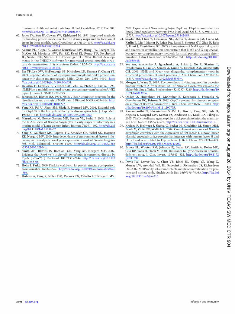
maximum likelihood. Acta Crystallogr. D Biol. Crystallogr. 57:1373–1382.http://dx.doi.org/10.1107/S0907444901012471.
63. Jones TA, Zou JY, Cowan SW, Kjeldgaard M. 1991. Improved methodsfor building protein models in electron density maps and the location oferrors in these models. Acta Crystallogr. A 47:110 –119. http://dx.doi.org/10.1107/S0108767390010224.
64. Adams PD, Gopal K, Grosse-Kunstleve RW, Hung LW, Ioerger TR,McCoy AJ, Moriarty NW, Pai RK, Read RJ, Romo TD, SacchettiniJC, Sauter NK, Storoni LC, Terwilliger TC. 2004. Recent develop-ments in the PHENIX software for automated crystallographic struc-ture determination. J. Synchrotron Radiat. 11:53–55. http://dx.doi.org/10.1107/S0909049503024130.
65. Lin YP, Lee DW, McDonough SP, Nicholson LK, Sharma Y, Chang YF.2009. Repeated domains of leptospira immunoglobulin-like proteins in-teract with elastin and tropoelastin. J. Biol. Chem. 284:19380 –19391. http://dx.doi.org/10.1074/jbc.M109.004531.
66. Delaglio F, Grzesiek S, Vuister GW, Zhu G, Pfeifer J, Bax A. 1995.NMRPipe: a multidimensional spectral processing system based on UNIXpipes. J. Biomol. NMR 6:277–293.
67. Johnson BA, Blevins RA. 1994. NMR View: A computer program for thevisualization and analysis of NMR data. J. Biomol. NMR 4:603– 614. http://dx.doi.org/10.1007/BF00404272.
68. Yang XF, Pal U, Alani SM, Fikrig E, Norgard MV. 2004. Essential rolefor OspA/B in the life cycle of the Lyme disease spirochete. J. Exp. Med.199:641– 648. http://dx.doi.org/10.1084/jem.20031960.
69. Maruskova M, Esteve-Gassent MD, Sexton VL, Seshu J. 2008. Role ofthe BBA64 locus of Borrelia burgdorferi in early stages of infectivity in amurine model of Lyme disease. Infect. Immun. 76:391– 402. http://dx.doi.org/10.1128/IAI.01118-07.
70. Yang X, Goldberg MS, Popova TG, Schoeler GB, Wikel SK, HagmanKE, Norgard MV. 2000. Interdependence of environmental factors influ-encing reciprocal patterns of gene expression in virulent Borrelia burgdor-feri. Mol. Microbiol. 37:1470 –1479. http://dx.doi.org/10.1046/j.1365-2958.2000.02104.x.
71. Smith AH, Blevins JS, Bachlani GN, Yang XF, Norgard MV. 2007.Evidence that RpoS (�S) in Borrelia burgdorferi is controlled directly byRpoN (�54/�N). J. Bacteriol. 189:2139 –2144. http://dx.doi.org/10.1128/JB.01653-06.
72. Holm L, Park J. 2000. DaliLite workbench for protein structure comparison.Bioinformatics 16:566–567. http://dx.doi.org/10.1093/bioinformatics/16.6.566.
73. Hubner A, Yang X, Nolen DM, Popova TG, Cabello FC, Norgard MV.
2001. Expression of Borrelia burgdorferi OspC and DbpA is controlled by aRpoN-RpoS regulatory pathway. Proc. Natl. Acad. Sci. U. S. A. 98:12724 –12729. http://dx.doi.org/10.1073/pnas.231442498.
74. Snyder DA, Chen Y, Denissova NG, Acton T, Aramini JM, Ciano M,Karlin R, Liu J, Manor P, Rajan PA, Rossi P, Swapna GV, Xiao R, RostB, Hunt J, Montelione GT. 2005. Comparisons of NMR spectral qualityand success in crystallization demonstrate that NMR and X-ray crystal-lography are complementary methods for small protein structure deter-mination. J. Am. Chem. Soc. 127:16505–16511. http://dx.doi.org/10.1021/ja053564h.
75. Yee AA, Savchenko A, Ignachenko A, Lukin J, Xu X, Skarina T,Evdokimova E, Liu CS, Semesi A, Guido V, Edwards AM, ArrowsmithCH. 2005. NMR and X-ray crystallography, complementary tools instructural proteomics of small proteins. J. Am. Chem. Soc. 127:16512–16517. http://dx.doi.org/10.1021/ja053565�.
76. Morgan A, Wang X. 2013. The novel heparin-binding motif in decorin-binding protein A from strain B31 of Borrelia burgdorferi explains thehigher binding affinity. Biochemistry 52:8237– 8245. http://dx.doi.org/10.1021/bi401376u.
77. Onder O, Humphrey PT, McOmber B, Korobova F, Francella N,Greenbaum DC, Brisson D. 2012. OspC is potent plasminogen receptoron surface of Borrelia burgdorferi. J. Biol. Chem. 287:16860 –16868. http://dx.doi.org/10.1074/jbc.M111.290775.
78. Ramamoorthi N, Narasimhan S, Pal U, Bao F, Yang XF, Fish D,Anguita J, Norgard MV, Kantor FS, Anderson JF, Koski RA, Fikrig E.2005. The Lyme disease agent exploits a tick protein to infect the mamma-lian host. Nature 436:573–577. http://dx.doi.org/10.1038/nature03812.
79. Kraiczy P, Hellwage J, Skerka C, Becker H, Kirschfink M, Simon MM,Brade V, Zipfel PF, Wallich R. 2004. Complement resistance of Borreliaburgdorferi correlates with the expression of BbCRASP-1, a novel linearplasmid-encoded surface protein that interacts with human factor H andFHL-1 and is unrelated to Erp proteins. J. Biol. Chem. 279:2421–2429.http://dx.doi.org/10.1074/jbc.M308343200.
80. Brown EL, Wooten RM, Johnson BJ, Iozzo RV, Smith A, Dolan MC,Guo BP, Weis JJ, Hook M. 2001. Resistance to Lyme disease in decorin-deficient mice. J. Clin. Invest. 107:845– 852. http://dx.doi.org/10.1172/JCI11692.
81. Davis IW, Leaver-Fay A, Chen VB, Block JN, Kapral GJ, Wang X,Murray LW, Arendall WB, III, Snoeyink J, Richardson JS, RichardsonDC. 2007. MolProbity: all-atom contacts and structure validation for pro-teins and nucleic acids. Nucleic Acids Res. 35:W375–W383. http://dx.doi.org/10.1093/nar/gkm216.
Fortune et al.
3198 iai.asm.org Infection and Immunity
on August 26, 2014 by U
NIV
TE
XA
S S
W M
ED
CT
R 904
http://iai.asm.org/
Dow
nloaded from
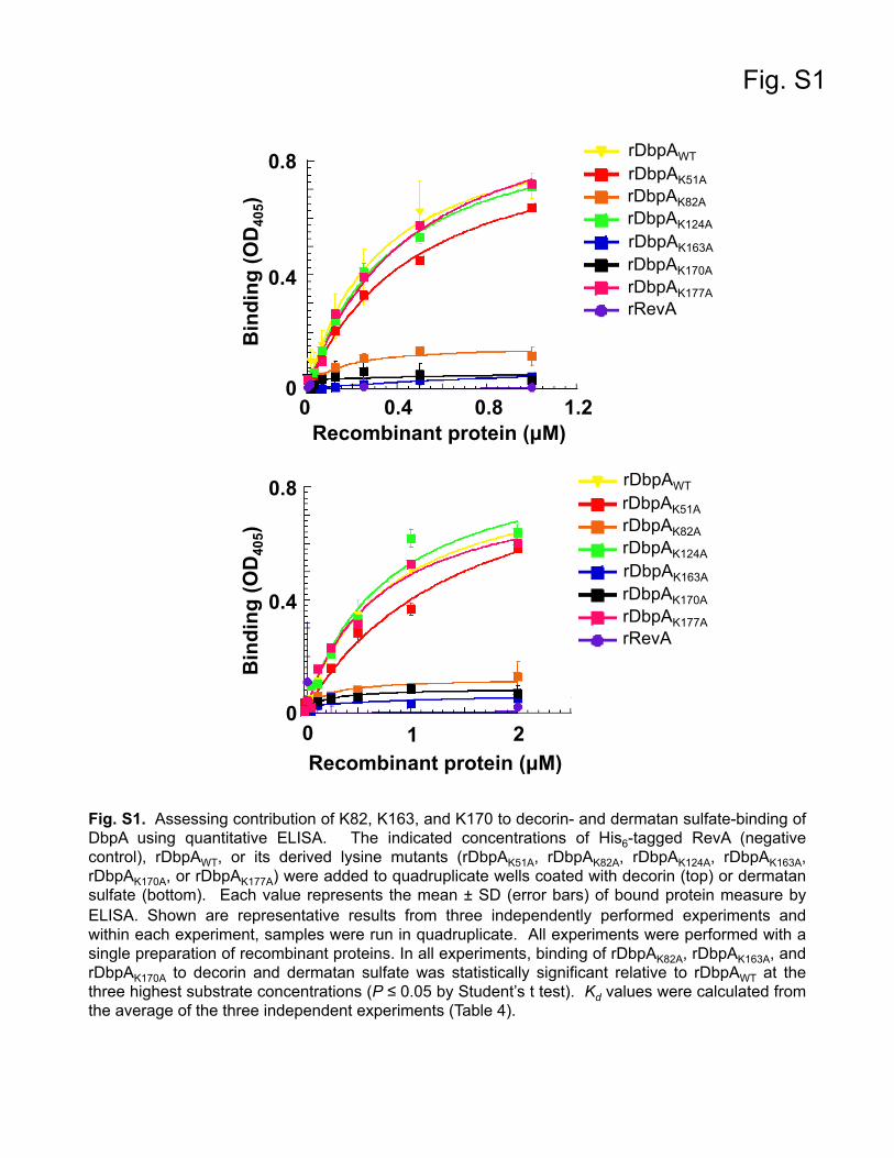
Fig. S1
Fig. S1. Assessing contribution of K82, K163, and K170 to decorin- and dermatan sulfate-binding of DbpA using quantitative ELISA. The indicated concentrations of His6-tagged RevA (negative control), rDbpAWT, or its derived lysine mutants (rDbpAK51A, rDbpAK82A, rDbpAK124A, rDbpAK163A, rDbpAK170A, or rDbpAK177A) were added to quadruplicate wells coated with decorin (top) or dermatan sulfate (bottom). Each value represents the mean ± SD (error bars) of bound protein measure by ELISA. Shown are representative results from three independently performed experiments and within each experiment, samples were run in quadruplicate. All experiments were performed with a single preparation of recombinant proteins. In all experiments, binding of rDbpAK82A, rDbpAK163A, and rDbpAK170A to decorin and dermatan sulfate was statistically significant relative to rDbpAWT at the three highest substrate concentrations (P ≤ 0.05 by Student’s t test). Kd values were calculated from the average of the three independent experiments (Table 4).
0
0.4
0.8
Bin
ding
(OD
405)
B
indi
ng (O
D40
5)
rDbpAWT rDbpAK51A rDbpAK82A
rDbpAK170A
rDbpAK163A
0
1 2 0
0.4 0.8 1.2
Recombinant protein (µM)
Recombinant protein (µM)
0
0.4
0.8
rDbpAK177A
rDbpAK124A
rRevA
rDbpAWT
rDbpAK82A
rDbpAK170A
rDbpAK163A
rDbpAK177A
rDbpAK124A
rRevA
rDbpAK51A
Fig. S2
Fig. S2. Assessing contribution of K82, K163, and K170 to decorin- and dermatan sulfate-binding of DbpA by SPR. Profiles of the relative SPR responses for decorin- and dermatan sulfate-binding to rDbpA are shown. Binding in Response Units (R.U.) was measured by SPR. 1 µM or 3 µM His6-tagged rDbpAWT or its derived mutants (rDbpAK51A, rDbpAK82A, rDbpAK124A, rDbpAK163A, rDbpAK170A, or rDbpAK177A) were flowed over a surface coated with 10 µg of decorin (top) or dermatan sulfate (bottom), respectively. Shown is a representative of six experiments performed on three independent occasions; within each experiment, samples were run in duplicate. All experiments were performed with a single preparation of recombinant proteins. The kon, koff, and Kd values (Table 4) were determined from the average of these six experiments.
0
25
50
Bin
ding
(R.U
)
0 125 250 Time (sec)
0
125
250
Bin
ding
(R.U
)
0 125 250 Time (sec)
rDbpAWT
rDbpAK82A
rDbpAK170A
rDbpAK163A
rDbpAK177A
rDbpAK124A
rRevA
rDbpAK51A
rDbpAWT
rDbpAK82A
rDbpAK170A
rDbpAK163A
rDbpAK177A
rDbpAK124A
rRevA
rDbpAK51A

Fig. S3
Fig. S3. Evaluation of in vitro growth characteristics of the DbpA lysine point mutants. Cultures of Bb297, BbKH500, BbKH501, BbDbpAK82A, BbDbpAK163A, and BbDbpAK170A were grown for seven days and the cell density measured daily beginning at three days post-inoculation. Values in the growth curves represent the mean cell counts ± SEM (error bars). Shown are representative data from one of three independently repeated growth curve experiments that generated equivalent results. For each culture and timepoint, bacteria were enumerated in 20 individual microscopic fields.
BbKH500
BbDbpAK82A BbDbpAK163A
Bb297
BbDbpAK170A
BbKH501
DbpA Growth Curve
0 2 4 6 8103
104
105
106
107
108
109
Days
Bac
teria
/mL
297DbpA-DbpA-/DbpAK163AK82AK170A