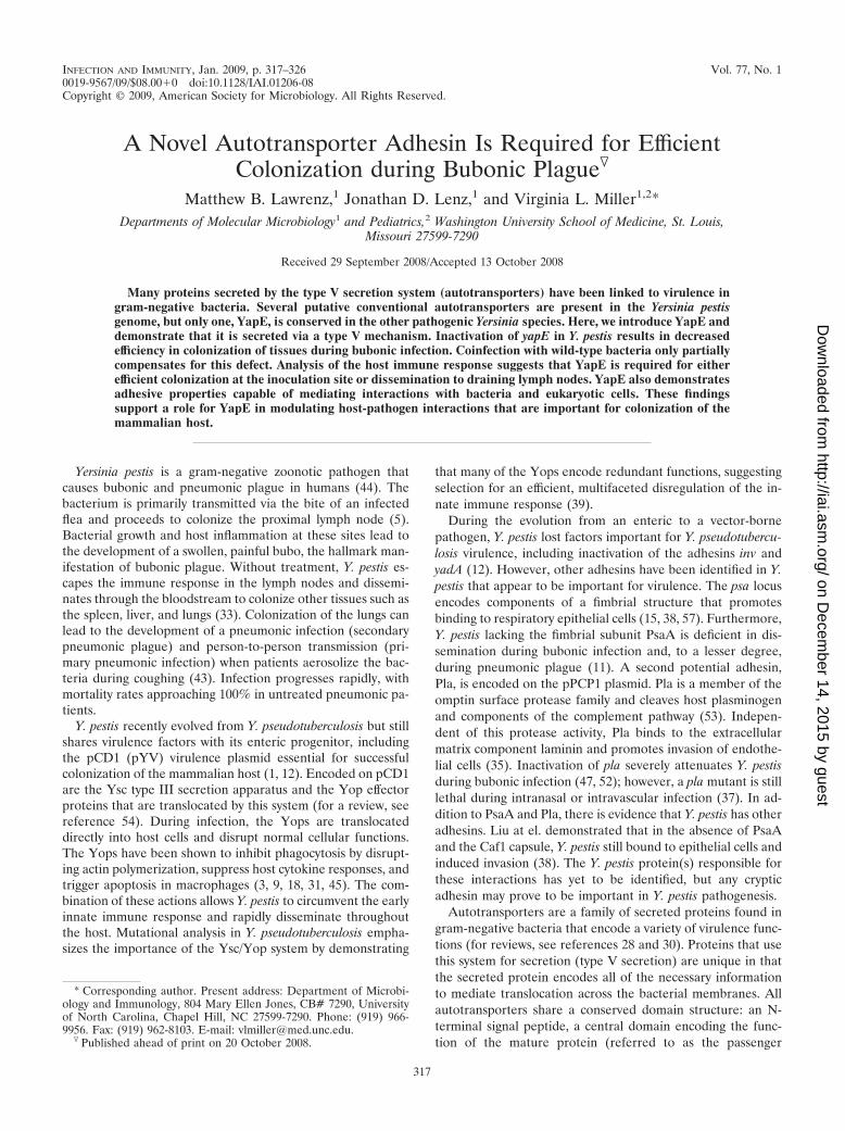
A Novel Autotransporter Adhesin Is Required for Efficient Colonization during Bubonic Plague
10
INFECTION AND IMMUNITY, Jan. 2009, p. 317–326 Vol. 77, No. 1 0019-9567/09/$08.000 doi:10.1128/IAI.01206-08 Copyright © 2009, American Society for Microbiology. All Rights Reserved. A Novel Autotransporter Adhesin Is Required for Efficient Colonization during Bubonic Plague Matthew B. Lawrenz, 1 Jonathan D. Lenz, 1 and Virginia L. Miller 1,2 * Departments of Molecular Microbiology 1 and Pediatrics, 2 Washington University School of Medicine, St. Louis, Missouri 27599-7290 Received 29 September 2008/Accepted 13 October 2008 Many proteins secreted by the type V secretion system (autotransporters) have been linked to virulence in gram-negative bacteria. Several putative conventional autotransporters are present in the Yersinia pestis genome, but only one, YapE, is conserved in the other pathogenic Yersinia species. Here, we introduce YapE and demonstrate that it is secreted via a type V mechanism. Inactivation of yapE in Y. pestis results in decreased efficiency in colonization of tissues during bubonic infection. Coinfection with wild-type bacteria only partially compensates for this defect. Analysis of the host immune response suggests that YapE is required for either efficient colonization at the inoculation site or dissemination to draining lymph nodes. YapE also demonstrates adhesive properties capable of mediating interactions with bacteria and eukaryotic cells. These findings support a role for YapE in modulating host-pathogen interactions that are important for colonization of the mammalian host. Yersinia pestis is a gram-negative zoonotic pathogen that causes bubonic and pneumonic plague in humans (44). The bacterium is primarily transmitted via the bite of an infected flea and proceeds to colonize the proximal lymph node (5). Bacterial growth and host inflammation at these sites lead to the development of a swollen, painful bubo, the hallmark man- ifestation of bubonic plague. Without treatment, Y. pestis es- capes the immune response in the lymph nodes and dissemi- nates through the bloodstream to colonize other tissues such as the spleen, liver, and lungs (33). Colonization of the lungs can lead to the development of a pneumonic infection (secondary pneumonic plague) and person-to-person transmission (pri- mary pneumonic infection) when patients aerosolize the bac- teria during coughing (43). Infection progresses rapidly, with mortality rates approaching 100% in untreated pneumonic pa- tients. Y. pestis recently evolved from Y. pseudotuberculosis but still shares virulence factors with its enteric progenitor, including the pCD1 (pYV) virulence plasmid essential for successful colonization of the mammalian host (1, 12). Encoded on pCD1 are the Ysc type III secretion apparatus and the Yop effector proteins that are translocated by this system (for a review, see reference 54). During infection, the Yops are translocated directly into host cells and disrupt normal cellular functions. The Yops have been shown to inhibit phagocytosis by disrupt- ing actin polymerization, suppress host cytokine responses, and trigger apoptosis in macrophages (3, 9, 18, 31, 45). The com- bination of these actions allows Y. pestis to circumvent the early innate immune response and rapidly disseminate throughout the host. Mutational analysis in Y. pseudotuberculosis empha- sizes the importance of the Ysc/Yop system by demonstrating that many of the Yops encode redundant functions, suggesting selection for an efficient, multifaceted disregulation of the in- nate immune response (39). During the evolution from an enteric to a vector-borne pathogen, Y. pestis lost factors important for Y. pseudotubercu- losis virulence, including inactivation of the adhesins inv and yadA (12). However, other adhesins have been identified in Y. pestis that appear to be important for virulence. The psa locus encodes components of a fimbrial structure that promotes binding to respiratory epithelial cells (15, 38, 57). Furthermore, Y. pestis lacking the fimbrial subunit PsaA is deficient in dis- semination during bubonic infection and, to a lesser degree, during pneumonic plague (11). A second potential adhesin, Pla, is encoded on the pPCP1 plasmid. Pla is a member of the omptin surface protease family and cleaves host plasminogen and components of the complement pathway (53). Indepen- dent of this protease activity, Pla binds to the extracellular matrix component laminin and promotes invasion of endothe- lial cells (35). Inactivation of pla severely attenuates Y. pestis during bubonic infection (47, 52); however, a pla mutant is still lethal during intranasal or intravascular infection (37). In ad- dition to PsaA and Pla, there is evidence that Y. pestis has other adhesins. Liu at el. demonstrated that in the absence of PsaA and the Caf1 capsule, Y. pestis still bound to epithelial cells and induced invasion (38). The Y. pestis protein(s) responsible for these interactions has yet to be identified, but any cryptic adhesin may prove to be important in Y. pestis pathogenesis. Autotransporters are a family of secreted proteins found in gram-negative bacteria that encode a variety of virulence func- tions (for reviews, see references 28 and 30). Proteins that use this system for secretion (type V secretion) are unique in that the secreted protein encodes all of the necessary information to mediate translocation across the bacterial membranes. All autotransporters share a conserved domain structure: an N- terminal signal peptide, a central domain encoding the func- tion of the mature protein (referred to as the passenger * Corresponding author. Present address: Department of Microbi- ology and Immunology, 804 Mary Ellen Jones, CB# 7290, University of North Carolina, Chapel Hill, NC 27599-7290. Phone: (919) 966- 9956. Fax: (919) 962-8103. E-mail: [email protected]. Published ahead of print on 20 October 2008. 317 on December 14, 2015 by guest http://iai.asm.org/ Downloaded from
-
Upload
louisville -
Category
Documents
-
view
1 -
download
0
Transcript of A Novel Autotransporter Adhesin Is Required for Efficient Colonization during Bubonic Plague
INFECTION AND IMMUNITY, Jan. 2009, p. 317–326 Vol. 77, No. 10019-9567/09/$08.00�0 doi:10.1128/IAI.01206-08Copyright © 2009, American Society for Microbiology. All Rights Reserved.
A Novel Autotransporter Adhesin Is Required for EfficientColonization during Bubonic Plague�
Matthew B. Lawrenz,1 Jonathan D. Lenz,1 and Virginia L. Miller1,2*Departments of Molecular Microbiology1 and Pediatrics,2 Washington University School of Medicine, St. Louis,
Missouri 27599-7290
Received 29 September 2008/Accepted 13 October 2008
Many proteins secreted by the type V secretion system (autotransporters) have been linked to virulence ingram-negative bacteria. Several putative conventional autotransporters are present in the Yersinia pestisgenome, but only one, YapE, is conserved in the other pathogenic Yersinia species. Here, we introduce YapE anddemonstrate that it is secreted via a type V mechanism. Inactivation of yapE in Y. pestis results in decreasedefficiency in colonization of tissues during bubonic infection. Coinfection with wild-type bacteria only partiallycompensates for this defect. Analysis of the host immune response suggests that YapE is required for eitherefficient colonization at the inoculation site or dissemination to draining lymph nodes. YapE also demonstratesadhesive properties capable of mediating interactions with bacteria and eukaryotic cells. These findingssupport a role for YapE in modulating host-pathogen interactions that are important for colonization of themammalian host.
Yersinia pestis is a gram-negative zoonotic pathogen thatcauses bubonic and pneumonic plague in humans (44). Thebacterium is primarily transmitted via the bite of an infectedflea and proceeds to colonize the proximal lymph node (5).Bacterial growth and host inflammation at these sites lead tothe development of a swollen, painful bubo, the hallmark man-ifestation of bubonic plague. Without treatment, Y. pestis es-capes the immune response in the lymph nodes and dissemi-nates through the bloodstream to colonize other tissues such asthe spleen, liver, and lungs (33). Colonization of the lungs canlead to the development of a pneumonic infection (secondarypneumonic plague) and person-to-person transmission (pri-mary pneumonic infection) when patients aerosolize the bac-teria during coughing (43). Infection progresses rapidly, withmortality rates approaching 100% in untreated pneumonic pa-tients.
Y. pestis recently evolved from Y. pseudotuberculosis but stillshares virulence factors with its enteric progenitor, includingthe pCD1 (pYV) virulence plasmid essential for successfulcolonization of the mammalian host (1, 12). Encoded on pCD1are the Ysc type III secretion apparatus and the Yop effectorproteins that are translocated by this system (for a review, seereference 54). During infection, the Yops are translocateddirectly into host cells and disrupt normal cellular functions.The Yops have been shown to inhibit phagocytosis by disrupt-ing actin polymerization, suppress host cytokine responses, andtrigger apoptosis in macrophages (3, 9, 18, 31, 45). The com-bination of these actions allows Y. pestis to circumvent the earlyinnate immune response and rapidly disseminate throughoutthe host. Mutational analysis in Y. pseudotuberculosis empha-sizes the importance of the Ysc/Yop system by demonstrating
that many of the Yops encode redundant functions, suggestingselection for an efficient, multifaceted disregulation of the in-nate immune response (39).
During the evolution from an enteric to a vector-bornepathogen, Y. pestis lost factors important for Y. pseudotubercu-losis virulence, including inactivation of the adhesins inv andyadA (12). However, other adhesins have been identified in Y.pestis that appear to be important for virulence. The psa locusencodes components of a fimbrial structure that promotesbinding to respiratory epithelial cells (15, 38, 57). Furthermore,Y. pestis lacking the fimbrial subunit PsaA is deficient in dis-semination during bubonic infection and, to a lesser degree,during pneumonic plague (11). A second potential adhesin,Pla, is encoded on the pPCP1 plasmid. Pla is a member of theomptin surface protease family and cleaves host plasminogenand components of the complement pathway (53). Indepen-dent of this protease activity, Pla binds to the extracellularmatrix component laminin and promotes invasion of endothe-lial cells (35). Inactivation of pla severely attenuates Y. pestisduring bubonic infection (47, 52); however, a pla mutant is stilllethal during intranasal or intravascular infection (37). In ad-dition to PsaA and Pla, there is evidence that Y. pestis has otheradhesins. Liu at el. demonstrated that in the absence of PsaAand the Caf1 capsule, Y. pestis still bound to epithelial cells andinduced invasion (38). The Y. pestis protein(s) responsible forthese interactions has yet to be identified, but any crypticadhesin may prove to be important in Y. pestis pathogenesis.
Autotransporters are a family of secreted proteins found ingram-negative bacteria that encode a variety of virulence func-tions (for reviews, see references 28 and 30). Proteins that usethis system for secretion (type V secretion) are unique in thatthe secreted protein encodes all of the necessary informationto mediate translocation across the bacterial membranes. Allautotransporters share a conserved domain structure: an N-terminal signal peptide, a central domain encoding the func-tion of the mature protein (referred to as the passenger
* Corresponding author. Present address: Department of Microbi-ology and Immunology, 804 Mary Ellen Jones, CB# 7290, Universityof North Carolina, Chapel Hill, NC 27599-7290. Phone: (919) 966-9956. Fax: (919) 962-8103. E-mail: [email protected].
� Published ahead of print on 20 October 2008.
317
on Decem
ber 14, 2015 by guesthttp://iai.asm
.org/D
ownloaded from
domain), and a C-terminal beta domain that mediates trans-location of the passenger domain across the outer membrane.
The importance of the trimeric autotransporter YadA in thepathogenesis of Y. pseudotuberculosis and Y. enterocolitica hasbeen evident through several studies. YadA has been impli-cated in autoaggregation, host cell binding, and serum resis-tance and is required for enteric infection (8, 21, 42); however,YadA does not appear to contribute to Y. pestis virulence, sinceit is a pseudogene in all of the Y. pestis biovars (51). Recently,at least 10 additional putative autotransporters have been iden-tified in the genomes of Y. pestis (58, 29; M. B. Lawrenz, J. D.Lenz, and V. L. Miller, unpublished data). These novel auto-transporters have been named Yaps for Yersinia autotrans-porter proteins. The Yaps demonstrate limited sequence sim-ilarity to other characterized proteins, but three appear to havesome adherence properties (23, 58). Escherichia coli expressingYapN and YapK agglutinate red blood cells, and YapC dem-onstrates autoagglutination and adherence to epithelial cells.Although many autotransporters from other bacteria havebeen implicated as important virulence factors, it has yet to bedetermined whether the Yaps contribute to Y. pestis virulence.
In this study, we introduce the YapE autotransporter of Y.pestis and demonstrate its importance in efficient colonizationof the mammalian host. In addition, we analyzed the cytokineresponse of infected animals to help decipher possible defectsin �yapE colonization. Finally, we demonstrate that YapE canmediate binding to eukaryotic cells in an apparent cell-type-specific manner.
MATERIALS AND METHODS
Bacterial strains, growth conditions, and animals. Wild-type (WT) Y. pestisstrain CO92 was provided by the U.S. Army, Fort Dietrich, MD (19). The �pla,�yapE, �lacZ, and �psaA �caf1 mutants have in-frame deletions in the indicatedgenes generated using the lambda red recombinase method essentially as de-scribed previously (11, 16, 37). The �yapE mutant was complemented with yapEplus 600 bp of upstream DNA at the attTn7 site using the mini-Tn7 systemdeveloped by Choi et al. (14). The kanamycin resistance cassette was removedfrom the mutant strains using the FRT recombinase provided by a version ofpLH29 which has been modified by replacing the chloramphenicol resistancegene with an ampicillin resistance gene. Ampicillin and kanamycin sensitivity wasconfirmed prior to passage in animals. A tetracycline-inducible yapE strain wasgenerated in the avirulent WT pCD1(�), WT pCD1(�)�pla, and WT pCD1(�)
�psaA �caf1 strains using the mini-Tn7-based system described by Lathem et al.(37). For adherence assays using E. coli, yapE was introduced between the KpnIand HindIII sites of the plasmid pLP-PROTet-6xHN (Clontech, Palo Alto, CA)downstream of the tetracycline promoter and transformed into E. coliDH5�PRO (Clontech) or the �ompT strain UT5600 (New England Biolabs,Ipswich, MA) containing the plasmid pVM1286 harboring the tetracycline re-pressor (Clontech). Y. pestis and E. coli were cultivated on brain heart infusion(BHI) or Luria-Bertani (LB) (Difco, Sparks, MD) plates or broth, respectively,with the addition of kanamycin (50 �g ml�1), ampicillin (50 �g ml�1), specti-nomycin (100 �g ml�1) or X-Gal (5-bromo-4-chloro-3-indolyl-�-D-galacto-pyranoside; 100 �g ml�1) when needed. No alterations in the in vitro growthcharacteristics of the Y. pestis mutant strains compared to the parental strainwere observed.
All animal experiments were approved by the Animal Studies Committee ofWashington University (protocol 20050189). Four- to six-week-old femaleC57BL/6 mice (The Jackson Laboratory, Bar Harbor, ME) were maintained ina barrier facility and allowed free access to sterilized food and water. Animalswere anesthetized prior to infection with a mixture of ketamine-HCl (2 mg permouse) and xylazine-HCl (0.32 mg per mouse) by intraperitoneal injection. Micewere inoculated with Y. pestis subcutaneously (�102 CFU) or intranasally (�105
CFU) as described previously (11, 36). At the indicated time points after inoc-ulation, mice were euthanized by an intraperitoneal injection of sodium pento-barbital at a dose of 75 mg/kg of body weight. Tissues were aseptically harvested
and macerated, and the bacterial load of each tissue was determined by platingserial dilutions.
RNA isolation and reverse transcription-PCR (RT-PCR). An overnight cul-ture of WT Y. pestis was subcultured into BHI to an optical density at 600 nm(OD600) of 0.2 and grown for �8 h at 26°C to an OD600 of 3.5. RNA was isolatedby using the RiboPure-Bacteria kit (Ambion, Austin, TX). Contaminating DNAwas removed from the samples with the DNA-free DNase kit (Ambion), andcDNA was generated from 2.0 �g of total DNase-treated RNA by using Super-script III reverse transcriptase (Invitrogen, Carlsbad, CA).
Surface proteolysis and Western blot analysis. To determine the surfacelocalization of YapE, E. coli cultures were grown overnight, diluted 1:50 in freshmedia, and grown for 2 h at 37°C. Y. pestis cultures were grown overnight, diluted1:25 in fresh media, and grown for 3 h at 26°C. YapE expression was induced bythe addition of anhydrous tetracycline (Sigma, St. Louis, MO) at 10 ng ml�1 (E.coli) or 100 ng ml�1 (Y. pestis), and cultures were grown for an additional 2 h.Samples from induced cultures equivalent to an OD600 of 1.0 were harvested,washed twice with 1� phosphate-buffered saline (PBS)–5 mM MgCl2 (pH 7.5)(resuspension buffer), and resuspended in 450 �l of the same buffer. Then, 50 �lof proteinase K (4 mg ml�1) was added to each sample, followed by incubationon ice for 30 min, and 50 �l of 50 mM phenylmethylsulfonyl fluoride was addedto inhibit further proteolysis. Samples were washed twice with resuspensionbuffer, and cell pellets were resuspended in Laemmli buffer containing 10%�-mercaptoethanol. Samples were boiled for 10 min, separated by sodiumdodecyl sulfate-polyacrylamide gel electrophoresis, and transferred to Immobilon-Pmembranes (Millipore, Billerica, MA) for Western blot analysis. Anti-YapEserum was generated in rabbits (Cocalico Biological, Reamstown, PA) againstthe passenger domain (amino acids 42 to 709) of YapE, preabsorbed againstE. coli lysates, and used at a concentration of 1:1,000. Anti-MalE rabbitserum (New England Biolabs) was used at a dilution of 1:10,000. Proteinsfrom culture supernatants were isolated from induced cultures as describedpreviously (55, 59).
Host cytokine response. To determine host cytokine response, superficialcervical lymph nodes from either naive or mice infected with WT or the �yapEmutant were harvested at 24, 36, and 48 h postinfection (n 5 for each timepoint), directly immersed into RNAlater (Ambion), and stored at 4°C. RNA wasisolated from the lymph nodes by using the RiboPure RNA isolation system(Ambion). Contaminating DNA was removed from the samples and cDNAgenerated as described above. Quantitative RT-PCR was performed to deter-mine cytokine transcript levels using SYBR Green (Qiagen, Valencia, CA) and900 nM gene-specific primers (26), and data were normalized to the GAPDH(glyceraldehyde-3-phosphate dehydrogenase) transcript. The relative foldchange was calculated by using the ��CT method (4) comparing infected to naivelymph nodes.
Competitive index. �lacZ (fully virulent) and �yapE strains were grown in BHIfor 15 h at 26°C. A 1:1 mixture of �lacZ and �yapE strains was made and dilutedto �103 CFU/ml in 1� PBS. Next, 100 �l of the diluted mixture was plated onBHI agar containing X-Gal to differentiate between the �lacZ strain (whitecolonies) and the �yapE strains (blue colonies), and the ratio between the twostrains was determined (CFU of �yapE/CFU of �lacZ input ratio). Mice werechallenged with 100 �l of the mixture subcutaneously, and tissues were harvestedas described above. Serial dilutions of macerated tissues were plated on BHI agarcontaining X-Gal to determine the recovered ratio (CFU of �yapE per tissue/CFU of �lacZ per tissue). A competitive index score was generated by dividingthe recovered ratio by the input ratio. A score of less than zero indicates that therecovered ratio differs from the input ratio and, specifically, fewer �yapE bacteriaare present in the tissues. Similar infections were performed with a mixedinfection of the �lacZ and yapE complemented �yapE strains.
Bacterial settling and adherence assays. To analyze settling of E. coli express-ing YapE in broth cultures, bacteria were grown overnight, diluted 1:50 in freshmedium, and grown for 2 h at 37°C on a roller drum. YapE expression wasinduced by the addition of 10 ng of anhydrous tetracycline ml�1, and cultureswere grown for an additional 2 h. Cultures were removed from the rotator andallowed to sit statically, and samples were harvested at a specific depth todetermine the changes in absorbance at 600 nm over time.
For adherence assays, wells of a 24-well plate were seeded with 4 � 105 or 2 �105 A549 human lung or HEp-2 human epithelial cells, respectively, and grownovernight at 37°C with 5% CO2 to obtain ca. 80% confluent monolayers. YapEexpression was induced in E. coli as described above with either 10 or 20 ng ofanhydrous tetracycline ml�1. For Y. pestis, YapE expression was induced asdescribed above with 100 ng of anhydrous tetracycline ml�1. Bacteria werediluted to an OD600 of 0.3 (E. coli) or 0.003 (Y. pestis) ml�1 in fresh tissue culturemedium, and 1 ml of diluted culture was added to each well. Plates werecentrifuged at 200 � g for 5 min to initiate bacterium-cell interactions and
318 LAWRENZ ET AL. INFECT. IMMUN.
on Decem
ber 14, 2015 by guesthttp://iai.asm
.org/D
ownloaded from
incubated at 37°C for either 2 h (E. coli) or 30 min (Y. pestis). To removenonadherent bacteria, the culture medium was removed, and wells were washedfour times with PBS. Adherent bacteria were recovered by treatment with 0.02%trypsin for 10 min and 1% Triton for 5 min (to lyse the eukaryotic cells). Serialdilutions of recovered bacteria were plated on agar. To calculate the percentageof adherent bacteria, the number of adherent bacteria was divided by thenumber of total bacteria as determined from an independent well. For mi-croscopy, cells were fixed and stained by using the Diff Quick Stain kit(IMEB, Inc., San Marcos, CA).
RESULTS
Characterization of the Yersinia autotransporter YapE. TheY. pestis CO92 genome encodes 10 conventional autotransport-ers (Lawrenz et al., unpublished data) but only yapE(YPO3984) is conserved in the enteric pathogens Y. pseudotu-berculosis IP32953 (YPTB3824, 97% amino acid identity) andY. enterocolitica 8081(YE4059, 65% amino acid identity).YapE orthologs are also found in the other Y. pestis biovars, Y.pestis subspecies Microtus and Pestoides F (100% amino acididentity for both), Y. mollaretii (62% amino acid identity), andY. bercovieri (60% amino acid identity). Outside the Yersiniagenus, YapE has little similarity to other proteins, with the
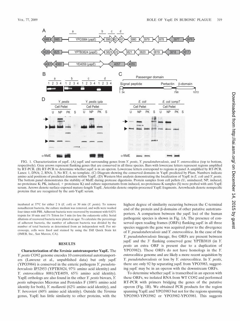
highest degree of similarity occurring between the C-terminalend of the protein and �-domains of other putative autotrans-porters. A comparison between the yapE loci of the humanpathogenic species is shown in Fig. 1A. The presence of con-served open reading frames (ORFs) flanking yapE in all threespecies suggests the gene was acquired prior to the divergenceof Y. pseudotuberculosis and Y. enterocolitica. In the case of theY. pseudotuberculosis lineage, five ORFs are present betweenyapE and the 3 flanking conserved gene YPTB3818 (in Y.pestis an extra ORF is present due to a duplication ofYPO3982). These ORFs do not have homologs in the Y.enterocolitica genome and are likely a more recent acquisition byY. pseudotuberculosis or loss by Y. enterocolitica. In Y. pestis,there are only 92 bp separating yapE from YPO3983, suggest-ing yapE may be in an operon with the downstream ORFs.
To determine whether yapE is transcribed in an operon withthese ORFs, we isolated RNA from WT CO92 and performedRT-PCR with primers bridging the genes of the putativeoperon (Fig. 1B). We obtained PCR products for the regionspanning YapE and YPO3983 but not for the regions spanningYPO3983-YPO3982 or YPO3982-YPO3981. This suggests
FIG. 1. Characterization of yapE. (A) yapE and surrounding genes from Y. pestis, Y. pseudotuberculosis, and Y. enterocolitica (top to bottom,respectively). Gray arrows represent flanking genes that are conserved in all three species. Bars with lowercase letters represent regions amplifiedby RT-PCR. (B) RT-PCR to determine whether yapE is in an operon. Lowercase letters correspond to regions in panel A amplified by RT-PCR.Lanes: 1, DNA; 2, RNA; 3, No RT; 4, no template. (C) Diagram showing the conserved domains in YapE predicted by Pfam. Numbers indicateamino acid positions of predicted domains within YapE. (D) Western blot analysis demonstrating the localization of YapE in E. coli and Y. pestis.The bottom panel demonstrates the stability of MalE during protease digestions. Protein samples from cell pellets (U, uninduced; NP, induced,no proteinase K; PK, induced, � proteinase K) and culture supernatants from induced, no-proteinase-K samples (S) were probed with anti-YapEserum. Arrows denote surface-exposed mature-length YapE. Asterisks denote omptin-processed YapE fragments. Arrowheads denote nonspecificproteins that are recognized by the anti-YapE serum.
VOL. 77, 2009 ROLE OF YapE IN BUBONIC PLAGUE 319
on Decem
ber 14, 2015 by guesthttp://iai.asm
.org/D
ownloaded from
that yapE is transcribed monocistronically with YPO3983 butnot the two downstream ORFs. However, it is possible thatYPO3982 and YPO3981 are also part of the operon, but ourRT-PCRs yielded negative results due to decreased stability ofthe transcript at the 3 end.
Sequence analysis of YapE reveals the presence of threeputative domains that are important for autotransporter trans-location. Computational analysis predicts an N-terminal signalpeptide, an autotransporter conserved pertactin domain (PfamID PF03212), and a C-terminal �-domain, which is requiredfor translocation of all autotransporters (Fig. 1C) (25). Thepresence of these domains, particularly the signal peptide andthe �-domain, strongly suggests that YapE is an autotrans-porter. To demonstrate experimentally that YapE is a secretedautotransporter, we determined the accessibility of the proteinto proteinase K. Proteinase K is unable to diffuse across theouter membrane of gram-negative bacteria and can only cleaveouter surface proteins in intact bacteria. Therefore, if YapE isa functional autotransporter, it should be accessible to prote-olysis by proteinase K. E. coli and Y. pestis containing inducibleyapE were incubated with or without proteinase K as describedin Materials and Methods. An inducible system was used be-cause we were unable to visualize YapE protein expressedfrom its native promoter by Western blotting (data not shown).Induction of YapE resulted in the appearance of two addi-tional proteins that are recognized by the anti-YapE antibody(Fig. 1D). The largest protein migrated near the predictedmolecular mass (�108 kDa) for YapE. We did not observe thepresence of reactive bands indicating trimerization of YapE.Within 30 min of incubation with proteinase K, both proteinswere completely digested in E. coli, and a truncated protein,likely representing the �-domain (protected from digestion)and a small region of the passenger domain, remained in the Y.pestis samples. As a control, we determined the stability of theperiplasmic protein MalE after proteinase K treatment. Thelevels of MalE did not change after protease treatment, indi-cating that the bacteria remained intact throughout the exper-iment.
The presence of two proteins upon induction of YapEexpression suggests that YapE is processed after translocation.Protein processing has been observed for several autotrans-porters and is a mechanism to secrete portions of the passen-ger domain into the environment. Many autotransporters re-quire outer surface proteases such as omptins for processing,but a select group have autocatalytic mechanisms for release(17). To determine whether the smaller protein in the inducedsamples resulted from cleavage of the larger protein, we har-vested culture supernatant from YapE induced cultures andlooked for the presence of YapE in these samples by Westernblotting. In both E. coli and Y. pestis cultures, we discovered an�37-kDa reactive protein (Fig. 1D). This protein was notpresent in uninduced cultures (data not shown), indicatingthat, at least in vitro, YapE can be processed after transloca-tion and secreted. Furthermore, deletion of the omptins ompTin E. coli or pla in Y. pestis resulted in the loss of both thesmaller protein in the cell pellet and the �37-kDa protein fromthe supernatant (Fig. 1D). Taken together with the surfaceproteolysis results, these data confirm that YapE is a func-tional, translocated autotransporter that can be processed bymembers of the omptin family of proteases.
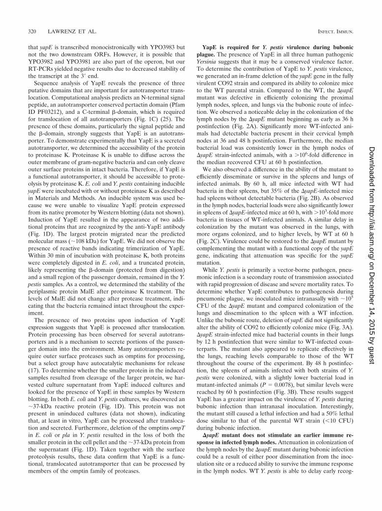
YapE is required for Y. pestis virulence during bubonicplague. The presence of YapE in all three human pathogenicYersinia suggests that it may be a conserved virulence factor.To determine the contribution of YapE to Y. pestis virulence,we generated an in-frame deletion of the yapE gene in the fullyvirulent CO92 strain and compared its ability to colonize miceto the WT parental strain. Compared to the WT, the �yapEmutant was defective in efficiently colonizing the proximallymph nodes, spleen, and lungs via the bubonic route of infec-tion. We observed a noticeable delay in the colonization of thelymph nodes by the �yapE mutant beginning as early as 36 hpostinfection (Fig. 2A). Significantly more WT-infected ani-mals had detectable bacteria present in their cervical lymphnodes at 36 and 48 h postinfection. Furthermore, the medianbacterial load was consistently lower in the lymph nodes of�yapE strain-infected animals, with a �106-fold difference inthe median recovered CFU at 60 h postinfection.
We also observed a difference in the ability of the mutant toefficiently disseminate or survive in the spleens and lungs ofinfected animals. By 60 h, all mice infected with WT hadbacteria in their spleens, but 35% of the �yapE-infected micehad spleens without detectable bacteria (Fig. 2B). As observedin the lymph nodes, bacterial loads were also significantly lowerin spleens of �yapE-infected mice at 60 h, with �103-fold morebacteria in tissues of WT-infected animals. A similar delay incolonization by the mutant was observed in the lungs, withmore organs colonized, and to higher levels, by WT at 60 h(Fig. 2C). Virulence could be restored to the �yapE mutant bycomplementing the mutant with a functional copy of the yapEgene, indicating that attenuation was specific for the yapEmutation.
While Y. pestis is primarily a vector-borne pathogen, pneu-monic infection is a secondary route of transmission associatedwith rapid progression of disease and severe mortality rates. Todetermine whether YapE contributes to pathogenesis duringpneumonic plague, we inoculated mice intranasally with �105
CFU of the �yapE mutant and compared colonization of thelungs and dissemination to the spleen with a WT infection.Unlike the bubonic route, deletion of yapE did not significantlyalter the ability of CO92 to efficiently colonize mice (Fig. 3A).�yapE strain-infected mice had bacterial counts in their lungsby 12 h postinfection that were similar to WT-infected coun-terparts. The mutant also appeared to replicate effectively inthe lungs, reaching levels comparable to those of the WTthroughout the course of the experiment. By 48 h postinfec-tion, the spleens of animals infected with both strains of Y.pestis were colonized, with a slightly lower bacterial load inmutant-infected animals (P 0.0078), but similar levels werereached by 60 h postinfection (Fig. 3B). These results suggestYapE has a greater impact on the virulence of Y. pestis duringbubonic infection than intranasal inoculation. Interestingly,the mutant still caused a lethal infection and had a 50% lethaldose similar to that of the parental WT strain (�10 CFU)during bubonic infection.
�yapE mutant does not stimulate an earlier immune re-sponse in infected lymph nodes. Attenuation in colonization ofthe lymph nodes by the �yapE mutant during bubonic infectioncould be a result of either poor dissemination from the inoc-ulation site or a reduced ability to survive the immune responsein the lymph nodes. WT Y. pestis is able to delay early recog-
320 LAWRENZ ET AL. INFECT. IMMUN.
on Decem
ber 14, 2015 by guesthttp://iai.asm
.org/D
ownloaded from
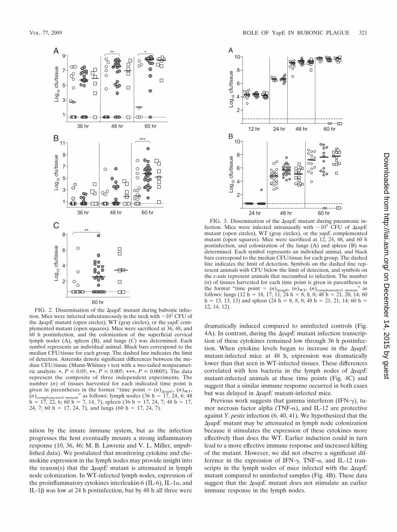
nition by the innate immune system, but as the infectionprogresses the host eventually mounts a strong inflammatoryresponse (10, 36, 46; M. B. Lawrenz and V. L. Miller, unpub-lished data). We postulated that monitoring cytokine and che-mokine expression in the lymph nodes may provide insight intothe reason(s) that the �yapE mutant is attenuated in lymphnode colonization. In WT-infected lymph nodes, expression ofthe proinflammatory cytokines interleukin-6 (IL-6), IL-1�, andIL-1� was low at 24 h postinfection, but by 48 h all three were
dramatically induced compared to uninfected controls (Fig.4A). In contrast, during the �yapE mutant infection transcrip-tion of these cytokines remained low through 36 h postinfec-tion. When cytokine levels began to increase in the �yapEmutant-infected mice at 48 h, expression was dramaticallylower than that seen in WT-infected tissues. These differencescorrelated with less bacteria in the lymph nodes of �yapEmutant-infected animals at these time points (Fig. 4C) andsuggest that a similar immune response occurred in both casesbut was delayed in �yapE mutant-infected mice.
Previous work suggests that gamma interferon (IFN- ), tu-mor necrosis factor alpha (TNF-�), and IL-12 are protectiveagainst Y. pestis infection (6, 40, 41). We hypothesized that the�yapE mutant may be attenuated in lymph node colonizationbecause it stimulates the expression of these cytokines moreeffectively than does the WT. Earlier induction could in turnlead to a more effective immune response and increased killingof the mutant. However, we did not observe a significant dif-ference in the expression of IFN- , TNF-�, and IL-12 tran-scripts in the lymph nodes of mice infected with the �yapEmutant compared to uninfected samples (Fig. 4B). These datasuggest that the �yapE mutant does not stimulate an earlierimmune response in the lymph nodes.
FIG. 3. Dissemination of the �yapE mutant during pneumonic in-fection. Mice were infected intranasally with �105 CFU of �yapEmutant (open circles), WT (gray circles), or the yapE complementedmutant (open squares). Mice were sacrificed at 12, 24, 48, and 60 hpostinfection, and colonization of the lungs (A) and spleen (B) wasdetermined. Each symbol represents an individual animal, and blackbars correspond to the median CFU/tissue for each group. The dashedline indicates the limit of detection. Symbols on the dashed line rep-resent animals with CFU below the limit of detection, and symbols onthe x-axis represent animals that succumbed to infection. The number(n) of tissues harvested for each time point is given in parentheses inthe format “time point (n)�yapE, (n)WT, (n)complemented mutant” asfollows: lungs (12 h 18, 17, 11; 24 h 8, 8, 0; 48 h 21, 20, 14; 60h 13, 13, 13) and spleen (24 h 8, 8, 0; 48 h 21, 21, 14; 60 h 12, 14, 12).
FIG. 2. Dissemination of the �yapE mutant during bubonic infec-tion. Mice were infected subcutaneously in the neck with �102 CFU ofthe �yapE mutant (open circles), WT (gray circles), or the yapE com-plemented mutant (open squares). Mice were sacrificed at 36, 48, and60 h postinfection, and the colonization of the superficial cervicallymph nodes (A), spleen (B), and lungs (C) was determined. Eachsymbol represents an individual animal. Black bars correspond to themedian CFU/tissue for each group. The dashed line indicates the limitof detection. Asterisks denote significant differences between the me-dian CFU/tissue (Mann-Whitney t test with a two-tailed nonparamet-ric analysis: *, P � 0.05; **, P � 0.005; ***, P � 0.0005). The datarepresent the composite of three independent experiments. Thenumber (n) of tissues harvested for each indicated time point isgiven in parentheses in the format “time point (n)�yapE, (n)WT,(n)complemented mutant” as follows: lymph nodes (36 h 17, 24, 6; 48h 17, 22, 6; 60 h 7, 14, 7), spleen (36 h 17, 24, 7; 48 h 17,24, 7; 60 h 17, 24, 7), and lungs (60 h 17, 24, 7).
VOL. 77, 2009 ROLE OF YapE IN BUBONIC PLAGUE 321
on Decem
ber 14, 2015 by guesthttp://iai.asm
.org/D
ownloaded from
Coinfection with WT does not fully complement attenuationof the �yapE mutant. Competitive infections are often used tobetter understand the impact of factors on virulence. In manycases, coinfection can enhance the attenuation of the mutant(7, 39); however, it has been speculated that WT bacteria couldcomplement the defects of some mutants in trans, especiallymutants in secreted virulence factors (13). To determine theeffect of the WT on the ability of the �yapE mutant to causedisease, we infected mice subcutaneously with a 1:1 mixture of
a �lacZ derivative of WT and the �yapE mutant and moni-tored the colonization of the lymph nodes, spleen, and lungs bydetermining the competitive index (Fig. 5). In contrast to thephenotype we observed in single infections, the �yapE mutantefficiently colonized the lymph nodes during coinfection tolevels comparable to WT, represented by a competitive indexscore of �1.0 at all time points. However, we recovered fewer�yapE bacteria in the spleens and lungs of coinfected mice at60 h postinfection (median competitive index scores of 0.006
FIG. 4. Cytokine response in the lymph nodes during bubonic infection of mice. Mice were infected subcutaneously in the neck with �102 CFUof WT (black circles) or the �yapE mutant (open circles), and RNA was harvested from the superficial cervical lymph nodes of five mice at 24,36, and 48 h postinfection. The expression of IL-6, IL-1�, and IL-1� (A) or IFN- , TNF-�, and IL-12 (B) was determined by quantitative RT-PCRand is represented as the fold difference over levels from uninfected lymph nodes. (C) Two mice were sacrificed at each time point to determinethe approximate CFU/tissue.
FIG. 5. Colonization by the �yapE mutant during mixed infection with WT CO92. Mice were infected subcutaneously in the neck with a 1:1mixture of the �yapE mutant and �lacZ WT (open symbols) or the yapE complemented mutant and �lacZ WT (gray symbols). Lymph nodes(circles), spleen (squares), and lungs (triangles) were harvested at 24, 36, 48, and 60 h postinfection. Serial dilutions were plated on agar containingX-Gal to differentiate between the test and �lacZ WT strains, and CFU of each were determined. The ratio of recovered mutant bacteria to �lacZWT was compared to the equivalent ratio of the inocula to generate a competitive index (C.I.) score. Median competitive index scores arerepresented by black bars. Scores less than one indicate attenuation in colonization by the test strain.
322 LAWRENZ ET AL. INFECT. IMMUN.
on Decem
ber 14, 2015 by guesthttp://iai.asm
.org/D
ownloaded from
and 0.012, respectively), indicating that the mutant is still at-tenuated in the colonization of these tissues. Complementationof the �yapE strain with yapE in the Tn7 att site restoredvirulence and resulted in no differences in colonization of tis-sues during coinfection. These results indicate that the WT isable to complement the virulence defect of the �yapE mutantin the lymph nodes but not during dissemination to and/orcolonization of the spleen or lungs of coinfected mice.
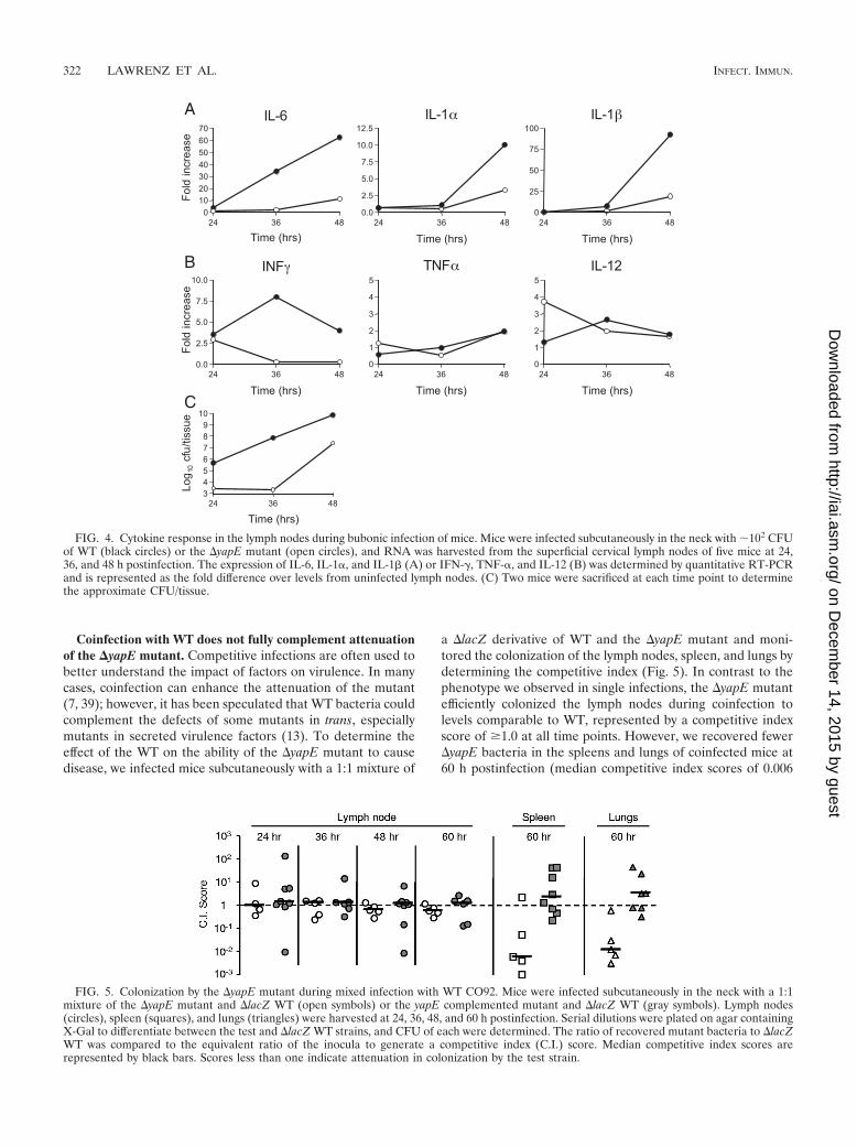
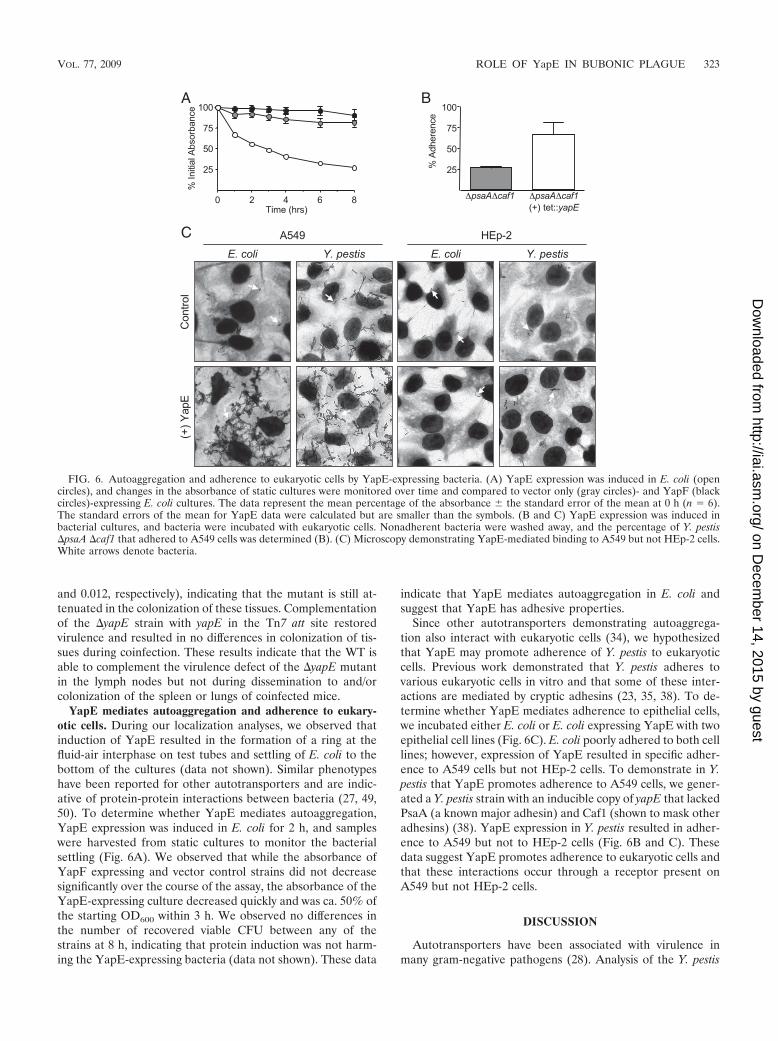
YapE mediates autoaggregation and adherence to eukary-otic cells. During our localization analyses, we observed thatinduction of YapE resulted in the formation of a ring at thefluid-air interphase on test tubes and settling of E. coli to thebottom of the cultures (data not shown). Similar phenotypeshave been reported for other autotransporters and are indic-ative of protein-protein interactions between bacteria (27, 49,50). To determine whether YapE mediates autoaggregation,YapE expression was induced in E. coli for 2 h, and sampleswere harvested from static cultures to monitor the bacterialsettling (Fig. 6A). We observed that while the absorbance ofYapF expressing and vector control strains did not decreasesignificantly over the course of the assay, the absorbance of theYapE-expressing culture decreased quickly and was ca. 50% ofthe starting OD600 within 3 h. We observed no differences inthe number of recovered viable CFU between any of thestrains at 8 h, indicating that protein induction was not harm-ing the YapE-expressing bacteria (data not shown). These data
indicate that YapE mediates autoaggregation in E. coli andsuggest that YapE has adhesive properties.
Since other autotransporters demonstrating autoaggrega-tion also interact with eukaryotic cells (34), we hypothesizedthat YapE may promote adherence of Y. pestis to eukaryoticcells. Previous work demonstrated that Y. pestis adheres tovarious eukaryotic cells in vitro and that some of these inter-actions are mediated by cryptic adhesins (23, 35, 38). To de-termine whether YapE mediates adherence to epithelial cells,we incubated either E. coli or E. coli expressing YapE with twoepithelial cell lines (Fig. 6C). E. coli poorly adhered to both celllines; however, expression of YapE resulted in specific adher-ence to A549 cells but not HEp-2 cells. To demonstrate in Y.pestis that YapE promotes adherence to A549 cells, we gener-ated a Y. pestis strain with an inducible copy of yapE that lackedPsaA (a known major adhesin) and Caf1 (shown to mask otheradhesins) (38). YapE expression in Y. pestis resulted in adher-ence to A549 but not to HEp-2 cells (Fig. 6B and C). Thesedata suggest YapE promotes adherence to eukaryotic cells andthat these interactions occur through a receptor present onA549 but not HEp-2 cells.
DISCUSSION
Autotransporters have been associated with virulence inmany gram-negative pathogens (28). Analysis of the Y. pestis
FIG. 6. Autoaggregation and adherence to eukaryotic cells by YapE-expressing bacteria. (A) YapE expression was induced in E. coli (opencircles), and changes in the absorbance of static cultures were monitored over time and compared to vector only (gray circles)- and YapF (blackcircles)-expressing E. coli cultures. The data represent the mean percentage of the absorbance � the standard error of the mean at 0 h (n 6).The standard errors of the mean for YapE data were calculated but are smaller than the symbols. (B and C) YapE expression was induced inbacterial cultures, and bacteria were incubated with eukaryotic cells. Nonadherent bacteria were washed away, and the percentage of Y. pestis�psaA �caf1 that adhered to A549 cells was determined (B). (C) Microscopy demonstrating YapE-mediated binding to A549 but not HEp-2 cells.White arrows denote bacteria.
VOL. 77, 2009 ROLE OF YapE IN BUBONIC PLAGUE 323
on Decem
ber 14, 2015 by guesthttp://iai.asm
.org/D
ownloaded from
CO92 genome revealed the presence of several putative con-ventional autotransporters. YapE is unique in this group sinceit is the only one that is also found in Y. pseudotuberculosis andY. enterocolitica and appears to have been acquired prior to thedivergence of these species. Its conservation in other Yersiniaspecies suggests that YapE provides a selective advantage forthe genus, perhaps during infection. Furthermore, because thevector-borne lifestyle of Y. pestis differs considerably from itsenteric counterparts, the functions of YapE may not be specificto an enteric lifestyle.
One advantage of the type V secretion mechanism is thatwhile all autotransporters use the same basic mechanism tomigrate across the bacterial cell envelope, the system imposeslittle restriction on the final localization of the passenger do-main (17). Some autotransporters remain associated with thebacterium, either as intact polypeptides or through noncova-lent interactions with the outer membrane after proteolyticprocessing. Others are released after translocation and are freeto diffuse away from the bacterium. In the case of YapE, wedemonstrated that the passenger domain primarily remainsassociated with the bacterium, but some polypeptides are pro-cessed, resulting in the release of a portion of the passengerdomain into the supernatant. YapE does not appear to auto-proteolytically cleave its passenger domain, as has been shownfor several members of the SPATE family of autotransporters(reviewed in reference 17), but is processed by outer mem-brane omptins (Pla in Y. pestis and OmpT in E. coli). Omptincleavage is not unique to YapE; autotransporters from otherbacteria are processed by omptins (32). It is unclear at this timehow omptin processing affects YapE. Pla processing may bepart of the normal maturation process of YapE, resulting in afunctional protein. Conversely, cleavage may allow Y. pestis torelease itself from YapE-mediated interactions with eukaryoticcells. A similar release mechanism has been proposed for theHap adhesin of Haemophilus influenzae, although Hap relieson autoproteolysis for cleavage (24).
Omptin processing may also determine the surface localiza-tion of YapE. In Shigella flexneri cleavage of the autotrans-porter IcsA by the omptin SopA is required for polar localiza-tion of the protein and actin-based intracellular motility (20,48). Location-specific cleavage could explain the presence ofboth full-length and processed versions of YapE in Y. pestis.Finally, it is possible that increased expression of YapE withour inducible system may artificially promote omptin cleavage.However, other Yersinia autotransporters expressed by thissystem are not sensitive to OmpT cleavage (unpublished data).Therefore, we believe that omptin processing of YapE is not aresult of overexpression.
We have demonstrated that a yapE-null mutant is signifi-cantly attenuated in colonizing tissues during bubonic infectioncompared to WT. Attenuation is first observed in the draininglymph nodes. Three scenarios may explain this delay in colo-nization: (i) the �yapE mutant is defective in colonization ofthe subcutaneous tissue, (ii) the mutant is defective for dis-semination to the proximal lymph node, or (iii) the mutantdisseminates to the tissue at the same rate but is less successfulat surviving in the tissue. These scenarios are not necessarilymutually exclusive, but at this time, our data support either ofthe first two as more likely to be occurring. First, in animalswith detectable bacteria in the lymph nodes, the mutant colo-
nizes the tissue to levels comparable to the WT. Second, if themutant was killed more efficiently in the lymph nodes, onemight expect that antigen presentation and activation of theinnate system would be stimulated earlier. In contrast to thishypothesis, we did not observe an earlier or more intenseimmune response in mutant-infected lymph nodes. In fact, theproinflammatory response was delayed in mutant-infectedmice, likely because the mutant arrives at the lymph node laterthan the WT. Furthermore, once initiated, the inflammatoryresponse to the �yapE mutant appears similar to the WTinfection.
In addition to delayed lymph node colonization, we observedthat the �yapE mutant was attenuated in dissemination to thespleen and lungs. Colonization of the lymph nodes is thoughtto be a prerequisite for infection of other tissues; therefore, thedefect in dissemination to deeper tissues could be a directconsequence of delayed colonization of the lymph nodes. How-ever, mixed infections demonstrated that the �yapE mutantwas still attenuated in efficient colonization of spleens andlungs even when the lymph nodes were colonized. Similarly, wedid not observe differences in colonization of the initial tissueduring intranasal inoculation but detected subtle defects incolonization of the spleens by the �yapE mutant. We alsoobserved that some mice succumbed by 60 h to infections withWT and yapE complemented strains but not with the �yapEmutant. Together, these results suggest that YapE may con-tribute independently both to colonization of the lymph nodesand to dissemination to deeper tissues, possibly through dis-tinct functions; it is not unprecedented for autotransporters tobe multifunctional (2, 28).
Dissemination defects similar to those we observed for the�yapE mutant have been previously reported for another Y.pestis virulence factor, the surface protein Pla (52, 56). Inacti-vation of pla does not alter colonization of the inoculation siteduring subcutaneous infection but results in delayed spleencolonization. It has been speculated that Pla activates hostplasminogen to degrade fibrin deposits, allowing Y. pestis todisseminate from the inoculation site. We have shown that Plaprocesses YapE and, due to similar defects in dissemination bythe two mutants, it is possible that lack of the processed formof YapE in the �pla mutant may contribute to the attenuateddissemination phenotype of the �pla mutant. However, webelieve that Pla likely also contributes to virulence indepen-dent of YapE. This is evidenced by the significantly higher 50%lethal dose of the �pla mutant (52) and its decreased ability tocolonize the lungs during intranasal infection (37). In the fu-ture, we will determine the effect of yapE inactivation on col-onization of the subcutaneous tissue during bubonic infection.Comparing the abilities of the �yapE, �pla, and �yapE �plamutants to colonize the inoculation site will help clarify therole of Pla processing of YapE in virulence.
The passenger domain of YapE shares little similarity toother described autotransporters, making it difficult to predictthe function(s) of the protein. We have demonstrated thatYapE mediates interactions between bacteria and with eukary-otic cells. Adhesins are common virulence factors in manybacteria, suggesting that an adherence defect in the YapEmutant could be responsible for attenuation in the mousemodel. Identification of a novel adhesin in Y. pestis is of par-ticular interest due to its lack of the major enteric Yersinia
324 LAWRENZ ET AL. INFECT. IMMUN.
on Decem
ber 14, 2015 by guesthttp://iai.asm
.org/D
ownloaded from
adhesins. In Y. pestis, the adhesins inv and yadA are bothpseudogenes, containing naturally occurring mutations. It hasbeen proposed for Y. enterocolitica and Y. pseudotuberculosisthat YadA and Inv binding promotes efficient translocation ofYop effectors into mammalian cells (22, 45). If similar intimateinteractions are required for Yop translocation by Y. pestis,then other adhesins must be responsible. In addition to YapE,pH6 antigen (PsaA), Pla, and YapC have been shown to con-tribute to adherence in Y. pestis (23, 35, 38). It is unclear at thistime whether these adhesins promote Yop translocation, butone could envision that decreased Yop translocation could beresponsible for the attenuation we observed for the YapEmutant. Furthermore, the specificity of YapE binding suggeststhat YapE mediates interactions with specific cells and/or tis-sues. Our future studies will seek to determine the YapEreceptor on eukaryotic cells and define the role of YapE inYop translocation in the context of the other known Y. pestisadhesins.
Mixed infections demonstrated that attenuation in the�yapE mutant can be at least partially complemented by WTbacteria, restoring colonization of the lymph nodes by themutant. To our knowledge, this is the first report of trans-comple-mentation in bacterial coinfections. More surprising was thatthe WT could complement a mutation in a bacterium-associ-ated adhesin. However, as stated earlier, autotransporters canencode multiple functions. For example, YadA has beenshown to mediate adherence, invasion, and resistance to killingby complement (21). We have not ruled out the possibility thatYapE may also be multifunctional. Furthermore, the secretedand bacterial associated proteins may contribute independentvirulence functions. It is possible that the secreted portion ofYapE is responsible for the delayed colonization of the lymphnodes, while systemic dissemination is more dependent on thecell surface form of YapE. This would explain why WT bacte-ria can trans-complement colonization of the lymph node butnot later stages of infection by the mutant. As we begin tobetter understand the interactions of YapE with the mamma-lian host, new functions for YapE may be revealed.
ACKNOWLEDGMENTS
This study was supported by funds from National Institutes ofHealth grant AI064313 (V.L.M.).
We thank Greer Kaufman for help with intranasal infections. The�psaA �caf1 mutant was generated by Jason Cathelyn. The �lacZmutant was generously provided by Joshua Fisher and William Gold-man.
REFERENCES
1. Achtman, M., K. Zurth, G. Morelli, G. Torrea, A. Guiyoule, and E. Carniel.1999. Yersinia pestis, the cause of plague, is a recently emerged clone ofYersinia pseudotuberculosis. Proc. Natl. Acad. Sci. USA 96:14043–14048.
2. Alamuri, P., and H. L. Mobley. 2008. A novel autotransporter of uropatho-genic Proteus mirabilis is both a cytotoxin and an agglutinin. Mol. Microbiol.68:997–1017.
3. Andersson, K., N. Carballeira, K. E. Magnusson, C. Persson, O. Stendahl,H. Wolf-Watz, and M. Fallman. 1996. YopH of Yersinia pseudotuberculosisinterrupts early phosphotyrosine signalling associated with phagocytosis.Mol. Microbiol. 20:1057–1069.
4. Applied Biosytems. 1997. User bulletin no. 2: ABI Prism 7700 sequencedetection system. Applied Biosystems, Foster City, CA.
5. Bacot, A., and C. Martin. 1914. Observations on the mechanism of trans-mission of plague by fleas. J. Hyg. 13:423–439.
6. Barton, E. S., D. W. White, J. S. Cathelyn, K. A. Brett-McClellan, M. Engle,M. S. Diamond, V. L. Miller, and H. W. Virgin IV. 2007. Herpesvirus latencyconfers symbiotic protection from bacterial infection. Nature 447:326–329.
7. Beuzon, C. R., and D. W. Holden. 2001. Use of mixed infections with Sal-monella strains to study virulence genes and their interactions in vivo. Mi-crobes Infect. 3:1345–1352.
8. Bliska, J. B., M. C. Copass, and S. Falkow. 1993. The Yersinia pseudotuber-culosis adhesin YadA mediates intimate bacterial attachment to and entryinto Hep-2 cells. Infect. Immun. 61:3914–3921.
9. Boland, A., and G. R. Cornelis. 1998. Role of YopP in suppression of tumornecrosis factor alpha release by macrophages during Yersinia infection. In-fect. Immun. 66:1878–1884.
10. Bubeck, S. S., A. M. Cantwell, and P. H. Dube. 2007. Delayed inflammatoryresponse to primary pneumonic plague occurs in both outbred and inbredmice. Infect. Immun. 75:697–705.
11. Cathelyn, J. S., S. D. Crosby, W. W. Lathem, W. E. Goldman, and V. L.Miller. 2006. RovA, a global regulator of Yersinia pestis, specifically requiredfor bubonic plague. Proc. Natl. Acad. Sci. USA 103:13514–13519.
12. Chain, P. S., E. Carniel, F. W. Larimer, J. Lamerdin, P. O. Stoutland, W. M.Regala, A. M. Georgescu, L. M. Vergez, M. L. Land, V. L. Motin, R. R.Brubaker, J. Fowler, J. Hinnebusch, M. Marceau, C. Medigue, M. Simonet,V. Chenal-Francisque, B. Souza, D. Dacheux, J. M. Elliott, A. Derbise, L. J.Hauser, and E. Garcia. 2004. Insights into the evolution of Yersinia pestisthrough whole-genome comparison with Yersinia pseudotuberculosis. Proc.Natl. Acad. Sci. USA 101:13826–13831.
13. Chiang, S. L., J. J. Mekalanos, and D. W. Holden. 1999. In vivo geneticanalysis of bacterial virulence. Annu. Rev. Microbiol. 53:129–154.
14. Choi, K. H., J. B. Gaynor, K. G. White, C. Lopez, C. M. Bosio, R. R.Karkhoff-Schweizer, and H. P. Schweizer. 2005. A Tn7-based broad-rangebacterial cloning and expression system. Nat. Methods 2:443–448.
15. Cowan, C., H. A. Jones, Y. H. Kaya, R. D. Perry, and S. C. Straley. 2000.Invasion of epithelial cells by Yersinia pestis: evidence for a Y. pestis-specificinvasin. Infect. Immun. 68:4523–4530.
16. Datsenko, K. A., and B. L. Wanner. 2000. One-step inactivation of chromo-somal genes in Escherichia coli K-12 using PCR products. Proc. Natl. Acad.Sci. USA 97:6640–6645.
17. Dautin, N., and H. D. Bernstein. 2007. Protein secretion in gram-negativebacteria via the autotransporter pathway. Annu. Rev. Microbiol. 61:89–112.
18. Denecker, G., W. Declercq, C. A. Geuijen, A. Boland, R. Benabdillah, M. vanGurp, M. P. Sory, P. Vandenabeele, and G. R. Cornelis. 2001. Yersiniaenterocolitica YopP-induced apoptosis of macrophages involves the apop-totic signaling cascade upstream of Bid. J. Biol. Chem. 276:19706–19714.
19. Doll, J. M., P. S. Zeitz, P. Ettestad, A. L. Bucholtz, T. Davis, and K. Gage.1994. Cat-transmitted fatal pneumonic plague in a person who traveled fromColorado to Arizona. Am. J. Trop. Med. Hyg. 51:109–114.
20. Egile, C., H. d’Hauteville, C. Parsot, and P. J. Sansonetti. 1997. SopA, theouter membrane protease responsible for polar localization of IcsA inShigella flexneri. Mol. Microbiol. 23:1063–1073.
21. El Tahir, Y., and M. Skurnik. 2001. YadA, the multifaceted Yersinia adhesin.Int. J. Med. Microbiol. 291:209–218.
22. Falgarone, G., H. S. Blanchard, F. Virecoulon, M. Simonet, and M. Breban.1999. Coordinate involvement of invasin and Yop proteins in a Yersiniapseudotuberculosis-specific class I-restricted cytotoxic T cell-mediated re-sponse. J. Immunol. 162:2875–2883.
23. Felek, S., M. B. Lawrenz, and E. S. Krukonis. 2008. The Yersinia pestisautotransporter YapC mediates host cell binding, autoaggregation and bio-film formation. Microbiology 154:1802–1812.
24. Fink, D. L., and J. W. St. Geme III. 2003. Chromosomal expression of theHaemophilus influenzae Hap autotransporter allows fine-tuned regulation ofadhesive potential via inhibition of intermolecular autoproteolysis. J. Bacte-riol. 185:1608–1615.
25. Finn, R. D., J. Mistry, B. Schuster-Bockler, S. Griffiths-Jones, V. Hollich, T.Lassmann, S. Moxon, M. Marshall, A. Khanna, R. Durbin, S. R. Eddy, E. L.Sonnhammer, and A. Bateman. 2006. Pfam: clans, web tools, and services.Nucleic Acids Res. 34:D247–D251.
26. Handley, S. A., and V. L. Miller. 2007. General and specific host responsesto bacterial infection in Peyer’s patches: a role for stromelysin-1 (matrixmetalloproteinase-3) during Salmonella enterica infection. Mol. Microbiol.64:94–110.
27. Hasman, H., T. Chakraborty, and P. Klemm. 1999. Antigen-43-mediatedautoaggregation of Escherichia coli is blocked by fimbriation. J. Bacteriol.181:4834–4841.
28. Henderson, I. R., and J. P. Nataro. 2001. Virulence functions of autotrans-porter proteins. Infect. Immun. 69:1231–1243.
29. Henderson, I. R., J. P. Nataro, R. Cappello, and C. Stein. 2000. Evolutionaryorigins of the autotransporter proteins. Direct submission. NCBI, NIH,Bethesda, MD.
30. Henderson, I. R., F. Navarro-Garcia, M. Desvaux, R. C. Fernandez, and D.Ala’Aldeen. 2004. Type V protein secretion pathway: the autotransporterstory. Microbiol. Mol. Biol. Rev. 68:692–744.
31. Iriarte, M., and G. R. Cornelis. 1998. YopT, a new Yersinia Yop effectorprotein, affects the cytoskeleton of host cells. Mol. Microbiol. 29:915–929.
32. Jacob-Dubuisson, F., R. Fernandez, and L. Coutte. 2004. Protein secretionthrough autotransporter and two-partner pathways. Biochim. Biophys. Acta1694:235–257.
VOL. 77, 2009 ROLE OF YapE IN BUBONIC PLAGUE 325
on Decem
ber 14, 2015 by guesthttp://iai.asm
.org/D
ownloaded from
33. Janssen, W. A., G. M. Fukui, and M. J. Surgalla. 1958. A study of the fateof Pasteurella pestis following intracardial injection into guinea pigs. J. Infect.Dis. 103:183–187.
34. Klemm, P., R. M. Vejborg, and O. Sherlock. 2006. Self-associating autotrans-porters, SAATs: functional and structural similarities. Int. J. Med. Microbiol.296:187–195.
35. Lahteenmaki, K., M. Kukkonen, and T. K. Korhonen. 2001. The Pla surfaceprotease/adhesin of Yersinia pestis mediates bacterial invasion into humanendothelial cells. FEBS Lett. 504:69–72.
36. Lathem, W. W., S. D. Crosby, V. L. Miller, and W. E. Goldman. 2005.Progression of primary pneumonic plague: a mouse model of infection,pathology, and bacterial transcriptional activity. Proc. Natl. Acad. Sci. USA102:17786–17791.
37. Lathem, W. W., P. A. Price, V. L. Miller, and W. E. Goldman. 2007. Aplasminogen-activating protease specifically controls the development ofprimary pneumonic plague. Science 315:509–513.
38. Liu, F., H. Chen, E. M. Galvan, M. A. Lasaro, and D. M. Schifferli. 2006.Effects of Psa and F1 on the adhesive and invasive interactions of Yersiniapestis with human respiratory tract epithelial cells. Infect. Immun. 74:5636–5644.
39. Logsdon, L. K., and J. Mecsas. 2003. Requirement of the Yersinia pseudo-tuberculosis effectors YopH and YopE in colonization and persistence inintestinal and lymph tissues. Infect. Immun. 71:4595–4607.
40. Lukaszewski, R. A., D. J. Kenny, R. Taylor, D. G. Rees, M. G. Hartley, andP. C. Oyston. 2005. Pathogenesis of Yersinia pestis infection in BALB/c mice:effects on host macrophages and neutrophils. Infect. Immun. 73:7142–7150.
41. Nakajima, R., and R. R. Brubaker. 1993. Association between virulence ofYersinia pestis and suppression of gamma interferon and tumor necrosisfactor alpha. Infect. Immun. 61:23–31.
42. Pepe, J. C., M. R. Wachtel, E. Wagar, and V. L. Miller. 1995. Pathogenesisof defined invasion mutants of Yersinia enterocolitica in a BALB/c mousemodel of infection. Infect. Immun. 63:4837–4848.
43. Perry, R. D., and J. D. Fetherston. 1997. Yersinia pestis: etiologic agent ofplague. Clin. Microbiol. Rev. 10:35–66.
44. Prentice, M. B., and L. Rahalison. 2007. Plague. Lancet 369:1196–1207.45. Rosqvist, R., A. Forsberg, M. Rimpilainen, T. Bergman, and H. Wolf-Watz.
1990. The cytotoxic protein YopE of Yersinia obstructs the primary hostdefense. Mol. Microbiol. 4:657–667.
46. Sebbane, F., D. Gardner, D. Long, B. B. Gowen, and B. J. Hinnebusch. 2005.Kinetics of disease progression and host response in a rat model of bubonicplague. Am. J. Pathol. 166:1427–1439.
47. Sebbane, F., C. O. Jarrett, D. Gardner, D. Long, and B. J. Hinnebusch. 2006.Role of the Yersinia pestis plasminogen activator in the incidence of distinctsepticemic and bubonic forms of flea-borne plague. Proc. Natl. Acad. Sci.USA 103:5526–5530.
48. Shere, K. D., S. Sallustio, A. Manessis, T. G. D’Aversa, and M. B. Goldberg.1997. Disruption of IcsP, the major Shigella protease that cleaves IcsA,accelerates actin-based motility. Mol. Microbiol. 25:451–462.
49. Sherlock, O., M. A. Schembri, A. Reisner, and P. Klemm. 2004. Novel rolesfor the AIDA adhesin from diarrheagenic Escherichia coli: cell aggregationand biofilm formation. J. Bacteriol. 186:8058–8065.
50. Sherlock, O., R. M. Vejborg, and P. Klemm. 2005. The TibA adhesin/invasinfrom enterotoxigenic Escherichia coli is self recognizing and induces bacterialaggregation and biofilm formation. Infect. Immun. 73:1954–1963.
51. Skurnik, M., and H. Wolf-Watz. 1989. Analysis of the yopA gene encodingthe Yop1 virulence determinants of Yersinia spp. Mol. Microbiol. 3:517–529.
52. Sodeinde, O. A., Y. V. Subrahmanyam, K. Stark, T. Quan, Y. Bao, and J. D.Goguen. 1992. A surface protease and the invasive character of plague.Science 258:1004–1007.
53. Suomalainen, M., J. Haiko, P. Ramu, L. Lobo, M. Kukkonen, B. Westerlund-Wikstrom, R. Virkola, K. Lahteenmaki, and T. K. Korhonen. 2007. Usingevery trick in the book: the Pla surface protease of Yersinia pestis. Adv. Exp.Med. Biol. 603:268–278.
54. Viboud, G. I., and J. B. Bliska. 2005. Yersinia outer proteins: role in modu-lation of host cell signaling responses and pathogenesis. Annu. Rev. Micro-biol. 59:69–89.
55. Walker, K. A., and V. L. Miller. 2004. Regulation of the Ysa type IIIsecretion system of Yersinia enterocolitica by YsaE/SycB and YsrS/YsrR. J.Bacteriol. 186:4056–4066.
56. Welkos, S. L., A. M. Friedlander, and K. J. Davis. 1997. Studies on the roleof plasminogen activator in systemic infection by virulent Yersinia pestisstrain CO92. Microb. Pathog. 23:211–223.
57. Yang, Y., J. J. Merriam, J. P. Mueller, and R. Isberg. 1996. The psa locus isresponsible for thermoinducible binding of Yersinia pseudotuberculosis tocultured cells. Infect. Immun. 64:2483–2489.
58. Yen, Y. T., A. Karkal, M. Bhattacharya, R. C. Fernandez, and C. Statho-poulos. 2007. Identification and characterization of autotransporter proteinsof Yersinia pestis KIM. Mol. Membr. Biol. 24:28–40.
59. Young, B. M., and G. M. Young. 2002. YplA is exported by the Ysc, Ysa, andflagellar type III secretion systems of Yersinia enterocolitica. J. Bacteriol.184:1324–1334.
Editor: V. J. DiRita
326 LAWRENZ ET AL. INFECT. IMMUN.
on Decem
ber 14, 2015 by guesthttp://iai.asm
.org/D
ownloaded from