Multiflora rose invasion amplifies prevalence of Lyme disease ...

JOURNAL OF CLINICAL MICROBIOLOGY,0095-1137/99/$04.0010
Apr. 1999, p. 890–896 Vol. 37, No. 4
Copyright © 1999, American Society for Microbiology. All Rights Reserved.
Isolation of Lyme Disease Borrelia from Puffins (Fratercula arctica)and Seabird Ticks (Ixodes uriae) on the Faeroe Islands
ÅSA GYLFE,1 BJORN OLSEN,1,2 DARIUS STRASEVICIUS,1 NURIA MARTI RAS,3 PAL WEIHE,4
LAILA NOPPA,1 YNGVE OSTBERG,1 GUY BARANTON,3 AND SVEN BERGSTROM1*
Departments of Microbiology1 and Infectious Diseases,2 Umeå University, S-901 87 Umeå, Sweden; Unite deBacteriologie Moleculaire et Medicale, Institut Pasteur, Paris, France3; and Department of Occupational
and Public Health, The Faeroese Hospital System, FR 100 Torshavn, The Faeroe Islands4
Received 7 August 1998/Returned for modification 22 October 1998/Accepted 21 December 1998
This is the first report on the isolation of Lyme disease Borrelia from seabirds on the Faeroe Islands and thecharacteristics of its enzootic cycle. The major components of the Borrelia cycle include the puffin (Fraterculaarctica) as the reservoir and Ixodes uriae as the vector. The importance of this cycle and its impact on the spreadof human Lyme borreliosis have not yet been established. Borrelia spirochetes isolated from 2 of 102 sampledpuffins were compared to the borreliae previously obtained from seabird ticks, I. uriae. The rrf-rrl intergenic spac-er and the rrs and the ospC genes were sequenced and a series of phylogenetic trees were constructed. Sequencedata and restriction fragment length polymorphism analysis grouped the strains together with Borrelia garinii.In a seroepidemiological survey performed with residents involved in puffin hunting on the Faeroe Islands, 3of 81 serum samples were found to be positive by two commonly used clinical tests: a flagellin-based enzyme-linked immunosorbent assay (ELISA) and Western blotting. These three positive serum samples also had highoptical density values in a whole-cell ELISA. The finding of seropositive Faeroe Islanders who are regularly ex-posed to I. uriae indicate that there may be a transfer of B. garinii by this tick species to humans.
Lyme disease, the most prevalent tick-borne zoonosis inNorth America and Europe, is a multisystemic disorder causedby the spirochete Borrelia burgdorferi sensu lato. In recentyears, the taxonomic and phylogenetic relationships betweendifferent B. burgdorferi sensu lato species have become moreextensive and complicated. Three genomic species of B. burg-dorferi sensu lato with human pathogenic relevance are recog-nized: B. burgdorferi sensu stricto, Borrelia garinii, and Borreliaafzelii (4, 11). The main reservoir hosts for these spirochetesare small mammals and birds (17, 18, 23, 24, 39), with ticks inthe Ixodes ricinus complex as the principal vectors (1). Recent-ly, an alternative enzootic cycle involving B. garinii, Ixodes uriaeticks, and seabirds was described (31). On a mammal-free is-land, B. garinii was isolated from I. uriae, and B. burgdorferisensu lato DNA was amplified from soft parts of a tick-infectedrazorbill (Alca torda) (31). In contrast to the terrestrial enzo-otic cycles of B. garinii, the circulation of Borrelia in the marineenvironment seems to be primarily restricted to colonial sea-birds and the tick species associated with those seabirds,I. uriae (31). We have previously suggested that B. garinii is thespecies that infects I. uriae ticks in both the northern and thesouthern hemispheres (32). The occurrence of identical Borre-lia flagellin (flaB) genes in the northern and southern hemi-spheres, as well as within the hemispheres, is compatible withthe theory of an exchange of B. garinii by seabirds (9, 32).
The objectives of this study were to investigate the impor-tance of the seabird-I. uriae-B. garinii cycle for the spread ofLyme disease Borrelia and the possibility that humans mayacquire Lyme disease from I. uriae.
MATERIALS AND METHODSCultivation and isolation of spirochetes. At the end of July, during the tradi-
tional puffin-hunting season at Nolsoy, Faeroe Islands, puffin (Fratercula arctica)
blood was collected from the hematoma that forms immediately after cervicaldislocation. Blood was inoculated into BSK-II medium (5) supplemented with10% rabbit serum (Sigma Chemical Co., St. Louis, Mo.) and 1.3% Bacto Gelatin(Difco Laboratories, Detroit, Mich.). To avoid contamination, 100 mg of phos-phomycin (Sigma) per ml and 50 mg of sulfamethoxazole (Sigma) per ml wereadded to the medium. The cultures were incubated at 32°C and were checkedweekly by phase-contrast microscopy. Contaminated cultures were purified byfiltration with 0.2-mm-pore-size, 115-ml sterile filter units (Nalgene Labware,Nalge Nunc International, Rochester, N.Y.). The origins of the Lyme diseaseBorrelia strains used in this study are listed in Table 1. For subsequent proteinand DNA analyses, the borreliae were grown to the exponential growth phase.
To investigate whether mammals living in the puffin colony were infected withBorrelia, blood samples from 28 domestic sheep (Ovis aries) were taken, andthree mice (Mus musculus) were caught and tested for Borrelia. Tissue samples(kidney, liver, spleen, heart, urinary bladder, and ear) were taken from the threemice, and blood was obtained from two of the mice. The samples from the sheepwere collected at the beginning of October, and the samples from the mice werecollected at the end of July. All blood samples and biopsy specimens wereinoculated into BSK-II medium that was supplemented with antibiotics, rabbitserum, and gelatin as described above.
Protein profiles and Western blot analysis. Protein analyses were performedwith whole-cell Borrelia preparations as described previously (20). Ten micro-grams of protein was separated by sodium dodecyl sulfate (SDS)-polyacrylamidegel electrophoresis (PAGE). Proteins were stained with Coomassie brilliant blueR-250 (Sigma) or were electroblotted onto a Hybond-N nylon filter (Amersham,Little Chalfont, Buckinghamshire, England). Molecular mass standards rangedfrom 14.3 to 220 kDa (Rainbow markers; Amersham). The Western blot analysiswas performed as described by Jonsson et al. (20). FlaB, OspA, OspB, and OspCwere characterized with murine monoclonal antibodies H9724 (6), H5332 (7),and 84C (13) and rabbit polyclonal OspC-specific antiserum (9), respectively.Detection was performed with alkaline phosphatase-conjugated secondary anti-bodies (DAKO, Glostrup, Denmark), and the blot was developed with 5-bromo-4-chloro-3-indolyl phosphate (Sigma) as the substrate.
Serum samples. The seroepidemiological study included 81 residents from theFaeroe Islands, where puffin hunting is a traditional annual event. All residentswho are involved in the puffin hunting on these islands, and who are therebyexposed to I. uriae, were asked to participate. The ages of the participants variedfrom 12 to 73 years (for men [n 5 62], mean 6 standard deviation age, 32.4 613.8 years; for women [n 5 19], mean 6 standard deviation age, 39.6 6 17.7years). Half of the blood samples were taken in the spring, before the puffin-hunting season, and the other half were taken after the season ended, in theautumn.
Previously sampled sera from 146 healthy blood donors from a tick-free areain northern Sweden served as a control group (10).
ELISA. A whole-cell enzyme-linked immunosorbent assay (ELISA) was pre-pared by coating each well of a microtiter plate overnight at room temperature
* Corresponding author. Mailing address: Department of Micro-biology, Umeå University, S-901 87 Umeå, Sweden. Phone: 46-90-7856726. Fax: 46-90-772630. E-mail: [email protected].
890

(RT) with 250 ng of a mixture of Far03, IP90, B31, and ACA1 whole-cell proteinpreparations in 50 ml of phosphate-buffered saline (PBS). Proteins, prepared asdescribed previously (20), were quantified in a spectrophotometer by the Bio-Rad protein assay (Bio-Rad Laboratories, Hercules, Calif.) according to themanufacturer’s instructions. Unbound proteins were removed by washing threetimes with PBS (pH 7.4) and once with distilled water before vacuum drying.Serum samples were diluted 1:200 in PBS containing 0.5% nonfat dairy milk and0.05% Tween 20 (Merck-Schuchardt, Hohenbrunn, Germany). In each of thetwo wells assigned for the individual sample, 100 ml of diluted serum was placedand the plates were incubated at RT for 2 h. After extensive washing with 0.1 MNaCl containing 0.1% Tween 20, the plates were incubated for 1 h with perox-idase-conjugated rabbit anti-human immunoglobulin G (DAKO) diluted in PBScontaining 0.5% nonfat dairy milk and 0.05% Tween 20. The wells were washedagain, and 100 ml of citrate buffer (pH 5.0) containing chromogen (500 mg ofo-phenylenediamine dihydrochloride per ml) and 0.02% hydrogen peroxide wasadded to the wells. After incubation in the dark for 60 min, 100 ml of 0.85 Msulfuric acid was added to stop the reaction. The optical density (OD) at 490 nmwas measured with a microtiter plate photometer (Microstation; Kebo Biomed,Spånga, Sweden). Positive and negative control sera were included on each plateto standardize the OD values between runs. As a positive control, a mixture ofserum from six patients with a diagnosis of Lyme disease was used. The sera weremixed to ensure a large enough volume to cover all the experiments. As anegative control, we used a serum sample from a healthy blood donor that hada low reactivity in the ELISA.
For comparison, the sera from the Faeroe Islanders were tested for antibodiesagainst B. burgdorferi sensu lato flagellin (FlaB) by the flagellin ELISA (Lymeborreliosis ELISA; DAKO) according to the manufacturer’s instructions.
Sera that were positive by the flagellin ELISA were further confirmed to bepositive by Western blotting (Lyme disease B. burgdorferi genogroup 2 Westernblot IgG; MRL Diagnostics, Cypress, Calif.). Both the flagellin ELISA and theWestern blot assay are clinically used for the diagnosis of Lyme disease.
Statistical analyses. The OD values obtained from the whole-cell ELISA ofthe Faeroe Islands test group and the Swedish control group were compared bythe Mann-Whitney U test. A cutoff level for the whole-cell ELISA was definedat the 95th percentile of the OD values for the control group. Sera with ODvalues above the cutoff level were considered positive. The proportion of sero-positive samples in the two groups was compared by the chi-square test. P valuesof 0.05 or less were considered to indicate statistical significance. All P valueswere based on two-sided tests of significance. SPSS (version 7.5) for Windows(SPSS Inc., Chicago, Ill.) was used for the statistical analyses.
Restriction fragment length polymorphism (RFLP) analysis. Total BorreliaDNA was prepared by standard methods. Briefly, the cultures were centrifugedand the pellet was washed with 50 mM Tris-HCl (pH 7.4). The cells were lysedon ice in 50 mM Tris-HCl (pH 7.4)–0.25% sucrose–50 mM EDTA–1.5 mg oflysozyme (Sigma) per ml–2% SDS. Following proteinase K (Boehringer Mann-heim, Mannheim, Germany) digestion at 55°C for 30 min, the DNA was purifiedby repeated phenol-chloroform extractions and a final extraction with chloro-form-isoamyl alcohol (24:1). RNase A (Sigma) was added to a final concentra-tion of 10 mg/ml, the mixture was incubated at RT for 1 h, and the RNase A wasremoved by a phenol-chloroform and a chloroform-isoamyl alcohol extraction.DNA was precipitated with 2.5 volumes of 99.7% ethanol and 1/10 volume of 3M sodium acetate (pH 5.2). The pellet was washed with 70% ice-cold ethanol,dried, and resuspended in a minimal volume of TE (10 mM Tris-HCl, 1 mMEDTA [pH 8.0]). The DNA was digested with the HpaI restriction enzyme(Boehringer Mannheim), and the fragments were separated on a 0.7% agarosegel in TBE (89 mM Tris-borate, 89 mM boric acid, 2 mM EDTA) and blottedonto a Hybond N nylon filter. The membranes were prehybridized in a buffercontaining 0.25 M Na2HPO4 (pH 7.2), 1% bovine serum albumin, 1 mM EDTA,and 7% SDS for 30 min and hybridized with a 32P-labeled rrf-directed probe (27),5S94F (59-GAGTAGGTTATTGCCAGGG-39), for 2 h at 47°C in the samebuffer. Following hybridization, the membranes were washed three times for 15min each time at 47°C in 53 SSC (13 SSC is 0.15 M NaCl plus 0.015 M sodium
citrate)–0.1% SDS, 23 SSC–0.1% SDS, and 13 SSC–0.1% SDS. After a finalrinse in 0.13 SSC at RT, the membranes were exposed on a phosphor screen(Molecular Dynamics Ltd., Kemsing, England).
The variable spacer region between the rrfA and the rrlB genes was amplifiedby PCR with the flanking primers 59-CTGCGAGTTCGCGGGAGA-39 and 59-TCCTAGGCATTCACCATA-39 as described previously (35). The PCR prod-ucts were digested with MseI (New England Biolabs, Beverly, Mass.) and thefragments were separated on a 16% acrylamide–0.8% bisacrylamide gel for 3 hat 100 V (35).
Ribosomal gene organization. The organization of the rrl and rrf genes wasexamined by a multiple-primer PCR assay (27). Eight different sized fragmentscovering the rrlA-rrfA-rrlB region were amplified. The forward primer (59-GGGAAGCCTTCCTCAAGA-39) in all PCR mixtures was complementary to a re-gion at the end of the rrl gene. The reverse primers were complementary todifferent parts of the rrf and rrl genes. Amplicon sizes varied from 166 to 3,090bp.
PCR amplification, cloning, and DNA sequencing. The amplification of ap-proximately 600 bp from the ospC gene was performed with the primer setOspC3 (59-AAGTGC(AG)ATATTAATGACTTTA-39) and OspC4 (59-TTTTTTGGACTTTCTGCCACA-39) as described previously (28). The rrf-rrl intergenicspacer was amplified as described above. The ospC gene and the rrf-rrl intergenicspacer were sequenced with the Cy5-AutoRead sequencing kit (Pharmacia Bio-tech, Uppsala, Sweden) as described elsewhere (28).
For subsequent sequencing of a 1,432-bp rrs gene fragment, the DNA wasamplified with the 16S-F (59-TTGATCCTGGCTTAGAACT-39) and 16S-R (59-CGGGTTAGAATAATAGCTT-39) oligonucleotides targeting each end of thegene (27). The amplicons were cloned into the pT7Blue T-vector (Novagen,Madison, Wis.) according to the manufacturer’s instructions. Competent Esch-erichia coli DH5a was transformed with the recombinant plasmids (3). Plasmidclones containing a fragment of the expected size were sequenced by multiple-primer-directed sequencing of the rrs gene. The dye terminator cycle sequencingready reaction kit (Perkin-Elmer, Foster City, Calif.) and an automatic se-quencer (ABI PRISM 377 DNA sequencer; Perkin-Elmer) were used.
Nucleotide sequences and the deduced amino acid sequences of OspC werealigned with sequences available in the databases with the Clustal V software(16) and with VSM software, version 2.0, written by Ruimy et al. (36).Evolutionary distances were computed with the molecular evolutionary ge-netics analysis (MEGA) software (22). Phylogenetic trees were constructed bythe neighbor-joining method (37) and the unweighted pair group method withmathematical averages with MEGA software. Confidence intervals were assessedby bootstrap analysis (1,000 replicates).
PCR conditions. DNA amplifications were carried out in a 50-ml solutioncontaining 10 mM Tris-HCl (pH 8.3), 50 mM KCl, 1.5 mM MgCl2, 200 mMdeoxynucleoside triphosphates, 1 U of Taq DNA polymerase (Boehringer Mann-heim), and 10 pmol of each primer. Chromosomal DNA or boiled Borreliacultures were used as templates. The cycling conditions were 94°C for 30 s, 55°Cfor 1 min, and 72°C for 1 min for a total of 35 cycles in an automated thermalincubator (Perkin-Elmer). For amplification of fragments larger than 1.5 kb, theextension time at 55°C was 2 min. The amplified DNA was visualized by agarosegel electrophoreses and subsequent ethidium bromide staining.
Nucleotide sequence accession numbers. All sequences were deposited in theEMBL database and were assigned the following accession numbers: for rrssequences, AJ009749 (Far01), AJ009750 (Far02), AJ009752 (Far03), AJ009753(Far04), and AJ009751 (Fis01); for ospC sequences, AF080264 (Far02) andAF080263 (Far03); and for rrf-rrl intergenic spacer sequences, AF080262 (Far02)and AF080261 (Far03).
RESULTS
Isolation and characterization of spirochetes from I. uriaeand puffins. Spirochetes were isolated from the blood of 2 of102 sampled puffins. Approximately two-thirds of the culturesbecame heavily contaminated and were discarded. The two iso-lates that were obtained were successfully purified by filtrationbefore further investigation. No ticks were found on the 28sheep or the three mice examined, and no spirochetes werefound in the cultures from blood or biopsy specimens fromthese mammals.
Whole-cell lysates of Borrelia were analyzed by SDS-PAGE,and the protein profiles are presented in Fig. 1. The expressionof FlaB, OspA, OspB, and OspC was investigated by Westernblot analysis with the monoclonal antibodies H9724, H5332,and 84C and OspC-specific antiserum, respectively (data notshown). Reactivities with monoclonal and polyclonal antibod-ies to the respective proteins are depicted with arrowheads inFig. 1. All isolates expressed FlaB, OspA, OspB, and OspC.
TABLE 1. Borrelia strains used in this study
Strain Origin(reference or source)
Far01............ I. uriae, Faeroe Islands (9)Far02............ I. uriae, Faeroe Islands (9)Far03............ Puffin blood, Faeroe Islands (this study)Far04............ Puffin blood, Faeroe Islands (this study)Fis01............. I. uriae, Iceland (9)B31 ............... ATCC 35210. B. burgdorferi sensu strictoESP1............. I. ricinus, Spain (R. C. Johnson). B. burgdorferi sensu strictoACAI ........... Human isolate, Sweden (2). B. afzeliiIp90 .............. Ixodes persulcatus, Chabarovsk (21). B. garinii20047............ I. ricinus, France (J. F. Anderson). B. gariniiNT29 ............ I. persulcatus, Japan (M. Fukunaga). B. garinii
VOL. 37, 1999 LYME DISEASE ON THE FAEROE ISLANDS 891

Genetic characterization of ribosomal genes. Strains Far01,Far02, Far03, Far04, and Fis01 had typical B. garinii RFLPpatterns when the HpaI-digested genomic DNA was hybrid-ized with an rrf-directed probe (25, 38) (Fig. 2A). Furthermore,separation of the MseI restriction fragments of the rrf-rrl inter-
genic spacer amplicons showed a pattern typical for EuropeanB. garinii isolates (35) (Fig. 2B).
A multiple-primer PCR assay revealed that the ribosomalgene organization of the isolates was identical to that describedpreviously for B. burgdorferi sensu lato, i.e., rrs, followed by atandem repeat of rrl and rrf (38).
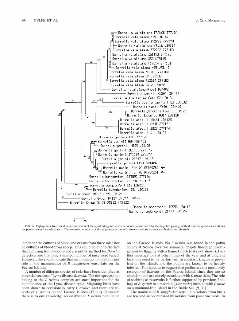
Nucleotide sequence analysis of rrs and ospC genes and rrf-rrl intergenic spacer. The four isolates from the Faeroe Islandsand the Icelandic isolate were subjected to rrs gene sequencing,and a phylogenetic tree was constructed (Fig. 3). The isolateshad a mutual identity within the 1,393-bp rrs sequence. Phylo-genetic analysis grouped the rrs sequence together with B. ga-rinii strains. Four unique substitutions in the rrs sequence werefound at positions 183, 185, 186, and 257 according to thenumbering for E. coli rrsB. The T at position 185 and the G atposition 186 are unique to B. burgdorferi sensu lato. The G atposition 183 and the G at position 257 are not found in anyother B. garinii rrs sequence in the nucleotide sequence data-bases. The ospC gene and the rrf-rrl intergenic spacer in thetwo strains isolated from ticks and birds (Far02 and Far03,respectively) were sequenced. Both the ospC and the rrf-rrlintergenic spacer sequences from the two strains were identi-cal. The phylogenetic trees obtained from the rrf-rrl intergenicspacer sequences (Fig. 4) and from the ospC sequences (datanot shown) were consistent with the tree obtained from the rrssequences. Isolates Far02 and Far03 appeared on the samephylogenetic branch as the B. garinii species.
Serologic investigation of the Faeroe Island human popula-tion in contact with I. uriae. Among the 81 serum samples fromI. uriae-exposed Faeroe Islanders, 3 reacted strongly in all thetests that were performed. These sera gave high OD values inthe whole-cell ELISA and fulfilled the criteria for a positivediagnosis by both the flagellin ELISA and Western blotting. Atotal of seven serum samples were positive by the flagellinELISA. Four of them reacted mainly with flagellin (FlaB) andwith two proteins at 60 and 75 kDa in the Western blot. TheOD values obtained by the whole-cell ELISA (Fig. 5) weresignificantly higher for the Faeroe Islands test group than forthe Swedish control group (Mann-Whitney U test; P , 0.001).The 95th percentile cutoff level for the control group (OD,0.331) resulted in 11 (13.6%) positive serum samples in the testgroup and six (4.1%) positive serum samples in the controlgroup, which was a significant difference (Pearson chi-squarevalue, 6.7; one degree of freedom; P 5 0.009). Two serumsamples in the test group were positive by the flagellin ELISA,Western blotting and the whole-cell ELISA. One additionalserum sample was positive by the two commercial tests and bythe whole-cell ELISA had an OD value (0.279) that was closeto the cutoff level. We did not find gender, age, or time ofsampling to be confounding factors.
DISCUSSION
Previously, we have shown that seabirds may be reservoirsfor Lyme disease Borrelia and may be important for the spreadof this spirochete (32). To further investigate this, genotypicand phenotypic analyses were performed with Borrelia strainsisolated from I. uriae and puffins from the Faeroe Islands. Webelieve that the Faeroe Islands are a part of the area of ende-micity for the Atlantic Lyme disease Borrelia. The major par-ticipants in this marine enzootic cycle are abundantly breedingseabirds, such as the puffins, I. uriae as the vector, and theB. garinii spirochete.
The puffin is a common seabird on the Faeroe Islands, withcolonies along the slopes often maintaining several thousandpairs. These dense colonies may facilitate the transmission of
FIG. 1. Borrelia whole-cell protein preparations separated on SDS–12.5%polyacrylamide gels and silver stained.
FIG. 2. RFLP analysis. (A) Hybridization of HpaI-digested total BorreliaDNA with an rrl-directed probe. (B) PCR-amplified rrf-rrl intergenic spacerfragments digested with MseI and separated by PAGE.
892 GYLFE ET AL. J. CLIN. MICROBIOL.

ectoparasites (12). The small number of isolates detected inthis study is probably not a reflection of a low proportion ofinfected birds, but it is more likely due to sampling difficultiessince this area has large numbers of infected I. uriae ticks. In aprevious study, we detected B. burgdorferi sensu lato DNA in32% of the ticks on Nolsoy, Faeroe Islands (32). This paradox,with a large number of infected vectors and a small number ofpositive cultures for specimens from the reservoir, may haveseveral explanations. First, contamination of the cultures was aserious problem because inoculation was performed underfield conditions. Second, the reservoir hosts, in this case, puf-fins, may be spirochetemic for shorter periods than mammals.In previous studies, chickens and canary finches were spiro-
chetemic only for a short period of time following experimentalinfection (8, 33, 34). Third, it is easier to isolate spirochetesfrom the skin than from blood (40), and since we did notsample the skin, we may have underestimated the number ofinfected puffins.
Seabirds are natural hosts for I. uriae, although the tick isknown to bite a variety of mammals including humans whengiven the opportunity (29). However, the rate of survival ofI. uriae was shown to decrease after feeding on hosts other thanseabirds (30). There are unlikely to be any other significantBorrelia reservoirs other than seabirds on the Faeroe Islands.Rodents and sheep are present only in small numbers through-out the puffin colonies (19). B. burgdorferi sensu lato was found
FIG. 3. Phylogenetic tree based on a comparison of the rrs sequences and constructed by the neighbor-joining method. Bootstrap values are shown (as percentages)for each branch. The accession numbers of the sequences are noted. An arrow indicates the sequence obtained in this study.
VOL. 37, 1999 LYME DISEASE ON THE FAEROE ISLANDS 893
in neither the cultures of blood and organs from three mice nor28 cultures of blood from sheep. This could be due to the factthat culturing from blood is not a sensitive method for Borreliadetection and that only a limited number of mice were tested.However, this could indicate that mammals do not play a majorrole in the maintenance of B. burgdorferi sensu lato on theFaeroe Islands.
A number of different species of ticks have been identified aspotential vectors of Lyme disease Borrelia. The tick species thatbelong to the I. ricinus complex are most important for themaintenance of the Lyme disease cycle. Migrating birds havebeen shown to occasionally carry I. ricinus, and there are re-ports of I. ricinus on the Faeroe Islands (15, 19). However,there is to our knowledge no established I. ricinus population
on the Faeroe Islands. No I. ricinus was found in the puffincolony at Nolsoy over two summers, despite thorough investi-gations by flagging with a flannel cloth (data not shown). Fur-ther investigations at other times of the year and at differentlocations need to be performed. In contrast, I. uriae is preva-lent on the islands, and the puffins are known to be heavilyinfested. This leads us to suggest that puffins are the most likelyreservoir of Borrelia on the Faeroe Islands since they are soabundant and are closely associated with I. uriae ticks. The roleof seabirds as reservoirs is further supported by previous find-ings of B. garinii in a razorbill (Alca torda) infested with I. uriaeon a mammal-free island in the Baltic Sea (9, 31).
The numbers of B. burgdorferi sensu lato isolates from birdsare few and are dominated by isolates from passerine birds. In
FIG. 4. Phylogenetic tree based on a comparison of the rrf-rrl intergenic spacer sequences constructed by the neighbor-joining method. Bootstrap values are shown(as percentages) for each branch. The accession numbers of the sequences are noted. Arrows indicate sequences obtained in this study.
894 GYLFE ET AL. J. CLIN. MICROBIOL.

this study we have successfully isolated spirochetes from puf-fins, and to our knowledge, this is the first report of isolatesfrom a wild, nonpasserine species. Determination of whichBorrelia genospecies can be found in seabirds is of particularinterest. The isolates that we obtained were all shown to beB. garinii by RFLP analysis and DNA sequencing of parts ofthe rrf-rrl intergenic spacer and the ospC and the rrs genes. TheRFLP analyses used in this study have previously been shownto readily discriminate between different B. burgdorferi sensulato isolates (25, 35, 38). Construction of phylogenetic treesgrouped all sequenced parts of the genome together with B. ga-rinii.
The approximately 580-bp ospC sequence fragments fromstrains Far02 and Far03 differed by less than six nucleotidesfrom Central European isolates M57 and N34 (26, 41), and thiscorresponds to a single amino acid substitution in the deducedsequence. This indicates a possible contact between the marineand the terrestrial Borrelia cycles by lateral transfer and re-combination of the ospC gene (26). Although the marine en-zootic cycle is separated from the terrestrial cycle, there areareas where interactions such as DNA transfer could theoret-ically occur (9).
The finding of B. burgdorferi sensu lato in the puffin popu-lation and in I. uriae on the Faeroe Islands prompted us toinitiate a serological survey of the human population. Peopleinvolved in the traditional puffin hunting were selected sincethey are regularly exposed to the Borrelia vector I. uriae. Usinga number of techniques, we determined that the Faeroe Is-landers had elevated titers of antibodies against B. burgdorferisensu lato compared to those for a Swedish control group;however, none of them had symptoms of Lyme disease. Todiscriminate between cross-reactive and genuine Borreliaantibody responses, the use of an ELISA in concert with West-ern blotting increased the specificity (14). These results indi-cate a possible transmission of B. garinii or, at least, B. ga-rinii antigens to humans. However, the low rate of Lymedisease on the Faeroe Islands may be explained by the resi-dents’ protective clothing, which prevents tick bites. The ecol-ogy of Lyme disease is complicated and involves different res-
ervoirs and tick vectors. The importance of birds, particularlyseabirds, as a vehicle for Borrelia-infected ticks and of B. burg-dorferi sensu lato on the Faeroe Islands is evident. Our findingsindicate a transfer of B. garinii to humans by the vector I. uriae.The epidemiological and clinical importance of B. garinii onthe Faeroe Islands and other regions with similar ecologicalcomponents needs further investigation.
ACKNOWLEDGMENTS
Jens-Kjeld Jensen and Jonas Bonnedahl are acknowledged for prac-tical help during the fieldwork on the Faeroe Islands. We are indebtedto Jana Jass, Pierre Martin, and Dominic McCafferty for critical read-ing of the manuscript. The monoclonal antibodies H9724 and H5332and the OspC-specific antiserum were kindly provided by Alan G.Barbour. Monoclonal antibody 84C was a kind gift from Denee D.Thomas. We also thank Goran Hallmans and Soren Holmgren forkindly providing the serum samples from the Swedish blood donorsand Katharina Ornstein for providing the serum samples from Lymedisease patients. Mats Sellin is acknowledged for valuable advice con-cerning ELISA.
This work was supported by the Swedish Medical Research Council(grant 07922), the Swedish Council for Forestry and Agricultural Re-search (grant 23.0161), and Frodskaparsetur Føroya. N.M.R. was sup-ported by the Commission of the European Communities (ERBFMBI-CT96-0684).
REFERENCES
1. Anderson, J. F. 1989. Epizootiology of Borrelia in Ixodes tick vectors andreservoir hosts. Rev. Infect. Dis. 11:1451–1459.
2. Åsbrink, E., A. Hovmark, and B. Hederstedt. 1984. The spirochetal etiologyof acrodermatitis chronica atrophicans Herxheimer. Acta Dermatol. Vene-reol. 64:506–512.
3. Ausubel, F. M., R. Brent, R. E. Kingston, D. D. Moore, J. G. Seidman, J. A.Smith, and K. Struhl. 1992. Current protocols in molecular biology. GreenePublishing Associates and Wiley-Interscience, New York, N.Y.
4. Baranton, G., D. Postic, I. Saint Girons, P. Boerlin, J. C. Piffaretti, M.Assous, and P. A. D. Grimont. 1992. Delineation of Borrelia burgdorferi sensustricto, Borrelia garinii sp. nov., and group VS461 associated with Lymeborreliosis. Int. J. Syst. Bacteriol. 42:378–383.
5. Barbour, A. G. 1984. Isolation and cultivation of Lyme disease spirochetes.Yale J. Biol. Med. 57:521–525.
6. Barbour, A. G., S. F. Hayes, R. A. Heiland, M. E. Schrumpf, and S. L.Tessier. 1986. A Borrelia-specific monoclonal antibody binds to a flagellarepitope. Infect. Immun. 52:549–554.
7. Barbour, A. G., S. L. Tessier, and W. J. Todd. 1983. Lyme disease spirochetesand ixodid tick spirochetes share a common surface antigenic determinantdefined by a monoclonal antibody. Infect. Immun. 41:795–804.
8. Bishop, K. L., M. I. Khan, and S. W. Nielsen. 1994. Experimental infectionof northern bobwhite quail with Borrelia burgdorferi. J. Wildl. Dis. 30:506–513.
9. Bunikis, J., B. Olsen, V. Fingerle, J. Bonnedahl, B. Wilske, and S. Berg-strom. 1996. Molecular polymorphism of the Lyme disease agent Borreliagarinii in northern Europe is influenced by a novel enzootic Borrelia focus inthe North Atlantic. J. Clin. Microbiol. 34:364–368.
10. Bunikis, J., B. Olsen, G. Westman, and S. Bergstrom. 1995. Variable serumimmunoglobulin responses against different Borrelia burgdorferi sensu latospecies in a population at risk for and patients with Lyme disease. J. Clin.Microbiol. 33:1473–1478.
11. Canica, M. M., F. Nato, L. du Merle, J. C. Maize, G. Baranton, and D.Postic. 1993. Monoclonal antibodies for identification of Borrelia afzelii sp.nov. associated with late cutaneous manifestations of Lyme borreliosis.Scand. J. Infect. Dis. 25:441–448.
12. Clifford, C. M. 1979. Tick-borne viruses of seabirds, p. 83–100. In E. Kurstak(ed.), Arctic and tropical arboviruses. Academic Press, Inc., New York, N.Y.
13. Comstock, L. E., E. Fikrig, R. J. Shoberg, R. A. Flavell, and D. D. Thomas.1993. A monoclonal antibody to OspA inhibits association of Borrelia burg-dorferi with human endothelial cells. Infect. Immun. 61:423–431.
14. Dressler, F., J. A. Whalen, B. N. Reinhardt, and A. C. Steere. 1993. Westernblotting in the serodiagnosis of Lyme disease. J. Infect. Dis. 167:392–400.
15. Hallas, T. E. 1990. Skovflåt fundet på Færøerne. Dansk. Vet. Tidsskr. 73:1089–1090.
16. Higgins, D. G., and P. M. Sharp. 1989. Fast and sensitive multiple sequencealignments on a microcomputer. Comput. Appl. Biosci. 5:151–153.
17. Humair, P. F., D. Postic, R. Wallich, and L. Gern. 1998. An avian reservoir(Turdus merula) of the Lyme borreliosis spirochetes. Zentralbl. Bakteriol.Parasitenkd. Infektionskr. Hyg. Abt. 1 Orig. 287:521–538.
18. Humair, P. F., N. Vittoz, M. Siegenthaler, A. Aeschlimann, and L. Gern.
FIG. 5. Box plot of whole-cell ELISA OD values. The tops and the bottomsof the boxes define the first and the third quartiles, respectively. The horizontallines inside the boxes indicate the median. The error bars define extreme values,and the squares define outliers. The reference line crossing the figure indicatesthe cutoff level for positive sera (OD, 0.331).
VOL. 37, 1999 LYME DISEASE ON THE FAEROE ISLANDS 895

1990. Mammalian and avian reservoirs for Borrelia burgdorferi in a Lymeborreliosis focus in Switzerland. Rev. Suisse Zool. 97:783.
19. Jensen, J.-K. Personal communication.20. Jonsson, M., L. Noppa, A. G. Barbour, and S. Bergstrom. 1992. Heteroge-
neity of outer membrane proteins in Borrelia burgdorferi: comparison of ospoperons of three isolates of different geographic origins. Infect. Immun. 60:1845–1853.
21. Kriuchechnikov, V. N., E. I. Korenberg, S. V. Shcherbakov, I. V. Kovalevskii,and M. L. Levin. 1988. Identification of Borrelia isolated in the USSR fromIxodes persulcatus Schulze ticks. Zh. Mikrobiol. Epidemiol. Immunobiol. 12:41–44.
22. Kumar, S., K. Tamura, and N. Masatoshi. 1993. MEGA: molecular evolu-tionary genetics analysis, version 1.01. The Pennsylvania State University,University Park.
23. Kurtenbach, K., M. Peacey, S. G. T. Rijpkema, A. N. Hoodless, P. A. Nuttall,and S. E. Randolph. 1998. Differential transmission of the genospecies ofBorrelia burgdorferi sensu lato by game birds and small rodents in England.Appl. Environ. Microbiol. 64:1169–1174.
24. Lane, R. S., J. Piesman, and W. Burgdorfer. 1991. Lyme borreliosis: relationof its causative agent to its vectors and hosts in North America and Europe.Annu. Rev. Entomol. 36:587–609.
25. Liveris, D., A. Gazumyan, and I. Schwartz. 1995. Molecular typing of Bor-relia burgdorferi sensu lato by PCR-restriction fragment length polymorphismanalysis. J. Clin. Microbiol. 33:589–595.
26. Livey, I., C. P. Gibbs, R. Schuster, and F. Dorner. 1995. Evidence for lateraltransfer and recombination in OspC variation in Lyme disease Borrelia. Mol.Microbiol. 18:257–269.
27. Marconi, R. T., D. Liveris, and I. Schwartz. 1995. Identification of novelinsertion elements, restriction fragment length polymorphism patterns, anddiscontinuous 23S rRNA in Lyme disease spirochetes: phylogenetic analysesof rRNA genes and their intergenic spacers in Borrelia japonica sp. nov. andgenomic group 21038 (Borrelia andersonii sp. nov.) isolates. J. Clin. Micro-biol. 33:2427–2434.
28. Marti Ras, N., D. Postic, M. Foretz, and G. Baranton. 1997. Borrelia burg-dorferi sensu stricto, a bacterial species “made in the U.S.A.”? Int. J. Syst.Bacteriol. 47:1112–1117.
29. Mehl, R., and T. Traavik. 1983. The tick Ixodes uriae (Acari: Ixodidae) inseabird colonies in Norway. Fauna Norv. Ser. B 30:94–107.
30. Nuttall, G. H. F. 1913. Observations on the biology of Ixodidae. Parasitology6:68–118.
31. Olsen, B., T. G. T. Jaenson, L. Noppa, J. Bunikis, and S. Bergstrom. 1993.A Lyme borreliosis cycle in seabirds and Ixodes uriae ticks. Nature 362:340–342.
32. Olsen, B., D. C. Duffy, T. G. T. Jaenson, Å. Gylfe, J. Bonnedahl, and S.Bergstrom. 1995. Transhemispheric exchange of Lyme disease spirochetes byseabirds. J. Clin. Microbiol. 33:3270–3274.
33. Olsen, B., Å. Gylfe, and S. Bergstrom. 1996. Canary finches (Serinus canaria)as an avian infection model for Lyme borreliosis. Microb. Pathog. 20:319–324.
34. Piesman, J., M. C. Dolan, M. E. Schriefer, and T. R. Burkot. 1996. Ability ofexperimentally infected chickens to infect ticks with the Lyme disease spi-rochete, Borrelia burgdorferi. Am. J. Trop. Med. Hyg. 54:294–298.
35. Postic, D., M. V. Assous, P. A. D. Grimont, and G. Baranton. 1994. Diversityof Borrelia burgdorferi sensu lato evidenced by restriction fragment lengthpolymorphism of rrf (5S)-rrl (23S) intergenic spacer amplicons. Int. J. Syst.Bacteriol. 44:743–752.
36. Ruimy, R., V. Breittmayer, P. Elbaze, B. Lafay, O. Boussemart, M. Gauthier,and R. Christen. 1994. Phylogenetic analysis and assessment of the generaVibrio, Photobacterium, Aeromonas, and Plesiomonas deduced from small-subunit rRNA sequences. Int. J. Syst. Bacteriol. 44:416–426.
37. Saitou, N., and M. Nei. 1987. The neighbor-joining method: a new methodfor reconstructing phylogenetic trees. Mol. Biol. Evol. 4:406–425.
38. Schwartz, J. J., A. Gazumyan, and I. Schwartz. 1992. rRNA gene organiza-tion in the Lyme disease spirochete, Borrelia burgdorferi. J. Bacteriol. 174:3757–3765.
39. Talleklint, L., and T. G. T. Jaenson. 1994. Transmission of Borrelia burgdor-feri s.l. from mammal reservoirs to the primary vector of Lyme borreliosis,Ixodes ricinus (Acari: Ixodidae), in Sweden. J. Med. Entomol. 31:880–886.
40. Wilske, B., and V. Preac-Mursic. 1993. Microbiological diagnosis of Lymeborreliosis, p. 270–272. In K. Weber and W. Burgdorfer (ed.), Aspects ofLyme borreliosis. Springer-Verlag, Berlin, Germany.
41. Wilske, B., S. Jauris-Heipke, R. Lobentanzer, I. Pradel, V. Preac-Mursic, D.Rossler, E. Soutschek, and R. C. Johnson. 1995. Phenotypic analysis of outersurface protein C (OspC) of Borrelia burgdorferi sensu lato by monoclonalantibodies: relationship to genospecies and OspA serotype. J. Clin. Micro-biol. 33:103–109.
896 GYLFE ET AL. J. CLIN. MICROBIOL.
Copyright © 2022 FDOKUMEN